Embed Size (px)
Citation preview
Abstract— Diagnosing the abnormal regions invarious medical images is one of the criticalissues because these images contain differenttypes of random noises and attenuationartifacts. This paper proposes an automaticmorphological segmentation and region growingmethod to change the representation of animage into something that is more meaningfuland easier to analyze. There are severalmethods that intend to perform segmentation,but it is difficult to adapt easily anddiagnose accurately. To resolve this problem,this paper aims to present an adaptableautomatic morphological segmentation andregion growing method that can be applied toany type of medical image which is exactlydiagnosed even with the small changes thatoccur in the image. This proposed method isbased in a model of morph function whichapplies the morphological operator to a grayscale image. Morphological segment techniqueis used to segment the image and selecting thespecific image objects, thinning the object todiagnose the region. After using amorphological operation to expose the basicelements within an image, it is often usefulto extract and analyze specific informationabout those image elements. The region growfunction performs region growing for a givenregion within an N-dimensional array byfinding all pixels within the array that areconnected to neighboring region pixels andthat fall within provided constraints. Thistechnique was applied to segment the medicalimage, diagnosing the interested object fromthe grown region in all medical images.
Keywords—Morphology, Segmentation, Regiongrowing, Diagnosing
I. INTRODUCTION
Medical image Processing is atechnique to enhance raw images receivedfrom cameras placed on medical diagnosingdevices or pictures taken from variousdevices which is processed by the specialsystem. Imaging technology in Medicinemade the doctors to see the interiorportions of the body for easy diagnosis.Segmentation of medical images allowsearly diagnosis of disease. Automatingthis process provides several benefitsincluding accuracy in output, easy way ofdiagnosing the complex images, minimizingthe difficult task. CT scanner, Ultrasoundand Magnetic Resonance Image processingtechniques developed for analyzing remotesensing data may be modified to analyzethe outputs of medical imaging systems toget best advantage to analyze symptoms ofthe patients with ease. This paper proposes an automaticmorphological segmentation and regiongrowing method to change therepresentation of an image into somethingthat is more meaningful and easier toanalyze any type of medical images.Segmentation involves separating an imageinto regions corresponding to objects. Thegoal of image segmentation is to clusterpixels into salient image regions, i.e.,regions corresponding to individualsurfaces, objects, or natural parts ofobjects. This approach was extended to afully automatic and complete segmentationmethod by using the pixels with the
Automatic Morphological Segmentationand Region Growing Method of Diagnosing
Medical Images K.Martin Sagayam#1,V.Ayyappan#2 ,Dr. S. Palani *3
1. # Lecturer, Anna University – MIT Campus, Chromepet,Chennai , Tamil Nadu. [email protected]
2. # Lecturer, Sudharsan Engineering College, sathyamangalam, Pudukottai Dist., Tamil Nadu. [email protected]
3.* Professor and P.G Dean, Department of ECE, Sudharsan Engineering College, Pudukottai Dist., TamilNadu.
1
smallest gradient length. The not yetsegmented image region is taken as a seedpoint. After segmentation, the infectedregion is identified by comparing thevalues of original image with the valuesof reference image. Then the diagnosedpart is enhanced for region growing.Region growing is a simple region-basedimage segmentation method. It is alsoclassified as a pixel-based imagesegmentation method, since it involves theselection of initial points. This approachto segmentation examines neighboringpixels of initial “seed points” anddetermines whether the pixel neighborsshould be added to the region.
II. AUTOMATIC MORPHOLOGICAL SEGMENTATIONIn this section, first the input medicalimage is automatically segmented by usingmorphological technique to diagnose theabnormal region.
A. Morphological Structuring Element to an Image Morphological operations apply astructuring element or morphological maskto an image. A structuring element that isapplied to an image must be 2 dimensional,having the same number of dimensions asthe array to which it is applied. Amorphological operation passes thestructuring element, of an empiricallydetermined size and shape, over an image.The operation compares the structuringelement to the underlying image andgenerates an output pixel based upon thefunction of the morphological operation.The size and shape of the structuringelement determines what is extracted ordeleted from an image. In general, smallerstructuring elements preserve finerdetails within an image than largerelements. Morphological operations can be applied toeither binary or grayscale images. Whenapplied to a binary image, the operationreturns pixels that are either black,having a logical value of 0, or white,having a logical value of 1. Each imagepixel and its neighboring pixels arecompared against the structuring element
to determine the pixel's value in theoutput image. With grayscale images, pixelvalues are determined by taking aneighborhood minimum or neighborhoodmaximum value. The structuring elementprovides the definition of the shape ofthe neighborhood.
B. Multilevel Morphological Segmentation Image segmentation is usually the firsttask of any image analysis process. Allsubsequent tasks, such as featureextraction and object recognition relyheavily on the quality of thesegmentation.Without a good segmentationalgorithm an object may never berecognizable. The purpose of imagesegmentation is to partition an image intomeaningful regions with respect to aparticular application .The segmentationis based on measurements taken from theimage and might be grey level, colour,texture, depth or motion. Usually imagesegmentation is an initial and vital stepin a series of processes aimed at overallimage understanding. Segmentations ofsimple gray-level images can provideuseful information about the surfaces inthe scene. Segmentation is an essentialingredient in a wide range of imageprocessing tasks and a building block ofmany visualization environments.
III. DIAGNOSING
After segmentation, the abnormal object isdiagnosed by analyzing the image shapesand selecting specific image objects.
A. Detecting Edges of Image ObjectsDetecting edges of image object by usingMorph Gradient function applies thegradient operation to a grayscale image.This operation highlights object edges bysubtracting an eroded version of theoriginal image from a dilated version.Repeatedly applying the gradient operatoror increasing the size of the structuringelement results in wider edges.
2
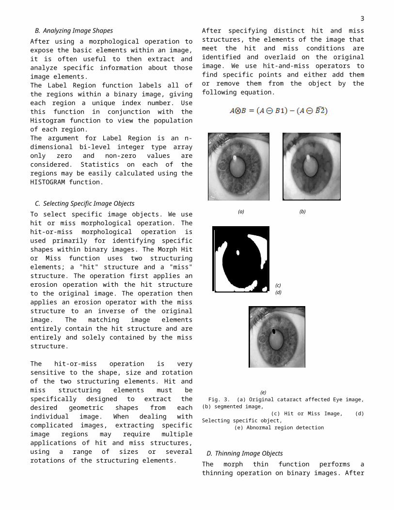
B. Analyzing Image Shapes After using a morphological operation toexpose the basic elements within an image,it is often useful to then extract andanalyze specific information about thoseimage elements. The Label Region function labels all ofthe regions within a binary image, givingeach region a unique index number. Usethis function in conjunction with theHistogram function to view the populationof each region.The argument for Label Region is an n-dimensional bi-level integer type arrayonly zero and non-zero values areconsidered. Statistics on each of theregions may be easily calculated using theHISTOGRAM function.
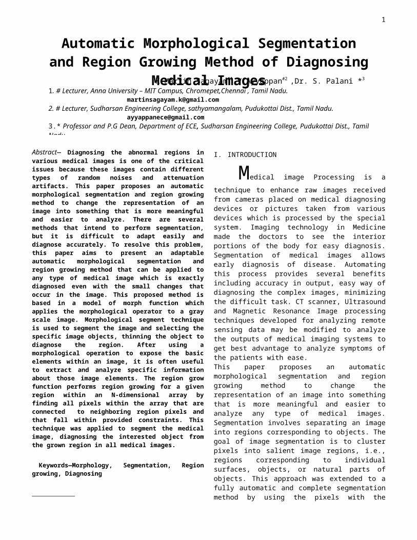
C. Selecting Specific Image Objects To select specific image objects. We usehit or miss morphological operation. Thehit-or-miss morphological operation isused primarily for identifying specificshapes within binary images. The Morph Hitor Miss function uses two structuringelements; a "hit" structure and a "miss"structure. The operation first applies anerosion operation with the hit structureto the original image. The operation thenapplies an erosion operator with the missstructure to an inverse of the originalimage. The matching image elementsentirely contain the hit structure and areentirely and solely contained by the missstructure.
The hit-or-miss operation is verysensitive to the shape, size and rotationof the two structuring elements. Hit andmiss structuring elements must bespecifically designed to extract thedesired geometric shapes from eachindividual image. When dealing withcomplicated images, extracting specificimage regions may require multipleapplications of hit and miss structures,using a range of sizes or severalrotations of the structuring elements.
After specifying distinct hit and missstructures, the elements of the image thatmeet the hit and miss conditions areidentified and overlaid on the originalimage. We use hit-and-miss operators tofind specific points and either add themor remove them from the object by thefollowing equation.
(a) (b)
(c) (d)
(e)Fig. 3. (a) Original cataract affected Eye image,
(b) segmented image, (c) Hit or Miss Image, (d)Selecting specific object,
(e) Abnormal region detection
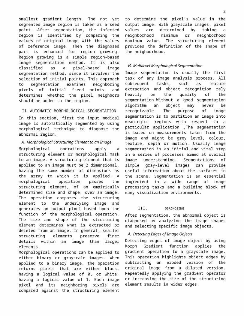
D. Thinning Image Objects The morph thin function performs athinning operation on binary images. After
3
designating "hit" and "miss" structures,the thinning operation applies the hit-or-miss operator to the original image andthen subtracts the result from theoriginal image. The thinning operation is typicallyapplied repeatedly, leaving only pixel-wide linear representations of the imageobjects. The thinning operation halts whenno more pixels can be removed from theimage. This occurs when the thinningoperation (applying the hit and missstructures and subtracting the result)produces no change in the input image. Atthis point, the thinned image is identicalto the input image. When repeatedly applying the thinningoperation, successive iteration uses hitand miss structures that have had theindividual elements of the structuresrotated one position clockwise. Forexample, the following 3-by-3 arrays showthe initial structure (left) and thestructure after rotating the elements oneposition clockwise around the centralvalue (right).
h0 = [[0, 0, 0], h1 = [[0,0, 0], [0, 1, 0],[1, 1, 0], [1, 1, 1]][1, 1, 0]]
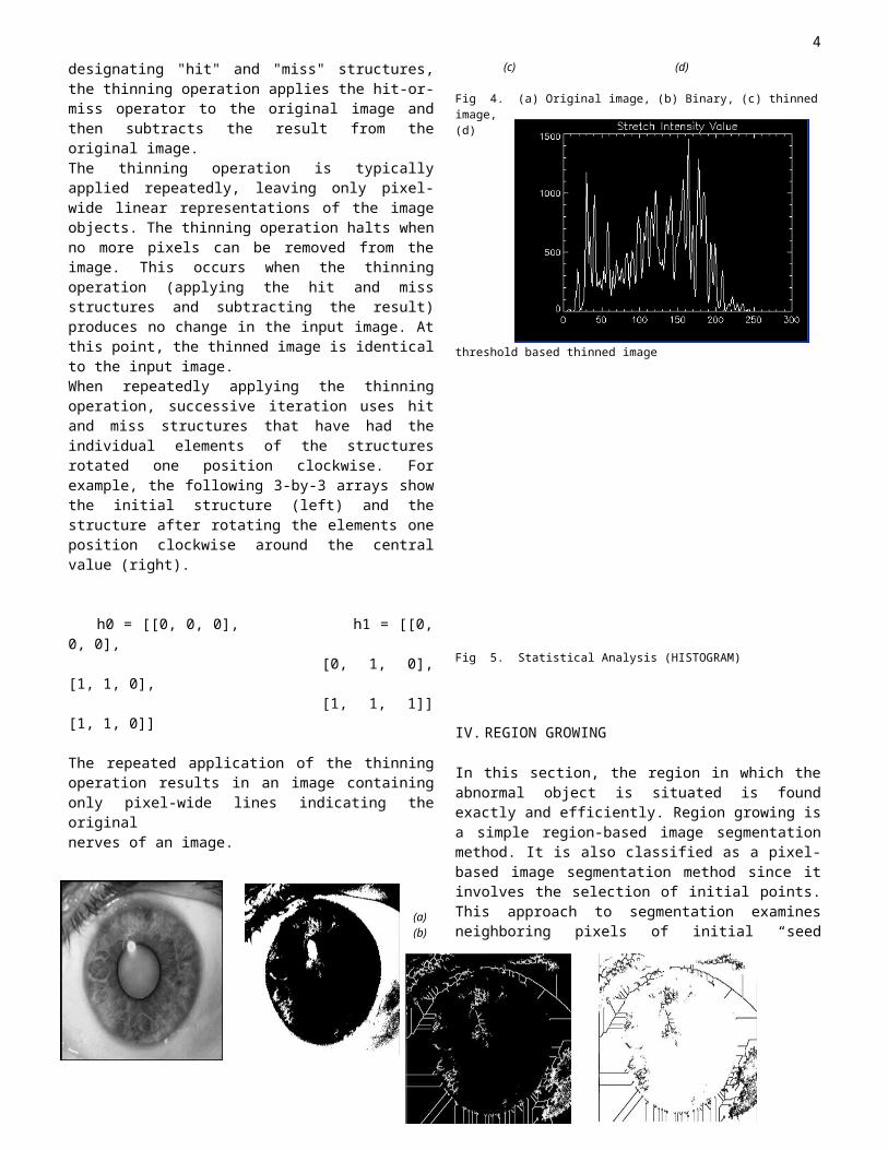
The repeated application of the thinningoperation results in an image containingonly pixel-wide lines indicating theoriginal nerves of an image.
(a) (b)
(c) (d)
Fig 4. (a) Original image, (b) Binary, (c) thinnedimage,(d)
threshold based thinned image
Fig 5. Statistical Analysis (HISTOGRAM)
IV. REGION GROWING
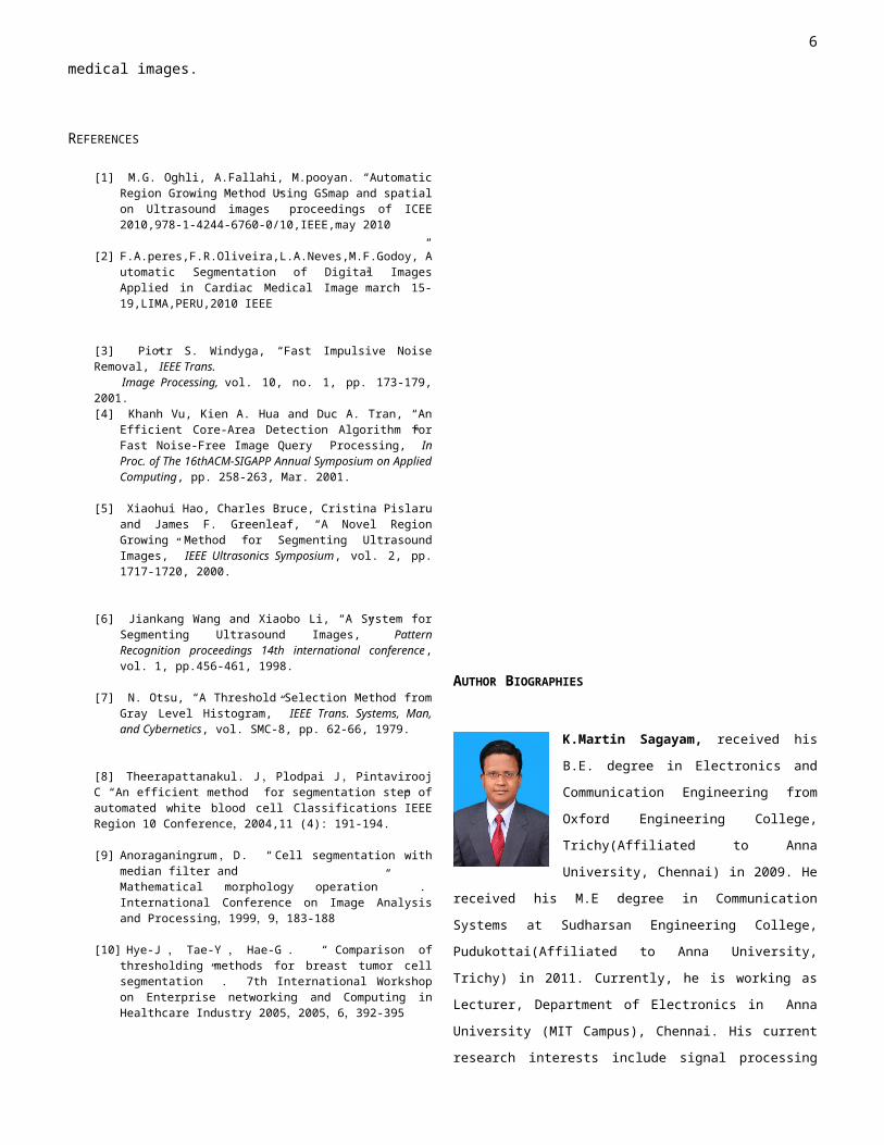
In this section, the region in which theabnormal object is situated is foundexactly and efficiently. Region growing isa simple region-based image segmentationmethod. It is also classified as a pixel-based image segmentation method since itinvolves the selection of initial points.This approach to segmentation examinesneighboring pixels of initial “seed
4
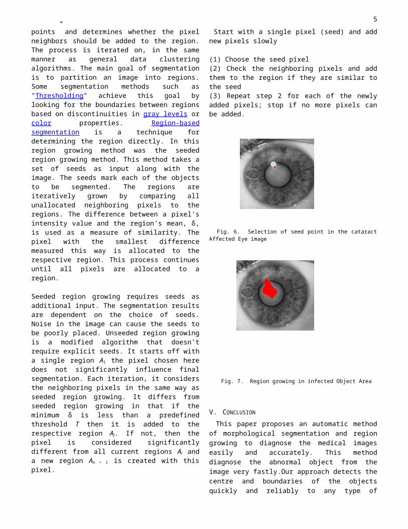
points” and determines whether the pixelneighbors should be added to the region.The process is iterated on, in the samemanner as general data clusteringalgorithms. The main goal of segmentationis to partition an image into regions.Some segmentation methods such as"Thresholding" achieve this goal bylooking for the boundaries between regionsbased on discontinuities in gray levels orcolor properties. Region-basedsegmentation is a technique fordetermining the region directly. In thisregion growing method was the seededregion growing method. This method takes aset of seeds as input along with theimage. The seeds mark each of the objectsto be segmented. The regions areiteratively grown by comparing allunallocated neighboring pixels to theregions. The difference between a pixel'sintensity value and the region's mean, δ,is used as a measure of similarity. Thepixel with the smallest differencemeasured this way is allocated to therespective region. This process continuesuntil all pixels are allocated to aregion.
Seeded region growing requires seeds asadditional input. The segmentation resultsare dependent on the choice of seeds.Noise in the image can cause the seeds tobe poorly placed. Unseeded region growingis a modified algorithm that doesn'trequire explicit seeds. It starts off witha single region A1 the pixel chosen heredoes not significantly influence finalsegmentation. Each iteration, it considersthe neighboring pixels in the same way asseeded region growing. It differs fromseeded region growing in that if theminimum δ is less than a predefinedthreshold T then it is added to therespective region Aj. If not, then thepixel is considered significantlydifferent from all current regions Ai anda new region An + 1 is created with thispixel.
Start with a single pixel (seed) and addnew pixels slowly
(1) Choose the seed pixel(2) Check the neighboring pixels and addthem to the region if they are similar tothe seed(3) Repeat step 2 for each of the newlyadded pixels; stop if no more pixels canbe added.
Fig. 6. Selection of seed point in the cataractAffected Eye image
Fig. 7. Region growing in infected Object Area
V. CONCLUSIONThis paper proposes an automatic method
of morphological segmentation and regiongrowing to diagnose the medical imageseasily and accurately. This methoddiagnose the abnormal object from theimage very fastly.Our approach detects thecentre and boundaries of the objectsquickly and reliably to any type of
5
medical images.
REFERENCES
[1] M.G. Oghli, A.Fallahi, M.pooyan. “AutomaticRegion Growing Method Using GSmap and spatialon Ultrasound images” proceedings of ICEE2010,978-1-4244-6760-0/10,IEEE,may 2010
[2] F.A.peres,F.R.Oliveira,L.A.Neves,M.F.Godoy,”Automatic Segmentation of Digital ImagesApplied in Cardiac Medical Image”march 15-19,LIMA,PERU,2010 IEEE
[3] Piotr S. Windyga, “Fast Impulsive NoiseRemoval,” IEEE Trans. Image Processing, vol. 10, no. 1, pp. 173-179,2001.[4] Khanh Vu, Kien A. Hua and Duc A. Tran, “An
Efficient Core-Area Detection Algorithm forFast Noise-Free Image Query Processing,” InProc. of The 16thACM-SIGAPP Annual Symposium on AppliedComputing, pp. 258-263, Mar. 2001.
[5] Xiaohui Hao, Charles Bruce, Cristina Pislaruand James F. Greenleaf, “A Novel RegionGrowing Method for Segmenting UltrasoundImages,” IEEE Ultrasonics Symposium, vol. 2, pp.1717-1720, 2000.
[6] Jiankang Wang and Xiaobo Li, “A System forSegmenting Ultrasound Images,” PatternRecognition proceedings 14th international conference,vol. 1, pp.456-461, 1998.
[7] N. Otsu, “A Threshold Selection Method fromGray Level Histogram,” IEEE Trans. Systems, Man,and Cybernetics, vol. SMC-8, pp. 62-66, 1979.
[8] Theerapattanakul. J,Plodpai J,PintaviroojC “An efficient method for segmentation step ofautomated white blood cell Classifications”IEEERegion 10 Conference,2004,11 (4): 191-194.
[9] Anoraganingrum, D. “ Cell segmentation withmedian filter andMathematical morphology operation” .International Conference on Image Analysisand Processing,1999,9,183-188
[10] Hye-J , Tae-Y , Hae-G . “ Comparison ofthresholding methods for breast tumor cellsegmentation” . 7th International Workshopon Enterprise networking and Computing inHealthcare Industry 2005,2005,6,392-395
AUTHOR BIOGRAPHIES
K.Martin Sagayam, received his
B.E. degree in Electronics and
Communication Engineering from
Oxford Engineering College,
Trichy(Affiliated to Anna
University, Chennai) in 2009. He
received his M.E degree in Communication
Systems at Sudharsan Engineering College,
Pudukottai(Affiliated to Anna University,
Trichy) in 2011. Currently, he is working as
Lecturer, Department of Electronics in Anna
University (MIT Campus), Chennai. His current
research interests include signal processing
6
and VLSI system design using field-
programmable gate arrays (FPGAs) and system-
on-chip (SOC). He is student member of Indian
Society of Engineers & Technicians. He has
published 1 papers in international journals.
He has published 3 paper national conferences.
He is a student member of ISTE.
Mr.V.Ayyappan received his
B.E. degree in Electronics and
Communication Engineering from
Shanmuganathan Engineering
College, Pudukottai dist
(Affiliated to Anna
University, Chennai) in 2006.
He received M.B.A degree at Alagappa
University, Karaikudi in 2008. He has
experience in both teaching and industry. He
received M.E degree in Communication Systems
at Sudharsan Engineering College, Pudukottai
Dist (Affiliated to Anna
University, Trichy). His current research
interests include Digital Image and Signal
Processing. He is a Graduate student Member
of IEEE.
Prof.Dr. S. Palani is Dean and
Professor, Department of
Electronics & Communication
Engineering in Sudharsan
Engineering College,
sathiyamangalam, pudukottai
Dist. He obtained his Bachelor
Degree in Electrical Engineering in the year
of 1966(University of Madras), M.Tech in
Control Systems Engineering from IIT,
Kharagpur (1968) and Ph.D. in Control Systems
Engineering from University of Madras in 1982.
He has wide teaching experience over four
decades.
He has published more than 40 Research Papers
in reputed national and international journals
and conferences. Under his guidance
four candidates were awarded Ph.D degree and 9
candidates are doing Ph.D in Anna University.
7


