Embed Size (px)
Citation preview
ARTICLE
Single-cell multi-omics reveals dyssynchrony of theinnate and adaptive immune system in progressiveCOVID-19Avraham Unterman 1,2✉, Tomokazu S. Sumida 3,4✉, Nima Nouri 5,6,7, Xiting Yan1,8, Amy Y. Zhao1,9,10,
Victor Gasque 11,12, Jonas C. Schupp 1,13, Hiromitsu Asashima3,4, Yunqing Liu8, Carlos Cosme Jr.1,
Wenxuan Deng8, Ming Chen8, Micha Sam Brickman Raredon 1,14,15, Kenneth B. Hoehn 5, Guilin Wang16,
Zuoheng Wang 8, Giuseppe DeIuliis 1, Neal G. Ravindra 11,12, Ningshan Li8,17, Christopher Castaldi18,
Patrick Wong4, John Fournier19, Santos Bermejo1, Lokesh Sharma 1, Arnau Casanovas-Massana 20,
Chantal B. F. Vogels 20, Anne L. Wyllie 20, Nathan D. Grubaugh20, Anthony Melillo5, Hailong Meng5,
Yan Stein 2, Maksym Minasyan1, Subhasis Mohanty21, William E. Ruff 3,4, Inessa Cohen3,4,
Khadir Raddassi3,4, The Yale IMPACT Research Team*, Laura E. Niklason22, Albert I. Ko 20,
Ruth R. Montgomery 10, Shelli F. Farhadian 3,21, Akiko Iwasaki 4,23, Albert C. Shaw21, David van Dijk 11,12,
Hongyu Zhao 8,9,17,24, Steven H. Kleinstein 4,5,24, David A. Hafler 3,4,26, Naftali Kaminski 1,26 &
Charles S. Dela Cruz 1,25,26
Dysregulated immune responses against the SARS-CoV-2 virus are instrumental in severe
COVID-19. However, the immune signatures associated with immunopathology are poorly
understood. Here we use multi-omics single-cell analysis to probe the dynamic immune
responses in hospitalized patients with stable or progressive course of COVID-19, explore
V(D)J repertoires, and assess the cellular effects of tocilizumab. Coordinated profiling of gene
expression and cell lineage protein markers shows that S100Ahi/HLA-DRlo classical mono-
cytes and activated LAG-3hi T cells are hallmarks of progressive disease and highlights the
abnormal MHC-II/LAG-3 interaction on myeloid and T cells, respectively. We also find
skewed T cell receptor repertories in expanded effector CD8+ clones, unmutated IGHG+ B
cell clones, and mutated B cell clones with stable somatic hypermutation frequency over time.
In conclusion, our in-depth immune profiling reveals dyssynchrony of the innate and adaptive
immune interaction in progressive COVID-19.
https://doi.org/10.1038/s41467-021-27716-4 OPEN
A full list of author affiliations appears at the end of the paper.
NATURE COMMUNICATIONS | (2022) 13:440 | https://doi.org/10.1038/s41467-021-27716-4 | www.nature.com/naturecommunications 1
1234
5678
90():,;
SARS-CoV-2, the virus that causes coronavirus disease 2019(COVID-19), has caused global infection in pandemicproportions already leading to over five million deaths
worldwide1. Infected patients can range from being asympto-matic, to having mild-moderate disease, or more severe diseaserequiring intensive care unit (ICU)-level care that may includemechanical ventilation and extracorporeal membrane oxygena-tion (ECMO)2. Intensive research efforts are actively ongoing tobetter understand the pathogenesis and treatment options of thisnew disease. COVID-19 associated hospitalization data havesuggested severe disease disproportionately affects older indivi-duals, those with pre-existing comorbidities, and Black and His-panic individuals3.
There is accumulating evidence to suggest that dysregulatedinflammation plays a significant role in the mortality and mor-bidity of the disease4. Patients with severe COVID-19 exhibitsubstantial immune changes including lymphopenia andincreased blood levels of inflammatory biomarkers such asC-Reactive Protein (CRP), IL-1β, TNF-α, IL-8, and IL-64–8. Themagnitude and severity of this inflammatory response have dri-ven attention to interventions that modulate immune responsesin COVID-19 from corticosteroids to specific cytokineinhibitors9. The signaling pathways driven by IL-1β, TNF-α, andIL-6 have been implicated in the pathogenesis of COVID-1910,and antibodies against IL-6 receptor have shown early promise,including our own experience9; recent large-scale clinical trialshave highlighted the efficacy of tocilizumab, a humanized anti-IL-6 receptor monoclonal antibody, in hospitalized COVID-19patients11. In contrast to early reports emphasizing cytokinestorm as a feature of COVID-19, recent studies with deeperprofiling of immune cells and with larger cohorts suggest not onlya hyper-activated inflammatory response, but also an aberrantlysuppressed immune signature12–16. These seemingly conflictingresults might stem from differences in disease severity and/orfrom cross-sectional observations at a single time-point that mayvary across studies. Given that COVID-19 is an acute viral dis-ease, it is crucial to explore changes in the immune systemresponse across time.
Here, we employ a single-cell multi-omics approach to studythe dynamics of the innate and adaptive immune systemresponses in COVID-19, and explore the molecular mechanismsthat contribute to disease progression. Our results show adynamic type-1 interferon response across all cell types thatwanes over time with association to a decrease in viral load and ismore prominent in progressive COVID-19 patients. We highlightthe abnormal MHC-II/LAG-3 interaction on myeloid and T cells,respectively. TCR and BCR repertoire analysis demonstrate thealtered adaptive immune response in early disease with anexpansion of effector CD8+ T cells and unmutated plasmablasts.Lastly, we characterize the effects of tocilizumab treatment onperipheral blood immune cells. Our in-depth immune profilingreveals dyssynchrony of the innate and adaptive immune inter-action in progressive COVID-19, which may contribute todelayed virus clearance.
ResultsPBMC subtypes shift across time and disease severity inCOVID-19. In the current study, we sought to gain deeperinsight into the immune response of COVID-19 patients acrossdisease severities and time course of the disease. To that end, weadopted a multimodality single-cell approach to study 18 PBMCsamples from 10 patients at various time-points. Age- and sex-matched healthy subjects (n= 13), whose samples were collectedbefore the COVID-19 pandemic, were used as controls. Single-cell RNA-sequencing (scRNA-seq) was performed using a
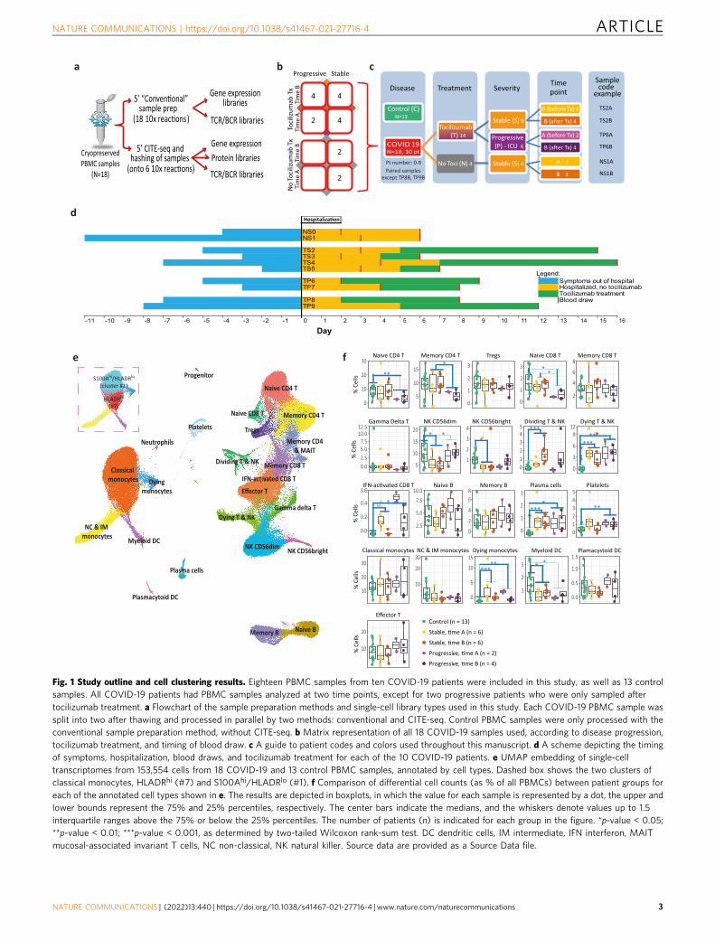
droplet-based single-cell platform (10x Chromium)17, in order toconstruct 5′ gene expression libraries, as well as surface proteinlibraries (CITE-seq)18, T cell receptor (TCR) libraries and B cellreceptor (BCR) libraries (Fig. 1a). Following filtration andcleanup, 153,554 cells were included in the scRNA-seq analysis.In addition, we obtained clinical and laboratory information onall patients, including viral loads and cytokine panels.
Our samples were derived from both stable and progressiveCOVID-19 patients as part of the Yale COVID-19 IMPACT(Implementing Medical and Public Health Action AgainstCoronavirus CT) Biorepository. Critical patients (n= 4) whorequired treatment in the ICU and eventually succumbed to thedisease were defined as having “progressive” disease, while“stable” disease defined severe patients (n= 6) hospitalized ininternal medicine wards and eventually recovered and discharged.We analyzed PBMCs from two separate blood samples for eachpatient, an early (A) and a late (B) time-point, except for twoprogressive patients (TP8, TP9) for whom only a single samplewas available (Fig. 1b–d). Eighty percent of subjects (8/10) weretreated with tocilizumab according to clinical parameters, withthe time-point A and time-point B samples obtained before andafter the initiation of the treatment, respectively. Baselinecharacteristics (Supplementary Table 1), including age and sex,were similar for both control and COVID-19 patients, whileindividuals of European ancestry were more prevalent in thecontrols. Progressive patients did not differ from the stable groupwith regard to baseline characteristics, comorbidities, and time-lines (Fig. 1d and Supplementary Table 1). The progressivepatients had significantly higher modified-SOFA score, a prog-nostic severity score, at both time points (SupplementaryTable 1).
SARS-CoV-2 RNA was not detected in any of our PBMCsamples. In addition, we did not detect the expression of ACE2,the functional host receptor for SARS-CoV-219, which maydiminish the likelihood of PBMC infection.
Applying Louvain clustering to the filtered and integratedSeurat object, and plotting in uniform manifold approximationand projection (UMAP) space, 22 cell types were identified andmanually annotated (Fig. 1e and Supplementary Fig. 1) across 30cell clusters (Supplementary Fig. 2a), with a good overlap betweendifferent samples and subgroups (Supplementary Fig. 2b, c).Automated annotation using SingleR package20 (SupplementaryFig. 2d) supported the results of the manual annotation. Goodoverlap was also noted between cells processed with and withoutCITE-seq (i.e., non-CITE, Supplementary Fig. 2e), except for thedying monocytes cluster which was reduced in CITE samples,possibly due to the exclusion of these dying cells during theadditional staining process. Importantly, viability was similar(approximately 85–90%) for CITE and non-CITE samples beforeloading the cells to the 10× Chromium Chip. A detailedcomparison between the CITE and non-CITE samples (Supple-mentary Fig. 3) showed a high similarity of gene expression, anddata sets were therefore combined for subsequent analysis.
Several differences in the relative abundance of specific celltypes were detected across control, stable, and progressivesamples at the two time points (Fig. 1f). Some notable statisticallysignificant differences were a relative decrease in naive T cells(both CD4+ and CD8+) in progressive patients, as well as anincrease in plasmablasts and dividing T & NK cells in COVID-19patients vs controls. Cells belonging to the interferon (IFN)-activated CD8 T cell cluster (Fig. 1e and Supplementary Fig. 4), asmall cluster of 191 cells characterized by very high expression ofIFN stimulated genes (ISGs), were found almost exclusively inCOVID-19 patients (p= 0.006), especially at time point A.
Some differences in relative cell proportions were noted for theinnate immune arm as well. The classical monocytes population
ARTICLE NATURE COMMUNICATIONS | https://doi.org/10.1038/s41467-021-27716-4
2 NATURE COMMUNICATIONS | (2022) 13:440 | https://doi.org/10.1038/s41467-021-27716-4 | www.nature.com/naturecommunications
ba
SeverityTreatmentDisease
COVIDN=18, 10 pt
19
Tocilizumab (T) 14
Stable (S) 8
Progressive (P) - ICU 6
No Toci (N) 4 Stable (S) 4
Control (C)
Time point
A (before Tx) 4
B (a�er Tx) 4
A (before Tx) 2
B (a�er Tx) 4
A 2
B 2
Samplecode
example
TS2A
TS2B
TP6A
TP6B
NS1A
NS1B
c
N=13
-
-
Pt number: 0-9Paired samples
except TP8B, TP9B
4 4
2 4
Progressive Stable
Toci
lizum
ab T
x
Tim
e A
T
ime
B
2
2
No
Toci
lizum
ab T
x
Ti
me
A
Tim
e B
Cryopreserved PBMC samples
(N=18)
5sample prep
(18 10x )
5’ CITE-seq and hashing of samples
Gene expression libraries
TCR/BCR libraries
Gene expression Protein libraries
TCR/BCR libraries
NS0NS1
TS2TS3TS4TS5
Legend:TP6 Symptoms out of hospitalTP7 Hospitalized, no tocilizumab
Tocilizumab treatmentTP8 Blood drawTP9
-11 -10 -9 -8 -7 -6 -5 -4 -3 -2 -1 0 1
dHospitaliza�on
e
Naive CD4 T
NC & IM monocytes
Memory CD4 TNaive CD8 T
NK CD56dim
Classical monocytes
Memory CD4& MAIT
Effector T
Dividing T & NK
Naive B
Myeloid DCNK CD56bright
Memory CD8 T
Memory B
Platelets Tregs
Plasmacytoid DC
Gamma delta TDying T & NK
Plasma cells
Dyingmonocytes
Progenitor
IFN-activated CD8 T
Neutrophils
HLADRhi
(#7)
S100Ahi/HLADRlo
(cluster #1)
Control (n = 13)
Progressiv
Progressiv
Stab
Stab
0
10
20
30
5
10
15
0
1
2
3
0
1
2
3
2
4
6
8
10
20
0.0
0.2
0.4
0.6
0
5
10
15
1
2
3
0.0
0.5
1.0
1.5
2.5
5.0
7.510.0
2
4
68
0
1
2
3
012345
5
10
15
20
1
2
3
4
012345
0
3
6
912
10
20
30
10
20
30
f
2 3 4 5 6 7 8 9 10 11 12 13 14 15 16
Day
Naive CD4 T Memory CD4 T Tregs Naive CD8 T Memory CD8 T
Effector T
NK CD56dim NK CD56bright Dividing T & NK Dying T & NK
Classical monocytes
PlateletsPlasma cellsMemory BNaive B
Plamacystoid DCMyeloid DCDying monocytesNC & IM monocytes
% C
ells
% C
ells
% C
ells
% C
ells
0.02.55.07.5
10.012.5
Gamma Delta T
% C
ells
**
*****
***
****
*****
**
**
* **
*
* *
*NS
0
Fig. 1 Study outline and cell clustering results. Eighteen PBMC samples from ten COVID-19 patients were included in this study, as well as 13 controlsamples. All COVID-19 patients had PBMC samples analyzed at two time points, except for two progressive patients who were only sampled aftertocilizumab treatment. a Flowchart of the sample preparation methods and single-cell library types used in this study. Each COVID-19 PBMC sample wassplit into two after thawing and processed in parallel by two methods: conventional and CITE-seq. Control PBMC samples were only processed with theconventional sample preparation method, without CITE-seq. b Matrix representation of all 18 COVID-19 samples used, according to disease progression,tocilizumab treatment, and timing of blood draw. c A guide to patient codes and colors used throughout this manuscript. d A scheme depicting the timingof symptoms, hospitalization, blood draws, and tocilizumab treatment for each of the 10 COVID-19 patients. e UMAP embedding of single-celltranscriptomes from 153,554 cells from 18 COVID-19 and 13 control PBMC samples, annotated by cell types. Dashed box shows the two clusters ofclassical monocytes, HLADRhi (#7) and S100Ahi/HLADRlo (#1). f Comparison of differential cell counts (as % of all PBMCs) between patient groups foreach of the annotated cell types shown in e. The results are depicted in boxplots, in which the value for each sample is represented by a dot, the upper andlower bounds represent the 75% and 25% percentiles, respectively. The center bars indicate the medians, and the whiskers denote values up to 1.5interquartile ranges above the 75% or below the 25% percentiles. The number of patients (n) is indicated for each group in the figure. *p-value < 0.05;**p-value < 0.01; ***p-value < 0.001, as determined by two-tailed Wilcoxon rank-sum test. DC dendritic cells, IM intermediate, IFN interferon, MAITmucosal-associated invariant T cells, NC non-classical, NK natural killer. Source data are provided as a Source Data file.
NATURE COMMUNICATIONS | https://doi.org/10.1038/s41467-021-27716-4 ARTICLE
NATURE COMMUNICATIONS | (2022) 13:440 | https://doi.org/10.1038/s41467-021-27716-4 | www.nature.com/naturecommunications 3
sub-clustered into two distinct populations (dashed box inFig. 1e): one with low expression of HLA-DR, a majorhistocompatibility complex (MHC) class II molecule (cluster#1), subsequently referred to as S100Ahi/HLA-DRlo monocyte,and an HLA-DRhi monocyte (cluster #7) as further discussedbelow. The proportion of S100Ahi/HLA-DRlo monocyte clusterwas higher in COVID-19 patients compared to controls(Supplementary Fig. 5a, b), which is consistent with previousstudies15,21. On the other hand, non-classical monocytes (andtheir marker FCGR3A) were decreased in COVID-19 (Supple-mentary Fig. 5), which is also consistent with recentstudies13,16,21. Myeloid dendritic cells (DC) were also decreasedin COVID-19 patients (Fig. 1f).
To conclude, our comprehensive atlas of PBMCs in stable andprogressive COVID-19 patients at different time points, captureddynamic shifts in the relative abundance of specific cell types,reflective of the immune system response to the virus.
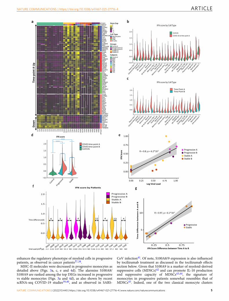
Type-1 interferon signature dominates peripheral immunecells in COVID-19. We further analyzed gene expression changesin each cell type as well as alterations over the time course of thedisease. We observed that type-1 interferon (IFN-I) response waselevated in COVID-19 across all cell types, especially at time-point A(Fig. 2a, c), and more so in progressive subjects (Fig. 2d). Asexpected, there was a strong correlation between the IFN-I score andthe concurrent viral load at the sample level (R= 0.8; Fig. 2e, f).Conventional ISGs, such as IFI6, IFI44L, LY6E, and ISG15, weremarkedly increased in COVID-19 patients compared to healthycontrols across all major cell types in PBMCs (Fig. 2a). These resultsare in agreement with recently published studies22,23, which iden-tified a strong IFN-I response in various subpopulations of PBMCsderived from COVID-19 patients. Amphiregulin (AREG), a ligandfor epidermal growth factor receptor (EGFR) not known as a majorISG in humans, is barely detectable in healthy control PBMCs but issignificantly increased in COVID-19 patients’ monocytes, T cells,NK cells, and DCs (Supplementary Fig. 6). Although AREG isknown to play important roles in wound repair and resolution ofinflammation24, its expression has also been reported to be increasedin viral infections of the lung25 and induce severe lung pathology ina mouse model of SARS-CoV infection26. IFN-I signaling plays animportant role in AREG induction within myeloid cells in mice27. Arecent report using bulk RNA-sequencing showed an increase ofAREG in the PBMCs of COVID-19 patients28, supporting apotential role of AREG in SARS-CoV-2-induced lung pathology.
IFN-I response decreases over time in correlation with virusclearance. The time course of COVID-19 disease is characterizedby shifts in many genes (Fig. 2a) and ligand–receptor interactions(Supplementary Fig. 7)29. As expected, the IFN-I score markedlydecreases over time from time-point A (earlier blood draw) to B(later one) in all patients and all cell types, corresponding to adecrease in viral loads between those time-points (Fig. 2f).Notably, the decrease in IFN-I score between time-point A and Bcorrelates strongly with the time difference between them(R= 0.97, Fig. 2g). Symptom onset is reported to occur at amedian of 5.2 days after infection7, and since blood draw A wastaken at least 5 days after symptom onset (Fig. 1d), at this time-point our patients would be expected to be on the descendingslope of the viral load curve30. This is consistent with ourobservations of a uniform decrease in viral load and IFN-I scorebetween the two time points. However, in two out of four pro-gressive patients (and none of the six stable ones), both IFN-Iscore and viral load remained relatively high at time-point B. Thegene expression signature of these two patients at time-point B(TP7B, TP8B) resembles the signature of other patients at the
earlier time point A, while the other patients at time point B arecloser to the healthy controls’ gene expression signature (Fig. 2a).This observation is consistent with a recent publication4, sug-gesting that some progressive patients are slower in clearing thevirus, possibly due to immunosuppressive mechanisms discussedin the following sections. Altogether, these findings suggest thatin most patients, the initially elevated IFN-I response decreasesover time together with the decrease in viral loads. Interestingly,in some progressive patients, the IFN-I response seems to persist,concordantly with decreased viral clearance.
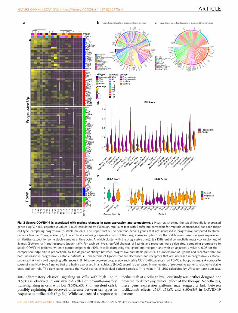
Marked gene expression changes differentiate progressive fromstable patients. We observed marked gene expression differencesbetween stable and progressive patients that span across all celllineages (Fig. 3a–c and Supplementary Data 1–4). The expressionof ISGs is increased in all cell types in progressive subjects(Fig. 3a, d). Interestingly, there is an increased expression of thesuppressive cytokine IL10 in myeloid cells and several additionalcell types in progressive patients (Supplementary Fig. 8a). Levelsof IL-10 in plasma are known to be increased in severe COVID-19, as reported in our recent study4 as well as by others7,31. IFN-Ihas been reported to induce IL-10 expression, thus limitingimmune-related tissue damage in certain conditions32,33. Similarto ISGs, the level of plasma IL-10 decreases from time-point A toB (Supplementary Fig. 8b), although in a larger cohort of patientsthis decrease was only seen in stable non-ICU patients and the IL-10 level was kept higher in ICU patients (Supplementary Fig. 8d).We observed a modest positive correlation (R= 0.50, Supple-mentary Fig. 8c) between the IFN-I score in PBMCs and plasmaIL-10 levels, which may support an association between thestrength of the IFN-I response and the suppressive IL-10 responseobserved in COVID-19 patients.
In addition, we observed a decrease in MHC-II transcripts inantigen-presenting cells (APCs) of progressive subjects comparedto stable ones, with the latter being more similar to that of controlsubjects (Fig. 3c, e and Supplementary Fig. 9). Increased IL-10 isknown to downregulate the expression of MHC-II34,35, possiblyexplaining this observed decrease in progressive subjects.
Together, this suppressive signature of increased IL-10 anddecreased MHC-II in progressive patients might serve as adouble-edged sword: on the one hand, decreasing inflammationand protecting tissues from immune-related damage, and on theother hand hampering the ability to mount an effective antiviralresponse, as will be further discussed in the next section.
Progressive patients exhibit S100Ahi/HLA-DRlo myeloid phe-notype. In order to better understand the transcriptional differ-ences between stable and progressive COVID-19 monocytes, wesub-clustered them after excluding cells from control subjects.This yielded 23,701 monocytes in seven clusters (Fig. 4a andSupplementary Fig. 10). We identified a clear separation betweencells of stable and progressive patients, which is driven in part byincreased expression of ISGs in progressive patients (Figs. 3a, 4aand Supplementary Data 1). Regulatory and tissue repair-associated genes are increased in progressive vs stable mono-cytes, including CD163 (Fig. 4b), IL1R2 (Fig. 4c), AREG (Fig. 4d),the co-inhibitory receptor HAVCR2 (encoding TIM-3), and itsligand LGALS9 (encoding Galectin-9; Fig. 3b), and IL10 (Sup-plementary Fig. 8). The expression of RNASE2, encoding a pro-tein with antiviral activity (mainly against single-stranded RNAviruses)36, is also increased in progressive patients (Fig. 3a). Ofnote, LGALS9 expression was increased not only in myeloid cellsbut also in B and CD4 T cells in progressive patients (Fig. 3b andSupplementary Fig. 9). This indicates a potential role for theTIM-3/Gal-9 pathway in myeloid/T cells interaction that
ARTICLE NATURE COMMUNICATIONS | https://doi.org/10.1038/s41467-021-27716-4
4 NATURE COMMUNICATIONS | (2022) 13:440 | https://doi.org/10.1038/s41467-021-27716-4 | www.nature.com/naturecommunications
enhances the regulatory phenotype of myeloid cells in progressivepatients, as observed in cancer patients37,38.
MHC-II molecules were decreased in progressive monocytes asdetailed above (Figs. 3a, c, e and 4d). The alarmins S100A8/S100A9 are ranked among the top DEGs increased in progressivevs stable monocytes (Figs. 3a and 4d), as also shown by recentscRNA-seq COVID-19 studies39,40, and as observed in SARS-
CoV infection41. Of note, S100A8/9 expression is also influencedby tocilizumab treatment as discussed in the tocilizumab effectssection below. Given that S100A9 is a marker of myeloid-derivedsuppressive cells (MDSCs)42 and can promote IL-10 productionand suppressive capacity of MDSCs43,44, the signature ofmonocytes in progressive patients somewhat resembles that ofMDSCs45. Indeed, one of the two classical monocyte clusters
a b
d
Effec
tor T
NK CD56dim
Mem
ory CD4 T
Naive B
NC & IM
monocy
tes
Mem
ory CD8 T
Dying T
& N
K
Mem
ory B
Gamma d
elta T
Mem
ory CD4 &
MAIT
Tregs
Naive C
D8 T
Dying m
onocytes
Platele
ts
NK CD56brig
ht
Mye
loid DC
Dividing T
& N
K
Plasma c
ells
Plasmac
ytoid DC
Proge
nitor
Neutro
phils
Controls
−0.5
0.0
0.5
1.0
1.5
2.0
IFN score by Cell Type
Naive C
D4 T
Classic
al M
onocytes
0.0
0.5
1.0
1.5
2.0 Time Point ATime Point B
Effec
tor T
NK CD56dim
Mem
ory CD4 T
Naive B
NC & IM
monocy
tes
Mem
ory CD8 T
Dying T
& N
K
Mem
ory B
Gamma d
elta T
Mem
ory CD4 &
MAIT
Tregs
Naive C
D8 T
Dying m
onocytes
Platele
ts
NK CD56brig
ht
Mye
loid DC
Dividing T
& N
K
Plasma c
ells
Plasmac
ytoid DC
Proge
nitor
Neutro
phils
Naive C
D4 T
Classic
al M
onocytes
IFN score by Cell Typec
Gene Exp
Groups
Cell Type−2−1012
MonocytesCD4CD8B cells
ControlProgressive AProgressive BStable AStable B
ControlNS0NS1TP6TP7TP8TP9TS2TS3TS4TS5
Subjects
0.0
0.5
1.0
1.5
2.0
NS0ANS0B
NS1ANS1B
TP6ATP6B
TP7ATP7B
TP8BTP9B
TS2ATS2B
TS3ATS3B
TS4ATS4B
TS5ATS5B
Progressive AProgressive BStable AStable B
Time difference(d):
IFI27IFITM3IFI6ISG15IFITM3PLAC8LY6EAPOBEC3ACLUCCL2IFI44LMT2ACTSLS100A8MX1C15orf48RNASE2SIGLEC1AREGIFI44LIFI6CD69IFITM1LY6ES100A8AREGFOSSOCS3DDIT4CXCR4TXNIPISG15SLC2A3XAF1IFI6IFI44LS100A8ISG15IFITM1OASLLY6ES100A9ISG20IFIT2CXCR4XAF1EIF2AK2IFI27MX1IFIT3IFI44LIFITM1IFI6IFI27XAF1MX1ISG20IRF7ISG15LY6ERGS1CALRBST2STAT1EIF2AK2FCGR3AEEF1A1EEF2EEF1B2EIF3LLGALS2LTBCD79BTCL1ATMSB4X
C27
C29
C30
C31
C32
C33
C34
C35
C36
C37
C38
C39
C40
NS0A
NS1A
TS2A
TS3A
TS4A
TS5A
TP6A
TP7A
NS0B
NS1B
TS2B
TS3B
TS4B
TS5B
TP6B
TP7B
TP8B
TP9B
groupssubjects
cell t
ype
Tim
e-po
int A
Up
Dow
n
4 3 7 4 12 4 12 4
Viral Load (x106/μl): 2.2 0.02 ND0.07 53.3 ND 1160 251 52.5 ND 9.66 0.11 ND ND 37.2 ND ND ND
R = 0.97, p = 6.1*10-5
Tim
e D
iffe
ren
ce b
etw
ee
n A
an
d B
10
5
0.25 0.5 0.75
f g
e
Progressive AProgressive BStable AStable B
R = 0.8, p = 6.2*10-5
1.00
0.75
0.50
0.25
0.00
0.00 0.25 0.50 0.75 1.00
IFN
Sco
re
Log Viral Load
ProgressiveStable
IFN Score Difference between Time A to B
Controls
Control
Progre
ssive
Stable
IFN score
−0.5
0.0
0.5
1.0
1.5
2.0****
****
****
****
NATURE COMMUNICATIONS | https://doi.org/10.1038/s41467-021-27716-4 ARTICLE
NATURE COMMUNICATIONS | (2022) 13:440 | https://doi.org/10.1038/s41467-021-27716-4 | www.nature.com/naturecommunications 5
(cluster #1, Fig. 1f—dashed box) is enriched with MDSCsassociated genes (S100A8, S100A9, IL1R2, IL10) with lowexpression of MHC-II. Furthermore, this monocyte clusterexhibits highly overlapped transcriptional features with a recentlyidentified monocyte population in severe sepsis46. Unexpectedly,pro-inflammatory monocyte markers such as IL1B and TNF aredownregulated in COVID-19 monocytes relative to controls(Supplementary Fig. 11), both in progressive and in stablepatients, although IL1B was slightly less downregulated in stablepatients. This observation is consistent with recent reportshighlighting an immunosuppressive phenotype in severe respira-tory failure in COVID-19 patients47.
Taken together, these findings revealed a skewed regulatorysignature of monocytes in progressive patients, which resemblesimmunoparalysis48. Given that many of these genes associatedwith an immunosuppressive phenotype are regulated downstreamof IFN-I signaling (AREG, IL1R2, S100A8, S100A9, IL10), thisshift of classical monocytes toward MDSC-like suppressive cellsmight stem from the strong IFN-I response. In addition, ourconnectome analysis highlights the enhanced TIM-3/Gal-9circuit, which may contribute to the aberrant regulatory myeloidsignature. This potentially premature shift to a resolution phasemight interrupt appropriate antiviral immune responses, con-tributing to the delayed virus clearance and deleterious clinicalmanifestations observed in severe COVID-194.
CD8+ T cells exhibit an enhanced effector signature in pro-gressive patients. We next attempted to examine the geneexpression differences in CD8+ T-cell subpopulation between thedisease conditions. A detailed analysis of sub-clustered 19,458CD8 T cells (Fig. 4e and Supplementary Fig. 12a) showed a clearseparation between stable and progressive patients, driven mainlyby a higher expression of ISGs in the progressive patients (Figs. 3aand 4e), but also by higher expression of effector cytokines suchas GZMB (Fig. 3a). Most of the cells from the IFN-activatedCD8+ T cell cluster are located in the progressive pole andoverlap with the effector T cell cluster (Fig. 4e). There are clearshifts in the gene expression profile from the early time-point Ato the late time-point B (Supplementary Fig. 12b–d) that aremainly driven by the decrease in ISG signature (Fig. 2a). Thedifferential connectivity map analysis demonstrates an increasedexpression of the co-inhibitory receptor LAG3 in T lymphocytesof progressive patients, while its ligands, which are MHC-IImolecules, are decreased in antigen-presenting cells (Fig. 3b, c).This mismatch, which was validated by flow cytometry (Fig. 4f), ispart of the immune system dyssynchrony we observed in pro-gressive patients that required ICU admission.
Tocilizumab effects differ across cell types and associate withlevels of IL6R and IL6ST. Eight of ten COVID-19 patients in ourstudy were treated with tocilizumab, an anti-IL-6 receptor (IL-6R)antibody. We further examined the differential gene expressionpattern that is associated with tocilizumab treatment. IL6R ishighly expressed in monocytes, dendritic cells, neutrophils, CD4+
T cells (including FoxP3 regulatory T cells (Tregs)) and naiveCD8+ T cells (Fig. 5a). On the other hand, IL6R expression is lowin the other types of lymphocytes including memory CD8+,effector CD4+ & CD8+ T cells, gamma-delta T cells, B cells andNK cells. IL6ST (encoding gp130), responsible for signal trans-duction of IL-6 following binding to IL-6R, is expressed in alltypes of PBMCs (Fig. 5b). To identify the transcriptional effects oftocilizumab treatment in COVID-19 patients, we compared geneexpression changes from time point A to B for patients in thetocilizumab treatment group versus those not treated with toci-lizumab (Fig. 5c and Supplementary Fig. 13). We highlight sixtocilizumab responsive genes (ARID5A, BCL3, PIM1, SOCS3,BATF, MYC) that are associated with IL-6 pathway and known tobe perturbed by tocilizumab treatment in rheumatoid arthritispatients49. Of note, those transcriptional changes by tocilizumabare observed mainly in the cell types that highly express both IL6Rand IL6ST, such as naive CD4+ T cells, memory CD4+ T cells,naive CD8+ T cells, and Tregs. To quantify this effect, we gen-erated an IL-6 score (a composite score of the aforementioned sixtocilizumab responsive genes). We demonstrated a significantdecrease of IL-6 score in CD4+ T cells in all patients who receivedtocilizumab, but not in ones who did not (Fig. 5d and Supple-mentary Data 10).
Next, we sought to identify the other genes that are perturbedby tocilizumab in COVID-19 patients. To minimize theconfounding effects of disease-related gene expression changesover time, we focused on genes that are not decreased over timein the non-tocilizumab group but significantly decreased follow-ing tocilizumab treatment (log-fold-change [logFC] > 0.4, Fig. 5e).We demonstrate that S100A8 and S100A9 expression are highlydownregulated by tocilizumab treatment across the majority ofthe cell types, but not changed or even slightly increased in non-tocilizumab group, leading to a large logFC difference (Fig. 5e).Given that a positive feedforward loop between S100A8/9 and IL-6can drive pro-inflammatory circuit50–52 and that elevated serumS100A8/9 is one of the hallmarks of severe COVID-19 patients53,54,it is possible that tocilizumab can exert its effect partly throughthe inhibition of S100A8/9 expression in COVID-19. Of interest,the expression of IL6R is higher than that of IL6ST in myeloidcells, while it is lower in all other cell types, leading to a differencein IL6R/IL6ST ratio. According to a recent study55, thisratio determines the type of response to IL-6 signaling:
Fig. 2 Strong interferon response is observed in COVID-19 samples. a Heatmap showing the top differentially expressed genes (logFC > 0.5, adjustedp-value < 0.05 calculated by Wilcoxon rank-sum test with Bonferroni correction for multiple comparisons) for each major cell type, comparing time-pointA and B. The level of expression of these genes in control samples is shown as well. The upper part of the heatmap depicts genes that are increased attime-point A compared to B (marked “time-point A up”). b–g IFN-I scores were calculated based on the expression of 12 ISGs for each sample. b IFN-Iscore is markedly increased in all cell types in COVID-19 at time point A, relative to controls. c IFN-I score decreases from time-point A to B in nearly all celltypes. d IFN-I score is higher in progressive vs stable COVID-19 patients, and at time-point, A (earlier blood draw) compared to time-point B (later one).****p-value < 1E−300 calculated by Wilcoxon rank-sum test. e, f Viral load for each patient was calculated based on RT-qPCR analysis of nasopharyngealswabs or saliva samples. e Shown is a scatter plot of scaled log viral load vs scaled IFN-I score for all COVID-19 samples. Correlation coefficient (R) andp-value are indicated. Error bands denote a 95% confidence interval. p-value was calculated based on an F-test for the significance of the regression model.f Shown is a violin plot depicting the IFN-I score for each sample, with the corresponding viral loads indicated below the plot. Arrows mark the timedifference (in days) between paired samples (i.e., from the same patient) at two time-points: A (early/before tocilizumab treatment) and B (late/aftertocilizumab). g Scatter plot for the 8 paired samples, showing a very high correlation between the time difference from sample A to B and the respectivechange in scaled IFN-I score during that time. Correlation coefficient (R) and p-value are indicated. Error bands denote a 95% confidence interval. p-valuewas calculated based on an F-test for the significance of the regression model. IFN interferon, ND not detectable. FC fold-change. Source data are providedas a Source Data file.
ARTICLE NATURE COMMUNICATIONS | https://doi.org/10.1038/s41467-021-27716-4
6 NATURE COMMUNICATIONS | (2022) 13:440 | https://doi.org/10.1038/s41467-021-27716-4 | www.nature.com/naturecommunications
anti-inflammatory classical signaling in cells with high IL6R/IL6ST (as observed in our myeloid cells) or pro-inflammatorytrans-signaling in cells with low IL6R/IL6ST (non-myeloid cells),possibly explaining the observed difference between cell types inresponse to tocilizumab (Fig. 5e). While we detected a response to
tocilizumab at a cellular level, our study was neither designed norpowered to detect any clinical effect of the therapy. Nonetheless,these gene expression patterns may suggest a link betweentocilizumab effects, IL6R, IL6ST, and S100A8/9 in COVID-19patients.
aM
ono
Up
CD4
Up
CD8
Up
B U
pM
ono
Dow
nCD
8 Do
wn
B Do
wn
IFI6IFITM1ISG15IFITM3APOBEC3AIL1R2LY6ECCL2S100A8CD163AREGRNASE1RNASE2RETNMT2ATHBS1CLULMNAIFI6IFI44LISG15LY6EIFITM1S100A8IFITM3MX1EIF2AK2MT2AISG20IFIT3FOSSOCS1AREGZFP36S100A9DDIT4TRIM22PRDM1ARID5AIFI6IFIT2IFITM1OASLS100A8LY6EISG20IFIT3ISG15IFI44LADGRE5S100A9EIF2AK2MT2ALAG3GZMBXAF1DDIT4TSC22D3MX1HERPUD1RNF213CCL4SP100IFITM3SOCS1IFITM1IFI6IFI44LMX1ZFP36L2LY6EISG15EIF2AK2TRIM22S100A8TESCOASLXAF1IRF7IFITM3ZNF331BTG1TXNIPSTAT1IFIT3TGFB1IL1BFCGR3AHLA−DQB1HLA−DPB1GZMKLTBCMC1LTBCD79BTCL1A
TP7A
TS2A
TP6A
TP7B
TS4A
TP8B
NS0
ATP
6BTP
9BTS
5AN
S1A
NS1
BTS
3ATS
2BTS
5BN
S0B
TS4B
TS3B
groupssubjects
cell
type
Prog
ress
ive
Up
Dow
n
SELPLGRNASE2LGALS9IL1RN
IL10ANXA1ADAM17
CCL2C1QBHLA−DQB1
LGALS9HLA−DQA2
LGA
LS9
HLA
−DQ
B1
HLA
−DP
B1
GZ
MB
CC
L41L
3LC
C9SL
AGL
HLA
−DQ
A2
LGA
LS9
HLA
−DQ
B1VC
AN
RN
ASE2IL1R
NCCL4ANXA1HLA−DQB1HLA−DPB1
HLA−DPA1CCL3L1
HLA−DQA2
CCR1
SELLTLR2
IL1R2
FPR2
DYSF
HAVCR2
IGF2RLAIR1
ITGA5
LAG3
LAG
3 SE
LLIL
10R
AIL
10R
A
LAG
3
IL10
RA
SELL
FPR2
DYSF
HAVCR2
LAG3
Plasma cellsNK cellsNC & IM monocytesNaive CD4 TNaive BMemory CD8 TMemory CD4 TMemory BEffector TDendrClassical monocytesDividing T & NK
TNFSF13IL1B
HLA−DRB5HLA−DRB1HLA−DRA
HLA−DQB1
HLA−DQA1HLA−DPB1
HLA−D
PA1
HLA
−DM
B
HLA
−DM
A
HLA
−DR
B5
HLA
−DR
AH
LA−
DQ
B1
HLA
−D
QA
1
1B
PD
−AL
HHLA
−D
PA1
HLA
−D
MA
HLA
−DR
B5
HLA
−DR
A
HLA
−DQ
A1
HLA
−DM
A
HLA
−DQ
B1
HLA
−DQ
B1
HLA−D
MA
CALM
3
COL1
8A1
HLA−D
RB5
HLA−DRA
HLA−DQB1HLA−DQA1HLA−DPB1HLA−DPA1HLA−DMB
HLA−DMAHLA−DRA
HLA−DMA
ITGA5
IL1R2
LAG3
LAG
3
LAG
3
FAS
SE
LL
LAG3
NK cellsNC & IM monocytesNaive CD4 TNaive BMemory BEffector TDendrClassical monocytesMemory CD8 TDividing T & NK
Ligands and receptors increased in progressive Ligands decreased and receptors increased in progressiveb c
0.0
0.5
1.0
1.5
2.0
Classic
al monocyt
es
Effector T
NK CD56dim
Memory CD4 T
Naive B
NC & IM
monocyt
es
Memory CD8 T
Dying T
& NK
Memory B
Memory CD4 &
MAIT
Gamma d
elta T
Tregs
Naive CD8 T
Dying m
onocytes
Platelets
NK CD56bright
Myeloid DC
Dividing T
& NK
Plasma c
ells
Plasmac
ytoid DC
Progenito
r
Neutrophils
vated CD8 T
Stable
IFN Score
Naive CD4 T
Progressive
d
−1
0
1
2
NS0A
NS0B
NS1A
NS1B
TP6ATP6B
TP7ATP7B
TP8BTP9B
TS2A
TS2B
TS3A
TS3B
TS4A
TS4B
TS5A
TS5B
−1
0
1
2
Control
Progressi
veSta
ble
Disease Severity
ControlProgressiveStable
Patient
e
gene exp
−2−1012
cell typeMonocytesCD4CD8B cells
groupsProgressive AProgressive BStable AStable B
subjectsNS0NS1TP6TP7TP8TP9TS2TS3TS4TS5
****HLA2 Score HLA2 Score
****
Fig. 3 Severe COVID-19 is associated with marked changes in gene expression and connectome. a Heatmap showing the top differentially expressedgenes (logFC > 0.5, adjusted p-values < 0.05 calculated by Wilcoxon rank-sum test with Bonferroni correction for multiple comparisons) for each majorcell type, comparing progressive to stable patients. The upper part of the heatmap depicts genes that are increased in progressive compared to stablepatients (marked “progressive up”). Hierarchical clustering separates most of the progressive samples from the stable ones based on gene expressionsimilarities (except for some stable samples at time point A, which cluster with the progressive ones). b, c Differential connectivity maps (connectomes) ofligands (bottom half) and receptors (upper half). For each cell type, log-fold changes of ligands and receptors were calculated, comparing progressive tostable COVID-19 patients; we only plotted edges with >10% of cells expressing the ligand and receptor, and with an adjusted p-value < 0.05 for thecomparison; edge size is proportional to the degree of change between progressive and stable patients. b Connectome of ligands and receptors that areboth increased in progressive vs stable patients. c Connectome of ligands that are decreased and receptors that are increased in progressive vs stablepatients. d A violin plot depicting differences in IFN-I score between progressive and stable COVID-19 patients in all PBMC subpopulations. e A compositescore of nine HLA type 2 genes that are highly expressed in all subjects (HLA2 score) is decreased in monocytes of progressive patients relative to stableones and controls. The right panel depicts the HLA2 scores of individual patient samples. ****p-value < 1E−300 calculated by Wilcoxon rank-sum test.
NATURE COMMUNICATIONS | https://doi.org/10.1038/s41467-021-27716-4 ARTICLE
NATURE COMMUNICATIONS | (2022) 13:440 | https://doi.org/10.1038/s41467-021-27716-4 | www.nature.com/naturecommunications 7
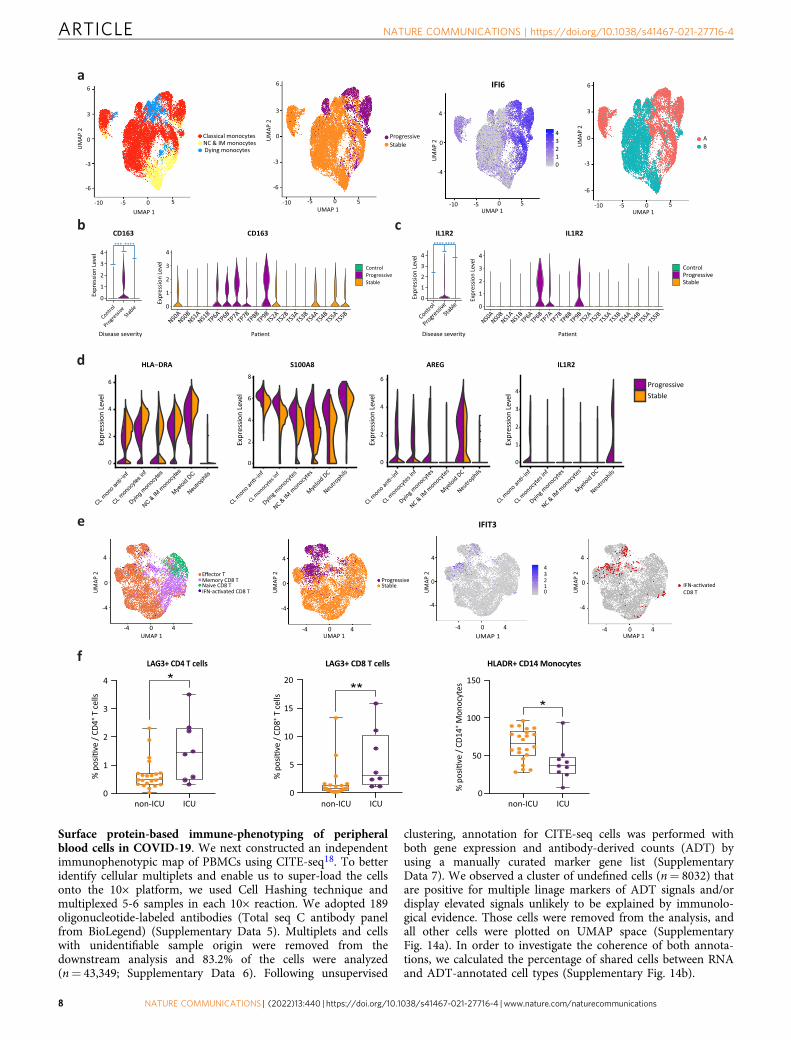
Surface protein-based immune-phenotyping of peripheralblood cells in COVID-19. We next constructed an independentimmunophenotypic map of PBMCs using CITE-seq18. To betteridentify cellular multiplets and enable us to super-load the cellsonto the 10× platform, we used Cell Hashing technique andmultiplexed 5-6 samples in each 10× reaction. We adopted 189oligonucleotide-labeled antibodies (Total seq C antibody panelfrom BioLegend) (Supplementary Data 5). Multiplets and cellswith unidentifiable sample origin were removed from thedownstream analysis and 83.2% of the cells were analyzed(n= 43,349; Supplementary Data 6). Following unsupervised
clustering, annotation for CITE-seq cells was performed withboth gene expression and antibody-derived counts (ADT) byusing a manually curated marker gene list (SupplementaryData 7). We observed a cluster of undefined cells (n= 8032) thatare positive for multiple linage markers of ADT signals and/ordisplay elevated signals unlikely to be explained by immunolo-gical evidence. Those cells were removed from the analysis, andall other cells were plotted on UMAP space (SupplementaryFig. 14a). In order to investigate the coherence of both annota-tions, we calculated the percentage of shared cells between RNAand ADT-annotated cell types (Supplementary Fig. 14b).
b
0
1
2
3
4
NS0ANS0
BNS1
ANS1
BTP6A
TP6BTP7A
TP7BTP8B
TP9BTS2
ATS2
BTS3
ATS3
BTS4
ATS4
BTS5
ATS5
B
Expr
essio
n Le
vel
0
1
2
3
4
Control
Progressi
veSta
ble
Disease severity
Expr
essio
n Le
v el
c
0
1
2
3
4
Control
Progressi
veSta
ble
Expr
essio
n Le
vel
0
2
4
6Ex
pres
sion
Leve
l
0
1
2
3
4
CL monocyt
es inf
Dying m
onocytes
Myeloid DC
Neutrophils
Expr
essio
n Le
vel
CL monocyt
es inf
Dying m
onocytes
NC & IM
monocyt
es
Myeloid DC
Neutrophils
ProgressiveStable
-10 -5 0 5
-6
-3
0
3
6
-6
-3
0
3
6
-10 -5 0 5 -10 -5 0 5 -10 -5 0 5
4
0
-4
-6
-3
0
3
6
UMAP 1 UMAP 1 UMAP 1 UMAP 1
UM
AP 2
UM
AP 2
UM
AP 2 UM
AP 2
Classical monocytesNC & IM monocytesDying monocytes
ProgressiveStable
43210
AB
IFI6
0
1
2
3
4
NS0ANS0
BNS1
ANS1
BTP6A
TP6BTP7A
TP7BTP8B
TP9BTS2
ATS2
BTS3
ATS3
BTS4
ATS4
BTS5
ATS5
B
Expr
essio
n Le
vel
IL1R2AREG
0
2
4
6
8
Expr
essio
n Le
vel
S100A8
CL monocyt
es inf
Dying m
onocytes
NC & IM
monocyt
es
Myeloid DC
Neutrophils
d
0
2
4
6
Expr
essio
n Le
vel
HLA−DRA
CL mon
o anti
−inf
CL mon
ocyte
s inf
Dying m
onoc
ytes
NC & IM m
onoc
ytes
Myeloi
d DC
Neutro
phils
a
ControlProgressiveStable
ControlProgressiveStable
Disease severity
NC & IM
monocyt
es
Effector TMemory CD8 TNaive CD8 T
4
0
-4
4
0
-4
-4 0 4 -4 0 4 -4 0 4 -4 0 4
4
0
-4
4
0
-4
UMAP 1
UM
AP 2
UMAP 1UMAP 1 UMAP 1
UM
AP 2
UM
AP 2
UM
AP 2
ProgressiveStable
43210 CD8 T
e
LAG3+ CD4 T cells LAG3+ CD8 T cellsf HLADR+ CD14 Monocytes
IFIT3
CD163 CD163*** ****
IL1R2 IL1R2**** ****
0
1
2
3
4
0
5
10
15
20**
⁺ T ce
lls
⁺ T ce
lls
0
50
100
150
*
*
⁺ Mon
ocyt
es
non-ICU ICU non-ICU ICU non-ICU ICU
ARTICLE NATURE COMMUNICATIONS | https://doi.org/10.1038/s41467-021-27716-4
8 NATURE COMMUNICATIONS | (2022) 13:440 | https://doi.org/10.1038/s41467-021-27716-4 | www.nature.com/naturecommunications
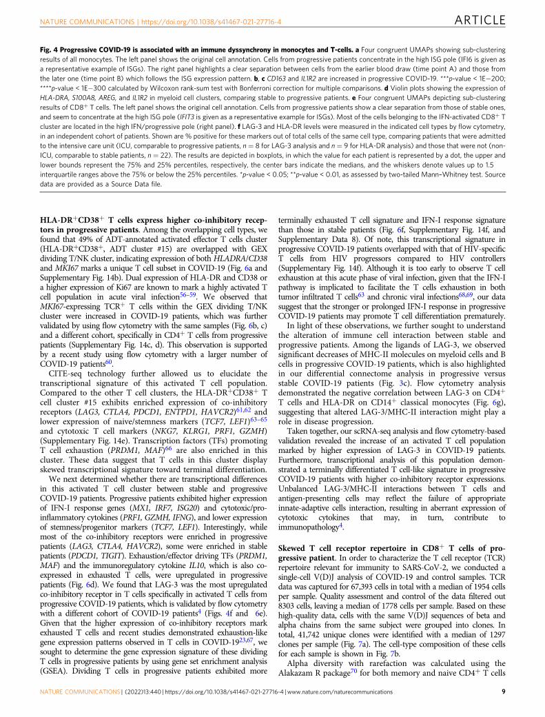
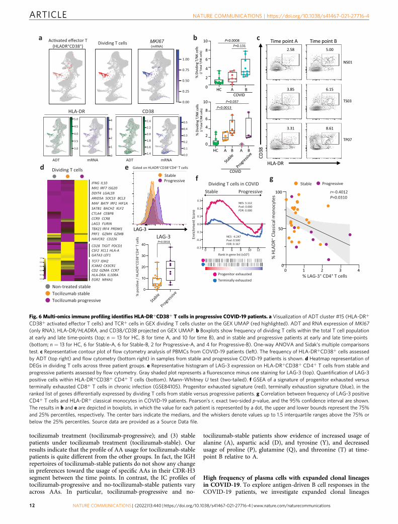
HLA-DR+CD38+ T cells express higher co-inhibitory recep-tors in progressive patients. Among the overlapping cell types, wefound that 49% of ADT-annotated activated effector T cells cluster(HLA-DR+CD38+, ADT cluster #15) are overlapped with GEXdividing T/NK cluster, indicating expression of both HLADRA/CD38and MKI67 marks a unique T cell subset in COVID-19 (Fig. 6a andSupplementary Fig. 14b). Dual expression of HLA-DR and CD38 ora higher expression of Ki67 are known to mark a highly activated Tcell population in acute viral infection56–59. We observed thatMKI67-expressing TCR+ T cells within the GEX dividing T/NKcluster were increased in COVID-19 patients, which was furthervalidated by using flow cytometry with the same samples (Fig. 6b, c)and a different cohort, specifically in CD4+ T cells from progressivepatients (Supplementary Fig. 14c, d). This observation is supportedby a recent study using flow cytometry with a larger number ofCOVID-19 patients60.
CITE-seq technology further allowed us to elucidate thetranscriptional signature of this activated T cell population.Compared to the other T cell clusters, the HLA-DR+CD38+ Tcell cluster #15 exhibits enriched expression of co-inhibitoryreceptors (LAG3, CTLA4, PDCD1, ENTPD1, HAVCR2)61,62 andlower expression of naive/stemness markers (TCF7, LEF1)63–65
and cytotoxic T cell markers (NKG7, KLRG1, PRF1, GZMH)(Supplementary Fig. 14e). Transcription factors (TFs) promotingT cell exhaustion (PRDM1, MAF)66 are also enriched in thiscluster. These data suggest that T cells in this cluster displayskewed transcriptional signature toward terminal differentiation.
We next determined whether there are transcriptional differencesin this activated T cell cluster between stable and progressiveCOVID-19 patients. Progressive patients exhibited higher expressionof IFN-I response genes (MX1, IRF7, ISG20) and cytotoxic/pro-inflammatory cytokines (PRF1, GZMH, IFNG), and lower expressionof stemness/progenitor markers (TCF7, LEF1). Interestingly, whilemost of the co-inhibitory receptors were enriched in progressivepatients (LAG3, CTLA4, HAVCR2), some were enriched in stablepatients (PDCD1, TIGIT). Exhaustion/effector driving TFs (PRDM1,MAF) and the immunoregulatory cytokine IL10, which is also co-expressed in exhausted T cells, were upregulated in progressivepatients (Fig. 6d). We found that LAG-3 was the most upregulatedco-inhibitory receptor in T cells specifically in activated T cells fromprogressive COVID-19 patients, which is validated by flow cytometrywith a different cohort of COVID-19 patients4 (Figs. 4f and 6e).Given that the higher expression of co-inhibitory receptors markexhausted T cells and recent studies demonstrated exhaustion-likegene expression patterns observed in T cells in COVID-1923,67, wesought to determine the gene expression signature of these dividingT cells in progressive patients by using gene set enrichment analysis(GSEA). Dividing T cells in progressive patients exhibited more
terminally exhausted T cell signature and IFN-I response signaturethan those in stable patients (Fig. 6f, Supplementary Fig. 14f, andSupplementary Data 8). Of note, this transcriptional signature inprogressive COVID-19 patients overlapped with that of HIV-specificT cells from HIV progressors compared to HIV controllers(Supplementary Fig. 14f). Although it is too early to observe T cellexhaustion at this acute phase of viral infection, given that the IFN-Ipathway is implicated to facilitate the T cells exhaustion in bothtumor infiltrated T cells63 and chronic viral infections68,69, our datasuggest that the stronger or prolonged IFN-I response in progressiveCOVID-19 patients may promote T cell differentiation prematurely.
In light of these observations, we further sought to understandthe alteration of immune cell interaction between stable andprogressive patients. Among the ligands of LAG-3, we observedsignificant decreases of MHC-II molecules on myeloid cells and Bcells in progressive COVID-19 patients, which is also highlightedin our differential connectome analysis in progressive versusstable COVID-19 patients (Fig. 3c). Flow cytometry analysisdemonstrated the negative correlation between LAG-3 on CD4+
T cells and HLA-DR on CD14+ classical monocytes (Fig. 6g),suggesting that altered LAG-3/MHC-II interaction might play arole in disease progression.
Taken together, our scRNA-seq analysis and flow cytometry-basedvalidation revealed the increase of an activated T cell populationmarked by higher expression of LAG-3 in COVID-19 patients.Furthermore, transcriptional analysis of this population demon-strated a terminally differentiated T cell-like signature in progressiveCOVID-19 patients with higher co-inhibitory receptor expressions.Unbalanced LAG-3/MHC-II interactions between T cells andantigen-presenting cells may reflect the failure of appropriateinnate-adaptive cells interaction, resulting in aberrant expression ofcytotoxic cytokines that may, in turn, contribute toimmunopathology4.
Skewed T cell receptor repertoire in CD8+ T cells of pro-gressive patient. In order to characterize the T cell receptor (TCR)repertoire relevant for immunity to SARS-CoV-2, we conducted asingle-cell V(D)J analysis of COVID-19 and control samples. TCRdata was captured for 67,393 cells in total with a median of 1954 cellsper sample. Quality assessment and control of the data filtered out8303 cells, leaving a median of 1778 cells per sample. Based on thesehigh-quality data, cells with the same V(D)J sequences of beta andalpha chains from the same subject were grouped into clones. Intotal, 41,742 unique clones were identified with a median of 1297clones per sample (Fig. 7a). The cell-type composition of these cellsfor each sample is shown in Fig. 7b.
Alpha diversity with rarefaction was calculated using theAlakazam R package70 for both memory and naive CD4+ T cells
Fig. 4 Progressive COVID-19 is associated with an immune dyssynchrony in monocytes and T-cells. a Four congruent UMAPs showing sub-clusteringresults of all monocytes. The left panel shows the original cell annotation. Cells from progressive patients concentrate in the high ISG pole (IFI6 is given asa representative example of ISGs). The right panel highlights a clear separation between cells from the earlier blood draw (time point A) and those fromthe later one (time point B) which follows the ISG expression pattern. b, c CD163 and IL1R2 are increased in progressive COVID-19. ***p-value < 1E−200;****p-value < 1E−300 calculated by Wilcoxon rank-sum test with Bonferroni correction for multiple comparisons. d Violin plots showing the expression ofHLA-DRA, S100A8, AREG, and IL1R2 in myeloid cell clusters, comparing stable to progressive patients. e Four congruent UMAPs depicting sub-clusteringresults of CD8+ T cells. The left panel shows the original cell annotation. Cells from progressive patients show a clear separation from those of stable ones,and seem to concentrate at the high ISG pole (IFIT3 is given as a representative example for ISGs). Most of the cells belonging to the IFN-activated CD8+ Tcluster are located in the high IFN/progressive pole (right panel). f LAG-3 and HLA-DR levels were measured in the indicated cell types by flow cytometry,in an independent cohort of patients. Shown are % positive for these markers out of total cells of the same cell type, comparing patients that were admittedto the intensive care unit (ICU, comparable to progressive patients, n = 8 for LAG-3 analysis and n = 9 for HLA-DR analysis) and those that were not (non-ICU, comparable to stable patients, n = 22). The results are depicted in boxplots, in which the value for each patient is represented by a dot, the upper andlower bounds represent the 75% and 25% percentiles, respectively, the center bars indicate the medians, and the whiskers denote values up to 1.5interquartile ranges above the 75% or below the 25% percentiles. *p-value < 0.05; **p-value < 0.01, as assessed by two-tailed Mann–Whitney test. Sourcedata are provided as a Source Data file.
NATURE COMMUNICATIONS | https://doi.org/10.1038/s41467-021-27716-4 ARTICLE
NATURE COMMUNICATIONS | (2022) 13:440 | https://doi.org/10.1038/s41467-021-27716-4 | www.nature.com/naturecommunications 9
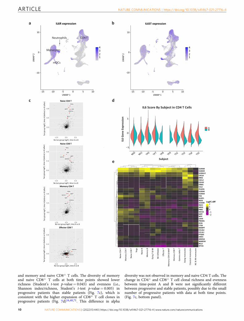
and memory and naive CD8+ T cells. The diversity of memoryand naive CD8+ T cells at both time points showed lowerrichness (Student’s t-test p-value= 0.045) and evenness (i.e.,Shannon index/richness, Student’s t-test p-value= 0.005) inprogressive patients than stable patients (Fig. 7c), which isconsistent with the higher expansion of CD8+ T cell clones inprogressive patients (Fig. 7d)16,40,71. This difference in alpha
diversity was not observed in memory and naive CD4 T cells. Thechange in CD4+ and CD8+ T cell clonal richness and evennessbetween time-point A and B were not significantly differentbetween progressive and stable patients, possibly due to the smallnumber of progressive patients with data at both time points.(Fig. 7c, bottom panel).
a b IL6ST expression
4321
-10
0
10
-15 -10 -5 0 5 10UMAP 1
UM
AP 2
IL6R expression
4321
-15 -10 -5 0 5 10
-10
0
10
UMAP 1
UM
AP 2
Neutrophils
Monocytes
mDCs
CD4 T
d
−1
0
1
NS0NS1
TP6TP7
TP8TP9
TS2 TS3 TS4 TS5
AB
IL6_score1
IL6
Gen
e Ex
pres
sion
IL6 Score By Subject in CD4 T Cells
Subject
S100A8S100A9S100A12CXCL8EREGCXCL2CD163STAB1CCL4PLSCR1S100A6S100A4FOSDUSP1RGS2JCHAINRGCCAREGPIM1SOCS3BATFMYCLDHAPRDM1IFI35CTSDNAMPTCCL4L2CCL3L1PTGDSSTAT3ARID5AMARCKSBCL3MT2A
i aN
vT
4 DCe
ome
Mr
T4DC
y
iaN
vT
8D Ce
Tsg er i a
Nv
Be
ome
Mr
By
KN
&T
gni yD
m i d6 5DCK
N
E ffTr otce
ome
Mr
T I AM
&4D C
y
ome
Mr
T8DC
y
Ta tl ed
am
maG
sety c on om
gni yD
set y co nom l a ci ss al C
se tyc on om
MI&
CN
−2−1012
LogFC diff
e
c
BCL3
PIM1
BATFARID5A
MYC
0.0
0.4
0.8
Toci
gro
up lo
gFC
f
No toci group logFC−0.4 0.0 0.4
Naive CD4 T
Naive CD8 T
Memory CD4 TNo toci group logFC
−0.4 0.0 0.4
−0.4 0.0 0.4
Effector CD8 TNo toci group logFC
−0.4 0.0 0.4No toci group logFC
PIM1
BATF
MYC
ARID5ASOCS3
BCL3
PIM1
MYC
BATFSOCS3
ARID5A
BCL3
BATFARID5A
MYC
BCL3
SOCS3PIM1
0.0
0.4
0.8
Toci
gro
up lo
gFC
f
0.0
0.4
0.8
Toci
gro
up lo
gFC
f
0.0
0.4
0.8
Toci
gro
up lo
gFC
f
ARTICLE NATURE COMMUNICATIONS | https://doi.org/10.1038/s41467-021-27716-4
10 NATURE COMMUNICATIONS | (2022) 13:440 | https://doi.org/10.1038/s41467-021-27716-4 | www.nature.com/naturecommunications
Identification of COVID-19-specific CDR3 regions. In order toidentify characteristics of the TCR regions that may confer spe-cificity to SARS-CoV-2, we used GLIPH272 to assess the simi-larity of complementarity-determining region 3 (CDR3)sequences among COVID-19 patients. We specifically looked forCDR3 motifs in β chains that were shared across several COVID-19 patients but in none of the 13 control subjects. Stringent filterswere applied to the GLIPH2 CDR3 specificity groups (or clusters)to improve accuracy, including requiring Fisher’s score < 0.0001,≥3 unique TCRs in the specificity group, and significant V-genebias (p < 0.05). The filtered specificity groups with any clone fromcontrol samples were filtered out to enhance the likelihood ofspecificity to SARS-CoV-2 instead of to other common virusessuch as cytomegalovirus. After heavy filtering, 24 and 172 groupsremained for CD8+ and CD4+ T cells, respectively. Most of theidentified specificity groups included clones from different sam-ples, suggesting a large similarity in the CDR3 sequence in thepotential SARS-CoV-2-specific clones (Fig. 7e and SupplementaryFig. 15). To further enhance the specificity to clonally-expandedSARS-CoV-2 responsive T cells, we focused on 10 CD8+ and 12CD4+ T cell groups that have clones from ≥3 subjects with atleast one of these clones with ≥2 cells. The V and J gene usageanalysis showed a strong usage bias for J gene in 3 CD8+ groupsand 1 CD4+ group (Fig. 7f, g). Some VJ combinations showed adominant usage such as TRAV5/TRAJ12/TRBJ2-7/TRBV5-6 forcluster 1 in CD8+ T cells (Fig. 7f and Supplementary Figs. 16, 17).For the CD4+ T groups, there is no obvious V gene usage biasand the J gene usage is dominated by TRBJ2-5 (Fig. 7f, g).
Among the 10 and 12 putative SARS-CoV-2-specific andexpanded groups, we further chose those that include clones from≥3 different COVID-19 patients with ≥55% clones having morethan one cell, resulting in five and two groups for CD8+ andCD4+ T cells, respectively. The chosen clusters were also the topfive and two clone clusters with the best composition score byGLIPH2, which measures the strength of a specificity group basedon global/local similarities, enrichment of common V-genes, alimited CDR3 length distribution, expanded clones (ECs), andcluster size. This suggests that the chosen specificity groups arelikely from SARS-CoV-2-specific ECs and shared across COVID-19 patients with a highly conserved CDR3 amino acid (AA)sequence. All specificity groups are identified based on globalsimilarities in the CDR3 region, except for cluster IV in CD8+
T cells, whose member clones have different CDR3 lengths butshare the motif “QDIG”. The CDR3 sequence motifs of thespecificity groups with global similarity are shown in Fig. 7f, g.We confirmed that our samples were not biased by HLA genotype(Supplementary Figs. 18 and 19). The CDR3 motifs in Fig. 7f, gwere compared to those found in two recent SARS-CoV-2 studieswith TCR repertoire data73,74, which collected samples mainlyfrom recovered and convalescent SARS-CoV-2 patients. Thecomparison showed that our CD8+ specificity group V motif(TNTGE) had a similar pattern to a motif (TGTGE) found inSchultheiß et al. 74. The study did not find this motif among the
top 31 motifs shared among recovered SARS-CoV-2 patients butfound it shared between longitudinal samples during activedisease and at recovery from one patient with mild disease andrecovered patients, suggesting the specificity of this motif toSARS-CoV-2. This overlap validates the specificity of our CD8group V motif to SARS-CoV-2 infection. It also demonstrates thepower and importance of our TCR analysis due to samplecollection during active disease and GLIPH2 analysis with theexclusion of specificity groups present in control samples.
Single-cell V(D)J B cell receptor repertoire analysis. For eachsample, a summary of the number of cells, frequency of each B celltype (naive B, memory B, and plasma cells), and frequency of eachisotype (IGHM/D/G/A) is provided in Fig. 8a–c, respectively. Overall,the single-cell V(D)J library contains 7177 cells distributed across18 samples. Gene usage and mutation frequency dynamics acrossthree cell types, per each patient and time point, are shown inSupplementary Figs. 20 and 21, respectively.
COVID-19 patients with stable status show higher mutationfrequency and longer CDR-H3 length. IGHV/IGHJ mutationfrequency and CDR-H3 length varied by antibody isotype andcell type within COVID-19 patients (Fig. 8d–f). IGHM memoryand plasma B cells had mutation frequencies significantlylower than 5% (mutations/nucleotide, p-value < 0.01), regardlessof treatment or disease progression group. As expected, IGHGmemory B cells and plasma cells had mutation frequencies higherthan IGHM cells. In particular, plasma cells in stable COVID-19patients without treatment had mutation frequencies significantlyhigher than 5% (mean= 5.6 ± 3%; p-value < 0.01). Memory cellsin stable patients under tocilizumab treatment had even highermutation frequencies (mean= 7.5 ± 4.6%).
The CDR-H3 length of IGHG and IGHM B cells generally variedbetween 10 and 20 AAs (we used 15 AAs as a reference point fordownstream comparisons) across all cell types (Fig. 8d–f). However,the CDR-H3 length of IGHG plasma cells in stable COVID-19patients was significantly larger than 15 AAs (p-value < 0.01), whilethe CDR-H3 length of IGHG memory cells do not show significantdifferences from 15 AAs (mean= 15.2 ± 3.9 AAs across allsamples). On average, CDR-H3 lengths of IGHG plasma cells werelarger in stable no-tocilizumab patients (mean= 18.4 ± 5.4 AAs)than in stable tocilizumab-treated patients (mean= 17.2 ± 5 AAs).
Stable patients under treatment do not show change in CDR-H3 amino acid usage. We sought to investigate the differences inCDR-H3 AA usage between the two blood draw time points (Aand B) (Fig. 8g). We tackled this query by calculating the con-ditional information content (IC) of each AA in the CDR-H3segment at time point B with respect to time point A (see“Methods” section). We averaged the conditional ICs for patientsbelonging to three different groups: (1) stable patients under notreatment (no-tocilizumab-stable); (2) progressive patients under
Fig. 5 Tocilizumab exerts differential gene expression effects in different immune cells. a, b UMAP representations of IL6R (a) and IL6ST (b) expressionin PBMCs. Note that IL6R is highest for monocytes, dendritic cells, CD4+ T cells (including Tregs), and naive CD8+ T cells, while IL6ST expression is similarin the majority of cell types. c Scatter plots of the logFC from time-point A to B in patients treated (Y axes) compared to those not treated with tocilizumab(X axes) for several T cell subtypes. This comparative model demonstrates a marked effect of tocilizumab on IL-6 pathway genes (shown in red) in CD4+
and naive CD8+ T cells, but not in effector CD8+ T cells, in which IL6R expression is low (see Supplementary Fig. 13 for the full panel with all cell types).d IL-6 score in CD4+ T cells is decreased at time-point B in all the patients that were treated with tocilizumab, but not in the non-treated patients (NS0,NS1). e A heatmap showing the expression of genes that were significantly differentially expressed (LogFC > 0.4, adjusted p-value < 0.05 calculated byWilcoxon rank-sum test with Bonferroni correction for multiple comparisons) between time point A and B in tocilizumab-treated patients, but not inpatients that were not treated by tocilizumab, across PBMC subtypes. All the entries in the heatmap matrix are the differences of logFC in tocilizumab andin non-tocilizumab groups. Also shown is a hierarchical clustering according to cell types (horizontal) and individual genes (vertical).
NATURE COMMUNICATIONS | https://doi.org/10.1038/s41467-021-27716-4 ARTICLE
NATURE COMMUNICATIONS | (2022) 13:440 | https://doi.org/10.1038/s41467-021-27716-4 | www.nature.com/naturecommunications 11
tocilizumab treatment (tocilizumab-progressive); and (3) stablepatients under tocilizumab treatment (tocilizumab-stable). Ourresults indicate that the profile of AA usage for tocilizumab-stablepatients is quite different from the other groups. In fact, the IGHrepertoires of tocilizumab-stable patients do not show any changein preferences toward the usage of specific AAs in their CDR-H3segment between the time points. In contrast, the IC profiles oftocilizumab-progressive and no-tocilizumab-stable patients varyacross AAs. In particular, tocilizumab-progressive and no-
tocilizumab-stable patients show evidence of increased usage ofalanine (A), aspartic acid (D), and tyrosine (Y), and decreasedusage of proline (P), glutamine (Q), and threonine (T) at time-point B relative to A.
High frequency of plasma cells with expanded clonal lineagesin COVID-19. To explore antigen-driven B cell responses in theCOVID-19 patients, we investigate expanded clonal lineages
a b(HLADR⁺CD38⁺)
Dividing T cells
HLA-DR
MKI67(mRNA)
3.0
3.5
4.0
4.5
5.0
0
1
2
3
4
0.00
0.25
0.50
0.75
1.00
0.0
0.1
0.2
0.3
0.4
0.5
1.4
1.6
1.8
2.0
2.2
2.4
CD38
mRNA mRNA
0
2
4
6
8
10 P=0.0008
% D
ivid
ing
T/NK
cells
(/ T
otal
T/N
K ce
lls)
HLA-DR
CD38
2.58 5.00
3.85 6.15
3.31 8.61
NS01
TS03
TP07
Time point A Time point B
A B0
2
4
6
8
10
A B
COVID
Stable
Progressi
ve
c
Non-treated stableTocilizumab stableTocilizumab progressive
IFNGMX1DDIT4
MAF
IRF7 ISG20LGALS9SOCS3ARID5A
IL10
BCL3BATF
GZMH
FURINPRDM1
LAG3
IRF1 HIF1ASATB1
GZMB
CTLA4CCR9 CCR8
BACH2 KLF2CEBPB
IRF4
CD226
TBX21PRF1HAVCR2
CD28XCL1CSF2 HLA-A
GATA3
TIGIT
LEF1
PDCD1
ICAM2CD2 GZMA
TCF7 IDH2
HLA-DRA
CX3CR1
EGR2
CCR7IL10RA
NR4A10.0
0.2
0.4
0.6
0.8
1.0
dStableProgressive
% D
ivid
ing
T/NK
cells
(/ T
otal
T/N
K c e
lls)
f
Rank in gene list (x103)
Stable ProgressiveDividing T cells in COVID
FDR: 0.167Pval: 0.500NES: -6.247
Terminally exhausted
FDR: 0.000Pval: 0.000NES: 3.112
Progenitor exhausted
0 2 4 6 8 10 12
0.0
0.2
Enric
hmen
t Sco
re
-0.2
-0.3
0.3
0.1
0.1
COVIDA B
P=0.0013
⁺CD3
8⁺CD
4⁺ T
cel
ls
0
10
20
30
40
Stable
Progressi
ve
LAG-3P=0.0018
HC
HC
ADT ADT
LAG-3
eg
0 1 2 3 40
50
100
% H
LADR
⁺ Cla
ssic
al m
onoc
ytes
% LAG-3⁺ CD4⁺ T cells
r=-0.4012P=0.0310
Gated on HLADR⁺CD38⁺CD4⁺ T cells
Stable Progressive
Dividing T cells
P=0.131
P=0.037
Fig. 6 Multi-omics immune profiling identifies HLA-DR+CD38+ T cells in progressive COVID-19 patients. a Visualization of ADT cluster #15 (HLA-DR+
CD38+ activated effector T cells) and TCR+ cells in GEX dividing T cells cluster on the GEX UMAP (red highlighted). ADT and RNA expression of MKI67(only RNA), HLA-DR/HLADRA, and CD38/CD38 projected on GEX UMAP. b Boxplots show frequency of dividing T cells within the total T cell populationat early and late time-points (top; n = 13 for HC, 8 for time A, and 10 for time B), and in stable and progressive patients at early and late time-points(bottom; n = 13 for HC, 6 for Stable-A, 6 for Stable-B, 2 for Progressive-A, and 4 for Progressive-B). One-way ANOVA and Sidak’s multiple comparisonstest. c Representative contour plot of flow cytometry analysis of PBMCs from COVID-19 patients (left). The frequency of HLA-DR+CD38+ cells assessedby ADT (top right) and flow cytometry (bottom right) in samples from stable and progressive COVID-19 patients is shown. d Heatmap representation ofDEGs in dividing T cells across three patient groups. e Representative histogram of LAG-3 expression on HLA-DR+CD38+ CD4+ T cells from stable andprogressive patients assessed by flow cytometry. Gray shaded plot represents a fluorescence minus one staining for LAG-3 (top). Quantification of LAG-3positive cells within HLA-DR+CD38+ CD4+ T cells (bottom). Mann–Whitney U test (two-tailed). f GSEA of a signature of progenitor exhausted versusterminally exhausted CD8+ T cells in chronic infection (GSE84105). Progenitor exhausted signature (red), terminally exhaustion signature (blue), in theranked list of genes differentially expressed by dividing T cells from stable versus progressive patients. g Correlation between frequency of LAG-3 positiveCD4+ T cells and HLA-DR+ classical monocytes in COVID-19 patients. Pearson’s r, exact two-sided p-value, and the 95% confidence interval are shown.The results in b and e are depicted in boxplots, in which the value for each patient is represented by a dot, the upper and lower bounds represent the 75%and 25% percentiles, respectively. The center bars indicate the medians, and the whiskers denote values up to 1.5 interquartile ranges above the 75% orbelow the 25% percentiles. Source data are provided as a Source Data file.
ARTICLE NATURE COMMUNICATIONS | https://doi.org/10.1038/s41467-021-27716-4
12 NATURE COMMUNICATIONS | (2022) 13:440 | https://doi.org/10.1038/s41467-021-27716-4 | www.nature.com/naturecommunications
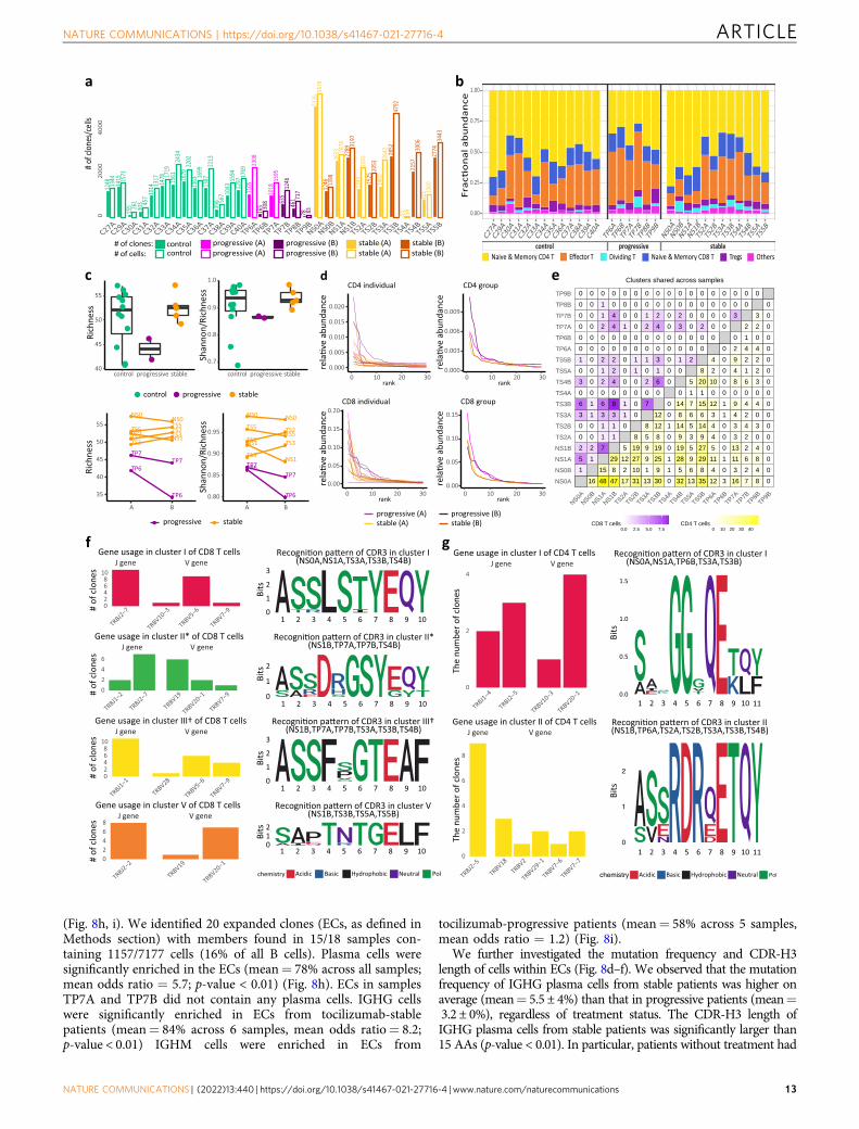
(Fig. 8h, i). We identified 20 expanded clones (ECs, as defined inMethods section) with members found in 15/18 samples con-taining 1157/7177 cells (16% of all B cells). Plasma cells weresignificantly enriched in the ECs (mean= 78% across all samples;mean odds ratio = 5.7; p-value < 0.01) (Fig. 8h). ECs in samplesTP7A and TP7B did not contain any plasma cells. IGHG cellswere significantly enriched in ECs from tocilizumab-stablepatients (mean= 84% across 6 samples, mean odds ratio= 8.2;p-value < 0.01) IGHM cells were enriched in ECs from
tocilizumab-progressive patients (mean= 58% across 5 samples,mean odds ratio = 1.2) (Fig. 8i).
We further investigated the mutation frequency and CDR-H3length of cells within ECs (Fig. 8d–f). We observed that the mutationfrequency of IGHG plasma cells from stable patients was higher onaverage (mean= 5.5 ± 4%) than that in progressive patients (mean=3.2 ± 0%), regardless of treatment status. The CDR-H3 length ofIGHG plasma cells from stable patients was significantly larger than15 AAs (p-value < 0.01). In particular, patients without treatment had
a
c e
f
d
b
control progressive stableC2
7AC2
9AC3
0AC3
1AC3
2AC3
3AC3
4AC3
5AC3
6AC3
7AC3
8AC3
9AC4
0ATP
6ATP
6BTP
7ATP
7BTP
8BTP
9BNS0
ANS0
BNS1
ANS1
BTS
2ATS
2BTS
3ATS
3BTS
4ATS
4BTS
5ATS
5B
0.00
0.25
0.50
0.75
1.00
Naive & Memory CD4 T Naive & Memory CD8 TEffector T TregsDividing T Others
g
40
45
50
55
control progressive stable
Rich
ness
control progressive stable
0.7
0.8
0.9
1.0
control progressive stable
Shan
non/
Rich
ness
NS0NS0
NS1NS1
TP6
TP6
TP7TP7
TS2TS2
TS3TS3
TS5 TS5
35
40
45
50
55
A B
Rich
ness
a aprogressive stable
NS0 NS0
NS1
NS1TP6
TP6
TP7TP7
TS2TS2
TS3
TS3
TS5TS5
0.80
0.85
0.90
0.95
A B
Shan
non/
Rich
ness
0.000
0.005
0.010
0.015
0.020
0 10 20 30rank
CD4 individual
0.000
0.003
0.006
0.009
rank
CD4 group
0.00
0.05
0.10
0.15
0.20
rank
progressive (A) progressive (B)stable (A) stable (B)
CD8 individual
0.00
0.05
0.10
0.15
rank
CD8 group
0 10 20 30
0 10 20 30 0 10 20 30
1248 13
4414
15 1571
155 26
130
3 457 10
14 1317
1478 17
1915
6124
3416
7022
0213
63 1699
1387
2213
396 56
7 1043
1594
1297
1769
1105
2308
165 30
810
1815
9563
512
4646
1 717
78 8351
2612
46 1398
2633 28
7827
89 3197
1345
2265
1525
2053
1460
2642 28
5247
9255 58
2157
3006
821 10
6027
7434
43
020
0040
00
C27AC29A
C30AC31A
C32AC33A
C34AC35A
C36AC37A
C38AC39A
C40ATP6A
TP6BTP7A
TP7BTP8B
TP9BNS0A
NS0BNS1A
NS1BTS2A
TS2BTS3A
TS3BTS4A
TS4BTS5A
TS5B
# of c
lones
/cell
s
control# of clones:# of cells:
progressive (A)progressive (A)
progressive (B)progressive (B)
astable (A)stable (A)
stable (B)stable (B)
5676
control
J gene V gene
TRBJ2−7
TRBV10−3
TRBV5−6
TRBV7−902468
10
# of
clo
nes
Gene usage in cluster I of CD8 T cells
J gene V gene
TRBJ1−2
TRBJ2−7
TRBV19
TRBV20−1
TRBV7−90246
# of
clo
nes
Gene usage in cluster II* of CD8 T cells
J gene V gene
TRBJ1−1
TRBV28
TRBV5−6
TRBV7−902468
10
# of
clo
nes
Gene usage in cluster III† of CD8 T cells
J gene V gene
TRBJ2−2
TRBV19
TRBV20−102468
# of
clo
nes
Gene usage in cluster V of CD8 T cells
0
1
2
3
Bits
(NS0A,NS1A,TS3A,TS3B,TS4B)
0
1
2
Bits
(NS1B,TP7A,TP7B,TS4B)
0
1
2
3
Bits
† (NS1B,TP7A,TP7B,TS3A,TS3B,TS4B)
012
Bits
chemistry Acidic Basic Hydrophobic Neutral Pol
(NS1B,TS3B,TS5A,TS5B)
1 2 3 4 5 6 7 8 9 10
1 2 3 4 5 6 7 8 9 10
1 2 3 4 5 6 7 8 9 10
1 2 3 4 5 6 7 8 9 10
J gene V gene
TRBJ1−4
TRBJ2−5
TRBV10−3
TRBV20−10
2
4
The
num
ber o
f clo
nes
Gene usage in cluster I of CD4 T cells
J gene V gene
TRBJ2−5
TRBV18TRBV2
TRBV29−1
TRBV7−6
TRBV7−70
2
4
6
8
The
num
ber o
f clo
nes
Gene usage in cluster II of CD4 T cells
0.0
0.5
1.0
1.5
1
Bits
(NS0A,NS1A,TP6B,TS3A,TS3B)
2 3 4 5 6 7 8 9 10 11
chemistry Acidic Basic Hydrophobic Neutral Pol
0
1
2
Bits
(NS1B,TP6A,TS2A,TS2B,TS3A,TS3B,TS4B)
1 2 3 4 5 6 7 8 9 10 11
NATURE COMMUNICATIONS | https://doi.org/10.1038/s41467-021-27716-4 ARTICLE
NATURE COMMUNICATIONS | (2022) 13:440 | https://doi.org/10.1038/s41467-021-27716-4 | www.nature.com/naturecommunications 13
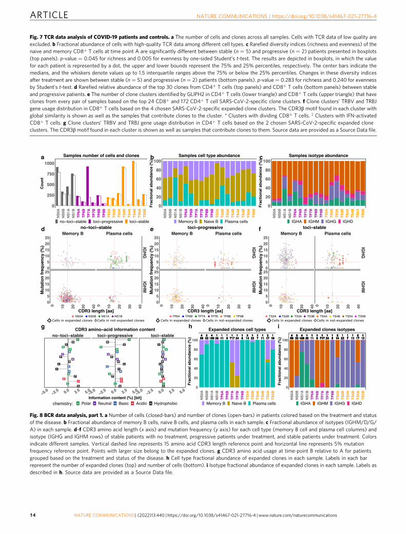
Fig. 7 TCR data analysis of COVID-19 patients and controls. a The number of cells and clones across all samples. Cells with TCR data of low quality areexcluded. b Fractional abundance of cells with high-quality TCR data among different cell types. c Rarefied diversity indices (richness and evenness) of thenaive and memory CD8+ T cells at time point A are significantly different between stable (n = 5) and progressive (n = 2) patients presented in boxplots(top panels). p-value = 0.045 for richness and 0.005 for evenness by one-sided Student’s t-test. The results are depicted in boxplots, in which the valuefor each patient is represented by a dot, the upper and lower bounds represent the 75% and 25% percentiles, respectively. The center bars indicate themedians, and the whiskers denote values up to 1.5 interquartile ranges above the 75% or below the 25% percentiles. Changes in these diversity indicesafter treatment are shown between stable (n = 5) and progressive (n = 2) patients (bottom panels). p-value = 0.283 for richness and 0.240 for evennessby Student’s t-test. d Rarefied relative abundance of the top 30 clones from CD4+ T cells (top panels) and CD8+ T cells (bottom panels) between stableand progressive patients. e The number of clone clusters identified by GLIPH2 in CD4+ T cells (lower triangle) and CD8+ T cells (upper triangle) that haveclones from every pair of samples based on the top 24 CD8+ and 172 CD4+ T cell SARS-CoV-2-specific clone clusters. f Clone clusters’ TRBV and TRBJgene usage distribution in CD8+ T cells based on the 4 chosen SARS-CoV-2-specific expanded clone clusters. The CDR3β motif found in each cluster withglobal similarity is shown as well as the samples that contribute clones to the cluster. * Clusters with dividing CD8+ T cells. † Clusters with IFN-activatedCD8+ T cells. g Clone clusters’ TRBV and TRBJ gene usage distribution in CD4+ T cells based on the 2 chosen SARS-CoV-2-specific expanded cloneclusters. The CDR3β motif found in each cluster is shown as well as samples that contribute clones to them. Source data are provided as a Source Data file.
Fig. 8 BCR data analysis, part 1. a Number of cells (closed-bars) and number of clones (open-bars) in patients colored based on the treatment and statusof the disease. b Fractional abundance of memory B cells, naive B cells, and plasma cells in each sample. c Fractional abundance of isotypes (IGHM/D/G/A) in each sample. d–f CDR3 amino acid length (x axis) and mutation frequency (y axis) for each cell type (memory B cell and plasma cell columns) andisotype (IGHG and IGHM rows) of stable patients with no treatment, progressive patients under treatment, and stable patients under treatment. Colorsindicate different samples. Vertical dashed line represents 15 amino acid CDR3 length reference point and horizontal line represents 5% mutationfrequency reference point. Points with larger size belong to the expanded clones. g CDR3 amino acid usage at time-point B relative to A for patientsgrouped based on the treatment and status of the disease. h Cell type fractional abundance of expanded clones in each sample. Labels in each barrepresent the number of expanded clones (top) and number of cells (bottom). i Isotype fractional abundance of expanded clones in each sample. Labels asdescribed in h. Source data are provided as a Source Data file.
ARTICLE NATURE COMMUNICATIONS | https://doi.org/10.1038/s41467-021-27716-4
14 NATURE COMMUNICATIONS | (2022) 13:440 | https://doi.org/10.1038/s41467-021-27716-4 | www.nature.com/naturecommunications
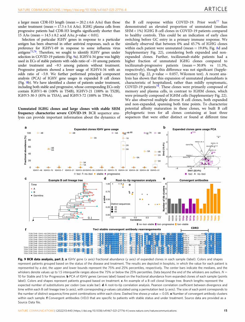
a larger mean CDR-H3 length (mean= 20.2 ± 6.6 AAs) than thoseunder treatment (mean= 17.5 ± 5.4 AAs). IGHG plasma cells fromprogressive patients had CDR-H3 lengths significantly shorter than15 AAs (mean= 14.5 ± 8.2 acid AAs; p-value < 0.01).
Selection of particular IGHV genes in response to a particularantigen has been observed in other antiviral responses, such as thepreference for IGHV1-69 in response to some influenza virusantigens75,76. Therefore, we sought to identify IGHV genes underselection in COVID-19 patients (Fig. 9a). IGHV4-34 gene was highlyused in ECs of stable patients with odds ratio of ~10 among patientsunder treatment and ~9.5 among patients without treatment.Progressive patients showed a lower usage of IGHV4-34 with anodds ratio of ~5.9. We further performed principal componentanalysis (PCA) of IGHV gene usages in expanded B cell clones(Fig. 9b). We have identified a cluster of patients under treatment,including both stable and progressive, whose corresponding ECs onlycontain IGHV1-46 (100% in TS4B), IGHV3-21 (100% in TS2B),IGHV3-30-3 (85% in TS3A), and IGHV3-72 (100% in TP6A).
Unmutated IGHG clones and large clones with stable SHMfrequency characterize severe COVID-19. BCR sequence ana-lysis can provide important information about the dynamics of
the B cell response within COVID-19. Prior work77 hasdemonstrated an elevated proportion of unmutated (medianSHM < 1%) IGHG B cell clones in COVID-19 patients comparedto healthy controls. This could be an indication of early classswitching before GC entry in a primary immune response. Wesimilarly observed that between 0% and 45.7% of IGHG cloneswithin each patient were unmutated (mean= 19.8%; Fig. 8d andSupplementary Fig. 22), considering both expanded and non-expanded clones. Further, tocilizumab-stable patients had ahigher fraction of unmutated IGHG clones compared totocilizumab-progressive patients (mean= 30.8% vs 11.3%,respectively), though this difference was not significant (Supple-mentary Fig. 22, p-value = 0.057, Wilcoxon test). A recent ana-lysis has shown that this expansion of unmutated plasmablasts ischaracteristic of hospitalized, rather than mildly symptomatic,COVID-19 patients78. These clones were primarily composed ofmemory and plasma cells, in contrast to IGHM clones, whichwere primarily composed of IGHM cells (Supplementary Fig. 22).We also observed multiple diverse B cell clones, both expandedand non-expanded, spanning both time points. To characterizepotential affinity maturation in these clones, we built B cellphylogenetic trees for all clones containing at least threesequences that were either distinct or found at different time
4B
0A0B
2B
1A3A
3A
6A
1A
1B 8B
2A
5A
5B
0A
7A7B
5A
0A
0B
6B
2A
0
20
40
60
80
100
IGHV1−46 IGHV2−70 IGHV3−7 IGHV3−21 IGHV3−30 IGHV3−30−3 IGHV3−33 IGHV3−72 IGHV4−34 IGHV4−39 IGHV4−59 IGHV5−51
Fra
ctio
nal
abu
nd
ance
(%
)
toci no−toci stable progressive
Expanded clones IGHV gene usagea
NS0A
NS0B
NS1A
NS1B
TP6A
TP6B
TP7ATP7BTP8B
TS2A
TS2B
TS3A
TS4B
TS5ATS5B
IGHV3−30
IGHV3−33IGHV3−7
IGHV3−30−3IGHV2−70
IGHV4−34
IGHV4−39
IGHV5−51
IGHV4−59
IGHV3−21IGHV1−46
IGHV3−72
−1
0
1
2
3
−4 −2 0 2Dim1 (22.4%)
Dim
2 (1
5.4%
)
a atoci no−toci
Expanded clones gene usage PCAb
Time
ABGermline
Example B cell lineage treec
0.00
0.25
0.50
0.75
1.00
−1.0 −0.5 0.0 0.5 1.0Correlation
p v
alu
e
Distinct sequences204060
Root−to−tip regression analysisd
0
1
2
3
4
5
6
NS
0A
NS
0B
NS
1A
NS
1B
TP
6A
TP
6B
TP
7A
TP
7B
TS
2A
TS
2B
TS
3A
TS
3B
TS
4B
TS
5A
TS
5B
Nu
mb
er o
f A
b c
lust
ers
Convergent antibodiese
no−toci−stable toci−progressive toci−stable
TS3B,TS4B: IGHV1-46/CDR3/IGHJ3 TS3B,TS4B: IGHV3-23/CDR3/IGHJ6 TS3A,TS4B: IGHV3-33/CDR3/IGHJ6 TS3A,TS4B: IGHV3-33/CDR3/IGHJ6 TS3B,TS4B: IGHV3-48/CDR3/IGHJ3 TS3B,TS4B: IGHV3-48/CDR3/IGHJ3 TS3B,TS4B: IGHV4-59/CDR3/IGHJ6TS3A,TS3B,TS4B: IGHV5-51/CDR3/IGHJ4TS3A,TS3B,TS4B: IGHV5-51/CDR3/IGHJ4TS3A,TS3B,TS4B: IGHV5-51/CDR3/IGHJ4TS3A,TS3B,TS4B: IGHV5-51/CDR3/IGHJ4
Toci stable convergent antibody rearrangements CDR3f
0.1
Time
ABGermline
Fig. 9 BCR data analysis, part 2. a IGHV gene (x axis) fractional abundance (y axis) of expanded clones in each sample (label). Colors and shapesrepresent patients grouped based on the status of the disease and treatment. The results are depicted in boxplots, in which the value for each patient isrepresented by a dot, the upper and lower bounds represent the 75% and 25% percentiles, respectively. The center bars indicate the medians, and thewhiskers denote values up to 1.5 interquartile ranges above the 75% or below the 25% percentiles. Data beyond the end of the whiskers are outliers. N =10 for Stable and 5 for Progressive. b PCA of IGHV genes (arrows label) based on the fractional abundance from expanded clones of each sample (pointslabel). Colors and shapes represent patients grouped based on treatment. c An example of a B cell clonal lineage tree. Branch lengths represent theexpected number of substitutions per codon (see scale bar). d A root-to-tip correlation analysis. Pearson correlation coefficient between divergence andtime within each B cell lineage tree (x axis), with corresponding p-values calculated using a permutation test (y axis). The size of each point corresponds tothe number of distinct sequence/time point combinations within each clone. Dashed line shows p-value = 0.05. e Number of convergent antibody clusterswithin each sample. f Convergent antibodies (VDJ) that are specific to patients with stable status and under treatment. Source data are provided as aSource Data file.
NATURE COMMUNICATIONS | https://doi.org/10.1038/s41467-021-27716-4 ARTICLE
NATURE COMMUNICATIONS | (2022) 13:440 | https://doi.org/10.1038/s41467-021-27716-4 | www.nature.com/naturecommunications 15
points (largest shown in Fig. 9c). We observed a relatively highlevel of SHM in these clones at time point A (mean= 4.7%). Wethen used a phylogenetic root-to-tip correlation test (see “Meth-ods” section) to determine if divergence from the germlinesequence increased between time points A and B in these 20clones. None showed a significant positive correlation betweensample time and divergence from the sequences’ most recentcommon ancestor (i.e., p-value > 0.05; Fig. 9d). These resultsindicate a lack of measurable SHM accumulation between timepoints in these clones. Taken together, the observed expansion ofunmutated plasmablasts and lack of measurable B cell evolution isconsistent with prior work showing evidence of disruptedgerminal-center reactions in hospitalized COVID-19 patients79.
Convergent antibody rearrangements are elicited in COVID-19patients. We identified 19 convergent antibody clusters acrosseight patients. 79% (15/19) of convergent clusters included anti-bodies from tocilizumab-stable patients, 47% (9/19) includedantibodies from no-tocilizumab-stable patients, and 31% (6/19) ofconvergent clusters included antibodies from tocilizumab-progressive patients (Fig. 9e). We next focused on thetocilizumab-stable patients, which were represented in the mostconvergent clusters. We identified six convergent clusters whichwere composed only of tocilizumab-stable patients (Fig. 9f).These convergent antibody clusters spanned between twopatients, and were composed of IGHV1-46/IGHJ3, IGHV3-23/IGHJ6, IGHV3-33/IGHJ6, IGHV3-48/IGHJ3, IGHV4-59/IGHJ6,IGHV5-51/IGHJ4 genes.
Taken together, our multi-omics single-cell analysis revealeddynamic immune responses in patients with a stable andprogressive manifestation of COVID-19 under tocilizumabtreatment. Our comprehensive immune profiling underscoresthe overwhelming IFN-I response and desynchronized adaptiveand innate immune interaction in COVID-19. Joint profiling ofgene expression and surface proteins uncovered hyper IFN-Iresponse in activated T cell subset in progressive patients.Excessive regulatory innate immune response and unique co-inhibitory receptor expression in activated T cells are hallmarks ofprogressive disease. Skewed T cell repertories in CD8+ T cell anduniquely enriched VDJ sequences are identified in COVID-19patients. Plasmablasts expansion without acquiring somatichypermutation is consistent with an early primary and/orextrafollicular response during the acute phase of SARS-CoV-2infection. Lastly, we characterize the cellular response totocilizumab, including decreased expression of S100A8/9 intocilizumab-treated patients across most cell types.
DiscussionAlthough initial studies in COVID-19 patients with severerespiratory failure revealed a dysregulated immune system withhyper-inflammatory responses and lymphopenia12–14,80–82, thereare critical deficits in our understanding of the mechanismsunderlying this immune dysfunction. Here, we applied multi-modal single-cell analysis using 5′ scRNA-seq, CITE-seq, TCR,and BCR sequencing on 18 samples from 10 patients withCOVID-19, which were compared to 13 control samples. Ourexperimental design allowed us to determine: (1) the dynamics ofthe immune response in COVID-19 over time; (2) generalimmune cell features in COVID-19 patients; (3) the specificimmune signature associated with progressive disease; (4) an in-depth exploration of adaptive immunity using T cell and B cellrepertoire analysis in COVID-19; and (5) the effects of tocilizu-mab treatment. Our unbiased systems biology approach utilizingnovel multimodal single-cell analysis techniques reveals thetemporal dynamics of immune responses to this disease. It
highlights the unique immune features that distinguish stable andprogressive COVID-19 patients. The immunological networkscharacterized in our study improve the understanding of theabundant cellular interactions and effects that promote pathologyin severe disease, including dyssynchrony of the innate andadaptive immune response.
We observed dominant effects of a type-1 IFN response acrossall immune cells in all COVID-19 patients, especially at the earliertime-point A, consistent with the acute viral infection83. In thisregard, we highlight the IFN-activated CD8+ T cell cluster as theextreme archetype of type-1 IFN response in T cells, which wasalmost exclusively found in COVID-19 patients, especially attime-point A (Fig. 1f). Type-1 IFN response was the main driverof gene expression changes between progressive and stable sub-jects, as depicted in Fig. 2a. Progressive patients had higherexpression of ISGs in all major cell types. Type-1 IFN response, asreflected by the IFN gene score, decreased overwhelmingly fromtime point A to B in all patients, and was highly correlated withtime (R= 0.97, Fig. 2f, g). This is not surprising given that thefirst blood draw was obtained at least 5 days after the beginning ofsymptoms, the onset of which occurs a median of 5.2 days afterinfection84. At this time-point, patients are expected to be on thedescending slope of the viral load curve30, which type-1 IFNresponse closely follows (R= 0.8 for the correlation between log10viral load and IFN score, Fig. 2e). Interestingly, the ISG signatureat the later time-point B returned towards control level in allpatients except for two out of four in the progressive group(Fig. 2a), in which both IFN score and viral load remainedrelatively high. This is consistent with a previous study in whichsevere COVID-19 patients had a higher viral load and longervirus shedding than mild cases85.
The joint profiling of gene expression and surface proteins shedlight on a specific T cell population that expressed higherHLADR, CD38, and cell cycle markers (MKI67, PCNA, AURKA)and expanded with time. Although there was no significant dif-ference in frequency noted between progressive vs stable COVID-19 patients in our cohort, we demonstrated the enrichment ofISGs and a unique set of co-inhibitory receptors (LAG-3 andTIM-3, coded by HAVCR2) in progressive patients. Interestingly,TIGIT and PD-1 (coded by PDCD1) were relatively higher instable patients together with TCF7 and LEF1, the markers forprogenitor exhausted T cells63. In general, T cell exhaustion isobserved in chronic infectious diseases and cancer where T cellsreceive sustained stimuli to be activated for long-term in animmunoregulatory milieu86,87. In contrast, our observation insevere SARS-CoV-2 infection indicates that a unique activated Tcell phenotype can also be induced in acute viral infection88,which is not commonly seen. Among signatures observed inprogressive patients, elevated LAG3 expression was detectedacross most T/NK cell subsets, except for the Treg cluster16,40,71.Given that the expression of MHC-II molecules (which are theligands for LAG-3) is markedly downregulated in APCs in pro-gressive COVID-19 patients, the disruption of LAG3-MHC-IIinteraction might play a critical role in COVID-19 immuno-pathology. While the trigger-inducing LAG-3 on T cells is notwell understood, co-expression of ISGs in T cells from progressivepatients suggests a possible role of IFN-I on LAG-3 expression.Further studies focusing on the molecular mechanisms by whichSARS-CoV-2 induces LAG-3 on T cells are warranted.
A skewed phenotype of myeloid cells toward a regulatory/immunosuppressive signature has been previously reported insevere COVID-1913,14. In contrast, a pro-inflammatory monocytephenotype has been shown by others in patients with severeCOVID-1980–82. Our findings of an anti-inflammatory monocytecluster that is increased in COVID-19 patients, along with sup-pressive/tissue-repair gene expression changes in monocytes that
ARTICLE NATURE COMMUNICATIONS | https://doi.org/10.1038/s41467-021-27716-4
16 NATURE COMMUNICATIONS | (2022) 13:440 | https://doi.org/10.1038/s41467-021-27716-4 | www.nature.com/naturecommunications
are accentuated in progressive patients (including CD163, IL1R2,AREG, MRC1, HAVCR2, LGALS9, IL10), support the formerevidence. We also observed the downregulation of IL-1β mRNAexpression in COVID-19 patients, indicating a shift from pro-inflammatory to a regulatory phenotype in monocytes inCOVID-19. Our multimodal single-cell analysis demonstratedthat these regulatory monocytes are transcriptionally dis-tinguished from the other classical monocytes and resemblemyeloid-derived suppressor cells (MDSCs), a heterogeneousmyeloid cell population characterized by strong immunosup-pressive function increased in chronic infection and cancer. Theincreased fraction of MDSC-like anti-inflammatory monocytesfacilitates the resolution of inflammation during acute viralinfection, transitioning from inflammation to tissue repair89,90.However, an inadequate anti-inflammatory/tissue repair responsecan delay virus clearance, cause chronic inflammation, and leadto excessive tissue damage and even tissue fibrosis91–94. There-fore, a regulated transition of the immune response frominflammation to tissue repair with appropriate kinetics is essentialto restore tissue homeostasis.
We observed an increased expression of the suppressive cyto-kine IL10 in myeloid cells and several additional cell types in theprogressive patients (Supplementary Fig. 8a). Levels of IL-10 inplasma are known to be increased in severe COVID-19, asreported in our recent study4 as well as by others7,31. While IL-10is critical to protect the host from tissue damage during acuteimmune responses, it also exhibits a detrimental immunopatho-genic effect during acute viral infections by downregulation ofMHC-II expression92. Our data clearly demonstrated the upre-gulation of IL-10 and the downregulation of MHC-II expressionin monocytes which may contribute to the detrimental clinicalcourse in progressive patients compared with stable ones.
Our T cell receptor repertoire analysis shows a higher expan-sion/dominance and a lower richness of CD8+ but not CD4+ Tcell clones in progressive COVID-19 in progressive patientsrelative to stable patients. In an attempt to identify specific clo-notypes that are relevant to T cell response against SARS-CoV-2,we studied the CDR3 motifs that are shared by several COVID-19patients but absent from the control subjects. Using thisapproach, we identified 10 CD8+ and 12 CD4+ T cell specificitygroups, and described their specific V & J gene usage patterns.Moreover, we describe the specific CDR3 motifs that have thehighest likelihood of being COVID-19 specific. In future experi-ments, these TCRs will be investigated for antigen specificity.
During viral infection, B cells are critical for the production ofprotective antibodies. The establishment of a diverse repertoire ofantibodies is imperative to protect a host from pathogens, as wellas to generate effective immune responses. One key finding is thatCDR-H3 amino acid usage profiles were highly variable amongpatients (Fig. 8g). In particular, there was no preference for anyamino acid between early and late time points in stable patientswho received tocilizumab. In contrast, progressive and stablepatients who were not treated with tocilizumab showed a dif-ferent profile of CDR-H3 amino acid selection which generallyvaried across amino acids with a trend toward increased usage ofalanine (A), aspartic acid (D), and tyrosine (Y) at time-point Brelative to A. The importance of CDR-H3-tyrosine for optimalantibody binding was previously shown for the influenza Avirus95. Another finding focused on the detection of convergentantibodies with highly similar VDJs across COVID-19 patients(Fig. 8n, o).
Our V(D)J B cell receptor repertoire analysis further suggests acomplex B cell response in COVID-19. Consistent with theexpectations of a primary immune response, we observed an highproportion of unmutated IGHG B cell clones, which has beenreported in at least one other analysis of BCR repertoires from
COVID-19 patients77. However, we also observed multiplemutated B cell clones that were persistent across the two mea-sures time points, but did not measurably accumulate SHMbetween time points (Fig. 8l, m)96. These could result from cross-reactivity of memory B cells with other common corona-viruses,which has been documented in T cells86. Such memory B cellswould have already accumulated SHM and likely avoid germinal-center re-entry97. This scenario may account for the quickexpansion of plasma cells in COVID-19 patients (Fig. 1f), whichhas also been reported by others14,60. It is also possible these areclones are non-coronavirus-specific persistent clones sometimesobserved in healthy older patients98. Importantly, these analyseswere performed over a short time interval, and used a relativelysmall number of B cells with unknown specificity. We note,however, that the lack of observable SHM increase between timepoints is consistent with another recent study which found thatlevels of SHM in SARS-CoV-2-specific antibodies were stable(~3%) between 8 and 42 days post-diagnosis99.
The relatively small sample size (18 COVID-19 samples and 13controls) is a limitation of this study, especially for the analysis ofcertain subgroups (e.g., only four samples from patients who didnot receive tocilizumab). However, it is larger than manyCOVID-19 single-cell multi-omics studies published to-date14,80,81,100, and the similarity of baseline characteristicsbetween stable and progressive patients and in comparison, tocontrols (Supplementary Table 1) helps increase confidence inour results. Although the timing of blood draw A (time-point A)relative to hospitalization was consistent across subjects, thetiming of blood draw B (time-point B) was variable. We mitigatedthat by taking into account the variable time span between thetwo blood draws in some of the analyses, e.g., for the analysis ofIFN score changes over time shown in Fig. 3a, b. This uniqueexploration of gene expression changes over time adds anessential dynamic layer that is critical to understand the biologyof an acute viral disease, although tocilizumab treatment mayhave influenced the gene expression changes. However, whencomparing the gene expression at time point A and B (Fig. 2a),most differentially expressed genes are related to the strong type-1 interferon response rather than tocilizumab effects. This led usto conclude that the response to the virus (rather than to thetreatment) is the main driver of gene expression changes overtime. Nonetheless, we devoted a part of the results to explore theeffects of tocilizumab by comparing gene expression changes inthe untreated patients across time to those of the treated patients,and uncovered genes (in each cell type) whose change is likely tobe attributed to tocilizumab effects rather than time. Lastly, ouranalysis mostly relied on RNA-based analyses including geneexpression and TCR/BCR repertoire analysis, with some protein-level validation by CITE-seq and flow cytometry. Additionalmechanistic validation, while beyond the scope of this study, iswarranted in future studies.
In conclusion, our in-depth multi-omics assessment of per-ipheral immune cells at single-cell resolution across patientseverities and time highlights the desynchronized adaptive andinnate immune response in progressive COVID-19 patients. Aprominent type-1 interferon response is observed across allimmune cells, especially in progressive patients, and wanes overtime in correlation to the decrease in viral loads. Excessive reg-ulatory innate immune response and LAG-3 positive activatedT cells are the hallmarks of progressive disease. Skewed T cellreceptor repertoires in CD8+ T cell and uniquely enriched V(D)Jsequences are identified in COVID-19 patients. B cell receptorrepertoire analysis reveals a high level of IGHG B cell clones withlittle or no somatic hypermutation, consistent with an early pri-mary and/or extrafollicular immune response, as well as mutatedclones which may reflect stimulation of pre-existing memory B
NATURE COMMUNICATIONS | https://doi.org/10.1038/s41467-021-27716-4 ARTICLE
NATURE COMMUNICATIONS | (2022) 13:440 | https://doi.org/10.1038/s41467-021-27716-4 | www.nature.com/naturecommunications 17
cells. Overall, our comprehensive immune profiling underscoresthe desynchronized innate and adaptive immune interaction inprogressive COVID-19, which may lead to delayed virus clear-ance. This high-resolution understanding of the immune cellprofiles underlying severe COVID-19 will enhance our ability todevelop immunomodulatory therapeutic approaches to preventprogression in COVID-19 patients.
MethodsSettings. The study was performed on deidentified, cryopreserved PBMC samplesof 10 COVID-19 patients and 13 matched controls, obtained with informed con-sent on a protocol approved by Yale Human Research Protection Program Insti-tutional Review Boards (FWA00002571, Protocol ID. 2000027690).
Patients and samples. Ten COVID-19 patients hospitalized at Yale-New HavenHospital (YNHH) were recruited for this study. All were confirmed to haveCOVID-19 by RT-PCR testing of nasopharyngeal samples. Four of the patientswere “Progressive” (TP6, TP7, TP8, TP9), defined as patients who requiredadmission to the intensive care unit (ICU) and eventually succumbed to the dis-ease. At the same time, the other 6 were “Stable” (NS0, NS1, TS2, TS3, TS4, TS5),defined as patients hospitalized in non-ICU internal medicine wards who wereeventually discharged. Eight patients (80%) were treated with Tocilizumab, ahumanized anti-IL6 receptor antibody. Only patients NS0 and NS1 did not receivethis drug (designated with “N”). Tocilizumab was given once at a dose of 8 mg/kg(up to a maximal dose of 800 mg), as part of the COVID-19 treatment algorithmused in the Yale-New Haven Health System; patients who required ≥3 l/min O2 or≥2 l/min but with CRP > 70 were treated with tocilizumab. All patients were treatedwith antivirals (Atazanavir, except for patient NS1 which was treated withRemdesivir) and with Hydroxychloroquine (except for NS1). Two progressivesubjects were treated with corticosteroids: patient TP7 was treated with Prednisone40 mg daily for 2–3 days just before the blood draw A, and patient TP9 was treatedwith Methylprednisolone 120 mg daily for 1–2 days prior to blood draw B. Noother immunosuppressive, immunomodulatory, or antiviral agents were used.
Eighteen blood samples were collected from these ten patients, at different timepoints as described in the results section and Fig. 1b, c. Thirteen control subjectswere recruited prior to the COVID-19 pandemic. Baseline characteristics ofCOVID-19 patients and controls are presented in Supplementary Table 1. Thetiming of symptom onset, hospitalization, tocilizumab treatment, and blood drawsfor each patient is shown in Fig. 1d.
Isolation of PBMC and cryopreservation. PBMCs were isolated from wholeblood using density gradient centrifugation, according to the following protocol:Histopaque 20 ml was added to a 50 ml SepMate tube, then overlaid with freshblood 1:1 diluted in PBS 2% fetal bovine serum (FBS) and centrifuged at 1200×g for10 minutes. The PBMC layer was collected by quickly pouring the remainingcontents above the SepMate insert into a fresh tube, and washed once with PBS at650 × g for 10 min. The supernatant was decanted and ACK red blood cell lysisbuffer (2 ml/sample) was added for 2 minutes; another wash with PBS 2% FBS wasdone, followed by centrifugation at 290 × g to remove platelets and supernatantaspirated. Following resuspension of the pellet, PBMCs were cryopreserved inaliquots of 5 × 10⁶ cells using 10% DMSO in heat inactivated-FBS as the cryo-preservation solution. Cryovials were placed in a freezing container (Mr. Frosty)and transferred immediately to a −80 °C freezer for >24 h before being transferredto long-term liquid nitrogen storage.
Sample preparation and 10x barcoding. All sample processing steps were done ina biosafety level 2+ laboratory. Samples were thawed in a water bath at 37 °C for~2 min without agitation, and removed from the water bath when a tiny ice crystalstill remains. After thawing, cells were gently transferred to a 50 mL conical tubeusing a wide-bore pipette tip, the cryovial was rinsed with a cold growth medium(10% FBS in DMEM) to recover leftover cells, and the rinse medium was addeddropwise (1 drop per 5 s) to the 50 ml conical tube while gently shaking the tube.Next, we conducted serial dilutions with cold growth medium a total of 5 times by1:1 volume addition with ~1 min wait between additions. Cold growth medium wasadded at a speed of 3–5 ml/s, achieving a final volume of 32 ml. The cells were thencentrifuged at 300 × g for 5 minutes at 4 °C, and the supernatant was removedwithout disrupting the cell pellet. The pellet was resuspended in 1× PBS with 0.04%BSA, and the sample was filtered with a 40 μM strainer. Cell concentration wasdetermined using Trypan blue staining with a Countess automated cell counter(ThermoFisher). Following this cell count, each sample was split into two parts(Fig. 1a): one was immediately loaded onto the 10x Chromium Next GEM Chip G,according to the manufacturer’s user guide (document number CG000208, revisionE, February 2020), and the other was further processed for CITE-seq as describedin the next section, and then loaded to the 10x Chromium Chip G. In total, weloaded 18 “conventional” samples into 18 Chip G lanes (aiming for recovery of10,000 cells per lane), and 17 out of 18 “CITE-seq” samples into 6 Chip G lanes(each lane containing 5-6 pooled hashed samples, as portrayed in Supplementary
Data 6). One out of 18 CITE-seq samples (TP8B) was not pooled because of verylow cell concentration.
CITE-seq and cell hashing. The lyophilized Total-seq C human panel (BioLegend)was resuspended with 35 μl of wash buffer, vortexed for 10 s, and incubated for5 min at RT. Total-seq C human Hashtag antibodies (Biolegend) were centrifugedat 20,000 × g for 10 min and 6-fold diluted with wash buffer (2% FBS and 1mMEDTA in PBS). To maximize performance, both were centrifuged at 20,000 × g for10 min just before adding to the cells. See Supplementary Data 5 for a list ofantibodies, clones, and barcodes used for CITE-seq and hashing samples.
PBMCs from each sample were reconstituted with wash buffer at theconcentration of 10–20 × 106 cells/ml and incubated on ice for 10 min with 5ul ofHuman FC block (BD Biosciences) and 5 μl of TrueStain Monocyte Blocker(Biolegend). 10–20 μl (0.1–0.2 × 10⁶ cells) were transferred into a new tube andincubated on ice for 30 min with 5 μl of CITE-seq panels and 5 μl of Hashtagantibodies prepared as above. Cells were washed twice with wash buffer and with2% FBS in PBS for the third wash. Samples were pooled into one tube based on cellcounts, and super-loaded onto the 10× Chromium Chip G, aiming for recovery of~20,000 cells per sample. See Supplementary Data 5 for the details of 6 pooledsamples (CITE#1-CITE#6).
cDNA libraries preparation and sequencing. The loaded Chip G was placed inthe 10x Chromium controller to create Gel Beads-in-emulsion (GEMs). The nextsteps were carried out according to the manufacturer’s user guide, including GEM-RT incubation, post-GEM-RT Dynabead cleanup, and cDNA amplification. ThecDNA samples were used to construct 4 types of cDNA libraries, according to thesteps outlined in the user guide: gene expression libraries, T-cell receptor libraries,B-cell receptor libraries, and cell surface protein libraries (the latter only forsamples processed with CITE-seq). cDNA libraries were then sequenced on anIllumina Novaseq 6000 platform.
Flow cytometry. Freshly isolated PBMCs were incubated with FC block reagent(Biolegend) for 10 min and stained with LIVE/DEAD Fixable Aqua Dead Cell Stainkit (ThermoFisher) for 20 min at 4 °C. Following a wash, cells were then blockedwith Human TruStan FCX (BioLegend) for 10 min at RT. Cocktails of the followingantibodies were directly added to this mixture for 30 min at RT. BB515 anti-HLA-DR (G46-6), BV605 anti-CD3 (UCHT1), BV785 anti-CD4 (SK3), APCFire750anti-CD8 (SK1), BV421 anti-CCR7 (G043H7), AlexaFluor 700 anti-CD45RA(HI100), PE anti-PD1 (EH12.2H7), APC anti-TIM3 (F38-2E2), BV711 anti-CD38(HIT2), BB700 anti-CXCR5 (RF8B2), PE-Cy7 anti-CD127 (HIL-7R-M21), PE-CF594 anti-CD25 (BC96), BV711 anti-CD127 (HIL-7R-M21), BV421 anti-LAG-3(11C3C65). Cells were washed two times with staining buffer and acquired on a BDFortessa or Cytoflex flow cytometer. FlowJo software (Treestar) was used foranalysis.
SARS-CoV-2 viral load measurements. Nasopharyngeal swabs and saliva sam-ples were collected from COVID-19 diagnosed inpatients at -Yale-New HavenHospital, as described elsewhere101. We extracted total nucleic acid using theMagMax Viral/Pathogen Nucleic Acid Isolation kit (ThermoFisher Scientific,Waltham, MA, USA) with 300 µl of input sample eluted into 75 µl, using a slightlymodified protocol (dx.doi.org/10.17504/protocols.io.bg3pjymn). A total of 5 µl ofextracted nucleic acid was used as input in the RT-qPCR assay for SARS-CoV-2detection, as described elsewhere102. Briefly, we used the Luna Universal ProbeOne-Step RT-qPCR kit (New England Biolabs, Ipswich, MA, USA) with the CDC2019-nCoV_N1, 2019-nCoV_N2, and human RNase P (RP) primer-probe sets(Integrated DNA Technologies, Coralville, IA, USA). Viral RNA copy numberswere calculated based on 10-fold dilution standard curves of the previously gen-erated nucleocapsid (N) transcript standard102.
Data processing of raw sequencing reads. Raw sequencing reads were demul-tiplexed using Cell Ranger mkfastq pipeline to create FASTQ files. Next, CellRanger count pipeline (v3.1) was employed in order to perform alignment (usingSTAR), filtering, barcode counting, and UMI counting. We have used GRCh38(Ensembl 93) as the genome reference (corresponding to Cell Ranger referenceGRCh38-3.0.0).
ScRNA-seq sample aggregation. 10× cell ranger count filtered output data ofPBMCs from thirteen healthy controls were added to that of the eighteen COVID-19 samples. Seurat package103,104 (v3.1) was used for all downstream analyses. 10×gene expression matrices for each sample were converted and combined into oneSeurat object. Cells with mitochondrial gene percentages higher than 12% and cellswith less than 200 genes were excluded from the study to filter out dead and dyingcells. For CITE-seq samples, following de-hashing, cell barcodes of multiplets (i.e.,with 2 or more hashing antibody signals) or uncertain origin (i.e., with no clearhashing signal) were also removed. After these filtering steps, the gene-barcodematrix contained 35,538 genes and 163,452 barcoded cells.
ARTICLE NATURE COMMUNICATIONS | https://doi.org/10.1038/s41467-021-27716-4
18 NATURE COMMUNICATIONS | (2022) 13:440 | https://doi.org/10.1038/s41467-021-27716-4 | www.nature.com/naturecommunications
Integration, principal components analysis, and clustering. In accordance withthe standard Seurat preprocessing workflow, sample gene expressions were nor-malized using Seurat’s “LogNormalize” method103,104. The “FindVariableFeatures”function selected the 3000 genes with the highest variance to mean ratio using the“vst” method. To remove single-subject effects, samples were integrated on asubject level using 2000 anchors with a dimensionality of 30104. The integrated datawere then scaled with the “ScaleData” function.
Principal Component Analysis (PCA) was performed on the integrated data,and the first 30 Principal Components (PCs) were used in the “FindNeighbors”algorithm. The Louvain modularity optimization algorithm in “FindClusters”generated the clusters while the resolution was set to 0.75. Thirty PCs were used inthe “RunUMAP” function to create the final UMAP, and thirty clusters weregenerated from the aforementioned pipeline (Supplementary Fig. 2a).
These thirty clusters were first annotated with the SingleR software(Supplementary Fig. 2d) and then annotated manually (Fig. 1e) by using cell-specific markers (Supplementary Fig. 1) plotted on UMAP space, and by examiningthe output of “FindAllMarkers” per cluster. Five clusters out of thirty wereremoved; namely: a nonspecific cluster of low UMI cells (cluster #8), monocyte-platelet multiplets (#22), B and T/NK multiplets (#24), erythroid cellcontamination from a single subject (#25), and B cell-platelet multiplets (#29).Following the removal of these clusters, the final Seurat object contained153,554 cells.
Cell type proportions analysis. For each subject, the number of cells within agiven cell type was normalized by the subject’s total number of cells. For each celltype, cell proportions were plotted in a boxplot by disease group, namely by (1)controls, (2) stable patients at time point A, (3) stable patients at time point B, (4)progressive patients at time point A, and (5) progressive patients at time point B.We used the Wilcoxon rank-sum test to compare the cell proportions of thefollowing three groups: control subjects, stable patients, and progressive patients.
Differential gene expression analysis. The “FindMarkers” function was used toidentify differentially expressed genes (DEGs) across the following conditions: percell type, (1) time point A versus time point B; (2) controls versus COVID-19patients at time point A; and (3) progressive vs stable. For the time point com-parison (comparison 1), the logistic regression test for differential expression withsubjects set as latent variables was used to account for paired samples. For com-parisons (2) and (3), the default Wilcoxon rank test was used. Genes were rankedby absolute log2 fold-change (logFC), and those with p-values > 0.05 (adjusted formultiple comparisons) were removed.
Heatmap visualization of DEGs. DEGs were visualized as heatmaps which weregenerated by using the ComplexHeatmap package105. Cell types were binned intomonocytes, CD4+ T cells, CD8+ T cells, and B cells, and “FindMarkers” dis-tinguished DEGs for each cell type bin for (1) time point A versus time point B and(2) progressive versus stable. Genes with greater than 0.5 absolute logFC wereincluded in visualization and EnrichR pathway analysis. Samples for the pro-gressive versus stable time-point were hierarchically clustered.
Gene pathway annotation. Gene list outputs from the “FindMarkers” functionwere fed into EnrichR for pathway and ontology analysis106,107. Gene set enrich-ment analysis108 was also performed on “Dividing T cells” cluster usingKEGG109–111 and MSigDB Hallmark gene sets112, and custom gene sets (Supple-mentary Data 8).
Gene list score analysis. Seurat Function “AddModuleScore” was used to com-bine the expression of genes from IFN Score A113 (ISG15, IFI44, IFI27, CXCL10,RSAD2, IFIT1, IFI44L, CCL8, XAF1, GBP1, IRF7, CEACAM1). This function wasalso used to combine other gene list scores as well, including scores for HLA type II(HLA-DRA, HLA-DQA1, HLA-DPA1, HLA-DRB1, HLA-DPB1, HLA-DRB5, HLA-DQB1, HLA-DMA, HLA-DMB) and the IL6 pathway (ARID5A, SOCS3, PIM1,BCL3, BATF, MYC)49. The differences in gene list scores were compared between(1) control versus COVID-19 patients, (2) time point A and time point B, and (3)progressive versus stable patients. For consistency with DEG analysis andassumption of non-normality, Wilcoxon rank-sum tests were conducted on thegene set “module scores” to compare between any two given conditions and cor-rected for multiple comparisons (i.e., family-wise error rate (FWER)).
Demultiplexing (de-hashing) of CITE-seq samples. In order to demultiplex cellsin the CITE-seq samples and attribute them a biological sample, hashing antibody-derived tag (ADT) counts were normalized by library size, square-root trans-formed, and normalized for every row in the data matrix of each CITE-seq sample.To account for the inherent background noise of ADT and accurately identify a cellas tagged by a hashing ADT, histograms of each hashing ADT counts in eachCITE-seq sample were used to determine the optimal threshold of significance forhashing ADTs. As distributions appeared bimodal for the majority of hashing, wemanually set the threshold between the two modes.
Based on the previous threshold, data matrix rows with two or more significantADT were flagged as doublets, and rows with zero significant ADT flagged asunidentified, thus removed for downstream analysis.
CITE-seq ADT preprocessing and downstream analysis. Once the cells weredemultiplexed and hashing ADT counts were removed, the remaining ADT counts(192) for each CITE-seq sample were combined into one single matrix. The countsfor the remaining 43,349 cells were normalized by library size and square roottransformed. We visualized the data set using Uniform Manifold Approximationand Projection (UMAP). Cells were clustered using the Louvain communitydetection on a 15-nearest neighborhood graph and were manually annotated usinga panel of ADT markers for each cell type (cell types include: CD4+ T cells, CD8+
T cells, B cells, NK cells, monocytes, macrophages, DCs, plasma cells, neutrophils,eosinophils, platelets, and red blood cells). We also manually annotated the clustersbased on surface marker lists proposed by the Human Immunology ProjectConsortium (HIPC) (https://www.immuneprofiling.org) (Supplementary Data 7).As gene expression (GEX) data from CITE-seq was incorporated in the standardscRNA-seq analysis, there were two different annotations: one based on GEX, andone based on ADT. To measure the concordance between the two annotations, thepercentage of shared cells between each annotated cluster was computed (Sup-plementary Fig. 14b).
Differential expression analysis was performed using the Wilcoxon rank-sumtest, and p-values were adjusted for multiple hypothesis testing using theBenjamini–Hochberg correction. Data preprocessing and analysis (for ADTanalysis only) was performed in Python (version 3.8.0) using Scanpy (version1.4.6)114.
Differential connectivity (connectomic) analysis. For connectomic analysis, thecell parcellation shown in Fig. 1e was used except for IFN-activated CD8+ T cells,which were lumped into the Effector T cell cluster. These data were then mappedagainst a version of the FANTOM5 database of ligand–receptor interactions,modified to include additional immunomodulatory cues of interest to the authors(Supplementary Data 9). Each parcellation, in a given experimental condition, wasthen treated as a single signaling node for network analysis. Average expressionvalues were calculated for all ligand and receptor genes on a per-cell-type basis.Then an unfiltered edgelist (connectome) was created linking all producers of aligand to all producers of a receptor, with associated quantitative edge attributes, aspreviously described.
To compare experimental conditions, the connectomes from two experimentalconditions were directly compared to yield log-fold changes for the sending(ligand) side and receiving (receptor) side of all edges. In addition, a “perturbationscore” was calculated, which allows plotting of differential edges proportional to thedegree of change, allowing both negative and positive log-fold changes andincorporating information from both sides of a given edge. The perturbation scorethat we used was defined, for every cell vector from Celli to Cellj forligand–receptor mechanism (k), as:
scoreijk ¼ logCell
Ltestki � Cell
Lcontrolki
CellLcontrolki
0@
1A
������
������´ log
CellRtestk
j � CellRcontrolk
j
CellRcontrolk
j
0@
1A
������
������Edges were then plotted which (1) had >10% of the sending and receiving
cluster expressing the given ligand and receptor, respectively, and (2) whichdisplayed an adjusted p-value < 0.05 via a Wilcoxon Rank-Sum test comparingidentical cell types to each other across experimental conditions.
Chord diagram plotting was performed using a custom implementation of thecirclize package with directed edge thickness between cell type nodes proportionalto the above-described perturbation score, scaled per-plot. The software used forthe connectomic analysis is available at https://github.com/msraredon/Connectome (https://doi.org/10.5281/zenodo.5574620) (ref:https://doi.org/10.1101/2021.01.21.427529v1).
Tocilizumab treatment effect analysis. To investigate the treatment effects oftocilizumab on transcription levels for different cell types, we conducted differentialexpression analysis between the two sampling time points for patients in thetocilizumab treatment group and those in the non-tocilizumab group separately.The logFC from these two separate analyses, i.e., for the tocilizumab group andnon-tocilizumab group, was scatter plotted for each cell type in order to identifygenes in which the differential expression pattern observed between the two-timepoints is due to a treatment effect rather than the natural course of the diseaseprogression (Fig. 5c). In addition, we investigated the correlations across cell typesand compared results between the tocilizumab and non-tocilizumab groups(Supplementary Fig. 13). Six IL-6 pathway-related genes, which are known to beassociated with tocilizumab treatment49, are highlighted in red (Fig. 5c and Sup-plementary Fig. 13). All the entries in the heatmap matrix (Fig. 5e) are the dif-ferences in logFC between tocilizumab and non-tocilizumab groups.
T cell receptor V(D)J data processing. The raw sequencing reads of the T cellreceptor (TCR) libraries were processed using the Cell Ranger V(D)J pipeline by10x Genomics™, which assembled read-pairs into V(D)J contigs for each cell,
NATURE COMMUNICATIONS | https://doi.org/10.1038/s41467-021-27716-4 ARTICLE
NATURE COMMUNICATIONS | (2022) 13:440 | https://doi.org/10.1038/s41467-021-27716-4 | www.nature.com/naturecommunications 19
identified cell barcodes from targeted cells, annotated the assembled contigs withV(D)J segment labels and located the CDR3 regions. We only considered V(D)Jcontigs with high confidence defined by cell ranger under the default settings fordownstream analysis. Contigs that were not recognized as either alpha chain orbeta chain and cells with no beta chains were removed. Only the alpha and betachains with the largest UMI count were kept for cells with more than one alphaand/or beta chains. After the filtering, each cell has only one beta chain contig andzero or one alpha chain contig.
The data were further examined and processed for sample-to-samplecontamination and potential cell doublets. First, we removed cells with cellbarcodes found in more than 2 samples. Second, cells barcodes overlapped betweenTCR and BCR data were extracted and checked for their cell types determinedbased on the scRNA-seq gene expression data. Only cell barcodes from T cells werekept. Finally, we checked the gene expression-based cell types of all cells, and cellswithout an assigned cell type or not belonging to the T cell category were removed.The T cell category includes 13 cell types: Naive CD4+ T, Tregs, Naive CD8+ T,Effector T, NK CD56dim, Memory CD4+ T, NK CD56bright, Dividing T & NK,Memory CD8+ T, Dying T & NK, Memory CD4+ & MAIT, Gamma-delta T, andIFN-activated CD8+ T.
TCR clone Identification. Before defining clones, we re-annotated the contigsusing Change-O70. A TCR clone was defined as a group of cells sharing an identicalnucleic acid sequence of TCR alpha chain and beta chain in the repertoire of thesame subject.
Specificity group identification by GLIPH2. It was observed that antigen-specificpools of TCRs were enriched for similar CDR3 sequences115. To identify cloneclusters of TCRs with a high probability of sharing antigen specificity (specificitygroups), we applied GLIPH272 to cluster CD4+ and CD8+ TCR clones from allsamples. Clones from the same cluster are predicted to bind the same antigen.Significant clonal groups reported by GLIPH2 were identified based on either localmotif-based similarity (shared CDR3 amino acid motifs are comparatively rare in areference population of naive T-cells) or global similarity (CDR3 differing by up toone amino acid). GLIPH2 assesses the quality of clusters by their global/localsimilarities, cluster size, and enrichment of common V-genes, a limited CDR3length distribution, and clonally-expanded clones. The confidence of identifiedclusters was examined by Fisher’s exact test, which tests for the enrichment ofunique CDR3s in each cluster compared to the reference naive CD4+ and CD8+ Tcell repertoire provided in GLIPH2. The V and J gene usage was calculated as thefrequency of clones with the corresponding genes in a given clone cluster.
B cell receptor V(D)J data processing. B cell receptor (BCR) V(D)J repertoiredata processing and analysis were carried out using tools in the Immcantationframework (www.immcantation.org). V(D)J genes were re-assigned from Cell-Ranger output using IgBLAST v.1.15.0. Cells with multiple IGH V(D)J sequenceswere assigned to the most abundant IGH V(D)J sequence by UMI count. FollowingV(D)J gene annotation, non-functional sequences were removed from furtheranalysis, and functional V(D)J sequences were assigned into clonal groups usingChange-O v.1.0.0. Sequences were first partitioned based on common IGHV geneannotations, IGHJ gene annotations, and junction lengths (the junction region isdefined as the complementarity-determining region-3 plus the conserved flankingamino acid residues). Within these groups, sequences differing from one anotherby a length normalized Hamming distance of 0.15 within the junction region weredefined as clones by single-linkage clustering116 using the DefineClones functionfrom Change-O v.1.0.0 package. This distance threshold was determined at anequal distance between the two modes of the within-sample bimodal distance-to-nearest histogram across all patients. The distance-to-nearest distribution wascalculated using distToNearest function from SHazaM v.1.0.0 in R v.3.6.3.Germline sequences were then reconstructed for each sequence with D segmentand N/P regions masked (replaced with “N” nucleotides) using the Create-Germlines.py function within Change-O v.1.0.0. The IMGT/GENE-DB v3.1.26reference database was used to assign B cell gene segments.
Expanded B cell clonal lineages identification. We identified expanded clonallineages based on the fractional abundance of each lineage. The fractional abun-dance of a lineage is defined as the number of cells within that lineage divided bythe total number of cells observed in the repertoire at a given time point. Expandedlineages were identified among lineages with fractional abundance above 1% of therepertoire at either time point. To account for the low sequencing depth, we furtherrequired expanded clones to contain at least 5 cells.
Analysis of somatic hypermutation (SHM) from single-cell V(D)J library.Mutations in IGHV and IGHJ relative to germline sequences were quantified usingSHazaM v.1.0.0 in R v.3.6.3.
CDR3 amino acids information content. For a given patient we computed thefrequency of observed amino acids in the CDR-H3 segment for each time point Aand B. Then, the fold changes were calculated as the log2 ratio of each amino acid
frequency at B divided by the corresponding amino acid frequency at A. Finally,each full change was multiplied by the frequency of amino acid at B to calculate theconditional information content of the given amino acid.
Convergent antibody identification. To identify putative SARS-CoV-2-specificantibody signatures, we first grouped together heavy chain sequences that utilizedthe same IGHV and IGHJ gene, and had CDR-H3 regions with the same length.We then grouped these sequences using single-linkage clustering with a thresholdof 85% amino acid identity in the CDR-H3 sequence. Within these clusters, weidentified sequences that were found in at least two COVID-19 patients.
Identification of unmutated IGHG clones. As specified in a recent study77, B cellclones consisting of any cellular subtype (naive, memory, plasma) were separatedby isotype. These isotype-specific clonal clusters were considered “unmutated” ifthe median SHM frequency of their constituent sequences was <1%.
Lineage tree analysis. B cell lineage trees were built for all clones found at bothtime points using IgPhyML v1.1.3117 and Change-O v1.0.070. Within each time-point, identical sequences and those differing only by ambiguous characters (e.g.,“N”) were collapsed. Only clones containing at least three distinct sequences (i.e.,sequences that were either unique or sampled at different time points) wereincluded. We estimated maximum likelihood tree topologies and branch lengthsfor each clone, as well as repertoire-wide model parameters, shared among allclones, using the GY94 model118. Using these tree topologies, we then estimatedmaximum likelihood branch lengths for each clone and repertoire-wide substitu-tion model parameters using the HLP19 model with separate ω parameters forFWR and CDR partitions and separate h parameters for all six canonical somatichypermutation (SHM) hot- and cold-spot motifs117. Branches with lengths < 0.001were collapsed to zero. Trees were visualized using ggtree v2.0.2119. We used aroot-to-tip correlation test120 to test for evidence of continued SHM between timepoints within these B cell lineage trees. For each tip we calculated the divergence,which is the sum of branch lengths leading to the most recent common ancestor(MRCA) of all observed sequences. Predicted germline sequences were excludedbecause their sampling time is unknown. Clones in which all sequences wereequally diverged from the MRCA were discarded. We then calculated the Pearsoncorrelation coefficient between divergence and time point (A = 0, B = 1). If B cellclones continued to accumulate SHM between time points, we would expect apositive correlation between divergence and time. We tested the significance of thiscorrelation by randomizing time point labels within each tree, re-calculating thecorrelation between divergence and time, and repeating for 10,000 repetitions. Wecalculated the p-value that the correlation was positive as the proportion of repe-titions in which the observed correlation was less than or equal to the correlation inrandomized trees.
Reporting summary. Further information on research design is available in the NatureResearch Reporting Summary linked to this article.
Data availabilityOur data have been deposited in the GEO database under accession code GSE155224.The results can be further explored through the COVID-19 Cell Atlas Data Mining Site(www.covidcellatlas.com). This user-friendly site has a graphical user interface for quickvisualization of our scRNA-seq data, which allows users to (1) explore the expressionlevels of single genes or gene sets of interest across all cell types and (2) conductcomparisons of COVID-19 vs controls, progressive vs stable patients, and early vs latetime points across all immune cells in our data set. Source data are provided withthis paper.
Code availabilityThe software used for the connectomic analysis is available at https://github.com/msraredon/Connectome. (https://doi.org/10.5281/zenodo.5574620) (ref: https://doi.org/10.1101/2021.01.21.427529v1).
Received: 28 July 2020; Accepted: 3 December 2021;
References1. WHO. WHO Coronavirus Disease (COVID-19) Dashboard (2021).2. Berlin, D. A., Gulick, R. M. & Martinez, F. J. Severe covid-19. N. Engl. J. Med.
383, 2451–2460 (2020).3. Cummings, M. J. et al. Epidemiology, clinical course, and outcomes of
critically ill adults with COVID-19 in New York City: a prospective cohortstudy. Lancet 395, 1763–1770 (2020).
4. Lucas, C. et al. Longitudinal analyses reveal immunological misfiring in severeCOVID-19. Nature 584, 463–469 (2020).
ARTICLE NATURE COMMUNICATIONS | https://doi.org/10.1038/s41467-021-27716-4
20 NATURE COMMUNICATIONS | (2022) 13:440 | https://doi.org/10.1038/s41467-021-27716-4 | www.nature.com/naturecommunications
5. Del Valle, D. M. et al. An inflammatory cytokine signature helps predictCOVID-19 severity and death. Nat. Med. 26, 1636–1643 (2020).
6. Velavan, T. P. & Meyer, C. G. Mild versus severe COVID-19: laboratorymarkers. Int. J. Infect. Dis. 95, 304–307 (2020).
7. Huang, C. et al. Clinical features of patients infected with 2019 novelcoronavirus in Wuhan, China. Lancet 395, 497–506 (2020).
8. Zhou, F. et al. Clinical course and risk factors for mortality of adult inpatientswith COVID-19 in Wuhan, China: a retrospective cohort study. Lancet 395,1054–1062 (2020).
9. Price, C. C. et al. Tocilizumab treatment for cytokine release syndrome inhospitalized COVID-19 patients: survival and clinical outcomes. Chest 158,1397–1408 (2020).
10. Vabret, N. et al. Immunology of COVID-19: current state of the science.Immunity 52, 910–941 (2020).
11. Investigators, R.-C. et al. Interleukin-6 receptor antagonists in critically illpatients with covid-19. N. Engl. J. Med. 384, 1491–1502 (2021).
12. Lombardi, A. et al. Early phases of COVID-19 are characterized by a reductionof lymphocyte populations and the presence of atypical monocytes. Front.Immunol. 11, 560330 (2020).
13. Schulte-Schrepping, J. et al. Severe COVID-19 is marked by a dysregulatedmyeloid cell compartment. Cell 182, 1419–1440 (2020).
14. Wilk, A. J. et al. A single-cell atlas of the peripheral immune response to severeCOVID-19. Nat. Med. 26, 1070–1076 (2020).
15. Liu, N. et al. Single-cell analysis of COVID-19, sepsis, and HIV infectionreveals hyperinflammatory and immunosuppressive signatures in monocytes.Cell Rep. 37, 109793 (2021).
16. Stephenson, E. et al. Single-cell multi-omics analysis of the immune responsein COVID-19. Nat. Med. 27, 904–916 (2021).
17. Adams, T. S. et al. Single-cell RNA-seq reveals ectopic and aberrant lung-residentcell populations in idiopathic pulmonary fibrosis. Sci. Adv. 6, eaba1983 (2020).
18. Stoeckius, M. et al. Simultaneous epitope and transcriptome measurement insingle cells. Nat. Methods 14, 865–868 (2017).
19. Hoffmann, M. et al. SARS-CoV-2 cell entry depends on ACE2 and TMPRSS2and is blocked by a clinically proven protease inhibitor. Cell 181,271–280.e278 (2020).
20. Aran, D. et al. Reference-based analysis of lung single-cell sequencing reveals atransitional profibrotic macrophage. Nat. Immunol. 20, 163–172 (2019).
21. Wilk, A. J. et al. Multi-omic profiling reveals widespread dysregulation of innateimmunity and hematopoiesis in COVID-19. J. Exp. Med. 218, e20210582 (2021).
22. Huang, L. et al. Dynamic blood single-cell immune responses in patients withCOVID-19. Signal Transduct. Target Ther. 6, 110 (2021).
23. Zhang, J. Y. et al. Single-cell landscape of immunological responses in patientswith COVID-19. Nat. Immunol. 21, 1107–1118 (2020).
24. Zaiss, D. M. W., Gause, W. C., Osborne, L. C. & Artis, D. Emerging functionsof amphiregulin in orchestrating immunity, inflammation, and tissue repair.Immunity 42, 216–226 (2015).
25. Acciani, T. H., Suzuki, T., Trapnell, B. C., & Le Cras, T. D. Epidermal growthfactor receptor signalling regulates granulocyte-macrophage colony-stimulating factor production by airway epithelial cells and established allergicairway disease. Clin. Exp. Allergy 46, 317–328 (2016).
26. Venkataraman, T., Coleman, C. M. & Frieman, M. B. Overactive epidermalgrowth factor receptor signaling leads to increased fibrosis after severe acuterespiratory syndrome coronavirus infection. J. Virol. 91, e00182-17 (2017).
27. Seo, S. U. et al. Type I interferon signaling regulates Ly6C(hi) monocytes andneutrophils during acute viral pneumonia in mice. PLoS Pathog. 7, e1001304(2011).
28. Xiong, Y. et al. Transcriptomic characteristics of bronchoalveolar lavage fluidand peripheral blood mononuclear cells in COVID-19 patients. Emerg.Microbes Infect. 9, 761–770 (2020).
29. Raredon, M. S. B. et al. Single-cell connectomic analysis of adult mammalianlungs. Sci. Adv. 5, eaaw3851 (2019).
30. He, X. et al. Temporal dynamics in viral shedding and transmissibility ofCOVID-19. Nat. Med. 26, 672–675 (2020).
31. Han, H. et al. Profiling serum cytokines in COVID-19 patients reveals IL-6 andIL-10 are disease severity predictors. Emerg. Microbes Infect. 9, 1123–1130 (2020).
32. McNab, F. W. et al. Type I IFN induces IL-10 production in an IL-27-independent manner and blocks responsiveness to IFN-gamma for productionof IL-12 and bacterial killing in Mycobacterium tuberculosis-infectedmacrophages. J. Immunol. 193, 3600–3612 (2014).
33. Jiang, L. et al. Type I IFN signaling facilitates the development of IL-10-producing effector CD8(+) T cells during murine influenza virus infection.Eur. J. Immunol. 46, 2778–2788 (2016).
34. Forsyth, K. S. & Eisenlohr, L. C. Giving CD4+ T cells the slip: viralinterference with MHC class II-restricted antigen processing and presentation.Curr. Opin. Immunol. 40, 123–129 (2016).
35. Lee, J. et al. The MHC class II antigen presentation pathway in humanmonocytes differs by subset and is regulated by cytokines. PLoS ONE 12,e0183594 (2017).
36. Lu, L., Li, J., Moussaoui, M. & Boix, E. Immune modulation by humansecreted rnases at the extracellular space. Front. Immunol. 9, 1012 (2018).
37. Yasinska, I. M. et al. The Tim-3-Galectin-9 pathway and its regulatorymechanisms in human breast cancer. Front. Immunol. 10, 1594 (2019).
38. Goncalves Silva, I. et al. The immune receptor Tim-3 acts as a trafficker in aTim-3/galectin-9 autocrine loop in human myeloid leukemia cells.Oncoimmunology 5, e1195535 (2016).
39. Lee, J. S. et al. Immunophenotyping of COVID-19 and influenza highlights therole of type I interferons in development of severe COVID-19. Sci. Immunol.5, eabd1554 (2020).
40. Ren, X. et al. COVID-19 immune features revealed by a large-scale single-celltranscriptome atlas. Cell 184, 1895–1913.e1819 (2021).
41. Reghunathan, R. et al. Expression profile of immune response genes inpatients with severe acute respiratory syndrome. BMC Immunol. 6, 2 (2005).
42. Zhao, F. et al. S100A9 a new marker for monocytic human myeloid-derivedsuppressor cells. Immunology 136, 176–183 (2012).
43. Yang, J. et al. Calcium-binding proteins S100A8 and S100A9: investigation oftheir immune regulatory effect in myeloid cells. Int. J. Mol. Sci. 19, 1833(2018).
44. Huang, M. et al. S100A9 regulates MDSCs-mediated immune suppression viathe RAGE and TLR4 signaling pathways in colorectal carcinoma. Front.Immunol. 10, 2243 (2019).
45. Goulart, M. R. et al. Phenotypic and transcriptomic characterization of caninemyeloid-derived suppressor cells. Sci. Rep. 9, 3574 (2019).
46. Reyes, M. et al. An immune-cell signature of bacterial sepsis. Nat. Med. 26,333–340 (2020).
47. Giamarellos-Bourboulis, E. J. et al. Complex immune dysregulation inCOVID-19 patients with severe respiratory failure. Cell Host Microbe 27,992–1000.e1003 (2020).
48. Muszynski, J. A., Thakkar, R. & Hall, M. W. Inflammation and innate immunefunction in critical illness. Curr. Opin. Pediatr. 28, 267–273 (2016).
49. Saito, Y. et al. AT-rich-interactive domain-containing protein 5A functions asa negative regulator of retinoic acid receptor-related orphan nuclear receptorgammat-induced Th17 cell differentiation. Arthritis Rheumatol. 66,1185–1194 (2014).
50. Bah, I., Kumbhare, A., Nguyen, L., McCall, C. E. & El Gazzar, M. IL-10induces an immune repressor pathway in sepsis by promoting S100A9 nuclearlocalization and MDSC development. Cell Immunol. 332, 32–38 (2018).
51. Rodriguez-Barrueco, R. et al. Inhibition of the autocrine IL-6-JAK2-STAT3-calprotectin axis as targeted therapy for HR-/HER2+ breast cancers. GenesDev. 29, 1631–1648 (2015).
52. Wang, S. et al. S100A8/A9 in inflammation. Front. Immunol. 9, 1298 (2018).53. Shi, H. et al. Neutrophil calprotectin identifies severe pulmonary disease in
COVID-19. J. Leukoc. Biol. 109, 67–72 (2021).54. Chen, L. et al. Elevated serum levels of S100A8/A9 and HMGB1 at hospital
admission are correlated with inferior clinical outcomes in COVID-19patients. Cell Mol. Immunol. 17, 992–994 (2020).
55. Reeh, H. et al. Response to IL-6 trans- and IL-6 classic signalling is determinedby the ratio of the IL-6 receptor alpha to gp130 expression: fusingexperimental insights and dynamic modelling. Cell Commun. Signal 17, 46(2019).
56. Ndhlovu, Z. M. et al. Magnitude and kinetics of CD8+ T cell activation duringhyperacute HIV infection impact viral set point. Immunity 43, 591–604(2015).
57. Kaur, A., Hale, C. L., Ramanujan, S., Jain, R. K. & Johnson, R. P. Differentialdynamics of CD4(+) and CD8(+) T-lymphocyte proliferation and activationin acute simian immunodeficiency virus infection. J. Virol. 74, 8413–8424(2000).
58. Lindgren, T. et al. Longitudinal analysis of the human T cell response duringacute hantavirus infection. J. Virol. 85, 10252–10260 (2011).
59. Heidema, J. et al. Dynamics of human respiratory virus-specific CD8+ T cellresponses in blood and airways during episodes of common cold. J. Immunol.181, 5551–5559 (2008).
60. Mathew, D. et al. Deep immune profiling of COVID-19 patients revealsdistinct immunotypes with therapeutic implications. Science 369, eabc8511(2020).
61. Wherry, E. J. & Kurachi, M. Molecular and cellular insights into T cellexhaustion. Nat. Rev. Immunol. 15, 486–499 (2015).
62. Anderson, A. C., Joller, N. & Kuchroo, V. K. Lag-3, Tim-3, and TIGIT: co-inhibitory receptors with specialized functions in immune regulation.Immunity 44, 989–1004 (2016).
63. Miller, B. C. et al. Subsets of exhausted CD8(+) T cells differentially mediatetumor control and respond to checkpoint blockade. Nat. Immunol. 20,326–336 (2019).
64. Wu, T. et al. The TCF1-Bcl6 axis counteracts type I interferon to repressexhaustion and maintain T cell stemness. Sci. Immunol. 1, eaai8593 (2016).
65. Paley, M. A. et al. Progenitor and terminal subsets of CD8+ T cells cooperateto contain chronic viral infection. Science 338, 1220–1225 (2012).
NATURE COMMUNICATIONS | https://doi.org/10.1038/s41467-021-27716-4 ARTICLE
NATURE COMMUNICATIONS | (2022) 13:440 | https://doi.org/10.1038/s41467-021-27716-4 | www.nature.com/naturecommunications 21
66. Chihara, N. et al. Induction and transcriptional regulation of the co-inhibitorygene module in T cells. Nature 558, 454–459 (2018).
67. Sekine, T. et al. Robust T cell immunity in convalescent individuals withasymptomatic or mild COVID-19. Cell 183, 158–168.e114 (2020).
68. Honke, N. et al. Immunoactivation induced by chronic viral infection inhibitsviral replication and drives immunosuppression through sustained IFN-Iresponses. Eur. J. Immunol. 46, 372–380 (2016).
69. Snell, L. M., McGaha, T. L. & Brooks, D. G. Type I interferon in chronic virusinfection and cancer. Trends Immunol. 38, 542–557 (2017).
70. Gupta, N. T. et al. Change-O: a toolkit for analyzing large-scale B cellimmunoglobulin repertoire sequencing data. Bioinformatics 31, 3356–3358 (2015).
71. Su, Y. et al. Multi-omics resolves a sharp disease-state shift between mild andmoderate COVID-19. Cell 183, 1479–1495.e1420 (2020).
72. Huang, H., Wang, C., Rubelt, F., Scriba, T. J. & Davis, M. M. Analyzing theMycobacterium tuberculosis immune response by T-cell receptor clusteringwith GLIPH2 and genome-wide antigen screening. Nat. Biotechnol. 38,1194–1202 (2020).
73. Bieberich, F. et al. A single-cell atlas of lymphocyte adaptive immunerepertoires and transcriptomes reveals age-related differences in convalescentCOVID-19 patients. Front. Immunol. 12, 701085 (2021).
74. Schultheiss, C. et al. Next-generation sequencing of T and B cell receptorrepertoires from COVID-19 patients showed signatures associated withseverity of disease. Immunity 53, 442–455.e444 (2020).
75. Avnir, Y. et al. IGHV1-69 polymorphism modulates anti-influenza antibodyrepertoires, correlates with IGHV utilization shifts and varies by ethnicity. Sci.Rep. 6, 20842 (2016).
76. Chen, F., Tzarum, N., Wilson, I. A. & Law, M. VH1-69 antiviral broadlyneutralizing antibodies: genetics, structures, and relevance to rational vaccinedesign. Curr. Opin. Virol. 34, 149–159 (2019).
77. Nielsen, S. C. A. et al. Human B cell clonal expansion and convergent antibodyresponses to SARS-CoV-2. Cell Host Microbe 28, 516–525 e515 (2020).
78. Hoehn, K. B. et al. Cutting edge: distinct B cell repertoires characterizepatients with mild and severe COVID-19. J. Immunol. 206, 2785–2790 (2021).
79. Kaneko, N. et al. Loss of Bcl-6-expressing T follicular helper cells andgerminal centers in COVID-19. Cell 183, 143–157.e113 (2020).
80. Wen, W. et al. Immune cell profiling of COVID-19 patients in the recoverystage by single-cell sequencing. Cell Discov. 6, 31 (2020).
81. Guo, C. et al. Tocilizumab treatment in severe COVID-19 patients attenuatesthe inflammatory storm incited by monocyte centric immune interactionsrevealed by single-cell analysis. Nat. Commun. 11, 3924 (2020).
82. Zhou, Y. et al. Pathogenic T-cells and inflammatory monocytes inciteinflammatory storms in severe COVID-19 patients. Natl. Sci. Rev. 7, 998–1002(2020).
83. Koyama, S., Ishii, K. J., Coban, C. & Akira, S. Innate immune response to viralinfection. Cytokine 43, 336–341 (2008).
84. Li, Q. et al. Early transmission dynamics in Wuhan, China, of novelcoronavirus-infected pneumonia. N. Engl. J. Med. 382, 1199–1207 (2020).
85. Liu, Y. et al. Viral dynamics in mild and severe cases of COVID-19. LancetInfect. Dis. 20, 656–657 (2020).
86. Wherry, E. J. T cell exhaustion. Nat. Immunol. 12, 492–499 (2011).87. Blank, C. U. et al. Defining ‘T cell exhaustion’. Nat. Rev. Immunol. 19,
665–674 (2019).88. Erickson, J. J. et al. Acute viral respiratory infection rapidly induces a CD8+ T
cell exhaustion-like phenotype. J. Immunol. 195, 4319–4330 (2015).89. Kumar, V., Patel, S., Tcyganov, E. & Gabrilovich, D. I. The nature of myeloid-
derived suppressor cells in the tumor microenvironment. Trends Immunol. 37,208–220 (2016).
90. Ostrand-Rosenberg, S. & Fenselau, C. Myeloid-derived suppressor cells:immune-suppressive cells that impair antitumor immunity and are sculptedby their environment. J. Immunol. 200, 422–431 (2018).
91. Brooks, D. G. et al. Interleukin-10 determines viral clearance or persistencein vivo. Nat. Med. 12, 1301–1309 (2006).
92. Rojas, J. M., Avia, M., Martin, V. & Sevilla, N. IL-10: a multifunctionalcytokine in viral infections. J. Immunol. Res. 2017, 6104054 (2017).
93. Braga, T. T., Agudelo, J. S. & Camara, N. O. Macrophages during the fibroticprocess: M2 as friend and foe. Front. Immunol. 6, 602 (2015).
94. Wynn, T. A. & Ramalingam, T. R. Mechanisms of fibrosis: therapeutictranslation for fibrotic disease. Nat. Med. 18, 1028–1040 (2012).
95. Avnir, Y. et al. Molecular signatures of hemagglutinin stem-directedheterosubtypic human neutralizing antibodies against influenza A viruses.PLoS Pathog. 10, e1004103 (2014).
96. Drummond, A. J., Pybus, O. G., Rambaut, A., Forsberg, R. & Rodrigo, A. G.Measurably evolving populations. Trends Ecol. Evol. 18, 481–488 (2003).
97. Mesin, L. et al. Restricted clonality and limited germinal center reentry characterizememory B cell reactivation by boosting. Cell 180, 92–106.e111 (2020).
98. Wang, C. et al. Effects of aging, cytomegalovirus infection, and EBV infectionon human B cell repertoires. J. Immunol. 192, 603–611 (2014).
99. Kreer, C. et al. Longitudinal isolation of potent near-germline SARS-CoV-2-neutralizing antibodies from COVID-19 patients. Cell 182, 843–854.e12 (2020).
100. Mudd, P. A. et al. Distinct inflammatory profiles distinguish COVID-19 frominfluenza with limited contributions from cytokine storm. Sci. Adv. 6,eabe3024 (2020).
101. Wyllie, A. L. et al. Saliva or nasopharyngeal swab specimens for detection ofSARS-CoV-2. N. Engl. J. Med. 383, 1283–1286 (2020).
102. Vogels, C. B. F. et al. Analytical sensitivity and efficiency comparisons of SARS-COV-2 qRT-PCR primer-probe sets. Nat. Microbiol. 5, 1299–1305 (2020).
103. Butler, A., Hoffman, P., Smibert, P., Papalexi, E. & Satija, R. Integrating single-cell transcriptomic data across different conditions, technologies, and species.Nat. Biotechnol. 36, 411–420 (2018).
104. Stuart, T. et al. Comprehensive integration of single-cell data. Cell 177,1888–1902 e1821 (2019).
105. Gu, Z., Eils, R. & Schlesner, M. Complex heatmaps reveal patterns and correlationsin multidimensional genomic data. Bioinformatics 32, 2847–2849 (2016).
106. Chen, E. Y. et al. Enrichr: interactive and collaborative HTML5 gene listenrichment analysis tool. BMC Bioinformatics 14, 128 (2013).
107. Kuleshov, M. V. et al. Enrichr: a comprehensive gene set enrichment analysisweb server 2016 update. Nucleic Acids Res. 44, W90–W97 (2016).
108. Subramanian, A. et al. Gene set enrichment analysis: a knowledge-basedapproach for interpreting genome-wide expression profiles. Proc. Natl Acad.Sci. USA 102, 15545–15550 (2005).
109. Kanehisa, M. & Goto, S. KEGG: kyoto encyclopedia of genes and genomes.Nucleic Acids Res. 28, 27–30 (2000).
110. Kanehisa, M., Sato, Y., Furumichi, M., Morishima, K. & Tanabe, M. Newapproach for understanding genome variations in KEGG. Nucleic Acids Res.47, D590–D595 (2019).
111. Kanehisa, M. Toward understanding the origin and evolution of cellularorganisms. Protein Sci. 28, 1947–1951 (2019).
112. Liberzon, A. et al. The Molecular Signatures Database (MSigDB) hallmarkgene set collection. Cell Syst. 1, 417–425 (2015).
113. El-Sherbiny, Y. M. et al. A novel two-score system for interferon statussegregates autoimmune diseases and correlates with clinical features. Sci. Rep.8, 5793 (2018).
114. Wolf, F. A., Angerer, P. & Theis, F. J. SCANPY: large-scale single-cell geneexpression data analysis. Genome Biol. 19, 15 (2018).
115. Glanville, J. et al. Identifying specificity groups in the T cell receptorrepertoire. Nature 547, 94–98 (2017).
116. Gupta, N. T. et al. Hierarchical clustering can identify B cell clones with highconfidence in Ig repertoire sequencing data. J. Immunol. 198, 2489–2499 (2017).
117. Hoehn, K. B. et al. Repertoire-wide phylogenetic models of B cell molecularevolution reveal evolutionary signatures of aging and vaccination. Proc. NatlAcad. Sci. USA 116, 22664–22672 (2019).
118. Nielsen, R. & Yang, Z. Likelihood models for detecting positively selectedamino acid sites and applications to the HIV-1 envelope gene. Genetics 148,929–936 (1998).
119. Yu, G., Lam, T. T., Zhu, H. & Guan, Y. Two methods for mapping andvisualizing associated data on phylogeny using Ggtree. Mol. Biol. Evol. 35,3041–3043 (2018).
120. Murray, G. G. et al. The effect of genetic structure on molecular dating andtests for temporal signal. Methods Ecol. Evol. 7, 80–89 (2016).
AcknowledgementsWe would like to thank Yale Environmental Health and Safety (EHS) office, particularlyDr. Maren Schniederberend, for providing the safety guidance for working with COVID-19 samples and appreciate the helpful discussions we had with Dr. Michela Comi. Aboveall we wish to thank our families; your support was paramount to our success in thisherculean project. This study was supported by the National Institutes of Health (P01AI39671, P01 AI073748 (D.A.H); 1R01 HL141852-01A1, U01 HL145567 (N.K); RO1HL126094 (C.D.C.); R01 AI104739 (S.H.K.); 5R01 AI121207 (A.I.K.); U19 AI089992:Human Immunology Project Consortium (HIPC)); National Library of Medicine award(5T15LM007056) (N.N.); VA Merit BX004661, Department of Defense grant(PR181442) (C.D.C.); R21 LM012884 (X.Y.); F30 HL143906 (S.R.); NIH R35GM143072(D.D.); Merck Investigator Studies Program (SFF); the Ludwig Family Foundation; theBeatrice Kleinberg Neuwirth Fund; Yale School of Public Health; and Department ofInternal Medicine at Yale School of Medicine.
Author contributionsOverall study design; A.U., T.S.S., C.D.C., D.A.H., and N.K. Biospecimen collection/processing; A.U., T.S.S., J.C.S., H.A., G.W., G.D., C.C., P.W., M.M., S.M., W.E.R., K.R.,the Yale IMPACT Research Team. Epidemiological and clinical data collection; A.U.,P.W., J.F., S.B., L.S., C.B.F.V., A.L.W., N.D.G., M.M., I.C., the Yale IMPACT ResearchTeam, A.I.K., S.F.F., A.I., A.C.S., and C.D.C. Data analysis; A.U., T.S.S., N.N., X.Y.,A.Y.Z., V.G., J.C.S., H.A., Y.L., C.C.Jr., W.D., M.C., M.S.B.R., K.B.H., G.W., Z.W., N.G.R.,
ARTICLE NATURE COMMUNICATIONS | https://doi.org/10.1038/s41467-021-27716-4
22 NATURE COMMUNICATIONS | (2022) 13:440 | https://doi.org/10.1038/s41467-021-27716-4 | www.nature.com/naturecommunications
N.L., A.C., A.M., H.M., Y.S., D.V.D., H.Z., S.H.K., L.E.N., and R.R.M. Original draftwriting; A.U., T.S.S., N.N., X.Y., V.G., H.A., W.D., M.C., K.B.H., H.Z., S.H.K., Y.S.,D.A.H., N.K., and C.D.C. Reviewing and editing the manuscript; all authors participatedin editing the manuscript. Supervising the study; D.A.H., N.K., and C.D.C.
Competing interestsS.H.K. receives consulting fees from Northrop Grumman. L.E.N. is a founder andshareholder in Humacyte, Inc, which is a regenerative medicine company. Humacyteproduces engineered blood vessels from allogeneic smooth muscle cells for vascularsurgery. L.E.N.’s spouse has equity in Humacyte, and L.E.N. serves on Humacyte’s Boardof Directors. L.E.N. is an inventor on patents that are licensed to Humacyte and thatproduce royalties for L.E.N. L.E.N. has received an unrestricted research gift to supportresearch in her laboratory at Yale. Humacyte did not fund these studies, and Humacytedid not influence the conduct, description, or interpretation of the findings in this report.N.D.G. is a paid consultant for Tempus Labs and the National Basketball Association.A.I.K. received consulting fees from Tata Sons and Regeneron and is a recipient of grantson COVID-19 from Merck, Regeneron, and Serimmune all of which are outside thesubmitted work. K.B.H. receives consulting fees from Prellis Biologics. C.D.C. is a reci-pient of a grant from BMS. A.U. reports personal consulting fees from BoehringerIngelheim, RemedyCell, Augmanity Nano, and 1E Therapeutics in the last 36 months,and Equity in RemedyCell, all outside the submitted work. D.A.H. has received researchfunding from Bristol-Myers Squibb, Novartis, Sanofi, and Genentech. He has been aconsultant for Bayer Pharmaceuticals, Bristol Myers Squibb, Compass Therapeutics,EMD Serono, Genentech, Juno Therapeutics, Novartis Pharmaceuticals, Proclara Bios-ciences, Sage Therapeutics, and Sanofi Genzyme. Further information regarding fundingis available on: https://openpaymentsdata.cms.gov/physician/166753/general-payments.N.K. reports personal fees from Boehringer Ingelheim, Third Rock, Pliant, Samumed,NuMedii, Indalo, Theravance, LifeMax, Three Lake Partners, RohBar, Astra Zeneca,Veracyte, Augmanity, CSL Behring, and Thyron in the last 36 months, and reports equityin Pliant and Thyron. N.K. is also a recipient of a grant from Veracyte BoehringerIngelheim, BMS, and non-financial support from MiRagen and Astra Zeneca. All outsidethe submitted work; in addition, N.K. has patents on New Therapies in PulmonaryFibrosis and ARDS (optioned to biotech) and Peripheral Blood Gene Expression asbiomarkers in IPF (licensed to biotech). T.S.S., N.N., X.Y., A.Y.Z., V.G., J.C.S., H.A., Y.L.,
C.C.Jr., W.D., M.C., M.S.B.R., G.W., Z.W., G.D., N.G.R., N.L., C.C., P.W., J.F., S.B., L.S.,A.C., C.B.F.V., A.L.W., A.M., H.M., Y.S., M.M., S.M., W.E.R, I.C., K.R., R.R.M., S.F.F.,A.I., A.C.S., D.V.D., and H.Z. declare no competing interests.
Additional informationSupplementary information The online version contains supplementary materialavailable at https://doi.org/10.1038/s41467-021-27716-4.
Correspondence and requests for materials should be addressed to Avraham Untermanor Tomokazu S. Sumida.
Peer review information Nature Communications thanks the anonymous reviewer(s) fortheir contribution to the peer review of this work.
Reprints and permission information is available at http://www.nature.com/reprints
Publisher’s note Springer Nature remains neutral with regard to jurisdictional claims inpublished maps and institutional affiliations.
Open Access This article is licensed under a Creative CommonsAttribution 4.0 International License, which permits use, sharing,
adaptation, distribution and reproduction in any medium or format, as long as you giveappropriate credit to the original author(s) and the source, provide a link to the CreativeCommons license, and indicate if changes were made. The images or other third partymaterial in this article are included in the article’s Creative Commons license, unlessindicated otherwise in a credit line to the material. If material is not included in thearticle’s Creative Commons license and your intended use is not permitted by statutoryregulation or exceeds the permitted use, you will need to obtain permission directly fromthe copyright holder. To view a copy of this license, visit http://creativecommons.org/licenses/by/4.0/.
© The Author(s) 2022
1Section of Pulmonary, Critical Care and Sleep Medicine, Department of Internal Medicine, School of Medicine, Yale University, New Haven, CT,USA. 2Pulmonary Institute, Tel Aviv Sourasky Medical Center, Tel Aviv University, Tel Aviv, Israel. 3Department of Neurology, School ofMedicine, Yale University, New Haven, CT, USA. 4Department of Immunobiology, School of Medicine, Yale University, New Haven, CT, USA.5Department of Pathology, Yale School of Medicine, New Haven, CT, USA. 6Center for Medical Informatics, Yale School of Medicine, New Haven,CT, USA. 7The Jackson Laboratory for Genomic Medicine, Farmington, CT, USA. 8Department of Biostatistics, Yale School of Public Health, YaleUniversity, New Haven, CT, USA. 9Department of Genetics, Yale School of Medicine, New Haven, CT, USA. 10Department of Internal Medicine,Yale School of Medicine, New Haven, CT, USA. 11Department of Computer Science, Yale University, New Haven, CT, USA. 12CardiovascularResearch Center, Section of Cardiovascular Medicine, Department of Internal Medicine, Yale School of Medicine, New Haven, CT, USA.13Department of Respiratory Medicine, Hannover Medical School and Biomedical Research in End-stage and Obstructive Lung Disease Hannover,German Lung Research Center (DZL), Hannover, Germany. 14Department of Biomedical Engineering, Yale University, New Haven, CT, USA.15Medical Scientist Training Program, Yale School of Medicine, New Haven, CT, USA. 16Yale Center for Genome Analysis/Keck BiotechnologyResource Laboratory, Department of Molecular Biophysics and Biochemistry, Yale School of Medicine, New Haven, CT, USA. 17SJTU-Yale JointCenter for Biostatistics and Data Science, Department of Bioinformatics and Biostatistics, School of Life Sciences and Biotechnology, ShanghaiJiao Tong University, Shanghai, China. 18Yale Center for Genome Analysis, Yale School of Medicine, New Haven, CT, USA. 19School of Medicine,Yale University, New Haven, CT, USA. 20Department of Epidemiology of Microbial Diseases, Yale School of Public Health, New Haven, CT, USA.21Section of Infectious Diseases, Department of Internal Medicine, Yale School of Medicine, Yale University, New Haven, CT, USA.22Departments of Anesthesiology & Biomedical Engineering, Yale University, New Haven, CT, USA. 23Howard Hughes Medical Institute, ChevyChase, MD, USA. 24Inter-Departmental Program in Computational Biology and Bioinformatics, Yale University, New Haven, CT, USA. 25WestHaven Veterans Affair Medical Center, West Haven, CT, USA. 26These authors jointly supervised this work: David A. Hafler, Naftali Kaminski,Charles S. Dela Cruz. *A list of authors and their affiliations appears at the end of the paper. ✉email: [email protected]; [email protected]
The Yale IMPACT Research Team
Allison Nelson10, Denise Shepard21, Michael Rainone1 & Xiaohua Peng1
A full list of members and their affiliations appears in the Supplementary Information.
NATURE COMMUNICATIONS | https://doi.org/10.1038/s41467-021-27716-4 ARTICLE
NATURE COMMUNICATIONS | (2022) 13:440 | https://doi.org/10.1038/s41467-021-27716-4 | www.nature.com/naturecommunications 23


