Embed Size (px)
Citation preview

Seediscussions,stats,andauthorprofilesforthispublicationat:https://www.researchgate.net/publication/264643891
SingleMoleculeForceSpectroscopyRevealsForce-EnhancedBindingofCalciumIonsbyGelsolin
ARTICLEinNATURECOMMUNICATIONS·AUGUST2014
ImpactFactor:11.47·DOI:10.1038/ncomms5623·Source:PubMed
CITATIONS
9
READS
34
10AUTHORS,INCLUDING:
WenfeiLi
NanjingUniversity
50PUBLICATIONS502CITATIONS
SEEPROFILE
MengQin
NanjingUniversity
40PUBLICATIONS470CITATIONS
SEEPROFILE
LeslieDBurtnick
UniversityofBritishColumbia-Vancouver
72PUBLICATIONS1,564CITATIONS
SEEPROFILE
RobertCRobinson
AgencyforScience,TechnologyandResear…
101PUBLICATIONS2,699CITATIONS
SEEPROFILE
Availablefrom:BoXue
Retrievedon:09February2016

ARTICLE
Received 10 Feb 2014 | Accepted 8 Jul 2014 | Published 7 Aug 2014
Single-molecule force spectroscopy revealsforce-enhanced binding of calcium ions by gelsolinChunmei Lv1,*, Xiang Gao1,*, Wenfei Li1,*, Bo Xue2, Meng Qin1, Leslie D. Burtnick3, Hao Zhou4, Yi Cao1,
Robert C. Robinson2,5 & Wei Wang1
Force is increasingly recognized as an important element in controlling biological processes.
Forces can deform native protein conformations leading to protein-specific effects.
Protein–protein binding affinities may be decreased, or novel protein–protein interaction sites
may be revealed, on mechanically stressing one or more components. Here we demonstrate
that the calcium-binding affinity of the sixth domain of the actin-binding protein gelsolin (G6)
can be enhanced by mechanical force. Our kinetic model suggests that the calcium-binding
affinity of G6 increases exponentially with force, up to the point of G6 unfolding. This implies
that gelsolin may be activated at lower calcium ion levels when subjected to tensile forces.
The demonstration that cation–protein binding affinities can be force-dependent provides
a new understanding of the complex behaviour of cation-regulated proteins in stressful
cellular environments, such as those found in the cytoskeleton-rich leading edge and at
cell adhesions.
DOI: 10.1038/ncomms5623 OPEN
1 National Laboratory of Solid State Microstructure, Department of Physics, Nanjing University, 22 Hankou Road, Nanjing 210093, China. 2 Institute ofMolecular and Cell Biology, A*STAR, 61 Biopolis Drive, Singapore 138673, Singapore. 3 Department of Chemistry and Centre for Blood Research, Life SciencesInstitute, University of British Columbia, Vancouver, British Columbia, Canada V6T 1Z1. 4 State Key Laboratory of Medicinal Chemical Biology, College of LifeSciences, Nankai University, 94 Weijin Road, Tianjin 300071, China. 5 Department of Biochemistry, National University of Singapore, 8 Medical Drive,Singapore 117597, Singapore. * These authors contributed equally to this work. Correspondence and requests for materials should be addressed to Y.C.(email: [email protected]) or to R.C.R. (email: [email protected]) or to W.W. (email: [email protected]).
NATURE COMMUNICATIONS | 5:4623 | DOI: 10.1038/ncomms5623 | www.nature.com/naturecommunications 1
& 2014 Macmillan Publishers Limited. All rights reserved.

Gelsolin is an actin-binding protein that regulates thearchitecture of the actin cytoskeleton by severing, cappingand uncapping actin filaments1,2. Through controlling
actin organization, gelsolin influences cellular processes, such ascell motility, apoptosis, phagocytosis and platelet activation3. Toperform these functions, gelsolin needs to cycle between activatedand inactivated states, which are largely regulated by calcium ionconcentrations ([Ca2þ ]). Under physiological conditions, half-maximal activity of gelsolin requires a calcium concentration ofB10mM, but this can be significantly decreased by lowering thepH or through tyrosine phosphorylation4,5. The sixth domain ofgelsolin (G6) serves as the gatekeeper of the globular inactiveprotein. Upon calcium binding, the kinked central helix of G6 isstraightened (Fig. 1a,b), which is proposed to trigger theseparation of the C-terminal helix of G6 from the seconddomain (G2), transforming the inactive globular form of gelsolininto an extended pearl-necklace-like active form6–8. Moleculardynamics simulations on G6 and structural comparisons with itshomologous domain from villin, V6, indicate that the calcium-free structure of the sixth domain is held in a strainedconformation within the intact protein due to interactions withthe sequences outside G6 (ref. 9). During the activation ofgelsolin, domains G3 and G6 undergo large rotations andtranslations of 440 Å with respect to the neighbouring domains.Such conformational changes may lead to tensile forces beinggenerated between the domains.
Although it has been predicted that force can alter the nativestructure of biomolecules, and consequently their affinities for
ligands, experimentally verifying such predictions remainschallenging10–14. In particular, it was difficult to quantitativelymeasure the binding affinities of ligands in the presence ofdifferent forces. With the development of single-molecule forcespectroscopy techniques, it is now possible to apply forces toindividual biomolecules and study their folding/unfolding andstructural dynamics15–19. Recently, we and others haveestablished a force spectroscopy based binding assay to directlymeasure the equilibrium dissociation constants (Kd) of ligands toproteins at the single-molecule level, based on differentmechanical stabilities of the ligand-bound and ligand-free formsof proteins20,21. This method was established for a slow-exchangeligand system, where the binding and dissociation of ligands fromproteins during the force spectroscopy experiments can beneglected.
Here we extended this method to address whether force canregulate calcium binding of G6 and thus influence function ofgelsolin. We find that the binding of calcium ions to G6 is a fastequilibrium. By considering the exchange rate between calcium-free and calcium-bound forms, we are able to quantify the Kd ofthe interaction between calcium ions and G6. We determine thatthe dissociation constant of this interaction is force-dependent,and moreover and somewhat counter-intuitively, our modelsuggests that the applied tensile force increases, rather thandecreases, the binding affinity. Our results imply that G6 can beactivated at lower calcium concentrations when subjected totensile forces from neighbouring domains during conformationalchanges. Inter-domain tensions will be an inbuilt feature of
250200150100500
For
ce
Apo G6
Holo G6
18 nm
35.8 nm
100
pN
GB1 G6
806040200
80
60
40
20
0
Apo G6 Polyprote in (GB1-G6)4
Extension (nm) Pulling speed (nm s–1)
Unfolding force (pN)
Unf
oldi
ng fo
rce
(pN
)
Apo G6Holo G6
Apo G6Holo G6
100 1,000 10,000
18 nm
35.2 nm
Holo G6
F
F
F
F
Fre
quen
cy (
×10
3 )
40
20
0
Figure 1 | Mechanical properties of G6 probed by single-molecule AFM. The structure of G6 in the absence of Ca2þ (PDB ID: 3FFN) (a) and in the
presence of Ca2þ (shown as black sphere), (PDB ID: 1P8X) (b). Upon Ca2þ binding, the central long helix is straightened. The schematic representations
are shown in rainbow colours from blue (N termini) to red (C termini). The C-terminal latch sequence is not shown because it becomes unstructured in
holo G6 and was not evident in the crystal structures of gelsolin-actin complexes (Supplementary Fig. 1). However, the C-terminal helix sequence was
included in the polyprotein (GB1–G6)4 for single-molecule AFM experiments. The arrows indicate the direction of applied force is from the N to C terminus
of G6. (c) A schematic of the hetero-polyprotein (GB1–G6)4 used for single-molecule force spectroscopy experiments. Red circles and blue squares
represent G6 and GB1 domains, respectively. (d) Typical set of approaching (grey line) and retraction traces for mechanical unfolding of calcium-free apo or
calcium-bound holo forms of (GB1–G6)4 at a pulling speed of 400 nm s� 1. WLC fitting of consecutive unfolding events (red and blue lines) identifies that
all saw-tooth-like peaks result from the unfolding of G6 and GB1 domains with contour length increments of 35 nm and 18 nm, respectively. The unfolding
force for G6 increases from B20 to B40 pN upon saturation with Ca2þ (at B50mM) while the unfolding force for GB1 remains the same. (e) Unfolding
force histograms of apo and holo G6. (f) Pulling speed dependency of apo and holo G6. The unfolding force increases with increasing pulling speed. Error
bars represent standard deviations. The experimental data can be numerically fitted using a two-state unfolding model (solid lines). The unfolding rate
constant at zero force (k0) and the unfolding distance (Dxu) are 0.09 s� 1 and 1.73 nm for apo G6, and 5.0� 10� 5 s� 1 and 1.60 nm for holo G6,
respectively.
ARTICLE NATURE COMMUNICATIONS | DOI: 10.1038/ncomms5623
2 NATURE COMMUNICATIONS | 5:4623 | DOI: 10.1038/ncomms5623 | www.nature.com/naturecommunications
& 2014 Macmillan Publishers Limited. All rights reserved.

multiple-domain proteins. Thus, this may serve as a generalmechanism for cooperativity and communication betweendomains.
ResultsMechanical stability of calcium-free and calcium-bound G6. Tostudy the unfolding of G6 (Supplementary Fig. 1) using atomicforce microscopy (AFM) based single-molecule force spectro-scopy, we engineered a soluble, folded polyprotein (GB1–G6)4
(Fig. 1c and Supplementary Fig. 2), in which G6 is splicedalternatively with the well-characterized protein domain GB1(refs 22,23). The mechanical unfolding features of GB1 serve as afingerprint to identify the mechanical unfolding signal of G6(ref. 24). GB1 neither shows any measurable interactions with G6nor affects the calcium-binding affinity of G6 (SupplementaryFig. 3). GB1 also does not change the thermal stability of G6(Supplementary Fig. 4). The mechanical stability of GB1 remainsthe same in the presence of calcium ions (Supplementary Fig. 5).Taken together, these results demonstrate GB1 to be an idealfingerprint protein for the study of the calcium binding of G6 insingle-molecule AFM experiments. (GB1–G6)4 was stretched byphysical adsorption between a cantilever tip and a glass surface.Due to such an uncontrolled attachment strategy, the number ofprotein domains being unfolded varies in different unfoldingtraces, as has been well-documented25. The force-extensioncurves of (GB1–G6)4 are characterized by saw-tooth-likeunfolding patterns (Fig. 1d) in which lower-force unfoldingpeaks are followed by higher-force unfolding peaks of B150 pNand a contour length increment (DLc) of B18 nm thatcorrespond to the unfolding of GB1 in the polyprotein22–24.Since G6 alternates with GB1 within the hybrid polyprotein, thelower-force peaks preceding GB1 unfolding events result from themechanical unfolding of G6. Fitting the unfolding events of G6using the worm-like chain (WLC) model for polymer elasticityyields a DLc of 35.2±3.2 nm (mean±s.d., n¼ 150). The enlarged
figure displaying the WLC fittings to the unfolding events of G6 isshown in Supplementary Fig. 6. Since G6 comprises of 116 aminoacids (640–755 of gelsolin) and the distance between the N and Ctermini of a folded G6 domain in its apo form is 3.5 nm,stretching a G6 domain from the folded state to the unfolded stateshould give rise to a contour length increment of 38.8–42.9 nm(0.365–0.4 nm were chosen for the contour length per amino acid;0.365–0.4 nm/aa� 116 aa–3.5 nm¼ 38.8–42.9 nm). The differ-ence between our experimentally measured and the calculatedvalues may come from the unfolding of the C-terminal extensionto G6 (residues 737–755, Supplementary Fig. 1). The unfoldingforce for calcium-free G6 is 23.9±6.1 pN (mean±s.d., n¼ 150),indicating the low mechanical stability of G6. However, at asaturating calcium concentration of 50 mM, the unfolding forceincreases dramatically to 41.0±6.1 pN (mean±s.d., n¼ 171),indicating that the mechanical stability of G6 is increased bycalcium binding. The unfolding force distributions for calcium-free and calcium-bound G6 are summarized in Fig. 1e. To makesure such an increase in the unfolding force for G6 was notsimply due to a change in ionic strength of the solution onaddition of calcium ions, we performed single-molecule AFMexperiments on (GB1–G6)4 in the presence of 5 mM magnesiumions (Supplementary Fig. 7). The unfolding forces determined forG6 and GB1 are both unaffected by the presence of magnesiumions, suggesting that the observed increase in mechanical stabilityof G6 is not due to non-specific binding at elevated ionic strength.In the presence of saturating levels of calcium ions, the contourlength increment of G6 remains unchanged at 35.4±3.0 nm(mean±s.d., n¼ 171). Therefore, the C-terminal extension to G6still unfolds before the unfolding of the rest of the G6 domain. Tofurther illustrate this, we measured the contour length incrementof G6 and GB1 at various concentrations of calcium ions(Supplementary Fig. 8). The contour length increment of G6remains unchanged, even at a [Ca2þ ] of 5 mM. To obtain thekinetic parameters underlying the free energy landscape of theunfolding of calcium-free and calcium-bound G6, we measured
806040200
50250
20100
20100
30150
40200
40200
40200
10500
[Ca2+] =0 µM
0.05 µM
0.50 µM
5.00 µM
15.0 µM
25.0 µM
37.5 µM
50.0 µM
# O
f eve
nts
Unfolding force (pN)200150100500
100 pN
Extension (nm)
For
ce
Figure 2 | Mechanical unfolding of G6 at different [Ca2þ ]. (a) Representative force-extension profiles for unfolding of (GB1–G6)4 in the presence
of various [Ca2þ ] at a pulling speed of 400 nm s� 1. The [Ca2þ ] at which the traces were measured are shown on the same row in b. Red lines correspond
to the WLC fitting to the G6 unfolding events. (b) The unfolding forces histograms at different [Ca2þ ]. The solid lines are Gaussian fittings of these
data. The unfolding forces of G6 show unimodal distributions at all Ca2þ concentrations and the widths of the distributions remain constant. This indicates
that the calcium-free and calcium-bound conformations are in fast equilibrium upon stretching.
NATURE COMMUNICATIONS | DOI: 10.1038/ncomms5623 ARTICLE
NATURE COMMUNICATIONS | 5:4623 | DOI: 10.1038/ncomms5623 | www.nature.com/naturecommunications 3
& 2014 Macmillan Publishers Limited. All rights reserved.

the average unfolding force at different pulling speeds. Consistentwith most other proteins that have been studied usingsingle-molecule AFM, the unfolding force for both forms of G6increase logarithmically with respect to pulling speed (Fig. 1f).This pulling speed dependency can be adequately reproduced bynumerical fitting using either a simple two-state unfoldingmodel26,27 or a Monte Carlo simulation28 (SupplementaryMaterial, Supplementary Fig. 9). The spontaneous unfoldingrates for calcium-free and calcium-bound G6 (kua0 and kuh0) are0.09 s� 1 and 5.0� 10� 5 s� 1, respectively, and the unfoldingdistances for calcium-free (apo) and calcium-bound (holo) G6(Dxua and Dxuh) are 1.73 nm and 1.60 nm, respectively.
Unimodal unfolding force distributions for G6 at intermediate[Ca2þ ]. To obtain the binding isotherm describing the interac-tion between calcium and G6, we studied the unfolding forcedistribution for G6 at intermediate [Ca2þ ]. For slow-exchangeligands, the dissociation and association of ligands can beneglected in force spectroscopy experiments. The apo and holoforms of a protein can coexist at intermediate ligand concentra-tions. However, the unfolding force for G6 is in sharp contrastwith those of slow-exchange ligand binding proteins at non-saturating [Ca2þ ]. As shown in Fig. 2a, instead of observing twopopulations of unfolding forces of B20 pN and B40 pN corre-sponding to calcium-free and calcium-bound forms, respectively,we found a gradual shift of the unfolding force for G6 along withan increase of [Ca2þ ], from 23.9±6.1 pN in the absence of cal-cium to 34.2±7.0 pN at 5 mM, and to 38.1±7.5 pN at 25 mM.Further increase of [Ca2þ ] has little effect on the unfolding forcefor G6, indicating that the calcium-binding site is saturated atB25mM. To more precisely describe the variation of unfoldingforce at different calcium concentrations, we summarize theunfolding force histograms for G6 from many single-moleculetrajectories (Fig. 2b). Clearly, these histograms show unimodaldistributions without a clear distinction between the calcium-freeand calcium-bound forms. Moreover, the widths of the dis-tributions are B10 pN at each [Ca2þ ] studied. This suggests thatthe unimodal unfolding force distributions are not the simplesum of unfolding events of calcium-free and calcium-boundforms. Since G6 can only adopt calcium-free and calcium-boundstructures, it is intriguing to observe such an ‘averaged’ con-formation of G6 that shows intermediate mechanical stabilities.Similar gradual increase of mechanical unfolding forces uponmetal ion binding has also been observed for calmodulin and thiswas hypothesized to arise from the fast equilibrium of the apo andholo conformations29. There might be other explanations forthe observed unimodal distribution of forces at intermediateconcentrations. For example, G6 may undergo continuousstructural changes upon the increasing calcium concentrations;however, such a model is not supported by structural biologywhich has identified a single Ca2þ -binding site in G6 (Fig. 1b).Nonetheless, such a unimodal distribution of unfolding forces atintermediate calcium concentrations makes it impossible tocalculate directly the contributions of the bound and unboundforms of G6 from force-extension traces. A kinetic model thatconsiders the interconversion rates between calcium-free andcalcium-bound forms is required to obtain the dissociationconstant.
A force-independent binding model. In this trial, and ultimatelyunsuccessful, model, we consider the effect of a dynamicequilibrium between the calcium-free and calcium-bound formsof G6 on the unfolding force for G6. As shown in Fig. 3a,there are two different types of kinetics involved: the calcium-dependent interconversion kinetics between calcium-free and
calcium-bound forms of G6 and the force-dependent unfoldingkinetics of the calcium-free and calcium-bound forms. Since theinterconversion between calcium-free and calcium-bound formsis force independent, we term this model as a ‘force-independentbinding model’. We define the conversion rate from calcium-freeto calcium-bound as kon and the reverse conversion rate as koff.Therefore,
kon ¼ km½Ca2þ � ð1Þ
Kd ¼ koff=km ð2Þwhere, km is the cation binding rate; [Ca2þ ] is the calcium ionconcentration; and Kd is the equilibrium dissociation constant,which was measured using isothermal titration calorimetry (ITC)(Supplementary Fig. 3).
The unfolding rates for the apo calcium-free form (kua) andthe holo calcium-bound form (kuh) can be estimated using theBell–Evans equation as follows30,31:
kua ¼ kua0exp F tð ÞbDxuað Þ ð3Þ
kuh ¼ kuh0exp F tð ÞbDxuhð Þ ð4ÞwHere kua0, kuh0, Dxua and Dxuh are as defined as above;b¼ 1/kBT (kB is the Boltzmann constant; and T is the absolutetemperature). F(t) is the applied force at time t, which can becalculated using a WLC model32.
F tð Þ ¼ 1bp
1
4 1� vt=Lcð Þ2� 1
4þ vt
Lc
� �ð5Þ
where, v is the pulling speed; p is the persistence length; Lc is thecontour length; and b is as defined above.
Using numerical fitting, we estimate the effect of kon and koff onthe unfolding forces of G6 at the intermediate [Ca2þ ] of 25 mM(Supplementary Fig. 10). Although the interconversion kineticsbetween the calcium-free and calcium-bound states is not directlyassociated with the force-dependent unfolding rates, it signifi-cantly affects the unfolding force for G6 through a kineticnetwork depicted in Fig. 3a. At a cation dissociation rate, koff,o10 s� 1, the unfolding forces for the calcium-free and calcium-bound G6 are well-separated in the unfolding force histogramsand show a bimodal distribution (Supplementary Fig. 10).However, when the cation dissociation rate is 410 s� 1 ando400 s� 1, the unfolding forces for the calcium-free and calcium-bound G6 gradually shift towards each other and merge into asingle broad distribution. Further increase in the cation bindingrate leads to the complete degeneration of the unfolding forcehistogram into a combined, unimodal distribution, characterizedby a width of the distribution similar to those calculated for theindividual calcium-free and calcium-bound forms. Based on thissimulation, it is clear that the calcium-binding rate to G6 shouldbe more than 1.7� 103 s� 1 to warrant a single unimodalunfolding force distribution for G6 at the intermediate calciumconcentration (koff41,000 s� 1). However, while this kineticmodel can adequately reproduce the unimodal distribution ofunfolding forces at the intermediate [Ca2þ ], it fails to match theaverage unfolding forces (Supplementary Fig. 10 and Fig. 3b).Such a deviation is not unique to a particular data set at a given[Ca2þ ] but a common feature for the unfolding force distribu-tions for G6 across a range of calcium concentrations (Fig. 3b).Moreover, the experimentally measured forces vary with the[Ca2þ ], a behaviour that is not predicted by the simulatedunfolding forces (Fig. 3b). The average unfolding forces fromboth experimental and numerical fittings at different calciumconcentrations are summarized in Fig. 3c. Clearly, using the Kd
obtained from the ITC measurements (14.4 mM), the simulatedunfolding forces are much lower than those from the
ARTICLE NATURE COMMUNICATIONS | DOI: 10.1038/ncomms5623
4 NATURE COMMUNICATIONS | 5:4623 | DOI: 10.1038/ncomms5623 | www.nature.com/naturecommunications
& 2014 Macmillan Publishers Limited. All rights reserved.

experiments. In AFM experiments, proteins are tethered on thesurface and the cantilever tip. Surface tethering could possiblyaffect calcium binding due to geometric restrictions, leading to aslight increase in Kd; however the polyprotein approach used hereshould overcome any such effect. To match the unfolding forces,we need to assume a Kd of 0.05 mM, which is B280 fold smallerthan that obtained from the ITC measurement and is lower thanthe measured Kd for any known gelsolin domain determinedusing a variety of experimental methods (0.1–50 mM)4,5,33–37.Moreover, even with such an artificially low ‘assumed’ Kd, theslope of the change of unfolding force with respect to the [Ca2þ ]cannot be fully reproduced in the simulation. Therefore, a moreaccurate kinetic model is required to understand the dynamics ofG6 under force.
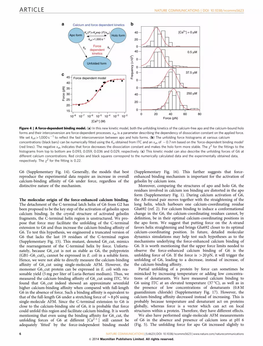
A force-dependent binding model. In the second kinetic model,we consider that the unfolding kinetics of the calcium-free andcalcium-bound forms, and their interconversion, are force-dependent processes (Fig. 4a). See Supplementary Methods andSupplementary Fig. 11 for the detailed derivation of this model.
KdðFÞ ¼ KdexpðFbxahÞ ð6Þwhere Kd and b are as defined above. Kd(F) is the dissociationconstant at a given force F, and xah is the measure of thedependency of dissociation constant on the applied force. Forxaho0, force will decrease the dissociation constant and increasethe binding affinity; for xah40, force will decrease the bindingaffinity; for xah¼ 0, force has no effect on the binding affinity.Here Kd(F) is exponentially dependent on the applied forcebecause force lowers the free energy barrier between the apo andholo forms linearly while Kd(F) depends exponentially on thedifference in free energy (see Supplementary Methods). Similar tothe force-independent binding model, to warrant a single unim-odal unfolding force distribution for G6 at the intermediate
calcium concentration, the koff should be higher than 1,000 s� 1
in the force-dependent binding model (Supplementary Fig. 12).Using this force-dependent binding model, the unfolding force
histograms, at various [Ca2þ ], can be fitted numerically using theKd obtained from ITC and a xah of � 0.7 nm (Fig. 4b). Thenegative xah indicates that force decreases the dissociationconstant and that the calcium-bound form is more stable underforce. A stretching force of 10 pN increases the calcium-bindingaffinity of G6 by a factor of 5.5. Moreover, this model can alsoreproduce the average unfolding forces over the full range ofcalcium concentrations (Fig. 4c). Intriguingly, in this model, evenat a stretching force of 20 pN (the unfolding force for calcium-freeG6), the Kd is actually much larger than the optimum Kd used to‘fit’ the experimental data using the force-independent model.This is due to the coupling of the interconversion of calcium-freeand calcium-bound forms to their unfolding processes in theforce-dependent binding model. It is worth mentioning thatthe force-dependent binding model more accurately predicts theslope of the force changes with respect to the [Ca2þ ] than theforce-independent model in combination with the artificially lowKd. Therefore, these experimental data provide strong support forthe force-dependent binding model. A further test of this modelcomes from measuring the pulling speed-dependent unfoldingforces of G6 at the intermediate [Ca2þ ] of 5 mM. The new force-dependent binding model satisfactorily predicts the experimen-tally obtained unfolding forces over the range of pulling speedsusing the same parameters that were used to simulate theunfolding forces at various [Ca2þ ]. In contrast, the force-independent binding model fails to reproduce the unfoldingforces over the same range of pulling speeds (SupplementaryFig. 13). We have also tested whether the experimental data canbe fitted by other models, such as one that includes the presenceof an additional force-revealed calcium-binding site in G6, andanother that takes into account non-specific calcium binding to
40
20
0
[Ca2+] = 0 µM
45
40
35
30
25
20
15
For
ce (
pN)
10–8 10–7 10–6 10–5 10–4 10–3 10–2
[Ca2+] (M)
Kd = 0.05 µMKd = 14.4 µM
40
20
0
0.5 µM
40
20
0
37.5 µM
40
20
0
806040200Force (pN)
250 µM
Holo form
Unfolded form
Apo form
ka
koff
kon
khForce
dependentkinetics
Calcium dependent kinetics
Fre
quen
cy (
×10
3 )
Figure 3 | A force-independent binding model. (a) The kinetic model that considers rapid dynamics between the calcium-free apo and the calcium-bound
holo conformations. The unfolding of both the apo and holo forms are force-dependent processes and the interconversion between the apo and holo
forms depends on the calcium concentration and is force independent. (b) This kinetic model fails to reproduce the unfolding force histograms at
various calcium concentrations. Black bars correspond to the experimentally obtained unfolding force histograms and red lines correspond to the numeric
fitting results using the kinetic model depicted in a The w2 for the fittings to the histograms from top to bottom are 0.093, 0.096, 0.308 and 0.290,
respectively. (c) This kinetic model cannot describe the unfolding forces of G6 at different calcium concentrations. Red circles correspond to the
numerically calculated data using a Kd of 14.4mM, as determined from ITC measurements. Blue circles correspond to the numerically calculated data using
a Kd of 0.05mM that best ‘fits’ the experimental data. However, the slope of the unfolding force versus [Ca2þ ] relationship cannot be fully reproduced.
The w2 for numerically calculated data using Kds of 14.4 and 0.05mM are 14.0 and 0.33, respectively.
NATURE COMMUNICATIONS | DOI: 10.1038/ncomms5623 ARTICLE
NATURE COMMUNICATIONS | 5:4623 | DOI: 10.1038/ncomms5623 | www.nature.com/naturecommunications 5
& 2014 Macmillan Publishers Limited. All rights reserved.
G6 (Supplementary Fig. 14). Generally, the models that bestreproduce the experimental data require an increase in overallcalcium-binding affinity of G6 under force, regardless of thedistinctive nature of the mechanism.
The molecular origin of the force-enhanced calcium binding.The detachment of the C-terminal latch helix of G6 from G2 hasbeen proposed to be the key step of the activation of gelsolin uponcalcium binding. In the crystal structure of activated gelsolinfragments, the C-terminal helix region is unstructured. We pro-pose that force may facilitate the unfolding of the C-terminalextension to G6 and thus increase the calcium-binding affinity ofG6. To test this hypothesis, we engineered a truncated version ofG6 that lacks the last 19 residues of the C-terminal region(Supplementary Fig. 15). This mutant, denoted G6_cut, mimicsthe rearrangement of the C-terminal helix by force. Unfortu-nately, because G6_cut is not as stable as G6, the polyprotein(GB1–G6_cut)4 cannot be expressed in E. coli in a soluble form.Hence, we were not able to directly measure the calcium-bindingaffinity of G6_cut using single-molecule AFM. However, themonomer G6_cut protein can be expressed in E. coli with rea-sonable yield (5 mg per liter of Luria-Bertani medium). Thus, wemeasured the calcium-binding affinity of G6_cut using ITC. Wefound that G6_cut indeed showed an approximate sevenfoldhigher calcium-binding affinity when compared with full-lengthG6 in the absence of force. Such a binding affinity is equivalent tothat of the full-length G6 under a stretching force of B8 pN usingsingle-molecule AFM. Since the C-terminal extension to G6 isclose to the calcium-binding site of G6, it is probable that forcecould unfold this region and facilitate calcium binding. It is worthmentioning that even using the binding affinity for G6_cut, theunfolding forces of G6 at different [Ca2þ ] still cannot beadequately ‘fitted’ by the force-independent binding model
(Supplementary Fig. 16). This further suggests that force-enhanced binding mechanism is important for the activation ofgelsolin by calcium ions.
Moreover, comparing the structures of apo and holo G6, theresidues involved in calcium ion binding are distorted in the apoform (Supplementary Fig. 1). During calcium activation of G6,the AB-strand pair moves together with the straightening of thelong helix, which harbours one calcium-coordinating residueGlu692 (ref. 2). For calcium binding to induce a conformationalchange in the G6, the calcium-coordinating residues cannot, bydefinition, be in their optimal calcium-coordinating positions inthe apo form. We suggest that putting force on the A-strandfavors helix straightening and brings Glu692 closer to its optimalcalcium-coordinating position. In future, detailed moleculardynamics simulations may help test such hypotheses as to themechanisms underlying the force-enhanced calcium binding ofG6. It is worth mentioning that the upper force limits needed toobserve the force-enhanced calcium binding of G6 is theunfolding force of G6. If the force is 420 pN, it will trigger theunfolding of G6, leading to a decrease, instead of increase, ofthe calcium-binding affinity.
Partial unfolding of a protein by force can sometimes bemimicked by increasing temperature or adding low concentra-tions of denaturants. We have measured calcium binding toG6 using ITC at an elevated temperature (37 �C), as well as inthe presence of low concentrations of denaturants (0.8 Mguanidinium chloride) (Supplementary Fig. 17). However, thecalcium-binding affinity decreased instead of increasing. This isprobably because temperature and denaturant act on proteinsglobally, whereas force is a vector which can act on localstructures within a protein. Therefore, they have different effects.
We also have performed single-molecule AFM measurementsof G6 in the absence and presence of calcium ions at pH 6(Fig. 5). The unfolding force for apo G6 increased slightly to
40
20
0
[Ca2+] = 0 µM
45
40
35
30
25
20
15
For
ce (
pN)
10–8 10–7 10–6 10–5 10–4 10–3 10–2
[Ca2+] (M)
40
0
0
0.5 µM
0
0
0
37.5 µM
40
20
0806040200
Force (pN)
250 µM
Kd (F )=Kdexp (F�xah)
koff >1,000 s–1
Calcium and force dependent kinetics
Holo form
Unfolded form
Apo form
ka kh
Force dependent model
Forcedependent
kinetics
Fre
quen
cy (
×10
3 )Figure 4 | A force-dependent binding model. (a) In this new kinetic model, both the unfolding kinetics of the calcium-free apo and the calcium-bound holo
forms and their interconversion are force-dependent processes. xah is a parameter describing the dependency of dissociation constant on the applied force.
We set koff41,000 s� 1 to reflect the fast interconversion between apo and holo forms. (b) The unfolding force histograms at various calcium
concentrations (black bars) can be numerically fitted using the Kd obtained from ITC and an xah of �0.7 nm based on the ‘force-dependent binding model’
(red lines). The negative xah indicates that force decreases the dissociation constant and makes the holo form more stable. The w2 for the fittings to the
histograms from top to bottom are 0.093, 0.059, 0.036 and 0.029, respectively. (c) This kinetic model can also describe the unfolding forces of G6 at
different calcium concentrations. Red circles and black squares correspond to the numerically calculated data and the experimentally obtained data,
respectively. The w2 for the fitting is 0.22.
ARTICLE NATURE COMMUNICATIONS | DOI: 10.1038/ncomms5623
6 NATURE COMMUNICATIONS | 5:4623 | DOI: 10.1038/ncomms5623 | www.nature.com/naturecommunications
& 2014 Macmillan Publishers Limited. All rights reserved.

B23 pN at pH 6.0, indicating that G6 is mechanically somewhatmore stable at low pH. However, the unfolding forces of G6 in thepresence of micromolar to millimolar of calcium at pH 6.0 areobviously lower than those at pH 7.4. Although low pH mayactivate gelsolin, it decreases the calcium-binding affinity of G6.This indicates that the activation mechanism of G6 by low pHand calcium ions may be unique.
DiscussionAlthough it has been widely recognized that force can regulateligand binding and Protein–protein interactions, the presentfinding that force can enhance the calcium ion binding affinity forG6 is, to our knowledge, the first experimental verification of thisforce-amplified ligand binding concept at the molecular level.Moreover, accurate measurement of the unfolding forces atdifferent [Ca2þ ] allowed us to quantitatively address the effect offorce on the equilibrium binding constant. We found that aphysiologically relevant force of B10 pN could increase thebinding affinity by more than fivefold. We propose a parameter,xah, to quantify the dependence of the dissociation constant onthe stretching force. The larger the absolute value of xah, thegreater is the effect of force on the binding affinity. A negative xah
indicates that the binding affinity is increased by force and apositive xah reflects weaker binding under force. The kineticmodel presented here can serve as a general physical model todeal with such complex force-dependent binding systems.However, this model may not be applicable for protein–ligandinteractions that involve intermediate states in which thetwo-state binding assumption is not valid.
Notably, the force-enhanced binding affinity demonstratedhere is distinct from the well-established catch-bond mechanismsfound in many ligand–receptor systems38–40. In the catch-bondmechanism, the lifetime of a bond increases with the appliedforce. However, lifetime is a kinetic quantity, while in our systemthe binding affinity is a thermodynamic quantity that describesthe equilibrium characteristics of an interaction. Moreover, in oursystem, force is not directly applied between the ligand andreceptor. Instead, regulation of the binding affinity is throughthe conformational change of a protein induced by an appliedforce. For rigid protein structures, the effect of force on thebinding affinity may be not pronounced. However, for proteinswith malleable structures, force could change the structuresof both apo and holo states, leading to a change of bindingaffinity. Furthermore, the force-enhanced binding mechanismpresented in this article is also conceptually different from the
60
40
20
0
100806040200
120
100
80
60
40
20
0
100806040200
60
50
40
30
20
10
0
100806040200
60
50
40
30
20
10
0
100806040200
pH 6.00 µM Ca2+
pH 6.0100 µM Ca2+
pH 6.025 µM Ca2+
Unfolding force (pN)
# O
f eve
nts
# O
f eve
nts
# O
f eve
nts
# O
f eve
nts
40
35
30
25
10–8 10–7 10–6 10–5 10–4 10–3 10–2 10–1
[Ca2+] (M)
Unf
oldi
ng fo
rce
(pN
)
Exp. data at pH 7.4Exp. data at pH 6.0Sim. data from force-dependent bindingmodel
Unfolding force (pN) Unfolding force (pN)
pH 6.05,000 µM Ca2+
Unfolding force (pN)
Figure 5 | The change of the unfolding forces for G6 at different [Ca2þ ] at pH 6. (a) The unfolding force histograms of G6 at different [Ca2þ ],
pH 6 (pulling speed: 400 nm s� 1). (b) The dependency of the unfolding forces of G6 on the concentrations of calcium ions at pH 7.4 and 6.0.
Exp., experimental; Sim., Simulation.
NATURE COMMUNICATIONS | DOI: 10.1038/ncomms5623 ARTICLE
NATURE COMMUNICATIONS | 5:4623 | DOI: 10.1038/ncomms5623 | www.nature.com/naturecommunications 7
& 2014 Macmillan Publishers Limited. All rights reserved.

force-activated binding mechanism illustrated by others10–13. Inour case, force only shifts the equilibrium between calcium-freeand calcium-bound states. Even in the absence of force, a certainlevel of binding affinity is retained. In our experiments, we usedthe unfolding forces at different calcium concentrations as aprobe for the force-dependent calcium binding by G6. This doesnot indicate that the force-enhanced calcium binding of G6 is dueto the unfolding of G6. As shown in Supplementary Fig. 11, forcepreferentially stabilizes the holo conformation over the apoconformation and thus increases the calcium-binding affinity ofG6. The denatured G6 does not show any measurable calcium-binding affinity. Therefore, this leads to an upper boundary forthe force-enhanced calcium-binding affinity. The maximumcalcium-binding affinity of G6 is achieved at the unfoldingforce of holo G6.
It is interesting to consider the physiological importance ofstretching forces enhancing the binding affinity between calciumand G6. During the activation of gelsolin, calcium ion bindingcauses small conformational changes within the calcium-bounddomains (Fig. 1a,b, and Supplementary Fig. 1) and largeconformational changes between domains2. It has beenproposed that calcium binding to G6 controls the overallactivation of gelsolin by releasing the tail latch that locks theinactive conformation of gelsolin41. This process may be requiredto create strain to dislodge the tail C-terminal helix, andultimately, to reveal the actin-filament binding site on G2(Fig. 6). Thus, we propose that occupation of one or morecalcium-binding sites within the first five domains of gelsolin willproduce strain through the conformational restrictions placed onthese domains by the tail C-terminal helix interaction with G2(Fig. 6a), and as a consequence lead to a strain-induced increasein the binding of calcium to G6 (Fig. 6b). Consequently, gelsolindisplays cooperativity in binding calcium ions that is mediatedthrough changes in structure and, we propose, resultant changesin strain (Fig. 6c). We speculate that this produces a moleculethat is finely tuned to react to [Ca2þ ] changes and at the sametime contains a safety switch at inappropriate calcium levels.Further structure biology and molecular dynamics simulationsstudies will be needed to characterize intermediate activationconformations to verify the proposed role of strain in theactivation process.
A second area where strain-induced calcium binding may beimportant is in gelsolin’s interactions with actin. Based on crystalstructures, the linker regions connecting G1–G2 and G3–G4 ofgelsolin are very much elongated when it is bound to the actinfilament2. The elongation of a peptide chain typically requires atensile force. Based on simple polymer elasticity models, such asthe WLC model or the freely-jointed chain model, activatedgelsolin typically will be subjected to a tensile force ofseveral piconewtons (see Supplementary Methods for detailedcalculation). Such minute forces are sufficiently large to alter
the calcium-binding affinity to gelsolin and thus its thresholdfor calcium activation mechanism. Furthermore, gelsolinundoubtedly undergoes changes in tension as it wraps itselfover the surface of the actin filament during the severing process.Fluctuations in the filament will also create strain since gelsolininteracts with three actin protomers and encircles the filament.Should the force-enhanced calcium ion binding be common tothe other domains of gelsolin (G1–G5), then this suggests thatcalcium binding will favour the portion of gelsolin that is boundto a filament over the free gelsolin pool. Thus, we speculate that atclose to threshold calcium levels, gelsolin will remain bound tothe filament and severing will be driven to completion, while freegelsolin may be able to return to the quiescent state throughcalcium dissociation.
Quantifying the tensile force in gelsolin and other proteinsunder physiological conditions is challenging due to the lack ofefficient in vivo force sensors. Nevertheless, a common trend isemerging that several proteins that regulate the force-generatingactin polymerization machine are themselves subjected to forceregulation. We are left with the question as to whether the force-dependent change in calcium-binding affinity is unique to G6 or acommon feature shared by all gelsolin domains. Addressing thisquestion will be an important step towards further understandingthe activation mechanism of gelsolin. Due to the similar sizes ofall gelsolin domains, it is not possible to assign different unfoldingpeaks for individual domains, within full-length gelsolin, to probetheir calcium-binding affinities. Other techniques, such as usingforster resonance energy transfer pairs to label the two ends of theG6 domain within full-length gelsolin may allow simultaneousmeasurement of the fluorescence change upon stretching insingle-molecule AFM studies, and thus might open up theopportunity to study G6 within full-length gelsolin in the future.
MethodsProtein engineering. The gene encoding (GB1–G6)4 hybrid polyprotein wasconstructed using standard molecular biology techniques to splice GB1 and G6(P06396) in an iterative approach. (GB1–G6)4 was expressed in BL21 and purifiedby Ni2þ -affinity chromatography. EGTA (1 mM) was added to the protein sampleto remove the residual free Ca2þ in buffer. Then the purified polyprotein samplewas dialyzed and kept at 4 �C in Tris-HCl buffer (10 mM, pH 7.4, 10 mM NaCl) ata concentration of 0.3 mg ml� 1.
Single-molecule AFM. Single-molecule AFM experiments were carried out on acommercial AFM (ForceRobot 300, JPK, Berlin, Germany). All the force-extensionexperiments were carried out in Tris-HCl buffer (10 mM, pH 7.4, 10 mM NaCl and0–5 mM CaCl2 calibrated with EGTA42). Protein sample (150 ml) was directlydeposited on a freshly cleaved glass surface for 20 min. Then, the sample chamberwas filled with 1.5 ml buffer before the measurement. Typically, experimentsproceeded o12 h to avoid the concentration changes in Ca2þ from evaporation ofwater. The spring constant of the AFM cantilevers (Biolever-RC-150VB-70 fromOlympus) was calibrated using the equipartition theorem before each experiment,with a typical value of 6 pN nm� 1, and was recalibrated every 4–6 h during eachexperiment. The pulling speed was 400 nm s� 1 for all traces unless otherwiseindicated.
1 23
5 4 6
1 23
5 4 6
1 23
5 4 6
Calcium free Strained intermediate Tail latch release
TailCa2+ Ca2+
Figure 6 | Cartoon highlighting the initial activation stages of gelsolin. (a) Six-domain gelsolin contains a homologous calcium-binding site in each
domain (white circles) and a C-terminal tail that interacts with domain 2 to lock the inactive conformation (coloured in purple). (b) Occupation of any of
these sites by a calcium ion will begin the activation process that drives conformational reorganization and concomitantly creates inter-domain strain,
leading to the C-terminal tail under tension. Here we show a calcium ion occupying the site on G2. Greater strain may be induced through filling more of the
calcium-binding sites. (c) The resulting inter-domain strain will increase the affinity of G6 for calcium, resulting in calcium ion binding to G6 and the release
of the tail latch, which in turn exposes the F-actin-binding site on G2.
ARTICLE NATURE COMMUNICATIONS | DOI: 10.1038/ncomms5623
8 NATURE COMMUNICATIONS | 5:4623 | DOI: 10.1038/ncomms5623 | www.nature.com/naturecommunications
& 2014 Macmillan Publishers Limited. All rights reserved.

References1. Silacci, P. et al. Gelsolin superfamily proteins: key regulators of cellular
functions. Cell. Mol. Life Sci. 61, 2614–2623 (2004).2. Nag, S., Larsson, M., Robinson, R. C. & Burtnick, L. D. Gelsolin: the tail of a
molecular gymnast. Cytoskeleton (Hoboken) 70, 360–384 (2013).3. Sun, H. Q., Yamamoto, M., Mejillano, M. & Yin, H. L. Gelsolin, a
multifunctional actin regulatory protein. J. Biol. Chem. 274, 33179–33182(1999).
4. Kiselar, J. G., Janmey, P. A., Almo, S. C. & Chance, M. R. Visualizing theCa2þ -dependent activation of gelsolin by using synchrotron footprinting. Proc.Natl Acad. Sci. USA 100, 3942–3947 (2003).
5. Ashish et al. Global structure changes associated with Ca2þ activation offull-length human plasma gelsolin. J. Biol. Chem. 282, 25884–25892 (2007).
6. Burtnick, L. D. et al. The crystal structure of plasma gelsolin: implications foractin severing, capping, and nucleation. Cell 90, 661–670 (1997).
7. Robinson, R. C. et al. Domain movement in gelsolin: a calcium-activatedswitch. Science 286, 1939–1942 (1999).
8. Burtnick, L. D., Urosev, D., Irobi, E., Narayan, K. & Robinson, R. C. Structureof the N-terminal half of gelsolin bound to actin: roles in severing, apoptosisand FAF. EMBO J. 23, 2713–2722 (2004).
9. Wang, H. et al. Helix straightening as an activation mechanism in the gelsolinsuperfamily of actin regulatory proteins. J. Biol. Chem. 284, 21265–21269(2009).
10. del Rio, A. et al. Stretching single talin rod molecules activates vinculin binding.Science 323, 638–641 (2009).
11. Friedland, J. C., Lee, M. H. & Boettiger, D. Mechanically activated integrinswitch controls alpha5beta1 function. Science 323, 642–644 (2009).
12. Chabria, M., Hertig, S., Smith, M. L. & Vogel, V. Stretching fibronectin fibresdisrupts binding of bacterial adhesins by physically destroying an epitope. Nat.Commun. 1, 135 (2010).
13. Rognoni, L., Stigler, J., Pelz, B., Ylanne, J. & Rief, M. Dynamic force sensing offilamin revealed in single-molecule experiments. Proc. Natl Acad. Sci. USA 109,19679–19684 (2012).
14. Yao, M. et al. Mechanical activation of vinculin binding to talin locks talin in anunfolded conformation. Sci. Rep. 4, 4610 (2014).
15. Stigler, J., Ziegler, F., Gieseke, A., Gebhardt, J. C. & Rief, M. The complexfolding network of single calmodulin molecules. Science 334, 512–516 (2011).
16. Cao, Y. & Li, H. Dynamics of protein folding and cofactor bindingmonitored by single-molecule force spectroscopy. Biophys. J. 101, 2009–2017(2011).
17. Javadi, Y., Fernandez, J. M. & Perez-Jimenez, R. Protein foldingunder mechanical forces: a physiological view. Physiol. (Bethesda) 28, 9–17(2013).
18. Kaiser, C. M., Goldman, D. H., Chodera, J. D., Tinoco, Jr I. & Bustamante, C.The ribosome modulates nascent protein folding. Science 334, 1723–1727(2011).
19. Elms, P. J., Chodera, J. D., Bustamante, C. & Marqusee, S. The molten globulestate is unusually deformable under mechanical force. Proc. Natl Acad. Sci. USA109, 3796–3801 (2012).
20. Cao, Y., Balamurali, M. M., Sharma, D. & Li, H. A functional single-moleculebinding assay via force spectroscopy. Proc. Natl Acad. Sci. USA 104,15677–15681 (2007).
21. Cao, Y., Er, K. S., Parhar, R. & Li, H. A force-spectroscopy-based single-molecule metal-binding assay. ChemPhysChem 10, 1450–1454 (2009).
22. Cao, Y., Lam, C., Wang, M. & Li, H. Nonmechanical protein can havesignificant mechanical stability. Angew. Chem. Int. Ed. 45, 642–645 (2006).
23. Cao, Y. & Li, H. Polyprotein of GB1 is an ideal artificial elastomeric protein.Nat. Mater. 6, 109–114 (2007).
24. Lv, C. et al. Low folding cooperativity of HP35 revealed by single-moleculeforce spectroscopy and molecular dynamics simulation. Biophys. J. 102,1944–1951 (2012).
25. Rief, M., Gautel, M., Oesterhelt, F., Fernandez, J. M. & Gaub, H. E. Reversibleunfolding of individual titin immunoglobulin domains by AFM. Science 276,1109–1112 (1997).
26. Williams, P. M. et al. Hidden complexity in the mechanical properties of titin.Nature 422, 446–449 (2003).
27. Brockwell, D. J. et al. Mechanically unfolding the small, topologically simpleprotein L. Biophys. J. 89, 506–519 (2005).
28. Gao, X. et al. Single-molecule experiments reveal the flexibility of a Per-ARNT-Sim domain and the kinetic partitioning in the unfolding pathway under force.Biophys. J. 102, 2149–2157 (2012).
29. Stigler, J. & Rief, M. Calcium-dependent folding of single calmodulinmolecules. Proc. Natl Acad. Sci. USA 109, 17814–17819 (2012).
30. Bell, G. I. Models for the specific adhesion of cells to cells. Science 200, 618–627(1978).
31. Evans, E. & Ritchie, K. Strength of a weak bond connecting flexible polymerchains. Biophys. J. 76, 2439–2447 (1999).
32. Marko, J. F. & Siggia, E. D. Stretching DNA. Macromolecules 28, 8759–8770(1995).
33. Lin, K. M., Mejillano, M. & Yin, H. L. Ca2þ regulation of gelsolin by itsC-terminal tail. J. Biol. Chem. 275, 27746–27752 (2000).
34. Zapun, A., Grammatyka, S., Deral, G. & Vernet, T. Calcium-dependentconformational stability of modules 1 and 2 of human gelsolin. Biochem. J.350(Pt 3): 873–881 (2000).
35. Pope, B., Maciver, S. & Weeds, A. Localization of the calcium-sensitive actinmonomer binding site in gelsolin to segment 4 and identification of calciumbinding sites. Biochemistry 34, 1583–1588 (1995).
36. Chen, C. D. et al. Furin initiates gelsolin familial amyloidosis in the Golgithrough a defect in Ca(2þ ) stabilization. EMBO J. 20, 6277–6287 (2001).
37. Chumnarnsilpa, S. et al. The crystal structure of the C-terminus of adseverinreveals the actin-binding interface. Proc. Natl Acad. Sci. USA 106, 13719–13724(2009).
38. Thomas, W. E., Trintchina, E., Forero, M., Vogel, V. & Sokurenko, E. V.Bacterial adhesion to target cells enhanced by shear force. Cell 109, 913–923(2002).
39. Marshall, B. T. et al. Direct observation of catch bonds involving cell-adhesionmolecules. Nature 423, 190–193 (2003).
40. Akiyoshi, B. et al. Tension directly stabilizes reconstituted kinetochore-microtubule attachments. Nature 468, 576–579 (2010).
41. Choe, H. et al. The calcium activation of gelsolin: insights from the 3A structureof the G4-G6/actin complex. J. Mol. Biol. 324, 691–702 (2002).
42. Schoenmakers, T. J., Visser, G. J., Flik, G. & Theuvenet, A. P. CHELATOR:an improved method for computing metal ion concentrations in physiologicalsolutions. Biotechniques 12, 870–874 (1992).
AcknowledgementsWe thank Prof. Hongbin Li for providing the plasmid of GB1. This work was supportedby the NSFC (Nos. 31170813 and 11074115) to Y.C., the NSFC (No. 91127026 and11334004) and the National Basic Research Programme of China (973 ProgrammeNo.2013CB834100) to W.W., and the Agency for Science, Technology and Research(A*STAR), Singapore to B.X. and R.C.R.
Author contributionsC.L. constructed the polyprotein, performed the single-molecule AFM experiments andanalyzed the data. X.G. performed the thermal melting experiments, proposed the kineticmodel and performed the numeric fitting. W.L. proposed the molecule mechanism andperformed simulations. M.Q. assisted the experiments and involved in discussions. X.G.,H.Z. and B.X. performed ITC measurements. Y.C., R.C.R., L.D.B. and W.W. designed theresearch and wrote the paper with input from other authors. Y.C. and W.W. alsosupervised the project.
Additional informationSupplementary Information accompanies this paper at http://www.nature.com/naturecommunications
Competing financial interests: The authors declare no competing financial interests.
Reprints and permission information is available online at http://npg.nature.com/reprintsandpermissions/
How to cite this article: Lv, C. et al. Single-molecule force spectroscopy revealsforce-enhanced binding of calcium ions by gelsolin. Nat. Commun. 5:4623doi: 10.1038/ncomms5623 (2014).
This work is licensed under a Creative Commons Attribution-NonCommercial-ShareAlike 4.0 International License. The images or
other third party material in this article are included in the article’s Creative Commonslicense, unless indicated otherwise in the credit line; if the material is not included underthe Creative Commons license, users will need to obtain permission from the licenseholder to reproduce the material. To view a copy of this license, visit http://creativecommons.org/licenses/by-nc-sa/4.0/
NATURE COMMUNICATIONS | DOI: 10.1038/ncomms5623 ARTICLE
NATURE COMMUNICATIONS | 5:4623 | DOI: 10.1038/ncomms5623 | www.nature.com/naturecommunications 9
& 2014 Macmillan Publishers Limited. All rights reserved.


