Embed Size (px)
Citation preview
Behavioral/Systems/Cognitive
Single-Unit Firing in Rat Perirhinal Cortex Caused by FearConditioning to Arbitrary and Ecological Stimuli
Sharon C. Furtak,1 Timothy A. Allen,1 and Thomas H. Brown1,2
Departments of 1Psychology and 2Cellular and Molecular Physiology, Yale University, New Haven, Connecticut 06520
Pretraining lesions of rat perirhinal cortex (PR) severely impair pavlovian fear conditioning to a 22 kHz ultrasonic vocalization (USV) cue.However, PR lesions are without significant effect when the cue is a continuous tone at the same or a lower frequency. Here we examinedfear-conditioning-produced changes in single-unit firing elicited in rat PR by a 22 kHz tone cue or a 22 kHz USV cue. Chronic recordingelectrodes were introduced from the lateral surface of the skull. Altogether, 200 well isolated units were studied in 28 rats. Overall, 73% ofthe recorded single units (145 of 200 units) evidenced statistically significant firing changes in response to the tone or USV conditionalstimulus (CS) after it had been paired several times with an aversive unconditional stimulus (US). Interestingly, 33% of units (66 of 200units) that were initially CS-unresponsive became CS-responsive after conditioning. After conditioning, there were two notable differ-ences between single-unit responses elicited by the USV cue and those elicited by the tone cue. First, 11% of the units (14 of 123 units)recorded from the USV-conditioned group displayed a precisely timed increase in firing rate during the 260 ms interval in which the UShad previously occurred. This US-timed response was unique to the USV-conditioned group. Second, the mean latency of cue-elicitedfiring was �30 ms longer in the USV-conditioned group than in the tone-conditioned group. These cue-specific differences in acquiredfiring latencies and acquired firing patterns suggest that spectrotemporal properties of a CS can control the essential circuitry orneurophysiological mechanisms underlying fear conditioning.
Key words: timing; medial temporal lobe; ultrasonic vocalization; amygdala; temporal encoding; auditory fear conditioning
IntroductionPavlovian fear conditioning is rapid and persistent, propertiesthat make it well suited for investigating the neurophysiologicalbasis of associative learning and memory (Fanselow and LeDoux,1999; LeDoux, 2000; Pare et al., 2004; Fanselow and Poulos,2005). During fear conditioning, a conditional stimulus (CS) ac-quires the ability to elicit fear-related conditional responses(CRs), such as freezing behavior, after being paired with an aver-sive unconditional stimulus (US). Considerable evidence hasbeen adduced in support of the hypothesis that the lateral nucleusof the amygdala (LA) is critically involved in the acquisition offear-related CRs (Fanselow and LeDoux, 1999; LeDoux, 2000;Gale et al., 2004; Maren and Quirk, 2004; Maren, 2005).
Several studies have demonstrated that perirhinal cortex (PR),which is laterally adjacent to and reciprocally connected with LA,is also essential for normal CR acquisition to certain types ofstimuli. For example, both pretraining and posttraining PR le-sions or inactivations impair fear conditioning to contextualstimuli (Corodimas and LeDoux, 1995; Sacchetti et al., 1999;Tassoni et al., 1999; Bucci et al., 2000; Bucci et al., 2002; Sacchettiet al., 2002; Lindquist et al., 2004; Padlubnaya et al., 2006a). Ad-
ditionally, pretraining PR lesions impair auditory fear condition-ing when the cue is a 22 kHz ultrasonic vocalization (USV)(Lindquist et al., 2004; Padlubnaya et al., 2006a), whereas theselesions are without effect when the cue is a continuous tone (Ro-manski and LeDoux, 1992; Bucci et al., 2000; Lindquist et al.,2004; Padlubnaya et al., 2006a) (but see Sacchetti et al., 2002).
In rats with temporal or rhinal cortical damage, fear condi-tioning to tones is thought to be supported by direct thalamicprojections to LA (Romanski and LeDoux, 1992; Doron and Le-Doux, 2000; LeDoux, 2000). In general, the contributions of cor-tical processing to fear conditioning are still unclear (Cahill et al.,1999; Fanselow and LeDoux, 1999; Weinberger, 2004; Boatmanand Kim, 2006). One hypothesis asserts that cortical processing isnecessary for normal fear conditioning to “complex” but not“simple” stimuli (Yaniv et al., 2001; Lindquist et al., 2004; Yanivet al., 2004). Compared with continuous tones, USVs are obvi-ously more complex. In particular, USVs are temporally discon-tinuous, consisting of a “bout” or series of “calls,” and the indi-vidual calls contain unique frequency and amplitudemodulations (Barfield and Geyer, 1972; Blanchard et al., 1991;Brudzynski et al., 1993; Brudzynski, 2005; Allen et al., 2007a). It isworth mentioning that PR lesions and inactivations also impairfear acquisition and expression to olfactory and visual stimuli(Herzog and Otto, 1997; Otto et al., 2000; Schulz et al., 2004;Shulz-Klaus et al., 2005).
To understand better the role of PR in auditory fear condi-tioning, the present study examined cue-elicited single-unit ac-tivity before, during, and after delay fear conditioning to either a
Received April 12, 2007; revised Sept. 17, 2007; accepted Sept. 18, 2007.This work was supported by National Institutes of Health Grant MH058405 and Yale University. We thank Dianna
Kholodar-Smith for comments on this manuscript and Michael Domjan and Mark Laubach for useful discussion.Correspondence should be addressed to Thomas H. Brown, Department of Psychology, Yale University, 2 Hill-
house Avenue, New Haven, CT 06520. E-mail: [email protected]:10.1523/JNEUROSCI.1653-07.2007
Copyright © 2007 Society for Neuroscience 0270-6474/07/2712277-15$15.00/0
The Journal of Neuroscience, November 7, 2007 • 27(45):12277–12291 • 12277
22 kHz continuous tone or a 22 kHz USV. Continuous tones andUSVs are respective exemplars of what Domjan and coworkersterm “arbitrary” and “ecological” stimuli (Domjan et al., 2004).Rodent USVs have long been recognized as ethologically impor-tant social signals (Anderson, 1954; Sewell, 1967; Barfield andGeyer, 1972; Sales and Pye, 1974; Blanchard et al., 1991; Knutsonet al., 2002; Brudzynski, 2005; Brudzynski and Holland, 2005).The first goal of the present study was to discover whether fearconditioning causes changes in CS-elicited firing in rat PR. If so,the second goal was to characterize the plasticity and to discoverany cue-specific differences in firing that might point to differ-ences in the essential underlying circuitry or neurophysiologicalmechanisms.
Materials and MethodsSubjects. This study used 28 experimentally naive male Sprague Dawleyrats weighing 250 – 400 g. Each animal was singly housed on a 12 hlight/dark cycle with ad libitum access to food and water. Animals werehandled for 3 d before surgery. Experiments were conducted in accor-dance with the Society for Neuroscience policies and the Yale UniversityAnimal Resources Center guidelines on the care and use of animals.
Surgery. A single surgery was performed on each animal to implantboth a recording and stimulating electrode (see below). Animals wereanesthetized with a mixture of ketamine (100 mg/kg, i.p.) and xylazine(20 mg/kg, i.p.) and secured in a stereotaxic instrument. Body tempera-ture was regulated with a heating pad (37°C; Braintree Scientific, Brain-tree, MA). The skin above the dorsal surface of the skull was cut along themidline and the right lateral edge and pulled back to expose the skull.Four holes were drilled through the dorsal surface of the skull andthreaded with stainless-steel anchor screws. A ground wire was attachedto one of these screws and used as an electrical reference during single-unit recording.
An eight-channel recording electrode bundle (described below) waschronically implanted in PR from the lateral surface of the skull. Thisprocedure was developed to minimize cortical damage and to improvethe accuracy of electrode placements (Allen et al., 2007a). The lateralsurface of the skull was exposed by pulling the temporal muscle awayfrom the skull with tissue spreaders. A 2 mm trephination was made atthe intersection of the zygomatic arch, the parietal bone, and the tempo-ral bone, exposing the temporal cortex just dorsal to PR. Two additionalanchor screws were placed in the lateral surface of the skull on either sideof the trephination. A micromanipulator drove the electrode bundle tip1–2 mm into PR at a 45° angle relative to the horizontal and coronalplanes. Dental cement permanently secured the tip of the electrode bun-dle into place on the lateral surface. The electrode pin set was then ce-mented on the dorsal surface of the skull, anchored by the dorsal screws.
A bipolar stimulating electrode was inserted underneath the back skin,guided by stainless-steel tubing. The tips of the electrode were directed tothe lumbar region bilaterally. Each tip was secured on either side of (�2cm from) the spine with a suture to the overlying skin. At the end of theimplantation procedure, incisions were sutured and an antibiotic oint-ment was applied. Animals were given 10% acetaminophen in theirdrinking water (v/v; Silarx Pharmaceuticals, Spring Valley, NY) and al-lowed 72 h to recover.
Recording and stimulating electrode assembly. The design of the elec-trode assembly allowed the microwire bundle tip to be secured to thelateral surface of the skull, while placing the pin set in a vertical orienta-tion on the dorsal surface of the skull (supplemental Fig. 1, available atwww.jneurosci.org as supplemental material) (Allen et al., 2007a). Elec-trode bundles consisted of eight insulated tungsten microwires, 25 �m indiameter (California Fine Wire, Grover Beach, CA). One end of each wirewas stripped and secured to a 10-pin strip connector (Microtech, Booth-wyn, PA). Conductive silver paint was applied to the pins to ensure goodelectrical connections. The other end was threaded through 30-gaugestainless-steel tubing, 5 mm in length. Excess wire was coated with latexbetween the stainless-steel tubing and the pin connector, providing pro-tection and flexibility (supplemental Fig. 1, available at www.jneurosci.
org as supplemental material). Before implantation, the bundle tip wascut at a 45° angle �2 mm from the guide tubing. The typical tip resistanceranged from 150 to 300 k�, measured at 1000 Hz (Impedance CheckModule; Frederick Haer Company, Bowdoinham, ME). There was noelectrical continuity among wires.
For the stimulating bipolar electrode, two 6-inch Teflon-coatedstainless-steel wires (0.25 mm diameter; A-M Systems, Carlsborg, WA)were connected to two pins of a strip connector (Microtech). Protectionand insulation was achieved with polyethylene tubing (Intramedic PE90;BD Diagnostic Systems, Sparks, MD). The terminal 4 mm of the stimu-lating wires were stripped of insulation.
Electrophysiological recordings. Recordings and fear conditioning weredone in a Plexiglas observation chamber that was enclosed in a sound-attenuating Faraday chamber. Pin sets were attached to a unity-gain headstage and commutator that passed the signal to a preamplifier (15,000�gain; high- and low-pass filtered at 154 Hz and 13 kHz, respectively;Plexon, Dallas, TX). The signal was then processed by a MultichannelAcquisition Processor (Plexon), which allowed real-time thresholdingand waveform discrimination (SortClient; Plexon). Electrical activitywas also monitored with a digital oscilloscope (TDS 30334; Tektronix,Richardson, TX). Single units that had a signal-to-noise ratio (peak-to-peak signal divided by the root mean square of the noise) of at least 5:1were stored for additional off-line analysis. Cluster cutting techniqueswere used with principle components, spike width, and spike amplitudeto isolate individual waveforms (Offline Sorter; Plexon). Autocorrelo-grams were also used to examine single-unit isolations (supplementalFig. 1, right, available at www.jneurosci.org as supplemental material).Cross-correlograms between single units recorded on different wireswere used to reduce possible redundancy.
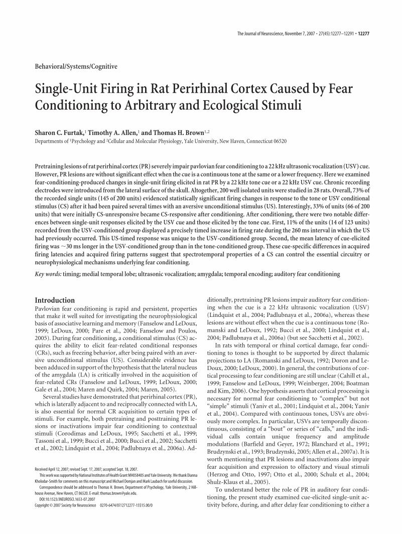
Auditory stimuli. One of two auditory stimuli was used as a CS: acontinuous 22 kHz tone [10 ms rise time, 7.712 s duration, 65 db soundpressure level (SPL)] (Fig. 1 A) or a 22 kHz USV that was pre-recordedfrom a conspecific (7.712 s duration, 65 db SPL) (Fig. 1 B). The USVcontained 11 calls (Fig. 1 B). Auditory stimuli were presented free fieldwith an Enhanced Real-Time Processor RP2.1 [Tucker Davis Technolo-gies (TDT), Alachua, FL] and electrostatic speakers (ED1; TDT) locatedon the top of a Plexiglas chamber, �50 cm from the floor. Loudnessmeasurements were made at floor level using an ultrasonic decibel meter(Ultraprobe 9000; UE Systems, Elmsford, NY). The tones were producedwith digital-tone-generator software (RPvds; TDT) in conjunction withan Enhanced Real-Time Processor RP2.1 (TDT).
The pre-recorded 22 kHz USV was elicited from a naive rat by anunsignaled foot shocks delivered through a grid floor (1 s, 1 mA; Coul-bourn Instruments, Lehigh Valley, PA). USVs were digitally recorded(RP2.1; TDT) and stored on a computer (100 kHz sampling rate, 32 bit).The recorded USV was played back to subjects through the RP2.1 andED1. So-called “22 kHz USVs,” which actually can range in frequencyfrom 18 to 32 kHz, are commonly produced by rats in response to aver-sive or threatening stimuli, by stimuli (contexts or cues) that have beenassociated with aversive or threatening stimuli, or after ejaculation
Figure 1. Spectrograms of the two auditory stimuli that were used as cues for fear-conditioning. A, A 22 kHz tone lasting 7.712 s (65 db SPL). B, A 22 kHz USV lasting 7.712 s (65 dbSPL). The bout of 11 calls is centered at �19 kHz (range, 19 –22 kHz). The mean � SE callduration was 575 � 44.4 ms, and the mean intercall interval was 132 � 1.5 ms.
12278 • J. Neurosci., November 7, 2007 • 27(45):12277–12291 Furtak et al. • Cortical Plasticity in Fear Conditioning
(Barfield and Geyer, 1972; Blanchard et al., 1991; van der Poel and Mic-zek, 1991; Brudzynski et al., 1993; Choi and Brown, 2003; Brudzynski,2007; Litvin et al., 2007). Twenty-two kHz USVs have been measured asdefensive CRs (Lee et al., 2001; Choi and Brown, 2003; Koo et al., 2004;Moyer and Brown, 2006) and used as CSs in fear-conditioning para-digms (Lindquist et al., 2004; Padlubnaya et al., 2006a).
Somatosensory stimulus. The bipolar stimulating electrode was used todeliver an aversive US. The bipolar stimulating electrode was designed toisolate the current density away from the single-unit electrodes, therebyallowing simultaneous recordings during shock presentations in preex-posure and conditioning trials. The back-shock stimulus consisted of sixshocks (10 ms, 5 mA) at 20 Hz delivered via a constant-current generator(BSI-2; Bak Electronics, Mount Airy, MD). Although we did not quantifythe magnitude of the unconditional behavioral responses to the somato-sensory stimulus, it is worth noting that each shock elicited a brief scurryaround the chamber, typically followed by freezing seconds later. Over-all, the response to a back shock was very similar to what we observe inresponse to a grid-floor shock.
Behavioral procedures. Fear conditioning consisted of three stages: pre-exposure, conditioning, and testing/extinction. The three stages wererecorded with a miniature IR-CCD camera (Circuit Specialists, Mesa,AZ) connected to a video cassette recorder for later off-line behavioralanalysis. During the preexposure stage, most of the animals (21 of 28)received pseudoconditioning trials in which the US was explicitly un-paired with the CS (10 CS and US presentations). The CSs and USs werepresented in random order with a 90 � 30 s interval between stimuli. Asubset of animals (7 of 28) instead received 10 CS-alone preexposuretrials in which the CS to CS intervals were 180 � 60 s. The US was omittedin this group to eliminate any pairing-independent effects on responsesto the cues. Possible evidence of sensitization or context conditioningwas examined within and between groups. Among the seven animals thatreceived CS-alone preexposure trials, the degree of shock responsivenesswas determined during the conditioning stage when the back shock waspresented.
Immediately after preexposure, subjects were given 20 conditioningtrials in which the CS always coterminated with a brief back-shock US.The interstimulus interval (ISI) was 7.452 s, and the inter-trial intervalwas 120 � 60 s. After conditioning trials, subjects were returned to theirhome cages in the animal vivarium. At least 4 h later, they were broughtback to the original conditioning context. After a 2 min baseline period,each animal received 40 CS-alone test trials (ISI, 15 � 5 s) in whichfreezing served as the CR (Blanchard and Blanchard, 1969). Time spentfreezing was scored by a blind observer on a continuous minute-by-minute basis with the aid of a stop watch. For each animal, the percentagefreezing was calculated, for each minute, by dividing the total time freez-ing (in seconds) by 60. For this initial study of conditioning effects on PRsingle-unit responses, we decided not to use a context shift because ofknown and unknown complications that might arise in connection withcontextual control of cue-elicited single-unit activity (Hobin et al., 2003).One suspects that some of the cue-specific conditioning effects reportedhere may depend on the conditioning context. Context shifts wouldcertainly be worth exploring, because PR is known to be critical forcontext conditioning (Bucci et al., 2000, 2002; Lindquist et al., 2004;Padlubnaya et al., 2006a).
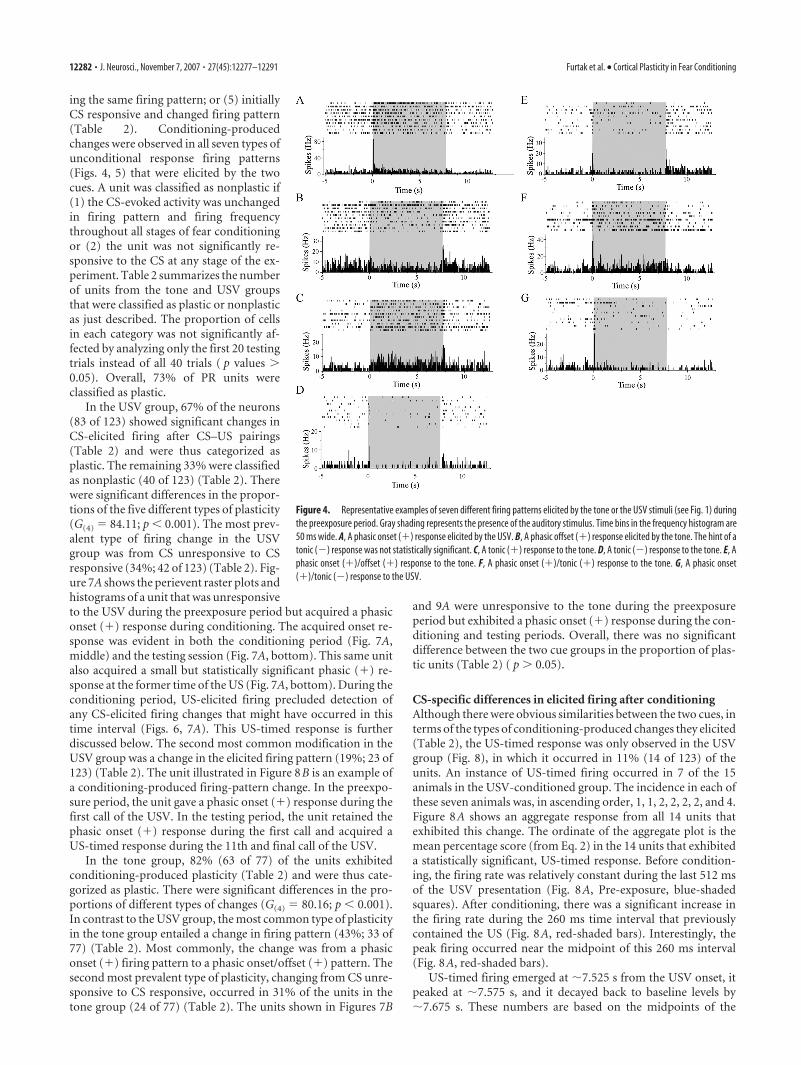
Classification of firing patterns. The classification scheme, which wasbased on firing patterns that are commonly elicited by auditory stimuli inother brain regions (Heil, 1997; Chimoto et al., 2002), was intended to beas simple as possible and yet still capture some potentially importantdifferences between stimuli and among units. Stimulus-elicited firingpatterns were classified along three axes (Allen et al., 2007a) according towhether they entailed (1) an increase or decrease in firing frequency, (2)a phasic or a tonic firing change, and (3) firing in response to the stimulusonset or offset. Not all possible logical combinations were observed.Among the firing patterns that were actually observed, the four simplestones were designated as follows: phasic onset (�), a transient increase inthe firing rate to the stimulus onset; phasic offset (�), a transient increasein firing rate to the stimulus offset; tonic (�), a sustained increase infiring rate during the stimulus presentation; and tonic (�), a sustaineddecrease in firing rate during the stimulus presentation. A subtype of
phasic onset (�) firing consisted of phasic (�) responses to successivecalls of a USV. Elsewhere, this subtype was termed a “call-related” firingpattern (Allen et al., 2007a).
Other observed firing patterns, which consisted of combinations of thesimpler ones, included the following three: phasic onset (�)/offset (�), atransient increase in firing rate to both the stimulus onset and offset;phasic onset (�)/tonic (�), a transient increase in firing rate to thestimulus onset with a smaller but significant increase in the sustainedfiring rate during the entire stimulus presentation; and phasic onset (�)/tonic (�), a transient increase in firing rate to the stimulus onset and asustained firing rate decrease during the remainder of the stimulus pre-sentation. The application of this classification scheme is further eluci-dated in Results.
Because the somatosensory stimulus was so brief (260 ms), no distinc-tion was made between firing to the onset or offset of the stimulus orbetween tonic and phasic patterns of firing. Responses to the somatosen-sory stimulus were most distinguishable based on the duration of theincreased firing. The firing patterns elicited by the back shock were cat-egorized into either a short-duration or a long-duration increase in firingrate. Short-duration responses were operationally defined as a statisti-cally significant increase in firing rate that lasted �250 ms (from 0 to 250ms). Long-duration responses, which could last several seconds after theshock presentation, were operationally defined as a statistically signifi-cant increase firing rate during both the first 250 ms (from 0 to 250 ms)and the next 250 ms (from 250 to 500 ms) after the presentation of theback shock.
Statistical analysis. Graphical and statistical analyses were performedwith NEX software (Plexon), custom-made Microsoft (Seattle, WA) Ex-cel spreadsheets; R 2.3.0 (http://www.r-project.org/), and SPSS 14.0(SPSS, Chicago, IL). To classify the auditory stimulus-elicited firing pat-tern, 10 planned t tests were performed on each unit that compared itsspontaneous baseline firing rate with its firing rate during a specific timebin during or immediately after the presentation of the stimulus. Unlessspecifically indicated otherwise, a Bonferroni’s correction was used togive a familywise � � 0.05. This conservative correction reflects ourinterest in larger effect sizes plus the need for multiple t tests to exploredifferent types of firing patterns. When any one of the t tests was signif-icant, the single unit was classified as “responsive.” Responsive units werethen subclassified, in terms of type of CS-elicited firing pattern, based onwhich t tests were significant (see above, Classification of firing pattern).Two-tailed tests were used because the firing rate might increase or de-crease. Bonferroni’s corrections were not applied to follow-up aggregateanalyses of units that were classified as “unresponsive.” Because any pos-sible effects were already known to be small, the emphasis was on statis-tical power rather than protection against � errors.
Spontaneous (baseline) single-unit activity was defined by the averagefiring rate during the 5 s period just before the presentation of the audi-tory stimulus. Phasic onset (�) and phasic offset (�) responses weretested for statistical significance within the first four time bins (first 200ms) after the auditory stimulus onset or offset, respectively, comparedwith the spontaneous baseline firing rate. Tonic responses were tested forstatistical significance by comparing the average firing rate throughoutthe auditory stimulus presentation (7.712 s) with the average spontane-ous baseline firing rate. A Welsch–Satterwaite solution was used to cor-rect the degrees of freedom for unequal sample sizes. This correction wasmade because there were more time bins during the stimulus presenta-tion (which lasted 7.712 s) than during the baseline period (which lasted5 s).
Shock-elicited firing was examined using t tests for dependent samplesthat compared the mean firing rate during each of the first five 50 ms timebins after the shock with the mean spontaneous baseline firing rate dur-ing the 5 s period before the stimulus presentation. During the testingstage of the experiment, this same analysis examined the presence of a“US-timed” response. An additional t test was conducted to classify ashock-responsive unit as giving either a short-duration or a long-duration response (see above, Classification of firing pattern). This ad-ditional t test compared the average firing rate in the five time bins in thenext 250 ms period after the shock to the spontaneous baseline firing rate.
Furtak et al. • Cortical Plasticity in Fear Conditioning J. Neurosci., November 7, 2007 • 27(45):12277–12291 • 12279
Visual confirmation of all response patterns was based on perievent ras-ter plots and histograms with a 50 ms bin width.
CS-elicited firing patterns were analyzed during the preexposurephase, the conditioning phase, and the testing phase. Each “plastic” unitwas classified according to whether it became CS responsive, became CSunresponsive, changed firing pattern, or maintained firing pattern andchanged firing frequency. Units that were classified as maintaining thesame firing pattern were further analyzed for changes in firing frequencybetween experimental stages using ANOVA. Each “nonplastic” unit wasclassified according to whether it maintained its firing pattern and firingfrequency or was CS unresponsive during all stages of the experiment.Log-likelihood ratio tests (G tests) (Sokal and Rohlf, 1995) analyzeddifferences in the proportions of units in various response categories orexperimental conditions. Trends in spontaneous firing across condition-ing trials were examined using ANOVAs (stage � group).
Firing latency measurements were based on histograms with a 10 msbin width. Latencies were defined by the first time bin, after the stimuluspresentation, to show a statistically significant change in firing rate com-pared with the rate during the preexposure stage of the experiment.Latencies are reported throughout as midpoints or limits of the appro-priate time bins and sometimes as the range of time bins. Two types ofplots of “aggregate” responses were constructed. In the first type, Z scoreswere calculated to facilitate comparisons with previous studies in LA(Repa et al., 2001; Hobin et al., 2003). In these plots, the firing level foreach unit and time bin was transformed as follows:
Z � �RA � RB)/SB , (1)
where RA is the firing rate in a given time bin after the presentation of theauditory stimulus, RB is the mean firing rate averaged across the 5 sbaseline period immediately before the presentation of the auditorystimulus, and SB is the SD of the firing rate during the baseline period. Forgraphical purposes, mean Z scores were calculated for each time bin andplotted as a function of time. Probabilistic inferences regarding changesin mean Z scores were based on two-tailed t tests. The second type ofaggregate plot was based on percentage changes in the firing rate as afunction of time. For each neuron and time bin, a percentage change wascalculated as follows:
P � �RA/RB) � 100% , (2)
where RA and RB are defined as above. The mean value of P was plottedagainst time.
The behavioral CR (freezing) was explored using a two-way ANOVA,with one within-subjects factor (time) and one between-subjects factor(USV vs tone group). Extinction was assessed using dependent t tests thatcompared freezing during the first and last minute of the testing/extinc-tion session. Pearson’s bivariate correlation quantified the linear associ-ation of extinction of freezing with time.
Histological determination of recording site. At the end of the recordingsessions, subjects were given an overdose of sodium pentobarbital (100mg/kg, i.p.) and a small marking lesion was made by passing currentbetween two of the recording wires (3–5 s, 50 �A direct current; LesionMaker; Grass Instruments, Quincy, MA). The animals were transcardi-ally perfused with 0.1 M PBS, followed by 4% paraformaldehyde. Brainswere removed and placed in 4% paraformaldehyde for 24 h and cryopro-tected in 30% sucrose for at least 3 d. Horizontal sections (70 �m) weresliced with a freezing microtome, mounted on gelatin-coated slides, andNissl-stained with cresyl violet. Brain sections were examined under aZeiss (Thornwood, NY) Axioskop light microscope to determine thelocation of the marking-lesion site.
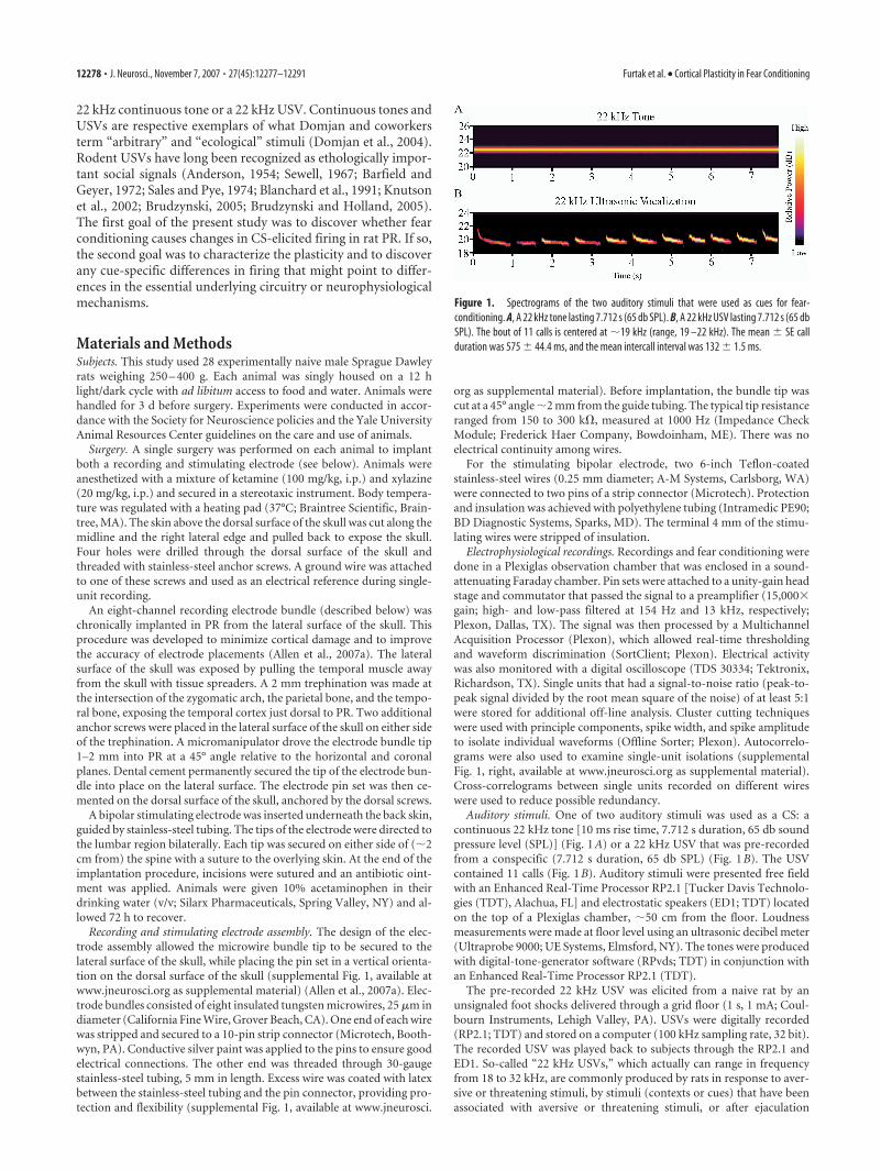
ResultsNumber, persistence, and waveforms of PR unitsA total of 200 well isolated PR units were analyzed in 28 rats. Theanalysis only included units that were recorded throughout allthree stages of the experiment (for 6 –7 h from the beginning ofthe preexposure period). Figure 2A illustrates three sets of super-imposed waveforms (�1000 each) from the same unit in the
preexposure period (left), the conditioning period (middle), andthe testing period (right). These waveforms include the 1 ms timesegment that was digitized for subsequent off-line analysis. Theillustrated waveform is typical in that an initial negative phase isfollowed by a positive phase. In some cases, the large negativephase was preceded by a small positive phase, and, in others, theentire waveform was narrower. The narrower waveforms mayreflect activity in “fast-spiking” PR neurons that are thought to beinhibitory (Faulkner and Brown, 1999; McGann et al., 2001;Moyer et al., 2002; Moyer and Brown, 2007). Figure 2B showsspontaneous firing in the same unit during the intertrial stage ofconditioning. The longer time segment reveals that the large pos-itive phase of the waveform was followed by a much smaller andmore prolonged negative phase, identified in Figure 2B by theasterisk. This triphasic waveform is representative of most of thePR units included in the present analysis.
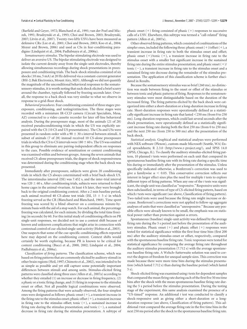
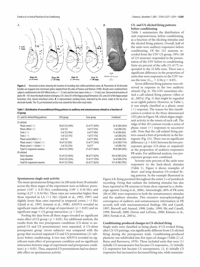
Histological determination of recording sitesNissl-stained brain sections verified that all recording tips werewithin the boundaries of PR (Burwell et al., 1995; Burwell andAmaral, 1998; Burwell, 2001; Furtak et al., 2007b). The reliabilityof the lateral implantation procedure in targeting PR (Allen et al.,2007a) was attributable to the fact that temporal cortex was di-rectly visualized during the implantation. Marking lesions weremost frequently in layers II/III and V of PR. The locations of theelectrode tips ranged from �4.0 to �6.0 mm (anteroposterior)and �6.8 to �7.6 mm (dorsoventral) relative to bregma (Fig.3A). Figure 3B shows an example of a representative markinglesion in a horizontal section that corresponds to Plate 98 of thePaxinos and Watson rat atlas (1998). Cortical damage was mini-mized (Fig. 3B) because of the short distance traveled by themicrowire bundles and because this short distance eliminated theneed to insert a microwire guide tube into the brain.
Figure 2. A representative PR unit illustrated on two different timescales. A, Superimposedvoltage waveforms from the preexposure, conditioning, and testing phases of the experiment.The indicated threshold voltage (arrow) triggered the capture of 1 ms time segments (300 �sbefore the threshold crossing and 700 �s after the threshold crossing) that were used off-linefor single-unit analysis. Characteristically, an initial negative phase was followed by a positivephase. B, A portion of an oscilloscope trace showing spontaneous firing in the same unit duringthe intertrial interval of the conditioning stage of the experiment. This timescale shows that thepositive phase of the voltage waveform was followed by a smaller and more prolonged nega-tive phase (to the left of the asterisk).
12280 • J. Neurosci., November 7, 2007 • 27(45):12277–12291 Furtak et al. • Cortical Plasticity in Fear Conditioning
Spontaneous single-unit activityThe mean spontaneous firing rates (in 200 units from 28 animals)across the three stages of the experiment were as follows: preex-posure (3.87 � 0.33 Hz); conditioning (3.89 � 0.34 Hz); andtesting (4.27 � 0.34 Hz). These rates are somewhat higher thanthose reported in LA (�1 Hz) (Quirk et al., 1995, 1997) andslightly lower than rates reported in temporal cortex (�5 Hz)(Quirk et al., 1997; Armony et al., 1998). ANOVA revealed nosignificant main effect of stage of experiment ( p 0.05) and nosignificant stage � CS group interaction ( p 0.05).
Pooling the data from all three stages revealed no significantmain effect of CS group ( p 0.05). For additional analysis, theresults from the two preexposure conditions (CS-alone vs un-paired CS and US presentations) were separated. A CS-alonepreexposure group (seven subjects) was compared with thegroup that received unpaired CS and US presentations (21 sub-jects) before conditioning. A two-way ANOVA revealed no sig-nificant main effect of preexposure condition and no significantinteraction between stage of experiment and preexposure condi-tion ( p 0.05). Thus, unpaired US presentations had no detect-able effect on spontaneous activity.
CS- and US-elicited firing patternsbefore conditioningTable 1 summarizes the distribution ofunit responsiveness, before conditioning,as a function of the eliciting stimulus andthe elicited firing pattern. Overall, half ofthe units were auditory responsive beforeconditioning. Of the 123 neurons re-corded from the USV CS group, 39% (48of 123 neurons) responded to the presen-tation of the USV before to conditioning.Sixty-six percent of the cells (51 of 77) re-sponded to the 22 kHz tone. There was asignificant difference in the proportion ofunits that were responsive to the USV ver-sus the tone (G(1) 4.56; p � 0.05).
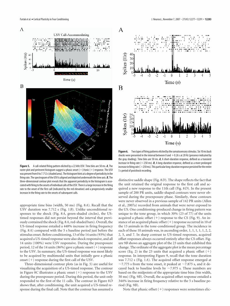
Seven different firing patterns were ob-served in response to the two auditorystimuli (Fig. 4). The USV sometimes elic-ited a call-related firing pattern (Allen etal., 2007a) (Fig. 5) that might be regardedas an eighth pattern. However, in Table 1,it was simply classified as a phasic onset(�) response. The reason for this classifi-cation is evident in the three-dimensional(3D) plot in Figure 5B, which aligns single-unit activity to the onsets of each call. Theridge of this 3D contour reveals a series ofphasic onset (�) responses to successivecalls. Note that the call-related firing pat-tern caused a hint of periodicity in the his-togram (Fig. 5A). There was no significantdifference ( p 0.05) between the two pre-exposure groups (CS-alone or unpaired)in the proportion of auditory-responsivePR units. For additional analysis, two pre-exposure groups were combined.
Seventy-nine percent of the units wereresponsive to the back-shock stimulus(Table 1). Figure 6 shows examples ofshort- and long-duration US-evoked fir-ing patterns. In the example illustrated in
Figure 6B, firing persisted throughout the entire 5 s of postshockrecording. Firing that outlasts the initiating stimulus has alsobeen reported in PR neurons in brain slices exposed to a cholin-ergic agonist (Leung et al., 2006). Interestingly, 40% of PR units(80 of 200) were responsive to both the somatosensory stimulusand the auditory stimulus. This extensive neurophysiologicalconvergence of auditory and somatosensory information in PRaccords well with neuroanatomical findings (Shi and Cassell,1997; Burwell and Amaral, 1998; Linke, 1999; Shi and Cassell,1999; Burwell, 2000; Doron and LeDoux, 2000; Kimura et al.,2003; Furtak et al., 2007a).
Conditioning-produced changes in CS-elicited firingSingle units were classified as being plastic if CS-evoked firing,after CS–US pairings, was significantly different from CS-elicitedfiring during the preexposure trials. Conditioning-producedplasticity was subdivided into five types of changes (Table 2) (cf.Bures and Burresova, 1970). These included units that were (1)initially CS unresponsive but became CS responsive, (2) initiallyCS responsive but became CS unresponsive, (3, 4) initially CSresponsive but increased or decreased firing rate, while maintain-
Figure 3. Horizontal sections showing the location of recording sites within perirhinal cortex. A, Placements of 28 electrodebundles are mapped onto stereotaxic plates adapted from the atlas of Paxinos and Watson (1998). Results were combined fromsubjects conditioned to the USV (filled circles; n 15 rats) and the tone (open circles; n 13 rats) cues. Dorsoventral locations ofplates 98 –101 show the depth relative to bregma. CA1, Area CA1 of the hippocampal formation; CA3, area CA3 of the hippocampalformation; LEnt, lateral entorhinal cortex. B, A representative marking lesion, indicated by the arrow, made at the tip of anelectrode bundle. The 70 �m horizontal section was stained for Nissl with cresyl violet.
Table 1. Distribution of unconditional firing patterns to auditory and somatosensory stimuli as a function ofCS group
CS- and US-elicited firing patterns USV group Tone group Combined
CS-elicitedPhasic onset (�) 30 of 123 (24%) 22 of 77 (29%) 52 of 200 (26%)Phasic offset (�) 0 of 123 4 of 77 (5%) 4 of 200 (2%)Tonic (�) 5 of 123 (4%) 6 of 77 (8%) 11 of 200 (6%)Tonic (�) 1 of 123 (1%) 1 of 77 (1%) 3 of 200 (2%)Phasic onset/offset (�) 0 of 123 6 of 77 (8%) 6 of 200 (3%)Phasic onset (�)/tonic (�) 10 of 123 (8%) 12 of 77 (16%) 22 of 200 (11%)Phasic onset (�)/tonic (�) 2 of 123 (2%) 0 of 77 1 of 200 (1%)Total CS-responsive neurons 48 of 123 (39%) 51 of 77 (66%) 99 of 200 (50%)
US-elicitedShort-duration 55 of 123 (45%) 48 of 77 (62%) 103 of 200 (52%)Long-duration 39 of 123 (32%) 15 of 77 (19%) 54 of 200 (27%)Total US-responsive neurons 94 of 123 (76%) 63 of 77 (82%) 157 of 200 (79%)
Furtak et al. • Cortical Plasticity in Fear Conditioning J. Neurosci., November 7, 2007 • 27(45):12277–12291 • 12281
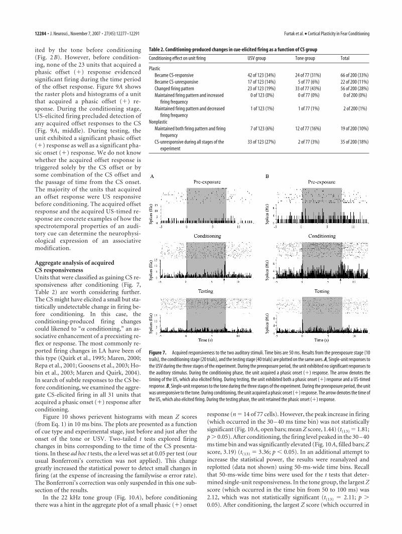
ing the same firing pattern; or (5) initiallyCS responsive and changed firing pattern(Table 2). Conditioning-producedchanges were observed in all seven types ofunconditional response firing patterns(Figs. 4, 5) that were elicited by the twocues. A unit was classified as nonplastic if(1) the CS-evoked activity was unchangedin firing pattern and firing frequencythroughout all stages of fear conditioningor (2) the unit was not significantly re-sponsive to the CS at any stage of the ex-periment. Table 2 summarizes the numberof units from the tone and USV groupsthat were classified as plastic or nonplasticas just described. The proportion of cellsin each category was not significantly af-fected by analyzing only the first 20 testingtrials instead of all 40 trials ( p values 0.05). Overall, 73% of PR units wereclassified as plastic.
In the USV group, 67% of the neurons(83 of 123) showed significant changes inCS-elicited firing after CS–US pairings(Table 2) and were thus categorized asplastic. The remaining 33% were classifiedas nonplastic (40 of 123) (Table 2). Therewere significant differences in the propor-tions of the five different types of plasticity(G(4) 84.11; p � 0.001). The most prev-alent type of firing change in the USVgroup was from CS unresponsive to CSresponsive (34%; 42 of 123) (Table 2). Fig-ure 7A shows the perievent raster plots andhistograms of a unit that was unresponsiveto the USV during the preexposure period but acquired a phasiconset (�) response during conditioning. The acquired onset re-sponse was evident in both the conditioning period (Fig. 7A,middle) and the testing session (Fig. 7A, bottom). This same unitalso acquired a small but statistically significant phasic (�) re-sponse at the former time of the US (Fig. 7A, bottom). During theconditioning period, US-elicited firing precluded detection ofany CS-elicited firing changes that might have occurred in thistime interval (Figs. 6, 7A). This US-timed response is furtherdiscussed below. The second most common modification in theUSV group was a change in the elicited firing pattern (19%; 23 of123) (Table 2). The unit illustrated in Figure 8B is an example ofa conditioning-produced firing-pattern change. In the preexpo-sure period, the unit gave a phasic onset (�) response during thefirst call of the USV. In the testing period, the unit retained thephasic onset (�) response during the first call and acquired aUS-timed response during the 11th and final call of the USV.
In the tone group, 82% (63 of 77) of the units exhibitedconditioning-produced plasticity (Table 2) and were thus cate-gorized as plastic. There were significant differences in the pro-portions of different types of changes (G(4) 80.16; p � 0.001).In contrast to the USV group, the most common type of plasticityin the tone group entailed a change in firing pattern (43%; 33 of77) (Table 2). Most commonly, the change was from a phasiconset (�) firing pattern to a phasic onset/offset (�) pattern. Thesecond most prevalent type of plasticity, changing from CS unre-sponsive to CS responsive, occurred in 31% of the units in thetone group (24 of 77) (Table 2). The units shown in Figures 7B
and 9A were unresponsive to the tone during the preexposureperiod but exhibited a phasic onset (�) response during the con-ditioning and testing periods. Overall, there was no significantdifference between the two cue groups in the proportion of plas-tic units (Table 2) ( p 0.05).
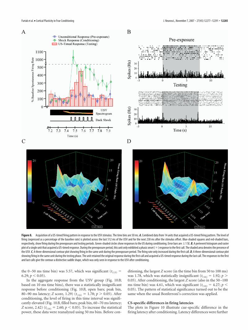
CS-specific differences in elicited firing after conditioningAlthough there were obvious similarities between the two cues, interms of the types of conditioning-produced changes they elicited(Table 2), the US-timed response was only observed in the USVgroup (Fig. 8), in which it occurred in 11% (14 of 123) of theunits. An instance of US-timed firing occurred in 7 of the 15animals in the USV-conditioned group. The incidence in each ofthese seven animals was, in ascending order, 1, 1, 2, 2, 2, 2, and 4.Figure 8A shows an aggregate response from all 14 units thatexhibited this change. The ordinate of the aggregate plot is themean percentage score (from Eq. 2) in the 14 units that exhibiteda statistically significant, US-timed response. Before condition-ing, the firing rate was relatively constant during the last 512 msof the USV presentation (Fig. 8A, Pre-exposure, blue-shadedsquares). After conditioning, there was a significant increase inthe firing rate during the 260 ms time interval that previouslycontained the US (Fig. 8A, red-shaded bars). Interestingly, thepeak firing occurred near the midpoint of this 260 ms interval(Fig. 8A, red-shaded bars).
US-timed firing emerged at �7.525 s from the USV onset, itpeaked at �7.575 s, and it decayed back to baseline levels by�7.675 s. These numbers are based on the midpoints of the
Figure 4. Representative examples of seven different firing patterns elicited by the tone or the USV stimuli (see Fig. 1) duringthe preexposure period. Gray shading represents the presence of the auditory stimulus. Time bins in the frequency histogram are50 ms wide. A, A phasic onset (�) response elicited by the USV. B, A phasic offset (�) response elicited by the tone. The hint of atonic (�) response was not statistically significant. C, A tonic (�) response to the tone. D, A tonic (�) response to the tone. E, Aphasic onset (�)/offset (�) response to the tone. F, A phasic onset (�)/tonic (�) response to the tone. G, A phasic onset(�)/tonic (�) response to the USV.
12282 • J. Neurosci., November 7, 2007 • 27(45):12277–12291 Furtak et al. • Cortical Plasticity in Fear Conditioning
appropriate time bins (width, 50 ms) (Fig. 8A). Recall that theUSV duration was 7.712 s (Fig. 1B). Unlike unconditional re-sponses to the shock (Fig. 8A, green-shaded circles), the US-timed responses did not persist beyond the interval that previ-ously contained the shock (Fig. 8A, red-shaded bars). Overall, theUS-timed response entailed a 448% increase in firing frequency(Fig. 8A) compared with the 5 s baseline period just before thestimulus onset. Before conditioning, 13 of the 14 units (93%) thatacquired a US-timed response were also shock responsive, and all14 units (100%) were USV responsive. During the preexposureperiod, 12 of the 14 units (86%) gave a phasic onset (�) responseto the USV. In summary, the US-timed response was most likelyto be acquired by multimodal units that initially gave a phasiconset (�) response during the first call of the USV.
Three-dimensional contour plots (as in Fig. 5) are useful forvisualizing the acquisition of a US-timed response. The contourin Figure 8C illustrates a phasic onset (�) response to the USVduring the preexposure period. During this period, the unit onlyresponded to the first of the 11 calls. The contour in Figure 8Dshows that, after conditioning, the unit acquired a US-timed re-sponse during the final call. Note that the contour has assumed a
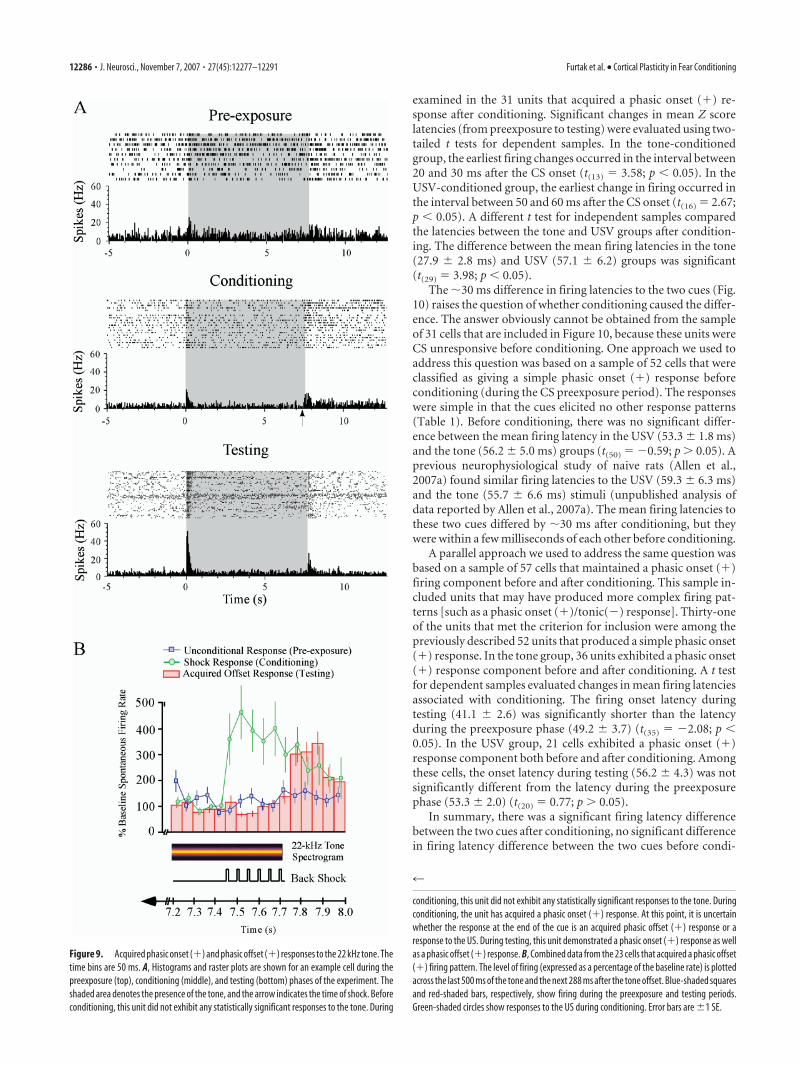
distinctive saddle shape (Fig. 8D). The shape reflects the fact thatthe unit retained the original response to the first call and ac-quired a new response to the 11th call (Fig. 8D). In the presentsample of 200 PR units, saddle-shaped contours were never ob-served during the preexposure phase. Similarly, these contourswere never observed in a previous sample of 142 PR units (Allenet al., 2007a) recorded from animals that were never exposed tothe US. One conditioning-produced change in firing pattern wasunique to the tone group, in which 30% (23 of 77) of the unitsacquired a phasic offset (�) response to the CS (Fig. 9). An in-stance of an acquired phasic offset (�) response occurred in 10 ofthe 13 animals in the tone-conditioned group. The incidence ineach of these 10 animals was, in ascending order, 1, 1, 1, 1, 1, 2, 2,2, 5, and 7. In sharp contrast to US-timed responses, acquiredoffset responses always occurred entirely after the CS offset. Fig-ure 9B shows an aggregate plot of the 23 units that exhibited thischange. The ordinate of the aggregate plot is the mean percentagescore (Eq. 2) in the 23 units that acquired a phasic offset (�)response. In interpreting Figure 9, recall that the tone durationwas 7.712 s (Fig. 1A). The acquired offset response emerged at�7.775 s from the tone onset, it peaked at �7.875 s, and it de-cayed back to baseline levels by �7.975 s. These numbers arebased on the midpoints of the appropriate time bins (bin width,50 ms) (Fig. 9B). Overall, the acquired offset response entailed a344% increase in firing frequency relative to the 5 s baseline pe-riod (Fig. 9B).
Note that phasic offset (�) responses were sometimes elic-
Figure 5. A call-related firing pattern elicited by a 22 kHz USV. Time bins are 50 ms. A, Theraster plot and perievent histogram suggest a phasic onset (�)/tonic (�) response. The USVwas present from 0 to 7.712 s (shaded area). The histogram hints at a degree of periodicity in thefiring rate. The spectrogram of the USV is aligned and depicted underneath the time axis. B, Thethree-dimensional contour plot reveals that the apparent periodicity in the histogram is asso-ciated with firing to the onsets of individual calls of the USV. There is a large increase in the firingrate to the onset of the first call (indicated by the red elevation) and a progressively smallerincrease in the firing rate to the onsets of subsequent calls.
Figure 6. Two types of firing patterns elicited by the somatosensory stimulus. Six 10 ms backshocks were presented in the interval between 0 and �0.26 s at 20 Hz (presence indicated bythe gray shading). Time bins are 50 ms. A, A short-duration response, defined as a transientincrease in firing rate (�250 ms). B, A long-duration response, defined as a more prolongedincrease in firing rate (250 ms). This particular long-duration response persisted for the entire5 s period of postshock recording.
Furtak et al. • Cortical Plasticity in Fear Conditioning J. Neurosci., November 7, 2007 • 27(45):12277–12291 • 12283
ited by the tone before conditioning(Fig. 2 B). However, before condition-ing, none of the 23 units that acquired aphasic offset (�) response evidencedsignificant firing during the time periodof the offset response. Figure 9A showsthe raster plots and histograms of a unitthat acquired a phasic offset (�) re-sponse. During the conditioning stage,US-elicited firing precluded detection ofany acquired offset responses to the CS(Fig. 9A, middle). During testing, theunit exhibited a significant phasic offset(�) response as well as a significant pha-sic onset (�) response. We do not knowwhether the acquired offset response istriggered solely by the CS offset or bysome combination of the CS offset andthe passage of time from the CS onset.The majority of the units that acquiredan offset response were US responsivebefore conditioning. The acquired offsetresponse and the acquired US-timed re-sponse are concrete examples of how thespectrotemporal properties of an audi-tory cue can determine the neurophysi-ological expression of an associativemodification.
Aggregate analysis of acquiredCS responsivenessUnits that were classified as gaining CS re-sponsiveness after conditioning (Fig. 7,Table 2) are worth considering further.The CS might have elicited a small but sta-tistically undetectable change in firing be-fore conditioning. In this case, theconditioning-produced firing changescould likened to “� conditioning,” an as-sociative enhancement of a preexisting re-flex or response. The most commonly re-ported firing changes in LA have been ofthis type (Quirk et al., 1995; Maren, 2000;Repa et al., 2001; Goosens et al., 2003; Ho-bin et al., 2003; Maren and Quirk, 2004).In search of subtle responses to the CS be-fore conditioning, we examined the aggre-gate CS-elicited firing in all 31 units thatacquired a phasic onset (�) response afterconditioning.
Figure 10 shows perievent histograms with mean Z scores(from Eq. 1) in 10 ms bins. The plots are presented as a functionof cue type and experimental stage, just before and just after theonset of the tone or USV. Two-tailed t tests explored firingchanges in bins corresponding to the time of the CS presenta-tions. In these ad hoc t tests, the � level was set at 0.05 per test (ourusual Bonferroni’s correction was not applied). This changegreatly increased the statistical power to detect small changes infiring (at the expense of increasing the familywise � error rate).The Bonferroni’s correction was only suspended in this one sub-section of the results.
In the 22 kHz tone group (Fig. 10A), before conditioningthere was a hint in the aggregate plot of a small phasic (�) onset
response (n 14 of 77 cells). However, the peak increase in firing(which occurred in the 30 – 40 ms time bin) was not statisticallysignificant (Fig. 10A, open bars; mean Z score, 1.44) (t(13) 1.81;p 0.05). After conditioning, the firing level peaked in the 30 – 40ms time bin and was significantly elevated (Fig. 10A, filled bars; Zscore, 3.19) (t(13) 3.36; p � 0.05). In an additional attempt toincrease the statistical power, the results were reanalyzed andreplotted (data not shown) using 50-ms-wide time bins. Recallthat 50-ms-wide time bins were used for the t tests that deter-mined single-unit responsiveness. In the tone group, the largest Zscore (which occurred in the time bin from 50 to 100 ms) was2.12, which was not statistically significant (t(13) 2.11; p 0.05). After conditioning, the largest Z score (which occurred in
Figure 7. Acquired responsiveness to the two auditory stimuli. Time bins are 50 ms. Results from the preexposure stage (10trails), the conditioning stage (20 trials), and the testing stage (40 trials) are plotted on the same axes. A, Single-unit responses tothe USV during the three stages of the experiment. During the preexposure period, the unit exhibited no significant responses tothe auditory stimulus. During the conditioning phase, the unit acquired a phasic onset (�) response. The arrow denotes thetiming of the US, which also elicited firing. During testing, the unit exhibited both a phasic onset (�) response and a US-timedresponse. B, Single-unit responses to the tone during the three stages of the experiment. During the preexposure period, the unitwas unresponsive to the tone. During conditioning, the unit acquired a phasic onset (�) response. The arrow denotes the time ofthe US, which also elicited firing. During the testing phase, the unit retained the phasic onset (�) response.
Table 2. Conditioning-produced changes in cue-elicited firing as a function of CS group
Conditioning effect on unit firing USV group Tone group Total
PlasticBecame CS-responsive 42 of 123 (34%) 24 of 77 (31%) 66 of 200 (33%)Became CS-unresponsive 17 of 123 (14%) 5 of 77 (6%) 22 of 200 (11%)Changed firing pattern 23 of 123 (19%) 33 of 77 (43%) 56 of 200 (28%)Maintained firing pattern and increased 0 of 123 (0%) 0 of 77 (0%) 0 of 200 (0%)
firing frequencyMaintained firing pattern and decreased 1 of 123 (1%) 1 of 77 (1%) 2 of 200 (1%)
firing frequencyNonplastic
Maintained both firing pattern and firing 7 of 123 (6%) 12 of 77 (16%) 19 of 200 (10%)frequency
CS-unresponsive during all stages of the 33 of 123 (27%) 2 of 77 (3%) 35 of 200 (18%)experiment
12284 • J. Neurosci., November 7, 2007 • 27(45):12277–12291 Furtak et al. • Cortical Plasticity in Fear Conditioning
the 0 –50 ms time bin) was 5.57, which was significant (t(13) 6.29; p � 0.05).
In the aggregate response from the USV group (Fig. 10B;based on 10 ms time bins), there was a statistically insignificantresponse before conditioning (Fig. 10B, open bars; peak bin,80 –90 ms latency; Z score, 1.29) (t(16) 1.78; p 0.05). Afterconditioning, the level of firing in this time interval was signifi-cantly elevated (Fig. 10B, filled bars; peak bin, 60 –70 ms latency;Z score, 2.42) (t(16) 2.60; p � 0.05). To increase the statisticalpower, these data were reanalyzed using 50 ms bins. Before con-
ditioning, the largest Z score (in the time bin from 50 to 100 ms)was 1.78, which was statistically insignificant (t(16) 1.92; p 0.05). After conditioning, the largest Z score (also in the 50 –100ms time bin) was 4.61, which was significant (t(16) 4.27; p �0.05). The pattern of statistical significance turned out to be thesame when the usual Bonferroni’s correction was applied.
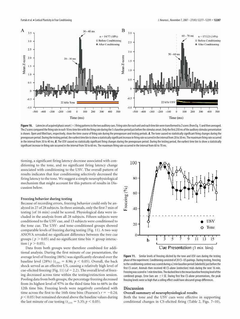
CS-specific differences in firing latenciesThe plots in Figure 10 illustrate cue-specific difference in thefiring latency after conditioning. Latency differences were further
Figure 8. Acquisition of a US-timed firing pattern in response to the USV stimulus. The time bins are 50 ms. A, Combined data from 14 units that acquired a US-timed firing pattern. The level offiring (expressed as a percentage of the baseline rate) is plotted across the last 512 ms of the USV and for the next 238 ms after the stimulus offset. Blue-shaded squares and red-shaded bars,respectively, show firing during the preexposure and testing periods. Green-shaded circles show responses to the US during conditioning. Error bars are �1 SE. B, A perievent histogram and rasterplot of a single unit that acquired a US-timed response. During the preexposure period, this unit only exhibited a phasic onset (�) response to the first call. The shaded area denotes the presence ofthe USV. C, A three-dimensional contour plot showing firing in the same unit during the preexposure period. The firing rate only increased during the first call. D, A three-dimensional contour plotshowing firing in the same unit during the testing phase. The unit retained the original response during the first call and acquired a US-timed response during the last call. The responses to the firstand last calls give the contour a distinctive saddle shape, which was only seen in response to the USV after conditioning.
Furtak et al. • Cortical Plasticity in Fear Conditioning J. Neurosci., November 7, 2007 • 27(45):12277–12291 • 12285
examined in the 31 units that acquired a phasic onset (�) re-sponse after conditioning. Significant changes in mean Z scorelatencies (from preexposure to testing) were evaluated using two-tailed t tests for dependent samples. In the tone-conditionedgroup, the earliest firing changes occurred in the interval between20 and 30 ms after the CS onset (t(13) 3.58; p � 0.05). In theUSV-conditioned group, the earliest change in firing occurred inthe interval between 50 and 60 ms after the CS onset (t(16) 2.67;p � 0.05). A different t test for independent samples comparedthe latencies between the tone and USV groups after condition-ing. The difference between the mean firing latencies in the tone(27.9 � 2.8 ms) and USV (57.1 � 6.2) groups was significant(t(29) 3.98; p � 0.05).
The �30 ms difference in firing latencies to the two cues (Fig.10) raises the question of whether conditioning caused the differ-ence. The answer obviously cannot be obtained from the sampleof 31 cells that are included in Figure 10, because these units wereCS unresponsive before conditioning. One approach we used toaddress this question was based on a sample of 52 cells that wereclassified as giving a simple phasic onset (�) response beforeconditioning (during the CS preexposure period). The responseswere simple in that the cues elicited no other response patterns(Table 1). Before conditioning, there was no significant differ-ence between the mean firing latency in the USV (53.3 � 1.8 ms)and the tone (56.2 � 5.0 ms) groups (t(50) �0.59; p 0.05). Aprevious neurophysiological study of naive rats (Allen et al.,2007a) found similar firing latencies to the USV (59.3 � 6.3 ms)and the tone (55.7 � 6.6 ms) stimuli (unpublished analysis ofdata reported by Allen et al., 2007a). The mean firing latencies tothese two cues differed by �30 ms after conditioning, but theywere within a few milliseconds of each other before conditioning.
A parallel approach we used to address the same question wasbased on a sample of 57 cells that maintained a phasic onset (�)firing component before and after conditioning. This sample in-cluded units that may have produced more complex firing pat-terns [such as a phasic onset (�)/tonic(�) response]. Thirty-oneof the units that met the criterion for inclusion were among thepreviously described 52 units that produced a simple phasic onset(�) response. In the tone group, 36 units exhibited a phasic onset(�) response component before and after conditioning. A t testfor dependent samples evaluated changes in mean firing latenciesassociated with conditioning. The firing onset latency duringtesting (41.1 � 2.6) was significantly shorter than the latencyduring the preexposure phase (49.2 � 3.7) (t(35) �2.08; p �0.05). In the USV group, 21 cells exhibited a phasic onset (�)response component both before and after conditioning. Amongthese cells, the onset latency during testing (56.2 � 4.3) was notsignificantly different from the latency during the preexposurephase (53.3 � 2.0) (t(20) 0.77; p 0.05).
In summary, there was a significant firing latency differencebetween the two cues after conditioning, no significant differencein firing latency difference between the two cues before condi-
Figure 9. Acquired phasic onset (�) and phasic offset (�) responses to the 22 kHz tone. Thetime bins are 50 ms. A, Histograms and raster plots are shown for an example cell during thepreexposure (top), conditioning (middle), and testing (bottom) phases of the experiment. Theshaded area denotes the presence of the tone, and the arrow indicates the time of shock. Beforeconditioning, this unit did not exhibit any statistically significant responses to the tone. During
4
conditioning, this unit did not exhibit any statistically significant responses to the tone. Duringconditioning, the unit has acquired a phasic onset (�) response. At this point, it is uncertainwhether the response at the end of the cue is an acquired phasic offset (�) response or aresponse to the US. During testing, this unit demonstrated a phasic onset (�) response as wellas a phasic offset (�) response. B, Combined data from the 23 cells that acquired a phasic offset(�) firing pattern. The level of firing (expressed as a percentage of the baseline rate) is plottedacross the last 500 ms of the tone and the next 288 ms after the tone offset. Blue-shaded squaresand red-shaded bars, respectively, show firing during the preexposure and testing periods.Green-shaded circles show responses to the US during conditioning. Error bars are �1 SE.
12286 • J. Neurosci., November 7, 2007 • 27(45):12277–12291 Furtak et al. • Cortical Plasticity in Fear Conditioning
tioning, a significant firing latency decrease associated with con-ditioning to the tone, and no significant firing latency changeassociated with conditioning to the USV. The overall pattern ofresults indicates that fear conditioning selectively decreased thefiring latency to the tone. We suggest a simple neurophysiologicalmechanism that might account for this pattern of results in Dis-cussion below.
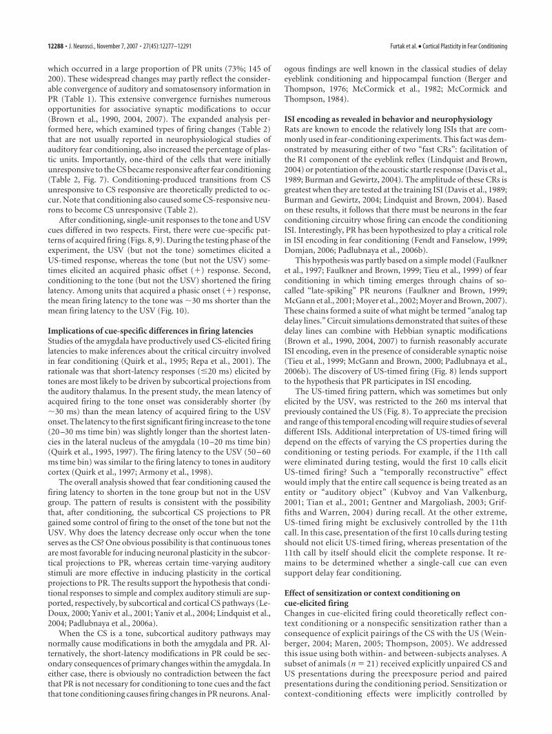
Freezing behavior during testingBecause of recording errors, freezing behavior could only be an-alyzed in 27 of 28 subjects. In three animals, only the first 7 min oftesting (of 16 min) could be scored. Physiological data were in-cluded in the analysis from all 28 subjects. Fifteen subjects wereconditioned to the USV cue, and 13 subjects were conditioned tothe tone cue. The USV- and tone-conditioned groups showedcomparable levels of freezing during testing (Fig. 11). A two-wayANOVA revealed no significant difference between the two cuegroups ( p 0.05) and no significant time bin � group interac-tion ( p 0.05).
Data from both groups were therefore combined for addi-tional analysis. During the first minute of cue presentation, theaverage level of freezing (86%) was significantly elevated over thebaseline level (28%) (t(26) 8.96; p � 0.05). Overall, the backshock served as an effective US, causing a relatively high level ofcue-elicited freezing (Fig. 11) (d 2.2). The overall level of freez-ing decreased across time within the testing/extinction session.Pooling data from both groups, the percentage freezing decreasedfrom its highest level of 97% in the third time bin to 66% in the12th time bin. Freezing levels were negatively correlated withtime across the first to the 16th time bins (Pearson’s r �0.24;p � 0.05) but remained elevated above the baseline values duringthe last minute of cue testing (t(23) 5.35; p � 0.05).
DiscussionOverall summary of neurophysiological resultsBoth the tone and the USV cues were effective in supportingconditional changes in CS-elicited firing (Table 2, Figs. 7–10),
Figure 11. Similar levels of freezing elicited by the tone and USV cues during the testingphase of the experiment. Conditioning consisted of 20 CS–US pairings. During testing, freezingto the conditioning context was scored during a 2 min baseline period (labeled b) just before thefirst CS onset. Animals then received 40 CS-alone (extinction) trials during the next 16 min.Freezing was scored in 1 min time bins. The dashed line is the mean baseline freezing level of thecombined groups. Error bars are �1 SE. During first few CS-alone presentations, the peakfreezing levels were so high that a ceiling effect could have obscured group differences.
Figure 10. Latencies of acquired phasic onset (�) firing patterns to the two auditory cues. Firing rates for each unit and each time bin were transformed to Z scores (from Eq. 1) and then averaged.The Z scores compared the firing rate in each 10 ms time bin with the firing rate during the 5 s baseline period just before the stimulus onset. Only the first 250 ms of the auditory stimulus presentationis shown. Open and filled bars, respectively, show the time course of firing rate during the preexposure and testing periods. A, The tone caused no statistically significant firing changes during thepreexposure period. During the testing period, the earliest time bin to show a statistically significant increase in firing rate occurred in the interval from 20 to 30 ms. The maximum firing rate occurredin the interval from 30 to 40 ms. B, The USV caused no statistically significant firing changes during the preexposure period. During the testing period, the earliest time bin to show a statisticallysignificant increase in firing rate occurred in the interval from 50 to 60 ms. The maximum firing rate occurred in the interval from 60 to 70 ms.
Furtak et al. • Cortical Plasticity in Fear Conditioning J. Neurosci., November 7, 2007 • 27(45):12277–12291 • 12287
which occurred in a large proportion of PR units (73%; 145 of200). These widespread changes may partly reflect the consider-able convergence of auditory and somatosensory information inPR (Table 1). This extensive convergence furnishes numerousopportunities for associative synaptic modifications to occur(Brown et al., 1990, 2004, 2007). The expanded analysis per-formed here, which examined types of firing changes (Table 2)that are not usually reported in neurophysiological studies ofauditory fear conditioning, also increased the percentage of plas-tic units. Importantly, one-third of the cells that were initiallyunresponsive to the CS became responsive after fear conditioning(Table 2, Fig. 7). Conditioning-produced transitions from CSunresponsive to CS responsive are theoretically predicted to oc-cur. Note that conditioning also caused some CS-responsive neu-rons to become CS unresponsive (Table 2).
After conditioning, single-unit responses to the tone and USVcues differed in two respects. First, there were cue-specific pat-terns of acquired firing (Figs. 8, 9). During the testing phase of theexperiment, the USV (but not the tone) sometimes elicited aUS-timed response, whereas the tone (but not the USV) some-times elicited an acquired phasic offset (�) response. Second,conditioning to the tone (but not the USV) shortened the firinglatency. Among units that acquired a phasic onset (�) response,the mean firing latency to the tone was �30 ms shorter than themean firing latency to the USV (Fig. 10).
Implications of cue-specific differences in firing latenciesStudies of the amygdala have productively used CS-elicited firinglatencies to make inferences about the critical circuitry involvedin fear conditioning (Quirk et al., 1995; Repa et al., 2001). Therationale was that short-latency responses (�20 ms) elicited bytones are most likely to be driven by subcortical projections fromthe auditory thalamus. In the present study, the mean latency ofacquired firing to the tone onset was considerably shorter (by�30 ms) than the mean latency of acquired firing to the USVonset. The latency to the first significant firing increase to the tone(20 –30 ms time bin) was slightly longer than the shortest laten-cies in the lateral nucleus of the amygdala (10 –20 ms time bin)(Quirk et al., 1995, 1997). The firing latency to the USV (50 – 60ms time bin) was similar to the firing latency to tones in auditorycortex (Quirk et al., 1997; Armony et al., 1998).
The overall analysis showed that fear conditioning caused thefiring latency to shorten in the tone group but not in the USVgroup. The pattern of results is consistent with the possibilitythat, after conditioning, the subcortical CS projections to PRgained some control of firing to the onset of the tone but not theUSV. Why does the latency decrease only occur when the toneserves as the CS? One obvious possibility is that continuous tonesare most favorable for inducing neuronal plasticity in the subcor-tical projections to PR, whereas certain time-varying auditorystimuli are more effective in inducing plasticity in the corticalprojections to PR. The results support the hypothesis that condi-tional responses to simple and complex auditory stimuli are sup-ported, respectively, by subcortical and cortical CS pathways (Le-Doux, 2000; Yaniv et al., 2001; Yaniv et al., 2004; Lindquist et al.,2004; Padlubnaya et al., 2006a).
When the CS is a tone, subcortical auditory pathways maynormally cause modifications in both the amygdala and PR. Al-ternatively, the short-latency modifications in PR could be sec-ondary consequences of primary changes within the amygdala. Ineither case, there is obviously no contradiction between the factthat PR is not necessary for conditioning to tone cues and the factthat tone conditioning causes firing changes in PR neurons. Anal-
ogous findings are well known in the classical studies of delayeyeblink conditioning and hippocampal function (Berger andThompson, 1976; McCormick et al., 1982; McCormick andThompson, 1984).
ISI encoding as revealed in behavior and neurophysiologyRats are known to encode the relatively long ISIs that are com-monly used in fear-conditioning experiments. This fact was dem-onstrated by measuring either of two “fast CRs”: facilitation ofthe R1 component of the eyeblink reflex (Lindquist and Brown,2004) or potentiation of the acoustic startle response (Davis et al.,1989; Burman and Gewirtz, 2004). The amplitude of these CRs isgreatest when they are tested at the training ISI (Davis et al., 1989;Burman and Gewirtz, 2004; Lindquist and Brown, 2004). Basedon these results, it follows that there must be neurons in the fearconditioning circuitry whose firing can encode the conditioningISI. Interestingly, PR has been hypothesized to play a critical rolein ISI encoding in fear conditioning (Fendt and Fanselow, 1999;Domjan, 2006; Padlubnaya et al., 2006b).
This hypothesis was partly based on a simple model (Faulkneret al., 1997; Faulkner and Brown, 1999; Tieu et al., 1999) of fearconditioning in which timing emerges through chains of so-called “late-spiking” PR neurons (Faulkner and Brown, 1999;McGann et al., 2001; Moyer et al., 2002; Moyer and Brown, 2007).These chains formed a suite of what might be termed “analog tapdelay lines.” Circuit simulations demonstrated that suites of thesedelay lines can combine with Hebbian synaptic modifications(Brown et al., 1990, 2004, 2007) to furnish reasonably accurateISI encoding, even in the presence of considerable synaptic noise(Tieu et al., 1999; McGann and Brown, 2000; Padlubnaya et al.,2006b). The discovery of US-timed firing (Fig. 8) lends supportto the hypothesis that PR participates in ISI encoding.
The US-timed firing pattern, which was sometimes but onlyelicited by the USV, was restricted to the 260 ms interval thatpreviously contained the US (Fig. 8). To appreciate the precisionand range of this temporal encoding will require studies of severaldifferent ISIs. Additional interpretation of US-timed firing willdepend on the effects of varying the CS properties during theconditioning or testing periods. For example, if the 11th callwere eliminated during testing, would the first 10 calls elicitUS-timed firing? Such a “temporally reconstructive” effectwould imply that the entire call sequence is being treated as anentity or “auditory object” (Kubvoy and Van Valkenburg,2001; Tian et al., 2001; Gentner and Margoliash, 2003; Grif-fiths and Warren, 2004) during recall. At the other extreme,US-timed firing might be exclusively controlled by the 11thcall. In this case, presentation of the first 10 calls during testingshould not elicit US-timed firing, whereas presentation of the11th call by itself should elicit the complete response. It re-mains to be determined whether a single-call cue can evensupport delay fear conditioning.
Effect of sensitization or context conditioning oncue-elicited firingChanges in cue-elicited firing could theoretically reflect con-text conditioning or a nonspecific sensitization rather than aconsequence of explicit pairings of the CS with the US (Wein-berger, 2004; Maren, 2005; Thompson, 2005). We addressedthis issue using both within- and between-subjects analyses. Asubset of animals (n 21) received explicitly unpaired CS andUS presentations during the preexposure period and pairedpresentations during the conditioning period. Sensitization orcontext-conditioning effects were implicitly controlled by
12288 • J. Neurosci., November 7, 2007 • 27(45):12277–12291 Furtak et al. • Cortical Plasticity in Fear Conditioning
comparing single-unit firing in unpaired and paired phases ofthe experiment. A within-subjects analysis was motivated bythe known heterogeneity of PR neurons (Faulkner and Brown,1999; Allen et al., 2007a; Furtak et al., 2007b; Moyer andBrown, 2007). The remaining animals (n 7) received CS-alone presentations during preexposure period and enabled abetween-subjects statistical evaluation of possible US effects.There were no significant differences between the two preex-posure groups in terms of spontaneous firing rates, the overalllevel of single-unit responsiveness, or the range of cue-elicitedfiring patterns.
Most importantly, US-timed firing (Fig. 8) cannot be ex-plained in terms of context conditioning or sensitization. First,this firing pattern was only seen after explicit CS–US pairings.Second, the firing change was limited to the small interval withinwhich the US had previously occurred (Fig. 8A). In fact, the peakfiring rate occurred within 50 ms of the midpoint of the 260 msinterval that previously contained the US (Fig. 8A). Sensitizationor context conditioning cannot explain a sudden increase in fir-ing in such a small time interval. US-timed firing is perhaps thestrongest possible evidence of plasticity that specifically dependson CS–US pairings. Differential conditioning procedures shouldprove useful in further evaluating sensitization or context condi-tioning effects (Hobin et al., 2003; Weinberger, 2004). A theoret-ical limitation is that differential conditioning changes the natureof the learning task and increases its complexity. A practical prob-lem is that stimulus generalization between cue pairs can be quiteasymmetrical (Bang et al., 2006).
Origin of conditioning-produced changes in CS-elicited firingAuditory fear conditioning is known to cause changes in CS-elicited firing in the amygdala and auditory cortex (for review, seeMaren and Quirk, 2004; Weinberger, 2004). Because the amyg-dala and auditory cortex both project to PR, the firing changesobserved in the present study could have originated in one ofthese other structures. Some of the types of plasticity documentedhere have not been described in the amygdala or auditory cortex.However, direct comparisons with these other structures cannotbe made because of differences in procedures and analysis. Theobvious next step is to perform simultaneous recordings, usingthe procedures described here, from the amygdala, PR, and au-ditory cortex. The origins of plasticity can be further evaluated bycombining these recordings with reversible inactivations ofamygdala, PR, or auditory cortex (Allen et al., 2007b).
Perspectives on continuous tones and USVs asconditional stimuliBehavioral responses to tones and USVsContinuous tones and auditory social signals are respective ex-emplars of arbitrary and ecological stimuli (Domjan et al., 2004).Behavioral studies have demonstrated that arbitrary and etho-logical stimuli can differ with respect to several pavlovian condi-tioning phenomena, including extinction, second-order condi-tioning, blocking, and the effects of increasing the conditioningISI (for review, see Domjan et al., 2004). Endres et al. (2007)recently reported that fear conditioning to a 22 kHz USV is moreresistant to extinction than fear conditioning to a 22 kHz tone.They also found that, before conditioning, tones and USVs wereequally “neutral” in terms of the elicitation of freezing behavior.Our studies also found no differences in unconditional freezingelicited by USVs and tones (Tankhiwale et al., 2007). These find-ings suggest that defensive responses to 22 kHz USVs are notinnate.
Neurophysiological responses to tones and USVsThe results show that, before conditioning, tones and USVs elicitfiring in approximately the same proportion of PR units, al-though there are some interesting differences in the elicited firingpatterns (Table 1) (Allen et al., 2007a). Tone-elicited firing wasnot unexpected. After all, tones elicit firing in the auditory thal-amus and the auditory cortex (Bordi and LeDoux, 1994; Rut-kowski et al., 2003), two structures that furnish most of the audi-tory input to PR (Burwell and Amaral, 1998; Linke, 1999; Doronand LeDoux, 2000; Kimura et al., 2003; Furtak et al., 2007a). Atthe outset of our studies (Allen et al., 2007a), there was no basisfor predicting the prevalence of USV-elicited firing in PR beforeor after conditioning. Neurophysiological responses to auditorysocial signals had been reported in monkeys (Rauschecker andTian, 2000; Romanski et al., 2005) but not rats.
Tones and USVs were equally effective in supporting single-unit plasticity in PR. However, after conditioning, the firing la-tencies were considerably shorter when the CS was a tone. Areasonable explanation is that acquired firing changes elicited bythe tone onset are supported by the subcortical CS pathways toPR. The latter might reflect direct projections from the auditorythalamus to PR (Doron and LeDoux, 2000; Furtak et al., 2007a)or indirect projections from auditory thalamus to the amygdalaand from there to PR (Shi and Cassell, 1997, 1999; Pikkararinenand Pitkanen, 2001; Furtak et al., 2007a). Either way, the resultssuggest that subcortical projections to PR can gain control offiring to the onset of a tone but not to the onset of a USV. Thus,different CS pathways support responses to tones and USVsafter conditioning.
Final conclusions and theoretical implicationsSix neurophysiological findings were most significant in terms ofunderstanding conditioning-produced neuronal plasticity in PR.First, before conditioning, nearly half of the sampled neuronswere responsive to both the US and at least one of the two audi-tory CSs. Each of these convergence sites is potentially subject toassociative synaptic modifications during fear conditioning(Brown et al., 1990, 2004, 2007). One would therefore expectboth cues to support widespread plasticity. Second, in agreementwith this expectation, conditioning to both cues did in fact causewidespread changes in elicited firing, showing that PR is normallyengaged regardless of whether the cue is a tone or a USV.
Third, many CS-unresponsive PR neurons became CS re-sponsive after conditioning. These transitions from unresponsiveto responsive are theoretically predicted to occur within the es-sential memory circuits. Fourth, some PR neurons acquired US-timed firing in response to the USV but not the tone. This dis-covery is consistent with previous suggestions (Fanselow andLeDoux, 1999; Domjan, 2006; Padlubnaya et al., 2006b) that PRplays a role in ISI encoding. Fifth, before conditioning, the firinglatencies to the tone (53 � 2 ms) and the USV (56 � 5 ms) werecomparable (Allen et al., 2007). These relatively long firing laten-cies suggest that, before conditioning, cue-elicited firing was me-diated by cortical rather than subcortical pathways to PR.
Sixth, conditioning to the tone caused the firing latency todecrease substantially (by 30 ms), whereas conditioning to theUSV had no effect on the firing latency. Based on the firing laten-cies before and after conditioning, we suggest that subcortical CSpathways gained control of firing to the onset of the tone but notthe USV. The same experiments need to be conducted on single-unit responses in the amygdala. If conditional unit responses toUSVs and tones similarly occur at long and short latencies, re-spectively, this could help explain why PR lesions impair condi-
Furtak et al. • Cortical Plasticity in Fear Conditioning J. Neurosci., November 7, 2007 • 27(45):12277–12291 • 12289
tioning to USVs but not tones. The overall findings from thepresent study support the hypothesis (Lindquist et al., 2004; Pad-lubnaya et al., 2006a) that the spectrotemporal properties of a cuecan determine critical aspects of the fear-conditioning circuitry(Yaniv et al., 2001, 2004).
ReferencesAllen TA, Furtak SC, Brown TH (2007a) Single-unit responses to 22 kHz
ultrasonic vocalizations in rat perirhinal cortex. Behav Brain Res182:327–336.
Allen TA, Narayanan NS, Kholodar-Smith DB, Zhao YJ, Laubach M, BrownTH (2007b) Imaging the spread of reversible inactivation using a fluo-rescent GABAA agonist. Soc Neurosci Abstr 33:533.29.
Anderson JW (1954) The production of ultrasonic sounds by laboratoryrats and other mammals. Science 119:808 – 809.
Armony JL, Quirk GJ, LeDoux JE (1998) Differential effects of amygdalalesions on early and late plastic components of auditory cortex spiketrains during fear conditioning. J Neurosci 18:2592–2601.
Bang S, Allen TA, Jones LK, Boguszewski P, Brown TH (2006) Asymmetri-cal generalization gradients toward social alarm calls in rats given differ-ential fear conditioning. Soc Neurosci Abstr 32:67.15.
Barfield RJ, Geyer LA (1972) Sexual behavior: ultrasonic postejaculatorysong of the male rat. Science 176:1349 –1350.
Berger TW, Thompson RF (1976) Neuronal substrate of classical condi-tioning in the hippocampus. Science 192:483– 485.
Blanchard RJ, Blanchard DC (1969) Crouching as an index of fear. J CompPhysiol Psychol 67:370 –375.
Blanchard RJ, Blanchard DC, Agullana R, Weiss SM (1991) Twenty-twokHz alarm cries to presentation of a predator, by laboratory rats living invisible burrow systems. Physiol Behav 50:967–972.
Boatman JA, Kim JJ (2006) A thalamo-cortico-amygdala pathway mediatesauditory fear conditioning in the intact brain. Eur J Neurosci 24:894 –900.
Bordi F, LeDoux JE (1994) Response properties of single units in areas of ratauditory thalamus that project to the amygdala. I. Acoustic dischargepatterns and frequency receptive fields. Exp Brain Res 98:261–274.
Brown TH, Kairiss EW, Keenan CL (1990) Hebbian synapses: biophysicalmechanisms and algorithms. Annu Rev Neurosci 13:475–511.
Brown TH, Byrne JH, Labar K, LeDoux J, Lindquist DH, Thompson RF,Teyler TJ (2004) Learning and memory: basic mechanisms. In: Frommolecules to networks: an introduction to cellular and molecular neuro-science (Byrne JH, Roberts JL, eds), pp 499 –574. San Diego: Academic.
Brown TH, Zhao Y, Leung V (2007) Hebbian synapses. In: Encyclopedia ofneuroscience, Ed 4 (Adelman G, Smith BH, eds). New York: ElsevierScience.
Brudzynski SM (2005) Principles of rat communication: quantitative pa-rameters of ultrasonic calls in rats. Behav Genet 35:85–92.
Brudzynski SM (2007) Ultrasonic calls of rats as indicator variables of neg-ative or positive states. Acetylcholine-dopamine interaction and acousticcoding. Behav Brain Res 182:261–273.
Brudzynski SM, Holland G (2005) Acoustic characteristics of air puff-induced 22-kHz alarm calls in direct recordings. Neurosci Biobehav Rev29:1169 –1180.
Brudzynski SM, Bihari F, Ociepa D, Fu XW (1993) Analysis of 22 kHz ul-trasonic vocalization in laboratory rats: long and short calls. Physiol Be-hav 54:215–221.
Bucci DJ, Phillips RG, Burwell RD (2000) Contributions of postrhinal andperirhinal cortex to contextual information processing. Behav Neurosci114:882– 894.
Bucci DJ, Saddoris MP, Burwell RD (2002) Contextual fear discriminationis impaired by damage to the postrhinal or perirhinal cortex. Behav Neu-rosci 116:479 – 488.
Bures J, Burresova O (1970) Plasticity in single neurons and neural popula-tions. In: Short-term changes in neuronal activity and behavioral (HornG, Hinde RA, eds), pp 363– 403. Cambridge, UK: Cambridge UP.
Burman MA, Gewirtz JC (2004) Timing of fear expression in trace and delayconditioning measured by fear-potentiated startle in rats. Learn Mem11:205–212.
Burwell RD (2000) The parahippocampal region: corticocortical connectiv-ity. Ann NY Acad Sci 911:25– 42.
Burwell RD (2001) Borders and cytoarchitecture of the perirhinal and pos-trhinal cortices in the rat. J Comp Neurol 437:17– 41.
Burwell RD, Amaral DG (1998) Cortical afferents of the perirhinal, postrhi-nal, and entorhinal cortices of the rat. J Comp Neurol 398:179 –205.
Burwell RD, Witter MP, Amaral DG (1995) Perirhinal and postrhinal cor-tices of the rat: a review of the neuroanatomical literature and comparisonwith findings from the monkey brain. Hippocampus 5:390 – 408.
Cahill L, Weinberger NM, Roozendaal B, McGaugh JL (1999) Is the amyg-dala a locus of “conditioned fear”? Some questions and caveats. Neuron23:227–228.
Chimoto S, Kitama T, Qin L, Sakayori S, Sato Y (2002) Tonal responsepatterns of primary auditory cortex neurons in alert cats. Brain Res934:34 – 42.
Choi JS, Brown TH (2003) Central amygdala lesions block ultrasonic vocal-ization and freezing as conditional but not unconditional responses.J Neurosci 23:8713– 8721.
Corodimas KP, LeDoux JE (1995) Disruptive effects of posttraining perirhi-nal cortex lesions on conditioned fear: contributions of contextual cues.Behav Neurosci 109:613– 619.
Davis M, Schlesinger LS, Sorenson CA (1989) Temporal specificity of fearconditioning: effects of different conditioned stimulus-unconditionedstimulus intervals on the fear-potentiated startle effect. J Exp PsycholAnim Behav Process 15:295–310.
Domjan M (2006) The principles of learning and behavior: active learningedition, Ed 5. Belmont, CA: Thomson-Wadsworth.
Domjan M, Cusato B, Krause M (2004) Learning with arbitrary versus eco-logical conditioned stimuli: evidence from sexual conditioning. PsycholBull Rev 11:232–246.
Doron NN, LeDoux JE (2000) Cells in the posterior thalamus project toboth amygdala and temporal cortex: a quantitative retrograde double-labeling study in the rat. J Comp Neurol 425:257–274.
Endres T, Widmann, K, Fendt M (2007) Are rats predisposed to learn 22kHz calls as danger-predicting signals? Behav Brain Res, in press.
Fanselow MS, LeDoux JE (1999) Why we think plasticity underlying Pav-lovian fear conditioning occurs in the basolateral amygdala. Neuron23:229 –232.
Fanselow MS, Poulos AM (2005) The neuroscience of mammalian associa-tive learning. Annu Rev Psychol 56:207–234.
Faulkner B, Brown TH (1999) Morphology and physiology of neurons inthe rat perirhinal-lateral amygdala area. J Comp Neurol 411:613– 642.
Faulkner B, Tieu KH, Brown TH (1997) Mechanism for temporal encodingin fear conditioning. In: Computational neuroscience (Bower, JM ed), pp641– 645. New York: Plenum.
Fendt M, Fanselow MS (1999) The neuroanatomical and neurochemicalbasis of conditioned fear. Neurosci Biobehav Rev 23:743–760.
Furtak SC, Wei SM, Agster KL, Burwell RD (2007a) Functional neuroanat-omy of the parahippocampal region in the rat: the perirhinal and postrhi-nal cortices. Hippocampus 17:709 –722.
Furtak SC, Moyer JR, Brown TH (2007b) Morphology and ontogeny of ratperirhinal cortical neurons. J Comp Neurol 505:493–510.
Gale GD, Anagnostaras SG, Godsil BP, Mitchell S, Nozawa T, Sage JR, Wilt-gen B, Fanselow MS (2004) Role of the basolateral amygdala in the stor-age of fear memories across the adult lifetime of rats. J Neurosci24:3810 –3815.
Gentner T, Margoliash D (2003) Neuronal populations and single cells rep-resenting learned auditory objects. Nature 424:669 – 674.
Goosens KA, Hobin JA, Maren S (2003) Auditory-evoked spike firing in thelateral amygdala and pavlovian fear conditioning: mnemonic code or fearbias. Neuron 40:1013–1022.
Griffiths T, Warren J (2004) What is an auditory object? Nat Rev Neurosci5:887– 892.
Heil P (1997) Auditory cortical onset responses revisited. I. First-spike tim-ing. J Neurophysiol 77:2616 –2641.
Herzog C, Otto T (1997) Odor-guided fear conditioning in rats. 2. Lesionsof the anterior perirhinal cortex disrupt fear conditioning to the explicitconditioned stimulus but not to the training context. Behav Neurosci111:1265–1272.
Hobin JA, Goosens KA, Maren S (2003) Context-dependent neuronal ac-tivity in the lateral amygdala represents fear memories after extinction.J Neurosci 23:8410 – 8416.
Kimura A, Donishi T, Sakoda T, Hazama M, Tamai Y (2003) Auditory tha-lamic nuclei projections to the temporal cortex in the rat. Neuroscience117:1003–1016.
12290 • J. Neurosci., November 7, 2007 • 27(45):12277–12291 Furtak et al. • Cortical Plasticity in Fear Conditioning
Knutson B, Burgdorf J, Panksepp J (2002) Ultrasonic vocalizations as indi-ces of affective states in rats. Psychol Bull 128:961–977.
Koo JW, Han JS, Kim JJ (2004) Selective neurotoxic lesions of basolateraland central nuclei of the amygdala produce differential effects on fearconditioning. J Neurosci 24:7654 –7662.
Kubvoy M, Van Valkenburg D (2001) Auditory and visual objects. Cogni-tion 80:97–126.
LeDoux JE (2000) Emotion circuits in the brain. Annu Rev Neurosci23:155–184.
Lee HJ, Choi JS, Brown TH, Kim JJ (2001) Amygdalar NMDA receptors arecritical for the expression of multiple conditioned fear responses. J Neu-rosci 21:4116 – 4124.
Leung VL, Zhao Y, Brown TH (2006) Graded persistent firing in neurons ofrat perirhinal cortex. Soc Neurosci Abstr 32:636.18.
Lindquist DH, Brown TH (2004) Temporal encoding in fear conditioningrevealed through associative reflex facilitation. Behav Neurosci118:395– 402.
Lindquist DH, Jarrard LE, Brown TH (2004) Perirhinal cortex supports de-lay fear conditioning to rat ultrasonic social signals. J Neurosci24:3610 –3617.
Linke R (1999) Organization of projections to temporal cortex originatingin the thalamic posterior intralaminar nucleus of the rat. Exp Brain Res127:314 –320.
Litvin Y, Blanchard DC, Blanchard RJ (2007) Rat 22kHz ultrasonic vocal-izations as alarm cries. Behav Brain Res 182:166 –172.
Maren S (2000) Auditory fear conditioning increases CS-elicited spike fir-ing in the lateral amygdala even after extensive overtraining. Eur J Neu-rosci 12:4047– 4054.
Maren S (2005) Synaptic mechanisms of associative memory in the amyg-dala. Neuron 47:783–786.
Maren S, Quirk GJ (2004) Neuronal signaling of fear memory. Nat RevNeurosci 5:844 – 852.
McCormick DA, Thompson RF (1984) Cerebellum: essential involvementin the classically conditioned eyelid response. Science 223:296 –299.
McCormick DA, Clark GA, Lavond DG, Thompson RF (1982) Initial local-ization of the memory trace for a basic form of learning. Proc Natl AcadSci USA 79:2731–2735.
McGann JP, Brown TH (2000) Fear conditioning model predicts key tem-poral aspects of conditioned response production. Psychobiology28:303–313.
McGann JP, Moyer Jr JR, Brown TH (2001) Predominance of late-spikingneurons in layer VI of rat perirhinal cortex. J Neurosci 21:4969 – 4976.
Moyer Jr JR, Brown TH (2006) Impaired trace and contextual fear condi-tioning in aged rats. Behav Neurosci 120:612– 624.
Moyer Jr JR, Brown TH (2007) Visually-guided patch-clamp recordings inbrain slices. In: Patch-clamp analysis: advanced techniques for patch-clamp analysis, Ed 3 (Walz W, ed), pp 169 –228. Totowa, NJ: Humana.
Moyer Jr JR, McNay EC, Brown TH (2002) Three classes of pyramidal neu-rons in layer V of rat perirhinal cortex. Hippocampus 12:218 –234.
Otto T, Cousens G, Herzog C (2000) Behavioral and neuropsychologicalfoundations of olfactory fear conditioning. Behav Brain Res 110:119 –128.
Padlubnaya DB, Allen TA, Brown TH (2006a) Perirhinal lesion effects ondelay dear conditioning depend on stimulus features of the conditionalstimulus. Soc Neurosci Abstr 32:67.14.
Padlubnaya DB, Parekh NH, Brown TH (2006b) Neurophysiological theoryof kamin blocking in fear conditioning. Behav Neurosci 120:337–352.
Pare D, Quirk GJ, LeDoux JE (2004) New vistas on amygdala networks inconditioned fear. J Neurophysiol 92:1–9.
Paxinos G, Watson C (1998) The rat brain in stereotaxic coordinates, Ed 3.San Diego: Academic.
Pikkarainen M, Pitkanen A (2001) Projections from the lateral, basal andaccessory basal nuclei of the amygdala to the perirhinal and postrhinalcortices in rat. Cereb Cortex 11:1064 –1082.
Quirk GJ, Repa JC, LeDoux JE (1995) Fear conditioning enhances short-latency auditory responses of lateral amygdala neurons: parallel record-ings in the freely behaving rat. Neuron 15:1029 –1039.
Quirk GJ, Armony JL, LeDoux JE (1997) Fear conditioning enhances differ-ent temporal components of tone-evoked spike trains in auditory cortexand lateral amygdala. Neuron 19:613– 624.
Rauschecker JP, Tian B (2000) Mechanisms and streams for processing of“what” and “where” in auditory cortex. Proc Natl Acad Sci USA97:11800 –11806.
Repa JC, Muller J, Apergis J, Desrochers TM, Zhou Y, LeDoux JE (2001)Two different lateral amygdala cell populations contribute to the initia-tion and storage of memory. Nat Neurosci 4:724 –731.
Romanski LM, LeDoux JE (1992) Equipotentiality of thalamo-amygdalaand thalamo-cortico-amygdala circuits in auditory fear conditioning.J Neurosci 12:4501– 4509.
Romanski LM, Averbeck BB, Diltz M (2005) Neural representation of vo-calizations in the primate ventrolateral prefrontal cortex. J Neurophysiol93:734 –747.
Rutkowski RG, Miasnikov AA, Weinberger NM (2003) Characterisation ofmultiple physiological fields within the anatomical core of rat auditorycortex. Hear Res 181:116 –130.
Sacchetti B, Lorenzini CA, Baldi E, Tassoni G, Bucherelli C (1999) Auditorythalamus, dorsal hippocampus, basolateral amygdala, and perirhinal cor-tex role in the consolidation of conditioned freezing to context and toacoustic conditioned stimulus in the rat. J Neurosci 19:9570 –9578.
Sacchetti B, Baldi E, Lorenzini CA, Bucherelli C (2002) Differential contri-bution of some cortical sites to the formation of memory traces support-ing fear conditioning. Exp Brain Res 146:223–232.
Sales G, Pye D (1974) Ultrasonic communication by animals. London:Chapman and Hall.
Schulz B, Fendt M, Richardson R, Schnitzler H-U (2004) Temporary inac-tivation of the perirhinal cortex by muscimol injections block acquisitionand expression of fear-potentiated startle. Eur J Neurosci 19:713–720.
Shulz-Klaus B, Fendt M, Schnitzler H-U (2005) Temporary inactivation ofthe rostral perirhinal cortex induces an anxiolytic-like effect on theyohimbine-enhanced startle response. Behav Brain Res 163:168 –173.
Sewell GD (1967) Ultrasound in adult rodents. Nature 215:512.Shi CJ, Cassell MD (1997) Cortical, thalamic, and amygdaloid projections
of rat temporal cortex. J Comp Neurol 382:153–175.Shi CJ, Cassell MD (1999) Perirhinal cortex projections to the amygdaloid
complex and hippocampal formation in the rat. J Comp Neurol406:299 –328.
Sokal RR, Rohlf FJ (1995) Biometry, Ed 3. New York: Freeman andCompany.
Tankhiwale AA, Bang S, Allen TA, Boguszewski P, Brown TH (2007) Videoanalysis of unconditional and conditional freezing elicited by rat ultra-sonic vocalizations. Annual Meeting of the Pavlovian Society, Austin, TX,October.
Tassoni G, Lorenzini CA, Baldi E, Sacchetti B, Bucherelli C (1999) A pecu-liar pattern of temporal involvement of rat perirhinal cortex in memoryprocessing. Behav Neurosci 113:1161–1169.
Thompson RF (2005) In search of memory traces. Annu Rev Psychol56:1–23.
Tian B, Reser D, Durham A, Kustov A, Rauscheker J (2001) Functionalspecialization in rhesus monkey auditory cortex. Science 292:290 –293.
Tieu KH, Keidel AL, McGann JP, Faulkner B, Brown TH (1999) Perirhinal-amygdala circuit-level computational model of temporal encoding in fearconditioning. Psychobiology 27:1–25.
van der Poel AM, Miczek KA (1991) Long ultrasonic calls in male rats fol-lowing mating, defeat and aversive stimulation: frequency modulationand bout structure. Behaviour 119:127–142.
Weinberger NM (2004) Specific long-term memory traces in primary audi-tory cortex. Nat Rev Neurosci 5:279 –290.
Yaniv D, Schafe GE, LeDoux JE, Richter-Levin G (2001) A gradient of plas-ticity in the amygdala revealed by cortical and subcortical stimulation, invivo. Neuroscience 106:613– 620.
Yaniv D, Desmedt A, Jaffard R, Richter-Levin G (2004) The amygdala andappraisal processes: stimulus and response complexity as an organizingfactor. Brain Res Rev 44:179 –186.
Furtak et al. • Cortical Plasticity in Fear Conditioning J. Neurosci., November 7, 2007 • 27(45):12277–12291 • 12291


