Embed Size (px)
Citation preview
This article appeared in a journal published by Elsevier. The attachedcopy is furnished to the author for internal non-commercial researchand education use, including for instruction at the authors institution
and sharing with colleagues.
Other uses, including reproduction and distribution, or selling orlicensing copies, or posting to personal, institutional or third party
websites are prohibited.
In most cases authors are permitted to post their version of thearticle (e.g. in Word or Tex form) to their personal website orinstitutional repository. Authors requiring further information
regarding Elsevier’s archiving and manuscript policies areencouraged to visit:
http://www.elsevier.com/copyright
Author's personal copy
Single-walled carbon nanotubes dispersed in aqueous mediavia non-covalent functionalization: Effect of dispersanton the stability, cytotoxicity, and epigenetic toxicityof nanotube suspensions
Alla L. Alpatova a, Wenqian Shan a, Pavel Babica b, Brad L. Upham b,c,Adam R. Rogensues a, Susan J. Masten a, Edward Drown d,1, Amar K. Mohanty d,2,Evangelyn C. Alocilja e, Volodymyr V. Tarabara a,*a Department of Civil and Environmental Engineering, Michigan State University, East Lansing, MI 48824, USAb Center for Integrative Toxicology, Michigan State University, East Lansing, MI 48824, USAc Department of Pediatrics and Human Development, Michigan State University, East Lansing, MI 48824, USAd School of Packaging, Michigan State University, East Lansing, MI 48824, USAe Department of Biosystems and Agricultural Engineering, Michigan State University, East Lansing, MI 48824, USA
a r t i c l e i n f o
Article history:
Received 2 June 2009
Received in revised form
14 September 2009
Accepted 17 September 2009
Available online 30 September 2009
Keywords:
Single-walled carbon nanotubes
Dispersion
Non-covalent functionalization
Cytotoxicity
Epigenetic toxicity
a b s t r a c t
As the range of applications for carbon nanotubes (CNTs) rapidly expands, understanding
the effect of CNTs on prokaryotic and eukaryotic cell systems has become an important
research priority, especially in light of recent reports of the facile dispersion of CNTs in
a variety of aqueous systems including natural water. In this study, single-walled carbon
nanotubes (SWCNTs) were dispersed in water using a range of natural (gum arabic,
amylose, Suwannee River natural organic matter) and synthetic (polyvinyl pyrrolidone,
Triton X-100) dispersing agents (dispersants) that attach to the CNT surface non-covalently
via different physiosorption mechanisms. The charge and the average effective hydrody-
namic diameter of suspended SWCNTs as well as the concentration of exfoliated SWCNTs
in the dispersion were found to remain relatively stable over a period of 4 weeks. The
cytotoxicity of suspended SWCNTs was assessed as a function of dispersant type and
exposure time (up to 48 h) using general viability bioassay with Escherichia coli and using
neutral red dye uptake (NDU) bioassay with WB-F344 rat liver epithelia cells. In the E. coli
viability bioassays, three types of growth media with different organic loadings and salt
contents were evaluated. When the dispersant itself was non-toxic, no losses of E. coli and
WB-F344 viability were observed. The cell viability was affected only by SWCNTs dispersed
using Triton X-100, which was cytotoxic in SWCNT-free (control) solution. The epigenetic
toxicity of dispersed CNTs was evaluated using gap junction intercellular communication
(GJIC) bioassay applied to WB-F344 rat liver epithelial cells. With all SWCNT suspensions
except those where SWCNTs were dispersed using Triton X-100 (wherein GJIC could not be
measured because the sample was cytotoxic), no inhibition of GJIC in the presence of
SWCNTs was observed. These results suggest a strong dependence of the toxicity of
* Corresponding author. Tel.: þ517 432 1755; fax: þ517 355 0250.E-mail address: [email protected] (V.V. Tarabara).
1 Present address: Department of Chemical Engineering and Materials Science, Michigan State University, East Lansing, MI 48824, USA.2 Present address: Department of Plant Agriculture, University of Guelph, 50 Stone Rd. E., Guelph, Ontario, N1 G 2W1 Canada.
Avai lab le a t www.sc iencedi rec t .com
journa l homepage : www.e lsev ie r . com/ loca te /wat res
0043-1354/$ – see front matter ª 2009 Elsevier Ltd. All rights reserved.doi:10.1016/j.watres.2009.09.042
w a t e r r e s e a r c h 4 4 ( 2 0 1 0 ) 5 0 5 – 5 2 0
Author's personal copy
SWCNT suspensions on the toxicity of the dispersant and point to the potential of non-
covalent functionalization with non-toxic dispersants as a method for the preparation of
stable aqueous suspensions of biocompatible CNTs.
ª 2009 Elsevier Ltd. All rights reserved.
1. Introduction
The discovery (Iijima, 1991) and subsequent extensive char-
acterization of carbon nanotubes (CNTs) have revealed a class
of materials with extraordinary electrical, mechanical, and
thermal properties (Tasis et al., 2006). The wider application of
CNTs in electronic, optical, sensing, and biomedical fields has
been impeded by the low solubility of as-produced CNTs in
polar liquids and by the strong tendency of CNTs to aggregate
due to hydrophobic–hydrophobic interactions (Lin et al., 2004).
Dispersing CNTs and ensuring long-term stability of CNTs
suspended in a liquid medium have proved especially chal-
lenging for aqueous systems.
1.1. Dispersing CNTs in aqueous media
Recent efforts on the development of efficient and facile
methods of dispersing CNTs in aqueous media have been
focused on the hydrophilization of CNT with molecules that
bind to the CNT surface non-covalently (O’Connell et al., 2001;
Bandyopadhyaya et al., 2002; Star et al., 2002; Islam et al., 2003;
Moore et al., 2003; Didenko et al., 2005; Wang et al., 2005;
Bonnet et al., 2007; Grossiord et al., 2007; Hyung et al., 2007; Liu
et al., 2007). Such non-covalent functionalization has great
promise as the modification-induced changes in the elec-
tronic and mechanical properties of CNTs are minimized
(Yang et al., 2007). Various surfactants (Islam et al., 2003;
Moore et al., 2003; Grossiord et al., 2007), synthetic polymers
(e.g., polyvinyl pyrrolidone (O’Connell et al., 2001; Didenko
et al., 2005), poly(ethylene glycol) (Vaisman et al., 2006), pol-
yphosphazene (Park et al., 2006)), natural organic matter
(NOM) (Hyung et al., 2007; Liu et al., 2007; Saleh et al., 2009),
biomolecules (e.g., proteins (Karajanagi et al., 2006; Zong et al.,
2007), aminoacids (Georgakilas et al., 2002), DNA (Enyashin
et al., 2007)), and carbohydrates (e.g., cyclodextrines (Dodziuk
et al., 2003), amylose (Bonnet et al., 2007), starch (Star et al.,
2002), and GA (Bandyopadhyaya et al., 2002)) have been eval-
uated as dispersants for CNTs. Dispersion via non-covalent
functionalization is based on the direct contact between
a CNT and a dispersant molecule (Liu et al., 1998; Grossiord
et al., 2007). Such modification of the CNT surface facilitates
the disaggregation (i.e. debundling) of CNT bundles into
smaller diameter bundles (Liu et al., 1998) or even individual
CNTs (O’Connell et al., 2002; Hyung et al., 2007) and leads to
the stabilization of suspended CNTs via steric or electrostatic
repulsion mechanisms or both. (See Supporting Documenta-
tion (SD), Section S.1.1, for a brief review of mechanisms of
non-covalent functionalization of CNTs.)
The dispersion of CNTs in water has been enhanced by
mixing (Didenko et al., 2005; Hyung et al., 2007), sonication
(Bandyopadhyaya et al., 2002; Liu et al., 2007; Salzmann et al.,
2007), or mixing followed by sonication (O’Connell et al., 2001,
2002; Star et al., 2002; Islam et al., 2003; Moore et al., 2003;
McDonald et al., 2006; Grossiord et al., 2007). These treatments
were applied both in the absence (Salzmann et al., 2007) and in
the presence of solubilizing agents: NOM (Hyung et al., 2007),
Triton X-100 (Islam et al., 2003), Triton X-405 (Chappell et al.,
2009), PVP-1300 (Didenko et al., 2005), GA (Bandyopadhyaya
et al., 2002), and starch (Star et al., 2002). A three-step
approach to solubilizing single-walled carbon nanotubes
(SWCNTs) with amylose was suggested by Kim et al. (Kim
et al., 2004): dispersion of SWCNT in water by sonication fol-
lowed by treatment with amylose in dimethylsulfoxide
(DMSO)–H2O mixture, followed by sonication allowing for
molecularly controlled encapsulation of CNTs.
1.2. Stability of CNT suspensions in water
Previous studies of the long-term changes in suspensions of
dispersed CNTs have focused on monitoring changes in the
concentration of suspended CNTs (Jiang and Gao, 2003; Tseng
et al., 2006; Lee et al., 2007; Marsh et al., 2007). By measuring
UV–vis absorption at certain wavelength: 253 nm (Jiang and
Gao, 2003), 300 nm (Sinani et al., 2005), 500 nm (Bahr et al.,
2001; Lee et al., 2007), 530 nm (Marsh et al., 2007), and 800 nm
(Hyung et al., 2007) the change in the concentration of sus-
pended CNTs with time was determined.
Aqueous suspensions of non-functionalized CNTs are
known to be unstable. There are considerable quantitative
differences, however, in the reported stability data for non-
functionalized CNTs. The concentration of unmodified multi-
walled carbon nanotubes (MWCNTs) suspended in deionized
water was reported to decline 86 % over 2 h in one study
(Marsh et al., 2007) and only 50% over 500 h in another study
(Jiang and Gao, 2003). The suspensions of unmodified SWCNTs
in deionized water were found to completely precipitate after
only 4 h (Tseng et al., 2006).
It was found that non-covalent modification of CNT
surface drastically improved the stability of CNT suspensions
(Jiang and Gao, 2003; Tseng et al., 2006; Hyung et al., 2007; Lee
et al., 2007; Marsh et al., 2007). The stability of the suspension
of non-covalently functionalized CNTs was found to be
a function of the type (Hyung et al., 2007; Lee et al., 2007) and
concentration (Chappell et al., 2009) of the dispersant, CNT
length (Marsh et al., 2007) and the presence of inorganic salts
in the solution (Saleh et al., 2009). SWCNTs stabilized with
oligothiophene-terminated poly(ethylene glycol) produced
suspensions that were significantly more stable than CNTs
dispersed in water with the aid of sodium dodecyl sulfate
(SDS) and Pluronic F127 (Lee et al., 2007). CNT suspensions in
model Suwannee River NOM (SRNOM) solutions and in
Suwannee River water were found to be considerably more
stable than suspensions of CNTs dispersed in 1% aqueous
solution of SDS (Hyung et al., 2007). Chappell et al. showed
w a t e r r e s e a r c h 4 4 ( 2 0 1 0 ) 5 0 5 – 5 2 0506
Author's personal copy
that the stability of MWCNTs dispersed using two types of
humic acids, Triton X-405, Brij 35, and SDS was enhanced in
dose-dependent manner (Chappell et al., 2009). In a study of
the aggregation kinetics of MWCNTs in aquatic systems,
Suwannee River humic acid was shown to significantly
enhance stability of MWCNTs suspensions in the presence of
monovalent and divalent salts (Saleh et al., 2009). While
studying the stability of MWCNTs in water, Marsh and co-
workers (Marsh et al., 2007) observed that wrapping of the
annealed CNTs with 1% SDS or charge doping increased their
stability in the suspension; the authors demonstrated that
shorter CNTs form more stable dispersions than longer CNTs
of the same diameter. In summary, there is growing evidence
that by the appropriate choice of a dispersant that modifies
CNT surface non-covalently, highly stable CNT suspensions
can be produced. However, to date there have been no systematic
investigations that correlated long-term changes in size and charge
of CNTs dispersed via non-covalent functionalization with the
dispersion stability.
1.3. Toxicity of CNTs
1.3.1. Toxicity of CNTs towards eukaryotic cells: cytotoxicityMost nanomaterial toxicity studies have been performed with
mammalian cells, in particular with lung and skin cell
cultures reflecting the understanding that the most likely
routes of an organism’s exposure to nanomaterials are
respiratory and dermal contact. With the development of
methods of CNT dispersion in aqueous media, the assessment
of the toxicity of such dispersed CNTs becomes very impor-
tant in view of their increased mobility and potential to enter
water supplies. While the toxicity of CNTs was studied with
respect to the type of the CNT (MWCNT versus SWCNT) (Ding
et al., 2005; Jia et al., 2005; Kang et al., 2008a), surface func-
tionalization (Sayes et al., 2006; Yu et al., 2007; Meng et al.,
2009; Yun et al., 2009), CNT length (Muller et al., 2005; Kang
et al., 2008b; Simon-Deckers et al., 2008), exposure dose
(Cherukuri et al., 2006; Flahaut et al., 2006; Sayes et al., 2006;
Kang et al., 2007; Pulskamp et al., 2007; Yang et al., 2008, 2009;
Ye et al., 2009), degree of purification (Fiorito et al., 2006;
Flahaut et al., 2006; Elias et al., 2007; Simon-Deckers et al.,
2008), and degree of dispersion (Wick et al., 2007), only very
limited information exists on the biocompatibility of CNTs as
a function of surface coating.
Several hypotheses have been put forth to explain the likely
pathways of CNT toxicity: (i) oxidative stress induced by the
formation of reactive oxygen species (ROS) generated at the
surface of CNTs (Manna et al., 2005; Yang et al., 2008; Ye et al.,
2009); (ii) the presence of residual catalyst, which is used during
the CNT manufacturing process (Kang et al., 2008a);
(iii) physical contact between a CNT and a cell (Kang et al., 2007);
or (iv) a combination of these factors (Kang et al., 2008a). The
dispersant used for the stabilization of CNTs in suspension was
suggested as another possible cause of the observed nanotube
toxicity (Sayes et al., 2006) (See SD, Section S.1.2 for a brief
review of possible mechanisms of CNT toxicity).
In most studies of the toxicity of non-covalently func-
tionalized CNTs the dispersants were surfactants. In vivo
assessment of the toxicity of SWCNTs modified with Pluronic
F108 administered intravenously to rabbits showed the
absence of the acute toxicity (Cherukuri et al., 2006). Pluronic
F-127-coated MWCNTs did not affect cell viability, apoptosis,
and ROS formation in mouse and human neuroblastoma cells
(Vittorio et al., 2009), and mouse cerebral cortex (Bardi et al.,
2009). In contrast, MWCNTs dispersed using Pluronic F68
caused cell death, changes in cell size and complexity, ROS
production, interleukin-8 (IL-8) gene expression and nuclear
factor (NF)-jB activation (Ye et al., 2009). CNTs dispersed using
Tween 80 were toxic to mesothelioma cells (Wick et al., 2007),
led to an inflammation of murine allergic airway with
augmented humoral immunity (Inoue et al., 2009), and
induced inflammatory and fibrotic responses when intra-
cheally administrated to rats (Muller et al., 2005). More ROS
were observed upon exposure of human lung epithelial or
primary bronchial epithelial cells to SWCNTs dispersed using
dipalmitoylphosphatidylcholine, a major component of lung
surfactant (Herzog et al., 2009), as compared to dipalmitoyl-
phosphatidylcholine-free samples. Low toxicity was observed
in in vivo experiments when SWCNTs were dispersed using
Tween 80 and intravenously injected into mice (Yang et al.,
2008). In another study, SWCNTs dispersed in 1% SDS aqueous
solution showed no cytotoxicity with respect to the human
alveolar epithelial cells (Worle-Knirsch et al., 2006). CNTs
dispersed using Pluronic PF-127 solution did not affect
viability, apoptosis and ROS generation in the human neuro-
blastoma cells after 3 days of incubation; however, cell
proliferation decreased as incubation time increased to 2
weeks (Vittorio et al., 2009). In the only paper that mentioned
the potential toxicity effect of the dispersant, the authors
suggested that excess surfactant was responsible for the
observed increase in toxicity; in this work, the controlled
exposure of cells to 1% Pluronic F108 produced a 10% decrease
in the cells viability (Sayes et al., 2006).
Very little is known about the effect of non-covalent wrapping
with dispersants other than surfactants on the biocompatibility of
CNTs. In vitro cytotoxicity of CNTs wrapped with poly(methyl
vinylketone) decorated with a-N-acetylgalactosamine (Chen
et al., 2006), nano-1 peptide (Chin et al., 2007) and cholesterol-
end-capped poly(2-methacryloyloxyethyl phosphorylcholine)
(Xu et al., 2008) were examined after their contact with human
lung epithelial-like cells, normal skin fibroblasts and human
umbilical vein endothelial cell line. No impact on cell growth
and proliferation was demonstrated in all three cases. Cyto-
toxicity of GA-stabilized MWCNTs upon exposure to A549 cells
was observed by LDH, XTT and MTT bioassays in (Simon-
Deckers et al., 2008). Two possible reasons were hypothesized
to be responsible for this effect: (i) increased availability of GA-
stabilized MWCNTs and (ii) different intercellular accumula-
tion pathway of GA-stabilized MWCNTs as compared to when
carbon nanotubes were exposed in bundles.
1.3.2. Toxicity of CNTs towards eukaryotic cells: genotoxicityand epigenetic toxicityMost toxicological studies of CNTs focused on the evaluation of
cytotoxicity (Sayes et al., 2006; Elias et al., 2007; Gutierrez et al.,
2007; Kang et al., 2007; Wick et al., 2007; Kang et al., 2008a,b;
Yang etal., 2008; Bardi etal., 2009; Herzog et al., 2009; Inoue et al.,
2009; Kang et al., 2009; Vittorio et al., 2009; Yang et al., 2009).
However, genotoxicity (Kang et al., 2008a; Di Sotto et al., 2009;
Lindberg et al., 2009; Wirnitzer et al., 2009; Yang et al., 2009) and
w a t e r r e s e a r c h 4 4 ( 2 0 1 0 ) 5 0 5 – 5 2 0 507
Author's personal copy
epigenetic toxicity (see SD, Section S.1.3) (Upham et al., 1994);
(Trosko et al., 1998) are other possible causes of cell damage or
other adverse effects. The genotoxicity of SWCNTs dispersed
using fetal bovine serum at (5–10) mg/mL concentration, with
respect to primary mouse embryo fibroblasts was demon-
strated using Comet assay (Yang et al., 2009). In another recent
study, CNTs (dispersed in BEGM cell culture medium and sub-
jected to ultrasonication) induced a dose-dependent increase in
DNA damage as indicated by Comet assay and caused a signifi-
cant increase in micronucleated cells (micronucleus assay) in
human bronchial epithelial cells (Lindberg et al., 2009). Kang
et al. observed high levels of stress-related gene products in
Escherichia coli upon its exposure to CNTs, with the quantity and
magnitude of expression being much higher in the presence of
SWCNTs (Kang et al., 2008a); in this study CNTs were either
deposited on a PVDF membrane surface or dispersed in saline
solution. No mutagenic effect was observed in Salmonella
microcosme test with baytubes� (high purity MWCNTs
agglomerates, sonicated 10 min in deionized water) (Wirnitzer
et al., 2009) and in bacterial reverse mutation assay (Ames test)
with Salmonella typhimurium TA 98 and TA 100 strains and
with E. coli WP2uvrA strain exposed to MWCNTs dispersed in
DMSO (DiSotto et al., 2009). There have been no reports to date on the
epigenetic toxicity of carbon nanomaterials.
1.3.3. Toxicity of CNTs in prokaryotic cellsMost toxicity studies have focused on the effect of CNTs on
mammalian cell lines. Only limited information exists on the
cytotoxic effects of CNTs towards bacterial cells. Recently,
antimicrobial activity of SWCNTs (Kang et al., 2007, 2008a,
2009) and MWCNTs (Kang et al., 2008a,b, 2009) either sus-
pended in aqueous solution (Kang et al., 2007, 2008a) or
deposited on the surface of a PVDF microfilter (Kang et al.,
2007, 2008a,b, 2009) towards gram-negative bacteria (E. coli
and Pseudomonas aeruginosa (Kang et al., 2007, 2008a,b, 2009)
and gram-positive (Staphylococcus epidermidis and Bacillus sub-
tilis (Kang et al., 2009)) bacteria was reported. It was suggested
that membrane damage to the cells was caused by the direct
physical contact between the CNT and cell (Kang et al., 2007)
or by a combination of direct physical contact and oxidative
stress (Kang et al., 2008a). No effect on the percentage of E. coli
inactivation was observed upon exposure of SRNOM-stabi-
lized SWCNTs as compared of SWCNTs dispersed in the
absence of SRNOM (Kang et al., 2009). Significant antimicrobial
activity of CNTs composite films against Staphylococcus aureus
and Staphylococcus warneri was reported in a separated study
(Narayan et al., 2005). In contrast to the findings of above
studies, no inhibition of E. coli growth and proliferation was
reported in a study where microchannel-structured MWCNTs
scaffolds were immersed into the culture medium with the
cells (Gutierrez et al., 2007).
1.4. Effect of growth media on the toxicity ofnanoparticles
The existing literature highlights the importance of the
growth media in cytotoxicity testing and links the apparent
cytotoxic effect of the nanomaterial to the salt and organic
content of the culture and related physicochemical charac-
teristics of the nanomaterial (Lyon et al., 2005; Tong et al.,
2007). Lyon et al. showed that in media with high salt
content, the size of nC60 aggregates tends to increase as
compared to that observed in low salt media (Lyon et al.,
2005). In another study, MWCNTs were found to stimulate
growth of unicellular protozoan Tetrahymena pyriformis in
growth medium, which contained proteose peptone, yeast
extract, and glucose; the increase in growth was attributed to
the formation of peptone-MWCNTs conjugates, which were
taken up by the microorganism (Zhu et al., 2006). While the
aforementioned studies indicate that salt and organic
composition of the medium, in which exposure studies are
performed, may influence the interaction of CNTs with
bacteria, there have been no studies that comparatively evaluated
CNT toxicity in growth media with different organic loadings and
salt contents.
1.5. Objectives of this study
This study addressed some of the knowledge gaps identified
above. Aqueous suspensions of SWCNTs functionalized by
a range of non-covalently bound dispersants of natural (NOM,
GA, amylose) and synthetic (PVP, Triton X-100) origin, were
prepared and evaluated in terms of their physicochemical and
toxicity properties. The study pursued the following
objectives:
(i) To evaluate the long-term stability and its physicochemical
determinants for non-covalently functionalized SWCNTs as
a function of dispersant type. The evolution of the concen-
tration, size, and charge of suspended SWCNTs was
studied over the period of 28 days.
(ii) To assess time-dependent cytotoxicity of non-covalently func-
tionalized SWCNTs with respect to bacteria and mammalian
cells as a function of dispersant type and growth media. The
effect of SWCNTs on E. coli was studied by measuring cell
viability after 3 h, 24 h, and 48 h of exposure in three
types of growth media with different organic loadings
and salt contents. The cytotoxicity of dispersed SWCNTs
towards mammalian cells was assessed in NDU bioassay
with rat liver epithelial cells after 30 min and 24 h of
incubation.
(iii) To assess time-dependent epigenetic toxicity of non-covalently
functionalized SWCNTs as a function of dispersant type. In this
study, we evaluated epigenetic toxicity of the SWCNTs to
rat liver epithelial cells as a function of dispersant type in
GJIC bioassay after 30 min and 24 h of incubation.
2. Approach
The set of chemically diverse dispersants was chosen based
on their demonstrated effectiveness in solubilizing CNTs
(Bandyopadhyaya et al., 2002; O’Connell et al., 2002; Star et al.,
2002; Islam et al., 2003; Moore et al., 2003; Hyung et al., 2007;
Liu et al., 2007) and potential adverse effects (Burnette, 1960;
Chourasia and Jain, 2004; Dayeh et al., 2004; Schmitt et al.,
2008). NOM, GA and PVP have LD50 doses of (54.8–58.5) mg
(intravenous administration in mice) (EMEA, 1999), 2,000 mg
(oral administration in rats) (Schmitt et al., 2008), and
100,000 mg (oral administration in rats) (Burnette, 1960) per kg
w a t e r r e s e a r c h 4 4 ( 2 0 1 0 ) 5 0 5 – 5 2 0508
Author's personal copy
of body weight, respectively. Amylose has been reported to be
used in a colon-specific drug delivery due to its low toxicity
and high biodegradability (Chourasia and Jain, 2004). Triton
X-100 was reported to be toxic to protozoa, fish, and
mammalian cells (Dayeh et al., 2004).
Three batches of dispersed SWCNTs were prepared. The
first batch was used to comprehensively evaluate the long-
term stability of SWCNT suspensions over a period of 4 weeks
in terms of concentration, effective hydrodynamic diameter,
and z-potential of stabilized SWCNTs.
The second batch was used to evaluate the viability of E.
coli cells after their contact with dispersed SWCNTs by the
quantification of colony forming units. To elucidate the
effects of ionic and organic composition of the growth
medium, the cytotoxicity of the SWCNTs suspended in three
growth media of different organic and salt compositions
was evaluated. First, in order to assess the acute cytotox-
icity of dispersed SWCNTs, we conducted experiments in
0.1 M NaCl. Then, in order to investigate the effect of
SWCNTs on the ability of E. coli form colonies over time, we
employed three types of growth media: (i) LB medium with
higher organic chemical and salt content, (ii) MD medium
with low salt content and organic load, and (iii) 0.1 M NaCl
solution.
The third batch was used to evaluate cyto- and epigenetic
toxicity of the dispersed SWCNTs against rat liver epithelial
cells by the (i) NDU and (ii) GJIC bioassays. The NDU assesses
cell viability by measuring the accumulation of neutral red
dye in lysosomes, which depends on the cell’s capacity to
maintain pH gradients through the maintaining membrane
integrity and production of adenosine triphosphate. The
amount of dye incorporated by the cell is quantified spectro-
photometrically (Borenfreund and Puerner, 1985). The NDU
bioassay has been used to assess cytotoxicity of CNTs (Flahaut
et al., 2006). The GJIC bioassay (Borenfreund and Puerner,
1985; Weis et al., 1998; Herner et al., 2001; Satoh et al., 2003)
utilizes the ability of epigenetic tumor promoters to alter level
of GJIC (Yamasaki, 1990; Trosko et al., 1991). The degree of GJIC
was quantified by measuring the distance (area) the fluores-
cent dye (�1200 Da) travels (occupies) between the cells after
a given time. Both NDU and GJIC bioassays were carried out
with WB-F344 rat liver epithelial cells exposed to dispersed
SWCNTs for different periods of time. The WB-F344 cell line
was chosen because these normal diploid rat liver epithelial
cells have already used in numerous studies of cytotoxic or
epigenetic effects (e.g., Herner et al., 2001) thus allowing us to
have a comparative basis.
3. Materials and methods
3.1. CNTs
SWCNTs (purity> 90%), produced by catalytic chemical vapor
deposition, were obtained from Cheap Tubes, Inc (Brattleboro,
VT) and used as received. The SWCNTs were used as obtained,
allowing us to mimic what might occur in the environment.
As indicated by the manufacturer, the SWCNTs had an inside
diameter in the range of 0.8 nm to 1.6 nm, an outer diameter in
the range of 1 nm to 2 nm and were 5 mm to 30 mm in length.
3.2. Non-covalent functionalization: dispersants anddispersion procedures
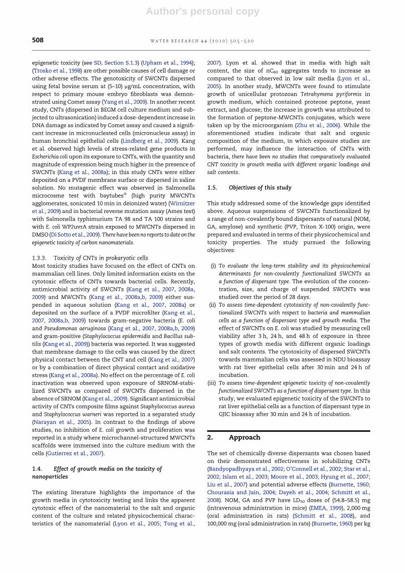
3.2.1. DispersantsGum arabic (approx. 250 kDa; a complex mixture of saccha-
rides and glycoproteins obtained from the acacia tree), PVP
(approx. 29 kDa), Triton X-100 (approx. 625 Da; polyethylene
glycol p-(1,1,3,3-tetramethylbutyl)-phenyl ether) and amylose
(molecular weight not specified; a polymeric form of glucose
and a constituent of potato starch) were purchased from
Sigma–Aldrich (Milwaukee, WI). SRNOM reverse osmosis
isolation was obtained from International Humic Substances
Society (St Paul, MN). The molecular structure of dispersants
used in this study is presented in Fig. 1.
3.2.2. Dispersion procedureAqueous solutions of PVP, Triton X-100 were prepared by
dissolving PVP (40 mg) and Triton X-100 (0.4 mL) in 40 mL of
water and adjusting the pH of both solutions to 7 with 0.1 M
HCl. The aqueous solution of GA was prepared by mixing 1 g of
GA in 50 mL of water and adjusting the pH of the mixture to 7
with 0.1 M HCl; this mixture was left to settle for 24 h and then
40 mL of supernatant was collected for use in the stability or
toxicity studies. To prepare NOM solutions, two flasks of
40 mL of water, each containing 8 mg of NOM, were stirred for
24 h. Each solution was then filtered through a 0.22 mm filter
under vacuum. The pH of one solution was kept at its original
value (approx. 3.5) while the pH in another solution was
adjusted to 7 with 0.1 M NaOH. SWCNTs were added to the
above solutions to result in a 1 mg (SWCNT)/mL loading and
were sonicated in Aquasonic 50 T water bath (VWR Scientific
Products Corp, West Chester, PA) for 20 min.
SWCNTs were dispersed with amylose based on modified
version of the three-step approach reported by Kim et al. (Kim
et al., 2004). SWCNTs (40 mg) were sonicated in 25 mL of water
(pH 7) at approximately 75 W for 5 min using Sonicator 3000
(Misonix, Inc., Farmingdale, NY) equipped with a microprobe.
100 mg of amylose in 6.28 mL of DMSO were prepared and
added to the sonicated suspension of SWCNT in water so that
the DMSO/water ratio was 20% by volume. The resulting
mixture was sonicated for another 5 min in order to remove
the excess amylose and DMSO. The suspension was soni-
cated, centrifuged and refilled with water four times (See SD,
Section S.2.3).
Following the sonication, SWCNTs suspensions were
divided into 12 mL aliquots and each aliquot was transferred
into 15 mL centrifuge tube. After 24 h standing time, the top
4 mL of each suspension was withdrawn in order to be used in
the stability or toxicity studies. Overall, three batches of
dispersed SWCNT suspensions were prepared and were used,
correspondingly, for 1) the study of the long-term stability of
SWCNTs suspensions, 2) the general viability bioassay with
E. coli, and 3) NDU and GJIC bioassays.
3.3. Physicochemical characterization of SWCNTsuspensions
In order to quantitatively assess the long-term stability
SWCNT suspensions, a combination of characterization
methods was employed.
w a t e r r e s e a r c h 4 4 ( 2 0 1 0 ) 5 0 5 – 5 2 0 509
Author's personal copy
3.3.1. UV–vis spectrophotometry. Quantification of SWCNTsconcentration in suspensionThe absorbance of SWCNT suspensions over the (200–800) nm
wavelength range was measured over a period of 4 weeks
(Multi-spec 1501, Shimadzu, Kyoto, Japan). For each type of
SWCNT suspension prepared, the absorbance of SWCNT-free
solution of the corresponding dispersant was used as
a baseline.
SWCNT suspensions used in our toxicity studies likely
contained both individual and bundled SWCNTs. In fact, DLS
measurements indirectly confirmed (see Section 4.2) the
presence of dispersed SWCNT bundles and direct TEM
imaging (see SD, Section S.3.3, Fig. S5) also showed the pres-
ence of SWCNTs bundles (note that TEM results need to be
interpreted with caution as evaporation-induced aggregation
could have contributed to bundling during TEM sample
preparation). Incomplete exfoliation is the most likely and
most environmentally relevant scenario; unfortunately, it also
entails difficulties with quantifying the total concentration of
suspended nanotubes as larger CNT bundles tend to separate
from the suspension.
In this study, we used UV–vis spectrophotometry to esti-
mate the concentration of exfoliated SWCNTs in suspensions.
It is known that only fully exfoliated SWCNTs absorb in the
(200–1200) nm wavelength range. Bundled NTs do not absorb
significantly in this range due to the tunnel coupling between
metallic and semiconductive SWNTs (Lauret et al., 2004;
Grossiord et al., 2007). Therefore, the UV absorbance by
a SWCNT suspension can be used to selectively measure the
concentration of individually dispersed (i.e. fully exfoliated or
debundled) SWCNTs.
To determine the concentration of suspended exfoliated
SWCNTs, we first prepared a separate set of suspensions with
ten-fold reduced SWCNT loading with respect to the CNT
loading used in long-term stability and toxicity studies. Four
suspensions (with PVP, NOM pH 3.5, NOM pH 7, and GA used
(a-1)
OOH
OH
OH
OH
CH2OH
(a-2)
OCH3
OH
OH OH
OH
GALP
ARAF
ARAF
GALP GALP GALP GALP
GALPRHAP
GA
ARAF
ARAF
ARAF
GALP
GALPRHAP
GA
ARAF
1
1
1 1
1
1
1
1 1
1
1
1
1
1
1
3
3
3 3
3
3
3
3 3
1
6
6
6
6
4 4
3
6 6
GALP = D-GALACTOPYRANOSE ARAF = L-ARABOFURANOSE GA = D-GLUCURONIC ACID RHAP = L-RHAMNOPYRANOSE
(a-3)O
OH
OH
OH
H2COH
(a-4)
O
OH
OH
HOOC
OH OH
N O
n
NH2
O
OHH
COOHHOOC
COOH
OHOH
OH
C8 H17
OOHn O
OH
COH
OH
O OO O
O O
n
a
b
d
c
e
Fig. 1 – Molecular structure of solubilizers: (a) Gum arabic (a-1) galactose, (a-2) rhamnose, (a-3) arabinose, (a-4) glucuonic
acid; (b) Poly(vinyl pyrrolidone); (c) A building block of humic acids, which has a compositional similarity to SRNOM; Dots
represent chiral centers; (d) Triton X-100; (e) Amylose.
w a t e r r e s e a r c h 4 4 ( 2 0 1 0 ) 5 0 5 – 5 2 0510
Author's personal copy
as dispersants) with 10-fold reduced SWCNT content (0.1 g/L)
were prepared using the same procedures as described in
Section 3.2 except that they were subjected to rigorous, pro-
longed sonication for 1 h at power of (70–80) W using horn
sonicator (Sonicator 3000, Misonix, Inc., Farmingdale, NY). No
settling of SWCNTs was observed over the short term
following the application of this treatment indicating
complete dispersion of SWCNTs. UV–vis absorbance spectra
were recorded at different dilutions and a calibration curve for
absorption at 500 nm (Bahr et al., 2001; Huang et al., 2002;
Sinani et al., 2005; Lee et al., 2007; Salzmann et al., 2007) as
a function of concentration was constructed, and coefficient
of molecular extinction, 3, was determined. This coefficient
was used to estimate the concentration of exfoliated SWCNTs
in suspensions used in long-term stability and toxicity
experiments. Note that by subjecting suspended SWCNTs to
a very intense sonication treatment we aimed at maximizing
the extent of exfoliation; however, it was not possible to
ascertain that the exfoliation was complete. Thus coefficients
of molecular extinction and exfoliated SWCNT concentrations
determined using the recorded calibration curves were esti-
mated values. SWCNT characteristics measured upon the
preparation of CNT suspensions are given in Table S1 in SD.
3.3.2. Size and charge of dispersed SWCNTsThe effective hydrodynamic diameter and z-potential of sus-
pended SWCNTs were determined after 1, 4, 7, 14, 21, and 28
days of settling. The size and charge were measured by
dynamic light scattering (DLS) and phase analysis light scat-
tering techniques, respectively (ZetaPALS, BI_MAS Option,
Brookhaven Instrument Corp., Holtsville, NY). The Smo-
luchowski equation was applied to convert the measured
electrophoretic mobility of dispersed SWCNT to z-potential.
Transmission electron microscopy (TEM) imaging was used as
an auxiliary method to aid in the interpretation of dynamic
light scattering data (see SD, Sections S.2.5 and S.3.4).
3.4. Toxicity assessment: E. coli viability assay
3.4.1. Media preparationLuria–Bertani (LB) growth medium was prepared according to
the standard procedure (Atlas, 1993). Minimal Davis medium
with 90% reduced potassium phosphate concentration
(MD medium) was prepared (Fortner et al., 2005). 0.1 M NaCl
solution was prepared by dissolving 9 g of NaCl in 1 L of water
(pH 7) and autoclaving it for 15 min at 1 bar and 121 �C. For the
toxicity assessments, each component of LB, MD and NaCl
media was prepared in 4-fold higher concentration as
compared to original protocol and the aliquot part of the cor-
responding medium was added to SWCNTs suspension
(as further described in ‘‘Quantification of cell viability’’
Section). Luria–Bertani Petri plates were prepared according to
the published method (Atlas, 1993).
3.4.2. Preparation of E. coli cultureE. coli K12 stock was prepared in glycerol and stored at �80 �C.
Prior to use, the stock was defrosted, and 30 mL of LB medium
were inoculated with 5 mL of the stock. After overnight growth
at 37 �C, 5 mL of this suspension was spread onto LB agar plate
and cultured at 37 �C. Once distinct colonies were formed, the
agar plate was transferred to the refrigerator and kept at 4 �C
for up to one month. E. coli suspensions to be used in SWCNT
cytotoxicity studies were prepared by scraping one colony
from the surface of a Petri plate by aseptic loop and immersing
the loop into 10 mL of LB or MD media in a 50 mL centrifuge
tube. Tubes were placed on a shaker in an incubator 37 �C for
12 h. When 0.1 M NaCl was used as an exposure medium in
colony forming units bioassay, E. coli were first grown in the LB
medium, centrifuged for 5 min at 2250 rpm and washed with
0.1 M NaCl as follows: the supernatant was decanted and
replaced with an equal volume of 0.1 M NaCl, vortexed and
resuspended by centrifugation. The washing procedure was
repeated twice, presuming that after this treatment most of
the remaining organic constituents of LB medium were
removed from the E. coli suspension.
3.4.3. Quantification of cell viabilityThe SWCNTs suspension (1.425 mL), growth medium (475 mL)
and 100 mL of the stock E. coli suspension were transferred into
a 15 mL centrifuge tube and incubated under gentle shaking at
37 �C. Samples were taken after 3, 24, and 48 h and a series of
dilutions (104–106) was prepared for each sample. Five samples
of 10 mL and 20 mL from each dilution were placed onto an agar
plate and incubated at 37 �C until distinct colonies developed.
Colony forming units (CFU)/1 mL were calculated for each
sample. Each experiment was run in triplicates with negative
(bacterial suspension with the corresponding amount of
ultrapure water) and vehicle (solution of the corresponding
dispersant) controls, herein called vehicle control I and
vehicle control II, respectively. The results are reported as
a fraction of control (FOC)� standard deviation (STD), calcu-
lated as the ratio of the average number of colonies grown
after E. coli exposure to SWCNTs suspensions or vehicle
control II to the average number of colonies grown in vehicle
control I plates.
3.5. Toxicity assessment: neutral red dye uptakebioassay
For the NDU bioassay we adapted a published procedure
(Borenfreund and Puerner, 1985; Weis et al., 1998; Satoh et al.,
2003). A solution (0.015 w/v) of neutral red dye (3-amino-7-
(dimethylamino)-2-methylphenazine hydrochloride) in
D-medium was incubated at 37 �C for 2 h and filtered through
a 0.22 mm syringe filter (Millipore Corp., New Bedford, MA) to
remove undissolved dye and ensure sterile conditions.
Confluent WB-F344 cells (see SD, Section S.2.6 for details on the
preparation of the cells) were exposed to 500 mL of each of
SWCNTs suspension and incubated at 37 �C for 30 min and 24 h
under gentle shaking in a humidified atmosphere containing
5% CO2. After cells were exposed to SWCNTs, the exposure
medium was removed by aspiration and the cells were washed
with 1 mL of phosphate saline buffer (PBS). Following washing,
2 mL of the neutral red dye solution per plate was added and
the cells were incubated for 1 h at 37 �C in the humidified
atmosphere containing 5% CO2. Upon incubation, the cells
were rinsed three times with PBS, and 2 mL of aqueous solution
containing 1% acetic acid and 50% ethanol was added to each
plate to lyse the cells. 1.5 mL of the lysate was transported into
2 mL microcentrifuge tube and the optical density was
w a t e r r e s e a r c h 4 4 ( 2 0 1 0 ) 5 0 5 – 5 2 0 511
Author's personal copy
recorded at 540 nm using a Beckman DU 7400 diode array
detector (Beckman Instruments, Inc., Schaumburg, IL). The
background absorbance was measured at 690 nm and then
subtracted from the original absorbance. Each experiment was
conducted in triplicates. The neutral red dye uptake was
reported as the FOC (absorption of neutral red in the chemically
treated sample divided by the absorption of neutral red in the
nontreated control I sample). FOC values of 1.0 indicates non-
cytotoxic response while FOC values<0.8 indicate that less dye
was absorbed by cells and that chemical is cytotoxic at that
dose (El-Fouly et al., 1987; Satoh et al., 2003).
3.6. Toxicity assessment: gap junction intercellularcommunication (GJIC) bioassay
The principles of GJIC bioassay were originally described in
(Borenfreund and Puerner, 1985) and further developed in
(Weis et al., 1998; Herner et al., 2001; Satoh et al., 2003).
SWCNTs suspensions (500 mL) were introduced to the cell
culture plates with confluent WB-F344 cells and incubated for
30 min and 24 h at 37 �C in humidified atmosphere containing
5% CO2 under gentle shaking. Then, cells were washed with
PBS solution and 1 mL of 0.05% solution of Lucifer yellow
fluorescent dye in PBS buffer was added to each plate. Three
different scrapes were made on the bottom of a cell culture
plate using a surgical steel scalpel blade. The dye was absor-
bed through the monolayer of confluent cells. The transfer of
dye through gap junctions lasted 3 min, followed by a thor-
ough rinse with PBS solution to remove extracellular dye. Cells
were fixed with 0.5 mL of 4% formalin solution in PBS. The
migration of the dye in the cells was observed at 200�magnification using a Nikon epifluorescence microscope
equipped with a Nikon Cool Snap EZ CCD camera and the
images were digitally acquired using a Nikon NIS-Elements
F2.2 imaging system. The fluorescence area of the dye
migration from the scrape line was quantified using Image J
1.40 g (a public domain Java image processing program, http://
rsbweb.nih.gov/ij/index.html). Each experiment was per-
formed in duplicate with same controls as in the NDU
bioassay. Data are reported as the FOC, which is the ratio of
the area of dye spread in the chemically treated sample to the
area of dye spread in the vehicle control I samples. A FOC
value of 1.0 corresponds to full communication between cells,
FOC values of (0.5–0.9) represent partial GJIC inhibition,
(0.3–0.5) values correspond to significant inhibition of the GJIC
and values <0.3 denote complete inhibition of the GJIC (Weis
et al., 1998). Student’s t-tests at 95% confidence interval were
run for all three bioassays to determine whether the differ-
ence between the results obtained in the control I plates and
the results in the tested plates were significant.
The concentrations of SWCNTs upon addition to suspen-
sion of cells in the general E. coli viability bioassay, and in NDU
and GJIC bioassays are reported in Table 1.
4. Results and discussion
The knowledge of the evolution of physicochemical properties
(e.g., charge and size) of dispersed CNTs can provide insights
into the basis for both CNT stability and possible toxicity.
4.1. Concentration of dispersed SWCNTs in suspensions
First, SWCNTs were suspended directly in ultrapure water
without a dispersant; this suspension was used as a control.
After sonication for 20 min, the mixture became a lightly
colored suspension that completely separated into a dark
subnatant layer and a clear supernatant after only 1 h of
settling. In contrast, 20 min of sonication of the SWCNT–water
mixture in the presence of a dispersant produced relatively
stable colored suspensions (see SD, Fig. S1) that precipitated
gradually with the precipitation rate varying as a function of
the type of dispersant. The amount of the precipitate that
formed at the bottom of a centrifuge tube visibly increased
with settling time for all types of SWCNT suspensions.
Immediately after sonication, all SWCNT suspensions
prepared using the different dispersants appeared black. After
4 weeks of settling, in suspensions of NOM-stabilized and
amylose-stabilized SWCNTs the color of the aqueous solution
of the dispersant was revealed. After 7 weeks of settling, the
color change could be detected in all samples with the
suspension of amylose-stabilized SWCNTs (see SD, Fig. S1)
undergoing the most dramatic change to an almost trans-
parent supernatant phase (Figure S1 (b)).
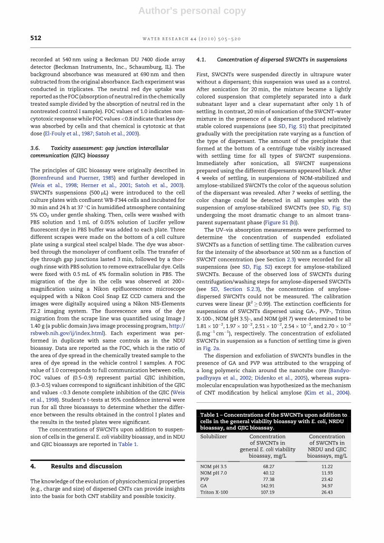
The UV–vis absorption measurements were performed to
determine the concentration of suspended exfoliated
SWCNTs as a function of settling time. The calibration curves
for the intensity of the absorbance at 500 nm as a function of
SWCNT concentration (see Section 2.3) were recorded for all
suspensions (see SD, Fig. S2) except for amylose-stabilized
SWCNTs. Because of the observed loss of SWCNTs during
centrifugation/washing steps for amylose-dispersed SWCNTs
(see SD, Section S.2.3), the concentration of amylose-
dispersed SWCNTs could not be measured. The calibration
curves were linear (R2� 0.99). The extinction coefficients for
suspensions of SWCNTs dispersed using GA-, PVP-, Triton
X-100-, NOM (pH 3.5)-, and NOM (pH 7) were determined to be
1.81� 10�2, 1.97� 10�2, 2.51� 10�2, 2.54� 10�2, and 2.70� 10�2
(L mg�1 cm�1), respectively. The concentration of exfoliated
SWCNTs in suspension as a function of settling time is given
in Fig. 2a.
The dispersion and exfoliation of SWCNTs bundles in the
presence of GA and PVP was attributed to the wrapping of
a long polymeric chain around the nanotube core (Bandyo-
padhyaya et al., 2002; Didenko et al., 2005), whereas supra-
molecular encapsulation was hypothesized as the mechanism
of CNT modification by helical amylose (Kim et al., 2004).
Table 1 – Concentrations of the SWCNTs upon addition tocells in the general viability bioassay with E. coli, NRDUbioassay, and GJIC bioassay.
Solubilizer Concentrationof SWCNTs in
general E. coli viabilitybioassay, mg/L
Concentrationof SWCNTs in
NRDU and GJICbioassays, mg/L
NOM pH 3.5 68.27 11.22
NOM pH 7.0 40.12 11.93
PVP 77.38 23.42
GA 142.91 34.97
Triton X-100 107.19 26.43
w a t e r r e s e a r c h 4 4 ( 2 0 1 0 ) 5 0 5 – 5 2 0512
Author's personal copy
Triton X-100 stabilizes nanotubes by the formation of hemi-
micelles that cover nanotube surface with benzene rings
providing p–p stacking between the surfactant molecule and
nanotube core (Islam et al., 2003). The exfoliation (debundling)
of SWCNTs in the presence of SRNOM was suggested to occur
through the interaction of the aromatic moieties of natural
organic matter and nanotube surface (Hyung et al., 2007; Liu
et al., 2007; Hyung and Kim, 2008). Despite the differences in
the physiosorption mechanisms responsible for the stabili-
zation of CNTs in aqueous suspensions, all dispersants were
effective, albeit to somewhat different extents.
For all SWCNT suspensions, the calculated values of the
concentration of exfoliated SWCNTs correlated well to the
visual observations described above. As seen from Fig. 2a, the
dispersion efficiency in terms of the concentration of
dispersed SWCNTs was a function of the type of the disper-
sant, with GA producing suspensions having highest
concentration of SWCNTs. The data were consistent among
the three prepared batches of SWCNTs (see SD, Table S1).
Didenko et al. (Didenko et al., 2005) suggested that after
covering one single carbon nanotube with a long polymeric
molecule, the remaining strands would react with other
uncovered or partially covered SWCNTs thus bundling several
nanotubes together. We speculate that this mechanism where
one polymer molecule links two or more nanotubes may be
responsible for the higher dispersing efficiency of GA (by far
the largest molecule among the target dispersants) towards
SWCNTs compared to other dispersants. This hypothesis is
supported by the measurements of the effective hydrody-
namic diameter (Fig. 2b), and the estimation of the length of
suspended particles with the size and the length of GA/
SWCNTs being higher than those of PVP, Triton X-100 and
SRNOM (both pHs). As the measured diameter of GA-stabilized
SWCNTs aggregates (which could also be expected to be
rather porous) was still relatively small, gravity settling did
not lead to a significant removal of GA-stabilized SWCNTs
from the dispersion.
When comparing the solubilizing ability of SRNOM at
different pH, one could see that SRNOM is a more efficient
dispersant at pH 3.5 than at pH 7. This is consistent with the
findings by Huyng et al. (Hyung and Kim, 2008) who reported
that adsorption of SRNOM to MWCNTs increased when pH
decreased due to more compact and coiled conformation of
NOM at acidic pHs. When pH increases, carboxylic and
phenolic groups of SRNOM deprotonate resulting in higher
electrostatic repulsion between a CNT and a SRNOM molecule
and, as a consequence, in a lower amount of organic matter
adsorbed on the CNT surface. The better dispersion of
0
40
80
120
160
200
Time (d)
Co
ncen
tratio
n (m
g/L
)
0
200
400
600
800
1000
Time (d)
Effective d
iam
eter (n
m)
-60
-45
-30
-15
0
Time (d)
-45
-30
-15
0
0 7 14 21 28 0 7 14 21 28
0 7 14 21 28 0 7 14 21 28Time (d)
-p
oten
tial (m
V)
-p
oten
tial (m
V)
a b
dc
Fig. 2 – The time-dependent characterization of (a) estimated concentration of the exfoliated SWCNTs in the dispersion,
(b) effective diameter and (c) z-potential of dispersed SWCNTs, and (d) z-potential of dispersants alone (-B- for GA/SWCNTs,
-,- for PVP/SWCNTs, -6- for NOM3.5/SWCNTs, -:- for NOM7/SWCNTs, --- for Triton X-100/SWCNTs, -C- for Amylose/
SWCNTs). Notes: (1) The z-potential measurement was not conducted for amylose solution, because amylose is not water-
soluble under room temperature. (2) Reported are results of only those measurements that were statistically significant, i.e.
when sufficiently high photon count rates were recorded in dynamic light scattering measurements. (3) The absorption by
suspensions of amylose-stabilized SWCNTs was not measured because the nanotube content in these suspensions could
not be precisely determined; this was due to the incomplete separation at the centrifugation step of the suspension
preparation process.
w a t e r r e s e a r c h 4 4 ( 2 0 1 0 ) 5 0 5 – 5 2 0 513
Author's personal copy
SWCNTs at pH 3.5 could also be attributed, in part, to the steric
hindrance imposed by SRNOM when a higher surface density
of NOM on the surface of CNT bundles results in stronger
repulsion between CNTs. This observation is supported by the
long-term z-potential measurements (Fig. 2c), where the
surface charge of stabilized SRNOM/SWCNTs at pH 3.5
became less negative up to day 7, and then stabilized with
increasing settling time. At the same time the surface charge
of SWCNTs dispersed in SRNOM solution at pH 7 gradually
increased after day 5. Indeed, had the electrostatic repulsion
been the sole mechanism of SWCNTs stabilization in SRNOM
solutions, the surface charge on the SWCNTs would have
more rapidly become less negative at pH 3.5 than at pH 7. In
summary, in terms of the effectiveness of SWCNT stabiliza-
tion, the dispersants were ranked as follows: GA>Triton
X-100> PVP>NOM (pH 3.5)>NOM (pH 7).
4.2. Hydrodynamic size of dispersed SWCNTs
The hydrodynamic size of CNTs in a suspension is an
important characteristic that affects the stability and,
possibly, toxicity of dispersed CNTs. It should be noted that
the effective hydrodynamic diameter of suspended SWCNTs
measured using DLS can only be used as a very rough esti-
mate. While the DLS data are interpreted with the assumption
that the primary scatterers are spherical with an aspect ratio
of 1, dispersed SWCNTs are long and tubular, with a very high
aspect ratio (see SD, Fig. S3). To obtain a better approximation
of the size distribution of dispersed SWCNT, the multimodal
size distribution model was used. TEM imaging was employed
to obtain auxiliary information on the size and morphology of
stabilized SWCNT. Even though the measurement of effective
hydrodynamic diameter cannot be relied on to compare the
sizes of suspended SWCNTs in different dispersion media,
these measurements can be used to compare how the average
particle size for a given dispersant changes with settling time.
The values of effective diameter as a function of time for
different SWCNT suspensions are given in Fig. 2b.
Generally, the effective size of SWCNTs dispersed using
Triton X-100, GA and NOM did not change significantly with
increasing settling time (Fig. 2b). In the case of amylose-
dispersed SWCNTs, a slight decrease in the effective size was
observed due to settling of larger and unstable SWCNT
aggregates, which left behind more uniformly sized smaller
amylose-stabilized CNT clusters. The concentration of
SWCNTs in the suspension was negatively correlated with the
effective size of SWCNT. The notable apparent exception was
the suspension of SWCNTs dispersed using GA. However, it
should be noted that the aqueous suspension of GA
contained GA colloids of the size that was 1) comparable to the
size of dispersed SWCNTs and 2) decreased during the 4 weeks
of settling (see SD, Fig S5). Thus, the effective size measure-
ments for SWCNTs dispersed using GA should be interpreted
with caution.
4.3. Charge of dispersed SWCNTs
For all suspensions, the z-potential of dispersed SWCNTs was
negative and nearly constant over the entire duration of the
experiment (Fig. 2c and SD, Table S1). Each z-potential
measurement for SWCNT suspensions was accompanied by
a measurement of the z-potential of the dispersants in
a SWCNT-free aqueous solution (Fig. 2d). After 1 d, only the GA
solution scattered light sufficiently to enable a statistically
meaningful z-potential measurement. After 4 d, solutions of
NOM (pH 7), PVP, and Triton X-100 also scattered light
0
0.2
0.4
0.6
0.8
1
1.2
Fractio
n o
f co
ntro
l (F
OC
)
NOM 3.5 NOM 7 GA Amylose PVP Triton
0
0.2
0.4
0.6
0.8
1
1.2
Fractio
n o
f co
ntro
l (F
OC
)
NOM 3.5 NOM 7 GA Amylose PVP Triton
0
0.2
0.4
0.6
0.8
1
1.2
Fractio
n o
f co
ntro
l (F
OC
)
NOM 3.5 NOM 7 GA Amylose PVP Triton
a
c
b
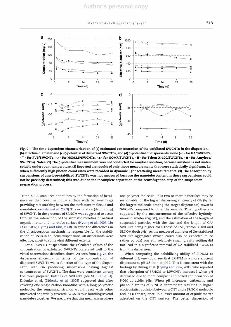
Fig. 3 – Viability of E. coli in (a) 0.1 M NaCl, (b) LB medium,
and (c) MD medium. Exposure time: (a) 3 h, (b) 48 h, (c) 48 h.
Open, hatched and cross-hatched bars correspond to
control I, control II and dispersed SWCNTs. In the case of
amylose ultrapure water was used as control.
w a t e r r e s e a r c h 4 4 ( 2 0 1 0 ) 5 0 5 – 5 2 0514
Author's personal copy
sufficiently. After 7 d, all the dispersants except for NOM (pH
3.5) achieved acceptable count rates during z-potential
measurements. The gradual change in the scattering ability of
the dispersant solutions may correspond to the aggregation of
the dispersant molecules. The dispersants were ranked in
order from the most to the least negative z-potential of
dispersed SWCNTs: NOM (pH 7)>NOM (pH 3.5)>GA z Triton
X-100> PVP z amylose. The highly negative charge of NOM-
stabilized SWCNTs was likely due to the charge of NOM. The
low stability of amylose-dispersed SWCNTs could be attrib-
uted to the combination of low surface charge and steric
repulsion between stabilized SWCNTs in the dispersion.
4.4. Assessment of cell toxicity in prokaryotic andeukaryotic systems
4.4.1. General viability bioassay with E. coli (prokaryotic)Immediately after E. coli cells were exposed to SWCNTs sus-
pended in growth medium as well as after 3 h of exposure, no
SWCNT aggregation was visually observed (see SD, Table S2)
in experiments with all three types of the media – 0.1 M NaCl,
MD (medium with lower salt organics content) and LB
(medium with higher salt and organics content). After 24 h of
incubation, limited precipitation was observed for all SWCNT
suspensions in LB and MD media. After 48 h of incubation
more precipitation occurred in each type of media.
No inhibition of E. coli colony forming ability was observed
after 3 h of incubation with amylose-, NOM-, GA- and PVP-
stabilized SWCNTs in 0.1 M NaCl (Fig. 3a). The FOC values did
not decline in any of these samples and no significant differ-
ence in the influence on CFU counts was found between
dispersed SWCNTs and solutions of corresponding disper-
sants. When the bacteria suspension was brought in contact
with Triton X-100-stabilized SWCNTs, approx. 25% loss of the
cell viability was measured as compared to vehicle control I
samples. However, there was no statistical difference between
FOC of Triton-stabilized SWCNTs and the control (solution of
Triton X-100 only). It remains unclear if the observed cyto-
toxicity of the these suspensions was due to i) SWCNTs
stabilized by Triton X-100 or ii) the residual ‘‘free’’ (i.e. not
associated with suspended SWCNTs) Triton X-100 potentially
present in the solution or iii) the combined effect of both
Triton X-100/SWCNT and dissolved Triton X-100. The
complete separation of the Triton X-100/SWCNT and
dissolved Triton X-100 could not be accomplished using
centrifugation. Even for very long centrifugation times, the
supernatant had grayish color indicating that some fraction of
SWCNTs was not removed.
0
0.2
0.4
0.6
0.8
1
1.2
Frac
tio
n o
f c
on
tro
l (F
OC
)
NOM3.5 NOM7 GA Amylose PVP Triton
0
0.2
0.4
0.6
0.8
1
1.2
Fra
ctio
n o
f c
on
tro
l (F
OC
)
NOM3.5 NOM7 GA Amylose PVP Triton
ab
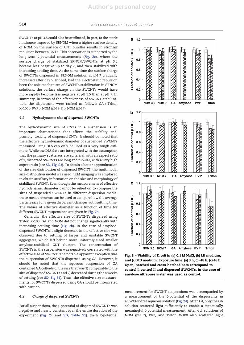
Fig. 4 – Neutral red dye uptake by WB-F344 cells as a function of dispersant type (a – 30 min, b – 24 h of exposure). Open,
hatched and cross-hatched bars correspond to control I, control II and dispersed SWCNTs. In the case of amylose control I
and control II were ultrapure water.
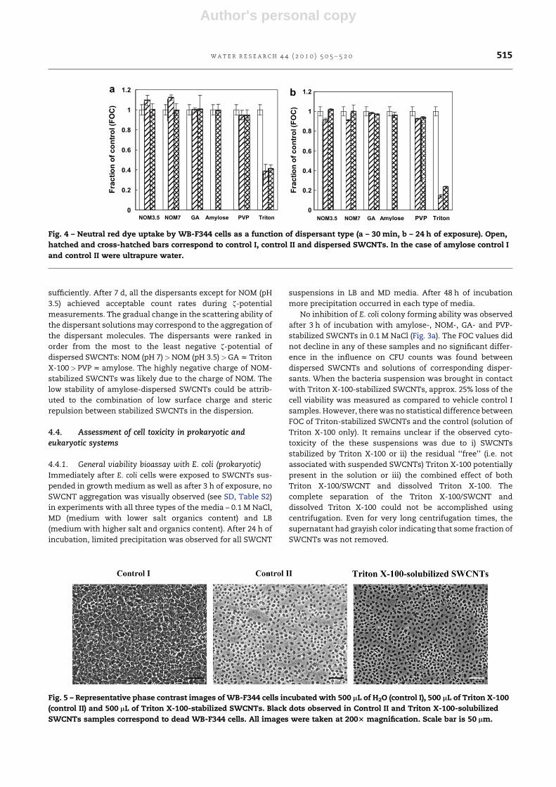
Fig. 5 – Representative phase contrast images of WB-F344 cells incubated with 500 mL of H2O (control I), 500 mL of Triton X-100
(control II) and 500 mL of Triton X-100-stabilized SWCNTs. Black dots observed in Control II and Triton X-100-solubilized
SWCNTs samples correspond to dead WB-F344 cells. All images were taken at 2003 magnification. Scale bar is 50 mm.
w a t e r r e s e a r c h 4 4 ( 2 0 1 0 ) 5 0 5 – 5 2 0 515
Author's personal copy
The ability of E. coli to grow and to form colonies in the
presence of amylose-, GA-, PVP-, and NOM-stabilized SWCNTs
in LB medium mimicked both control samples regardless of
the contact time (see SD, Fig. 3b and Fig. S6). On the contrary,
21 and 18 % mortality rates after 3 h of incubation were
observed for E. coli in Triton X-100/SWCNTs suspension and
Triton X-100 solution, respectively. After 24 h of contact, the
number of colonies grown on the Petri plates decreased by
30 % when bacteria were in contact with Triton X-100-stabi-
lized SWCNTs and by 27 % when bacteria were in Triton X-100
only solution (see SD, Fig. S6) as compared to vehicle control I
plates. As the exposure time increased to 48 h, E. coli resumed
its growth; with FOC of both Triton X-100 suspensions
approaching values for vehicle control I sample (Fig. 3b). When
dispersed SWCNTs were tested in MD medium, no losses in
cell viability for amylose-, GA-, PVP- and NOM-stabilized
SWCNTs were measured (Fig. 3c; also see SD, Fig. S7). In the
case of Triton X-100 suspensions, the reduction in E. coli
survival was observed after 3 h of exposure; comparable
losses of the E. coli viability were also observed for both Triton
X-100-stabilized SWCNTs and the Triton X-100 only solution.
However, after 24 h of exposure, fewer E. coli colonies were
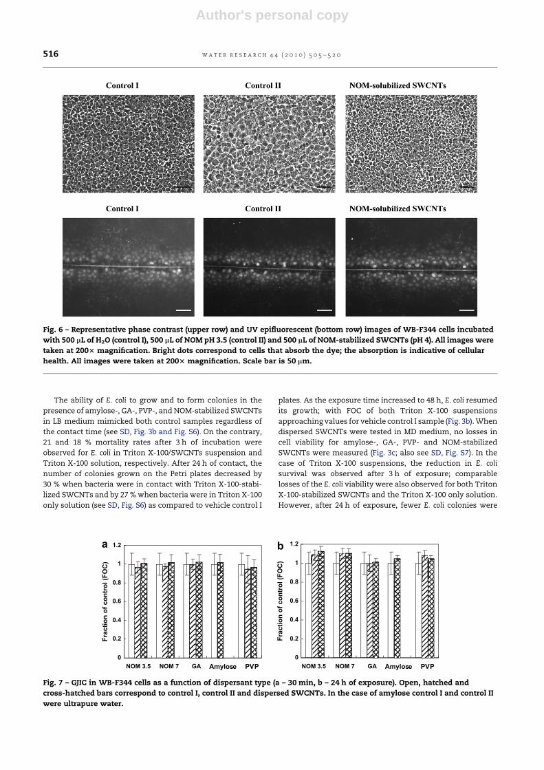
Fig. 6 – Representative phase contrast (upper row) and UV epifluorescent (bottom row) images of WB-F344 cells incubated
with 500 mL of H2O (control I), 500 mL of NOM pH 3.5 (control II) and 500 mL of NOM-stabilized SWCNTs (pH 4). All images were
taken at 2003 magnification. Bright dots correspond to cells that absorb the dye; the absorption is indicative of cellular
health. All images were taken at 2003 magnification. Scale bar is 50 mm.
0
0.2
0.4
0.6
0.8
1
1.2
Fra
ctio
n o
f c
on
tro
l (F
OC
)
Fra
ctio
n o
f c
on
tro
l (F
OC
)
NOM 3.5 NOM 7 GA Amylose PVP
0
0.2
0.4
0.6
0.8
1
1.2
NOM 3.5 NOM 7 GA Amylose PVP
a b
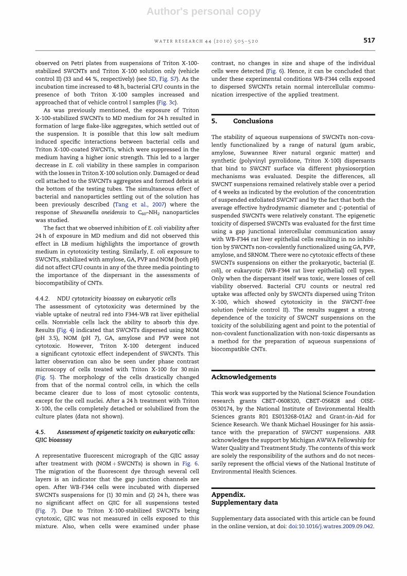
Fig. 7 – GJIC in WB-F344 cells as a function of dispersant type (a – 30 min, b – 24 h of exposure). Open, hatched and
cross-hatched bars correspond to control I, control II and dispersed SWCNTs. In the case of amylose control I and control II
were ultrapure water.
w a t e r r e s e a r c h 4 4 ( 2 0 1 0 ) 5 0 5 – 5 2 0516
Author's personal copy
observed on Petri plates from suspensions of Triton X-100-
stabilized SWCNTs and Triton X-100 solution only (vehicle
control II) (33 and 44 %, respectively) (see SD, Fig. S7). As the
incubation time increased to 48 h, bacterial CFU counts in the
presence of both Triton X-100 samples increased and
approached that of vehicle control I samples (Fig. 3c).
As was previously mentioned, the exposure of Triton
X-100-stabilized SWCNTs to MD medium for 24 h resulted in
formation of large flake-like aggregates, which settled out of
the suspension. It is possible that this low salt medium
induced specific interactions between bacterial cells and
Triton X-100-coated SWCNTs, which were suppressed in the
medium having a higher ionic strength. This led to a larger
decrease in E. coli viability in these samples in comparison
with the losses in Triton X-100 solution only. Damaged or dead
cell attached to the SWCNTs aggregates and formed debris at
the bottom of the testing tubes. The simultaneous effect of
bacterial and nanoparticles settling out of the solution has
been previously described (Tang et al., 2007) where the
response of Shewanella oneidensis to C60–NH2 nanoparticles
was studied.
The fact that we observed inhibition of E. coli viability after
24 h of exposure in MD medium and did not observed this
effect in LB medium highlights the importance of growth
medium in cytotoxicity testing. Similarly, E. coli exposure to
SWCNTs, stabilized with amylose, GA, PVP and NOM (both pH)
did not affect CFU counts in any of the three media pointing to
the importance of the dispersant in the assessments of
biocompatibility of CNTs.
4.4.2. NDU cytotoxicity bioassay on eukaryotic cellsThe assessment of cytotoxicity was determined by the
viable uptake of neutral red into F344-WB rat liver epithelial
cells. Nonviable cells lack the ability to absorb this dye.
Results (Fig. 4) indicated that SWCNTs dispersed using NOM
(pH 3.5), NOM (pH 7), GA, amylose and PVP were not
cytotoxic. However, Triton X-100 detergent induced
a significant cytotoxic effect independent of SWCNTs. This
latter observation can also be seen under phase contrast
microscopy of cells treated with Triton X-100 for 30 min
(Fig. 5). The morphology of the cells drastically changed
from that of the normal control cells, in which the cells
became clearer due to loss of most cytosolic contents,
except for the cell nuclei. After a 24 h treatment with Triton
X-100, the cells completely detached or solubilized from the
culture plates (data not shown).
4.5. Assessment of epigenetic toxicity on eukaryotic cells:GJIC bioassay
A representative fluorescent micrograph of the GJIC assay
after treatment with (NOMþ SWCNTs) is shown in Fig. 6.
The migration of the fluorescent dye through several cell
layers is an indicator that the gap junction channels are
open. After WB-F344 cells were incubated with dispersed
SWCNTs suspensions for (1) 30 min and (2) 24 h, there was
no significant affect on GJIC for all suspensions tested
(Fig. 7). Due to Triton X-100-stabilized SWCNTs being
cytotoxic, GJIC was not measured in cells exposed to this
mixture. Also, when cells were examined under phase
contrast, no changes in size and shape of the individual
cells were detected (Fig. 6). Hence, it can be concluded that
under these experimental conditions WB-F344 cells exposed
to dispersed SWCNTs retain normal intercellular commu-
nication irrespective of the applied treatment.
5. Conclusions
The stability of aqueous suspensions of SWCNTs non-cova-
lently functionalized by a range of natural (gum arabic,
amylose, Suwannee River natural organic matter) and
synthetic (polyvinyl pyrrolidone, Triton X-100) dispersants
that bind to SWCNT surface via different physiosorption
mechanisms was evaluated. Despite the differences, all
SWCNT suspensions remained relatively stable over a period
of 4 weeks as indicated by the evolution of the concentration
of suspended exfoliated SWCNT and by the fact that both the
average effective hydrodynamic diameter and z-potential of
suspended SWCNTs were relatively constant. The epigenetic
toxicity of dispersed SWCNTs was evaluated for the first time
using a gap junctional intercellular communication assay
with WB-F344 rat liver epithelial cells resulting in no inhibi-
tion by SWCNTs non-covalently functionalized using GA, PVP,
amylose, and SRNOM. There were no cytotoxic effects of these
SWCNTs suspensions on either the prokaryotic, bacterial (E.
coli), or eukaryotic (WB-F344 rat liver epithelial) cell types.
Only when the dispersant itself was toxic, were losses of cell
viability observed. Bacterial CFU counts or neutral red
uptake was affected only by SWCNTs dispersed using Triton
X-100, which showed cytotoxicity in the SWCNT-free
solution (vehicle control II). The results suggest a strong
dependence of the toxicity of SWCNT suspensions on the
toxicity of the solubilizing agent and point to the potential of
non-covalent functionalization with non-toxic dispersants as
a method for the preparation of aqueous suspensions of
biocompatible CNTs.
Acknowledgements
This work was supported by the National Science Foundation
research grants CBET-0608320, CBET-056828 and OISE-
0530174, by the National Institute of Environmental Health
Sciences grants R01 ES013268-01A2 and Grant-in-Aid for
Science Research. We thank Michael Housinger for his assis-
tance with the preparation of SWCNT suspensions. ARR
acknowledges the support by Michigan AWWA Fellowship for
Water Quality and Treatment Study. The contents of this work
are solely the responsibility of the authors and do not neces-
sarily represent the official views of the National Institute of
Environmental Health Sciences.
Appendix.Supplementary data
Supplementary data associated with this article can be found
in the online version, at doi: doi:10.1016/j.watres.2009.09.042.
w a t e r r e s e a r c h 4 4 ( 2 0 1 0 ) 5 0 5 – 5 2 0 517
Author's personal copy
r e f e r e n c e s
Atlas, R.M., 1993. Handbook of Microbiological Media. CRC Press,Boka Raton.
Bahr, J.L., Mickelson, E.D., Bronikovski, M.J., Smalley, R.E., Tour, J.M.,2001. Dissolution of small diameter single-wall carbonnanotubes in organic solvents. Chemical Communications,193–194.
Bandyopadhyaya, R., Nativ-Roth, E., Regev, O., Yerushalmi-Rozen, R., 2002. Stabilization of individual carbon nanotubesin aqueous solutions. Nano Letters 2 (1), 25–28.
Bardi, G., Tognini, P., Ciofani, G., Raffa, V., Costa, M.,Pizzorusso, T., 2009. Pluronic-coated carbon nanotubes donot induce degeneration of cortical neurons in vivo and invitro. Nanomedicine: Nanotechnology, Biology, and Medicine5, 96–104.
Bonnet, P., Albertini, D., Bizot, H., Bernard, A., Chauvet, O., 2007.Amylose/SWNT composites: from solution to film – synthesis,characterization and properties. Composite Science andTechnology 67, 817–821.
Borenfreund, E., Puerner, J.A., 1985. Toxicology determined invitro by morphological alterations and neutral red absorption.Toxicology Letters 24, 119–124.
Burnette, L.W., 1960. A review of the physiological properties ofPVP. Proceedings of the Scientific Section of the Toilet GoodsAssociation 38, 1–4.
Chappell, M.A., George, A.J., Dontsova, K.M., Porter, B.E., Price, C.L.,Zhou, P., Morikawa, E., Kennedy, A.J., Steevens, J.A., 2009.Surfactive stabilization of multi-walled carbon nanotubedispersions with dissolved humic substances. EnvironmentalPollution 157, 1081–1087.
Chen, X., Tam, U.C., Czlapinski, J.L., Lee, G.S., Rabuka, D., Zettl, A.,Bertozzi, C.R., 2006. Interfacing carbon nanotubes with livingcells. Journal of American Chemical Society 128, 6292–6293.
Cherukuri, P., Gannon, C.J., Leeuw, T.K., Schmidt, H.K.,Smalley, R.E., Curley, S.A., Weisman, R.B., 2006. Mammalianpharmacokinetics of carbon nanotubes using intrinsic near-infrared fluorescence. Proceedings of the National Academy ofSciences 103, 18882–18886.
Chin, S.F., Baughman, R.H., Dalton, A.B., Diekmann, G.R.,Draper, R.K., Mikoryak, C., Musselman, I.H., Poenitzsch, V.Z.,Xie, H., Pantano, P., 2007. Amphiphilic helicalpeptide enhances the uptake of single-walled carbonnanotubes by living cells. Experimental Biological Medicine232 (9), 1236–1244.
Chourasia, M.K., Jain, S.K., 2004. Polysaccharides for colontargeted drug delivery. Drug Delivery 11 (2), 129–148.
Dayeh, V.R., Chow, S.L., Schirmer, K., Lynn, D.H., Bols, N.C.,2004. Evaluating the toxicity of Triton X-100 to protozoan,fish, and mammalian cells using fluorescent dyes asindicators of cell viability. Ecotoxicology and EnvironmentalSafety 57 (3), 375–382.
Di Sotto, A., Chiaretti, M., Carru, G.A., Bellucci, S., Mazzantia, G.,2009. Multi-walled carbon nanotubes: lack of mutagenicactivity in the bacterial reverse mutation assay. ToxicologyLetters 184, 192–197.
Didenko, V.V., Moore, V.C., Baskin, D.S., Smalley, R.E., 2005.Visualization of individual single-walled carbon nanotubes byfluorescent polymer wrapping. Nano Letters 5 (8), 1563–1567.
Ding, L.H., Stilwell, J., Zhang, T.T., Elboudware, J.O., Jiang, H.J.,Selegue, J.P., Cooke, P.A., Gray, J.W., Chen, F.F., 2005. Molecularcharacterization of the cytotoxic mechanism of multiwallcarbon nanotubes and nanoonions on human skin fibroblast.Nano Letters 5, 2448–2464.
Dodziuk, H., Ejchart, A., Anczewski, W., Ueda, H., Krinichnaya, E.,Dolgonosa, G., Kutner, W., 2003. Water solubilization,
determination of the number of different types of single-wallcarbon nanotubes and their partial separation with respect todiameters by complexation with h-cyclodextrin. ChemicalCommunications, 986–987.
El-Fouly, M.H., Trosko, J.E., Chang, C.C., 1987. Scrape-loading anddye transfer: a rapid and simple technique to study gapjunctional intercellular communications. Experimental CellResearch 168, 422–430.
Elias, A.L., Carrero-Sanchez, J.C., Terrones, H., Endo, M.,Laclette, H.P., Mauricio, M., 2007. Viability studies of purecarbon- and nitrogen-doped nanotubes with EntamoebaHistolytica: from amoebicidal to biocompatible structures.Small 3 (10), 1723–1729.
Enyashin, A.N., Gemming, S., Seifert, G., 2007. DNA-wrappedcarbon nanotubes. Nanotechnology 18, 245702.
Fiorito, S., Serafino, A., Andreola, F., Bernier, P., 2006. Effects offullerenes and single-wall carbone nanotubes on murine andhuman macrophages. Carbon 44, 1100–1105.
Flahaut, E., Durrieu, M.C., Remy-Zolghadri, M., Bareille, R.,Baquey, C., 2006. Investigation of the cytotoxicity of CCVDcarbon nanotubes towards human umbilical vein endothelialcells. Carbon 44 (6), 1093–1099.
Fortner, J.D., Lyon, D.Y., Sayes, C.M., Boyd, A.M., Falkner, J.C.,Hotze, E.M., Alemany, L.B., Tao, Y.J., Guo, W., Ausman, K.D.,Colvin, V.L., Hughes, J.B., 2005. C60 in water: nanocrystalformation and microbial response. Environmental Scienceand Technology 39 (11), 4307–4316.
Georgakilas, V., Tagmatarchis, N., Pantarotto, D., Bianco, A.,Briand, J.-P., Prato, M., 2002. Amino acid functionalisation ofwater soluble carbon nanotubes. Chemical Communications,3050–3051.
Grossiord, N., Schhoo, P., Meuldijk, J., Koning, C.E., 2007.Determination of the surface coverage of exfoliated carbonnanotubes by surfactant molecules in aqueous solution.Langmuir 23, 3646–3653.
Gutierrez, M.C., Garcia-Carvajal, Z.Y., Hortiguela, M.J., Yuste, L.,Rojo, F., Ferrer, M.L., Monte, F., 2007. Biocompatible MWCNTscaffolds for immobilization and proliferation of E. coli. Journalof Materials Chemistry 17, 2992–2995.
Herner, H.A., Trosko, J.E., Masten, S.J., 2001. The epigenetictoxicity of pyrene and related ozonation byproductscontaining an aldehyde functional group. EnvironmentalScience and Technology 35 (17), 3576–3583.
Herzog, E., Byrne, H.J., Davoren, M., Casey, A., Duschl, A.,Oostingh, G.J., 2009. Dispersion medium modulates oxidativestress response of human lung epithelial cells upon exposureto carbon nanomaterial samples. Toxicology and AppliedPharmacology 236, 276–281.
Huang, W.J., Taylor, S., Fu, K.F., Lin, Y., Zhang, D.H., Hanks, T.W.,Rao, A.M., Sun, Y.P., 2002. Attaching proteins to carbonnanotubes via diimide-activated amidation. Nano Letters 2 (4),311–314.
Humic acids and their sodium salts. EMEA-The European agencyfor the evaluation of medicinal products. Available via. http://www.emea.europa.eu/pdfs/vet/mrls/055499en.pdf, 1999.
Hyung, H., Fortner, J.D., Hughes, J.B., Kim, J.H., 2007. Natural organicmatter stabilizes carbon nanotubes in the aqueous phase.Environmental Science and Technology 41 (1), 179–184.
Hyung, H., Kim, J.-H., 2008. Natural organic matter (NOM)adsorption to multi-walled carbon nanotubes: effect of NOMcharacteristics and water quality parameters. EnvironmentalScience and Technology 42 (12), 4416–4421.
Iijima, S., 1991. Helical microtubules of graphitic carbon. Nature354 (6348), 56–58.
Inoue, K.-i., Koike, E., Yanagisawa, R., Hirano, S., Nishikawa, M.,Takano, H., 2009. Effects of multi-walled carbon nanotubes ona murine allergic airway inflammation model. Toxicology andApplied Pharmacology 237 (3), 306–316.
w a t e r r e s e a r c h 4 4 ( 2 0 1 0 ) 5 0 5 – 5 2 0518
Author's personal copy
Islam, M.F., Rojas, E., Bergey, D.M., Johnson, A.T., Yodh, A.G.,2003. High weight fraction surfactant solubilization ofsingle-wall carbon nanotubes in water. Nano Letters 3 (2),269–273.
Jia, G., Wang, H., Yan, L., Wang, X., Pei, R., Yan, T., Zhao, Y.,Guo, X., 2005. Cytotoxicity of carbon nanomaterials: single-wall nanotube, multi-wall nanotube, and fullerene.Environmental Science and Technology 39, 1378–1383.
Jiang, J., Gao, L., 2003. Production of aqueous colloidal dispersionsof carbon nanotubes. Journal of Colloid and Interface Science260, 89–94.
Kang, S., Herzberg, M., Rodrigues, D.F., Elimelech, M., 2008a.Antibacterial effects of carbon nanotubes: size does matter!Langmuir 24 (13), 6409–6413.
Kang, S., Mauter, M.S., Elimelech, M., 2008b. Physicochemicaldeterminants of multiwalled carbon nanotube bacterialcytotoxicity. Environmental Science and Technology 42 (19),7528–7534.
Kang, S., Mauter, M.S., Elimelech, M., 2009. Microbial cytotoxicityof carbon-based nanomaterials: implications for river waterand wastewater effluent. Environmental Science andTechnology 43 (7), 2648–2653.
Kang, S., Pinault, M., Pfefferle, L.D., Elimelech, M., 2007. Single-walled carbon nanotubes exhibit strong antimicrobial activity.Langmuir 23 (17), 8670–8673.
Karajanagi, S.S., Yang, H.C., Asuri, P., Sellitto, E., Dordick, J.S.,Kane, R.S., 2006. Protein-assisted solubilization of single-walled carbon nanotubes. Langmuir 22 (4), 1392–1395.
Kim, O.-K., Je, J., Baldwin, J.W., Kooi, S., Pehrsson, P.E., Buckley, L.J.,2004. Solubilization of single-wall carbon nanotubes bysupramolecular encapsulation of helical amylose. Journal ofAmerican Chemical Society 125, 4426–4427.
Lauret, J.-S., Voisin, C., Cassabois, G., Roussignol, P., Delalande, C.,Capes, L.E., Valentin, E., Jost., O., 2004. Photocreated carrierdynamics in isolated carbon nanotubes. SemiconductorScience and Technology 19, S486–S488.
Lee, J.U., Huh, J., Kim, K.H., Park, C., Jo, W.H., 2007. Aqueoussuspension of carbon nanotubes via non-covalentfunctionalization with oligothiophene-terminatedpoly(ethylene glycol). Carbon 45, 1051–1057.
Lin, Y., Taylor, S., Li, H.P., Fernando, K.A.S., Qu, L.W., Wang, W.,Gu, L.R., Zhou, B., Sun, Y.P., 2004. Advances towardbioapplications of carbon nanotubes. Journal of MaterialsChemistry 14 (4), 527–541.
Lindberg, H.K., Falck, G.C.-M., Suhonen, S., Vippola, M.,Vanhalab, E., Catalana, J., Savolainena, K., Norppa, H., 2009.Genotoxicity of nanomaterials: DNA damage and micronucleiinduced by carbon nanotubes and graphite nanofibres inhuman bronchial epithelial cells in vitro. Toxicology Letters186, 166–173.
Liu, J., Rinzler, A.G., Dai, H.J., Hafner, J.H., Bradley, R.K., Boul, P.J.,Lu, A., Iverson, T., Shelimov, K., Huffman, C.B., Rodriguez-Macias, F., Shon, Y.S., Lee, T.R., Colbert, D.T., Smalley, R.E.,1998. Fullerene pipes. Science 280 (5367), 1253–1256.
Liu, Y.Q., Gao, L., Zheng, S., 2007. Debundling of single-walledcarbon nanotubes by using natural polyelectrolytes.Nanotechnology 18 (36), 365702.
Lyon, D.Y., Fortner, J.D., Sayes, C.M., Colvin, V.L., Hughes, J.B.,2005. Bacterial cell association and antimicrobial activity ofa C60 water suspension. Environmental Toxicology andChemistry 24, 2757–2762.
Manna, S.K., Sarkar, S., Barr, J., Wise, K., Barrera, E.V., Jejelowo, O.,Rice-Ficht, A., Ramesh, G.T., 2005. Single-walled carbonnanotube induces oxidative stress and activates nucleartranscription factor-kB in human keratinocytes. Nano Letters5, 1676–1684.
Marsh, D.H., Rance, G.A., Zaka, M.H., Whitby, R.J., Khlobystov, A.N.,2007. Comparison of the stability of multiwalled carbon
nanotube dispersions in water. Physical Chemistry ChemicalPhysics 9 (40), 5490–5496.
McDonald, T.J., Engtrakul, C., Jones, M., Rumbles, G., Heben, M.J.,2006. Kinetics of PL quenching during single-walledcarbon nanotube rebundling and diameter-dependentsurfactant interactions. Journal of Physical Chemistry B 110,25339–25346.
Meng, J., Yanga, M., Song, L., Kong, H., Wang, C.Y., Wang, R.,Wang, C., Xie, S.S., Xua, H.Y., 2009. Concentration control ofcarbon nanotubes in aqueous solution and its influence on thegrowth behavior of fibroblasts. Colloids and Surfaces B:Biointerfaces 71, 148–153.
Moore, V.C., Strano, M.S., Haroz, E.H., Hauge, R.H., Smalley, R.E.,Schmidt, J., Talmon, Y., 2003. Individually suspended single-walled carbon nanotubes in various surfactants. Nano Letters3 (10), 1379–1382.
Muller, J., Huaux, F., Moreau, N., Misson, P., Heilier, J.-F., Delos, M.,Arras, M., Fonseca, A., Nagy, J.B., Lison, D., 2005. Respiratorytoxicity of multi-wall carbon nanotubes. Toxicology andApplied Pharmacology 207 (31), 221–231.
Narayan, R.J., Berry, C.J., Brigmon, R.L., 2005. Structural andbiological properties of carbon nanotube composite films.Materials Science and Engineering B 123, 123–129.
O’Connell, M.J., Bachilo, S.M., Huffman, C.B., Moore, V.C.,Strano, M.S., Haroz, E.H., Rialon, K.L., Boul, P.J., Noon, W.H.,Kittrell, C., Ma, J., Hauge, R.H., Weisman, R.B., Smalley, R.E.,2002. Band gap fluorescence from individual single-walledcarbon nanotubes. Science 297, 593–596.
O’Connell, M.J., Boul, P.J., Ericson, L.M., Huffman, C., Wang, Y.,Haroz, E., 2001. Reversible water-solubilization of single-walled carbon nanotubes by polymer wrapping. ChemicalPhysics Letters 342, 265–271.
Park, H.J., Heo, H.Y., Lee, S.C., Park, M., Lee, S.-S., Kim, J., Chang, J.Y.,2006. Dispersion of single-walled carbon nanotubes in waterwith polyphosphazene polyelectrolyte. Journal of Inorganic andOrganometallic Polymers and Materials 16 (4) 359–364.
Pulskamp, K., Worle-Knirsch, J.M., Hennrich, F., Kern, K., Krug, H.F.,2007. Human lung epithelial cells show biphasic oxidative burstafter single-walled carbon nanotube contact. Carbon 45, 2241–2249.
Saleh, N.D., Pfefferle, L.D., Elimelech, M., 2009. Aggregationkinetics of multiwalled carbon nanotubes in aquatic systems:measurements and environmental implications.Environmental Science and Technology 42 (21), 7963–7969.
Salzmann, C.G., Chu, B.T.T., Tobias, G., Llewellyn, S.A.,Green, M.L.H., 2007. Quantitative assessment of carbonnanotube dispersions by Raman spectroscopy. Carbon 45 (5),907–912.
Satoh, A.Y., Trosco, J., Masten, S.J., 2003. Epigenetic toxicity ofhydroxylated biphenyls and hydroxylated polychlorinatedbiphenyls on normal rat liver epithelial cells. EnvironmentalScience and Technology 37, 2727–2733.
Sayes, C.M., Liang, F., Hudsona, J.L., Mendeza, J., Guob, W.,Beach, J.M., Moore, V.C., Doyle, C.D., West, J.L., Billups, W.E.,Ausmanb, K.D., Colvin, V.L., 2006. Functionalization densitydependence of single-walled carbon nanotubes cytotoxicity invitro. Toxicology Letters 161, 135–142.
Schmitt, D., Tran, N., Riefler, S., Jacoby, J., Merkel, D., Marone, P.,Naouli, N., 2008. Toxicologic evaluation of modified gumacacia: mutagenicity, acute and subchronic toxicity. Food andChemical Toxicology 46, 1048–1054.
Simon-Deckers, A., Gouget, B., Mayne-L’Hermiteb, M., Herlin-Boimeb, N., Reynaudb, C., Carrierea, M., 2008. In vitroinvestigation of oxide nanoparticle and carbon nanotubetoxicity and intracellular accumulation in A549 humanpneumocytes. Toxicology 253, 137–146.
Sinani, V.A., Gheith, M.K., Yaroslavov, A.A., Rakhnyanskaya, A.A.,Sun, K., Mamedov, A.A., Wicksted, J.P., Kotov, N.A., 2005.
w a t e r r e s e a r c h 4 4 ( 2 0 1 0 ) 5 0 5 – 5 2 0 519
Author's personal copy
Aqueous dispersions of single-wall and multiwall carbonnanotubes with designed amphiphilic polycations. Journal ofAmerican Chemical Society 127 (10), 3463–3472.
Star, A., Steuerman, D.W., Heath, J.R., Stoddart, J.F., 2002.Starched carbon nanotubes. Angewandte ChemieInternational Edition 41 (14), 2508–2512.
Tang, Y.J., Ashcroft, J.M., Chen, D., Min, G., Kim, C.-H.,Murkhejee, B., Larabell, C., Keasling, J.D., Chen, F.F., 2007.Charge-associated effects of fullerene derivatives on microbialstructural integrity and central metabolism. Nano Letters 7 (3),754–760.
Tasis, D., Tagmatarchis, N., Bianco, A. and Prato, M., 2006.Chemistry of carbon nanotubes. 106 (3), 1105–1136.
Tong, Z., Bischoff, M., Nies, L., Applegate, B., Turco, R., 2007.Impact of fullerene (C60) on a soil microbial community.Environmental Science and Technology 41, 2985–2991.
Trosko, J.E., Chang, B.V., Madhucar, J.E., Klauning, J.E., 1991.Chemical, oncogene, and growth factor inhibition of gapjunctional intercellular communication: an alternativehypothesis to carcinogenesis. Pathobiology 58, 265–278.
Trosko, J.E., Chang, C.-C., Upham, B., Wilson, M., 1998. Epigenetictoxicology as toxicant-induced changes in intracellularsignalling leading to altered gap junctional intercellularcommunication. Toxicology Letters 102–103, 71–78.
Tseng, C.-H., Wang, C.-C., Chen, C.-Y., 2006. Modification ofmulti-walled carbon nanotubes by plasma treatment andfurther use as templates for growth of CdS nanocrystals.Nanotechnology 17, 5602–5612.
Upham, B.L., Masten, S.J., Lochwood, B.R., Trosko, J.E., 1994.Nongenotoxic effects of polycyclic aromatic hydrocarbons andtheir ozonation by-products on the intercellularcommunication of rat liver epithelial cells. Fundamental andApplied Toxicology 23, 470–475.
Vaisman, L., Marom, G., Wagner, H.D., 2006. Dispersions ofsurface-modified carbon nanotubes in water-soluble andwater-insoluble polymers. Advanced Functional Materials 16(3), 357–363.
Vittorio, O., Raffa, V. and Cuschieri, A., Influence of purity and surfaceoxidation on cytotoxicity of multi-wall carbon nanotubes withhuman neuroblastoma cells. Nanomedicine: Nanotechnology,Biology, and Medicine, in press, doi:10.1016/j.nano.2009.02.006.
Wang, Y., Iqbal, Z., Malhotra, S.V., 2005. Functionalization ofcarbon nanotubes with amines and enzymes. ChemicalPhysics Letters 402, 96–101.
Weis, L.M., Rummel, A.M., Masten, S.J., Trosko, J.E., Upham, B.L.,1998. Bay or baylike regions of polycyclic aromatichydrocarbons were potent inhibitors of gap junctional
intercellular communication. Environmental HealthPerspectives 106 (1), 17–22.
Wick, P., Manser, P., Limbach, L.K., Dettlaff-Weglikowskab, U.,Krumeich, F., Roth, S., Stark, W.J., Bruinink, A., 2007. Thedegree and kind of agglomeration affect carbon nanotubecytotoxicity. Toxicology Letters 168, 121–131.
Wirnitzer, U., Herbold, B., Voetz, M., Ragot, J., 2009. Studies on thein vitro genotoxicity of baytubes�, agglomerates of engineeredmulti-walled carbon-nanotubes (MWCNT). Toxicology Letters186, 160–165.
Worle-Knirsch, J.M., Pulskamp, K., Krug, H.F., 2006. Oops they didit again! Carbon nanotubes hoax scientists in viability assays.Nano Letters 6 (6), 1261–1268.
Xu, F.-M., Xu, J.-P., Ji, J., Shen, J.-C., 2008. A novel biomimeticpolymer as amphiphilic surfactant for soluble andbiocompatible carbon nanotubes (CNTs). Colloids andSurfaces B: Biointerfaces 67, 67–72.
Yamasaki, Y., 1990. Gap junctional intercellular communicationand carcinogenesis. Carcinogenesis 11, 1051–1058.
Yang, H., Liu, C., Yang, D., Zhanga, H., Xia, Z., 2009. Comparativestudy of cytotoxicity, oxidative stress and genotoxicity inducedby four typical nanomaterials: the role of particle size, shapeand composition. Journal of Applied Toxicology 29, 69–78.
Yang, S.-T., Wang, X., Jia, G., Guc, Y., Wang, T., Nie, H., Ge, C.,Wang, H., Liu, Y., 2008. Long-term accumulation and lowtoxicity of single-walled carbon nanotubes in intravenouslyexposed mice. Toxicology Letters 181, 182–189.
Yang, Z., Chen, X.H., Chen, C.S., 2007. Noncovalent-wrappedsidewall multi-walled carbon nanotubes functionalizationwith polyimide. Polymer Composites 28 (1), 36–41.
Ye, S.-F., Wu, Y.-H., Hou, Z.-Q., Zhang, Q.-Q., 2009. ROS and NF-jBare involved in upregulation of IL-8 in A549 cells exposed tomulti-walled carbon nanotubes. Biochemical and BiophysicalResearch Communications 379, 643–648.
Yu, B.Z., Yang, J.S., Li, W.X., 2007. In vitro capability of multi-walledcarbon nanotubes modified with gonadotrophin releasinghormone on killing cancer cells. Carbon 45 (10), 1921–1927.
Yun, Y., Dong, Z., Tan, Z., Schulz, M.J., Shanov, V., 2009. Fibroblastcell behavior on chemically functionalized carbonnanomaterials. Materials Science and Engineering C 29, 719–725.
Zhu, Y., Ran, T., Li, Y., Guo, J., Li, W., 2006. Dependence of thecytotoxicity of multi-walled carbon nanotubes on the culturemedium. Nanotechnology 16, 4668–4674.
Zong, S., Cao, Y., Jua, H., 2007. Direct electron transfer of hemoglobinimmobilized in multiwalled carbon nanotubes enhancedgrafted collagen matrix for electrocatalytic detection ofhydrogen peroxide. Electroanalysis 19 (7–8), 841–846.
w a t e r r e s e a r c h 4 4 ( 2 0 1 0 ) 5 0 5 – 5 2 0520


