Embed Size (px)
Citation preview

Thomas Jefferson UniversityJefferson Digital Commons
Department of Radiology Faculty Papers Department of Radiology
10-26-2006
Sixty-four-slice multidetector computedtomography: the future of ED cardiac careAlexander T. LimkakengThomas Jefferson University
Ethan HalpernThomas Jefferson University, [email protected]
Kevin M. TakakuwaThomas Jefferson University, [email protected]
This Article is brought to you for free and open access by the Jefferson Digital Commons. The Jefferson Digital Commons is a service of ThomasJefferson University's Academic & Instructional Support & Resources Department (AISR). The Commons is a showcase for Jefferson books andjournals, peer-reviewed scholarly publications, unique historical collections from the University archives, and teaching tools. The Jefferson DigitalCommons allows researchers and interested readers anywhere in the world to learn about and keep up to date with Jefferson scholarship. This articlehas been accepted for inclusion in Department of Radiology Faculty Papers by an authorized administrator of the Jefferson Digital Commons. Formore information, please contact: [email protected].
Recommended CitationLimkakeng, Alexander T.; Halpern, Ethan; and Takakuwa, Kevin M., "Sixty-four-slice multidetectorcomputed tomography: the future of ED cardiac care" (2006). Department of Radiology FacultyPapers. Paper 2.http://jdc.jefferson.edu/radiologyfp/2

Sixty-four–slice multidetector computed tomography: the future ofED cardiac care
Alexander T. Limkakeng MDa,*, Ethan Halpern MDb, Kevin M. Takakuwa MDa
aDepartment of Emergency Medicine, Chest Pain Center, Thomas Jefferson University, Philadelphia, PA 19107-5004, USAbDepartment of Radiology, Thomas Jefferson University, Philadelphia, PA 19107-5004, USA
*Corresponding author. Tel.: +1 215 955 6844; fax: +1 215 923 6225. E-mail address: [email protected] (A.T.Limkakeng).
AbstractMultidetector computed tomography (MDCT) imaging, a technological advance over traditionalCT, is a promising possible alternative to cardiac catheterization for evaluating patients withchest pain in the emergency department (ED). In comparison with traditional CT, MDCT offersincreased spatial and temporal resolution that allows reliable visualization of the coronaryarteries. In addition, a “triple scan,” which includes evaluation for pulmonary embolism andthoracic aortic dissection, can be incorporated into a single study. This test will enableemergency physicians to rapidly evaluate patients for life-threatening illnesses and may allowsafer and earlier discharges of many patients with chest pain in comparison with a traditionalrule-out protocol. In this article, we will highlight the technological advances of MDCT imaging,review the literature on coronary angiography via MDCT, and discuss the future of thistechnology as it relates to the ED.
1. Introduction
Evaluation for acute coronary syndrome accounts for more than 5 million emergency department(ED) visits [1]. Symptom presentation varies widely, and initial workup is often nondiagnostic [2-7]. However, cardiac disease remains a leading cause of morbidity, mortality, and medicolegalburden [8]. Thus, emergency physicians are required to have a low threshold to admit patients toan inpatient setting or observation unit for additional testing to “rule out” coronary artery disease[9,10], at great cost to the health care system and inconvenience to the patient.
Cardiac catheterization is currently the gold standard test for identifying coronary artery disease.In 2003, at least 1.4 million of these procedures were performed in the inpatient setting alone[8,11]. Approximately one fourth of patients undergoing the test will have normal coronaryvessels and will not require percutaneous intervention [12,13]. Although relatively safe, theprocedure carries the risk of complications such as bleeding, infection, stroke, and coronaryvessel damage or clotting. According to recent American Heart Association statistics [8], cardiaccatheterization carried an inhospital mortality rate of 1.0%, with an average length of stay of 3.7days. In addition, the test requires an interventional cardiologist and specialized space andequipment. The mean charge for patients hospitalized for diagnostic cardiac catheterization was$24,893 [8]. Because of limitations of time, cost, risk, and availability, most ED patients withchest pain do not currently receive cardiac catheterization [1,8].

Multidetector computed tomography (MDCT) has emerged as a promising test for diagnosingcoronary artery disease. This new technique may prove to be the new diagnostic gold standard.In addition to coronary artery disease, MDCT can diagnose other life-threatening diseases in anundifferentiated ED chest pain population. In this article, we seek to provide the emergencyphysician an overview of this technology and discuss future applications.
2. Methods
We reviewed all articles published from 1995 to present regarding MDCT for coronaryangiography. A PubMed search was performed using the terms “multi-detector CT,” ”noninvasivecoronary angiography,” “multi-slice,” “angiography,” “coronary,” and “MDCT.” We focused ourreview to articles reporting sensitivity and specificity values in reference to cardiaccatheterization.
3. Background
3.1. Technical Aspects of CT/MDCT
Sir Godfrey Hounsfield introduced medical imaging by CT scanning in the 1970s [14]. A CTscanner consists of an x-ray source that emits radiation, a detector unit that detects the emittedradiation and converts it into an electric signal, and a computer that transforms the raw x-raydata into an image (Fig. 1). The x-ray source and the detectors are positioned opposite of eachother within a ring-shaped housing called the gantry. X-ray radiation is tightly collimated along athin plane within the gantry termed the xy-imaging plane. The patient, centered within the bore ofthe gantry, is moved in a direction perpendicular to the xy-imaging plane, along what is termedthe z-axis. Image detail within the xy-imaging plane is quantified by spatial resolution andcontrast resolution and is determined to a large extent by the detector array. The time to acquirea single image in the xy-imaging plane is generally equal to the time required to rotate thedetector array 1808 and is termed temporal resolution [15,16].
The single detector row CT scanner created 2-dimensional planar images, or “slices,” one at atime as the patient was advanced through the gantry. Single row CT scanners requireapproximately 1.0 to 2.0 seconds per slice. The first helical scanner was introduced in 1988 withslip-ring technology that allowed continuous rotation of the x-ray tube along with continuouspatient motion. However, single row helical detector CT scanner still required 0.5 to 1.0 secondfor acquisition of each slice. Inasmuch as the craniocaudal length of the heart approaches 10cm, a single detector row scanner requires 50 to 100 seconds to scan through a heart. Motionartifact, from continuous beating of the heart, prevents adequate visualization of coronaryvessels using single row CT [15,16].
In the 1990s, CT scanners with multiple detector rings were introduced, allowing simultaneousacquisition of multiple slices. The first double detector row scanner was introduced in 1992. WithMDCT, the patient is moved continuously through the gantry as the x-ray source anddetectors rotate within the gantry. The rate at which the table is advanced for each rotation of thedetector array is termed the pitch. For cardiac imaging, spatial resolution is typically 0.5 mm inthe xy-imaging plane. Image resolution along the z-axis, also known as slice thickness, is relatedto detector thickness and pitch. Thinner slices are obtained at the expense of increased radiationexposure by reducing the pitch. A typical pitch for imaging of the coronary arteries is 0.2 with aslice thickness of 0.5 to 0.67 mm. The data from these scans can be reconstructed in any planebecause the resolution is isotropic; that is, individual point resolution is equal in the x-, y-, and z-

axes.
Early-generation 2-, 4-, and 8-slice CT scans created images that were not of sufficient quality toreliably identify or exclude coronary artery disease [17].The current decade has witnessed arapid increase in the number of detector rows used in MDCT. Sixteen-slice CT scannersprovided a marked improvement in both spatial and temporal resolution needed to visualize thecoronary artery lumen. Initial studies reported sensitivity and specificity rates of 59% to 95% and79% to 98%, respectively, primarily in patients with stable angina pectoris referred for cardiaccatheterization (Table 1). The rate of adequate visualization of coronary vessels in these studiesranged from 68% to 96% [18-28]. These results have compared favorably with other modalitiessuch as intravascular ultrasound and cardiac magnetic resonance imaging [29,30] but are stillinadequate. In the largest multicenter study to date, Garcia and colleagues have confirmed someof the problems with 16-slice scans. In 238 outpatients, almost 30% of the scans had images ofinsufficient quality to determine coronary vessel disease, and sensitivity and specificity were poor[31].
Sixty-four–slice MDCT increases spatial and temporal resolution further and reduces overallscan time to as short as 8 seconds. Newer scanners in development use 256 detector rows orflat-panel technology that actually eliminates the need for individual rows of detectors.
Temporal resolution has been a major obstacle to cardiac CT imaging. Most cardiac imagingmust be performed within the quiescent period of the heartbeat during diastole. At a heart rate of60 beats per minute, the quiescent period is no more than 200 milliseconds. Multidetector CTprovided increased z-axis coverage in a shorter time interval, but it did not address the issue ofcardiac motion until the introduction of electrocardiogram gating. Electrocardiogram gating is thecoordination of image acquisition with the cardiac cycle to address cyclical changes in theposition of the heart with each heartbeat. Because the length of diastole is closely related to theheart rate, ß-blockers are given to lower the heart rate for coronary CT.
Temporal resolution of MDCT in this decade has been further improved by faster gantry rotationspeeds. More recently, one manufacturer has introduced a scanner with 2 x-ray sources thatmay double the temporal resolution and eliminate the need for the use of ß-blockers.Improvements in temporal resolution reduce motion artifact by allowing scans to acquire data atthe same quiescent phase of the cardiac cycle over multiple beats of the heart [18].
4. The present
4.1. Sixty-four–slice MDCT in outpatients
Five studies comparing 64-slice CT with cardiac catheterization in outpatients suspected ofhaving coronary artery disease have been published (Table 2) [32-36]. Leschka et al reportedtheir experience on 67 consecutive patients who were referred for suspected coronary arterydisease or before coronary artery bypass graft [32]. Patients with prior stents or coronary arterybypass grafts were excluded. Vessels >1.5 mm were evaluated, and all vessels imaged were ofadequate image quality. Based on individual vessels, sensitivity and specificity for >50% stenosiswere 94% and 97%, respectively; and on a patient level, no false-negative or false-positiveresults were reported.
Mollet et al studied 70 patients who were scheduled for cardiac catheterization for atypical chestpain, stable or unstable angina, or non-ST elevation myocardial infarction [33]. They excludedthose with previous stents or coronary artery bypass grafts. In addition, 18 patients were

excluded from analysis because of logistical issues such as scheduling of the study, arrhythmias,renal insufficiency, or contrast allergy. All vessel sizes were imaged, with 97% of vesselsadequately imaged to allow interpretation. They reported patient-based sensitivity and specificityof 100% and 92%, respectively, for >50% vessel stenosis, misidentifying only one patient withnormal coronaries by cardiac catheterization. They used 2 observers for MDCT, with aninterobserver ĸ of 0.73.
Ropers et al reported use of 64-slice MDCT in 84 stable patients referred for cardiaccatheterization for suspected coronary artery disease, excluding patients with contraindicationsto contrast and radiation or with previously documented coronary artery disease [34]. Vessels>1.5 mm were analyzed, with 96% of the vessels being adequately imaged. They found patient-based sensitivity and specificity of 96% and 91%, respectively, compared with cardiaccatheterization, with only 4% of vessels inadequately visualized.
Raff et al studied 84 consecutive patients being referred for cardiac catheterization for suspectedcoronary artery disease, excluding 14 patients with arrhythmias or contra-indications to contrastand ß-adrenergic blocking medication [35]. They reported sensitivity and specificity of 95% and90%, respectively, for >50% stenosis. They imaged all vessel sizes and found that only 88% ofvessels were imaged with high quality, but no patients needed to be excluded because of imagequality.
Furthermore, Leber et al imaged 59 patients scheduled for cardiac catheterization, excludingthose with atrial fibrillation, contraindications to contrast, previous coronary artery bypass graft,or more than one coronary stent [36].In addition, 4 patients were excluded because ofinadequate CT imaging. They included patients with only one stent and those whose heart rateswere >60 and found sensitivity of only 88% for vessels with >50% stenosis. Their limitedaccuracy was largely due to inclusion of patients with stents: in such patients, 6 of 13 vesselswere incorrectly identified.
These studies demonstrate the technical capability of 64slice MDCT to accurately identifypatients with >50% coronary stenosis. Although follow-up for adverse effects was not routinely orsystematically performed, in the 332 patients studied, there were no reported adverse effectsfrom the test, indicating its safety in a selectively screened population. The high negativepredictive values reported suggest a role for MDCT as a noninvasive screening test for coronaryartery disease.
However, several limitations apply to these results as a group. First, the study populations werestable outpatients who were being referred for cardiac catheterization. There was a highprevalence of coronary artery disease in these populations. Therefore, it is uncertain whetherthese results will translate to an ED population that is acutely symptomatic and has a differentprevalence of coronary artery disease.
A second limitation is the use of 50% stenosis as the criterion for detecting a coronary lesion.This is a lower threshold than what is considered clinically significant based on angiography. Ithas been shown that lesions <50% portend a slight risk for myocardial infarction at 3 years [37].Although this may increase specificity of the test, it comes at a cost of decreased sensitivity. Ofthe aforementioned studies, only two examined MDCT’s ability to quantify lesion severity, findinga correlation coefficient of 0.54 and 0.76 compared with cardiac catheterization [34,36]. Itremains to be seen whether MDCT can accurately quantify the percentage of coronary vesselstenosis, information that is valuable in guiding treatment.

A final limitation of some MDCT studies has been the exclusion of some segments or scans fromanalysis [38,39]. Although somewhat limited in research settings, the number of “nondiagnostic”or technically inadequate studies may be substantial in real-life settings. Nonetheless, theexisting studies demonstrate the promise of this technology and indicate that this is fertile groundfor emergency medicine research.
4.2. Emergency medicine literature
The literature on MDCT in ED patients is rapidly emerging. Gallagher and colleaguesprospectively compared the accuracy of 64-slice MDCT with that of traditional stress tests in 92low-risk ED observation patients with symptoms suggestive of coronary artery disease [40].Patients with ischemic electrocardiographic findings; positive cardiac markers; existingcardiomyopathy, coronary artery disease, or heart failure; irregular heart rhythm; renalinsufficiency; or a contraindication to contrast or ß-blockade medication were excluded. Sevenpatients’ MDCT images were of insufficient quality to evaluate. All patients received both nuclearimaging stress tests and MDCT. The results of all studies were available to the treatingphysicians, who determined which patients subsequently underwent cardiac catheterization.Outcomes were defined as 30-day adverse cardiac events or cardiac catheterization showingN70% stenosis. A total of 7 patients in the study group were found to have coronary arterydisease, all by cardiac catheterization. Multidetector CT identified 6 of these patients. Elevenpatients’ MDCT results showed >50% stenosis; 6 were ultimately found to have >70% stenosison catheterization. They failed to find significant differences between MDCT and nuclear stresstesting in terms of sensitivity (86% vs 71%, respectively), specificity (92% vs 90%), or negative(99% vs 97%) and positive (50% vs 38%) predictive values. Among patients with discordantresults between MDCT and nuclear stress tests, there was 1 patient with a negative MDCT butpositive stress test and 2 patients with a positive MDCT but negative stress test who ultimatelywere determined to have acute coronary syndrome.
The Departments of Radiology, Emergency Medicine, and Cardiology of Massachusetts GeneralHospital reported their experience using 64-slice MDCT in ED patients with chest pain who werebeing admitted despite an initially normal or nondiagnostic workup [41,42]. Patients wereexcluded if they had pain for >24 hours, evidence of hemodynamic instability or definite acutecoronary syndrome, an arrhythmia, or a contraindication to iodinated contrast. All eligible patientsreceived an MDCT, then standard clinical care throughout hospital admission. Two reviewers,blinded to the MDCT results, assigned the diagnosis of acute coronary syndrome to studypatients based on evidence for myocardial infarction or unstable angina after the patient’s indexhospitalization workup. In an initial trial of 40 patients using this protocol, MDCT identified all 5patients with acute coronary syndrome, without any false-negative results [41]. At the same time,the MDCT results indicated or could not exclude stenosis on 9 patients who were determined notto be having acute coronary syndrome. In a second, more recent trial of 103 patients, theyidentified all 14 patients with acute coronary syndrome without any false-negative results in the72 patients without MDCT findings. However, a significant stenosis was detected or could not beexcluded in 30 patients, providing an overall positive predictive value of 47%. Fifteen of the 17inconclusive studies were attributed to either previous stent placement or severe vesselcalcification. Follow-up was completed in 81 of the 89 patients without acute coronary syndromeon an average of 5 months after the index hospitalization, and there were no subsequentadverse outcomes. Logistic regression analysis indicated that information on plaque severityenhanced risk stratification using traditional risk factors or a clinical gestalt estimate [42].

White et al studied 78 ED patients with acute chest pain, excluding those who were clinicallyunstable, had definite myocardial infarction, or deemed unlikely to have a significant cause ofchest pain [43]. Patients consented to the 16-slice MDCT as an additional study. Nine patientswere excluded because of loss of data or patient being unavailable for the MDCT afterconsenting. Approximately 1 month after each patient’s ED visit, the final diagnosis wasdetermined by consensus of a group consisting of an emergency physician, a cardiologist, and aradiologist. Diagnoses were based on clinical data (49%), radionuclide testing (22%), cardiaccatheterization (16%), and stress echo (9%), depending on which tests the patient ultimatelyreceived. Multidetector CT diagnosed 4% of patients with noncardiac diseases for which CT isthe standard reference technique of diagnosis. They noted high sensitivity (83%) and specificity(96%) for >50% stenosis using a 16-slice CT scanner, with only 2 false-positive and 2 false-negative results in the 69 patients.
Another study evaluated 66 consecutive patients admitted to the hospital for “acute chest painsyndrome” with a 16-slice CT scanner [44]. Vessels >2 mm were analyzed, with 3.1% of vesselsinadequately imaged. After excluding 7 patients with inadequate studies, they found a vessel-based sensitivity and specificity of 80% and 89% for >50% stenosis compared with cardiaccatheterization, respectively. The rate of coronary artery disease in this group was high (88.8%had acute myocardial infarction or unstable angina), and the average time between cardiaccatheterization and MDCT was 4 days.
Chase et al [45] reported their experience with 64-slice MDCT in 41 patients at low risk forischemia with a Thrombolysis In Myocardial Infarction (TIMI) risk score of 0 to 2 in abstract formonly. Patients with negative MDCT results (<50% stenosis) were immediately discharged homeinstead of being placed in an observation unit. Thirty-three patients from this group weredischarged, and none had any adverse events within 30 days. Although analysis of this study islimited in the absence of more detailed information on their methods and results, they aresuggestive of the possibilities of MDCT in ED patients.
Savino and colleagues reported their initial experience on 23 ED patients with chest pain withnondiagnostic electrocardiograms and cardiac markers, finding moderate to severe coronaryartery disease in 8 patients, all of which were later confirmed angiographically. Two patients withpulmonary embolism were diagnosed on the basis of the CT scan and treated with fibrinolytics.They note that 9 patients with normal scans were discharged from the ED, but no follow-up wasreported. Whether this experience can be replicated in a population with lower prevalence ofdisease remains to be seen [46].
Moloo et al [47] compared MDCT with myocardial perfusion imaging via single photon emissionCT and found a slightly lower rate of diagnostic abnormality using the MDCT. However, the rateof agreement was high (93%) when both studies were diagnostic; and only 1 of 63 patients hadsingle photon emission CT perfusion defects with a negative MDCT, whereas there were 2patients with abnormal MDCT examinations and normal single photon emission CT studies.Currently, there is not enough data to determine whether MDCT is better at identifying high-riskED patients than proven modalities such as stress testing [48]; but if so, it would represent anadvance in diagnosis, as MDCT can be more readily available than stress testing.
Although it was not the primary focus of some studies of ED patients, the ability of MDCT todetect noncardiac disease processes in patients with chest pain seems promising. White et aldiagnosed 3 patients with non-coronary diseases: one patient each with pulmonary embolism,pericardial effusion, and pneumonia [43]. One study of 151 low-risk patients with chest pain
found that 11.9% of patients had significant noncardiac findings, including hiatal hernias,esophageal inflammation, pulmonary infiltrate, and pericardial effusion. An additional 6.6% hadfindings requiring follow-up such as enlarged lymph nodes or noncalcified masses [49].
Two investigators have estimated the financial impact of using MDCT in ED patients with chestpain. Nagurney et al [50] performed an analysis that showed that MDCT significantly changedthe posttest probability of low-risk ED patients with chest pain with and without coronary arterydisease. This would have resulted in 13 of 35 fewer admissions using MDCT. Khare [51] foundthat an MDCT-only strategy was less costly yet more effective than 3 other strategies(electrocardiogram stress observation unit, echocardiogram stress observation unit, or enzymetesting without observation unit) based on marginal cost-effectiveness.
4.3. MDCT technical limitations
Use of MDCT requires knowledge of its technical considerations to optimize results. As withother contrast-enhanced CT scans, the timing of dye injection is critical to highlight the areas ofinterest. The study is contraindicated in patients with contrast dye allergy or renal insufficiency.Patients need to be able to lie still and hold their breath for the duration of the study. Anadditional limitation to the technology is that the patient must have a regular cardiac rhythm.Patients with irregular rhythms have been excluded from studies on MDCT [32-36].
4.3.1. Radiation exposure
One of the majors concerns regarding MDCT is radiation exposure. Because of the low pitch andthin slice thickness, the amount of radiation to which the patient is exposed is greater than thatwith conventional CT scanning. Doses of radiation exposure range from 3.9 to 10.1 mSv, whichis approximately equal to the radiation of a sestamibi scan (approximately 8-10 mSv)[16,25,52,53]. One method for limiting dose is using an electrocardiogram-dependent tubecurrent modulation in which the tube pulses radiation triggered by R waves detected byelectrocardiogram [54]. This method was used by Ropers and colleagues and resulted inradiation doses averaging 7.45 mSv for men and 10.24 mSv for women [55]. However, thistechnique does not allow retrospective reconstruction of images in additional phases of thecardiac cycle and will result in suboptimal image quality whenever there is a slight arrhythmia. Inour experience, retrospective reconstruction in systolic phases is useful in approximately 20% ofpatients.
4.3.2. Heart rate
The first studies of MDCT recognized that heart rate had significant impact on the quality ofvessel imaging. Faster heart rates were associated with more motion artifact, which wasconsistently the leading cause of inadequate images. In studies aimed specifically at evaluatingthis effect, a statistically significant inverse correlation between heart rate and vessel visibilitywas found [23,53,56]. For this reason, it has become standard protocol to administer ß-blockermedication to patients whose heart rates are >60 beats per minute. This premedicationsignificantly decreases heart rates and improves images [34].
In all of the 64-slice scan studies except that of Leschka and colleagues, patients werepremedicated with ß-blockers. Leschka et al noted that 2 significant lesions were missedbecause of motion artifact associated with increased heart rate [32]. Mollet et al premedicatedpatients with a heart rate >70, but motion artifacts still accounted for 60% of the images rated aspoor [33]. Raff et al noted that sensitivity and specificity dropped to 88% and 71%, respectively,
for patients with a heart rate >70 [35].
However, although motion artifact and increased heart rate accounted for a high proportion ofunevaluable images, these represent a minority of the total number of studies. In one study, 6 of9 patients with heart rates >70 could still be imaged without deleterious effect [36]. Thus,although it seems prudent to premedicate patients without contra-indications before 64-sliceMDCT, increased heart rate is not an absolute contraindication to MDCT.
4.3.3. Obesity
Because of greater tissue radiation interference, obese patients are more likely to have lower-quality CT scan images. One study found that although patients in the lowest body mass index(BMI) category had significantly higher-quality images, there were no differences in sensitivityand specificity among groups [20].Fifty percent of patients in the study of Raff et al had a BMI
≥30 kg/m2. For these patients, sensitivity and specificity dropped to 90% and 86%, respectively.
These patients accounted for all but one of their inaccurate results [35]. Other studies failed tosystematically report the effect of obesity on image quality.
4.3.4. Coronary calcification
Heavily calcified coronary vessels can present a problem for CT scan images by creatingblooming and beam hardening artifact. Blooming refers to the appearance that the calcifiedplaque is larger than its true size. Circumferential calcified plaque, for example, may appear toocclude the lumen of a patent vessel. Beam hardening refers to artifacts in the CT imageadjacent to dense materials. There is often a dark area adjacent to calcified plaque that is relatedto beam hardening, and that mimics the appearance of soft plaque. The amount of calcification apatient demonstrates on CT scan can be quantified using a system called the Agatston score[57,58]. Calcifications accounted for 32% of unevaluable images in one series. This study foundthat by limiting analysis to patients with Agatston score equivalents <1000, the sensitivityincreased from 72% to 98% [21]. Another study increased sensitivity from 59% to 93% in similarfashion [51].
The presence of calcium within the coronary circulation remains problematic despite advancesin 64-slice CT scanners. Ropers et al found that 64% of unevaluable segments were due tocalcifications [34]. Leschka and colleagues reported that half of the vessels segments theyimaged had calcifications, 18% severe enough to cause artifacts. This resulted in 8 false-negative and all 24 of their false-positive readings [32]. Mollet et al found a 5.8% false-positivereading rate in vessels of patients with a calcium score >400 Agatston units compared with 3.2%in other patients [33]. Raff et al noted that specificity dropped to 67% for patients with a calciumscore >400 Agatston units [35]. New techniques are under development that use dual-energy CTto differentiate calcium from true vessel lumen and to limit the artifact resulting from coronarycalcium. However, until these new technologies are implemented, CT images of patients with ahigh level of calcifications should be interpreted with caution despite the increased visualizationaccomplished with 64-slice CT scans.
5. Conclusions
We believe MDCT will be a vital tool to the emergency physician. The current generation ofMDCT scanners has been able to visualize coronary vessel luminal stenosis at a levelcomparable with cardiac catheterization. Given its high negative predictive value for coronary

artery disease and the rapidity with which it can be performed, it will likely become a part of thestandard workup of ED patients with acute chest pain, possibly allowing immediate discharge ofpatients found to be free of stenosis. In addition, important prognostic information such as thelevel of plaque calcification, the degree of stenosis, and the ejection fraction can be determined[11,15,16].
Unlike cardiac catheterization, other intrathoracic diseases causing chest pain in theundifferentiated ED patient can be identified [40,46]. The cost of the study (approximately $2000)is considerably less than cardiac catheterization [8,11]. Furthermore, MDCT adapts familiartechnology that is more widely available in EDs than modalities such as stress testing. It ispossible that MDCT may prove to be more sensitive for detecting coronary artery disease lesionsthan cardiac catheterization and may become the new gold standard for detection of coronaryartery disease.
Before this occurs, more ED-based MDCT scan studies are needed to determine the feasibilityand utility of this technology (Table 3). As more EDs upgrade their CT technology and moreradiologists become trained to read the studies, more data will emerge. Many studies on the useof MDCT in ED populations are currently under way. Ideal studies would include a representativeED population of symptomatic patients and follow them for a period of time to correlate MDCTresults with patient outcomes. They would also report the rates of inaccurate results due toincreased heart rates, obesity, and coronary calcifications. Cost analysis is an additional,important area for future investigation. If these studies continue to show such promising results,then MDCT will truly become the future of ED cardiac care.
References
[1] McCaig LF, Burt CW. National Hospital Ambulatory Medical Care Survey: 2003 emergencydepartment summary. Advance data from vital and health statistics. no. 358. Hyattsville(MD)7 National Center for Health Statistics; 2005.
[2] Goldman L, Cook EF, Brand DA, et al. A computer protocol to predict myocardial infarction inemergency department patients with chest pain. N Engl J Med 1988;318(13):797 -803.
[3] Goldman L, Weinberg M, Weisberg M, et al. A computer-derived protocol to aid in thediagnosis of emergency room patients with acute chest pain. N Engl J Med 1982;307(10):588-96.
[4] Pope JH, Ruthazer R, Beshansky JR, et al. Clinical features of emergency departmentpatients presenting with symptoms suggestive of acute cardiac ischemia: a multicenter study.J Thromb Thrombolysis 1998;6(1):63 -74.
[5] Jayes Jr RL, Beshansky JR, D’Agostino RB, et al. Do patients’ coronary risk factor reportspredict acute cardiac ischemia in the emergency department? A multicenter study. J ClinEpidemiol 1992;45(6):621 -6.
[6] Swap CJ, Nagurney JT. Value and limitations of chest pain history in the evaluation ofpatients with suspected acute coronary syndromes. JAMA 2005;294(20):2623 -9.
[7] Limkakeng Jr A, Gibler WB, Pollack C, et al. Combination of Goldman risk and initial cardiac

troponin I for emergency department chest pain patient risk stratification. Acad Emerg Med2001;8(7):696 -702.
[8] American Heart Association C. Heart disease and stroke statistics— 2006 update. Dallas(TX)7 American Heart Association; 2006.
[9] Pope JH, Selker HP. Acute coronary syndromes in the emergency department: diagnosticcharacteristics, tests, and challenges. Cardiol Clin 2005;23(4):423 -51.
[10] Lee TH, Rouan GW, Weisberg MC, et al. Clinical characteristics and natural history ofpatients with acute myocardial infarction sent home from the emergency room. Am J Cardiol1987;60(4):219-24.
[11] Schussler JM, Dockery WD, Moore TR, et al. Computed tomographic coronary angiography:experience at Baylor University Medical Center/Baylor Jack and Jane Hamilton Heart andVascular Hospital. Proc (Bayl Univ Med Cent) 2005;18(3):228 -33.
[12] Bashore TM, Bates ER, Berger PB, et al. American College of Cardiology/Society forCardiac Angiography and Interventions clinical expert consensus document on cardiaccatheterization laboratory standards. A report of the American College of Cardiology taskforce on clinical expert consensus documents. J Am Coll Cardiol 2001;37(8):2170 -214.
[13] Scanlon PJ, Faxon DP, Audet AM, et al. American College of Cardiology/American HeartAssociation guidelines for coronary angiography: executive summary and recommendations.A report of the American College of Cardiology/American Heart Association task force onpractice guidelines. Circulation 1999;99:2345 -57.
[14] Beckmann EC. CT scanning the early days. Br J Radiol 2006;79(937):5 -8.
[15] Schoenhagen P, Stillman AE, Halliburton SS, et al. CT of the heart: principles, advances,clinical uses. Cleve Clin J Med 2005;72(2): 127-38.
[16] de Roos A, Kroft LJ, Bax JJ, et al. Cardiac applications of multislice computed tomography.Br J Radiol 2006;79(937):9 -16.
[17] Ritman EL. Cardiac computed tomography imaging: a history and some future possibilities.Cardiol Clin 2003;21(4):491 -513, vii.
[18] Achenbach S, Giesler T, Ropers D, et al. Detection of coronary artery stenoses by contrast-enhanced, retrospectively electrocardiographically-gated, multislice spiral computedtomography. Circulation 2001;103(21):2535 -8.
[19] Achenbach S, Ropers D, Pohle FK, et al. Detection of coronary artery stenoses using multi-detector CT with 16 x 0.75 collimation and 375 ms rotation. Eur Heart J 2005;26(19):1978 – 86.
[20] Burgstahler C, Beck T, Kuettner A, et al. Image quality and diagnostic accuracy of 16-slicemultidetector computed tomography for the detection of coronary artery disease in obesepatients. Int J Obes (Lond) 2006.
[21] Heuschmid M, Kuettner A, Schroeder S, et al. Electrocardiogram-gated 16-MDCT of the

coronary arteries: assessment of image quality and accuracy in detecting stenoses. AJR AmJ Roentgenol 2005;184(5):1413 -9.
[22] Hoffmann U, Moselewski F, Cury RC, et al. Predictive value of 16slice multidetector spiralcomputed tomography to detect significant obstructive coronary artery disease in patients athigh risk for coronary artery disease: patient-versus segment-based analysis. Circulation2004;110(17):2638 -43.
[23] Hoffmann MH, Shi H, Schmitz BL, et al. Noninvasive coronary angiography with multislicecomputed tomography. JAMA 2005;293(20):2471 -8.
[24] Kopp AF, Kuttner A, Heuschmid M, et al. Multidetector-row CT cardiac imaging with 4 and16 slices for coronary CTA and imaging of atherosclerotic plaques. Eur Radiol 2002;12(Suppl2):S17-S24.
[25] Kuettner A, Beck T, Drosch T, et al. Diagnostic accuracy of noninvasive coronary imagingusing 16-detector slice spiral computed tomography with 188 ms temporal resolution. J AmColl Cardiol 2005;45(1):123 -7.
[26] Kuettner A, Trabold T, Schroeder S, et al. Noninvasive detection of coronary lesions using16-detector multislice spiral computed tomography technology: initial clinical results. J AmColl Cardiol 2004;44(6):1230 -7.
[27] Nieman K, Cademartiri F, Lemos PA, et al. Reliable noninvasive coronary angiography withfast submillimeter multislice spiral computed tomography. Circulation 2002;106(16):2051 -4.
[28] Ropers D, Baum U, Pohle K, et al. Detection of coronary artery stenoses with thin-slicemulti-detector row spiral computed tomography and multiplanar reconstruction. Circulation2003;107(5): 664-6.
[29] Kefer J, Coche E, Legros G, et al. Head-to-head comparison of three-dimensional navigator-gated magnetic resonance imaging and 16-slice computed tomography to detect coronaryartery stenosis in patients. J Am Coll Cardiol 2005;46(1):92 -100.
[30] Achenbach S, Moselewski F, Ropers D, et al. Detection of calcified and noncalcifiedcoronary atherosclerotic plaque by contrast-enhanced, submillimeter multidetector spiralcomputed tomography: a segment-based comparison with intravascular ultrasound.Circulation 2004;109(1):14 -7.
[31] Garcia MJ, Lessick J, Hoffmann MH, CATSCAN Study Investigators. Accuracy of 16-rowmultidetector computed tomography for the assessment of coronary artery stenosis. JAMA2006;296(4): 403-11.
[32] Leschka S, Alkadhi H, Plass A, et al. Accuracy of MSCT coronary angiography with 64-slicetechnology: first experience. Eur Heart J 2005;26(15):1482 -7.
[33] Mollet NR, Cademartiri F, van Mieghem CA, et al. High-resolution spiral computedtomography coronary angiography in patients referred for diagnostic conventional coronaryangiography. Circulation 2005;112(15):2318-23.

[34] Ropers D, Rixe J, Anders K, et al. Usefulness of multidetector row spiral computedtomography with 64- x 0.6-mm collimation and 330-ms rotation for the noninvasive detectionof significant coronary artery stenoses. Am J Cardiol 2006;97(3):343 -8.
[35] Raff GL, Gallagher MJ, O’Neill WW, et al. Diagnostic accuracy of noninvasive coronaryangiography using 64-slice spiral computed tomography. J Am Coll Cardiol 2005;46(3):552 -7.
[36] Leber AW, Knez A, von Ziegler F, et al. Quantification of obstructive and nonobstructivecoronary lesions by 64-slice computed tomography: a comparative study with quantitativecoronary angiography and intravascular ultrasound. J Am Coll Cardiol 2005;46(1):147 -54.
[37] Ellis S, Alderman E, Cain K, et al. Prediction of risk of anterior myocardial infarction by lesionseverity and measurement method of stenoses in the left anterior descending coronarydistribution: a CASS Registry Study. J Am Coll Cardiol 1988;11(5):908 -16.
[38] Garcia M. Noninvasive coronary angiography: hype or new paradigm? JAMA2005;293(20):2531 -3.
[39] Sechtem U, Vohringer M. The clinical role of bnon-invasiveQ coronary angiography bymultidetector spiral computed tomography: yet to be defined. Eur Heart J 2005;26(26):1942-4.
[40] Gallagher MJ, Ross MA, Raff GL, Goldstein JA, O’Neill WW, O’Neil B. The diagnosticaccuracy of 64-slice computed tomography coronary angiography compared with stressnuclear imaging in emergency department low-risk chest pain patients. Ann Emerg Med2006;49(2):125 -36.
[41] Hoffmann U, Pena AJ, Moselewski F, Ferencik M, Abbara S, Cury RC, et al. MDCT in earlytriage of patients with acute chest pain. AJR Am J Roentgenol 2006;187(5):1240 -7.
[42] Hoffmann U, Nagurney JT, Moselewski F, Pena AJ, Ferencik M, Chae CU, et al. Coronarymultidetector computer tomography in assessment of patients with acute chest pain.Circulation 2006 [114 article in press available viahttp://circ.ahajournals.org/rapidaccess.shtml accessed 11/14/06].
[43] White CS, Kuo D, Kelemen M, et al. Chest pain evaluation in the emergency department:can MDCT provide a comprehensive evaluation? AJR Am J Roentgenol 2005;185(2):533-40.
[44] Ghersin E, Litmanovich D, Dragu R, et al. 16-MDCT coronary angiography versus invasivecoronary angiography in acute chest pain syndrome: a blinded prospective study. AJR Am JRoentgenol 2006;186(1):177 -84.
[45] Chase M, Brown AM, Robey JL, et al. Clinical implementation of computed tomography inemergency department patients with low risk chest pain. Acad Emerg Med 2006;13(5):s103 -4 [abstract].
[46] Savino G, Herzog C, Costello P, et al. 64 slice cardiovascular CT in the emergencydepartment: concepts and first experiences. La Radiologia Medica 2006;111(4):481 -96.
[47] Moloo J, Pena AJ, Nichols JH, et al. Multidetector computed tomography (MDCT) coronaryangiography vs. myocardial perfusion imaging for early triage of patients with suspected
acute coronary syndrome. Acad Emerg Med 2006;13(5):s104 [abstract].
[48] Amsterdam EA, Kirk JD, Diercks DB, et al. Immediate exercise testing to evaluate low-riskpatients presenting to the emergency department with chest pain. J Am Coll Cardiol 2002;40:251-6.
[49] Romey A, Ross MA, Gallagher M, et al. Noncardiac findings by multislice computedtomographic coronary angiography aid patient diagnosis. Acad Emerg Med 2006;13(5):s189[abstract].
[50] Nagurney JT, Moselewski F, Pena AJ, et al. Multidetector computed tomography of thecoronary arteries improves path probabilities in emergency department patients beingadmitted with chest pain. Acad Emerg Med 2006;13(5):s104.
[51] Khare R. Cost-effectiveness decision analysis model comparing 64-slice computedtomography with other means of evaluating chest pain. Acad Emerg Med 2006;13(5):S105.
[52] Achenbach S, Ropers D, Regenfus M, et al. Noninvasive coronary angiography by magneticresonance imaging, electron-beam computed tomography, and multislice computedtomography. Am J Cardiol 2001;88(2A):70E -3E.
[53] Schroeder S, Kopp AF, Kuettner A, et al. Influence of heart rate on vessel visibility innoninvasive coronary angiography using new multislice computed tomography: experience in94 patients. Clin Imaging 2002;26(2):106 -11.
[54] Kuettner A, Kopp AF, Schroeder S, et al. Diagnostic accuracy of multidetector computedtomography coronary angiography in patients with angiographically proven coronary arterydisease. J Am Coll Cardiol 2004;43(5):831 -9.
[55] Ropers D, Ulzheimer S, Wenkel E, et al. Investigation of aortocoronary artery bypass graftsby multislice spiral computed tomography with electrocardiographic-gated imagereconstruction. Am J Cardiol 2001;88(7):792 -5.
[56] Hoffmann MH, Shi H, Manzke R, et al. Noninvasive coronary angiography with 16-detectorrow CT: effect of heart rate. Radiology 2005;234(1):86 -97.
[57] Agatston AS, Janowitz WR, Hildner FJ, et al. Quantification of coronary artery calcium usingultrafast computed tomography. J Am Coll Cardiol 1990;15(4):827 -32.
[58] Detrano R, Hsiai T, Wang S, et al. Prognostic value of coronary calcification andangiographic stenoses in patients undergoing coronary angiography. J Am Coll Cardiol1996;27(2): 285-90.

FIGURES & TABLES
Fig. 1 An example of an MDCT scanner. Image courtesy of Philips Medical Systems.
Table 1 Sixteen-slice MDCT scan studies_______________________________________________________________________________________________Authors Year N Patient-based Sensitivity Specificity PPV NPV Calcium Premedication Inadequate Adequate
unit of scores used images visualizationanalysis reported excluded rate
_______________________________________________________________________________________________________________________
Achenbach et al 2001 64 Y 91 84 59 98 N N Y 68Kopp et al 2002 102 N 93 97 81 99 N N N nrNieman et al 2002 59 Y 95 86 80 97 Y Y N 93Ropers et al 2003 77 N 92 93 79 97 N Y N 88Achenbach et al 2004 22 N 82 88 91 76 Y Y N NRHoffman et al 2004 33 Y 90 75 86 81 N Y N 83Kuettner et al 2004 60 Y 72 97 72 97 Y Y N 79Achenbach et al 2005 50 Y 94 96 69 99 N Y Y 96Burgstahler et al 2005 117 N 83 97 88 95 N Y Y NRHeuscmid et al 2005 37 Y 59 87 61 87 Y Y N 88Hoffman et al 2005 103 Y 95 98 87 99 N Y Y 94Kefer et al 2005 52 Y 82 79 46 95 Y N N NRKuettner et al 2005 124 N 85 98 91 96 Y Y N 94Kuettner et al 2005 72 Y 82 98 87 97 Y Y N 93
JACC_______________________________________________________________________________________________________________________
Results patient-based where available.PPV indicates positive predictive value; NPV, negative predictive value; NR, not reported.
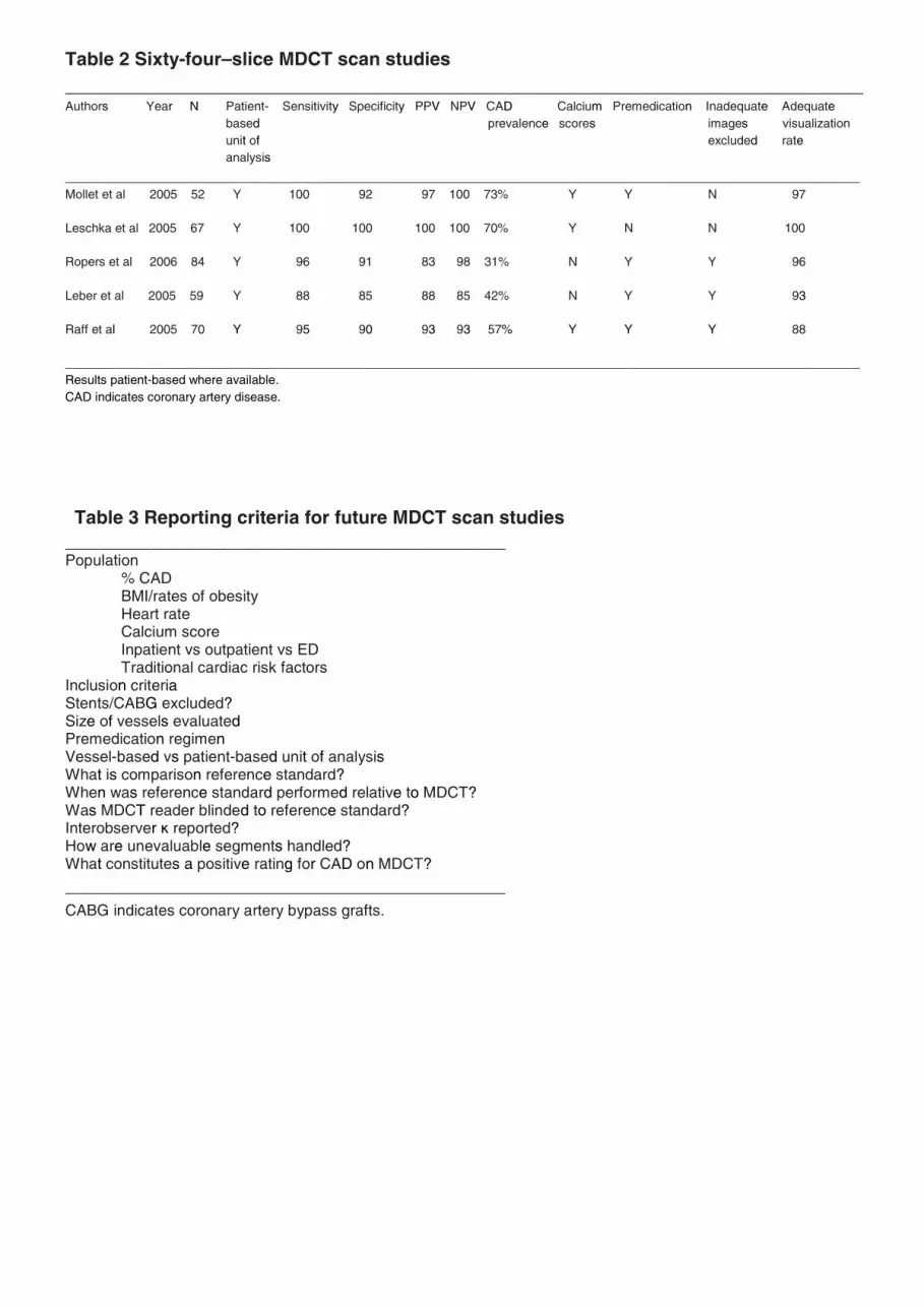
Table 2 Sixty-four–slice MDCT scan studies_____________________________________________________________________________Authors Year N Patient- Sensitivity Specificity PPV NPV CAD Calcium Premedication Inadequate Adequate
based prevalence scores images visualizationunit of excluded rateanalysis
___________________________________________________________________________________________________________________Mollet et al 2005 52 Y 100 92 97 100 73% Y Y N 97
Leschka et al 2005 67 Y 100 100 100 100 70% Y N N 100
Ropers et al 2006 84 Y 96 91 83 98 31% N Y Y 96
Leber et al 2005 59 Y 88 85 88 85 42% N Y Y 93
Raff et al 2005 70 Y 95 90 93 93 57% Y Y Y 88
___________________________________________________________________________________________________________________Results patient-based where available.CAD indicates coronary artery disease.
Table 3 Reporting criteria for future MDCT scan studies___________________________________________________Population
% CADBMI/rates of obesityHeart rateCalcium scoreInpatient vs outpatient vs EDTraditional cardiac risk factors
Inclusion criteriaStents/CABG excluded?Size of vessels evaluatedPremedication regimenVessel-based vs patient-based unit of analysisWhat is comparison reference standard?When was reference standard performed relative to MDCT?Was MDCT reader blinded to reference standard?Interobserver ĸ reported?How are unevaluable segments handled?What constitutes a positive rating for CAD on MDCT?
___________________________________________________
CABG indicates coronary artery bypass grafts.