Embed Size (px)
Citation preview

Neuron, Vol. 45, 301–313, January 20, 2005, Copyright ©2005 by Elsevier Inc. DOI 10.1016/j.neuron.2004.12.044
Spatial Memory in the Rat Requiresthe Dorsolateral Band of the Entorhinal Cortex
the neocortical output structures in which hippocampalmemories are implemented for long-term retention.However, in terms of connectivity, the entorhinal cortex
Hill-Aina Steffenach,1 Menno Witter,1,2
May-Britt Moser,1 and Edvard I. Moser1,*1Centre for the Biology of Memory
stands out as the primary interface between the hippo-Norwegian University of Science and Technologycampus and neocortex, linking the hippocampus withNO-7489 Trondheimnearly all other association cortices (Witter et al., 1989;NorwayBurwell, 2000; Lavenex and Amaral, 2000; Witter and2 Research Institute NeuroscienceAmaral, 2004). This pattern of organization suggests thatDepartment of Anatomythe entorhinal cortex plays a fundamental role in theVU University Medical Centercomputations that take place both before and after corti-Amsterdam 1007 MBcal information enters the hippocampus.The Netherlands
If hippocampal memories depend on interactions withthe neocortex, one would expect lesions of the entorhi-nal cortex to severely impair hippocampal-dependentSummarymemory, with the profile of impairment mirroring thatobserved after damage to the hippocampus. Surpris-The extensive connections of the entorhinal cortexingly, this prediction has not been upheld in studies ofwith the hippocampus and the neocortex point to thisspatial memory after selective fiber-sparing lesions ofregion as a major interface in the hippocampal-neo-the entorhinal cortex. While large mechanical or electro-cortical interactions underlying memory. We askedlytic lesions in the parahippocampal cortex impairedwhether hippocampal-dependent recall of spatial mem-performance in a number of spatial tasks (Olton et al.,ory depends on the entorhinal cortex, and, if so, which1978; Jarrard et al., 1984; Schenk and Morris, 1985;parts are critical. After training in a Morris water maze,Rasmussen et al., 1989; Cho and Kesner, 1996), selec-rats received fiber-sparing lesions in the dorsolateraltive excitotoxic damage to the entorhinal cortex gener-band of the entorhinal cortex, which mediates muchally failed to mirror the pronounced spatial learningof the visuospatial input to the dorsal hippocampus.impairment normally observed after lesions of the hippo-These lesions entirely disrupted retention and re-campus (Bouffard and Jarrard, 1988; Pouzet et al., 1999;tarded new learning. Spatial memory was spared byBannerman et al., 2001a, 2001b; Galani et al., 2002;lesions in the ventromedial band, which connects pri-Oswald et al., 2003; Burwell et al., 2004; Jarrard et al.,marily with ventral hippocampus, but these lesions2004; but see Holscher and Schmidt, 1994 and Cho andreduced defensive behavior on an elevated plus maze,Jaffard, 1994, 1995; for review, see Aggleton et al., 2000).mirroring the effects of damage to ventral hippocam-The simplest interpretation of the spared spatial learningpus. The results suggest that the functional differ-in animals with selective entorhinal lesions is that theences between dorsal and ventral hippocampus re-visuospatial information that is required to solve hippo-flect their connectivity with modules of the entorhinalcampal-dependent spatial learning tasks can reach thecortex that are differently linked to the rest of thehippocampus via sparser routes. Such routes includecortex.the direct connections with the presubiculum and para-subiculum (Witter et al., 1988) and the perirhinal andIntroductionpostrhinal cortices (Naber et al., 1999, 2001), and thedirect connections with subcortical structures such as
Converging evidence suggests that the hippocampus isthe midline nuclei of the thalamus (Herkenham, 1978;
essential for fast encoding and storage of new episodicWouterlood et al., 1990; Dolleman-van der Weel and
memories. The involvement of the hippocampus in Witter, 1996). These inputs and their reciprocal outputsmemory is particularly evident in tasks where subjects may be sufficient for the retention of spatial memoriesmust recall spatial relations between landmarks in order (see Aggleton et al., 2000). On the other hand, severalto locate a hidden goal object (O’Keefe and Nadel, 1978; studies have provided evidence for a modular organiza-Morris et al., 1982; Maguire et al., 1998; Teng and Squire, tion of the entorhinal cortex, such that it is organized1999; Ekstrom et al., 2003), although it is clear from into recurrently connected bands that run parallel tostudies in both humans and animals that the role of the rhinal sulcus and cut across the medial and lateralthe hippocampus extends to other types of declarative subdivisions of the area (Witter et al., 1989; Dolorfo andmemory as well (Scoville and Milner, 1957; Squire, 1992; Amaral, 1998a, 1998b). Spatially modulated neuronsEichenbaum, 2000). have been observed only in the most caudal portion of
The encoding and consolidation of episodic memories the dorsolateral band, i.e., the part made up by theis thought to rely on interactions between the hippocam- medial entorhinal cortex near the postrhinal cortex andpus and other cortical structures (McClelland et al., the rhinal fissure (Fyhn et al., 2004). This area receives1995; Squire and Alvarez, 1995). The exact routes by most of the cortical visuospatial input to the entorhinalwhich information about location and other elements of cortex (Burwell, 2000). More ventromedially located neu-episodes reach the hippocampus are not known, nor are rons exhibit little or no spatial modulation (Fyhn et al.,
2004). Based on the extreme dorsocaudal position ofthe spatially modulated neurons, we hypothesized that*Correspondence: [email protected]

Neuron302
Figure 1. Coronal Sections Stained with Cre-syl Violet, Indicative for Neuronal Cell Bodies,after a Representative Cytotoxic Lesion of theDorsolateral Band of the Entorhinal Cortex
Sections are shown for rat 10647 at five levelsbetween the anterior and posterior poles ofthe entorhinal cortex (middle columns: lowmagnification; left and right columns: highmagnification). Yellow traces show outlinesof the entorhinal cortex at each coronal level.Red traces indicate borders of the lesion (in-cluding areas with partial cell loss). Adjacentcortical areas are indicated (PER, perirhinalcortex; POR, postrhinal cortex; PAS, para-subiculum; PRS, presubiculum; S, subiculum;AE, amygdaloentorhinal transition area). Notethat the lesion covers nearly the entire antero-posterior extent of the entorhinal cortex. Thedorsolateral band (closest to the perirhinaland postrhinal cortices) is extensively le-sioned. The damage stretches into the inter-mediate band in some places, and there isminor damage to the perirhinal and postrhi-nal cortices.
previous lesion studies may have missed that particular The lesions in the dorsolateral band were generallylimited to the target area, forming a bilateral strip ofpart of the entorhinal cortex. The present study thus
examined the ability of rats to recall spatial information damage beneath the rhinal fissure and the postrhinalborder (Figures 1 and 2). The lesions always extendedafter lesions that specifically targeted the dorsolateral
band and compared their performance to that of animals into the intermediate band, but the ventromedial bandwas spared. The lesions generally involved both superfi-with sham lesions or lesions in the ventromedial band.cial layers II and III and deep layers V and VI, althoughthe damage to layers II and III was usually larger. TheResultsdamage was generally more extensive in the dorsocau-dal part of the band than in the ventrorostral part. TheLesions Aimed at the Dorsolateral Band
The first aim of the study was to determine whether lesions covered 38.8% � 3.5% of the area of the entorhi-nal cortex as measured in the unfolded maps (mean �spatial memory requires the dorsolateral band of the
entorhinal cortex. A total of 124 rats were trained to SEM; range, 9.8%–56.1%). The border between dam-aged and healthy tissue was sharp, implying that theasymptote in a conventional reference memory task in
the Morris water maze. Within 36 hr after the last training density of intact neurons within the spared parts of ento-rhinal cortex was within the normal range.session, the rats received N-methyl-D-aspartate (NMDA)-
induced excitotoxic lesions in the dorsolateral band or Most dorsolateral lesions caused some additionaldamage to the adjacent parts of the perirhinal or postrhi-sham lesions. Recall was tested on a probe trial 7 days
later. This part of the study was conducted in five replica- nal cortices, and in the overlying temporal cortex aroundthe cannula tracks (Figure 1). The amount of such dam-tions, each containing approximately five lesioned ani-
mals and five sham animals. age varied between animals. Three rats were excluded

Spatial Memory and the Entorhinal Cortex303
Figure 2. Unfolded Maps Showing Extent ofLesions in the Entorhinal Cortex and Perfor-mance on the Retention Test for Each Animalin the Dorsolateral Band Group
(A) Average unfolded map based on the entiresample of individual maps. To construct eachmap, the lateral border of the entorhinal cor-tex in each brain section was mapped ontothe vertical line in the center of each diagram,and sections were unfolded onto straightlines perpendicular to the line that representsthe lateral border. L, left hemisphere; R, righthemisphere; A, anterior; P, posterior. A sche-matic representation of the three parallelbands in entorhinal cortex is indicated. Thedark gray area represents the dorsolateralband, light gray represents the intermediateband, and white represents the ventrome-dial band.(B–D) Outline of lesions (left) and swim pathson the postsurgical retention test (right) forindividual animals with lesions in the dorso-lateral band. Animals are grouped accordingto the amount of inadvertent damage to adja-cent structures ([B], perirhinal damage only;[C], perirhinal and postrhinal damage; [D],damage in both hippocampal and perirhinalor postrhinal areas). Individual maps are indi-cated with stippled gray contours. Lesionsare indicated in red. The solid black contourshows the average unfolding. In the swimpath diagrams, the position of the unavailableplatform is marked by a red circle (southeastquadrant). Note that none of the three groupsperformed above chance level on the reten-tion test.(E) Swim paths on the postoperative retentiontest for 9 of the 28 sham-operated dorsolat-eral control animals (ranked as number 1, 4,6, 9, 13, 17, 20, 21, and 25, with time in thetarget zone as the ranking variable; rat 10324was number 1, rat 10275 was number 4, rat10295 was number 6, etc.).
from the dorsolateral group because of extensive hippo- the platform remained unavailable for 60 s (Figure 3A).Most rats showed a strong bias toward the platformcampal damage. Two were excluded because the ento-
rhinal cortex was essentially spared on one side, and position during the search period, spending an averageof 43.6% � 2.0% of the search time within a circularfive rats were taken out because the most posterior
brain sections (containing the dorsocaudal portion of zone around the platform (mean � SEM). The zone cov-ered 12% of the pool surface. The rats were ranked,the medial entorhinal cortex) were lost when the brains
were cut. Among the remaining animals in the dorsolat- matched, and assigned to surgery groups according tothe proportion of time they spent in the platform zone.eral group, five had additional damage only in the perirhi-
nal cortex. Three animals had some damage in the post- The effect of the lesions was tested on a second probetrial 7 days after the surgery (Figure 3B). While the sham-rhinal cortex as well, mostly in the anterior half. Six
animals had minor lesions in the subiculum or CA1 of operated animals continued to search at the traininglocation (38.0% � 3.0% in the platform zone), those withthe ventral hippocampus, in addition to some damage
to the perirhinal or postrhinal cortices. Behavioral results lesions in the dorsolateral band displayed no memoryof the target location, spending only 8.7% � 2.9% ofwere first analyzed separately for these three sub-
groups, but in the absence of any detectable behavioral total search time in the circular zone, which was no morethan the time they spent in the corresponding zones ofdifferences, the subgroups were subsequently com-
bined. the three other quadrants. Thirteen of the 14 rats withlesions in the dorsolateral band had swim times nearAll rats learned to approach the platform within �10 s
during the 6 days of preoperative training (Supplemental chance level (range, 0%–16.1% in the target zone; theexpected time with a nonbiased search pattern wouldFigure S1A at http://www.neuron.org/cgi/content/full/
45/2/301/DC1/). The escape latencies decreased quickly be 12%). One outlier spent 41.2% in the target zone (rat10951; 26.0% lesion). This rat had less damage to theto �20 s during the first 2 days and then decreased
slowly during the remainder of the pretraining phase. On dorsocaudal pole than other rats in the dorsolateralband group (Figure 2B; see posterior end of the unfoldedday 7, retention was tested on a probe trial during which

Neuron304
Figure 3. Spatial Memory in the Water Mazeafter Lesions in the Dorsolateral Band of theEntorhinal Cortex
(A and B) Probe trials at the end of pretraining(A) and 7 days after pretraining and surgery(B). Bars indicate time spent in a circular zonearound the platform position (black) and incorresponding zones of the three other quad-rants (means � SEM; see inset). Chance levelis 12%.(C) Latency to locate the hidden platform dur-ing reversal training after surgery (means �
SEM).(D) Probe trial after postoperative trainingwith the platform at a new location.
map). There was no significant correlation within the Lesions Aimed at the Perirhinaland Postrhinal Corticesdorsolateral group between lesion size and time spent
in the target zone (r � �0.43; n � 14; p � 0.13). A The lesions in the dorsolateral band were accompaniedby minor to moderate damage in adjacent parts of therepeated-measures analysis of variance of time spent
in the platform zone and in corresponding zones of the perirhinal or anterior postrhinal cortices (Figure 1). Todetermine whether the retention impairment in the dor-three other pool quadrants showed a significant group �
quadrant effect [F(3, 120) � 23.9; p � 0.001] with a solateral group was caused by this additional damage,we prepared a separate group of animals with lesionssignificant group difference in the target zone [t(40) �
6.2; p � 0.001]. that specifically targeted the affected areas of perirhinaland postrhinal cortex. The damage to the perirhinal cor-The search times of animals in the dorsolateral group
were not related to the amount of accompanying dam- tex in these animals was fairly complete and selective(Figure 4). The lesion also included postrhinal cortex,age in adjacent structures. The five rats with additional
damage only in the perirhinal cortex spent between but primarily the anterior part of the area. There wasonly very minor damage to the entorhinal cortex, mostly4.5% and 16.1% of the trial in the platform zone (Figure
2B). The zone times of the three rats with inadvertent at the adjacent dorsolateral tip of layer II. The size ofthe perirhinal and postrhinal lesions was generally largerlesions in both the perirhinal and postrhinal cortices
were 3.0%, 0%, and, in the case of the previously men- than that in the animals with entorhinal lesions.Lesions in the perirhinal and postrhinal cortices didtioned outlier (rat 10951), 41.2% (Figure 2C). The six
rats with additional minor damage in the subiculum or not impair retention on the postoperative probe trial(Figure 5). Rats with such lesions spent as much timehippocampus spent between 0% and 13.5% in the tar-
get circle (Figure 2D). in the platform zone as simultaneously trained sham-operated animals [Figure 5B; quadrant: F(3, 30) � 9.4,The ability of the rats with entorhinal lesions to learn
a new, modified version of the task was tested by pre- p � 0.001; group � quadrant interaction: F � 1]. Therate of learning in the reversal task was unimpaired (Fig-senting the platform in the opposite quadrant at the end
of the probe trial and using this position as the goal ure 5C; group difference in escape latencies: F � 1),and the lesioned animals searched extensively in theon subsequent trials. The rats received four blocks of
training over the course of 8 hr (one block of four trials correct quadrant on the final probe trial [Figure 5D;quadrant: F(3, 30) � 16.9, p � 0.001; group � quadrantand three blocks of two trials). Escape latencies de-
creased in both groups [block effect: F(3, 132) � 19.1; interaction: F(3, 30) � 2.1, p � 0.10]. These results sug-gest that damage to the perirhinal and postrhinal corti-p � 0.001] but were longer in the animals with dorsolat-
eral lesions [Figure 3C; group effect: F(1, 44) � 8.1; p � ces did not contribute detectably to the retention impair-ment of the dorsolateral entorhinal group, consistent0.01]. When the new memory was probed at the end of
training, both groups searched in the platform zone, and with the fact that the impairment in the latter group wasindependent of the amount of accompanying damagethe group � quadrant interaction effect was no longer
significant [F(3, 108) � 2.1; p � 0.10; Figure 3D]. The to the perirhinal and postrhinal cortices.Finally, we checked whether the impairment after le-main effect of quadrant was significant [F(3, 108) � 42.5;
p � 0.001]. There was no correlation between lesion sions of the entorhinal cortex was due to needle-inducedmechanical damage to overlying areas. An additionalsize and time in the target zone in the dorsolateral group
(r � 0.01; n � 14). sham group was prepared in which the needle was low-

Spatial Memory and the Entorhinal Cortex305
Figure 4. Coronal Sections Showing CresylViolet Stains of Neuronal Cell Bodies after aRepresentative Cytotoxic Control Lesion inthe Perirhinal and Anterior Postrhinal Cor-tices
Sections are taken slightly more anterior thanthose in Figure 1. Yellow traces indicate bor-ders of the entorhinal cortex, blue traces indi-cate borders of the perirhinal and postrhinalcortices, and red indicates the lesion. Abbre-viations as in Figure 1. Note that both theperirhinal and anterior postrhinal corticeswere extensively lesioned, yet this animal ex-hibited good retention (29.4% in the platformzone on the postoperative retention test).
ered into the dorsolateral band of entorhinal cortex with- the hippocampus. These rats were tested together witheight sham-operated rats in two replications.out infusion of the excitotoxin. The needle tracks caused
similar damage in the perirhinal, postrhinal, and temporal The lesions were generally limited to the ventromedialband, forming a bilateral strip of damage along the bor-cortices as in the group with entorhinal lesions (Supple-
mental Figure S2 at http://www.neuron.org/cgi/content/ der with the parasubiculum (Figures 6 and 7). The lesionsgenerally extended into the intermediate band, but thefull/45/2/301/DC1/). Rats with such mechanical lesions
spent as much time in the platform zone on the postop- dorsolateral band was spared. Both deep and superficiallayers were involved. The damage was more extensiveerative retention test as a group of simultaneously
trained sham-operated animals in which the needle was in the caudal part of the ventromedial band than in therostral part. On average, the ventromedial lesions cov-not inserted [Supplemental Figure S3B; quadrant: F(3,
30) � 21.6, p � 0.001; group � quadrant interaction: ered 12.3% � 1.1% of the area of the entorhinal cortexas measured in the unfolded maps (mean � SEM). TheF(3, 42) � 1.1, p � 0.3]. The two groups performed
similarly also after postoperative reversal training [Sup- range was 7.8%–20.1%. Ventromedial band lesionswere thus generally smaller than the lesions in the dorso-plemental Figure S3C; quadrant: F(3, 18) � 5.6, p � 0.01;
group � quadrant interaction: F � 1]. lateral band, although the largest ventromedial lesionscaused a larger percentage of damage in the targetband than the smallest dorsolateral lesions. The borderLesions Aimed at the Ventromedial Band
We next asked if spatial memory specifically depends between damaged and healthy tissue was sharp, similarto what was observed for the dorsolateral band lesions.on the dorsolateral band of the entorhinal cortex, or
if more ventromedial parts are also required. Ten rats Lesions in the ventromedial band caused some addi-tional damage in adjacent parts of the parasubiculumreceived bilateral lesions in the ventromedial band, us-
ing an oblique injection track that bypassed most of and presubiculum, as well as occasional minor damage

Neuron306
Figure 5. Recall after Excitotoxic Control Le-sions in the Perirhinal and Anterior Postrhi-nal Cortices
(A and B) Probe trials at the end of pretraining(A) and 7 days after pretraining and surgery(B).(C) Latency to locate the hidden platform dur-ing reversal training after surgery (means �
SEM).(D) Probe trial after postoperative trainingwith the platform at a new location. Symbolsas in Figure 3.
in the subiculum and dentate gyrus around the cannula The plus maze has two open and two enclosed arms.During their first exposure to the maze, rats typicallytracks. The amount of such damage was highly variable
between animals. Three rats were excluded from the make few entries into the open arms. Clinically effectiveanxiolytics reduce this avoidance (Pellow et al., 1985;ventromedial group because of more extensive hippo-
campal damage. Kjelstrup et al., 2002). Six of the rats with lesions inthe ventromedial band were tested on the plus maze.Lesions in the ventromedial band had no significant
effect on retention of the spatial task (Figure 8). The Whereas the sham-operated animals avoided the openarms, rats with lesions in the ventromedial band visitedlesioned rats continued to search near the platform in
the same way as the simultaneously trained sham-oper- the open arms more frequently [Figures 8E and 8F; groupdifference: t(8) � 2.6; p � 0.05; two-tailed Student’s tated group (means of 32.7% and 35.5%, respectively,
in the target zone; Figure 8B). Seven rats had damage test], although the total time spent on these arms wasnot significantly enhanced [t(8) � 1.3; p � 0.2]. Thealmost exclusively in the ventromedial band. These ani-
mals showed consistently good retention (zone times results show a dissociation of function between the dor-solateral and ventromedial bands of the entorhinal cor-from 20.6% to 48.9%; Figure 7). Three rats had additional
damage to the intermediate band (zone times of 12.4%, tex and suggest that the lesions were large enough todisrupt one function of the ventromedial band of entorhi-26.0%, and 55.0%; Figure 7). Within the ventromedial
group, there was no significant correlation between le- nal cortex.sion size and time spent in the target zone (r � �0.46; n �8; p � 0.18). A repeated-measures analysis of variance of Discussiontime spent in the four quadrant zones showed a signifi-cant effect of quadrant [F(3, 48) � 20.1; p � 0.001] The present study aimed to determine whether the ento-
rhinal cortex is necessary for the acquisition and reten-but no group � quadrant interaction (F � 1). Duringsubsequent reversal training, the escape latencies de- tion of a hippocampal-dependent spatial reference
memory task. We showed first that lesions that includedcreased equally in the two groups [Figure 8C; group andgroup � block effects: F � 1; block: F(3, 48) � 28.4, the dorsolateral band of the entorhinal cortex entirely
and consistently disrupted recall of a spatial navigationp � 0.001], and there was no difference in their searchpattern on the final probe trial [Figure 8D; group � quad- task acquired before the lesion. Second, lesions in the
ventromedial band failed to affect spatial memory butrant interaction: F � 1; quadrant: F(3, 48) � 15.3, p �0.001]. reduced avoidance of the open arms on a plus maze test
of defensive behavior. The results suggest a modularLesions in the ventromedial band were generallysmaller than those in the dorsolateral band. Although organization of the entorhinal cortex with an essential
role for the dorsolateral band in spatial memory and thethe relative amount of damage overlapped, the ventro-medial lesions may have been too small to impair any ventromedial band in control of defensive behavior.behavior. To determine whether the lesions were largeenough to functionally perturb the circuitry of the ventro- The Entorhinal Cortex Is Necessary
for Spatial Memorymedial band, we tested a subset of the animals withventromedial lesions in a task known to depend on the The results point to a key role for the dorsolateral band
of the entorhinal cortex in spatial computation or spatialventral hippocampus, to which the ventromedial bandis strongly connected (Dolorfo and Amaral, 1998a). The representation. They are consistent with the existence
of neurons with sharp and coherent place fields caudallyelevated plus maze is such a task (Kjelstrup et al., 2002).

Spatial Memory and the Entorhinal Cortex307
Figure 6. Coronal Sections Stained with Cre-syl Violet after a Representative Cytotoxic Le-sion of the Ventromedial Band of the Entorhi-nal Cortex
Sections are shown for rat 10948 at approxi-mately the same five anteroposterior levelsas in Figure 1. Abbreviations as in Figure 1.Note that the ventromedial parts of the ento-rhinal cortex are lesioned on both sides. Thelesion stretches into parts of the intermediateband on the right side but leaves the dorsolat-eral band intact. Note additional damage tothe subiculum, presubiculum, and parasubi-culum.
in layers II and III of this area (Fyhn et al., 2004). Although ability of rats to solve spatial memory tasks after fiber-sparing lesions in the entorhinal cortex may suggestsuch neurons have multiple fields dispersed over the
entire recording arena, their information rate is compara- that cortical inputs to the hippocampus other than thosefrom the entorhinal cortex must be sufficient for spatialble to that of place cells in CA1, and collectively they
signal the rat’s position as accurately as place cells learning (see Aggleton et al., 2000 and Jarrard et al.,2004 for discussion). The only significant cortical inputsin the hippocampus. Cells with similarly sharp spatial
modulation have not so far been observed further up- that could mediate such a function are those originatingin the perirhinal and postrhinal cortices (Naber et al.,stream of the hippocampus, such as in the presubiculum
(Taube et al., 1990; Cacucci et al., 2004) and parasubicu- 1999, 2001) and the pre- and parasubiculum (Witter etal., 1988). However, except perhaps for the parasubicu-lum (Taube, 1995) or the perirhinal and postrhinal corti-
ces (Taube et al., 1990; Burwell et al., 1998; Burwell and lar projection to the dentate gyrus (Witter et al., 1988),direct inputs from these areas are sparse and reach onlyHafeman, 2003; Fyhn et al., 2004), implying that the
dorsocaudal portion of the dorsolateral band may con- limited parts of the transverse axis of the hippocampus(Naber et al., 1999, 2001). An alternative explanation oftribute directly to the computation of spatial information.
The present study shows that the dorsolateral band not why previous studies with excitotoxic lesions failed toimpair spatial memory is that the entorhinal lesions mayonly expresses spatial information but is also necessary
for storage, consolidation, or retrieval of such infor- have been incomplete. While the ventral part of the ento-rhinal cortex was damaged successfully in most of themation.
The complete disruption of retention in the water maze studies, the extent of damage to more dorsal and caudalregions is uncertain, as sections including this part wereis at odds with the relatively preserved spatial learning
ability of rats with excitotoxic lesions of the entorhinal generally not included in figures showing outlines of thedamage. The reported injection sites and the recon-cortex reported in previous studies (Bouffard and Jar-
rard, 1988; Pouzet et al., 1999; Bannerman et al., 2001a, structions of three studies that aimed to include themore dorsal areas (Bannerman et al., 2001a; Galani et2001b; Galani et al., 2002; Oswald et al., 2003; Burwell
et al., 2004; Jarrard et al., 2004). At initial glance, the al., 2002; Oswald et al., 2003) suggest that the lesions

Neuron308
spared the dorsocaudal area in which the strongest spa-tial modulation has been reported (Fyhn et al., 2004). Thesparing may account for the lack of significant effectson spatial memory. This suggestion is in line with ourfindings in one animal (rat 10951) where the lesion largelymissed the dorsocaudal pole of the entorhinal cortexand no spatial impairment was detected. Although thelesions of the remaining rats were far from completewith respect to removing the entire entorhinal cortex,or even removing the entire dorsolateral band, they allincluded the dorsocaudal pole. The complete and con-sistent disruption of retention in these animals suggeststhat the dorsocaudal pole of the medial entorhinal cortexplays an essential role in spatial memory. This doesnot rule out an additional role for sparser inputs to thehippocampus like the moderately dense projection fromthe parasubiculum (Kesner and Giles, 1998; Liu et al.,2001, 2004; Jarrard et al., 2004), but our results suggestthat these alternative connections are not sufficient formaintaining and expressing spatial memory.
While the present study clearly points to a necessaryrole for the dorsolateral band of the entorhinal cortexin spatial memory, the exact functions performed byneurons in this region remain elusive. One outstandingissue is whether the entorhinal cortex is differently in-volved during encoding, storage, and retrieval of spatialmemory. Lesions of the dorsolateral band abolished re-tention when the spatial task was learned before surgerybut had a weaker effect on new learning. Escape laten-cies were longer during reversal learning, but there wasno significant disruption of the search pattern on thefinal probe trial. At least two factors may account for theweaker impairment of new learning. First, the entorhinallesions were not complete. Remaining tissue in the dor-solateral band, or in the intermediate and ventromedialbands, may have been sufficient to support new learn-ing. Studies in the hippocampus have shown that reten-tion of spatial memory in the water maze requires theintegrity of large parts of the hippocampus, whereas lessis required for new spatial learning (Moser and Moser,1998a). It is possible that encoding in the intact animalengages a wide entorhinal and hippocampal networkand that this distributed network must be activated in itsentirety for successful retrieval of the stored information,rendering retention more vulnerable to lesions than newlearning. Second, the effects on new learning may de-pend on the exact requirements of the behavioral task. Inthe present reversal task, only the platform was moved;other landmarks remained stationary. Although no mem-ory was detected on the postoperative retention trial,further training may facilitate the retrieval of memoriesstored before surgery that were not accessible on theinitial test (de Hoz et al., 2004). Such memories mayspeed up learning when the majority of sensory cuesremain unaltered. Performance may also have benefited
also in the intermediate band). Symbols as in Figure 2. (C) SwimFigure 7. Unfolded Maps Showing Extent of Lesions in the Entorhi- paths on the postoperative retention test for all eight sham-operatednal Cortex and Performance on the Postoperative Retention Test ventromedial control animals, ranked according to time spent in thefor Each Animal with Damage in the Ventromedial Band target zone (top row: rank 1–4; bottom row: rank 5–8). Rat 10870Animals are grouped according to the selectivity of the lesions ([A], spent a significant part of the trial floating between northwest anddamage primarily in the ventromedial band; [B], significant damage southwest and in southeast.
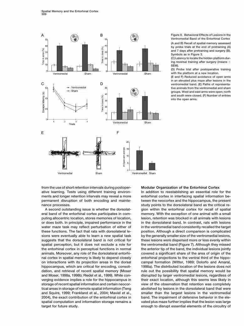
Spatial Memory and the Entorhinal Cortex309
Figure 8. Behavioral Effects of Lesions in theVentromedial Band of the Entorhinal Cortex
(A and B) Recall of spatial memory assessedby probe trials at the end of pretraining (A)and 7 days after pretraining and surgery (B).Symbols as in Figure 3.(C) Latency to locate the hidden platform dur-ing reversal training after surgery (means �
SEM).(D) Probe trial after postoperative trainingwith the platform at a new location.(E and F) Reduced avoidance of open armsin an elevated plus maze after lesions in theventromedial band. (E) Paths of representa-tive animals from the ventromedial and shamgroups. West and east arms were open; northand south were closed. (F) Number of entriesinto the open arms.
from the use of short retention intervals during postoper- Modular Organization of the Entorhinal CortexIn addition to reestablishing an essential role for theative learning. Tests using different training environ-
ments and longer retention intervals may reveal a more entorhinal cortex in interfacing spatial information be-tween the neocortex and the hippocampus, the presentpermanent disruption of both encoding and mainte-
nance processes. study points to the dorsolateral band as the critical re-gion within the entorhinal cortex for recall of spatialA second outstanding issue is whether the dorsolat-
eral band of the entorhinal cortex participates in com- memory. With the exception of one animal with a smalllesion, retention was blocked in all animals with lesionsputing allocentric location, stores memories of location,
or does both. In principle, impaired performance in the in the dorsolateral band. In contrast, rats with lesionsin the ventromedial band consistently recalled the targetwater maze task may reflect perturbation of either of
these functions. The fact that rats with dorsolateral le- position. Although a direct comparison is complicatedby the generally smaller size of the ventromedial lesions,sions were eventually able to learn a new spatial task
suggests that the dorsolateral band is not critical for these lesions were dispersed more or less evenly withinthe ventromedial band (Figure 7). Although they missedspatial perception, but it does not exclude a role for
the entorhinal cortex in perceptual functions in normal the anterior tip of the band, the individual lesions jointlycovered a significant share of the area of origin of theanimals. Moreover, any role of the dorsolateral entorhi-
nal cortex in spatial memory is likely to depend closely entorhinal projections to the ventral third of the hippo-campal formation (Witter, 1989; Dolorfo and Amaral,on interactions with its projection areas in the dorsal
hippocampus, which are critical for encoding, consoli- 1998a). The distributed location of the lesions does notrule out the possibility that spatial memory would bedation, and retrieval of recent spatial memory (Moser
and Moser, 1998a, 1998b; Riedel et al., 1999). While con- disrupted by larger ventromedial lesions, regardless oftheir exact location, although this seems less likely inverging evidence implies a role for the hippocampus in
storage of recent spatial information and certain neocor- view of the observation that retention was completelyabolished by lesions in the dorsolateral band that weretical areas in storage of remote spatial information (Teng
and Squire, 1999; Frankland et al., 2004; Maviel et al., smaller than the largest lesions in the ventromedialband. The impairment of defensive behavior in the ele-2004), the exact contribution of the entorhinal cortex in
spatial computation and information storage remains a vated plus maze further implies that the lesion was largeenough to disrupt essential elements of the circuitry oftarget for future study.

Neuron310
Table 1. Stereotaxic Coordinates and Injection Volume of NMDA
Group Anteroposterior Mediolateral Dorsoventral Angle (�) Volume (�l)
Dorsolateral �5.2a �6.8 bottom 0.5 0 0.04�5.8 �6.8 bottom 0.5 0 0.03�6.4 �6.7 bottom 0.5 0 0.04�7.0 �6.4 bottom 0.5 0 0.04�7.6 �6.4 bottom 0.5 0 0.03�8.2 �5.8 7.3b 0 0.04
�4.7 6.7 0 0.04�8.6 �5.6 7.0 0 0.04
�4.5 6.5 0 0.04Perirhinal/postrhinal �5.2a �6.8 7.3b 0 0.04
�5.8 �6.8 7.2 0 0.04�6.4 �6.7 6.9 0 0.04�7.0 �6.4 6.9 0 0.04�7.6 �6.4 6.5 0 0.04
Ventromedial 1.1c �1.0 bottom 0.3 22d 0.040.7 �1.0 bottom 0.3 22 0.040.4 �1.0 bottom 0.3 22 0.040.1 �1.0 bottom 0.3 22 0.04�0.4 �1.0 bottom 0.3 26 0.04�0.9 �1.0 bottom 0.3 26 0.04�1.4 �1.0 bottom 0.3 26 0.04
For the deepest injection sites, the needle was lowered to the bottom of the brain and retracted 0.5 or 0.3 mm before infusion.a Relative to bregma ( is anterior).b Relative to dura at anteroposterior 1.0, mediolateral 1.0.c Relative to lambda ( is anterior).d Angled in coronal plane with tip pointing laterally and ventrally.
the ventromedial band. Together, these observations hippocampus as compared to the weak spatial modula-tion of neurons at intermediate levels (Jung et al., 1994)suggest that the dorsolateral and ventromedial bands
have different roles, as predicted from the intrinsic and and the near absence of spatial modulation at the ventralpole (K.G. Kjelstrup et al., 2003, Soc. Neurosci., ab-extrinsic organization of the entorhinal cortex (Witter et
al., 1989; Dolorfo and Amaral, 1998a, 1998b). Further stract). Lesions of the ventral pole of the hippocampusinhibit food neophobia (Bannerman et al., 2002) andstudies may determine whether spatial memory requires
the entire extent of the dorsolateral band, including the attenuate avoidance of open arms on an elevated plusmaze (Kjelstrup et al., 2002), whereas dorsal lesions domore ventral areas targeted in previous excitotoxic le-
sion studies, or only the dorsocaudal pole in which sharp not, pointing to a specific role for the ventral hippocam-pus in avoidance of fear-associated stimuli and possiblyposition-related firing was recorded (Fyhn et al., 2004).
Cutting across the medial and lateral subdivisions of the other emotion-dependent behaviors. Together with theanatomical tracing studies, these findings suggest thatentorhinal cortex, the dorsolateral band may comprise
subdivisions mediating input of very different origin to differentiation along the septotemporal axis of the hip-pocampus is a direct consequence of the different corti-the hippocampus (Naber et al., 1997; Burwell and
Amaral, 1998a, 1998b; Burwell, 2000). Recent data cal connections of these areas mediated through topo-graphically organized modules in the entorhinal cortexshowing place-modulated activity in medial but not lat-
eral entorhinal cortex (E.L. Hargreaves et al., 2002, Soc. (Witter et al., 1989; Dolorfo and Amaral, 1998a; Bur-well, 2000).Neurosci., abstract) suggest that spatial memory may
involve mainly the medial entorhinal cortex componentof the dorsolateral band. Experimental Procedures
The modular organization of the entorhinal cortex pro-Subjectsvides a rationale for the functional differentiation be-A total of 124 naive male Long-Evans rats (300–450 g) were housedtween dorsal and ventral parts of the hippocampusin groups of four to six in large transparent polycarbonate cages
(Moser and Moser, 1998b). The effects of lesions in the (59 � 38 � 20 cm) with food and water available ad libitum. Theydorsal and ventral hippocampus mirror those observed were kept on a 12 hr:12 hr light/dark schedule and tested in thehere after lesions in the dorsolateral and ventromedial dark phase.bands of the entorhinal cortex, respectively. Spatialmemory is impaired in a number of tasks by lesions of Behavioral Training
All rats were trained in a white Morris water maze (diameter, 198the dorsal hippocampus but not by equally sized lesionscm; height, 50 cm; water depth, 40 cm; water temperature, 23�C �in the ventral hippocampus (Moser et al., 1993, 1995;2�C; see Moser and Moser, 1998a). One quadrant contained a re-Bannerman et al., 2002; Pothuizen et al., 2004; but seemotely controlled escape platform (11 cm diameter) that could bede Hoz et al., 2003 and Ferbinteanu et al., 2003). Themoved between an available level (submerged 1.5 cm) and an un-
stronger involvement of the dorsal hippocampus in spa- available level (submerged 21 cm). The rats received 6 days oftial tasks is matched by the presence of strong location- training, each day comprising two blocks of four consecutive trials
(maximal trial length 120 s; time on platform 30 s; four start positionsspecific firing in most pyramidal neurons in the dorsal

Spatial Memory and the Entorhinal Cortex311
varied in a predetermined and pseudorandom order). Position data HistologyThe rats were killed with an overdose of Equithesin and perfusedwere collected at 50 Hz (Axona Ltd., Herts, UK). A probe trial with
the platform unavailable for 60 s was conducted at the beginning intracardially with saline and 4% formaldehyde, and the brains werestored in formaldehyde. Frozen sections were cut coronally (30 �m)of day 7. The rats were released from the pool side opposite to the
platform. All rats were allowed to escape on the platform at the end and stained with cresyl violet. Every second section from the poste-rior end of the cerebrum to the anterior end of the entorhinal cortexof the trial to prevent extinction. Time spent in a circular zone around
the platform was compared with time in corresponding zones of the was placed under a microscope attached to a Leica DC200 camera.Images were grabbed and merged by Adobe Photoshop (versionother pool quadrants (zones of 35 cm radius, each covering 12%
of the pool surface). Only rats that spent �20% of the search time 9), and outlines of the entorhinal cortex and the lesion were tracedin Photoshop or, for the illustrations, in Corel Draw (version 11).in this zone were operated and tested further (106/124 rats). These
rats were ranked, matched, and assigned to the different surgerygroups according to the proportion of time they spent around the Unfolded Mapsplatform. Unfoldings of the entorhinal cortex were prepared according to
procedures described previously (Insausti et al., 1997). For conve-nience, the lateral border of the entorhinal cortex in each section
Surgery was mapped onto a straight line, and sections were unfolded ontoWithin 36 hr after the probe trial on day 7, the rats were anesthetized straight lines perpendicular to the line that represents the lateralwith Equithesin (pentobarbital and chloral hydrate; 1.0 ml/250 g border. Lesions were mapped onto the surface of layer II andbody weight), and lesions of the entorhinal cortex were made by mapped only when they included layer II or III. Lesions in layers V andbilateral injection of NMDA (Sigma). NMDA was dissolved in phos- VI are described in the text but were not included in the unfolding. Inphate-buffered saline (pH 7.4; 0.1 M) and injected with a 1 �l Hamil- case the borders of the entorhinal cortex were not obvious becauseton syringe mounted to the stereotaxic frame. Lesions in the dorso- of lesions, we estimated them on the basis of corresponding sec-lateral band of entorhinal cortex were made by infusing 0.03–0.04 tions in nonlesioned animals. We did not differentiate between cy-�l NMDA over 10–20 s at nine stereotaxic positions in each hemi- toarchitectonically defined subdivisions within the entorhinal cortex.sphere, using bregma, midline, and dura at anteroposterior 1.0 and All unfoldings were represented as individual files in Corel Drawmediolateral 1.0 as references for the anteroposterior, mediolateral, (version 11). The outlines of all complete maps were grouped, andand dorsoventral injection coordinates, respectively (Table 1). A an average unfolding was created. On this unfolding, a schematicsubset of these coordinates was used also for perirhinal and postrhi- representation of the three parallel bands in entorhinal cortex wasnal control lesions, except that NMDA was injected more dorsally indicated (Figures 2 and 7). All individual maps were subsequentlythan for the entorhinal lesions (Table 1). Lesions in the ventromedial scaled to best fit on top of this average map by linear scaling in theband were made by using an angled approach (to avoid hippocam- x and/or y direction in Corel Draw. For each animal, the area of thepal damage), with lambda and the midline as references for the lesion was expressed as a proportion of the total area of the entorhi-anteroposterior and mediolateral positions. The syringe was angled nal cortex in the rat’s individual map.either 22� or 26� in the coronal plane, with the needle pointing later-ally and ventrally (Table 1). For both types of lesions, the syringe
Acknowledgmentswas lowered to each target position 2 min before the injectionstarted. After the injection, the syringe was left in place for 7 min
We thank F. Tuvnes for help with the plus maze experiment; andbefore it was retracted. In sham-operated rats, the syringe wasI. Hammer, K. Haugen, K. Jenssen, and H. Waade for technicallowered into the dorsal neocortex, but no NMDA was infused. Aassistance. Supported by the Centre of Excellence scheme of theseparate sham-operated control group was operated to control forNorwegian Research Council.mechanical damage at deeper levels caused by the needle penetra-
tions. The coordinates used in these animals were similar to thoseReceived: September 28, 2004used for dorsolateral band lesions in the entorhinal cortex. Four ofRevised: November 11, 2004the 106 rats were lost after surgery.Accepted: December 6, 2004Published: January 19, 2005
Retention Test and New LearningSeven days after completion of pretraining and surgery, a second Referencesretention test was conducted. Again, the platform was kept in itslower position for the first 60 s, and the swim pattern was recorded. Aggleton, J.P., Vann, S.D., Oswald, C.J., and Good, M. (2000). Identi-The platform was raised at the opposite quadrant of the pool, and fying cortical inputs to the rat hippocampus that subserve allocentricthe rats were then retrained with this new goal position. This phase spatial processes: a simple problem with a complex answer. Hippo-of training consisted of one block of four trials and three subsequent campus 10, 466–474.blocks of two trials. The blocks were separated by 2 hr intervals.
Bannerman, D.M., Yee, B.K., Lemaire, M., Wilbrecht, L., Jarrard, L.,Two hours after the last block, another probe test was conducted.
Iversen, S.D., Rawlins, J.N., and Good, M.A. (2001a). The role of theentorhinal cortex in two forms of spatial learning and memory. Exp.Brain Res. 141, 281–303.Elevated Plus MazeBannerman, D.M., Yee, B.K., Lemaire, M., Jarrard, L., Iversen, S.D.,The maze consisted of four equally illuminated white steel armsRawlins, J.N., and Good, M.A. (2001b). Contextual fear conditioning(12 � 50 cm) radiating at square angles from a central platformis disrupted by lesions of the subcortical, but not entorhinal, connec-(12 � 12 cm) 50 cm above the floor. The maze was placed in a silenttions to the hippocampus. Exp. Brain Res. 141, 304–311.and dimly lit room (3 � 4 min; background noise, �55 dB at 50
kHz; light intensity on open arms, 30 lux). Two opposite arms were Bannerman, D.M., Deacon, R.M., Offen, S., Friswell, J., Grubb, M.,enclosed by 40 cm high walls of white steel. The other two arms and Rawlins, J.N. (2002). Double dissociation of function within thewere open but had transparent plastic ledges (0.3 cm) to prevent hippocampus: spatial memory and hyponeophagia. Behav. Neu-the rats from falling. Rats were released from the central platform, rosci. 116, 884–901.with their face pointing toward an enclosed arm. An observer watch- Bouffard, J.P., and Jarrard, L.E. (1988). Acquisition of a complexing the animal’s behavior on a monitor behind a curtain counted place task in rats with selective ibotenate lesions of hippocampalentries into open and closed arms. An entry was scored when the formation: combined lesions of subiculum and entorhinal cortexrat moved into the arm with all four paws. The rat had to leave the versus hippocampus. Behav. Neurosci. 102, 828–834.arm entirely before another entry was scored. Position was tracked
Burwell, R.D. (2000). The parahippocampal region: corticocorticalat 50 Hz (Axona Ltd., Herts, UK), and time spent in each arm andconnectivity. Ann. N Y Acad. Sci. 911, 25–42.in the center were calculated. After each trial, the maze was cleaned
with water. Burwell, R.D., and Amaral, D.G. (1998a). Cortical afferents of the

Neuron312
perirhinal, postrhinal, and entorhinal cortices of the rat. J. Comp. of the rat: cytoarchitectonic subdivisions and the origin and distribu-tion of cortical efferents. Hippocampus 7, 146–183.Neurol. 398, 179–205.
Burwell, R.D., and Amaral, D.G. (1998b). Perirhinal and postrhinal Jarrard, L.E., Okaichi, H., Steward, O., and Goldschmidt, R.B. (1984).On the role of hippocampal connections in the performance of placecortices of the rat: interconnectivity and connections with the ento-
rhinal cortex. J. Comp. Neurol. 391, 293–321. and cue tasks: comparisons with damage to hippocampus. Behav.Neurosci. 98, 946–954.Burwell, R.D., and Hafeman, D.M. (2003). Positional firing properties
of postrhinal cortex neurons. Neuroscience 119, 577–588. Jarrard, L.E., Davidson, T.L., and Bowring, B. (2004). Functionaldifferentiation within the medial temporal lobe in the rat. Hippocam-Burwell, R.D., Shapiro, M.L., O’Malley, M.T., and Eichenbaum, H.pus 14, 434–449.(1998). Positional firing properties of perirhinal cortex neurons. Neu-
roreport 9, 3013–3018. Jung, M.W., Wiener, S.I., and McNaughton, B.L. (1994). Comparisonof spatial firing characteristics of units in dorsal and ventral hippo-Burwell, R.D., Saddoris, M.P., Bucci, D.J., and Wiig, K.A. (2004).campus of the rat. J. Neurosci. 14, 7347–7356.Corticohippocampal contributions to spatial and contextual learn-
ing. J. Neurosci. 24, 3826–3836. Kesner, R.P., and Giles, R. (1998). Neural circuit analysis of spatialworking memory: role of pre- and parasubiculum, medial and lateralCacucci, F., Lever, C., Wills, T.J., Burgess, N., and O’Keefe, J. (2004).entorhinal cortex. Hippocampus 8, 416–423.Theta-modulated place-by-direction cells in the hippocampal for-
mation in the rat. J. Neurosci. 24, 8265–8277. Kjelstrup, K.G., Tuvnes, F.A., Steffenach, H.-A., Murison, R., Moser,E.I., and Moser, M.-B. (2002). Reduced fear expression after lesionsCho, Y.H., and Jaffard, R. (1994). The entorhinal cortex and a delayedof the ventral hippocampus. Proc. Natl. Acad. Sci. USA 99, 10825–non-matching-to-place task in mice: emphasis on preoperative10830.training and presentation procedure. Eur. J. Neurosci. 6, 1265–1274.Lavenex, P., and Amaral, D.G. (2000). Hippocampal-neocortical in-Cho, Y.H., and Jaffard, R. (1995). Spatial location learning in miceteraction: a hierarchy of associativity. Hippocampus 10, 420–430.with ibotenate lesions of entorhinal cortex or subiculum. Neurobiol.
Learn. Mem. 64, 285–290. Liu, P., Jarrard, L.E., and Bilkey, D.K. (2001). Excitotoxic lesions ofthe pre- and parasubiculum disrupt object recognition and spatialCho, Y.H., and Kesner, R.P. (1996). Involvement of entorhinal cortexmemory processes. Behav. Neurosci. 115, 112–124.or parietal cortex in long-term spatial discrimination memory in rats:
Retrograde amnesia. Behav. Neurosci. 110, 436–442. Liu, P., Jarrard, L.E., and Bilkey, D.K. (2004). Excitotoxic lesions ofthe pre- and parasubiculum disrupt the place fields of hippocampalde Hoz, L., Knox, J., and Morris, R.G.M. (2003). Longitudinal axis ofpyramidal cells. Hippocampus 14, 107–116.the hippocampus: both septal and temporal poles of the hippocam-
pus support water maze spatial learning depending on the training Maguire, E.A., Burgess, N., Donnett, J.G., Frackowiak, R.S., Frith,C.D., and O’Keefe, J. (1998). Knowing where and getting there: aprotocol. Hippocampus 13, 587–603.human navigation network. Science 280, 921–924.de Hoz, L., Martin, S.J., and Morris, R.G.M. (2004). Forgetting, re-
minding, and remembering: the retrieval of lost spatial memory. Maviel, T., Durkin, T.P., Menzaghi, F., and Bontempi, B. (2004). Sitesof neocortical reorganization critical for remote spatial memory.PLoS Biol. 2(8): e225 DOI: 10.1371/journal.pbio.0020225.Science 305, 96–99.Dolleman-Van Der Weel, M.J., and Witter, M.P. (1996). Projections
from the nucleus reuniens thalami to the entorhinal cortex, hippo- McClelland, J.L., McNaughton, B.L., and O’Reilly, R.C. (1995). Whythere are complementary learning systems in the hippocampus andcampal field CA1, and the subiculum in the rat arise from different
populations of neurons. J. Comp. Neurol. 364, 637–650. neocortex: insights from the successes and failures of connectionistmodels of learning and memory. Psychol. Rev. 102, 419–457.Dolorfo, C.L., and Amaral, D.G. (1998a). Entorhinal cortex of the rat:
topographic organization of the cells of origin of the perforant path Morris, R.G.M., Garrud, P., Rawlins, J.N.P., and O’Keefe, J. (1982).Place navigation impaired in rats with hippocampal lesions. Natureprojection to the dentate gyrus. J. Comp. Neurol. 398, 25–48.297, 681–683.Dolorfo, C.L., and Amaral, D.G. (1998b). Entorhinal cortex of the rat:
organization of intrinsic connections. J. Comp. Neurol. 398, 49–82. Moser, M.-B., and Moser, E.I. (1998a). Distributed encoding andretrieval of spatial memory in the hippocampus. J. Neurosci. 18,Eichenbaum, H. (2000). A cortical-hippocampal system for declara-7535–7542.tive memory. Nat. Rev. Neurosci. 1, 41–50.Moser, M.-B., and Moser, E.I. (1998b). Functional differentiation inEkstrom, A.D., Kahana, M.J., Caplan, J.B., Fields, T.A., Isham, E.A.,the hippocampus. Hippocampus 8, 608–619.Newman, E.L., and Fried, I. (2003). Cellular networks underlying
human spatial navigation. Nature 425, 184–188. Moser, E.I., Moser, M.-B., and Andersen, P. (1993). Spatial learningimpairment parallels the magnitude of dorsal hippocampal lesions,Ferbinteanu, J., Ray, C., and McDonald, R.J. (2003). Both dorsalbut is hardly present following ventral lesions. J. Neurosci. 13, 3916–and ventral hippocampus contribute to spatial learning in Long-3925.Evans rats. Neurosci. Lett. 345, 131–135.Moser, M.-B., Moser, E.I., Forrest, E., Andersen, P., and Morris,Frankland, P.W., Bontempi, B., Talton, L.E., Kaczmarek, L., andR.G.M. (1995). Spatial learning with a minislab in the dorsal hippo-Silva, A.J. (2004). The involvement of the anterior cingulate cortexcampus. Proc. Natl. Acad. Sci. USA 92, 9697–9701.in remote contextual fear memory. Science 304, 881–883.Naber, P.A., Caballero-Bleda, M., Jorritsma-Byham, B., and Witter,Fyhn, M., Molden, S., Witter, M.P., Moser, E.I., and Moser, M.B.M.P. (1997). Parallel input to the hippocampal memory system(2004). Spatial representation in the entorhinal cortex. Sciencethrough peri- and postrhinal cortices. Neuroreport 8, 2617–2621.305, 1258–1264.Naber, P.A., Witter, M.P., and Lopez da Silva, F.H. (1999). PerirhinalGalani, R., Obis, S., Coutureau, E., Jarrard, L., and Cassel, J.C.cortex input to the hippocampus in the rat: evidence for parallel(2002). A comparison of the effects of fimbria-fornix, hippocampal,pathways, both direct and indirect. A combined physiological andor entorhinal cortex lesions on spatial reference and working mem-anatomical study. Eur. J. Neurosci. 11, 4119–4133.ory in rats: short versus long postsurgical recovery period. Neuro-
biol. Learn. Mem. 77, 1–16. Naber, P.A., Witter, M.P., and Lopes da Silva, F.H. (2001). Evidencefor a direct projection from the postrhinal cortex to the subiculumHerkenham, M. (1978). The connections of the nucleus reuniensin the rat. Hippocampus 11, 105–117.thalami: evidence for a direct thalamo-hippocampal pathway in the
rat. J. Comp. Neurol. 177, 589–610. O’Keefe, J., and Nadel, L. (1978). The Hippocampus as a CognitiveMap (Oxford: Clarendon Press).Holscher, C., and Schmidt, W.J. (1994). Quinolinic acid lesion of the
rat entorhinal cortex pars medialis produces selective amnesia in Olton, D.S., Walker, J.A., and Gage, F.H. (1978). Hippocampal con-nections and spatial discrimination. Brain Res. 139, 295–308.allocentric working memory (WM), but not in egocentric WM. Behav.
Brain Res. 63, 187–194. Oswald, C.J., Bannerman, D.M., Yee, B.K., Rawlins, J.N.P., Honey,R.C., and Good, M. (2003). Entorhinal cortex lesions disrupt theInsausti, R., Herrero, M.T., and Witter, M.P. (1997). Entorhinal cortex

Spatial Memory and the Entorhinal Cortex313
transition between the use of intra- and extramaze cues for naviga-tion in the water maze. Behav. Neurosci. 117, 588–595.
Pellow, S., Chopin, P., File, S.E., and Briley, M. (1985). Validation ofopen:closed arm entries in an elevated plus-maze as a measure ofanxiety in the rat. J. Neurosci. Methods 14, 149–167.
Pothuizen, H.H., Zhang, W.N., Jongen-Relo, A.L., Feldon, J., andYee, B.K. (2004). Dissociation of function between the dorsal andthe ventral hippocampus in spatial learning abilities of the rat: awithin-subject, within-task comparison of reference and workingspatial memory. Eur. J. Neurosci. 19, 705–712.
Pouzet, B., Welzl, H., Gubler, M.K., Broersen, L., Veenman, C.L.,Feldon, J., Rawlins, J.N.P., and Yee, B.K. (1999). The effects ofNMDA-induced retrohippocampal lesions on performance of fourspatial memory tasks known to be sensitive to hippocampal damagein the rat. Eur. J. Neurosci. 11, 123–140.
Rasmussen, M., Barnes, C.A., and McNaughton, B.L. (1989). A sys-tematic test of cognitive mapping, working-memory, and temporaldiscontiguity theories of hippocampal function. Psychobiol. 17,335–348.
Riedel, G., Micheau, J., Lam, A.G., Roloff, E.L., Martin, S.J., Bridge,H., de Hoz, L., Poeschel, B., McCulloch, J., and Morris, R.G.M.(1999). Reversible neural inactivation reveals hippocampal partici-pation in several memory processes. Nat. Neurosci. 2, 898–905.
Schenk, F., and Morris, R.G.M. (1985). Dissociation between compo-nents of spatial memory in rats after recovery from the effects ofretrohippocampal lesions. Exp. Brain Res. 58, 11–28.
Scoville, W.B., and Milner, B. (1957). Loss of recent memory afterbilateral hippocampal lesions. J. Neurol. Neurosurg. Psychiatry20, 11–21.
Squire, L.R. (1992). Memory and the hippocampus: a synthesis fromfindings with rats, monkeys, and humans. Psychol. Rev. 99, 195–231.
Squire, L.R., and Alvarez, P. (1995). Retrograde amnesia and mem-ory consolidation: a neurobiological perspective. Curr. Opin. Neuro-biol. 5, 169–177.
Taube, J.S. (1995). Place cells recorded in the parasubiculum offreely moving rats. Hippocampus 5, 569–583.
Taube, J.S., Muller, R.U., and Ranck, J.B., Jr. (1990). Head-directioncells recorded from the postsubiculum in freely moving rats. I. De-scription and quantitative analysis. J. Neurosci. 10, 420–435.
Teng, E., and Squire, L.R. (1999). Memory for places learned longago is intact after hippocampal damage. Nature 400, 675–677.
Witter, M.P. (1989). Connectivity of the rat hippocampus. In Neurol-ogy and Neurobiology, Volume 52: The Hippocampus—New Vistas,V. Chan-Palay and C. Kohler, eds. (New York: Alan Liss, Inc.), pp.53–69.
Witter, M.P., and Amaral, D.G. (2004). The hippocampal formation.In The Rat Nervous System, Third Edition, G. Paxinos, ed. (SanDiego, CA: Elsevier Academic Press), pp. 637–703.
Witter, M.P., Holtrop, R., and van de Loosdrecht, A.A. (1988). Directprojections from the periallocortical subicular complex to the fasciadentata in the rat: An anatomical tracing study using phaseolusvulgaris leucoagglutinin. Neurosci. Res. Commun. 2, 61–68.
Witter, M.P., Groenewegen, H.J., Lopes da Silva, F.H., and Lohman,A.H.M. (1989). Functional organization of the extrinsic and intrinsiccircuitry of the parahippocampal region. Prog. Neurobiol. 33,161–254.
Wouterlood, F.G., Saldana, E., and Witter, M.P. (1990). Projectionfrom the nucleus reuniens thalami to the hippocampal region: lightand electron microscopic tracing study in the rat with the antero-grade tracer Phaseolus vulgaris-leucoagglutinin. J. Comp. Neurol.296, 179–203.


