Embed Size (px)
Citation preview
Research review
Stickymucilages and exudates of plants: putativemicroenvironmentaldesign elements with biotechnological value
Author for correspondence:Kirsten Krause
Tel: +47 77646415
Email: [email protected]
Received: 16 April 2019
Accepted: 19 August 2019
Andrew F. Galloway1 , Paul Knox2 and Kirsten Krause1
1Department for Arctic andMarine Biology, Faculty for Biosciences, Fisheries and Economics, UiT The Arctic University of Norway,
Breivika, Tromsø 9037, Norway; 2Centre for Plant Sciences, Faculty of Biological Sciences, University of Leeds, Leeds LS2 9JT, UK
New Phytologist (2019)doi: 10.1111/nph.16144
Key words: bioadhesive, extrafloral nectar,exudate,mucilage, polysaccharides, secretion.
Summary
Plants produce awide array of secretions both above and below ground. Known asmucilages or
exudates, they are secreted by seeds, roots, leaves and stems and fulfil a variety of functions
including adhesion, protection, nutrient acquisition and infection. Mucilages are generally
polysaccharide-rich and often occur in the form of viscoelastic gels and in many cases have
adhesive properties. In some cases, progress is being made in understanding the structure–function relationships of mucilages such as for the secretions that allow growing ivy to attach to
substrates and the biosynthesis and secretion of the mucilage compounds of the Arabidopsis
seed coat. Work is just beginning towards understanding root mucilage and the proposed
adhesive polymers involved in the formation of rhizosheaths at root surfaces and for the
secretions involved in host plant infection by parasitic plants. In this article, we summarise
knowledge on plant exudates and mucilages within the concept of their functions in
microenvironmental design, focusing in particular on their bioadhesive functions and the
molecules responsible for them.Wedrawattention to areas of future knowledgeneed, including
the microstructure of mucilages and their compositional and regulatory dynamics.
Introduction
Land plants have a great ability to adapt to a diverse range ofenvironments. Being sessile and unable to evade unfavourableconditions, their survival depends critically on their ability to sensetheir environment and, if possible, create favourable local condi-tions. To meet these demands, plants have developed a range ofstrategies, including morphological adaptations, long-distancecommunication via volatiles or synthesis of protective metabolites.Plants have also evolved ways to bioengineer favourable microen-vironments in their immediate surroundings by the secretion orexudation ofmucilages from various surfaces.Mucilage, defined bySasse et al. (2018) as a matrix of high-molecular-weight com-pounds, is secreted as a viscoelastic gel that is often polysaccharide-rich. The wider term exudates encompasses mucilages but can alsoinclude compounds of lower molecular weight and more solublehigh-molecular-weight polysaccharide and protein componentsthat may not contribute to gel-like structures. Depending on theplant species, exudates andmucilages can be secreted by almost any
plant organ, and seemingly from all clades of the angiosperms(Brown et al., 2017), and in a few cases multiple exudate types withdifferent origins have been described for the same species or genus(e.g. seed coat, haustorial and stem exudates for parasitic dodder(Cuscuta spp.)) (Schaffner, 1976; Lyshede, 1984; Vaughn, 2002).Mucilages and other exudates provide an effective means to executea variety of functions beyond the confines of their tissues and organsurfaces. Classic examples include the capacity of climbing plants toclimb up vertical surfaces using sticky tendrils or roots (Endress &Thomson, 1977; Groot et al., 2003; Huang et al., 2016). Othertypes of exudates are produced by leaves or by stems and havebecome known as extrafloral nectars (Deynze et al., 2018; Pierce,2019). Below ground, the roots are equally adept in producingmucilages that have an impact on the properties of their immediatesoil environment (Sasse et al., 2018). Each type of secretionessentially consists of a unique blend of molecules that, dependingon its location, serves a specific set of functions. These functionsrange from attracting beneficial microbiota (Haichar et al., 2014)and insects (Deynze et al., 2018), modifying soils for enhanced
� 2019 The AuthorsNew Phytologist � 2019 New Phytologist Trust
New Phytologist (2019) 1www.newphytologist.com
This is an open access article under the terms of the Creative Commons Attribution License, which permits use,distribution and reproduction in any medium, provided the original work is properly cited.
Review
nutrient and water uptake (Galloway et al., 2018) or surfaceanchorage for climbing (Bowling & Vaughn, 2008) to attachmentto hosts before their infection (Vaughn, 2002).
Some molecules within mucilages have bioadhesive properties,making them sought-after materials for biotechnological, biomed-ical or agricultural applications (Favi et al., 2014; George &Suchitra, 2019). Important breakthroughs have been made in thelast few years in investigations of the sticky adhesives used byEnglish ivy andby sundew (Zhang et al., 2010; Lenaghan&Zhang,2012), and a current focus on molecular genetic investigations ofseed coats (Golz et al., 2018; Sechet et al., 2018) and therhizosheath (Sasse et al., 2018) underpins the potential of thisfield. With this review we first highlight the wealth of the differentsticky mucilage types that are produced by plants. We furtherelaborate on the current state of the art regarding the analysis of themolecules lending bioadhesive plant secretions their specificfunctions and point out where future efforts could be focused.
Plant exudate and mucilage diversity
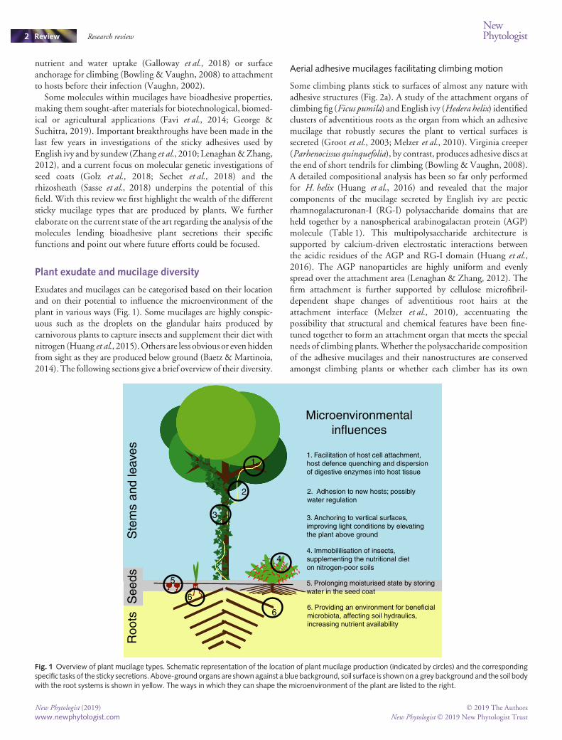
Exudates and mucilages can be categorised based on their locationand on their potential to influence the microenvironment of theplant in various ways (Fig. 1). Some mucilages are highly conspic-uous such as the droplets on the glandular hairs produced bycarnivorous plants to capture insects and supplement their diet withnitrogen (Huang et al., 2015).Others are less obvious or evenhiddenfrom sight as they are produced below ground (Baetz & Martinoia,2014). The following sections give a brief overview of their diversity.
Aerial adhesive mucilages facilitating climbing motion
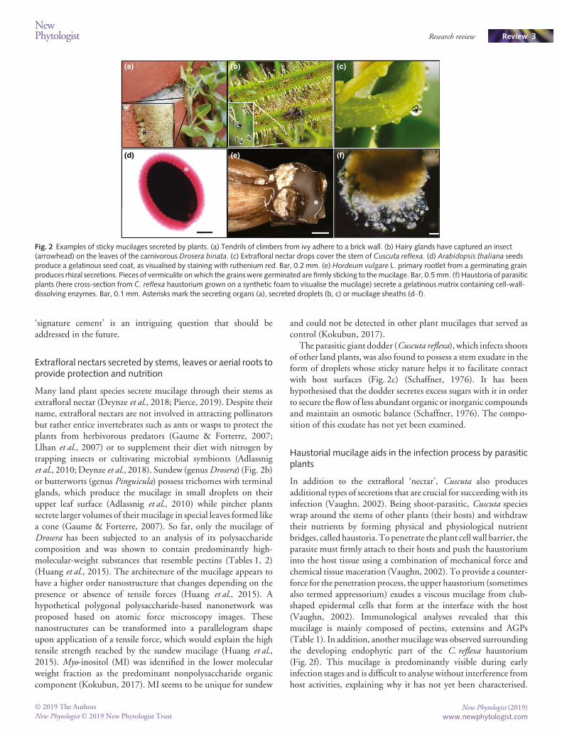
Some climbing plants stick to surfaces of almost any nature withadhesive structures (Fig. 2a). A study of the attachment organs ofclimbing fig (Ficus pumila) andEnglish ivy (Hedera helix) identifiedclusters of adventitious roots as the organ from which an adhesivemucilage that robustly secures the plant to vertical surfaces issecreted (Groot et al., 2003; Melzer et al., 2010). Virginia creeper(Parhenocissus quinquefolia), by contrast, produces adhesive discs atthe end of short tendrils for climbing (Bowling & Vaughn, 2008).A detailed compositional analysis has been so far only performedfor H. helix (Huang et al., 2016) and revealed that the majorcomponents of the mucilage secreted by English ivy are pecticrhamnogalacturonan-I (RG-I) polysaccharide domains that areheld together by a nanospherical arabinogalactan protein (AGP)molecule (Table 1). This multipolysaccharide architecture issupported by calcium-driven electrostatic interactions betweenthe acidic residues of the AGP and RG-I domain (Huang et al.,2016). The AGP nanoparticles are highly uniform and evenlyspread over the attachment area (Lenaghan & Zhang, 2012). Thefirm attachment is further supported by cellulose microfibril-dependent shape changes of adventitious root hairs at theattachment interface (Melzer et al., 2010), accentuating thepossibility that structural and chemical features have been fine-tuned together to form an attachment organ that meets the specialneeds of climbing plants.Whether the polysaccharide compositionof the adhesive mucilages and their nanostructures are conservedamongst climbing plants or whether each climber has its own
Ste
ms
and
leav
esR
oots
Microenvironmentalinfluences
4. Immobililisation of insects, supplementing the nutritional diet on nitrogen-poor soils
5. Prolonging moisturised state by storing water in the seed coat
3. Anchoring to vertical surfaces, improving light conditions by elevating the plant above ground
2. Adhesion to new hosts; possibly water regulation
1. Facilitation of host cell attachment,host defence quenching and dispersion of digestive enzymes into host tissue
6. Providing an environment for beneficialmicrobiota, affecting soil hydraulics, increasing nutrient availability
See
ds
1.
2
3
4
6
6
5
Fig. 1 Overview of plant mucilage types. Schematic representation of the location of plant mucilage production (indicated by circles) and the correspondingspecific tasks of the sticky secretions. Above-ground organs are shown against a blue background, soil surface is shownon a grey background and the soil bodywith the root systems is shown in yellow. The ways in which they can shape the microenvironment of the plant are listed to the right.
New Phytologist (2019) � 2019 The Authors
New Phytologist� 2019 New Phytologist Trustwww.newphytologist.com
Review Research reviewNewPhytologist2
‘signature cement’ is an intriguing question that should beaddressed in the future.
Extrafloral nectars secreted by stems, leaves or aerial roots toprovide protection and nutrition
Many land plant species secrete mucilage through their stems asextrafloral nectar (Deynze et al., 2018; Pierce, 2019). Despite theirname, extrafloral nectars are not involved in attracting pollinatorsbut rather entice invertebrates such as ants or wasps to protect theplants from herbivorous predators (Gaume & Forterre, 2007;Llhan et al., 2007) or to supplement their diet with nitrogen bytrapping insects or cultivating microbial symbionts (Adlassniget al., 2010;Deynze et al., 2018). Sundew (genusDrosera) (Fig. 2b)or butterworts (genus Pinguicula) possess trichomes with terminalglands, which produce the mucilage in small droplets on theirupper leaf surface (Adlassnig et al., 2010) while pitcher plantssecrete larger volumes of theirmucilage in special leaves formed likea cone (Gaume & Forterre, 2007). So far, only the mucilage ofDrosera has been subjected to an analysis of its polysaccharidecomposition and was shown to contain predominantly high-molecular-weight substances that resemble pectins (Tables 1, 2)(Huang et al., 2015). The architecture of the mucilage appears tohave a higher order nanostructure that changes depending on thepresence or absence of tensile forces (Huang et al., 2015). Ahypothetical polygonal polysaccharide-based nanonetwork wasproposed based on atomic force microscopy images. Thesenanostructures can be transformed into a parallelogram shapeupon application of a tensile force, which would explain the hightensile strength reached by the sundew mucilage (Huang et al.,2015). Myo-inositol (MI) was identified in the lower molecularweight fraction as the predominant nonpolysaccharide organiccomponent (Kokubun, 2017). MI seems to be unique for sundew
and could not be detected in other plant mucilages that served ascontrol (Kokubun, 2017).
The parasitic giant dodder (Cuscuta reflexa), which infects shootsof other land plants, was also found to possess a stem exudate in theform of droplets whose sticky nature helps it to facilitate contactwith host surfaces (Fig. 2c) (Schaffner, 1976). It has beenhypothesised that the dodder secretes excess sugars with it in orderto secure the flow of less abundant organic or inorganic compoundsand maintain an osmotic balance (Schaffner, 1976). The compo-sition of this exudate has not yet been examined.
Haustorial mucilage aids in the infection process by parasiticplants
In addition to the extrafloral ‘nectar’, Cuscuta also producesadditional types of secretions that are crucial for succeeding with itsinfection (Vaughn, 2002). Being shoot-parasitic, Cuscuta specieswrap around the stems of other plants (their hosts) and withdrawtheir nutrients by forming physical and physiological nutrientbridges, called haustoria.Topenetrate the plant cell wall barrier, theparasite must firmly attach to their hosts and push the haustoriuminto the host tissue using a combination of mechanical force andchemical tissue maceration (Vaughn, 2002). To provide a counter-force for the penetration process, the upper haustorium (sometimesalso termed appressorium) exudes a viscous mucilage from club-shaped epidermal cells that form at the interface with the host(Vaughn, 2002). Immunological analyses revealed that thismucilage is mainly composed of pectins, extensins and AGPs(Table 1). In addition, anothermucilage was observed surroundingthe developing endophytic part of the C. reflexa haustorium(Fig. 2f). This mucilage is predominantly visible during earlyinfection stages and is difficult to analyse without interference fromhost activities, explaining why it has not yet been characterised.
(a) (b) (c)
(d) (e) (f)
Fig. 2 Examples of sticky mucilages secreted by plants. (a) Tendrils of climbers from ivy adhere to a brick wall. (b) Hairy glands have captured an insect(arrowhead) on the leaves of the carnivorous Drosera binata. (c) Extrafloral nectar drops cover the stem of Cuscuta reflexa. (d) Arabidopsis thaliana seedsproduce a gelatinous seed coat, as visualised by staining with ruthenium red. Bar, 0.2 mm. (e) Hordeum vulgare L. primary rootlet from a germinating grainproduces rhizal secretions. Pieces of vermiculite onwhich the grainswere germinated are firmly sticking to themucilage. Bar, 0.5 mm. (f) Haustoria of parasiticplants (here cross-section from C. reflexa haustorium grown on a synthetic foam to visualise the mucilage) secrete a gelatinous matrix containing cell-wall-dissolving enzymes. Bar, 0.1 mm. Asterisks mark the secreting organs (a), secreted droplets (b, c) or mucilage sheaths (d–f).
� 2019 The Authors
New Phytologist� 2019 New Phytologist TrustNew Phytologist (2019)
www.newphytologist.com
NewPhytologist Research review Review 3
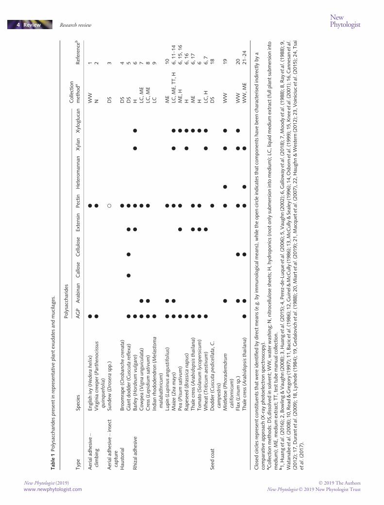
Tab
le1Polysaccharides
presentin
representative
plantex
udates
andmucilages.
Typ
eSp
ecies
Polysaccharides
Collection
methoda
Referen
ceb
AGP
Arabinan
Callose
Cellulose
Extensin
Pectin
Heteroman
nan
Xylan
Xyloglucan
Aerialadhesive–
clim
bing
Englishivy(H
edera
helix)
●●
WW
1Virginiacreeper
(Parthenocissus
quinquefolia)
●●
N2
Aerialadhesive–insect
capture
Sundew
(Drosera
spp.)
○DS
3
Hau
storial
Broomrape(O
robanchecrenata)
●DS
4Giantdodder
(Cuscuta
reflexa)
●●
●●
●DS
5Rhizalad
hesive
Barley(H
ordeum
vulgare)
●●
●●
●H
6Cowpea
(Vignaunguiculata)
●●
●LC
,ME
7Cress
(Lepidium
sativum)
●●
●LC
,ME
8Indianrhododen
dron(M
elastoma
malabathricum)
●LC
9
Lupin(Lupinusangustifolius)
●●
●ME
10
Maize
(Zeamays)
●●
●●
●LC
,ME,
TT,H
6,11–1
4Pea
(Pisum
sativum)
●●
●●
ME,
H6,15,16
Rap
esee
d(Brassicanapus)
●●
●H
6,16
Thalecress(Arabidopsisthailana)
●●
●●
ME
6,17
Tomato(Solanum
lycopersicum)
●●
●●
H6
Whea
t(Triticum
aestivum)
●●
●●
LC,H
6,7
Seed
coat
Dodder
(Cuscuta
pedicellata,C.
campestris)
●DS
18
Mistletoe( Phorademdrum
californicum)
●●
●●
●W
W19
Flax
(Linum
sp.)
●●
●●
●W
W20
Thalecress(Arabidopsisthailana)
●●
●●
●●
WW,ME
21–2
4
Closedcirclesrepresentconstituen
tsthat
wereiden
tified
bydirectmea
ns(e.g.byim
munologicalmea
ns),while
theopen
circleindicates
that
componen
tshavebee
ncharacterisedindirectlybya
comparativeap
proach(X-ray
photoelectronspectroscopy).
aCollectionmethods:DS,dissolved
insolven
t;W
W,water
washing;N
,nitrocellulose
shee
ts;H,hyd
roponics(rootonlysubmersioninto
med
ium);LC
,liquid
med
ium
extract(fullplantsubmersioninto
med
ium);ME,
med
ium
extract;TT,testtubeman
ualcollection.
b1,H
uan
getal.(2016);2,B
owling&Vau
ghn(2008);3,H
uan
getal.(2015);4,P
erez-de-Lu
queetal.(2006);5,V
aughn(2002);6,G
alloway
etal.(2018);7,M
oodyetal.(1988);8,R
ayetal.(1988);9,
Watan
abeetal.(2008);10,R
ead&Gregory(1997);11,Bacicetal.(1986);12,G
uinel&McC
ully
(1986);13,M
cCully
&Sealey
(1996);14,O
sborn
etal.(1999);15,K
nee
etal.(2001);16,C
annesan
etal.
(2012);17,D
urantetal.(2009);18,Lyshed
e(1984);19,G
edalovich
etal.(1988);20,M
iartetal.(2019);21,M
acquet
etal.(2007),22,H
aughn&Western
(2012);23,V
oiniciucetal.(2015);24,T
sai
etal.(2017).
New Phytologist (2019) � 2019 The Authors
New Phytologist� 2019 New Phytologist Trustwww.newphytologist.com
Review Research reviewNewPhytologist4
BesidesCuscuta, the root parasiteOrobanche crenatawas also foundto produce a pectin-richmucilage at the interface between itself andits hosts (Perez-de-Luque et al., 2006). O. crenata forms similarhost attachments as C. reflexa but with the difference that itconnects to the host root system below ground. Interestingly, themucilage that the parasite secretes and that may help the parasite toinfect susceptible hosts appeared to fill host xylem vessels inresistant hosts and ultimately led to the death of the parasite (Perez-de-Luque et al., 2006). Future investigations need to focus oncomprehensive analyses of the haustorial mucilage composition inthese two and other species, and also link possible differences incomposition to differences in infection strategies, host specificityand host responses. In this context, and with the tensile strength ofthe infection organ in mind, a nanostructural analysis of themucilages involved in parasitic plant infection would be veryinteresting.
Desiccation prevention and surface adhesion properties ofseed coat mucilage
The production of a coat of gelatinous material derived from cellwall polysaccharides is fairly widely found in seeds of land plants(Fig. 2d). A prominent example of seed coat mucilage is the viscinproduced by hemiparasitic mistletoe species. This mucilageprotects the seeds during their passage through the guts of birdsand ensures that the seed is firmly attached to the branches of itshost trees when excreted by the birds. The viscin mucilage ofPhoradendron californicum has been subject to extensive biochem-ical characterisation and was found to consist predominantly ofhighly branched xylans, arabinans, pectic rhamnogalacturonansand xyloglucans (Gedalovich et al., 1988). A comparative studyinvolving P. californicum and two other Viscaceae species indicatedthat compositional differences, mainly in the type of neutral sugars,exist between the species and that these may be specific enough toserve as taxonomic markers (Gedalovich-Shedletzky et al., 1989).
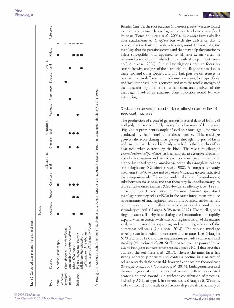
In the model land plant Arabidopsis thaliana, specialisedmucilage secretory cells (MSCs) in the outer integument producelarge amounts ofmucilaginous hydrophilic polysaccharides in ringsaround a central columella that is compositionally similar to asecondary cell wall (Haughn&Western, 2012). Themucilaginousrings in each cell dehydrate during seed maturation but rapidlyexpandwhen in contact withwater during imbibition of thematureseed, accompanied by rupturing and rapid degradation of theoutermost cell walls (Golz et al., 2018). The released mucilageenvelope can be divided into an inner and an outer layer (Haughn& Western, 2012), and this organization provides coherence andstability (Voiniciuc et al., 2015). The outer layer is a poor adhesivedue to its higher content of unbranched pectic RG-I that stretchesout into the soil (Tsai et al., 2017), whereas the inner layer hasstrong adhesive properties and contains pectins in a matrix ofcellulose scaffolds that span this layer and connect it to the seed coat(Macquet et al., 2007; Voiniciuc et al., 2015). Linkage analyses andthe investigation ofmutants impaired in several cell-wall-associatedproteins pointed towards a significant contribution of proteins,including AGPs of type I, in the seed coats (Haughn & Western,2012) (Table 1). The analysis of flaxmucilage revealed thatmany ofT
able
2Carbohyd
ratespresentin
plantmucilages.
Typ
eSp
ecies
Sugars
Referen
cea
Arabinose
Fucose
Galactose
Galacturonic
acid
Glucose
Glucuronic
acid
Man
nose
Rhan
mose
Sucrose
Uronic
acid
Xylose
Aerial
adhesive
Sundew
(Drosera
spp.)
●●
●●
●1
Extrafl
oral
nectar
Giantdodder
(Cuscuta
reflexa)
●2
Jute
mallow
(Corchorusolitorius)
●●
●●
●●
3Maize
(Zeamays)
●●
●●
●4
Seed
coat
Pignut(H
yptissuaveolens)
●●
●●
●●
5Mistletoe(Phorademdrum
californicum,Acreythobium
americanum,Phthirusa
pyrifolia)
●●
●6
a1,Huan
getal.(2015);2,Schaffner
(1976);3,L
lhan
etal.(2007);4,Dey
nze
etal.(2018);5,G
owda(1984);6,Ged
alovich-Shed
letzky
etal.(1989).
� 2019 The Authors
New Phytologist� 2019 New Phytologist TrustNew Phytologist (2019)
www.newphytologist.com
NewPhytologist Research review Review 5
these components are located in distinct domains or layers (Miartet al., 2019), suggesting a highly sophisticated and complexregulatory network in charge of seed coat production. High-throughput screens for phenotypic differences in the mucilaginousseed coats of Arabidopsis mutants have helped to identify specificgenes that secure proper seed coat production (Golz et al., 2018,and references herein). In particular, these studies have highlightedthe contribution of transcription factors to MSC differentiationand have led to a proposed hierarchical network involving threedistinct tiers or levels of regulation (Golz et al., 2018).
Seed coats are a potential source for carbon and thereforetentatively a source for nourishing the developing seed, althoughsome studies suggest that this resource is not utilised (Huang et al.,2004). Rather, the seed coat mucilage seems to enhance wateravailability to the seed andmake it less vulnerable to short-term dryspells (Huang et al., 2004). It provides a barrier to the environmentand keeps the seed moist due to its water-holding capacity. It isfurther implicated in the mediation of germination under water-logged conditions (Haughn & Western, 2012), in soil seed bankmaintenance and in seed dispersal (Yang et al., 2012; Voiniciucet al., 2015; Tsai et al., 2017). The specific set of functions can varybetween species. It is plausible that seed coatmucilage could also beinvolved in the recruitment of beneficial soil microbiota (ashypothesised for root bioadhesives), which is a crucial foothold forplant growth, although experimental evidence for this has yet to begenerated.
Root exudates bioengineer rhizospheres for sustainedresource uptake
While disputed, most data agree that plants invest a considerableamount of their resources into releasing exudates from roots(Fig. 2e), and estimates that between 10% and 40% of photosyn-thate ‘currency’ is spent on root exudates have been put forward(Newman, 1985;McNear, 2013). Root exudates, including a greatcomplexity of both low- and high-molecular-weight components,influence zones of soil at root surfaces known as rhizospheres (Baetz& Martinoia, 2014). The release of polysaccharide-rich mucilagefrom root tips is well established and thismay lubricate roots to easepenetration through deeper layers of soil and may also be involvedin forming a protective barrier (Bacic et al., 1986; Read&Gregory,1997). It is proposed that bioadhesive mucilage components ofexudates are important factors, along with root hairs, in theformation of cylinders of soil around roots known as rhizosheaths.Rhizosheaths could enable plants to sustain and increase nutrientand water uptake from the soil (Traore et al., 2000; Brown et al.,2017; Pang et al., 2017; Galloway et al., 2018). Rhizosheathbioengineering by some grass species during periods of drought hasbeen observed, where the grasses increased the thickness of theirrhizosheaths (Hartnett et al., 2012). This increase in structuralintegrity was thought to be caused by a combination ofmechanisticforce exerted by root hairs as well as increasedmucilage production.Xyloglucan, an otherwise abundant cell wall component, has beendemonstrated to be released by plants and is a soil-binding factorthat could enhance water infiltration and aeration (Galloway et al.,2018). Adhesiveness is likely to be only one functional aspect of
root high-molecular-weight exudates in the bioengineering of soil.There will be highly complex interactions between the structurallydiverse molecules released from plant roots and the soils and soilmicrobiomes, which themselves are also highly heterogenous. Thecapacity for putative bioengineering to alter the physical microen-vironments will be considerable. It appears that this will involvediffering impacts on soil aggregation through varied viscosities andsurface properties, on drying/wetting cycles and the potential forhydrophobicity of mucilage after drying and binding properties ingeneral, including the sequestering of heavy metals (Ray et al.,1988; Watt et al., 1994; Dennis et al., 2010; Naveed et al., 2017;Kroener et al., 2018). Similarities between some of these aspects ofroot mucilage properties and impacts and those of other plantsecretions will be a fruitful area for future research in conjunctionwith detailed structural characterisations.
Regarding the mechanisms involved in root mucilage secretion,the leading hypothesis is that mucilage could be secreted fromcontinually lysing epidermal cells on the root body (Read &Gregory, 1997). However, other reports indicate that higheramounts of mucilage could be secreted than what would bepredicted from lysing cells (Guinel&McCully, 1986). Approachesusing hydroponics, where roots are not subjected to penetrativeforces, have also detected continual secretion (Galloway et al.,2018). An important future goal will be to untangle thepolysaccharide secretions from root tips and the possible secretionfrom root hairs that promote rhizosheath formation. Difficulties incollecting pure enough samples for immuno- and physicochemicalanalyses have been one limiting factor. The isolation of root-derived high-molecular-weight polymers from hydroponic mediahas provided amore reliable and scalable samplingmethod (Akhtaret al., 2018) but uses an artificial environment removed from soil.Nevertheless, it is a valuable approach to understanding theformation of rhizosheaths that will hopefully in the future besupported by molecular genetic approaches.
Molecules in the mucilage conferring bioadhesiveproperties
Due to the range of molecules present inmucilages and due to theirvarying accessibility, a variety of methods have been used to isolatethem (Table 1). Despite the fact that the composition of each typeof mucilage has yet to be fully deciphered, some mono- andpolysaccharides as well as glycoproteins that are typically also keyarchitectural components within the plant cell wall appear to bevery common in the plant mucilages (Tables 1, 2) (Voiniciuc et al.,2018). AGPs and pectins (RG-I) are the key components in mostmucilages, appearing in aerial, haustorial, rhizal and seed coatmucilages (Table 1). The adhesive properties of both groups ofmolecules are well documented, particularly with their use asemulsifiers in industry (Nakauma et al., 2008). Xyloglucan alongwith b-1,3-glucans have recently been shown to have soil-bindingproperties (Akhtar et al., 2018;Galloway et al., 2018) and are targetpolymers for our understanding of rhizosheath formation. Nanos-tructure analysis of climbing plant and insectivourous plantmucilages has revealed highly ordered structures that contributeto extreme tensile strengths. An application of the techniques used
New Phytologist (2019) � 2019 The Authors
New Phytologist� 2019 New Phytologist Trustwww.newphytologist.com
Review Research reviewNewPhytologist6
to visualise these structures to other types of mucilages promises tobe a revealing undertaking, and can uncover whether there aretentative common structures related to adhesion or other commonfunctions, despite the differences in the mucilage compositions.
Molecular mechanisms of the secretion of mucilage
Themolecularmechanisms ofmucilage secretion have beenmainlyinvestigated using the Arabidopsis seed coat as a model, wherespecial secretory cells are used to produce and exude the mucilagecomponents (Sechet et al., 2018). Polysaccharides are formedeither through monosaccharide cytosolic synthesis or within theGolgi apparatus by glycosyl-transferase enzymes (Voiniciuc et al.,2018). The exception are glycoproteins, in which the proteindomain is formed within the rough endoplasmic reticulum and isattached to the polysaccharide domain within the Golgi apparatus(Voiniciuc et al., 2018). How these two domains localise togetherremains unknown. The key proteins involved in the production ofcellulose, pectin and hemicellulose moieties of mucilage includeCellulose Synthase-like (CSL) A2, Galaturonosyl Transferase-like(GATL) 5, Galaturonosyl Transferase (GAUT) 11 and RhamnoseBiosynthesis (RHM)2 (Arsovski et al., 2010;Tsai et al., 2017). Theproducts of these enzymes are packaged into vesicles and areexported to the plasma membrane where they join the apoplastpathway for secretion (Tsai et al., 2017). Following the secretion ofmucilage, Pectin Methylesterase Inhibitor (PMEI) 6, beta-Xylosi-dase (BXL) 1 and beta-Galactosidase (BGAL) 6 play a role inassembling mucilage to stabilise its final form (Arsovski et al.,2010), although how they do this remains unclear. Beforemucilagebiosynthesis, the epidermal cell walls of the seed coat undergomajor changes to accommodate such levels of secretion. For othermucilages, the principal pathways of production and shuttling tothe apoplastic compartment are probably the same as for the seedcoats although in some cases (e.g. in root mucilage) sloughing andlysing of cells contribute to mucilage production (Cannesan et al.,2012).
Conclusion and future perspectives
Exudates and mucilages enable plants to exert influences on theirimmediate surroundings, creating microenvironments that can befavourable for their growth. Some molecules present in thesemucilages possess naturally adhesive properties that have potentialbiotechnological and biomedical value in the form of glues orwound covers – although the precise bioadhesivemolecules presentin the secretions have rarely been identified. These adhesives couldalso be used to prevent or even reverse the process of soil erosion,thus potentially increasing our ability to produce sufficient food forgrowing populations. Their application as blueprints for natural-based soil conditioners could boost the abundance of soil aggregatesand water retention, thus preventing its degradation. However, todate,most of the insight into themolecular regulation, biochemicalcomposition and structure–function relationships of mucilages hasbeen based on a few key model species. In the case of the mostprominent model, the thale cress A. thaliana, it does not have anecological or economical value per se and can at best serve as a guide
to look for similar genes, molecules and networks. This requiresgood genomic knowledge on other species withmore prominent orpotentially more valuable mucilages. A very promising approachthat has yielded major breakthroughs in recent years is the use oftechniques enabling visualisation of nanostructures (Huang et al.,2015, 2016). Such techniques will potentially allow novel insightsinto generation of mucilages and exudates, particularly whencombined with molecular approaches.
Together, the benefits from translational approaches using plantmucilages and exudates as a basis for biotechnological applicationsgive ample incentive to learn more about the common and theunique molecules that are part of the different plant mucilages andto understand their synthesis, properties and functions.
Acknowledgements
Weare indebted toBj€ornUsadel (Germany) and FlorianHofmann(Germany) for sharing seed coat mucilage images. This work wassupported by a grant from Tromsø Forskningsstiftelse to KK, andthe Biotechnology & Biological Sciences Research Council (BB/K017489/10) to PK.
ORCID
Andrew F. Galloway https://orcid.org/0000-0002-7276-3758Paul Knox https://orcid.org/0000-0002-9231-6891Kirsten Krause https://orcid.org/0000-0001-9739-2466
References
Adlassnig W, Lendl T, Peroutka M, Lang I. 2010.Deadly glue – adhesive traps ofcarnivorous plants. In: Von Byern J, Grunwald I, eds. Biological adhesives: fromnature to technical and medical application. Vienna, Austria: Springer, 15–28.
Akhtar J, Galloway AF, Nikolopoulos G, Field KJ, Knox PJ. 2018. A quantitative
method for the high throughput screening for the soil adhesion properties of plant
and microbial polysaccharides and exudates. Plant and Soil 428: 57–65.Arsovski AA, Haughn GW, Western TL. 2010. Seed coat mucilage cells of
Arabidopsis thaliana as a model for plant cell wall research. Plant Signaling &Behavior 5: 796–801.
Bacic A,Moody SF,ClarkeAE. 1986. Structural analysis of secreted root slime from
maize (Zea mays L.). Plant Physiology 80: 771–777.BaetzU,Martinoia E. 2014.Root exudates: the hidden part of plant defense.Trendsin Plant Science 19: 90–98.
Bowling AJ, Vaughn KC. 2008. Structural and immunocytochemical
characterisation of the adhesive tendril of Virginia creeper (Parthenocissusquinquefolia [L.] Planch.). Protoplasma 232: 153–163.
Brown LK, George TS, Neugebauer K, White PJ. 2017. The rhizosheath – apotential trait for future agricultural sustainability occurs in orders throughout the
angiosperms. Plant and Soil 418: 115–128.Cannesan MA, Durand C, Burel C, Gangneux C, Lerouge P, Ishii T, Laval K,
Follet-Gueye ML, Driouich A, Vicr�e-Gibouin M. 2012. Effect of
Arabinogalactan proteins from the root caps of pea and Brassica napus onAohanomyces euteiches zoospore chemotaxis and germination. Plant Physiology159: 1658–1670.
Dennis PG, Miller AJ, Hirsch PR. 2010. Are root exudates more important than
other sources of rhizodeposits in structuring rhizosphere bacterial communities?
FEMS Microbiology Ecology 72: 313–327.Deynze VA, Zamora P, Delaux P, Heitmann C, Jayaraman D, Rajasekar S,
GrahamD,Maeda J, GibsonD, Schwartz KD et al. 2018.Nitrogen fixation in a
� 2019 The Authors
New Phytologist� 2019 New Phytologist TrustNew Phytologist (2019)
www.newphytologist.com
NewPhytologist Research review Review 7
landrace of maize is supported by a mucilage-associated diazotrophic microbiota.
PLoS Biology 16: e2006352.Durant C, Vicr�e-Gibouin M, Follet-Gueye ML, Duponchel L, Moreau M,
LerougeP,DriouichA.2009.Theorganizationpatternof root border-like cells of
Arabidopsis is dependent on cell wall homogalacturonan. Plant Physiology 150:1411–1421.
Endress AG, Thomson WW. 1977. Adhesion of the Boston ivy tendril. CanadianJournal of Botany 55: 918–924.
Favi PM, Yi S, Lenaghan SC, Xia L, Zhang M. 2014. Inspiration from the natural
world: from bio-adhesives to bio-inspired adhesives. Journal of Adhesion Scienceand Technology 28: 290–319.
GallowayAF, PedersenMJ,MerryB,Marcus SE, Blacker J, BenningLG, FieldKJ,
Knox JP. 2018. Xyloglucan is released by plants and promotes soil particle
aggregation. New Phytologist 217: 1128–1136.GaumeL, Forterre Y. 2007.Aviscoelastic deadly fluid in carnivorous pitcher plants.
PLoS ONE 2: e1185.
Gedalovich E, Kuijt J, Carpita NC. 1988. Chemical composition of viscin, an
adhesive involved in dispersal of the parasite Phoradendron californicum(Viscaceae). Physiology and Molecular Plant Pathology 32: 61–76.
Gedalovich-Shedletzky E, Delmer D, Kuijt J. 1989. Chemical composition of
viscin mucilage from three mistletoe species – a comparison. Annals in Botany 64:249–252.
George B, Suchitra TV. 2019. Plant-derived bioadhesives for wound dressing and
drug delivery system. Fitoterapia 137: 104214.Golz JF, Allen PJ, Li SF, Parish RW, JayawardanaNU, Bacic A,DoblinMS. 2018.
Layers of regulation – insights into the role of transcription factors controlling
mucilage production in the Arabidopsis seed coat. Plant Science 272: 179–192.Gowda DC. 1984. Polysaccharide components of the seed-coat mucilage from
Hyptis suaveolens. Phytochemistry 23: 337–338.Groot EP, Sweeney EJ, Rost TL. 2003. Development of the adhesive pad on
climbingfig (Ficus pumila) stems fromclusters of adventitious roots.Plant andSoil248: 85–96.
Guinel FC, McCully ME. 1986. Some water-related physical properties of maize
root-cap mucilage. Plant, Cell & Environment 9: 657–666.Haichar FZ, Santaella C, Heulin T, Achouak W. 2014. Root exudates mediated
interactions belowground. Soil Biology & Biochemistry 77: 69–80.Hartnett DC, Wilson GWT, Ott JP, Setshogo M. 2012. Variation in root system
traits among African semi-arid savanna grasses: implications for drought
tolerance. Australian Ecology 38: 383–392.HaughnGW,Western TL. 2012.Arabidopsis seed coatmucilage is a specialised cell
wall that can be used as a model for genetic analysis of plant cell wall structure and
function. Frontiers in Plant Science 3: 64.Huang Y, Wang Y, Sun L, Agrawal R, Zhang M. 2015. Sundew adhesive: a
naturally occurring hydrogel. Interface 12. doi: 10.1098/rsif.2015.0226Huang Y, Wang Y, Tan L, Sun L, Petrosino J, Cui MZ, Hao F, Zhang M. 2016.
Nanospherical arabinogalactan proteins are a key component of the high-strength
adhesive secreted by English ivy. Proceedings of the National Academy of Science,USA 113: 3193–23102.
Huang Z, Gutterman Y, Osborne DJ. 2004. Value of the mucilaginous pellicle to
seeds of the sand-stabilizing desert woody shrub Artemisia sphaerocephala(Asteraceae). Trees 18: 669–676.
Knee EM, Gong F, GaoM, TeplitskiM, Jones AR, Foxworthly A,Mort AJ, Bauer
WD.2001.Rootmucilage frompea and its utilization by rhizosphere bacteria as a
sole carbon source.Molecular Plant and Microbe Interaction 14: 775–784.KokubunT. 2017.Occurrence ofmyo-inositol and alkyl-substituted polysaccharidein the prey-trapping mucilage of Drosera capensis. Naturwissenschaften 109: 83.
Kroener E, HolzM, ZarebanadkoukiM, AhmedM, Carminati A. 2018. Effects of
mucilage on rhizosphere hydraulic functions depend on soil particle size. VadoseZone Journal 17: 170056.
Lenaghan SC, Zhang M. 2012. Real-time observation of the secretion of a
nanocomposite adhesive from English ivy (Hedera helix). Plant Science 183: 206–211.
Llhan S, Savaroglu F, Colak F. 2007.The in vitro antimicrobial activity of different
parts of Corchorus olitorius extracts. International Journal of Natural andEngineering Science 1: 59–61.
LyshedeOB. 1984. Seed structure and germination inCuscuta pedicellatawith some
notes on C. campestris. Nordic Journal of Botany 4: 669–674.
Macquet A, RaletM-C,Kronenberger J,Marion-Poll A,NorthHM. 2007. In situ,chemical and macromolecular study of the composition of Arabidopsis thalianaseed coat mucilage. Plant & Cell Physiology 48: 984–999.
McCully ME, Sealey IJ. 1996. The expansion of maize root-cap mucilage during
hydration. 2. Observations on soil-grown roots by cryo-scanning electron
microscopy. Physiologia Plantarum 97: 454–462.McNearDH. 2013.The rhizosphere – roots, soil and everything in between.NatureEducation 4: 1.
Melzer B, Steinbrecher T, Seidel R, Kraft O, Schwaiger R, Speck T.
2010. The attachment strategy of English ivy: a complex mechanism
acting on several hierarchical levels. Journal of the Royal Society Interface7: 1383–1389.
Miart F, Fournet F, Dubrulle N, Petit E, Demailly H., Dupont L, Zabijak L,
Marcelo P,BoudaoudA, PineauC et al. 2019.Cytological approaches combined
with chemical analysis reveals the layered nature of flaxmucilage.Frontiers in PlantScience 10: Article 684.
Moody SF, Clarke AE, Bacic A. 1988. Structural analysis of secreted slime from
wheat and cowpea roots. Phytochemistry 27: 2857–2861.NakaumaM,FunamiaT,NodaS, Ishihara S,Al-Assaf S,NishinariK,PhillipsGO.
2008.Comparison of sugar beet pectin, soybean soluble polysaccharide, and gum
arabic as food emulsifiers. 1. Effect of concentration, pH, and salts on the
emulsifying properties. Food Hydrocolloids 22: 1254–1267.Naveed M, Brown LK, Raffan AC, George TS, Benough AG, Roose T, Sinclair I,
Koebernick N, Coooper L, Hackett CA et al. 2017. Plant exudates may stabilise
or weaken soil depending on species, origin and time. European Journal of SoilScience 68: 806–816.
Newman EI. 1985. The rhizosphere: carbon sources and microbial populations.Ecological interactions in soil. Oxford, UK: Blackwell Scientific Publications.
Osborn HMI, Lochey l, Mosley L, Read D. 1999. Analysis of polysaccharides and
monosaccharides in the root mucilage of maize (Zea mays L.) by gaschromatography. Journal of Chromatography 831: 267–276.
Pang J, Ryan MH, Siddique KHM, Simpson RJ. 2017. Unwrapping the
rhizosheath. Plant and Soil 418: 129–139.Perez-de-Luque A, Lozano MD, Cubero JI, Gonzalez-Melendi P, Risueno MC,
Rubiales D. 2006.Mucilage production during the incompatible interaction
between Orobanche crenata and Vicia sativa. Journal of Experimental Biology 57:931–942.
Pierce MP. 2019. The ecological and evolutionary importance of nectar-secreting
galls. Ecosphere 10: e02670.Ray TC, Callow JA, Kennedy JF. 1988. Composition of root mucilage
polysaccharides fromLepidium sativum. Journal of ExperimentalBotany39: 1249–12614.
ReadDB,Gregory JP. 1997. Surface tension and viscosity of axenicmaize and lupin
root mucilages. New Phytologist 137: 623–628.Sasse J, Martinoia E, Northen T. 2018. Feed your friends: do plant exudates shape
the root microbiome? Trends in Plant Science 23: 25–41.Schaffner G. 1976. Extraflorale Nektarien bei Cuscuta. Plant Biology 92: 721–729.
Sechet J, Marion-Poll A, North HM. 2018. Emerging functions for cell
wall polysaccharides accumulated during eudicot seed development.
Plants 7: 81.Traore O, Groleau-Renuad V, Plantureux S, Tubeileh A, Boeuf-Tremblay V.
2000. Effect of root mucilage and modelled root exudates on soil structure. SoilScience 51: 575–581.
Tsai AY-L, Kunieda T, Rogalski J, Foster LJ, Ellis BE, Haughn GW. 2017.
Identification and characterisation of Arabidopsis seed coat mucilage proteins.
Plant Physiology 173: 1059–1074.VaughnKC.2002.Attachmentof the parasiticweeddodder to the host.Protoplasma219: 227–237.
Voiniciuc C, PaulyM,Usadel B. 2018.Monitoring polysaccharide dynamics in the
plant cell wall. Plant Physiology 174: 2590–2600.Voiniciuc C, Yang B, SchmidtMH, GunlM, Usadel B. 2015. Starting to gel: how
Arabidopsis seed coat epidermal cells produce specialized secondary cell walls.
International Journal of Molecular Sciences 16: 3452–3473.Watanabe T, Misawa S, Hiradate S, Osaki M. 2008. Characterization of root
mucilage fromMelastoma malabathricum, with emphasis on its roles in
aluminium accumulation. New Phytologist 178: 581–589.
New Phytologist (2019) � 2019 The Authors
New Phytologist� 2019 New Phytologist Trustwww.newphytologist.com
Review Research reviewNewPhytologist8
Watt M, McCully ME, Canny MJ. 1994. Formation and stabilization of
rhizosheaths of Zea mays L. (Effect of soil water content). Plant Physiology 106:179–186.
Yang XJ, Baskin JM, Baskin CC, Huang ZY. 2012.More than just a coating:
ecological importance, taxonomic occurrence and phylogenetic relationships of
seed coatmucilage.Perspectives in Plant Ecology Evolution and Systematics 14: 434–442.
Zhang M, Lenaghan SC, Xia L, Dong L, He W, Henson WR, Fan X. 2010.
Nanofibers and nanoparticles from the insect-capturing adhesive of the Sundew
(Drosera) for cell attachment. Journal of Nanobiotechnology 8: 20.
New Phytologist is an electronic (online-only) journal owned by the New Phytologist Trust, a not-for-profit organization dedicatedto the promotion of plant science, facilitating projects from symposia to free access for our Tansley reviews and Tansley insights.
Regular papers, Letters, Research reviews, Rapid reports and both Modelling/Theory and Methods papers are encouraged. We are committed to rapid processing, from online submission through to publication ‘as ready’ via Early View – our average timeto decision is <26 days. There are no page or colour charges and a PDF version will be provided for each article.
The journal is available online at Wiley Online Library. Visit www.newphytologist.com to search the articles and register for tableof contents email alerts.
If you have any questions, do get in touch with Central Office ([email protected]) or, if it is more convenient,our USA Office ([email protected])
For submission instructions, subscription and all the latest information visit www.newphytologist.com
� 2019 The Authors
New Phytologist� 2019 New Phytologist TrustNew Phytologist (2019)
www.newphytologist.com
NewPhytologist Research review Review 9


