Embed Size (px)
Citation preview
RESEARCH ARTICLE Open Access
Tumor-derived granulocyte colony-stimulating factor diminishes efficacy ofbreast tumor cell vaccinesSruthi Ravindranathan1, Khue G. Nguyen2,3, Samantha L. Kurtz1, Haven N. Frazier4, Sean G. Smith1,5,Bhanu prasanth Koppolu1,5, Narasimhan Rajaram1 and David A. Zaharoff1,2,3,4,5*
Abstract
Background: Although metastasis is ultimately responsible for about 90% of breast cancer mortality, the vastmajority of breast-cancer-related deaths are due to progressive recurrences from non-metastatic disease. Currentadjuvant therapies are unable to prevent progressive recurrences for a significant fraction of patients with breastcancer. Autologous tumor cell vaccines (ATCVs) are a safe and potentially useful strategy to prevent breast cancerrecurrence, in a personalized and patient-specific manner, following standard-of-care tumor resection. Given thehigh intra-patient and inter-patient heterogeneity in breast cancer, it is important to understand which factorsinfluence the immunogenicity of breast tumor cells in order to maximize ATCV effectiveness.
Methods: The relative immunogenicity of two murine breast carcinomas, 4T1 and EMT6, were compared in aprophylactic vaccination-tumor challenge model. Differences in cell surface expression of antigen-presentation-related and costimulatory molecules were compared along with immunosuppressive cytokine production. CRISPR/Cas9 technology was used to modulate tumor-derived cytokine secretion. The impacts of cytokine deletion onsplenomegaly, myeloid-derived suppressor cell (MDSC) accumulation and ATCV immunogenicity were assessed.
Results: Mice vaccinated with an EMT6 vaccine exhibited significantly greater protective immunity than micevaccinated with a 4T1 vaccine. Hybrid vaccination studies revealed that the 4T1 vaccination induced both local andsystemic immune impairments. Although there were significant differences between EMT6 and 4T1 in theexpression of costimulatory molecules, major disparities in the secretion of immunosuppressive cytokines likelyaccounts for differences in immunogenicity between the cell lines. Ablation of one cytokine in particular,granulocyte-colony stimulating factor (G-CSF), reversed MDSC accumulation and splenomegaly in the 4T1 model.Furthermore, G-CSF inhibition enhanced the immunogenicity of a 4T1-based vaccine to the extent that allvaccinated mice developed complete protective immunity.
Conclusions: Breast cancer cells that express high levels of G-CSF have the potential to diminish or abrogate theefficacy of breast cancer ATCVs. Fortunately, this study demonstrates that genetic ablation of immunosuppressivecytokines, such as G-CSF, can enhance the immunogenicity of breast cancer cell-based vaccines. Strategies thatcombine inhibition of immunosuppressive factors with immune stimulatory co-formulations already underdevelopment may help ATCVs reach their full potential.
Keywords: Breast cancer vaccine, Autologous tumor cell vaccine, MDSCs, Breast cancer immunogenicity
* Correspondence: [email protected] of Biomedical Engineering, University of Arkansas, Fayetteville,AR, USA2Cell and Molecular Biology Program, University of Arkansas, Fayetteville, AR,USAFull list of author information is available at the end of the article
© The Author(s). 2018 Open Access This article is distributed under the terms of the Creative Commons Attribution 4.0International License (http://creativecommons.org/licenses/by/4.0/), which permits unrestricted use, distribution, andreproduction in any medium, provided you give appropriate credit to the original author(s) and the source, provide a link tothe Creative Commons license, and indicate if changes were made. The Creative Commons Public Domain Dedication waiver(http://creativecommons.org/publicdomain/zero/1.0/) applies to the data made available in this article, unless otherwise stated.
Ravindranathan et al. Breast Cancer Research (2018) 20:126 https://doi.org/10.1186/s13058-018-1054-3
BackgroundIn 2018, approximately 41,400 breast-cancer-relateddeaths will occur in the USA [1]. About 90% of thesedeaths will be due to metastases. Since only about 4% ofthe 265,000+ new patients with breast cancer are typicallydiagnosed with stage IV metastatic cancer, the vast major-ity of breast-cancer-related deaths are due to the recur-rence and progression of breast cancers initially diagnosedat stages I–III. In an attempt to prevent tumor recurrence,approximately four out of every five patients with breastcancer receive adjuvant therapies such as chemotherapy,hormone therapy, and/or radiotherapy following tumorresection [2]. Even with state-of-the-art adjuvant treat-ments, the 5-year recurrence rates for stage I, II, and IIIbreast cancer are 7%, 11%, and 13%, respectively [3]. After10 years, the overall breast cancer recurrence rate in-creases to 20% [3]. Furthermore, side effects associatedwith current adjuvant therapies can be life-altering andeven life-threatening [4]. Thus, strategies capable of moreeffectively and more safely preventing progressive breastcancer recurrences, particularly after standard-of-caretumor resection, are urgently needed.Adjuvant breast cancer vaccines are of interest due to
their potential to educate a patient’s immune system torecognize and eliminate occult tumor cells before a recur-rence can develop. In particular, autologous tumor cellvaccines (ATCVs) comprise a promising class of vaccinescapable of inducing personalized, polyclonal anti-tumorimmune responses [5–14]. Patient/tumor-specific poly-clonal immune responses are especially relevant for breastcancer with high intra-patient and inter-patient molecularheterogeneity that facilitates resistance to targeted therap-ies [15–19]. Because ATCVs are generated from a patient’sown malignant cells, they present a complete and person-alized library of tumor-associated antigens (TAAs). Incontrast, peptide-based vaccines deliver one or a coupledifferent peptides and are prone to tumor escape throughdownregulation of the targeted epitope(s). Furthermore,since ATCVs are “antigen agnostic,” they could be used inthe management of any subtype of breast cancer includingtriple-negative breast cancers (TNBCs), which lack hor-mone and human epidermal growth factor receptor 2(HER2) receptors, the usual targets for breast cancertherapies.While ATCVs have been shown to be safe and active in
numerous clinical studies, a major barrier to their wide-spread clinical use is inconsistent, if not limited, immuno-genicity. Patient-derived cancer cells, which form the basis ofthe vaccine, have undergone extensive immunoediting toavoid elimination by the host’s immune system [20].Common mechanisms that cancer cells use during immuneescape include (1) downregulation of major histocompatibil-ity complex (MHC I/II) molecules and development ofdefects in antigen presentation; (2) downregulation of
costimulatory molecules, such as B7–1 and B7–2; (3) upreg-ulation of immunoinhibitory molecules, such as pro-grammed death-ligand 1 (PD-L1); (4) loss or modification oftumor-associated antigen(s); and (5) increased production ofimmunosuppressive factors such as indoleamine 2,3-dioxy-genase (IDO), IL-10 and tumor growth factor (TGF)β [21].As a result, nearly all ATCVs currently under developmentutilize strategies to boost tumor cell immunogenicitythrough one or more of the following: transfection of autolo-gous tumor cells with costimulatory molecules [22–26], con-jugation of immunostimulatory moieties to autologoustumor cells [10, 27]; co-formulation with immunostimulatorymolecules [6, 8, 27–30]; or engineering autologous tumorcells to secrete adjuvant cytokines [9, 31–41]. Employingthese strategies has demonstrated significant increases in an-titumor immunity against various malignancies in clinicalstudies [8–10, 26, 27, 33, 36, 37, 39, 41–43].For breast cancer, ATCV clinical studies have been
limited to three completed [44–46] and two active trials[47, 48]. All three completed studies show promise ingenerating antitumor responses [49]. Despite the rela-tively small number of clinical studies, breast cancer re-mains an ideal indication for ATCV deployment as (1)62% of breast cancer cases are diagnosed at stage I,where the tumor is still localized in the breast with min-imal impact on the patient’s immune status [50]; (2)nearly all patients with breast cancer undergo tumor re-section, thus ensuring a source of tumor cells for ATCVproduction; and (3) the vast majority of patients withbreast cancer have minimal, if any, detectable diseaseafter resection so the tumor burden is low.Because of the aforementioned heterogeneity in breast
cancer, it is expected that breast ATCVs will display varyingdegrees of immunogenicity. Thus, the goal of this studywas to begin to define the primary determinants of ATCVimmunogenicity by comparing two murine models ofbreast adenocarcinoma, 4T1 and EMT6: 4T1 is a poorlyimmunogenic murine breast cancer cell line that sharesmany features with human stage IV breast cancer [51–53].EMT6 on the other hand, is a highly aggressive, yet im-munogenic cell line [54–56]. By understanding the keydrivers of breast cancer immunogenicity, we may be able todirectly and more efficiently enhance ATCVs during ex vivomodifications. At the very least, data gathered could beused to identify which patients are better candidates for ad-juvant ATCV therapy. During the study, we observed thatmyeloid-derived suppressor cells (MDSCs) played a domin-ant role in influencing breast ATCV immunogenicity. Theimmunosuppressive role of MDSCs in breast cancer pro-gression and metastasis is well-documented [57–60]. Inparticular, the levels of circulating MDSCs were found tocorrelate with clinical stage and metastatic tumor burden[61]. However, to the best of our knowledge, the influenceof MDSCs on ATCV efficacy has not been explored. Thus,
Ravindranathan et al. Breast Cancer Research (2018) 20:126 Page 2 of 17
the focus of the latter stages of this study shifted towardsidentifying and blocking the origin of breast-cancer-relatedMDSCs as a strategy to enhance ATCV immunogenicity.
MethodsCell cultureMurine breast adenocarcinoma cells 4T1 and EMT6 werepurchased from American Type Culture Collection (Manas-sas, VA, USA). The rest of the breast cancer cells, namely4T07, 67NR, 66Cl4, 168FARN were a generous gift from DrFred Miller (Karmanos Cancer Institute, Detroit, MI, USA).All cell lines except EMT6 cells were maintained in Dulbec-co’s modified eagle medium (DMEM), supplemented with10% fetal bovine serum (FBS) and 1% penicillin/streptomycin(P/S). EMT6 cells were maintained in Roswell Park Memor-ial Institute-1640 (RPMI-1640) medium, supplemented with15% FBS and 1% P/S. All cells were cultured at 37 °C in ahumidified incubator with 5% CO2.
MiceAll experimental procedures were approved by the Institu-tional Animal Care and Use Committee at University ofArkansas. Female Balb/cByJ mice were purchased from TheJackson Laboratory (Bar Harbor, ME, USA) and were housedin microisolator cages. Mice were utilized for experiments at8–12 weeks of age and animal care followed The Guide forCare and Use of Laboratory Animals (National ResearchCouncil).
In vitro proliferation assayThe 4T1 and EMT6 cells were irradiated at 0, 20, 40, 60,80, or 100 Gy using a Gammacell 1000 cesium irradiator.Cells were then plated in triplicate on a 96-well plate andincubated at 37 °C for 24, 48, 72, or 96 h. After incubation,20 μl of CellTiter 96 Aqueous One Solution Reagent fromPromega (Madison, WI, USA) was added to each well andincubated for another hour. Using a Biotek Synergy 2 platereader from Biotek Instruments Inc. (Winooski, VT,USA), absorbance was measured at 490 nm and comparedto the absorbance of similarly treated known numbers ofirradiated 4T1/EMT6 cells to determine the number of vi-able cells in the sample wells.
Expression of MHC and costimulatory moleculesIrradiated (100 Gy) and non-irradiated 4T1 and EMT6 cells(5 × 10⁵) were stained with fluorochrome-conjugatedanti-CD80 (clone 16-10A1), anti-CD86 (clone GL1), anti-H-2Kb (MHC I) (clone AF6–88.5), anti-I-Ad/I-Ed (MHC II)(clone M5/114.15.2), anti-CD54 (ICAM-1) (clone 3E2), andanti-CD95 (FasR) (clone Jo2) (BD Biosciences). Cells wereanalyzed on a FACSCantoII and differences in median fluor-escence intensities (ΔMFI) between unstained and stainedcells were determined using FlowJo software (Tree Star, SanCarlos, CA, USA).
In vitro cytokine analysisThe cells (5 × 10⁵ 4T1 or EMT6 cells, untouched or irradi-ated, and 5 × 10⁵ untouched 4T07, 67NR, 168FARN or66Cl4 cells) were seeded in separate T25 flasks and culturedfor 48 h. Cell culture supernatants were collected and cen-trifuged to remove any non-adherent cells and stored at −80 °C until analysis. From the untouched and irradiated4T1 or EMT6 cells, levels of monocyte-colony stimulatingfactor (M-CSF), vascular endothelial growth factor (VEGF),transforming growth factor-β (TGF-β), interleukin-6 (IL-6),monocyte chemotactic protein (MCP-1), GM-CSF andG-CSF in cell culture supernatants were quantified. On theother hand, the cell culture supernatants from untouched4T07, 67NR, 168FARN and 66Cl4 were only evaluated forG-CSF. Levels of M-CSF, VEGF and TGF-β were analyzedusing ELISA kits from R&D systems Inc. (Minneapolis,MN, USA) and Biolegend (San Diego, CA, USA). Levels ofIL-6, MCP-1, GM-CSF, and G-CSF were analyzed using acytometric bead array (CBA) on a FACSCantoII from BDBiosciences.
CRISPR/Cas9 genomic deletion of G-CSFUsing the CRISPR design tool provided by the Zhang labor-aoty at Massachussetts Institute of Technology (MIT)(http://crispr.mit.edu/), a 20-bp guide sequence targeting theG-CSF gene in 4T1 cells was identified. Guide sequenceswere cloned into separate pCas-Guide-EF1a-green fluores-cent protein (GFP) plasmid via Origene’s cloning service.Plasmids were amplified in Escherichia coli and isolated viaQIAGEN Plasmid Maxi Kit. For transfection, plasmid encod-ing guide RNA (gRNA) (10 μg) was mixed with Lipofecta-mine™ 3000 reagent (ThermoFisher) and added to 1 × 10⁶
4T1 cells pre-seeded in a 6-well plate. After 48 h, cells ex-pressing GFP were sorted using a FACSAriaIII system (BDBiosciences). Sorted cells were subsequently cloned by limit-ing dilution. G-CSF expression was quantified by enzyme-linked immunosorbent assay (ELISA) from R&D systemsInc. (Minneapolis, MN, USA). A mixture of clones produ-cing lower than detectable levels of G-CSF were identifiedand denoted as 4T1.G-CSF−.
Prophylactic vaccination studiesTumor cell vaccines were generated by irradiating 4T1or EMT6 cells at 100 Gy using a Gammacell 1000cesium irradiator. Mice were subcutaneously vaccinatedwith a primary and booster vaccine 10 days apart, whichcomprised 1 × 10⁶ irradiated 4T1 cells (4T1 vaccine) or5 × 105 irradiated EMT6 cells (EMT6 vaccine). For micein the ipsilateral and contralateral hybrid vaccine groups,1 × 106 irradiated 4T1 cells and 5 × 10⁵ irradiated EMT6cells were subcutaneously injected on the same and op-posite flanks, respectively. In some instances, where theeffect of G-CSF on overall survival was investigated,mice received 4T1.G-CSF− cells in place of 4T1 cells.
Ravindranathan et al. Breast Cancer Research (2018) 20:126 Page 3 of 17
Further, all vaccinated mice were challenged with 1 × 10⁶
live 4T1, 5 × 10⁵ live EMT6 cells or 1 × 10⁶ live4T1.G-CSF− cells, 10 days after the booster vaccine.Tumor volumes were recorded 2–3 times per weekusing the formula:V = (w × w × l)/2,
where V is tumor volume, w is tumor width and l istumor length.
G-CSF in serum from miceWhen tumor volumes in mice bearing 4T1, 4T1.GCSF−,4T07, 67NR, 168FARN and 66Cl4 reached about 500mm3, about 400–500 μl of blood was collected in micro-centrifuge tubes by submandibular bleeding. After allow-ing the blood to clot for 30 min at room temperature,samples were centrifuged at 2000 × g for 10 min at 4 °C.The serum was carefully collected from each sample andthe levels of G-CSF were determined by ELISA (R&Dsystems Inc.; Minneapolis, MN, USA).
Tissue collection and analysis of immune cell subsetsSpleens and draining lymph nodes (DLNs) from 4T1 and4T1.GCSF− tumor-bearing mice were isolated when tumorsreached 500–700 mm3. Single cell suspensions were pre-pared by mechanically dissociating spleen and DLNs with asyringe plunger and passing samples through a 40-μm nylonmesh cell strainer. Splenocytes were additionally treated withammonium-chloride-potassium buffer (Lonza, Allendale, NJ,USA) for 10 min to lyse red blood cells. Single cell suspen-sions were then blocked with purified rat anti-mouse CD16/CD32 monoclonal antibody (BD Biosciences) and stainedwith fluorochrome-conjugated anti-CD11b (clone M1/70),anti-CD19 (clone 1D3), anti-Ly6G and Ly6C (cloneRB6-8C5), anti-CD25 (clone PC61), anti-CD4 (clone GK1.5),and anti-CD3ε (clone 145-2C11) (BD Biosciences).Cells were then rinsed, fixed and permeabilized with
1× Perm/Wash buffer from BD Biosciences. The perme-abilized cells were further stained with fluorochrome-conjugated anti-FoxP3 and read on a BD FACSCanto IIflow cytometer. Frequencies of MDSCs, T cells, B cells,and regulatory T cells (Tregs) in the single cell suspen-sions were determined using FlowJo software (Tree Star,San Carlos, CA, USA). For mice bearing 4T07, 67NR,66Cl4 and 168FARN tumors, only the spleens were iso-lated and stained for MDSCs.
Statistical analysisAll data were analyzed using GraphPad Prism software,version 7 (GraphPad Software, Inc., San Diego, CA,USA). For all in vivo vaccine studies, Kaplan-Meiertumor-free survival curves were plotted and statisticalcomparisons made using the log rank test. For all otherstudies, data are represented as mean ± standard devi-ation. For the experiments that compare cytokine release
and expression of MHC and costimulatory molecules by4T1 and EMT6 before and after irradiation, statisticalcomparisons were made using two-way analysis of variance(ANOVA) followed by Tukey’s multiple comparisonpost-hoc test. For experiments where different immune cellsubsets in spleen and DLN of mice bearing 4T1 or4T1.G-CSF− tumors are compared to subsets in naïve mice,statistical comparisons were made using the Kruskal-Wallistest followed by Dunn’s post-hoc test. For all other experi-ments, statistical comparisons were made using one-wayANOVA followed by Tukey’s post-hoc analysis.
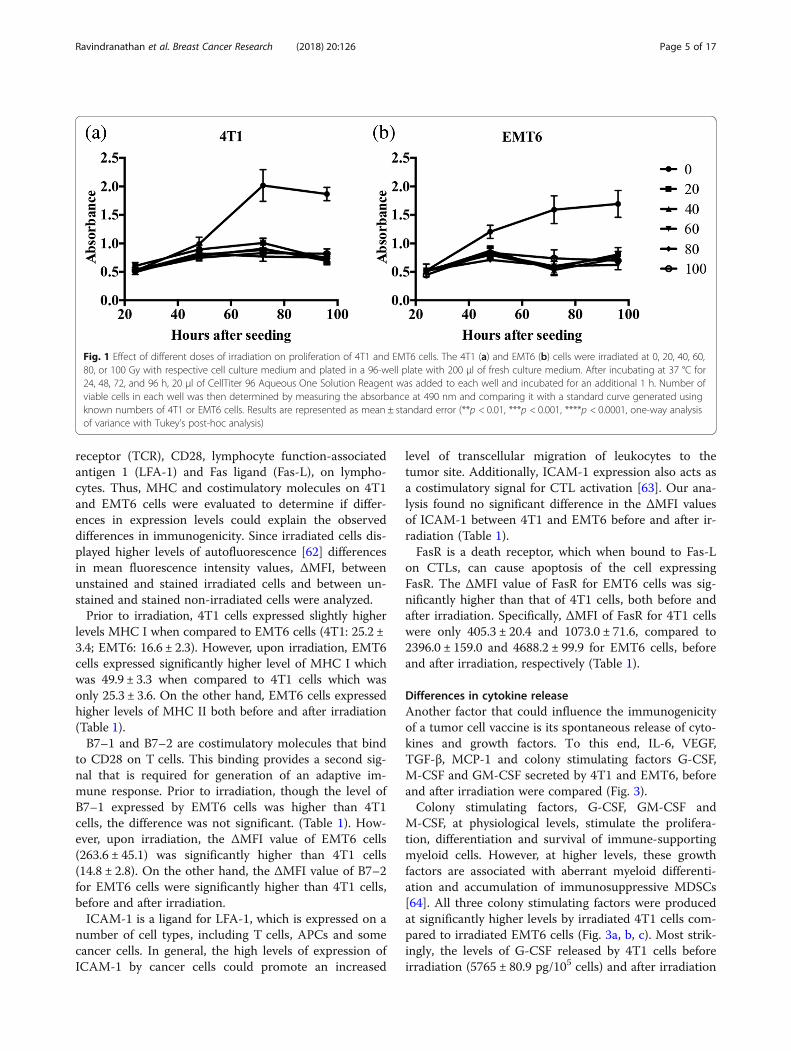
ResultsEffect of irradiation on proliferation of breast cancer cellsPrior to using irradiated 4T1 or EMT6 cells as tumor cellvaccines, an appropriate dose of irradiation that effectivelyprevents tumor cell proliferation was determined using anin-vitro proliferation assay. In the absence of irradiation,both 4T1 and EMT6 cells effectively proliferated over thetime observed in this study (24–96 h). However, in thepresence of varying doses of irradiation (20–100 Gy),there was no significant difference in viable cell numbersduring the study period (Fig. 1).
Immunogenicity of murine breast carcinoma linesA standard prophylactic vaccine model was used to evalu-ate the immunogenicities of 4T1 and EMT6 cells. Micewere vaccinated twice with either irradiated 4T1 or EMT6cells and challenged with live 4T1 or EMT6 cells, respect-ively. While all mice in both 4T1 and EMT6 controlgroups developed tumors, upon vaccination, some of themice in the EMT6 vaccinated group did not develop anytumor. Moreover, the mice that developed tumors in theEMT6 vaccinated group, showed a delayed tumor inci-dence when compared to the EMT6 control (Fig. 2a). Inboth groups, vaccinated mice exhibited some level of pro-tective immunity as demonstrated by extended survivalcompared to mice in unvaccinated control groups (Fig. 2b).However, EMT6 vaccinated mice exhibited higher overallsurvival with a p value < 0.01 when compared to EMT6control. On the other hand, 4T1 vaccinated mice had a pvalue < 0.05 when compared to 4T1 control. Additionally,while all mice in the 4T1 vaccinated group developed andsuccumbed to tumors within 50 days of tumor inocula-tion, 75% of the mice in the EMT6 vaccinated group aretumor free survivors.
Costimulatory molecule and MHC expression on breastcancer cell linesThe elaboration of robust adaptive immunity requiresantigen presentation in MHC I or MHC II complexes(signal 1) and simultaneous engagement of costimulatorymolecules (signal 2), such as B7–1, B7–2, ICAM-1 andFasR, on APCs, with their cognate receptors, i.e. T cell
Ravindranathan et al. Breast Cancer Research (2018) 20:126 Page 4 of 17
receptor (TCR), CD28, lymphocyte function-associatedantigen 1 (LFA-1) and Fas ligand (Fas-L), on lympho-cytes. Thus, MHC and costimulatory molecules on 4T1and EMT6 cells were evaluated to determine if differ-ences in expression levels could explain the observeddifferences in immunogenicity. Since irradiated cells dis-played higher levels of autofluorescence [62] differencesin mean fluorescence intensity values, ΔMFI, betweenunstained and stained irradiated cells and between un-stained and stained non-irradiated cells were analyzed.Prior to irradiation, 4T1 cells expressed slightly higher
levels MHC I when compared to EMT6 cells (4T1: 25.2 ±3.4; EMT6: 16.6 ± 2.3). However, upon irradiation, EMT6cells expressed significantly higher level of MHC I whichwas 49.9 ± 3.3 when compared to 4T1 cells which wasonly 25.3 ± 3.6. On the other hand, EMT6 cells expressedhigher levels of MHC II both before and after irradiation(Table 1).B7–1 and B7–2 are costimulatory molecules that bind
to CD28 on T cells. This binding provides a second sig-nal that is required for generation of an adaptive im-mune response. Prior to irradiation, though the level ofB7–1 expressed by EMT6 cells was higher than 4T1cells, the difference was not significant. (Table 1). How-ever, upon irradiation, the ΔMFI value of EMT6 cells(263.6 ± 45.1) was significantly higher than 4T1 cells(14.8 ± 2.8). On the other hand, the ΔMFI value of B7–2for EMT6 cells were significantly higher than 4T1 cells,before and after irradiation.ICAM-1 is a ligand for LFA-1, which is expressed on a
number of cell types, including T cells, APCs and somecancer cells. In general, the high levels of expression ofICAM-1 by cancer cells could promote an increased
level of transcellular migration of leukocytes to thetumor site. Additionally, ICAM-1 expression also acts asa costimulatory signal for CTL activation [63]. Our ana-lysis found no significant difference in the ΔMFI valuesof ICAM-1 between 4T1 and EMT6 before and after ir-radiation (Table 1).FasR is a death receptor, which when bound to Fas-L
on CTLs, can cause apoptosis of the cell expressingFasR. The ΔMFI value of FasR for EMT6 cells was sig-nificantly higher than that of 4T1 cells, both before andafter irradiation. Specifically, ΔMFI of FasR for 4T1 cellswere only 405.3 ± 20.4 and 1073.0 ± 71.6, compared to2396.0 ± 159.0 and 4688.2 ± 99.9 for EMT6 cells, beforeand after irradiation, respectively (Table 1).
Differences in cytokine releaseAnother factor that could influence the immunogenicityof a tumor cell vaccine is its spontaneous release of cyto-kines and growth factors. To this end, IL-6, VEGF,TGF-β, MCP-1 and colony stimulating factors G-CSF,M-CSF and GM-CSF secreted by 4T1 and EMT6, beforeand after irradiation were compared (Fig. 3).Colony stimulating factors, G-CSF, GM-CSF and
M-CSF, at physiological levels, stimulate the prolifera-tion, differentiation and survival of immune-supportingmyeloid cells. However, at higher levels, these growthfactors are associated with aberrant myeloid differenti-ation and accumulation of immunosuppressive MDSCs[64]. All three colony stimulating factors were producedat significantly higher levels by irradiated 4T1 cells com-pared to irradiated EMT6 cells (Fig. 3a, b, c). Most strik-ingly, the levels of G-CSF released by 4T1 cells beforeirradiation (5765 ± 80.9 pg/105 cells) and after irradiation
Fig. 1 Effect of different doses of irradiation on proliferation of 4T1 and EMT6 cells. The 4T1 (a) and EMT6 (b) cells were irradiated at 0, 20, 40, 60,80, or 100 Gy with respective cell culture medium and plated in a 96-well plate with 200 μl of fresh culture medium. After incubating at 37 °C for24, 48, 72, and 96 h, 20 μl of CellTiter 96 Aqueous One Solution Reagent was added to each well and incubated for an additional 1 h. Number ofviable cells in each well was then determined by measuring the absorbance at 490 nm and comparing it with a standard curve generated usingknown numbers of 4T1 or EMT6 cells. Results are represented as mean ± standard error (**p < 0.01, ***p < 0.001, ****p < 0.0001, one-way analysisof variance with Tukey’s post-hoc analysis)
Ravindranathan et al. Breast Cancer Research (2018) 20:126 Page 5 of 17
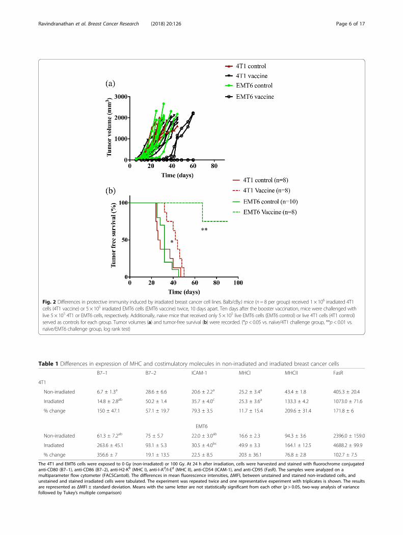
Fig. 2 Differences in protective immunity induced by irradiated breast cancer cell lines. Balb/cByJ mice (n= 8 per group) received 1 × 10⁶ irradiated 4T1cells (4T1 vaccine) or 5 × 10⁵ irradiated EMT6 cells (EMT6 vaccine) twice, 10 days apart. Ten days after the booster vaccination, mice were challenged withlive 5 × 10⁵ 4T1 or EMT6 cells, respectively. Additionally, naive mice that received only 5 × 10⁵ live EMT6 cells (EMT6 control) or live 4T1 cells (4T1 control)served as controls for each group. Tumor volumes (a) and tumor-free survival (b) were recorded. (*p< 0.05 vs. naïve/4T1 challenge group, **p< 0.01 vs.naïve/EMT6 challenge group, log rank test)
Table 1 Differences in expression of MHC and costimulatory molecules in non-irradiated and irradiated breast cancer cells
B7–1 B7–2 ICAM-1 MHCI MHCII FasR
4T1
Non-irradiated 6.7 ± 1.3a 28.6 ± 6.6 20.6 ± 2.2a 25.2 ± 3.4a 43.4 ± 1.8 405.3 ± 20.4
Irradiated 14.8 ± 2.8ab 50.2 ± 1.4 35.7 ± 4.0c 25.3 ± 3.6a 133.3 ± 4.2 1073.0 ± 71.6
% change 150 ± 47.1 57.1 ± 19.7 79.3 ± 3.5 11.7 ± 15.4 209.6 ± 31.4 171.8 ± 6
EMT6
Non-irradiated 61.3 ± 7.2ab 75 ± 5.7 22.0 ± 3.0ab 16.6 ± 2.3 94.3 ± 3.6 2396.0 ± 159.0
Irradiated 263.6 ± 45.1 93.1 ± 5.3 30.5 ± 4.0bc 49.9 ± 3.3 164.1 ± 12.5 4688.2 ± 99.9
% change 356.6 ± 7 19.1 ± 13.5 22.5 ± 8.5 203 ± 36.1 76.8 ± 2.8 102.7 ± 7.5
The 4T1 and EMT6 cells were exposed to 0 Gy (non-irradiated) or 100 Gy. At 24 h after irradiation, cells were harvested and stained with fluorochrome conjugatedanti-CD80 (B7–1), anti-CD86 (B7–2), anti-H2-Kb (MHC I), anti-I-Ad/I-Ed (MHC II), anti-CD54 (ICAM-1), and anti-CD95 (FasR). The samples were analyzed on amultiparameter flow cytometer (FACSCantoII). The differences in mean fluorescence intensities, ΔMFI, between unstained and stained non-irradiated cells, andunstained and stained irradiated cells were tabulated. The experiment was repeated twice and one representative experiment with triplicates is shown. The resultsare represented as ΔMFI ± standard deviation. Means with the same letter are not statistically significant from each other (p > 0.05, two-way analysis of variancefollowed by Tukey’s multiple comparison)
Ravindranathan et al. Breast Cancer Research (2018) 20:126 Page 6 of 17
(5334 ± 114.2 pg/105 cells) were exceptionally high evenwhen compared to the levels released by EMT6 cells be-fore (1100 ± 98.84 pg/105 cells) and after irradiation(1760 ± 145.1 pg/105 cells) (Fig. 3a).IL-6 has been shown to exhibit both pro- and
anti-tumor activities. Among its suppressive activities,IL-6 directly promotes cancer cell proliferation, survivaland metastasis while indirectly supporting angiogenesisin the tumor microenvironment [65]. Here, IL-6 was re-leased at modest levels with no major changes before ir-radiation (4T1: 62.6 ± 8.4 pg/105 cells; EMT6: 25.6 ±3.7 pg/105 cells) or after irradiation (4T1: 21.3 ± 1.8 pg/105 cells; EMT6: 44.3 ± 6.6 pg/105 cells) (Fig. 3d).Tumor-derived MCP-1 promotes infiltration of monocytes
and macrophages. MCP-1 as well as VEGF are associatedwith promoting angiogenesis [66, 67]. Additionally, tumorsecretion of VEGF blocks normal myeloid differentiation,resulting in MDSC accumulation [68, 69]. 4T1 cells pro-duced higher levels of MCP-1 only after irradiation (4T1:1596 ± 123.6 pg/105 cells; EMT6: 744.7 ± 58.91 pg/105 cells)(Fig. 3e). On the contrary, EMT6 cells produced higher levelsof VEGF before (833 ± 41.19 pg/105 cells) and after (371.3 ±8.09 pg/105 cells) irradiation, when compared to 4T1 cellsbefore (10 ± 1.1 pg/105 cells) and after (8.6 ± 0.6 pg/105 cells)(Fig. 3f).TGF-β, an immunosuppressive cytokine that plays a
role in the induction of Tregs, was produced at higherlevels by EMT6 cells before irradiation (4T1: 108 ±7.6 pg/105 cells; EMT6: 832 ± 49 pg/105 cells) (Fig. 3g).However, upon irradiation, the difference between celllines was not statistically significant (4T1: 355 ± 22.1 pg/105 cells; EMT6: 274 ± 17 pg/105 cells) (Fig. 3g).
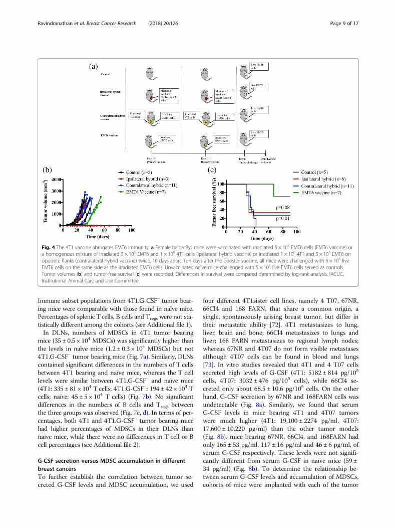
Local and systemic effects of 4T1 mediatedimmunosuppressionBased on differences in cytokine release (Fig. 3), we ex-plored if immunosuppressive cytokines released by 4T1cells would abrogate the protective immunity establishedby the irradiated EMT6 vaccine. To explore the potentialfor localized immune suppression, mice were immunizedwith a heterogeneous mixture of irradiated 4T1 andEMT6 cells (ipsilateral hybrid vaccine). To explore pos-sible systemic immune suppression mediated by 4T1 cells,mice were vaccinated with irradiated 4T1 cells and irradi-ated EMT6 cells on opposite flanks (contralateral hybridvaccine). The efficacy of these vaccines was comparedusing mice immunized with irradiated EMT6 cells alone(EMT6 vaccine). The study design is shown in Fig. 4a.When all groups of mice were challenged with live
EMT6 cells, unlike the EMT6 vaccine group, both ipsilat-eral and contralateral hybrid vaccine group did not exhibita delayed tumor incidence (Fig. 4b). Additionally, the pres-ence of irradiated 4T1 cells in the hybrid vaccines dimin-ished the protective immunity induced by irradiated
EMT6 cells, as the percentage of tumor free survival inthe ipsilateral and contralateral vaccine groups were onlyabout 34% and 27%, respectively (Fig. 4c). The long term,tumor-free survival for mice receiving the EMT6 vaccinealone was significantly higher at 71%.
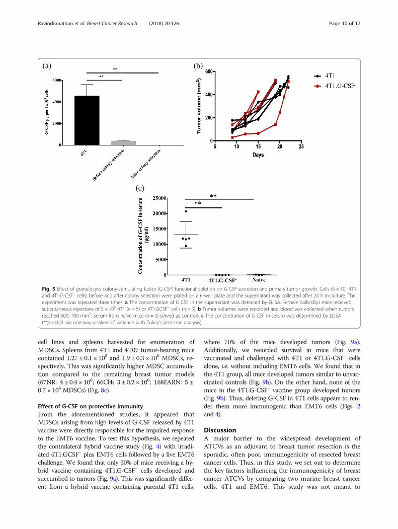
The immunosuppressive role of G-CSFDue to the exceptionally high levels of G-CSF producedby 4T1 cells with and without irradiation, we hypothe-sized that it played a key role in inhibiting the efficacy ofipsilateral and contralateral vaccines. To test this hy-pothesis, we functionally deleted the G-CSF gene viaCRISPR/Cas9 genomic editing.4T1 cells before G-CSF deletion released 4550 ±
604 pg G-CSF per 105 cells, whereas after G-CSF dele-tion, cells only released 386 ± 31 pg G-CSF/105 cells.Clonal selection led to the propagation of a 4T1 colony,called 4T1.G-CSF−, that released lower than detectablelevels of G-CSF in vitro (Fig. 5a). The G-CSF deletiondid not affect tumor establishment or growth in vivo(Fig. 5b). To further verify G-CSF ablation, G-CSF serumconcentrations were assessed in mice bearing 4T1.G-CSF−
tumors when tumor volumes reached 500–700 mm3.4T1.G-CSF− tumor bearing mice contained only 10 ±2.9 pg/ml of G-CSF in their sera, which was comparableto G-CSF in the sera of naïve mice (59 ± 34 pg/ml). Onthe other hand, mice with similarly sized, unmodified 4T1tumors contained 13,096 ± 1947 pg/ml G-CSF in theirsera (Fig. 5c).Additionally, spleens and DLNs from 4T1 and
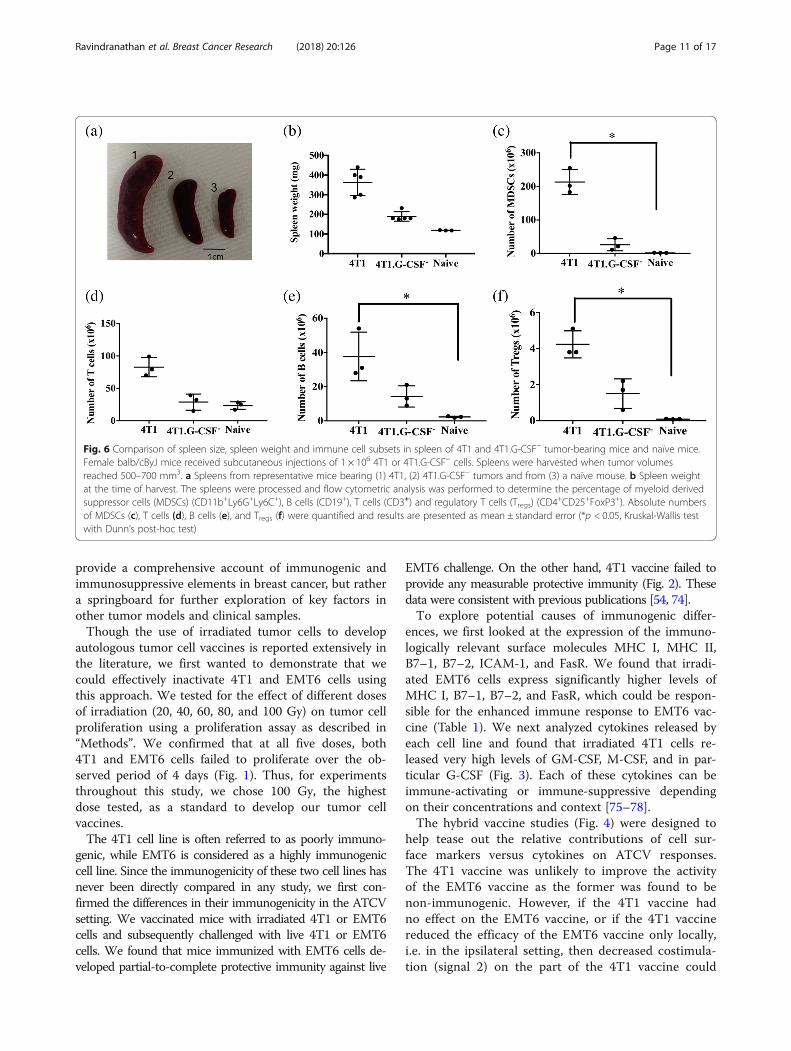
4T1.G-CSF− tumor bearing mice were harvested to de-termine the frequency of T cells, B cells, MDSCs andTregs in each lymphoid tissue. MDSCs were of particularinterest as immature myeloid cells have often been associ-ated with high levels of colony stimulating factors [70, 71].Prior to immunophenotyping we noted remarkable differ-ences in the sizes and weights of spleens removed from micebearing 4T1 tumors, 4T1.GCSF− tumors or no tumors(Fig. 6a, b). G-CSF appeared to be driving the extremesplenomegaly observed in 4T1-bearing mice. In addition,spleens from 4T1 tumor bearing mice contained significantlyhigher levels of MDSCs (213 ± 21 × 106 MDSCs) when com-pared to naive (1.7 ± 0.3 × 10⁶ MDSCs) spleens. On the otherhand, there was no significant difference between the levelsof MDSCs in naïve and 4T1.G-CSF− tumor bearing mice(26 ± 10 × 106 MDSCs)(Fig. 6c). The same trends held whenanalyzing the percentages of MDSCs in the spleens of micefrom the three groups (see Additional file 1).Though there was no significant difference in the number
of T cells in the spleens of mice among the different groups,there was a significant difference in the number of B cellsand Tregs between 4T1 tumor bearing and naïve mice (4T1:37 ± 8 × 10⁶ B cells and 4.2 ± 0.5 × 10⁶ Tregs; naïve: 2.3 ±0.4 × 10⁶ B cells and 0.08 ± 0.01 × 10⁶ Tregs) (Fig. 6d, e, f).
Ravindranathan et al. Breast Cancer Research (2018) 20:126 Page 7 of 17
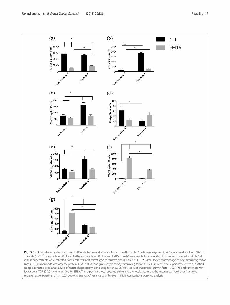
Fig. 3 Cytokine release profile of 4T1 and EMT6 cells before and after irradiation. The 4T1 or EMT6 cells were exposed to 0 Gy (non-irradiated) or 100 Gy.The cells (5 × 10⁵ non-irradiated (4T1 and EMT6) and irradiated (4T1 Irr and EMT6 Irr) cells) were seeded on separate T25 flasks and cultured for 48 h. Cellculture supernatants were collected from each flask and centrifuged to remove debris. Levels of IL-6 (a), granulocyte-macrophage colony-stimulating factor(GM-CSF) (b), monocyte chemotactic protein-1 (MCP-1) (c), and granulocyte colony-stimulating factor (G-CSF) (d) in cell-free supernatants were quantifiedusing cytometric bead array. Levels of macrophage colony-stimulating factor (M-CSF) (e), vascular endothelial growth factor (VEGF) (f), and tumor growthfactor-beta (TGF-β) (g) were quantified by ELISA. The experiment was repeated thrice and the results represent the mean ± standard error from onerepresentative experiment (*p< 0.05, two-way analysis of variance with Tukey’s multiple comparisons post-hoc analysis)
Ravindranathan et al. Breast Cancer Research (2018) 20:126 Page 8 of 17
Immune subset populations from 4T1.G-CSF− tumor bear-ing mice were comparable with those found in naïve mice.Percentages of splenic Tcells, B cells and Tregs were not sta-tistically different among the cohorts (see Additional file 1).In DLNs, numbers of MDSCs in 4T1 tumor bearing
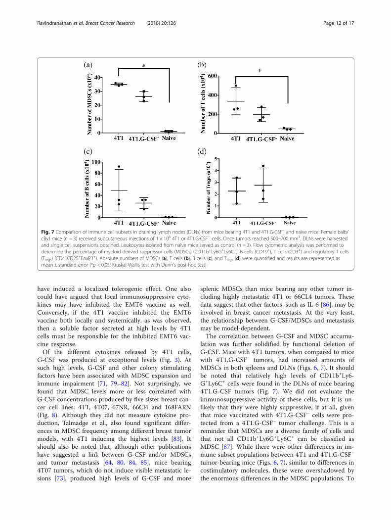
mice (35 ± 0.5 × 10⁴ MDSCs) was significantly higher thanthe levels in naïve mice (1.2 ± 0.3 × 10⁴ MDSCs) but not4T1.G-CSF− tumor bearing mice (Fig. 7a). Similarly, DLNscontained significant differences in the numbers of T cellsbetween 4T1 bearing and naïve mice, whereas the T celllevels were similar between 4T1.G-CSF− and naïve mice(4T1: 335 ± 81 × 104 T cells; 4T1.G-CSF−: 194 ± 42 × 104 Tcells; naïve: 45 ± 5 × 10⁴ T cells) (Fig. 7b). No significantdifferences in the numbers of B cells and Tregs betweenthe three groups was observed (Fig. 7c, d). In terms of per-centages, both 4T1 and 4T1.G-CSF− tumor bearing micehad higher percentages of MDSCs in their DLNs thannaïve mice, while there were no differences in T cell or Bcell percentages (see Additional file 2).
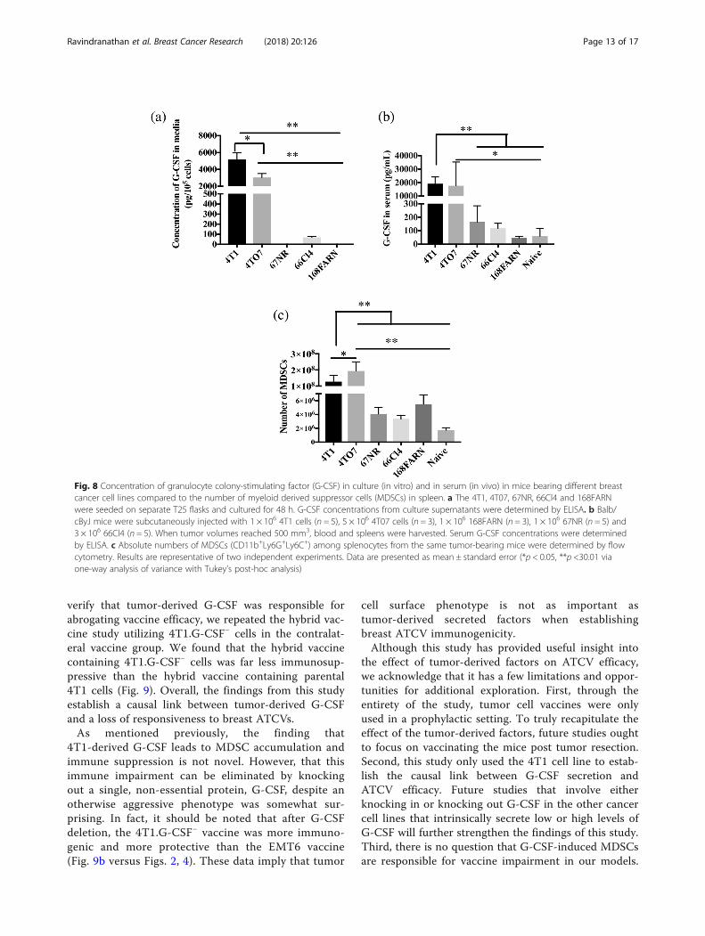
G-CSF secretion versus MDSC accumulation in differentbreast cancersTo further establish the correlation between tumor se-creted G-CSF levels and MDSC accumulation, we used
four different 4T1sister cell lines, namely 4 T07, 67NR,66Cl4 and 168 FARN, that share a common origin, asingle, spontaneously arising breast tumor, but differ intheir metastatic ability [72]. 4T1 metastasizes to lung,liver, brain and bone; 66Cl4 metastasizes to lungs andliver; 168 FARN metastasizes to regional lymph nodes;whereas 67NR and 4T07 do not form visible metastasesalthough 4T07 cells can be found in blood and lungs[73]. In vitro studies revealed that 4T1 and 4 T07 cellssecreted high levels of G-CSF (4T1: 5182 ± 814 pg/105
cells, 4T07: 3032 ± 476 pg/105 cells), while 66Cl4 se-creted only about 68.5 ± 10.6 pg/105 cells. On the otherhand, G-CSF secretion by 67NR and 168FARN cells wasundetectable (Fig. 8a). Similarly, we found that serumG-CSF levels in mice bearing 4T1 and 4T07 tumorswere much higher (4T1: 19,100 ± 2274 pg/ml, 4T07:17,600 ± 10,220 pg/ml) than the other tumor models(Fig. 8b). mice bearing 67NR, 66Cl4, and 168FARN hadonly 165 ± 53 pg/ml, 117 ± 16 pg/ml and 46 ± 6 pg/ml, ofserum G-CSF respectively. These levels were not signifi-cantly different from serum G-CSF in naïve mice (59 ±34 pg/ml) (Fig. 8b). To determine the relationship be-tween serum G-CSF levels and accumulation of MDSCs,cohorts of mice were implanted with each of the tumor
Fig. 4 The 4T1 vaccine abrogates EMT6 immunity. a Female balb/cByJ mice were vaccinated with irradiated 5 × 10⁵ EMT6 cells (EMT6 vaccine) ora homogenous mixture of irradiated 5 × 10⁵ EMT6 and 1 × 10⁶ 4T1 cells (ipsilateral hybrid vaccine) or irradiated 1 × 10⁶ 4T1 and 5 × 10⁵ EMT6 onopposite flanks (contralateral hybrid vaccine) twice, 10 days apart. Ten days after the booster vaccine, all mice were challenged with 5 × 10⁵ liveEMT6 cells on the same side as the irradiated EMT6 cells. Unvaccinated naïve mice challenged with 5 × 10⁵ live EMT6 cells served as controls.Tumor volumes (b) and tumor-free survival (c) were recorded. Differences in survival were compared determined by log-rank analysis. IACUC,Institutional Animal Care and Use Committee
Ravindranathan et al. Breast Cancer Research (2018) 20:126 Page 9 of 17
cell lines and spleens harvested for enumeration ofMDSCs. Spleens from 4T1 and 4T07 tumor-bearing micecontained 1.27 ± 0.1 × 10⁸ and 1.9 ± 0.3 × 10⁸ MDSCs, re-spectively. This was significantly higher MDSC accumula-tion compared to the remaining breast tumor models(67NR: 4 ± 0.4 × 10⁶; 66Cl4: 3 ± 0.2 × 10⁶; 168FARN: 5 ±0.7 × 10⁶ MDSCs) (Fig. 8c).
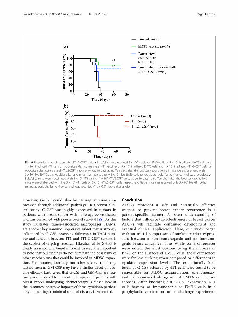
Effect of G-CSF on protective immunityFrom the aforementioned studies, it appeared thatMDSCs arising from high levels of G-CSF released by 4T1vaccine were directly responsible for the impaired responseto the EMT6 vaccine. To test this hypothesis, we repeatedthe contralateral hybrid vaccine study (Fig. 4) with irradi-ated 4T1.GCSF− plus EMT6 cells followed by a live EMT6challenge. We found that only 30% of mice receiving a hy-brid vaccine containing 4T1.G-CSF− cells developed andsuccumbed to tumors (Fig. 9a). This was significantly differ-ent from a hybrid vaccine containing parental 4T1 cells,
where 70% of the mice developed tumors (Fig. 9a).Additionally, we recorded survival in mice that werevaccinated and challenged with 4T1 or 4T1.G-CSF− cellsalone, i.e. without including EMT6 cells. We found that inthe 4T1 group, all mice developed tumors similar to unvac-cinated controls (Fig. 9b). On the other hand, none of themice in the 4T1.G-CSF− vaccine group developed tumors(Fig. 9b). Thus, deleting G-CSF in 4T1 cells appears to ren-der them more immunogenic than EMT6 cells (Figs. 2and 4).
DiscussionA major barrier to the widespread development ofATCVs as an adjuvant to breast tumor resection is thesporadic, often poor, immunogenicity of resected breastcancer cells. Thus, in this study, we set out to determinethe key factors influencing the immunogenicity of breastcancer ATCVs by comparing two murine breast cancercells, 4T1 and EMT6. This study was not meant to
Fig. 5 Effect of granulocyte colony-stimulating factor (G-CSF) functional deletion on G-CSF secretion and primary tumor growth. Cells (5 × 10⁵ 4T1and 4T1.G-CSF− cells) before and after colony selection were plated on a 6-well plate and the supernatant was collected after 24 h in culture. Theexperiment was repeated three times. a The concentration of G-CSF in the supernatant was detected by ELISA. Female balb/cByJ mice receivedsubcutaneous injections of 5 × 10⁵ 4T1 (n = 5) or 4T1.GCSF− cells (n = 5). b Tumor volumes were recorded and blood was collected when tumorsreached 500–700 mm3. Serum from naïve mice (n = 3) served as controls. c The concentration of G-CSF in serum was determined by ELISA(**p < 0.01 via one-way analysis of variance with Tukey’s post-hoc analysis)
Ravindranathan et al. Breast Cancer Research (2018) 20:126 Page 10 of 17
provide a comprehensive account of immunogenic andimmunosuppressive elements in breast cancer, but rathera springboard for further exploration of key factors inother tumor models and clinical samples.Though the use of irradiated tumor cells to develop
autologous tumor cell vaccines is reported extensively inthe literature, we first wanted to demonstrate that wecould effectively inactivate 4T1 and EMT6 cells usingthis approach. We tested for the effect of different dosesof irradiation (20, 40, 60, 80, and 100 Gy) on tumor cellproliferation using a proliferation assay as described in“Methods”. We confirmed that at all five doses, both4T1 and EMT6 cells failed to proliferate over the ob-served period of 4 days (Fig. 1). Thus, for experimentsthroughout this study, we chose 100 Gy, the highestdose tested, as a standard to develop our tumor cellvaccines.The 4T1 cell line is often referred to as poorly immuno-
genic, while EMT6 is considered as a highly immunogeniccell line. Since the immunogenicity of these two cell lines hasnever been directly compared in any study, we first con-firmed the differences in their immunogenicity in the ATCVsetting. We vaccinated mice with irradiated 4T1 or EMT6cells and subsequently challenged with live 4T1 or EMT6cells. We found that mice immunized with EMT6 cells de-veloped partial-to-complete protective immunity against live
EMT6 challenge. On the other hand, 4T1 vaccine failed toprovide any measurable protective immunity (Fig. 2). Thesedata were consistent with previous publications [54, 74].To explore potential causes of immunogenic differ-
ences, we first looked at the expression of the immuno-logically relevant surface molecules MHC I, MHC II,B7–1, B7–2, ICAM-1, and FasR. We found that irradi-ated EMT6 cells express significantly higher levels ofMHC I, B7–1, B7–2, and FasR, which could be respon-sible for the enhanced immune response to EMT6 vac-cine (Table 1). We next analyzed cytokines released byeach cell line and found that irradiated 4T1 cells re-leased very high levels of GM-CSF, M-CSF, and in par-ticular G-CSF (Fig. 3). Each of these cytokines can beimmune-activating or immune-suppressive dependingon their concentrations and context [75–78].The hybrid vaccine studies (Fig. 4) were designed to
help tease out the relative contributions of cell sur-face markers versus cytokines on ATCV responses.The 4T1 vaccine was unlikely to improve the activityof the EMT6 vaccine as the former was found to benon-immunogenic. However, if the 4T1 vaccine hadno effect on the EMT6 vaccine, or if the 4T1 vaccinereduced the efficacy of the EMT6 vaccine only locally,i.e. in the ipsilateral setting, then decreased costimula-tion (signal 2) on the part of the 4T1 vaccine could
Fig. 6 Comparison of spleen size, spleen weight and immune cell subsets in spleen of 4T1 and 4T1.G-CSF− tumor-bearing mice and naïve mice.Female balb/cByJ mice received subcutaneous injections of 1 × 10⁶ 4T1 or 4T1.G-CSF− cells. Spleens were harvested when tumor volumesreached 500–700 mm3. a Spleens from representative mice bearing (1) 4T1, (2) 4T1.G-CSF− tumors and from (3) a naïve mouse. b Spleen weightat the time of harvest. The spleens were processed and flow cytometric analysis was performed to determine the percentage of myeloid derivedsuppressor cells (MDSCs) (CD11b+Ly6G+Ly6C+), B cells (CD19+), T cells (CD3+) and regulatory T cells (Tregs) (CD4
+CD25+FoxP3+). Absolute numbersof MDSCs (c), T cells (d), B cells (e), and Tregs (f) were quantified and results are presented as mean ± standard error (*p < 0.05, Kruskal-Wallis testwith Dunn’s post-hoc test)
Ravindranathan et al. Breast Cancer Research (2018) 20:126 Page 11 of 17
have induced a localized tolerogenic effect. One alsocould have argued that local immunosuppressive cyto-kines may have inhibited the EMT6 vaccine as well.Conversely, if the 4T1 vaccine inhibited the EMT6vaccine both locally and systemically, as was observed,then a soluble factor secreted at high levels by 4T1cells must be responsible for the inhibited EMT6 vac-cine response.Of the different cytokines released by 4T1 cells,
G-CSF was produced at exceptional levels (Fig. 3). Atsuch high levels, G-CSF and other colony stimulatingfactors have been associated with MDSC expansion andimmune impairment [71, 79–82]. Not surprisingly, wefound that MDSC levels more or less correlated withG-CSF concentrations produced by five sister breast can-cer cell lines: 4T1, 4T07, 67NR, 66Cl4 and 168FARN(Fig. 8). Although they did not measure cytokine pro-duction, Talmadge et al., also found significant differ-ences in MDSC frequency among different breast tumormodels, with 4T1 inducing the highest levels [83]. Itshould also be noted that, although other publicationshave suggested a link between G-CSF and/or MDSCsand tumor metastasis [64, 80, 84, 85], mice bearing4T07 tumors, which do not induce visible metastatic le-sions [73], produced high levels of G-CSF and more
splenic MDSCs than mice bearing any other tumor in-cluding highly metastatic 4T1 or 66CL4 tumors. Thesedata suggest that other factors, such as IL-6 [86], may beinvolved in breast cancer metastasis. At the very least,the relationship between G-CSF/MDSCs and metastasismay be model-dependent.The correlation between G-CSF and MDSC accumu-
lation was further solidified by functional deletion ofG-CSF. Mice with 4T1 tumors, when compared to micewith 4T1.G-CSF− tumors, had increased amounts ofMDSCs in both spleens and DLNs (Figs. 6, 7). It shouldbe noted that relatively high levels of CD11b+Ly6-G+Ly6C+ cells were found in the DLNs of mice bearing4T1.G-CSF tumors (Fig. 7). We did not evaluate theimmunosuppressive activity of these cells, but it is un-likely that they were highly suppressive, if at all, giventhat mice vaccinated with 4T1.G-CSF− cells were pro-tected from a 4T1.G-CSF− tumor challenge. This is areminder that MDSCs are a diverse family of cells andthat not all CD11b+Ly6G+Ly6C+ can be classified asMDSC [87]. While there were other differences in im-mune subset populations between 4T1 and 4T1.G-CSF−
tumor-bearing mice (Figs. 6, 7), similar to differences incostimulatory molecules, these were overshadowed bythe enormous differences in the MDSC populations. To
Fig. 7 Comparison of immune cell subsets in draining lymph nodes (DLNs) from mice bearing 4T1 and 4T1.G-CSF− and naïve mice. Female balb/cByJ mice (n = 3) received subcutaneous injections of 1 × 10⁶ 4T1 or 4T1.G-CSF− cells. Once tumors reached 500–700 mm3, DLNs were harvestedand single cell suspensions obtained. Leukocytes isolated from naïve mice served as control (n = 3). Flow cytometric analysis was performed todetermine the percentage of myeloid derived suppressor cells (MDSCs) (CD11b+Ly6G+Ly6C+), B cells (CD19+), T cells (CD3+) and regulatory T cells(Tregs) (CD4
+CD25+FoxP3+). Absolute numbers of MDSCs (a), T cells (b), B cells (c), and Tregs (d) were quantified and results are represented asmean ± standard error (*p < 0.05, Kruskal-Wallis test with Dunn’s post-hoc test)
Ravindranathan et al. Breast Cancer Research (2018) 20:126 Page 12 of 17
verify that tumor-derived G-CSF was responsible forabrogating vaccine efficacy, we repeated the hybrid vac-cine study utilizing 4T1.G-CSF− cells in the contralat-eral vaccine group. We found that the hybrid vaccinecontaining 4T1.G-CSF− cells was far less immunosup-pressive than the hybrid vaccine containing parental4T1 cells (Fig. 9). Overall, the findings from this studyestablish a causal link between tumor-derived G-CSFand a loss of responsiveness to breast ATCVs.As mentioned previously, the finding that
4T1-derived G-CSF leads to MDSC accumulation andimmune suppression is not novel. However, that thisimmune impairment can be eliminated by knockingout a single, non-essential protein, G-CSF, despite anotherwise aggressive phenotype was somewhat sur-prising. In fact, it should be noted that after G-CSFdeletion, the 4T1.G-CSF− vaccine was more immuno-genic and more protective than the EMT6 vaccine(Fig. 9b versus Figs. 2, 4). These data imply that tumor
cell surface phenotype is not as important astumor-derived secreted factors when establishingbreast ATCV immunogenicity.Although this study has provided useful insight into
the effect of tumor-derived factors on ATCV efficacy,we acknowledge that it has a few limitations and oppor-tunities for additional exploration. First, through theentirety of the study, tumor cell vaccines were onlyused in a prophylactic setting. To truly recapitulate theeffect of the tumor-derived factors, future studies oughtto focus on vaccinating the mice post tumor resection.Second, this study only used the 4T1 cell line to estab-lish the causal link between G-CSF secretion andATCV efficacy. Future studies that involve eitherknocking in or knocking out G-CSF in the other cancercell lines that intrinsically secrete low or high levels ofG-CSF will further strengthen the findings of this study.Third, there is no question that G-CSF-induced MDSCsare responsible for vaccine impairment in our models.
Fig. 8 Concentration of granulocyte colony-stimulating factor (G-CSF) in culture (in vitro) and in serum (in vivo) in mice bearing different breastcancer cell lines compared to the number of myeloid derived suppressor cells (MDSCs) in spleen. a The 4T1, 4T07, 67NR, 66Cl4 and 168FARNwere seeded on separate T25 flasks and cultured for 48 h. G-CSF concentrations from culture supernatants were determined by ELISA. b Balb/cByJ mice were subcutaneously injected with 1 × 10⁶ 4T1 cells (n = 5), 5 × 10⁶ 4T07 cells (n = 3), 1 × 10⁶ 168FARN (n = 3), 1 × 10⁶ 67NR (n = 5) and3 × 106 66Cl4 (n = 5). When tumor volumes reached 500 mm3, blood and spleens were harvested. Serum G-CSF concentrations were determinedby ELISA. c Absolute numbers of MDSCs (CD11b+Ly6G+Ly6C+) among splenocytes from the same tumor-bearing mice were determined by flowcytometry. Results are representative of two independent experiments. Data are presented as mean ± standard error (*p < 0.05, **p <30.01 viaone-way analysis of variance with Tukey’s post-hoc analysis)
Ravindranathan et al. Breast Cancer Research (2018) 20:126 Page 13 of 17
However, G-CSF could also be causing immune sup-pression through additional pathways. In a recent clin-ical study, G-CSF was highly expressed in tumors inpatients with breast cancer with more aggressive diseaseand was correlated with poorer overall survival [88]. As thisstudy illustrates, tumor-associated macrophages (TAMs)are another key immunosuppressive subset that is stronglyinfluenced by G-CSF. Assessing differences in TAM num-ber and function between 4T1 and 4T1.G-CSF− tumors isthe subject of ongoing research. Likewise, while G-CSF isclearly an important target in breast cancer, it is importantto note that our findings do not eliminate the possibility ofother mechanisms that could be involved in MDSC expan-sion. For instance, knocking out other colony stimulatingfactors such as GM-CSF may have a similar effect on vac-cine efficacy. Last, given that G-CSF and GM-CSF are rou-tinely administered to prevent neutropenia in patients withbreast cancer undergoing chemotherapy, a closer look atthe immunosuppressive impacts of these cytokines, particu-larly in a setting of minimal residual disease, is warranted.
ConclusionATCVs represent a safe and potentially effectiveweapon to prevent breast cancer recurrence in apatient-specific manner. A better understanding offactors that influence the effectiveness of breast cancerATCVs will facilitate continued development andeventual clinical application. Here, our study beganwith an initial comparison of surface marker expres-sion between a non-immunogenic and an immuno-genic breast cancer cell line. While some differenceswere noted, the most obvious being the increase inB7–1 on the surfaces of EMT6 cells, these differenceswere far less striking when compared to differences incytokine expression levels. The exceptionally highlevels of G-CSF released by 4T1 cells were found to beresponsible for MDSC accumulation, splenomegaly,and the associated abrogation of EMT6 vaccine re-sponses. After knocking out G-CSF expression, 4T1cells became as immunogenic as EMT6 cells in aprophylactic vaccination-tumor challenge experiment.
Fig. 9 Prophylactic vaccination with 4T1.G-CSF− cells. a Balb/cByJ mice received 5 × 10⁵ irradiated EMT6 cells or 5 × 10⁵ irradiated EMT6 cells and1 × 106 irradiated 4T1 cells on opposite sides (contralateral 4T1 vaccine) or 5 × 10⁵ irradiated EMT6 cells and 1 × 10⁶ irradiated 4T1.G-CSF− cells onopposite sides (contralateral 4T1.G-CSF− vaccine) twice, 10 days apart. Ten days after the booster vaccination, all mice were challenged with5 × 105 live EMT6 cells. Additionally, naive mice that received only 5 × 10⁵ live EMT6 cells served as controls. Tumor-free survival was recorded. bBalb/cByJ mice were vaccinated with 1 × 10⁶ 4T1 cells or 1 × 10⁶ 4T1.G-CSF− cells, twice 10 days apart. Ten days after the booster vaccination,mice were challenged with live 5 × 10⁵ 4T1 cells or 5 × 10⁵ 4T1.G-CSF− cells, respectively. Naive mice that received only 5 × 10⁵ live 4T1 cells,served as controls. Tumor-free survival was recorded (**p < 0.01, log-rank analysis)
Ravindranathan et al. Breast Cancer Research (2018) 20:126 Page 14 of 17
These results imply that similar inhibition of immuno-suppressive signals may help enhance, if notstandardize, the immunogenicity of breast ATCVs.
Additional files
Additional file 1: Figure S1. Comparison of percentage of immune cellsubsets in spleen of 4T1 and 4T1.G-CSF– tumor-bearing mice and naïvemice. (PDF 60 kb)
Additional file 2: Figure S2. Comparison of percentage of immune cellsubsets in DLNs of 4T1 and 4T1.G-CSF– tumor-bearing mice and naïvemice (PDF 60 kb)
AbbreviationsANOVA: Analysis of variance; APC: Antigen presenting cell; ATCV: Autologoustumor cell vaccine; CBA: Cytokine bead array; CRISPR: Clustered regularlyinterspaced short palindromic repeats; DLN: Draining lymph node;ELISA: Enzyme-linked immunosorbent assay; FasR: Fas receptor; G-CSF: Granulocyte colony-stimulating factor; GM-CSF: Granulocyte-macrophage colony-stimulating factor; ICAM-1: Intracellular adhesionmolecule-1; IDO: Indoleamine-pyrrole 2,3-dioxygenase; IL-10: Interleukin-10;IL-6: Interleukin-6; LFA-1: Lymphocyte function-associated antigen-1; MCP-1: Monocyte chemotactic protein-1; M-CSF: Macrophage colony-stimulatingfactor; MDSC: Myeloid-derived suppressor cell; MFI: Median fluorescenceintensity; MHC: Major histocompatibility complex; PD-1: Programmed celldeath protein 1; PD-L1: Programmed death-ligand 1; TAM: Tumor associatedmacrophage; TCR: T cell receptor; TGF-b: Tumor growth factor-beta;TNBC: Triple-negative breast cancer; Treg: Regulatory T cell; VEGF: Vascularendothelial growth factor
FundingThis work was supported by grants from the National Institutes of Health,National Cancer Institute (R01CA172631, R15CA176648), and the ArkansasBreast Cancer Research Programs.
Availability of data and materialsThe data that support the findings of this study are available from thecorresponding author upon reasonable request.
Authors’ contributionsSR and DAZ designed the research. SR, KGN, SLK, HNF, SGS, and BKperformed the experiments. SR, KGN, SGS, BK, NR, and DAZ analyzed andinterpreted the data. SR and DAZ wrote and edited the manuscript. Allauthors read and approved the final manuscript.
Ethics approvalAll experiments involving laboratory animals were approved by theInstitutional Animal Care and Use Committee at University of Arkansas.
Consent for publicationNot applicable.
Competing interestsThe authors declare that they have no competing interests.
Publisher’s NoteSpringer Nature remains neutral with regard to jurisdictional claims inpublished maps and institutional affiliations.
Author details1Department of Biomedical Engineering, University of Arkansas, Fayetteville,AR, USA. 2Cell and Molecular Biology Program, University of Arkansas,Fayetteville, AR, USA. 3Department of Microbiology and Immunology,University of North Carolina, Chapel Hill, NC, USA. 4Honors College, Universityof Arkansas, Fayetteville, AR, USA. 5Joint Department of BiomedicalEngineering, University of North Carolina, Chapel Hill, NC and North CarolinaState University, Raleigh, NC, USA.
Received: 2 August 2017 Accepted: 25 September 2018
References1. Siegel RL, Miller KD, Jemal A. Cancer statistics, 2018. Ca-Cancer J Clin. 2018;
68(1):7–30.2. Weigelt B, Peterse JL, van ’t Veer LJ. Breast cancer metastasis: markers and
models. Nat Rev Cancer. 2005;5(8):591–602.3. Brewster AM, Hortobagyi GN, Broglio KR, Kau SW, Santa-Maria CA, Arun B,
Buzdar AU, Booser DJ, Valero V, Bondy M, et al. Residual risk of breastcancer recurrence 5 years after adjuvant therapy. J Natl Cancer Inst. 2008;100(16):1179–83.
4. Baum M. Harms from breast cancer screening outweigh benefits if deathcaused by treatment is included. BMJ. 2013;346:f385.
5. Baskar S, Kobrin CB, Kwak LW. Autologous lymphoma vaccines inducehuman T cell responses against multiple, unique epitopes. J Clin Invest.2004;113(10):1498–510.
6. Curry WT Jr, Gorrepati R, Piesche M, Sasada T, Agarwalla P, Jones PS,Gerstner ER, Golby AJ, Batchelor TT, Wen PY, et al. Vaccination withirradiated autologous tumor cells mixed with irradiated GM-K562 cellsstimulates antitumor immunity and T lymphocyte activation in patientswith recurrent malignant glioma. Clin Cancer Res. 2016;22(12):2885–96.
7. de Gruijl TD, van den Eertwegh AJ, Pinedo HM, Scheper RJ. Whole-cellcancer vaccination: from autologous to allogeneic tumor- and dendriticcell-based vaccines. Cancer Immunol Immunother. 2008;57(10):1569–77.
8. Hanna MG Jr, Hoover HC Jr, Vermorken JB, Harris JE, Pinedo HM. Adjuvantactive specific immunotherapy of stage II and stage III colon cancer with anautologous tumor cell vaccine: first randomized phase III trials showpromise. Vaccine. 2001;19(17–19):2576–82.
9. Luiten RM, Kueter EWM, Mooi W, Gallee MPW, Rankin EM, Gerritsen WR, CliftSM, Nooijen WJ, Weder P, van de Kasteele WF, et al. Immunogenicity,including vitiligo, and feasibility of vaccination with autologous GM-CSF-transduced tumor cells in metastatic melanoma patients. J Clin Oncol. 2005;23(35):8978–91.
10. Manne J, Mastrangelo MJ, Sato T, Berd D. TCR rearrangement in lymphocytesinfiltrating melanoma metastases after administration of autologousdinitrophenyl-modified vaccine. J Immunol. 2002;169(6):3407–12.
11. Ophir E, Bobisse S, Coukos G, Harari A, Kandalaft LE. Personalized approachesto active immunotherapy in cancer. Biochim Biophys Acta. 2016;1865(1):72–82.
12. Parmiani G, Pilla L, Maccalli C, Russo V. Autologous versus allogeneic cell-based vaccines? Cancer J. 2011;17(5):331–6.
13. Thompson PL, Dessureault S. Tumor cell vaccines. Adv Exp Med Biol. 2007;601:345–55.
14. Wittke S, Baxmann S, Fahlenkamp D, Kiessig ST. Tumor heterogeneity as arationale for a multi-epitope approach in an autologous renal cell cancertumor vaccine. Oncotargets Ther. 2016;9:523–37.
15. Ellsworth RE, Blackburn HL, Shriver CD, Soon-Shiong P, Ellsworth DL.Molecular heterogeneity in breast cancer: state of the science andimplications for patient care. Semin Cell Dev Biol. 2017;64:65–72.
16. Lehmann BD, Pietenpol JA. Clinical implications of molecular heterogeneityin triple negative breast cancer. Breast. 2015;24:S36–40.
17. Ma D, Jiang YZ, Liu XY, Liu YR, Shao ZM. Clinical and molecular relevance ofmutant-allele tumor heterogeneity in breast cancer. Breast Cancer Res Treat.2017;162(1):39–48.
18. Braga S. Resistance to targeted therapies in breast cancer. Methods MolBiol. 2016;1395:105–36.
19. Zuo WJ, Jiang YZ, Yu KD, Shao ZM. Activating HER2 mutations promoteoncogenesis and resistance to HER2-targeted therapies in breast cancer.Cancer Res. 2015;75.
20. Schreiber RD, Old LJ, Smyth MJ. Cancer immunoediting: integratingimmunity's roles in cancer suppression and promotion. Science. 2011;331(6024):1565–70.
21. Beatty GL, Gladney WL. Immune escape mechanisms as a guide for cancerimmunotherapy. Clin Cancer Res. 2015;21(4):687–92.
22. Litzinger MT, Foon KA, Tsang KY, Schlom J, Palena C. Comparative analysisof MVA-CD40L and MVA-TRICOM vectors for enhancing theimmunogenicity of chronic lymphocytic leukemia (CLL) cells. Leukemia Res.2010;34(10):1351–7.
23. Sharma RK, Yolcu ES, Elpek KG, Shirwan H. Tumor cells engineered tocodisplay on their surface 4-1BBL and LIGHT costimulatory proteins as a
Ravindranathan et al. Breast Cancer Research (2018) 20:126 Page 15 of 17
novel vaccine approach for cancer immunotherapy. Cancer Gene Ther.2010;17(10):730–41.
24. Mazzocco M, Martini M, Rosato A, Stefani E, Matucci A, Dalla Santa S, DeSanctis F, Ugel S, Sandri S, Ferrarini G, et al. Autologous cellular vaccineovercomes cancer immunoediting in a mouse model of myeloma.Immunology. 2015;146(1):33–49.
25. Sule-Suso J, Arienti F, Melani C, Colombo MP, Parmiani G. A B7-1-transfectedhuman melanoma line stimulates proliferation and cytotoxicity of autologousand allogeneic lymphocytes. Eur J Immunol. 1995;25(10):2737–42.
26. Fishman M, Hunter TB, Soliman H, Thompson P, Dunn M, Smilee R, Farmelo MJ,Noyes DR, Mahany JJ, Lee JH, et al. Phase II trial of B7-1 (CD-86) transduced,cultured autologous tumor cell vaccine plus subcutaneous interleukin-2 fortreatment of stage IV renal cell carcinoma. J Immunother. 2008;31(1):72–80.
27. Lotem M, Merims S, Frank S, Hamburger T, Nissan A, Kadouri L,Cohen J, Straussman R, Eisenberg G, Frankenburg S, et al. Adjuvantautologous melanoma vaccine for macroscopic stage III disease:survival, biomarkers, and improved response to CTLA-4 blockade. JImmunol Res. 2016;2016:8121985.
28. Berd D, Maguire HC Jr, McCue P, Mastrangelo MJ. Treatment of metastaticmelanoma with an autologous tumor-cell vaccine: clinical and immunologicresults in 64 patients. J Clin Oncol. 1990;8(11):1858–67.
29. Pyo KH, Lee YW, Lim SM, Shin EH. Immune adjuvant effect of a Toxoplasmagondii profilin-like protein in autologous whole-tumor-cell vaccination inmice. Oncotarget. 2016;7(45):74107–19.
30. Yannelli JR, Wouda R, Masterson TJ, Avdiushko MG, Cohen DA.Development of an autologous canine cancer vaccine system for resectablemalignant tumors in dogs. Vet Immunol Immunop. 2016;182:95–100.
31. Olivares J, Kumar P, Yu Y, Maples PB, Senzer N, Bedell C, Barve M, Tong A, PappenBO, Kuhn J, et al. Phase I trial of TGF-beta 2 antisense GM-CSF gene-modifiedautologous tumor cell (TAG) vaccine. Clin Cancer Res. 2011;17(1):183–92.
32. Cicchelero L, de Rooster H, Sanders NN. Various ways to improve wholecancer cell vaccines. Expert Rev Vaccines. 2014;13(6):721–35.
33. Soiffer R, Hodi FS, Haluska F, Jung K, Gillessen S, Singer S, Tanabe K, Duda R,Mentzer S, Jaklitsch M, et al. Vaccination with irradiated, autologousmelanoma cells engineered to secrete granulocyte-macrophage colony-stimulating factor by adenoviral-mediated gene transfer augmentsantitumor immunity in patients with metastatic melanoma. J Clin Oncol.2003;21(17):3343–50.
34. Goldberg JM, Fisher DE, Demetri GD, Neuberg D, Allsop SA, Fonseca C,Nakazaki Y, Nemer D, Raut CP, George S, et al. Biologic activity ofautologous, granulocyte-macrophage colony-stimulating factor secretingalveolar soft-part sarcoma and clear cell sarcoma vaccines. Clin Cancer Res.2015;21(14):3178–86.
35. Chen X, Ni J, Meng H, Li D, Wei Y, Luo Y, Wu Y. Interleukin15: a potentadjuvant enhancing the efficacy of an autologous wholecell tumor vaccineagainst Lewis lung carcinoma. Mol Med Rep. 2014;10(4):1828–34.
36. Salgia R, Lynch T, Skarin A, Lucca J, Lynch C, Jung K, Hodi FS, Jaklitsch M,Mentzer S, Swanson S, et al. Vaccination with irradiated autologous tumorcells engineered to secrete granulocyte-macrophage colony-stimulatingfactor augments antitumor immunity in some patients with metastatic non-small-cell lung carcinoma. J Clin Oncol. 2003;21(4):624–30.
37. Simons JW, Mikhak B, Chang JF, DeMarzo AM, Carducci MA, Lim M, WeberCE, Baccala AA, Goemann MA, Clift SM, et al. Induction of immunity toprostate cancer antigens: results of a clinical trial of vaccination withirradiated autologous prostate tumor cells engineered to secretegranulocyte-macrophage colony-stimulating factor using ex vivo genetransfer. Cancer Res. 1999;59(20):5160–8.
38. Alkayyal AA, Tai LH, Kennedy MA, de Souza CT, Zhang JQ, Lefebvre C, Sahi S,Ananth AA, Mahmoud AB, Makrigiannis AP, et al. NK-cell recruitment isnecessary for eradication of peritoneal carcinomatosis with an IL12-expressingMaraba virus cellular vaccine. Cancer Immunol Res. 2017;5(3):211–21.
39. Ghisoli M, Barve M, Mennel R, Lenarsky C, Horvath S, Wallraven G, PappenBO, Whiting S, Rao D, Senzer N, et al. Three-year follow up of GMCSF/bi-shRNA(furin) DNA-transfected autologous tumor immunotherapy (Vigil) inmetastatic advanced Ewing's sarcoma. Mol Ther. 2016;24(8):1478–83.
40. Zhao L, Mei Y, Sun Q, Guo L, Wu Y, Yu X, Hu B, Liu X, Liu H. Autologous tumorvaccine modified with recombinant new castle disease virus expressing IL-7promotes antitumor immune response. J Immunol. 2014;193(2):735–45.
41. Russell HV, Strother D, Mei Z, Rill D, Popek E, Biagi E, Yvon E, Brenner M,Rousseau R. A phase 1/2 study of autologous neuroblastoma tumor cells
genetically modified to secrete IL-2 in patients with high-riskneuroblastoma. J Immunother. 2008;31(9):812–9.
42. May M, Brookman-May S, Hoschke B, Gilfrich C, Kendel F, Baxmann S, WittkeS, Kiessig ST, Miller K, Johannsen M. Ten-year survival analysis for renalcarcinoma patients treated with an autologous tumour lysate vaccine in anadjuvant setting. Cancer Immunol Immun. 2010;59(5):687–95.
43. Nemunaitis J, Sterman D, Jablons D, Smith JW 2nd, Fox B, Maples P,Hamilton S, Borellini F, Lin A, Morali S, et al. Granulocyte-macrophagecolony-stimulating factor gene-modified autologous tumor vaccines in non-small-cell lung cancer. J Natl Cancer Inst. 2004;96(4):326–31.
44. Ahlert T, Sauerbrei W, Bastert G, Ruhland S, Bartik B, Simiantonaki N,Schumacher J, Hacker B, Schumacher M, Schirrmacher V. Tumor-cell numberand viability as quality and efficacy parameters of autologous virus-modifiedcancer vaccines in patients with breast or ovarian cancer. J Clin Oncol. 1997;15(4):1354–66.
45. Jiang XP, Yang DC, Elliott RL, Head JF. Vaccination with a mixed vaccine ofautogenous and allogeneic breast cancer cells and tumor associated antigensCA15-3, CEA and CA125 - Results in immune and clinical responses in breastcancer patients. Cancer Biother Radio. 2000;15(5):495–505.
46. Elliott RL, Head JF. Adjuvant breast cancer vaccine improves disease specificsurvival of breast cancer patients with depressed lymphocyte immunity.Surg Oncol. 2013;22(3):172–7.
47. Vaccination with autologous breast cancer cells engineered to secretegranulocyte-macrophage colony-stimulating factor (GM-CSF) inmetastatic breast cancer patients (NCT00317603). Available at:clinicaltrials.gov. Accessed 2 Aug 2017.
48. Autologous vaccination with lethally irradiated, autologous breast cancercells engineered to secrete GM-CSF in women with operable breast cancer(NCT00880464). Available at: clinicaltrials.gov. Accessed 2 Aug 2017.
49. Kurtz SL, Ravindranathan S, Zaharoff DA. Current status of autologous breasttumor cell-based vaccines. Expert Rev Vaccines. 2014;13(12):1439–45.
50. Berg W, Hendrick E, Kopans D, Smith R. Frequently asked questions aboutmammography and the USPSTF recommendations: a guide forpractitioners. Reston: Society of Breast Imaging; 2009.
51. Brockstedt DG, Diagana M, Zhang Y, Tran K, Belmar N, Meier M, Yang A,Boissiere F, Lin A, Chiang Y. Development of anti-tumor immunity against anon-immunogenic mammary carcinoma through in vivo somatic GM-CSF,IL-2, and HSVtk combination gene therapy. Mol Ther. 2002;6(5):627–36.
52. Majumdar AS, Zolotorev A, Samuel S, Tran K, Vertin B, Hall-Meier M, AntoniB-A, Adeline E, Philip M, Philip R. Efficacy of herpes simplex virus thymidinekinase in combination with cytokine gene therapy in an experimentalmetastatic breast cancer model. Cancer Gene Ther. 2000;7(7):1086.
53. Tsai SJ, Gransbacher B, Tait L, Miller FR, Heppner GH. Induction of antitumorimmunity by interleukin-2 gene-transduced mouse mammary tumor cellsversus transduced mammary stromal fibroblasts. JNCI. 1993;85(7):546–53.
54. Gorczynski RM, Chen Z, Erin N, Khatri I, Podnos A. Comparison of immunityin mice cured of primary/metastatic growth of EMT6 or 4THM breast cancerby chemotherapy or immunotherapy. PLoS One. 2014;9(11):e113597.
55. Rockwell SC, Kallman RF, Fajardo LF. Characteristics of a serially transplantedmouse mammary tumor and its tissue-culture-adapted derivative. J NatlCancer Inst. 1972;49(3):735–49.
56. Korbelik M, Dougherty GJ. Photodynamic therapy-mediated immune responseagainst subcutaneous mouse tumors. Cancer Res. 1999;59(8):1941–6.
57. Shou D, Wen L, Song Z, Yin J, Sun Q, Gong W. Suppressive role of myeloid-derived suppressor cells (MDSCs) in the microenvironment of breast cancerand targeted immunotherapies. Oncotarget. 2016;7(39):64505.
58. Gonda K, Shibata M, Ohtake T, Matsumoto Y, Tachibana K, Abe N, Ohto H,Sakurai K, Takenoshita S. Myeloid-derived suppressor cells are increased andcorrelated with type 2 immune responses, malnutrition, inflammation, andpoor prognosis in patients with breast cancer. Oncol Lett. 2017;14(2):1766–74.
59. Toor SM, Khaja ASS, El Salhat H, Faour I, Kanbar J, Quadri AA, Albashir M,Elkord E. Myeloid cells in circulation and tumor microenvironment of breastcancer patients. Cancer Immunol Immunother. 2017;66(6):753–64.
60. Markowitz J, Wesolowski R, Papenfuss T, Brooks TR, Carson WE. Myeloid-derivedsuppressor cells in breast cancer. Breast Cancer Res Treat. 2013;140(1):13–21.
61. Diaz-Montero CM, Salem ML, Nishimura MI, Garrett-Mayer E, Cole DJ,Montero AJ. Increased circulating myeloid-derived suppressor cells correlatewith clinical cancer stage, metastatic tumor burden, and doxorubicin-cyclophosphamide chemotherapy. Cancer Immunol Immunother. 2009;58(1):49–59.
Ravindranathan et al. Breast Cancer Research (2018) 20:126 Page 16 of 17

62. Schaue D, Ratikan JA, Iwamoto KS. Cellular autofluorescence followingionizing radiation. PLoS One. 2012;7(2):e32062.
63. Deeths MJ, Mescher MF. ICAM-1 and B7-1 provide similar but distinctcostimulation for CD8+ T cells, while CD4+ T cells are poorly costimulatedby ICAM-1. Eur J Immunol. 1999;29(1):45–53.
64. Talmadge JE, Gabrilovich DI. History of myeloid-derived suppressor cells. NatRev Cancer. 2013;13(10):739–52.
65. Fisher DT, Appenheimer MM, Evans SS. The two faces of IL-6 in the tumormicroenvironment. Semin Immunol. 2014;26(1):38–47.
66. Hoeben A, Landuyt B, Highley MS, Wildiers H, Van Oosterom AT, De BruijnEA. Vascular endothelial growth factor and angiogenesis. Pharmacol Rev.2004;56(4):549–80.
67. Niu J, Azfer A, Zhelyabovska O, Fatma S, Kolattukudy PE. Monocytechemotactic protein (MCP)-1 promotes angiogenesis via a noveltranscription factor, MCP-1-induced protein (MCPIP). J Biol Chem. 2008;283(21):14542–51.
68. Bronte V, Serafini P, Apolloni E, Zanovello P. Tumor-induced immune dysfunctionscaused by myeloid suppressor cells. J Immunother. 2001;24(6):431–46.
69. Almand B, Clark JI, Nikitina E, van Beynen J, English NR, Knight SC, CarboneDP, Gabrilovich DI. Increased production of immature myeloid cells incancer patients: a mechanism of immunosuppression in cancer. J Immunol.2001;166(1):678–89.
70. Gabrilovich DI, Ostrand-Rosenberg S, Bronte V. Coordinated regulation ofmyeloid cells by tumours. Nat Rev Immunol. 2012;12(4):253–68.
71. Waight JD, Hu Q, Miller A, Liu S, Abrams SI. Tumor-derived G-CSF facilitatesneoplastic growth through a granulocytic myeloid-derived suppressor cell-dependent mechanism. PLoS One. 2011;6(11):e27690.
72. Aslakson CJ, Miller FR. Selective events in the metastatic process defined byanalysis of the sequential dissemination of subpopulations of a mousemammary tumor. Cancer Res. 1992;52(6):1399–405.
73. Heppner GH, Miller FR, Shekhar PM. Nontransgenic models of breast cancer.Breast Cancer Res. 2000;2(5):331–4.
74. Pulaski BA, Ostrand-Rosenberg S. Reduction of established spontaneousmammary carcinoma metastases following immunotherapy with majorhistocompatibility complex class II and B7.1 cell-based tumor vaccines.Cancer Res. 1998;58(7):1486–93.
75. Parmiani G, Castelli C, Pilla L, Santinami M, Colombo MP, Rivoltini L.Opposite immune functions of GM-CSF administered as vaccine adjuvant incancer patients. Ann Oncol. 2007;18(2):226–32.
76. Sivakumar R, Atkinson MA, Mathews CE, Morel M. G-CSF: a friend or foe.Immunome Res. 2015;S2:007.
77. Martins A, Han J, Kim SO. The multifaceted effects of granulocyte colony-stimulating factor in immunomodulation and potential roles in intestinalimmune homeostasis. IUBMB Life. 2010;62(8):611–7.
78. Eubank TD, Galloway M, Montague CM, Waldman WJ, Marsh CB. M-CSFinduces vascular endothelial growth factor production and angiogenicactivity from human monocytes. J Immunol. 2003;171(5):2637–43.
79. Serafini P, Carbley R, Noonan KA, Tan G, Bronte V, Borrello I. High-dosegranulocyte-macrophage colony-stimulating factor-producing vaccinesimpair the immune response through the recruitment of myeloidsuppressor cells. Cancer Res. 2004;64(17):6337–43.
80. Kowanetz M, Wu X, Lee J, Tan M, Hagenbeek T, Qu X, Yu L, Ross J, KorsisaariN, Cao T, et al. Granulocyte-colony stimulating factor promotes lungmetastasis through mobilization of Ly6G+Ly6C+ granulocytes. Proc NatlAcad Sci U S A. 2010;107(50):21248–55.
81. Waight JD, Netherby C, Hensen ML, Miller A, Hu Q, Liu S, Bogner PN, FarrenMR, Lee KP, Liu KB, et al. Myeloid-derived suppressor cell development isregulated by a STAT/IRF-8 axis. J Clin Investig. 2013;123(10):4464–78.
82. Rutella S, Zavala F, Danese S, Kared H, Leone G. Granulocyte colony-stimulating factor: a novel mediator of T cell tolerance. J Immunol. 2005;175(11):7085–91.
83. Donkor MK, Lahue E, Hoke TA, Shafer LR, Coskun U, Solheim JC, Gulen D,Bishay J, Talmadge JE. Mammary tumor heterogeneity in the expansion ofmyeloid-derived suppressor cells. Int Immunopharmacol. 2009;9(7–8):937–48.
84. Agarwal S, Lakoma A, Chen Z, Hicks J, Metelitsa LS, Kim ES, Shohet JM. G-CSF promotes neuroblastoma tumorigenicity and metastasis via STAT3-dependent cancer stem cell activation. Cancer Res. 2015;75(12):2566–79.
85. Anderson RL, Swierczak A, Cao Y, Hamilton JA. G-CSF promotes metastasisin preclinical models of breast cancer. Asia-Pac J Clin Onco. 2014;10:89–90.
86. Oh K, Lee OY, Shon SY, Nam O, Ryu PM, Seo MW, Lee DS. A mutualactivation loop between breast cancer cells and myeloid-derived suppressor
cells facilitates spontaneous metastasis through IL-6 trans-signaling in amurine model. Breast Cancer Res. 2013;15(5):R79.
87. Ostrand-Rosenberg S, Sinha P. Myeloid-derived suppressor cells: linkinginflammation and cancer. J Immunol. 2009;182(8):4499–506.
88. Hollmen M, Karaman S, Schwager S, Lisibach A, Christiansen AJ, MaksimowM, Varga Z, Jalkanen S, Detmar M. G-CSF regulates macrophage phenotypeand associates with poor overall survival in human triple-negative breastcancer. Oncoimmunology. 2016;5(3):e1115177.
Ravindranathan et al. Breast Cancer Research (2018) 20:126 Page 17 of 17


