Embed Size (px)
Citation preview
METHODOLOGY ARTICLE Open Access
StralSV: assessment of sequence variability withinsimilar 3D structures and application to polioRNA-dependent RNA polymeraseAdam T Zemla1*, Dorothy M Lang1, Tanya Kostova2, Raul Andino3 and Carol L Ecale Zhou1*
Abstract
Background: Most of the currently used methods for protein function prediction rely on sequence-basedcomparisons between a query protein and those for which a functional annotation is provided. A serious limitationof sequence similarity-based approaches for identifying residue conservation among proteins is the low confidencein assigning residue-residue correspondences among proteins when the level of sequence identity between thecompared proteins is poor. Multiple sequence alignment methods are more satisfactory–still, they cannot providereliable results at low levels of sequence identity. Our goal in the current work was to develop an algorithm thatcould help overcome these difficulties by facilitating the identification of structurally (and possibly functionally)relevant residue-residue correspondences between compared protein structures.
Results: Here we present StralSV (structure-alignment sequence variability), a new algorithm for detecting closelyrelated structure fragments and quantifying residue frequency from tight local structure alignments. We applyStralSV in a study of the RNA-dependent RNA polymerase of poliovirus, and we demonstrate that the algorithmcan be used to determine regions of the protein that are relatively unique, or that share structural similarity withproteins that would be considered distantly related. By quantifying residue frequencies among many residue-residue pairs extracted from local structural alignments, one can infer potential structural or functional importanceof specific residues that are determined to be highly conserved or that deviate from a consensus. We furtherdemonstrate that considerable detailed structural and phylogenetic information can be derived from StralSVanalyses.
Conclusions: StralSV is a new structure-based algorithm for identifying and aligning structure fragments that havesimilarity to a reference protein. StralSV analysis can be used to quantify residue-residue correspondences andidentify residues that may be of particular structural or functional importance, as well as unusual or unexpectedresidues at a given sequence position. StralSV is provided as a web service at http://proteinmodel.org/AS2TS/STRALSV/.
BackgroundAccurate sequence alignments between related proteinsare important for many bioinformatics applications thatinvolve comparative analysis. Derived from calculatedalignments, residue-residue correspondences allow con-struction of sequence motifs and profiles important inbuilding homology models or in predicting protein func-tions. Most of the currently used methods for protein
function prediction rely on sequence-based comparisonsbetween a query protein and those for which a functionalannotation is provided. A serious limitation of sequencesimilarity-based approaches for identifying residue conser-vation among proteins is the lack of, or very low, confi-dence in assigning residue-residue correspondencesamong proteins when the level of sequence identitybetween the compared proteins is poor. Indeed, it wasshown by Rost [1] that more than 95% of all pair-wisealignments occurring in the so-called twilight zone (20-35% sequence identity) may be incorrect [2]. Multiplesequence alignment methods are more satisfactory–still,
* Correspondence: [email protected]; [email protected] Security Computing Applications Division, Lawrence LivermoreNational Laboratory, Livermore, CA 94550, USAFull list of author information is available at the end of the article
Zemla et al. BMC Bioinformatics 2011, 12:226http://www.biomedcentral.com/1471-2105/12/226
© 2011 Zemla et al; licensee BioMed Central Ltd. This is an Open Access article distributed under the terms of the Creative CommonsAttribution License (http://creativecommons.org/licenses/by/2.0), which permits unrestricted use, distribution, and reproduction inany medium, provided the original work is properly cited.
they cannot provide reliable results at low levels ofsequence identity, especially if the number of available clo-sely related proteins is small (i.e., when the protein familyhas rather few members, or the list of related proteins thathas been identified is short).Having 3D structural information for a given protein
can be especially useful in deriving functional annotation[3]. Structure comparison algorithms provide muchhigher confidence in assignment of residue-residue corre-spondences than do sequence-based algorithms. Never-theless even calculated structural alignments may beinaccurate: for some compared proteins, or regionstherein, more than one possible superposition can rea-sonably be reported, and it may be difficult to decidewhich alignment is most satisfactory [4,5]. Rigid bodystructural superpositions on the chain level have limita-tions when comparing multi-domain proteins with differ-ent conformations between domains. Comparisons onthe domain level may yield better results, but splitting ofstructures into domains can be problematic, and there isno reliable method that can do this automatically. Evenwithin compared structural domains, significant devia-tions can be observed in some loop regions, or due tolarge insertions or different conformations of structuralmotifs, all of which can significantly affect detection ofstructural residue-residue correspondences when rigidbody approaches are used for alignment calculations.Several algorithms have been proposed to facilitate flex-ible protein structure alignment calculations [6-8], butthe complexity of such calculations remains a challengingdevelopment goal. Another difficulty in identifying simi-lar regions in compared protein structures lies in the pos-sibility that analogous regions in structurally relatedproteins may display differences in sequential ordering ofthe motifs due to circular permutations or convergentevolution [9,10]. The majority of the existing flexible pro-tein structure alignment algorithms report only sequen-tial alignments, and there are very few (with varyinglevels of success) that can detect and align structuresbetween which there are differences in the ordering oftheir structure motifs [11-13].The accuracy of calculated structural alignments can
also depend on the nature of compared structural mod-els. The atomic coordinates obtained from experimen-tally solved structures (x-ray crystallography or nuclearmagnetic resonance spectroscopy) are always associatedwith some degree of uncertainty resulting from experi-mental errors from the intrinsic flexibility of the pro-teins or from atom vibrations [14]. Such structuraldeviations may sometimes significantly affect the calcu-lated alignments and lead to incorrect conclusions aboutsequence motifs, profiles, or possible residue substitu-tions within analyzed functional regions in proteins. Theaccuracy of calculated residue-residue correspondences
can be improved by refinement methods that evaluateresults produced by different structure-based alignmentprograms or explore sequence-based alignments using,for example, the Conserved Domain Database (CDD) asa set of reference alignments [15].Our goal in the current work was to develop an algo-
rithm that could address these difficulties and facilitatethe identification of structurally (and possibly function-ally) relevant residue-residue correspondences betweencompared protein structures. Our approach is to firstdetect similar structural motifs, and consequently derivestructure-based alignments from the calculated localsuperpositions of corresponding similar regions. OurStralSV algorithm detects structurally similar regionswithin a given pair of protein structures, and reports resi-due-residue correspondences only from those localregions that are contained within a larger, similar struc-tural context. When for a given reference structure astructure-based search is performed on a set of proteinsfrom the Protein Data Bank (PDB), StralSV identifies allstructurally similar fragments from that set, evaluates thecalculated structure-based alignments between the query(reference) motif (designated “segment” in this work) andthe detected structure fragments, and quantifies theobserved sequence variability at each residue position onthe query structure. Here we describe how the StralSValgorithm works, and we apply StralSV in a study of theRNA-dependent RNA polymerase of poliovirus.
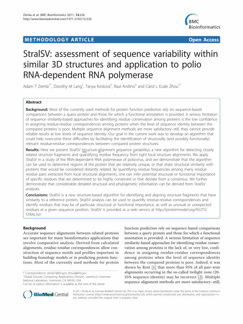
MethodsDescription of the StralSV algorithmStralSV is an algorithm that identifies protein structuralfragments having a 3D structure similar to that of a querystructure, performs structure-based alignments betweenthe query and the fragments, and quantifies at each posi-tion along the query structure the sequence variabilityrepresented among the selected fragments relative to thequery. StralSV takes as input a query structure of interest,a database of protein structures, and various parameters(discussed below) that control the selection of fragmentsfrom the database and the sequence variability calcula-tions. Figure 1 illustrates the steps in the algorithm. Thealgorithm uses a sliding-window approach for breakingthe query structure into overlapping segments, each ofwhich is independently used to identify from the inputdatabase protein structure fragments with 3D similarity. Arecommended (default) window_size parameter is set to90 amino acids in length, although an arbitrary length canbe chosen. The query structure is thus split into overlap-ping segments of length window_size; overlaps are bydefault 1/2 the length of the window_size. A final segmentis taken one window_size in length extending from theC-terminus to ensure that all portions of the query struc-ture are represented within a segment of exactly the
Zemla et al. BMC Bioinformatics 2011, 12:226http://www.biomedcentral.com/1471-2105/12/226
Page 2 of 17
window_size. Each so-calculated query segment is thencompared to all protein structures in the database usingthe LGA (local-global alignment) code [16] to identifystructure fragments with sufficient structure similarity tothe query segment. The LGA_S score is used to evaluatestructure similarity between a query segment and detectedsimilar fragments. Calculated LGA_S scores range from0% to 100% and reflect a percentage of residues from thequery segment that are identified as structurally alignedwith a given similar fragment. In the StralSV algorithm, avalue of LGA_S of at least 50% is used as a cutoff toensure that there is sufficient structural similarity betweenthe segment and the fragment over at least half the lengthof the segment. LGA’s distance cutoff parameter deter-mines the maximum allowable distance between alphacarbons (Ca) of superimposed amino acids within a calcu-lated alignment; typically this parameter is set to 4.0 Å,and this default is used for StralSV calculations with win-dow_size values of 90 residues. Thus, fragments with suffi-cient structure similarity to the query segment areidentified. Each fragment is then evaluated to determinethe tightness of its alignment to the query segment.The criteria for tight structure similarities in local
regions (spans), described by Zemla et al. [17], are used
to identify ranges within the alignment that have tightlocal superpositions. Each residue-residue pair from thealignment (closest superimposed residues from the querysegment and database fragment) is assigned a score bycalculating the local RMSD (root mean square deviation)among the surrounding residue-residue pairs. A continu-ous set of at least three residue-residue pairs that fulfilthe RMSD cutoff of 0.5 Å comprises a span. A desiredsize of a calculated span (span_size; shortest acceptabletight local alignment without gaps) is used as an inputparameter to StralSV, and is typically specified as 3, 5, or7, but can be of arbitrary length. Our previous experience[17], suggests that 5 is a reasonable minimum lengthover which to impose a tight alignment; 3 is the mini-mum value for span_size that is meaningful (since any 2Ca atoms can be perfectly aligned), and 7 imposes strin-gency that tends to eliminate capture of some related(desired) structure fragments. For the work reported herewe selected a minimum span length of 5 (span_size = 5).From each alignment is extracted a set of spans. Allalignments that contain at least one span of length noless than the specified minimum span length are deemed“qualified hits”. (For an illustration of a “span”, see addi-tional file 1: StralSV-RdRp_Suppl_Figure 1.docx.) All
Figure 1 Overview of the process and data types used in the StralSV algorithm.
Zemla et al. BMC Bioinformatics 2011, 12:226http://www.biomedcentral.com/1471-2105/12/226
Page 3 of 17
residue-residue pairs that are contained within a span’salignment are used to calculate the sequence variabilitydata at the corresponding position in the query structure.Note that not all residues from calculated structure align-ments contribute to the variability statistics at a givenposition in the query segment; regions in which the localRMSD distances between corresponding residues exceed0.5 Å induce breakpoints between spans. Also, becausethe algorithm uses overlapping segments, duplicationsare appropriately factored out in calculating the sequencevariability.A frequency matrix (Table 1) is constructed for each
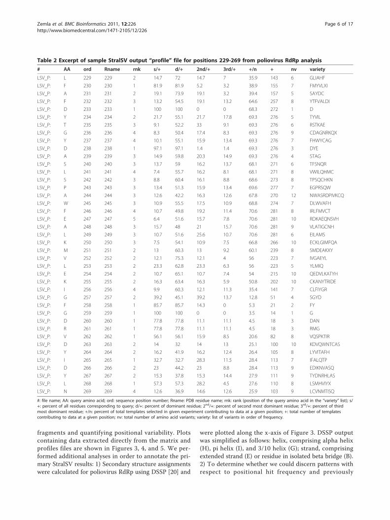
position in the query structure by tallying the frequencyat which each amino-acid (the 20 standard amino acidsplus ‘X’, corresponding to unusual or modified residues)is observed within the spans. From this matrix areextracted statistics describing the number of positionalhits (residue-residue correspondences contributingsequence variability data) per position and a list of resi-dues observed at each position, ordered by frequency ofoccurrence. These statistics are used to construct avariability profile (Table 2), which can be used to iden-tify positions at which there is relatively high or lowsequence variability in structure context. The sampleprofile given in Table 2 shows the observed residues forpositions 229 through 269 for polio RNA-dependentRNA polymerase (RdRp) run as the query proteinagainst the complete PDB database (released on Novem-ber 24, 2009).
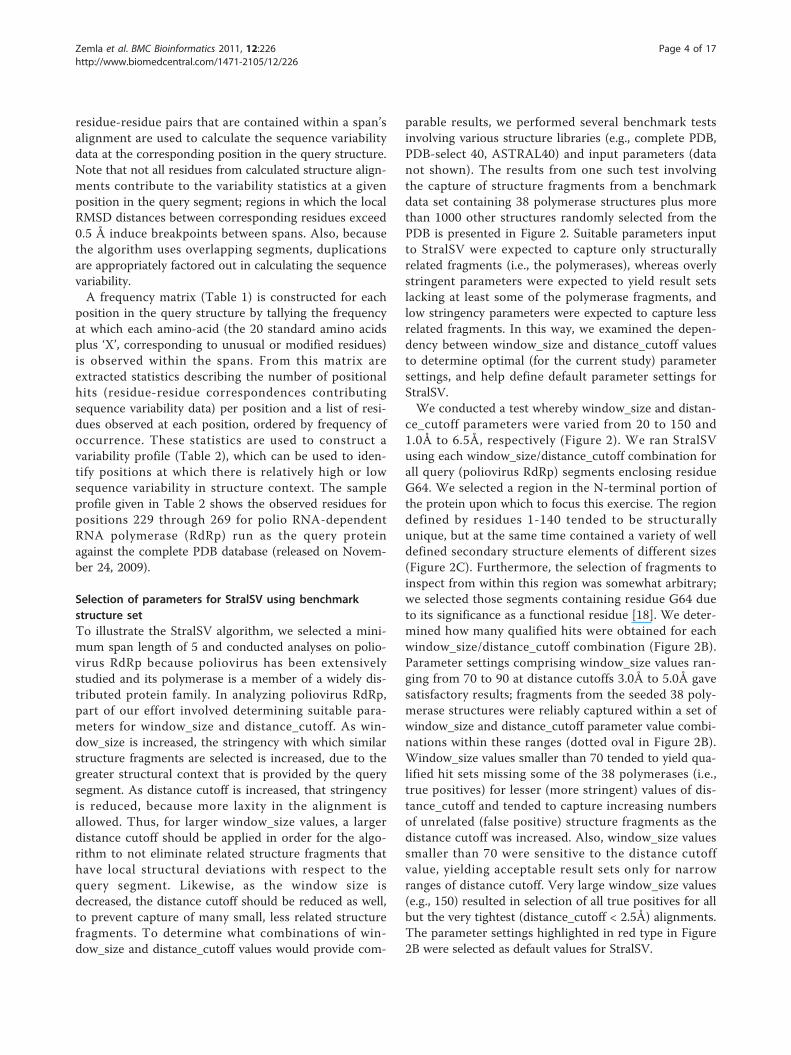
Selection of parameters for StralSV using benchmarkstructure setTo illustrate the StralSV algorithm, we selected a mini-mum span length of 5 and conducted analyses on polio-virus RdRp because poliovirus has been extensivelystudied and its polymerase is a member of a widely dis-tributed protein family. In analyzing poliovirus RdRp,part of our effort involved determining suitable para-meters for window_size and distance_cutoff. As win-dow_size is increased, the stringency with which similarstructure fragments are selected is increased, due to thegreater structural context that is provided by the querysegment. As distance cutoff is increased, that stringencyis reduced, because more laxity in the alignment isallowed. Thus, for larger window_size values, a largerdistance cutoff should be applied in order for the algo-rithm to not eliminate related structure fragments thathave local structural deviations with respect to thequery segment. Likewise, as the window size isdecreased, the distance cutoff should be reduced as well,to prevent capture of many small, less related structurefragments. To determine what combinations of win-dow_size and distance_cutoff values would provide com-
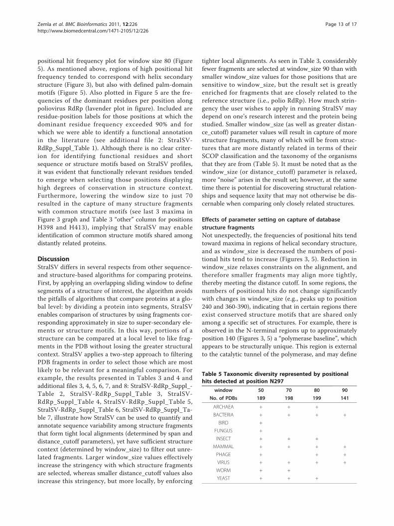
parable results, we performed several benchmark testsinvolving various structure libraries (e.g., complete PDB,PDB-select 40, ASTRAL40) and input parameters (datanot shown). The results from one such test involvingthe capture of structure fragments from a benchmarkdata set containing 38 polymerase structures plus morethan 1000 other structures randomly selected from thePDB is presented in Figure 2. Suitable parameters inputto StralSV were expected to capture only structurallyrelated fragments (i.e., the polymerases), whereas overlystringent parameters were expected to yield result setslacking at least some of the polymerase fragments, andlow stringency parameters were expected to capture lessrelated fragments. In this way, we examined the depen-dency between window_size and distance_cutoff valuesto determine optimal (for the current study) parametersettings, and help define default parameter settings forStralSV.We conducted a test whereby window_size and distan-
ce_cutoff parameters were varied from 20 to 150 and1.0Å to 6.5Å, respectively (Figure 2). We ran StralSVusing each window_size/distance_cutoff combination forall query (poliovirus RdRp) segments enclosing residueG64. We selected a region in the N-terminal portion ofthe protein upon which to focus this exercise. The regiondefined by residues 1-140 tended to be structurallyunique, but at the same time contained a variety of welldefined secondary structure elements of different sizes(Figure 2C). Furthermore, the selection of fragments toinspect from within this region was somewhat arbitrary;we selected those segments containing residue G64 dueto its significance as a functional residue [18]. We deter-mined how many qualified hits were obtained for eachwindow_size/distance_cutoff combination (Figure 2B).Parameter settings comprising window_size values ran-ging from 70 to 90 at distance cutoffs 3.0Å to 5.0Å gavesatisfactory results; fragments from the seeded 38 poly-merase structures were reliably captured within a set ofwindow_size and distance_cutoff parameter value combi-nations within these ranges (dotted oval in Figure 2B).Window_size values smaller than 70 tended to yield qua-lified hit sets missing some of the 38 polymerases (i.e.,true positives) for lesser (more stringent) values of dis-tance_cutoff and tended to capture increasing numbersof unrelated (false positive) structure fragments as thedistance cutoff was increased. Also, window_size valuessmaller than 70 were sensitive to the distance cutoffvalue, yielding acceptable result sets only for narrowranges of distance cutoff. Very large window_size values(e.g., 150) resulted in selection of all true positives for allbut the very tightest (distance_cutoff < 2.5Å) alignments.The parameter settings highlighted in red type in Figure2B were selected as default values for StralSV.
Zemla et al. BMC Bioinformatics 2011, 12:226http://www.biomedcentral.com/1471-2105/12/226
Page 4 of 17
Analysis of poliovirus RdRpParametersBased on parameters applied in Zemla et al. 2007 [17] andthe analysis described above, we used StralSV to analyzesequence variability in structure context for poliovirusRdRp (PDB: 1ra6; [19]) using minimum span length of 5,
LGA_S cutoff 55%, and window_size/distance_cutoff com-binations 50/2.5Å, 70/3.5Å, 80/4.0Å, and 90/4.0Å.Plotting of StralSV resultsStralSV produced variability matrices and sequence pro-files (not shown; for excerpts see Tables 1 and 2) fromwhich were extracted data for analyzing related structure
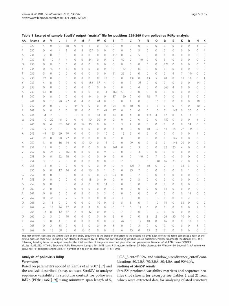
Table 1 Excerpt of sample StralSV output “matrix” file for positions 229-269 from poliovirus RdRp analysis
AA Rname A V L I P M F W G S T C Y N Q D E K R H X
L 229 4 0 21 10 0 0 1 0 103 0 0 0 0 0 0 0 0 0 0 4 0
F 230 0 4 4 3 0 8 127 0 0 0 0 0 5 0 0 0 0 0 0 0 4
A 231 30 0 0 0 0 0 0 0 0 116 0 2 5 0 0 4 0 0 0 0 0
F 232 8 10 7 4 0 0 34 0 0 0 49 0 140 0 0 5 0 0 0 0 0
D 233 0 0 0 0 0 0 0 0 0 0 0 0 0 0 0 272 0 0 0 0 0
Y 234 0 49 4 11 0 0 0 0 0 0 152 0 60 0 0 0 0 0 0 0 0
T 235 5 0 0 0 0 0 0 0 0 91 25 0 0 0 0 0 4 7 144 0 0
G 236 23 0 0 0 0 0 0 0 23 0 0 139 0 13 5 48 0 11 13 0 1
Y 237 4 0 0 0 0 0 152 37 4 0 0 7 28 0 0 0 0 0 0 44 0
D 238 0 0 0 0 0 0 0 0 0 0 0 0 4 0 0 268 4 0 0 0 0
A 239 41 0 0 0 0 0 0 0 14 165 56 0 0 0 0 0 0 0 0 0 0
S 240 0 0 0 0 0 0 44 0 0 37 160 0 0 18 7 0 0 0 5 0 0
L 241 0 151 20 22 0 4 0 44 0 0 0 4 0 0 16 0 0 0 0 10 0
S 242 0 0 0 0 44 0 0 0 0 24 165 10 0 3 13 0 0 4 0 10 0
P 243 0 0 0 0 37 0 0 7 44 17 0 0 0 0 10 0 142 0 20 0 0
A 244 34 7 0 8 10 0 0 44 0 14 0 4 0 114 4 12 0 6 13 0 0
W 245 10 20 48 0 0 0 10 30 0 0 0 0 0 0 0 152 0 0 0 4 0
F 246 0 4 32 140 0 14 30 0 0 0 3 4 0 0 0 0 0 0 54 0 0
E 247 19 2 0 0 0 0 0 0 0 7 0 0 0 10 12 44 18 22 145 2 0
A 248 44 135 59 10 0 0 0 0 10 0 12 5 0 5 0 0 0 0 0 1 0
L 249 20 0 30 72 0 11 0 0 0 3 0 0 0 0 0 0 145 0 0 0 0
K 250 3 0 16 14 0 10 10 0 15 0 0 29 0 0 5 0 144 20 0 0 0
M 251 11 0 0 0 0 31 0 0 0 144 0 0 3 0 0 22 20 4 0 0 4
V 252 8 27 2 168 0 0 0 0 9 0 0 0 3 0 0 0 6 0 0 0 0
L 253 0 0 52 10 0 14 0 0 0 0 0 0 140 0 7 0 0 0 0 0 0
E 254 3 13 9 0 0 0 0 0 0 0 2 0 1 0 140 16 23 7 0 1 0
K 255 12 0 0 2 0 0 0 0 0 0 4 128 7 10 0 2 1 33 3 0 0
I 256 0 0 17 14 0 0 16 0 1 0 0 85 7 0 0 0 0 0 1 0 0
G 257 0 0 0 0 0 0 0 0 20 23 0 0 7 0 0 1 0 0 0 0 0
F 258 0 0 0 0 0 0 18 0 0 0 0 0 3 0 0 0 0 0 0 0 0
G 259 0 0 0 0 0 0 0 0 14 0 0 0 0 0 0 0 0 0 0 0 0
D 260 2 0 0 0 0 0 0 0 0 0 0 0 0 2 0 14 0 0 0 0 0
R 261 0 0 0 0 0 2 0 0 2 0 0 0 0 0 0 0 0 0 14 0 0
V 262 0 46 0 2 5 0 0 0 0 7 3 0 0 0 13 0 0 4 2 0 0
D 263 2 13 0 0 0 0 0 10 0 2 5 3 0 7 12 14 0 32 0 0 0
Y 264 4 13 44 12 0 0 3 0 0 0 10 0 17 0 0 0 0 0 0 2 0
I 265 13 0 12 37 2 0 32 0 0 0 7 0 0 0 10 0 0 0 0 0 0
D 266 2 3 0 10 0 0 0 0 0 2 0 0 0 8 2 26 50 10 0 0 0
Y 267 3 0 4 0 0 0 0 0 0 2 42 0 17 10 0 16 0 0 10 7 0
L 268 0 2 63 2 0 5 0 0 0 31 0 0 2 0 0 0 0 0 0 3 2
N 269 0 13 38 3 0 10 0 0 0 3 6 15 0 13 2 0 0 0 0 0 0
The first column contains the amino acid of the query sequence at the position indicated in the second column. Each row in the table comprises a tally of theamino acids of each type (including non-standard indicated by ‘X’) from the corresponding positions in all qualified template fragments (positional hits). Thefollowing heading from the output provides the total number of templates searched plus other run parameters. Number of all PDB chains (SEQRES.all_list.11_03_09): 147,634; Structure: Polio RNApolym; Length: 461; MIN span: 5; Structure similarity: 55; LGA distance: 4.0; Window: 90; Legend: ‘s’ AA referencesequence, ‘d’ dominant amino acid, ‘+’ number of hits per position (max ‘+’: n = 398).
Zemla et al. BMC Bioinformatics 2011, 12:226http://www.biomedcentral.com/1471-2105/12/226
Page 5 of 17
fragments and quantifying positional variability. Plotscontaining data extracted directly from the matrix andprofiles files are shown in Figures 3, 4, and 5. We per-formed additional analyses in order to annotate the pri-mary StralSV results: 1) Secondary structure assignmentswere calculated for poliovirus RdRp using DSSP [20] and
were plotted along the x-axis of Figure 3. DSSP outputwas simplified as follows: helix, comprising alpha helix(H), pi helix (I), and 3/10 helix (G); strand, comprisingextended strand (E) or residue in isolated beta bridge (B).2) To determine whether we could discern patterns withrespect to positional hit frequency and previously
Table 2 Excerpt of sample StralSV output “profile” file for positions 229-269 from poliovirus RdRp analysis
# AA ord Rname rnk s/+ d/+ 2nd/+ 3rd/+ +/n + nv variety
LSV_P: L 229 229 2 14.7 72 14.7 7 35.9 143 6 GLIAHF
LSV_P: F 230 230 1 81.9 81.9 5.2 3.2 38.9 155 7 FMYVLXI
LSV_P: A 231 231 2 19.1 73.9 19.1 3.2 39.4 157 5 SAYDC
LSV_P: F 232 232 3 13.2 54.5 19.1 13.2 64.6 257 8 YTFVALDI
LSV_P: D 233 233 1 100 100 0 0 68.3 272 1 D
LSV_P: Y 234 234 2 21.7 55.1 21.7 17.8 69.3 276 5 TYVIL
LSV_P: T 235 235 3 9.1 52.2 33 9.1 69.3 276 6 RSTKAE
LSV_P: G 236 236 4 8.3 50.4 17.4 8.3 69.3 276 9 CDAGNRKQX
LSV_P: Y 237 237 4 10.1 55.1 15.9 13.4 69.3 276 7 FHWYCAG
LSV_P: D 238 238 1 97.1 97.1 1.4 1.4 69.3 276 3 DYE
LSV_P: A 239 239 3 14.9 59.8 20.3 14.9 69.3 276 4 STAG
LSV_P: S 240 240 3 13.7 59 16.2 13.7 68.1 271 6 TFSNQR
LSV_P: L 241 241 4 7.4 55.7 16.2 8.1 68.1 271 8 VWILQHMC
LSV_P: S 242 242 3 8.8 60.4 16.1 8.8 68.6 273 8 TPSQCHKN
LSV_P: P 243 243 3 13.4 51.3 15.9 13.4 69.6 277 7 EGPRSQW
LSV_P: A 244 244 3 12.6 42.2 16.3 12.6 67.8 270 12 NWASRDPIVKCQ
LSV_P: W 245 245 3 10.9 55.5 17.5 10.9 68.8 274 7 DLWVAFH
LSV_P: F 246 246 4 10.7 49.8 19.2 11.4 70.6 281 8 IRLFMVCT
LSV_P: E 247 247 5 6.4 51.6 15.7 7.8 70.6 281 10 RDKAEQNSVH
LSV_P: A 248 248 3 15.7 48 21 15.7 70.6 281 9 VLATIGCNH
LSV_P: L 249 249 3 10.7 51.6 25.6 10.7 70.6 281 6 EILAMS
LSV_P: K 250 250 3 7.5 54.1 10.9 7.5 66.8 266 10 ECKLGIMFQA
LSV_P: M 251 251 2 13 60.3 13 9.2 60.1 239 8 SMDEAKXY
LSV_P: V 252 252 2 12.1 75.3 12.1 4 56 223 7 IVGAEYL
LSV_P: L 253 253 2 23.3 62.8 23.3 6.3 56 223 5 YLMIQ
LSV_P: E 254 254 2 10.7 65.1 10.7 7.4 54 215 10 QEDVLKATYH
LSV_P: K 255 255 2 16.3 63.4 16.3 5.9 50.8 202 10 CKANYTRIDE
LSV_P: I 256 256 4 9.9 60.3 12.1 11.3 35.4 141 7 CLFIYGR
LSV_P: G 257 257 2 39.2 45.1 39.2 13.7 12.8 51 4 SGYD
LSV_P: F 258 258 1 85.7 85.7 14.3 0 5.3 21 2 FY
LSV_P: G 259 259 1 100 100 0 0 3.5 14 1 G
LSV_P: D 260 260 1 77.8 77.8 11.1 11.1 4.5 18 3 DAN
LSV_P: R 261 261 1 77.8 77.8 11.1 11.1 4.5 18 3 RMG
LSV_P: V 262 262 1 56.1 56.1 15.9 8.5 20.6 82 8 VQSPKTIR
LSV_P: D 263 263 2 14 32 14 13 25.1 100 10 KDVQWNTCAS
LSV_P: Y 264 264 2 16.2 41.9 16.2 12.4 26.4 105 8 LYVITAFH
LSV_P: I 265 265 1 32.7 32.7 28.3 11.5 28.4 113 7 IFALQTP
LSV_P: D 266 266 2 23 44.2 23 8.8 28.4 113 9 EDIKNVASQ
LSV_P: Y 267 267 2 15.3 37.8 15.3 14.4 27.9 111 9 TYDNRHLAS
LSV_P: L 268 268 1 57.3 57.3 28.2 4.5 27.6 110 8 LSMHVIYX
LSV_P: N 269 269 4 12.6 36.9 14.6 12.6 25.9 103 9 LCVNMTISQ
#: file name; AA: query amino acid; ord: sequence position number; Rname: PDB residue name; rnk: rank (position of the query amino acid in the “variety” list); s/+: percent of all residues corresponding to query; d/+: percent of dominant residue; 2nd/+: percent of second most dominant residue; 3rd/+: percent of thirdmost dominant residue; +/n: percent of total templates selected in given experiment contributing to data at a given position; +: total number of templatescontributing to data at a given position; nv: total number of amino acid variants; variety: list of variants in order of frequency.
Zemla et al. BMC Bioinformatics 2011, 12:226http://www.biomedcentral.com/1471-2105/12/226
Page 6 of 17
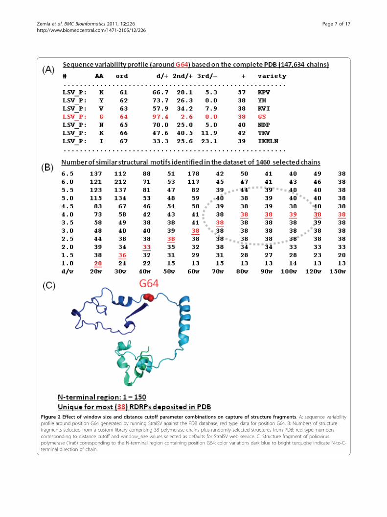
Figure 2 Effect of window size and distance cutoff parameter combinations on capture of structure fragments. A: sequence variabilityprofile around position G64 generated by running StralSV against the PDB database; red type: data for position G64. B: Numbers of structurefragments selected from a custom library comprising 38 polymerase chains plus randomly selected structures from PDB; red type: numberscorresponding to distance cutoff and window_size values selected as defaults for StralSV web service. C: Structure fragment of polioviruspolymerase (1ra6) corresponding to the N-terminal region containing position G64; color variations dark blue to bright turquoise indicate N-to-C-terminal direction of chain.
Zemla et al. BMC Bioinformatics 2011, 12:226http://www.biomedcentral.com/1471-2105/12/226
Page 7 of 17
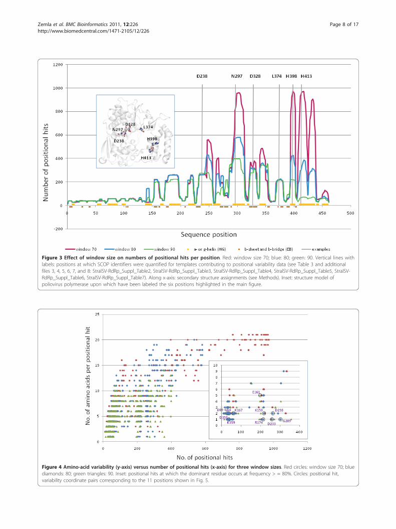
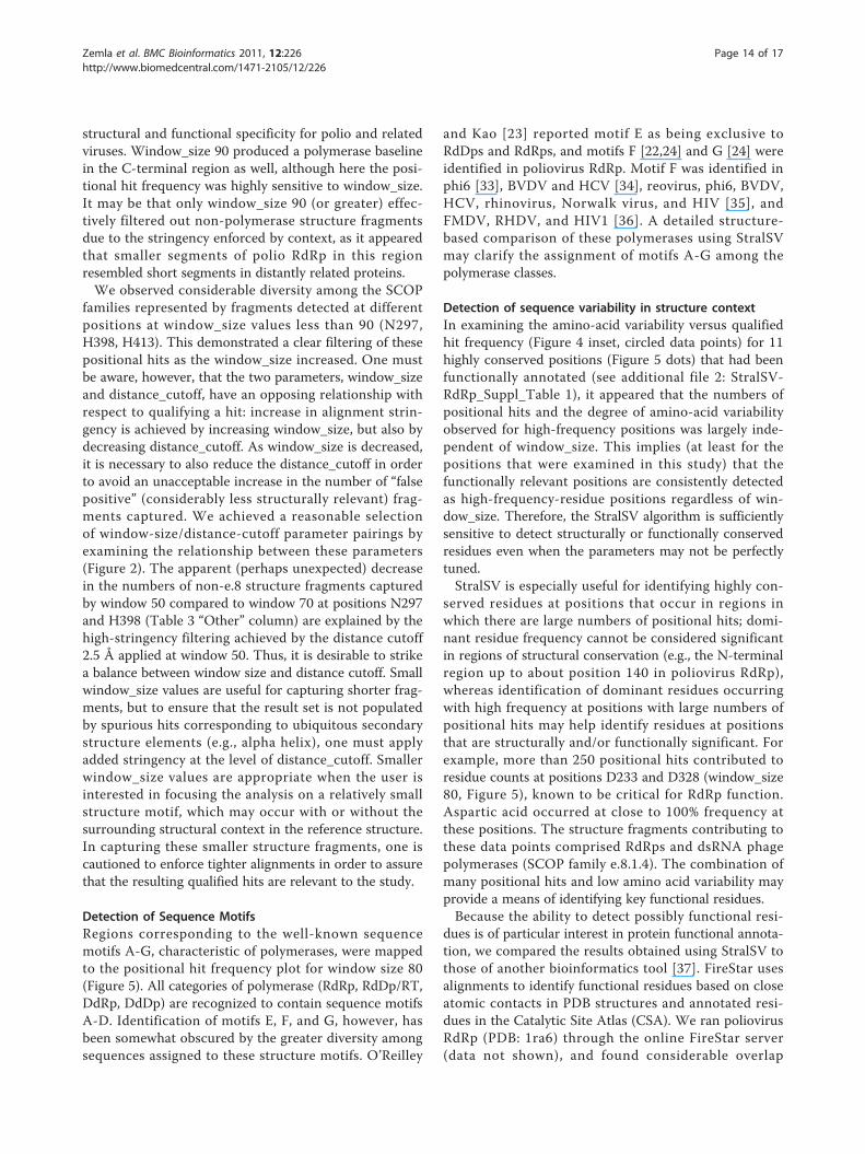
Figure 3 Effect of window size on numbers of positional hits per position. Red: window size 70; blue: 80; green: 90. Vertical lines withlabels: positions at which SCOP identifiers were quantified for templates contributing to positional variability data (see Table 3 and additionalfiles 3, 4, 5, 6, 7, and 8: StralSV-RdRp_Suppl_Table2, StralSV-RdRp_Suppl_Table3, StralSV-RdRp_Suppl_Table4, StralSV-RdRp_Suppl_Table5, StralSV-RdRp_Suppl_Table6, StralSV-RdRp_Suppl_Table7). Along x-axis: secondary structure assignments (see Methods). Inset: structure model ofpoliovirus polymerase upon which have been labeled the six positions highlighted in the main figure.
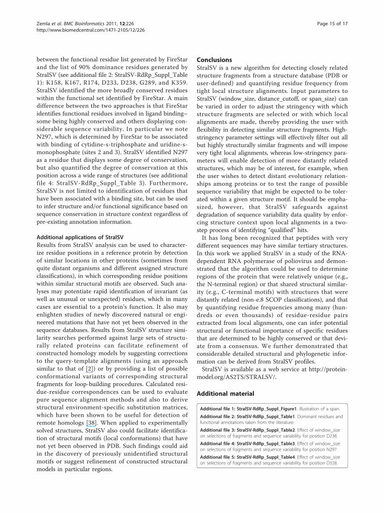
Figure 4 Amino-acid variability (y-axis) versus number of positional hits (x-axis) for three window sizes. Red circles: window size 70; bluediamonds: 80; green triangles: 90. Inset: positional hits at which the dominant residue occurs at frequency > = 80%. Circles: positional hit,variability coordinate pairs corresponding to the 11 positions shown in Fig. 5.
Zemla et al. BMC Bioinformatics 2011, 12:226http://www.biomedcentral.com/1471-2105/12/226
Page 8 of 17
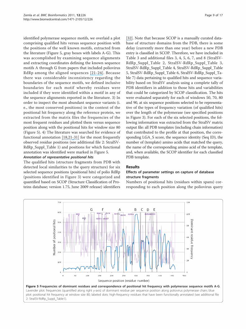
identified polymerase sequence motifs, we overlaid a plotcomprising qualified hits versus sequence position withthe positions of the well known motifs, extracted fromthe literature (Figure 5, gray boxes with labels A-G). Thiswas accomplished by examining sequence alignmentsand extracting coordinates defining the known sequencemotifs A through G from papers that included poliovirusRdRp among the aligned sequences [21-24]. Becausethere was considerable inconsistency regarding theboundaries of the sequence motifs, we defined inclusiveboundaries for each motif whereby residues wereincluded if they were identified within a motif in any ofthe sequence alignments reported in the literature. 3) Inorder to inspect the most abundant sequence variants (i.e., the most conserved positions) in the context of thepositional hit frequency along the reference protein, weextracted from the matrix files the frequencies of themost frequent residues and plotted them versus sequenceposition along with the positional hits for window size 80(Figure 5). 4) The literature was searched for evidence offunctional annotation [18,21-31] for the most frequentlyobserved residue positions (see additional file 2: StralSV-RdRp_Suppl_Table 1) and positions for which functionalannotation was identified were marked in Figure 5.Annotation of representative positional hitsThe qualified hits (structure fragments from PDB withdetected local similarities to the query structure) for sixselected sequence positions (positional hits) of polio RdRp(positions identified in Figure 3) were categorized andquantified based on SCOP (Structure Classification of Pro-teins database; version 1.75, June 2009 release) identifiers
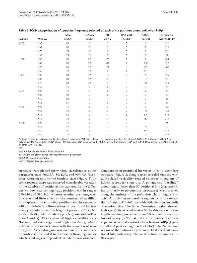
[32]. Note that because SCOP is a manually curated data-base of structure domains from the PDB, there is somedelay (currently more than one year) before a new PDBentry is classified in SCOP. Therefore, we have included inTable 3 and additional files 3, 4, 5, 6, 7, and 8 (StralSV-RdRp_Suppl_Table 2, StralSV-RdRp_Suppl_Table 3,StralSV-RdRp_Suppl_Table 4, StralSV-RdRp_Suppl_Table5, StralSV-RdRp_Suppl_Table 6, StralSV-RdRp_Suppl_Ta-ble 7) data pertaining to qualified hits and sequence varia-bility based on StralSV analysis using a complete tally ofPDB identifiers in addition to those hits and variabilitiesthat could be categorized by SCOP classification. The hitswere evaluated separately for each of windows 50, 70, 80and 90, at six sequence positions selected to be representa-tive of the types of frequency variation (of qualified hits)over the length of the polymerase (see specified positionsin Figure 3). For each of the six selected positions, the fol-lowing information was extracted from the StralSV matrixoutput file: all PDB templates (including chain information)that contributed to the profile at that position, the corre-sponding LGA_S score, the sequence identity (Seq ID), thenumber of (template) amino acids that matched the query,the name of the corresponding amino acid of the template,and, when available, the SCOP identifier for each classifiedPDB template.
ResultsEffects of parameter settings on capture of databasestructure fragmentsNumbers of positional hits (residues within spans) cor-responding to each position along the poliovirus query
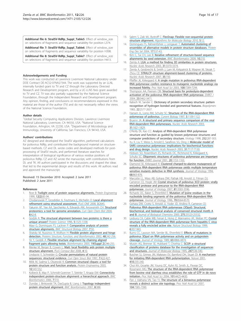
Figure 5 Frequencies of dominant residues and correspondence of positional hit frequency with polymerase sequence motifs A-G.Lavender plot: frequencies (quantified along right y-axis) of dominant residue per sequence position along poliovirus polymerase chain; blueplot: positional hit frequency at window size 80; labeled dots: high-frequency residues that have been functionally annotated (see additional file2: StralSV-RdRp_Suppl_Table1).
Zemla et al. BMC Bioinformatics 2011, 12:226http://www.biomedcentral.com/1471-2105/12/226
Page 9 of 17
structure were plotted for window_size/distance_cutoffparameter pairs 70/3.5Å, 80/4.0Å, and 90/4.0Å (here-after referring only to the window_size) (Figure 3). Insome regions, there was observed considerable variationin the numbers of positional hits captured for the differ-ent window_size settings (e.g., positions within ranges290-310 and 390-440), whereas at other positions, win-dow_size had little effect on the numbers of qualifiedhits captured (most notably positions within ranges 1-240 and 360-390). Observations of positional hit fre-quency variations over the length of poliovirus RdRp ledto identification of a variability profile (illustrated in Fig-ures 3 and 5). The regions of high variability were“locked” between regions of high specificity, whichexhibited little or no change with the variation of win-dow_size. As window_size was increased, the numbersof positional hits tended to decrease in those regions forwhich window_size-dependent variability was observed.
Comparison of positional hit variabilities to secondarystructure (Figure 3, along x-axis) revealed that the win-dow-related variability tended to occur in regions ofhelical secondary structure. A polymerase “baseline”,amounting to fewer than 50 positional hits (correspond-ing primarily to polymerase structures) was observedalong the entirety of the poliovirus chain (Figure 3 x-axis). All polymerase baseline regions, with the excep-tion of region 410-461, were identifiable independentlyof window_size. The latter C-terminal region showedhigh specificity at window size 90. In this region, lower-ing the window_size value to just 70 resulted in the cap-ture of many (> 900) structure fragments that haveapparent structural similarity to poliovirus RdRp (Figure3, tall red peaks at right side of plot). The N-terminalregion of the poliovirus protein yielded the least posi-tional hits, indicating relative structural uniqueness inthis region.
Table 3 SCOP categorization of template fragments selected at each of six positions along poliovirus RdRp
RdRp DsPhage RT DNA pol1 Other Templates
Position Window e.8.1.4 e.8.1.6 e.8.1.2 e.8.1.1 not e.8 with SCOP ID
D238 w90 85 32 0 0 3 120
w80 85 32 0 0 0 119
w70 79 32 0 0 0 111
w50 79 0 0 0 0 79
N297 w90 85 32 78 0 9 204
w80 85 32 41 2 160 320
w70 85 32 71 3 289 480
w50 78 32 0 4 197 311
D328 w90 84 32 3 0 6 125
w80 84 32 8 0 17 141
w70 84 32 4 0 34 154
w50 71 2 0 0 3 76
L374 w90 77 0 0 0 2 79
w80 80 0 0 0 3 83
w70 82 0 1 0 3 86
w50 59 0 0 0 2 61
H398 w90 79 0 0 0 3 82
w80 80 0 0 0 48 128
w70 68 0 0 2 376 446
w50 36 0 0 0 182 218
H413 w90 37 0 0 0 1 38
w80 45 0 0 0 54 99
w70 40 0 0 2 392 434
w50 37 0 0 13 695 745
Position: residue and position number in poliovirus polymerase; Window: window_size parameter settings (w: window); RdRp e.8.1.4: RNA-dependent RNApolymerase; DsPhage e.8.1.6: dsRNA phage RNA-dependent RNA-polymerase; RT e.8.1.2: Reverse transcriptase; DNA pol1 e.8.1.1: DNA polymerase I; Other not e.8:all other SCOP families.
KEY:
e.8.1.4 RdRp RNA-dependent RNA-polymerase
e.8.1.6 DsPhage dsRNA phage RNA-dependent RNA-polymerase
e.8.1.2 RT Reverse transcriptase
e.8.1.1 DNApol1 DNA polymerase I
Zemla et al. BMC Bioinformatics 2011, 12:226http://www.biomedcentral.com/1471-2105/12/226
Page 10 of 17
Composition of captured structure fragments at specificpositionsWe selected six positions (Figure 3, residues numbered attop of plot) at which to examine the diversity of structurefragments captured at each of four window_size values(Table 3, additional files 3, 4, 5, 6, 7, and 8: StralSV-RdRp_Suppl_Table 2, StralSV-RdRp_Suppl_Table 3,StralSV-RdRp_Suppl_Table 4, StralSV-RdRp_Suppl_Table5, StralSV-RdRp_Suppl_Table 6, StralSV-RdRp_Suppl_Ta-ble 7). Orientations within the structure of poliovirus RdRpof these six positions are indicated on the structure modelshown in the inset of Figure 3. Qualified hits were exam-ined for positions at which numbers of positional hits wererelatively invariant (D238, D328, L374) or highly variant(N297, H398, H413) among the calculations run at the fourwindow_size values. (In some cases preferences for selec-tion of a particular residue for this analysis was based onexistence of a functional annotation for that residue and,therefore, of biological interest (e.g., D238, N297; [21,29]).We included window_size 50 in this analysis in order todetermine what effect a very small (low stringency) win-dow_size value might have on the structure-function diver-sity of qualified hits (i.e., would it greatly increase thediversity?). For each position and each window_size, quali-fied hits were categorized by SCOP concise classificationstrings. Hits were categorized into four families in the e.8.1(DNA/RNA polymerases) superfamily: e.8.1.1 (DNA poly-merase I), e.8.1.2 (Reverse transcriptase), e.8.1.4 (RNA-dependent RNA-polymerase), and e.8.1.6 (dsRNA phageRNA-dependent RNA-polymerase), and into various othernon-e.8 SCOP classifications.At positions D238, D328, and L374, qualification of
structure fragments (and resulting positional hits) waslargely independent of window_size, and few unrelated(non-e.8) fragments were captured, indicating that thestructure motifs in the surrounding regions were largelylimited to the families detected (e.g., e.8.1.4 and e.8.1.6)(Table 3, positions D238 and D328). For positionsN297, H398, and H413, at which there was considerablediversity in the SCOP families detected at smaller win-dow_size values (< 90), omission of these more distantlyrelated positional hits was observed as window_sizeincreased. Overall it was observed that window_size 90resulted predominantly in capture of structure frag-ments from the structure family to which poliovirusRdRp belongs (e.8.1.4). Therefore, at all six positions,window_size 90 effectively filtered from among thenearly 150,000 PDB chains only those members of thepolymerase/transcriptase families.
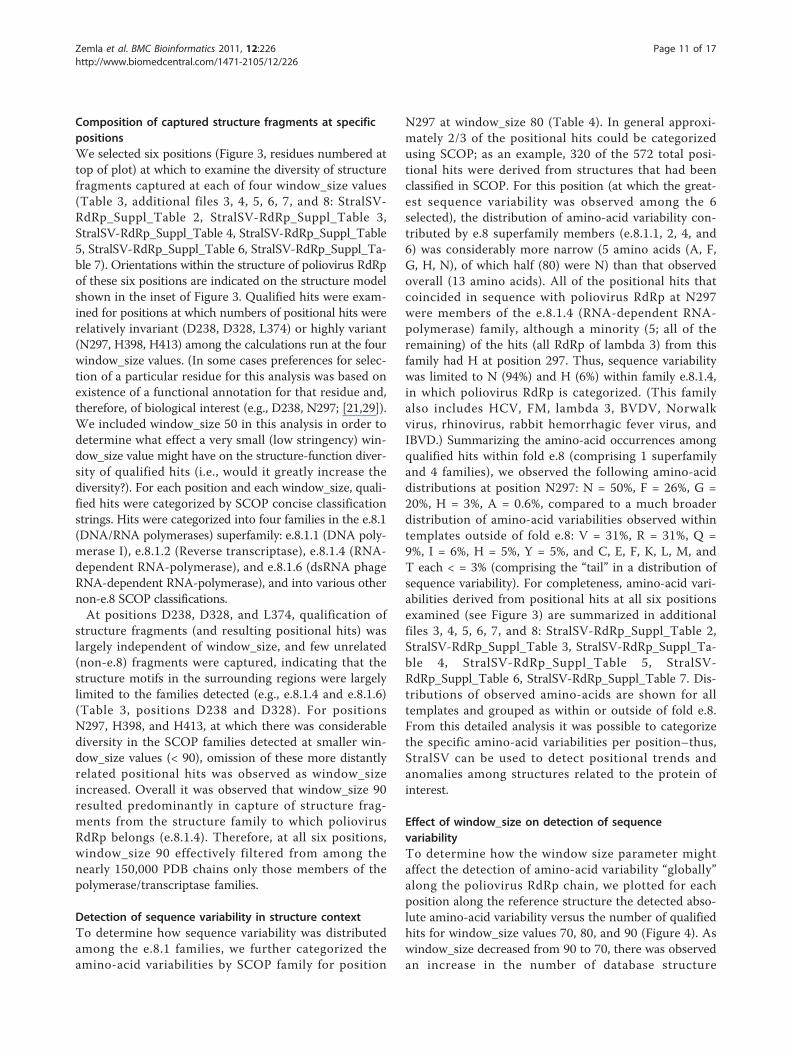
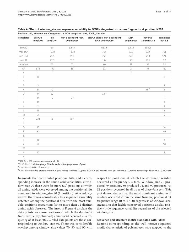
Detection of sequence variability in structure contextTo determine how sequence variability was distributedamong the e.8.1 families, we further categorized theamino-acid variabilities by SCOP family for position
N297 at window_size 80 (Table 4). In general approxi-mately 2/3 of the positional hits could be categorizedusing SCOP; as an example, 320 of the 572 total posi-tional hits were derived from structures that had beenclassified in SCOP. For this position (at which the great-est sequence variability was observed among the 6selected), the distribution of amino-acid variability con-tributed by e.8 superfamily members (e.8.1.1, 2, 4, and6) was considerably more narrow (5 amino acids (A, F,G, H, N), of which half (80) were N) than that observedoverall (13 amino acids). All of the positional hits thatcoincided in sequence with poliovirus RdRp at N297were members of the e.8.1.4 (RNA-dependent RNA-polymerase) family, although a minority (5; all of theremaining) of the hits (all RdRp of lambda 3) from thisfamily had H at position 297. Thus, sequence variabilitywas limited to N (94%) and H (6%) within family e.8.1.4,in which poliovirus RdRp is categorized. (This familyalso includes HCV, FM, lambda 3, BVDV, Norwalkvirus, rhinovirus, rabbit hemorrhagic fever virus, andIBVD.) Summarizing the amino-acid occurrences amongqualified hits within fold e.8 (comprising 1 superfamilyand 4 families), we observed the following amino-aciddistributions at position N297: N = 50%, F = 26%, G =20%, H = 3%, A = 0.6%, compared to a much broaderdistribution of amino-acid variabilities observed withintemplates outside of fold e.8: V = 31%, R = 31%, Q =9%, I = 6%, H = 5%, Y = 5%, and C, E, F, K, L, M, andT each < = 3% (comprising the “tail” in a distribution ofsequence variability). For completeness, amino-acid vari-abilities derived from positional hits at all six positionsexamined (see Figure 3) are summarized in additionalfiles 3, 4, 5, 6, 7, and 8: StralSV-RdRp_Suppl_Table 2,StralSV-RdRp_Suppl_Table 3, StralSV-RdRp_Suppl_Ta-ble 4, StralSV-RdRp_Suppl_Table 5, StralSV-RdRp_Suppl_Table 6, StralSV-RdRp_Suppl_Table 7. Dis-tributions of observed amino-acids are shown for alltemplates and grouped as within or outside of fold e.8.From this detailed analysis it was possible to categorizethe specific amino-acid variabilities per position–thus,StralSV can be used to detect positional trends andanomalies among structures related to the protein ofinterest.
Effect of window_size on detection of sequencevariabilityTo determine how the window size parameter mightaffect the detection of amino-acid variability “globally”along the poliovirus RdRp chain, we plotted for eachposition along the reference structure the detected abso-lute amino-acid variability versus the number of qualifiedhits for window_size values 70, 80, and 90 (Figure 4). Aswindow_size decreased from 90 to 70, there was observedan increase in the number of database structure
Zemla et al. BMC Bioinformatics 2011, 12:226http://www.biomedcentral.com/1471-2105/12/226
Page 11 of 17
fragments that contributed positional hits, and a corre-sponding increase in the amino-acid variabilities: at win-dow_size 70 there were far more (32) positions at whichall amino-acids were observed among the positional hitscompared to window_size 80 (1 position). At window_-size 90 there was considerably less sequence variabilitydetected among the positional hits, with the most vari-able positions accounting for no more than 14 distinctamino acids observed. The inset in Figure 4 displays thedata points for those positions at which the dominant(most frequently observed) amino-acid occurred at a fre-quency of at least 80%. Circled data points are those cor-responding to window_size 80. There was considerableoverlap among window_size values 70, 80, and 90 with
respect to positions at which the dominant residueoccurred at frequency > = 80%. Window_size 70 pro-duced 79 positions, 80 produced 74, and 90 produced 79;62 positions occurred in all three of these data sets. Thisplot demonstrates that the most dominant amino-acidresidues occurred within the same (narrow) positional hitfrequency range (0 to < 400) regardless of window_size,suggesting that highly conserved positions display rela-tively little sequence variability regardless of the selectedwindow_size.
Sequence and structure motifs associated with RdRpsRegions corresponding to the well-known sequencemotifs characteristic of polymerases were mapped to the
Table 4 Effect of window_size on sequence variability in SCOP-categorized structure fragments at position N297
Position: 297, Window: 80, Categories: 33, PDB templates: 399, SCOP_IDs: 320
Templates all PDBtemplates
e.8templates
RNA-dependent RNApolymerase
dsRNA phage RNA-dependentRNA polymerase
DNApolymerase
I
Reversetranscriptase
Templatesnot e.8
ScopID e.8 e.8.1.4 e.8.1.6 e.8.1.1 e.8.1.2
max LGA 100.0 100.0 76.9 57.0 59.2 76.8
ave LGA 75.4 85.2 75.1 57.0 56.4 57.8
ave ID 27.3 37.3 13.4 3.7 18.6 6.2
matches 51 61 40 31 39 35
AA 572 160 85 32 2 41 160
A 1 1 1
C 8 3
D 1
E 3 1
F 67 42 1 41 1 5
G 44 32 32 2
H 14 5 5 3 8
I 19 9
K 2 2
L 11 5
M 2 2
N 224 80 80 4
P
Q 15 14
R 82 49
S
T 15 4
V 54 50
W
Y 10 8
X1F297 (N = 41): reverse transcriptase of HIV.2G297 (N = 32): dsRNA phage RNA-dependent RNA polymerase of phi6.3H297 (N = 5): RdRp of lambda 3.4N297 (N = 80): RdRp proteins from HCV (21), FM (8), lambda3 (5), polio (6), BVDV (3), Norwalk virus (3), rhinovirus (3), rabbit hemorrhagic fever virus (2), IBDV (1).
Zemla et al. BMC Bioinformatics 2011, 12:226http://www.biomedcentral.com/1471-2105/12/226
Page 12 of 17
positional hit frequency plot for window size 80 (Figure5). As mentioned above, regions of high positional hitfrequency tended to correspond with helix secondarystructure (Figure 3), but also with defined palm-domainmotifs (Figure 5). Also plotted in Figure 5 are the fre-quencies of the dominant residues per position alongpoliovirus RdRp (lavender plot in figure). Included areresidue-position labels for those positions at which thedominant residue frequency exceeded 90% and forwhich we were able to identify a functional annotationin the literature (see additional file 2: StralSV-RdRp_Suppl_Table 1). Although there is no clear criter-ion for identifying functional residues and shortsequence or structure motifs based on StralSV profiles,it was evident that functionally relevant residues tendedto emerge when selecting those positions displayinghigh degrees of conservation in structure context.Furthermore, lowering the window size to just 70resulted in the capture of many structure fragmentswith common structure motifs (see last 3 maxima inFigure 3 graph and Table 3 “other” column for positionsH398 and H413), implying that StralSV may enableidentification of common structure motifs shared amongdistantly related proteins.
DiscussionStralSV differs in several respects from other sequence-and structure-based algorithms for comparing proteins.First, by applying an overlapping sliding window to definesegments of a structure of interest, the algorithm avoidsthe pitfalls of algorithms that compare proteins at a glo-bal level: by dividing a protein into segments, StralSVenables comparison of structures by using fragments cor-responding approximately in size to super-secondary ele-ments or structure motifs. In this way, portions of astructure can be compared at a local level to like frag-ments in the PDB without losing the greater structuralcontext. StralSV applies a two-step approach to filteringPDB fragments in order to select those which are mostlikely to be relevant for a meaningful comparison. Forexample, the results presented in Tables 3 and 4 andadditional files 3, 4, 5, 6, 7, and 8: StralSV-RdRp_Suppl_-Table 2, StralSV-RdRp_Suppl_Table 3, StralSV-RdRp_Suppl_Table 4, StralSV-RdRp_Suppl_Table 5,StralSV-RdRp_Suppl_Table 6, StralSV-RdRp_Suppl_Ta-ble 7, illustrate how StralSV can be used to quantify andannotate sequence variability among structure fragmentsthat form tight local alignments (determined by span anddistance_cutoff parameters), yet have sufficient structurecontext (determined by window_size) to filter out unre-lated fragments. Larger window_size values effectivelyincrease the stringency with which structure fragmentsare selected, whereas smaller distance_cutoff values alsoincrease this stringency, but more locally, by enforcing
tighter local alignments. As seen in Table 3, considerablyfewer fragments are selected at window_size 90 than withsmaller window_size values for those positions that aresensitive to window_size, but the result set is greatlyenriched for fragments that are closely related to thereference structure (i.e., polio RdRp). How much strin-gency the user wishes to apply in running StralSV maydepend on one’s research interest and the protein beingstudied. Smaller window_size (as well as greater distan-ce_cutoff) parameter values will result in capture of morestructure fragments, many of which will be from struc-tures that are more distantly related in terms of theirSCOP classification and the taxonomy of the organismsthat they are from (Table 5). It must be noted that as thewindow_size (or distance_cutoff) parameter is relaxed,more “noise” arises in the result set; however, at the sametime there is potential for discovering structural relation-ships and sequence laxity that may not otherwise be dis-cernable when comparing only closely related structures.
Effects of parameter setting on capture of databasestructure fragmentsNot unexpectedly, the frequencies of positional hits tendtoward maxima in regions of helical secondary structure,and as window_size is decreased the numbers of posi-tional hits tend to increase (Figures 3, 5). Reduction inwindow_size relaxes constraints on the alignment, andtherefore smaller fragments may align more tightly,thereby meeting the distance cutoff. In some regions, thenumbers of positional hits do not change significantlywith changes in window_size (e.g., peaks up to position240 and 360-390), indicating that in certain regions thereexist conserved structure motifs that are shared onlyamong a specific set of structures. For example, there isobserved in the N-terminal regions up to approximatelyposition 140 (Figures 3, 5) a “polymerase baseline”, whichappears to be structurally unique. This region is externalto the catalytic tunnel of the polymerase, and may define
Table 5 Taxonomic diversity represented by positionalhits detected at position N297
window 50 70 80 90
No. of PDBs 189 198 199 141
ARCHAEA + + +
BACTERIA + + + +
BIRD +
FUNGUS +
INSECT + + +
MAMMAL + + + +
PHAGE + + +
VIRUS + + + +
WORM + +
YEAST + + +
Zemla et al. BMC Bioinformatics 2011, 12:226http://www.biomedcentral.com/1471-2105/12/226
Page 13 of 17
structural and functional specificity for polio and relatedviruses. Window_size 90 produced a polymerase baselinein the C-terminal region as well, although here the posi-tional hit frequency was highly sensitive to window_size.It may be that only window_size 90 (or greater) effec-tively filtered out non-polymerase structure fragmentsdue to the stringency enforced by context, as it appearedthat smaller segments of polio RdRp in this regionresembled short segments in distantly related proteins.We observed considerable diversity among the SCOP
families represented by fragments detected at differentpositions at window_size values less than 90 (N297,H398, H413). This demonstrated a clear filtering of thesepositional hits as the window_size increased. One mustbe aware, however, that the two parameters, window_sizeand distance_cutoff, have an opposing relationship withrespect to qualifying a hit: increase in alignment strin-gency is achieved by increasing window_size, but also bydecreasing distance_cutoff. As window_size is decreased,it is necessary to also reduce the distance_cutoff in orderto avoid an unacceptable increase in the number of “falsepositive” (considerably less structurally relevant) frag-ments captured. We achieved a reasonable selectionof window-size/distance-cutoff parameter pairings byexamining the relationship between these parameters(Figure 2). The apparent (perhaps unexpected) decreasein the numbers of non-e.8 structure fragments capturedby window 50 compared to window 70 at positions N297and H398 (Table 3 “Other” column) are explained by thehigh-stringency filtering achieved by the distance cutoff2.5 Å applied at window 50. Thus, it is desirable to strikea balance between window size and distance cutoff. Smallwindow_size values are useful for capturing shorter frag-ments, but to ensure that the result set is not populatedby spurious hits corresponding to ubiquitous secondarystructure elements (e.g., alpha helix), one must applyadded stringency at the level of distance_cutoff. Smallerwindow_size values are appropriate when the user isinterested in focusing the analysis on a relatively smallstructure motif, which may occur with or without thesurrounding structural context in the reference structure.In capturing these smaller structure fragments, one iscautioned to enforce tighter alignments in order to assurethat the resulting qualified hits are relevant to the study.
Detection of Sequence MotifsRegions corresponding to the well-known sequencemotifs A-G, characteristic of polymerases, were mappedto the positional hit frequency plot for window size 80(Figure 5). All categories of polymerase (RdRp, RdDp/RT,DdRp, DdDp) are recognized to contain sequence motifsA-D. Identification of motifs E, F, and G, however, hasbeen somewhat obscured by the greater diversity amongsequences assigned to these structure motifs. O’Reilley
and Kao [23] reported motif E as being exclusive toRdDps and RdRps, and motifs F [22,24] and G [24] wereidentified in poliovirus RdRp. Motif F was identified inphi6 [33], BVDV and HCV [34], reovirus, phi6, BVDV,HCV, rhinovirus, Norwalk virus, and HIV [35], andFMDV, RHDV, and HIV1 [36]. A detailed structure-based comparison of these polymerases using StralSVmay clarify the assignment of motifs A-G among thepolymerase classes.
Detection of sequence variability in structure contextIn examining the amino-acid variability versus qualifiedhit frequency (Figure 4 inset, circled data points) for 11highly conserved positions (Figure 5 dots) that had beenfunctionally annotated (see additional file 2: StralSV-RdRp_Suppl_Table 1), it appeared that the numbers ofpositional hits and the degree of amino-acid variabilityobserved for high-frequency positions was largely inde-pendent of window_size. This implies (at least for thepositions that were examined in this study) that thefunctionally relevant positions are consistently detectedas high-frequency-residue positions regardless of win-dow_size. Therefore, the StralSV algorithm is sufficientlysensitive to detect structurally or functionally conservedresidues even when the parameters may not be perfectlytuned.StralSV is especially useful for identifying highly con-
served residues at positions that occur in regions inwhich there are large numbers of positional hits; domi-nant residue frequency cannot be considered significantin regions of structural conservation (e.g., the N-terminalregion up to about position 140 in poliovirus RdRp),whereas identification of dominant residues occurringwith high frequency at positions with large numbers ofpositional hits may help identify residues at positionsthat are structurally and/or functionally significant. Forexample, more than 250 positional hits contributed toresidue counts at positions D233 and D328 (window_size80, Figure 5), known to be critical for RdRp function.Aspartic acid occurred at close to 100% frequency atthese positions. The structure fragments contributing tothese data points comprised RdRps and dsRNA phagepolymerases (SCOP family e.8.1.4). The combination ofmany positional hits and low amino acid variability mayprovide a means of identifying key functional residues.Because the ability to detect possibly functional resi-
dues is of particular interest in protein functional annota-tion, we compared the results obtained using StralSV tothose of another bioinformatics tool [37]. FireStar usesalignments to identify functional residues based on closeatomic contacts in PDB structures and annotated resi-dues in the Catalytic Site Atlas (CSA). We ran poliovirusRdRp (PDB: 1ra6) through the online FireStar server(data not shown), and found considerable overlap
Zemla et al. BMC Bioinformatics 2011, 12:226http://www.biomedcentral.com/1471-2105/12/226
Page 14 of 17
between the functional residue list generated by FireStarand the list of 90% dominance residues generated byStralSV (see additional file 2: StralSV-RdRp_Suppl_Table1): K158, K167, R174, D233, D238, G289, and K359.StralSV identified the more broadly conserved residueswithin the functional set identified by FireStar. A maindifference between the two approaches is that FireStaridentifies functional residues involved in ligand binding–some being highly conserved and others displaying con-siderable sequence variability. In particular we noteN297, which is determined by FireStar to be associatedwith binding of cytidine-s-triphosphate and uridine-s-monophosphate (sites 2 and 3). StralSV identified N297as a residue that displays some degree of conservation,but also quantified the degree of conservation at thisposition across a wide range of structures (see additionalfile 4: StralSV-RdRp_Suppl_Table 3). Furthermore,StralSV is not limited to identification of residues thathave been associated with a binding site, but can be usedto infer structure and/or functional significance based onsequence conservation in structure context regardless ofpre-existing annotation information.
Additional applications of StralSVResults from StralSV analysis can be used to character-ize residue positions in a reference protein by detectionof similar locations in other proteins (sometimes fromquite distant organisms and different assigned structureclassifications), in which corresponding residue positionswithin similar structural motifs are observed. Such ana-lyses may potentiate rapid identification of invariant (aswell as unusual or unexpected) residues, which in manycases are essential to a protein’s function. It also mayenlighten studies of newly discovered natural or engi-neered mutations that have not yet been observed in thesequence databases. Results from StralSV structure simi-larity searches performed against large sets of structu-rally related proteins can facilitate refinement ofconstructed homology models by suggesting correctionsto the query-template alignments (using an approachsimilar to that of [2]) or by providing a list of possibleconformational variants of corresponding structuralfragments for loop-building procedures. Calculated resi-due-residue correspondences can be used to evaluatepure sequence alignment methods and also to derivestructural environment-specific substitution matrices,which have been shown to be useful for detection ofremote homologs [38]. When applied to experimentallysolved structures, StralSV also could facilitate identifica-tion of structural motifs (local conformations) that havenot yet been observed in PDB. Such findings could aidin the discovery of previously unidentified structuralmotifs or suggest refinement of constructed structuralmodels in particular regions.
ConclusionsStralSV is a new algorithm for detecting closely relatedstructure fragments from a structure database (PDB oruser-defined) and quantifying residue frequency fromtight local structure alignments. Input parameters toStralSV (window_size, distance_cutoff, or span_size) canbe varied in order to adjust the stringency with whichstructure fragments are selected or with which localalignments are made, thereby providing the user withflexibility in detecting similar structure fragments. High-stringency parameter settings will effectively filter out allbut highly structurally similar fragments and will imposevery tight local alignments, whereas low-stringency para-meters will enable detection of more distantly relatedstructures, which may be of interest, for example, whenthe user wishes to detect distant evolutionary relation-ships among proteins or to test the range of possiblesequence variability that might be expected to be toler-ated within a given structure motif. It should be empha-sized, however, that StralSV safeguards againstdegradation of sequence variability data quality by enfor-cing structure context upon local alignments in a two-step process of identifying “qualified” hits.It has long been recognized that peptides with very
different sequences may have similar tertiary structures.In this work we applied StralSV in a study of the RNA-dependent RNA polymerase of poliovirus and demon-strated that the algorithm could be used to determineregions of the protein that were relatively unique (e.g.,the N-terminal region) or that shared structural similar-ity (e.g., C-terminal motifs) with structures that weredistantly related (non-e.8 SCOP classifications), and thatby quantifying residue frequencies among many (hun-dreds or even thousands) of residue-residue pairsextracted from local alignments, one can infer potentialstructural or functional importance of specific residuesthat are determined to be highly conserved or that devi-ate from a consensus. We further demonstrated thatconsiderable detailed structural and phylogenetic infor-mation can be derived from StralSV profiles.StralSV is available as a web service at http://protein-
model.org/AS2TS/STRALSV/.
Additional material
Additional file 1: StralSV-RdRp_Suppl_Figure1. Illustration of a span.
Additional file 2: StralSV-RdRp_Suppl_Table1. Dominant residues andfunctional annotations taken from the literature.
Additional file 3: StralSV-RdRp_Suppl_Table2. Effect of window_sizeon selections of fragments and sequence variability for position D238.
Additional file 4: StralSV-RdRp_Suppl_Table3. Effect of window_sizeon selections of fragments and sequence variability for position N297.
Additional file 5: StralSV-RdRp_Suppl_Table4. Effect of window_sizeon selections of fragments and sequence variability for position D328.
Zemla et al. BMC Bioinformatics 2011, 12:226http://www.biomedcentral.com/1471-2105/12/226
Page 15 of 17
Additional file 6: StralSV-RdRp_Suppl_Table5. Effect of window_sizeon selections of fragments and sequence variability for position L374.
Additional file 7: StralSV-RdRp_Suppl_Table6. Effect of window_sizeon selections of fragments and sequence variability for position H398.
Additional file 8: StralSV-RdRp_Suppl_Table7. Effect of window_sizeon selections of fragments and sequence variability for position H413.
Acknowledgements and FundingThis work was conducted at Lawrence Livermore National Laboratory underDOE Contract DE-AC52-07NA27344. The work was supported by an LLNLinternally funded grant to TV and CZ through the Laboratory DirectedResearch and Development program, and by a UC-LLNS fees grant awardedto TV and CZ. TV was also partially supported by the National ScienceFoundation, through the Independent Research and Development program.Any opinion, finding, and conclusions or recommendations expressed in thismaterial are those of the author (TV) and do not necessarily reflect the viewsof the National Science Foundation.
Author details1Global Security Computing Applications Division, Lawrence LivermoreNational Laboratory, Livermore, CA 94550, USA. 2National ScienceFoundation, Arlington, VA 22230, USA. 3Department of Microbiology andImmunology, University of California, San Francisco, CA 94143, USA.
Authors’ contributionsAZ designed and developed the StralSV algorithm, performed calculationsfor poliovirus RdRp, and contributed the background material on structure-based methods. CZ and DL wrote codes and developed methods for post-processing of StralSV results, and performed literature searches forinterpretation of biological significance of various residue positions ofpoliovirus RdRp. CZ and AZ wrote the manuscript, with contributions fromDL and TK. All authors participated in the discussions and shaped the ideasthat led to the experimental design and results of this work. All authors readand approved the manuscript.
Received: 15 December 2010 Accepted: 2 June 2011Published: 2 June 2011
References1. Rost B: Twilight zone of protein sequence alignments. Protein Engineering
1999, 12(2):85-94.2. Chodanowski P, Grosdidier A, Feytmans E, Michielin O: Local alignment
refinement using structural assessment. PLoS ONE 2008, 3:2645.3. Yakunin AF, Yee AA, Savchenko A, Edwards AM, Arrowsmith CH: Structural
proteomics: a tool for genome annotation. Curr Opin Chem Biol 2004,8:42-8.
4. Godzik A: The structural alignment between two proteins: is there aunique answer? Protein Science 1996, 5:1325-1338.
5. Mayr G, Domingues FS, Lackner P: Comparative analysis of proteinstructure alignments. BMC Structural Biology 2007, 7:50.
6. Shatsky M, Nussinov R, Wolfson H: Flexible protein alignment and hingedetection. Proteins Structure, Function, and Bioinformatics 2002, 48:242-256.
7. Ye Y, Godzik A: Flexible structure alignment by chaining alignedfragment pairs allowing twists. Bioinformatics 2003, 19(Suppl 2):246-255.
8. Menke M, Berger B, Cowen L: Matt: local flexibility aids protein multiplestructure alignment. PLoS Comput Biol 2008, 4:10.
9. Lindqvist Y, Schneider G: Circular permutations of natural proteinsequences: structural evidence. Curr Opin Struct Biol 1997, 7:422-427.
10. Milik M, Szalma S, Olszewski K: Common structural cliques: a tool forprotein structure and function analysis. Protein Engineering 2003,16:543-552.
11. Kolbeck B, May P, Schmidt-Goenner T, Steinke T, Knapp EW: Connectivityindependent protein-structure alignment: a hierarchical approach. BMCBioinformatics 2006, 7:510.
12. Dundas J, Binkowski TA, DasGupta B, Liang J: Topology independentprotein structural alignment. BMC Bioinformatics 2007, 8:388.
13. Salem S, Zaki MJ, Bystroff C: FlexSnap: Flexible non-sequential proteinstructure alignment. Algorithms for Molecular Biology 2010, 5:12.
14. Domingues FS, Rahnenführer J, Lengauer T: Automated clustering ofensembles of alternative models in protein structure databases. ProteinEng Des Sel 2004, 17:537-43.
15. Kim C, Tai CH, Lee B: Iterative refinement of structure-based sequencealignments by seed extension. BMC Bioinformatics 2009, 10:210.
16. Zemla A: LGA: a method for finding 3D similarities in protein structures.Nucleic Acids Research 2003, 31:3370-3374.
17. Zemla A, Geisbrecht B, Smith J, Lam M, Kirkpatrick B, Wagner M, Slezak T,Zhou CE: STRALCP–structure alignment-based clustering of proteins.Nucleic Acids Research 2007, 35:150.
18. Pfeiffer JK, Kirkegaard K: A single mutation in poliovirus RNA-dependentRNA polymerase confers resistance to mutagenic nucleotide analogs viaincreased fidelity. Proc Natl Acad Sci 2003, 100:7289-7294.
19. Thompson AA, Peersen OB: Structural basis for proteolysis-dependentactivation of the poliovirus RNA-dependent RNA polymerase. EMBO J2004, 23:3462-3471.
20. Kabsch W, Sander C: Dictionary of protein secondary structure: patternrecognition of hydrogen bonded and geometrical features. Biopolymers1983, 22:2577-2637.
21. Hansen JL, Long AM, Schultz SC: Structure of the RNA-dependent RNApolymerase of poliovirus. Current Biology 1997, 5:1109-1122.
22. Bruenn JA: A structural and primary sequence comparison of the viralRNA-dependent RNA polymerases. Nucleic Acids Research 2003,31:1821-1829.
23. O’Reilly EK, Kao CC: Analysis of RNA-dependent RNA polymerasestructure and function as guided by known polymerase structures andcomputer predictions of secondary structure. Virology 1998, 252:287-303.
24. Xu X, Liu Y, Weiss S, Arnold E, Sarafinos SG, Ding J: Molecular model ofSARS coronavirus polymerase: implications for biochemical functionsand drug design. Nucleic Acids Research 2003, 31:7117-7130.
25. Hobson SD, Rosenblum ES, Richards OC, Richmond K, Kirkegaard K,Schultz SC: Oligomeric structures of poliovirus polymerase are importantfor function. EMBO Journal 2001, 20:1153-1163.
26. Diamond SE, Kirkegaard K: Clustered charged-to-alanine mutagenesis ofpoliovirus RNA-dependent RNA polymerase yields multiple temperaturesensitive mutants defective in RNA synthesis. Journal of Virology 1994,68:863-876.
27. Marcotte LL, Wass AB, Gohara DW, Pathak HB, Arnold JJ, Filman DJ,Cameron CE, Hogle JM: Crystal structure of poliovirus 3CD protein: virallyencoded protease and precursor to the RNA-dependent RNApolymerase. Journal of Virology 2007, 81:3583-3596.
28. Richards OC, Baker S, Ehrenfeld E: Mutation of lysine residues in thenucleotide binding segments of the poliovirus RNA-dependent RNApolymerase. Journal of Virology 1996, 70:8564-8570.
29. Gohara DW, Crotty S, Arnold JJ, Yoder JD, Andino R, Cameron CE:Poliovirus RNA-dependent RNA polymerase (3Dpol): Structural,biochemical, and biological analysis of conserved structural motifs Aand B. Journal of Biological Chemistry 2000, 275:25523-25532.
30. Lesburg CA, Cable MB, Ferrari E, Hong Z, Mannarino AF, Weber PC: Crystalstructure of the RNA-dependent RNA polymerase form hepatitis C virusreveals a fully encircled active site. Nature Structural Biology 1999,6:937-943.
31. Burns CC, Lawson MA, Semler BL, Ehrenfeld E: Effects of mutations inpoliovirus 3Dpol on RNA polymerase activity and on polyproteincleavage. Journal of Virology 1989, 63:4866-4874.
32. Murzin AG, Brenner SE, Hubbard T, Chothia C: SCOP: a structuralclassification of proteins database for the investigation of sequencesand structures. Journal of Molecular Biology 1995, 247:536-540.
33. Butcher SJ, Grimes JM, Makeyev EV, Bamford DH, Stuart DI: A mechanismfor initiating RNA-dependent RNA polymerization. Nature 2001,410:235-240.
34. Choi KH, Groarke JM, Young DC, Kuhn RJ, Smith JL, Pevear DC,Rossmann MG: The structure of the RNA-dependent RNA polymerasefrom bovine viral diarrhea virus establishes the role of GTP in de novoinitiation. Proc Natl Acad Sci 2004, 101:4425-4430.
35. Pan J, Vakharia VN, Tao YJ: The structure of a birnavirus polymerasereveals a distinct active site topology. Proc Natl Acad Sci 2007,104:7385-7390.
Zemla et al. BMC Bioinformatics 2011, 12:226http://www.biomedcentral.com/1471-2105/12/226
Page 16 of 17
36. Ferrer-Orta C, Arias A, Perez-Luque R, Escarmis C, Domingo E, Verdaguer N:Structure of foot-and-mouth disease virus RNA-dependent RNApolymerase and its complex with a template-primer RNA. Journal ofBiological Chemistry 2004, 279:47212-47221.
37. Lopez G, Valencia A, Tress ML: Firestar–prediction of functionallyimportant residues using structural templates and alignment reliability.Nucleic Acids Research 35:W573-W577.
38. Gelly JC, Chiche L, Gracy J: EvDTree: structure-dependent substitutionprofiles based on decision tree classification of 3D environments. BMCBioinformatics 2005, 6:4.
doi:10.1186/1471-2105-12-226Cite this article as: Zemla et al.: StralSV: assessment of sequencevariability within similar 3D structures and application to polio RNA-dependent RNA polymerase. BMC Bioinformatics 2011 12:226.
Submit your next manuscript to BioMed Centraland take full advantage of:
• Convenient online submission
• Thorough peer review
• No space constraints or color figure charges
• Immediate publication on acceptance
• Inclusion in PubMed, CAS, Scopus and Google Scholar
• Research which is freely available for redistribution
Submit your manuscript at www.biomedcentral.com/submit
Zemla et al. BMC Bioinformatics 2011, 12:226http://www.biomedcentral.com/1471-2105/12/226
Page 17 of 17


