Embed Size (px)
Citation preview
Biochem. J. (2005) 385, 289–299 (Printed in Great Britain) 289
Ribosomal protein L7a binds RNA through two distinctRNA-binding domainsGiulia RUSSO*, Monica CUCCURESE*, Gianluca MONTI†, Annapina RUSSO*, Angela AMORESANO†, Pietro PUCCI†‡and Concetta PIETROPAOLO*1
*Dipartimento di Biochimica e Biotecnologie Mediche, Universita Federico II, Via Sergio Pansini 5 Napoli, I-80131 Italy, †Dipartimento di Chimica Organica e Biologica, UniversitaFederico II, Complesso Universitario Monte S. Angelo, Via Cinthia 4, I-80126 Italy, and ‡CEINGE Biotecnologie Avanzate S.C.a.r.l., Via Comunale Margherita 482 Napoli, I-80145 Italy
The human ribosomal protein L7a is a component of the majorribosomal subunit. We previously identified three nuclear-localiz-ation-competent domains within L7a, and demonstrated that thedomain defined by aa (amino acids) 52–100 is necessary, althoughnot sufficient, to target the L7a protein to the nucleoli. We nowdemonstrate that L7a interacts in vitro with a presumably G-richRNA structure, which has yet to be defined. We also demonstratethat the L7a protein contains two RNA-binding domains: oneencompassing aa 52–100 (RNAB1) and the other encompassingaa 101–161 (RNAB2). RNAB1 does not contain any knownnucleic-acid-binding motif, and may thus represent a new classof such motifs. On the other hand, a specific region of RNAB2
is highly conserved in several other protein components of theribonucleoprotein complex. We have investigated the topology ofthe L7a–RNA complex using a recombinant form of the proteindomain that encompasses residues 101–161 and a 30mer poly(G)oligonucleotide. Limited proteolysis and cross-linking experi-ments, and mass spectral analyses of the recombinant proteindomain and its complex with poly(G) revealed the RNA-bindingregion.
Key words: RNA-binding motif, small nuclear RNA (snRNA),ribosomal protein, ribosome biogenesis, limited proteolysisanalysis.
INTRODUCTION
The assembly of ribosomes takes place in the nucleolus andrequires a number of co-ordinated events prior to nuclear exportof the mature subunit to the cytoplasm [1]. These events includenucleolar transcription and processing of 18 S, 28 S and 5.8 SrRNA, nuclear import and nucleolar accumulation of ribosomalproteins (r-proteins), as well as nuclear transcription and nucleolaraccumulation of 5 S rRNA. Thus the biogenesis of eukaryoticribosomes requires complex intracellular trafficking to ensurethat all the components necessary for efficient ribosome subunitassembly are present in the nucleoli. Import signals have beenmapped in a number of r-proteins [2,3]; a common feature is theirvery basic nature and a greater complexity as compared with theclassical nuclear localization sequence [4]. r-proteins enter the nu-cleus via a non-classical pathway [5]; once in the nucleus, they aresequestered in the nucleolus by different pathways, depending ona specific binding host in the nucleolus, as occurs for some non-ribosomal nucleolar proteins [6,7].
We previously showed that nucleolar accumulation of L7ain the nucleoli requires the presence of an essential domainthat spans aa (amino acids) 52–100, and a positively chargedstretch of amino acids that includes a nuclear localizationsequence [3]. These results support the hypothesis that nucleolartargeting is mediated by interactions between one or moreprotein domains and macromolecules residing in the nucleolus[8,9].
The ribosome is an ancient protein–RNA complex, and r-pro-teins may have been among the first proteins to set up strategies to
contact RNA. A putative RNA-binding motif (residues 129–160)has been identified in L7a on the basis of a predicted secondarystructure similarity with an RNA-binding domain identifiedin some r-proteins and non-r-proteins [10]. Consequently, L7abelongs to a family of proteins (L7ae) that share a conservedregion which is larger than the previously predicted RNA-bindingdomain, and that spans aa 135–189 in human L7a. This family in-cludes Haloarcula marismortui L7ae, human r-protein S12, yeastr-protein L30, yeast protein NHP2 and the human orthologue15.5 kDa tri-snRNP (small nuclear ribonucleoprotein) [11,12].Most of these proteins are components of the RNP (ribonu-cleoprotein) complex. Furthermore, H. marismortui L7ae, yeastr-protein L30 and human 15.5 kDa tri-snRNP are known to binda conserved RNA structural motif [13–15]. As a step toward amore detailed understanding of the mechanism of nucleolaraccumulation of L7a, we have investigated the RNA-bindingability of L7a. Our results show that, in addition to the predictedRNA-binding domain (RNAB2), the domain previously shown tobe essential for nucleolar accumulation of the human L7a r-protein[3] also exerts RNA-binding activity (RNAB1).
In the present study we report results leading to the definitionof the L7a RNA-binding domains and the analysis of theirspecificities. A recombinant form of RNAB2, i.e. L7a(101–161),was expressed in Escherichia coli, purified and fully chara-cterized. Topological investigation of this mutant protein and itscomplex with poly(G), carried out by limited proteolysis andcross-linking analysis, led to the definition of the RNA-bindingregion and provided experimental evidence of conformationalchanges induced by RNA–protein interactions.
Abbreviations used: aa, amino acids; EMSA, electrophoretic mobility-shift assay; ESMS, electrospray MS; GST, glutathione S-transferase; IPTG,isopropyl β-D-thiogalactoside; LCMS, liquid chromatography MS; MALDI, matrix-assisted laser-desorption ionization; MALDI–TOF, MALDI–time-of-flight;RNP, ribonucleoprotein; r-protein, ribosomal protein; SECIS, selenocysteine insertion sequence; SBP2, SECIS-binding protein 2; snoRNA, small nucleolarRNA; snRNA, small nuclear RNA; snRNP, small nuclear RNP; TFA, trifluoroacetic acid.
1 To whom correspondence should be addressed (email [email protected]).
c© 2005 Biochemical Society
290 G. Russo and others
EXPERIMENTAL
Plasmid constructs
To express recombinant r-protein L7a and L7a deletion mutants,we used the pRSET (Invitrogen) and pQE (Qiagen) vectors, bothof which direct the production of histidine-tagged recombinantproteins. The pQE31/L7a construct encoding an L7a–His cDNAwas generated by inserting an EcoR1–Pst1 fragment of L7a/pBTM116, containing the entire L7a coding region, into the SmaI–Pst1 sites of pQE31 after filling in the 3′-recessed end generatedby EcoR1. To obtain cDNA constructs encoding different domainsof the L7a protein, we used appropriate synthetic oligonucleotideprimers and PCR amplification of the L7a cDNA template (pL7a)[3]. The primers used to amplify the cDNA coding aa 101–161of L7a, i.e. construct L7a(101–161), were designed to carry therecognition sites for BamH1 and HindIII at the 5′- and 3′-terminirespectively to allow cloning in the corresponding sites of theexpression vector. The constructs L7a(1–40) (aa 1–40 of L7a)and L7a(1–161) (aa 1–161 of L7a) were generated by inserting thePCR products in the Pst1 and EcoR1 sites of pRSETC. The vectorL7a(52–100) (aa 52–100 of L7a) was constructed by insertion ofthe PCR product in the BamH1–EcoR1 sites of pRSETC. For theexpression of human 15.5 kDa protein, a cDNA was obtainedby reverse transcription PCR on total RNA from HeLa cells.For the amplification step, oligonucleotide primers were designedusing the cDNA sequence published in the GenBank® (accessionnumber D50420). The resulting cDNA coding for the 15.5 kDaprotein was inserted in the BamH1–Sal1 sites of a pGEX4T-3,which allows the production of a fusion protein GST (glutathioneS-transferase)–15.5 kDa. All plasmid inserts were verified bysequencing.
Expression and purification of recombinant proteins
In order to produce the r-protein L7a and mutants thereof E. coliBL21(DE3)pLysS cells (Invitrogen) were transformed with eachrecombinant pRSET plasmid DNA. E. coli M15pREP cells(Qiagen) were transformed with each recombinant pQE plasmid.To produce the fusion protein GST–15.5 kDa, E. coli BL21(DE3)cells (Invitrogen) were transformed with the pGEX4T-3 derivedrecombinant plasmid. All recombinant proteins, with the excep-tion of L7a(101–161), were expressed by growing cells to a D600
of 0.5 at 37 ◦C, and then inducing expression by exposing culturesfor 3 h to 1 mM IPTG (isopropyl β-D-thiogalactoside) at 37 ◦C.L7a(101–161) was produced by growing cells at 30 ◦C to a D600
of 0.5 and inducing protein expression at 30 ◦C with 1 mM IPTGfor 1 h. All His-tagged recombinant proteins were purified byaffinity chromatography on Ni2+-nitrilotriacetate agarose beads(Qiagen), according to the manufacturer’s instructions. Afterpurification, the recombinant proteins used in filter-binding ex-periments [L7a–His and L7a(101–161)] were dialysed against50 mM Tris (pH 7.4)/200 mM KCl. Purification of the GST andGST–15.5 kDa proteins was achieved by affinity chromatographyon GSH–agarose beads (Pharmacia). The recombinant proteinwas eluted by using 10 mM GSH in 50 mM Tris/HCl, pH 8.0.After purification, the recombinant protein was dialysed against50 mM Tris/HCl, pH 7.4, containing 10% glycerol.
Concentrations of recombinant proteins were estimated withthe Bradford reagent (Bio-Rad protein assay) and checked bySDS/PAGE.
Nucleic acids
L7a mRNA, to be used in protein binding assays and in vitrotranslation experiments, was transcribed from a plasmid intowhich full-length human L7a cDNA [16] had been cloned adjacent
to the phage SP6 RNA polymerase promoter (PGEM-4Z vector).RNA was synthesized in vitro by SP6 RNA polymerase accordingto the manufacturer’s recommendations (Promega), and 50 µCiof [α-32P]CTP (Amersham) were included for the synthesis ofradiolabelled RNA. The amount of RNA recovered was deter-mined by measuring either the radioactivity present in the trans-cript or A260. For 5′-end labelling, gel-purified human 28 SRNA from HeLa cells was first dephosphorylated by using calfalkaline phosphatase (Biolabs) for 30 min at 55 ◦C. RNA wasthen extracted twice with phenol and 5′-end-labelled with phageT4 polynucleotide kinase (Roche) and 50 µCi of [γ -32P]ATP(5000 Ci/mmol, Amersham) for 1 h at 37 ◦C. Synthetic poly(G)(Eurogentec, Seraing, Belgium) and poly(C) (Sigma) were 5′-end-labelled by the same procedure without the dephosphorylationstep. Total RNA was labelled in vivo by incubating HeLa cellswith [32P]Pi (40 µCi/ml) for 2 h.
Cell-free translation
For cell-free translation, the Rabbit Reticulocyte Lysate System(Promega) was programmed with human L7a mRNA (10 µg)obtained by in vitro transcription. Translation reactions wereperformed using 17.5 µl of reticulocyte lysate and 20 µCi of [35S]-methionine (1000 Ci/mmol, Amersham). Translation was allowedto proceed for 90 min, according to the conditions indicated bythe manufacturer (Promega). Aliquots of the translation pro-duct were used in the EMSAs (electrophoretic mobility-shiftassays).
Northwestern experiments
Nitrocellulose filters carrying L7a–His protein or derived peptidesand molecular mass protein markers (Gibco) as a control wereprepared by electrophoretically transferring purified recombinantproteins resolved by SDS/PAGE (15% gel) on to a nitrocellulosemembrane. Filters were incubated overnight at room temperature(25 ◦C) in a binding buffer (10 mM Tris/HCl, pH 7.4, 50 mMNaCl, 1 mM EDTA, 0.02% BSA, 0.02% Ficoll 400, 0.02%PVDF 150). The filters were then probed at room temperaturefor 1 h with labelled RNA (100000 cpm/ml) in the binding buffercontaining 2 mg/ml heparin (porcine intestinal mucosa). Blotswere washed three times for 15 min with binding buffer, air-driedand exposed to X-ray film for autoradiography.
Filter-binding assay
For filter-binding assays, 10 fmol of labelled RNA (L7a mRNA,human 28 S rRNA) were incubated at 60 ◦C for 15 min in 100 µlof TMK buffer (20 mM Tris/HCl, pH 7.4, 4 mM MgCl2, 200 mMKCl) and allowed to cool slowly to room temperature. L7aor derived peptides were added at the indicated concentrationsin TMK buffer containing 20 % glycerol, 1 mM dithiothreitol,0.5 µg/ml tRNA and 4 µg/ml BSA. The protein/RNA mixtureswere incubated for 30 min at room temperature and then filteredthrough a wet nitrocellulose filter (Schleicher and Schuell,BA85120) under gentle suction. The filter was washed twice with300 µl of TMK buffer and dried at 80 ◦C. The percentage of 32Pinput retained on the filter was determined by Cerenkov countingin a Beckman scintillation counter. The dissociation constant (Kd)corresponds to the protein concentration at which half of theamount of RNA bound at saturation is retained on the filter [17].All filter-binding experiments were performed at least four timesand the results are expressed as the average amount of boundnucleic acid.
For competition experiments, unlabelled competitor RNA[ribo-homopolymer poly(A), poly(C) and poly(U) from Sigma,
c© 2005 Biochemical Society
RNA binding activity of human ribosomal protein L7a 291
poly(G) from Eurogentec, yeast tRNA from Roche, poly(A)−
RNA extracted from HeLa cells] or DNA (M13 single-strandgenomic DNA, pGEM4Z DNA) was added to the binding re-actions, which contained 10 fmol of labelled L7a mRNA, beforethe addition of L7a–His (500 nM).
EMSA
A U4 snRNA oligonucleotide (nucleotides 26-47, [12]) wasobtained by Eurogentec, 5′-end labelled by using T4 polynu-cleotide kinase and [γ -32P]ATP (5000 Ci/mmol, Amersham),and gel-purified prior to use. Aliquots of the translation productsof L7a mRNA, 14 µM recombinant GST or GST–15 kDa proteinwere incubated with 0.250 pmol of 32P-labelled U4 snRNAoligonucleotide in the presence of 20 µg of yeast tRNA (finalvolume of 20 µl in a buffer containing 20 mM Hepes, pH 7.9,150 mM KCl, 1.5 mM MgCl2, 0.2 mM EDTA, pH 8, 0.1% TritonX-100) for 1 h at 4 ◦C [12]. The RNA–protein complexes wereresolved by electrophoresis on a native 6% polyacrylamide gelin Tris/borate buffer (0.090 M Tris/borate, 0.002 M EDTA) andvisualized by autoradiography.
Characterization of L7a(101–161)
Aliquots of L7a(101–161) were desalted on a Phenomenex JupiterC4 reverse-phase column (250 mm × 2.1 mm, 300 Å pore size)eluted at 0.2 ml/min. Solvent A was 0.1% TFA (trifluoroaceticacid) in water, solvent B was acetonitrile containing 5% waterand 0.07% TFA. The protein was eluted by a linear gradient ofsolvent B from 5 to 95% in 15 min. Desalted proteins (approx.1 nmol) were incubated with trypsin or endoprotease V8 in 50 mMammonium bicarbonate, pH 8.5, at 37 ◦C overnight using anenzyme-to-substrate ratio of 1:50. Peptide mixtures were eitherdirectly analysed by MALDI (matrix-assisted laser-desorptionionization) MS or fractionated on a Phenomenex Jupiter C18reverse-phase column using the solvent system described abovewith a linear gradient of solvent B from 5 to 65% in 60 minat a flow rate of 0.2 ml/min. Peptides were manually collected,vacuum dried and analysed by ESMS (electrospray MS).
Limited proteolysis experiments
Limited proteolysis experiments were carried out on 3 nmol ofpurified L7a(101–161) using trypsin, endoproteinase Lys-C, GluCprotease, chymotrypsin and subtilisin as enzymic probes. Allenzymic digestions were performed in 50 mM Tris/HCl buffer,pH 7.5, at 25 ◦C using enzyme-to-substrate ratios ranging from1:100 to 1:5000 (see Table 1). The extent of digestion wasmonitored by taking samples of the reaction mixture at intervalsfrom 15 to 60 min. The hydrolysis reaction was quenched bylowering the pH to about 2.0 with 0.1% TFA and the peptidemixture was fractionated by reverse-phase HPLC as describedabove. Individual fractions were collected and identified byESMS or MALDI-MS. The L7a(101–161)–RNA complex wasobtained by incubation of the protein and RNA in a molar ratioof 1:1.1 for 15 min at 25 ◦C in 50 mM Tris/HCl buffer, pH 7.5,containing 200 mM KCl. After limited proteolysis experimentson the complex, the products were analysed by LCMS (liquidchromatography MS). The elution was performed with a Pheno-menex Jupiter C18 reverse-phase column equilibrated in 5%formic acid and 0.05% TFA in water (solvent A). The peptidemixtures generated during the proteolysis experiments wereeluted by increasing the concentration of solvent B (95%acetonitrile containing 5% formic acid and 0.05% TFA) from5 to 65% in 60 min.
Chemical cross-linking experiments
Cross-linking experiments were carried out by irradiating sampleswith a UV lamp (Spectronics Corporation) at 254 nm. In a typicalexperiment, an aliquot of the protein was combined with thepolynucleotide in a 1:1.1 molar ratio in 50 mM Tris/HCl buffer,pH 7.5. The sample was then irradiated at 254 nm for 20 minat 25 ◦C at a distance of 7 cm. The cross-linked product was thendigested with trypsin [enzyme/substrate ratio, 1:100 (w/w)] for 6 hat 25 ◦C in the same buffer, and enzymic hydrolysis was carried outwith T1 ribonuclease (300 units at 37 ◦C for 30 min). The resultingpeptide mixture was then directly analysed by MALDI-MS.
MS
Direct ESMS measurements and LCMS analyses were performedusing a ZQ single quadruple instrument (Waters), equipped withan Alliance HPLC. For direct mass spectral analysis, sampleswere injected into the ion source by a Harward syringe pump ata flow rate of 3 µl/min. For LCMS analysis, the peptides werefractionated on a 2690 Alliance system using a PhenomenexJupiter C18 reverse-phase column at a flow rate of 0.2 ml/min.Data were acquired and processed using Masslynx software(Waters-Micromass). Horse heart myoglobin was used to calibratethe instrument (average molecular mass 16951.5 Da); all massesare reported as average masses.
The MALDI–time-of-flight (MALDI–TOF) mass spectrawere recorded with a Voyager DE-PRO instrument (AppliedBiosystems). A mixture of analyte and matrix solution (10 mg/mlα-cyano-hydroxycinnamic acid in 66 % acetonitrile, 0.1 % TFA,in MilliQ water or a matrix, obtained by mixing in a 18:1:1 ratio40 mg/ml picolinic acid in 66 % acetonitrile, 3-hydroxypicolinicacid in 66% acetonitrile, ammonium citrate 40 mg/ml, in MilliQwater) was applied to the metallic sample plate and dried atroom temperature. Mass calibration was performed using externalpeptide standards. Raw data were analysed using the computersoftware provided with the instrument and are reported as mono-isotopic masses.
RESULTS
Human r-protein L7a can bind RNA in vitro
The RNA-binding ability of L7a was first tested in vitro byusing an L7a protein produced by a cell-free translation system.With an assay widely used to test the RNA-binding activity ofa protein whose RNA target is unknown [18], we evaluated theability of L7a to bind poly(A), poly(G), poly(C) and poly(U) ribo-homopolymers or single-stranded DNA immobilized on agarosebeads. High-affinity binding was observed only with poly(G),and was stable at NaCl concentrations up to 0.5 M (results notshown). The RNA-binding activity of L7a was confirmed byNorthwestern (Figure 1A) and filter-binding assays (Figure 1B).For these experiments, the cDNA encoding L7a was expressedin E. coli as a fusion protein with an N-terminal His-tag, andpurified by affinity chromatography on a Ni2+-nitrilotriacetic acidcolumn. Figure 1(A) shows the results of a typical Northwesternexperiment, demonstrating that the recombinant L7a protein isable to bind RNA from HeLa cells, 32P-labelled in vivo, inthe presence of 1 mg/ml heparin (total RNA). In addition, therecombinant His–L7a protein was able to interact with 32P-labelled poly(G), and with radiolabelled L7a mRNA synthesizedby an in vitro transcription system. The heparin-resistant bindingand lack of binding to the molecular mass marker proteins
c© 2005 Biochemical Society
292 G. Russo and others
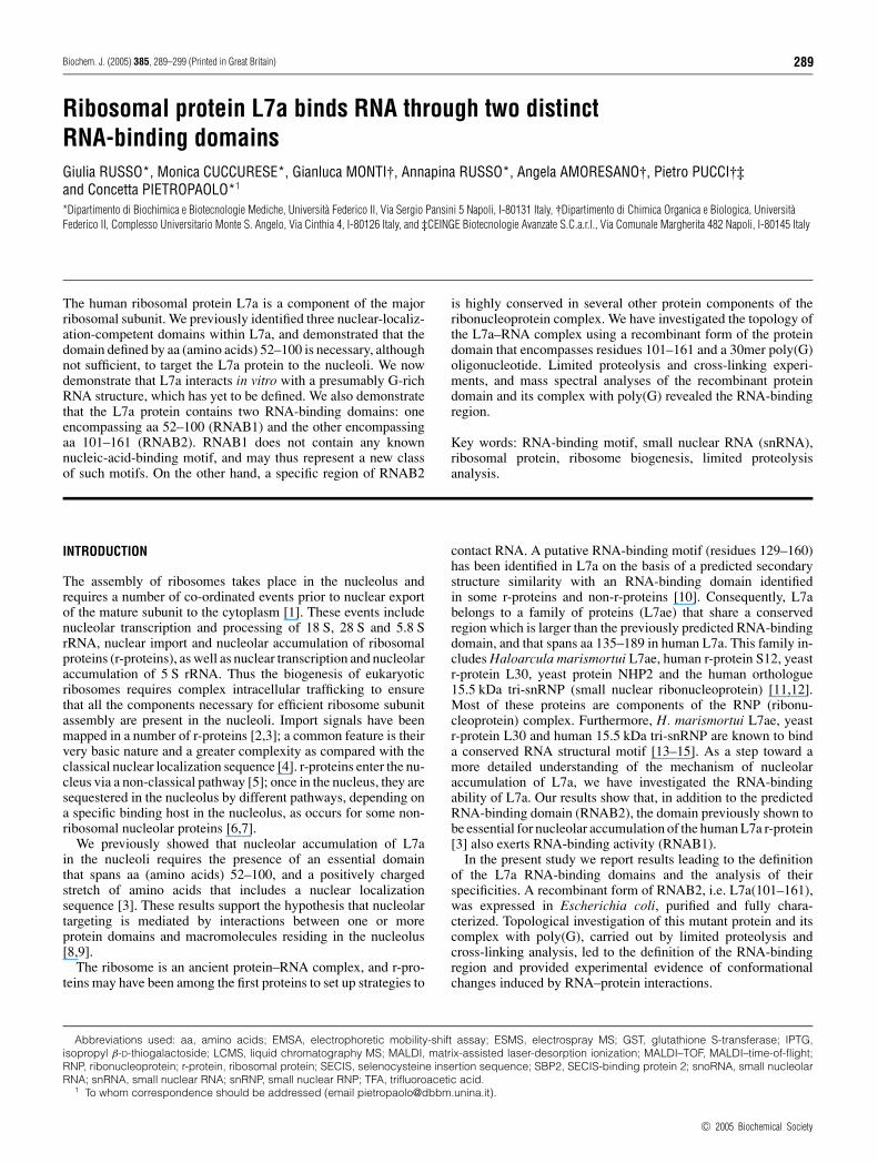
Figure 1 Analysis of RNA-binding ability of human r-protein L7a
Purified recombinant L7a–His protein (L7a) and molecular mass marker proteins (MW) wereloaded on to an SDS gel. Upon electrophoresis, gel samples were stained with CoomassieBrilliant Blue (A, left-hand panel), or transferred on to a nitrocellulose membrane, probed withthe indicated radiolabelled RNAs and subjected to autoradiography (A, right-hand panels; foran explanation for the lanes marked MW see the text). (B) Plot of binding activity of L7a–Histo radiolabelled RNAs. For details of nucleic acid labelling and filter-binding assay, see theExperimental section.
(Figure 1A) demonstrate that the binding of L7a to RNA instringent conditions was specific.
We tested the RNA-binding capacity of the purified recom-binant L7a protein also by filter-binding assay (Figure 1B). L7amRNA, transcribed in vitro, and 28 S rRNA, electroluted fromagarose gel, were used as ligands in the filter-binding analysis.L7a was able to bind, in a dose-dependent manner, its own mRNAand 28 S rRNA, whereas it failed to bind poly(C), thus confirmingthe results of the Northwestern assay. We determined from a graphan apparent Kd of 75 nM for L7a mRNA and of 85 nM for 28 SrRNA. The Kd values are in the range observed when assayingthe RNA-binding activity of individual r-proteins in vitro; a co-operative effect mediated by interacting macromolecules ensuresa greater affinity in vivo [19].
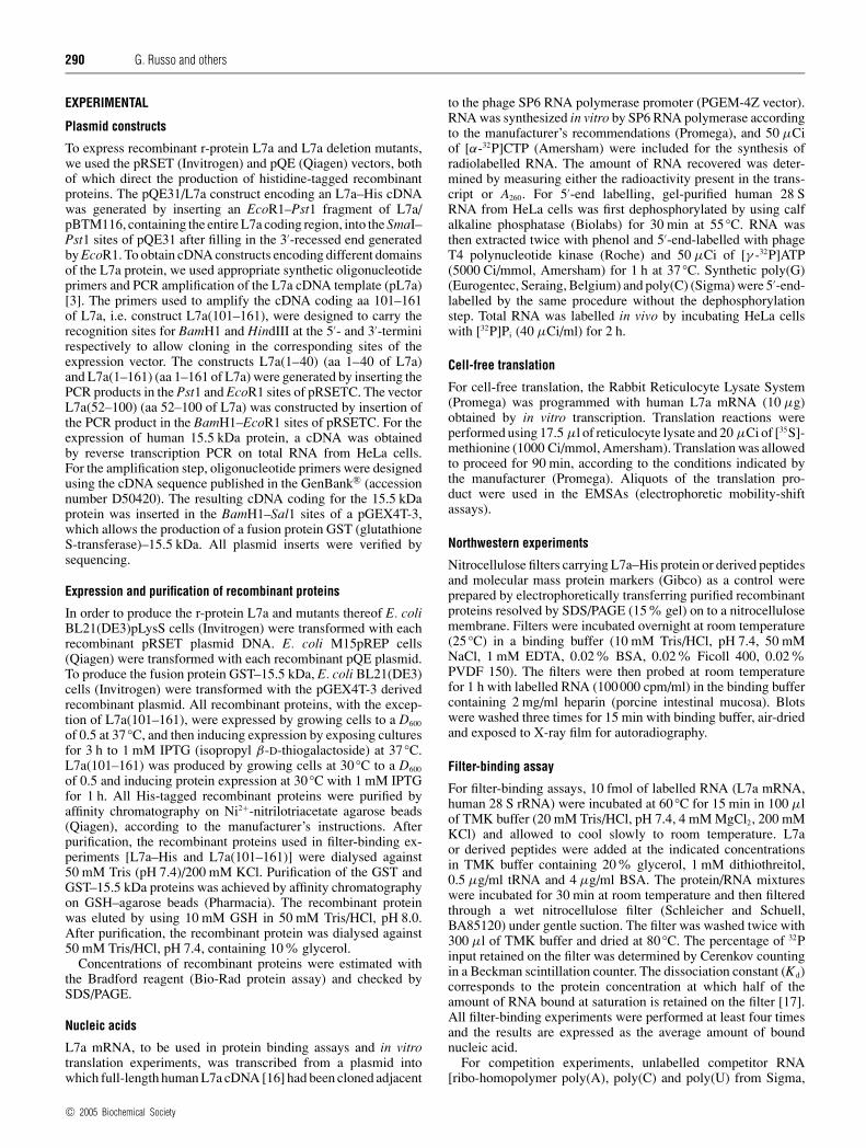
We also used the His–L7a protein to analyse the RNA-bindingspecificity of L7a in a filter-binding competition assay. Variousnucleic acids served as competitors for the binding of 32P-labelledL7a mRNA to His–L7a. The L7a mRNA was chosen because itis able to bind L7a (see Figure 1), and it can be obtained as apure single species by in vitro transcription. The binding activ-ity of L7a to its own mRNA was considered to be 100%; asinternal standard, we used unlabelled L7a mRNA as competitor.The results of this analysis are shown in Figure 2. RNA bindingwas specifically competed by low concentrations of unlabelledpoly(G), but not by an excess of other ribo-homopolymers (asan example, the results obtained with poly(C) are reported inFigure 2), or single-stranded DNA.
Figure 2 RNA-binding specificity of human r-protein L7a
Filter-binding assay of competition of the binding of L7a–His to radiolabelled L7a mRNA byincreasing amounts of unlabelled nucleic acids. The binding reaction contained 10 fmol oflabelled L7a mRNA, to which a competitor nucleic acid was added before the addition of 500 nML7a–His. The assay was performed as described in the Experimental section. The bound RNAvalues were corrected for the radioactivity retained on the filter when no L7a protein was addedto the labelled L7a mRNA. The RNA-binding activity of L7a protein to radiolabelled L7a mRNA inthe absence of a competitor was considered as 100 %.
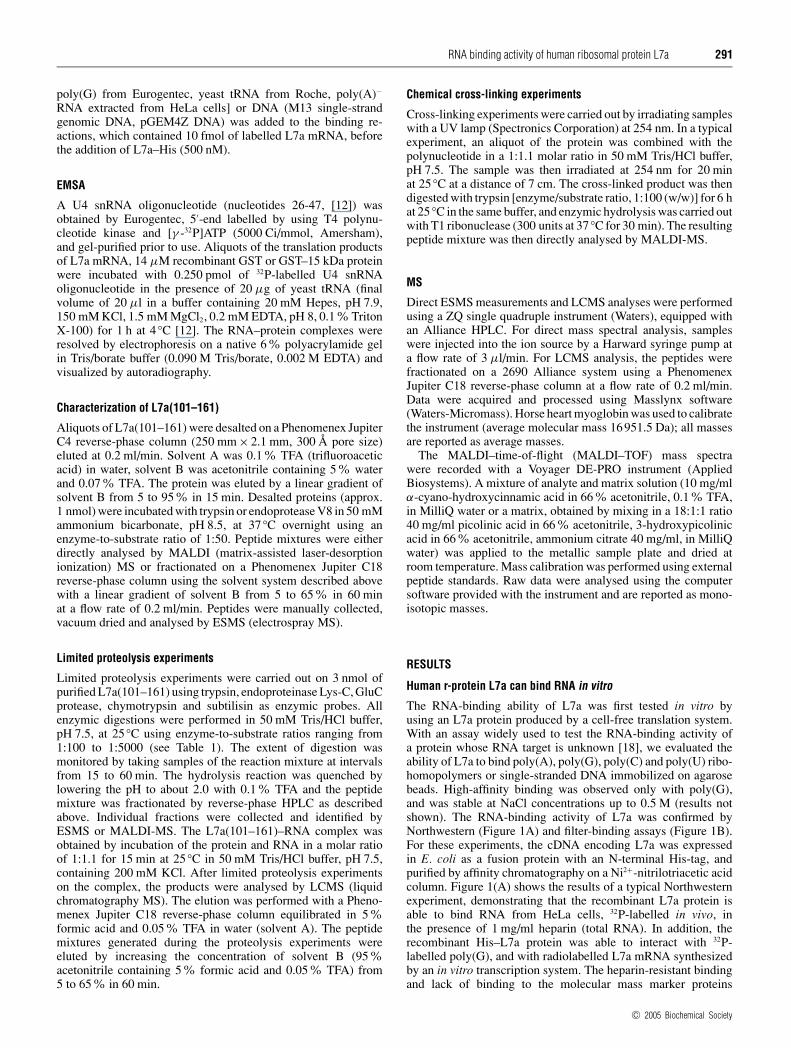
Figure 3 Definition of RNA-binding domains of the human r-protein L7a
(A) Purified recombinant L7a-His deletion mutants were subjected to electrophoresis on anSDS/PAGE gel and then transferred on to a nitrocellulose membrane. The filter was probed witha commercially available antiserum against the His-tag (Santa Cruz Biotechnology) (left-handpanel). The RNA-binding activity of all deletion mutants was tested by Northwestern experimentsusing radiolabelled L7a mRNA (right-hand panel). (B) Summary of the RNA-binding ability ofL7a deletion mutants. Striped boxes indicate the domain required for nucleolar targeting of theprotein, and dotted regions indicate the putative RNA-binding motif [10].
Protein L7a carries two RNA-binding motifs
In a previous study, we dissected the L7a protein to identifydomains that could mediate the nuclear import and nucleolartargeting of the protein [3], and demonstrated that a domainspanning aa 52–100 is essential for the nucleolar accumulationof L7a (Figure 3B). In an attempt to understand the relationshipbetween RNA-binding activity and the subcellular localization
c© 2005 Biochemical Society
RNA binding activity of human ribosomal protein L7a 293
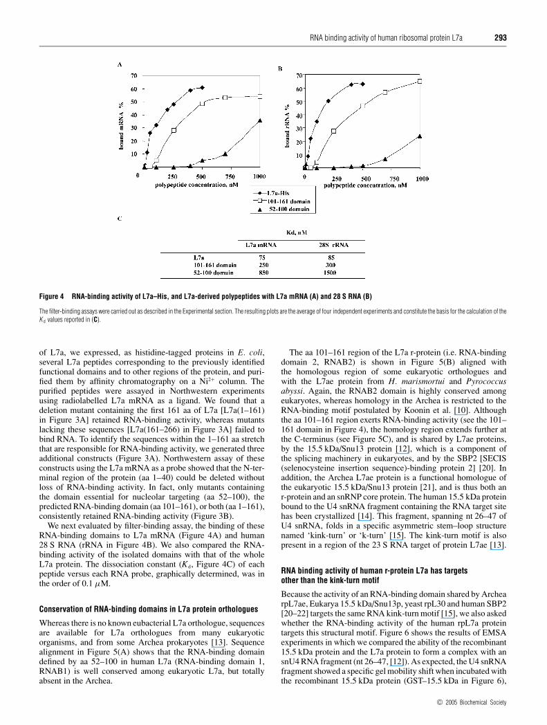
Figure 4 RNA-binding activity of L7a–His, and L7a-derived polypeptides with L7a mRNA (A) and 28 S RNA (B)
The filter-binding assays were carried out as described in the Experimental section. The resulting plots are the average of four independent experiments and constitute the basis for the calculation of theK d values reported in (C).
of L7a, we expressed, as histidine-tagged proteins in E. coli,several L7a peptides corresponding to the previously identifiedfunctional domains and to other regions of the protein, and puri-fied them by affinity chromatography on a Ni2+ column. Thepurified peptides were assayed in Northwestern experimentsusing radiolabelled L7a mRNA as a ligand. We found that adeletion mutant containing the first 161 aa of L7a [L7a(1–161)in Figure 3A] retained RNA-binding activity, whereas mutantslacking these sequences [L7a(161–266) in Figure 3A] failed tobind RNA. To identify the sequences within the 1–161 aa stretchthat are responsible for RNA-binding activity, we generated threeadditional constructs (Figure 3A). Northwestern assay of theseconstructs using the L7a mRNA as a probe showed that the N-ter-minal region of the protein (aa 1–40) could be deleted withoutloss of RNA-binding activity. In fact, only mutants containingthe domain essential for nucleolar targeting (aa 52–100), thepredicted RNA-binding domain (aa 101–161), or both (aa 1–161),consistently retained RNA-binding activity (Figure 3B).
We next evaluated by filter-binding assay, the binding of theseRNA-binding domains to L7a mRNA (Figure 4A) and human28 S RNA (rRNA in Figure 4B). We also compared the RNA-binding activity of the isolated domains with that of the wholeL7a protein. The dissociation constant (Kd, Figure 4C) of eachpeptide versus each RNA probe, graphically determined, was inthe order of 0.1 µM.
Conservation of RNA-binding domains in L7a protein orthologues
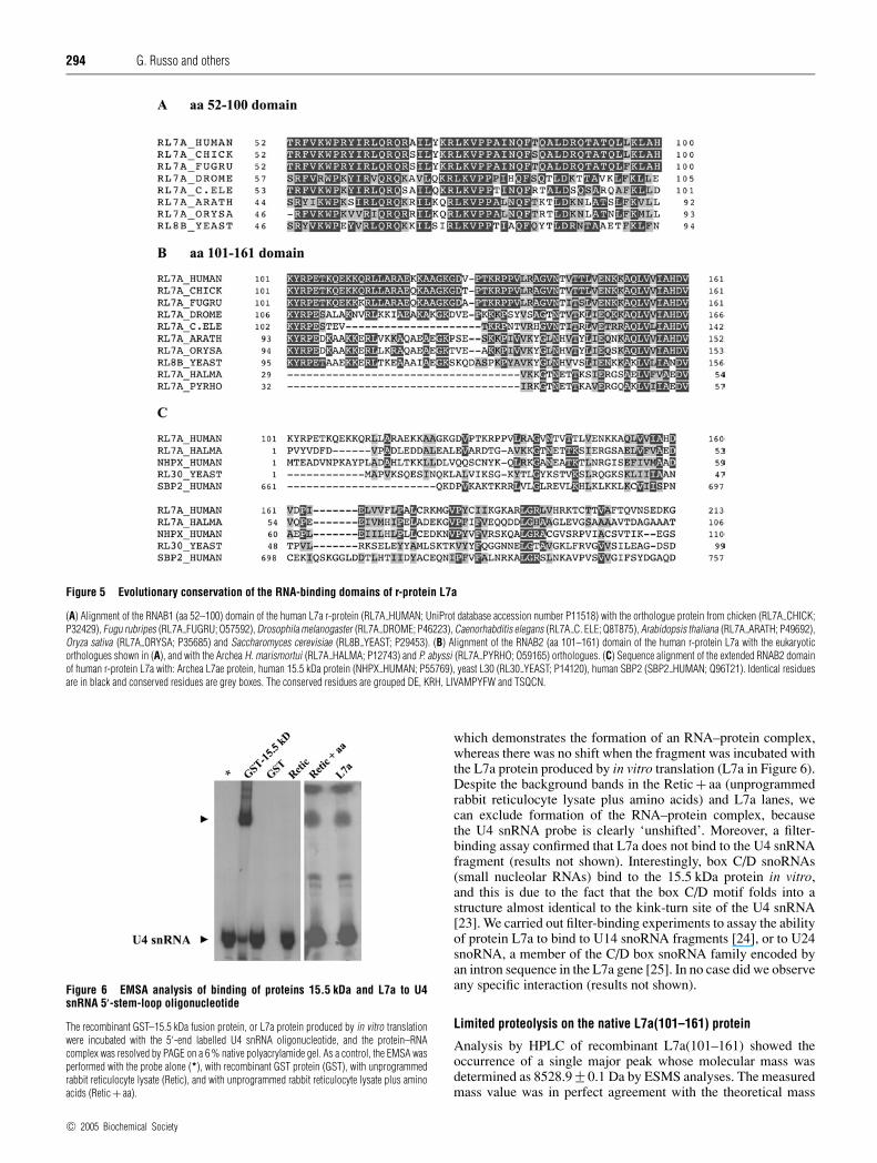
Whereas there is no known eubacterial L7a orthologue, sequencesare available for L7a orthologues from many eukaryoticorganisms, and from some Archea prokaryotes [13]. Sequencealignment in Figure 5(A) shows that the RNA-binding domaindefined by aa 52–100 in human L7a (RNA-binding domain 1,RNAB1) is well conserved among eukaryotic L7a, but totallyabsent in the Archea.
The aa 101–161 region of the L7a r-protein (i.e. RNA-bindingdomain 2, RNAB2) is shown in Figure 5(B) aligned withthe homologous region of some eukaryotic orthologues andwith the L7ae protein from H. marismortui and Pyrococcusabyssi. Again, the RNAB2 domain is highly conserved amongeukaryotes, whereas homology in the Archea is restricted to theRNA-binding motif postulated by Koonin et al. [10]. Althoughthe aa 101–161 region exerts RNA-binding activity (see the 101–161 domain in Figure 4), the homology region extends further atthe C-terminus (see Figure 5C), and is shared by L7ae proteins,by the 15.5 kDa/Snu13 protein [12], which is a component ofthe splicing machinery in eukaryotes, and by the SBP2 [SECIS(selenocysteine insertion sequence)-binding protein 2] [20]. Inaddition, the Archea L7ae protein is a functional homologue ofthe eukaryotic 15.5 kDa/Snu13 protein [21], and is thus both anr-protein and an snRNP core protein. The human 15.5 kDa proteinbound to the U4 snRNA fragment containing the RNA target sitehas been crystallized [14]. This fragment, spanning nt 26–47 ofU4 snRNA, folds in a specific asymmetric stem–loop structurenamed ‘kink-turn’ or ‘k-turn’ [15]. The kink-turn motif is alsopresent in a region of the 23 S RNA target of protein L7ae [13].
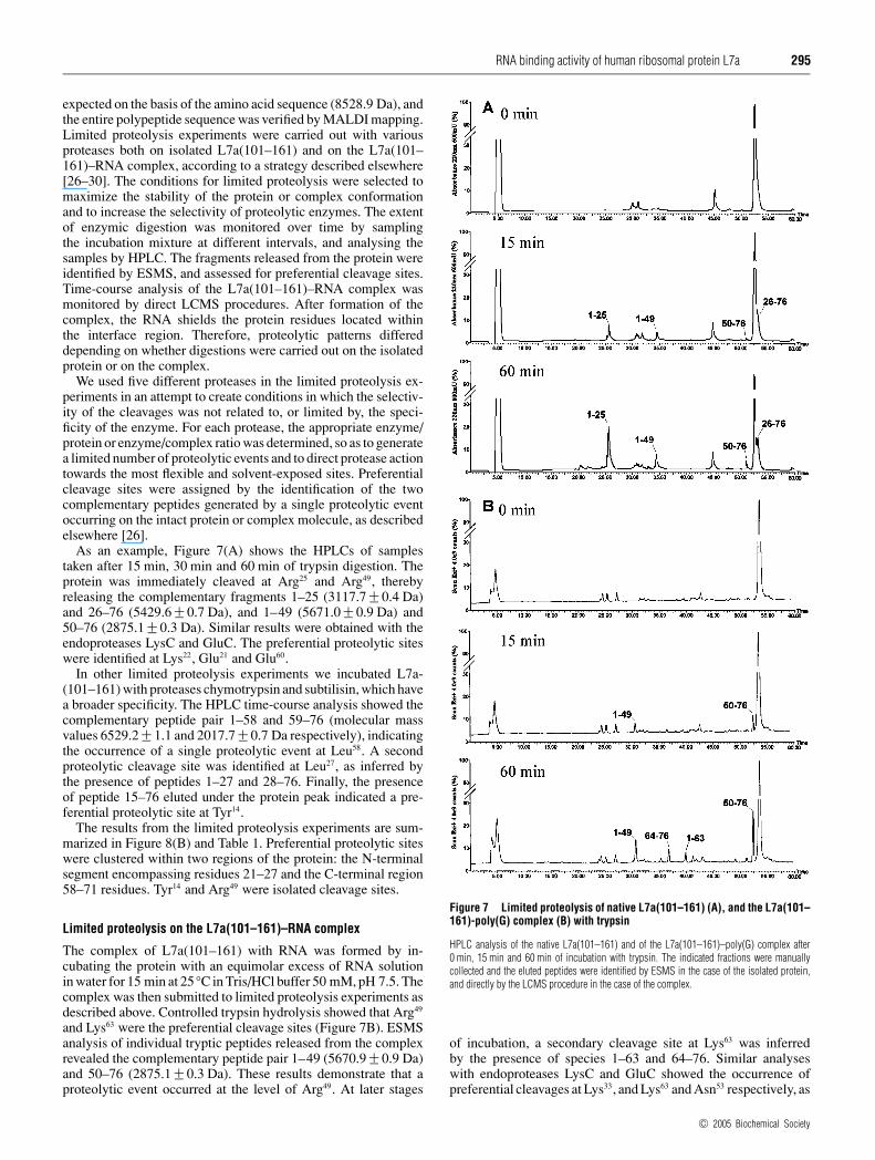
RNA binding activity of human r-protein L7a has targetsother than the kink-turn motif
Because the activity of an RNA-binding domain shared by ArchearpL7ae, Eukarya 15.5 kDa/Snu13p, yeast rpL30 and human SBP2[20–22] targets the same RNA kink-turn motif [15], we also askedwhether the RNA-binding activity of the human rpL7a proteintargets this structural motif. Figure 6 shows the results of EMSAexperiments in which we compared the ability of the recombinant15.5 kDa protein and the L7a protein to form a complex with ansnU4 RNA fragment (nt 26–47, [12]). As expected, the U4 snRNAfragment showed a specific gel mobility shift when incubated withthe recombinant 15.5 kDa protein (GST–15.5 kDa in Figure 6),
c© 2005 Biochemical Society
294 G. Russo and others
Figure 5 Evolutionary conservation of the RNA-binding domains of r-protein L7a
(A) Alignment of the RNAB1 (aa 52–100) domain of the human L7a r-protein (RL7A HUMAN; UniProt database accession number P11518) with the orthologue protein from chicken (RL7A CHICK;P32429), Fugu rubripes (RL7A FUGRU; O57592), Drosophila melanogaster (RL7A DROME; P46223), Caenorhabditis elegans (RL7A C. ELE; Q8T875), Arabidopsis thaliana (RL7A ARATH; P49692),Oryza sativa (RL7A ORYSA; P35685) and Saccharomyces cerevisiae (RL8B YEAST; P29453). (B) Alignment of the RNAB2 (aa 101–161) domain of the human r-protein L7a with the eukaryoticorthologues shown in (A), and with the Archea H. marismortui (RL7A HALMA; P12743) and P. abyssi (RL7A PYRHO; O59165) orthologues. (C) Sequence alignment of the extended RNAB2 domainof human r-protein L7a with: Archea L7ae protein, human 15.5 kDa protein (NHPX HUMAN; P55769), yeast L30 (RL30 YEAST; P14120), human SBP2 (SBP2 HUMAN; Q96T21). Identical residuesare in black and conserved residues are grey boxes. The conserved residues are grouped DE, KRH, LIVAMPYFW and TSQCN.
Figure 6 EMSA analysis of binding of proteins 15.5 kDa and L7a to U4snRNA 5′-stem-loop oligonucleotide
The recombinant GST–15.5 kDa fusion protein, or L7a protein produced by in vitro translationwere incubated with the 5′-end labelled U4 snRNA oligonucleotide, and the protein–RNAcomplex was resolved by PAGE on a 6 % native polyacrylamide gel. As a control, the EMSA wasperformed with the probe alone (*), with recombinant GST protein (GST), with unprogrammedrabbit reticulocyte lysate (Retic), and with unprogrammed rabbit reticulocyte lysate plus aminoacids (Retic + aa).
which demonstrates the formation of an RNA–protein complex,whereas there was no shift when the fragment was incubated withthe L7a protein produced by in vitro translation (L7a in Figure 6).Despite the background bands in the Retic + aa (unprogrammedrabbit reticulocyte lysate plus amino acids) and L7a lanes, wecan exclude formation of the RNA–protein complex, becausethe U4 snRNA probe is clearly ‘unshifted’. Moreover, a filter-binding assay confirmed that L7a does not bind to the U4 snRNAfragment (results not shown). Interestingly, box C/D snoRNAs(small nucleolar RNAs) bind to the 15.5 kDa protein in vitro,and this is due to the fact that the box C/D motif folds into astructure almost identical to the kink-turn site of the U4 snRNA[23]. We carried out filter-binding experiments to assay the abilityof protein L7a to bind to U14 snoRNA fragments [24], or to U24snoRNA, a member of the C/D box snoRNA family encoded byan intron sequence in the L7a gene [25]. In no case did we observeany specific interaction (results not shown).
Limited proteolysis on the native L7a(101–161) protein
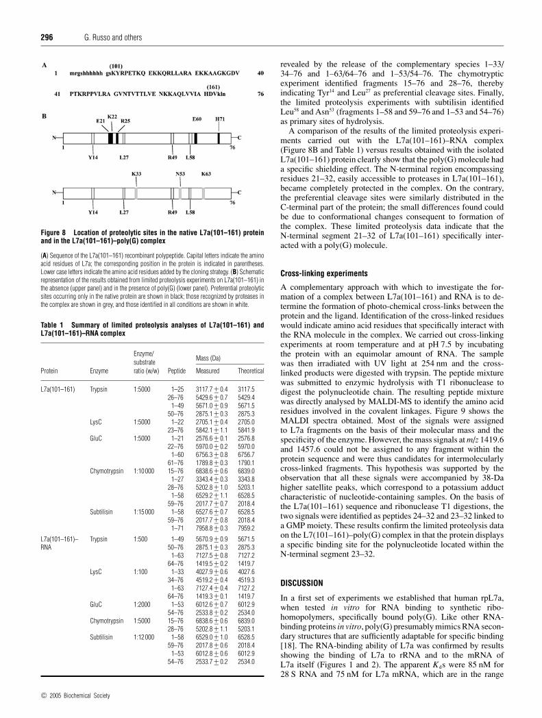
Analysis by HPLC of recombinant L7a(101–161) showed theoccurrence of a single major peak whose molecular mass wasdetermined as 8528.9 +− 0.1 Da by ESMS analyses. The measuredmass value was in perfect agreement with the theoretical mass
c© 2005 Biochemical Society
RNA binding activity of human ribosomal protein L7a 295
expected on the basis of the amino acid sequence (8528.9 Da), andthe entire polypeptide sequence was verified by MALDI mapping.Limited proteolysis experiments were carried out with variousproteases both on isolated L7a(101–161) and on the L7a(101–161)–RNA complex, according to a strategy described elsewhere[26–30]. The conditions for limited proteolysis were selected tomaximize the stability of the protein or complex conformationand to increase the selectivity of proteolytic enzymes. The extentof enzymic digestion was monitored over time by samplingthe incubation mixture at different intervals, and analysing thesamples by HPLC. The fragments released from the protein wereidentified by ESMS, and assessed for preferential cleavage sites.Time-course analysis of the L7a(101–161)–RNA complex wasmonitored by direct LCMS procedures. After formation of thecomplex, the RNA shields the protein residues located withinthe interface region. Therefore, proteolytic patterns differeddepending on whether digestions were carried out on the isolatedprotein or on the complex.
We used five different proteases in the limited proteolysis ex-periments in an attempt to create conditions in which the selectiv-ity of the cleavages was not related to, or limited by, the speci-ficity of the enzyme. For each protease, the appropriate enzyme/protein or enzyme/complex ratio was determined, so as to generatea limited number of proteolytic events and to direct protease actiontowards the most flexible and solvent-exposed sites. Preferentialcleavage sites were assigned by the identification of the twocomplementary peptides generated by a single proteolytic eventoccurring on the intact protein or complex molecule, as describedelsewhere [26].
As an example, Figure 7(A) shows the HPLCs of samplestaken after 15 min, 30 min and 60 min of trypsin digestion. Theprotein was immediately cleaved at Arg25 and Arg49, therebyreleasing the complementary fragments 1–25 (3117.7 +− 0.4 Da)and 26–76 (5429.6 +− 0.7 Da), and 1–49 (5671.0 +− 0.9 Da) and50–76 (2875.1 +− 0.3 Da). Similar results were obtained with theendoproteases LysC and GluC. The preferential proteolytic siteswere identified at Lys22, Glu21 and Glu60.
In other limited proteolysis experiments we incubated L7a-(101–161) with proteases chymotrypsin and subtilisin, which havea broader specificity. The HPLC time-course analysis showed thecomplementary peptide pair 1–58 and 59–76 (molecular massvalues 6529.2 +− 1.1 and 2017.7 +− 0.7 Da respectively), indicatingthe occurrence of a single proteolytic event at Leu58. A secondproteolytic cleavage site was identified at Leu27, as inferred bythe presence of peptides 1–27 and 28–76. Finally, the presenceof peptide 15–76 eluted under the protein peak indicated a pre-ferential proteolytic site at Tyr14.
The results from the limited proteolysis experiments are sum-marized in Figure 8(B) and Table 1. Preferential proteolytic siteswere clustered within two regions of the protein: the N-terminalsegment encompassing residues 21–27 and the C-terminal region58–71 residues. Tyr14 and Arg49 were isolated cleavage sites.
Limited proteolysis on the L7a(101–161)–RNA complex
The complex of L7a(101–161) with RNA was formed by in-cubating the protein with an equimolar excess of RNA solutionin water for 15 min at 25 ◦C in Tris/HCl buffer 50 mM, pH 7.5. Thecomplex was then submitted to limited proteolysis experiments asdescribed above. Controlled trypsin hydrolysis showed that Arg49
and Lys63 were the preferential cleavage sites (Figure 7B). ESMSanalysis of individual tryptic peptides released from the complexrevealed the complementary peptide pair 1–49 (5670.9 +− 0.9 Da)and 50–76 (2875.1 +− 0.3 Da). These results demonstrate that aproteolytic event occurred at the level of Arg49. At later stages
Figure 7 Limited proteolysis of native L7a(101–161) (A), and the L7a(101–161)-poly(G) complex (B) with trypsin
HPLC analysis of the native L7a(101–161) and of the L7a(101–161)–poly(G) complex after0 min, 15 min and 60 min of incubation with trypsin. The indicated fractions were manuallycollected and the eluted peptides were identified by ESMS in the case of the isolated protein,and directly by the LCMS procedure in the case of the complex.
of incubation, a secondary cleavage site at Lys63 was inferredby the presence of species 1–63 and 64–76. Similar analyseswith endoproteases LysC and GluC showed the occurrence ofpreferential cleavages at Lys33, and Lys63 and Asn53 respectively, as
c© 2005 Biochemical Society
296 G. Russo and others
Figure 8 Location of proteolytic sites in the native L7a(101–161) proteinand in the L7a(101–161)–poly(G) complex
(A) Sequence of the L7a(101–161) recombinant polypeptide. Capital letters indicate the aminoacid residues of L7a; the corresponding position in the protein is indicated in parentheses.Lower case letters indicate the amino acid residues added by the cloning strategy. (B) Schematicrepresentation of the results obtained from limited proteolysis experiments on L7a(101–161) inthe absence (upper panel) and in the presence of poly(G) (lower panel). Preferential proteolyticsites occurring only in the native protein are shown in black; those recognized by proteases inthe complex are shown in grey, and those identified in all conditions are shown in white.
Table 1 Summary of limited proteolysis analyses of L7a(101–161) andL7a(101–161)–RNA complex
Enzyme/substrate Mass (Da)
Protein Enzyme ratio (w/w) Peptide Measured Theoretical
L7a(101–161) Trypsin 1:5000 1–25 3117.7 +− 0.4 3117.526–76 5429.6 +− 0.7 5429.4
1–49 5671.0 +− 0.9 5671.550–76 2875.1 +− 0.3 2875.3
LysC 1:5000 1–22 2705.1 +− 0.4 2705.023–76 5842.1 +− 1.1 5841.9
GluC 1:5000 1–21 2576.6 +− 0.1 2576.822–76 5970.0 +− 0.2 5970.0
1–60 6756.3 +− 0.8 6756.761–76 1789.8 +− 0.3 1790.1
Chymotrypsin 1:10 000 15–76 6838.6 +− 0.6 6839.01–27 3343.4 +− 0.3 3343.8
28–76 5202.8 +− 1.0 5203.11–58 6529.2 +− 1.1 6528.5
59–76 2017.7 +− 0.7 2018.4Subtilisin 1:15 000 1–58 6527.6 +− 0.7 6528.5
59–76 2017.7 +− 0.8 2018.41–71 7958.8 +− 0.3 7959.2
L7a(101–161)– Trypsin 1:500 1–49 5670.9 +− 0.9 5671.5RNA 50–76 2875.1 +− 0.3 2875.3
1–63 7127.5 +− 0.8 7127.264–76 1419.5 +− 0.2 1419.7
LysC 1:100 1–33 4027.9 +− 0.6 4027.634–76 4519.2 +− 0.4 4519.3
1–63 7127.4 +− 0.4 7127.264–76 1419.3 +− 0.1 1419.7
GluC 1:2000 1–53 6012.6 +− 0.7 6012.954–76 2533.8 +− 0.2 2534.0
Chymotrypsin 1:5000 15–76 6838.6 +− 0.6 6839.028–76 5202.8 +− 1.1 5203.1
Subtilisin 1:12 000 1–58 6529.0 +− 1.0 6528.559–76 2017.8 +− 0.6 2018.4
1–53 6012.8 +− 0.6 6012.954–76 2533.7 +− 0.2 2534.0
revealed by the release of the complementary species 1–33/34–76 and 1–63/64–76 and 1–53/54–76. The chymotrypticexperiment identified fragments 15–76 and 28–76, therebyindicating Tyr14 and Leu27 as preferential cleavage sites. Finally,the limited proteolysis experiments with subtilisin identifiedLeu58 and Asn53 (fragments 1–58 and 59–76 and 1–53 and 54–76)as primary sites of hydrolysis.
A comparison of the results of the limited proteolysis experi-ments carried out with the L7a(101–161)–RNA complex(Figure 8B and Table 1) versus results obtained with the isolatedL7a(101–161) protein clearly show that the poly(G) molecule hada specific shielding effect. The N-terminal region encompassingresidues 21–32, easily accessible to proteases in L7a(101–161),became completely protected in the complex. On the contrary,the preferential cleavage sites were similarly distributed in theC-terminal part of the protein; the small differences found couldbe due to conformational changes consequent to formation ofthe complex. These limited proteolysis data indicate that theN-terminal segment 21–32 of L7a(101–161) specifically inter-acted with a poly(G) molecule.
Cross-linking experiments
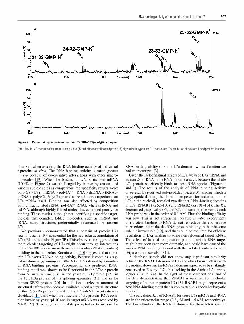
A complementary approach with which to investigate the for-mation of a complex between L7a(101–161) and RNA is to de-termine the formation of photo-chemical cross-links between theprotein and the ligand. Identification of the cross-linked residueswould indicate amino acid residues that specifically interact withthe RNA molecule in the complex. We carried out cross-linkingexperiments at room temperature and at pH 7.5 by incubatingthe protein with an equimolar amount of RNA. The samplewas then irradiated with UV light at 254 nm and the cross-linked products were digested with trypsin. The peptide mixturewas submitted to enzymic hydrolysis with T1 ribonuclease todigest the polynucleotide chain. The resulting peptide mixturewas directly analysed by MALDI-MS to identify the amino acidresidues involved in the covalent linkages. Figure 9 shows theMALDI spectra obtained. Most of the signals were assignedto L7a fragments on the basis of their molecular mass and thespecificity of the enzyme. However, the mass signals at m/z 1419.6and 1457.6 could not be assigned to any fragment within theprotein sequence and were thus candidates for intermolecularlycross-linked fragments. This hypothesis was supported by theobservation that all these signals were accompanied by 38-Dahigher satellite peaks, which correspond to a potassium adductcharacteristic of nucleotide-containing samples. On the basis ofthe L7a(101–161) sequence and ribonuclease T1 digestions, thetwo signals were identified as peptides 24–32 and 23–32 linked toa GMP moiety. These results confirm the limited proteolysis dataon the L7(101–161)–poly(G) complex in that the protein displaysa specific binding site for the polynucleotide located within theN-terminal segment 23–32.
DISCUSSION
In a first set of experiments we established that human rpL7a,when tested in vitro for RNA binding to synthetic ribo-homopolymers, specifically bound poly(G). Like other RNA-binding proteins in vitro, poly(G) presumably mimics RNA secon-dary structures that are sufficiently adaptable for specific binding[18]. The RNA-binding ability of L7a was confirmed by resultsshowing the binding of L7a to rRNA and to the mRNA ofL7a itself (Figures 1 and 2). The apparent Kds were 85 nM for28 S RNA and 75 nM for L7a mRNA, which are in the range
c© 2005 Biochemical Society
RNA binding activity of human ribosomal protein L7a 297
Figure 9 Cross-linking experiment on the L7a(101–161)–poly(G) complex
Partial MALDI-MS spectrum of the cross-linked product (A) and of the control isolated protein (B) digested with trypsin and T1 ribonuclease. The attribution of the cross-linked peptides is shown.
observed when assaying the RNA-binding activity of individualr-proteins in vitro. The RNA-binding activity is much greaterin vivo because of co-operative interactions with other macro-molecules [19]. When the binding of L7a to its own mRNA(100% in Figure 2) was challenged by increasing amounts ofvarious nucleic acids as competitors, the specificity results were:poly(G) > L7a mRNA > poly(A)− RNA > dsDNA > tRNA >ssDNA > poly(C). Poly(G) proved to be a better competitor thanL7a mRNA itself. Binding was also affected by competitionwith unfractionated rRNA [poly(A)− RNA], whereas tRNA anddsDNA, although highly folded molecules, competed poorly forbinding. These results, although not identifying a specific target,indicate that complex folded molecules, such as mRNA andrRNA, carry structures preferentially recognized by proteinL7a.
We previously demonstrated that a domain of protein L7aspanning aa 52–100 is essential for the nucleolar accumulation ofL7a ([3], and see also Figure 3B). This observation suggested thatthe nucleolar targeting of L7a might occur through interactionsof the 52–100 aa domain with macromolecules (RNA or protein)residing in the nucleolus. Koonin et al. [10] suggested that r-pro-tein L7a exerts RNA-binding activity, because it contains a sig-nature domain (spanning aa 130–160 in L7a) shared by a numberof RNA-binding proteins. Subsequently, the predicted RNA-binding motif was shown to be functional in the L7ae r-proteinfrom H. marismortui [13], in the yeast rpL30 protein [22], inthe 15.5 kDa protein of the splicing apparatus [21], and in thehuman SBP2 protein [20]. In addition, a relevant amount ofstructural information became available when a crystal structureof the 15.5 kDa protein bound to the U4 snRNA target site waselucidated [14], and when the structure of the protein–RNA com-plex involving yeast rpL30 and its target mRNA was resolved byNMR [22]. This large body of data prompted us to analyse the
RNA-binding ability of some L7a domains whose function wehad characterized [3].
Given the lack of natural targets of L7a, we used L7a mRNA andhuman 28 S rRNA in the RNA-binding assays, because the wholeL7a protein specifically binds to these RNA species (Figures 1and 2). The results of the analysis of RNA binding activityof several L7a-derived polypeptides (Figure 3), among which apolypeptide defining the domain competent for accumulation ofL7a in the nucleoli, revealed two distinct RNA-binding domainsin L7a: RNAB1 (aa 52–100) and RNAB2 (aa 101–161). The Kd,determined graphically (Figure 4C), for each peptide versus eachRNA probe was in the order of 0.1 µM. Thus the binding affinitywas low. This is not surprising, because in vitro experimentsof r-protein binding to RNA do not reproduce the cooperativeinteractions that make the RNA–protein binding in the ribosomesubunit irreversible [19], and that could be required for efficientregulation of L7a binding to some non-ribosomal target RNAs.The effect of lack of co-operation plus a spurious RNA targetmight have been even more dramatic, and could have caused theweaker RNA binding obtained with the isolated protein domains(Figure 4, and see also [31]).
A database search did not show any significant similaritybetween the RNAB1 domain of L7a and other known RNA-bind-ing motifs. However, the RNAB1 domain appeared to be strikinglyconserved in Eukarya L7a, but lacking in the Archea L7a ortho-logues (Figure 5A). In the light of these observations, and ofthe data demonstrating that RNAB1 is essential for nucleolartargeting of human r-protein L7a [3], RNAB1 might represent anew RNA-binding motif that is committed to a special eukaryoticfunction.
The calculated Kds of RNAB1 for these L7a mRNA and rRNAare in the micromolar range (0.8 µM and 1.5 µM, respectively).The low affinity of the RNAB1 domain for these RNA species
c© 2005 Biochemical Society
298 G. Russo and others
may be because we used non-specific targets, i.e. L7a mRNAand rRNA, or because of a lack of co-operative interactions withother macromolecules that regulate the RNA-binding efficiencyin vivo. The identification of a specific physiological RNAtarget of RNAB1 will clarify the function of this L7a proteindomain.
The RNAB2 domain is highly conserved among Eukarya L7aorthologues (Figure 5B), whereas the similarity with the ArcheaL7ae protein is restricted to the region that includes the RNA-binding motif predicted by Koonin et al. [10]. However, thesimilarity extends further at the C-terminus in proteins that re-cognize the secondary structure motif designated kink-turn [15](Figure 5C). Since binding to a kink-turn seemed to be acommon functional feature of L7ae proteins, we carried outexperiments to verify whether human r-protein L7a would bind toan oligonucleotide fragment of spliceosomal U4 snRNA, whichhad been crystallized bound to the 15.5 kDa protein [14]. Toour surprise, neither the whole protein L7a (see Figure 6) norits isolated RNA-binding domains bound to the U4 snRNAoligonucleotide. Archea L7a was recently found to bind in vitroto Archea sRNAs of the C/D box family, which contain a kink-turn RNA motif [32]. By filter-binding assay, we did not detectany specific interaction of human r-protein L7a with U14 orU24 snoRNA, both of which belong to the C/D box family ofEukarya snoRNAs.
In the absence of a natural RNA target for the human L7ar-protein, we carried out a structural analysis of the complexformed by the RNAB2 domain of r-protein L7a and poly(G). Ouraim was to obtain information about the structural modificationsundergone by the r-protein after its binding to the surrogate RNAtarget. We used the limited proteolysis technique followed byESMS analysis of the products (Figures 7 and 8, and Table 1), andcross-linking experiments followed by MALDI-MS to identifyamino acid residues involved in covalent linkages (Figure 9). Theresults obtained with the two methodological approaches led tothe identification of a region (KQRLLARAEK) that was shieldedfrom proteases consequent to binding to poly(G) and that wasinvolved in covalent linkages after UV cross-linking. Allmanget al. [20] addressed the interesting issue of how the various L7aeproteins specifically recognize their cognate RNA, despite a highdegree of similarity in their RNA-binding domain by comparingthe amino acid residues relevant for contact with the cognateRNA in human SBP2 and 15.5 kDa proteins [20]. They identified12 aa residues that are important for SECIS RNA binding: mostcontact residues were predicted from the sequence similaritybetween SBP2 and 15.5 kDa RNA-binding domains, albeit theircontribution to binding efficiency was different. The contact roleplayed by three residues could not be predicted from sequencesimilarity, which suggested that non-conserved amino acids couldbe instrumental in determining the specificity of interaction. Ourfindings lend weight to this hypothesis. In addition, they raise thepossibility that a region flanking the RNA-binding domain mightcontribute to the specificity of binding of a protein to its cognateRNA.
This work has been supported by MIUR, Progetti di Ricerca di Interesse Nazionale (PRIN2003) and Fondo Investimenti Ricerca di Base (FIRB). We thank Jean A. Gilder for editingthe text.
REFERENCES
1 Hernandez-Verdun, D., Roussel, P. and Gebrane-Younes, J. (2002) Emerging concepts ofnucleolar assembly. J. Cell Sci. 115, 2265–2270
2 Rudt, F. and Pieler, T. (2001) Cytosolic import factor- and Ran-independent nucleartransport of ribosomal protein L5. Eur. J. Cell Biol. 80, 661–668
3 Russo, G., Ricciardelli, G. and Pietropaolo, C. (1997) Different domains cooperate totarget the human ribosomal L7a protein to the nucleus and to the nucleoli. J. Biol. Chem.272, 5229–5235
4 Damelin, M., Silver, P. A. and Corbett, A. H. (2002) Nuclear protein transport.Methods Enzymol. 351, 587–607
5 Jakel, S., Mingot, J. M., Schwarzmaier, P., Hartmann, E. and Gorlich, D. (2002) Importinsfulfil a dual function as nuclear import receptors and cytoplasmic chaperones for exposedbasic domains. EMBO J. 21, 377–386
6 Rosorius, O., Fries, B., Stauber, R. H., Hirschmann, N., Bevec, D. and Hauber, J. (2000)Human ribosomal protein L5 contains defined nuclear localization and export signals.J. Biol. Chem. 275, 12061–12068
7 Etheridge, K. T., Banik, S. S., Armbruster, B. N., Zhu, Y., Terns, R. M., Terns, M. P. andCounter, C. M. (2002) The nucleolar localization domain of the catalytic subunit of humantelomerase. J. Biol. Chem. 277, 24764–24770
8 Lee, C. H., Chang, S. C., Chen, C. J. and Chang, M. F. (1998) The nucleolin bindingactivity of hepatitis delta antigen is associated with nucleolus targeting. J. Biol. Chem.273, 7650–7656
9 Lixin, R., Efthymiadis, A., Henderson, B. and Jans, D. A. (2001) Novel properties of thenucleolar targeting signal of human angiogenin. Biochem. Biophys. Res. Commun. 284,185–193
10 Koonin, E. V., Bork, P. and Sander, C. (1994) A novel RNA-binding motif in omnipotentsuppressors of translation termination, ribosomal proteins and a ribosome modificationenzyme? Nucleic Acids Res. 22, 2166–2167
11 Henras, A., Henry, Y., Bousquet-Antonelli, C., Noaillac-Depeyre, J., Gelugne, J. P. andCaizergues-Ferrer, M. (1998) Nhp2p and Nop10p are essential for the function of H/ACAsnoRNPs. EMBO J. 17, 7078–7090
12 Nottrott, S., Hartmuth, K., Fabrizio, P., Urlaub, H., Vidovic, I., Ficner, R. and Luhrmann, R.(1999) Functional interaction of a novel 15.5 kD [U4/U6.U5] tri-snRNP protein with the5′ stem-loop of U4 snRNA. EMBO J. 18, 6119–6133
13 Ban, N., Nissen, P., Hansen, J., Moore, P. B. and Steitz, T. A. (2000) The completeatomic structure of the large ribosomal subunit at 2.4 A resolution. Science 289,905–920
14 Vidovic, I., Nottrott, S., Hartmuth, K., Luhrmann, R. and Ficner, R. (2000) Crystal structureof the spliceosomal 15.5 kD protein bound to a U4 snRNA fragment. Mol. Cell 6,1331–1342
15 Klein, D. J., Schmeing, T. M., Moore, P. B. and Steitz, T. A. (2001) The kink-turn:a new RNA secondary structure motif. EMBO J. 20, 4214–4221
16 De Falco, S., Russo, G., Angiolillo, A. and Pietropaolo, C. (1993) Human L7a ribosomalprotein: sequence, structural organization, and expression of a functional gene.Gene 126, 227–235
17 Carey, J. and Uhlenbeck, O. C. (1983) Kinetic and thermodynamic characterization of theR17 coat protein–ribonucleic acid interaction. Biochemistry 22, 2610–2615
18 Fabre, E., Boelens, W. C., Wimmer, C., Mattaj, I. W. and Hurt, E. C. (1994) Nup145p isrequired for nuclear export of mRNA and binds homopolymeric RNA in vitro via a novelconserved motif. Cell 78, 275–289
19 Draper, D. E. (1994) RNA–protein interactions in ribosomes. In Frontiers inMolecular Biology, vol. 6 (Hames, B. D. and Glover, D. M., eds.), pp. 82–102,Oxford University Press
20 Allmang, C., Carbon, P. and Krol, A. (2002) The SBP2 and 15.5 kD/Snu13p proteinsshare the same RNA binding domain: identification of SBP2 amino acids important toSECIS RNA binding. RNA 8, 1308–1318
21 Kuhn, J. F., Tran, E. J. and Maxwell, E. S. (2002) Archaeal ribosomal protein L7 is afunctional homolog of the eukaryotic 15.5 kD/Snu13p snoRNP core protein.Nucleic Acids Res. 30, 931–941
22 Mao, H., White, S. A. and Williamson, J. R. (1999) A novel loop-loop recognition motif inthe yeast ribosomal protein L30 autoregulatory RNA complex. Nat. Struct. Biol. 6,1139–1147
23 Watkins, N. J., Segault, V., Charpentier, B., Nottrott, S., Fabrizio, P., Bachi, A., Wilm, M.,Rosbash, M., Branlant, C. and Luhrmann, R. (2000) A common core RNP structure sharedbetween the small nucleoar box C/D RNPs and the spliceosomal U4 snRNP. Cell 103,457–466
24 Watkins, N. J., Newman, D. R., Kuhn, J. F. and Maxwell, E. S. (1998) In vitro assembly ofthe mouse U14 snoRNP core complex and identification of a 65-kDa box C/D-bindingprotein. RNA 4, 582–593
25 Qu, L. H., Henry, Y., Nicoloso, M., Michot, B., Azum, M. C., Renalier, M. H.,Caizergues-Ferrer, M. and Bachellerie, J. P. (1995) U24, a novel intron-encoded smallnucleolar RNA with two 12 nt long, phylogenetically conserved complementarities to 28 SrRNA. Nucleic Acids Res. 23, 2669–2676
26 Zappacosta, F., Pessi, A., Bianchi, E., Venturini, S., Sollazzo, M., Tramontano, A.,Marino, G. and Pucci, P. (1996) Probing the tertiary structure of proteins by limitedproteolysis and mass spectrometry: the case of Minibody. Protein Sci. 5, 802–813
c© 2005 Biochemical Society
RNA binding activity of human ribosomal protein L7a 299
27 Zappacosta, F., Ingallinella, P., Scaloni, A., Pessi, A., Bianchi, E., Sollazzo, M.,Tramontano, A., Marino, G. and Pucci, P. (1997) Surface topology of Minibodyby selective chemical modifications and mass spectrometry. Protein Sci. 6,1901–1909
28 Scaloni, A., Miraglia, N., Orru, S., Amodeo, P., Motta, A., Marino, G. and Pucci, P. (1998)Topology of the calmodulin–melittin complex. J. Mol. Biol. 277, 945–958
29 Orru, S., Dal Piaz, F., Casbarra, A., Biasiol, G., De Francesco, R., Steinkuhler, C. andPucci, P. (1999) Conformational changes in the NS3 protease from hepatitis C virusstrain Bk monitored by limited proteolysis and mass spectrometry. Protein Sci. 8,1445–1454
30 Monti, M., Principe, S., Giorgetti, S., Mangione, P., Merlini, G., Clark, A., Bellotti, V.,Amoresano, A. and Pucci, P. (2002) Topological investigation of amyloid fibrils obtainedfrom beta2-microglobulin. Protein Sci. 11, 2362–2369
31 von Mikecz, A., Neu, E., Krawinkel, U. and Hemmerich, P. (1999) Human ribosomalprotein L7 carries two nucleic acid-binding domains with distinct specificities.Biochem. Biophys. Res. Commun. 258, 530–536
32 Rozhdestvensky, T. S., Tang, T. H., Tchirkova, I. V., Brosius, J., Bachellerie, J. P. andHuttenhofer, A. (2003) Binding of L7Ae protein to the K-turn of archaeal snoRNAs:a shared RNA binding motif for C/D and H/ACA box snoRNAs in Archaea.Nucleic Acids Res. 31, 869–877
Received 8 March 2004/25 August 2004; accepted 13 September 2004Published as BJ Immediate Publication 13 September 2004, DOI 10.1042/BJ20040371
c© 2005 Biochemical Society


