Embed Size (px)
Citation preview

Gastroenterology 2014;147:48–64
REVIEWSAND
PERSPECTIVES
REVIEWS IN BASIC AND CLINICAL GASTROENTEROLOGYAND HEPATOLOGY
Robert F. Schwabe and John W. Wiley, Section Editors
Strategies to Inhibit Entry of HBV and HDV Into HepatocytesStephan Urban,1,2 Ralf Bartenschlager,1,2 Ralf Kubitz,3 and Fabien Zoulim4
1Department of Infectious Diseases, Molecular Virology, University Hospital Heidelberg, Heidelberg, Germany; 2German Centerfor Infection Research, Heidelberg University, Heidelberg, Germany; 3Department of Gastroenterology, Hepatology andInfectious Diseases, Medical Faculty, Heinrich Heine University, Düsseldorf, Germany; and 4INSERM Unité 1052, CancerResearch Center of Lyon, Lyon University, Lyon, France
Although there has been much research into the patho-genesis and treatment of hepatitis B virus (HBV) andhepatitis D virus (HDV) infections, we still do notcompletely understand how these pathogens enter hepa-tocytes. This is because in vitro infection studies have onlybeen performed in primary human hepatocytes. Develop-ment of a polarizable, HBV-susceptible human hepatomacell line and studies of primary hepatocytes from Tupaiabelangeri have provided important insights into the viraland cellular factors involved in virus binding and infection.The large envelope (L) protein on the surface of HBV andHDV particles has many different functions and is requiredfor virus entry. The L protein mediates attachment of vi-rions to heparan sulfate proteoglycans on the surface ofhepatocytes. The myristoylated N-terminal preS1 domainof the L protein subsequently binds to the sodium taur-ocholate cotransporting polypeptide (NTCP, encoded bySLC10A1), the recently identified bona fide receptor forHBV and HDV. The receptor functions of NTCP and virusentry are blocked, in vitro and in vivo, by Myrcludex B, asynthetic N-acylated preS1 lipopeptide. Currently, the onlyagents available to treat chronic HBV infection target theviral polymerase, and no selective therapies are availablefor HDV infection. It is therefore important to study thetherapeutic potential of virus entry inhibitors, especiallywhen combined with strategies to induce immune-medi-ated killing of infected hepatocytes.
Abbreviations used in this paper: aa, amino acids; AGL, antigenic loop;
Keywords: Hepatitis B Virus Receptor; Sodium TaurocholateCotransporting Polypeptide; SCL10A1.
hronic hepatitis B virus (HBV) infection is a major
cccDNA, covalently closed circular DNA; cyNTCP, cynomolgus monkeysodium taurocholate cotransporting polypeptide; DMSO, dimethyl sulf-oxide; HBIG, hepatitis B immune globulin; HBsAg, hepatitis B surfaceantigen; HBV, hepatitis B virus; HDV, hepatitis D virus; hNTCP, humansodium taurocholate cotransporting polypeptide; HSPG, heparin sulfateproteoglycan; IFN, interferon; L, large; M, middle; mRNA, messenger RNA;NTCP, sodium taurocholate cotransporting polypeptide; PHH, primaryhuman hepatocytes; PKC, protein kinase C; PTH, primary Tupaia hepa-tocytes; RNP, ribonucleoprotein complex; S, small; SVP, subviral particle;TM, transmembrane; TNF, tumor necrosis factor; uPA, urokinase plas-minogen activator.© 2014 by the AGA Institute0016-5085/$36.00
http://dx.doi.org/10.1053/j.gastro.2014.04.030
Cpublic health problem worldwide. Persistentlyinfected people are at high risk for development of cirrhosisand hepatocellular carcinoma.1 Coinfection or superinfectionwith hepatitis D virus (HDV), a satellite virus that requires theHBV envelope proteins for dissemination, accelerates andworsens the disease.2 Chronic HBV infection is treated withinterferon (IFN)-a (or pegylated form) and/or nucleos(t)ideanalogues.3 IFN drugs induce a sustained virologic response,in which serum levels of HBV DNA remain <2000 IU/mLafter cessation of treatment, in approximately one-third ofpatients. Nucleos(t)ide analogues suppress viral replication
in most patients. However, long-term therapy is required;even then, virus elimination is rare.4
Eradication of HBV and HDV is a challenge. New drugsthat target different steps of the viral replication process arerequired.5 Patients coinfected with HBV and HDV are treatedwith IFN-a; however, as for HBV monoinfection, sustainedvirologic response is rarely achieved. Patients have beentreated with a combination of IFN-a and a nucleos(t)ideanalogue, but rates of success are disappointing2; the addi-tion of a potent nucleos(t)ide analogue to pegylated IFN-atherapy did not increase the rate of HDV clearance.6
Cell culture and animal models of HBV and HDV infectionare required to study the replication cycles of these viruses,disease pathogenesis, and the mechanisms of antiviralagents.7,8 The recent identification of sodium taurocholatecotransporting polypeptide (NTCP, encoded by SLC10A1) as areceptor for HBV infection9 should help researchers over-come their dependence on primary hepatocytes and differ-entiated HepaRG cells and facilitate studies of viralpathogenesis and new antiviral agents. We summarize ourknowledge of HBV and HDV entry into hepatocytes andpresent current in vitro and in vivo infection systems.We alsodiscuss potential directions of research following the dis-covery of the HBV and HDV receptor as well as the concept ofentry inhibition for the treatment of HBV and HDV infections.
Cell Culture SystemsSystems for culturing primary human hepatocytes
(PHH)10 and hepatocytes from Tupaia belangeri (PTH),11,12

July 2014 Inhibited Entry of HBV and HDV in Hepatocytes 49
REVIEW
SAN
DPE
RSPE
CTIVES
as well as the hepatic cell line HepaRG,13 are used to studyinfection. These systems support the complete replicationcycles of HBV and HDV under conditions that induce andmaintain a differentiated state of the cells. The hepatomacell lines HepG2 and HuH7 are not susceptible to HBVinfection but support replication of the viral genome andassembly of infectious virions after transfection of more-than-genome-length plasmids.13 Moreover, cell lines withstable integration of HBV (HepG2.2.15 or HepAd38) areuseful for studying the late replication steps and in drugscreening.14,15
Early attempts to infect PHH revealed donor-dependentdifferences in infection efficiency.9 The medium additivesdimethyl sulfoxide (DMSO), hydrocortisone, and poly-ethylene glycol increase the efficacy and reproducibility ofinfection.9,16 Infection of HepaRG cells, which occurs onlywhen these cells are differentiated, also requires thesesubstances. Incubation of cells with 2% DMSO for 2 weeksrenders a subpopulation susceptible to infection with HBVand HDV.12,17 It is unclear how DMSO stimulates infection.One effect is induction and sustained expression of NTCPduring HepaRG cell differentiation18 and PHH cultivation.12
DMSO also increases HBV replication when added to HepG2cells stably transfected with HBV,19 indicating that multiplemechanisms are involved. Polyethylene glycol increasesbinding of HBV and HDV to cells, which is mediated byincreased association of virus particles with glycosamino-glycans.20 Constitutive expression of human NTCP (hNTCP)in HepG2 or HuH7 cells makes them susceptible to HBV orHDV infection, extending the repertoire of cell culture sys-tems,21 and will allow for high-content screening for anti-viral agents or host cell factors that promote or restrict viralreplication. However, as for PHH, PTH, and HepaRG cells,efficient infection is only achieved in the presence of DMSOand polyethylene glycol.22
Animal ModelsChimpanzees are the only immune-competent animal
model for HBV infection or HBV and HDV coinfection. Theyhave been used to study viral infectivity and the naturalcourse of HBV infections, especially the immune processes ofviral clearance.23 These studies, along with those performedin woodchucks using woodchuck HBV, have led to comple-mentary concepts of how infected cells are eliminated duringacute infection. One pathway involves cytolytic T cell–medi-ated killing of hepatocytes,24 whereas the other, which is lessunderstood, involves clearance by noncytolytic cells, tumornecrosis factor (TNF)-a, and IFN-g.23 Studies of infectedchimpanzees were essential for understanding virus-inducedimmune response and pathogenesis. However, ethical con-siderations limit detailed analyses. Therefore, immune-competent small animal models are urgently needed.
Although attempts to infect mice and rats with HBV andHDV failed, researchers made the surprising observationthat HBV infects tree shrews (Tupaia belangeri), resulting intransient viremia, virus clearance, and hepatitis B surfaceantigen (HBsAg) seroconversion.10 HBV infection of Tupaiabelangeri persists when the animals are inoculated soon
after birth25 and progresses via a similar course as inhumans. Although Tupaia belangeri can be bred in captivity,the lack of immunologic tools for their study is a seriouslimitation.
It has been possible to study virus replication in animmune-deficient host using chimeric mice. Several modelshave been developed.26 The most frequently used systemtakes advantage of immune-deficient urokinase plasminogenactivator (uPA-SCID) transgenic mice with xenografts of PHHor PTH cells.27,28 Another model takes advantage of micelacking the gene encoding fumaryl acetoacetate hydrolase(Fah), the recombination activating gene 2 (Rag2), and thegene encoding the g chain of interleukin 2 receptor (Il2rg).29
In mice with PHH or PTH cell xenografts,>50% of PHH formclonal islands with accurate cell architecture. HBV infectionkinetics in these animals is slow but finally results in up to100% of infected transplanted cells. HBV titers, as well asHBsAg and hepatitis B e antigen (HBeAg) levels in serum,compare with those of infected patients. uPA-SCID mice withPHH xenografts have been used to study the dynamics ofhepatocyte turnover, including the stability of covalentlyclosed circular DNA (cccDNA),30 and the mechanisms of ac-tion of drugs.31 These mice were used to show the ability ofthe NTCP inhibitor Myrcludex B to block HBV and HDVentry.32–34
Transfection of hepatoma cells with HBV-encoding plas-mids resulted in transcription of viral RNAs, HBV genomereplication, and virus production. Likewise, HBV transgenicmice produce virions and have been used to study the im-mune response to and pathogenesis of HBV infection. How-ever, these mice are immune tolerant and are therefore notan ideal model of infected patients. Moreover, hepatocytes ofthesemice do not support de novo entry of virions or producedetectable amounts of cccDNA, the template for transcriptionof viral messenger RNA (mRNA).35 These animals aretherefore inappropriate for studying early infection events,the dynamics of virus spread, or mechanisms that affect theregulation and transcriptional activity of cccDNA. One alter-native to overcome these limitations is hydrodynamic injec-tion of HBV DNA into mice.36 This results in long-term in vivotransfection of hepatocytes, resulting in viral gene expressionand replication. The technique is used to simulate acuteinfection for investigation of immune responses and evalua-tion of antiviral drugs.36,37 An alternative to hydrodynamictransfer of HBV-encoding plasmids involves transfer of HBVgenomes via adenoviral vectors. This simulates acute infec-tion and, when low levels of adenovirus vector are trans-ferred, persistent HBV infection.38,39
Host Factors and Methods toDetermine Viral Entry
No high-throughput approaches have been undertakento identify host factors required for HBV or HDV infections.Host pattern recognition factors recognize HBV, asdescribed in a recent review.40 Factors that regulate tran-scription have been identified and characterized throughanalyses of specific DNA motifs within the viral cccDNA.41
Additionally, cellular factors have been identified that

50 Urban et al Gastroenterology Vol. 147, No. 1
REVIEWSAND
PERSPECTIVES
participate in the maturation, assembly, and egress of vi-rions and subviral particles (SVPs).42 Until recently, almostnothing was known about host factors involved in earlyinfection events, including the specific receptor(s) or pro-teins involved in transport of nucleocapsids to the nucleusor enzymes that convert (repair) relaxed circular DNA tocccDNA (for a comprehensive illustration of the HBV repli-cation cycle, see Urban et al43). Identification of these fac-tors is crucial for our understanding of viral replication, thedevelopment of small animal models, and the identificationof drug targets. Recently developed cell culture systems(notably HepG2hNTCP cells) might be used to identify thesefactors.22
The identical envelope protein composition of thegenetically dissimilar HBV and HDV particles allows fordiscrimination between host factors involved in prefusion
Figure 1. HBV, HDV, and empty SVPs. (A) HBV virion with aDNA-containing nucleocapsid, which is enclosed in thecompactly packed protein envelope comprising the L, M, andS surface protein. The preS1, preS2, and S domains areindicated. The nucleocapsid incorporates the viral polymer-ase which is covalently attached to the partially double-stranded DNA genome. The capsid is built of 240 coreprotein dimers in an icosahedral arrangement. (B) Spherical22 nm SVP consisting of mostly S and M protein. (C) Fila-mentous SVP comprising L, M, and S proteins. SVPs areproduced in 1000-fold to 10,000-fold excess during viralreplication. (D) Infectious HDV particle that includes the L, M,and S proteins of the HBV envelope, which enclose the HDVRNP. (E) A noninfectious HDV particle that contains only the Sprotein in its envelope.
and postfusion events of each virus (Figure 1). It is assumedthat both viruses enter hepatocytes by similar or evenidentical mechanisms.44 This assumption is based on thefinding that HBV and HDV use the same receptor21,22 andare sensitive to peptide entry inhibitors, suramin, orneutralizing antibodies.45 However, on release intothe cytoplasm, HBV nucleocapsids or the HDV-ribonucleoprotein complex (RNP) follow different pathsand require different host factors. HDV is less restricted byliver-specific postentry factors than HBV and even replicatesin nonhepatoma cells from nonhuman species.46 Therefore,during early stages of infection, both viruses use the samehost factors,20,47–49 whereas in later events, viruses usedifferent host factors.
Viral Determinants of Cell EntryHBV is a small enveloped virus. It contains a partially
double-stranded DNA genome of approximately 3.2 kilo-bases, which is packaged into an icosahedral nucleo-capsid.50 In contrast to large enveloped viruses, the maturenucleocapsid of HBV induces an ordered arrangement ofthe envelope proteins through defined interactions amongproteins during envelopment (Figure 1A).51 The in-framecoded proteins incorporated into the virus shell arecalled the large (L), middle (M), and small (S) envelopeproteins (Figure 2B).
One peculiarity of HBV is the ability of the envelopeproteins to form noninfectious, spherical and filamentousSVPs with a diameter of 22 nm; these are the major con-stituents of the clinically relevant HBsAg (Figure 1B and C).In the presence of nucleocapsids, specific interaction withthe L protein results in envelopment and release of maturevirions.52 Virions and SVPs contain different ratios of L, M,and S proteins, with the highest L protein content found inthe virus particle. L, M, and S proteins share the C-terminal Sdomain, which contains 4 putative membrane-spanninghelices. The 2 N-terminal extensions (preS2 and preS1/2)of M and L proteins have diverse functions, most impor-tantly binding of the nucleocapsid during envelopment52
and receptor binding during entry.53–59 The L protein isnot synthesized with a signal sequence but is inserted intothe membrane via the transmembrane (TM)-1 domain.Thus, the preS1 domain initially faces the cytoplasm andresides in the interior of the virus particle. The very N-ter-minus of the preS1 domain is modified by myristic acid.60
Subsequently, the preS1 domain undergoes a complexposttranslational translocation process,61 resulting in itsexposure on the virion surface.52
The envelope of HDV is structurally related to that ofHBV, as HDV exploits the HBV surface proteins for RNPenvelopment and particle release.46 In contrast to HBV,the RNP complex of HDV contacts the cytosolic loop of theS domain62 and not the capsid binding site within preS(Figure 2). This results in weaker contacts between theenvelope and the RNP and can allow for the formation ofinfectious (L protein containing) and noninfectious (onlyS protein containing) HDV particles (Figure 1D and E).Productive HDV propagation therefore requires synthesis

Figure 2. Determinants of infectivity and domain structure of HBV surface proteins. (A) Localization of the infectivitydeterminants in the HBV envelope proteins. N-terminal myristoylation of L protein is required for plasma membraneassociation (PM-interaction). The preS1 sequence (2–48) specifically interacts with NTCP. Within this sequence, a highlyconserved motif (9-NPLGF(F/L)P-15) is crucial for binding. Furthermore, aa 49–75 are also required for infection. Theprecise function of this sequence is not known but may also target NTCP. C-terminal to the NTCP binding site is a spacerregion that contains the nucleocapsid binding sequence required for envelopment of HBV. The first TM helix of the Sdomain contains 3 short hydrophobic (4) clusters, which are presumably involved in membrane fusion. A complexdisulfide-bridged conformation in the AGL of the S domain is required for HBV and HDV infection. This part probablycontributes to HSPG binding and membrane fusion. (B) The 3 HBV envelope proteins (L, M, and S protein) share the Sdomain with 4 putative TM domains (I–IV). The M protein contains, in addition to -domain S, a hydrophilic N-terminalextension of 55 aa called the preS2 domain. The L protein contains an additional 107 aa (preS1) that becomes myr-istoylated at the N-terminus, at glycine 2. A peptide that mimics the myristoylated N-terminal 47 aa of the HBV L protein(Myrcludex B) inhibits HBV by inactivating the receptor function of NTCP. (C) Proposed topology of the HBV L protein ininfectious HBV and HDV particles with the preS domain facing outside the virion. Note that the myristoylated receptorbinding site within the preS1 domain is presumably buried in the viral membrane. The disulfide-linked cysteine moieties,as a part of the AGL, are required for infectivity.
July 2014 Inhibited Entry of HBV and HDV in Hepatocytes 51
REVIEW
SAN
DPE
RSPE
CTIVES
of HBV surface protein in the same cell. HDV can enterhepatocytes de novo in the absence of HBV and initiateefficient HDV RNA replication and expression of dAg.However, no secretion of infectious particles occurs. Theself-complementary rod-like HDV genome replicates via arolling circle mechanism using the host DNA-dependentRNA polymerase II.46 Accordingly, none of the clinicallyapproved polymerase inhibitors interfere with HDVgenome replication in vitro, which is reflected in theineffectiveness of treating HDV-infected patients withnucleoside analogues.6,63,64 Considering the similaritiesand differences between the 2 viruses, therapeutic stra-tegies for HDV and HBV coinfected patients should aim atcommon early infection events, such as receptorinterference.
Determinants of Infectivity in Envelope ProteinsUsing PHHs, 2 groups showed that N-terminal myr-
istoylation of the HBV L protein is essential for virusinfectivity.65,66 Because myristoylation also occurs in the Lproteins of woodchuck HBV and duck HBV,67 this modifi-cation has a general role in hepadnaviral infection, pre-sumably by increasing receptor binding.68 In a systematicapproach, LeSeyec et al69 mapped functionally importantsites in the preS1 and preS2 domains of the L protein usingHBV genotype D. Consecutive deletions of 5 amino acids(aa) within the N-terminal 77 aa of the L protein (genotypeD) blocked infectivity. Conversely, deletions within the C-terminal of this region (aa 78–87)58 and within the preS2domain (aa 114–163) did not affect infectivity.69 Mutationsaffecting aa 88–108 of preS1 and the first 5 aa of preS2 are

52 Urban et al Gastroenterology Vol. 147, No. 1
REVIEWSAND
PERSPECTIVES
involved in capsid binding (Figure 2) and interfere withHBV assembly, so functional characterization of thissequence for HBV infectivity is impossible. The use of HDVas a surrogate model verified the importance of the N-terminal 75 aa of preS1 and ruled out a contribution of thenucleocapsid binding site and a proposed translocationmotif70 within the preS2 domain for infection.71–73 Finally,it was shown that M protein–deficient HBV with a ran-domized preS2 sequence within the L protein is infec-tious.74 The failure to functionally separate myristoylationfrom the required preS1 sequences provides evidence thatthat these infectivity determinants act in a concertedmanner.75
A second determinant of infectivity is located in theantigenic loop (AGL) of the S domain between TM-II andTM-III. Mutation analysis revealed that Gly-119, Pro-120,Cys-121, Arg-122, and Cys-124 are required for infectivity.76
Moreover, Cys-121 and Cys-149 are involved in the forma-tion of a disulfide network, which upholds a distinct AGLconformation.77 Interestingly, incubation of HDV particleswith membrane-impermeable reducing agents resulted in aloss of infectivity. This indicates that reduction of disulfidebridges during viral entry might be important for triggeringa postbinding step. A plausible hypothesis is that the di-sulfide bridge keeps the S domain in a spring-loaded, pre-fusion state. Such a mechanism has been proposed for duckHBV.78 Within the AGL, a second region (aa 137–148) iscrucial for protective neutralization by vaccine-inducedantibodies. Recent reports indicate vaccine escape muta-tion within that part of the S domain.79–81 A third regioncomprising 3 hydrophobic (4) clusters located in the TM1domain of the L protein has been identified.82 Its role forinfectivity is restricted to the L protein (Figure 2A). Thesame domain is essential for duck HBV, which is involved inmembrane fusion.83 It is not clear how fusion of the viraland cellular membranes occurs.
Besides these envelope protein determinants, the lipidcomposition of the viral membrane is crucial for infectivity.Although depletion of cholesterol from cellular membraneswith methyl-b-cyclodextrin has little effect on infection,depletion from the virus membrane strongly reducesinfectivity in a reversible manner.84 This indicates a raft-independent localization of the virus receptor and sug-gests that a defined membrane association of HBV envelopeproteins is required for productive entry.
The importance of the preS1 domain and the AGL forvirus entry into hepatocytes is also reflected by theneutralizing activity of antibodies against epitopes withinthe preS1 domain and the AGL of the S domain.55,85,86 Incontrast, most antibodies directed against the preS2 domainare not neutralizing. Consistent with the requirement of ahighly structured arrangement of the AGL, only conforma-tional S antibodies have neutralizing activity.85 Antibodiesagainst epitopes within the preS1 domain also displaystrong neutralizing activity because they bind the virion-enriched L protein, thereby preventing receptor interac-tion. AGL-specific antibodies are used in hepatitis B immuneglobulin (HBIG) to prevent HBV infection in scenarios suchas during liver transplantation.
HBV preS-Derived Peptides Block Infection byReceptor Interference
Evidence that the preS1 domain of the L protein isinvolved in recognition of a hepatocyte-specific receptorcame from the observation that a myristoylated peptide, thepreS1 infectivity determinant, comprising aa 2–78 (LeSeyecet al58), inhibited HBV infection of HepaRG cells and PHH.12
Consistent with this observation, L protein–containing SVPbound specifically to PTH.55 Because the synthetic lip-opeptides inhibit HBV and HDV infection when adminis-tered to cells before virus inoculation, direct inactivation ofa receptor was proposed.57 Fatty acid moiety analyses ofPHH and HepaRG cells revealed that myristic acid at the N-terminus of the HBV preS1 peptide can be replaced by otherfatty acids or even cholesterol.56,57
Mutational sequence analyses revealed the requirementfor aa 9-NPLGF(F/L)P-15.53,54,56,87 This motif, located atslightly variable positions within the N-terminus of thepreS1 domain of the 7 HBV genotypes (Glebe et al56), isconserved among human and primate hepadnaviruses,which infect woolly monkeys, orangutans, gorillas, chim-panzees,88,89 and even bats,90 the only variation being aPhe-Leu polymorphism at position 14. Accordingly, preSlipopeptides derived from woolly monkey HBV57 and batHBV L protein90 interfere with infection by HBV and HDV, sohepadnaviruses that encode this motif must target homol-ogous receptors. Remarkably, the aa that are C-terminal andN-terminal to this essential receptor binding site contribute,in a complex manner, to the inhibitory activity, with 2 partsincreasing (aa 2–8 and 18–48) and 1 part decreasing (aa49–78) inhibitory activities.56,57 The most powerful inhibi-tor (aa 2–48) was derived from a consensus sequence(Figure 2B) and has a median inhibitory concentration ofw80 pmol/L. This peptide, called Myrcludex B, is beingtested in phase 2 trials.
Preclinical StudiesInterference with virus entry protects naive hepatocytes
against the initiation of infection and the spreading virusduring infection. In persistent infections, long-term inhibi-tion of viral entry may result in clearance of virus duringnatural or immune-mediated turnover of infected cells.Entry inhibition is clinically important for the prevention ofinfection in the course of postexposure prophylaxis, organtransplantation, reactivation after therapeutic immunosup-pression, or perinatal transfer of virus from infectedmothers to children.
HBIGs have been shown to prevent infection by HBV andHDV. In patients undergoing liver transplantation, admin-istration of HBIGs during and after organ transplantationreduces reinfection of grafts from 100% to 20%–30%.91
HBIGs are also effective in patients with HBV and HDV co-infection.92 Combined administration with nucleos(t)ideanalogues in HBV-infected patients reduces the proportionof reinfected organs to <5%. Moreover, the use of nucle-os(t)ide analogues with a high barrier to resistance allowsfor a reduced duration of HBIG treatment after trans-plantation for HBV-infected patients.93 HBIGs can also

July 2014 Inhibited Entry of HBV and HDV in Hepatocytes 53
REVIEW
SAN
DPE
RSPE
CTIVES
prevent vertical transmission (from infected mothers tonewborn infants) together with the HBV vaccine. However,prophylaxis is not effective for neonates of highly viremicmothers, indicating that protective neutralization dependson the virus dose. Therapy with nucleos(t)ide analoguesduring the last trimester of pregnancy reduces viral load,improving prophylaxis with the combination of HBIGs andthe vaccine.94,95 However, these approaches are not effec-tive against variants of virus with mutations in the AGL ofthe S domain, which escape HBIG recognition.96,97 Thesemutants can infect liver grafts and neonates despite pro-phylaxis.98 Clinical trials of monoclonal antibodies againstthe S protein reported increased rates of virus suppressionand HBsAg clearance. However, these trials were conductedat a time when the most potent nucleos(t)ide analogueswere not yet available and were not continued.99,100
Myrcludex B and other related HBV preS-derived lip-opeptides were tested in uPA-SCID mice carrying PHH orPTH cells before infection.34 Myrcludex B was also admin-istered after infection and found to block virus spread in theliver.33 These findings indicate that HBV spreads throughoutthe liver by an HBV receptor–dependent, MyrcludexB–sensitive pathway. Myrcludex B also blocked HDV infec-tion in mice, making it the first selective drug against HDV.32
Pharmacokinetic studies of Myrcludex B in differentspecies showed its specific accumulation in the liver of evennon–HBV-susceptible animals, such as mice, rats, anddogs.101 Peptides with mutations in the essential receptorbinding site (9-NPLGF(F/L)P-15) do not accumulate in theliver, indicating the presence of a binding-competent HBVpreS receptor in these animals. Surprisingly, Myrcludex Bdisperses to multiple organs in cynomolgus monkeys.Although they are more closely related to humans thanother models, livers of cynomolgus monkeys do not havebinding-competent HBV preS receptors.101 Researchersrecently reported low-level HBV infection of macaques fromMauritius Island.102 These monkeys are closely related tocynomolgus monkeys, so it will be interesting to investigatereceptor use in these animals. In vitro studies of primaryhepatocytes from these different species confirmed thein vivo results.59 Furthermore, the studies showed a selec-tive binding of Myrcludex B to a basolaterally sorted re-ceptor of only differentiated primary hepatocytes andHepaRG cells but not HepG2 and HuH7 cells. The recep-tor–Myrcludex B complex undergoes slow endocytosis, islinked to the actin cytoskeleton, and shows little lateralmovement within the membrane. Myrcludex B binding re-quires myristoylation and the integrity of the NPLGF(F/L)Pmotif.59 Remarkably, binding saturates at concentrationshigher than those required to inhibit infection, indicating adominant inhibitory effect. Myrcludex B did not produce anyadverse effects in short-term and long-term toxicity studiesof rats and dogs. Pharmacokinetic studies in 3 chimpanzeesverified the hepatotropism of the peptide and the tolera-bility in a species closely related to humans (S. Urban, un-published data, February 2012).
Researchers previously attempted to inhibit viral entrywith suramin, which inhibits binding to heparan sulfateproteoglycans (HSPGs) on the membrane of hepatocytes and
blocks entry of HBV or HDV in vitro.20,47,48,103 Moreover,binding of synthetic antilipopolysaccharide peptides toheparin sulfate moieties inhibits infection with a variety ofenveloped viruses, including human immunodeficiency vi-rus, herpes simplex virus, hepatitis C virus, and HBV.104
Host Determinants of Viral EntryResearchers searched for the HBV receptor through
biochemical analyses using the virus itself, SVPs, and re-combinant surface protein fragments and peptides. PHH andhepatic cell lines were used as sources for receptor identi-fication. More than 20 candidates that interact with L, M, orS protein have been described.105 However, none of themfulfilled the criteria for a functional receptor, which medi-ates specific binding of virus and makes cells susceptible toHBV infection.
Studies of HepaRG cells, PHH, and PTH found that hep-arin and highly sulfated polysaccharides interfere withHBV20,48 and HDV infection.47,49 HBV and HDV infection aresensitive to heparinases or sodium chlorate, indicating thatthe viruses use HSPGs as attachment sites. Arg-122 and Lys-141 of the AGL are involved in HDV particle binding toHSPGs.49 In addition, the preS1 domain has an importantrole and mediates selectivity for virions.20 HSPGs, which areenriched in the space of Disse, are apparently required forclose approximation of the virus before specific receptorengagement.
In identifying NTCP as the receptor for HBV and HDV,Yan et al21 took advantage of HBV preS-derived lip-opeptides, cross-linking membrane proteins from PTH tothe HBV preS receptor binding site106 defined in infectioninhibition assays.87 They showed that expression of hNTCPin HEK293T cells allowed them to bind a MyrcludexB–derived peptide but not a mutant with an Asn-9-Lys ex-change within the 9-NPLGF(F/L)P-15 motif. Knockdown ofhNTCP with small hairpin RNAs significantly reducedinfection of HepaRG cells by HBV and HDV, whereasconstitutive expression of hNTCP in HepG2 and HuH7 cellsmade them susceptible to infection by these viruses. Theinvestigators showed that NTCP from cynomolgus monkeys(cyNTCP) does not bind preS-derived lipopeptides, and itsexpression in HepG2 cells does not allow them to becomeinfected. This is due to a sequence difference at aa 157–165of cyNTCP. Schieck et al101 and Meier et al59 showed thatMyrcludex B cannot bind to hepatocytes from cynomolgusmonkeys and is not targeted to the liver of this species.
Using a different approach, Ni et al22 took advantage ofthe observation that DMSO induces HepaRG cell differenti-ation and expression of the HBV receptor.59 Comparing theexpression profiles of undifferentiated and differentiatedHepaRG cells and using stringent selection criteria (exclu-sive basolateral expression, slow endocytosis, and no lateralmovement), they found hNTCP to be the most stronglyinduced membrane protein. They confirmed the findings ofYan et al,21 showing that hNTCP-expressing HepG2 andHuH7 cells support high-level infection by HBV and HDVwhen cultured in the presence of DMSO. A further increaseof DMSO to 2.5% to 3.0% increased the proportion of
54 Urban et al Gastroenterology Vol. 147, No. 1
REVIEWSAND
PERSPECTIVES
infected HepG2hNTCP cells to more than 80%, a prerequisitefor analyses based on bulk measurements.22 This effect wasverified by another research group.107
The identification of a sequence in cyNTCP that wasrequired for HBV and HDV binding led to studies to deter-mine whether other sequences in hNTCP versus mouseNTCP are required for infection. Mouse and human chimericNTCPs were generated and analyzed for their ability to bindHBV and HDV and promote infection.22,108 As expected fromin vitro and in vivo binding data,59,101 mouse NTCP boundHBV preS lipopeptides but did not make cells susceptible toinfection with HDV or HBV. Analysis of mouse NTCP/hNTCPchimeras revealed that replacement of aa 84–87 of mouseNTCP with the human sequence allowed for HBV entry.Remarkably, expression of this chimera in CHO cells (non-hepatocytes) allowed for HDV infection.108
These findings indicate that the species specificity ofHBV is determined primarily by recognition of the NTCPreceptor. At least 2 different sites are involved: one requiredfor HBV preS binding (aa 157–165) and one involved in apostbinding event (aa 84–87). hNTCP allows for even non-hepatic, nonhuman cells to be infected with HDV, so it isunlikely that host- and liver-specific coreceptors arerequired for entry by this virus. However, HBV does notinfect these cells, indicating that additional factors arerequired for infection of human cells with this virus.
NTCPNTCP participates in the enterohepatic circulation of
bile salts (Figure 3) and localizes to the sinusoidal/baso-lateral membrane of hepatocytes. Its expression isapproximately 50% lower in female than male rats,109
possibly due to transcriptional regulation by estrogens.No sex differences in expression have been observed inhumans.110 The higher incidence of HBV in men is probablynot a result of sex differences in expression of NTCP. Majorsubstrates of NTCP are conjugated bile salts such as taur-ocholate, which is transported with a Km of w6 mmol/L.111
Substances such as cyclosporin A, ouabain, vecuronium,pregnenolone sulfates, bumetanide, irbesartan, andezetimibe112–114 inhibit NTCP-mediated transport of bilesalts, whereas statins and the antimycotic drug mica-fungin115,116 are transported by NTCP. Bupivacaine, lido-caine, and quinidine increase the activity of NTCP withoutbeing transported.117 Etezimib118 and cyclosporin A119,120
interfere with HBV entry, possibly by blocking NTCP re-ceptor function. Asp-115, Glu-257, and Cys-266 in NTCPare conserved and required for its transport activity121;they are proposed binding sites for sodium ions and bilesalts, which are presumably transported in a 2:1 stoichi-ometry.122 A recent investigation indicates that aa resi-dues, which are crucial for HBV and HDV entry, overlapwith that for bile salt uptake by NTCP.123
Regulation of NTCP ExpressionRegulation of NTCP expression has mainly been studied
in rodents, although mechanisms of transcriptional regu-lation in humans differ considerably. In human liver
sections, down-regulation of NTCP correlated withincreased levels of TNF-a and interleukin 1b after expo-sure to lipopolysaccharide.124 In line with this observation,interleukin b125 or TNF-a and interleukin 6126 reducedexpression of NTCP in PHH. However, neither hepatocytenuclear factor 1a nor the retinoid X receptor–retinoic acidreceptor heterodimer or small heterodimer partner 1(SHP1, encoded by PTPN6) activated the promoter regionof human SLC10A1127 (which encodes NTCP), as expectedfrom rat studies. Instead, a glucocorticoid responseelement was identified in the SLC10A1 promoter,128 whichmay contribute to the reactivation of HBV inglucocorticoid-treated patients.
Uptake of bile salts by NTCP increases their intracellularconcentrations and leads to activation of the farnesoid Xreceptor. Farnesoid X receptor activates expression of SHP1,which reduces levels of SLC10A1 mRNA and interferes withthe glucocorticoid response element. These receptors havealso been shown to be involved in HBV biosysnthesis.129 Onthe other hand, the peroxisome proliferator-activated re-ceptor g coactivator 1a, an important integrator of externalstimuli such as oxidative stress, low temperature, or exer-cise endurance, increases the effect of glucocorticoids on theSLC10A1 promoter.128 In healthy human liver, levels ofSLC10A1 mRNA vary approximately 40-fold.117 Livers ofhuman fetuses at 14 to 20 weeks of gestation have <2% ofthe relative level of SLC10A1 mRNA compared with adultliver.130
Levels of NTCP in fetal liver might determine the risk ofmother-to-infant transmission of HBV; vertical transmissionaccounts for almost 50% of new HBV infections. Livers ofpatients with inflammation-induced cholestasis, advancedprimary biliary cirrhosis,131 progressive familial intra-hepatic cholestasis,132 or biliary atresia133 have reducedlevels of NTCP,124,134 whereas livers of obese patients withnonalcoholic fatty liver disease or nonalcoholic steatohe-patitis have up-regulated NTCP.135 It is not clear howchanges in NTCP expression under these conditions affectthe course of HBV infection. Compared with healthy livertissue, human hepatocellular carcinoma cells have signifi-cant reductions of SLC10A1 mRNA and NTCP protein. NTCPwas not detected on the membranes of hepatocellular car-cinoma cells,136 so it cannot be used as a direct therapeutictarget molecule to address transformed hepatocytes. How-ever, the exclusive expression in differentiated hepatocytesmakes it a promising candidate for liver-specific drug tar-geting and liver imaging using HBV preS-derivedpeptides.101,137
Intracellular Trafficking of NTCPUnderstanding the intracellular trafficking pathways of
NTCP will provide important insights into the cellular pro-cesses of HBV entry. NTCP is sorted in the basolateralmembrane of hepatocytes. Its retrieval from intracellularpools regulates plasma membrane levels, a process that isinhibited by nocodazole and involves microtubules. Inaddition, insertion of NTCP into the plasma membrane iscytochalasin D sensitive, indicating the requirement of

Figure 3. Enterohepatic circulation of bile acids. Primary bile acids are synthesized within hepatocytes and conjugated toglycine or taurine. They are secreted into bile canaliculi by the BSEP or the multi-drug resistance associated protein 2 (MRP2).Bile acids reach the small bowel via the bile duct. More than 90% of bile acids are reabsorbed within the terminal ileum by theapical sodium-dependent bile acid transporter (ASBT). At the basolateral membrane of ileocytes, bile acids are secreted intothe circulation by the organic solute transporters (OST) a and b. On the way back to the liver via the portal vein, bile acids arepartially bound to albumin. Bile acids reenter the hepatocytes mainly via NTCP and organic anion-transporting polypeptides(OATPs). As part of an overflow mechanism, bile acids may be resecreted from hepatocytes into the blood by the multi-drugresistance associated proteins 3 and 4 (MRP3 and 4). Bile acids that have not been reabsorbed by the terminal ileum enter thecolon, where they are dehydroxylated and deconjugated by intestinal bacteria. Unconjugated bile acid may be reabsorbed viadiffusion and enter the portal circulation.
July 2014 Inhibited Entry of HBV and HDV in Hepatocytes 55
REVIEW
SAN
DPE
RSPE
CTIVES
microfilaments.138,139 On exposure to adenosine 30,50-cyclicmonophosphate, NTCP in the plasma membrane isdephosphorylated at Ser-226,140 which requires phospha-tidylinositol 3-kinase141 and protein kinase C (PKC)-z.142
NTCP is found in early and recycling endosomes andcolocalizes with Rab4, Rab11, and transferrin receptor.These endosomes move along microtubules, driven bykinesin 1 and dynein.143 NTCP was detected in early but notin late endosomes.143 However, when PKC isoforms areactivated with a phorbol ester, NTCP becomes degraded
within lysosomes,144 so it must pass the late endosomecompartment. Stimulation of Ca2þ-dependent PKC-a in-duces endocytosis of NTCP.145 Likewise, taurochenodeox-ycholate, but not taurocholate, induces NTCP retrievalin vivo in a PKC- and protein phosphatase 2B–dependentmanner.146 In rats, endocytosis depends on a dileucine motifat position 221 and 222, which is in close vicinity to Ser-226. Alanine substitution of Ser-226 or Thr-225 reducesPKC-dependent endocytosis and trafficking from the Golgito the plasma membrane, indicating combined effects with
56 Urban et al Gastroenterology Vol. 147, No. 1
REVIEWSAND
PERSPECTIVES
the dileucine motif.144 In hNTCP, the corresponding regioncomprises 2 leucines followed by an isoleucine. In blacksubjects, a genetic variant (p.I223T) was detected with anallele frequency of 5.5%,147 resulting in reduced surfaceexpression and a defect in bile salt transport. Other NTCPsingle nucleotide polymorphisms such as p.S267F(c.800C>T), p.I279T (c.836T>C), and p.K314E (c.940A>G)have been identified.147,148 In American subjects of Chinesedescent and Vietnamese subjects, the p.S267F variant wasfound at an allele frequency of approximately 7.5% and 9.2%, respectively.148 This single nucleotide polymorphism hasreduced ability for bile salt transport and is defective in HBVreceptor function123 (Yi Ni and Stephan Urban, unpublisheddata, February 2013). It will be interesting to analyze thisand other single nucleotide polymorphisms and relate theirprevalence with HBV infection.
NTCP Structure and Interaction With the HBVSurface Protein
Structural information on NTCP is based on topologi-cal analyses with antibodies, the localization of N- andC-terminal fusion tags, and model predictions based oncomparison with other bile salt transporters. Using the
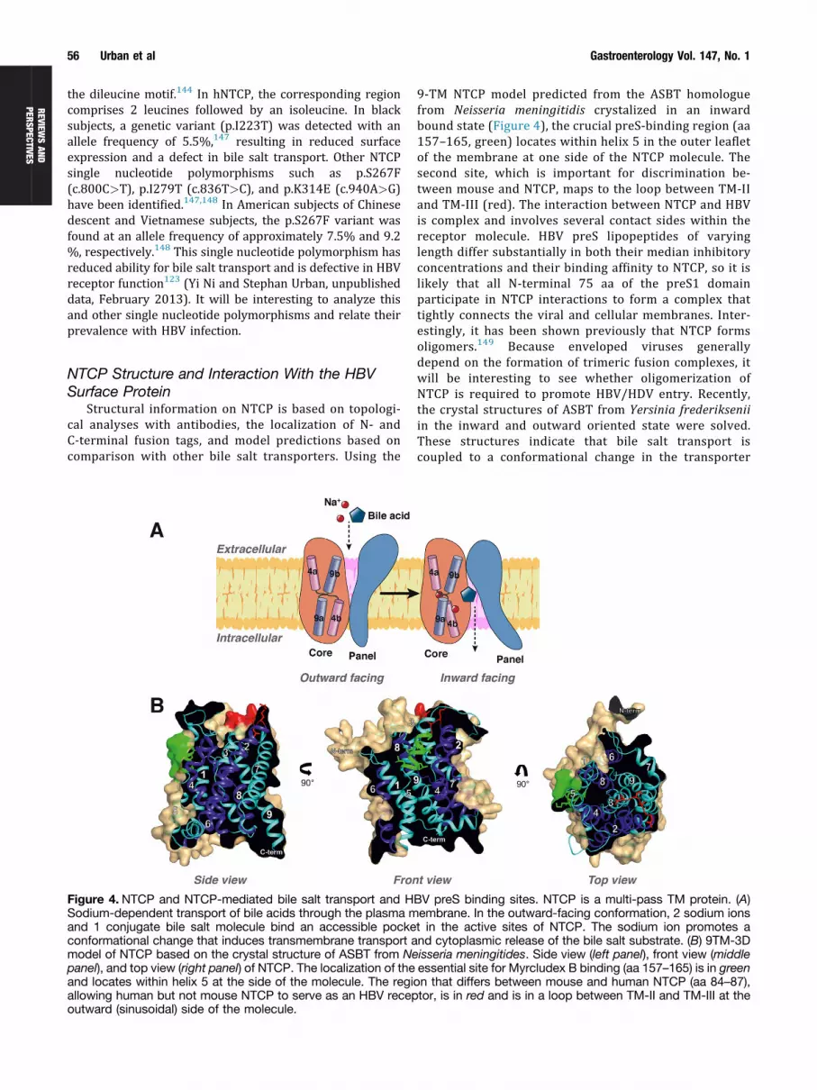
Figure 4. NTCP and NTCP-mediated bile salt transport and HSodium-dependent transport of bile acids through the plasma mand 1 conjugate bile salt molecule bind an accessible pockeconformational change that induces transmembrane transport amodel of NTCP based on the crystal structure of ASBT from Nepanel), and top view (right panel) of NTCP. The localization of theand locates within helix 5 at the side of the molecule. The regiallowing human but not mouse NTCP to serve as an HBV recepoutward (sinusoidal) side of the molecule.
9-TM NTCP model predicted from the ASBT homologuefrom Neisseria meningitidis crystalized in an inwardbound state (Figure 4), the crucial preS-binding region (aa157–165, green) locates within helix 5 in the outer leafletof the membrane at one side of the NTCP molecule. Thesecond site, which is important for discrimination be-tween mouse and NTCP, maps to the loop between TM-IIand TM-III (red). The interaction between NTCP and HBVis complex and involves several contact sides within thereceptor molecule. HBV preS lipopeptides of varyinglength differ substantially in both their median inhibitoryconcentrations and their binding affinity to NTCP, so it islikely that all N-terminal 75 aa of the preS1 domainparticipate in NTCP interactions to form a complex thattightly connects the viral and cellular membranes. Inter-estingly, it has been shown previously that NTCP formsoligomers.149 Because enveloped viruses generallydepend on the formation of trimeric fusion complexes, itwill be interesting to see whether oligomerization ofNTCP is required to promote HBV/HDV entry. Recently,the crystal structures of ASBT from Yersinia frederikseniiin the inward and outward oriented state were solved.These structures indicate that bile salt transport iscoupled to a conformational change in the transporter
BV preS binding sites. NTCP is a multi-pass TM protein. (A)embrane. In the outward-facing conformation, 2 sodium ionst in the active sites of NTCP. The sodium ion promotes and cytoplasmic release of the bile salt substrate. (B) 9TM-3Disseria meningitides. Side view (left panel), front view (middleessential site for Myrcludex B binding (aa 157–165) is in greenon that differs between mouse and human NTCP (aa 84–87),tor, is in red and is in a loop between TM-II and TM-III at the

July 2014 Inhibited Entry of HBV and HDV in Hepatocytes 57
REVIEW
SAN
DPE
RSPE
CTIVES
promoted by the rotation of 2 intramembranous a-heli-ces.150 Because it is likely that NTCP-mediated bile salttransport proceeds by a similar mechanism, it will beinteresting to study whether this conformational change,driven by the sodium gradient across the membranes, isrequired to promote infection.
Clinical ImplicationsAlthough there are many potential clinical applications
of entry inhibitors and/or NTCP blockers, the followingconsiderations are only concepts, which require preclinicaland clinical studies. Inhibition of HBV and HDV entry withmolecules that target NTCP could be a new prophylactic andtherapeutic strategy, as summarized in Figure 5. These in-hibitors could be an important alternative to HBIGs forprevention of viral recurrence during immunosuppressivetherapies or after liver transplantation, in cases of accidentalnosocomial infection, or to stop vertical transmission. Theywould be especially useful in highly endemic areas, where
Figure 5. (A) Prophylacticand (B) therapeutic strate-gies for possible futurecombination strategies ofentry inhibitors in combi-nation with approveddrugs.
effective and cheap prophylaxis is needed most. For patientswith chronic viral infection, entry inhibitors could speedHBsAg clearance and provide the opportunity for treatmentwith a finite duration. Current treatments for chronic HDVinfection provide low rates of HDV clearance, so new op-tions are necessary.
Preventing TransmissionEntry inhibitors might replace HBIGs, which are expen-
sive and not regularly supplied to some parts of the world,or could be administered with the HBV vaccine for peopleexposed to HBV-infected fluids. To prevent vertical trans-mission, entry inhibitors might be administered to neonatessoon after birth together with the HBV vaccine; in motherswith high levels of HBV viremia, administration of nucleos(t)ide analogues might still be necessary during the thirdtrimester of pregnancy. New strategies to prevent viralrecurrence in transplanted organs might be tested with theadministration of nucleos(t)ide analogues, before andtransplantation, and entry inhibitors instead of HBIG right

58 Urban et al Gastroenterology Vol. 147, No. 1
REVIEWSAND
PERSPECTIVES
after transplantation. In livers transplanted in patients withHBV and HDV coinfection, HDV was shown to establishlatent, asymptomatic infections without the apparentassistance of active HBV replication. Entry inhibitors mighttherefore prevent recurrence of HBV infection in thegraft.151
Treatment of Chronic InfectionAs discussed earlier, current treatments for chronic HBV
infection suppress viral replication but rarely result in viralclearance, loss of HBsAg, or even HBsAg seroconversion.New, more effective strategies are required, which mightinvolve combinations of approved drugs or the developmentof novel agents that target thus far unaddressed steps ofviral replication.
The mechanisms involved in viral clearance during nat-ural infection include T cell–mediated killing of infectedhepatocytes, noncytolytic clearance of cccDNA by unknownmechanisms, and hepatocyte turnover and replacement bynoninfected cells. Blocking the spread of HBV to naïve cellscould accelerate the process of virus elimination. Along withnucleos(t)ide analogue–based therapy, entry inhibitorsmight neutralize residual viruses that are still produced,sometimes below the detection limit of polymerase chainreaction–based assays.
In chronically infected patients, entry inhibition willterminate de novo infections of naïve hepatocytes and thosethat arise during hepatocyte turnover. The degree of viruselimination by entry inhibition will therefore depend on thegrade of inflammation and accordingly the rate of cell killingor natural hepatocyte turnover. The half-life of infectedhepatocytes in the liver of chronic HBV carriers is notknown but is presumably shorter than that of healthyhepatocytes.
Although the latest generation of nucleos(t)ide ana-logues suppress viral replication, it is likely that hepato-cytes can still become infected. Because these drugscannot prevent the formation of new cccDNA during denovo virus entry into cells,152 the combination of nucle-os(t)ide analogues and entry inhibitors might speed upelimination of infected cells. Because removal of nucle-os(t)ide analogues in chronically infected patients resultsin virological and biochemical relapse but provokes amarked percentage of viral clearance,153 the addition ofan entry inhibitor that prevents reformation of cccDNAcould increase this therapeutic effect. The immunemodulator IFN-a might also be combined with an entryinhibitor. IFN-a, which decreases HDV replication, mightbe combined with an entry inhibitor to block the spread ofHDV; this interesting approach requires testing in clinicaltrials.
Inactivation of NTCPProlonged inactivation of the receptor function of NTCP
with antibodies, cyclosporin A, ezetimibe, or Myrcludex Bpresumably affects bile transport by the molecule. Thismight increase serum levels of conjugated bile salts, whichare primarily transported by NTCP. In the long-term, these
changes could affect expression levels of compensatingtransporters such as organic anion-transporting poly-peptides and other pathways that change with intrahepaticlevels of bile salts and are controlled by farnesoid X receptoror other nuclear receptors.
Thus, the clinical consequences of NTCP inhibitionare unclear. However, subjects who are homozygous forthe single nucleotide polymorphism c.800C>T, whichalmost completely disrupts NTCP-mediated transport ofbile salts, have been described.148 Myrcludex B in-activates NTCP function at concentrations far belowthose required to block bile salt transport. Thus, atherapeutic window exists that allows only partial inac-tivation of NTCP bile salt transport but complete block ofHBV or HDV entry.119 NTCP also transports substratesother than bile salts, so blockade of NTCP could affect thepharmacokinetics and effectiveness of certain drugs.These aspects need to be considered when Myrcludex Bor other NTCP inhibitors are used in the clinic. Inter-estingly, inhibitors of NTCP-mediated bile salt transportmight be used to treat certain forms of cholestasis. Pre-venting reimport of bile salts into hepatocytes that donot secrete them into canaliculi might be used to treatacute biliary cholestasis.
Future DirectionsThe identification of NTCP as the bona fide HBV and HDV
receptor was a major breakthrough. Basic researchers nowhave an improved system for studying HBV entry (Figure 6)and can establish cell lines that are easily infectable andsupport efficient replication of HBV and HDV. These systemswill be instrumental for studying the early steps of HBV andHDV replication and identifying other cell componentsrequired for productive infection. These could include fac-tors involved in intracellular trafficking of virus particles,nuclear import of HBV nucleocapsids, and formation ofcccDNA. The identification of this receptor can also be usedto establish immune-competent transgenic mice that can beinfected with either virus. Although mouse cells supportHBV assembly and secretion after artificial delivery of theHBV genome, it remains to be determined which otherfactors besides NTCP (eg, factors required for nucleocapsiddelivery to the nucleus or DNA repair) codetermine speciesspecificity.
Robust infection systems are urgently needed to estab-lish assays to characterize clinical isolates and increase ourknowledge about viral determinants of host range andpathogenesis. Moreover, it is now possible to developscreening assays to identify cellular factors that promote orrestrict HBV and HDV replication cycles. These assays coulduse RNA interference or transgenic overexpression ap-proaches, like those conducted in HIV-1154 or hepatitis Cvirus research.155,156 These types of high-throughputscreening formats are also suitable to search for agentsthat inhibit HBV replication in a more general way, such ascell factors that are exploited by the viruses for their ownreplication. This approach was used to identify cyclophilin Aas a target for anti–hepatitis C virus therapy157 and the

Figure 6.Model of HBVentry into hepatocytes.Enveloped HBV and HDVparticles circulating in thesinusoidal blood enter thespace ofDisse, probably bypassage through the fe-nestrae of sinusoidalendothelial cells. Throughreversible attachment toHSPGs, L protein–enrichedvirions are concentrated atthe surface of hepatocytes,allowing them to contactthe integral membraneprotein receptor NTCP. In aslow transition process, theN-terminus of the HBV Lprotein is subsequentlyreleased from its intra-membranous state andforms an irreversible re-ceptor complexwithNTCP.After endocytosis by anunknown mechanism, theviral and cellular mem-branes fuse. Formation of afusion-active NTCP viruscomplex can be inhibitedby Myrcludex B.
July 2014 Inhibited Entry of HBV and HDV in Hepatocytes 59
REVIEW
SAN
DPE
RSPE
CTIVES
human immunodeficiency virus coreceptor CCR5, which istargeted by Maraviroc.158 The ability of Myrcludex B totarget the NTCP provides hope that new antiviral strategieswill become available in the near future.
References
1.Kwon H, Lok AS. Hepatitis B therapy. Nat Rev Gastro-enterol Hepatol 2011;8:275–284.2.Rizzetto M. Current management of delta hepatitis. LiverInt 2013;33(suppl 1):195–197.
3.Zoulim F, Locarnini S. Hepatitis B virus resistance tonucleos(t)ide analogues. Gastroenterology 2009;137:1593–1608.
4.Gish R, Jia JD, Locarnini S, et al. Selection of chronichepatitis B therapy with high barrier to resistance. LancetInfect Dis 2012;12:341–353.
5.Zoulim F. Are novel combination therapies needed forchronic hepatitis B? Antiviral Res 2012;96:256–259.
6.Heidrich B, Manns MP, Wedemeyer H. Treatment op-tions for hepatitis delta virus infection. Curr Infect DisRep 2013;15:31–38.
7.Dandri M, Lutgehetmann M, Petersen J. Experimentalmodels and therapeutic approaches for HBV. SeminImmunopathol 2013;35:7–21.
8.Dandri M, Locarnini S. New insight in the pathobiology ofhepatitis B virus infection. Gut 2012;61(suppl 1):i6–i17.
9.Yan H, Zhong G, Xu G, et al. Sodium taurocholatecotransporting polypeptide is a functional receptor forhuman hepatitis B and D virus. elife 2012;1:e00049.
10.Gripon P, Diot C, Theze N, et al. Hepatitis B virus infectionof adult human hepatocytes cultured in the presence ofdimethyl sulfoxide. J Virol 1988;62:4136–4143.
11.Walter E, Keist R, Niederost B, et al. Hepatitis B virusinfection of tupaia hepatocytes in vitro and in vivo.Hepatology 1996;24:1–5.
12.Yan RQ, Su JJ, Huang DR, et al. Human hepatitis B virusand hepatocellular carcinoma. I. Experimental infection

60 Urban et al Gastroenterology Vol. 147, No. 1
REVIEWSAND
PERSPECTIVES
of tree shrews with hepatitis B virus. J Cancer Res ClinOncol 1996;122:283–288.
13.Gripon P, Rumin S, Urban S, et al. Infection of a humanhepatoma cell line by hepatitis B virus. Proc Natl AcadSci U S A 2002;99:15655–15660.
14.Chang CM, Jeng KS, Hu CP, et al. Production of hepa-titis B virus in vitro by transient expression of cloned HBVDNA in a hepatoma cell line. EMBO J 1987;6:675–680.
15.Zhou T, Guo H, Guo JT, et al. Hepatitis B virus e antigenproduction is dependent upon covalently closed circular(ccc) DNA in HepAD38 cell cultures and may serve as acccDNA surrogate in antiviral screening assays. AntiviralRes 2006;72:116–124.
16.Acs G, Sells MA, Purcell RH, et al. Hepatitis B virusproduced by transfected Hep G2 cells causes hepatitis inchimpanzees. Proc Natl Acad Sci U S A 1987;84:4641–4644.
17.Gripon P, Diot C, Guguen-Guillouzo C. Reproduciblehigh level infection of cultured adult human hepato-cytes by hepatitis B virus: effect of polyethylene glycolon adsorption and penetration. Virology 1993;192:534–540.
18.Schulze A, Mills K, Weiss TS, et al. Hepatocyte polari-zation is essential for the productive entry of the hepatitisB virus. Hepatology 2011;55:373–383.
19.Kotani N, Maeda K, Debori Y, et al. Expression andtransport function of drug uptake transporters in differ-entiated HepaRG cells. Mol Pharm 2012;9:3434–3441.
20.Gripon P, Diot C, Corlu A, et al. Regulation by dime-thylsulfoxide, insulin, and corticosteroids of hepatitis Bvirus replication in a transfected human hepatoma cellline. J Med Virol 1989;28:193–199.
21.Schulze A, Gripon P, Urban S. Hepatitis B virus infectioninitiates with a large surface protein-dependent bindingto heparan sulfate proteoglycans. Hepatology 2007;46:1759–1768.
22.Ni Y, Lempp FA, Mehrle S, et al. Hepatitis B and D vi-ruses exploit sodium taurocholate co-transporting poly-peptide for species-specific entry into hepatocytes.Gastroenterology 2014;146:1070–1083.
23.Guidotti LG, Rochford R, Chung J, et al. Viral clearancewithout destruction of infected cells during acute HBVinfection. Science 1999;284:825–829.
24.Mason WS, Litwin S, Xu C, et al. Hepatocyte turnover intransient and chronic hepadnavirus infections. J ViralHepat 2007;14(suppl 1):22–28.
25.Wang Q, Schwarzenberger P, Yang F, et al. Experimentalchronic hepatitis B infection of neonatal tree shrews(Tupaia belangeri chinensis): a model to study molecularcauses for susceptibility and disease progression tochronic hepatitis in humans. Virol J 2012;9:170.
26.Dandri M, Lutgehetmann M, Volz T, et al. Small animalmodel systems for studying hepatitis B virus replicationand pathogenesis. Semin Liver Dis 2006;26:181–191.
27.Meuleman P, Leroux-Roels G. The human liver-uPA-SCID mouse: a model for the evaluation of antiviralcompounds against HBV and HCV. Antiviral Res 2008;80:231–238.
28.Dandri M, Petersen J. Chimeric mouse model of hepatitisB virus infection. J Hepatol 2012;56:493–495.
29.Bissig KD, Wieland SF, Tran P, et al. Human liverchimeric mice provide a model for hepatitis B and Cvirus infection and treatment. J Clin Invest 2010;120:924–930.
30.Lutgehetmann M, Volz T, Kopke A, et al. In vivo prolif-eration of hepadnavirus-infected hepatocytes inducesloss of covalently closed circular DNA in mice. Hepatol-ogy 2010;52:16–24.
31.Lutgehetmann M, Bornscheuer T, Volz T, et al. Hepa-titis B virus limits response of human hepatocytes tointerferon-alpha in chimeric mice. Gastroenterology2011;140:2074–2083.
32.Lutgehetmann M, Mancke LV, Volz T, et al. Humanizedchimeric uPA mouse model for the study of hepatitis Band D virus interactions and preclinical drug evaluation.Hepatology 2012;55:685–694.
33.Volz T, Allweiss L, Ben MBarek M, et al. The entry in-hibitor Myrcludex-B efficiently blocks intrahepatic virusspreading in humanized mice previously infected withhepatitis B virus. J Hepatol 2013;58:861–867.
34.Petersen J, Dandri M, Mier W, et al. Prevention ofhepatitis B virus infection in vivo by entry inhibitorsderived from the large envelope protein. Nat Biotechnol2008;26:335–341.
35.Chisari FV. Hepatitis B virus transgenic mice: models ofviral immunobiology and pathogenesis. Curr Top Micro-biol Immunol 1996;206:149–173.
36.Yang PL, Althage A, Chung J, et al. Hydrodynamic in-jection of viral DNA: a mouse model of acute hepatitis Bvirus infection. Proc Natl Acad Sci U S A 2002;99:13825–13830.
37.Ketzinel-Gilad M, Zauberman A, Nussbaum O, et al. Theuse of the hydrodynamic HBV animal model to studyHBV biology and anti-viral therapy. Hepatol Res 2006;34:228–237.
38.Huang LR, Gabel YA, Graf S, et al. Transfer of HBV ge-nomes using low doses of adenovirus vectors leads topersistent infection in immune competent mice. Gastro-enterology 2012;142:1447–1450.
39.Sprinzl MF, Oberwinkler H, Schaller H, et al. Transfer ofhepatitis B virus genome by adenovirus vectors intocultured cells and mice: crossing the species barrier.J Virol 2001;75:5108–5118.
40.Chang J, Block TM, Guo JT. The innate immuneresponse to hepatitis B virus infection: implications forpathogenesis and therapy. Antiviral Res 2012;96:405–413.
41.Quasdorff M, Protzer U. Control of hepatitis B virus at thelevel of transcription. J Viral Hepat 2010;17:527–536.
42.Prange R. Host factors involved in hepatitis B virusmaturation, assembly, and egress. Med MicrobiolImmunol 2012;201:449–461.
43.Urban S, Schulze A, Dandri M, et al. The replication cycleof hepatitis B virus. J Hepatol 2010;52:282–284.
44.Sureau C. The use of hepatocytes to investigate HDVinfection: the HDV/HepaRG model. Methods Mol Biol2010;640:463–473.
45.Taylor JM. Virus entry mediated by hepatitis B virusenvelope proteins. World J Gastroenterol 2013;19:6730–6734.

July 2014 Inhibited Entry of HBV and HDV in Hepatocytes 61
REVIEW
SAN
DPE
RSPE
CTIVES
46.Taylor JM. Hepatitis delta virus. Virology 2006;344:71–76.
47.Lamas LO, Schmidt TT, Schoneweis K, et al. Pro-teoglycans act as cellular hepatitis delta virus attachmentreceptors. PLoS One 2013;8:e58340.
48.Leistner CM, Gruen-Bernhard S, Glebe D. Role of gly-cosaminoglycans for binding and infection of hepatitis Bvirus. Cell Microbiol 2008;10:122–133.
49.Sureau C, Salisse J. A conformational heparan sulfate-binding site essential to infectivity overlaps with theconserved hepatitis B virus a-determinant. Hepatology2013;57:985–994.
50.Seeger C, Mason WS. Hepatitis B virus biology. Micro-biol Mol Biol Rev 2000;64:51–68.
51.Seitz S, Urban S, Antoni C, et al. Cryo-electron micro-scopy of hepatitis B virions reveals variability in envelopecapsid interactions. EMBO J 2007;26:4160–4167.
52.Bruss V. Hepatitis B virus morphogenesis. World JGastroenterol 2007;13:65–73.
53.Barrera A, Guerra B, Notvall L, et al. Mapping of thehepatitis B virus pre-S1 domain involved in receptorrecognition. J Virol 2005;79:9786–9798.
54.Engelke M, Mills K, Seitz S, et al. Characterization of ahepatitis B and hepatitis delta virus receptor binding site.Hepatology 2006;43:750–760.
55.Glebe D, Aliakbari M, Krass P, et al. Pre-s1 antigen-dependent infection of Tupaia hepatocyte cultureswith human hepatitis B virus. J Virol 2003;77:9511–9521.
56.Glebe D, Urban S, Knoop EV, et al. Mapping of thehepatitis B virus attachment site by use of infection-inhibiting preS1 lipopeptides and tupaia hepatocytes.Gastroenterology 2005;129:234–245.
57.Gripon P, Cannie I, Urban S. Efficient inhibition of hep-atitis B virus infection by acylated peptides derived fromthe large viral surface protein. J Virol 2005;79:1613–1622.
58.LeSeyec J, Chouteau P, Cannie I, et al. Infection processof the hepatitis B virus depends on the presence of adefined sequence in the pre-S1 domain. J Virol 1999;73:2052–2057.
59.Meier A, Mehrle S, Weiss TS, et al. The myristoylatedpreS1-domain of the hepatitis B virus L-protein mediatesspecific binding to differentiated hepatocytes. Hepatol-ogy 2013;58:31–42.
60.Persing DH, Varmus HE, Ganem D. The preS1 protein ofhepatitis B virus is acylated at its amino terminus withmyristic acid. J Virol 1987;61:1672–1677.
61.Lambert C, Prange R. Dual topology of the hepatitis Bvirus large envelope protein: determinants influencingpost-translational pre-S translocation. J Biol Chem 2001;276:22265–22272.
62.Komla-Soukha I, Sureau C. A tryptophan-rich motif in thecarboxyl terminus of the small envelope protein of hep-atitis B virus is central to the assembly of hepatitis deltavirus particles. J Virol 2006;80:4648–4655.
63.Boyd A, Miailhes P, Brichler S, et al. Effect of tenofovirwith and without interferon on HDV replication in HIV-HBV-HDV infected patients. AIDS Res Hum Retrovi-ruses 2013;29:1535–1540.
64.Kabacam G, Onder FO, Yakut M, et al. Entecavir treat-ment of chronic hepatitis D. Clin Infect Dis 2012;55:645–650.
65.Bruss V, Hagelstein J, Gerhardt E, et al. Myristylation ofthe large surface protein is required for hepatitis B virusin vitro infectivity. Virology 1996;218:396–399.
66.Gripon P, LeSeyec J, Rumin S, et al. Myristylation of thehepatitis B virus large surface protein is essential for viralinfectivity. Virology 1995;213:292–299.
67.Macrae DR, Bruss V, Ganem D. Myristylation of a duckhepatitis B virus envelope protein is essential for infec-tivity but not for virus assembly. Virology 1991;181:359–363.
68.De Falco S, Ruvo M, Verdoliva A, et al. N-terminal myr-istylation of HBV preS1 domain enhances receptorrecognition. J Pept Res 2001;57:390–400.
69.LeSeyec J, Chouteau P, Cannie I, et al. Role of the pre-S2 domain of the large envelope protein in hepatitis Bvirus assembly and infectivity. J Virol 1998;72:5573–5578.
70.Stoeckl L, Funk A, Kopitzki A, et al. Identification of astructural motif crucial for infectivity of hepatitis B vi-ruses. Proc Natl Acad Sci U S A 2006;103:6730–6734.
71.Gudima S, Meier A, Dunbrack R, et al. Two potentiallyimportant elements of the hepatitis B virus large enve-lope protein are dispensable for the infectivity of hepatitisdelta virus. J Virol 2007;81:4343–4347.
72.Blanchet M, Sureau C. Infectivity determinants of thehepatitis B virus pre-S domain are confined to the N-terminal 75 amino acid residues. J Virol 2007;81:5841–5849.
73.Lepere C, Regeard M, Le SJ, et al. The translocationmotif of hepatitis B virus envelope proteins is dispens-able for infectivity. J Virol 2007;81:7816–7818.
74.Ni Y, Sonnabend J, Seitz S, et al. The pre-s2 domain ofthe hepatitis B virus is dispensable for infectivity butserves a spacer function for L-protein-connected virusassembly. J Virol 2010;84:3879–3888.
75.LeDuff Y, Blanchet M, Sureau C. The pre-S1 and anti-genic loop infectivity determinants of the hepatitis B virusenvelope proteins are functionally independent. J Virol2009;83:12443–12451.
76.Abou-Jaoude G, Sureau C. Role of the antigenicloop of the hepatitis B virus envelope proteins ininfectivity of hepatitis delta virus. J Virol 2005;79:10460–10466.
77.Abou-Jaoude G, Sureau C. Entry of hepatitis delta virusrequires the conserved cysteine residues of the hepatitisB virus envelope protein antigenic loop and is blocked byinhibitors of thiol-disulfide exchange. J Virol 2007;81:13057–13066.
78.Grgacic EV, Schaller H. A metastable form of the largeenvelope protein of duck hepatitis B virus: low-pHrelease results in a transition to a hydrophobic, poten-tially fusogenic conformation. J Virol 2000;74:5116–5122.
79.Bian T, Yan H, Shen L, et al. Change in hepatitis B viruslarge surface antigen variant prevalence 13 years afterimplementation of a universal vaccination program inChina. J Virol 2013;87:12196–12206.

62 Urban et al Gastroenterology Vol. 147, No. 1
REVIEWSAND
PERSPECTIVES
80.Lai MW, Lin TY, Tsao KC, et al. Increased seroprevalenceof HBV DNA with mutations in the s gene among in-dividuals greater than 18 years old after completevaccination. Gastroenterology 2012;143:400–407.
81.Shouval D, Locarnini S. Increased prevalence of HBVenvelope mutants in Taiwan: an emerging public healthrisk or a false alarm? Gastroenterology 2012;143:290–293.
82.Lepere-Douard C, Trotard M, Le SJ, et al. The firsttransmembrane domain of the hepatitis B virus largeenvelope protein is crucial for infectivity. J Virol 2009;83:11819–11829.
83.Chojnacki J, Anderson DA, Grgacic EV. A hydrophobicdomain in the large envelope protein is essential forfusion of duck hepatitis B virus at the late endosome.J Virol 2005;79:14945–14955.
84.Bremer CM, Bung C, Kott N, et al. Hepatitis B virusinfection is dependent on cholesterol in the viral enve-lope. Cell Microbiol 2009;11:249–260.
85.Glebe D. Attachment sites and neutralising epitopes ofhepatitis B virus. Minerva Gastroenterol Dietol 2006;52:3–21.
86.Ryu CJ, Gripon P, Park HR, et al. In vitro neutralization ofhepatitis B virus by monoclonal antibodies against theviral surface antigen. J Med Virol 1997;52:226–233.
87.Schulze A, Schieck A, Ni Y, et al. Fine mapping of pre-Ssequence requirements for hepatitis B virus large enve-lope protein-mediated receptor interaction. J Virol 2010;84:1989–2000.
88.Bartholomeusz A, Schaefer S. Hepatitis B virus geno-types: comparison of genotyping methods. Rev MedVirol 2004;14:3–16.
89.Schaefer S. Hepatitis B virus taxonomy and hepatitis Bvirus genotypes. World J Gastroenterol 2007;13:14–21.
90.Drexler JF, Geipel A, Konig A, et al. Bats carrypathogenic hepadnaviruses antigenically related tohepatitis B virus and capable of infecting human he-patocytes. Proc Natl Acad Sci U S A 2013;110:16151–16156.
91.Samuel D, Muller R, Alexander G, et al. Liver trans-plantation in European patients with the hepatitis B sur-face antigen. N Engl J Med 1993;329:1842–1847.
92.Alvarado-Mora MV, Locarnini S, Rizzetto M, et al. Anupdate on HDV: virology, pathogenesis and treatment.Antivir Ther 2013;18:541–548.
93.Fox AN, Terrault NA. The option of HBIG-free prophylaxisagainst recurrent HBV. J Hepatol 2012;56:1189–1197.
94.Xu WM, Cui YT, Wang L, et al. Lamivudine in late preg-nancy to prevent perinatal transmission of hepatitis Bvirus infection: a multicentre, randomized, double-blind,placebo-controlled study. J Viral Hepat 2009;16:94–103.
95.Han GR, Cao MK, Zhao W, et al. A prospective andopen-label study for the efficacy and safety of telbivudinein pregnancy for the prevention of perinatal transmissionof hepatitis B virus infection. J Hepatol 2011;55:1215–1221.
96.Villet S, Pichoud C, Villeneuve JP, et al. Selection of amultiple drug-resistant hepatitis B virus strain in a liver-transplanted patient. Gastroenterology 2006;131:1253–1261.
97.Villet S, Billioud G, Pichoud C, et al. In vitro character-ization of viral fitness of therapy-resistant hepatitis Bvariants. Gastroenterology 2009;136:168–176.
98.Teo CG, Locarnini SA. Potential threat of drug-resistantand vaccine-escape HBV mutants to public health.Antivir Ther 2010;15:445–449.
99.Eren R, Ilan E, Nussbaum O, et al. Preclinical evaluationof two human anti-hepatitis B virus (HBV) monoclonalantibodies in the HBV-trimera mouse model and in HBVchronic carrier chimpanzees. Hepatology 2000;32:588–596.
100.Galun E, Eren R, Safadi R, et al. Clinical evaluation (phaseI) of a combination of two human monoclonal antibodiesto HBV: safety and antiviral properties. Hepatology 2002;35:673–679.
101.Schieck A, Schulze A, Gahler C, et al. Hepatitis B virushepatotropism is mediated by specific receptor recog-nition in the liver and not restricted to susceptible hosts.Hepatology 2013;58:43–53.
102.Dupinay T, Gheit T, Roques P, et al. Discovery ofnaturally occurring transmissible chronic hepatitis B virusinfection among Macaca fascicularis from Mauritius Is-land. Hepatology 2013;58:1610–1620.
103.Petcu DJ, Aldrich CE, Coates L, et al. Suramin inhibitsin vitro infection by duck hepatitis B virus, Rous sarcomavirus, and hepatitis delta virus. Virology 1988;167:385–392.
104.Krepstakies M, Lucifora J, Nagel CH, et al. A new class ofsynthetic peptide inhibitors blocks attachment and entryof human pathogenic viruses. J Infect Dis 2012;205:1654–1664.
105.Glebe D, Urban S. Viral and cellular determinantsinvolved in hepadnaviral entry. World J Gastroenterol2007;13:22–38.
106.Suchanek M, Radzikowska A, Thiele C. Photo-leucineand photo-methionine allow identification of protein-protein interactions in living cells. Nat Methods 2005;2:261–267.
107.Iwamoto M, Watashi K, Tsukuda S, et al. Evaluation andidentification of hepatitis B virus entry inhibitors usingHepG2 cells overexpressing a membrane transporterNTCP. Biochem Biophys Res Commun 2014;443:808–813.
108.Yan H, Peng B, He W, et al. Molecular determinantsrestricting mouse sodium taurocholate cotransportingpolypeptide for viral entry of Hepatitis B and D virus.J Virol 2013;87:7977–7991.
109.Simon FR, Fortune J, Iwahashi M, et al. Characterizationof the mechanisms involved in the gender differences inhepatic taurocholate uptake. Am J Physiol 1999;276:G556–G565.
110.Cheng X, Buckley D, Klaassen CD. Regulation of hepaticbile acid transporters Ntcp and Bsep expression. Bio-chem Pharmacol 2007;74:1665–1676.
111.Hagenbuch B, Meier PJ. Molecular cloning, chromo-somal localization, and functional characterization of ahuman liver Naþ/bile acid cotransporter. J Clin Invest1994;93:1326–1331.
112.Dong Z, Ekins S, Polli JE. Structure activity relationshipfor FDA approved drugs as inhibitors of the human

July 2014 Inhibited Entry of HBV and HDV in Hepatocytes 63
REVIEW
SAN
DPE
RSPE
CTIVES
sodium taurocholate co-transporting polypeptide(NTCP). Mol Pharm 2013;10:1008–1019.
113.Doring B, Lutteke T, Geyer J, et al. The SLC10 carrierfamily: transport functions and molecular structure. CurrTop Membr 2012;70:105–168.
114.Anwer MS, Stieger B. Sodium-dependent bile salttransporters of the SLC10A transporter family: more thansolute transporters. Pflugers Arch 2013;466:77–89.
115.Greupink R, Dillen L, Monshouwer M, et al. Interaction offluvastatin with the liver-specific Naþ -dependent taur-ocholate cotransporting polypeptide (NTCP). Eur JPharm Sci 2011;44:487–496.
116.Yanni SB, Augustijns PF, Benjamin DK Jr, et al. In vitroinvestigation of the hepatobiliary disposition mecha-nisms of the antifungal agent micafungin in humans andrats. Drug Metab Dispos 2010;38:1848–1856.
117.Kim RB, Leake B, Cvetkovic M, et al. Modulation bydrugs of human hepatic sodium-dependent bile acidtransporter (sodium taurocholate cotransporting poly-peptide) activity. J Pharmacol Exp Ther 1999;291:1204–1209.
118.Lucifora J, Esser K, Protzer U. Ezetimibe blocks hepa-titis B virus infection after virus uptake into hepatocytes.Antiviral Res 2013;97:195–197.
119.Nkongolo S, Ni Y, Lempp FA, et al. Cyclosporin A in-hibits hepatitis B and hepatitis D virus entry bycyclophilin-independent interference with the NTCP re-ceptor. J Hepatol 2014;60:723–731.
120.Watashi K, Sluder A, Daito T, et al. Cyclosporin A and itsanalogs inhibit hepatitis B virus entry into cultured he-patocytes through targeting a membrane transporterNTCP. Hepatology 2014;59:1726–1737.
121.Zahner D, Eckhardt U, Petzinger E. Transport of taur-ocholate by mutants of negatively charged amino acids,cysteines, and threonines of the rat liver sodium-dependent taurocholate cotransporting polypeptideNtcp. Eur J Biochem 2003;270:1117–1127.
122.Weinman SA. Electrogenicity of Na(þ)-coupled bile acidtransporters. Yale J Biol Med 1997;70:331–340.
123.Yan H, Peng B, Liu Y, et al. Viral entry of hepatitis B andD viruses and bile salts transportation share commonmolecular determinants on sodium taurocholatecotransporting polypeptide. J Virol 2014;88:3273–3284.
124.Elferink MG, Olinga P, Draaisma AL, et al. LPS-induceddown-regulation of MRP2 and BSEP in human liver isdue to a posttranscriptional process. Am J PhysiolGastrointest Liver Physiol 2004;287:G1008–G1016.
125.Le Vee M, Gripon P, Stieger B, et al. Down-regulation oforganic anion transporter expression in human hepa-tocytes exposed to the proinflammatory cytokineinterleukin 1beta. Drug Metab Dispos 2008;36:217–222.
126.Le VM, Lecureur V, Moreau A, et al. Differential regulationof drug transporter expression by hepatocyte growthfactor in primary human hepatocytes. Drug Metab Dis-pos 2009;37:2228–2235.
127.Jung D, Hagenbuch B, Fried M, et al. Role of liver-enriched transcription factors and nuclear receptors inregulating the human, mouse, and rat NTCP gene. Am JPhysiol Gastrointest Liver Physiol 2004;286:G752–G761.
128.Eloranta JJ, Jung D, Kullak-Ublick GA. The human Naþ-taurocholate cotransporting polypeptide gene is acti-vated by glucocorticoid receptor and peroxisomeproliferator-activated receptor-gamma coactivator-1alpha, and suppressed by bile acids via a small heter-odimer partner-dependent mechanism. Mol Endocrinol2006;20:65–79.
129.Reese VC, Moore DD, McLachlan A. Limited effects ofbile acids and small heterodimer partner on hepatitis bvirus biosynthesis in vivo. J Virol 2012;86:2760–2768.
130.Chen HL, Chen HL, Liu YJ, et al. Developmentalexpression of canalicular transporter genes in humanliver. J Hepatol 2005;43:472–477.
131.Kojima H, Nies AT, König J, et al. Changes in theexpression and localization of hepatocellular transportersand radixin in primary biliary cirrhosis. J Hepatol 2003;39:693–702.
132.Keitel V, Burdelski M, Warskulat U, et al. Expression andlocalization of hepatobiliary transport proteins in pro-gressive familial intrahepatic cholestasis. Hepatology2005;41:1160–1172.
133.Shneider BL, Fox VL, Schwarz KB, et al. Hepatic baso-lateral sodium-dependent-bile acid transporter expres-sion in two unusual cases of hypercholanemia and inextrahepatic biliary atresia. Hepatology 1997;25:1176–1183.
134.Zollner G, Fickert P, Zenz R, et al. Hepatobiliary trans-porter expression in percutaneous liver biopsies of pa-tients with cholestatic liver diseases. Hepatology 2001;33:633–646.
135.Bechmann LP, Kocabayoglu P, Sowa JP, et al. Free fattyacids repress small heterodimer partner (SHP) activationand adiponectin counteracts bile acid-induced liver injuryin superobese patients with nonalcoholic steatohepatitis.Hepatology 2013;57:1394–1406.
136.Zollner G, Wagner M, Fickert P, et al. Hepatobiliarytransporter expression in human hepatocellular carci-noma. Liver Int 2005;25:367–379.
137.Muller T, Mehrle S, Schieck A, et al. Liver imaging with anovel hepatitis B surface protein derived SPECT-tracer.Mol Pharm 2013;10:2230–2236.
138.Dranoff JA, McClure M, Burgstahler AD, et al. Short-termregulation of bile acid uptake by microfilament-dependent translocation of rat ntcp to the plasmamembrane. Hepatology 1999;30:223–229.
139.Sun AQ, Swaby I, Xu S, et al. Cell-specific basolateralmembrane sorting of the human liver Na(þ)-dependentbile acid cotransporter. Am J Physiol Gastrointest LiverPhysiol 2001;280:G1305–G1313.
140.Anwer MS, Gillin H, Mukhopadhyay S, et al. Dephos-phorylation of S226 facilitates plasma membrane reten-tion of Ntcp. J Biol Chem 2005;280:33687–33692.
141.Webster CR, Anwer MS. Role of the PI3K/PKB signalingpathway in cAMP-mediated translocation of rat liverNtcp. Am J Physiol 1999;277:G1165–G1172.
142.McConkey M, Gillin H, Webster CR, et al. Cross-talkbetween protein kinases Czeta and B in cyclic AMP-mediated sodium taurocholate co-transporting poly-peptide translocation in hepatocytes. J Biol Chem 2004;279:20882–20888.

64 Urban et al Gastroenterology Vol. 147, No. 1
REVIEWSAND
PERSPECTIVES
143.Sarkar S, Bananis E, Nath S, et al. PKCzeta is requiredfor microtubule-based motility of vesicles containing thentcp transporter. Traffic 2006;7:1078–1091.
144.Stross C, Kluge S, Weissenberger K, et al. A dileucinemotif is involved in plasma membrane expression andendocytosis of rat sodium taurocholate cotransportingpolypeptide (Ntcp). Am J Physiol Gastrointest LiverPhysiol 2013;305:G722–G730.
145.Stross C, Helmer A, Weissenberger K, et al. Proteinkinase C induces endocytosis of the sodium taur-ocholate cotransporting polypeptide. Am J PhysiolGastrointest Liver Physiol 2010;299:G320–G328.
146.Mühlfeld S, Domanova O, Berlage T, et al. Short-termfeedback regulation of bile salt uptake by bile salts in ratliver. Hepatology 2012;56:2387–2397.
147.Ho RH, Leake BF, Roberts RL, et al. Ethnicity-dependentpolymorphism in Naþ-taurocholate cotransportingpolypeptide (SLC10A1) reveals a domain critical for bileacid substrate recognition. J Biol Chem 2004;279:7213–7222.
148.Pan W, Song IS, Shin HJ, et al. Genetic polymorphismsin Naþ-taurocholate co-transporting polypeptide (NTCP)and ileal apical sodium-dependent bile acid transporter(ASBT) and ethnic comparisons of functional variants ofNTCP among Asian populations. Xenobiotica 2011;41:501–510.
149.Bijsmans IT, Bouwmeester RA, Geyer J, et al. Homo- andhetero-dimeric architecture of the human liver Na(þ)-dependent taurocholate co-transporting protein. Bio-chem J 2012;441:1007–1015.
150.Zhou X, Levin EJ, Pan Y, et al. Structural basis of thealternating-access mechanism in a bile acid transporter.Nature 2014;505:569–573.
151.Rizzetto M. Hepatitis delta: the virus and the disease.J Hepatol 1990;11(suppl 1):S145–S148.
152.Le GF, Pichoud C, Guerret S, et al. Characterization ofthe antiviral effect of 20,30-dideoxy-20, 30-didehydro-beta-L-5-fluorocytidine in the duck hepatitis B virus
infection model. Antimicrob Agents Chemother 2000;44:111–122.
153.Hadziyannis SJ, Sevastianos V, Rapti I, et al. Sustainedresponses and loss of HBsAg in HBeAg-negative pa-tients with chronic hepatitis B who stop long-termtreatment with adefovir. Gastroenterology 2012;143:629–636.
154.Brass AL, Dykxhoorn DM, Benita Y, et al. Identificationof host proteins required for HIV infection through afunctional genomic screen. Science 2008;319:921–926.
155.Li Q, Brass AL, Ng A, et al. A genome-wide geneticscreen for host factors required for hepatitis C viruspropagation. Proc Natl Acad Sci U S A 2009;106:16410–16415.
156.Schoggins JW, Wilson SJ, Panis M, et al. A diverse rangeof gene products are effectors of the type I interferonantiviral response. Nature 2011;472:481–485.
157.Bartenschlager R, Lohmann V, Penin F. The molecularand structural basis of advanced antiviral therapy forhepatitis C virus infection. Nat Rev Microbiol 2013;11:482–496.
158.Haqqani AA, Tilton JC. Entry inhibitors and their use inthe treatment of HIV-1 infection. Antiviral Res 2013;98:158–170.
Author names in bold designate shared co-first authorship.
Received November 22, 2013. Accepted April 21, 2014.
Reprint requestsAddress requests for reprints to: Prof Dr Stephan Urban, Department of InfectiousDiseases, Molecular Virology, University Hospital Heidelberg, Im NeuenheimerFeld 345, 69120 Heidelberg, Germany. e-mail: [email protected]; fax: (49) 6221-561946.
Conflicts of interestStephan Urban is co-applicant and co-inventor on patents protectingHBVpreSderived lipopetides (Myrcludex B) for the use of HBV/HDV entryinhibitors. The remaining authors disclose no conflicts.