Embed Size (px)
Citation preview

289
Structural disruption of the trans-Golgi network does not interferewith the acute stimulation of glucose and amino acid uptake byinsulin-like growth factor I in muscle cellsHarinder S. HUNDAL,* Philip J. BILAN, Theodoros TSAKIRIDIS, Andre MARETTEt and Amira KLIPtDivision of Cell Biology, The Hospital For Sick Children, 555 University Avenue, Toronto, Ontario, Canada M5G 1X8
The effects of insulin-like growth factor I (IGF-I) on glucose andamino acid uptake were investigated in fully differentiated L6muscle cells, in order to determine whether the two processes arefunctionally related. Transport of both glucose and amino acid(methylaminoisobutyric acid, MeAIB) was activated rapidly inresponse to IGF-I. Stimulation reached a peak within 30 minand was sustained for up to 90 min. Maximal activation of eitherglucose or MeAIB transport was achieved at 3 nM IGF-I; thehalf-maximal activation (ED50) of glucose transport was at107 pM and that ofMeAIB transport was at 36 pM. Stimulationof amino acid uptake occurred in the absence or presence ofglucose, suggesting that this response is not secondary toincreased glucose intake. Incubation ofcells for 1 h with BrefeldinA (5 ,ug/ml), which disassembles the Golgi apparatus and inhibitsthe secretory pathway in eukaryotic cells, had no effect on theacute IGF-I activation of glucose and MeAIB transport. More-over, Brefeldin A caused wide redistribution of the trans-Golgi
INTRODUCTION
There is current consensus that in skeletal muscle and adiposetissue the principal mechanism by which insulin acutely stimu-lates glucose transport consists ofmobilization ofan intracellularstore of GLUT4 glucose transporters to the plasma membrane(PM) (Cushman and Wardzala, 1980; Suzuki and Kono, 1980;Douen et al., 1990; Hirshman et al., 1990). However, the natureof the signalling pathway and the identity of the cellularorganelle(s) which participate in GLUT4 transporter storage andtranslocation in skeletal muscle still remain elusive. One mor-
phological study (at the electron-microscope level, using immuno-gold labelling of GLUT4 molecules) has suggested that theinternal pool includes the triad region (comprising the terminalcisternae of the sarcoplasmic reticulum and transverse tubules)(Friedman et al., 1991), whereas another study has proposed thatGLUT4 molecules may accrue in the trans-Golgi network (TGN),where they await to be recruited to the PM in response to theinsulin signal (Rodnick et al., 1992). However, a functional rolefor the TGN has not been established for the translocation ofglucose-transporter-containing vesicles. Evidence does exist sup-
porting the role of the Golgi apparatus (without specific im-plication of the TGN) in anterograde membrane traffic. This islargely based on studies with Brefeldin A (BFA), an isoprenoid
antigen TGN38, as assessed by subcellular fractionation, withoutaffecting the distribution of glucose transporters. The findingthat the degree of activation, time response and sensitivity toIGF-I and Brefeldin A were similar for both glucose and MeAIBtransport suggests commonalities in the IGF-I mechanism ofrecruitment of glucose transporters and stimulation of aminoacid transport through System A. An integral trans-Golginetwork does not appear to be required for the acute IGF-Istimulation of glucose or amino acid transport, even thoughstimulation of glucose transport occurs through recruitment ofglucose transporters from intracellular stores in these cells. Wepropose that the donor site of glucose transporters (and perhapsof amino acid transporters) involved in the acute response toIGF-I lies beyond the trans-Golgi network, perhaps in anendosomal compartment in close proximity to the plasmamembrane.
fungal metabolite from Eupenicillium brefeldianum which rapidlyinhibits protein traffic in the secretory pathway as a result ofdisassembly of the Golgi apparatus (Klausner et al., 1992). Innormal rat kidney (NRK) cells BFA induces a rapid redis-tribution of the cisternal Golgi proteins into the endoplasmicreticulum and the TGN, to extend and mix with the endosomalsystem (Lippincott-Schwartz et al., 1991). After its dispersal toendosomes, TGN markers accumulate around the microtubule-organizing centre (Ladinsky and Howell, 1992). Functionalimplications have been studied in secretory cells, where BFAinhibits constitutive and regulated secretion ofneutrotransmittersand hormones (Klausner et al., 1992).We have observed that the insulin-induced translocation of
GLUT4 to the PM can be reproduced in L6 muscle cells in culturein response to insulin and insulin-like growth factor-I (IGF-I)(Ramlal et al., 1988; Bilan et al., 1991, 1992a). The L6 muscle cellline studied was clonally selected for high fusion and expressesmany of the morphological, biochemical and electrical propertiesof rat skeletal-muscle fibres (Yaffe, 1968; Shainberg et al., 1971).Differentiation of L6 myoblasts into myotubes in culture isassociated with the appearance of muscle-specific proteins andthe ability of these cells to respond to insulin (Mitsumoto et al.,1991). In L6 myotubes activation of glucose transport by bothinsulin and IGF-I is accompanied by translocation to the PM of
Abbreviations used: IGF-1, insulin-like growth factor l; PM, plasma membrane; TGN, trans-Golgi network; BFA, Brefeldin A; 2DG, 2-deoxyglucose;MeAIB, a-methylaminoisobutyrate; a-MEM, a-Minimal Essential Medium; HBS, Hepes-buffered saline.
* Present address: Department of Anatomy and Physiology, University of Dundee, Dundee DD1 4HN, Scotland, U.K.t Present address: Department of Physiology and Endocrinology, CRML, Laval University Hospital Research Centre, 2705 Boulevard Laurier,
Ste-Foy, Qu6bec, Canada G1V 4G2.I To whom correspondence should be addressed.
289Biochem. J. (1994) 297, 289-295 (Printed in Great Britain)

290 H. S. Hundal and others
GLUT 1, GLUT3 and GLUT4 glucose transporters from anintracellular compartment (Bilan et al., 1992a,b; Mitsumoto andKlip, 1992). The L6 myotubes also display stimulation by insulinof amino acid uptake through System A (Hundal et al., 1992),which transports small neutral amino acids in a Na+-dependentmanner (Christensen, 1990). However, in contrast with ourknowledge of stimulation of glucose uptake, the nature of amino-acid-transport System A activation and whether IGF-I canacutely stimulate amino acid transport in muscle cells is poorlydefined. The protein(s) constituting this transport system havenot been isolated, and there are currently no pharmacological orimmunological tools to detect the presence of the constituents ofthis transport pathway.
In the present study we used differentiated L6 myotubes fortwo purposes: (a) to compare the regulation of amino-acid-transport System A and of glucose transport by IGF-I, in searchfor commonalities or differences that might reveal if stimulationof amino acid uptake can occur through recruitment of aminoacid transporters; and (b) to investigate the possibility that theTGN participates in the IGF-I stimulation of glucose and/oramino acid transport, by assessing whether BFA can preventthese responses. This would enable us to test the hypothesis thatvesicle recruitment through the secretory pathway participates inthe IGF-I stimulation of glucose and amino acid uptake. Wedemonstrate that the time course of stimulation and sensitivity toIGF-I of glucose and amino acid uptake are virtually identical,and that neither response is altered when the TGN is disruptedby BFA, in spite of widespread dispersion of the TGN-specificantigen TGN38.
MATERIALS AND METHODSMaterialsTissue-culture medium, fetal bovine serum and reagents wereobtained from GIBCO. Cytochalasin B, 2-deoxy-D-glucose(2DG), a-methylaminoisobutyrate (MeAIB) and BFA wereobtained from Sigma Chemical Co (St. Louis, MO, U.S.A.).Recombinant human IGF-I was kindly given by Dr. M. Vranic(University of Toronto). 2-Deoxy-D-[3H]glucose and a_-[1_4C]-methylaminoisobutyrate were purchased from DuPont (Boston,MA, U.S.A.).
AntibodiesA polyclonal antibody against TGN38 (Luzio et al., 1990) wasused in the present study and was kindly supplied by Dr. J. P.Luzio (University of Cambridge). This antibody was recentlyshown to recognize both TGN38/41 isoforms of this TGN-specific protein (Reaves et al., 1992). Polyclonal antibodiesspecific for the GLUT1 (RaGLUTTrans) or the GLUT4 (IRGT)isoforms of glucose transporters were obtained from East AcresBiologicals (Southbridge, MA, U.S.A.). A polyclonal antibodyspecific for the mouse/rat GLUT3 transporter was kindly givenby Dr. Ian Simpson (NIH, Bethesda, MD, U.S.A.).
Cell culture and incubationsMonolayers of L6 muscle cells were grown to the stage ofmyotubes as previously described (Koivisto et al., 1991; Mitsu-moto and Klip, 1992) in a.-Minimal Essential Medium (a-MEM)containing 5 mM glucose, in the presence of 2% fetal-bovine
100 units/ml penicillin, 100 ,ug/ml streptomycin, 250 ng/mlamphotericin B) at 37 °C in an atmosphere ofair/CO2 (19: 1). Thecells were grown in 3.5 cm-diameter dishes in 6-well plates fortransport measurements. Cells were routinely deprived of serumfor 5 h before uptake assays using serum-free a-MEM mediumcontaining 25 mM glucose. At 1 h before uptake assays, theywere also depleted of amino acids by using Hepes-buffered saline(HBS) (140 mM NaCl, 20 mM Hepes/Na, pH 7.4, 2.5 mMMgSO4, 5 mM KC1, 1 mM CaC12) containing 25 mM glucoseand 5 mg/ml fatty-acid-free BSA. IGF-I or BFA was added tothe incubations at the concentrations and times indicated in theFigure legends.
2DG and MeAIB transportA dual-isotope technique was used to allow the simultaneousmeasurement of 2DG and MeAIB transport in the same popu-lation of cells, as previously described (Hundal et al., 1992).Briefly, after incubation with hormones or drugs, cell monolayerswere rinsed with glucose-free HBS. Glucose and MeAIB uptakeswere quantified by using 10 ,uM 2-deoxy[3H]glucose (1 ,cCi/ml)and 10 #M [14C]MeAIB (1 aCi/ml) for 10 min, unless otherwisestated. Non-specific uptake was determined by quantifying cell-associated radioactivity in the presence of 10 ,uM cytochalasin B(for 2-deoxy-D-glucose) and a 10 mM saturating dose of un-
labelled MeAIB (for the amino acid). Uptake of 2-deoxy-D-[3H]glucose and [14C]MeAIB was terminated by rapidly aspir-ating the radioactive incubation medium, followed by threesuccessive washes of cell monolayers with ice-cold 0.9 % NaCl.Radioactivity associated with the cells was determined by celllysis in 0.05 M NaOH, followed by liquid-scintillation counting.Total cell protein was determined by the Bradford (1976) method.Experiments were assayed in triplicate and performed at leastthree times. In preliminary studies we established that MeAIB(10,M) transport in L6 myotubes was predominantly Na+-dependent. The replacement of NaCl with choline chloride in theuptake medium decreased MeAIB uptake to 15% of the uptakerate in the presence of Na+. When MeAIB uptake was measuredat 4 °C, the rate was substantially diminished (by nearly 5-fold),indicating that transport of MeAIB must be dependent on thefunction of a carrier protein at the cell surface, rather than bysimply entering cells by passive diffusion (a process less dependenton temperature). In preliminary experiments it was establishedthat the rate of MeAIB uptake was linear for the first 20 minafter exposure of L6 cells to 10 ,uM. Based on these preliminarystudies and the results of Figure 1, we chose assay conditions of10 min uptake at 10 #M MeAIB to measure initial rates ofuptake through the System A amino acid transporter.
Subcellular fractionation of L6 myotubes and Western blotsSubcellular membrane fractions were isolated by a modificationof a previously published method (Ramlal et al., 1988) asrecently described in detail (Bilan et al., 1992a). This methodyields initially a crude plasma-membrane fraction and a lightmicrosomal fraction. The crude plasma membranes are furtherfractionated in discontinuous sucrose gradients. The membranesbanding on top of 32% sucrose are enriched in plasma mem-
branes, as revealed by their abundant content of the a -Na+/K+-ATPase subunit, a plasma-membrane marker (Bilan et al.,1992a). In contrast, the light microsomes are devoid of ac-Na+/K+-ATPase subunit and are therefore considered as intra-cellular membranes. These microsomes are enriched in GLUT4glucose transporters and contain the insulin-responsive pool of
serum and 1 % antimycotic/antibiotic solution (final concns. GLUT I, GLUT3 and GLUT4 transporters (Bilan et al., 1992b;

Insulin-like growth factor action on glucose and system A transport
Mitsumoto and Klip, 1992; Sargeant et al., 1993). Membraneproteins (10 ,ug) were subjected to SDS/PAGE on 8 %-poly-acrylamide mini-gels and transferred to poly(vinylidene di-fluoride) (PVDF) membranes, which were incubated for 1 h atroom temperature with buffer A (50 mM Tris/HCl, pH 7.4,150 mM NaCI) containing 0.04% of the non-ionic surfactantNonidet P40 and 3 % BSA. PVDF membranes were incubatedfor 1 h at room temperature or overnight at 4 °C with anti-GLUT1 (1: 2000), anti-GLUT3 (1:500) or anti-GLUT4 (1: 1000)antibodies in buffer A, followed by 1: 1000 horseradish-peroxidase-conjugated anti-rabbit IgG and detection by the en-
hanced chemiluminescence procedure. TGN38 was detected withanti-TGN38 antibody (1: 1000), followed by 1251-labelled ProteinA (2 ,tCi/ 1O ml), exposure to Kodak X-Omat film at -80°Cand autoradiography.
RESULTSStimulation of glucose and MeAIB transport in L6 myotubes byIGF-IThe initial rate of MeAIB uptake was determined at variousconcentrations of MeAIB ranging from 10 to 600,uM andrevealed a typical saturation curve (Figure 1). In preliminary
250
O 2000.hQ , 150ma)<0. 100E.
O 50E
7-
6 -
a .0.
;: m 4 -m E
(D .q 3 -
~ f 2 -
[MeAIBI (pM)* - IGF1 (b)
-200 -100 0 100 200 300 400 500 600[MeAIBI (pM)
Figure 1 Effect of IGF-I on the transport kinetics of MeAIB in L6 myotubes
L6 myotubes were serum-deprived for 4 h in ac-MEM containing 25 mM glucose, followed byamino acid depletion in HBS plus 25 mM glucose for 1 h before measurement of MeAIB uptake.MeAIB uptake was measured, as described in the Materials and methods section, in cells thathad been exposed to 3 nM IGF-I during the last 45 min of the amino acid depletion period inthe presence of different extracellular MeAIB concentrations (a). The data in (a) were
transformed to give a linear graphical representation (b) allowing a more accurate assessmentof the maximal MeAIB transport capacity (V.a,) and a substrate-concentration value at which
transport was half-maximal (Ki). The values in (a) represent means+ S.E.M. from 4 separateexperiments each performed in triplicate, whereas those in (b) are based on the mean uptakevalue from the 4 experiments performed.
350m
u, 300.0
O 250_-O
200
Q 150
C 100
250ax
200
a-O
(D 150
X 100
(a)
o 3 6 9 120
0 30 60 90 120
(b)
,
0 30 60 90 120Time (min)
Figure 2 Time course of 2DG uptake (a) and MeAIB uptake (b) stimulatedby IGF-I
L6 myotubes were depleted of serum for 4 h in a-MEM containing 25 mM glucose, followedby amino acid depletion in HBS plus 25 mM glucose for 1 h before simultaneous determinationof 2DG and MeAIB uptake. L6 myotubes were incubated for various times with IGF-I (3 nM)in a manner so that the hormone-incubation period ended with the amino-acid-depletion period.The data are the means of three determinations from one experiment representative of three.
experiments it was established that the rate of MeAIB uptakewas linear for the first 20 min after exposure of L6 cells to 10,M,100,M or 500,M substrate (at 100,M MeAIB uptake was 236,614 and 1211 pmol/mg at 4, 10 and 20 min; at 500,M the ratewas 810 and 1560 pmol/mg at 10 and 20 min respectively). Theseconditions ensured the suitability of the uptake conditions toassess the kinetic parameters of MeAIB uptake. In cells exposedto 3 nM IGF-I for 30 min, the initial rates of uptake increasedand approached saturation beyond 600,uM MeAIB (Figure la).The apparent increase in the Vmax of transport was confirmed bya linear Hanes transformation of the data in Figure l(a). AHanes plot (in which the intercept on the abscissa gives - Km andthe slope 1/ Vmax.) was favoured over a conventional Lineweaver-Burk plot, since the distribution of errors involved in assessingthe kinetic parameters is more uniform in the former (Price andStevens, 1982). Transformation of the data using a least-squaresregression analysis gave Vmax = 115 + 6 pmol/min per mg ofprotein and Km= 154 + 6,M for basal MeAIB uptake, andVmax = 197 + 9 pmol/min per mg of protein, with no statisticallysignificant change (t = 2.229, P > 0.05) in the Km value at180 + 10uM for IGF-I-treated cells (Figure Ib). The activationby IGF-I represented a 64% increase in the maximal transportcapacity. The activation of 2DG uptake was also due to a Vmaxeffect (basal Vmax = 423 + 40 pmol/min per mg of protein; Vmaxfor IGF-I-treated cells = 965 + 103 pmol/min per mg of protein).Km was not significantly altered (t = 1.656, P > 0.1), from632 + 64 ,uM in the basal state compared with 765 + 32,M in theIGF-I-stimulated state.The time courses of response to IGF-I of glucose and amino
acid uptake are shown in Figures 2(a) and 2(b) respectively.
291
9 + IGF1
292 H. S. Hundal and others
300 1 (a)
.1
lo-12 10-ii 10-10 io-9
0.0041+0.0006 and for MeAIB it was 0.0059+0.001. Thedifference between these values corresponded to a Student's tvalue of 1.56 and P > 0. 1, indicating that the ED50 values are notstatistically different.The possibility was entertained that the stimulation ofMeAIB
uptake could be due to an increase in driving force caused by ahigher energy level in IGF-I-stimulated cells, which have anaugmented glucose uptake. Therefore, experiments were carriedout where IGF-I was presented to cells in the presence of 0, 5 and25 mM glucose, and MeAIB uptake was subsequently deter-mined. The fold stimulation of amino acid uptake by IGF-Iunder these conditions was 1.45 (n = 2), 1.51 (n = 2) and 1.67(n = 6) respectively. There were no statistically significant differ-ences among these stimulations when analysed by ANOVA.These results suggest that stimulation of amino acid uptake byIGF-I does not depend on the concomitant elevation of glucoseintake.
108 10-7
(b)
1i00 .. . . .-, . ... . .. .-,
10-12 10-11 10-10 10e 108 10-7[IGF-I] (M)
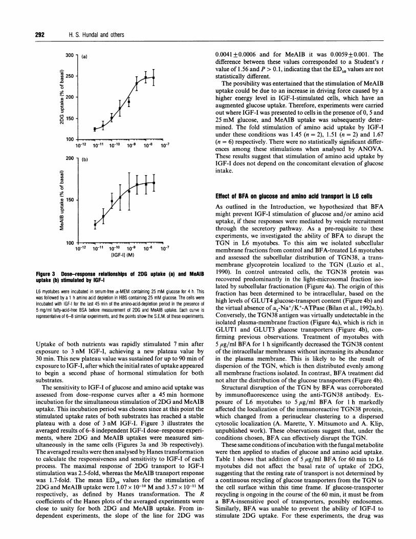
Figure 3 Dose-response relationships of 2DG uptake (a) and MeAIBuptake (b) stimulated by IGF-I
L6 myotubes were incubated in serum-free a-MEM containing 25 mM glucose for 4 h. Thiswas followed by a 1 h amino acid depletion in HBS containing 25 mM glucose. The cells wereincubated with IGF-I for the last 45 min of the amino-acid-depletion period in the presence of5 mg/ml fatty-acid-free BSA before measurement of 2DG and MeAIB uptake. Each curve isrepresentative of 6-8 similar experiments, and the points show the S.E.M. of these experiments.
Uptake of both nutrients was rapidly stimulated 7 min afterexposure to 3 nM IGF-I, achieving a new plateau value by30 min. This new plateau value was sustained for up to 90 min ofexposure to IGF-I, after which the initial rates ofuptake appearedto begin a second phase of hormonal stimulation for bothsubstrates.The sensitivity to IGF-I of glucose and amino acid uptake was
assessed from dose-response curves after a 45 min hormoneincubation for the simultaneous stimulation of2DG and MeAIBuptake. This incubation period was chosen since at this point thestimulated uptake rates of both substrates has reached a stableplateau with a dose of 3 nM IGF-I. Figure 3 illustrates theaveraged results of 6-8 independent IGF-I dose-response experi-ments, where 2DG and MeAIB uptakes were measured sim-ultaneously in the same cells (Figures 3a and 3b respectively).The averaged results were then analysed by Hanes transformationto calculate the responsiveness and sensitivity to IGF-I of eachprocess. The maximal response of 2DG transport to IGF-Istimulation was 2.5-fold, whereas the MeAIB transport responsewas 1.7-fold. The mean ED50 values for the stimulation of2DG and MeAIB uptake were 1.07 x 10⁻¹⁰ M and 3.57 x 10⁻¹¹ Mrespectively, as defined by Hanes transformation. The Rcoefficients of the Hanes plots of the averaged experiments were
close to unity for both 2DG and MeAIB uptake. From in-dependent experiments, the slope of the line for 2DG was
Effect of BFA on glucose and amino acid transport In L6 cellsAs outlined in the Introduction, we hypothesized that BFAmight prevent IGF-I stimulation of glucose and/or amino aciduptake, if these responses were mediated by vesicle recruitmentthrough the secretory pathway. As a pre-requisite to theseexperiments, we investigated the ability of BFA to disrupt theTGN in L6 myotubes. To this aim we isolated subcellularmembrane fractions from control and BFA-treated L6 myotubesand assessed the subcellular distribution of TGN38, a trans-membrane glycoprotein localized to the TGN (Luzio et al.,1990). In control untreated cells, the TGN38 protein was
recovered predominantly in the light-microsomal fraction iso-lated by subcellular fractionation (Figure 4a). The origin of thisfraction has been determined to be intracellular, based on thehigh levels ofGLUT4 glucose-transport content (Figure 4b) andthe virtual absence of al-Na+/K+-ATPase (Bilan et al., 1992a,b).Conversely, the TGN38 antigen was virtually undetectable in theisolated plasma-membrane fraction (Figure 4a), which is rich inGLUT1 and GLUT3 glucose transporters (Figure 4b), con-
firming previous observations. Treatment of myotubes with5 #zg/ml BFA for 1 h significantly decreased the TGN38 contentof the intracellular membranes without increasing its abundancein the plasma membrane. This is likely to be the result ofdispersion of the TGN, which is then distributed evenly amongall membrane fractions isolated. In contrast, BFA treatment didnot alter the distribution of the glucose transporters (Figure 4b).
Structural disruption of the TGN by BFA was corroboratedby immunofluorescence using the anti-TGN38 antibody. Ex-posure of L6 myotubes to 5 ,tg/ml BFA for 1 h markedlyaffected the localization of the immunoreactive TGN38 protein,which changed from a perinuclear clustering to a dispersedcytosolic localization (A. Marette, Y. Mitsumoto and A. Klip,unpublished work). These observations suggest that, under theconditions chosen, BFA can effectively disrupt the TGN.
These same conditions ofincubation with the fungal metabolitewere then applied to studies of glucose and amino acid uptake.Table 1 shows that addition of 5 #g/ml BFA for 60 min to L6myotubes did not affect the basal rate of uptake of 2DG,suggesting that the resting rate of transport is not determined bya continuous recycling of glucose transporters from the TGN tothe cell surface within this time frame. If glucose-transporterrecycling is ongoing in the course of the 60 min, it must be froma BFA-insensitive pool of transporters, possibly endosomes.Similarly, BFA was unable to prevent the ability of IGF-I tostimulate 2DG uptake. For these experiments, the drug was
0 250 -.0D
0oI-
zj 200 -
Q
0.
150-
100-
200
eno
a 150-
Cu4
10.
Q
ma

Insulin-like growth factor action on glucose and system A transport
(a)(kDa) C B C B106 G
P+'A..4 TGN3880 -
PM IM
(b)C B C B
(kDa) .. }.49.5 ^ GLUT1
49.5 45 4 GLUT3
49.5 > 4 GLUT4
PM IM
Figure 4 Effect of BFA on the subcellular distribution of T6N38 (a) and theGLUT1, GLUT3 and GLUT4 glucose transporters (b)
L6 myotubes grown in 10 cm-diameter culture dishes were incubated in serum-free x-MEMcontaining 25 mM glucose for 4 h. This was followed by a 1 h amino-acid-depletion in HBScontaining 25 mM glucose. BFA (5 ,ug/ml) was added from an ethanolic stock solution at thebeginning of the 1 h amino-acid-depletion period. Control cell dishes (C) received an equalvolume of ethanol instead of BFA (B). Cells were harvested and subjected to subcellularfractionation as outlined in the Materials and methods section. Plasma membranes (PM) andlight microsomes (IM) were isolated and analysed on Western blots by using specific antibodiesas outlined in the Materials and methods section. The distribution of the TGN38 antigen andof the GLUT1, GLUT3 and GLUT4 glucose transporters is illustrated.
Table 1 Effects of BFA on 2DG uptake and MeAIB uptake in L6 myotubes
L6 myotubes were incubated in serum-free a-MEM containing 25 mM glucose for 4 h. Thiswas followed by a 1 h amino acid depletion in HBS containing 25 mM glucose. BFA (5 ,ug/ml)was added at the beginning of the amino-acid-depletion period. Control cells received an equalvolume of ethanol instead of BFA. Cells were exposed to IGF-I (3 nM) for the last 45 min ofthe amino-acid-depletion period. Results are means + S.E.M. from 4 separate experiments eachperformed in triplicate. The statistical analysis (Student's t test) of the Basal versus IGF-1-stimulated transport in each case is given in the lower panel.
Uptake (pmol/min per mg of protein)
2-DG MeAIB
Transport -BFA + BFA -BFA + BFA
Basal 8.46 + 0.92 8.18 + 0.70 7.69+ 0.42 8.45+ 0.47+ IGF-I 13.63 + 1.5 13.56 +1.06 13.39 +1.01 11.54+ 0.57
Student's t 2.035 4.094 5.229 3.834P < 0.025 < 0.005 < 0.001 < 0.005
added for 60 min, with IGF-I present during the last 45 min ofthis incubation. IGF-I was fully capable of stimulating 2DGuptake under these conditions, and the fold response was identicalwith that observed in cells not exposed to BFA.We have previously demonstrated that IGF-I stimulates 2DG
uptake through recruitment of glucose transporters present in a
light-microsomal fraction isolated upon subcellular fractionation
of L6 myotubes (Bilan et al., 1992a). The lack of effect of BFAon the response of 2DG uptake to IGF-I suggested that theintracellular pool of glucose transporters was not affected by thefungal metabolite. This was indeed demonstrated by the lack ofeffect ofBFA on the subcellular distribution ofGLUTI, GLUT3or GLUT4 glucose transporters (Figure 4). These proteinsretained their original distribution after exposure to BFA,suggesting that neither the intracellular nor the plasma-mem-brane pools of glucose transporters reside on membranes ofTGN origin.The effect ofBFA on amino acid uptake was also investigated.
In the presence of BFA, basal MeAIB uptake was marginallyelevated but this difference was not statistically significant(Student's t = 1.206). The response of MeAIB uptake to IGF-Iwas slightly lower in BFA-treated than in untreated myotubes,but again this difference was not statistically significant (Student'st = 1.195). As a consequence of the small increase in the basalrate and the lower stimulated uptake, the fold increase in MeAIBtransport activity in response to IGF-I was lower in BFA-treatedcells. However, the stimulation by IGF-I was still highly statisti-cally significant (P < 0.005). Moreover, the apparent sensitivityof MeAIB uptake to BFA was not obvious at a higher dose ofBFA. In two separate experiments (each performed in triplicate)50 jug/ml BFA had no effect on either basal or IGF-I-stimulatedMeAIB uptake (basal uptake 6.97 versus 6.32 pmol/min per mgof protein in the absence and presence ofBFA respectively; IGF-I-stimulated uptake 9.21 versus 9.00 pmol/min per mg of proteinin the absence and presence of BFA respectively). Hence it isapparent that BFA did not alter the acute stimulatory effect ofIGF-I on amino acid uptake through System A.
DISCUSSIONThe ability of L6 muscle cells to respond to low concentrationsof insulin and IGF-I can be attributed to the expression of theirrespective receptors in the PM (Beguinot et al., 1985; Bilan et al.,1992a). IGF-I is a more potent stimulator of glucose transport inthese cells than is insulin through its own receptor (Bilan et al.,1992a). In addition, there is evidence that IGF-I is able to bindto the insulin receptor of L6 cells with greater efficacy thaninsulin itself (Burant et al., 1987). However, IGF-I binding to theinsulin receptor does not result in potentiation of insulin actionon glucose transport, indicating that the intracellular signals thatemanate from the two receptors (or hybrid receptors) convergeon a common signalling pathway or end-point (Lamphere andLienhard, 1992).
In the present study we determined parallelisms in the dose-response and time course of stimulation of glucose and aminoacid uptake by IGF-I, suggesting that similar mechanisms mayunderlie each response. The fold stimulation of glucose transportwas somewhat higher than that of MeAIB transport when amaximal dose of IGF-I was used. Differences in the foldstimulation do not necessarily imply differences in the underlyingmechanism, since these relative values depend in part on thebasal rate of transport for each substrate.The results presented also indicate that the stimulation of
amino acid uptake is not secondary to the increased glucoseintake, since IGF-I was able to stimulate MeAIB uptake to thesame extent in glucose-rich as in glucose-depleted medium.However, the stimulation of amino acid uptake could be sec-ondary to increases in the driving force created by the ionictransmembrane gradients. In rat skeletal muscle, the rapidstimulation of amino acid uptake by insulin is not prevented byouabain, and it has been proposed that this response to the
293

294 H. S. Hundal and others
hormone does not involve a change in the transmembrane ionicgradient (Guma et al., 1988).Given that stimulation of hexose transport in L6 cells can be
fully accounted for by translocation of glucose transporters froman intracellular light-microsomal pool to the plasma membrane(Ramlal et al., 1988), we investigated whether this traffic could beinterfered with by use of BFA, and if so whether amino aciduptake would be similarly affected. BFA effectively blocks trafficof secretory (Fugiwara et al., 1988; Hendricks et al., 1992),lysosomal (Lippincott-Schwartz et al., 1991) and membraneproteins (Shite et al., 1990), through its pharmacological actionon components of the Golgi complex. This compound, whenadministered to L6 myotubes, resulted in a dispersion of theTGN, based on morphological and biochemical redistribution ofthe specific TGN antigen, TGN38. However, under the sameexperimental conditions, BFA failed to interfere with the stimu-lation by IGF-I of either glucose or amino acid uptake. Whereasthe TGN38 no longer migrated with the light-microsomalfraction upon subcellular fractionation of BFA-treated myo-tubes, the intracellular glucose-transporter pool remained in thisfraction.The inability of BFA to prevent stimulation of glucose
transport by IGF-I in the face of substantial TGN disruptioncontests the view that glucose transporters are directly recruitedfrom the TGN to the plasma membrane. A role for the TGN asa donor site of the GLUTI protein was proposed by Blok et al.(1988) for insulin-treated 3T3-LI adipocytes, based on electron-microscopy observations of the subcellular distribution ofimmunogold-labelled GLUT1 proteins. Similarly, Rodnick et al.(1992) proposed that the TGN may represent the site ofintracellular GLUT4 transporter storage in skeletal muscle, basedon immunodetection by electron microscopy of this protein inperinuclear regions. It is possible to reconcile these observationswith the results of the present study by proposing that the TGNcontains a substantial amount of glucose transporters, whichthen furnish a select pool of vesicles that become the target forinsulin or IGF-I signals, providing transporters to the PM. Thisview is supported by immunofluorescence and confocal mi-croscopy observations made in L6 myotubes, indicating thatGLUT4 glucose transporters are largely localized to a perinuclearregion (Y. Mitsumoto and A. Klip, unpublished work). More-over, this scenario would be consistent with a recent suggestionthat two intracellular pools of glucose transporters exist in 3T3-LI adipocytes, only one of which is in direct exchange with thePM (Robinson and James, 1992). The regulated transporter-containing vesicles may be localized in close proximity to thePM, and may only require membrane fusion for the insertion ofextra copies of glucose transporters into the PM. This pathwaywould be predicted to be BFA-insensitive. Precedence for thisstatement are the observations by Klausner et al. (1992) andHunziker et al. (1992), who reported that the function of theendocytic pathway (with respect to endocytosis, endosomalacidification and lysosomal enzyme function) was not affected byBFA despite morphological changes caused to endosomes andlysosomes. Recently, Rosa et al. (1992) also demonstrated thatBFA inhibits the formation of constitutive secretory vesicles, butdoes not prevent secretion from pre-formed vesicles. In spite ofthese arguments, it cannot be ruled out at present that structuraldisruption of the TGN may not affect its function in intracellulartraffic.The results show that in L6 myotubes stimulation of MeAIB
uptake by IGF-I had a similar time course of activation andhormone-sensitivity compared with that of glucose transport.Such observations may denote a similar mechanism of activationof both membrane transport processes by IGF-I. If System A
transporters were translocated to the PM by acute IGF-I orinsulin action, the mechanism could be envisaged to involve thevesicle system that carries glucose transporters to the PM. UsingBFA, we were unable to inhibit selectively stimulation of glucoseor amino acid uptake by BFA. However, the results presentedappear to exclude the direct involvement of endoplasmic-reticulum-to-Golgi vesicle traffic and of the TGN in glucose-transporter recruitment and System A amino acid transportstimulation by IGF-I, based on the inability of BFA to preventIGF-I-mediated activation of glucose and amino acid -uptake.When this work was being completed, an exciting new com-munication by Kong et al. (1993) reported the identification of acDNA sequence cloned from the pig kidney-derived LLC-PK1cell line, which when expressed in COS-7 cells increased uptake ofMeAIB. The sequence codes for a polypeptide of 673 amino acidresidues, and has strong resemblance to the Na+-dependentglucose transporter of epithelia. Antibodies to the new protein,however, are not yet available, and furthermore it is not knownif this amino acid transporter is expressed in non-epithelial cells.When immunological tools are generated, it will be feasible toinvestigate in a direct way the possibility of recruitment of aminoacid transporters in response to hormones and growth factors.
We are indebted to Dr. Y. Mitsumoto for many useful discussions, and to R. Sargeantfor sound experimental advice. We thank Dr. J. P. Luzio (Cambridge University) forthe kind gift of TGN38 antibody. This work was supported by a grant from the MRC(Canada) to A.K. P.J.B. was the recipient of a MRC studentship award. T.T. wassupported by the University of Toronto and the Greek State Scholarship Foundation.H.S.H. was supported by a long-term researdh fellowship from the InternationalHuman Frontier Science Program. A.M. was supported by a post-doctoral fellowshipfrom the Medical Research Council.
REFERENCESBeguinot, F., Kahn, C. R., Moses, A. C. and Smith, R. J. (1985) J. Biol. Chem. 260,
15892-15898Bilan, P. J., Ramial, T. and Klip, A. (1991) in Molecular Biology and Physiology and
Insulin-Like Growth Factors (Raizada, M. K. and LeRoith, D., eds.), pp. 273-288,Plenum Press, New York
Bilan, P. J., Mitsumoto, Y., Ramlal, T. and Klip, A. (1 992a) FEBS Lett. 298, 285-290Bilan, P. J., Mitsumoto, Y., Maher, F., Simpson, I. A. and Klip, A. (1992b) Biochem.
Biophys. Res. Commun. 186, 1129-1137Blok, J., Gibbs, G. E., Lienhard, G. E., Slot, J. W. and Geuze, H. J. (1988) J. Cell Biol. 106,
69-76Bradford, M. M. (1976) Anal. Biochem. 71, 248-254Burant, C. F., Treutelar, M. K., Allen, D. A., Sens, D. A. and Buse, M. G. (1987) Biochem.
Biophys. Res. Commun. 147, 100-107Christensen, H. N. (1990) Physiol. Rev. 70, 43-77Cushman, S. W. and Wardzala, L. J. (1980) J. Biol. Chem. 255, 4758-4762Douen, A. G., Ramlal, T., Rastogi, S., Bilan, P. J., Cartee, G. D., Vranic, M., Hollozy, J. 0.
and Klip, A. (1990) J. Biol. Chem. 265, 13427-13430Friedman, J. E., Dudek, R. W., Whitehead, D. L., Downes, D. L., Frisell, W. R., Caro, J. F.
and Dohm, L. (1991) Diabetes 41, 150-154Fugiwara, T., Oda, K., Yokota, S., Takatsuki, A. and Ikehara, Y. (1988) J. Biol. Chem. 263,
18545-1 8552Guma, A., Testar, X., Palacin, M. and Zorzano, A. (1988) Biochem. J. 253, 625-629Hendricks, L. C., McClanahan, S. L., Palade, G. E. and Farquhar, M. G. (1992) Proc. Natl.
Acad. Sci. U.S.A. 89, 7242-7246Hirshman, M. F., Goodyear, L. J., Wardzala, L. J., Horton, E. D. and Horton, E. S. (1990)
J. Biol. Chem. 265, 987-991Hundal, H. S., Ramlal, T., Reyes, R., Leiter, L. A. and Klip, A. (1992) Endocrinology
(Baltimore) 131, 1165-1173Hunziker, W., Whitney, J. A. and Mellman, I. (1992) FEBS Lett. 307, 93-96Klausner, R. D., Donaldson, J. G. and Lippincott-Schwartz, J. (1992) J. Cell Biol. 116,
1071-1080Koivisto, U.-M., Martinez-Valdez, H., Bilan, P. J., Burdett, E., Ramlal, T. and Klip, A. (1991)
J. Biol. Chem. 266, 2615-2621Kong, C.-T., Yet, S.-F. and Lever, J. (1993) J. Biol. Chem. 268, 1509-1512Ladinsky, M. S. and Howell, K. E. (1992) Eur. J. Cell Biol. 59, 92-105Lamphere, S. and Lienhard, G. E. (1992) Endocrinology (Baltimore) 131, 2196-2202Lippincott-Schwartz, J., Yuan, L. C., Tipper, C., Amherdt, M., Orci, L. and Klausner, R. D.
(1991) Cell 67, 601-616

Insulin-like growth factor action on glucose and system A transport
Luzio, J. P., Brake, B., Banting, G., Howell, K. E., Braghetta, P. and Stanley, K. K. (1990)Biochem. J. 270, 97-102
Mitsumoto, Y. and Klip, A. (1992) J. Biol. Chem. 267, 4957-4962Mitsumoto, Y., Burdett, A., Grant, A. and Klip, A. (1991) Biochem. Biophys. Res. Commun.
175, 652-659Price, N. C. and Stevens, L. (1982) Fundamentals of Enzymology, Oxford University Press,
OxfordRamlal, T., Sarabia, V., Bilan, P. J. ad Klip, A. (1988) Biochem. Biophys. Res. Commun.
157, 1329-1335Reaves, B., Wilde, A. and Banting, G. (1992) Biochem. J. 283, 313-316Robinson, L. J. and James, D. E. (1992) Am. J. Physiol. 263, E383-E393
Rodnick, K. J., Slot, J. W., Studelska, D. R., Hanpeter, D. E., Robinson, L. J., Geuze, H. J.and James, D. E. (1992) J. Biol. Chem. 267, 6278-6285
Rosa, P., Barr, F., Stinchdombe, J. C., Binacchi, C. and Huttner, W. B. (1992) Eur. J. CellBiol. 59, 265-274
Sargeant, R., Mitsumoto, Y., Sarabia, V., Schillabeer, G. and Klip, A. (1993) J. Endocrinol.Invest. 16, 147-162
Shainberg, A., Yagil, G. and Yaffe, D. (1971) Dev. Biol. 25, 1-29Shite, S., Seguchi, T., Shimada, T., Ono, M. and Kuwano, M. (1990) Eur. J. Biochem. 191,
491-497Suzuki, K. and Kono, T. (1980) Proc. Natl. Acad. Sci. U.S.A. 77, 2542-2545Yaffe, D. (1968) Proc. Natl. Acad. Sci. U.S.A. 61, 477-483
Received 11 August 1993; accepted 31 August 1993
295


