Embed Size (px)
Citation preview

electronic reprint
Acta Crystallographica Section F
Structural Biologyand CrystallizationCommunications
ISSN 1744-3091
Editors: H. M. Einspahr and J. M. Guss
Structure of the conserved hypothetical protein MAL13P1.257 fromPlasmodium falciparum
Margaret A. Holmes, Frederick S. Buckner, Wesley C. Van Voorhis, ChristopherMehlin, Erica Boni, Thomas N. Earnest, George DeTitta, Joseph Luft, AngelaLauricella, Lori Anderson, Oleksandr Kalyuzhniy, Frank Zucker, Lori W.Schoenfeld, Wim G. J. Hol and Ethan A. Merritt
Copyright © International Union of Crystallography
Author(s) of this paper may load this reprint on their own web site provided that this cover page is retained. Republication of this article or itsstorage in electronic databases or the like is not permitted without prior permission in writing from the IUCr.
Acta Cryst. (2006). F62, 180–185 Holmes et al. � MAL13P1.257

structural genomics communications
180 doi:10.1107/S1744309106005847 Acta Cryst. (2006). F62, 180–185
Acta Crystallographica Section F
Structural Biologyand CrystallizationCommunications
ISSN 1744-3091
Structure of the conserved hypothetical proteinMAL13P1.257 from Plasmodium falciparum
Margaret A. Holmes,a,b
Frederick S. Buckner,a,c
Wesley C. Van Voorhis,a,c
Christopher Mehlin,a,b Erica
Boni,a,b Thomas N. Earnest,a,d
George DeTitta,a,e Joseph Luft,a,e
Angela Lauricella,a,e Lori
Anderson,a,b Oleksandr
Kalyuzhniy,a,b Frank Zucker,a,b
Lori W. Schoenfeld,a,b Wim G. J.
Hola,b,f and Ethan A. Merritta,b*
aStructural Genomics of Pathogenic Protozoa
(SGPP) Consortium, USA, bDepartment of
Biochemistry, University of Washington, Seattle,
WA 98195-7742, USA, cDepartment of
Medicine, University of Washington, Seattle,
WA 98195, USA, dLawrence Berkeley National
Laboratory, Berkeley, CA 94720, USA,eHauptman–Woodward Institute, Buffalo,
NY 14203, USA, and fHoward Hughes Medical
Institute, University of Washington, Seattle,
WA 98195, USA
Correspondence e-mail:
Received 27 December 2005
Accepted 16 February 2006
PDB Reference: MAL13P1.257, 1zso, r1zsosf.
The structure of a conserved hypothetical protein, PlasmoDB sequence
MAL13P1.257 from Plasmodium falciparum, Pfam sequence family PF05907,
has been determined as part of the structural genomics effort of the Structural
Genomics of Pathogenic Protozoa consortium. The structure was determined by
multiple-wavelength anomalous dispersion at 2.17 A resolution. The structure is
almost entirely �-sheet; it consists of 15 �-strands and one short 310-helix and
represents a new protein fold. The packing of the two monomers in the
asymmetric unit indicates that the biological unit may be a dimer.
1. Introduction
The present structure determination of Plasmodium falciparum
protein MAL13P1.257 (Kissinger et al., 2002) was undertaken as part
of the Structural Genomics of Pathogenic Protozoa (SGPP) consor-
tium effort targeting proteins from eukaryotic tropical pathogens.
One goal of structural genomics is to determine the structures of
proteins that are members of sequence families with unknown folds.
This protein was selected for structure determination because it is a
conserved hypothetical protein belonging to Pfam family PF05907
(DUF866) (Bateman et al., 2004), whose members have no significant
sequence homology to any structure in the PDB. This sequence
family contains eukaryotic proteins of unknown function. The
P. falciparum protein, whose SGPP identifier is Pfal004331AAA, is
156 amino acids long, has a molecular weight of 18.7 kDa and has a
theoretical pI of 4.6. Here, we report the structure of Pfal004331AAA
at 2.17 A resolution determined by multiple-wavelength anomalous
dispersion and refined to an R value of 0.183 and a free R value of
0.233. The structure contains 15 �-strands and one 310-helix. This
constitutes a three-dimensional fold not seen previously and provides
the first structural basis for homology modeling of other members of
the PF05907 sequence family. There are two closely associated
molecules in the crystallographic asymmetric unit; their large buried
surface area suggests that the biologically active unit may be a dimer.
2. Materials and methods
Ligase-independent cloning (LIC) was used to append a His tag to
the N-terminus and a TAA stop codon to the C-terminus of the
MAL13P1.257 gene of P. falciparum, giving MAHHHHHH-orf-TAA
(SGPP identifier Pfal004331AAA). The vector has a T7 promoter for
growth in Escherichia coli with auto-induction media (without the use
of IPTG). Selenomethionine protein was produced according to the
protocol of Studier (2005) in BL21 DE3 Star E. coli. Cells were lysed
by sonication in 25 mM HEPES buffer with 500 mM NaCl, 0.2%(w/v)
cholate, 0.1 mg ml�1 lysozyme, 1 mM �-mercaptoethanol and Roche
EDTA-free protease inhibitors. Lysates were centrifuged to remove
insoluble cell debris and the cleared lysates were tumbled with
Qiagen Ni–NTA superflow resin. Purification followed the protocol
reported by Mehlin et al. (2006). The resin was washed once with
10 mM imidazole and twice with 20 mM imidazole and eluted with
15 ml 250 mM imidazole in SGPP standard buffer (500 mM NaCl,
25 mM HEPES pH 7.25, 0.025% sodium azide, 5% glycerol). The# 2006 International Union of Crystallography
All rights reserved
electronic reprint

eluate was dialyzed overnight against standard buffer and aggregates
were separated by size-exclusion chromatography the next day.
Fractions were analyzed by SDS–PAGE and then pooled for
concentration to 12.4 mg ml�1. Dithiothreitol was added to 2 mM
before proteins were flash-frozen for shipment to the crystal-
screening and crystal-growth laboratories.
Pfal004331AAA was screened for crystallization at the Hauptman–
Woodward Institute (Luft et al., 2003). The protein was combined
with 1536 different crystallization cocktail solutions in a single plate
under mineral oil to prevent dehydration. Experiments were set up
using standard commercially available liquid-handling systems. Plates
were imaged over a four-week time course. Images were reviewed
and crystallization conditions were forwarded to the SGPP crystal-
growth laboratory in Seattle for optimization. There, crystallization
conditions found in the initial large-scale screen (1.0 M LiCl, 20%
PEG 6000, 0.1 M Tris pH 8) were optimized for pH, major precipitant
and additive concentrations using a vapor-diffusion sitting-drop
method. The crystallization conditions for the crystal used for
structure solution and initial phasing were 1.0 M LiCl, 30% PEG
6000, 0.1 M Tris pH 8, 298 K; conditions for the crystal used for
structure refinement were 1.0 M LiCl, 25 mM Mg(NO3)2, 25% PEG
6000, 0.1 M Tris pH 8, 298 K. The two crystals were cryoprotected in
solutions that contained 21% xylitol and 30% glycerol, respectively,
and flash-frozen in liquid nitrogen prior to shipping for data collec-
tion.
X-ray diffraction data were collected at the Advanced Light
Source on beamline 8.2.1 for the crystal used in MAD phasing and on
beamline 8.2.2 for the crystal used in refinement. All data were
integrated and scaled using HKL2000 (Otwinowski & Minor, 1997).
The asymmetric unit contains two monomers of Pfal004331AAA,
which contain a total of eight SeMet residues, including those in the
structural genomics communications
Acta Cryst. (2006). F62, 180–185 Holmes et al. � MAL13P1.257 181
Figure 2A ribbon representation of the A chain of Pfal004331AAA. The secondary-structure elements are colored as in Fig. 1. Each sheet is labeled alphabetically,denoting its position in the sequence. The figure was generated using PyMOL(DeLano, 2002).
Table 1Data-collection statistics.
Redundancy, completeness and Rmerge are as reported by HKL2000; I/�(I) is as reportedby TRUNCATE. Values in parentheses are for the highest resolution shell. Data beyond2.17 A were not used in refinement.
Se peak(phasing) Se inflection Se remote Refinement
Space group P212121 P212121
Unit-cell parameters(A)
a = 62.6, b = 71.0, c = 78.5 a = 62.2,b = 71.1,c = 78.6
VM (A3 Da�1) 2.2 2.2Wavelength (A) 0.97957 0.97972 0.95372 0.9795Resolution range (A) 50–2.50
(2.59–2.50)50–2.50
(2.59–2.50)50–2.50
(2.59–2.50)50–2.15
(2.23–2.15)No. of unique reflections 11846 11883 12763 18151Redundancy 6.9 (5.2) 6.8 (5.0) 6.8 (5.5) 5.9 (3.3)Completeness (%) 92.9 (28.1) 93.8 (37.2) 99.9 (99.5) 92.8 (57.7)Rmerge 0.049 (0.128) 0.064 (0.278) 0.072 (0.341) 0.093 (0.365)Mean I/�(I) 53 (14) 40 (6.9) 35 (5.8) 27 (3.7)Mean FOM for phasing 0.63
Table 2Refinement and model statistics.
Target ideal geometry is that of REFMAC v.5.2.0005. ’/ categorization is that ofPROCHECK. Wilson B factor is as reported by TRUNCATE. Values of (Biso + BTLS)were generated by expanding the TLS description and individual ADP values of therefined model into an equivalent anisotropic description using the CCP4 programsTLSEXTRACT and TLSANL (Collaborative Computational Project, Number 4, 1994).We report the resulting equivalent isotropic ADP as Beq = (Biso + BTLS). Values inparentheses are for the highest resolution shell.
Resolution range (A) 20–2.17 (2.22–2.17)Rwork, 18076 (899) reflections 0.183 (0.209)Rfree, 926 (51) reflections 0.233 (0.314)R.m.s.d. bonds (A) 0.017R.m.s.d. angles (�) 1.58Residues in most favored region of ’/ 260 [88%]Residues in additional allowed region of ’/ 36 [12%]Residues in generously allowed region of ’/ 1 [0.3%]Residues in disallowed region of ’/ 0No. of protein atoms 2647No. of water molecules 162Wilson B factor (A2) 32TLS groups chain A 9–27, 28–84, 85–118, 119–156TLS groups chain B 6–31, 32–83, 84–118, 119–156Mean [e.s.d.] of (Biso + BTLS) for atoms in chain A (A2) 37 [9]Mean [e.s.d.] of (Biso + BTLS) for atoms in chain B (A2) 46 [13]
Figure 1A topology diagram of the A chain of Pfal004331AAA, produced by the TOPStopology cartoon server (Michalopoulos et al., 2004). The triangles represent �-strands and the circle represents the 310-helix. The upward-pointing trianglesrepresent strands that come out of the plane of the diagram and downward-pointing triangles represent strands that go into the plane. The largest, six-stranded,�-sheet is colored yellow, the four-stranded sheet is blue, the three-stranded sheet isred, the two-stranded sheet is orange and the 310-helix is purple.
electronic reprint
N-terminal tag. The program SOLVE (Terwilliger, 2003) was used to
locate the Se atoms and to calculate phases. The program RESOLVE
was used for density modification, twofold NCS map averaging and
automatic tracing. SOLVE and RESOLVE were run a number of
times with both 3.0 and 2.5 A outer resolution limits, each time
finding four Se atoms, two in each of the two chains. A composite
model was assembled from the resultant autotraced models, using the
density-modified maps, with the program XFIT (McRee, 1999).
Refinement of this model was then carried out against the peak data
from the higher resolution data set using the program REFMAC5
(Murshudov et al., 1997) via the ccp4i interface (Potterton et al., 2004),
alternating with rounds of building in XFIT. No NCS restraints were
used in refinement. Data-collection statistics are shown in Table 1 and
refinement statistics are shown in Table 2.
In the last three cycles of refinement, each chain was described by
four TLS groups identified by the TLSMD server (Painter & Merritt,
2006) and TLS parameters were refined for each group (Table 2).
Eight N-terminal residues of chain A and five N-terminal residues of
chain B, including three well ordered residues from the His tag, were
not assigned to any TLS group. Refinement was monitored using
5% of the data reserved for Rfree. Final structure validation was
performed with PROCHECK (Laskowski et al., 1993) and
MolProbity (Lovell et al., 2003). The model of the A chain consists of
all 156 residues in the open reading frame, while the model of the B
chain additionally contains the last three residues of the His tag.
3. Results
The structure of Pfal004331AAA has been solved to 2.17 A resolu-
tion. The structure contains 15 �-strands, which form four �-sheets.
The largest sheet is comprised of six strands and runs the length of the
molecule. There are two smaller sheets, one of four strands and one of
three strands, which flank the large sheet. A short two-stranded sheet
and a short 310-helix complete the secondary structure. Prior to
determining the structure of Pfal004331AAA, an iterative PSI-
BLAST search (Altschul et al., 1997) for protein sequences with
significant similarity to Pfal004331AAA found none with an anno-
tated function or with a known structure. After the structure had
been determined, it was submitted to the DALI server (Holm &
Sander, 1993) and to the VAST server (Gibrat et al., 1996; Madej et
al., 1995) in order to search for structures with similar topology. No
significant match to any structure in the PDB was reported by either
server. Therefore, we conclude that the structure of Pfal004331AAA
constitutes a previously unobserved protein fold.
Fig. 1 shows a topology diagram of the monomer. The largest sheet
is composed of six strands [�A (Thr4–Glu13), �G (Phe61–Ile63), �H
(Ser72–Val77), �J (Arg94–Arg102), �N (Trp135–Asn139) and �O
(Met144–Asn156)] arranged in an antiparallel fashion, which account
for almost 30% of the total residues. This largest sheet extends the
length of the molecule and is flanked by three smaller �-sheets
(Fig. 2). One of these is an antiparallel four-stranded sheet [�B
(Val15–Phe19), �I (Ser84–Glu86), �L (Phe115–Asp119) and �M
(Leu124–Val128)], which is positioned at one end of the large
�-sheet. A three-stranded �-sheet [�C (Trp26–Asp32), �D (Thr38–
Phe44) and �K (Ile104–Phe109)] is positioned at the other end of the
large sheet. A small antiparallel two-stranded �-sheet [�E (Leu50–
Glu51) and �F (Thr58–Ala59)] lies across the large sheet from the
three-stranded sheet. There is a short 310-helix (Val88–Asn90)
between one edge of the large sheet and the four-stranded sheet.
The distribution of electrostatic potential at the protein surface is
unremarkable and there are no extensive hydrophobic patches. The
surface of the chain A monomer has one obvious cavity; it is the
largest pocket found by the CASTp server (Binkowski et al., 2003;
Liang et al., 1998) and has a volume of 310 A3. This pocket is formed
mainly by adjacent edges of the large sheet and the four-stranded
sheet and by the 310-helix which connects them (atoms from residues
5, 8, 79–86, 89 and 94–98). The cavity is diametrically opposite the
proposed dimer interface (see below). It is lined with both non-polar
and polar residues and is occupied by five water molecules (Fig. 3).
The equivalent cavity in chain B is smaller owing to different side-
chain conformations of Asn89 and Arg94 and contains only two well
ordered water molecules.
structural genomics communications
182 Holmes et al. � MAL13P1.257 Acta Cryst. (2006). F62, 180–185
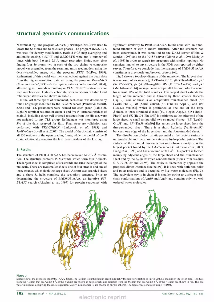
Figure 3Stereoview of the proposed Pfal004331AAA dimer. The A chain is on the right in green in roughly the same orientation as in Fig. 2; the B chain is on the left in gold. Residuesfrom the A chain that are within 4 A of the B chain are shown as purple sticks and residues from the B chain that are within 4 A of the A chain are shown in red. The fivewater molecules occupying the single significant cavity in monomer A are shown as purple spheres. The figure was generated using PyMOL.
electronic reprint

structural genomics communications
Acta Cryst. (2006). F62, 180–185 Holmes et al. � MAL13P1.257 183
Figure 4CLUSTALW multiple sequence alignment for representative sequences from Pfam family PF05907 (DUF866). Residue positions with sequence identity greater than 60%are shaded gray and residue positions that are 100% conserved are shaded cyan. Sequences are identified by their TrEMBL entry names. Residue numbers correspond tothose of the P. falciparum structure reported here. Bars indicating individual �-strands are colored as in Figs. 1 and 2. Figure generated using TEXshade (Beitz, 2000).
electronic reprint

The crystallographic asymmetric unit contains two closely asso-
ciated monomers. Their conformation is essentially identical, with an
r.m.s.d. of 0.49 A for 152 C� positions after superposition. Deviations
were observed only at the termini (residues 1–3 and 156) and at two
short loops involved in crystal-packing contacts (residues 54–57 and
79–81). The monomer–monomer interaction was characterized using
the Protein–Protein Interaction Server (Jones & Thornton, 1996). The
total interface accessible surface area of chain A is 890 A2 and that
for chain B is 860 A2. These areas and the associated gap volume
index of 3.0 are consistent with a weak homodimer. Fig. 3 shows the
proposed dimer. The two chains interact directly via 11 hydrogen
bonds and indirectly via a network of nine water molecules that are
trapped between the two chains, reducing the surface comple-
mentarity. The chromatogram from the size-exclusion step of protein
purification showed that about 20% of the protein ran as a dimer (not
shown); however, only protein from the major (80%) peak was used
for crystallization trials. We conclude that Pfal004331AAA most
likely acts as a weak dimer; the physiological dimerization state is not
certain.
4. Discussion
The structure of P. falciparum protein MAL13P1.257 is the first
structure to be determined of a member of Pfam sequence family
PF05907. The biological function of these proteins is unknown. This
family is currently represented by 48 sequences indexed by Interpro
12.0 (Mulder et al., 2005) with an average length of 165 residues; the
domain structure of 33 of these has been characterized (Bateman et
al., 2004). These are single-domain proteins, with certain exceptions.
A complete DUF866 domain appears at the N-terminus of a large
(1248 residues) coding sequence from Ustilago maydis, UM05492.1,
containing no other annotated domain structure. There is also strong
similarity to a 68-residue stretch of Aspergillus nidulans ORF
AN7606.2 (Q5AVS4_EMENI in Fig. 4), which contains in addition an
FAD-dependent phenol hydroxylase domain and an LSM domain
(Pfam PF01423). A somewhat longer stretch of sequence similarity is
found in Giberella zeae (Fig. 4; Q4IMT7_GIBZE), which contains a
second domain annotated as a mitochondrial RNA-processing
domain (Pfam PF08296).
There are two regions of high sequence conservation between the
present protein and the other PF05907 family members. One region is
a hydrophobic core formed by residues Tyr125, Trp135 and Val146
(Fig. 5). The second region spans �-strands J and K and consists of
residues 99–111. Curiously, several residues which are highly
conserved across other family members are not conserved in the
present P. falciparum protein. For example, it is striking that two
CXXC motifs are strongly conserved in all other family members
(Fig. 4 and additional sequences not shown) but are not present in
P. falciparum. These correspond to residues 32–35 and 65–68 in the
present structure. Neither the residues surrounding the surface cavity
nor the residues involved in the dimer interface are maintained across
the sequence family. These observations may indicate that specific
biological function is not conserved throughout the entire sequence
family. Nevertheless, the present structure provides a basis for
structural modeling of a previously uncharacterized eukaryotic
sequence family, one with a three-dimensional fold that has not
previously been observed.
We are grateful for the contributions of other SGPP consortium
members, including Peter Myler, Elizabeth Worthey, Tracy Arakaki,
Jurgen Bosch, Jonathan Caruthers, Mark Robien, Christophe
Verlinde, Larry de Soto and Martin Criminale. Portions of this work
were carried out at the Advanced Light Source, which is supported by
the Director, Office of Science, Office of Basic Energy Sciences of
the US Department of Energy under Contract No. DE-AC02-
05CH11231. This work was supported by NIH awards GM64655 and
GM62617.
References
Altschul, S. F., Madden, T. L., Schaffer, A. A., Zhang, J., Zhang, Z., Miller, W.& Lipman, D. J. (1997). Nucleic Acids Res. 25, 3389–3402.
Bateman, A., Coin, L., Durbin, R., Finn, R. D., Hollich, V., Griffiths-Jones, S.,Khanna, A., Marshall, M., Moxon, S., Sonnhammer, E. L. L., Studholme,D. J., Yeats, C. & Eddy, S. R. (2004). Nucleic Acids Res. 32, D138–D141.
Beitz, E. (2000). Bioinformatics, 16, 135–139.Binkowski, T. A., Naghibzadeh, S. & Liang, J. (2003). Nucleic Acids Res. 31,
3352–3355.Collaborative Computational Project, Number 4 (1994). Acta Cryst. D50,
760–763.DeLano, W. L. (2002). The PyMOL Molecular Graphics System. http://
www.pymol.org.Gibrat, J. F., Madej, T. & Bryant, S. H. (1996). Curr. Opin. Struct. Biol. 6,
377–385.Holm, L. & Sander, C. (1993). J. Mol. Biol. 233, 123–138.Jones, S. & Thornton, J. M. (1996). Proc. Natl Acad. Sci. USA, 93, 13–20.Kissinger, J. C. et al. (2002). Nature (London), 419, 490–492.Laskowski, R., MacArthur, M., Moss, D. & Thornton, J. (1993). J. Appl. Cryst.
26, 283–291.Liang, J., Edelsbrunner, H. & Woodward, C. (1998). Protein Sci. 7, 1884–1897.Lovell, S., Davis, I., Arendall, W. B. III, de Bakker, P., Word, J., Prisant, M.,
Richardson, J. & Richardson, D. (2003). Proteins, 50, 437–450.Luft, J. R., Collins, R. J., Fehrman, N. A., Lauricella, A. M., Veatch, C. K. &
DeTitta, G. T. (2003). J. Struct. Biol. 142, 170–179.McRee, D. E. (1999). J. Struct. Biol. 125, 156–165.Madej, T., Gibrat, J. F. & Bryant, S. H. (1995). Proteins, 23, 356–369.Mehlin, C., Boni, E., Buckner, F. S., Engel, L., Feist, T., Gelb, M., Haji, L., Kim,
D., Liu, C., Mueller, N., Myler, P. J., Reddy, J. T., Sampson, J. N.,Subramanian, E., Van Voorhis, W. C., Worthey, E., Zucker, F. & Hol, W. G. J.(2006). Mol. Biochem. Parasitol. In the press..
structural genomics communications
184 Holmes et al. � MAL13P1.257 Acta Cryst. (2006). F62, 180–185
Figure 5Electron density for a highly conserved hydrophobic core region. Residues Tyr125(Phe in other sequence family members), Trp135 and Val146 are highly conservedin the PF05907 sequence family. They form a hydrophobic core region anchoring�-strands M, N and O. The density shown is contoured at 2.5� in a �A-weighted(2mFo � DFc) electron-density map.
electronic reprint

Michalopoulos, I., Torrance, G. M., Gilbert, D. R. & Westhead, D. R. (2004).Nucleic Acids Res. 32, D251–D254.
Mulder, N. J. et al. (2005). Nucleic Acids Res. 33, D201–D205.Murshudov, G. N., Vagin, A. A. & Dodson, E. J. (1997). Acta Cryst. D53,
240–255.Otwinowski, Z. & Minor, W. (1997). Methods Enzymol. 276, 307–326.
Painter, J. & Merritt, E. A. (2006). J. Appl. Cryst. 39, 109–111.Potterton, L., McNicholas, S., Krissinel, E., Gruber, J., Cowtan, K., Emsley, P.,
Murshudov, G. N., Cohen, S., Perrakis, A. & Noble, M. (2004). Acta Cryst.D60, 2288–2294.
Studier, F. W. (2005). Protein Expr. Purif. 41, 207–234.Terwilliger, T. C. (2003). Methods Enzymol. 374, 22–37.
structural genomics communications
Acta Cryst. (2006). F62, 180–185 Holmes et al. � MAL13P1.257 185electronic reprint


