Embed Size (px)
Citation preview
Structure-activity relationships for a new family of sulfonylurea herbicides
Jian-Guo Wanga, Zheng-Ming Lia, Ning Maa, Bao-Lei Wanga, Lin Jianga, Siew SiewPangb,c, Yu-Ting Leeb, Luke W. Guddatb,* & Ronald G. Dugglebyb,*aElemento-Organic Chemistry Institute, State Key Laboratory of Elemento-Organic Chemistry, NationalPesticide Engineering Research Center, Nankai University 300071, Tianjin, P R China; bSchool of Molecularand Microbial Sciences, The University of Queensland, Brisbane 4072, QLD, Australia; cNovartis Institute forTropical Diseases, 10 Biopolis Road #05–01, 138670, Chromos, Singapore
Received 18 May 2005; accepted 7 November 2005
� Springer 2005
Key words: 3D-QSAR, acetohydroxyacid synthase, Arabidopsis thaliana, computational docking, herbi-cide, rational drug design, sulfonylurea
Summary
Acetohydroxyacid synthase (AHAS; EC 2.2.1.6) catalyzes the first common step in branched-chain aminoacid biosynthesis. The enzyme is inhibited by several chemical classes of compounds and this inhibition is thebasis of action of the sulfonylurea and imidazolinone herbicides. The commercial sulfonylureas contain apyrimidine or a triazine ring that is substituted at bothmeta positions, thus obeying the initial rules proposedby Levitt. Here we assess the activity of 69 monosubstituted sulfonylurea analogs and related compounds asinhibitors of pure recombinant Arabidopsis thaliana AHAS and show that disubstitution is not absolutelyessential as exemplified by our novel herbicide, monosulfuron (2-nitro-N-(4¢-methyl-pyrimidin)2¢-yl) phe-nyl-sulfonylurea), which has a pyrimidine ring with a single meta substituent. A subset of these compoundswas tested for herbicidal activity and it was shown that their effect in vivo correlates well with their potency invitro as AHAS inhibitors. Three-dimensional quantitative structure–activity relationships were developedusing comparative molecular field analysis and comparative molecular similarity indices analysis. For thelatter, the best result was obtained when steric, electrostatic, hydrophobic and H-bond acceptor factors weretaken into consideration. The resulting fields were mapped on to the published crystal structure of the yeastenzyme and it was shown that the steric and hydrophobic fields are in good agreement with sulfonylurea-AHAS interaction geometry.
Introduction
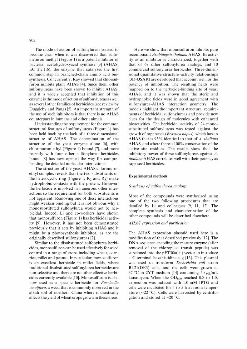
The herbicidal activity of sulfonylureas such as N-(p-cyanophenylaminocarbonyl) benzenesulfona-mide (Figure 1) was recognized nearly 40 yearsago [1]. Following an extensive synthetic programled by Levitt and colleagues [2] the first modern
sulfonylurea herbicide chlorsulfuron (Figure 1)was developed. Since that time, a large numberof other sulfonylurea herbicides have been identi-fied and are now used widely [1]. The generalfeatures of most active compounds (Figure 1) arean ortho-substituted aromatic ring attached to thesulfur atom, and a heterocyclic ring substituted inboth meta positions and attached to the distalnitrogen atom of the sulfonylurea bridge. Thisheterocyclic ring is either a pyrimidine (X = CH)or triazine (X = N).
*To whom correspondence should be addressed. Fax: +61-7-3365-4699; E-mail: [email protected]; [email protected]
Journal of Computer-Aided Molecular Design (2005) 19: 801–820DOI 10.1007/s10822-005-9028-9
The mode of action of sulfonylureas started tobecome clear when it was discovered that sulfo-meturon methyl (Figure 1) is a potent inhibitor ofbacterial acetohydroxyacid synthase [3] (AHAS;EC 2.2.1.6), the enzyme that catalyzes the firstcommon step in branched-chain amino acid bio-synthesis. Concurrently, Ray showed that chlorsul-furon inhibits plant AHAS [4]. Since then, othersulfonylureas have been shown to inhibit AHAS,and it is widely accepted that inhibition of thisenzyme is themodeof actionof sulfonylureas aswellas several other families of herbicides (see review byDuggleby and Pang) [5]. An important strength ofthe use of such inhibitors is that there is no AHAScounterpart in humans and other animals.
Understanding the requirement for the commonstructural features of sulfonylureas (Figure 1) hasbeen held back by the lack of a three-dimensionalstructure of AHAS. The determination of thestructure of the yeast enzyme alone [6], withchlorimuron ethyl (Figure 1) bound [7], and morerecently with four other sulfonylurea herbicidesbound [8] has now opened the way for compre-hending the detailed molecular interactions.
The structure of the yeast AHASÆchlorimuronethyl complex reveals that the two substituents onthe heterocyclic ring (Figure 1; R2 and R3) makehydrophobic contacts with the protein. However,the herbicide is involved in numerous other inter-actions so the requirement for both substituents isnot apparent. Removing one of these interactionsmight weaken binding but it is not obvious why amonosubstituted sulfonylurea would not be her-bicidal. Indeed, Li and co-workers have shownthat monosulfuron (Figure 1) has herbicidal activ-ity [9]. However, it has not been demonstratedpreviously that it acts by inhibiting AHAS and itmight be a photosynthesis inhibitor, as are theoriginally described sulfonylureas [2].
Similar to the disubstituted sulfonylurea herbi-cides,monosulfuron can beused effectively forweedcontrol in a range of crops including wheat, corn,rice, millet and peanut. In particular, monosulfuronis an excellent herbicide in millet fields, wheretraditional disubstituted sulfonylurea herbicides arenon-selective and there are no other effective herbi-cides currently available [10]. Monosulfuron is alsonow used as a specific herbicide for Puccinellatenuiflora, a weed that is commonly observed in thealkali soil of northern China where it drasticallyaffects the yield of wheat crops grown in these areas.
Here we show that monosulfuron inhibits purerecombinant Arabidopsis thaliana AHAS. Its activ-ity as an inhibitor is characterized, together withthat of 68 other sulfonylurea analogs, and 10commercial sulfonylurea herbicides. Three-dimen-sional quantitative structure–activity relationships(3D-QSAR) are developed that account well for thepotency of inhibition. The resulting fields weremapped on to the herbicide-binding site of yeastAHAS, and it was shown that the steric andhydrophobic fields were in good agreement withsulfonylurea-AHAS interaction geometry. Themodels highlight the important structural require-ments of herbicidal sulfonylureas and provide newclues for the design of molecules with enhancedbioactivities. The herbicidal activity of 24 mono-substituted sulfonylureas was tested against thegrowth of rape seeds (Brassica napus), which has anAHAS that is 93% identical to that of A. thalianaAHAS, andwhere there is 100%conservation of theactive site residues. The results show that theinhibitory power of these sulfonylureas against A.thalianaAHAS correlates well with their potency asrape seed herbicides.
Experimental methods
Synthesis of sulfonylurea analogs
Most of the compounds were synthesized usingone of the two following procedures that aredetailed by Li and colleagues [9, 11, 12]. Thecomplete synthesis and characterization of theother compounds will be described elsewhere.
AHAS expression and purification
The AHAS expression plasmid used here is amodification of that described previously [12]. TheDNA sequence encoding the mature enzyme (afterremoval of the chloroplast transit peptide) wassubcloned into the pET30a(+) vector to introducea C-terminal hexahistidine tag [13]. This plasmidwas used to transform Escherichia coli strainBL21(DE3) cells, and the cells were grown at37 �C in 2YT medium [14] containing 50 lg/mLkanamycin. When the OD600 reached 0.8 to 1.0,expression was induced with 1.0 mM IPTG andcells were incubated for 4 to 5 h at room temper-ature (�22 �C). Cells were harvested by centrifu-gation and stored at )20 �C.
802
O
OCH2CH3
S NH C
O
NH
N
NCl
OCH3
O
O
C OCH2CH3
S NH C
O
NH
N
NOCH3
OCH3
O
O
O
C
O
OCH3
S NH C
O
NH
N
N
NCH3
OCH3
O
O
Cl
S NH C
O
NH
N
N
NCH3
OCH3
O
O
C
S C
O
NH
N
NOCH3
OCH3
O
O
CH2 NH
O
OCH3
C
O
OCH3
S NH C
O
NH
N
N
NOCH2CH3
NHCH3
O
O
C
O
OCH3
S NH C
O
NH
N
NCH3
CH3
O
O
C
O
OCH3
S NH C
O
NH
N
N
NCH3
OCH3
O
OS
C
O
OCH2CH3
S NH C
O
NH
N
NOCH3
OCH3
O
O
NN
CH3
C
O
OCH3
S NH C
O
N
N
N
NCH3
OCH3
O
O
CH3
S NH C
O
NH
O
O
C NS NH C
O
NH
N
NCH3
O
ONO2
S NH C
O
NH
N
X
NR2
R3
O
O
R1
chlorimuron ethyl ethoxysulfuron
metsulfuron methyl chlorsulfuron
bensulfuron methyl sulfometuron methyl
ethametsulfuron methyl thifensulfuron methyl
pyrazosulfuron ethyl tribenuron methyl
N-(p-cyanophenylaminocarbonyl)benzenesulfonamidemonosulfuron
General structure for sulfonylurea herbicides
*
*
*
**
*
*
* *
*
*
*
Figure 1. Sulfonylurea herbicide structures. In the general structure, the atoms labeled with asterisks were used to overlay differentstructures for 3D-QSAR studies. Also shown are monosulfuron and N-(p-cyanophenylaminocarbonyl)benzenesulfonamide and thecomplete structures of 10 disubstituted sulfonylurea herbicides used in this study.
803
All subsequent operations were performed at4 �C, excluding light wherever possible. Thawedcells were resuspended in buffer (500 mM NaCl,20 mM Tris–Cl (pH 7.9), 10 lM FAD) containing5 mM imidazole, using approximately 5 mL per gwet weight of cells. Lysozyme (10 mg per g of cells)was added and lysis was carried out on ice for30 min. After sonication (5�20 s with 1 min restintervals), the lysate was clarified by centrifugationand filtered through a 0.45 lm membrane.
The hexahistidine-tagged AHAS was purifiedby immobilized nickel affinity chromatographyusing Novagen HisÆBind metal chelation resin.After applying the clarified lysate, the column waswashed with 3 volumes of 5 mM imidazole inbuffer and 3 volumes of 25 mM imidazole inbuffer. The enzyme was then eluted with 6 volumesof 400 mM imidazole in buffer. The solvent waschanged to 10 lM FAD, 5 mM DTT and 15% (v/v) glycerol in 50 mM potassium phosphate (pH7.0) by gel filtration, and the purified enzyme wasstored in small aliquots at )70 �C.
AHAS assays
AHAS activity was measured using the colorimet-ric assay as described previously [13] in 50 mMpotassium phosphate (pH 7.0) containing 50 mMpyruvate, 1 mM thiamine diphosphate, 10 mMMgCl2, and 10 lM FAD. After incubation for30 min at 37 �C, acetolactate was estimated [15].
Measurement of inhibition constants
Trial experiments with a wide range of inhibitorconcentrations were used to establish a suitableconcentration window. Subsequently, AHASactivity was measured at a series of inhibitorconcentrations within this window. Usually, atotal of 12 concentrations were used (includingno inhibitor), in duplicate. The data were analyzedby nonlinear regression using Equation 1 toestimate the values and standard errors for theapparent inhibition constant (Ki
app) and the unin-hibited rate (vo).
v ¼ vo=ð1þ ½I�=Kappi Þ ð1Þ
Some compounds gave less than 50% inhibi-tion at the highest concentration tested (usually
400 lM). Where the observed inhibition was in therange 15 to 40%, an approximate Ki
app wasestimated from Equation 1 and the highest inhib-itor concentration tested. If the observed inhibi-tion was less than 15%, Equation 1 was used toplace a lower limit on the value of Ki
app of ninetimes the highest inhibitor concentration tested(i.e. assuming that the inhibition is 10% or less).
In a few cases, there appeared to be a smallresidual activity (v¥) at high [I] so the data werere-analyzed using Equation 2.
v ¼ ðvo � v1Þ=ð1þ ½I�=Kappi Þ þ v1 ð2Þ
The resulting values for Kiapp obtained using
Equation 1 or 2 were usually similar and typicallydiffered by approximately 20%.
Measurement of herbicidal activity
The method was adapted from that describedelsewhere [16]. Rape seeds were obtained from alocal market in Tianjin, P.R. China. The com-pounds to be tested were made into an emulsion toaid dissolution. Seeds were soaked in distilledwater for 4 h before being placed on a filter paperin a 6-cm Petri plate, to which 2 mL of inhibitorsolution had been added in advance. Usually, 15seeds were used on each plate. The plate wasplaced in a dark room and allowed to germinatefor 48 h at 28 ± 1 �C. The lengths of 10 raperoots selected from each plate were measured andthe means were calculated. Preliminary screeningof a broad range of inhibitor concentrations wasused to identify a narrow concentration windowwhere inhibition of root growth occurred. Subse-quently, a series of six to nine different concentra-tions within this window were used in duplicate,plus a control without inhibitor. Generally, theratio between successive concentrations in theseries was 2.0 or 2.5.
Calculation of IC50 values
The average length of the roots for each trial wasused to calculate a percent inhibition, relative tothe control with no added inhibitor. These valueswere converted to probits using a standard statis-tical table. Weights were calculated in proportionto the inverse of the variance of the 10 probit
804
values obtained from the replicate measurements.The IC50 is the inhibitor concentration that gives aprobit of 5. Therefore, the data were then analyzedby weighted nonlinear regression using Equation 3(below) to give the best fit values and standarderrors for the slope (m) and the IC50
Probit ¼ mðlog½I� � log½IC50�Þ þ 5 ð3Þ
Molecular modeling of ligand structure,minimization and alignment
The 3D structure of each sulfonylurea was builtand modeled in SYBYL6.9 (Tripos Associates, St.Louis, MO) on an SGI Origin 350 server (R16000)and workstation (R4000). All structures wereconstructed based on the conformation of chlo-rimuron ethyl in complex with yeast AHAS. Theconformations of the AHAS inhibitors were fur-ther adjusted by molecular mechanics to refinebond lengths and angles using the Tripos forcefield and Gasteiger–Huckel charges. A nonbondcutoff of 8 A was utilized in the structural optimi-zation to consider the intramolecular interactions.The minimization terminated at an energy conver-gence of 0.005 kcal mol)1 A)1. All compoundswere superimposed upon chlorimuron ethyl tomatch the atoms in common (labeled with aster-isks in Figure 1).
3D-QSAR analysis
CoMFAandCoMSIA studies were performedwiththe 3D-QSAR module of SYBYL. The same align-ment was subjected to both 3D-QSAR investiga-tions. TheCoMFAsteric and electrostatic fields andthe CoMSIA steric, electrostatic, and hydrophobicand H-bond donor and acceptor fields were calcu-lated within a grid lattice with a resolution of 2 A.The extension was 10 A beyond every molecularboundary in every direction. An sp3 carbon probeatomwith a positive charge was used to generate theinteraction energies at each lattice point. ForCoMFA, the steric and electrostatic contributionswere truncated to 30 kcal/mol; for CoMSIA, thefive similarity-index fields were calculated when theattenuation factor was set to a value of 0.3.
3D-QSAR equations were derived by partialleast squares (PLS) analysis using the ‘‘Leave-One-Out’’ method to perform cross-validation. Prior to
the PLS analysis, CoMFA or CoMSIA columnswith a variance smaller than 2.0 kcal/mol werefiltered to improve the signal-to-noise ratio. Theoptimal number of components was selected asproviding the highest cross-validated q2 value(Equation 4). Subsequently, non-cross-validatedanalysis was carried out to calculate the conven-tional r2, the variance ratio (F) and the standarderror values (s, Equation 5) using the optimumnumber of components. Finally, the 3D-QSARmodels were produced from the non-cross-vali-dated calculations and the results were graphicallyinterpreted by field contribution plots.
q2 ¼ 1� RðYobs � YpredÞ2=RðYobs � YmeanÞ2
ð4Þ
s2 ¼ RðYobs � YcalcÞ2=ðm� k� 1Þ ð5Þ
In these equations, Yobs, Ypred and Ycalc are theobserved values of pKi
app, predicted values ofpKi
app from cross-validated analysis and calculatedvalues of pKi
app from non-cross-validated analysis.Ymean is the mean value of Yobs within the data set,m is the number of compounds and k is thenumber of PLS factors. ‘‘Std. dev*coeff’’ was usedto represent the graphical results in CoMFA andCoMSIA fields. Threshold values selected were80% for favored regions and 20% for unfavoredregions.
Results and discussion
Inhibition of AHAS by commercial disubstitutedsulfonylureas
Previously we [17] have characterized the inhibi-tion of pure recombinant A. thaliana AHAS by sixcommercial sulfonylureas (Table 1). The recombi-nant enzyme used in those studies consisted of thepresumed mature protein after deletion of the first85 residues constituting the chloroplast transitpeptide. The recombinant enzyme used here issimilar, except that it was engineered to include aneight-residue C-terminal peptide containing ahexahistidine tag to simplify purification. Thepotency of the inhibitors is similar (Table 1),showing that the addition of the affinity tag tothe protein has no major influence on its suscep-tibility to sulfonylureas. These commercial sulfo-
805
nylureas inhibit with apparent inhibition constantsin the range of 10 to 300 nM; the four compoundsthat we have not tested previously on therecombinant A. thaliana AHAS also have inhibi-tion constants that fall within this range.
Inhibition of AHAS by monosubstitutedsulfonylureas
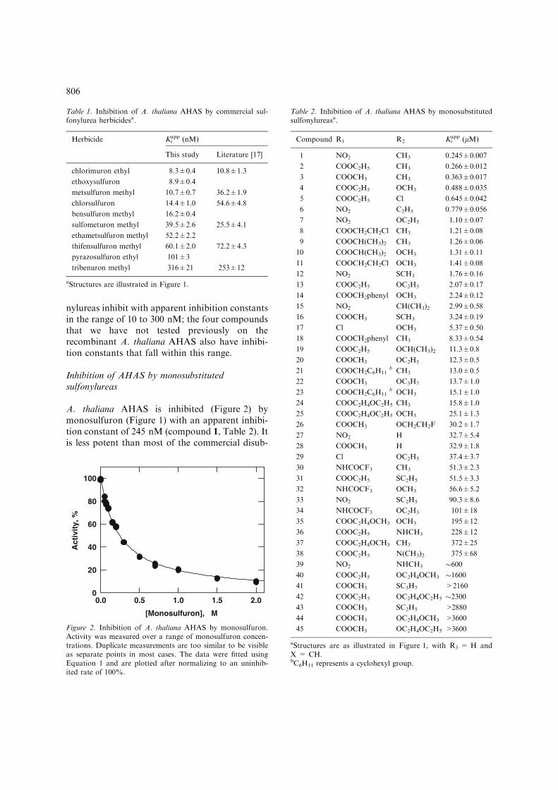
A. thaliana AHAS is inhibited (Figure 2) bymonosulfuron (Figure 1) with an apparent inhibi-tion constant of 245 nM (compound 1, Table 2). Itis less potent than most of the commercial disub-
Table 1. Inhibition of A. thaliana AHAS by commercial sul-fonylurea herbicidesa.
Herbicide Kiapp (nM)
This study Literature [17]
chlorimuron ethyl 8.3±0.4 10.8±1.3
ethoxysulfuron 8.9±0.4
metsulfuron methyl 10.7±0.7 36.2±1.9
chlorsulfuron 14.4±1.0 54.6±4.8
bensulfuron methyl 16.2±0.4
sulfometuron methyl 39.5±2.6 25.5±4.1
ethametsulfuron methyl 52.2±2.2
thifensulfuron methyl 60.1±2.0 72.2±4.3
pyrazosulfuron ethyl 101±3
tribenuron methyl 316±21 253±12
aStructures are illustrated in Figure 1.
Table 2. Inhibition of A. thaliana AHAS by monosubstitutedsulfonylureasa.
Compound R1 R2 Kiapp (lM)
1 NO2 CH3 0.245±0.007
2 COOC2H5 CH3 0.266±0.012
3 COOCH3 CH3 0.363±0.017
4 COOC2H5 OCH3 0.488±0.035
5 COOC2H5 Cl 0.645±0.042
6 NO2 C2H5 0.779±0.056
7 NO2 OC2H5 1.10±0.07
8 COOCH2CH2Cl CH3 1.21±0.08
9 COOCH(CH3)2 CH3 1.26±0.06
10 COOCH(CH3)2 OCH3 1.31±0.11
11 COOCH2CH2Cl OCH3 1.41±0.08
12 NO2 SCH3 1.76±0.16
13 COOC2H5 OC2H5 2.07±0.17
14 COOCH2phenyl OCH3 2.24±0.12
15 NO2 CH(CH3)2 2.99±0.58
16 COOCH3 SCH3 3.24±0.19
17 Cl OCH3 5.37±0.50
18 COOCH2phenyl CH3 8.33±0.54
19 COOC2H5 OCH(CH3)2 11.3±0.8
20 COOCH3 OC2H5 12.3±0.5
21 COOCH2C6H11b CH3 13.0±0.5
22 COOCH3 OC3H7 13.7±1.0
23 COOCH2C6H11b OCH3 15.1±1.0
24 COOC2H4OC2H5 CH3 15.8±1.0
25 COOC2H4OC2H5 OCH3 25.1±1.3
26 COOCH3 OCH2CH2F 30.2±1.7
27 NO2 H 32.7±5.4
28 COOCH3 H 32.9±1.8
29 Cl OC2H5 37.4±3.7
30 NHCOCF3 CH3 51.3±2.3
31 COOC2H5 SC2H5 51.5±3.3
32 NHCOCF3 OCH3 56.6±5.2
33 NO2 SC2H5 90.3±8.6
34 NHCOCF3 OC2H5 101±18
35 COOC2H4OCH3 OCH3 195±12
36 COOC2H5 NHCH3 228±12
37 COOC2H4OCH3 CH3 372±25
38 COOC2H5 N(CH3)2 375±68
39 NO2 NHCH3 �60040 COOC2H5 OC2H4OCH3 �160041 COOCH3 SC3H7 >2160
42 COOC2H5 OC2H4OC2H5 �230043 COOCH3 SC2H5 >2880
44 COOCH3 OC2H4OCH3 >3600
45 COOCH3 OC2H4OC2H5 >3600
aStructures are as illustrated in Figure 1, with R3 = H andX = CH.bC6H11 represents a cyclohexyl group.
[Monosulfuron], µM
0.0 0.5 1.0 1.5 2.0
Act
ivit
y, %
0
20
40
60
80
100
Figure 2. Inhibition of A. thaliana AHAS by monosulfuron.Activity was measured over a range of monosulfuron concen-trations. Duplicate measurements are too similar to be visibleas separate points in most cases. The data were fitted usingEquation 1 and are plotted after normalizing to an uninhib-ited rate of 100%.
806
stituted sulfonylureas tested, but has a similarapparent inhibition constant to that of tribenuronmethyl. This compound is used to kill broadleafweeds in cereal crops at application rates of 5 to30 g/ha, similar to those used for other sulfonylu-reas [18]. Thus, on the basis of its potency in vitro,we expect that monosulfuron would be an effectiveherbicide. Table 2 also lists the inhibition con-stants of a further 44 monosubstituted sulfonylu-reas that conform to the general structure ofFigure 1 (X = CH, R3 = H), listed in order ofpotency. As described later, the potencies of theseand a further 24 related compounds (see below)were subjected to 3D-QSAR analysis.
Sulfonylurea bridge modifications
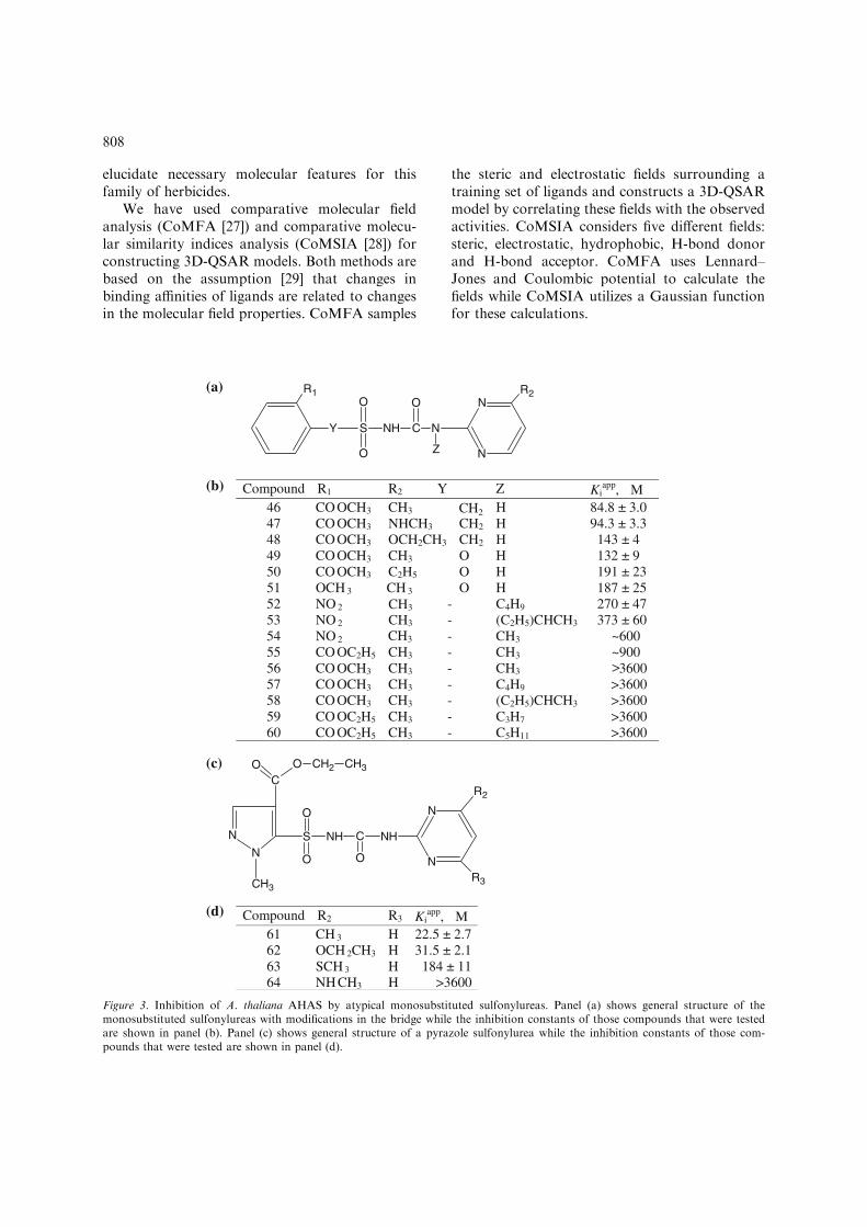
Three of the commercial sulfonylureas listed inTable 1 have modifications to the bridge connect-ing the aromatic and heterocyclic rings. Bensulfu-ron methyl has a methylene group in the bridge(see Figure 3a; Y = CH2) while ethoxysulfuronhas an oxygen atom in this position. Finally,tribenuron methyl has a methyl group in the Zposition. Fifteen monosubstituted analogs of thesecompounds were tested (Figure 3b). All of themproved to be very weak inhibitors with thestrongest (compound 46) being more than 250-fold less potent than the weakest of the commer-cial herbicides (tribenuron methyl). Clearly, thecombination of bridge modifications with a mono-substituted heterocyclic ring is detrimental forbinding to AHAS.
Pyrazoles, pyridines and thiazoles
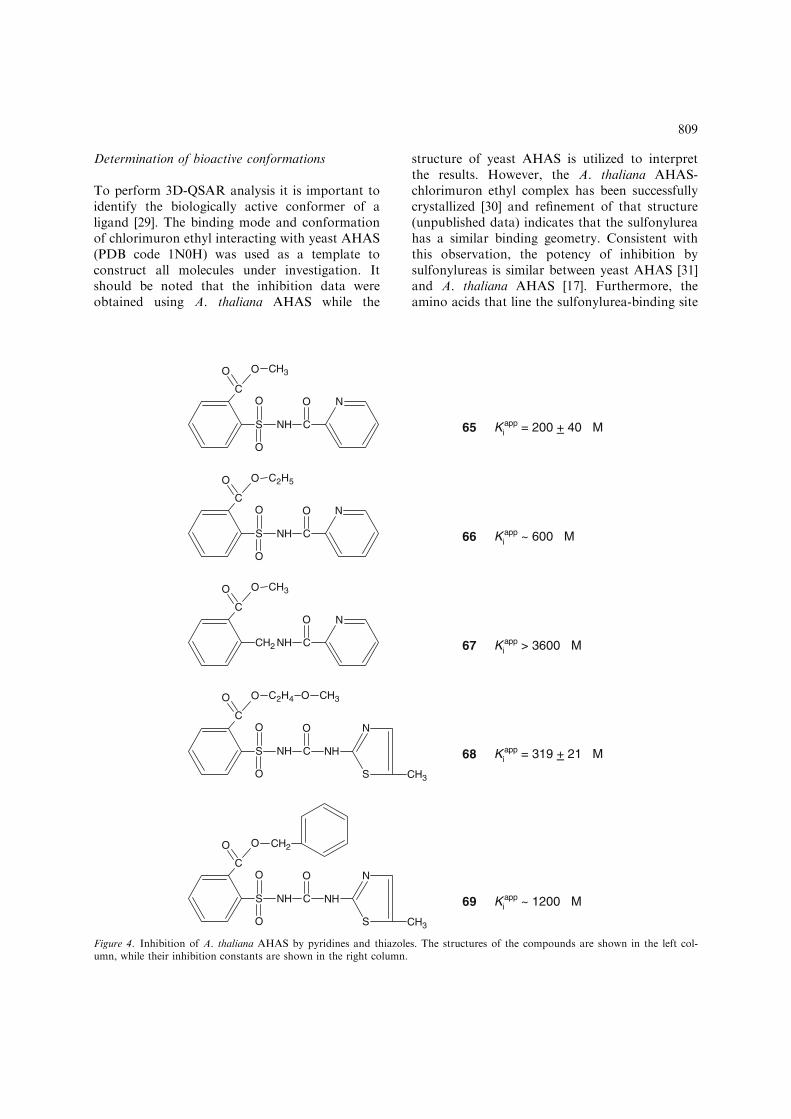
The herbicide pyrazosulfuron ethyl has a substi-tuted pyrazole ring in place of the normal aromaticring (Figure 3c; R2 = R3 = OCH3). Four mono-substituted analogs were tested as AHAS inhibi-tors (Figure 3d). Two of these compounds (61 and62) are moderately strong inhibitors with apparentKi values of around 25 lM, although these valuesare substantially higher than that for pyrazosulfu-ron ethyl (0.1 lM). Five other compounds, threepyridines with a truncated bridge and two thiaz-oles (Figure 4), were tested but none is a stronginhibitor of AHAS. This is not surprising in viewof their substantial structural differences fromconventional sulfonylureas.
3D-QSAR analyses
Previously published 3D-QSAR analyses of sul-fonylurea herbicides [19–26] suffer from severalshortcomings. Firstly, the assumed bioactive con-formation was based on either the structures ofsulfonylureas crystallized as free molecules[20–23, 25] or their low energy conformerscalculated theoretically [19, 24, 26]. However, itis now known that none of these conformationsmatch that observed when a sulfonylurea isbound to its target enzyme [7,8]. Secondly, mostactivity data employed in previous QSAR inves-tigations were obtained using in vivo experiments[19–26]. Herbicidal activity depends ultimatelyupon inhibition of AHAS, but there are differ-ences between in vivo and in vitro effects of thesecompounds due to barriers between the site ofapplication and the intracellular target, degrada-tion and detoxification by the plant, and so on.Thirdly, the structure of the herbicide-binding sitewas not known at the time, so it was not clearwhat specific interactions were being made toinhibit AHAS. Finally, although some QSARmodels demonstrate graphically the putativebinding mode [19], biophore [24] or stereoelec-tronic properties [26] of this family of herbicides,these models lack the ability to predict thebiological activity of new compounds. We arenow in a position to correct these deficiencies.
The publication of the crystal structure of yeastAHAS with bound chlorimuron ethyl [7], and withfour other members of the sulfonylurea family [8]has allowed the elucidation of the herbicide-bind-ing site of AHAS. This has also defined thebioactive conformation and provided insights intothe exact molecular interactions of this category ofherbicides. Further, as described above we havemeasured the apparent inhibition constants againsta pure plant AHAS of 69 sulfonylureas with asingle meta substituent, as well as the inhibition by10 commercial disubstituted sulfonylurea herbi-cides. The apparent inhibition constants of thesulfonylureas tested span the range from 10–9 M to10)3 M. These data provide for the first time anextensive and accurate data set that is not influ-enced by in vivo effects unrelated to the primaryenzyme-inhibitor interaction. These experimentaladvances now allow us to establish new correla-tions between the functions and structural charac-teristics, and to develop new 3D-QSAR models to
807
elucidate necessary molecular features for thisfamily of herbicides.
We have used comparative molecular fieldanalysis (CoMFA [27]) and comparative molecu-lar similarity indices analysis (CoMSIA [28]) forconstructing 3D-QSAR models. Both methods arebased on the assumption [29] that changes inbinding affinities of ligands are related to changesin the molecular field properties. CoMFA samples
the steric and electrostatic fields surrounding atraining set of ligands and constructs a 3D-QSARmodel by correlating these fields with the observedactivities. CoMSIA considers five different fields:steric, electrostatic, hydrophobic, H-bond donorand H-bond acceptor. CoMFA uses Lennard–Jones and Coulombic potential to calculate thefields while CoMSIA utilizes a Gaussian functionfor these calculations.
Compound R1 R2 Y Z Kiapp, µM
46 CO OCH3 CH3 H 84.8 ± 3.0 47 CO OCH3 NHCH3 CH2 H 94.3 ± 3.3 48 CO OCH3 OCH2CH3 CH2 H 143 ± 4 49 CO OCH3 CH3 O H 132 ± 9 50 CO OCH3 C2H5 O H 191 ± 23 51 OCH 3 CH 3 O H 187 ± 25 52 NO 2 - C4H9 270 ± 47 53 NO 2 - (C2H5)CHCH3 373 ± 60 54 NO 2 - CH3 ~600 55 CO OC2H5 CH3 - CH3 ~900 56 CO OCH3 CH3 - CH3 >3600 57 CO OCH3 CH3
CH3
CH3
CH3
- C4H9 >3600 58 CO OCH3 CH3 - (C2H5)CHCH3 >3600 59 CO OC2H5 CH3 - C3H7 >3600 60 CO OC2H5 CH3 - C5H11 >3600
Compound R2 R3 Kiapp, µM
61 CH 3 H 22.5 ± 2.762 OCH 2CH3 H 31.5 ± 2.163 SCH 3 H 184 ± 1164 NH CH3 H >3600
N
N
S
O
O
NH C
O
N
R2R1
Y
Z
N
N
CH3
CO O CH2 CH3
S
O
O
NH C
O
NH
N
N
R2
R3
(a)
(b)
(c)
(d)
CH2
Figure 3. Inhibition of A. thaliana AHAS by atypical monosubstituted sulfonylureas. Panel (a) shows general structure of themonosubstituted sulfonylureas with modifications in the bridge while the inhibition constants of those compounds that were testedare shown in panel (b). Panel (c) shows general structure of a pyrazole sulfonylurea while the inhibition constants of those com-pounds that were tested are shown in panel (d).
808
Determination of bioactive conformations
To perform 3D-QSAR analysis it is important toidentify the biologically active conformer of aligand [29]. The binding mode and conformationof chlorimuron ethyl interacting with yeast AHAS(PDB code 1N0H) was used as a template toconstruct all molecules under investigation. Itshould be noted that the inhibition data wereobtained using A. thaliana AHAS while the
structure of yeast AHAS is utilized to interpretthe results. However, the A. thaliana AHAS-chlorimuron ethyl complex has been successfullycrystallized [30] and refinement of that structure(unpublished data) indicates that the sulfonylureahas a similar binding geometry. Consistent withthis observation, the potency of inhibition bysulfonylureas is similar between yeast AHAS [31]and A. thaliana AHAS [17]. Furthermore, theamino acids that line the sulfonylurea-binding site
65 Kiapp = 200 + 40 µMS
O
O
NH C
OC
O O CH3
N
66 Kiapp ~ 600 µMS
O
O
NH C
OC
O O C2H5
N
67 Kiapp > 3600 µMCH2 NH C
OC
O O CH3
N
68 Kiapp = 319 + 21 µMS
O
O
NH C
OC
O O C2H4
N
S
O CH3
NH
CH3
69 Kiapp ~ 1200 µMS
O
O
NH C
OC
O O
N
S
NH
CH3
CH2
Figure 4. Inhibition of A. thaliana AHAS by pyridines and thiazoles. The structures of the compounds are shown in the left col-umn, while their inhibition constants are shown in the right column.
809
of the two enzymes are highly conserved. Thus, thestructure of yeast AHAS represents a high-qualitymodel of the plant enzyme.
Data set and alignment rule
A panel of 68 sulfonylureas selected from thosedescribed earlier was used to examine the rela-tionship between the inhibition data and themolecular physicochemical descriptors. Thesecompounds included the 10 commercial sulfonylu-reas shown in Table 1, and all of the 69 com-pounds shown in Table 2, Figure 3, and Figure 4,apart from 11 (41, 43, 44, 45, 56, 57, 58, 59, 60, 64and 67) for which an accurate Ki
app value was toohigh to be determined. The biological activity usedhere was expressed as pKi
app (i.e. )log10 Kiapp). In
this report the data have been subjected to threedifferent analyses: For Group 1, a set of 15molecules chosen at random (2, 6, 10, 16, 20, 27,35, 49, 51, 53, 54, 69, chlorsulfuron, sulfometuronmethyl and pyrazosulfuron ethyl) made up a testset, while the remaining 53 sulfonylureas weretreated as the training set. The test set was used tovalidate the predictive ability of the model derivedfrom the training set. The robustness of thepredictive power was therefore assessed. ForGroup 2, the commercial sulfonylureas were omit-ted to leave 58 monosubstituted sulfonylureas.These formed a training set to produce 3D-QSARmodels for the design of more potent monosub-stituted sulfonylureas. For Group 3, the whole setof 68 molecules was used to derive the mostcomprehensive 3D-QSAR analysis to date.
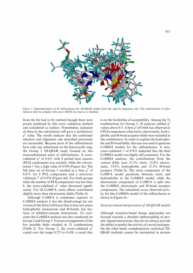
The alignment rule [29] used to superimpose theinhibitors under study is an important factor in thequality of 3D-QSAR. Atom-by-atom fit and fieldfit are the two basic methods used to superimposediverse molecules. When the structure of thebinding site is known, molecular docking isanother way to guide the alignment. We havetried docking all of the sulfonylureas into thebinding site of yeast AHAS. All molecules wereinitially docked successfully but their predictedconformations were not consistent with the knownlocation of chlorimuron ethyl in yeast AHAS [7].Therefore, the corresponding alignment could notbe used for the subsequent 3D-QSAR analysis.Instead, all 68 compounds were superimposed onchlorimuronethyl as rigid bodies. Most of thesulfonylureas with classic structures (Figure 1)
were fitted based upon the core set of atoms (thoselabeled with asterisks). For those with a length-ened or shortened sulfonylurea bridge, or withatypical ring structures, the maximum commonstructure was fitted to the template. Thus, for46–51, the benzyl ring was not superimposed whilefor 65–67 the pyrimidine ring was not included inthe superimposition. It should be noted that asingle substituent on the heterocycle may be placedin either the R2 or R3 positions (Figure 1).Preliminary analysis of the monosulfuron–A. tha-liana AHAS complex crystal structure (unpub-lished data) has revealed that the substituent islocated in the same position as the chlorine atomin chlorimuron ethyl. Accordingly, all monosub-stituted sulfonylureas were aligned in this manner.Where the disubstituted sulfonylureas differed inthe substituent at R2 and R3, the choice of whichsubstitutent occupied these positions was based onthe conformer that had the lowest energy in theSYBYL calculations. For chlorimuron ethyl, trib-enuron methyl, chlorsulfuron and metsulfuronmethyl, these calculations gave the same confor-mation as observed in the crystal structures of theyeast AHAS complexes [8]. The superposition ofall sulfonylureas is shown in Figure 5.
CoMFA and CoMSIA studies
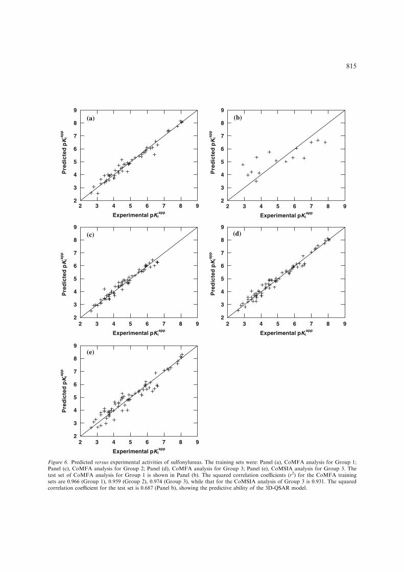
Starting from the structural alignment of Figure 5,comprehensive CoMFA and CoMSIA analyseswere performed and their respective models weredeveloped. Comparison of experimental and pre-dicted activities by CoMFA and CoMSIA for all ofthe compounds in this study is presented inTable 3. The cross-validated results were assessedby their q2 value (see Experimental Methods),where a value above 0.3 indicates that the proba-bility of chance correlation is less than 5% and avalue over 0.5 is highly significant. The results ofthese computations are summarized in Tables 4and 5. For Group 1, CoMFA analysis exhibited across-validated q2 of 0.459 and non-cross-validatedr2 of 0.966. This high degree of agreement betweenthe calculated and experimental inhibition con-stants is illustrated in Figure 6a. The predictiveability of this model was demonstrated by the r2predof 0.687 for the external test set of 15 molecules(Figure 6b). The model is relatively good, espe-cially given that the structures of the molecules inthe database are quite diverse. No compounds
y p y p
810
from the list had to be omitted though three werepoorly predicted by this cross validation methodand considered as outliers. Nonetheless, inclusionof these in the calculations still gave a satisfactoryq2 value. The results indicate that the conformerselection and alignment rule described previouslyare reasonable. Because most of the sulfonylureashave only one substituent on the heterocyclic ring,the Group 2 3D-QSAR study focused on thismonosubstituted series of sulfonylureas. A cross-validated q2 of 0.611 with 8 partial least squares(PLS) components was reached, while the conven-tional r2 has a high value of 0.959 (Figure 6c). Thefull data set of Group 3 resulted in a best q2 of0.673, for 8 PLS components and a non-crossvalidated r2 of 0.974 (Figure 6d). For both groupswhen the number of PLS components was less than8, the cross-validated q2 value decreased signifi-cantly. For all CoMFA, steric effects contributedslightly more than electrostatic fields (Table 4).
Although CoMFA is sometimes superior toCoMSIA analysis it has the disadvantage (in ourversion of the Sybyl software) that it does not assesshydrophobic interactions and H-bonds, key fea-tures of inhibitor-enzyme interactions. To over-come this CoMSIA analysis was also conducted onGroup 2 andGroup 3. Various arrangements of thefive possible fields resulted in 31 combinations(Table 5). For Group 2, the cross-validated q2
varied over the range 0.273 to 0.458, a result that
is on the borderline of acceptability. Among the 31combinations for Group 3, 20 analyses yielded q2
values above 0.5. A best q2 of 0.644 was observed at8 PLS components when steric, electrostatic, hydro-phobic and H-bond acceptor fields were included inthe combination. In order to explain the hydropho-bic andH-bond fields, this case was used to generateCoMSIA models for the sulfonylureas. A non-cross-validated r2 of 0.931 indicated that the finalCoMSIA model was highly self-consistent. For theCoMSIA analysis, the contributions from thevarious fields were 21.5% steric, 32.4% electro-static, 33.8% hydrophobic and 12.3% H-bondacceptor (Table 4). The steric component of theCoMFA model partitions between steric andhydrophobic in the CoMSIA model, while theelectrostatic component of CoMFA is split intothe CoMSIA electrostatic and H-bond acceptorcomponents. The calculated versus observed activ-ity for this CoMSIA model of the sulfonylureas isshown in Figure 6e.
Structure-based interpretation of 3D-QSAR models
Although structure-based design approaches arefocused towards a detailed understanding of pro-tein–ligand interactions, they do not always providethe ability to predict the activity of a novel analogue.On the other hand, complementary statistical 3D-QSAR methods cannot be interpreted in protein
( ), g p y Q
Figure 5. Superimposition of 68 sulfonylureas for 3D-QSAR studies from the rigid fit alignment rule. The conformation of chlo-rimuron ethyl in complex with yeast AHAS was used as a template.
811
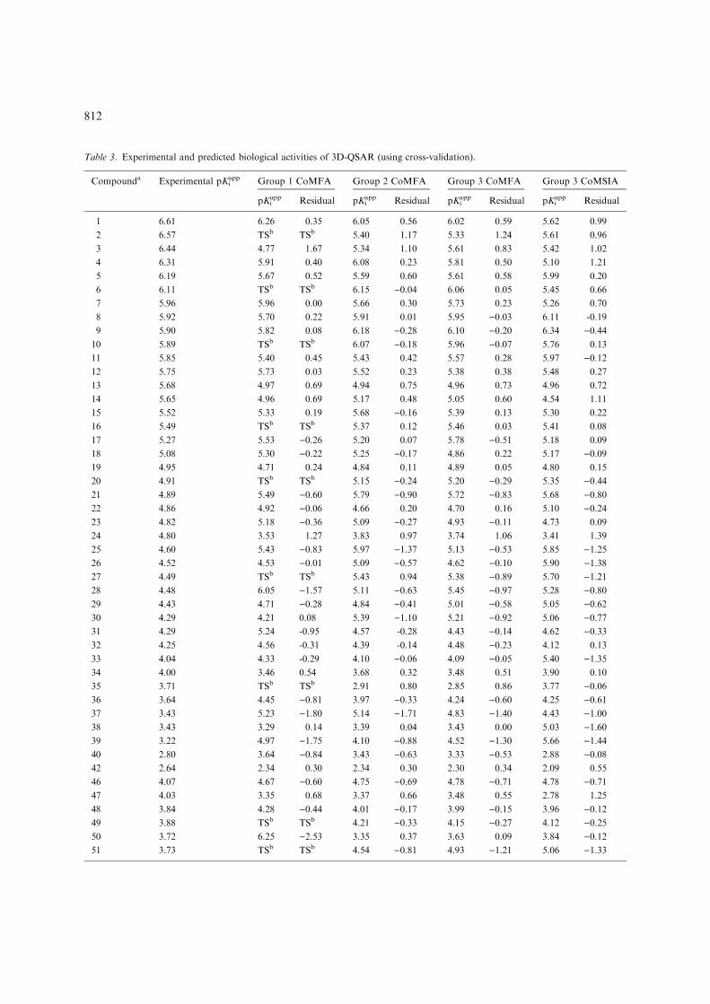
Table 3. Experimental and predicted biological activities of 3D-QSAR (using cross-validation).
Compounda Experimental pKiapp Group 1 CoMFA Group 2 CoMFA Group 3 CoMFA Group 3 CoMSIA
pKiapp Residual pKi
app Residual pKiapp Residual pKi
app Residual
1 6.61 6.26 0.35 6.05 0.56 6.02 0.59 5.62 0.99
2 6.57 TSb TSb 5.40 1.17 5.33 1.24 5.61 0.96
3 6.44 4.77 1.67 5.34 1.10 5.61 0.83 5.42 1.02
4 6.31 5.91 0.40 6.08 0.23 5.81 0.50 5.10 1.21
5 6.19 5.67 0.52 5.59 0.60 5.61 0.58 5.99 0.20
6 6.11 TSb TSb 6.15 )0.04 6.06 0.05 5.45 0.66
7 5.96 5.96 0.00 5.66 0.30 5.73 0.23 5.26 0.70
8 5.92 5.70 0.22 5.91 0.01 5.95 )0.03 6.11 -0.19
9 5.90 5.82 0.08 6.18 )0.28 6.10 )0.20 6.34 )0.4410 5.89 TSb TSb 6.07 )0.18 5.96 )0.07 5.76 0.13
11 5.85 5.40 0.45 5.43 0.42 5.57 0.28 5.97 )0.1212 5.75 5.73 0.03 5.52 0.23 5.38 0.38 5.48 0.27
13 5.68 4.97 0.69 4.94 0.75 4.96 0.73 4.96 0.72
14 5.65 4.96 0.69 5.17 0.48 5.05 0.60 4.54 1.11
15 5.52 5.33 0.19 5.68 )0.16 5.39 0.13 5.30 0.22
16 5.49 TSb TSb 5.37 0.12 5.46 0.03 5.41 0.08
17 5.27 5.53 )0.26 5.20 0.07 5.78 )0.51 5.18 0.09
18 5.08 5.30 )0.22 5.25 )0.17 4.86 0.22 5.17 )0.0919 4.95 4.71 0.24 4.84 0.11 4.89 0.05 4.80 0.15
20 4.91 TSb TSb 5.15 )0.24 5.20 )0.29 5.35 )0.4421 4.89 5.49 )0.60 5.79 )0.90 5.72 )0.83 5.68 )0.8022 4.86 4.92 )0.06 4.66 0.20 4.70 0.16 5.10 )0.2423 4.82 5.18 )0.36 5.09 )0.27 4.93 )0.11 4.73 0.09
24 4.80 3.53 1.27 3.83 0.97 3.74 1.06 3.41 1.39
25 4.60 5.43 )0.83 5.97 )1.37 5.13 )0.53 5.85 )1.2526 4.52 4.53 )0.01 5.09 )0.57 4.62 )0.10 5.90 )1.3827 4.49 TSb TSb 5.43 0.94 5.38 )0.89 5.70 )1.2128 4.48 6.05 )1.57 5.11 )0.63 5.45 )0.97 5.28 )0.8029 4.43 4.71 )0.28 4.84 )0.41 5.01 )0.58 5.05 )0.6230 4.29 4.21 0.08 5.39 )1.10 5.21 )0.92 5.06 )0.7731 4.29 5.24 -0.95 4.57 -0.28 4.43 )0.14 4.62 )0.3332 4.25 4.56 -0.31 4.39 -0.14 4.48 )0.23 4.12 0.13
33 4.04 4.33 -0.29 4.10 )0.06 4.09 )0.05 5.40 )1.3534 4.00 3.46 0.54 3.68 0.32 3.48 0.51 3.90 0.10
35 3.71 TSb TSb 2.91 0.80 2.85 0.86 3.77 )0.0636 3.64 4.45 )0.81 3.97 )0.33 4.24 )0.60 4.25 )0.6137 3.43 5.23 )1.80 5.14 )1.71 4.83 )1.40 4.43 )1.0038 3.43 3.29 0.14 3.39 0.04 3.43 0.00 5.03 )1.6039 3.22 4.97 )1.75 4.10 )0.88 4.52 )1.30 5.66 )1.4440 2.80 3.64 )0.84 3.43 )0.63 3.33 )0.53 2.88 )0.0842 2.64 2.34 0.30 2.34 0.30 2.30 0.34 2.09 0.55
46 4.07 4.67 )0.60 4.75 )0.69 4.78 )0.71 4.78 )0.7147 4.03 3.35 0.68 3.37 0.66 3.48 0.55 2.78 1.25
48 3.84 4.28 )0.44 4.01 )0.17 3.99 )0.15 3.96 )0.1249 3.88 TSb TSb 4.21 )0.33 4.15 )0.27 4.12 )0.2550 3.72 6.25 )2.53 3.35 0.37 3.63 0.09 3.84 )0.1251 3.73 TSb TSb 4.54 )0.81 4.93 )1.21 5.06 )1.33
812
structural terms, as the derived models are notalways based on the bioactive conformation of aparticular scaffold. Here, we plotted the variousfields of CoMFA or CoMSIA based models on thesulfonylurea binding site of yeastAHAS to interpretthe results. These superimpositions are shown inFigures 7 and 8.
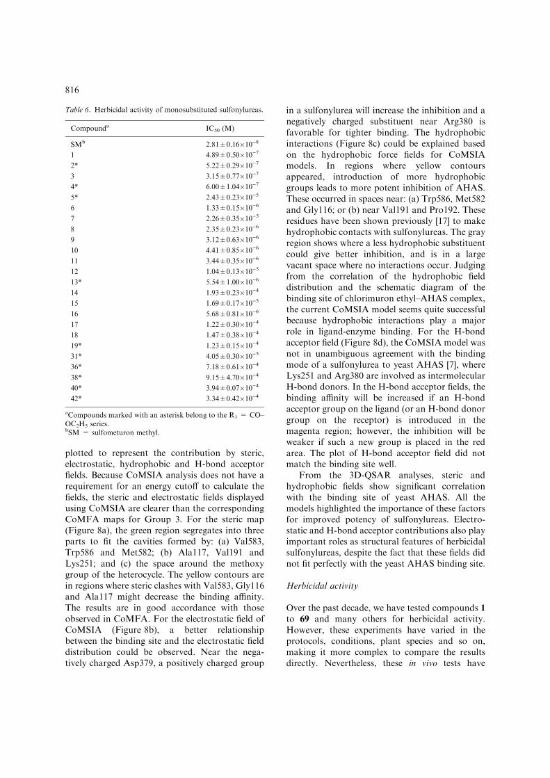
For the CoMFA models, the cross-validated q2
values of Group 2 and Group 3 were much higherthan that for Group 1. Therefore, the Group 2 andGroup 3 analyses were investigated further fortheir distributions of steric and electrostatic fieldsin space for residues within 5.5 A of the center ofthe ligand. 3D contour maps from both trainingsets exhibited strong similarities except for thesteric field (Figures 7a and 7c) near the heterocycleposition. This is not surprising because Group 3included 10 commercial sulfonylureas with substit-uents at both meta positions of the heterocyclewhile Group 2 only dealt with the monosubstituted
series. For the steric fields, green shows positionswhere introducing a bulky group in a sulfonylureawould be favorable for greater inhibition ofAHAS. In contrast, yellow indicates positionswhere decreasing the bulk of the sulfonylurea isfavored. The steric fields indicate that the greenregions are located in the cavities formed by: (a)Val583, Trp586 and Met582; (b) Ala117, Val191and Lys251; (c) Ala200, Phe201 and Asp379; and(d) a fairly vacant space near the methoxy group ofchlorimuron ethyl. The yellow regions are situatedin spaces where bulky conflicts occur (near Val583,Trp586 and Asp379) and weaken the binding. Forthe electrostatic fields (Figure 7b and d), blueregions indicate areas where positive charges arefavored for increased binding to the enzyme andred regions indicate spaces where negative chargesare advantageous for the activity of sulfonylureas.As shown by these electrostatic fields, sulfonylure-as need contributions from positively charged
Table 3. Continued.
Compounda Experimental pKiapp Group 1 CoMFA Group 2 CoMFA Group 3 CoMFA Group 3 CoMSIA
pKiapp Residual pKi
app Residual pKiapp Residual pKi
app Residual
52 3.57 4.62 )1.05 4.10 )0.53 4.32 )0.75 2.98 0.59
53 3.43 TSb TSb 3.38 0.05 3.48 )0.05 5.67 )2.2454 3.22 TSb TSb 4.06 )0.84 4.19 )0.97 3.25 )0.0355 3.05 4.68 )1.63 2.91 0.14 3.70 )0.66 3.59 )0.5461 4.65 4.99 )0.34 4.94 )0.29 4.87 )0.22 4.45 0.20
62 4.50 3.97 0.53 3.74 0.76 4.03 0.47 3.72 0.78
63 3.74 4.40 )0.66 4.46 )0.72 4.04 )0.30 4.55 )0.8165 3.70 2.63 1.07 3.13 0.57 2.47 1.23 2.90 0.80
66 3.22 4.67 )1.45 3.83 )0.61 4.17 )0.95 3.98 )0.7668 3.53 3.45 0.08 1.75 1.78 2.00 1.53 1.57 1.96
69 2.92 TSb TSb 4.00 )1.08 4.62 )1.70 4.81 )1.8971 7.79 7.77 0.02 7.69 0.10 7.01 0.78
72 8.08 4.19 3.89 5.52 2.56 6.58 1.49
73 7.84 TSb TSb 6.74 1.10 7.12 0.72
74 7.28 6.96 0.32 7.89 )0.61 6.84 0.44
75 8.05 6.93 1.12 6.95 1.10 6.64 1.41
76 7.97 7.03 0.94 7.90 0.07 8.43 )0.4677 7.00 TSb TSb 6.18 0.82 6.75 0.25
78 7.40 TSb TSb 7.01 0.39 6.32 1.08
79 7.22 5.28 1.94 5.11 2.11 6.98 0.24
80 6.50 5.17 1.33 4.76 1.73 6.08 0.42
aCompounds 71–80 represent bensulfuron methyl, chlorimuron ethyl, chlorsulfuron, ethametsulfuron methyl, ethoxysulfuron, met-sulfuron methyl, pyrazosufuron ethyl, sulfometuron methyl, thifensulfuron methyl and tribenuron methyl respectively.bTS = test set compounds. An accurate pKi
app could not be determined for compounds 41, 43, 44, 45, 56, 57, 58, 59, 60, 64 and 67 (seetext). The predicted sum of squares values (R(Yobs – Ypred)
2) for group 1 by CoMFA is 57.42, for group 2 by CoMFA is 25.60, forgroup 3 by CoMFA is 45.28 and for group 3 by CoMSIA is 51.93.
813
groups to achieve enhanced inhibition in mostareas of the electrostatic field, and only smallportions prefer negative charges. In the binding siteof yeast AHAS, Asp379 has a negative charge, theside-chains of Lys251 and Arg380 are positive, andall other amino acids bear no charge. There is noobvious relationship between the electrostatic fieldsand herbicide-binding site for the CoMFA resultsshown here. If the structure of Arabidopsis thalianaAHAS had been available and partial charges were
used for the active site (as suggested by a reviewer)then a correlation may have been obtained. How-ever, the two electrostatic maps from Group 2 andGroup 3 share significant similarity and the modelsare still useful for novel sulfonylurea design andinterpretation of the 3D-QSAR models.
To understand the enzymatic activities ofsulfonylureas in relation to their hydrophobicand H-bonding properties, the 3D-QSAR modelsfrom the CoMSIA analysis mentioned above were
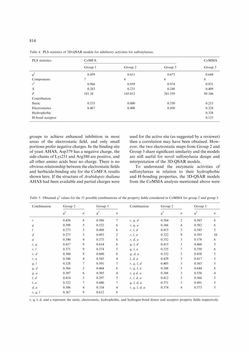
Table 4. PLS statistics of 3D-QSAR models for inhibitory activities for sulfonylureas.
PLS statistics CoMFA CoMSIA
Group 1 Group 2 Group 3 Group 3
q2 0.459 0.611 0.673 0.644
Components 7 8 8 8
r2 0.966 0.959 0.974 0.931
S 0.283 0.233 0.248 0.409
F 181.34 145.012 281.559 99.106
Contribution
Steric 0.533 0.600 0.550 0.215
Electrostatics 0.467 0.400 0.450 0.324
Hydrophobic 0.338
H-bond acceptor 0.123
Table 5. Obtained q2 values for the 31 possible combinations of the property fields considered in CoMSIA for group 2 and group 3.
Combination Group 2 Group 3 Combination Group 2 Group 3
q2 n q2 n q2 n q2 n
r 0.458 8 0.586 7 r, q, d 0.384 2 0.585 6
q 0.398 5 0.522 6 r, q, a 0.366 4 0.582 6
l 0.273 5 0.488 6 r, l, d 0.415 3 0.545 5
d 0.275 3 0.093 2 r, l, a 0.322 9 0.595 10
a 0.340 4 0.375 6 r, d, a 0.352 3 0.578 6
r, q 0.417 9 0.614 6 q, l, d 0.415 3 0.460 5
r, l 0.371 9 0.574 5 q, l, a 0.325 7 0.559 6
r, d 0.360 9 0.600 8 q, d, a 0.352 3 0.458 5
r, a 0.346 4 0.545 4 l, d, a 0.429 3 0.417 5
q, l 0.328 7 0.591 7 r, q, l, d 0.405 3 0.563 5
q, d 0.366 2 0.464 6 r, q, l, a 0.348 5 0.644 8
q, a 0.307 6 0.505 4 r, q d, a 0.368 3 0.558 4
l, d 0.414 3 0.297 5 r, l, d, a 0.412 3 0.566 5
l, a 0.322 7 0.440 7 q, l, d, a 0.371 3 0.491 5
d, a 0.306 4 0.334 4 r, q, l, d, a 0.374 4 0.573 5
r, q, l 0.367 9 0.612 8
r, q, l, d, and a represent the steric, electrostatic, hydrophobic, and hydrogen-bond donor and acceptor property fields respectively.
814
2
3
4
5
6
7
8
9
Experimental pKiapp
2 4 7 8 92
3
4
5
6
7
8
9
Pre
dic
ted
pK
iapp
Pre
dic
ted
pK
iapp
Pre
dic
ted
pK
iapp
Pre
dic
ted
pK
iapp
Pre
dic
ted
pK
iapp
2
3
4
5
6
7
8
9
2
3
4
5
6
7
8
9
2
3
4
5
6
7
8
9
3 5 6
Experimental pKiapp
2 4 7 8 93 5 6
Experimental pKiapp
2 4 7 8 93 5 6
Experimental pKiapp
2 4 7 8 93 5 6
Experimental pKiapp
2 4 7 8 93 5 6
(a)
(c)
(e)
(b)
(d)
Figure 6. Predicted versus experimental activities of sulfonylureas. The training sets were: Panel (a), CoMFA analysis for Group 1;Panel (c), CoMFA analysis for Group 2; Panel (d), CoMFA analysis for Group 3; Panel (e), CoMSIA analysis for Group 3. Thetest set of CoMFA analysis for Group 1 is shown in Panel (b). The squared correlation coefficients (r2) for the CoMFA trainingsets are 0.966 (Group 1), 0.959 (Group 2), 0.974 (Group 3), while that for the CoMSIA analysis of Group 3 is 0.931. The squaredcorrelation coefficient for the test set is 0.687 (Panel b), showing the predictive ability of the 3D-QSAR model.
815
plotted to represent the contribution by steric,electrostatic, hydrophobic and H-bond acceptorfields. Because CoMSIA analysis does not have arequirement for an energy cutoff to calculate thefields, the steric and electrostatic fields displayedusing CoMSIA are clearer than the correspondingCoMFA maps for Group 3. For the steric map(Figure 8a), the green region segregates into threeparts to fit the cavities formed by: (a) Val583,Trp586 and Met582; (b) Ala117, Val191 andLys251; and (c) the space around the methoxygroup of the heterocycle. The yellow contours arein regions where steric clashes with Val583, Gly116and Ala117 might decrease the binding affinity.The results are in good accordance with thoseobserved in CoMFA. For the electrostatic field ofCoMSIA (Figure 8b), a better relationshipbetween the binding site and the electrostatic fielddistribution could be observed. Near the nega-tively charged Asp379, a positively charged group
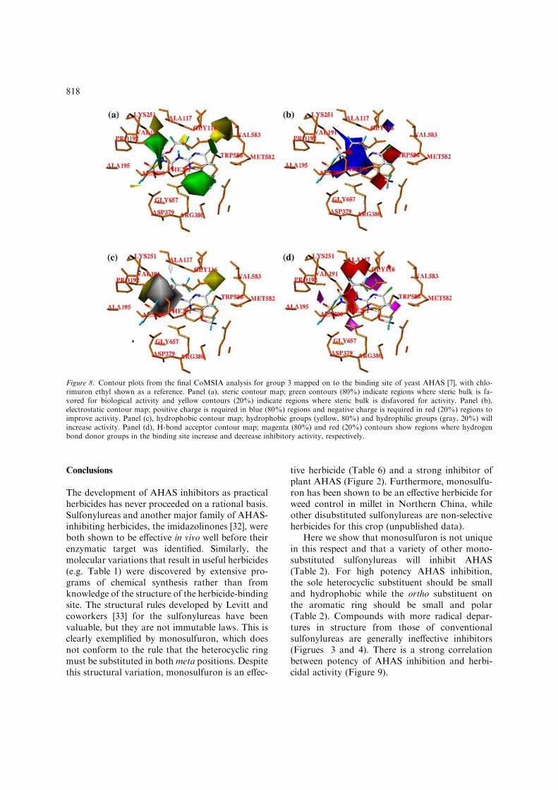
in a sulfonylurea will increase the inhibition and anegatively charged substituent near Arg380 isfavorable for tighter binding. The hydrophobicinteractions (Figure 8c) could be explained basedon the hydrophobic force fields for CoMSIAmodels. In regions where yellow contoursappeared, introduction of more hydrophobicgroups leads to more potent inhibition of AHAS.These occurred in spaces near: (a) Trp586, Met582and Gly116; or (b) near Val191 and Pro192. Theseresidues have been shown previously [17] to makehydrophobic contacts with sulfonylureas. The grayregion shows where a less hydrophobic substituentcould give better inhibition, and is in a largevacant space where no interactions occur. Judgingfrom the correlation of the hydrophobic fielddistribution and the schematic diagram of thebinding site of chlorimuron ethyl–AHAS complex,the current CoMSIA model seems quite successfulbecause hydrophobic interactions play a majorrole in ligand-enzyme binding. For the H-bondacceptor field (Figure 8d), the CoMSIA model wasnot in unambiguous agreement with the bindingmode of a sulfonylurea to yeast AHAS [7], whereLys251 and Arg380 are involved as intermolecularH-bond donors. In the H-bond acceptor fields, thebinding affinity will be increased if an H-bondacceptor group on the ligand (or an H-bond donorgroup on the receptor) is introduced in themagenta region; however, the inhibition will beweaker if such a new group is placed in the redarea. The plot of H-bond acceptor field did notmatch the binding site well.
From the 3D-QSAR analyses, steric andhydrophobic fields show significant correlationwith the binding site of yeast AHAS. All themodels highlighted the importance of these factorsfor improved potency of sulfonylureas. Electro-static and H-bond acceptor contributions also playimportant roles as structural features of herbicidalsulfonylureas, despite the fact that these fields didnot fit perfectly with the yeast AHAS binding site.
Herbicidal activity
Over the past decade, we have tested compounds 1to 69 and many others for herbicidal activity.However, these experiments have varied in theprotocols, conditions, plant species and so on,making it more complex to compare the resultsdirectly. Nevertheless, these in vivo tests have
Table 6. Herbicidal activity of monosubstituted sulfonylureas.
Compounda IC50 (M)
SMb 2.81±0.16�10)8
1 4.89±0.50�10)7
2* 5.22±0.29�10)7
3 3.15±0.77�10)7
4* 6.00±1.04�10)7
5* 2.43±0.23�10)5
6 1.33±0.15�10)6
7 2.26±0.35�10)5
8 2.35±0.23�10)6
9 3.12±0.63�10)6
10 4.41±0.85�10)6
11 3.44±0.35�10)6
12 1.04±0.13�10)5
13* 5.54±1.00�10)6
14 1.93±0.23�10)4
15 1.69±0.17�10)5
16 5.68±0.81�10)6
17 1.22±0.30�10)4
18 1.47±0.38�10)4
19* 1.23±0.15�10)4
31* 4.05±0.30�10)5
36* 7.18±0.61�10)4
38* 9.15±4.70�10)4
40* 3.94±0.07�10)4
42* 3.34±0.42�10)4
aCompounds marked with an asterisk belong to the R1 = CO–OC2H5 series.bSM = sulfometuron methyl.
816
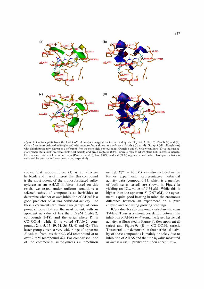
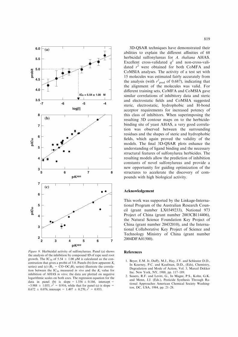
shown that monosulfuron (1) is an effectiveherbicide and it is of interest that this compoundis the most potent of the monosubstituted sulfo-nylureas as an AHAS inhibitor. Based on thisresult, we tested under uniform conditions aselected subset of compounds as herbicides todetermine whether in vitro inhibition of AHAS is agood predictor of in vivo herbicidal activity. Forthese experiments we chose two groups of com-pounds: those that are the most potent, with anapparent Ki value of less than 10 lM (Table 2,compounds 1–18); and the series where R1 isCO–OC2H5 while R2 is varied (Table 2, com-pounds 2, 4, 5, 13, 19, 31, 36, 38, 40 and 42). Thislatter group covers a very wide range of apparentKi values, from less than 0.3 lM (compound 2) toover 2 mM (compound 42). For comparison, oneof the commercial sulfonylureas (sulfometuron
methyl, Kiapp = 40 nM) was also included in the
former experiment. Representative herbicidalactivity data (compound 13, which is a memberof both series tested) are shown in Figure 9ayielding an IC50 value of 5.54 lM. While this ishigher than the apparent Ki (2.07 lM), the agree-ment is quite good bearing in mind the enormousdifference between an experiment on a pureenzyme and one using growing seedlings.
IC50 values for all compounds tested are shown inTable 6. There is a strong correlation between theinhibition of AHAS in vitro and the in vivo herbicidalactivity, as illustrated in Figure 9b (low apparent Ki
series) and Figure 9c (R1 = CO–OC2H5 series).This correlation demonstrates that herbicidal activ-ity of these compounds is mainly or solely due toinhibition of AHAS and that theKi value measuredin vitro is a useful predictor of their effect in vivo.
Figure 7. Contour plots from the final CoMFA analyses mapped on to the binding site of yeast AHAS [7]. Panels (a) and (b):Group 2 (monosubstituted sulfonylureas) with monosulfuron shown as a reference. Panels (c) and (d): Group 3 (all sulfonylureas)with chlorimuron ethyl shown as a reference. For the steric field contour maps (Panels a and c), yellow contours (20%) indicate re-gions where steric bulk decreases biological activity and green contours (80%) indicate regions where steric bulk increases activity.For the electrostatic field contour maps (Panels b and d), blue (80%) and red (20%) regions indicate where biological activity isenhanced by positive and negative charge, respectively.
817
Conclusions
The development of AHAS inhibitors as practicalherbicides has never proceeded on a rational basis.Sulfonylureas and another major family of AHAS-inhibiting herbicides, the imidazolinones [32], wereboth shown to be effective in vivo well before theirenzymatic target was identified. Similarly, themolecular variations that result in useful herbicides(e.g. Table 1) were discovered by extensive pro-grams of chemical synthesis rather than fromknowledge of the structure of the herbicide-bindingsite. The structural rules developed by Levitt andcoworkers [33] for the sulfonylureas have beenvaluable, but they are not immutable laws. This isclearly exemplified by monosulfuron, which doesnot conform to the rule that the heterocyclic ringmust be substituted in bothmeta positions. Despitethis structural variation, monosulfuron is an effec-
tive herbicide (Table 6) and a strong inhibitor ofplant AHAS (Figure 2). Furthermore, monosulfu-ron has been shown to be an effective herbicide forweed control in millet in Northern China, whileother disubstituted sulfonylureas are non-selectiveherbicides for this crop (unpublished data).
Here we show that monosulfuron is not uniquein this respect and that a variety of other mono-substituted sulfonylureas will inhibit AHAS(Table 2). For high potency AHAS inhibition,the sole heterocyclic substituent should be smalland hydrophobic while the ortho substituent onthe aromatic ring should be small and polar(Table 2). Compounds with more radical depar-tures in structure from those of conventionalsulfonylureas are generally ineffective inhibitors(Figrues 3 and 4). There is a strong correlationbetween potency of AHAS inhibition and herbi-cidal activity (Figure 9).
Figure 8. Contour plots from the final CoMSIA analysis for group 3 mapped on to the binding site of yeast AHAS [7], with chlo-rimuron ethyl shown as a reference. Panel (a), steric contour map; green contours (80%) indicate regions where steric bulk is fa-vored for biological activity and yellow contours (20%) indicate regions where steric bulk is disfavored for activity. Panel (b),electrostatic contour map; positive charge is required in blue (80%) regions and negative charge is required in red (20%) regions toimprove activity. Panel (c), hydrophobic contour map; hydrophobic groups (yellow, 80%) and hydrophilic groups (gray, 20%) willincrease activity. Panel (d), H-bond acceptor contour map; magenta (80%) and red (20%) contours show regions where hydrogenbond donor groups in the binding site increase and decrease inhibitory activity, respectively.
818
3D-QSAR techniques have demonstrated theirabilities to explain the different affinities of 68herbicidal sulfonylureas for A. thaliana AHAS.Excellent cross-validated q2 and non-cross-vali-dated r2 were obtained for both CoMFA andCoMSIA analyses. The activity of a test set with15 molecules was estimated fairly accurately fromthe analysis (with r2pred of 0.687), indicating thatthe alignment of the molecules was valid. Fordifferent training sets, CoMFA and CoMSIA gavesimilar correlations of inhibitory data and stericand electrostatic fields and CoMSIA suggestedsteric, electrostatic, hydrophobic and H-bondacceptor requirements for increased potency ofthis class of inhibitors. When superimposing theresulting 3D contour maps on to the herbicide-binding site of yeast AHAS, a very good correla-tion was observed between the surroundingresidues and the shapes of steric and hydrophobicfields, which again proved the validity of themodels. The final 3D-QSAR plots enhance theunderstanding of ligand binding and the necessarystructural features of sulfonylurea herbicides. Theresulting models allow the prediction of inhibitionconstants of novel sulfonylureas and provide anew opportunity for guiding optimization of thestructures to accelerate the discovery of com-pounds with high biological activity.
Acknowledgement
This work was supported by the Linkage-Interna-tional Program of the Australian Research Coun-cil (grant number LX0349233), National 973Project of China (grant number 2003CB114406),the Natural Science Foundation Key Project ofChina (grant number 20432010), and the Interna-tional Collaborative Key Project of Science andTechnology Ministry of China (grant number2004DFA01500).
References
1. Beyer, E.M. Jr, Duffy, M.J., Hay, J.V. and Schleuter D.D.,In Kearney, P.C. and Kaufman, D.D., (Eds), Chemistry,Degradation and Mode of Action, Vol. 3, Marcel DekkerInc. New York, NY, 1988, pp. 117–189.
2. Sauers, R.F. and Levitt, G., In Magee, P.S., Kohn, G.K.and Menn, J.J. (Eds.), Pesticide Synthesis Through Ra-tional Approaches American Chemical Society Washing-ton, DC, USA, 1984, pp. 21–28.
IC50 = 5.54 ± 1.00 µM
log[I]
-7 -6 -5 -4
pro
bit
3.5
4.0
4.5
5.0
5.5
6.0
pKiapp
5 6
pIC
50
3
4
5
6
7
8
pKiapp
2
pIC
50
2
3
4
5
6
7
7 8
3 4 5 6 7
(a)
(b)
(c)
Figure 9. Herbicidal activity of sulfonylureas. Panel (a) showsthe analysis of the inhibition by compound 13 of rape seed rootgrowth. The IC50 of 5.54 ± 1.00 lM is calculated as the con-centration that gives a probit of 5.0. Panels (b) (low apparent Ki
series) and (c) (R1 = CO–OC2H5 series) illustrate the correla-tion between the IC50 measured in vivo and the Ki value forinhibition of AHAS in vitro; the data are plotted on negativelogarithmic scales on both axes. The regression equation for thedata in panel (b) is slope = 1.556 ± 0.166, intercept =)3.988 ± 1.055, r2 = 0.916, while that for panel (c) is slope =0.672 ± 0.076, intercept = 1.497 ± 0.270, r2 = 0.953.
819
3. LaRossa, R.A. and Schloss, J.V., J. Biol. Chem., 259 (1984)8753.
4. Ray, T.B., Plant Physiol., 75 (1984) 827.5. Duggleby, R.G. and Pang, S.S., J. Biochem. Mol. Biol., 33
(2000) 1.6. Pang, S.S., Duggleby, R.G. and Guddat, L.W., J. Mol.
Biol., 317 (2002) 249.7. Pang, S.S., Guddat, L.W. and Duggleby, R.G., J. Biol.
Chem., 278 (2003) 7639.8. McCourt, J.A., Pang, S.S., Guddat, L.W. and Duggleby,
R.G., Biochemistry, 44 (2005) 2330.9. Li, Z.-M., Jia, G.-F., Wang, L.-X., Fan, C.-W. and Yang,
Z., Sulfonylurea compounds and their herbicidal applica-tions. (1998) Chinese Invention Patent ZL 94–1–18793.4.
10. Wang, L.-X., Li, Z.-M., Jia, G.-F., Chen, J.-P. and Wang,S-H. The application of monosulfuron in millet fields.(2003) Chinese Invention Patent ZL 98–1–00257.9.
11. Li, Z.-M., Jia, G.-F., Wang, L.-X., Lai, C.-M., Wang, H.-G. and Wang, R.-J., Chem. J. Chin. Univ.-Chin., 14 (1993)349.
12. Ye, G.-Z., Fan, Z.-J., Li, Z.-M., Li, Y.-H., Gao, F.-W. andWang, S.-H., Chem. J. Chin. Univ.-Chin., 24 (2003) 1599.
13. Chang, A.K. and Duggleby, R.G., Biochem. J., 327 (1997)161.
14. Sambrook, J., Fritsch, E.F. and Maniatis, T. MolecularCloning: A Laboratory Manual , 2nd ed., Cold SpringHarbor Laboratory Press, Cold Spring Harbor, New York,USA, 1989.
15. Singh, B.K., Stidham, M.A. and Shaner, D.L., Anal. Bio-chem., 171 (1988) 173.
16. Inamori, Y., Muro, C., Yamanaka, H., Osaka, K.,Hachiken, H., Tsujibo, H. and Miki, Y., Biosci. Biotech.Biochem., 58 (1994) 1150.
17. Chang, A.K. and Duggleby, R.G., Biochem. J., 333 (1998)765.
18. Brown, H.M., Pestic. Sci., 29 (1990) 263.19. Akagi, T., Pestic. Sci., 47 (1996) 309.20. Liu, J., Li, Z.-M., Wang, X., Ma, Y., Lai, C.-M., Jia, G.-F.
and Wang, L.-X., Sci. China, Ser., B41 (1998) 50.21. Yang, G.-F., Liu, H.-Y., Yang, X.-F. and Yang, H.-Z., Sci.
China, Ser., B42 (1999) 656.22. Yang, G., Liu, H. and Yang, H., Pestic. Sci., 55 (1999) 1143.23. Hou, T.J., Li, Z.M., Li, Z., Liu, J. and Xu, X.J., J. Chem.
Inf. Comput. Sci., 40 (2000) 1002.24. Ren, T.-R., Yang, H.-W., Gao, X., Yang, X.-L., Zhou, J.-J.
and Cheng, F.-H., Pest Manage. Sci., 56 (2000) 218.25. Li, Z.-M. and Lai, C.-M., Chin. J. Org. Chem., 21 (2001)
810.26. Galeazzi, R., Marucchini, C., Orena, M. and Zadra, C.,
Bioorg. Med. Chem., 10 (2002) 1019.27. Cramer, R.D. III, Paterson, D.E. and Bunce, J.D., J. Am.
Chem. Soc., 110 (1988) 5959.28. Klebe, G., Abraham, U. and Mietzner, T.J., Med. Chem.,
37 (1994) 4130.29. Klebe, G., In Kubinyi, H., Folkers, G. and Martin, Y.C.
(Eds.) 3D QSAR in Drug Design Vol 3: Recent Advances.Kluwer Academic Publishers, Dordrecht, The Netherlands,1998, pp. 87–105.
30. Pang, S.S., Guddat, L.W. and Duggleby, R.G., ActaCryst., D60 (2004) 153.
31. Duggleby, R.G., Pang, S.S., Yu, H. and Guddat, L.W.,Eur. J. Biochem., 270 (2003) 2895.
32. Los, M., In Magee, P.S., Kohn, G.K. and Menn, J.J. (Eds.)Pesticide Synthesis Through Rational Approaches. Ameri-can Chemical Society, Washington, DC, USA, 1984, pp.29–44.
33. Levitt G., In Baker D.R., Fenyes J.G., Moberg W.K.,(eds.), 1991, Synthesis and Chemistry of Agrochemicals II.ACS Symposium Series 443 (American Chemical SocietyWashington DC, USA) pp. 16–31.
820