Embed Size (px)
Citation preview
Histochemie 16, 186--194 (1968)
Studies on Uptake of Intraventricularly Administered Tritiated Noradrenaline and 5-Hydroxytryptamine
with Combined Fluorescence Histochenfical and Autoradiographic Techniques*
KJELL FUXE, TOMAS HSKFELT, MARTIN RITZ]~N, and URBAN UNGERSTEDT
Department of Histology and the Institute for Cell Research and Genetics, Karolinska Institutet, 10401 Stockholm, Sweden
Received September 21, 1968
Summary. Tritiated noradrenaline (NA) and 5-hydrexytryptamine (5-HT) (1.5--30 ~C) have been injected intraventricularly into normal or reserpine-nialamide pretreated rats 1/2 to 2 hours before the killing. Various parts of the brains were freeze-dried, reacted with formaldehyde gas and embedded in paraffin or Araldite. Before application of the stripping film emulsion many sections were photographed in the fluorescence microscope in order to perform a combined histochemical and autoradiographic study of the monoamine neurons. By such an approach it was possible to demonstrate 1. that the accumulation of radioactivity in cell bodies after 3H--NA and 3H--5-HT injection is localized to catecholamine (CA) and 5-HT cell bodies respectively; 2. that injected 3H--NA and aH--5-HT in the doses used relatively selectively are taken up into the NA and 5-HT nerve terminals respectively, since the distribution of grains in the sections follow that of the fluorescent terminals; 3. that the accumulation of silver grains only reaches the zone (200-400 ~) close to the ventricles and the ventral part of the subarachnoidal space. By grain counting it was possible to estimate that the degree of concentration of radioactivity in the monoamine cell bodies was up to 4 times that in the immediate surroundings. - - The Araldite sections consistently gave a better resolution in the autoradiographic picture than the paraffin sections. It is postulated that freeze-drying and plastic embedding for autoradiography will be a valuable method for the cellular demon- stration of certain biogenic amines which are not easily demonstrated by the histochemical fluorescence method and of other biologically active water-soluble compounds, since diffusion will be restricted to a minimum.
Introduct ion
I t has been shown by histochemical fluorescence methods t ha t noradrenal ine (NA) and 5-hydroxyt ryp tamine (5-HT) when injected in t ravent r icu la r ly are par t ly t aken up into the parts of the catecholamine (CA) and 5-HT neurons lying close to the ventricles and the vent ra l par t of the subarachnoidal space ( F ux~ and UNGERSTEDT, 1966, 1967, 1968). These studies have been performed on reserpinized rats or on rats t rea ted with synthesis inhibitors. Fur thermore , there exists evidence from electronmicroscopical autoradiographic studies on normal rats t ha t t r i t ia ted NA and 5-HT after in t ravent r icu la r injections are localized main ly to nonmyel ina ted fibres and presynapt ic s tructures (AGHAJANIAN and BLOOM, 1966, 1967). I n the present paper direct evidence will be given tha t the t r i t ia ted amines indeed are t aken up and concentra ted wi thin central CA and 5-HT nerve terminals and cell bodies after in t ravent r icu la r injections with the
* This work has been supported by grants from the Medical Research Council (14X-715- 04A, B 69-14 X-530-04) and by grants from "M. Bergvalls Stiftelse" and "E. och O. Ericssons Stiftelse".
Uptake of Tritiated Noradrcnaline and 5-HT 187
help of a combined fluorescence microscopic and autoradiographic study. The accumula t ion of t r i t ia ted amines in monoamine neurons of normal and reserpine- t reated rats have been compared and special a t t en t ion has been paid to the specificity of the uptake mechanism. Fur thermore , some quant i t a t ive aspects has been considered as to the concentra t ing capacity of the amine-uptake mechanism at the membrane of the central monoamine neurons.
Material and Methods About 50 female, Sprague-Dawley rats (b. wt. 150 g) have been used. The intraventricular
injections were made stereotactically into the lateral ventricle as described previously (FuxE and UNGERSTEDT, 1966). The following experiments have been performed:
1.3H--dl--NA (specific activity 1.24 C/mmole; Radiochemical Centre, Amersham, U.K.) was injected into rats pretreated with reserpine (10 mg/kg, tip., 12 hours before killing) and nialamide (500 mg/kg, i.p., 6 hours before killing). The doses given were 15 ~zC in 20 ~tl Krebs- Ringer-bicarbonate solution (4 rats) or 30 ~tC in 40 ~tl solution (4 rats). The rats were killed 30 minutes after the 3H--NA injection.
2.3H~NA was injected into normal rats in the same doses as described under l. Four rats were studied with each dose. The rats were killed 30 minutes after injection.
3. 3H--NA was injected into normal rats in a dose of 1.5 ~C (6 rats) and 15 ~C (6 rats). The rats of both groups were killed 30 minutes or 2 hours after the injection.
4. 3H--5-HT (spec. activity 5.65 C/mmole; Radiochemical Centre, Amersham, U.K.) was injected in a dose of 1.5 ~C (6 rats) and 15 ~C (6 rats). The rats were killed 30 minutes or 2 hours after injection.
The rats were sacrificed by decapitation under light chloroform anaesthesia. The telence- phalon, diencephalon, mesencephalon, pons and medulla oblongata were dissected out, freeze- dried and reacted with formaldehyde gas for 3 hours to ensure a strong binding of the amines to the tissue proteins (DAHLSTRSM and FUXE, 1964; HAMBURGER, MALlVIFORS, and SACHS, 1965). The brain pieces were either embedded in paraffin, sectioned (5 ~) and mounted in liquid paraffin or embedded in Araldite, sectioned (2--5 ~) and fixed directly to the slides by heating (H()KFELT, 1965). Some of the Araldite sections were mounted in immersion oil. The paraffin and Araldite sections were examined and photographed in the fluorescence micro- scope. As regards the paraffin sections, the coverslips were removed, the liquid paraffin washed away with xylol and the sections passed down an alcohol series before application of a stripping film emulsion (Kodak AR 10). Before coating the specimens with autoradiographic film, two sections of tritiated methaerylate, 50 ~C/g, were applied onto each slice. After developing, the grain densities over these sections served as references for quantitative autoradiography (see RrTZ~N, 1967). As to the Araldite sections the stripping film was applied after removing the immersion oil by xylol. After exposure for 7--40 days, the autoradiograms were developed, fixed, rinsed and dehydrated. Following this, they were mounted in Entellan or glycerine and examined in the light microscope. The areas previously photographed in the fluorescence microscope were studied carefully and photographed again in the microscope. It was usually possible to see photographic grains and the fluorescence at the same time in the microscope (see HAMMARSTROM, RITZEN, and ULLBERG, 1966).
Results
The Araldite sections generally showed a higher resolution t h a n the paraffin embedded mater ial (compare Figs. 1 and 2, 5 and 6), and therefore embedding in resin was preferred.
3H--NA Injection. The radioact ivi ty was main ly localized to a zone 200--400 thick close to the ventricles and the ventra l par t of the subarachnoidal space, which was in agreement with the fluorescence microscopical picture of the sections. The densi ty of the silver grains was highest in the areas rich in CA
188 K. FuxE, T. HOKFELT, ]V[. I~ITZ~N, and U. UNGERSTEDT:
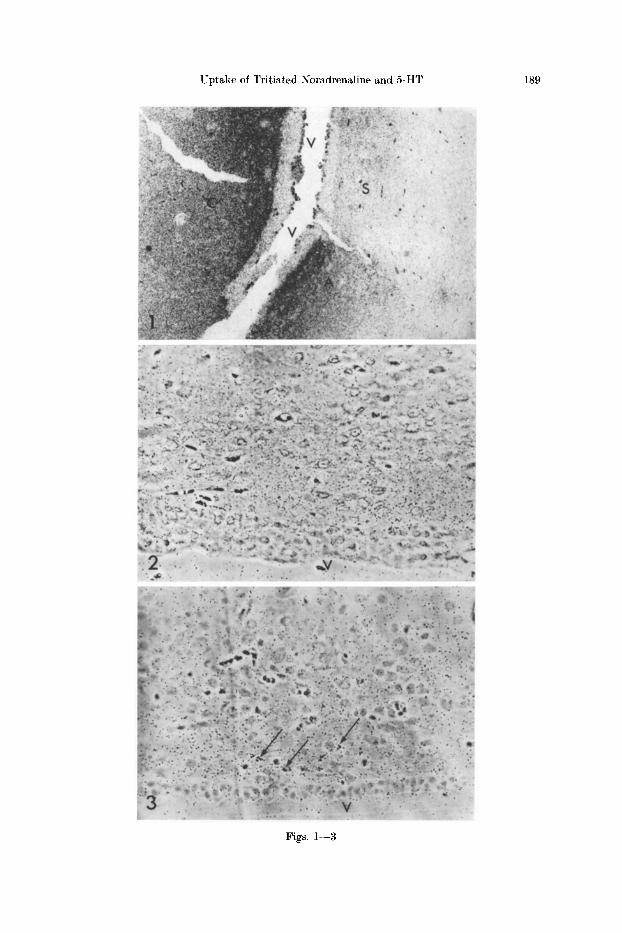
nerve terminals , especial ly when these areas were s i tua ted re la t ive ly close to the site of inject ion. The b lackening was very much higher in the nuc. cauda tus p u t a m e n and nuc. accumbens (Fig. 1), which have high concentra t ions of dopamine (DA) nerve te rmina ls (see F u x ] L 1965), t han in the septal a rea and the h ippocampa l fo rmat ion where the CA nerve te rmina ls are less nmnerous (Fig. 1). I n the per iven t r ieu la r area of the h y p o t h a l a m u s (Fig. 2) there was a good correla t ion be tween dens i ty of grains and number of NA nerve terminals . I n the nuc. per ivent r icu lar i s hypo tha l ami and in the parvocel lu lar pa r t of the nuc. paravent r icu lar i s , which have high amount s of NA nerve t e rmina l s (see F n x ~ , I965), the dens i ty of gra ins was high, forming pa t t e rn s s imilar to those formed by the NA nerve terminals . The gra ins were a lmos t exclus ively localized between the nerve cell bodies, where often even small aggregat ions of grains could be observed p r o b a b l y lying over N A varicosi t ies (Fig. 2, see also Figs. 3 and 4). Such dis t inct ions could only be made in the Ara ld i te sections and with use of the phase con t ras t microscope. However , the varicose fibres as revealed in the fluorescence microscope could not be followed in the au to rad iogram.
Per iven t r i cu la r areas poor in NA nerve terminals , on the o ther hand, showed a low dens i ty of grains. A large accumula t ion of au to rad iograph ic grains were also observed in la te ra l par t s of the ex te rna l layer of the median eminence, where the high accumula t ion of fluorescence is due to the presence of large amount s of DA te rmina ls (FuxE and H6KrELT, 1967). Pa r t i cu l a r ly in this area i t was easy to observe bo th au to rad iograph ic grains and specific green fluorescence in the fluorescence microscope. They showed a good correlat ion,
Regard ing the up t ake of t r i t i a t ed amines in to CA nerve t e rmina l s there was no difference be tween normal or reserpinized rats . However , a c learcut u p t a k e and accumula t ion of grains in CA cell bodies was only observed in the reserpine- n ia lamide p re t r ea t ed ra t s and no t in the normal rats . Thus, in reserpine-nia lamide p r e t r ea t ed ra ts there was a marked accumula t ion of r ad ioac t i v i t y in the DA cell
Fig. 1. Nuc. accumbens (A), nuc. caudatus putamen (C) and septal area (S) of a reserpine- nialamide pretreated rat after intraventricular injection of aH--NA (15 ~l, 30 minutes before killing). Paraffin section (10 ~z). There is a marked accumulation of radioactivity under the ependyma in the nuc. caudatus putamen and nuc. accumbens. In these two regions densely packed DA nerve terminals are present as revealed in fluorescence microscopic studies. The high activity is partly due to the fact that this section is localized near to the site of injection of labelled NA. Note that the density of the grains decreases continously with the distance from the ependyma. In the part of the septal area seen in the picture, on the other hand, only few grains are found. This corresponds well to fluorescence microscopic findings revealing com-
paratively few NA terminals in this region. • 120 (reduced to 9/10)
Fig. 2. The periventricular area of normal rat after an intraventricular injection of 3H--NA (15 ~l, 2 hours before killing). Araldite section (about 2 ~). The third ventricle (V) is seen in the lower part of the picture. There is a marked accumulation of autoradiographic grains in the ventricle-near zone. The grains are at least mainly localized between the nerve cell bodies. Large numbers of NA nerve terminals are found in this area as revealed in fluorescence
microscopic studies. • 120 (reduced to 3/10)
Fig. 3. Same area as in Fig. 2 of a normal rat after an intraventricular injection of aH--5-HT (15 [zl, 2 hours before killing), Araldite section (about 2 [z). There is a moderate accumulation of grains at least mainly localized between the nerve cell bodies. At some places (S ) a large number ot grains are lying close together indicating possible monoamine containing boutons.
• 120 (reduced to 9/10)
Uptake of Tritiated Noradrenaline and 5-HT 189
Figs. 1--3
190 K. FUXE, T. H•KFELT, M. t~ITZ~, and U. UNGERSTEDT:
bodies of the nuc. a rcua tus and in the CA cell bodies present among the radices n. oculomotor i i (see DAttLSTROM and Fux]~, 1964). I n Figs. 6 and 7 i t is demon- s t r a t ed t h a t the fluorescence and the au to rad iograph ic grains appear ing over a rcua te cell bodies af ter the in t r aven t r i cu l a r in ject ion are localized to ident ica l cel lular s t ructures in the doses used. No cer ta in or only s l ight accumula t ion of 3 H - - N A was observed wi th in the areas rich in 5 -HT nerve te rmina ls (e.g. nuc. suprachiasmat icus) or in 5 -HT cell bodies (e.g. nuc. r aphe dorsalis). This was t rue for bo th no rma l and reserp ine-n ia lamide t r ea t ed rats .
A low concent ra t ion of r ad ioac t i v i t y was observed in par t s which conta ined few monoamine nerve terminals , ind ica t ing t h a t there was some unspecific back ground rad ioac t iv i ty . This m a y be due to ex t raneurona l b inding or adsorp t ion of t r i t i a t ed amine and i ts metabol i tes . This background r ad ioac t i v i t y increased with dose and was higher 30 minutes t han 2 hours af ter inject ion. I n the ra ts p r e t r ea t ed wi th reserpine-nia lamide there was a d is t inc t accumula t ion of radio- ac t i v i t y in the capi l la ry walls. This is in agreement wi th fluorescence micro- scopical s tudies demons t r a t ing accumula t ion of fluorescence in cells, ma in ly in per icytes , of the capi l la ry walls (HAMBERG]~ and MASVOKA, 1965; FUXE and U]~GERSTEDT, 1966). - - A marked incorpora t ion of t r i t i a t ed N A was observed in the adrenergic nerve t e rmina l s of the pia l arteries.
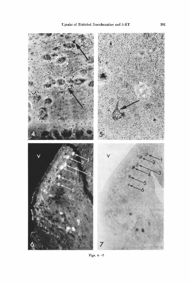
3H~5-HT Injection. Also in this case was the r ad ioac t i v i t y localized a lmost exclusively to the zone (200 400 ~z) close to the ventr ic les and the vent ra l pa r t of the subarachnoida l space. The dens i ty of au to rad iograph ic grains seemed to be re la t ive ly well corre la ted to the d i s t r ibu t ion of 5-HT nerve terminals . Thus, a high dens i ty was observed in the nuc. suprachiasmat icus , which is exclus ively rich in 5-HT nerve terminals . Much less r ad ioac t i v i t y was found in the nuc. cauda tus p u t a m e n and in the nuc. accumbens, where 5-HT nerve t e rmina l s are low in number and main ly DA nerve te rmina ls are present . I n the per ivent r icu lar area of the h y p o t h a l a m u s the dens i ty of grains was no rma l ly modera te to high and the grains over Ara ld i t e sections seemed to lie a lmost exclusively be tween the nerve cell bodies (Figs. 3 and 4). I n the h y p o t h a l a m u s wi th the except ion of the nuc. suprach iasmat icus i t was diff icult to correlate the d i s t r ibu t ion of grains wi th t h a t of the 5 -HT nerve terminals , since the l a t t e r are diff icult to observe due to the i r fineness and the r ap id pho todecompos i t ion of the f luorophore.
Fig. 4. Higher magnification of Fig. 3. Certain aggregations (7 ) of autoradiographic grains are observed which may represent boutons of monoamine nerve terminals. The grains are at
least mainly localized between the nerve cell bodies. E ependymal cells. • 480
Fig. 5. Nuc. raphe dorsalis of normal rat after an intraventricular injection of aH--5-HT (15 ~zl, 30 minutes before killing). Araldite section (about 2 iz). The aqueductus Sylvii is localized to the upper right in the picture. There is a marked accumulation of radioactivity in a 5-HT
nerve cell body (/~). • 300
Fig. 6. Nuc. arcuatus of a reserpine-nialamide pretreated rat given an intraventricular injec- tion of tritiated NA. Paraffin section (10 [~). There is a strong green fluorescence in cell bodies in the nuc. arcuatus due to an accumulation of amine in cell bodies of the tubero-infundibular
DA neurons. • 120
Fig. 7. The same section as in the previous figure, demonstrating the distribution of auto- radiographic grains. I t is evident that there is an accumulation of grains over the DA cell
bodies of the tubero-infundibular neurons. V third ventricle. • 120
Uptake of Tritiated Noradrenaline and 5-HT 191
Figs. 4--7
192 K. FuxE, T. HOKFELT, M. RITZ~N, and U. UNGERSTEDT:
However, in a way similar to that seen after a H - - N A injection aggregations of grains were often observed between the cell bodies (Figs. 3 and 4) probably lying over 5-HT varicosities.
In the nuc. raphe dorsalis an accumulation of autoradiographic grains were observed over certain cell bodies (Fig. 5) which in all probability represented the 5-HT cell bodies (see DAHLSTIC6M and FvxE, 1964). However, this accumulation has as yet only been observed 30 minutes after the intraventricular injection. To estimate the order of concentrating capacity of the 5-HT cell bodies the grain density was determined over the cell bodies, their immediate surroundings and the background over the ventricles. The concentration of radioactivity turned out to be up to 3 - -4 times higher in the cell body than in the surroundings of the cell. By means of the metacrylate reference sections the highest radioactivity within a cell body was estimated to around 100 ~C/g following injection of 15 ~tC 3H--5-HT.
After 3H--5-HT injection there was no accumulation of grains in the DA cell bodies of the areuate nucleus or in the CA cell bodies among the radices oculo- motorii. Nor was there any or only a slight accumulation of grains over the thick NA terminals in the inner layer and over the DA terminals in the outer layer of the median eminence, which is in marked contrast to the case after 3H- -NA injection. This gives further indication of a certain specificity of the uptake- mechanism in the CA neurons.
Discussion
By the use of combined autoradiography and fluorescence histochemistry it has been possible to demonstrate in the present study that intraventricularly administered tritiated NA and 5-HT are taken up and concentrated in the CA and 5-HT nerve terminals respectively with a elearcut degree of specificity. The tri t iated amines, however, only reach those terminals lying close to the ventricles and the ventral part of the subarachnoidal space.
An interesting finding was tha t the CA cell bodies particularly those of the arcuate nucleus did not show any certain accumulation of radioactivity after injection of aH- -NA into normal rats but only after reserpine-nialamide pre- treatment. This may at least part ly be explained by the fact tha t the injected 3H--NA is exposed to extraneuronal monoamineoxidase before it reaches the CA cells, which thus, will be offered less amounts of radioactive NA. This view is supported by the observations that radioactivity is accumulated in cell bodies of the substantia nigra after intraventricular injection of high doses of 3H--NA into niaiamide treated rats (DEscAlCRIES and DROZ, 1968). Another contributing factor to the observed differences may possibly also be that cytoplasmic NA (reserpine-nialamide treated rats) is not transported down as rapidly from the cell bodies as granular NA (normal rats).
The present paper gives further evidence that there exists a reserpine resistant amine concentrating mechanism in the DA and NA neurons for CA and in the 5-HT neurons for 5-HT and that the CA and 5-HT neurons of normal rats also can efficiently accumulate NA and 5-HT respectively. The degree of concentration could be estimated quantitatively by counting grain density and was found to be in the order of three times that in the immediate surroundings.
Uptake of Tritiated Noradrenalinc and 5-HT 193
The present paper illustrates that by use of freeze-dried, Araldite embedded tissues it is possible to demonstrate the localization of labeled amines at the cellular level. This autoradiographic technique involving stripping film emulsion will also make it possible to quantitate the degree of accumulation of amines at the cellular level. The technique used should be of importance in demonstrating the cellular localization of other types of amines which are not possible to demonstrate sufficiently well with the histochemical fluorescence method, e.g.
metaraminol, mctatyramine derivatives. These latter compounds give rise to a fluorescence which is masked by the background autofluorescence of nervous tissue. Furthermore, it may be possible to study the cellular localization of the uptake of biologically important water-soluble compounds such as tritiated histamine and acetylcholine in various parts of the central nervous system.
Araldite sections consistently gave a better resolution in the autoradiographic picture than the paraffin section. This may partly be due to the fact that the stripping film could be applied directly on the plastic sections, while the paraffin embedded sections must be taken through xylol and an alcohol series prior to autoradiography. This treatment with various solvents might cause a low but significant diffusion of the NA-formaldehyde reaction product. After embedding in Araldite any radioactive substance, that is insoluble in this resin, may be observed exactly in its original site in the section, regardless of whether it is bound to the tissue by the formaldehyde gas or not. In addition, the Araldite sections consistantly gave a better morphological picture compared to the paraffin sections as revealed in the phase-contrast-microscope.
Improvement of resolution by decreasing the ~hickness of the section does not become noticable until the thickness is less than 1--2 ~m, due to the pro- nounced self-absorption of the fl-particles emitted from the tritium. This self- absorption will also explain why it was generally not possible to follow (in the autoradiogram) the individual varicose nerve structures in the sections. Instead, only varying densities of grains were observed. Most of the radioactivity reaching the emulsion originates from the upper surface of the section. This is in marked contrast to the case in the fluorescence studies where the whole depth of the section is visualized.
In conclusion Araldite sections of freeze-dried tissue have proved to be a suitable tool in the study of the distribution of tritiated monoamines at the cellular level in the central nervous system. The results of the present paper show that in normal or reserpine-nialamide pretreated rats tritiated NA and 5-HT are mainly taken up into the central CA neurons and 5-HT neurons respectively. Thus, the amine-concentrating mechanism in the cell membrane in the CA neurons clearly prefers NA to 5-HT and vice versa holds true for the 5-HT neurons. - - The intraventricularly administered amines only reach the zone close to the ventricles and the ventral part of the subarachnoidal room.
References AG~AJANIAN, G. K., and F. E. BLOOM: Electronmicroscopic autoradiography of rat hypo-
thalamus after intraventricular Ha-norepinephrine. Science 153, 308--310 (1966). - - - - Localization of tritiated serotonin in rat brain by electronmicroscopie autoradiography.
J. Pharmacol. exp. Ther. 159, 23--30 (1967).
13 I-Iistochemie, Bd. 16
194 K. FVXE et al. : Uptake of Tri t iated Noradrenaline and 5-HT
DAttLSTR()M, A . : The intraneuronal distribution of noradrenaline and the t ranspor t and life- span of amine storage granules in the sympathetic adrenergic neuron. Stockholm : M.D. Thesis 1966.
- - , and K. FUXE: Evidence for the existence of monoamine-containing neurons in the central nervous system. Acta physiol, scand. 92, Suppl. 232, 1--55 (1964).
DESCARRIES, L., et B. DROZ: Incorporation de noradr6naline-3H(NA 3H) dans le syst~me nerveux central du ra t adulte. Etude radioautographique en microscopic 61ectronique. C.R. Acad. Sci. (Paris) 26~, 2480--2482 (1968).
FuxE, K. : Evidence for the existence of monoamine neurons in the central nervous system, IV. The distribution of monoamine nerve terminals in the central nervous system. Acta physiol, scan& 64, Suppl. 247, 39--85 (1965).
- - , and T. HOKFELT : The influence of central catecholamine neurons on the hormone secretion from the anterior and posterior pituitary. Neurosecretion. IV. Int . Syrup. on Neurosecre- tion, Strasbourg, Ju ly 1966.
- - , and U. UNGERSTEDT; Localization of eatecholamine uptake in ra t brain after intraventr i- cular injection. Life Sci. 5, 1817--1824 (1966).
- - - - Localization of 5-hydroxytryptamine uptake in ra t brain after in t raventr icular injec- tion. J . Pharm. Pharmacol. 16, 335 (1967).
- - - - Histochemical studies on the distribution of catecholamines and 5-hydroxytryptamine after intraventr icular injections. Histochemie 18, 16--28 (1968).
HAMBERGER, B., T. MALMFORS, and CH. SActts: Standardizat ion of paraformaldehyde and certain procedures for the histochemical demonstrat ion of catecholamines. J. Histochem. Cytochem. 18, 147 (1965).
- - , and D. MASUOKA : Localization of catecholamine uptake in ra t brain slices. Acta pharmacol. (Kbh.) 22, 363--368 (1965).
HAMMARSTROM, L., M. RITZI~N, and S. ULLBERG: Combined autoradiography and fluorescence microscopy. Localization of labelled 5-hydroxytryptophan in relation to endogenous 5-hydroxytryptamine in the gastrointestinal tract. Experientia (Basel) 22, 215 (1966).
HOKFELT, T.: A modification of the histoehemical fluorescence method for the demonstrat ion of catecholamine and 5-hydroxytryptamine, using Araldite as embedding medium. J . Histochem. Cytochem. 15, 518--519 (1965).
RITZ~N, M.: Mast cells and 5-HT. Uptake of labelled 5-hydroxytryptamine (5-HT) and 5-hydroxytryptophan in relation to storage or 5-HT in individual ra t mast cells. Acta physiol, scand. 70, 42--53 (1967).
- - A method for the autoradiographic determinations of absolute specific radioactivity in cells. Exp. Cell Res. 45, 250--252 (1967).
Prof. K. FVXE Dept. of Histology Karolinska Ins t i tu te t Solnav~gn 1 10401 Stockholm 60, Sweden


![Characterization and radioautography of [3H] LSD binding by rat brain slices in vitro: The effect of 5-hydroxytryptamine](https://img.pdfslide.net/doc/110x75/631d3873f26ecf94330a71c9/characterization-and-radioautography-of-3h-lsd-binding-by-rat-brain-slices-in.jpg)


![Binding properties of a selective tritiated vasopressin V2 receptor antagonist, [3H]-SR 121463](https://img.pdfslide.net/doc/110x75/6346608c596bdb97a9095c2b/binding-properties-of-a-selective-tritiated-vasopressin-v2-receptor-antagonist.jpg)


![[3H] muscimol receptors sites in the carp (Cyprinus carpio L.) brain: Binding assay and autoradiographic distribution](https://img.pdfslide.net/doc/110x75/6335f76ecd4bf2402c0b4f62/3h-muscimol-receptors-sites-in-the-carp-cyprinus-carpio-l-brain-binding-assay.jpg)


