Embed Size (px)
Citation preview
Neuropharmacology 40 (2001) 242–253www.elsevier.com/locate/neuropharm
Detailed distribution of Neurokinin 3 receptors in the rat, guineapig and gerbil brain: a comparative autoradiographic study
Xavier Langlois*, Cindy Wintmolders, Paula te Riele, Jose´e E. Leysen, Mirek JurzakDepartment of Biochemical Pharmacology, Janssen Research Foundation, B-2340 Beerse, Belgium
Received 5 May 2000; received in revised form 25 July 2000; accepted 31 July 2000
Abstract
The neurokinin 3 (NK3) receptor is predominantly expressed in the central nervous system (CNS). Species differences in neuroki-nin 3 (NK3) receptor pharmacology have led to the preferential use of guinea pigs and gerbils in the characterization of non-peptideNK3 antagonists. Little is known about the central localization of NK3 receptors in the CNS of these species. To study this,[3H]senktide and [3H]SR 142801 were used in autoradiography experiments to visualize the NK3 receptors in the guinea pig andgerbil brain and compared to with the distribution of [3H]senktide binding sites in the rat brain. In the three species, the NK3receptor was similarly distributed within the cerebral cortex, the zona incerta, the medial habenula, the amygdaloid complex, thesuperior colliculus and the interpeduncular nucleus. Outside of these structures, our study has revealed that each species displayeda specific distribution pattern of central NK3 receptors. The rat was the only species where NK3 receptors could be visualized inthe striatum, the supraoptic nucleus and the paraventricular nucleus of the hypothalamus. The guinea pig differed mainly from thetwo other species by the absence of detectable binding sites in the substantia nigra pars compacta and the ventral tegmental area.A specific localization of NK3 receptors in the anterodorsal and anteroventral thalamic nuclei characterized the gerbil. This lastspecies is also unique by in the higher level of NK3 receptors in the dorsal and median raphe nuclei. All these differences suggestthat the NK3 receptor mediates different functions in different species. 2000 Elsevier Science Ltd. All rights reserved.
Keywords:Neurokinin 3 receptor; Radioligand autoradiography; Rat; Guinea pig; Gerbil; [3H]Senktide; [3H]SR142801
1. Introduction
The three mammalian tachykinins, substance P (SP),neurokinin A (NKA) and neurokinin B (NKB), are afamily of structurally related peptides found in the per-iphery and the central nervous system (CNS). As neuro-transmitters, these peptides exert their biological activityvia the activation of three different neurokinin (NK)receptors that have been pharmacologically charac-terized and cloned (for a review, see Maggi, 1995). SPbinds preferentially to the NK1 receptor, NKA to theNK2 receptor, and NKB to the NK3 receptor. However,each of the tachykinins is able, at higher concentrations,to activate all three receptors and therefore shows poorselectivity (Maggi and Schwartz, 1997). Development ofselective compounds for one or the other NK receptor
* Corresponding author. Tel:+32-14-606016; fax:+32-14-605380.E-mail address:[email protected] (X. Langlois).
0028-3908/01/$ - see front matter 2000 Elsevier Science Ltd. All rights reserved.PII: S0028-3908 (00)00149-0
was one of the keys for a better understanding of theinvolvement of a particular NK receptor in a physiologi-cal process. This was first accomplished by the structuralmodification of endogenous tachykinins. Numerous pep-tide analogues have been synthesized, displaying selec-tive agonistic or antagonistic properties (Mussap et al.,1993), and used as radioligands for determining bindingparameters and distribution of each of the NK receptorsin native tissues. NK1 receptors are highly expressed inboth the CNS and peripheral tissues, NK2 receptors arecharacterized by a predominant expression in the periph-ery and NK3 receptors are found primarily in the CNS.In the last ten years, a major improvement was the intro-duction of non-peptide antagonists, these compoundsbeing highly selective, resistant to degradation by pep-tidases and therefore metabolically stable (Betancur etal., 1997).
SR142801 is the first potent non-peptide antagonistselective for the NK3 receptor to be described (Emonds-Alt et al., 1995). This compound has been used in

243X. Langlois et al. / Neuropharmacology 40 (2001) 242–253
numerous pharmacological investigations on NK3 recep-tor functions and is unanimously considered as the NK3antagonist of reference. A particular property ofSR142801, that which is also shared by structurally dif-ferent NK3 antagonists (Chung et al., 1995), is the dif-ference in its affinity for NK3 receptors from differentspecies. SR142801 has a higher affinity for the human,the gerbil and the guinea pig than for the rat NK3 recep-tor (Emonds-Alt et al., 1995). As a consequence, afterthe introduction of this compound, several pharmaco-logical studies on NK3 receptors were initiated in speciesdifferent from the rat. Biochemical and electrophysiol-ogical experiments were performed preferentially in theguinea pig (Jung et al., 1996; Nalivaiko et al., 1997;Marco et al., 1998) whereas the gerbil was mostly usedin behavioral studies (Emonds-Alt et al., 1995; Jung etal., 1996). As the rat is the animal of reference for theinvestigation of the CNS, the localization of NK3 recep-tors has been extensively investigated in this species byvarious techniques: radioligand autoradiography (Dam etal., 1990; Stoessl and Hill, 1990), in situ hybridization(Ding et al., 1996; Shughrue et al., 1996) and immunohi-stochemistry (Ding et al., 1996). However, the distri-bution of NK3 receptors has never been reported in ger-bil brain and only briefly described in the guinea pig(Dam and Quirion, 1994). Therefore, the results ofexperiments on the central functions of NK3 receptorsin the guinea pig and the gerbil are almost exclusivelydiscussed with reference to the known localization ofNK3 receptors in the rat brain. A critical question thathas been rarely raised is whether the NK3 receptor dis-plays the same pattern of distribution through these spec-ies.
To answer this question, we used [3H]senktide and[3H]SR142801 in autoradiography experiments tolocalize the NK3 receptors in the gerbil and the guineapig brain. In parallel, [3H]senktide was used to visualizethe distribution of NK3 receptors in the rat brain and toextend previous autoradiographic studies performed withthe same radioligand (Dam et al., 1990; Stoessl and Hill,1990). The comparison revealed striking differences inthe localization of NK3 receptors in the CNS betweenthe three species. The present study suggests that theNK3 receptors mediate different functions in these spec-ies. Our findings should help investigators to discusstheir future results on the central functions of NK3receptors in the gerbil and the guinea pig. Part of thepresent study has already been reported in abstract form(Langlois et al., 1999).
2. Experimental procedures
2.1. Brain tissue
Male Wistar Hannover rats (160–200 g), male DunkinHartley guinea pigs (300–350 g) and male Mongolian
gerbils (40–60 g) were supplied by Charles River(Germany). The animals were specifically pathogen freeat the time of arrival in our facilities. All efforts weremade to minimize animal suffering and to restrict thenumbers of animals used.
Animals were killed by decapitation and the brainswere removed. For radioligand binding assays, the brainswere dissected on ice and the cerebral cortices wereimmediately processed for membrane preparation. Forautoradiography experiments, the whole brains were rap-idly frozen in dry-ice-cooled 2-methylbutane (240°C).20 µm-thick frontal sections were cut using a ReichertJung 2800R cryostat microtome (Cambridge Instru-ments, Cambridge, UK) and thaw-mounted on Super-frost Plus slides (Menzel-Glaser, Madison, WI). The sec-tions were stored at220°C until use.
2.2. [3H]SR142801 saturation binding experiment onguinea pig and gerbil cortical membranes
Guinea pig and gerbil cortex were homogenized withan Ultra Turax homogenizer (Janke and Kunkel, IKALabortechnik, Staufen, Germany) in 40 vol per wetweight tissue of Tris–HCl buffer (50 mM, pH 7.4). Thetotal membrane fraction was collected by centrifugationand washed by two subsequent centrifugation runs (10min at 20,000g at 4°C in a Sorvall RC5B centrifuge).The final pellets were resuspended in 10 vol per wetweight wet tissue of Tris–HCl buffer (50 mM, pH 7.4)and stored at280°C. On the day of the experiment, themembranes were diluted in assay buffer (Tris–HCl 50mM, pH 7.4 containing 3 mM MnCl, 40µg/ml bacitra-cin, 4 µg/ml leupeptin, 2µg/ml chymostatin and 0.02%(w/v) bovine serum albumin (BSA)). Incubations wererun for 60 min at 25°C in a volume of 0.5 ml containing40–50 µg protein. Ligand concentration binding iso-therms were obtained with 10 concentrations of[3H]SR14201, in a range of 10 pM to 1 nM. Non-specificbinding was estimated in the presence of 10µMSR142801. The incubation was terminated by rapid fil-tration over Whatman GF/B filters and three washingsteps with 3 ml ice-cold Tris–HCl buffer. Binding ofligands to filters was measured by counting filters in 2ml of Packard Ultima Gold MV on a Packard Tri-Carb1600 CA liquid scintillation counter (Packard, USA).Ligand concentration binding isotherms were calculatedby nonlinear regression analysis according to algorithmsdescribed by Oestreicher and Pinto (1987). Themaximum number of binding sites (Bmax) and the appar-ent equilibrium dissociation constant (KD) were derivedfrom the curve fitting.
2.3. NK3 receptor autoradiography
[3H]senktide (63.5 Ci/mmol) autoradiography wasperformed essentially as described by Dam et al. (1990).

244 X. Langlois et al. / Neuropharmacology 40 (2001) 242–253
The brain sections were pre-incubated (3×5 min) in 50mM Tris–HCl pH 7.4 at room temperature. The sliceswere incubated for 90 min at room temperature in buffercontaining 50 mM Tris–HCl (pH 7.4), 3 mM MnCl2,0.02% (w/v) BSA, 40µg/ml bacitracin, 2µg/ml chymos-tatin, 4 µg/ml leupeptin and 3 nM [3H]senktide. Non-specific binding was determined by addition to the incu-bation medium of 10µM (N–Me–Phe7)–NKB for ratbrain sections or 10µM SR 142801 for guinea pig andgerbil brain sections. To stop the incubation, the slideswere washed (4×1 min) in Tris–HCl buffer, pH 7.4 at4°C followed by a rapid dip in cold distilled water anddrying under a stream of cold air. Sections were exposedto [3H]Hyperfilm (Amersham, UK) for 8 weeks.
[3H]SR142801 (76 Ci/mmol) autoradiography wasperformed according to the following procedure. Thebrain sections were pre-incubated (3×5 min) in 50 mMTris–HCl pH 7.4 with 0.3% (w/v) BSA at room tempera-ture. The slices were incubated for 60 min at room tem-perature in buffer containing 50 mM Tris–HCl (pH 7.4),3 mM MnCl2, 0.3% (w/v) BSA, 40µg/ml bacitracin, 2µg/ml chymostatin, 4 µg/ml leupeptin and 1 nM[3H]SR142801. Non-specific binding was determined byaddition of 10µM SR 142801 to the incubation medium.After incubation, the excess of radioligand was washedoff (3×10 min) in Tris–HCl buffer, pH 9.4 at 4°C fol-lowed by a rapid dip in cold distilled water. The rinseof sections in a basic buffer reduced the non-specificbinding without altering the amount of specific binding.After the washing, the slides were dried under a streamof cold air and exposed to [3H]Hyperfilm for 6 weeks.
In order to compare the distribution of both radioli-gands binding sites in guinea pig and gerbil brain, adjac-ent sections were used for [3H]senktide and[3H]SR142801 autoradiography.
2.4. Data analysis of anatomical studies
After development of films, autoradiograms were ana-lysed and quantified by use of an MCID M1 imageanalysis system (Imaging Research, St Catharines, Onta-rio, Canada). Optical densities in the anatomical regionsof interest were transformed into levels of bound radio-activity after calibration of the image analyser with grayvalues generated by the co-exposed [3H]microscalesstandards (Amersham, UK). The radioligand bindingsignal was expressed in fmol/mg of tissue equivalent(fmol/mteq). Identification of the rat and gerbil brainstructures was performed according to the atlas of Pax-inos and Watson (1986). The anatomical localization inthe guinea pig brain was determined with the atlas ofLuparello (1967).
2.5. Chemicals
[3H]senktide was obtained from NEN (USA) and[3H]SR142801 from Amersham (UK). Sanofi Research
(France) kindly provided SR142801. Bovine serum albu-min and chymostatin were from Sigma (USA). Bacitra-cin and leupeptin were obtained from Boehringer-Ingelheim (Germany). (N–Me–Phe7)–NKB was fromBachem (Switzerland).
3. Results
3.1. [3H]SR142801 saturation binding experiment onguinea pig and gerbil cortical membranes (Fig. 1)
In saturation binding experiments on cortical mem-branes, [3H]SR142801 displayed high affinity and satu-rable binding to a single site in both tissues examined.The [3H]SR142801 saturation binding parameters were:Bmax = 928±18 fmol/mg, KD = 0.07±0.01 nM for theguinea pig cortical membranes;Bmax = 439±54 fmol/mg,KD = 0.29±0.03 nM for the gerbil cortical membranes(mean±SEM of three independent experiments).
Fig. 1. Concentration binding isotherms on cortical membranes fromguinea pig (a) and gerbil (b) performed with [3H]SR142801. Total (h),specific (I) and non-specific (e) binding are depicted as a means ofa duplicate determination. The non-specific binding was determined inthe presence of 10µM SR142801.
245X. Langlois et al. / Neuropharmacology 40 (2001) 242–253
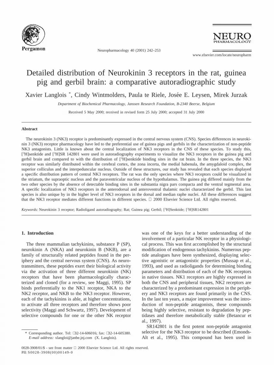
3.2. Specificity of [3H]senktide and [3H]SR142801binding in the autoradiography experiments (Fig. 2)
In the three species, [3H]senktide showed a very highlevel of specific binding (.95% of the total binding).The addition of (N–Me–Phe7)–NKB (10 µM) for ratbrain sections or SR142801 (10µM) for guinea pig andgerbil brain sections, in to the incubation medium,reduced the signal to film background (not shown). Inthe three species, [3H]senktide labeled the cerebral cor-tex whereas the rat was the only species displaying aspecific distribution of binding sites in the striatum.
1nM [3H]SR142801 did not show any specific labe-ling in rat brain sections. In contrast, in guinea pig andgerbil, [3H]SR142801 displayed a specific distributionpattern throughout the brain. A high level of non-specificbinding (|40% of the total binding) was observed with[3H]SR142801. In these two species, where the compari-son between the two radioligands was possible, the dis-tribution of [3H]SR142801 binding sites matched exactlywith that of [3H]senktide.
3.3. Distribution of NK3 receptors labeled by[3H]senktide in rat brain (Fig. 3 and Table 1)
The highest densities of [3H]senktide binding sites inthe rat brain were found in the paraventricular and thesupraoptic nucleus of the hypothalamus and in the sphe-noid nucleus of the pons. Intermediate levels of specificbinding were seen in the rostral caudate putamen, thezona incerta, the basolateral and basomedial amygdaloidnuclei, the amygdalo-hippocampal area, the ventral CA3field of the hippocampus, the ventral tegmental area, the
Fig. 2. Distribution of [3H]senktide (3 nM) and [3H]SR142801 (1nM) binding sites in the forebrain of the rat, the guinea pig and thegerbil. Non-specific binding was determined in the presence of 10µM(N–Me–Phe7)–NKB for rat brain sections and 10µM SR 142801 forguinea pig and gerbil brain sections. Note for [3H]SR142801 theabsence of specific binding in the rat brain sections and the high levelof non-specific binding (|40%) in the three species. The level of non-specific [3H]senktide binding was indistinguishable from the film back-ground (not shown). Specific [3H]senktide binding sites in the caudateputamen (Cpu) were found only in the rat.
substantia nigra pars compacta and the interpeduncularnucleus. Low to moderate levels of binding weredetected in the dorsomedial hypothalamus nucleus, thesupramammillary nucleus, the medial habenula, thesuperior colliculus, the median raphe nucleus and theposterodorsal tegmental nucleus. In the cortex, [3H]senk-tide binding sites are present in the layers IV and Vthroughout the brain with the highest densities in thecingulate cortex and the lowest densities in the retrospl-enial cortex. No specific [3H]senktide binding sites couldbe detected in the caudal part of the caudate putamenand the nucleus accumbens, the dorsal raphe nucleus andthe locus coeruleus (Table 1).
3.4. Distribution of NK3 receptors labeled by[3H]senktide and [3H]SR142801 in guinea pig brain(Fig. 3 and Table 1)
[3H]senktide and [3H]SR142801 displayed the samedistribution pattern in the guinea pig brain. Except theretrosplenial cortex, high densities of specific bindingsites were seen in the whole cerebral cortex, especially inthe mid- and deep cortical laminae. The other structuresshowing a high density of NK3 receptors in the guineapig brain were the medial habenula and several amygda-loid nuclei (basolateral, basomedial and anterior corticalnuclei). A much lower density of binding sites charac-terizes all the other brain structures of the guinea pigbrain. Low densities of NK3 receptors were seen in thezona incerta, the ventromedial hypothalamus, the supra-mammillary nucleus, the caudal CA2 field of the hippo-campus and the interpeduncular nucleus. Additionally,very low levels of binding sites could be detected in thedorsal lateral septum, the bed nucleus of the stria ter-minalis, the superior colliculus and the median raphenucleus. In these last brain areas, the localization of NK3receptors could be achieved only with [3H]senktidebecause of the too high level of non-specific binding dis-played by [3H]SR142801. No specific labeling could bedetected with either radioligand in the whole striatum,the substantia nigra and the ventral tegmental area. Simi-larly, the dorsal raphe nucleus, the paraventricular andsupraoptic nuclei of the hypothalamus and various nucleiof the pons, including the locus coeruleus, the sphenoidnucleus and the dorsotegmental nucleus, were devoid ofspecific binding sites.
3.5. Distribution of NK3 receptors labeled by[3H]senktide and [3H]SR142801 in the gerbil brain(Fig. 3 and Table 1)
In the gerbil, the distribution of [3H]senktide and[3H]SR142801 binding sites was identical throughout thebrain. The highest densities of NK3 sites were found inthe anterodorsal and anteroventral thalamic nuclei, thezona incerta, the basolateral and basomedial amygdaloid

246 X. Langlois et al. / Neuropharmacology 40 (2001) 242–253

247X. Langlois et al. / Neuropharmacology 40 (2001) 242–253
Fig
.3.
Com
paris
onof
the
dist
ribut
ion
ofN
K3
rece
ptor
sin
the
rat,
guin
eapi
gan
dge
rbil
brai
n.T
hera
tbr
ain
sect
ions
wer
eex
pose
dto
3nM
[3H
]sen
ktid
e.A
djac
ent
sect
ions
ofth
egu
inea
pig
and
gerb
ilbr
ain
wer
eex
pose
dto
3nM
[3 H
]sen
ktid
ean
d1
nM[3
H]S
R14
2801
.Aco
,Ant
erio
rco
rtica
lam
ygda
loid
nucl
eus;
AdT
,ant
erod
orsa
ltha
lam
icnu
cleu
s;A
hi,a
myg
dalo
hipp
ocam
pala
rea;
AvT
,ant
erov
entra
ltha
lam
icnu
cleu
s;B
LA,b
asol
ater
alam
ygda
loid
nucl
eus,
ante
rior
part;
BLV
,bas
olat
eral
amyg
dalo
idnu
cleu
s,ve
ntra
lpar
t;B
LP,b
asol
ater
alam
ygda
loid
nucl
eus,
post
erio
rpa
rt;B
MA
,ba
som
edia
lam
ygda
loid
nucl
eus,
ante
rior
part;
BM
P,b
asom
edia
lam
ygda
loid
nucl
eus,
post
erio
rpa
rt;B
ST
,bed
nucl
eus
ofth
est
riate
rmin
alis
;CA
2,fie
ldC
A2
ofA
mm
on’s
horn
;CA
3,fie
ldC
A3
ofA
mm
on’s
horn
;CeA
,cen
trala
myg
dalo
idnu
cleu
s;C
g,ci
ngul
ate
corte
x;C
Pu,
caud
ate
puta
men
;DG
,den
tate
gyru
s;D
R,d
orsa
lrap
henu
cleu
s;D
MH
,dor
som
edia
lhyp
otha
lam
icnu
cleu
s;D
cL,d
eep-
corti
call
ayer
;IP
,int
erpe
dunc
ular
nucl
eus;
LSD
,la
tera
lsep
tal
nucl
eus
dors
al;
McL
,m
id-c
ortic
alla
yer;
Mhb
,m
edia
lhab
enul
arnu
cleu
s;M
nR,
med
ian
raph
enu
cleu
s;P
DT
g,P
oste
rodo
rsal
tegm
enta
lnu
cleu
s;P
VN
,pa
rave
ntric
ular
thal
amic
nucl
eus;
RS
,re
trosp
leni
alco
rtex
;S
C,
supe
rior
colli
culu
s;S
NC
,su
bsta
ntia
nigr
a,pa
rsco
mpa
cta;
SoN
,su
prao
ptic
nucl
eus;
Sph
,sp
heno
idnu
cleu
s;S
uM,
supr
amam
mill
ary
nucl
eus;
VT
A,
vent
ralt
egm
enta
lare
a;V
MH
,ve
ntro
med
ialh
ypot
hala
mic
nucl
eus;
ZI,
zona
ince
rta.
Sca
leba
r:1
cm.

248 X. Langlois et al. / Neuropharmacology 40 (2001) 242–253
Table 1Comparison of the distribution of [3H]senktide binding sites in the rat brain and [3H]senktide and [3H]SR142801 binding sites in the guinea pigand gerbil brain. The list of brain structures is based on the autoradiograms presented in Fig. 3. Each of these structures has been shown to containspecific binding sites in at least one species. An exception concerns the locus coeruleus that is devoid of specific binding sites in the three species.When a specific signal has been detected, the densities have been quantified using a MCID M1 image analysis system (– not detectable, NQ: notquantified). Values are expressed in fmol/mg tissue equivalent (mean±SEM, n=3)
Anatomical region Rat Guinea pig Gerbil[3H]senktide [3H]senktide [3H]SR142801 [3H]senktide [3H]SR142801
CortexDeep-cortical layer – 63±2 89±7 – –Mid-cortical layer
Cingulate cortex 30±2 108±6 103±4 105±7 88±3Frontoparietal cortex 21±4 82±5 107±5 51±6 77±2Retrosplenial cortex 9±1 21±3 48±3 99±7 106±4
Basal gangliaCaudate putamen, anterior 15±1 – – – –Caudate putamen, posterior – – – – -
Septal and basal forebrainLateral septal nucleus, dorsal – 10±2 NQ – –Bed nucleus of the stria terminalis – 9±1 NQ – –
ThalamusAnterodorsal thalamic nucleus – – – 53±6 61±3Anteroventral thalamic nucleus – – – 47±5 55±5
Zona incerta 21±2 25±3 73±4 46±8 68±5
HypothalamusParaventricular nucleus 47±5 – – – –Supraoptic nucleus 35±4 – – – –Dorsomedial hypothalamus nucleus 15±5 – – 34±5 53±4Ventromedial hypothalamus – 19±3 23±4 – –Supramammillary nucleus 10±3 18±4 27±3 23±3 50±5
Medial habenula 13±2 97±6 175±12 17±3 31±2
AmygdalaBasolateral amygdaloid nucleus 23±3 72±7 101±9 50±6 75±5Basomedial amygdaloid nucleus 18±2 58±3 84±5 42±5 68±6Central amygdaloid nucleus – – – 31±5 56±5Anterior cortical amygdaloid nucleus – 105±10 161±6 – –Amygdalohippocampal area 15±4 – – 25±3 26±3
HippocampusCA2 – 26±5 65±10 – –CA3, ventral 16±4 – – – –Dentate gyrus, ventral – – – 25±3 56±5
MidbrainSuperior colliculus 6±2 5±2 NQ 10±2 33±4Ventral tegmental area 23±4 – – 32±1 76±3Substantia nigra, pars compacta 16±3 – – 10±3 16±2Interpeduncular nucleus 21±3 14±3 42±5 34±3 53±5Dorsal raphe nucleus – – – 32±3 45±6Median raphe nucleus 9±1 9±2 NQ 39±1 56±3
PonsSphenoid nucleus 41±2 – – 76±5 134±6Posterodorsal tegmental nucleus 11±2 – – – –Locus coeruleus – – – – –

249X. Langlois et al. / Neuropharmacology 40 (2001) 242–253
nucleus, the ventral tegmental area and the sphenoidnucleus of the pons. Cortical layers IV and V were alsoshown to contain high levels of NK3 receptors, parti-cularly in the cingulate and the retrosplenial cortex.Intermediate levels of binding sites were detected in thecentral amygdaloid nucleus, the dorsomedial hypothala-mus nucleus, the dentate gyrus of the hippocampus, thesubstantia nigra pars compacta and the interpeduncularnucleus. The dorsal and median raphe nuclei also con-tained an appreciable level of binding sites. Moderate tolow levels of NK3 receptors were seen in the medialhabenula and the superior colliculus. No specific bindingsites were detected in the whole striatum, in the paraven-tricular or supraoptic nuclei of the hypothalamus or inthe locus coeruleus.
4. Discussion
The present investigation compares the autoradio-graphic distribution of NK3 receptors in the rat, theguinea pig and the gerbil brain by using two differentradioligands: the peptide agonist [3H]senktide and thenon-peptide antagonist [3H]SR142801. The selectivity of[3H]senktide for the NK3 receptor has been well demon-strated by others (Guard et al., 1990; Renzetti et al.,1991) and our results confirm that [3H]senktide is a suit-able radioligand for the localization study of NK3 recep-tors, independent of the species used. Indeed, in the threespecies investigated, [3H]senktide displayed a highlyspecific pattern of distribution in brain. In contrast to[3H]senktide, the specific binding of [3H]SR142801could be detected in guinea pig and gerbil brain sectionsonly. Our results are in good agreement with the pharma-cological characterization of SR142801, showing thatthis compound has a much higher affinity for the gerbiland guinea pig than for the rat NK3 receptor (Emonds-Alt et al., 1995). To our knowledge, this is the first bind-ing study to use the radiolabeled non-peptide NK3 antag-onist [3H]SR142801 and consequently the selectivity andspecificity of this radioligand has to be discussed. Thesaturation binding experiments performed with[3H]SR142801 in guinea pig and gerbil cortical brainmembranes indicate that this radioligand binds appar-ently with a high affinity to one population of receptorsites in both species. In guinea pig and gerbil brain, thedistribution of binding sites of [3H]senktide and[3H]SR142801 within these species is identical through-out the brain, demonstrating that both radioligands bindto the same receptor. In rat brain, the lack of specificbinding of [3H]SR142801, explainable by the loweraffinity of the compound for the rat NK3 receptor, indi-cates also that this radioligand at nanomolar concen-trations does not bind specifically to any other rat recep-tor. Altogether, our results show that [3H]SR142801 isa highly specific radioligand for the NK3 receptor.
[3H]senktide labeling of the NK3 receptor in rat braincorresponds to previous autoradiographic studies perfor-med with the same radioligand (Dam et al., 1990; Stoessland Hill, 1990). The highest concentrations of NK3receptors were found in the paraventricular and the sup-raoptic nuclei of the hypothalamus. Very high densitiesof [3H]senktide binding sites were also seen in the sphe-noid nucleus of the pons. According to the rat brain atlasof Paxinos and Watson (1986), the sphenoid nucleus isdorsomedially located over the posterodorsal tegmentalnucleus. Stoessl and Hill (1990) have previouslyreported a high density of NK3 receptors in the dorsaltegmental nucleus without mentioning the sphenoidnucleus. Our study shows clearly that very few [3H]senk-tide binding sites are present in the posterodorsal teg-mental nucleus. This discrepancy could be explained bythe anatomical proximity of both nuclei that might haveconfounded their identification. Another misconceptionabout the localization of NK3 receptors concerns thelocus coeruleus. We were not able to detect any specificbinding sites in this nucleus but when radioligands otherthan [3H]senktide have been used by others, the NK3receptor has been described to be present in the locuscoeruleus (Beaujouan et al., 1986; Buck et al., 1986; Saf-froy et al., 1988). This discrepancy can be explained bythe lower specificity of radioligands like [125I]BoltonHunter conjugated-eledoisin ([125I]BHE) or [3H]NKBwhich, additionally to the NK3 receptors, bind with suf-ficient affinity to other NK receptors as well (Mussap etal., 1993). Thus, the labeling found in the locus coeru-leus with [125I]BHE (Beaujouan et al., 1986; Buck et al.,1986; Saffroy et al., 1988) and [3H]NKB (Saffroy et al.,1988) is most probably due to the binding to NK1 recep-tors, which are largely expressed in this nucleus (Nakayaet al., 1994). Our data on the anatomical localization of[3H]senktide binding sites in the rat pons are consistentwith an extensive immunocytochemical study performedin the rat brain with a specific anti-NK3 receptor anti-body (Ding et al., 1996). Indeed, the authors foundstrong immunolabeling in the sphenoid nucleus, a mod-erate labeling in the posterodorsal tegmental nucleus andan absence of signal in the locus coeruleus. If the wholerat brain is taken into consideration, our autoradio-graphic labeling superimposes well with the immunolab-eling reported by different authors (Ding et al., 1996;Mileusnic et al., 1999). The NK3 receptors are foundwith both techniques in the cortical layers IV and V,striatum, zona incerta, supraoptic nucleus, sphenoidnucleus, various amygdaloid nuclei, medial habenularnucleus, hippocampus, superior colliculus, interpeduncu-lar nucleus, ventral tegmental area, substantia nigra parscompacta and median raphe nucleus. This distribution inthe rat brain coincides also with that of the NK3 receptormRNA performed by in situ hybridization (Ding et al.,1996; Shughrue et al., 1996).
Recently, a NK3 receptor homologue named NK4 has

250 X. Langlois et al. / Neuropharmacology 40 (2001) 242–253
been identified in human (Donaldson et al., 1996; Krauseet al., 1997). [3H]senktide binds also with a high affinityto the NK4 receptor (Krause et al., 1997) which couldquestion the selectivity of the autoradiographic labelingreported by us and other investigators. However, accord-ing to the literature, this possibility is only hypothetical.First, the NK4 receptor has not yet been cloned in non-human species. Second, although NK4 mRNAs werefound in peripheral organs, no transcripts could bedetected in the human brain (Donaldson et al., 1996).Moreover, attempts to clone the NK4 receptor from therabbit brain have failed (Medhurst et al., 1999), whichsuggest again that this receptor might not be present inthe CNS. Finally, the identical distribution of [3H]senk-tide binding sites, NK3 mRNAs and NK3 immunoreac-tive sites in the rat brain proved that [3H]senktide labelsuniquely the NK3 receptor in this species. Starting fromthis statement, we have compared the distribution ofNK3 receptors in the rat brain with that of the guineapig and the gerbil.
The localization of [3H]senktide binding sites in theguinea pig brain has been previously reported by Damand Quirion (1994). Whereas we have used their proto-col and the same source of [3H]senktide (NEN), the levelof specific binding was much higher in our hands. Theuse of Superfrost plus slides in place of gelatin-coatedslides could explain the difference. This fact and the useof the specific NK3 antagonist [3H]SR142801 in parallelallowed us to state more definitely on the distribution ofNK3 receptors in the guinea pig brain since both radioli-gands displayed the same distribution pattern. High den-sities of NK3 receptors were found in all cortical layers,with a particular enrichment of binding sites in the mid-and deep cortical laminae. The medial habenula alsocontained very high densities of NK3 receptors. Theother brain area exhibiting a high level of binding sitesin the guinea pig brain was the amygdaloid complex. Incomparison with the previously named structures, therest of the guinea pig brain contained much lower den-sities of NK3 receptors. Intermediate to low levels ofbinding sites were found in the laterodorsal septum, thezona incerta, the mammillary bodies, the CA2 layer ofthe caudal hippocampus, the superior colliculus and theinterpeduncular nucleus. Extremely low levels of bind-ing sites could be detected in the raphe median nucleus.So far, even if not totally similar, the distribution of NK3receptors in guinea pig brain corresponds roughly towhat is observed in the rat brain. However, in severalareas, our study has shown clear differences between thetwo species. In contrast to the rat, no specific bindingsites have been detected in the rostral striatum, the para-ventricular and supraoptic nucleus of the thalamus, thesubstantia nigra pars compacta, the ventral tegmentalarea and the sphenoid nucleus of the guinea pig brain.
To our knowledge, neither in situ hybridization norimmunocytochemical experiments have been performed
in the guinea pig brain in an attempt to localize the NK3receptor. As a consequence, our autoradiographic datacannot be compared with other receptor distributiontechniques. Such comparison would have beenextremely helpful to confirm the apparent absence ofNK3 receptors in key dopamine-related structures of theguinea pig brain such as the substantia nigra pars com-pacta and the ventral tegmental area. In rat brain, theinvolvement of NK3 receptors in the control of braindopamine function is well documented. The NK3 agonistsenktide potently excites dopamine neurons within theventral tegmental area (Seabrook et al., 1995) and thesubstantia nigra pars compacta (Keegan et al., 1992) ofthe rat. Thus, the localization of NK3 receptors in thesetwo structures of the rat brain is in good agreement withthe senktide-induced excitation of dopamine neurons.Few investigations on NK3 receptor functions have beenperformed in guinea pigs and they are rarely in concord-ance with the receptor distribution presented in thisreport. For example, an in vitro electrophysiologicalstudy has shown that senktide increases the firing of nig-ral neurons in guinea pig (Nalivaiko et al., 1997). More-over, application of senktide with microdialysis probesin the region of dopamine cell bodies enhanced theextracellular concentration of dopamine throughout theirprojection areas including the striatum, the nucleusaccumbens and the prefrontal cortex (Marco et al.,1998). These effects seem specifically mediated by theactivation of NK3 receptors, since they are blocked bySR142801. An absence of a correlation between func-tional data and our localization study is also observed inthe guinea pig locus coeruleus. Similar to the situationin the rat, we were not able to detect any specific bindingsites in the locus coeruleus of the guinea pig. However,it has been shown that application of senktide on guineapig locus coeruleus slices induces excitatory responsesof noradrenergic neurons in this structure (Jung et al.,1996). The senktide-evoked response was completelyantagonized by SR142801, suggesting a specific NK3-mediated effect. To explain these discrepancies, it canbe assumed that the NK3 receptors could be so weaklyexpressed in these structures that autoradiography wouldnot be sensitive enough to detect them. On the otherhand, in another brain structure, the medial habenula,our autoradiographic study correlates well with electro-physiological data. Measurement of the excitatoryresponse of senktide in the medial habenula of differentspecies revealed a much higher proportion of NK3-sensi-tive neurons in the guinea pig than in the rat (Boden andWoodruff, 1994). In agreement with this electrophysiol-ogical study, our data show clearly that the guinea pighabenula contains a much higher density of [3H]senktidebinding sites than the rat habenula. Another possibleexplanation for the lack of correspondence betweenlocalization and functional study in the guinea pig couldbe the existence of a NK3 receptor subtype. So far, there

251X. Langlois et al. / Neuropharmacology 40 (2001) 242–253
is no pharmacological evidence to support this hypoth-esis and new functional studies, taking into considerationour results, should help to elucidate this possibility. Toconclude this comparison between rat and guinea pig,our data indicate that in any case the localization of theNK3 receptor in rat brain cannot be used for interpret-ation of results in another species. Unfortunately, mostprevious studies have done precisely this.
The localization of NK3 receptors in gerbil brain isreported for the first time in the present study. Similarto the rat brain, the gerbil cortical layers IV and V con-tain high densities of NK3 receptors, as is also the casein the ventral tegmental area, the substantia nigra parscompacta, various amygdala nuclei and the sphenoidnucleus. The NK3 receptor distribution in the substantianigra pars compacta and the ventral tegmental area is ingood agreement with results obtained in primary culturesof gerbil mesencephalic neurons, showing that senktideincreases dose-dependently the release of [3H]dopaminedose-dependently (Alonso et al., 1996). The zonaincerta, the interpeduncular nucleus, the medial habenulaand the superior colliculus of the gerbil contain moderateto low levels of binding sites, which is not different fromthe rat brain. However, in contrast to the rat, the rostralstriatum, the paraventricular hypothalamic nucleus andthe supraoptic nucleus of the gerbil are devoid of specificbinding sites. Inversely, some areas, which appear tocontain very low levels of NK3 receptors in the rat brain,if any at all, display a high level of binding sites in thegerbil. Indeed, the density of NK3 receptors is remark-ably high in the dorsal and median raphe of the gerbil.Remarkable too is the abundance of NK3 receptors inthe anterodorsal and anteroventral thalamic nuclei. Thesetwo nuclei are one of the gerbil brain structures contain-ing the highest densities of [3H]senktide and[3H]SR142801 binding sites. Interestingly, this particulardistribution is reported for the first time in the mam-malian CNS since this localization seems to be specificto the gerbil brain. Another specific localization in thegerbil brain was found in the ventral dentate gyrus.
To summarize our comparative autoradiographicstudy in the rat, the guinea pig and the gerbil, the follow-ing statements can be made. The NK3 receptors are simi-larly distributed among the three species within the cor-tex, the zona incerta, the medial habenula, theamygdaloid complex, the superior colliculus and theinterpeduncular nucleus. In these structures, only differ-ences in densities of binding sites exist. As far as theother brain areas are concerned, each species presents aspecific distribution of NK3 receptors. Clear differencesare seen in structures belonging to the different monoam-inergic systems, which is particularly interesting sincethe pharmacology of NK3 receptors has been often dis-cussed in relation to these systems (Giardina and Raveg-lia, 1997; Pritchard and Boden, 1995). The detection ofstriatal NK3 receptors exclusively in the rat, the apparent
absence of NK3 receptors in the substantia nigra and theventral tegmental area of the guinea pig, and the strongexpression of NK3 receptors in the dorsal and medianraphe of the gerbil are the most striking differencesobserved. The principal aim of neuroanatomical localiz-ation studies is to indicate the particular function of agiven receptor, depending on its specific localization.Thus, our data on NK3 receptor distribution may implythat this receptor mediates different functions in differentspecies. Interestingly, some investigators have reportedthat intracerebroventricular injection of senktide pro-duces mainly wet-dog shakes and face washing in therat and the guinea pig without influencing the motoractivity (Piot et al., 1995) whereas the same treatmentdecreases locomotion in the gerbil without inducing anyother behavioral changes (Jung et al., 1996). Increasesin locomotor activity in the guinea pig after centraladministration of senktide has been also reported (Yipand Chahl, 1997). All these differences in senktide-induced behavior could be explained by the differentlocalization of NK3 receptors among these species.
Since the study of Dietl and Palacios (1991), showingan absence of [125I]BH-eledoisin binding sites in severalmonkey and human brain areas, including the striatum,the hippocampus, the amygdala and the spinal cord, thepresence of NK3 receptors in primate brain remains con-troversial. At the same time, Buell et al. (1992) haveshown by polymerase chain reaction that the NK3mRNA is expressed at a very low level in several areasof the human brain. Because of its lack of selectivity,[125I]BH-eledoisin is not the most appropriate radioli-gand for labeling NK3 receptors, and the use of moreselective radioligands such as [3H]senktide or[3H]SR142801 would be highly preferable for re-evalu-ating the distribution of NK3 receptors in the primateCNS. Confirming this hypothesis, specific [3H]senktidebinding sites were found to be concentrated in the super-ficial layer of the dorsal horn in the lumbar spinal cordof the cynomolgus monkey (Guard and Watson, 1991)and the rhesus monkey (own preliminary results, notshown). In addition, immunocytochemistry with specificanti-NK3 antibodies has been recently used to localizethe NK3 receptors in the layers III/V of the human cor-tex (Mileusnic et al., 1999; Tooney et al., 2000). Anotherimportant issue in attempting to localize the NK3 recep-tor in primate brain would be to perform a distributionstudy in a large number of brain structures. In fact, limit-ing such study to brain areas that have been shown tocontain NK3 receptors in rodents might lead to anincomplete picture of the localization of NK3 receptorsin the primate brain. As discussed above, the distributionof central NK3 receptors in a given species cannot beextrapolated to other species. Some species-specificlocalization, like the rostral striatum in the rat or theanterodorsal and anteroventral thalamic nuclei in the ger-bil, could also occur in the primate brain.

252 X. Langlois et al. / Neuropharmacology 40 (2001) 242–253
In conclusion, our study has shown that marked differ-ences in the distribution of NK3 receptors exist in thebrain of the rat, the guinea pig and the gerbil, the experi-mental animals used most. The limited knowledge onthe neuroanatomical localization of NK3 receptors inprimate brain raises the question as to which species willbest predict the central effects of compounds interactingwith the NK3 receptor in humans. The development of aNK3 agonist and/or antagonist for particular therapeuticapplications will be dependent on the answer to thisquestion.
5. Note added in proof
During the proof editing process, the existence of theNK4 receptor has been refuted in several species includ-ing the rat and the guinea pig (Sarau, H.M., Mooney,J.L., Schmidt, D.B., Foley, J.J., Buckley, P.T., Giardina,G.A.M., Wang, D.Y., Lee, J.A., Hay, D.W.P., 2000. Evi-dence that the proposed novel human “Neurokinin-4”receptor is pharmacologically similar to the human NK3receptor but is not of human origin. Molecular Pharma-cology 58, 552–559).
References
Alonso, R., Fournier, M., Carayon, P., Petitpretre, G., Le Fur, G., Sou-brie, P., 1996. Evidence for modulation of dopamine-neuronalfunction by tachykinin NK3 receptor stimulation in gerbil mesen-cephalic cell cultures. European Journal Neuroscience 8, 801–808.
Beaujouan, J.C., Torrens, Y., Saffroy, M., Glowinski, J., 1986. Quanti-tative autoradiographic analysis of the distribution of binding sitesfor [125U]Bolton Hunter derivatives of eledoisin and substance Pin the rat brain. Neuroscience 18, 857–875.
Betancur, C., Azzi, M., Roste`ne, W., 1997. Nonpeptide antagonistsof neuropeptide receptors: tools for research and therapy. TrendsPharmacological Science 18, 372–386.
Boden, P., Woodruff, G.N., 1994. Presence of NK3-sensitive neuronesin different proportions in the medial habenula of guinea pig, ratand gerbil. British Journal of Pharmacology 112, 717–719.
Buck, S.H., Helke, C.J., Burcher, E., Shults, C.W., O’Donohue, T.,1986. Pharmacologic characterization and autoradiographic distri-bution of binding sites for iodinated tachykinins in the rat centralnervous system. Peptides 7, 1109–1120.
Buell, G., Schulz, M.F., Arkinstall, S.J., Maury, K., Missotten, M.,Adami, N., Talabot, F., Kawashima, E., 1992. Molecular character-isation, expression and localisation of human neurokinin-3 recep-tor. FEBS Letters 299, 90–95.
Chung, F.Z., Wu, L.H., Tian, Y., Vartanian, M.A., Lee, H., Bikker,J., Humblet, C., Pritchard, M.C., Raphy, J., Suman-Chauhan, N.,Horwell, D.C., Lalwani, N.D., Oxender, D.L., 1995. Two classesof structurally different antagonists display similar species prefer-ence for the human tachykinin neurokin 3 receptors. MolecularPharmacology 48, 712–716.
Dam, T.-V., Escher, E., Quirion, R., 1990. Visualization of neurokinin-3 receptor in rat brain using the highly selective ligand [3H]senk-tide. Brain Research 506, 175–179.
Dam, T.-V., Quirion, R., 1994. Comparative distribution of receptorstypes in the mammalian brain. In: Buck, S.H. (Ed.), The Tachyki-nin Receptors. Humana Press, Totowa, NJ, pp. 101–123.
Dietl, M.M., Palacios, J.M., 1991. Phylogeny of tachykinin receptorlocalization in the vertebrate central nervous system: apparentabsence of neurokinin-2 and neurokinin-3 binding sites in thehuman brain. Brain Research 539, 211–222.
Ding, Y.Q., Shigemoto, R., Takada, M., Ohishi, H., Nakanishi, S.,Mizuno, N., 1996. Localization of the neuromedin K receptor(NK3) in the central nervous system of the rat. Journal of Compara-tive Neurology 364, 290–310.
Donaldson, L.F., Haskell, C.A., Hanley, M.R., 1996. Functionalcharacterization by heterologous expression of a novel clonedtachykinin peptide receptor. Biochemistry Journal 320, 1–5.
Emonds-Alt, X., Bichon, D., Ducoux, J.P., Heaulme, M., Miloux, B.,Poncelet, M., Proietto, V., Van Broeck, D., Vilain, P., Neliat, G.,Soubrie, P., Le Fur, G., Brelie`re, J.C., 1995. SR 142801, the firstpotent non-peptide antagonist of the tachykinin NK3 receptor. LifeScience 56, 27–32.
Giardina, G.A.M., Raveglia, L.R., 1997. Neurokinin-3 receptor antag-onists. Experimental Opinion and Therapeutic Patents 7, 307–323.
Guard, S., Watson, S.P., 1991. Tachykinin receptor types: classificationand membrane signalling mechanisms. Neurochemistry Inter-national 18, 149–165.
Guard, S., Watson, S.P., Maggio, J.E., Too, H.P., Watling, K.J., 1990.Pharmacological analysis of [3H]-senktide binding to NK3 tachyki-nin receptors in guinea pig ileum longitudinal muscle-myentericplexus and cerebral cortex membranes. British Journal of Pharma-cology 99, 767–773.
Jung, M., Michaud, J.C., Steinberg, R., Barnouin, M.C., Hayar, A.,Mons, G., Souilhac, J., Emonds-Alt, X., Soubrie´, P., Le Fur, G.,1996. Electrophysiological, behavioural and biochemical evidencefor activation of brain noradrenergic systems following neurokininNK3 receptor stimulation. Neuroscience 74, 403–414.
Keegan, K.D., Woodruff, G.N., Pinnock, R.D., 1992. The selectiveNK3 receptor agonist senktide excites a subpopulation of dopam-ine-sensitive neurones in the rat substantia nigra pars compacta invitro. British Journal of Pharmacology 105, 3–5.
Krause, J.E., Staveteig, P.T., Nave Mentzer, J., Schmidt, S.K., Tucker,J.B., Brodbeck, R.M., Bu, J.-Y., Karpitskiy, V.V., 1997. Functionalexpression of a novel human neurokinin-3 receptor homolog thatbinds [3H]senktide and [125I-MePhe7]neurokinin B, and is respon-sive to tachykinin peptide agonists. Proceedings of the NationalAcademy of Science USA 94, 310–315.
Langlois, X., Wintmolders, C., Te Riele, P., Leysen, J.E., 1999. Com-parison of the NK3 receptors distribution in gerbil, guinea pig andrat brain using [3H]senktide and [3H]SR142801. Society of Neuros-cience Abstracts 25, 74–79.
Luparello, T.J., 1967. Stereotaxic Atlas of the Forebrain of the GuineaPig. Karger, Basel.
Maggi, C.A., 1995. The mammalian tachykinin receptors. GeneralPharmacology 26, 911–944.
Maggi, C.A., Schwartz, T.W., 1997. The dual nature of the tachykininNK1 receptor. Trends Pharmacological Science 18, 351–355.
Marco, N., Thirion, A., Mons, G., Bougault, I., Le Fur, G., Soubrie´,P., Steinberg, R., 1998. Activation of dopaminergic and cholinergicneurotransmission by tachykinin NK3 receptor stimulation: an invivo microdialysis approach in guinea pig. Neuropeptides 32,481–488.
Medhurst, A.D., Hirst, W.D., Jerman, J.C., Meakin, J., Roberts, J.C.,Testa, T., Smart, D., 1999. Molecular and pharmacological charac-terization of a functional tachykinin NK3 receptor cloned from therabbit iris sphincter muscle. British Journal of Pharmacology 128,627–636.
Mileusnic, D., Lee, J.M., Magnuson, D.J., Hejna, M.J., Krause, J.E.,Lorens, J.B., Lorens, S.A., 1999. Neurokinin-3 receptor dsitributionin rat and human brain: an immunohistochemical study. Neurosci-ence 89, 1269–1290.
Mussap, J.P., Geraghty, D.P., Burcher, E., 1993. Tachykinin receptors:

253X. Langlois et al. / Neuropharmacology 40 (2001) 242–253
a radioligand binding perspective. Journal of Neurochemistry 60,1987–2009.
Nakaya, Y., Kaneko, T., Shigemoto, R., Nakanishi, S., Mizuno, N.,1994. Immunohistochemical localization of substance P receptorsin the central nervous system of the adult rat. Journal of Compara-tive Neurology 347, 249–274.
Nalivaiko, E., Michaud, J.-C., Soubrie´, P., Le Fur, G., Feltz, P., 1997.Tachykinin neurokinin-1 and neurokinin-3 receptor-mediatedresponses in the guinea pig substantia nigra: and in vitro electro-physiological study. Neuroscience 78, 745–757.
Oestreicher, E.G., Pinto, G.F., 1987. A microcomputer program forfitting enzyme inhibition rate equation. Computers, Biology andMedicine 17, 53–67.
Paxinos, G., Watson, C., 1986. The Rat Brain in Stereotaxic Coordi-nates, 2nd ed. Academic Press, Sydney.
Piot, O., Betschart, J., Grall, I., Ravard, S., Garret, C., Blanchard, J.-C., 1995. Comparative behavioural profile of centrally administeredtachykinin NK1, NK2 and NK3 receptor agonists in the guinea pig.British Journal of Pharmacology 116, 2496–2502.
Pritchard, M.C., Boden, P., 1995. Tachykinin 3 receptors: biology anddevelopment of selective peptide and non peptide ligands. Drugsof the Future 20, 1163–1173.
Renzetti, A.R., Barsacchi, P., Criscuoli, M., Lucacchini, A., 1991.
Characterization of NK-3 binding sites in rat and guinea pigcortical membranes by the selective ligand [3H]senktide. Neuropep-tides 18, 107–114.
Saffroy, M., Beaujouan, J.-C., Torrens, Y., Besseyre, J., Bergstro¨m,L., Glowinski, J., 1988. Localization of tachykinin binding sites(NK1, NK2, NK3 ligands) in the rat brain. Peptides 9, 227–241.
Seabrook, G.R., Bowery, B.J., Hill, R.G., 1995. Pharmacology oftachykinin receptors on neurones in the ventral tegmental area ofrat brain slices. European Journal of Pharmacology 273, 113–119.
Shughrue, P.J., Lane, M.V., Merchenthaler, I., 1996. In situ hybridiz-ation analysis of the distribution of neurokinin-3 mRNA in the ratcentral nervous system. Journal of Comparative Neurology 372,395–414.
Stoessl, A.J., Hill, D.R., 1990. Autoradiographic visualization of NK-3 tachykinin binding sites in the rat brain, utilizing [3H]senktide.Brain Research 534, 1–7.
Tooney, P.A., Au, G.G., Chahl, L.L., 2000. Localization of tachykininNK1 and NK3 receptors in the human prefrontal and visual cortex.Neuroscience Letters 283, 185–188.
Yip, J., Chahl, L.A., 1997. Localization of Fos-like immunoreactivityinduced by the NK3 tachykinin receptor agonist, senktide, in theguinea pig brain. British Journal of Pharmacology 122, 715–725.


![[3H] muscimol receptors sites in the carp (Cyprinus carpio L.) brain: Binding assay and autoradiographic distribution](https://img.pdfslide.net/doc/110x75/6335f76ecd4bf2402c0b4f62/3h-muscimol-receptors-sites-in-the-carp-cyprinus-carpio-l-brain-binding-assay.jpg)


![Increasing intensities of wide band noise increase [14C]2-deoxyglucose uptake in gerbil central auditory structures](https://img.pdfslide.net/doc/110x75/63615b5ca514f501cd0cbe31/increasing-intensities-of-wide-band-noise-increase-14c2-deoxyglucose-uptake-in.jpg)


