Embed Size (px)
Citation preview
Talanta 66 (2005) 153–159
Studying the distribution pattern of selenium in nut proteins withinformation obtained from SEC-UV-ICP-MS and CE-ICP-MS
Sasi S. Kannamkumaratha,∗, Katarzyna Wrobelb,1, Rodolfo G. Wuillouda,c
a Department of Chemistry, University of Cincinnati, Cincinnati, OH 45221-0172, USAb Instituto de Investigaciones Cientificas, Universidad de Guanajuato, L. de Retana No. 5, 36000 Guanajuato, Mexico
c U.S. Food and Drug Administration, Forensic Chemistry Center, 6751 Steger Drive, Cincinnati, OH 45237, USA
Received 27 August 2004; accepted 28 October 2004Available online 8 December 2004
Abstract
In this work, size exclusion chromatography (SEC) with UV and inductively coupled plasma mass spectrometry (ICP-MS) detection wasused to study the association of selenium to proteins present in Brazil nuts (Bertholletia excelsa) under five different extraction conditions.A −1 ) inT ec ining nutp eptide, andS 7.5),a detection( s of theses cterizationo (CE) withI ence of then tion time of7©
K
1
Tpdtgd
suelps inrrowand,e ac-takelse-loss,
isnal
s of
0d
s expected, better solubilization of proteins was observed using 0.05 mol Lsodium hydroxide and 1% sodium dodecylsulfate (SDSris/HCl buffer (0.05 mol L−1, pH 8) as compared to 0.05 mol L−1 HCl, 0.05 mol L−1 Tris/HCl or hot water (60◦C). Due to non-destructivharacter of Tris-SDS treatment, this was applied for studying molecular weight (MW) distribution patterns of selenium-contaroteins. Three different SEC columns were used for obtaining complete MW distribution of selenium: Superdex 75, Superdex Puperdex 200 were tested with 50 mmol L−1 Tris buffer (pH 8), 150 mmol L−1 ammonium bicarbonate buffer (pH 7.8), phosphate (pHnd CAPS (pH 10.0) mobile phases. Using Superdex 200 column, the elution of at least three MW fractions was observed with UV200–10 kDa) and ICP-MS chromatogram showed the co-elution of selenium with the two earlier fractions. The apparent MWelenium-containing fractions were respectively about 107 and 50 kDa, as evaluated from the column calibration. For further charaf individual selenium species, the defatted nuts were hydrolyzed with proteinase K and analyzed by capillary electrophoresis
CP-MS detection. The suitability of CE for the separation of selenite, selenate, selenocystine and selenomethionine in the presut sample matrix is demonstrated. Complete separation of the above mentioned selenium species was obtained within a migramin. In the analysis of nut extracts with CE-ICP-MS, selenium was found to be present mainly as selenomethionine.2004 Elsevier B.V. All rights reserved.
eywords:SEC-UV-ICP-MS; Brazil nuts; CE-ICP-MS; Selenomethionine; Selenocystine; Selenoproteins; Speciation
. Introduction
Selenium is an essential trace element in the human body.his nutrient is an important part of antioxidant enzymes thatrotect cells against adverse effects of free radicals produceduring normal oxygen metabolism. Selenium is also essen-
ial for normal functioning of the immune system and thyroidland. Medical surveys show that increased selenium intakeecreases the risk of breast, colon, lung and prostate can-
∗ Corresponding author.E-mail address:[email protected] (S.S. Kannamkumarath).
1 At the University of Cincinnati while on the leave.
cer [1,2]. Furthermore, selenium contributes in better tiselasticity; acts by slowing down aging processes and hethe prevention and treatment of dandruff. There is a narange between the beneficial and toxic levels of seleniumin both cases the biological effects are dependent on thtual chemical form of the element. Excessive selenium inmay cause a moderate to high health risk[3]. The principamanifestation of selenium toxicity is a condition calledlenosis. Symptoms include gastrointestinal upsets, hairwhite blotchy nails, and mild nerve damage.
Eating a healthy and nutritionally balanced dietan important step towards achieving body’s nutritiorequirements. Within this context, appropriate dose
039-9140/$ – see front matter © 2004 Elsevier B.V. All rights reserved.oi:10.1016/j.talanta.2004.10.010
154 S.S. Kannamkumarath et al. / Talanta 66 (2005) 153–159
supplemental selenium not only enhance cellular defenseagainst oxidative damage but also may prevent certain typesof cancers, according to human nutritional studies[4,5].Organic selenium species (selenomethionine, selenocysteineand methylselenocysteine) have been shown more bioavail-able than inorganic species. However, supplementation thatprovides more than three times the Daily Reference Intake(DRI) is likely to cause toxic manifestations[6]. The arrayof commercial selenium-fortified products has increasedsignificantly in recent years, yet in most of the cases the exactcomposition as well as nutritional and toxicological data ofthese food supplements are not available or only partiallyknown.
Brazil nuts, which contain exceptionally high levels ofselenium can be considered an alternative dietary source ofthe element[7]. Previous studies carried out in our groupshowed the association of selenium to proteins in nuts, yetprotein fractionation was not undertaken[8]. The primaryspecies found after protein hydrolysis was selenomethionine[7–9].
Size exclusion chromatography (SEC) has proved to bea convenient technique for protein fractionation. In combi-nation with element specific detectors, like atomic absorp-tion spectrometry (AAS)[10–13], emission (AES)[14–18]or inductively coupled plasma mass spectrometry (ICP-MS)[ log-i ed ast inso ea entst e forf pre-s
in-v hist r ofe in aw rel-a em-p be ag ou-p ele-m l-e iums n thel
c-t eins.F ns,s pos-s in ar d int pri-m waso teind
2. Experimental
2.1. Instrumentation
The ICP-MS instrument used was an Agilent 7500 s (Ag-ilent Technologies, Tokyo, Japan) equipped with a grounded“ShieldTorch” system. The instrument was tuned by intro-ducing a solution of Se (10�g L−1) (SPEX CertiPrep, Inc.,Metuchen, NJ, USA). The ion lenses and quadrupole param-eters were set to assure maximum signal to noise ratio for78Se (23.6%) and82Se (9.2% abundant). ICP-MS was on-linecoupled with the high performance liquid chromatographicsystem, Agilent series 1100 equipped with an autosampler,a diode array detector and Chemstation data acquisition sys-tem. The coupling was through the 0.25 mm i.d. PEEK tub-ing from the outlet of the UV detector directly to the inletof the concentric nebulizer (MicroMist AR30-1-F02). SECcolumns used were: Superdex Peptide HR 10/300, Superdex7510/300 and Superdex 20010/300 (Amersham BiosciencesCorp., Piscataway, NJ, USA).
The CE instrument was a Waters Quanta 4000 capillary ionanalysis system (Waters Corporation, Milford, MA, USA).CE capillaries were 75�m i.d., 365�m o.d. (Polymicro Tech-nologies), and with a total length of 75 cm to the stainlesssteel tee make-up buffer junction. CE-ICP-MS data and peaka Soft-w so-l7 iumi entalo
n-t ex-t e. AM op-e paratet ions.S 0R-1 yli-d terso A)w
2
p Puret itrica fromF useb 10%( ith2 edt
8%)w m
19–21]it has been applied for speciation analysis in biocal matrices. In many applications, SEC has been ushe initial fractionation step for determining metalloproter metal bound to biomolecules[22–24]. Even though thdvantages of SEC for determining association of elem
o different MW fractions are already established, its usractionating selenium-containing nut proteins has notented previously.
The main attributes of capillary electrophoresis (CE)olve the high efficiency, versatility and low cost. Technique offers the ability to separate a large numbelectrically charged and non-charged compounds withide range of molecular weights in a single run andtively short time. Moreover, CE has been classicallyloyed for separating aminoacids and hence it couldood choice for resolving selenoaminoacids mixtures. Cling CE to ICP-MS has been used in a number ofent speciation studies[25–27]. However, to the knowdge of the author its application for investigating selenpeciation in nut samples has been never reported iiterature.
In this work, SEC with on-line UV and ICP-MS deteion was used to study the selenium binding to nut protive different extraction conditions and three SEC columpecified for different MWs ranges were tested. Theibility of using CE for separating selenium specieselatively complex matrix such as nuts is demonstratehis study. The confirmation of selenomethionine as theary species built into the protein structure of nutsbtained by CE-ICP-MS analysis of the enzymatic proigest.
reas were calculated using “ICP-MS Chromatographicare” (Agilent Technologies, Tokyo, Japan). The tuning
ution was introduced through self-aspiration. Both82Se and8Se were monitored during each CE run to verify selensotope patterns in the electropherograms. The instrumperation conditions are listed inTable 1.
A Thermix Model 610T stirring hot plate (Fisher Scieific, Pittsburgh, PA, USA) was used for the hot waterractions with a thermometer to monitor the temperaturodel RC5C centrifuge (Sorvall Instruments, DuPont)rated at 3500 rpm for 5 min was used as needed to se
he supernatant from undissolved material after extractonication was performed using a Branson Model B-220ultrasonic cleaner (Fisher Scientific). PVDF (polyvin
ene fluoride) low protein binding disposable syringe filf 0.45�m (Alltech Associates, Inc., Deerfield, IL, USere used for sample filtration.
.2. Reagents and samples
All water used was doubly deionized (18 M� cm) pre-ared by passing deionized water through a Nano
reatment system (Barnstead, Boston, MA, USA). Ncid, concentrated, certified ACS plus was purchasedisher Scientific. All glassware was cleaned prior toy washing with soap and water and then soaking inv/v) nitric acid overnight. They were then rinsed w% (v/v) nitric acid; triple rinsed with water, and allow
o dry.Selenomethionine (98% purity), and selenocystine (9
ere obtained from Aldrich (Milwaukee, WI, USA). Sodiu
S.S. Kannamkumarath et al. / Talanta 66 (2005) 153–159 155
Table 1ICP-MS, CE and SEC instrumental operation conditions
ICP-MS parametersForward power 1190 W (with shield torch)Plasma gas flow rate 15.6 L min−1
Carrier gas flow rate 1.08 L min−1
Nebulizer MicroMist AR30-1-F02Spray chamber Scott-type double pass (glass)Sampling depth 5.6 mmSampling and skimmer
conesNickel
Isotopes monitored 78Se,82Se,77SeCE operating conditions
Power supply −25 kVInjection 30 s hydrostaticCapillary i.d. 75�m; o.d. 365�m; 75 cm longElectrolyte solution Ammonium buffer pH 9.25 with 2%
(v/v) OFM anion-BTICP-MS make-up buffer Same as electrolyte solution without
OFM
SEC chromatographic parametersColumn Superdex 200 Superdex 75 Superdex PeptideSeparation range 600–10 kDa 70–3 kDa 14–0.18 kDaMobile phase 150 mmol L−1
(NH4)HCO3
(pH 7.8)
50 mmol L−1
Tris/HCl (pH 8)50 mmol L−1
Tris/HCl (pH 8)
Flow rate 0.5 mL min−1 0.70 mL min−1 0.60 mL min−1
Injection volume 100�L 100�L 100�L
selenate, sodium selenite and proteinase K were purchasedfrom Sigma (St. Louis, MO, USA). Calibration of SECcolumns was performed using a standard mixtures of(1) lysozyme (14.4 kDa), aprotinin (6.5 kDa), substance P(1.35 kDa) and (Gly)6 (0.36 kDa) and (2) ferritin (440 kDa),�-amylase (200 kDa), bovine serum albumin (66 kDa), myo-globin (17 kDa) and aprotinin (6.5 kDa) (Sigma reagents).A 50 mmol L−1 Tris (hydroxymethyl)aminomethane (Tris)and 150 mmol L−1 ammonium bicarbonate (Fisher Scien-tific, Fairlawn, NJ, USA) mobile phase solutions were pre-pared by dissolving the respective reagents in deionized wa-ter and adjusting the pH to 8 and 7.8, respectively withHCl (Merck, Darmstadt, Germany) and aqueous ammo-nia solutions. Mobile phase solutions of 10 mmol L−1 3-cyclohexylamino-1-propane-sulfonic acid (CAPS) (Aldrich,Milwaukee, WI, USA) and 100 mmol L−1 sodium phosphate(Aldrich, Milwaukee, WI, USA) were prepared by dissolvingthe individual reagents in deionized water and adjusting thepH to 10 and 7.5, respectively with NaOH (Merck, Darm-stadt, Germany) solution. Electrolyte solution for CE sepa-ration was the ammonium buffer at pH 9.25 (pH adjusted byadding 25 mmol L−1 nitric acid to aqueous ammonia) (Sigmareagents). The electrolyte contained 2% (v/v) CIA-Pak Wa-ters OFM anion-BT (Waters Corp., Milford, MA, USA) forinactivation of capillary surface. All reagents were of analyt-i as nod
e nos ns,
lipids were extracted with chloroform–methanol (2:1) and thefine powder of defatted material was obtained as describedearlier [8]. Speciation analysis was carried out on defattednuts.
2.3. Procedures
2.3.1. Protein extraction for SEC-ICP-MS analysisThe sample aliquots (0.5 g) were weighed in plastic tubes
and 10 mL of the extraction solution was added (three repli-cates). The extraction agents tested were: (1) 0.05 mol L−1
NaOH, (2) 0.05 mol L−1 HCl, (3) 0.05 mol L−1 Tris–HClbuffer, pH 8.0, (4) 1% SDS in Tris/HCl, pH 8.0 and (5) hot wa-ter at 60◦C. The tubes with extracting agents ((1)–(4)) wereagitated in a vortex for 30 min and centrifuged for 10 min at3500 rpm. The supernatants obtained were filtered through alow protein binding 0.45�m PVDF filters. In the case of hotwater extraction (5), 10 mL of deionized water were addedto the powdered nut samples and the mixture was kept at60◦C (30 min). For the purification of extract (4), proteinswere precipitated with 80% acetone (−14◦C, 30 min). Af-ter centrifugation (10 min, 3500 rpm), the supernatant waseliminated and the residue dissolved in 2 mL of 1% SDS inTris/HCl, pH 8.0. The obtained solutions were centrifugedand filtered through a 0.45�m PVDF low protein bindingfi ys-t
2om-
p a),a ly)( nsef me(a yo-g bra-t isom -fl (230a MSd
2C
dedi seK ateda gha hen,t u-r pro-t l andu re( di-
cal reagent grade and the presence of trace elements wetected in the working range.
Brazil nuts were purchased in the local market. Sincignificant amount of selenium was found in lipid fractio
t
lters prior to their introduction to the chromatographic sem.
.3.2. Fractionation of nut extracts by SEC-UV-ICP-MSCalibration of Superdex Peptide column was acc
lished with a standard mixture of lysozyme (14.4 kDprotinin (6.5 kDa), substance P (1.35 kDa) and (G60.36 kDa), showing in this range a good linear respoor logarithm of molecular weight versus retention tiR2 = 0.9864). A standard mixture of ferritin (440 kDa),�-mylase(200 kDa), bovine serum albumin (66 kDa), mlobin (17 kDa) and aprotinin (6.5 kDa) was used for cali
ion of Superdex 200 column (R2 = 0.9912). For the analysf nut extracts, the injection volume was 100�L. The chro-atographic conditions are given inTable 1. The column ef
uent passed first through the UV diode array detectornd 280 nm) and then directly introduced into the ICP-etector.
.3.3. Enzymatic hydrolysis of proteins prior toE-ICP-MS analysisAn aliquot of the ground sample (0.25 g) was suspen
n 5 mL Tris–HCl buffer (pH = 7.5) and 0.025 g of proteinawas added (three replicates). The mixtures were incub
t 37◦C for 20 h in darkness with shaking, filtered througlass fiber filter to remove the nut particulate and t
hrough a 0.45�m PVDF membrane filter. For further pification (elimination of polysaccharides and excess ofeinase K), the extracts were treated with 20% ethanoltrafiltered through 5 kDa centrifugal filters from MillipoMillipore Corp., Bedford, MA). The CE separation con
156 S.S. Kannamkumarath et al. / Talanta 66 (2005) 153–159
tions were partially adopted from the work done by Bendahlet al.[28] (Table 1).
3. Results and discussion
In the previous work carried out in our laboratory, theselenium binding to HMW compounds present in nut ex-tracts was demonstrated. Sodium hydroxide (0.1 mol L−1)was used for protein solubilization and the separation was car-ried out on a Superdex Peptide HR 10/300 column. ICP-MSdetection showed the elution of a single selenium-containingfraction corresponding to the elution of HMW compounds(MW > 10 kDa) [8]. Due to the limitations of the SuperdexPeptide column in resolving HMW fractions, investigation ofthe association of selenium to different HMW fractions wasnot achieved in that work. The present study was undertakenfor further characterization of selenium MW distribution innut proteins with the aim of determining a possible associ-ation of selenium to fractions of different MWs especiallyin the HMW region. Therefore, different extracting agentswere examined for protein solubilization from defatted nutsand the performance of three SEC columns was tested forseparating the selenium-containing fractions.
Several protocols that assure efficient extraction of pro-t -c za-t .) orb S).O pe-c sedi dis-t withsd ro-t pro-t aw do inT ofst s. Int d byS eckf thep singT on-a hro-m inedw s oft rms an SinceT wass atedi ofM ved
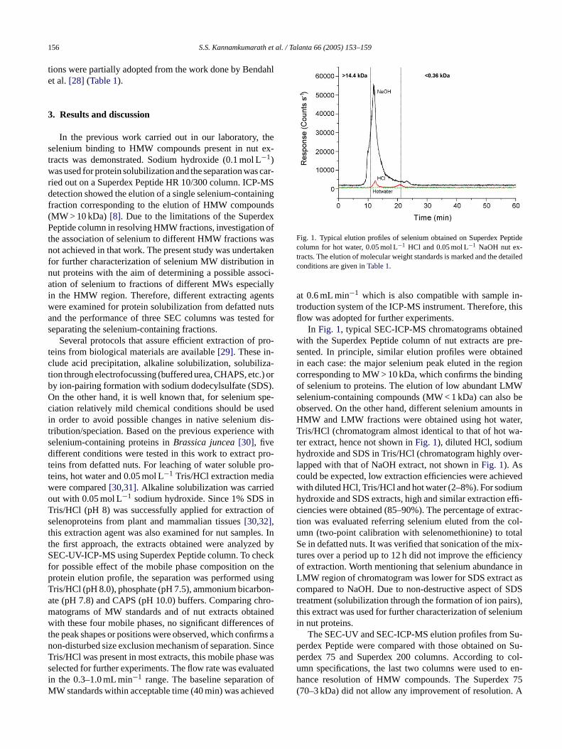
Fig. 1. Typical elution profiles of selenium obtained on Superdex Peptidecolumn for hot water, 0.05 mol L−1 HCl and 0.05 mol L−1 NaOH nut ex-tracts. The elution of molecular weight standards is marked and the detailedconditions are given inTable 1.
at 0.6 mL min−1 which is also compatible with sample in-troduction system of the ICP-MS instrument. Therefore, thisflow was adopted for further experiments.
In Fig. 1, typical SEC-ICP-MS chromatograms obtainedwith the Superdex Peptide column of nut extracts are pre-sented. In principle, similar elution profiles were obtainedin each case: the major selenium peak eluted in the regioncorresponding to MW > 10 kDa, which confirms the bindingof selenium to proteins. The elution of low abundant LMWselenium-containing compounds (MW < 1 kDa) can also beobserved. On the other hand, different selenium amounts inHMW and LMW fractions were obtained using hot water,Tris/HCl (chromatogram almost identical to that of hot wa-ter extract, hence not shown inFig. 1), diluted HCl, sodiumhydroxide and SDS in Tris/HCl (chromatogram highly over-lapped with that of NaOH extract, not shown inFig. 1). Ascould be expected, low extraction efficiencies were achievedwith diluted HCl, Tris/HCl and hot water (2–8%). For sodiumhydroxide and SDS extracts, high and similar extraction effi-ciencies were obtained (85–90%). The percentage of extrac-tion was evaluated referring selenium eluted from the col-umn (two-point calibration with selenomethionine) to totalSe in defatted nuts. It was verified that sonication of the mix-tures over a period up to 12 h did not improve the efficiencyof extraction. Worth mentioning that selenium abundance inL t asc SDSt irs),t niumi
u-p n Su-p col-u en-h 75( . A
eins from biological materials are available[29]. These inlude acid precipitation, alkaline solubilization, solubiliion through electrofocussing (buffered urea, CHAPS, etcy ion-pairing formation with sodium dodecylsulfate (SDn the other hand, it is well known that, for selenium siation relatively mild chemical conditions should be un order to avoid possible changes in native seleniumribution/speciation. Based on the previous experienceelenium-containing proteins inBrassica juncea[30], fiveifferent conditions were tested in this work to extract p
eins from defatted nuts. For leaching of water solubleeins, hot water and 0.05 mol L−1 Tris/HCl extraction mediere compared[30,31]. Alkaline solubilization was carrieut with 0.05 mol L−1 sodium hydroxide. Since 1% SDSris/HCl (pH 8) was successfully applied for extractionelenoproteins from plant and mammalian tissues[30,32],his extraction agent was also examined for nut samplehe first approach, the extracts obtained were analyzeEC-UV-ICP-MS using Superdex Peptide column. To ch
or possible effect of the mobile phase composition onrotein elution profile, the separation was performed uris/HCl (pH 8.0), phosphate (pH 7.5), ammonium bicarbte (pH 7.8) and CAPS (pH 10.0) buffers. Comparing catograms of MW standards and of nut extracts obtaith these four mobile phases, no significant difference
he peak shapes or positions were observed, which confion-disturbed size exclusion mechanism of separation.ris/HCl was present in most extracts, this mobile phaseelected for further experiments. The flow rate was evalun the 0.3–1.0 mL min−1 range. The baseline separation
W standards within acceptable time (40 min) was achie
MW region of chromatogram was lower for SDS extracompared to NaOH. Due to non-destructive aspect ofreatment (solubilization through the formation of ion pahis extract was used for further characterization of selen nut proteins.
The SEC-UV and SEC-ICP-MS elution profiles from Serdex Peptide were compared with those obtained oerdex 75 and Superdex 200 columns. According tomn specifications, the last two columns were used toance resolution of HMW compounds. The Superdex70–3 kDa) did not allow any improvement of resolution
S.S. Kannamkumarath et al. / Talanta 66 (2005) 153–159 157
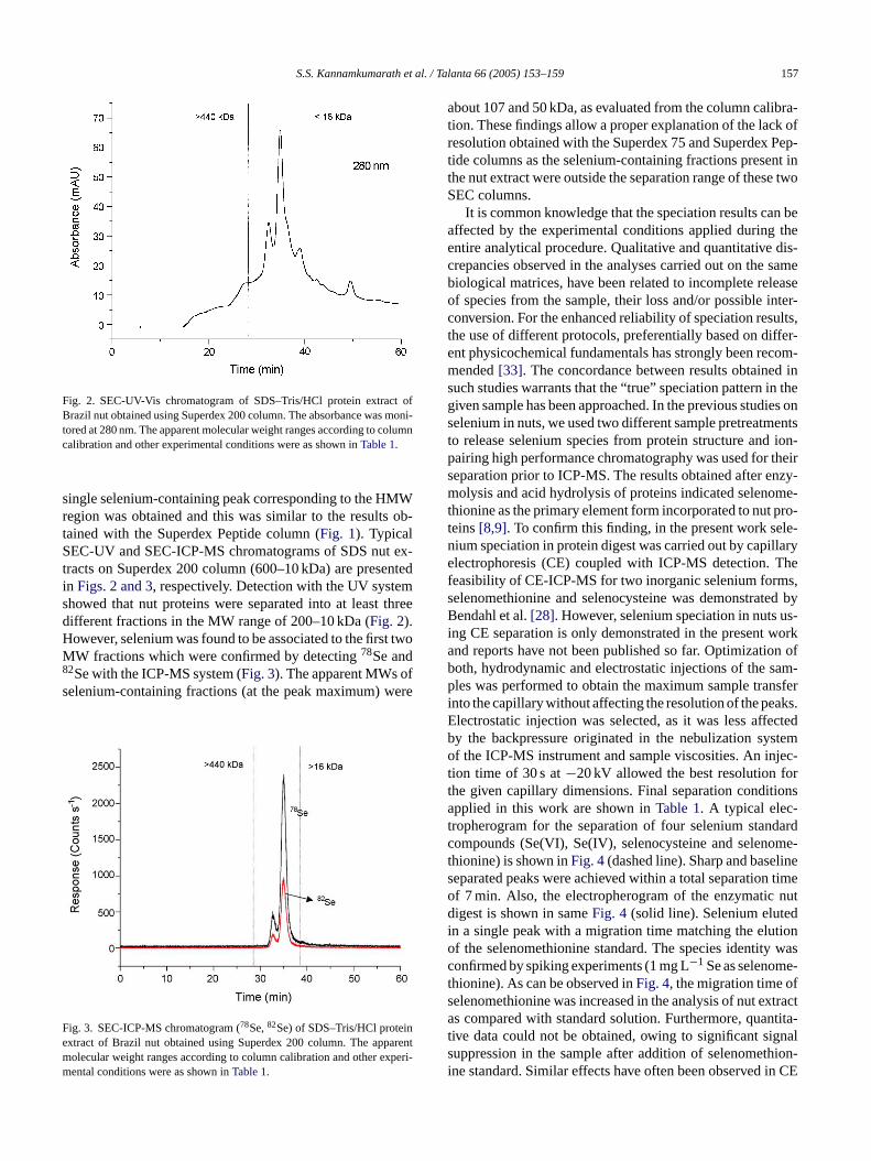
Fig. 2. SEC-UV-Vis chromatogram of SDS–Tris/HCl protein extract ofBrazil nut obtained using Superdex 200 column. The absorbance was moni-tored at 280 nm. The apparent molecular weight ranges according to columncalibration and other experimental conditions were as shown inTable 1.
single selenium-containing peak corresponding to the HMWregion was obtained and this was similar to the results ob-tained with the Superdex Peptide column (Fig. 1). TypicalSEC-UV and SEC-ICP-MS chromatograms of SDS nut ex-tracts on Superdex 200 column (600–10 kDa) are presentedin Figs. 2 and 3, respectively. Detection with the UV systemshowed that nut proteins were separated into at least threedifferent fractions in the MW range of 200–10 kDa (Fig. 2).However, selenium was found to be associated to the first twoMW fractions which were confirmed by detecting78Se and82Se with the ICP-MS system (Fig. 3). The apparent MWs ofselenium-containing fractions (at the peak maximum) were
F ne arentm xperi-m
about 107 and 50 kDa, as evaluated from the column calibra-tion. These findings allow a proper explanation of the lack ofresolution obtained with the Superdex 75 and Superdex Pep-tide columns as the selenium-containing fractions present inthe nut extract were outside the separation range of these twoSEC columns.
It is common knowledge that the speciation results can beaffected by the experimental conditions applied during theentire analytical procedure. Qualitative and quantitative dis-crepancies observed in the analyses carried out on the samebiological matrices, have been related to incomplete releaseof species from the sample, their loss and/or possible inter-conversion. For the enhanced reliability of speciation results,the use of different protocols, preferentially based on differ-ent physicochemical fundamentals has strongly been recom-mended[33]. The concordance between results obtained insuch studies warrants that the “true” speciation pattern in thegiven sample has been approached. In the previous studies onselenium in nuts, we used two different sample pretreatmentsto release selenium species from protein structure and ion-pairing high performance chromatography was used for theirseparation prior to ICP-MS. The results obtained after enzy-molysis and acid hydrolysis of proteins indicated selenome-thionine as the primary element form incorporated to nut pro-teins[8,9]. To confirm this finding, in the present work sele-n llarye Thef s,s ed byB us-i worka n ofb am-p nsferi ks.E ctedb stemo jec-t ort ionsat dardc ome-t lines timeo nutd di tiono wasc e-t fs xtracta ntita-t nals hion-i in CE
ig. 3. SEC-ICP-MS chromatogram (78Se,82Se) of SDS–Tris/HCl proteixtract of Brazil nut obtained using Superdex 200 column. The appolecular weight ranges according to column calibration and other eental conditions were as shown inTable 1.
ium speciation in protein digest was carried out by capilectrophoresis (CE) coupled with ICP-MS detection.
easibility of CE-ICP-MS for two inorganic selenium formelenomethionine and selenocysteine was demonstratendahl et al.[28]. However, selenium speciation in nuts
ng CE separation is only demonstrated in the presentnd reports have not been published so far. Optimizatiooth, hydrodynamic and electrostatic injections of the sles was performed to obtain the maximum sample tra
nto the capillary without affecting the resolution of the pealectrostatic injection was selected, as it was less affey the backpressure originated in the nebulization syf the ICP-MS instrument and sample viscosities. An in
ion time of 30 s at−20 kV allowed the best resolution fhe given capillary dimensions. Final separation conditpplied in this work are shown inTable 1. A typical elec-
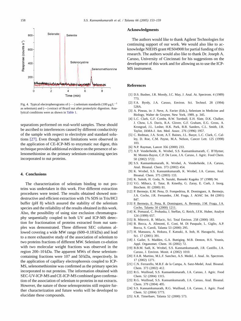
ropherogram for the separation of four selenium stanompounds (Se(VI), Se(IV), selenocysteine and selenhionine) is shown inFig. 4(dashed line). Sharp and baseeparated peaks were achieved within a total separationf 7 min. Also, the electropherogram of the enzymaticigest is shown in sameFig. 4 (solid line). Selenium elute
n a single peak with a migration time matching the eluf the selenomethionine standard. The species identityonfirmed by spiking experiments (1 mg L−1 Se as selenomhionine). As can be observed inFig. 4, the migration time oelenomethionine was increased in the analysis of nut es compared with standard solution. Furthermore, qua
ive data could not be obtained, owing to significant siguppression in the sample after addition of selenometne standard. Similar effects have often been observed
158 S.S. Kannamkumarath et al. / Talanta 66 (2005) 153–159
Fig. 4. Typical electropherograms of (- - -) selenium standards (100�g L−1
as selenium) and (—) extract of Brazil nut after proteolytic digestion. Ana-lytical conditions were as shown inTable 1.
separations performed on real-world samples. These shouldbe ascribed to interferences caused by different conductivityof the sample with respect to electrolyte and standard solu-tions [27]. Even though some limitations were observed inthe application of CE-ICP-MS to enzymatic nut digest, thistechnique provided additional evidence on the presence of se-lenomethionine as the primary selenium-containing speciesincorporated to nut proteins.
4. Conclusions
The characterization of selenium binding to nut pro-teins was undertaken in this work. Five different extractionprocedures were tested. The results obtained showed non-destructive and efficient extraction with 1% SDS in Tris/HClbuffer (pH 8) which assured the stability of the seleniumspecies and the reliability of the results obtained in this work.Also, the possibility of using size exclusion chromatogra-phy sequentially coupled to both UV and ICP-MS detec-tion for fractionation of proteins extracted from nut sam-ples was demonstrated. Three different SEC columns al-lowed covering a wide MW range (600–0.18 kDa) and leadto a more exhaustive study of the association of selenium totwo proteins fractions of different MW. Selenium co-elutionw ther ium-c y. Int CP-M eciesi withS ma-t cts.H fur-t d toe
Acknowledgments
The authors would like to thank Agilent Technologies forcontinuing support of our work. We would also like to ac-knowledge NIEHS grant #ES04908 for partial funding of thisresearch. The authors would also like to thank Dr. Joseph A.Caruso, University of Cincinnati for his suggestions on thedevelopment of this work and for allowing us to use the ICP-MS instrument.
References
[1] D.S. Bushee, J.R. Moody, J.C. May, J. Anal. At. Spectrom. 4 (1989)773.
[2] F.A. Byrdy, J.A. Caruso, Environ. Sci. Technol. 28 (1994)528A.
[3] A. Pineau, in: J. Neve, A. Favier (Eds.), Selenium in Medicine andBiology, Walter de Gruyter, New York, 1989, p. 345.
[4] L.C. Clark, G.F. Combs, B.W. Turnbull, E.H. Slate, D.K. Chalker,J. Chow, L.S. Davis, R.A. Glover, G.F. Graham, E.G. Gross, A.Krongrad, J.L. Lesher, H.K. Park, B.B. Sanders, C.L. Smith, J.R.Taylor, JAMA-J. Am. Med. Assoc. 276 (1996) 1957.
[5] C. Redman, J.A. Scott, A.T. Baines, J.L. Basye, L.C. Clark, C. Cal-ley, D. Roe, C.M. Payne, M.A. Nelson, Cancer Lett. 125 (1998)103.
[6] N.P. Rayman, Lancet 356 (2000) 233.er,
hem.
uso,
nal.
[[ org.
[ ejo,001)
[ J.A.
[ alyst
[[ .P.
[ al.
[ nis,
[ J.A.
[ om.
[ nal.
[ ood
[ nal.
[ ood
[
ith two molecular weight fractions was observed inegion 200–10 kDa. The apparent MWs of these selenontaining fractions were 107 and 50 kDa, respectivelhe application of capillary electrophoresis coupled to IS, selenomethionine was confirmed as the primary sp
ncorporated to nut proteins. The information obtainedEC-UV-ICP-MS and CE-ICP-MS combined gave confir
ion of the association of selenium to proteins in nut extraowever, the nature of those selenoproteins still require
her characterization and future works will be developelucidate these compounds.
[7] A.P. Vonderheide, K. Wrobel, S.S. Kannamkumarath, C. B’HymM. Montes-Bayon, C.P. De Leon, J.A. Caruso, J. Agric. Food C50 (2002) 5722.
[8] S.S. Kannamkumarath, K. Wrobel, A. Vonderheide, J.A. CarAnal. Bioanal. Chem. 373 (2002) 454.
[9] K. Wrobel, S.S. Kannamkumarath, K. Wrobel, J.A. Caruso, ABioanal. Chem. 375 (2003) 133.
10] K. Saitoh, H. Goda, N. Suzuki, Bunseki Kagaku 37 (1988) 94.11] V.G. Mihucz, E. Tatar, B. Kmethy, G. Zaray, E. Cseh, J. In
Biochem. 81 (2000) 81.12] P. Bermejo, E.M. Pena, D. Fompedrina, R. Dominguez, A. Berm
J.A. Cocho, J.R. Fernandez, J.M. Fraga, J. AOAC Int. 84 (2847.
13] P. Bermejo, E. Pena, R. Dominguez, A. Bermejo, J.M. Fraga,Cocho, Talanta 50 (2000) 1211.
14] K. Pomazal, C. Prohaska, I. Steffan, G. Reich, J.F.K. Huber, An124 (1999) 657.
15] B. Mitrovic, R. Milacic, Sci. Total Environ. 258 (2000) 183.16] B. Bocca, A. Alimonti, E. Coni, M. Di Pasquale, L. Giglio, A
Bocca, S. Caroli, Talanta 53 (2000) 295.17] H. Matsuura, A. Hokura, F. Katsuki, A. Itoh, H. Haraguchi, An
Sci. 17 (2001) 391.18] J. Gailer, S. Madden, G.A. Buttigieg, M.B. Denton, H.S. You
Appl. Organomet. Chem. 16 (2002) 72.19] B.B.M. Sadi, K. Wrobel, S.S. Kannamkumarath, J.R. Castillo,
Caruso, J. Environ. Monit. 4 (2002) 1010.20] F.A.R. Martino, M.L.F. Sanchez, A.S. Medel, J. Anal. At. Spectr
17 (2002) 1271.21] C.N. Ferrarello, M.R.F. de la Campa, A. Sanz-Medel, Anal. Bioa
Chem. 373 (2002) 412.22] R.G. Wuilloud, S.S. Kannamkumarath, J.A. Caruso, J. Agric. F
Chem. 52 (2004) 1315.23] R.G. Wuilloud, S.S. Kannamkumarath, J.A. Caruso, Anal. Bioa
Chem. 379 (2004) 495.24] S.S. Kannamkumarath, R.G. Wuilloud, J.A. Caruso, J. Agric. F
Chem. 52 (2004) 5773.25] A.R. Timerbaev, Talanta 52 (2000) 573.

S.S. Kannamkumarath et al. / Talanta 66 (2005) 153–159 159
[26] J.W. Olesik, in: J.A. Caruso, K.L. Sutton, K.L. Ackley (Eds.), Ele-mental Speciation, New Approaches for Trace Elemental Analysis,Comprehensive Analytical Chemistry XXIII, Elsevier, New York,2000, p. 151.
[27] S.S. Kannamkumarath, K. Wrobel, K. Wrobel, C. B’Hymer, J.A.Caruso, J. Chromatogr. A 975 (2002) 245.
[28] L. Bendahl, B. Gammelgaard, O. Jons, O. Farver, S.H. Hansen, J.Anal. At. Spectrom. 16 (2001) 38.
[29] C. Damerval, M. Zivy, F. Granier, D.D. Vienne, Advances in Elec-trophoresis, VCH, Weinheim, Germany, 1988.
[30] S. Mounicou, J. Meija, J.A. Caruso, Analyst 129 (2004) 116.[31] H. Chassaigne, C.C. Chery, G. Bordin, A.R. Rodriguez, J. Chro-
matogr. A 976 (2002) 409.[32] M.A. Beilstein, M.J. Tripp, P.D. Whanger, J. Inorg. Biochem. 15
(1981) 339.[33] A. Sanz-Medel, Analyst 120 (1995) 799.


