Embed Size (px)
Citation preview
Superficial and Shroud-like coloration of linen by shortlaser pulses in the vacuum ultraviolet
Paolo Di Lazzaro,1,* Daniele Murra,1 Enrico Nichelatti,2
Antonino Santoni,1 and Giuseppe Baldacchini3
1ENEA Research Center of Frascati, Department Applications of Radiation, P.O. Box 65, Frascati 00044, Italy2ENEA Research Center of Casaccia, Department Optical Components Development,
via Anguillarese 301, Rome 00123, Italy3Present address: via G. Quattrucci 246, Grottaferrata 00046, Italy
*Corresponding author: [email protected]
Received 23 July 2012; revised 19 October 2012; accepted 20 October 2012;posted 22 October 2012 (Doc. ID 173160); published 14 December 2012
We present a survey on five years of experiments of excimer laser irradiation of linen fabrics, seeking acoloration mechanism able to reproduce the microscopic complexity of the body image embedded onto theShroud of Turin. We achieved a superficial, Shroud-like coloration in a narrow range of irradiation para-meters. We also obtained latent coloration that appears after artificial or natural aging of linen followinglaser irradiations that, at first, did not generate any visible effect. Most importantly, we have recognizedphotochemical processes that account for both coloration and latent coloration. © 2012 Optical Societyof AmericaOCIS codes: 140.2180, 140.3390, 300.6280, 350.6670.
1. Introduction
The front and back images of a scourged man, barelyvisible on the linen cloth of the Shroud of Turin (seeFig. 1) possess particular physical and chemical char-acteristics [1] such that nobody was yet able to createan image identical in all its microscopic details, asdiscussed in a number of papers [2–21].
The inability to replicate the image on the Shroudmakes it impossible to formulate a reliable hypoth-esis on how the body image was made. As a partialjustification, scientists complain the Shroud hasbeen seldom accessible. Indeed, the most recent in-depth experimental analysis of the images on theShroud was carried out in 1978 by the multidisciplin-ary team of the Shroud of Turin Research Project(STURP). They used the most advanced instrumentsavailable at that time, which were supplied by
various manufacturers, having a commercial valueof over two million dollars. The Shroud was examinedby ultraviolet (UV), visible, and infrared spectrome-try, x-ray fluorescence spectrometry, microscopy,thermography, pyrolysis mass spectrometry, laser-microprobe Raman analyses, microchemical testing,and fiber sampling [3–13]. These analyses did notfind pigments or artist’s media on the Shroud, exceptfor some iron oxide particles, micrometer-size cinna-bar, and small traces of vermilion (HgS) [13]. How-ever, there is a large body of scientific evidence[8,11,12,16] that the microscopic observations re-ported in [13] cannot support the Shroud image isa painting. It is likely themicroscopic debris particleshave been transferred by contact of pigments fromartist’s copies of the Shroud that have been “sancti-fied” by pressing the two images together [17].
After years of exhaustive study and data evalua-tion, the STURP team achieved the following results.
(a) X-ray, fluorescence, and microchemistry re-sults preclude the possibility of paint being used
1559-128X/12/368567-12$15.00/0© 2012 Optical Society of America
20 December 2012 / Vol. 51, No. 36 / APPLIED OPTICS 8567
as a method for creating the image [7,8,11,12]. UVand infrared evaluation confirm these studies[3,5,6,10]. The body image is not painted or printed.
(b) Both kinetics studies and fluorescence mea-surements support the image was formed by alow-temperature process. In fact, the temperaturewas not high enough to change cellulose within thetime available for image formation, and no char wasproduced [12,14]. As a consequence, the body imagewas not made by a heated bas-relief.
(c) The Shroud’s image is superficial as the colorresides on the outer surface of the fibers that makeup the threads of the cloth [8,11]. Recent measure-ments on image fibers of the Shroud [19] confirmedthat the coloration depth is approximately 200 nm,which corresponds to the thickness of the primarycell wall of the linen fiber [22]. In a single linenthread, there are some 200 fibers.
(d) The colored (image) fibers are brittle, show“corroded” surfaces, and are more fragile than unco-lored fibers [8,11]. When illuminated by UV light of alamp, image fibers emit a reduced fluorescent lightcompared with fibers out of the image [3].
(e) The coloration of the Shroud image wasformed by an unknown process that caused oxida-tion, dehydration, and conjugation of polysaccharidestructure of fibers to produce a conjugated carbonylgroup as the chromophore [8,11,12]. To some extent,the color is a result of an accelerated aging process ofthe flax.
(f) The image seen at the macroscopic level is anareal density image. This means shading is notaccomplished by varying the color but by varyingthe number of colored fibers per unit area at themicroscopic level [5,9,11,12].
(g) The blood tests positive for human blood, andthere is no image on the Shroud beneath bloodstains[12,23]. UV illumination allows observation of typi-cal fluorescence of bilirubin around the main blood-stains [10], which would be consistent with ahemolytic process caused by torture. Independentanalyses of forensic pathologist Baima Bollone con-firmed STURP’s findings [24].
(h) The image fading has three-dimensional(3-D) information of the body encoded in it [14].
AllattemptstocreateaShroud-likeimagehavefailedto reproduce adequately the above characteristics.Some researchers have obtained coloration/imagesthat look similar [5,18,20,21], but no one has createdimages that match all microscopic and macroscopiccharacteristics of the Shroud image. The answer tothe question of how the image was produced or whatproduced the image is still unknown. This is the mainpoint of the “mystery of the Shroud.” Regardless ofthe age of the Shroud, either medieval (1260–1390)as dated by radiocarbon test [25,26] or older as itresults from other measurements [27], and whateverthe importance of historical documents about theexistence of the Shroud before 1260 [28,29], the mostimportant question remains the same: how did theimage of a man get on the Shroud?
In this paper we approach this unsolved problem,summarizing the main results of experiments car-ried out at the ENEA Frascati Research Centreaimed at identifying the physical and chemical pro-cesses able to generate a Shroud-like coloration.
2. Radiative Hypothesis
The results of STURP measurements, briefly sum-marized in Section 1, have important consequenceswhen seeking possible mechanisms of image forma-tion. Let us discuss some of these consequences,assuming that (according to the STURP conclusionhttp://www.shroud.com/78conclu.htm) the Shroudcovered a man when the body image and blood stainswere formed.
• The front and back images do not show thetypical deformations of a 3-D body put in contact witha two-dimensional cloth. As a consequence, we de-duce that the image was not formed by contact withthe body. This observation suggests the Shroud wasloosely draping the body, that is, it was not wrappedaround and in full contact with the whole body. Thismode is also supported by the gradations of imagebrightness that correlate with expected cloth–bodydistances: in fact, there is a geometrical relationshipthat corresponds to a body shape and a cloth drapingnaturally over that shape [14]. Moreover, this “drap-ing mode” is consistent with the lack of image of thebody’s sides. These considerations, combined withthe extreme shallowness of the image color, the ab-sence of pigments, and the subliminal, microscopiccomplexity of image at the fiber level, make unlikelyobtaining a Shroud-like image by contact methods(i.e., by chemicals), either in a modern laboratory[20], or a fortiori by amedieval forger. The hypothesisof the medieval forger is also ruled out by the anato-mical consistency of blood and serum versus wounds,including the presence of bilirubin, which is invisibleto the naked eye. This subliminal feature is onlyvisible by UV fluorescence photography [10] andrequires knowledge of anatomy and of forensic med-icine [30] not available in the Middle Ages.
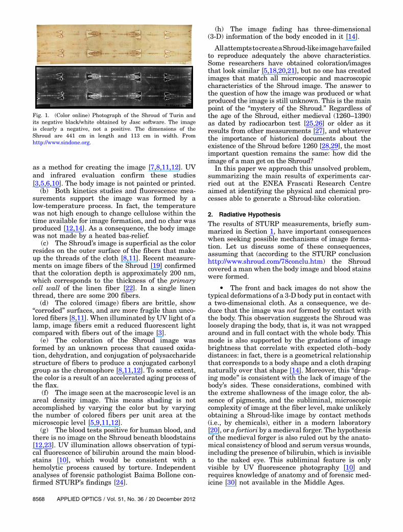
Fig. 1. (Color online) Photograph of the Shroud of Turin andits negative black/white obtained by Jasc software. The imageis clearly a negative, not a positive. The dimensions of theShroud are 441 cm in length and 113 cm in width. Fromhttp://www.sindone.org.
8568 APPLIED OPTICS / Vol. 51, No. 36 / 20 December 2012
• Under the blood there is no image. Thismeans that the blood stains occurred physically onthe Shroud before the body image [8,12]. As a conse-quence, the image was formed after the deposition ofthe corpse. Moreover, we observe that all the bloodstains have sharp outlines and are flawless, and thisposes a question if the corpse was removed fromthe cloth.
• On the Shroud there are no signs of putrefac-tions, which occur at the orifices about 40 h afterdeath. This means that the image does not dependon putrefaction gases and the corpse was wrappedin the Shroud not longer than two days.
In order to satisfy the conditions posed by these ex-perimental observations, some papers [14,15,18,19]have suggested that an electromagnetic energy inci-dent on a linen fabric could reproduce the main char-acteristics of the Shroud image, namely the absenceof pigments, the shallowness of the coloration, theimage in areas not in contact with the body, the gra-dient of the color, and the absence of image under theblood stains.
The first attempt to reproduce a Shroud-like colora-tion by electromagnetic energy used a CO2 laser emit-ting infrared radiation (wavelength λ � 10.6 μm).Theseexperimentsproducedanimageonalinenfabricsimilar to the Shroud at macroscopic level [21]. How-ever, microscopic analyses showed a bulky colorationandmany fibers carbonized, which are not compatiblewith theShroud image [1,12]. In fact, theCO2 infraredradiation excites the vibrational energy levels of theirradiated material, with consequent release of ther-mal energy, which heats the linen threads in the bulkof the fabric. On the contrary, it is well known that theenergy carried by short-wavelength radiation breaksthe chemical bonds of the irradiated material withoutinducing a significant heating (photochemical reac-tion). Moreover, linen has a molar absorptivity, whichincreases when decreasing the radiation wavelength.
Consequently, thesmaller thewavelength, thethinnerthe material necessary to absorb all the radiation.Therefore, we have chosen the UV radiation to obtainat least two of the main characteristics of the Shroudimage: a thin coloration depth and a low-temperatureimage formation.
3. Experimental Results by UV Laser Radiation
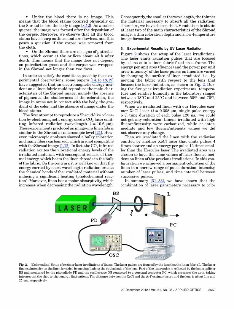
Figure 2 shows the setup of the laser irradiations.The laser emits radiation pulses that are focusedby a lens onto a linen fabric fixed on a frame. Theenergy per unit area (fluence) and the power per unitarea (intensity) of the laser pulses on linen are variedby changing the surface of linen irradiated, i.e., bymoving the fabric with respect to the lens thatfocuses the laser radiation, as shown in Fig. 2. Dur-ing the five year irradiation experiments, tempera-ture and relative humidity in the laboratory rangedbetween 18°C and 25°C and between 50% and 70%,respectively.
When we irradiated linen with our Hercules exci-mer XeCl laser (λ � 0.308 μm, single pulse energy5 J, time duration of each pulse 120 ns), we couldnot get any coloration. Linens irradiated with highfluence/intensity were carbonized, while at inter-mediate and low fluence/intensity values we didnot observe any change.
Then we irradiated the linen with the radiationemitted by another XeCl laser that emits pulses 4times shorter and an energy per pulse 12 times smal-ler than the Hercules laser. The irradiated area waschosen to have the same values of laser fluence inci-dent on linen of the previous irradiations. In this con-figuration we achieved a permanent coloration of thelinen in a narrow range of pulse duration, intensity,number of laser pulses, and time interval betweensuccessive pulses.
In summary [31–33], we have shown that thecombination of laser parameters necessary to color
Fig. 2. (Color online) Setup of excimer laser irradiations of linens. The laser pulses are focused by the lens ℓ on the linen fabric L. The laserfluence/intensity on the linen is varied bymoving L along the optical axis of the lens. Part of the laser pulse is reflected by the beam splitterBS and monitored by the photodiode PD and the oscilloscope OS connected to a personal computer PC, which processes the data, takinginto account the shot-to-shot energy fluctuations. The distance between the XeCl and the ArF excimer lasers and the lens is about 1 m and25 cm, respectively.
20 December 2012 / Vol. 51, No. 36 / APPLIED OPTICS 8569
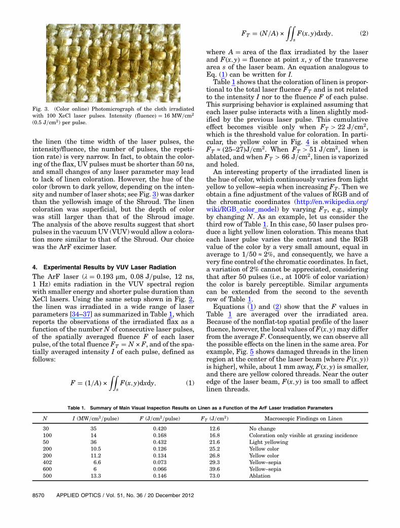
the linen (the time width of the laser pulses, theintensity/fluence, the number of pulses, the repeti-tion rate) is very narrow. In fact, to obtain the color-ing of the flax, UV pulses must be shorter than 50 ns,and small changes of any laser parameter may leadto lack of linen coloration. However, the hue of thecolor (brown to dark yellow, depending on the inten-sity and number of laser shots; see Fig. 3) was darkerthan the yellowish image of the Shroud. The linencoloration was superficial, but the depth of colorwas still larger than that of the Shroud image.The analysis of the above results suggest that shortpulses in the vacuumUV (VUV) would allow a colora-tion more similar to that of the Shroud. Our choicewas the ArF excimer laser.
4. Experimental Results by VUV Laser Radiation
The ArF laser (λ � 0.193 μm, 0.08 J=pulse, 12 ns,1 Hz) emits radiation in the VUV spectral regionwith smaller energy and shorter pulse duration thanXeCl lasers. Using the same setup shown in Fig. 2,the linen was irradiated in a wide range of laserparameters [34–37] as summarized in Table 1, whichreports the observations of the irradiated flax as afunction of the number N of consecutive laser pulses,of the spatially averaged fluence F of each laserpulse, of the total fluence FT � N × F, and of the spa-tially averaged intensity I of each pulse, defined asfollows:
F � �1=A� ×ZZ
sF�x; y�dxdy; (1)
FT � �N=A� ×ZZ
sF�x; y�dxdy; (2)
where A � area of the flax irradiated by the laserand F�x; y� � fluence at point x, y of the transversearea s of the laser beam. An equation analogous toEq. (1) can be written for I.
Table 1 shows that the coloration of linen is propor-tional to the total laser fluence FT and is not relatedto the intensity I nor to the fluence F of each pulse.This surprising behavior is explained assuming thateach laser pulse interacts with a linen slightly mod-ified by the previous laser pulse. This cumulativeeffect becomes visible only when FT > 22 J=cm2,which is the threshold value for coloration. In parti-cular, the yellow color in Fig. 4 is obtained whenFT ≈ �25–27�J=cm2. When FT > 51 J=cm2, linen isablated, and when FT > 66 J=cm2, linen is vaporizedand holed.
An interesting property of the irradiated linen isthe hue of color, which continuously varies from lightyellow to yellow–sepia when increasing FT. Then weobtain a fine adjustment of the values of RGB and ofthe chromatic coordinates (http://en.wikipedia.org/wiki/RGB_color_model) by varying FT, e.g., simplyby changing N. As an example, let us consider thethird row of Table 1. In this case, 50 laser pulses pro-duce a light yellow linen coloration. This means thateach laser pulse varies the contrast and the RGBvalue of the color by a very small amount, equal inaverage to 1=50 ≈ 2%, and consequently, we have avery fine control of the chromatic coordinates. In fact,a variation of 2% cannot be appreciated, consideringthat after 50 pulses (i.e., at 100% of color variation)the color is barely perceptible. Similar argumentscan be extended from the second to the seventhrow of Table 1.
Equations (1) and (2) show that the F values inTable 1 are averaged over the irradiated area.Because of the nonflat-top spatial profile of the laserfluence, however, the local values of F�x; y�may differfrom the average F. Consequently, we can observe allthe possible effects on the linen in the same area. Forexample, Fig. 5 shows damaged threads in the linenregion at the center of the laser beam [where F�x; y�)is higher], while, about 1 mm away, F�x; y� is smaller,and there are yellow colored threads. Near the outeredge of the laser beam, F�x; y� is too small to affectlinen threads.
Fig. 3. (Color online) Photomicrograph of the cloth irradiatedwith 100 XeCl laser pulses. Intensity �fluence� � 16 MW=cm2
(0.5 J=cm2) per pulse.
Table 1. Summary of Main Visual Inspection Results on Linen as a Function of the ArF Laser Irradiation Parameters
N I (MW=cm2=pulse) F (J=cm2=pulse) FT (J=cm2) Macroscopic Findings on Linen
30 35 0.420 12.6 No change100 14 0.168 16.8 Coloration only visible at grazing incidence50 36 0.432 21.6 Light yellowing200 10.5 0.126 25.2 Yellow color200 11.2 0.134 26.8 Yellow color402 6.6 0.073 29.3 Yellow–sepia600 6 0.066 39.6 Yellow–sepia500 13.3 0.146 73.0 Ablation
8570 APPLIED OPTICS / Vol. 51, No. 36 / 20 December 2012
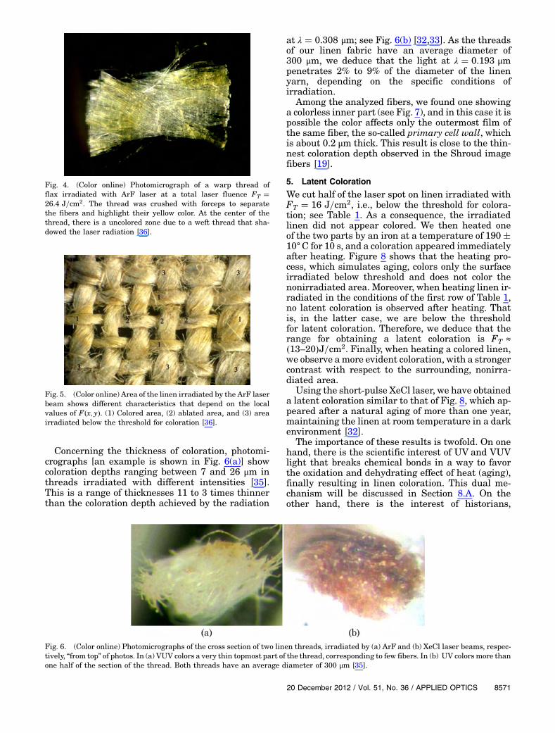
Concerning the thickness of coloration, photomi-crographs [an example is shown in Fig. 6(a)] showcoloration depths ranging between 7 and 26 μm inthreads irradiated with different intensities [35].This is a range of thicknesses 11 to 3 times thinnerthan the coloration depth achieved by the radiation
at λ � 0.308 μm; see Fig. 6(b) [32,33]. As the threadsof our linen fabric have an average diameter of300 μm, we deduce that the light at λ � 0.193 μmpenetrates 2% to 9% of the diameter of the linenyarn, depending on the specific conditions ofirradiation.
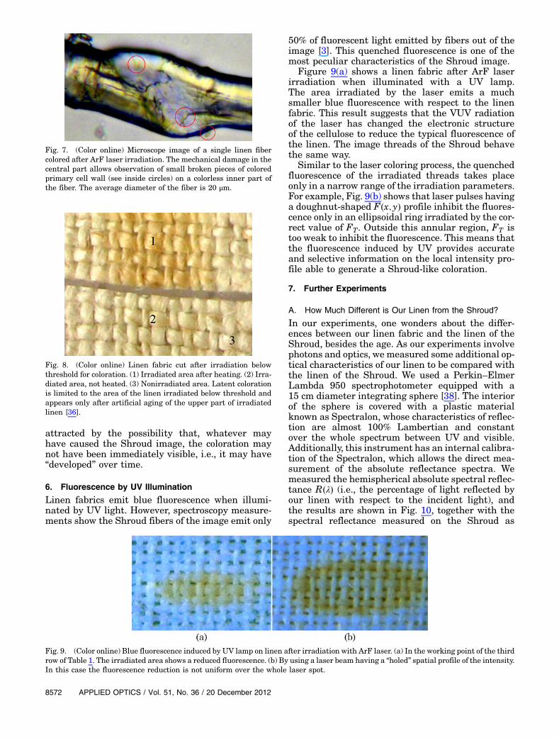
Among the analyzed fibers, we found one showinga colorless inner part (see Fig. 7), and in this case it ispossible the color affects only the outermost film ofthe same fiber, the so-called primary cell wall, whichis about 0.2 μm thick. This result is close to the thin-nest coloration depth observed in the Shroud imagefibers [19].
5. Latent Coloration
We cut half of the laser spot on linen irradiated withFT � 16 J=cm2, i.e., below the threshold for colora-tion; see Table 1. As a consequence, the irradiatedlinen did not appear colored. We then heated oneof the two parts by an iron at a temperature of 190�10°C for 10 s, and a coloration appeared immediatelyafter heating. Figure 8 shows that the heating pro-cess, which simulates aging, colors only the surfaceirradiated below threshold and does not color thenonirradiated area. Moreover, when heating linen ir-radiated in the conditions of the first row of Table 1,no latent coloration is observed after heating. Thatis, in the latter case, we are below the thresholdfor latent coloration. Therefore, we deduce that therange for obtaining a latent coloration is FT ≈
�13–20�J=cm2. Finally, when heating a colored linen,we observe amore evident coloration, with a strongercontrast with respect to the surrounding, nonirra-diated area.
Using the short-pulse XeCl laser, we have obtaineda latent coloration similar to that of Fig. 8, which ap-peared after a natural aging of more than one year,maintaining the linen at room temperature in a darkenvironment [32].
The importance of these results is twofold. On onehand, there is the scientific interest of UV and VUVlight that breaks chemical bonds in a way to favorthe oxidation and dehydrating effect of heat (aging),finally resulting in linen coloration. This dual me-chanism will be discussed in Section 8.A. On theother hand, there is the interest of historians,
Fig. 4. (Color online) Photomicrograph of a warp thread offlax irradiated with ArF laser at a total laser fluence FT �26.4 J=cm2. The thread was crushed with forceps to separatethe fibers and highlight their yellow color. At the center of thethread, there is a uncolored zone due to a weft thread that sha-dowed the laser radiation [36].
Fig. 5. (Color online) Area of the linen irradiated by the ArF laserbeam shows different characteristics that depend on the localvalues of F�x; y�. (1) Colored area, (2) ablated area, and (3) areairradiated below the threshold for coloration [36].
Fig. 6. (Color online) Photomicrographs of the cross section of two linen threads, irradiated by (a) ArF and (b) XeCl laser beams, respec-tively, “from top” of photos. In (a) VUV colors a very thin topmost part of the thread, corresponding to few fibers. In (b) UV colors more thanone half of the section of the thread. Both threads have an average diameter of 300 μm [35].
20 December 2012 / Vol. 51, No. 36 / APPLIED OPTICS 8571
attracted by the possibility that, whatever mayhave caused the Shroud image, the coloration maynot have been immediately visible, i.e., it may have“developed” over time.
6. Fluorescence by UV Illumination
Linen fabrics emit blue fluorescence when illumi-nated by UV light. However, spectroscopy measure-ments show the Shroud fibers of the image emit only
50% of fluorescent light emitted by fibers out of theimage [3]. This quenched fluorescence is one of themost peculiar characteristics of the Shroud image.
Figure 9(a) shows a linen fabric after ArF laserirradiation when illuminated with a UV lamp.The area irradiated by the laser emits a muchsmaller blue fluorescence with respect to the linenfabric. This result suggests that the VUV radiationof the laser has changed the electronic structureof the cellulose to reduce the typical fluorescence ofthe linen. The image threads of the Shroud behavethe same way.
Similar to the laser coloring process, the quenchedfluorescence of the irradiated threads takes placeonly in a narrow range of the irradiation parameters.For example, Fig. 9(b) shows that laser pulses havinga doughnut-shaped F�x; y� profile inhibit the fluores-cence only in an ellipsoidal ring irradiated by the cor-rect value of FT. Outside this annular region, FT istoo weak to inhibit the fluorescence. This means thatthe fluorescence induced by UV provides accurateand selective information on the local intensity pro-file able to generate a Shroud-like coloration.
7. Further Experiments
A. How Much Different is Our Linen from the Shroud?
In our experiments, one wonders about the differ-ences between our linen fabric and the linen of theShroud, besides the age. As our experiments involvephotons and optics, we measured some additional op-tical characteristics of our linen to be compared withthe linen of the Shroud. We used a Perkin–ElmerLambda 950 spectrophotometer equipped with a15 cm diameter integrating sphere [38]. The interiorof the sphere is covered with a plastic materialknown as Spectralon, whose characteristics of reflec-tion are almost 100% Lambertian and constantover the whole spectrum between UV and visible.Additionally, this instrument has an internal calibra-tion of the Spectralon, which allows the direct mea-surement of the absolute reflectance spectra. Wemeasured the hemispherical absolute spectral reflec-tance R�λ� (i.e., the percentage of light reflected byour linen with respect to the incident light), andthe results are shown in Fig. 10, together with thespectral reflectance measured on the Shroud as
Fig. 7. (Color online) Microscope image of a single linen fibercolored after ArF laser irradiation. The mechanical damage in thecentral part allows observation of small broken pieces of coloredprimary cell wall (see inside circles) on a colorless inner part ofthe fiber. The average diameter of the fiber is 20 μm.
Fig. 8. (Color online) Linen fabric cut after irradiation belowthreshold for coloration. (1) Irradiated area after heating. (2) Irra-diated area, not heated. (3) Nonirradiated area. Latent colorationis limited to the area of the linen irradiated below threshold andappears only after artificial aging of the upper part of irradiatedlinen [36].
Fig. 9. (Color online) Blue fluorescence induced by UV lamp on linen after irradiation with ArF laser. (a) In the working point of the thirdrow of Table 1. The irradiated area shows a reduced fluorescence. (b) By using a laser beam having a “holed” spatial profile of the intensity.In this case the fluorescence reduction is not uniform over the whole laser spot.
8572 APPLIED OPTICS / Vol. 51, No. 36 / 20 December 2012
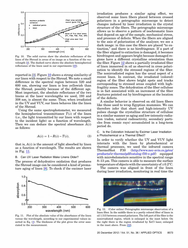
reported in [3]. Figure 10 shows a strong similarity ofour linen with respect to the Shroud. We note a smalldifference in the spectral region between 520 and600 nm, showing our linen is less yellowish thanthe Shroud, possibly because of the different age.Most important, the absolute reflectance of the twolinens at the laser wavelengths we used, 193 and308 nm, is almost the same. Thus, when irradiatedin the UV and VUV, our linen behaves like the linenof the Shroud.
Using the same spectrophotometer, we measuredthe hemispherical transmittance T�λ� of the linen(i.e., the light transmitted by our linen with respectto the incident light) as a function of wavelength.Then we can deduce the spectral absorbance A�λ�as follows:
A�λ� � 1 − R�λ� − T�λ�; (3)
that is, A�λ� is the amount of light absorbed by linenas a function of wavelength. The results are shownin Fig. 11.
B. Can UV Laser Radiation Make Linens Older?
The process of dehydrative oxidation that producesthe Shroud image can be considered a kind of prema-ture aging of linen [8]. To check if the excimer laser
irradiation produces a similar aging effect, weobserved some linen fibers placed between crossedpolarizers in a petrographic microscope to detectchanges induced by laser irradiation in the crystalstructure of the fibers. The petrographic microscopeallows us to observe a pattern of isochromatic linesthat depend on age of the sample, mechanical stress,and presence of defects. When the fibers are alignedto the axis of polarization of the analyzer, we see adark image: in this case the fibers are placed “to ex-tinction,” and there is no birefringence. If a part ofthe fiber aligned to extinction is damaged, it becomesbirefringent and appears bright because damaged re-gions have a different crystalline orientation thanthe fiber. Figure 12 shows a partially irradiated fiberof linen immersed in oil as observed in cross polari-zation to identify the stressed regions of the fiber.The nonirradiated region has the usual aspect of arecent linen. In contrast, the irradiated (colored)region of the fiber shows bright spots and trackscorresponding to stressed areas coupled with high-fragility zones. The dehydration of the fiber celluloseis in fact associated with an increment of the fiberfractures pointed out by birefringence at the locationof the defects.
A similar behavior is observed on old linen fiberslike those used to wrap Egyptian mummies. We cantherefore infer that short and high-intensity UVpulses change the crystalline structure of cellulosein a similar manner as aging and low-intensity radia-tion (radon, natural radioactivity, secondary parti-cles from cosmic rays) accumulated in a long-termperiod do.
C. Is the Coloration Induced by Excimer Laser Irradiationa Photochemical or a Thermal Effect?
In order to verify whether the UV and VUV lightinteracts with the linen by photochemical orthermal processes, we used the infrared cameraThermoShot F30 (http://www.nec‑avio.co.jp/en/products/ir‑thermo/pdf/catalog‑f30‑e.pdf) equippedwith microbolometers sensitive in the spectral range8–13 μm. This camera is able to measure the surfacetemperatureofobjectswith theuncertaintyof�0.2°C.
The camera was aligned in front of the linenduring laser irradiation, monitoring in real time the
Fig. 10. The solid curves show the absolute reflectance of thelinen of the Shroud in areas of no image as a function of the wa-velength [3]. The dashed curve shows the absolute hemisphericalreflectance of the linen used in our experiments [35].
Fig. 11. Plot of the absolute value of the absorbance of the linenversus the wavelength, according to our experimental values in-serted in Eq. (3). The thickness of the plot gives the error asso-ciated to the measurement.
Fig. 12. (Color online) Petrographic microscope observation of alinen fiber. In the middle there is a partly colored fiber of linen inoil 1.515 between crossed polarizers. The left part of the fiber is thenonirradiated region, which is enlarged in the inset below. Onthe right there is the region irradiated by XeCl laser, enlargedin the inset above. From [32].
20 December 2012 / Vol. 51, No. 36 / APPLIED OPTICS 8573
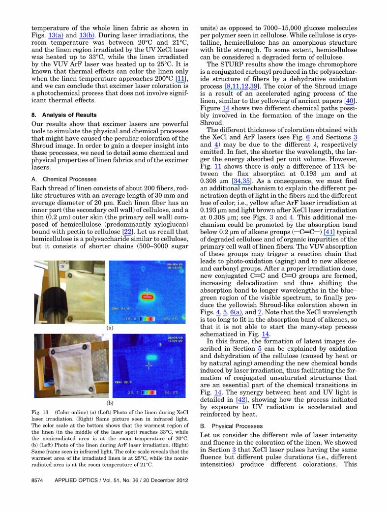
temperature of the whole linen fabric as shown inFigs. 13(a) and 13(b). During laser irradiations, theroom temperature was between 20°C and 21°C,and the linen region irradiated by the UV XeCl laserwas heated up to 33°C, while the linen irradiatedby the VUV ArF laser was heated up to 25°C. It isknown that thermal effects can color the linen onlywhen the linen temperature approaches 200°C [11],and we can conclude that excimer laser coloration isa photochemical process that does not involve signif-icant thermal effects.
8. Analysis of Results
Our results show that excimer lasers are powerfultools to simulate the physical and chemical processesthat might have caused the peculiar coloration of theShroud image. In order to gain a deeper insight intothese processes, we need to detail some chemical andphysical properties of linen fabrics and of the excimerlasers.
A. Chemical Processes
Each thread of linen consists of about 200 fibers, rod-like structures with an average length of 30 mm andaverage diameter of 20 μm. Each linen fiber has aninner part (the secondary cell wall) of cellulose, and athin (0.2 μm) outer skin (the primary cell wall) com-posed of hemicellulose (predominantly xyloglucan)bound with pectin to cellulose [22]. Let us recall thathemicellulose is a polysaccharide similar to cellulose,but it consists of shorter chains (500–3000 sugar
units) as opposed to 7000–15,000 glucose moleculesper polymer seen in cellulose. While cellulose is crys-talline, hemicellulose has an amorphous structurewith little strength. To some extent, hemicellulosecan be considered a degraded form of cellulose.
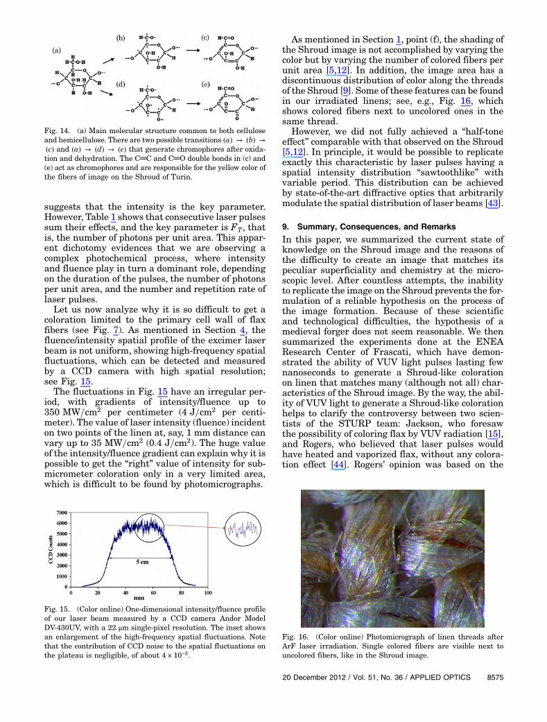
The STURP results show the image chromophoreis a conjugated carbonyl produced in the polysacchar-ide structure of fibers by a dehydrative oxidationprocess [8,11,12,39]. The color of the Shroud imageis a result of an accelerated aging process of thelinen, similar to the yellowing of ancient papers [40].Figure 14 shows two different chemical paths possi-bly involved in the formation of the image on theShroud.
The different thickness of coloration obtained withthe XeCl and ArF lasers (see Fig. 6 and Sections 3and 4) may be due to the different λ, respectivelyemitted. In fact, the shorter the wavelength, the lar-ger the energy absorbed per unit volume. However,Fig. 11 shows there is only a difference of 11% be-tween the flax absorption at 0.193 μm and at0.308 μm [34,35]. As a consequence, we must findan additional mechanism to explain the different pe-netration depth of light in the fibers and the differenthue of color, i.e., yellow after ArF laser irradiation at0.193 μmand light brown after XeCl laser irradiationat 0.308 μm; see Figs. 3 and 4. This additional me-chanism could be promoted by the absorption bandbelow 0.2 μm of alkene groups (─C═C─) [41] typicalof degraded cellulose and of organic impurities of theprimary cell wall of linen fibers. The VUVabsorptionof these groups may trigger a reaction chain thatleads to photo-oxidation (aging) and to new alkenesand carbonyl groups. After a proper irradiation dose,new conjugated C═C and C═O groups are formed,increasing delocalization and thus shifting theabsorption band to longer wavelengths in the blue–green region of the visible spectrum, to finally pro-duce the yellowish Shroud-like coloration shown inFigs. 4, 5, 6(a), and 7. Note that the XeCl wavelengthis too long to fit in the absorption band of alkenes, sothat it is not able to start the many-step processschematized in Fig. 14.
In this frame, the formation of latent images de-scribed in Section 5 can be explained by oxidationand dehydration of the cellulose (caused by heat orby natural aging) amending the new chemical bondsinduced by laser irradiation, thus facilitating the for-mation of conjugated unsaturated structures thatare an essential part of the chemical transitions inFig. 14. The synergy between heat and UV light isdetailed in [42], showing how the process initiatedby exposure to UV radiation is accelerated andreinforced by heat.
B. Physical Processes
Let us consider the different role of laser intensityand fluence in the coloration of the linen. We showedin Section 3 that XeCl laser pulses having the samefluence but different pulse durations (i.e., differentintensities) produce different colorations. This
Fig. 13. (Color online) (a) (Left) Photo of the linen during XeCllaser irradiation. (Right) Same picture seen in infrared light.The color scale at the bottom shows that the warmest region ofthe linen (in the middle of the laser spot) reaches 33°C, whilethe nonirradiated area is at the room temperature of 20°C.(b) (Left) Photo of the linen during ArF laser irradiation. (Right)Same frame seen in infrared light. The color scale reveals that thewarmest area of the irradiated linen is at 25°C, while the nonir-radiated area is at the room temperature of 21°C.
8574 APPLIED OPTICS / Vol. 51, No. 36 / 20 December 2012
suggests that the intensity is the key parameter.However, Table 1 shows that consecutive laser pulsessum their effects, and the key parameter is FT, thatis, the number of photons per unit area. This appar-ent dichotomy evidences that we are observing acomplex photochemical process, where intensityand fluence play in turn a dominant role, dependingon the duration of the pulses, the number of photonsper unit area, and the number and repetition rate oflaser pulses.
Let us now analyze why it is so difficult to get acoloration limited to the primary cell wall of flaxfibers (see Fig. 7). As mentioned in Section 4, thefluence/intensity spatial profile of the excimer laserbeam is not uniform, showing high-frequency spatialfluctuations, which can be detected and measuredby a CCD camera with high spatial resolution;see Fig. 15.
The fluctuations in Fig. 15 have an irregular per-iod, with gradients of intensity/fluence up to350 MW=cm2 per centimeter (4 J=cm2 per centi-meter). The value of laser intensity (fluence) incidenton two points of the linen at, say, 1 mm distance canvary up to 35 MW=cm2 (0.4 J=cm2). The huge valueof the intensity/fluence gradient can explain why it ispossible to get the “right” value of intensity for sub-micrometer coloration only in a very limited area,which is difficult to be found by photomicrographs.
As mentioned in Section 1, point (f), the shading ofthe Shroud image is not accomplished by varying thecolor but by varying the number of colored fibers perunit area [5,12]. In addition, the image area has adiscontinuous distribution of color along the threadsof the Shroud [9]. Some of these features can be foundin our irradiated linens; see, e.g., Fig. 16, whichshows colored fibers next to uncolored ones in thesame thread.
However, we did not fully achieved a “half-toneeffect” comparable with that observed on the Shroud[5,12]. In principle, it would be possible to replicateexactly this characteristic by laser pulses having aspatial intensity distribution “sawtoothlike” withvariable period. This distribution can be achievedby state-of-the-art diffractive optics that arbitrarilymodulate the spatial distribution of laser beams [43].
9. Summary, Consequences, and Remarks
In this paper, we summarized the current state ofknowledge on the Shroud image and the reasons ofthe difficulty to create an image that matches itspeculiar superficiality and chemistry at the micro-scopic level. After countless attempts, the inabilityto replicate the image on the Shroud prevents the for-mulation of a reliable hypothesis on the process ofthe image formation. Because of these scientificand technological difficulties, the hypothesis of amedieval forger does not seem reasonable. We thensummarized the experiments done at the ENEAResearch Center of Frascati, which have demon-strated the ability of VUV light pulses lasting fewnanoseconds to generate a Shroud-like colorationon linen that matches many (although not all) char-acteristics of the Shroud image. By the way, the abil-ity of VUV light to generate a Shroud-like colorationhelps to clarify the controversy between two scien-tists of the STURP team: Jackson, who foresawthe possibility of coloring flax by VUV radiation [15],and Rogers, who believed that laser pulses wouldhave heated and vaporized flax, without any colora-tion effect [44]. Rogers’ opinion was based on the
Fig. 14. (a) Main molecular structure common to both celluloseand hemicellulose. There are two possible transitions �a� → �b� →
�c� and �a� → �d� → �e� that generate chromophores after oxida-tion and dehydration. The C═C and C═O double bonds in (c) and(e) act as chromophores and are responsible for the yellow color ofthe fibers of image on the Shroud of Turin.
Fig. 15. (Color online) One-dimensional intensity/fluence profileof our laser beam measured by a CCD camera Andor ModelDV-430UV, with a 22 μm single-pixel resolution. The inset showsan enlargement of the high-frequency spatial fluctuations. Notethat the contribution of CCD noise to the spatial fluctuations onthe plateau is negligible, of about 4 × 10⁻³.
Fig. 16. (Color online) Photomicrograph of linen threads afterArF laser irradiation. Single colored fibers are visible next touncolored fibers, like in the Shroud image.
20 December 2012 / Vol. 51, No. 36 / APPLIED OPTICS 8575
failure of experiments made at Los Alamos usingexcimer lasers, but our results demonstrate thattheir failure was due to parameters (e.g., laser pulsewidth) outside the narrow range of values able togenerate the permanent linen coloration.
Let us summarize in the following the main resultswe achieved.
I. We obtained a linen coloration only in anarrow range of laser parameters. In particular, thetemporal duration of the single laser pulse must beshorter than 50 ns [31,32]. While a short laser pulsewidth is necessary for obtaining a coloration, theVUV laser spectrum allows a coloration limited tothe outer surface of the threads.
II. The most interesting results were obtainedwith VUV light. The permanent linen coloration isa threshold effect, i.e., the color is obtained only whenFT > 22 J=cm2; see Table 1. When the FT value isabove threshold, the linen is ablated and/or vapor-ized, while when FT < 13 J=cm2 the linen does notchange color. Even when FT is in the colorationrange, not all the irradiated fibers are colored (Figs. 5and 16) due to the spatial fluctuations of energy den-sity of the laser pulses shown in Fig. 15.III. We triggered a photochemical coloration pro-
cess. In fact, the thermal heating associated with UVand VUV radiation is within a few degrees centi-grade and therefore irrelevant for the purpose of col-oring by scorching linens; see Fig. 13. This result fitswith the “cold” coloration process of the Shroudestimated in [8,11,12].
IV. The hue of color depends on the wavelengthand on the numberN of laser pulses, which is propor-tional to FT. Irradiations at 0.308 μm generate abrownish coloration, while the 0.193 μm photons pro-duce a yellow color (see Fig. 4) comparable to the col-or of the Shroud image. In both cases, the contrastslowly increases with the number of laser pulses,allowing an accurate control of the RGB value byvarying FT.
V. The different hue of color obtained by UVand VUV radiation is due to different chains of photo-chemical reactions respectively triggered. In particu-lar, the VUV radiation at 0.193 μm is absorbed byalkene groups in degraded cellulose, whose numberincreases with FT , thus inducing a photolysis of thecellulose, which promotes the formation of chromo-phores; see Fig. 14. These chromophores determinethe yellow coloration of the fibers [8,12,39–41].VI. We observed an irradiated fiber whose colora-
tion was possibly confined in the primary cell wall[35,36], which is comparable with the thinnest col-oration depth observed in the fibers image of theShroud of Turin [8,11,19].VII. Aging can be a concause of linen coloration. Infact, after laser irradiations that, at first, do not gen-erate a visible coloration of linen, a latent colorationappears either by artificial (Fig. 8) or natural aging oflinen [32,35]. Latent coloration is interesting for thesynergy between UV, oxidation, and the dehydratingeffect of heat (or of aging), which triggers the
coloration process, and also for historians, attractedby the possibility that, whatever may have causedthe Shroud image, the coloration may have devel-oped over time.VIII. The partial inhibition of fluorescence inducedby VUV laser radiation (Fig. 9) is an additional fea-ture of our coloration similar to the Shroud image.The induced fluorescence is also capable to selec-tively recognize the uniformity of FT incident onlinen; see Figs. 9(a) and 9(b).IX. Both UV and VUV radiations coloring linen
are compatible with the absence of image underthe bloodstains on the Shroud because in this spec-tral region light is absorbed by very thin layers ofblood hemoglobin. According to [45] the UV lightmay be responsible for another special feature of theShroud: the red color of blood stains after so muchtime since their deposition.
X. Using a petrographic microscope, we haveobserved some defects induced by UV radiation inthe structure of irradiated linen fibers (see Fig. 12),similar to very old linen fabrics [11,46].XI. Absolute reflectance measurements show
that, when irradiated in the UV and VUV, our linenbehaves like the linen of the Shroud; see Fig. 10.
In summary, our results demonstrate that a shortand intense burst of directional VUV radiation cancolor a linen cloth so as to reproduce many of thepeculiar characteristics of the image on the Shroudof Turin, including the hue of color, the shallow pe-netration depth of the color, and the inhibition offluorescence.
The Shroud image has characteristics that wehave been able to reproduce only in part, for examplethe gross shading structure that is determined by theratio of yellow to uncolored fibers in a given area; seepoint (f) in Section 1 and Fig. 16. As discussed inSection 8.A, sophisticated diffractive optics could re-plicate these features, but this effort is far beyondour intention. In fact, our purpose was not to demon-strate that a battery of 10,000 lasers can accuratelyreproduce the image on the Shroud. Our main pur-pose was to perform accurate and reproducibleexperiments apt to understand the physical and che-mical mechanisms that might have played a role inthe generation of the Shroud body image, using apowerful and versatile tool such as the laser, regard-less of the source of energy that may have caused thisimage. In this frame, our experimental data can behelpful to scholars seeking a linen coloration by cor-ona discharge [18] or electrostatic discharge andradon emitted during seismic events [47], which in-volve UV and VUV light but are difficult to controland characterize.
This is not the conclusion; we are composing piecesof a fascinating and complex scientific puzzle. Theenigma of the body image of the Shroud of Turinis still “a challenge to our intelligence” [48].
The authors thank Professor Giulio Fanti(University of Padua) for photomicrographs of fibers,
8576 APPLIED OPTICS / Vol. 51, No. 36 / 20 December 2012
for photos of Fig. 13, and for useful discussions aboutthe manuscript contents.
References1. G. Fanti, J. A. Botella, F. Crosilla, F. Lattarulo, N. Svensson,
R. Schneider, and A. Wanger, “List of evidences of the TurinShroud,” in Proceedings of the International Workshop on theScientific Approch to the Acheiropoietos Images, P. Di Lazzaro,ed. (ENEA, 2010), pp. 67–75, http://www.acheiropoietos.info/proceedings/proceedings.php.
2. B. J. Culliton, “The mystery of the Shroud challenges20th-century science,” Science 201, 235–239 (1978).
3. R. Gilbert and M. Gilbert, “Ultraviolet visible reflectance andfluorescence spectra of the Shroud of Turin,” Appl. Opt. 19,1930–1936 (1980).
4. E. J. Jumper and W. Mottern, “Scientific investigation of theShroud of Turin,” Appl. Opt. 19, 1909–1912 (1980).
5. S. F. Pellicori, “Spectral properties of the Shroud of Turin,”Appl. Opt. 19, 1913–1920 (1980).
6. J. S. Accetta and J. S. Baumgart, “Infrared reflectance spectro-scopy and thermographic investigations of the Shroud ofTurin,” Appl. Opt. 19, 1921–1929 (1980).
7. R. A. Morris, L. A. Schwalbe, and J. R. London, “X-rayfluorescence investigation on the Shroud of Turin,” X-RaySpectrom. 9, 40–47 (1980).
8. J. H. Heller and A. D. Adler, “A chemical investigation of theShroud of Turin,” Can. Soc. Forensic Sci. J. 14, 81–103 (1981).
9. S. F. Pellicori and M. S. Evans, “The Shroud of Turin throughthe microscope,” Archaeology 34, 34–43 (1981).
10. V. D. Miller and S. F. Pellicori, “Ultraviolet fluorescence photo-graphy of the Shroud of Turin,” J. Biol. Photogr. 49, 71–85(1981).
11. L. A. Schwalbe and R. N. Rogers, “Physics and chemistry ofthe Shroud of Turin, a summary of the 1978 investigation,”Anal. Chim. Acta 135, 3–49 (1982).
12. E. J. Jumper, A. D. Adler, J. P. Jackson, S. F. Pellicori, J. H.Heller, and J. R. Druzik, “A comprehensive examination ofthe various stains and images on the Shroud of Turin,” inArchaeological Chemistry III, Vol. 205 of ACS Advances inChemistry Series (American Chemical Society, 1984),pp. 447–476.
13. W. C. McCrone and C. Skirius, “Light microscopical study ofthe Shroud of Turin,” Microscope 28, 105–111 (1980).
14. J. P. Jackson, E. J. Jumper, and W. R. Ercoline, “Correlation ofimage intensity on the Turin Shroud with the 3-D structure ofa human body shape,” Appl. Opt. 23, 2244–2270 (1984).
15. J. P. Jackson, “Is the image on the Shroud due to a processheretofore unknown to modern science?,” Shroud SpectrumInt. 34, 3–29 (1990).
16. T. Heimburger, “A detailed critical review of the chemical stu-dies on the Turin Shroud: facts and interpretations,” (2001),http://www.shroud.com/pdfs/thibault%20final%2001.pdf.
17. I. Piczek, “Is the Shroud of Turin a painting?,” (1995), re-trieved on 10 October 2012, http://www.shroud.com/piczek.htm.
18. G. Fanti, “Can corona discharge explain the body image of theTurin Shroud?,” J. Imaging Sci. Technol. 54, 020508 (2010).
19. G. Fanti, J. Botella, P. Di Lazzaro, R. Schneider, andN. Svensson, “Microscopic and macroscopic characteristicsof the Shroud of Turin image superficiality,” J. Imaging Sci.Technol. 54, 040201 (2010).
20. L. Garlaschelli, “Life-size reproduction of the Shroud ofTurin and its image,” J. Imaging Sci. Technol. 54, 040301(2010).
21. F. Ferrero, F. Testore, C. Tonin, and R. Innocenti, “Surfacedegradation of linen textiles induced by laser treatment,”AUTEX Res. J. 2, 109–114 (2002).
22. S. Perez and K. Mazeau, Polysaccharides: Structural Diversityand Functional Versatility, S. Dumitriu, Ed. (Dekker, 2004),Chap. 2.
23. J. H. Heller and A. D. Adler, “Blood on the Shroud of Turin,”Appl. Opt. 19, 2742–2744 (1980).
24. P. L. Baima Bollone, “Indagini identificative su fili dellaSindone,” G. Accad. Med. Torino 1, 228–239 (1982).
25. P. E. Damon, D. Donahue, B. Gore, A. Hatheway, A. Jull, T.Linick, P. Sercel, L. Toolin, C. Bronk, E. Hall, R. Hedges, R.Housley, I. Law, G. Bonani, S. Trumbore, W. Woelfli,J. Ambers, S. Bowman, M. Leese, and M. Tite, “Radiocarbondating of the Shroud of Turin,” Nature 337, 611–615(1989).
26. R. Van Haelst, “A critical review of the radiocarbon dating ofthe Shroud of Turin,” inProceedings of the InternationalWork-shop on the Scientific Approach to the Acheiropoietos Images,P. Di Lazzaro, ed. (ENEA, 2010), pp. 267–273, http://www.acheiropoietos.info/proceedings/proceedings.php.
27. R. N. Rogers, “Studies on the radiocarbon sample fromthe Shroud of Turin,” Thermochim. Acta 425, 189–194(2005).
28. D. Scavone, “Documenting the Shroud’s missing years,” inProceedings of the International Workshop on the ScientificApproach to the Acheiropoietos Images, P. Di Lazzaro, ed.(ENEA, 2010), pp. 87–94, http://www.acheiropoietos.info/proceedings/proceedings.php.
29. A. Nicolotti, I Templari e la Sindone. Storia di un falso(Salerno, 2011), Chaps. 1, 3, and 4.
30. N. Svensson, “Medical and forensic aspects on the mandepicted on the Shroud of Turin,” in Proceedings of the Inter-national Workshop on the Scientific Approach to the Acheiro-poietos Images, P. Di Lazzaro, ed. (ENEA, 2010), pp. 181–186,http://www.acheiropoietos.info/proceedings/proceedings.php.
31. G. Baldacchini, P. Di Lazzaro, D. Murra, and G. Fanti, “Color-azione di tessuti di lino con laser ad eccimeri e confronto conl’immagine sindonica,” Technical Report ENEA RT/2006/70/FIM (ENEA, 2006).
32. G. Baldacchini, P. Di Lazzaro, D. Murra, and G. Fanti,“Coloring linens with excimer lasers to simulate the bodyimage of the Turin Shroud,” Appl. Opt. 47, 1278–1285(2008).
33. P. Di Lazzaro, G. Baldacchini, G. Fanti, D. Murra, and A.Santoni, “Colouring fabrics with excimer lasers to simulateencoded images: the case of the Shroud of Turin,” Proc. SPIE7131, 71311R (2009).
34. P. Di Lazzaro, G. Baldacchini, G. Fanti, D. Murra, E.Nichelatti, and A. Santoni, “A physical hypothesis on theorigin of the body image embedded into the Turin Shroud,”in Proceedings of the International Conference on the Shroudof Turin: Perspectives on a Multifaceted Enigma, G. Fanti ed.(Libreria Progetto Padova, 2009), pp. 116–125, http://www.ohioshroudconference.com/papers/p01.pdf.
35. P. Di Lazzaro, D. Murra, A. Santoni, G. Fanti, E. Nichelatti,and G. Baldacchini, “Deep ultraviolet radiation simulates theTurin Shroud image,” J. Imaging Sci. Technol. 54, 040302(2010).
36. P. Di Lazzaro, D. Murra, A. Santoni, and G. Baldacchini,“Sub-micrometer coloration depth of linens by vacuumultraviolet radiation,” in Proceedings of the InternationalWorkshop on the Scientific Approach to the AcheiropoietosImages, P. Di Lazzaro, ed. (ENEA, 2010), pp. 3–10, http://www.acheiropoietos.info/proceedings/proceedings.php.
37. P. Di Lazzaro, D. Murra, A. Santoni, and G. Baldacchini, “Col-orazione simil-sindonica di tessuti di lino tramite radiazionenel lontano ultravioletto,” Technical Report ENEA RT/2011/14/ENEA (ENEA, 2011), retrieved 10 October 2012, http://opac.bologna.enea.it:8991/RT/2011/2011_14_ENEA.pdf.
38. http://www.perkinelmer.com/CMSResources/Images/44‑74789SPC_LAMBDA1050LAMBDA950.pdf, retrieved 10 October 2012.
39. G. Novelli, “La Sindone e la scienza chimica,” in Proceedings ofthe Worldwide Congress Sindone 2000, A. Russi and E.Marinelli, eds. (Gerni, 2002), pp. 175–181.
40. A. Mosca Conte, O. Pulci, A. Knapik, J. Bagniuk, R. Del Sole, J.Lojewska, and M. Missori, “Role of cellulose oxidation in theyellowing of ancient paper,” Phys. Rev. Lett. 108, 158301(2012).
41. A. Bos, “The UV spectra of cellulose and some model com-pounds,” J. Appl. Polym. Sci. 16, 2567–2576 (1972).
42. M. Yatagai and S. H. Zeronian, “Effect of ultraviolet light andheat on the properties of cotton cellulose,” Cellulose 1,205–214 (1994).
20 December 2012 / Vol. 51, No. 36 / APPLIED OPTICS 8577
43. See, e.g., F. Gori, “Diffractive optics, an introduction,” inDiffractive Optics and Optical Microsystems, S. Martellucciand A. N. Chester, eds. (Plenum, 1997), pp. 3–23.
44. R. N. Rogers, “Testing the Jackson ‘theory’ of image forma-tion,” (2004), retrieved 10 October 2012, http://www.shroud.com/pdfs/rogers6.pdf.
45. C. Goldoni, “The Shroud of Turin and the bilirubin bloodstains,” in Proceedings of the International Conference onthe Shroud of Turin: Perspectives on a Multifaceted Enigma,G. Fanti, ed. (Libreria Progetto Padova, 2009), pp. 442–445,http://www.ohioshroudconference.com/papers/p04.pdf.
46. R. N. Rogers, “The Shroud of Turin: radiation effects, agingand image formation,” (2005), retrieved 10 October 2012,http://www.shroud.com/pdfs/rogers8.pdf.
47. G. De Liso, “Shroud-like experimental image formation dur-ing seismic activity,” in Proceedings of the International Work-shop on the Scientific Approach to the Acheiropoietos Images,P. Di Lazzaro, ed. (ENEA, 2010), pp. 11–18, http://www.acheiropoietos.info/proceedings/proceedings.php.
48. Pope John Paul II, “The Shroud is a challenge to ourintelligence,” address during the visit to Turin (24 May1998).
8578 APPLIED OPTICS / Vol. 51, No. 36 / 20 December 2012