Embed Size (px)
Citation preview

[CANCER RESEARCH 45, 5648-5655, November 1985]
Surface Expression of Tumor-associated Antigens in Primary Cultured Human
Colonie Epithelial Cells from Carcinomas, Benign Tumors, and Normal
Tissues
Eileen Friedman,1 Ann Thor, Patricia Horan Hand, and Jeffrey Schlom
Laboratory of Gastrointestinal Cancer Research, Memorial Sloan-Kettering Cancer Center, New York, New York 10021 [E. F.], and Laboratory of Tumor Immunology ano
Biology, National Cancer Institute, NIH, Bethesda, Mary/and 20205 [A. T., P. H. H., J. S.J
ABSTRACT
A new method for the analysis of the binding of monoclonalantibodies to cell surface tumor-associated antigens utilizes 1-to 2-day primary cultures of human colonie carcinomas, adeno
mas, and normal epithelial tissue. The antibodies are added tothe live cells which form monolayer epithelial patches of severalhundred cells on the surface of the Retri dish by migration in acontinuous sheet from a small expiant. These epithelial patchesare then fixed with methanol and processed in situ using theindirect immunoperoxidase assay. Three monoclonal antibodies(MAbs) prepared against membrane-enriched fractions of human
metastatic breast cancer were assayed. MAb B1.1 bound toeach of 11 benign and each of 18 malignant colonie tumorstested. MAb B6.2 displayed similar reactivity, binding to each of7 adenomas and each of 15 carcinomas assayed. Both MAbsalso bound to normal colonie epithelial cells in both the live cellstudies presented here and in earlier studies (D. Stramignoni efa/., Int. J. Cancer, 31: 543-552, 1983). MAb B72.3 bound only
to tumor cells and not to normal epithelial cells in the live cellassay. This epitope was rapidly lost in culture. B72.3 reactivityon each of two carcinomas was decreased 9- to 36-fold whenprimary culture continued for 5-6 days. B72.3 bound to each of
20 tumors (15 carcinomas, 5 adenomas) when the cells werecultured for 1 or 2 days but on only 2 of 8 tumors when the cellswere cultured for 3 to 8 days. The B72.3 epitope was morestrongly expressed on the live cells in the expiant and on thosemonolayer cells directly adjacent to the expiant than on the cellsmore towards the edges of the patch colony. This implied thatthe cell flattening which occurred when cells migrated from theexpiant may have played some role in antigen loss. A very similarfraction of primary cultured carcinoma and adenoma cells boundeach MAb, indicating that these MAbs in live cell assay do notdistinguish between benign, noninvasive colonie tumors andinvasive carcinomas. The live cell assay was compared to thestandard assay utilizing sectioned, fixed tumors. In parallel assays of eight tumors the fraction of cells reactive in the indirectimmunoperoxidase assay was consistently higher on live cellsfor each of these MAbs than on fixed tissue. Due to this greatersensitivity the live cell assay was able to detect reactive cells intwo cases which were scored as negative (<1% positive cells)in the fixed tissue assay.
' To whom requests for reprints should be addressed. Recipient of National
Cancer Institute Grant R01 CA 28822 with some additional support from NationalCancer Institute Cancer Core Grant CA-08748 to Memorial Sloan-Kettering Cancer
Center.Received 11/26/84; revised 6/28/85; accepted 8/9/85.
INTRODUCTION
Three MAbs,2 designated B1.1, B6.2, and B72.3, prepared
against membrane-enriched fractions of human metastatic car
cinoma had been found previously to react with human coloncarcinoma (1, 2) as well as breast carcinoma, using fixed, sectioned tissue for assay. The MAb B72.3 binds to a M, 220,000-400,000 glycoprotein complex. MAb B6.2 binds to a M, 90,000component, while MAb B1.1 binds to an epitope of the M,180,000 CEA glycoprotein (3). MAb B72.3 binds to formalin-fixed sections of colonie tumors but not of normal colonie epithelial tissue, while the other two antibodies have a wider range ofspecificity, binding to both neoplastic and some normal tissues(1,2).
We have developed methods for the short-term primary cultureof epithelial monolayers from normal adult human colonie tissueand from benign and malignant colonie tumors (4-6). This newmethodology has allowed us to study the expression of thesethree epitopes on the surface of live colonie epithelial cells using1- to 2-day-old primary cultures. The use of live, unpermeabilized
cells would allow us to determine unequivocally whether theMAbs recognized cell surface determinants as well as intracel-lular epitopes. In addition the screening of both benign andmalignant colonie tumors as well as normal tissue would enableus to determine when in carcinoma evolution these antigenswere first presented on the cell surface. Colon carcinomas arefirst found as foci within benign tumors, strongly implying thatcertain premalignant cells of benign tumors are the direct precursors of carcinomas (7). Screening a series of tumors allowsmeasurement of the frequency of expression of the epitopes.Use of very-short-term cultures allows the study of antigen
expression on viable cells which have not been selected bycontinuous passage in culture and thus are more representativeof the tumor in vivo than are established cell lines.
MATERIALS AND METHODS
Primary Culture of Colonie Epithelial Cells. Portions of tumors werereceived from Surgical Pathology and immediately placed into tissueculture as described previously (4,6). Normal colonie epithelial cells werecultured from colonie biopsies as described (5). Epithelial patch colonieswere initiated in a series of gelatin-coated 35-mm tissue culture dishes.
The number initiated depended on the size of the tissue received andvaried from about 10 to 30 dishes.
Assay of Colonie Cells in Primary Culture. Cells cultured in 35-mmdishes were washed three times with PBS containing 1% -/-globulin-free
fetal calf serum and then overlaid with 0.6 ml of each MAb, PBS, or, as
2The abbreviations used are: MAb, monoclonal antibody; IPX, indirect ¡mmu-noperoxidase;CEA, carcinoembryonicantigen; PBS, phosphate-bufferedsaline.
CANCER RESEARCH VOL. 45 NOVEMBER 1985
5648
on March 30, 2016. © 1985 American Association for Cancer Research.cancerres.aacrjournals.org Downloaded from

ANTIGEN EXPRESSION IN VIABLE HUMAN CELLS
an additional control, MAb MOPC-21, which was unreactive with normal
colon or colon tumor (1, 2). The Retri dishes were covered and incubatedfor this and all other incubations at room temperature. After 1 h theplates were then washed again as above and fixed in methanol. Eitherthe cells were processed immediately using the IPX assay or stored inmethanol. The stored cells were not allowed to dry out. The fixed cellswere incubated with 1.5% hydrogen peroxide in methanol and then for15 min in a 10% solution of normal horse serum diluted in PBS containing0.1% bovine serum albumin. All the following reagents for the IPX assaywere part of the ABC kit from Vector Labs (Burlingame, CA). The colonieswere then incubated for 1 h with the secondary antibody, a 1:100 dilutionof biotinylated horse anti-mouse antibody diluted in PBS with 0.1%
bovine serum albumin. The dishes were washed twice in PBS and thenreacted with the avidin DH-biotinylated horseradish peroxidase H com
plex for 30 min. After PBS wash, the cells were reacted with a 0.06%solution of diaminobenzidine (Sigma Chemical Co., St. Louis, MO) containing 0.01% hydrogen peroxide. A 10x stock solution of diaminobenzidine was stored at -20°C shielded from light. A working solution was
prepared on the day of assay. After benzidine staining the cells arewashed once with PBS and counterstained with Harris' hematoxylin,
followed by a rinse with a saturated lithium carbonate solution. Thecolonies were scored at x100. The approximate size of the epithelialpatches were noted during scoring (10-50, 50-200, 200-500, 500-1000) for the calculation of percentage of antigen-positive cells per tumor.
For RBC rosetting, the cultured cells remained viable during the entireassay. Excess MAb was removed after a 45-min incubation by threewashes with PBS containing 1% -/-globulin-free fetal calf serum (Grand
Island Biological Co., Grand Island, NY). The epithelial patches wereoverlaid with 0.6 ml of a 0.5% solution of human O-type RBC coupledwith chromium oxide to a purified rabbit anti-mouse immunoglobulin (8,9). After another 45-min room temperature incubation, the plates were
washed again as above and the colonies scored visually using a phasemicroscope at a total magnification of xlOO. The RBC formed a haloaround the flattened epithelial patch; a positive colony resembled asunflower.
Assay of Sectioned Tumors. Briefly 5-^m sections of formalin-fixed
pieces of tumor on slides were incubated with the purified IgGs of testMAbs, and the IPX assay was performed as described (10). MAbs B1.1and B6.2 were used at 2 ¿<g/200̂I/slide and B72.3 was used at 8 ^g/200 Ml/slide. Isotype identical control MOPC-21 IgG was used at both 2
and 8 ^g/200 ^I/slide.Scoring of Immunoperoxidase on Fixed Tissue. The samples were
scored by a pathologist for the presence and location of the characteristicdark reddish-brown stain. The intensity of staining (++, strongly positive;
+, positive) and the location of the stain (cytoplasmic or apical) was alsonoted.
RESULTS
Primary Culture Assay for Analysis of Binding of Monoclonal Antibodies to Human Colonie Epithelial Cells. Biopsies ofhuman colonie carcinomas, adenomas, and normal tissue werepartially digested to colonie crypts and groups of adjacent cryptsand allowed to attach to a gelatin-coated Petri dish ("Materialsand Methods"). After 1 to 2 days the epithelial cells had migrated
in a continuous sheet from the attached crypts to form a mono-layer (4, 5). Normal epithelial cells in monolayer were assayedafter 24 h because the cells readily detached after longer culturetime. Epithelial cells in monolayer culture both from benign andfrom malignant tumors were also assayed for antigen expressionafter 1 to 2 days of migration in the majority of cases. Digestionof the remaining tumors led to much cell debris after plating,probably because of low viability of the original tumor cells. Thepresence of proteolytic enzymes in the cell debris which might
degrade cell surface components led us to delay the assays untilthe expiants had been cultured in fresh media for at least 1 to 2additional days. The cells then appeared healthy and no debriswas present.
The short culture time of 1 to 2 days ensured that the majorityof cultured cells which were assayed for antigen expressionwere originally in the tissue in vivo and did not result from celldivision in vitro. A complete cell cycle for tumor cells is estimatedto be 2.5 days in vitro (4)3 and only a fraction of these cells (25-
75%, depending on the tumor) underwent DNA synthesis anddivided in this time. Therefore in the 1-2 days allowed for
epithelial patch formation only a minority of the final tumor cellpopulation resulted from cell division in vitro, while the majoritysimply migrated from the expiant. The same was true of culturednormal epithelial cells which were assayed. Only the cells in Sphase, about 5-15% of the total, divided in our culture conditions
(5).The cells assayed constituted a variable fraction of the original
tumor. The culture conditions selected for those cells able toattach as expiants and then to migrate from the expiant to forma monolayer. Only the cells in monolayer were assayed becauseonly they could be reliably counted. Cells slid out from the testtube-like crypts of the expiant in a continuous sheet. Single
epithelial cells were never observed to migrate from the expiant.Therefore each epithelial patch was a flattened section of adjacent cells from a colonie crypt. Each patch was assayed becausethere is much evidence in the literature that tumors may beheterogeneous for antigen expression and other properties. Thedata on antigen expression given in Tables 1 and 2 give boththe percentage of total cells antigen positive and the number ofcells assayed. The number of tumor cells assayed for MAbB72.3, for example, varied widely from 290 to 34,700. Thisvariation was due to two major causes: the size of the fragmentof tumor made available for study; and viability of the tumor cellsdue to variable length of time between interruption of the bloodsupply of the tumor in the operating room and its arrival in thelaboratory. Equal aliquots of digest were placed in each dish (4,5). The likelihood of expiant attachment and subsequent cellmigration could not be determined. As a result the size of thedigest was the sole criterion for determining the number ofparallel primary cultures initiated. An average of 122 ±25 (SE)cells formed a monolayer around each tumor expiant, while thenormal cell digests gave rise to smaller cultures of 49 ±9 cellsper patch.
Because the epithelial patches from both benign and malignantcolonie tumors were attached very firmly to the Petri dish, thecells easily remained attached during the incubation with monoclonal antibodies and the subsequent processing ("Materials andMethods"). Normal epithelial cells were attached less firmly;
therefore the assays were conducted with more care.The binding of the MAbs tested in this study was assayed by
two indirect assays, indirect immunoperoxidase after methanolfixation of the antigen-antibody complex to the cell surface andsheep RBC rosetting of the antigen-antibody complex on live
cells. In an initial experiment we compared these two assaysusing MAb B1.1. Three days after seeding exponentially growinghuman breast carcinoma line MCF-7 cells in 35-mm plates, B1.1
(10 ng/m\) was bound to the small colonies in parallel plates andassayed by both methods. Of 351 of the MCF colonies 45 (13%)
3E. Friedman,unpublisheddata.
CANCER RESEARCH VOL. 45 NOVEMBER 1985
5649
on March 30, 2016. © 1985 American Association for Cancer Research.cancerres.aacrjournals.org Downloaded from

Table 1
Percentage of epithelial cells antigen positive by immunoperoxidase
The number of cells assayed for response to a particular MAb or to PBS was counted under the microscope and is listed under the percentage of cells antigenpositive. Epithelial patches were formed by radial migration of an epithelial sheet from an attached expiant ("Materials and Methods"). The live cells were incubated with
the MAbs at the following concentrations unless otherwise noted in the table: MOPC-21, lgG1 control, 10 jig/ml; B1.1, 10 ^g/ml; B6.2, 10 ^g/ml; B72.3, 20 ^g/ml. The
mean ±SE for the size of the epithelial patches cultured from adenomas and carcinomas in these experiments was 122 ±25 cells. The large variation in the number ofcells assayed from different tumors was due to the variability in viability and size of the fragment of tumor tissue received from surgical pathology. All tumor tissuereceived was used, and equal aliquots of tumor digest were plated on each Retri dish as described (4). Normal tissue was not received from operative specimensbecause the normal tissue from resected colons was uniformly nonviable (5). Minute biopsies were the source of the two normal tissues examined; therefore the totalnumber of colonies obtained was much smaller than those derived from tumors and averaged, in these experiments, 49 ±9 cells (mean ±SE).
% of antigen-positive cells (no. of cellsassayed)ControlsPathology3Carcinoma
757Carcinoma
831Carcinoma
904Carcinoma
963Adenoma
825,tubularAdenoma
836,villotubularCarcinoma
750Carcinoma
794Carcinoma
840Carcinoma
843Carcinoma
778Carcinoma
839Carcinoma
842Adenoma
838,tubularAdenoma
779.villotubularMAbs
at1/10MAbs
at1/100Adenoma
813,villotubularAdenoma
804, villotubularwith focalatypiaAdenoma
810, villotubularwith in situcarcinomaAdenoma
81 1, villotubularwith in situcarcinomaNormal.
796Normal,
892Days"111111222234644557811PBSNT00
(8,625)0
(1,800)0
(20,100)0
(2,400)0
(3,200)NT6
(2,670)0
(24,200)0
(44,100)0
(2,850)0
(3,150)NT0(12,750)0
(3,350)0(350)NT0
(4.000)0
(1,100)NT0(300)MOPC0(460)0
(18,750)0
(11,100)NT0
(1,260)0
(2,800)0
14010(1.770)0
(19,675)0
(67,650)NT0
(14,950)0
(3,500)0
(10,750)0
(300)0
(1.100)0(400)0
(120)0
(8.600)0
(180)0
(120)B1.1100
(175)100
(9.600)100(45,300)100(27.900)93
(16.020)99
(2.950)100
(540)100(4,380)100(69,100)91
(43,400)100(4.100)100(18,900)100(7,600)100
(7.615)100
(5,750)86(3,160)88
(800)100
(650)100
(600)99
(5,105)100(15,850)0
(25)0(570)Test
antiseraB6.2100
(280)100(1
1.450)100(32,700)100(55,200)96
(3.290)100(1.800)100
(445)99
(4,230)B72.3at
1/10B72.3at
1/10048
(17.160)88
(68,250)B72.3at1/10B72.3
at1/100NT100(21,750)100
(3,500)94
(16,495)100
(3,800)99(510)100(3,100)NTNTNTNTNT83
(310)B72.363(415)61
(6.250)25
(32,100)26
(23.250)48(11.650)57
(510)67(290)82
(8,060)44
(5,050)33
(6,985)41
(21,870)54
(34,700)4
(22,200)4
(30.200)0
(5.000)0
(33,100)NT11
(7.500)29
(2,370)15
(925)15
(3,500)0(350)0(500)0
(1.905)0
(7,500)0
(140)0
(490)" The Dukes' stage of carcinoma is noted. Carcinomas 1001
advanced stage, Dukes' B and nonmetastatic.6 Days in primary culture before assay.c NT, not tested.
778, and 839 were Dukes' C, metastatic to the regional lymph nodes. The remainder were the next less
5650
on March 30, 2016. © 1985 American Association for Cancer Research.cancerres.aacrjournals.org Downloaded from
ANTIGEN EXPRESSION IN VIABLE HUMAN CELLS
Table 2Decreasein 872.3 antigen detection with time in culture
For details see legend to Table 1.% of antigen-positivecells
(no. of cellsassayed)ControlsPathology
Days8Carcinoma
100516Carcinoma
1001 26PBSNT*NT0(610)0(50)MOPC0
(565)0
(625)0
(1950)0
(120)Test
antiseraB1.1100
(830)91
(720)100
(1030)100
(345)B72.345
(1000)5
(1690)36
(1905)1
(1010)8Days in primary culture beforeassay.bMT nrtt tnctoH
Table 3Percentageof antigen-positiveepithelialpatches by RBCrosetting
% of antigen-positivepatches (no. ofpatches assayed)
were surrounded by complete or partial rosettes while a verysimilar proportion, 91 of 734 colonies (12%), were antibodybinding by IPX staining ("Materials and Methods"). On this test
cell both assays gave comparable results suggesting similarsensitivity.
A minimum of 250 cells by IPX or two epithelial patches byRBC rosetting had to be scored for MAb B72.3 for a tumor tobe included in the screening assays shown in Tables 1 to 3. Ifthe culture dishes exhibited a wide variability in the number ofviable epithelial cells, those with the most colonies were usedfor assay of the test MAbs, and the remaining ones were usedfor the controls. Because of the smaller number of normal colonieepithelial cells cultured (see "Reactivity of Live Human ColonieEpithelial Cells to Monoclonal Antibody B72.3") the minimum
number needed for inclusion in the tables was 50 assayed forMAb B72.3.
Reactivity of Live Colonie Epithelial Cells to MonoclonalAntibody B1.1. B1.1 binds to a CEA epitope (3). By IPX assaythe vast majority (91-100%) of malignant cells from each of 13
carcinomas and of premalignant cells from each of 8 adenomasbound this MAb on their cell surface (Tables 1 and 2). In theRBC-rosetting assay, the RBC bound to the periphery and
sometimes the upper surface of each patch. The entire patchwas scored relative to the PBS and MOPC controls, and apositive colony had RBC surrounding at least one-half of its
periphery so that it resembled a sunflower. By RBC rosetting67% of patch colonies from five carcinomas and 68% of patchcolonies from three adenomas contained B1.1-positive cells (Ta
ble 3). When normal epithelial cells from colonie biopsies werescreened, B1.1 was found on each of three samples examinedby rosetting but on neither tested by IPX. On sectioned normaltissue assayed by IPX <3 percent of normal colonie epithelialcells scored positive (10). The RBC-rosetting assay appears
more sensitive than IPX in the detection of the B1.1 epitope onnormal colonie epithelial cells, while the IPX assay was moreeffective on tumor-derived cultures.
Reactivity of Live Human Colonie Epithelial Cells to MAbB6.2. The tumors which bound B1.1 also bound B6.2. B6.2bound to cells from each of 15 carcinomas and each of 7adenomas tested in primary culture (Tables 1 to 3). Thirteen of14 tumors assayed by IPX for both B6.2 and B1.1 gave resultssimilar to within 10% and were often identical (Tables 1 and 2).
Controls Test antisera
Pathology8Carcinoma
740Carcinoma
741Carcinoma
757Carcinoma
734Carcinoma
750Adenoma
739,villotubularAdenoma
756,villousAdenoma
763,tubularNormal
769Normal
774Normal
783Days"11112112111PBS0
NCC0
NC0
20NC0
430
NC0
(7)0
(1)NTNTNTMOPC0
NC0
NC0
130
NC0
340
NC0(10)0
(4)0
(4)0
(1)0(2)B1.159
(69)46
(102)100
(19)82(119)48(27)71(24)56
(9)78(18)B72.3
at100
(4)100
(6)100(1)B6.28(174)33(58)100(18)60
(173)6(18)100
(27)25
(4)93(14)1/10NTNTNTB72.314
(146)29(42)89(27)67(171)45(33)83(18)25
(8)90(10)56(16)0
(3)0
(1)o*(1)
Carcinomas750 and 757 were Dukes' B, nonmetastatic; carcinoma 734 wasmetastatic to the regional lymph nodes and thus Dukes' C; the remainingcarcinomas were the least invasiveclass, Dukes' A.
b Days in primary culture before assay.c NC, not counted; NT, not tested.
872.3 tested at 4 times the normal concentration. At least 2 tumor patcheswere assayed(about 250 cells) for uniformity with the minimumscoring in Table 1.
In these tumors at least 88% of the cells were reactive to eitherMAb, strongly suggesting that the same cells were expressingboth antigens. B6.2 bound to the only normal colonie biopsytested using IPX assay on live cells and to 2 of 11 samples ofnormal tissue after fixation, sectioning, and IPX (10).
Reactivity of Live Human Colonie Epithelial Cells to Monoclonal Antibody B72.3. The pattern of reactivity of B72.3 oncolonie cells differed from those of B1.1 and B6.2. While thelatter bound to both normal and tumor cells, B72.3 under theconditions of our assay system bound only to tumor cells. Normallive human colonie cells did not bind B72.3 at levels measurableby the assays used. Neither of two cultured biopsies by IPX andnone of three by RBC rosetting reacted to B72.3 (Tables 1 to3). Possibly a lower concentration of B72.3 epitopes was presenton normal cells than on tumor cells, and it was below the levelof detection with the amount of MAb used for screening. However, a 4-fold higher concentration of MAb (Table 3) detected no
reactivity. Each normal epithelial patch culture averaged only 49±9 cells. The number of normal cells assayed was lower thanthe number of tumor cells tested in the screening studies. Viablecells could not be cultured from surgical specimens of normal
CANCER RESEARCH VOL. 45 NOVEMBER 1985
5651
on March 30, 2016. © 1985 American Association for Cancer Research.cancerres.aacrjournals.org Downloaded from

ANTIGEN EXPRESSION IN VIABLE HUMAN CELLS
colon, but only from very minute colonie biopsies (5). These aretiny scrapings of the epithelial layer, much smaller than the tumorspecimens, which may account for the lower yield of culturedcells.
A second difference between the B72.3 epitope and the othertwo examined was its lability in tissue culture. Carcinoma 1005was assayed on days 1 and 5 after plating. Reactivities to MAbB1.1 on those days were, respectively, 100 and 91%, whilereactvity to B72.3 fell from 45% to 5% (Table 2). Carcinoma1001 was assayed on days 2 and 6 after plating. All of thecultured cells bound B1.1 on both days. However, binding ofB72.3 decreased markedly from 36% on day 2 to 1% on day 6.The loss of the B72.3 epitope by extended monolayer culture inthese two carcinomas explained the pattern of reactivity that wehad observed in our screening studies. B72.3 was found on eachof 15 carcinomas and 5 adenomas primary cultured for 1 to 2days, but only on 2 of 8 tumors cultured for 3 to 8 days (Tables1 to 3). The conclusion drawn from the two time course experiments and the tumor screening is that the B72.3 epitope isquickly lost when tumor cells are placed in monolayer culture,while the CEA species identified by B1.1 remains on the cellsurface.
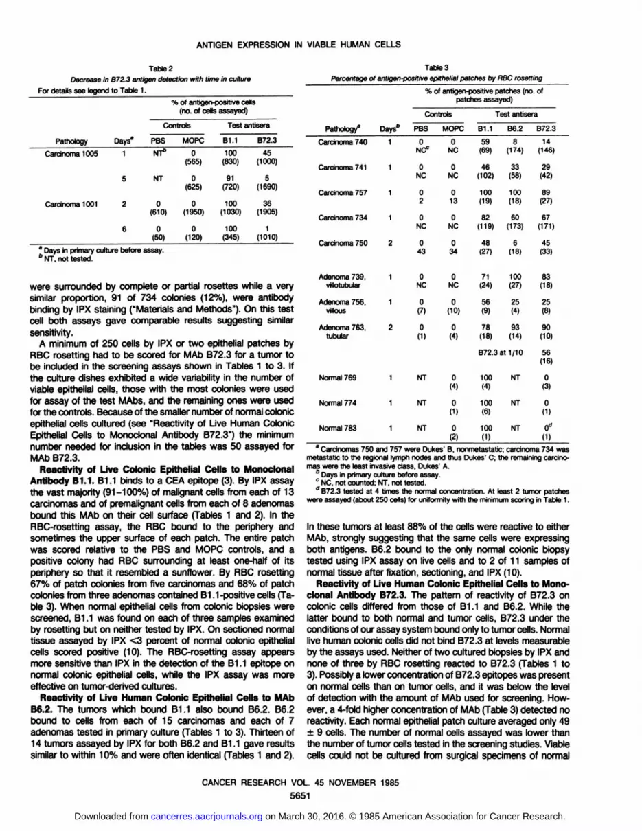
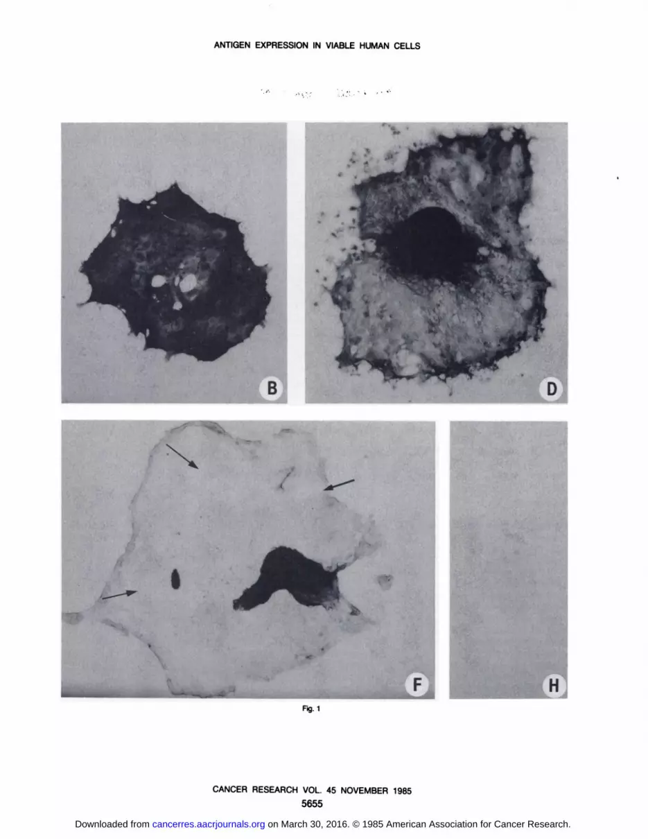
MAb B72.3 bound much more strongly to the cells in theexpiant than to the cells which had migrated onto the Retri dishto form the epithelial monolayer. Only the cells in monolayerculture were assayed for B72.3 reactivity in the screening studiesshown in Tables 1 to 3 because the cells in the expiant couldnot be counted reliably. This differential expression on cells withdifferent spatial configurations was true to some extent withB1.1 and B6.2 but was far more striking with B72.3. In Fig. 1, Dand F, primary colon carcinoma cultures were stained with B6.2and B72.3, respectively. Both MAbs react with the cells inexpiants in the center of each culture to the same extent asshown by equally dark gold benzidine staining. However, B6.2and B1.1, in Fig. 1, A and ß,reacted with the cells in themonolayer to a far greater extent than did B72.3. This lowerreactivity of MAb B72.3 was shown by its much weaker intensityof benzidine staining (Fig. 1F)- In some areas of the monolayersreacted with B72.3 no benzidine staining was seen at all. Theseare shown in Fig. 1F (arrows). The colonies in Fig. 1 (except forE) were not counterstained with hematoxylin so that the extentof benzidine staining would be clear by its intensity in black andwhite reproductions. Counterstained cultures binding MAb B72.3in Fig. 1E exhibited dark gold benzidine staining on the expiantand the monolayer cells directly around the expiant and nobenzidine staining, only blue counterstaining, on cells directlyadjacent in the monolayer. This transition point is marked byarrows in Fig. 1E. Studies from this laboratory have shown byelectron microscopy that the cultured monolayer cells are somewhat flattened compared to the cells in vivo or in the expiant (4,5). This change in spatial configuration may be one cause for theloss of B72.3 epitopes from the cell surface of carcinoma 1001and 1005 cells after 5 or 6 days in culture, although other factorsmay also play a role.
Response of Carcinomas Compared with That of Adenomas to Each MAb. Carcinomas begin as foci within adenomas(7). B72.3 reactivity was not found on live normal cells underthesame assay conditions under which it was detected on benignand malignant tumors. Possibly the frequency of distribution ofthe B72.3 epitope increased with tumor evolution. This was not
the case for the cell types studied here, however, because 53%of cultured adenoma cells and 50% of cultured carcinoma cellsbound the MAb by IPX. The distribution of cells reactive to B1.1and B6.2 were also identical on adenomas and carcinomas byIPX, scoring an average of 96 and 99%, respectively, for B1.1and 98 and 92%, respectively, for B6.2. Interpretation of theRBC resetting data was more difficult because an entire colon-ywas scored as either negative or positive whether or not itcontained a mixed population of cells. Given this limitation bothadenomas and carcinomas exhibited approximately equal binding to MAb B72.3 by resetting as an average of 66% of adenomapatch cultures and an average of 49% of carcinoma patchcultures bound the antibody. Thus these three MAbs in live cellassay do not distinguish between benign, noninvasive cells andinvasive carcinoma cells. This implies that the changes in cellmembranes during carcinoma evolution which give rise toexpression of a measurable amount of the B72.3 epitope occurat or before the transition to benign tumors from earlier nontu-
morous preneoplastic cells. The latter cells have been identifiedin grossly normal appearing epithelium but display a delayed andaberrant differentiation pattern (5, 6).
Comparison of Monoclonal Antibody Reactivity on ViableCultured Colonie Epithelial Cells and on Fixed, SectionedTumor Tissue. Portions of four carcinomas and four adenomaswere number coded and divided. One-half of each tumor wasplaced in PBS-buffered formalin, sectioned, and assayed with
the three test MAbs by IPX. Parallel assays were performed onthe remainder of each tumor in primary culture. A higher proportion of antigen-positive cells was found for each MAb tested
using the primary culture assay compared to the fixed tissueassay (Table 4). An average of 97% of live cells was reactive toB1.1 while only an average of 32% of epithelial cells in fixedtissue sections was positive. Similar results were observed with
Table 4
Comparison of fixed and live cells to monoclonal antibodiesThe percentage of MAb-binding cells by IPX assay for both live cultured cells
and fixed, sectioned tumors is given. The tumors in this table are a subset of thoselisted in Tables 1 and 2 and the number of live cells assayed is given there. Themethod for scoring sectioned tissue is described in "Materials and Methods."
Controls utilizing another lgG1 isotype MAb, MOPC-21 on sectioned tissue wereconsistently negative. The values for live cell assays with CA794 had the background values for MOPC control subtracted.
% of cellspositiveTumorCA843"CA831CA840CA794AD779MAbs
at1/10MAbsat1/100AD825AD836AD838B1Fixed6545357010NTNT1230.1Live911001009010086889399100B6.2Fixed50452<12NTNT1611Live881004889100991009610094B72.3Fixed1B72.3
at1/10B72.3
at1/10030610B72.3
at1/10B72.3
at1/100<1NTNT20106Live54446141723423291515485711
a CA, carcinoma;AD, adenoma;NT, not tested.
CANCER RESEARCH VOL. 45 NOVEMBER 1985
5652
on March 30, 2016. © 1985 American Association for Cancer Research.cancerres.aacrjournals.org Downloaded from

ANTIGEN EXPRESSION IN VIABLE HUMAN CELLS
MAb B6.2. The proportions of B6.2-reactive cells in culture andin sectioned tumors were 89 and 15%, respectively. More B72.3-
reactive cells were also observed in primary cultured tumorscompared to fixed, sectioned tumors, scoring an average of 47and 10% positive cells, respectively (Table 4). An apparentgreater sensitivity of the live cell assay was observed in twocases, MAb B6.2 on carcinoma 794 and MAb B72.3 on adenoma779. In fixed tissue sections both scored negative (<1% positivecells) while in live cell assay a substantial percentage of the cellswere antigen positive. In six other cases (B1.1 on adenomas 825and 836, B6.2 on carcinoma 840 and on adenomas 779 and825, and B72.3 on carcinoma 843) the fraction of fixed tumorcells reactive to the MAb was only 1-2%, a marginal value, whilea very much larger fraction of the live cells was reactive (48-
99%). Simply diluting the antibodies for assay of live cells did notgive the same percentage of positive live cells as was found withundiluted antibody on fixed cells. This suggested that, even ifthe live cell assay was more sensitive in some cases, there wasno simple quantitative relationship between the sensitivities ofthe two assays. Dilutions of 1:10 and 1:100 were made of eachMAb and assayed with live adenoma 779 cultures (Table 4).Antibody dilution to 1:100 caused a decrease in the intensity ofthe benzidine staining on the live cells but no decrease or arelatively small decrease in the fraction of positive cells. Thefraction of positive live cells with a 1:100 dilution of antibody didnot approach the percentage of positive fixed cells with undilutedantibody. For B6.2, B1.1, and B72.3 these values were, respectively, 100, 88, and 15% for live cells and 2, 10, and <1% forfixed cells.
DISCUSSIONA new method for the identification of colonie tumor-associated
cell surface antigens utilizes short-term epithelial cell cultures
from biopsies of malignant tumors, benign tumors, and normalhuman colonie tissue. We have directly compared the results ofthis screening with analysis of tumor tissue sections by IPX.Qualitatively both methods give similar results. Two of the MAbstested, B1.1 and B6.2, recognize epitopes on the surface of bothnormal and tumor colonie cells while MAb B72.3 appears to berestricted to the surface of tumor cells. These results confirmedan earlier study using only fixed sectioned material (10).
The B72.3 epitope was found on the cell surface of each of20 tumors (5 adenomas and 15 carcinomas) tested which hadbeen cultured for no more than 1 to 2 days. Longer primaryculture caused a marked decrease in its frequency. The B72.3epitope was found on only one of 18 established human coloncarcinoma cell lines (11, 12). The cells retaining the activity inthis line, LS-174T, comprised only a small fraction of the totalpopulation (13). The expression of the B72.3 epitope in LS-174T
cells was markedly enhanced under culture conditions on agarplugs which promoted three-dimensional growth (13). These
results were consistent with our observations that the B72.3epitope was present in greater concentration on expiant tumortissue than on cells which had migrated from the expiant to forma monolayer. Cells in the expiant retain their in vivo columnarcell morphology while those which have migrated to form amonolayer are more flattened (4,5). These data together suggestthat the B72.3 epitope is quickly lost when carcinoma cells areadapted to growth in culture in the great majority of cases andalso provides a cautionary note for the use of established celllines for screening studies for cell surface antigens. In vivo
experiments with experimental animals have shown that migration of buccal epithelial cells around the edges of a woundchanges their shape. These somewhat flattened cells also exhibitan altered pattern of antigens (14,15).
We have used primary culture for two reasons: to directly testfor the presence of cell surface epitopes; and to enhance thesurvival of certain cell surface epitopes which might be lost ormodified during the fixation and embedding procedures routinelyused prior to screening tissue for MAb response. A larger fractionof the cells placed into primary culture were reactive with eachMAb compared to the cells in fixed sectioned tumors by parallelassays with eight tumors (Table 4). The most likely explanationfor the difference in quantitation is that epitopes are retained oncultured cells which are partially destroyed by the processes offixation, embedding, and other steps in the preparation of tissuesections. Loss of the B72.3 epitope is one example of themodulation of antigen expression by in vitro culture. Other alterations in antigen expression by culture conditions may occur inour system although we have no direct evidence for them.Dairkee and Smith4 have found that, while normal mammary
epithelial cells are uniformly negative in vivo to a monoclonalantibody to a CEA-like protein, they become reactive to thisantibody after short-term culture.
In these studies we have examined the expression of threetumor-associated antigens on the surface of live cells in an
experimental system which was developed to leave the cells ina state as close as feasible to the in vivo state. The monolayersassayed were formed by a very gentle procedure selected toleave cell surface epitopes unscathed and resulted in the detection of epitopes on a greater fraction of cells than on cells infixed tissue. In addition the use of living cells has allowedmodulation of antigen expression in culture to be observed.
ACKNOWLEDGMENTSThe authors wish to thank Dr. Martin Lipkin for specimens of normal colonie
mucosa. Dr. Jun-ichi Sakamoto for the RBC preparations used for the rosettingassays, and KathleenCamright for technical assistance.
REFERENCES1. Colcher, D., Hand, P., Nuti, M., and Schlom, J. A spectrum of monoclonal
antibodies reactive with human mammary tumor cells. Proc. Nati. Acad. Sci.USA, 78. 3199-3203,1981.
2. Nuti, M., Teramoto, Y. A., Mariani-Costantini,R., Horan Hand, P., Colcher, D.,and Schlom,J. A monoclonalantibody (B72.3)defines patterns of distributionof a novel tumor-associated antigen in human mammary carcinoma cell populations. Int. J. Cancer,29. 539-545, 1982.
3. Colcher, D., Horan Hand, P., Nuti, M.. and Schlom, J. Differential binding tohumanmammaryand non-mammarytumors of monoclonalantibodies reactivewith carcinoembryonicantigen. Cancer Invest., 7: 131-142,1983.
4. Friedman,E. A., Higgins, P. J., Lipkin, M., Shinya, H., and Gelb, A. M. Tissueculture of humanepithelialcells from benigncolonietumors. In Vitro (Rockville),77:632-644,1981.
5. Friedman, E. A.. Gillin, S., and Lipkin, M. ^-O-Tetradecanoylphorbol-IS-acetate stimulation of DMA synthesis in cultured preneoplastic familial poly-posis colonie epithelial cells but not in normal colonie epithelial cells. CancerRes., 44: 4078-4086,1984.
6. Friedman, E., Urmacher, C., and Winawer, S. A model for human coloncarcinomaevolution basedon the differential responseof cultured preneoplastic. premalignant, and malignant cells to ^-O-tetradecanoylphorbol-IS-ace-tate. Cancer Res., 44: 1568-1578,1984.
7. Muto, T., Bussey, H. J. R., and Morson, B. C. The evolution of cancer of thecolon and rectum. Cancer (Phila.),36: 2251-2270, 1975.
8. Carey. T. E.. Takahashi,T., Resnick, L. A., Oettgen, H. F., and Old, L. J. Cellsurface antigensof humanmalignantmelanoma:mixed hemadsorptionassaysfor humoral immunity to cultured autologous melanomacells. Proc. Nati. Acad.Sci. USA, 73: 3278-3282,1976.
9. Shiku, H., Takahashi,T., Oettgen, H. F., and Old, L. J. Cell surface antigensof human malignant melanoma II. Serological typing with immune adherenceassays and definition of two new surface antigens. J. Exp. Med., 744: 873-
4S. Dairkee and H. Smith, personal communication.
CANCER RESEARCH VOL. 45 NOVEMBER 1985
5653
on March 30, 2016. © 1985 American Association for Cancer Research.cancerres.aacrjournals.org Downloaded from

ANTIGEN EXPRESSION IN VIABLE HUMAN CELLS
881,1976.10. Strarmgnoni, D., Bowen, R., Atkinson, B. F., and Schlom, J. Differential
reactivity of monoclonal antibodies with human colon adenocarcinomas andadenomas. Int. J. Cancer. 31: 543-552,1983.
11. Horan Hand, P., Colcher, D., Wunderlich, D., Muti, M., Teramoto, Y. A., Kufe,D.,andSchlom,J. Rationalbasis for the diagnostic,prognostic,and therapeuticutility of monoclonal antibodies in the management of human breast cancer.In: B. A. Chabner (ed.), RationalBasis for Chemotherapy,UCLA SymposiaonMolecular and Cellular Biology, Vol. 1. pp. 315-358. New York: Alan R. Liss,
Inc., 1982.12 Horan Hand, P., Nuti. M., Colcher, D., and Schlom, J. Definition of antigeni-cheterogeneity and modulation among human mammary carcinoma cell pop
ulations using monoclonal antibodies to tumor-associated antigens. CancerRes., 43: 728-735,1983.
13. Horan Hand. P., Colcher, D., Salomon,D., Ridge,J., Noguchi,P., and Schlom,J. Influence of spatial configuration of carcinoma cell populations on theexpression of a tumor-associated glycoprotein. Cancer Res., 45: 833-840,1985.
14. Dabelsteen, E., and Fejerskov, O. Loss of epithelial blood group antigen-Aduring wound healinginoral mucousmembrane.Acta Pathol.Microbiol.Scand.Sect. A, 82. 431-434,1974.
15. Mackenzie, I. C., Dabelsteen, E., and Zimmermann, K. The relationship between expression of epithelialB-like blood group antigen, cell movement, andcell proliferation.Acta Pathol. Microbiol. Scand. Sect. A., 85: 49-56,1977.
Fig. 1. Uve human colonie adenocarcinomacells in primary culture were stained by IPX with the following MAbs: B1.1 at 10»ig/ml(/landS); B6.2 at 10 fig/ml (CandD); B72.3 at 20 ¿ig/ml(£and F); G and H, PBS control. All photographs were taken at x 25 and printed at a total magnification of x 88 except E which was taken atx 50 and printed at x 176. The cells, except those in £,were not counterstained with hematoxylin; therefore all of the darkness is due to the intensityof the IPX reaction.The areas indicated in F by arrows are unstained areas of the monolayer, therefore unreactive to B72.3. In E the cells were counterstainedwith hematoxylin,and the linedemarcating the gold benzidine staining on the expiant and the blue counterstain of the cells in monolayer is indicated by arrows. A-F (except E) were taken ofadenocarcinoma 963 after 24 h of culture. £,adenocarcinoma 1001 after 2 days of culture. The photographs were taken at identical exposures using the automaticexposure meter of the Nikon microflex UFXsystem and an NB10 filter.
CANCER RESEARCH VOL. 45 NOVEMBER 1985
5654
on March 30, 2016. © 1985 American Association for Cancer Research.cancerres.aacrjournals.org Downloaded from
ANTIGEN EXPRESSION IN VIABLE HUMAN CELLS
Fig.1
CANCER RESEARCH VOL. 45 NOVEMBER 1985
5655
on March 30, 2016. © 1985 American Association for Cancer Research.cancerres.aacrjournals.org Downloaded from

1985;45:5648-5655. Cancer Res Eileen Friedman, Ann Thor, Patricia Horan Hand, et al. Benign Tumors, and Normal TissuesCultured Human Colonic Epithelial Cells from Carcinomas, Surface Expression of Tumor-associated Antigens in Primary
Updated version
http://cancerres.aacrjournals.org/content/45/11_Part_2/5648
Access the most recent version of this article at:
E-mail alerts related to this article or journal.Sign up to receive free email-alerts
Subscriptions
Reprints and
To order reprints of this article or to subscribe to the journal, contact the AACR Publications
Permissions
To request permission to re-use all or part of this article, contact the AACR Publications
on March 30, 2016. © 1985 American Association for Cancer Research.cancerres.aacrjournals.org Downloaded from


