Embed Size (px)
Citation preview

lable at ScienceDirect
European Journal of Medicinal Chemistry 92 (2015) 64e77
Contents lists avai
European Journal of Medicinal Chemistry
journal homepage: http: / /www.elsevier .com/locate/ejmech
Original article
Synthesis and biological evaluation of compact, conformationallyconstrained bifunctional opioid agonist e Neurokinin-1 antagonistpeptidomimetics
Karel Guillemyn a, Patrycia Kleczkowska d, g, Anna Lesniak d, Jolanta Dyniewicz d,Olivier Van der Poorten a, Isabelle Van den Eynde a, Attila Keresztes b, Eva Varga b,Josephine Lai b, Frank Porreca b, Nga N. Chung c, Carole Lemieux c, Joanna Mika e,Ewelina Rojewska e, Wioletta Makuch e, Joost Van Duppen f, Barbara Przewlocka e,Jozef Vanden Broeck f, Andrzej W. Lipkowski d, Peter W. Schiller c, Dirk Tourw�e a,Steven Ballet a, *
a Laboratory of Organic Chemistry, Departments of Chemistry and Bio-engineering Sciences, Vrije Universiteit Brussel, Pleinlaan 2, 1050 Brussels, Belgiumb Department of Pharmacology, University of Arizona, 1501 N. Campbell Ave, Tucson AZ, 85724-5050, USAc Department of Chemical Biology and Peptide Research, Clinical Research Institute, 110 Avenue Des Pins Ouest, Montreal, QC, H2W1R7, Canadad Neuropeptide Laboratory, Medical Research Centre, Polish Academy of Sciences, 5 Pawinskiego Street, PL 02-106, Warsaw, Polande Department of Pain Pharmacology, Institute of Pharmacology, Polish Academy of Sciences, Smetna 12, PL 31-343, Krak�ow, Polandf Animal Physiology and Neurobiology Department, University of Leuven (KU Leuven), Naamsestraat 59, 3000 Leuven, Belgiumg Department of Pharmacodynamics, Centre for Preclinical Research and Technology (CePT), Medical University of Warsaw, Warsaw, Poland
a r t i c l e i n f o
Article history:Received 6 October 2014Received in revised form26 November 2014Accepted 19 December 2014Available online 19 December 2014
This paper is dedicated to the career ofProfessor Andrzej W. Lipkowski.
Keywords:Hybrid peptidesOpioid agonismNK1R antagonismAcute painNeuropathic painTolerance
Abbreviations: %MPE, percent of maximal possiblecitrulline; DIC, N,N0-dicyclohexylcarbodiimide; DIPEA,DOR, d opioid receptor; GPI, guinea pig ileum; HOBt,MPE, maximal possible effect; MVD, mouse vas defeHPLC, reverse phase high pressure liquid chromatogrtetrafluoroborate; TES, triethylsilane; TMOF, trimethy* Corresponding author. Laboratory of Organic Chem
Belgium.E-mail addresses: [email protected] (K. Guillemy
[email protected] (O. Van der Poorte(E. Varga), [email protected] (J. Lai), frankp@[email protected] (J. Mika), [email protected]@gmail.com (B. Przewlocka), Jozeca (P.W. Schiller), [email protected] (D. Tourw�e), s
http://dx.doi.org/10.1016/j.ejmech.2014.12.0330223-5234/© 2014 Elsevier Masson SAS. All rights re
a b s t r a c t
A reported mixed opioid agonist e neurokinin 1 receptor (NK1R) antagonist 4 (Dmt-D-Arg-Aba-Gly-(30,50-(CF3)2)NMe-benzyl) was modified to identify important features in both pharmacophores. The newdual ligands were tested in vitro and subsequently two compounds (lead structure 4 and one of the newanalogues 22, Dmt-D-Arg-Aba-b-Ala-NMe-Bn) were selected for in vivo behavioural assays, which wereconducted in acute (tail-flick) and neuropathic pain models (cold plate and von Frey) in rats. Comparedto the parent opioid compound 33 (without NK1R pharmacophore), hybrid 22 was more active in theneuropathic pain models. Attenuation of neuropathic pain emerged from NK1R antagonism as demon-strated by the pure NK1R antagonist 6. Surprisingly, despite a lower in vitro activity at NK1R in com-parisonwith 4, compound 22was more active in the neuropathic pain models. Although potent analgesiceffects were observed for 4 and 22, upon chronic administration, both manifested a tolerance profilesimilar to that of morphine and cross tolerance with morphine in a neuropathic pain model in rat.
© 2014 Elsevier Masson SAS. All rights reserved.
effect; Aba, 4-amino-2-benzazepinone scaffold; CCI, chronic constriction injury; CNS, central nervous system; D-Cit, D-diisopropylethylamine; DMF, N,N-dimethylformamide; DML, designed multiple ligand; Dmt, 20,60-dimethyl-L-tyrosine;1-hydroxybenzotriazole; i-Bu, isobutyl; i.p., intraperitoneal; i.t., intrathecal; i.v., intravenous; MOR, m opioid receptor;rens; NK1, neurokinin-1; NMM, N-methylmorpholine; Pbf, 2,2,4,6,7-pentamethyldihydrobenzofuran-5-sulfonyl; RP-aphy; SP, substance P; SPPS, solid phase peptide synthesis; TBTU, O-(Benzotriazol-1-yl)-N,N,N0 ,N0-tetramethyluroniuml orthoformate.istry, Departments of Chemistry and Bioengineering Sciences, Vrije Universiteit Brussel, Pleinlaan 2, B-1050 Brussels,
n), [email protected] (P. Kleczkowska), [email protected] (A. Lesniak), [email protected] (J. Dyniewicz),n), [email protected] (I. Van den Eynde), [email protected] (A. Keresztes), [email protected] (F. Porreca), [email protected] (N.N. Chung), [email protected] (C. Lemieux),
krakow.pl (E. Rojewska), [email protected] (W. Makuch), [email protected] (J. Van Duppen),[email protected] (J. Vanden Broeck), [email protected] (A.W. Lipkowski), [email protected]@vub.ac.be (S. Ballet).
served.

K. Guillemyn et al. / European Journal of Medicinal Chemistry 92 (2015) 64e77 65
1. Introduction
Morphine has been the prime analgesic for the treatment ofsevere to moderate pain for centuries. Next to morphine, opioidligands such as the fentanyl family of painkillers are commonlyused in a clinical context. Despite the severe drawbacks that areassociated with these analgesics, they remain in use to date. Whenopioid ligands are administered for a longer period of time, sideeffects such as, for example, constipation, physical dependence andanalgesic tolerance emerge [1]. Moreover, opioids have less anal-gesic efficacy in neuropathic pain, a pathology which is consideredto be a major healthcare burden [2e6]. Neuropathic pain can becaused by a damaged nerve in the nervous system, be the result oftrauma, an infection, or diabetes [7], and it is characterized by anincreased sensitivity to non-noxious and noxious stimuli [8,9].Since neuropathic pain and opioid tolerance both share the featureof diminished morphine analgesia, a common underlying mecha-nism could be suggested.
One explanation for the lack of long-duration opioid efficiency isincreased release and expression of certain endogenous ligands andreceptors [10,11]. Being part of a larger biological network, activa-tion of the opioid system is compensated for by the production of“anti-opioids” or pain-enhancing ligands, such as cholecystokininand substance P. These neuroplastic changes in the CNS cannot becounteracted by the currently used treatments.
For this reason, and as an alternative to the co-administration ofseparate drugs, designed multiple ligands (DMLs, single chemicalentities that modulate multiple targets) with both opioid and non-opioid activity were developed [12e14]. Among other dual activityligands [12], hybrid peptides bearing both an opioid and NK1Rpharmacophore (e.g. compounds 1e3 in Fig. 1) were designed bythe teams of Lipkowski [15e18] and Hruby [19,20].
In these DMLs the opioid part induces the well-establishedanalgesic effect that is characteristic of opioid ligands, whereasantagonism at the NK1 receptors blocks the signals induced by anendogenous neurotransmitter, substance P (SP), one of the prono-ciceptive peptides involved in pain signalling [21,22]. Since pro-longed pain states and sustained opioid administration lead toincreased secretion of SP and enhanced expression of NK1 re-ceptors, blockage of NK1R could potentially counteract the pain-enhancing effect induced by SP and its receptor, and eventuallylead to prolonged antinociceptive efficacy. In addition, Gonzalezand coworkers showed that the selective neurokinin-1 receptorantagonist CI-1021 possessed a superior side effect profile, ascompared to morphine, and may have a therapeutic use for thetreatment of neuropathic pain [23]. All of the above findings sup-port the design and preparation of DMLs with a dual opioid ago-nists and neurokinin-1 antagonist profile for the development ofmore efficacious drugs for the treatment of acute and neuropathicpain.
Fig. 1. Selected examples of opioid a
Taking earlier work on mixed opioid-NK1R peptide DMLs intoconsideration [17e20], a chimeric opioid-NK1 peptidomimetic 4(Fig. 1) was previously reported by our group [24]. The opioidsubunit in 4 is derived from the m opioid receptor lead peptideDmt-D-Arg-Phe-Lys-NH2 ([Dmt1]DALDA) 5 [25], and contains aconstrained Aba (4-amino-2-benzazepinone) [26e28] moiety inposition 3. As this conformationally constrained amino acid wasalso the core structure of a newly developed NK1 antagonist [24],Ac-Aba-Gly-NMe-30,50e(CF3)2eBn (6) (corresponding to the bluepart in structure 4, Fig. 1), the combination of both ligands led tothe hybrid structure Dmt-D-Arg-Aba-Gly-NMe-30,50e(CF3)2eBn4, a DML with overlapping pharmacophores. The in vitro bio-logical evaluation of 4 showed that combination of the twocomponents gratifyingly resulted in good binding affinity forboth opioid and NK1R receptors [24]. Furthermore, the com-pound showed antinociceptive activity after intravenousadministration in vivo, and hence the hybrid structure proved,similarly to the parent opioid sequence Dmt-D-Arg-Aba-Gly-NH27 [29], capable of crossing the bloodebrain barrier (BBB). Un-fortunately, hybrid 4 still produced analgesic tolerance in naiveanimals [29]. In contrast, Vanderah and coworkers reported thatchronic administration of a peptidic DML with a similar dualactivity profile, H-Tyr-D-Ala-Gly-Phe-Met-Pro-Leu-Trp-O-30,50-Bn(CF3)2 8, was able to suppress antihyperalgesic tolerance [30].We suggested that this discrepancy in results could be due to thedifferent pain models used in both studies. Whereas Vanderahet al. [30] utilized a hyperalgesia animal model, our publishedfindings resulted from an acute pain model (tail-flick) [29]. Giventhe promising results of Vanderah et al., we pursued our effortsto develop a CNS active peptidomimetic able to attenuate opioid-related tolerance.
In this work we report structural modifications of hybrid leadstructure 4. Various modifications (Fig. 2) were performed to: i)increase opioid activity, ii) determine the key features for NK1Rbinding and antagonism, and iii) attempt to reduce the molecularweight of the lead structure.
2. Results and discussion
2.1. Synthesis
Lead structure 4 was previously prepared in two main steps: astandard solid phase synthesis using the preassembled Fmoc-Aba-Gly-OH dipeptide [31], followed by coupling of a protected tetra-peptide precursor to NMe-30,50-trifluoromethyl benzylamine insolution [24].
In this work, a direct solid phase assembly of the Aba buildingblock was developed. This strategy avoids a separate solution phasesynthesis of the Aba-Gly and Aba-b-Ala constrained dipeptides 13(n ¼ 1 or 2, Scheme 1).
gonist e NK1 antagonist DMLs.

K. Guillemyn et al. / European Journal of Medicinal Chemistry 92 (2015) 64e7766
The Aba-containing peptideswere prepared using Fmoc-Gly-OHor Fmoc-b-Ala-OH linked to 2-chlorotrityl resin as the solid sup-port, via the pathway depicted in Scheme 1. After removal of theFmoc protective group, a reductive amination with phthaloyl-protected ortho-formyl phenylalanine 11 in the presence ofNaBH3CN was performed. Aldehyde 11 was obtained by reductionof the corresponding ortho-cyano analogue following a literatureprocedure [32]. The desired secondary amine was formed to a highextent after 30 min, according to the Kaiser test. Longer reactiontimes led to overalkylation. Cyclizations with TBTU as the activatingagent yielded the solid-supported aminobenzazepinones 13. Toallow further elongation of the peptide, the resin-bound 13 wastreated with hydrazine monohydrate for phthaloyl deprotection.The remaining amino acids were coupled by standard SPPSmethods. More specifically, the coupling of Boc-Dmt-OH was per-formed using DIC/HOBt as the coupling mixture to avoid side re-actions, caused by the unprotected phenolic group, which occurupon use of TBTU/DIPEA mixtures. The protected peptide acids 16were obtained after mild acidic cleavage (1% TFA in CH2Cl2) fromthe solid support. They were of sufficient purity to allow a directcoupling with the different benzylamines in solution and thus didnot require intermediate purification. The synthesis of N-methyland N-isobutyl-30,50-bis(trifluoromethyl)benzylamines (18) couldbe achieved by reacting methyl- or isobutylamine with commer-cially available benzylchloride or 30,50-bis(trifluoromethyl)benzylchloride following a literature procedure [33]. To reduce formationof the double alkylated product, an excess of the amine was used.The crude amines were purified by flash chromatography to affordthe desired benzylamines in moderate yield (43e48%). Coupling ofthe protected tetrapeptide analogues to the set of amines wasperformed using a mixture of DIC/HOBt/DIPEA. Final deprotectionof the N-terminal Boc group and Pbf side chain protective groupwas achieved with the cleavage cocktail TFA/TES/H2O (95/2.5/2.5).Precipitation in cold ether, followed by preparative RP-HPLC puri-fication and lyophilisation yielded pure (>95%) peptide analogues20.
2.2. In vitro biological evaluation
In designed bifunctional ligands with high framework overlap,the risk of encountering undesired steric encumbrance increases.While highly merged structures take advantage of structuralcommonalities in the starting compounds e they present multipleligands with a lower molecular weight e the close proximity of thetwo pharmacophores can more readily lead to steric clashes duringthe drug-target recognition event. This phenomenon was alsoapparent for hybrid 4 (Table 1). Compared to the opioid parentcompound 7, creation of the opioid-NK1R hybrid ligand 4 resultedin a significantly reduced binding affinity (Kim (7) 0.15 nM and Kid
(7) 0.60 nM/ Kim (4) 0.42 nM and Kid (4) 10.4 nM) and activity (GPIIC50 (7) 0.32 nM and MVD IC50 (7) 0.42 nM / GPI IC50 (4) 8.51 nM
Fig. 2. Overview of the investigated structural modifications.
and MVD IC50 (4) 43.3 nM) at both MOR and DOR. Because of thedecreased binding affinity and reduced agonist potency, wedecided to replace Gly4 by a b-alanine residue [34]. The additionalmethylene, present in b-Ala, would shift the bulky C-terminalbenzyl group away from the opioid pharmacophore, and poten-tially lower the steric hindrance upon receptor binding. Gratify-ingly, the substitution improved both MOR and DOR binding 5-fold(21, Table 1). Retained DOR binding affinity in the low nanomolarrange would be in favour of the proposed synergy of mixed MOR e
DOR activity for enhanced in vivo antinociception [35e37].Whereas the improvement in DOR binding affinity was
translated into improved functional activity as determined in theMVD tissue bioassay (MVD IC50 (21) 6.20 nM), in the GPI assay,representative of MOR activation, and in comparison with 4, aslightly lower IC50 value (11.3 nM) was determined for 21. How-ever, the drop in NK1R binding and antagonism potency wasmore dramatic (Table 1). Insertion of the additional methyleneunit proved to be detrimental for recognition at the NK1R re-ceptor. In small peptidomimetic NK1R antagonists the two crucialaromatic groups of the key bis-aryl motif are in close proximity ofeach other [38]. The introduction of a methylene unit in 21 makesthe linker between these groups more flexible, and hence theycan move away from each other, potentially lowering bindingaffinity at NK1R. Here, this modification induced a 120-fold loss inNK1R binding and an almost two log unit drop in antagonism(pA2 (4) 7.80 / pA2 (21) 6.04). When the trifluoromethyl groupsin 21 were removed in an attempt to lower the molecular weightof the mixed ligands resulting in compound 22, picomolar MORbinding was preserved (Ki
m 0.08 nM), DOR binding increased by afactor of 10 and in vitro agonism was improved 6- and 3-fold atMOR and DOR, respectively. Removal of the CF3 groups alsoimproved NK1R binding affinity approximately 4 times. The in-crease was also apparent in the pA2 values (pA2 (21) 6.04 / pA2(22) 6.44). In a previous study [39], we demonstrated that D-Arg2
could be replaced by D-Cit, a modification that removes onepositive charge in the peptide, without loss of BBB permeability.Hence, this modification was also carried out in the present study,to give 23. Comparison with 22 showed that this change was welltolerated both at the opioid receptors and at the NK1 receptor.However, when carrying out the two modifications simulta-neously (CF3 group removal and D-Arg/D-Cit), a dramatic loss inNK1R binding was observed (Ki 3000 nM for 24). Again, theopioid system was more tolerant towards this double modifica-tion. The promising results obtained with compound 22 led to thereinvestigation of the bis-trifluoromethyl removal in the Gly4-bearing tetrapeptide. In compounds 25 and 26 this removal wasagain well-tolerated at both MOR and DOR, but dramaticallydecreased NK1R binding affinity. The N-methyl amide function atthe C-terminus was also confirmed to be of importance. Whereassecondary amides exist almost exclusively in the trans confor-mation, tertiary amides contain an appreciable amount of the cisconformer, which may be the conformer that is binding to thereceptor. The cis/trans ratio of the amide bond between residue 4and the C-terminal benzylamine moiety in lead compound 4 wasinvestigated by NMR analysis and was estimated to be 35:65.Results obtained by molecular modelling also confirmed one ofthe three lowest energy conformations in the NK1R pharmaco-phore to adopt the cis amide geometry [24], and hence thisorientation could be important for NK1R activity. In an attempt toincrease the cis/trans ratio, a N-iBu group was used instead of theN-Me motif. Although the bulkiness of the iBu group was ex-pected to promote the cis amide bond conformation, the biolog-ical data of compounds 27 to 30 clearly show that the presence ofthe iBu group did not enhance NK1R affinity. Only one of theseligands, compound 27, showed moderate nanomolar range NK1R

Scheme 1. Solid phase assembly of the constrained opioid tetrapeptide and solution phase coupling of NK1R pharmacophoric benzyl group. Reaction conditions: a) 20% 4-methylpiperidine in DMF, 5 þ 15 min: b) 2 eq. of 11 and 4 eq. NaCNBH3 in CH2Cl2/TMOF/0.5% AcOH, 30 min; c) 3 eq. TBTU in 0.4 M NMM in DMF, 3 h; d) 6 eq. NH2NH2.H2O inDMF, 18 h; e) 3 eq. Fmoc-D-Arg(Pbf)-OH or Fmoc-D-Cit-OH, 3 eq. TBTU in 0.4 M NMM in DMF, 1.5 h; f) 20% 4-methylpiperidine in DMF, 5 þ 15 min; g) 2 eq. Boc-Dmt-OH, 2 eq. DIC/HOBt in DMF, 2 h; h) 1% TFA in CH2Cl2, rt, 30 min; i) 5 eq. MeNH2, 1 eq. of 17, CH2Cl2, 50 �C, 16 h; j) 2 eq. DIPEA, 1.5 eq. DIC/HOBt, 1.5 eq. of 18, 15 h; k) TFA/TES/H2O 95/2.5/2.5, 3 h; l)preparative RP-HPLC purification.
Table 1Opioid and NK1 receptor affinity and activity of all parent and hybrid compounds.
Compound number Compound pA2a hNK1Rb GPIc MVDc MORc DORc Selectivity
(NK1R) (Ki nM) (IC50 nM) (IC50 nM) (Ki nM) (Ki nM) Ki DOR/Ki MOR
4 H-Dmt-D-Arg-Aba-Gly-NMe-30 ,50-(CF3)2-Bn 7.80 0.50 8.51 43.3 0.42 10.4 25.06 Ac-Aba-Gly-NMe-30 ,50-(CF3)2-Bn 8.40 27.0 nd nd nd nd nd7 H-Dmt-D-Arg-Aba-Gly-NH2 nd nd 0.32 0.42 0.15 0.60 4.021 H-Dmt-D-Arg-Aba-b-Ala-NMe-30 ,50-(CF3)2-Bn 6.04 59.7 11.3 6.20 0.08 2.14 25.322 H-Dmt-D-Arg-Aba-b-Ala-NMe-Bn 6.44 13.0 1.86 2.16 0.08 0.28 3.523 H-Dmt-D-Cit-Aba-b-Ala-NMe-30 ,50-(CF3)2-Bn 6.27 34.7 2.03 1.06 0.37 0.55 1.524 H-Dmt-D-Cit-Aba-b-Ala-NMe-Bn nd 3000 0.96 0.33 0.28 0.48 1.725 H-Dmt-D-Arg-Aba-Gly-NMe-Bn nd 1530 5.97 8.64 0.09 2.35 24.826 H-Dmt-D-Arg-Aba-Gly-NH-Bn nd 6190 4.44 3.50 0.14 0.62 4.427 H-Dmt-D-Arg-Aba-Gly-NiBu-30 ,50-(CF3)2-Bn nd 35 19.7d 14.5 nd nd nd28 H-Dmt-D-Arg-Aba-Gly-NiBu-Bn nd 1020 P.Ae 28.7 nd nd nd29 H-Dmt-D-Arg-Aba-b-Ala-NiBu-30 ,50-(CF3)2-Bn nd 743 nd nd nd nd nd30 H-Dmt-D-Arg-Aba-b-Ala-NiBu-Bn nd 308 nd nd nd nd nd31 H-Dmt-D-Arg-Phe-Sar-NMe-30 ,50-(CF3)2-Bn nd 734 2.95 2.31 0.62 1.7 2.732 H-Dmt-D-Arg-Phe-Sar-NMe-Bn nd 16050 3.10 7.76 0.10 4.43 43.433 H-Dmt-D-Arg-Aba-b-Ala-NH2 nd nd 0.80 0.24 1.34 17.0 12.7
All data of the two hybrids which were tested in vivo are presented in bold.a The pA2 values were calculated using Schild's equation [40].b Inhibitory constants (Ki) of NK1 receptor ligands, measured for the receptor prototype [3H]-SP in the presence of hNK1-CHO membranes. Results are means of three
independent experiments. Binding data were calculated using the nonlinear regression/one site competition fitting options of the GraphPad Prism Software.c Values represent means of 3e4 experiments. The GPI functional assay is representative of MOR activation, whereas the MVD is a DOR receptor-representative assay.
Binding affinities of compounds for MOR and DOR opioid receptors were determined by displacement of [3H]DAMGO ([D-Ala2, NMePhe4, Gly-ol5]enkephalin) and [3H]DSLET([D-Ser2, Leu5]enkephalin-Thr6) from rat brain membrane binding sites, respectively.
d Partial agonist, Ke ¼ 12,7 nM (m antagonist).e Ke ¼ 5.91 nM (m antagonist); nd: not determined.
K. Guillemyn et al. / European Journal of Medicinal Chemistry 92 (2015) 64e77 67
binding affinity. Due to its rather limited opioid activity, thiscompound was not investigated any further and the cis/trans ratiowas not determined experimentally.
Up to this point, the requirement of the conformationalconstraint, imposed by the Aba moiety, was not verified. In the‘open ring’ compounds 31 and 32, bearing a sarcosine in position4, the ring opening was not crucial for opioid affinity and activity,but led to a dramatic decrease in NK1R binding affinity. This could
be explained by the flexibility of the Phe3 side chain in the ringopened compounds, allowing it to move away from the C-ter-minal benzyl amide. In contrast, in the Aba containing peptides,the methylene anchorage of the aromatic side chain to thebackbone orients this side chain to the C-terminus possiblykeeping the two aromatics in the bis-aryl motif in close proximityof each other.

Fig. 4. The time dependent effect of intravenous (i.v.) administration in mice of hybrid22 (1.08; 10.8 mmol/kg), its parent compound 33 (1.21 mmol/kg), and morphine (M,12.5 mmol/kg) in the tail-flick assay (6e8 animals per group). Statistical analysis has
K. Guillemyn et al. / European Journal of Medicinal Chemistry 92 (2015) 64e7768
2.3. Behavioural study in rats and mice
Among the new analogues the one with a most suited profile forin vivo evaluation was judged to be hybrid 22. This DML has apotent and quite balanced MOR and DOR binding affinity andagonist potency profile (sub- and low nanomolar range, resp.), afeature believed to be beneficial to opioid antinociception[35e37,41,42], while still showing low nanomolar NK1R bindingaffinity, and a pA2 value of 6.44 (i.e. 1.5 log unit loss in antagonismpotency, compared to lead compound 4). The profile of compound22 differs from the lead structure in terms of opioid receptorselectivity (Ki
d/Kim (4) 25 vs. Ki
d/Kim (22) 3.5), opioid potency (IC50 GPI
(4) 8.51 nM and IC50 MVD (4) 43.3 nM vs. IC50 GPI (22) 1.86 nM andIC50 MVD (22) 2.16 nM), NK1R binding (Ki (4) 0.5 nM vs. Ki (22)13 nM), and NK1R antagonist potency (pA2 (4) 7.8 vs. pA2 (22) 6.4).The in vivo evaluation of 4 in an acute pain model by the tail-flickassay has been published previously [29]. From these results, itcould be concluded that lead peptide 4was able to cross the BBB, asit showed a potent antinociceptive effect after intravenousadministration.
shown significant differences between 22 (10.8 mmol/kg) and morphine (***p < 0.001);between 22 (1.08 mmol/kg) and morphine (##p < 0.01) as well as between 33 andmorphine ($$ p < 0.01 and $$$ p < 0.001).
2.3.1. Acute pain evaluation in ratsThe potencies of two new compounds (22 and 33) werecompared to the effect of the initial lead peptide 4 and its parentopioid structure 7, NK-1 antagonist 6 and morphine in naïve rats(Fig. 3). All tested compounds were administered i.t. at differentdoses in naïve rats and the effect was measured by the tail-flick test30 min after administration. Both hybrids (4 and 22) and theirparent compounds (7 and 33, resp.) increase the nociceptivethreshold. The antinociceptive potencies of the parent compounds(7 and 33) are clearly much higher in comparison with those of thetwo hybrids and morphine. In contrast, the effect of the parent NK-1 antagonist 6 was very weak as compared to the hybrids andopioid parent compounds in naïve animals. Moreover, in the sametest and after intravenous administration, structure 6 did not showany activity at all at doses up to 1.21 mmol/kg (not shown), whichcan be indicative of a low BBB permeation. Interestingly, leadstructure 4 generates a toxic effect (observed as seizures and deathof animals) at the dose of 9.5 nmol (not shown in Fig. 3). Unlikehybrid 4, the modified DML hybrid 22 did not induce a toxic effectat this higher dosage. Our results also indicate that both 22 and 33produced a long-lasting antinociceptive effect (2 h) after intra-thecal administration (data not shown).
Fig. 3. The effect of intrathecal (i.t.) administration of morphine (M, 0.35e35 nmol), hybrid 2(0.1e1 nmol) and its parent compound 7 (opioid agonist, 0.001e0.1 nmol) and compound 6naïve rats (7e12 animals per group). Inter-group differences were analysed by ANOVA Bonfrats. %MPE ¼ percentage of maximal possible effect.
2.3.2. Acute pain in miceWhen hybrid 22 and its parent opioid compound 33 were
evaluated after i.v. administration in mice, we observed that a10.8 mmol/kg dose of compound 22 gave way to a potent anti-nociceptive response that is approximately three times higher thanthe one of morphine at a slightly lower dose of 12.5 mmol/kg (Fig. 4).For comparison, when compound 4 was administered i.v. at a doseof 3.8 mmol/kg in a previous study, it showed an antinociceptivepotency which was 3.7 times higher than i.p. administeredmorphine (not shown) [29]. From these results it appears that bothcompounds, hybrids 4 and 22, have a similar potency that is su-perior to morphine. In contrast, parent peptide 33 still showed apotent response at a dose of 1.21 mmol/kg, which largely exceededthat of morphine (Fig. 4). The beneficial potency shift from 22 to 33might be attributed to a 10-fold shift in DOR potency (see MVDassay in Table 1). Several studies have provided evidence thatcompounds with a dual MOR/DOR activity present beneficialpharmacological effects in comparison to highly selective MORagonists [35].
2 (0.1e11 nmol) and its parent compound 33 (opioid agonist, 0.001e0.1 nmol), hybrid 4(NK-1 antagonist 1e50 nmol) measured in tail-flick test 30 min after administration inerroni's Multiple Comparison Test; *p < 0.05; ***p < 0.001 vs. V e vehicle treated naïve

K. Guillemyn et al. / European Journal of Medicinal Chemistry 92 (2015) 64e77 69
2.3.3. Neuropathic pain in ratsIn order to verify whether compounds 4 and 22 would be
capable of suppressing allodynia and hyperalgesia, induced bychronic constriction injury (CCI) to the sciatic nerve in rat, thesehybrids were examined in this neuropathic pain model with i.t.administration.
Seven days after CCI the two hybrids, their parent compounds,the NK-1 antagonist and morphine were administered i.t. in rats. Inthe model of neuropathic pain, the antiallodynic effect of morphineand both parent compounds (33 and 7) is weaker as compared tothe observed effect in the acute pain assay. This is in agreementwith literature in which a lower potency of m opioids for neuro-pathic pain treatment is described (Fig. 5A). In contrast to the pureopioids, the two hybrids (22 and 4) were significantly more activein this test. The strongest attenuation of allodynia was observedafter administration of hybrid 22, reaching 80% MPE at a dose of1 nmol (30% MPE in acute pain), and hybrid 4, reaching 60% MPEafter dose of 1 nmol (30% MPE in acute pain). At a higher dose of1 nmol, the two parent compounds 33 and 7 showed a toxic effect(marked on Fig. 5A and B as black bar), which was not observedafter administration of hybrid compounds 22 and 4 administered atthe same dose. Hybrid 4 also showed a toxic effect at the highestinvestigated dose (9.5 nmol, not shown). The highest decrease ofallodynia and lack of toxic signs were noticed for the hybrid com-pound 22, although the highest investigated dose of 11 nmol wasweaker in antagonizing allodynia and hyperalgesia than lowerdoses (not shown). This may be indicative of aweak toxic effect that
Fig. 5. The effect of intrathecal (i.t.) administration of following compounds in CCI neuropatcompound 33 (0.001e1 nmol), hybrid 4 (0.1e1 nmol), its parent compound 7 (0.001e1 nmol(B) on hyperalgesia as measured by cold plate test (6e12 animals per group). Both tests wereblack bars ¼ toxic effect. Inter-group differences were analysed by ANOVA Bonferroni's Mult
was difficult to observe. At this stage the NK-1 pharmacophoreappeared to be important for activity in the von Frey assay, a sup-position which was supported by the antiallodynic activity ofcompound 6. Compound 6 was significantly more potent than theopioid parent compounds.
The ability of both parent opioid peptides, the two investigatedhybrids and the NK-1R antagonist parent unit of hybrid 4 (i.e.compound 6) to lower hyperalgesiawas measured by the cold platetest. As shown in Fig. 5B, the hybrids, but not their parent opioidcompounds, decrease hyperalgesia. In comparison tomorphine andat the equal dose of 1 nmol, the effect of the hybrid compounds issignificantly stronger. In contrast to parent compounds 33 and 7,compound 6 is more potent in antagonizing hyperalgesia thanhybrid 4 and approximately similar to 22. Similar to the observa-tions made in the acute pain (tail-flick) test and the allodynia test(von Frey), the only hybrid lacking toxicity at the dose of 10 nmol i.t.is the hybrid structure with reduced NK1R binding and potency,compound 22 (not shown).
The ED50 values of compounds 4, 22 and morphine are pre-sented in Table 2. In contrast to the acute pain data, a reduction ofthe antinociceptive potency of morphine in the neuropathic painmodel (von Frey and cold pate) is observed. In contrast, in both teststhe hybrid compounds perform well in neuropathic and in acutepain attenuation. The ED50 values clearly indicate that a lower doseof 22 is needed for potent effects, as compared to 4 and morphine.Interestingly, and contrary to the classical opioids, the hybridsperform better in neuropathic pain attenuation. This observation
hic pain model in rats: morphine (M, 0.35e35 nmol), hybrid 22 (0.1e1 nmol), its parent) and NK-1 antagonist 6 (0.1e1 nmol) on (A) allodynia as measured by von Frey test andmade on day 7 after injury in CCI-subjected rats. V ¼ vehicle-treated CCI-exposed rats,iple Comparison Test; *p < 0.05, **p < 0.01, ***p < 0.001 vs. V-treated CCI-exposed rats.
K. Guillemyn et al. / European Journal of Medicinal Chemistry 92 (2015) 64e7770
indicates the potential value of these hybrids as drug candidates forneuropathic pain therapy.
2.3.4. Development of tolerance in neuropathic pain in ratsIn this study the development of tolerance was observed after
chronic administration of morphine and compounds 4 and 22,which were injected i.t. repeatedly for 6 days (once daily) startingfrom day 7 after CCI and lasting until day 13. The effect of theinvestigated compounds is similar to the long-term profile ofmorphine as demonstrated by the time course curves (Fig. 6A andC). However, in spite of the same time required for the developmentof tolerance with morphine, hybrid compound 22 exhibits the mostpotent antinociceptive effect, as demonstrated by the area undercurve values in both tests (Fig. 6B and D).
2.3.5. Cross tolerance between morphine and hybrid compounds 22and 4 in neuropathic pain model in rat
In order to check cross tolerance between the hybrid com-pounds and morphine, a cross tolerance study was conducted(Fig. 7). Starting from day 7 after CCI, animals were given dailyinjections of the hybrid compounds or morphine. Tolerance de-velops within 6 days, until day 13 after CCI (Fig. 6). The animals, thathad been rendered tolerant after subsequent administrations of 4and 22, were administered with a single dose of morphine on the13th day of the experiment. The lack of a ‘morphine effect’ afterchronic administration of the tested hybrids shows cross tolerancebetween these compounds. Moreover, in rats which were renderedtolerant to morphine, a single dose of the hybrid 22 was also noteffective.
3. Conclusions
Several bifunctional ligands were synthesised and tested in vitroand in vivo in acute and neuropathic pain models. The conforma-tionally constrained peptidomimetic ligands turned out to havesubnanomolar binding affinity and high agonist potency at theMOR and DOR receptors, and good to moderate binding affinity andantagonist activity at the neurokinin-1 receptor. The best in vitroactivity profiles were displayed by reference compound 4 and bythe newly synthesized hybrid 22. When compound 22 was testedin vivo it showed an activity in the acute pain model (i.v. adminis-tration) which appeared to be similar to the previously determinedactivity of hybrid 4 [29], and approximately 3 times higher than theone of morphine (Fig. 4). Both bifunctional compounds were alsotested in two in vivo models for neuropathic pain (tactile andthermal stimuli). Both ligands were significantly more potent thanmorphine in neuropathic rats at day 7 after chronic constrictioninjury (CCI) in the von Frey test (tactile allodynia). The effect waseven more pronounced in the cold plate test (thermal hyper-algesia), where 22 was more potent than 4, and much more potentthan morphine. Moreover, it is remarkable that the most activecompound in the neuropathic pain models (22) is not the most
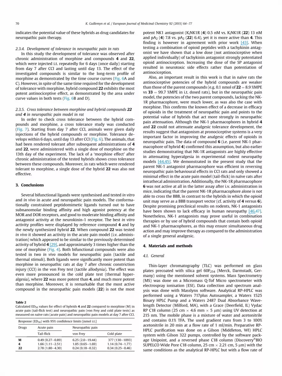
Table 2Calculated ED50 values for effect of hybrids 4 and 22 compared to morphine (M) inacute pain (tail-flick test) and neuropathic pain (von Frey and cold plate tests) asmeasured on naïve rats (acute pain) and neuropathic pain models at day 7 after CCI.
Response (ED50) with 95% confidence limits [nmol i.t.]
Drugs Acute pain Neuropathic pain
Tail-flick von Frey Cold plate
M 0.49 (0.27e0.89) 6.25 (2.0e19.44) 377 (130e1093)4 1.66 (1.11e2.51) 1.05 (0.65e1.69) 1.14 (0.74e1.77)22 2.78 (1.80e4.30) 0.24 (0.18e0.32) 0.34 (0.25e0.46)
potent NK1 antagonist (KiNK1R (4) 0.5 nM vs. KiNK1R (22) 13 nMand pA2 (4) 7.8 vs. pA2 (22) 6.4), yet it is more active than 4. Thisfinding is however in agreement with prior work [43]. Whentesting a combination of opioid peptides with a tachykinin antag-onist we have shown that a low dose (not antinociceptive whenapplied individually) of tachykinin antagonist strongly potentiatedopioid antinociception. Increasing the dose of the SP antagonistresulted in neurotoxic side effects rather than potentiation ofantinociception.
Also, an important result in this work is that in naïve rats theantinociceptive potencies of the hybrid compounds are weakerthan those of the parent compounds (e.g. 0.1 nmol of 22¼ 8.9 %MPEvs 33 ¼ 99.7 %MPE in i.t. dosed rats), but in the neuropathic painmodel, the potencies of the two parent compounds, lacking the NK-1R pharmacophore, were much lower, as was also the case withmorphine. This confirms the known effect of a decrease in efficacyof opioids in the treatment of neuropathic pain and points to thepotential value of hybrids that act more strongly in neuropathicpain attenuation. Although the NK-1 pharmacophores in hybrid 4and 22 could not attenuate analgesic tolerance development, theresults suggest that antagonism at pronociceptive systems is a veryimportant factor in improving the analgesic effects of opioids inneuropathic pain. The data of compound 6 (i.e. parent NK-1 phar-macophore of hybrid 4) confirmed this assumption, but also earlierstudies demonstrating that NK-1R antagonists are highly effectivein attenuating hyperalgesia in experimental rodent neuropathymodels [44,45]. We demonstrated in the present study that theparent NK-1 antagonist pharmacophore was efficient in reversingneuropathic pain behavioural effects in CCI rats and only showed aminimal effect in the acute pain model (tail-flick) in naïve rats afterintrathecal administration. Additionally, the NK-1R pharmacophore6 was not active at all in the latter assay after i.v. administration inmice, indicating that the parent NK-1R pharmacophore alone is notable to cross the BBB, in contrast to the hybrids in which the opioidunit may serve as a BBB transport vector (cf. activity of 4 versus 6).Despite promising preclinical results on rodents, NK-1 antagonistshave been shown to lack efficacy in human neuropathy [46,47].Nonetheless, NK-1 antagonists may prove useful in combinationtherapies or by use of hybrid compounds that contain both opioidand NK-1 pharmacophores, as this may ensure simultaneous drugaction andmay improve therapy as compared to the administrationof a single general analgesic.
4. Materials and methods
4.1. General
Thin-layer chromatography (TLC) was performed on glassplates precoated with silica gel 60F254 (Merck, Darmstadt, Ger-many) using the mentioned solvent systems. Mass Spectrometry(MS) was done on a Micromass Q-Tof Micro spectrometer withelectrospray ionisation (ESI). Data collection and spectrum anal-ysis was done with Masslynx software. Analytical RP-HPLC wasperformed using a Waters 717plus Autosampler, a Waters 1525Binary HPLC Pump and a Waters 2487 Dual Absorbance Wave-length Detector (Milford, MA), with a Grace (Deerfield, IL) VydacRP C18 column (25 cm � 4.6 mm � 5 mm) using UV detection at215 nm. The mobile phase is a mixture of water and acetonitrileand contains 0.1% TFA. The used gradient runs from 3 to 100%acetonitrile in 20 min at a flow rate of 1 ml/min. Preparative RP-HPLC purification was done on a Gilson (Middleton, WI) HPLCsystem with Gilson 322 pumps, controlled by the software pack-age Unipoint, and a reversed phase C18 column (Discovery®BIOSUPELCOWide Pore C18 column, 25 cm � 2.21 cm, 5 mm) with thesame conditions as the analytical RP-HPLC but with a flow rate of

Fig. 6. The development of tolerance to antiallodynic effect measured by (A) the von Frey test and (C) antihyperalgesic effect measured by the cold plate test after repeated oncedaily (6 days) intrathecal (i.t.) injections of morphine (M, 3.5 nmol), hybrid compound 4 (1 nmol) and 22 (1 nmol) from 7th day until 13th day after CCI (12e16 animals per group).The results are also presented as area under curve (AUC) in (B) von Frey test and (D) cold plate test. V ¼ vehicle-treated CCI-exposed rats. Inter-group differences were analysed byANOVA Bonferroni's Multiple Comparison Test; *p < 0.05 vs V-treated CCI-exposed rats.
K. Guillemyn et al. / European Journal of Medicinal Chemistry 92 (2015) 64e77 71
20 ml/min. After purification, the purity of all compounds wasevaluated as being more than 95% by analytical RP-HPLC. Allfractions were lyophilised using a Flexy-Dry lyophilizer (FTS Sys-tems, Warminster, PA). 1H and 13C NMR spectra were recorded at250 and 63 MHz on a Bruker Avance 250 spectrometer or at 500and 125 MHz on a Bruker Avance II 500 (Bruker Corp, Billerica,MA). Trimethylsilane (TMS) or residual solvent signals are used asinternal standard. The solvent used is mentioned in all cases, andthe abbreviations used are as follows: s (singlet), d (doublet), dd(double doublet), t (triplet) and m (multiplet).
4.2. General peptide synthesis
All peptides were synthesized manually by Fmoc-based solidphase peptide synthesis (SPPS) on 2-chlorotritylchloride resin(0.15 mmol scale). The first amino acid (Fmoc-Gly-OH or Fmoc-b-Ala-OH) was loaded onto the resin by use of 2 eq. Fmoc-protectedamino acid with 4 eq. DIPEA in CH2Cl2 for 2 h. The remainingchlorines were substituted by treatment of the resinwith a mixtureof MeOH/CH2Cl2/DIPEA (2:17:1) during 4 times 5 min. For normalcouplings, a 3-fold excess of the Fmoc-protected amino acids(Fmoc-D-Arg(Pbf)-OH, Fmoc-D-Cit-OH) and 3-fold excess ofcoupling reagent (TBTU) in 0.4 NMM in DMF was used for 1.5 h.Fmoc deprotection was carried out by treatment of the resin with20% 4-methylpiperidine in DMF for 5 and 15 min. After every re-action step, the resin was washed with DMF (3� 1 min), iPrOH (3�1 min) and CH2Cl2 (3� 1 min).
4.3. Peptide synthesis including Aba structures
4.3.1. Reductive aminationThe reductive amination was executed after Fmoc deprotection
of the first amino acid. The resin was first swollen in 0.5% AcOH inTMOF/CH2Cl2 for 30 min and filtered. Two equivalents of Phth-ortho-formyl phenylalanine [32] were dissolved in the samemixture and 4 eq. of NaBH3CN was dissolved in a minimumvolumeof DMF. Both solutions were added to the resin and the reactionvessel was shaken for 30 min. The course of the reaction wasmonitored after this time by the Kaiser test. When the test waspositive, the reactionwas left for another 30 min. The monitoring isrepeated until the reaction remains complete, which is indicated bya light red colour of the Kaiser test due to the presence of thesecondary amine.
4.3.2. Cyclisation towards the Aba building block (13)An excess of 3 eq. TBTU is added to the resin and shaken for 3 h.
4.3.3. Phthaloyl deprotection to 14The resin was treated with 6 eq of hydrazine monohydrate in
DMF for 18 h.
4.3.4. Boc-Dmt-OH couplingAn excess of 2 eq. Boc-Dmt-OH and 2 eq DIC/HOBt in DMF was
added to the peptide-resin. The reaction vessel was shaken for 3 h.

Fig. 7. Cross tolerance of hybrid compounds 22 and 4 with morphine measured in neuropathic pain. The influence of repeated 6 days i.t. administration of morphine (M, 3.5 nmol),hybrid 22 (1 nmol) and hybrid 4 (1 nmol) on the effect of a single dose of morphine (M, 3.5 nmol), or 22 (1 nmol) was measured on day 13 after CCI in rats (6e8 animals per group).The von Frey (A, B, C) and cold plate (D, E, F) tests were conducted 30 min after a single dose of vehicle, morphine or 22 administration. The data are presented as %MPE ±S.E.M.Inter-group differences were analysed by ANOVA Bonferroni's Multiple Comparison Test; ***P < 0.001 indicates a significant difference compared with group which receivedrepeated vehicle and then a single dose of vehicle (V þ V-treated CCI-exposed animals); $$$P < 0.001 indicates a significant difference compared with group which received repeatedvehicle and then single dose of morphine (V þ M; A, B, D, E) or 22 (Vþ22; C, F).
K. Guillemyn et al. / European Journal of Medicinal Chemistry 92 (2015) 64e7772
4.3.5. Cleavage from the solid supportThe fully protected peptide was cleaved from the resin with 1%
TFA in DMF for 30 min. The filtrate was concentrated and added tocold ether. The precipitated peptide was then dissolved in aceto-nitrile/H2O and lyophilised to get the compounds as a powder.
4.4. Coupling of the benzylamines to the peptide acids and finaldeprotection
The crude protected peptide was dissolved in a minimal amountof CH2Cl2. The solution was cooled in an ice-bath and DIPEA (2 eq.),DIC/HOBt (1.5 eq. each) are added. The mixture was stirred for30 min at 0 �C. Then the amine (1.5 eq.) was added to the solutionand the reaction mixture stirred again for 30 min with cooling. Thereaction was left to warm up to room temperature. When themixture of all the reagents does not dissolve sufficiently, somedrops of DMF are added and a clear solution is obtained. The re-action mixture was left to react for 15 h. At completion, the solventwas evaporated and the residue was treated with TFA/TES/H2O(95:2.5:2.5) for 3 h, after which the solvent was removed in vacuo.
4.5. Purification
The crude peptides were dissolved in H2O and acetonitrile wasadded until complete dissolving was observed. The solution was
injected on a Gilson preparative RP-HPLC. Fractions were collectedand combined and lyophilised. The peptides were obtained aswhite powders with a purity of >95% as determined by analyticalHPLC. The structures were confirmed by high-resolution electro-spray mass spectrometry.
4.6. Synthesis of benzylamine derivates
4.6.1. N-isobutyl benzylamine (18a)Isobutylamine (1.5 ml, 5 eq.) is dissolved in 16 ml methanol in a
two-headed round bottom flask equipped with magnetic stirrer,reflux column and septum. The reaction mixture is heated to 50 �Cin an oil bath. Next, benzyl chloride (347 ml, 1 eq.) is dissolved in16 ml methanol and added drop wise (during 1 h). The mixture isstirred overnight at 50 �C and the solvent is evaporated. The residueis dissolved in 28 ml CH2Cl2 and washed with a 20% NaOH-solution(3 � 60 ml) and water (2 � 60 ml). The organic phase is dried withMgSO4, filtered en evaporated. After evaporation, a pale yellow oil isobtained. The purification is done with flash chromatography(Davisil LC60A, 40e63 mm) with hexane/ethyl acetate 9:1. Afterevaporation of the solvent, 1 eq. of TFA is added to make a salt andthe solution was lyophilised. A white to yellow powder is obtainedwith a yield of 43%. N-isobutylbenzylamine. Yield: 48% (504 mg, TFAsalt); Formula: C11H17N; MW: 163.26 g/mol; TLC Rf ¼ 0.26 (EtOAc/chex 1:1); HPLC: tR ¼ 9.4 min; MS (ESþ): 164 [MþH]þ; 1HNMR

K. Guillemyn et al. / European Journal of Medicinal Chemistry 92 (2015) 64e77 73
(250 MHz, CDCl3): d (ppm) 0.92 (6H, d, CH3, J¼ 6.7 Hz), 1.97 (1H, m,CH), 2.64 (2H, m, CHeCH2), 3.90 (2H, s, NHeCH2-Bn), 7.30e7.42(5H, m, arom. H). 13CNMR (63MHz, CDCl3): d (ppm) 20.0 (CH3), 26.0(CH), 51.5 (CH2-Bn), 53.9 (CHeCH2eNH), 129.0 (CH arom.), 129.4(CH arom.), 130.2 (CH arom.), 130.4 (Cq arom.).
4.6.2. N-isobutyl-30,50-bistrifluoromethyl benzylamine (18b)Isobutylamine (1.5 ml, 5 eq.) is dissolved in 24 ml methanol in a
two-headed round bottom flask equipped with magnetic stirrer,reflux column and septum. The reaction mixture is heated to 50 �Cin an oil bath. Next, 30,50-trifluoromethylbenzylchloride (1 eq.,528 mg) is dissolved in 20 ml methanol and added drop wise(during 1 h). The mixture is stirred overnight at 50 �C and thesolvent is evaporated. The residue is dissolved in 150 ml dichloro-methane and washed with a 20% NaOH-solution (3 � 90 ml) andwater (2 � 90 ml). The organic phase is dried with MgSO4, filtereden evaporated. After evaporation, a light yellow oil is obtained. Thepurification is done with flash chromatography (Davisil LC60A,40e63 mm) with a gradient going from hexane/ethyl acetate 9:1 to7.5:2.5. After evaporation of the solvent, TFA is added to make a saltand lyophilised. A white to yellow powder is obtained with a yieldof 48%. N-isobutyl-30,50-bistrifluoromethyl benzylamine. Yield: 43%(354 mg, TFA salt); Formula: C13H15F6N; MW: 299.26 g/mol;Rf: ¼ 0.65 (EtOAc/petroleum ether 1:1), TLC: Rf ¼ 0.17 (EtOAc/hexane 1:9);HPLC: tR¼ 13.3 min;MS (ESþ): 300 [MþH]þ; 1HNMR(250 MHz, CD3OD): d (ppm) 1.06 (3H, d, CH3, J ¼ 6.7 Hz), 2.08 (1H,m, CH), 2.99 (2H, m, CHeCH2), 4.43 (2H, s, NHeCH2-Bn), 8.11e8.20(3H, m, arom. H). 13CNMR (63 MHz, CD3OD): d (ppm) 20.3 (CH3),27.3 (CH), 51.3 (CH2-Bn), 56.5 (CHeCH2eNH), 124.6 (q, CF3,1J ¼ 272.0 Hz), 132.1 (CH arom.), 133.4 (q, Cq arom., 2J ¼ 33.7 Hz),135.7 (CH arom.).
4.7. Peptide characterization
4.7.1. H-Dmt-D-Arg-Aba-Gly-NMe-30,50e(CF3)2eBn (4)Preparative HPLC yielded the desired compound (white powder,
34%). HPLC: tR ¼ 14.0 min. TLC Rf 0.72 (EBAW). HRMS (ESPþ) foundm/z 821.3536 [MþH]þ, C39H46F6N8O5 requires 821.3568.
4.7.2. H-Dmt-D-Arg-Aba-b-Ala-NMe-30,50e(CF3)2eBn (21)Preparative HPLC yielded the desired compound (white powder,
17%). HPLC: tR ¼ 14.0 min. TLC Rf 0.67 (EBAW). HRMS (ESPþ) foundm/z 835.3708 [MþH]þ, C40H48F6N8O5 requires 835.3724.
4.7.3. H-Dmt-D-Arg-Aba-b-Ala-NMe-Bn (22)Preparative HPLC yielded the desired compound (white powder,
34%). HPLC: tR ¼ 11.7 min. TLC Rf 0.64 (EBAW). HRMS (ESPþ) foundm/z 699.3955 [MþH]þ, C38H50N8O5 requires 699.3977.
4.7.4. H-Dmt-D-Cit-Aba-b-Ala-NMe-30,50e(CF3)2eBn (23)Preparative HPLC yielded the desired compound (white powder,
21%). HPLC: tR ¼ 14.6 min. TLC Rf 0.72 (EBAW). HRMS (ESPþ) foundm/z 836.3515 [MþH]þ, C40H47F6N7O6 requires 836.3565.
4.7.5. H-Dmt-D-Cit-Aba-b-Ala-NMe-Bn (24)Preparative HPLC yielded the desired compound (white powder,
34%). HPLC: tR ¼ 12.1 min. TLC Rf 0.70 (EBAW). HRMS (ESPþ) foundm/z 700.3790 [MþH]þ, C38H49N7O6 requires 700.3817.
4.7.6. H-Dmt-D-Arg-Aba-Gly-NMe-Bn (25)Preparative HPLC yielded the desired compound (white powder,
63%). HPLC: tR ¼ 11.7 min. TLC Rf 0.67 (EBAW). HRMS (ESPþ) foundm/z 685.3788 [MþH]þ, C37H48N8O5 requires 685.3820.
4.7.7. H-Dmt-D-Arg-Aba-GlyeNHeBn (26)Preparative HPLC yielded the desired compound (white powder,
22%). HPLC: tR ¼ 11.2 min. TLC Rf 0.66 (EBAW). HRMS (ESPþ) foundm/z 671.3694 [MþH]þ, C36H46N8O5 requires 671.3664.
4.7.8. H-Dmt-D-Arg-Aba-Gly-N-i-Bu-30,50e(CF3)2eBn (27)Preparative HPLC yielded the desired compound (white powder,
32%). HPLC: tR ¼ 14.3 min. TLC Rf 0.62 (EBAW). HRMS (ESPþ) foundm/z 863.4044 [MþH]þ, C42H52F6N8O5 requires 863.4037.
4.7.9. H-Dmt-D-Arg-Aba-Gly-N-i-Bu-Bn (28)Preparative HPLC yielded the desired compound (white powder,
15%). HPLC: tR ¼ 12.5 min. TLC Rf 0.61 (EBAW). HRMS (ESPþ) foundm/z 727.4304 [MþH]þ, C40H54N8O5 requires 727.4290.
4.7.10. H-Dmt-D-Arg-Aba-b-Ala-N-i-Bu-30,50e(CF3)2eBn (29)Preparative HPLC yielded the desired compound (white powder,
12%). HPLC: tR ¼ 14.6 min. TLC Rf 0.64 (EBAW). HRMS (ESPþ) foundm/z 877.4240 [MþH]þ, C43H54F6N8O5 requires 877.4194.
4.7.11. H-Dmt-D-Arg-Aba-b-Ala-N-i-Bu-Bn (30)Preparative HPLC yielded the desired compound (white powder,
21%). HPLC: tR ¼ 12.8 min. TLC Rf 0.57 (EBAW). HRMS (ESPþ) foundm/z 741.4465 [MþH]þ, C41H56N8O5 requires 741.4446.
4.7.12. H-Dmt-D-Arg-Phe-Sar-NMe-30,50e(CF3)2eBn (31)Preparative HPLC yielded the desired compound (white powder,
24%). HPLC: tR ¼ 13.7 min. TLC Rf 0.70 (EBAW). HRMS (ESPþ) foundm/z 823.3728 [MþH]þ, C39H48F6N8O5 requires 823.3724.
4.7.13. H-Dmt-D-Arg-Phe-Sar-NMe-Bn (32)Preparative HPLC yielded the desired compound (white powder,
31%). HPLC: tR ¼ 11.5 min. TLC Rf 0.66 (EBAW). HRMS (ESPþ) foundm/z 687.3949 [MþH]þ, C37H50N8O5 requires 687.3983.
4.7.14. H-Dmt-D-Arg-Aba-b-Ala-NH2 (33)Preparative HPLC yielded the desired compound (white powder,
49.5%). HPLC: tR ¼ 10.1 min. TLC Rf 0.44 (EBAW). HRMS (ESPþ)found m/z 595.3394 [MþH]þ, C30H43N8O5 requires 595.3351.
4.8. Functional NK1R assay [48]. Cell line and cell culture conditions
The Chinese hamster ovary K1 (CHO-K1) cell line, stablyexpressing human NK1 receptor (hereafter referred to as CHO-NK1 cells), was transfected with an apoaequorin expression vec-tor (pER2) using Fugene6 (Roche Applied Science). The cell lineand expression vector were obtained from Euroscreen (Belgium).The CHO-NK1 cells were cultured in sterile DMEM/HAM's F12medium (Sigma) supplemented with 10% foetal bovine serum,100 IU/mL penicillin, 100 mg/mL streptomycin, and 400 mg/mL G418 (Geneticin, Gibco) at 37 �C with 5% CO2 and were trypsinizedevery 3 days.
4.8.1. Aequorin charging protocolTransfected cells in the midlog phase were detached by
changing the growth medium for PBS buffer supplemented with5 mM EDTA (pH 8). The cells were spun down and incubated for 4 hat a concentration of 5 � 106 cells/mL in DMEM-F12 mediumwithout phenol red (Gibco) supplemented with 0.1% BSA (BSAmedium) and 5 mM coelenterazine h (Molecular Probes). Aftercoelenterazine loading, the cells were diluted 10-fold in the samemedium and incubated for an additional period of 30 min. The cellswere mildly shaken during the incubation periods.

K. Guillemyn et al. / European Journal of Medicinal Chemistry 92 (2015) 64e7774
4.8.2. Aequorin luminescence assayA dilution series of peptide agonist (SP was purchased from
Sigma) ranging from 10�11 to 10�4 Mwas distributed in a white 96-well plate. For investigating antagonism, the synthetic compoundswere added to these wells to obtain the desired concentrations(ranging from 10�8 to 10�4 M). One negative control sample (BSAmedium only) was included in each row of the 96-well plate. Theplate was loaded in a “Multimode Reader Mithras, LB940” (Bert-hold). The wells were screened one by one, and each measurementstarted at the moment of injection of 50 mL of the coelenterazine-loaded cell suspension, containing 2.5 � 104 cells. Light emissionwas measured every second for 30 s after which 50 mL of 10 nM ATPsolution (positive control) was injected. Each measurement wascarried out in duplicate. Light emission was recorded for an addi-tional period of 10 s per well, and the data were presented inrelative light units (RLU).
4.8.3. Data analysisLuminescence data (peak integration) were calculated using
MikroWin 2000 software (Berthold), which was linked to theMicrosoft Excel program. All statistical and curve-fitting analyseswere performed using Prism 4.0 (GraphPad) software. Data areexpressed in percentage (% RLU) of the maximal luminescence thatwas detected with 10�4 M SP (without antagonist). The competitivenature of antagonism was evaluated using the Schild plot method[40]. All antagonists analysed in this study provided linear regres-sion plots and were considered competitive. The pA2 values werecalculated using Schild's equation [49].
4.9. hNK1/CHO cell membrane preparation and radioligand bindingassay
Recombinant hNK1/CHO cells were grown to confluency in37 �C, 95% air and 5% CO2, humidified atmosphere, in a FormaScientific (Thermo Forma, OH) incubator in Ham's F12 mediumsupplemented with 10% foetal bovine serum, 100 IU/mL penicillin,100 mg/mL streptomycin, and 500 mg/mL geneticin. The confluentcell monolayers were then washed with Ca2þ, Mg2þ-deficientphosphate-buffered saline (PD buffer) and harvested in the samebuffer containing 0.02% EDTA. After centrifugation at 2700 rpm for12 min, the cells were homogenized in ice-cold 10 mM TriseHCland 1 mM EDTA, pH 7.4, buffer. A crude membrane fraction wascollected by centrifugation at 18000 rpm for 12 min at 4 �C, thepellet was suspended in 50 mM Tris-Mg buffer, and the proteinconcentration of the membrane preparation was determined byusing Bradford assay. Six different concentrations of the test com-pound were each incubated, in duplicates, with 20 mg of membranehomogenate, and 0.4 nM [3H]SP (135 Ci/mmol, PerkineElmer,United States) in 1 mL final volume of assay buffer (50 mM Tris, pH7.4, containing 5mMMgCl2, 50 mg/mL bacitracin, 30 mM bestatin,10 mM captopril, and 100 mM phenylmethylsulfonylfluoride) SP at10 mM was used to define the nonspecific binding. The sampleswere incubated in a shakingwater bath at 25 �C for 20min. The [3H]SP concentration and the incubation time were selected based onthe studies of Yamamoto et al. [20] The reaction was terminated byrapid filtration through Whatman grade GF/B filter paper (Gai-thersburg, MD) presoaked in 1% polyethyleneimine, washed fourtimes each with 2 mL of cold saline, and the filter bound radioac-tivity was determined by liquid scintillation counting (BeckmanLS5000 TD). The media and chemicals listed above were purchasedfrom Sigma (SigmaeAldrich, St. Louis, MO) unless otherwise stated.
4.9.1. Data analysisAnalysis of data collected from three independent experiments
performed in duplicates is done using GraphPad Prizm 4 software
(GraphPad, San Diego, CA). Log IC50 values for each test compoundwere determined from nonlinear regression. The inhibition con-stant (Ki) was calculated from the antilogarithmic IC50 value by theCheng and Prusoff equation [50,51].
4.10. Functional GPI and mouse vas deferens (MVD) assays
The GPI and MVD bioassays were carried out as described indetail elsewhere [52,53]. A doseeresponse curve was determinedwith [Leu [5]]enkephalin as standard for each ileum and vaspreparation, and IC50 values of the compounds being tested werenormalized according to a published procedure [54].
4.11. Opioid receptor binding assays
Opioid receptor binding studies were performed as described indetail elsewhere [52]. Binding affinities for m and d opioid receptorswere determined by displacing, respectively, [3H]DAMGO (MultiplePeptide Systems, San Diego, CA) and [3H]DSLET (Multiple PeptideSystems) from rat brain membrane binding sites. Incubations wereperformed for 2 h at 0 �C with [3H]DAMGO and [3H]DSLET atrespective concentrations of 0.72 and 0.78 nM. IC50 values weredetermined form log-dose displacement curves, and Ki values werecalculated from the IC50 values by means of the equation of Chengand Prusoff [50], using values of 1.3 and 2.6 nM for the dissociationconstants of [3H]DAMGO and [3H]DSLET, respectively.
4.12. In vivo analgesic test in naive rats and mice
The pain threshold to a thermal stimulus was assessed usingtail-flick latency evoked by noxious thermal stimulation as deter-mined with a tail-flick analgesic meter (Analgesia Meter; UgoBasile, Comerio, Italy) as described previously [55e57]. The tail-flick was used to measure spinal nociceptive responses tothermally-induced pain using male C57BI6 mice and Wistar rats.Mice (weighing 25e28 g) or rats (250e300 g) weremaintained on anormal lightedark cycle and testing occurred during the lightphase. Different doses of the drug were dissolved in saline or waterfor injection and injected intravenously (i.v.) in mice or intrathe-cally (i.t.) in rats. The effect was assessed at the following time pointpost-injection: 5, 15, 30, 60, 120, 180 min. The thermal stimulusemitted by a light bulb was applied to the tail at two-thirds of itslength until the animal vigorously withdrew the tail. The baselineresponse was approx. 2 s and the maximal exposure was set to 7 s(mice) and 9 s (rats) to avoid tissue damage. All experimentalprocedures used in this animal testing followed the guidelines onethical standards for the investigation of experimental pain in an-imals and were approved by the Animal Research Committees ofthe Medical Research Centre, Polish Academy of Sciences and wereapproved by the local Bioethics Committee (Krakow, Poland). Allin vivo activity was determined as a percentage of the maximalpossible effect (%MPE) and was calculated as: %MPE ¼ [posttreatment latency/cut-off latency] � 100.
4.13. In vivo analgesic tests in neuropathic pain model in rats
4.13.1. AnimalsMale Wistar rats (300e350 g) from Charles River (Sulzfeld,
Germany) were housed in cages lined with sawdust under a stan-dard 12/12 h light/dark cycle (lights on at 08.00 h) with food andwater available ad libitum. All experiments were performed ac-cording to the recommendations of the International Associationfor the Study of Pain (IASP) [58] and the National Institutes ofHealth Guide for the Care and Use of Laboratory Animals and wereapproved by the local Bioethics Committee (Krakow, Poland).

K. Guillemyn et al. / European Journal of Medicinal Chemistry 92 (2015) 64e77 75
4.13.2. Implantation of intrathecal cannulasThe rats were chronically implantedwith i.t. catheters according
to Yaksh and Rudy [59] under pentobarbital anaesthesia (60mg/kg;ip) as described previously [56,57]. The rats were placed on a ste-reotaxic table (David Kopf), and a sterile catheter (PE 10, INTRA-MEDIC, Clay Adams, Becton Dickinson and Company, Rutherford,NJ, USA.), flushed with sterile water prior to insertion, was carefullyintroduced through the atlanto-occipital membrane to the sub-arachnoid space at the rostral level of the spinal cord lumbarenlargement (L4-L6).
4.13.3. Chronic constriction injuryA chronic constriction injury (CCI) was produced according to
Bennett and Xie [60] as described previously [46,47]. The rightsciatic nerve was exposed under sodium pentobarbital anaesthesia(60 mg/kg; i.p.). Four ligatures (4/0 silk) were made around thenerve distal to the sciatic notch with 1 mm spacing until a brieftwitch in the respective hind limb was observed. After CCI, all an-imals developed allodynia and hyperalgesia.
4.13.4. Behavioural tests in neuropathic pain modelTwo different behavioural tests were used to assess analgesic
potency in neuropathic pain. The von Frey test evaluates tactile(mechanical) allodynia. After placing the animals in a plastic cagewith the floor made from metal mesh, a mechanical stimulus isapplied to the surface of the hind paw sole by use of the von Freyfilament. In the cold plate test which measures thermal hyper-algesia, the rats are placed on a cold plate of 5 �C and latency ofreaction is assessed. In both methods, the time of reaction wasmeasured automatically. The obtained signal is expressed as apercentage of the maximal potential effect (%MPE).
4.13.4.1. von Frey test. Mechanical allodynia in rats with CCI wasmeasured using an automatic von Frey apparatus (Dynamic PlantarAnesthesiometer Cat. No. 37400, Ugo Basile Italy). The animals wereplaced in plastic cages with wire net floors. They were acclimatisedto this environment for approximately 3 min prior to testing. Thevon Frey filament was applied to the midplantar surface of the hindpaw, and themeasurements were taken automatically, as describedpreviously by Mika et al. [57] The strength of the von Frey stimuliranged from 0.5 to 26 g in the rats.
4.13.4.2. Cold plate test. To assess the threshold to a cold stimulusin rats with CCI, the Cold/Hot Plate Analgesia Meter (No. 05044,Columbus Instruments, USA) was used, as described previously byMika et al. [57] The rats were placed on the cold plate (5 �C), and thetime until the shake of the hind paw was recorded. The cut-offlatency for this test was 30 s. In all cases, the injured paw wasthe first to react.
4.13.4.3. Tolerance development. Tolerance development was car-ried out in neuropathic painmodel. Day seven after CCI was the firstday of tested drugs i.t. injections which were continued up to 13thday. In this study morphine (3.5 nmol), 4 (1 nmol) and 22 (1 nmol)were administered i.t. once a day and the behavioural tests werecarried out 30 min after drug administration.
4.13.4.4. Cross tolerance. Cross tolerance measurement was con-ducted one day after tolerance development completion. When theanimals developed tolerance to analgesic effect of 4 and 22 a singledose of morphine was injected to check if there is a cross tolerancebetween these compounds and morphine. We also checkedwhether the hybrid 22will be effective in rats inwhom tolerance tomorphine was induced.
4.14. Chemicals administration
The chemicals used were obtained from the following sources:morphine hydrochloride (Polfa Kutno, Poland). All drugs were dis-solved in sterile water (water for injection). All drugs were injectedi.t. at a volume of 5 ml, followed by an injection of 10 ml of distilledwater to flush the catheter. For acute pain, studies were carried outon day 7 after catheter implantation and 30 min after drug admin-istration. In rats drugs were administered in a single i.t. injection atthe following doses: morphine (0.35, 3.5, 35 nmol), 22 (0.1, 1,11 nmol), 33 (0.001, 0.01, 0.1 nmol), 4 (0.1, 0.5,1 nmol), 7 (0.001, 0.01,0.1 nmol), and 6 (1,10, 50 nmol). Inmice drugswere administered ina single i.v. injection at the following doses:morphine 12.5 mmol/kg,22 1.08 and 10.8 mmol/kg, 33 1.21 mmol/kg. In neuropathic painmodel morphine (0.35, 3.5, 35 nmol), 22 (0.1, 0.4,1 nmol), 33 (0.001,0.01, 0.1,1 nmol), 4 (0.1, 0.5,1 nmol), 7 (0.001, 0.01, 0.1,1 nmol) and 6(0.1, 0.5, 1 nmol) were administered in a single i.t. injection on day7e14 after CCI and themeasurements were performed 30min afterdrug administration. The control groups received vehicle (water forinjection) injections according to the same schedule. The CCI to thesciatic nerve was performed 5e7 days after catheter implantation.After completion of the experiment, the animalswere killedwith anoverdose of pentobarbital (i.p.).
4.15. Data analysis
The behavioural data (6e16 rats per group) are presented as thepercentage of the maximal possible antinociceptive effect (%MPE ± SEM), which was calculated according to the followingequation: % MPE ¼ [(TL-BL)/(CUT-OFF-BL)] � 100%, where BL wasthe baseline latency and TL was the latency obtained after druginjection. The results of the experiments were statistically evalu-ated using one-way analysis of variance (ANOVA). The differencesbetween the treatment groups throughout the study were furtheranalysed with Bonferroni post-hoc tests.
Author information
Author contributions
KG, OVDP and IVDEwere in charge of ligand synthesis. AK, EV, JL,FP, JVD, JV contributed by providing the NK1 binding and antago-nism data. NNC, CL and PWS provided in vitro opioid data. PK, ALand AWL performed the tail-flick test in mice and JM, ER, WM andBP performed acute and neuropathic pain study in rats. Themanuscript was written through contributions of all authors. Allauthors have given approval to the final version of the manuscript.
Funding sources
The work of SB, DT and PWS was supported by a collaborationconvention between the Minist�ere du D�eveloppement Econo-mique, de l'Innovation et de l'Exportation du Qu�ebec and theResearch Foundatione Flanders (FWOVlaanderen) (PSR-SIIRI-417).The research of PWS was also supported by grants CIHR (MOP-89716), and the NIH (DA-004443). Supported by Institute of Phar-macology statutory founds (WM) and grant from National ScienceCenter Poland NCN2012/06/A/NZ4/00028 for (BP and JM).
Acknowledgements
Dr. Cecilia Betti is acknowledged for in-house reproduction ofthe total solid phase assembly methodology depicted in Scheme 2.We thank Dr. Emeric Miclet and Dr. Isabelle Correia for the NMRanalysis of hybrid 4.

K. Guillemyn et al. / European Journal of Medicinal Chemistry 92 (2015) 64e7776
References
[1] G.W. Pasternak, Opioids and their receptors: are we there yet? Neurophar-macology 76 (2014) 198e203, http://dx.doi.org/10.1016/j.neuropharm.2013.03.039.
[2] D. Labuz, H. Machelska, Stronger antinociceptive efficacy of opioids at theinjured nerve trunk than at its peripheral terminals in neuropathic pain,J. Pharmacol. Exp. Ther. 346 (2013) 535e544, http://dx.doi.org/10.1124/jpet.113.205344.
[3] M.H. Ossipov, Y. Lopez, M.L. Nichols, D. Bian, F. Porreca, The loss of anti-nociceptive efficacy of spinal morphine in rats with nerve ligation injury isprevented by reducing spinal afferent drive, Neurosci. Lett. 199 (1995) 87e90,http://dx.doi.org/10.1016/0304-3940(95)12022-V.
[4] C.J. Evans, D.E. Keith Jr., H. Morrison, K. Magendzo, R.H. Edwards, Cloning of adelta opioid receptor by functional expression, Science 258 (1992)1952e1955, http://dx.doi.org/10.1126/science.1335167.
[5] B.L. Kieffer, K. Befort, C. Gaveriaux-Ruff, C.G. Hirth, The delta-opioid receptor:isolation of a cDNA by expression cloning and pharmacological characteriza-tion, Proc. Natl. Acad. Sci. U. S. A. 89 (1992) 12048e12052, http://dx.doi.org/10.1073/pnas.89.24.12048.
[6] Y. Chen, A. Mestek, J. Liu, L. Yu, Molecular cloning of a rat kappa opioid re-ceptor reveals sequence similarities to the mu and delta opioid receptors,Biochem. J. 295 (1993) 625e628.
[7] M. Costigan, J. Scholz, C.J. Woolf, Neuropathic pain: a maladaptive response ofthe nervous system to damage, Annu. Rev. Neurosci. 32 (2009) 1e32, http://dx.doi.org/10.1146/annurev.neuro.051508.135531.
[8] B. Payne, M.A. Norfleet, Chronic pain and the family: a review, Pain 26 (1986)1e22.
[9] S.R. Chaplan, A.B. Malmberg, T.L. Yaksh, Efficacy of spinal NMDA receptorantagonism in formalin hyperalgesia and nerve injury evoked allodynia in therat, J. Pharmacol. Exp. Ther. 280 (1997) 829e838.
[10] T. King, L. Gardell, R. Wang, A. Vardanyan, M. Ossipov, T. Malan, T. Vanderah,S. Hunt, V. Hruby, J. Lai, F. Porreca, Role of NK-1 neurotransmission in opioid-induced hyperalgesia, Pain 116 (2005) 276e288, http://dx.doi.org/10.1016/j.pain.2005.04.014.
[11] T. Yamamoto, P. Nair, N. Jacobsen, V. Kulkarni, P. Davis, S.-W. Ma,E. Navratilova, H. Yamamura, T. Vanderah, F. Porreca, J. Lai, V. Hruby, Bio-logical and conformational evaluation of bifunctional compounds for opioidreceptor agonists and neurokinin 1 receptor antagonists possessing twopenicillamines, J. Med. Chem. 53 (2010) 5491e5501, http://dx.doi.org/10.1021/jm100157m.
[12] L. Costantino, D. Barlocco, Designed multiple ligands: basic research vs clinicaloutcomes, Curr. Med. Chem. 19 (2012) 3353e3387, http://dx.doi.org/10.2174/092986712801215883.
[13] R. Morphy, Z. Rankovic, Design of Multitarget Ligands. In Lead GenerationApproaches in Drug Discovery, John Wiley & Sons, Inc, Hoboken, 2010, pp.141e164.
[14] L. Gentilucci, New trends in the development of opioid peptide analogues asadvanced remedies for pain relief, Curr. Top. Med. Chem. 4 (2004) 19e38,http://dx.doi.org/10.2174/1568026043451663.
[15] A.W. Lipkowski, Cooperative reinforcement of opioid pharmacophores, Pol. J.Pharmacol. Pharm. 39 (1987) 585e596.
[16] A.W. Lipkowski, D.B. Carr, A. Misicka, K. Misterek, Biological activities of apeptide containing both casomorphin-like and substance P antagonist struc-tural characteristics, in: V. Brantl, H. Teschemacher (Eds.), B-Casomorphinsand Related Peptides: Recent Developments, VCH, Weinheim, 1994, pp.113e118.
[17] I.M. Bonney, S.E. Foran, J.E. Marchand, A.W. Lipkowski, D.B. Carr, Spinalantinociceptive effects of AA501, a novel chimeric peptide with opioid re-ceptor agonist and tachykinin receptor antagonist moieties, Eur. J. Pharmacol.488 (2004) 91e99, http://dx.doi.org/10.1016/j.ejphar.2004.02.023.
[18] S.E. Foran, D.B. Carr, A.W. Lipkowski, I. Maszczynska, J.E. Marchand, A. Misicka,M. Beinborn, A.S. Kopin, R.M. Kream, A substance P-opioid chimeric peptide asa unique nontolerance-forming analgesic, Proc. Natl. Acad. Sci. U. S. A. 97(2000) 7621e7626, http://dx.doi.org/10.1073/pnas.130181897.
[19] T. Yamamoto, P. Nair, J. Vagner, T. Largent-Milnes, P. Davis, S.-W. Ma,E. Navratilova, S. Moye, S. Tumati, J. Lai, H.I. Yamamura, T.W. Vanderah,F. Porreca, V.J. Hruby, A structureeactivity relationship study and combina-torial synthetic approach of C-terminal modified bifunctional peptides thatare d/m opioid receptor agonists and neurokinin 1 receptor antagonists, J. Med.Chem. 51 (2008) 1369e1376, http://dx.doi.org/10.1021/jm070332f.
[20] T. Yamamoto, P. Nair, P. Davis, S.-W. Ma, E. Navratilova, S. Moye, S. Tumati,J. Lai, T.W. Vanderah, H.I. Yamamura, F. Porreca, V.J. Hruby, Design, synthesis,and biological evaluation of novel bifunctional C-terminal-modified peptidesfor d/m opioid receptor agonists and neurokinin-1 receptor antagonists, J. Med.Chem. 50 (2007) 2779e2786, http://dx.doi.org/10.1021/jm061369n.
[21] T. Takaya, Discovery of neurokinin antagonists, Pure Appl. Chem. 68 (1996)875e880.
[22] J. Longmore, R.G. Hill, R.J. Hargreaves, Neurokinin-receptor antagonists:pharmacological tools and therapeutic drugs, Can. J. Physiol. Pharm. 75 (1997)612e621, http://dx.doi.org/10.1139/y97-069.
[23] M.I. Gonzalez, M.J. Field, J. Hughes, L. Singh, Evaluation of selective NK1 re-ceptor antagonist CI-1021 in animal models of inflammatory and neuropathicpain, J. Pharmacol. Exp. Ther. 294 (2000) 444e450.
[24] S. Ballet, D. Feytens, K. Buysse, N.N. Chung, C. Lemieux, S. Tumati, A. Keresztes,J. Van Duppen, J. Lai, E. Varga, F. Porreca, P.W. Schiller, J. Vanden Broeck,D. Tourwe
, Design of novel neurokinin 1 receptor antagonists based on con-formationally constrained aromatic amino acids and discovery of a potentchimeric opioid agonist-neurokinin 1 receptor antagonist, J. Med. Chem. 54(2011) 2467e2476, http://dx.doi.org/10.1021/jm1016285.
[25] P. Schiller, T. Nguyen, I. Berezowska, S. Dupuis, G. Weltrowska, N. Chung,C. Lemieux, Synthesis and in vitro opioid activity profiles of DALDA analogues,Eur. J. Med. Chem. 35 (2000) 895e901, http://dx.doi.org/10.1016/S0223-5234(00)01171-5.
[26] I. Van den Eynde, G. Laus, P.W. Schiller, P. Kosson, N.N. Chung, A.W. Lipkowski,D. Tourw�e, A new structural motif for m-opioid antagonists, J. Med. Chem. 48(2005) 3644e3648, http://dx.doi.org/10.1021/jm0491795.
[27] S. Ballet, D. Feytens, R.D. Wachter, M.D. Vlaeminck, E.D. Marczak, S. Salvadori,C.D. Graaf, D. Rognan, L. Negri, R. Lattanzi, L.H. Lazarus, D. Tourw�e, G. Balboni,Conformationally constrained opioid ligands: the Dmt-Aba and Dmt-Aiaversus Dmt-Tic scaffold, Bioorg. Med. Chem. Lett. 19 (2009) 433e437,http://dx.doi.org/10.1016/j.bmcl.2008.11.051.
[28] S. Ballet, R. De Wachter, K. Van Rompaey, C. T€omb€oly, D. Feytens, G. T€oth,L. Quartara, P. Cucchi, S. Meini, D. Tourw�e, Bradykinin analogs containing the4-amino-2-benzazepin-3-one scaffold at the C-terminus, J. Pept. Sci. 13 (2007)164e170, http://dx.doi.org/10.1002/psc.827.
[29] K. Guillemyn, P. Kleczkowska, A. Novoa, B. Vandormael, I. Van den Eynde,P. Kosson, M. Asim, P. Schiller, M. Spetea, A. Lipkowski, D. Tourwe, S. Ballet,In vivo antinociception of potentmu opioid agonist tetrapeptide analogues andcomparison with a compact opioid agonist e neurokinin 1 receptor antagonistchimera, Mol. Brain 5 (2012) 4,, http://dx.doi.org/10.1186/1756-6606-5-4.
[30] T. Largent-Milnes, T. Yamamoto, P. Nair, J. Moulton, V. Hruby, J. Lai, F. Porreca,T. Vanderah, Spinal or systemic TY005, a peptidic opioid agonist/neurokinin 1antagonist, attenuates pain with reduced tolerance, Br. J. Pharmacol. 161(2010) 986e1001, http://dx.doi.org/10.1111/j.1476-5381.2010.00824.x.
[31] S. Ballet, A. Frycia, J. Piron, N. Chung, P. Schiller, P. Kosson, A. Lipkowski,D. Tourwe, Synthesis and biological evaluation of constrained analogues of theopioid peptide H-Tyr-D-Ala-Phe-Gly-NH2 using 4-amino-2-benzazepin-3-onescaffold, J. Pept. Res. 66 (2005) 222e230, http://dx.doi.org/10.1111/j.1399-3011.2005.00291.x.
[32] K. Van Rompaey, I. Van den Eynde, N. De Kimpe, D. Tourw�e, A versatilesynthesis of 2-substituted 4-amino-1,2,4,5-tetrahydro-2-benzazepine-3-ones,Tetrahedron 59 (2003) 4421e4432, http://dx.doi.org/10.1016/S0040-4020(03)00583-0.
[33] Mondini, S., Dall Avo, M., Guerrato, A. Process for the preparation of 3,5-bistrifluoromethyl-N-methylbenzylamine. WO2007107818 A2, 2007.
[34] Y. Sasaki, A. Ambo, K. Suzuki, Studies on analgesic oligopeptides. VII. Solidphase synthesis and biological properties of Tyr-D-Arg-Phe-ß-Ala-NH2 and itsfluorinated aromatic amino acid derivatives, Chem. Pharm. Bull. 39 (1991)2316e2318, http://dx.doi.org/10.1248/cpb.39.2316.
[35] S. Ananthan, Opioid ligands with mixed mu/delta opioid receptor in-teractions: an emerging approach to novel analgesics, AAPS J. 8 (2006)E118eE125, http://dx.doi.org/10.1208/aapsj080114.
[36] E. Abdelhamid, M. Sultana, P. Portoghese, A. Takemori, Selective blockage ofdelta opioid receptors prevents the development of morphine tolerance anddependence in mice, J. Pharmacol. Exp. Ther. 258 (1991) 299e303.
[37] N. Dietis, R. Guerrini, G. Calo, S. Salvadori, D. Rowbotham, D. Lambert,Simultaneous targeting of multiple opioid receptors: a strategy to improveside- effect profile, Br. J. Anaesth. 103 (2009) 38e49, http://dx.doi.org/10.1093/bja/aep129.
[38] Y. Takeuchi, E.F.B. Shands, D.D. Beusen, G.R. Marshall, Derivation of a three-dimensional pharmacophore model of substance P antagonists bound to theneurokinin-1 receptor, J. Med. Chem. 41 (1998) 3609e3623, http://dx.doi.org/10.1021/jm9700171.
[39] A. Novoa, S. Van Dorpe, E. Wynendaele, M. Spetea, N. Bracke, S. Stalmans,C. Betti, N.N. Chung, C. Lemieux, J. Zuegg, M.A. Cooper, D. Tourw�e, B. DeSpiegeleer, P.W. Schiller, S. Ballet, Variation of the net charge, lipophilicity,and side chain flexibility in Dmt1-DALDA: effect on opioid activity and bio-distribution, J. Med. Chem. 55 (2012) 9549e9561, http://dx.doi.org/10.1021/jm3008079.
[40] H. Schild, pA2, a new scale for the measurement of drug antagonism, Br. J.Pharmacol. 2 (1947) 189e206.
[41] M.E. Fundytus, P.W. Schiller, M. Shapiro, G. Weltrowska, T.J. Coderre, Atten-uation of morphine tolerance and dependence with the highly selective d-opioid receptor antagonist TIPP[j], Eur. J. Pharmacol. 286 (1995) 105e108,http://dx.doi.org/10.1016/0014-2999(95)00554-X.
[42] P.W. Schiller, M.E. Fundytus, L. Merovitz, G. Weltrowska, T.M.D. Nguyen,C. Lemieux, N.N. Chung, T.J. Coderre, The opioid m agonist/d antagonist DIPP-NH2[J] produces a potent analgesic effect, no physical dependence, and lesstolerance than morphine in rats, J. Med. Chem. 42 (1999) 3520e3526, http://dx.doi.org/10.1021/jm980724þ.
[43] K. Misterek, I. Maszczynska, A. Dorociak, S.W. Gumulka, D.B. Carr,S.K. Dzyfelbein, A.W. Lipkowski, Spinal co-administration of peptide sub-stance P antagonist potentiates antinociceptive effect of opioid peptide, LifeSci. 54 (1994) 939e944.
[44] C.M. Cahill, T.J. Coderre, Attenuation of hyperalgesia in a rat model ofneuropathic pain after intrathecal pre- or post-treatment with a neurokinin-1antagonist, Pain 95 (2002) 277e285, http://dx.doi.org/10.1016/S0304-3959(01)00410-9.

K. Guillemyn et al. / European Journal of Medicinal Chemistry 92 (2015) 64e77 77
[45] M.J. Cumberbatch, E. Carlson, A. Wyatt, S. Boyce, R.G. Hill, N.M. Rupniak,Reversal of behavioural and electrophysiological correlates of experimentalperipheral neuropathy by the NK1 receptor antagonist GR205171 in rats,Neuropharmacology 37 (1998) 1535e1543, http://dx.doi.org/10.1016/S0028-3908(98)00125-7.
[46] D.J. Goldstein, O. Wang, B.D. Gitter, S. Iyengar, Dose-response study of theanalgesic effect of lanepitant in patients with painful diabetic neuropathy,Clin. Neuropharmacol. 24 (2001) 16e22, http://dx.doi.org/10.1097/00002826-200101000-00004.
[47] S.H. Sindrup, A. Graf, N. Sfikas, The NK1-receptor antagonist TKA731 in painfuldiabetic neuropathy: a randomised, controlled trial, Eur. J. Pain 10 (2006)567e571, http://dx.doi.org/10.1016/j.ejpain.2005.08.001.
[48] A. Janecka, J. Poels, J. Fichna, K. Studzian, J. Vanden Broeck, Comparison ofantagonist activity of spantide family at human neurokinin receptorsmeasured by aequorin luminescence-based functional calcium assay,Regul. Pept. 131 (2005) 23e28, http://dx.doi.org/10.1016/j.regpep.2005.05.006.
[49] O. Arunlakshana, H.O. Schild, D.H. Jenkinson, Some quantitative uses of drugantagonists, Br. J. Pharmacol. 120 (1997) 148e150, http://dx.doi.org/10.1111/j.1476-5381.1997.tb06792.x.
[50] C. Yung-Chi, W.H. Prusoff, Relationship between the inhibition constant (KI)and the concentration of inhibitor which causes 50 per cent inhibition (I50) ofan enzymatic reaction, Biochem. Pharm. 22 (1973) 3099e3108, http://dx.doi.org/10.1016/0006-2952(73)90196-2.
[51] W.D.M. Paton, The action of morphine and related substances on contractionand on acetylcholine output of coaxially stimulated guinea-pig ileum, Br. J.Pharmacol. Chemother. 12 (1957) 119e127, http://dx.doi.org/10.1111/j.1476-5381.1957.tb01373.x.
[52] J. DiMaio, T. Nguyen, C. Lemieux, P. Schiller, Synthesis and pharmacologicalcharacterization in vitro of cyclic enkephalin analogues: effect of the
conformational constraints on opioid receptor selectivity, J. Med. Chem. 25(1982) 1432e1438, http://dx.doi.org/10.1021/jm00354a008.
[53] A.A.Waterfield, F.M. Leslie, J.A.H. Lord, N. Ling, H.W. Kosterlitz, Opioid activitiesof fragments of b-endorphin and of its leucine65-analogue. Comparison of thebinding properties of methionine- and leucine-enkephalin, Eur. J. Pharmacol.58 (1979) 11e18, http://dx.doi.org/10.1016/0014-2999(79)90334-0.
[54] F. Mohamadi, N.G.J. Richards, W.C. Guida, R. Liskamp, M. Lipton, C. Caufield,G. Chang, T. Hendrickson, W.C. Still, Macromodel e an integrated softwaresystem for modeling organic and bioorganic molecules using molecular me-chanics, J. Comput. Chem. 11 (1990) 440e467, http://dx.doi.org/10.1002/jcc.540110405.
[55] F. D'Amour, D. Smith, A method for determining loss of pain sensation,J. Pharmacol. Exp. Ther. 72 (1941) 74e79.
[56] W. Makuch, J. Mika, E. Rojewska, M. Zychowska, B. Przewlocka, Effects ofselective and non-selective inhibitors of nitric oxide synthase on morphine-and endomorphin-1-induced analgesia in acute and neuropathic pain in rats,Neuropharmacology 75 (2013) 445e457, http://dx.doi.org/10.1016/j.neuropharm.2013.08.031.
[57] J. Mika, M. Osikowicz, W. Makuch, B. Przewlocka, Minocycline and pentox-ifylline attenuate allodynia and hyperalgesia and potentiate the effects ofmorphine in rat and mouse models of neuropathic pain, Eur. J. Pharmacol. 560(2007) 142e149, http://dx.doi.org/10.1016/j.ejphar.2007.01.013.
[58] M. Zimmermann, Ethical guidelines for pain research in humans, in: Inter-national Association for the Study of Pain, 1983.
[59] T.L. Yaksh, T.A. Rudy, Chronic catheterization of the spinal subarachnoidspace, Physiol. Behav. 17 (1976) 1031e1036, http://dx.doi.org/10.1016/0031-9384(76)90029-9.
[60] G.J. Bennett, Y.K. Xie, A peripheral mononeuropathy in rat that producesdisorders of pain sensation like those seen in man, Pain 33 (1988) 87e107,http://dx.doi.org/10.1016/0304-3959(88)90209-6.


![Conformationally-constrained indeno[2,1-c]quinolines – a new class of anti-mycobacterial agents](https://img.pdfslide.net/doc/110x75/6332c8cb83bb92fe980491ac/conformationally-constrained-indeno21-cquinolines-a-new-class-of-anti-mycobacterial.jpg)
![Inversion of selectivity in anion recognition with conformationally blocked calix[4]pyrroles](https://img.pdfslide.net/doc/110x75/6350d99d8624f8c9710eb61e/inversion-of-selectivity-in-anion-recognition-with-conformationally-blocked-calix4pyrroles.jpg)


