Embed Size (px)
Citation preview
Applied Clay Science 43 (2009) 172–181
Contents lists available at ScienceDirect
Applied Clay Science
j ourna l homepage: www.e lsev ie r.com/ locate /c lay
Synthesis and characterization of kaolinite-supported zero-valent iron nanoparticlesand their application for the removal of aqueous Cu2+ and Co2+ ions
Ç. Üzüm a, T. Shahwan a,⁎, A.E. Eroğlu a, K.R. Hallam b, T.B. Scott b, I. Lieberwirth c
a Department of Chemistry, Izmir Institute of Technology, Urla 35430, Izmir, Turkeyb Interface Analysis Centre, University of Bristol, 121 St Michael's Hill, Bristol BS2 8BS, United Kingdomc Max Planck Institute for Polymer Research, Ackermannweg 10, 55128 Mainz, Germany
⁎ Corresponding author. Tel.: +90 232 750 7540; fax:E-mail address: [email protected] (T. Shahwa
0169-1317/$ – see front matter © 2008 Elsevier B.V. Alldoi:10.1016/j.clay.2008.07.030
a b s t r a c t
a r t i c l e i n f oArticle history:
This study reports the synthe Received 11 February 2008Received in revised form 29 July 2008Accepted 30 July 2008Available online 5 August 2008Keywords:Nano-zero-valent ironKaoliniteUptakeCu2+
Co2+
sis and characterization of nano-scale zero-valent iron in the presence of kaoliniteclay (nZVI-kaol). The adsorbent, nZVI-kaol, was produced at initial Fe:kaolinite mass ratios of 1:1, 0.5:1, and0.2:1. The presence of kaolinite resulted in decreased aggregation of iron nanoparticles, yielding compositeswith iso-electric points (IEPs) around 6.7–7.0. The reduction in Fe2+ precursor concentration appeared todecrease further the extent of aggregation and the size of individual nZVI particles. The synthesized nZVI-kaolmaterials were then tested for the removal of aqueous Cu2+ and Co2+ ions. The investigated parameters in theuptake experiments included volume/mass (V/M) ratio, initial concentrations of Cu2+ and Co2+ ions, contacttime, pH, and repetitive application of the adsorbent. The adsorbents demonstrated high removal abilitiestowards both cations under the investigated conditions. Repetitive loading tests showed that significantremoval could still be achieved at small concentrations by samples reused several times. X-ray photoelectronspectroscopy (XPS) analysis showed that while Co2+ was mainly fixed by the oxyhydroxyl groups of ironnanoparticles, Cu2+ ions were fixed by a redox mechanism, leading to the formation of Cu2O and Cu0.
© 2008 Elsevier B.V. All rights reserved.
1. Introduction
Iron particles with nano-scale size have been recently reported todemonstrate excellent uptake capabilities toward various types ofmetallic ions (Ponder et al., 2000; Kanel et al., 2005, 2006; Li and Zhang,2006, 2007; Çelebi et al., 2007; Macdonald et al., 2007; Karabelli et al.,2008; Üzüm et al., in press). Although there are some uncertaintiesregarding the fundamental features of iron nanotechnology, theapplication of iron nanoparticles is considered as a rapidly emergingprominent technology with considerable potential benefits (Tratnyekand Johnson, 2006). Zero valent iron (ZVI) in powder or granular formhas been applied as a reactive material in permeable reactive barriers(PRBs) for the removal of different pollutants (Baird, 1999; Blowes et al.,2000). The application of ZVI in nano-size can improve the reactivity ofthematerial by virtue of the high surface to volume ratio and could alsobring about kinetic advantages for adsorption (Huber, 2005). Anotherimportant property of these nanoparticles is their enormous flexibilityfor in situ and ex situ applications (Zhang, 2003).
Modified iron nanoparticles have been synthesized in order toenhance the speed and efficiency of remediation (Zhang, 2003). Whenthe reduction of an aqueous iron salt is done in the presence of asupport material, the normally-observed aggregation of iron nano-particles was reported to decrease (Ponder et al., 2000; Zhang et al.,
+90 232 750 7509.n).
rights reserved.
2006). The resulting dispersion of iron nanoparticles offers a higherspecific surface area and consequently a higher reactivity of iron to theaqueous stream.
Adequate delivery of nZVI in soil and groundwater bodies is iden-tified as an important difficulty in the application of this technologyfor in situ remediation (Li et al., 2006). Synthesizing nZVI on mineralsthat can fit into the geochemical conditions might offer one of thesolutions to overcome the aforementioned problem. To our knowl-edge, no report is yet available in the literature on the application ofclay minerals for this purpose. In this study, kaolinite was tested as asupport material for iron nanoparticles. Kaolinite is known for itsstructural stability within geochemical conditions, ability to act asadsorbent and wide availability across the geomedia. By virtue ofthese properties, clays could be suitable for hosting nZVI in permeablereactive barriers or remediation systems.
Cobalt is among the elements encountered in industrial wastes.The radioisotope 60Co (t1/2=5.3 y) is extensively used in medicine forcancer treatment as well as for sterilization purposes. Copper is alsoamong the most common pollutants in industrial effluents. Thiselement is an essential trace element for living organisms, but itsintake at high levels can cause detrimental health effects.
The objective of this work in its first part was to synthesize andcharacterize nZVI in the presence of kaolinite at three different nZVI:kaolinite ratios. In the second part, two of the samples (those con-taining highest and lowest Fe contents) were tried as adsorbents ofaqueous Co2+ and Cu2+ ions. The experiments were performed under a

Fig. 1. XRD patterns of: (a) kaolinite; (b) nZVI-kaol1; (c) nZVI-kaol2; (d) nZVI-kaol5, and(e) pure nZVI. K: kaolinite, Q: quartz.
Fig. 2. (a,b) SEM images showing nZVI on the surface and edge sid
173Ç. Üzüm et al. / Applied Clay Science 43 (2009) 172–181
variety of reaction conditions; time, concentration, volume of solution/mass of adsorbent (V/M) ratio, pH, and repetitive loading. Finally, theuptake mechanisms of both cations were discussed. The aqueousconcentrations of Co and Cu were determined using Flame AtomicAbsorption Spectroscopy (FAAS). The nZVI-kaol samples were char-acterized using Transmission Electron Microscopy (TEM), SelectedArea Electron Diffraction (SAED), Scanning Electron Microscopy/Energy-Dispersive X-ray Analysis (SEM/EDX), X-ray PhotoelectronSpectroscopy (XPS) and powder X-ray Diffraction (XRD).
2. Experimental
2.1. Preparation of kaolinite-supported nZVI (nZVI-kaol)
Kaolinite used in this study was obtained from Fluka (03584). Thecharacteristics of the kaolinite are provided in Section 3.1. The synthesisof nZVI-kaolwas based on the borohydride reductionmethod, originallyreported for synthesizing nZVI from Fe2+ ions (Wang and Zhang, 1997;Wang et al., 2006). In the relevant experiments, FeCl2 ·4H2O and NaBH4
were used as iron and borohydride sources. The nZVI-kaol materialswere synthesized such that the Fe:kaolinite mass ratio was 1:1, 0.5:1, or0.2:1. For the 1:1 sample; 5.34 g FeCl2 ·4H2Owas dissolved in a 4/1 (v/v)ethanol/water mixture (24 mL ethanol+6 mL deionized water), then1.5 g kaolinite was added to this solution and the mixture was leftin an ultrasonic shaker for 30 min in order to disperse the kaolinitegrains. Meanwhile,1.0 M sodium borohydride solutionwas prepared bydissolving3.05gNaBH4 in100mLof deionizedwater. This offereda totalBH4
−/Fe2+ ratioof 3, providingexcess borohydride to ensure the reductionof Fe2+ ions.
The borohydride solutionwas then added drop wise to the aqueousFe2+–kaolinitemixturewhile stirring continuously on amagnetic stirrer.
es of kaolinite, (c,d) typical TEM images of nZVI on kaolinite.

174 Ç. Üzüm et al. / Applied Clay Science 43 (2009) 172–181
Black solid particles of nZVI appeared immediately following the additionof the first drop of NaBH4 solution. After the full addition of the borohy-dride solution, the mixture was left for a further 10 min of stirring. Thereduction of iron ions by borohydride ions can be represented by thereaction:
NFe2þðandFe2þðaqÞÞ þ 2BH−4ðaqÞ þ 6H2OðlÞ→NFe0ðsÞ þ Fe0ðsÞ þ 2BðOHÞ3ðaqÞ
þ 7H2ðgÞ↑
In the reaction, NFe2+ denotes iron ions attached to a kaolinite surface,NFe0(s) refers to nZVI dispersed on kaolinite, and Fe0(s) stands for nZVIretaining its chain-like structure. Vacuum filtration was used to separatethe solid from the liquid phase by multiple blue band Whatman filterpapers. At this stage, the solid particles were washed at least three timeswith 25mL portions of absolute ethanol. Finally, the synthesizedmaterialwas oven-dried at 50 °C overnight. The sampleswith Fe:kaolinite ratios of0.5:1 and 0.2:1 were synthesized following the same procedure exceptthat the amounts of iron and borohydride ions were accordingly de-creased. Throughout this text, the samples synthesized with Fe:kaoliniteratio of 1:1 are termed nZVI-kaol1; those synthesized at 0.5:1 ratio aretermed nZVI-kaol2; and the samples synthesized at 0.2:1 ratio are namednZVI-kaol5.
Fig. 3. (a) HR-TEM image showing the core-shell structure of an iron nanoparticle on kaolipolycrystalline SAED obtained from an area containing many iron nanoparticles.
The surface area of the nZVI-kaol sampleswas determined by the BET-N2method using aMicromeritics Gemini 5 instrument. The samplesweredegassed for 3 h at 353 K. In order to identify the iso-electric-point (IEP) ofnZVI-kaol samples, the zeta potential was measured for a series ofsuspensions in the pH range 6.0–12.0. Themeasurementswere conductedat a concentration of 0.1 g/L using a Zeta-Meter 3.0 instrument.
The particle size analysis of kaolinite was done using a MalvernMastersizer 2000 instrument. For this purpose, 2.5 g of kaolinite wasdissolved in 25 mL of Millipore water (18.2 MΩ) and the mixture wasintroduced to the instrument in small portions. Morewater was addedto the suspension during analysis to increase the signal intensity,while subjecting the mixtures to ultrasonic shaking.
TEM and SAED characterizationwere performed using a Tecnai F20instrument from FEI, operated at 200 kV acceleration voltage. Prior toanalysis, the samplewas dispersed in ethanol using an ultrasonic bath.Subsequently, a drop of the dispersion was applied to a holey carbonTEM support grid; excess solution was blotted off by a filter paper.
For XPS analysis, the samples were mounted in Al holders andanalyzed under high vacuum (b1×10−7 mbar) in a Thermo FisherScientific Escascope X-ray photoelectron spectrometer. Al–Kα radia-tion (1486.6 eV) was used for the analysis. High-resolution scans wereacquired with a 30 eV pass energy and 200 ms dwell times while
nite surface, (b) an EDX spectrum obtained from the core of an iron nanoparticle, (c) a

Fig. 4.HR-TEM images obtained for nZVI-kaol sample aged for 8months at two differentmagnifications.
Fig. 5. Variation of the zeta potential with pH for nZVI-kaol samples. □: nZVI-kaol1, ○:nZVI-kaol2, Δ: nZVI-kaol5.
175Ç. Üzüm et al. / Applied Clay Science 43 (2009) 172–181
survey spectra were acquired with 100 eV pass energy and 200 msdwell time. Data analysis was carried out using Pisces software.
XRD analysis was undertaken using a Philips X'Pert Pro instrumentand Cu–Kα radiation (λ=1.54 Å). Each sample was scanned within the2θ range of 15–90°. SEM/EDX analysis was performed using a PhilipsXL-30S FEG instrument. The solid samples were first sprinkled ontoadhesive carbon tapes supported onmetallic disks. Secondary electronimages of the sample surfaces were then recorded at different mag-nifications. Elemental and mapping EDX analysis was performed atrandomly selected areas on the solid surfaces to elucidate the atomicdistribution on the surface of the adsorbent material.
2.2. Uptake experiments
Throughout this study, Co2+ and Cu2+ solutions were prepared bydissolving 1.010 g of CoCl2 ·6H2O or 0.915 g of Cu(NO3)2 · 5/2H2O in250mL of Millipore deionized water (18.2 MΩ) in a volumetric flask toyield 1000.0 mg/L stock solutions. These solutions were then used inpreparing other solutions with lower concentrations by serial dilution.The experiments were carried out at ambient temperature andpressure using 50.0 mL falcon tubes. The tubes were kept on an orbitalshaker operated at 350 rpm, and were later centrifuged at 6000 rpm.The supernatant solutionswere transferred into clean falcon tubes andanalyzed for their elemental contents using AAS. This analysis wasdone using a Thermo Elemental SOLAAR M6 Series atomic absorptionspectrometer with air-acetylene flame.
To determine the effect of contact time on the extent of uptake ofboth metals, 0.050 g sample of nZVI-kaol1 or nZVI-kaol5 was addedinto 40.0 mL portions of 100.0 mg/L cation solutions (Co2+ or Cu2+).The studied contact times were 1 min, 5 min, 10 min, 30 min, 1 h, 2 h,4 h, 8 h, 16 h and 24 h.
In order to reveal the effect of initial metal ion concentration on theextent of uptake, 0.050 g of nZVI-kaol samples was added into 40.0 mLportionsofmetal solutions (Co2+ or Cu2+) having initial concentrationsof1.0, 5.0, 10.0, 50.0, 100.0, 250.0 and 500.0 mg/L. The mixtures wereshaken on the orbital shaker for 24 h and then removed for cen-trifugation. The experiments were performed using nZVI-kaol1 andnZVI-kaol5 samples. For the sake of comparison, parallel experimentswere performed using kaolinite.
The effect of V/M ratio on the uptake of Co2+ or Cu2+ was studied byadding 0.050 g of nZVI-kaol5 into 10.0, 20.0, 30.0 and 40.0mL portionsof 50.0 mg/L or 500.0 mg/L metal solutions in 50 mL falcon tubes. Theresulting V/M ratios were 200, 400, 600 and 800 mL/g, respectively.The tubes were shaken on an orbital shaker at 350 rpm for 24 h.
The effect of initial pH was studied at the values of 4.0, 6.0, 8.0 and10.0. The initial pHwas adjusted using 0.10M aqueous solutions of HCland NaOH. For this purpose, 0.050 g of nZVI-kaol5 was added into40.0mLportions of Co2+ or Cu2+ solution. Themixtureswere shaken for24 h and the pH of the solutions was measured at the end of shakingprocess.
To study the reusability of synthesized materials, a series of repetitiveexperiments was performed. In each experiment, 0.20 g of nZVI-kaol5sample was added into 10.0 mL of 5.0 mg/L or 100.0 mg/L metal ionsolution (Co2+ or Cu2+) in a 50.0 mL falcon tube. After a shaking period of45 min, the mixture was centrifuged, the supernatant solution wastransferred into a clean tube, acidified and kept for AAS analysis. Suc-cessive trials were repeated eight times using each nZVI-kaol5 sample.
3. Results and discussion
3.1. Characterization of nZNI synthesized on kaolinite
Kaolinite samples used in this study included some quartz as themain impurity. The EDX analysis of kaolinite indicated that theaverage elemental atomic percentages are 66.8% O,17.6% Si, 14.1% Al inaddition to minor quantities of Na, K, Mg and Ca. The specific surfacearea of kaolinite was measured by BET-N2 as 6.7 m2/g. The diameter ofthe kaolinite particles, analyzed using a Malvern Mastersizer 2000instrument, ranged between 0.4 and 52.5 μm, with more than 80% ofthese particles possessing a diameter varying between 1.1–17.4 μm.

Fig. 6. Variation of the adsorbed amount of: (a) Co2+; or (b) Cu2+ as a function of time.○:nZVI-kaol1, □: nZVI-kaol5.
Fig. 7. Variation of the equilibrium liquid concentration with concentration on the solidfor: (a) Co2+; or (b) Cu2+. ○: nZVI-kaol1, □: nZVI-kaol5.
Table 1% uptake of Co2+ and Cu2+ ions by nZVI-kaol5 at various V/M ratios, at initial metalconcentrations of 50.0 and 500.0 mg/L
[Me2+]0 (mg/L) M (g) V (mL) V/M (mL/g) % Uptake of Co2+ % Uptake of Cu2+
50.0 0.050 40.0 800 52 640.050 30.0 600 88 980.050 20.0 400 97 980.050 10.0 200 98 99
500.0 0.050 40.0 800 8 120.050 30.0 600 9 170.050 20.0 400 17 210.050 10.0 200 28 49
176 Ç. Üzüm et al. / Applied Clay Science 43 (2009) 172–181
Typical XRD patterns of kaolinite in addition to freshly preparednZVI-kaol samples are provided in Fig. 1. The formation of iron in itszero-valent (Fe0) state is characterized by its major reflection at 2θ of44.9°. Iron oxide signals were not detected in the XRD patterns of thefreshly prepared samples.
The morphology and nanoparticle distribution of nZVI on kaolinitestructurewere analyzedusingSEMandHR-TEM. Typical images of nZVI-kaol are shown in Fig. 2. SEM images (Fig. 2a,b) show the formation ofdispersed iron nanoparticles on the surface and edges of kaolinite. Ingeneral, the edge sites of the clay mineral appeared to contain morenanoparticles in comparison to the surface sites. As indicated by TEMimages (Fig. 2c,d), in addition to the dispersed nanoparticles, anotherportion of iron nanoparticles appears to persist with chain-like mor-phology that resembles that of pure nZVI. Qualitatively, the aggregatedportion of nZVI seemed to decrease as the Fe2+ precursor concentrationwas lowered. The size of individual nZVI dispersed on kaolinite structurevaried in the range 10–80 nm, and in general the size of dispersednanoparticles seemed to decrease as the Fe2+ precursor concentrationwas decreased. In addition, some of the HR-TEM images showed that asmall proportion of the iron nanoparticles were formed in betweenthe layers of kaolnite leading to some minimal intercalation of ironnanoparticles into kaolinte.
Iron nanoparticles dispersed on kaolinite surface demonstrated thecharacteristic core-shell structure with the shell being about 2–3 nmthick, as shownby theHR-TEM image in Fig. 3a. Closer inspection of theobtained HR-TEM images showed an absence of any high resolutionlattice fringes in the images of the shell structure, suggesting that the
shell is of amorphous nature. The weak O signal in the EDX spectrarecorded for iron nanoparticles confirmed that these nanoparticles aredominated by zero-valent iron, Fe0 (Fig. 3b).
The crystal structure of Fe0 was analyzed using Selected AreaElectron Diffraction (SAED). A typical polycrystalline SAED patternderived from many nanoparticles is presented as an inset in Fig. 3c.The pattern perfectly matches to a cubic structure with the spacegroup Im–3m indicating that the core is ferromagnetic α-Fe (ICDD No65-4899) with a lattice constant of a=2.867 Å. Moreover, there is noindication of any other crystal structure in the ED pattern, e.g.unidentified reflections, which corroborates that the iron oxide shell isof amorphous nature.

Fig. 8. Variation of % uptake of: (a) Co2+; and (b) Cu2+ with the number of successive usesof nZVI-kaol5.
Fig. 9. XPS spectra showing the Fe 2p peaks in: (a) a reference Fe sample; (b) ironnanoparticles on kaolinite surface after Co2+ uptake; (c) iron nanoparticles on kaolinitesurface after Cu2+ uptake.
177Ç. Üzüm et al. / Applied Clay Science 43 (2009) 172–181
The same analysis was also performed on nZVI-kaol samples agedfor 8 months under normal conditions. HR-TEM images showed thatiron nanoparticles largely retained their dispersion (Fig. 4a), and thatthe oxide-layer thickness did not exceed 5 nm (Fig. 4b).
The iso-electricpoints (IEPs)ofnZVI-kaol samplespreparedatdifferentprecursor concentrations were determined by measuring the zetapotentials at various pH values in the range 4.0–10.0. The results, givenin Fig. 5, indicate that the IEPs occurredwithin the pH range of 6.7–7.0. Forcomparison, the IEP of pure nZVI was determined as 8.1, while that ofkaolinite was 4.2. The given values of IEP for nZVI-kaol are, however, notrepresentative for the whole adsorbent because nZVI was partiallydispersed on kaolinite. None the less, in comparison to the IEP of purenZVI, the determined IEPs for nZVI-kaol samples could offer an advantagefor the removal of cations under environmental pH conditions.
The surface area of nZVI-kaol samples was determined by the BET-N2 method. The BET and Langmuir surface areas for the nZVI-kaol1material were 9.6 and 34.8m2/g, while those of the nZVI-kaol5 samplewere 6.9 and 24.7 m2/g, respectively.
Table 2The equilibrium concentrations and % uptake corresponding to the uptake of 100.0 mg/LCo2+ and Cu2+ ions by nZVI-kaol5 at various initial pH values
pH [Co2+]l(mg/L)
[Co2+]s(mg/g)
% Uptake [Cu2+]l(mg/L)
[Cu2+]s(mg/g)
% Uptake
4.0 43 45 57 36 51 646.0 39 49 61 18 65 828.0 19 65 81 2 78 9810.0 2 78 98 1 79 99
3.2. Results of uptake experiments
The kinetic experimentswere performed atmixing periods rangingfrom5min to 24 h. The results, given in Fig. 6, indicate that equilibrium
Fig. 10. XPS spectra showing: (a) Co 2p peaks; and (b) Cu 2p peaks obtained fromkaolinite-nZVI samples after Co2+ and Cu2+uptake. The initial concentration of the ionswas 500 mg/L.
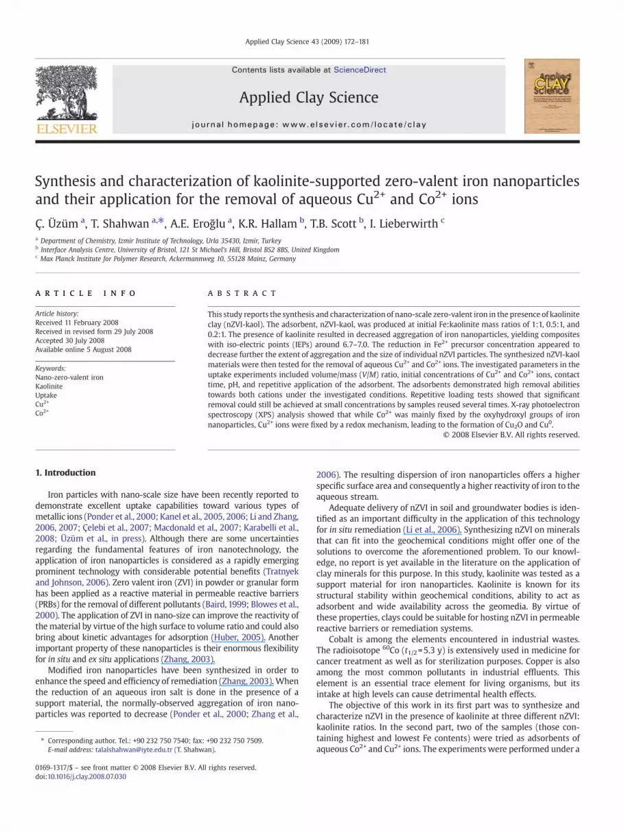
Fig. 11. Elemental EDXmapping images of: (a) Al, (b) Si, (c) Fe, and (d) Co obtained from a nZVI-kaol sample loaded with 1000mg/L Co2+ ions. The two EDX images in (e) and (f) show,respectively, the distribution of Fe and Cu obtained from an Fe-rich region on a nZVI-kaol sample loaded with 1000 mg/L Cu2+ ions.
178 Ç. Üzüm et al. / Applied Clay Science 43 (2009) 172–181
is approached within the first 2–3 h of mixing for both nZVI-kaol1 andnZVI-kaol5 at the studied Co2+ and Cu2+ concentration of 100.0 mg/L.
The effect of initial concentrationswas investigated in the range 1.0–500.0mg/Lwhile theV/M ratiowasfixed at 800mL/g (0.050 g adsorbentin 40.0mL solution). The obtained isotherms are presented in Fig. 7. Bothtypes of nZVI-kaol (nZVI-kaol1 and nZVI-kaol5) were more effective inthe removal of Cu2+ compared to Co2+. As estimated from the isotherms,the removal capacity of nZVI-kaol1 was around 25 mg/g for Co2+ and140 mg/g for Cu2+. When nZVI-kaol5 was applied, the removal capacitywas around 23mg/g for Co2+ and 32mg/g for Cu2+. The results suggest astrong correlation between the fixed amount of Cu2+ and the amount ofnZVI in the adsorbent, while the uptake capacity of Co2+ decreased onlyslightly. These observations are discussed in relation with the fixationmechanisms of both cations in the next section.
In the uptake experiments in which nZVI-kaol5 is applied, thecapacity of removal can be enhanced by increasing the amount of theadsorbent or reducing the volume of the solution— i.e. decreasing the
V/M ratio. A set of experiments was performed to evaluate the changein the extent of uptake with the applied V/M ratio at the two initialconcentrations of Co2+ and Cu2+ ions; 50.0 and 500.0 mg/L. Theobtained results, summarized in Table 1, suggest that decreasing theV/M ratio leads to an almost complete removal of metal ions at theinitial concentration of 50.0 mg/L, and increases the % uptake by up tofour times at an initial metal ion concentration as high as 500.0 mg/L.
Since Fe-based materials are susceptible to oxidation in a wetenvironment, other sets of experiments were conducted to evaluatethe effect of repetitive application on the removal capability of nZVI-kaol. These experiments were carried out at 5.0 and 100.0 mg/L initialconcentrations of Co2+ and Cu2+ using nZVI-kaol5 as adsorbent. Theresults are drawn in Fig. 8 in term of uptake percentages. The nZVI-kaol samples appeared to remove more than 90% of 5.0 mg/L Co2+ andCu2+ ions for 4–5 successive uses. When the metal concentration isincreased to 100.0 mg/L, a fast deterioration in the reactivity of theadsorbent is seen to take place following the first application.
179Ç. Üzüm et al. / Applied Clay Science 43 (2009) 172–181
The solutionpH is among themost critical factors that affect both thespeciation of metal ion in solution, and the speciation of the surface ofadsorbent in contact with the solution. A theoretical speciation analysisfor cobalt and copper ions in solution was performed over a wide pHrange using Visual MINTEQ software under various input conditions oftemperature, ionic strength, and concentration. The results of cobaltanalysis showed that up to pH values around 8, the dominant chemicalform of Co in aqueous media was Co2+. Beyond pH 9.0, CoOH+ and Co(OH)2(aq) became increasingly prominent. The analysis of copperindicated that Cu2+ is the dominating chemical form up to pH of 7.With the increase in pH, forms such as Cu(OH)+, Cu2(OH)22+, Cu3(OH)42+,Cu(OH)3−, and Cu(OH)2 were predicted to prevail.
The results of the experiments that investigated the effect of initialpH on the extent of uptake are given in Table 2. The values show thatthe removal percentages of both Co2+ and Cu2+ ions increased with theincrease in initial pH of themetal solutions. As noted previously in thistext, the IEP of nZVI-kaol samples was in the pH range 6.7–7.0. Theadsorptionproperties of oxide and oxyhydroxide groups on the shell ofiron nanoparticles are strongly affected by the solution pH. It isreasonable to assume that the adsorption and surface coordinationproperties of iron nanoparticles are similar to those of classical metaloxides/hydroxides (Li and Zhang, 2007).When the solutionpH exceedsthe IEP, the oxide surface becomes negatively charged and conse-quently surface complexation reactions would be enhanced. At highcation concentrations, the electrostatic forces might also make a con-tribution to the fixation process. Under such conditions, the pHwill beimportant in determining the thickness of the double layer at theinterface between the oxide surface and solution. If the pH is below theIEP, the double layer becomes thicker due to repulsive forces betweenthe positively charged surface and the cations, thus decreasing thechance for interaction between the adsorbate and the surface. As thepH increases beyond the IEP, the attractive forces between the negativecharge on the surface and the adsorbate cations cause the double layerto shrink leading to a higher chance of adsorbate–surface interaction.In line with the trend observed throughout this study, higher remo-val percentages were observed for Cu2+ compared to Co2+ at all initialsolution pH values.
In order to assess the uptake capacity of kaolinite, a separate set ofexperiments was performed using this clay mineral under conditionsidentical to those used to study the effect of concentration on nZVI-kaol.Within the studied range of concentrations, the uptake capacitiesof kaolinite toward Co2+ and Cu2+ ions did not exceed 5 and 8 mg/g,respectively, It was concluded that the nZVI component plays themajor role in the fixation of both cations by the composite adsorbent.This was also confirmed by the EDXmapping analysis presented in thenext section.
3.3. Mechanisms of uptake
The core-shell structure of iron nanoparticles enables them todemonstrate basically two uptake mechanisms toward metallic ions.The core, constituted of zero-valent iron, forms an electron source thatmight reduce ions possessing higher standard reduction potentialsthan iron. The shell, on the other hand, possesses hydroxyl groups atthe interfacewith the solution and is capable as a result tofix adsorbateions by surface complexation.
In this study, XPS analysis was conducted to reveal the oxidationstates of cobalt and copperfixed by iron nanoparticles on the surface ofnZVI-kaol samples. In addition, the chemical forms of Fe and O wereanalyzed to characterize the iron nanoparticles following the uptakeprocess. The ability to distinguish between different oxidation statesis one of the major strengths of the XPS technique. The related cha-racterization is based on the binding energy shifts demonstrated bythe photoelectrons when their atoms experience different chemicalenvironments. Atoms of a higher positive oxidation state tend to ex-hibit higher binding energies due to the extra coulombic interaction
between the photoelectron and the ion core. The obtained results fornZVI-kaol samples loadedwith Co2+ and Cu2+ ions are discussed belowseparately.
3.3.1. Co-Loaded nZVI-KaolAnXPS spectrum that shows the Fe 2p photoelectronprofile in a nZVI-
kaol sample after being exposed to Co2+ ions at 500.0 mg/L concentrationis shown inFig. 9 (curveb). TheXPS results indicate that ironnanoparticleson the kaolinite surface had undergonewet oxidation to form iron oxide/hydroxide species following the uptake of Co2+ ions. The Fe 2p3/2 line iscentered at 711.7±0.1 eV binding energy. A similar value was previouslyreported for Fe in iron oxyhydroxide (FeOOH) (Wagner et al., 1979). The O1s peak in the spectra is centered at 531.5 eV and displays strongasymmetry to the low binding energy side. In previous studies, O 1s inkaolinitewas reported to occur at 531.3 eV (Lombardi et al., 2006). On theother hand, O 1s peak originating fromO2− inα-Fe2O3 (hematite) and thatinγ-Fe2O3 (magheamite)were reported to occur at 530.0 eV and529.9 eV,respectively. The same study reported also that O 1s peak arising fromO2−
in the hydroxyl groups of α-FeOOH (goethite) and γ-FeOOH (lepidocro-cite) occurred at 531.0 eV and 531.1 eV, respectively (Grosvenor et al.,2004). Curve fitting of the O 1s profile in our samples indicated signalcontributions from O2− in metal oxides (530.0 eV), and also from a moredominant peak at 531.5 eV, arising from a combination of signals fromoxygen present in the kaolinite structure and in hydroxide phases e.g.FeOOHand/or Co(OH)2. The oxide-relatedpeak is estimated to account for~36% of the recorded signal.
The recorded Co 2p3/2 peaks in the current study, shown in Fig.10a,were located at 781.2±0.1 eV andwere asymmetric to the high bindingenergy side. This value, which is identical to the one we reportedearlier for Co-loaded nZVI (Üzümet al., inpress), occurswell above thatreported in the literature for metallic cobalt, Co0, i.e. 778.0±0.2 eV(Wagner et al., 1979; McIntyre and Cook, 1975), and is close to thosereported earlier for Co2+ in cobalt oxide (780.0±0.2 eV) (McIntyre andCook,1975;McIntyre et al.,1990), and cobalt hydroxide (782.0±0.1 eV)(Tan et al., 1991).
Although Co2+ possesses a standard reduction potential (=−0.28 V at298 K) that is somewhat larger than that of Fe2+ (=−0.44 V at 298 K), thereductionof Co2+ by Fe0might not takeplace. It is proposed that electrontransfer from Fe0 to the incoming cation, when equilibrium isestablished, might take place mainly via defects in the oxide shell(direct transfer), or through the conduction band of the oxide shell(indirect transfer) (Li and Zhang, 2007). For such a transfer to be ther-modynamically favorable, the energy of the electrons in the iron metaland/or the oxide layer must be more negative than the standardpotential of the redox couple of the incoming cation (Li and Zhang,2007). As a result, unless direct electron transfer is taking place (theextent of which is unclear), the redox process will be governed by theFermi energy (EF) of the oxide shell, the value ofwhich is reported tovarybetween −0.18 and −0.33 V (Balko and Tratnyek, 1998). With thestandard potential of the Co2+/Co0 redox couple in mind (=−0.28 V at298 K), an effective overlap between the unoccupied electron energylevels of the incoming Co2+ and the conduction band of the oxide shellto produce Co0 seems to be doubtful. This approach assumes that thegiven values for bulk iron are not very different from those of ironnanoparticles and as such shouldbe viewed as afirst approximation thatrequires further electrochemical consideration.
Based on the above, it ismost likely that the uptakemechanismof Co2+
on nZVI-kaol involves chemical complexation to the exposed hydroxylgroups in addition to precipitation at high metal ion concentrations.
3.3.2. Cu-Loaded nZVI-kaolThe recorded Fe 2p photoelectron profile from Cu-loaded nZVI-
kaol is shown in Fig. 9c. The Fe 2p3/2 line was centered at 711±0.1 eV.Unlike Fe 2p3/2 peak in Co-loaded nZVI-kaol sample, this bindingenergy value is very close to that reported previously (Grosvenor et al.,2004) for Fe in γ-Fe2O3 (711.0 eV) and is well below those reported for
180 Ç. Üzüm et al. / Applied Clay Science 43 (2009) 172–181
Fe in α-FeOOH (711.4 eV) and γ-FeOOH (711.5 eV). However, the O 1swas centered at 531.5 eV and displayed strong asymmetry to the lowbinding energy side, similar to that reported in the previous section.
The Cu 2p3/2 lines in the Cu-loaded nZVI-kaol were centered at abinding energy of 932.3±0.1 eV, as shown in Fig. 10b. Our measure-ment of the same peak for Cu-loaded pure nZVI coincides with thisobservation. The given value is very close to the Cu 2p3/2 linespreviously reported for Cu0 (932.6±0.2 eV) (Wagner et al., 1979), andCu+ in Cu2O (932.4±0.3 eV) (Wagner, 1975). The Auger parameter, apowerful tool for elucidating the electronic environment of an atom invarious compounds, was calculated in order to distinguish betweenthe two oxidation states of Cu. This parameter is defined as the sum ofthe relevant Cu 2p3/2 core ionization photoelectron energy, EB, and theappropriate Auger electron kinetic energy, EK for the same element ofinterest. In this case, the Auger line of interest is Cu LMM occurring at570.2 eV. Based on the measured values of EB (932.3 eV) and EK(1486.6–570.2 eV), the copper Auger parameter was here calculated as1848.7 eV, indicating that the recorded signal was derived from Cu+
rather than Cu0. This result shows that Cu2+ ions are reduced by Fe0
primarily to Cu+ (in the form of Cu2O). Moreover, the recorded Cu 2plines were asymmetric to the high binding energy side, indicating thatcomplete Cu reduction had not occurred, and that some small portionremained in a Cu2+ oxidation state.
The oxidation–reductionmechanism associated with the uptake ofCu2+ ions by iron nanoparticles is justified by the fact that the standardpotentials of the Cu2+/Cu0 (=+0.34 V at 298 K) and Cu2+/Cu+ (=+0.16 Vat 298 K) couples are both well above that of Fe2+/Fe0 (=−0.44 V at298 K). As a result, the energy of the electrons in the conduction bandsof iron metal and/or iron oxide will be distinctly more negative thanthe standard potential of both reduction couples, creating an effectivedriving force for electron transfer from zero-valent iron to the fixedCu2+ ions.
As the uptake experiments presented earlier in this text haveshown, under equivalent experimental conditions, the extent ofremoval of Cu2+ ions is greater than that of Co2+ ions. The uptaketrend seems to be closely related to the mechanism of removal, withthe redox route observed in the Cu2+ case leading to greater removalthan the adsorption mechanism operating with Co2+ case. As given inSection 3.2, the uptake capacity of nZVI-kaol1 towards Cu2+ wasaround 140mg/g, while that of Co2+ was 25mg/g.When the amount ofiron precursor is decreased to one-fifth of its initial amount, as is thecase in the nZVI-kaol5 composite, the removal capacity of Cu2+ goesdown to 32 mg/g, probably as a result of decrease in the amount ofelectron source (nZVI) in the adsorbent. In the case of Co2+, apparentlyfixed by a adsorption mechanism, the uptake capacity decreasesslightly to about 23 mg/g upon applying nZVI-kaol5 as the adsorbent.Since the extent of adsorption is dependent on the availability ofsurface sites, the observed result seems to originate from the en-hancement in the dispersion of nZVI on the kaolinite surface as aresult of decreasing the amount of Fe precursor during synthesis ofnZVI-kaol5.
The results of the XPS surveys conducted in this study for Co2+ andCu2+ uptake on nZVI-kaol coincide with those reported recently forthe uptake of each of the two ions on pure nZVI (Üzüm et al., in press;Karabelli et al., 2008). Hence, although preparing nZVI in the presenceof kaolinite affects the extent of aggregation of the nanoparticles, themechanism of interaction between the adsorbate cations and ironnanoparticles seems to be unaffected.
Energy-Dispersive X-ray (EDX)mapping analysiswas used to revealthe distribution of Co and Cu signals on the surface of nZVI-kaolsamples after loading them with 1000.0 mg/L initial concentrationsof Co2+ and Cu2+ ions. Typical elemental maps are shown in Fig. 11.The distributions of Al and Si elements (Fig. 11a,b), which form thebackbone of the clay mineral are observed to coincide except forsome Si-rich regions which possibly originate from minor amounts ofassociated quartz. The signals of Co and Cu showed weak correlation
with those of Al and Si, indicating that kaolinite fraction in the nZVI-kaol adsorbent was not favored during adsorption. On the other hand,Co signals seem to be strongly associated with those of Fe, suggestingselective binding to the nZVI component of the adsorbent (Fig. 11c,d).Here it must be noted that possible overlap might take place betweenthe EDX lines of Co Kαwith Fe Kβ due to their close energies, In the caseof Cu, the X-ray maps recorded in Fe-rich regions in the adsorbentreveal the formation of intense and distinct domains that do notcorrelate also with Fe signals in nZVI-kaol (Fig. 11e,f). Taking also intoconsideration the XPS findings, these domains can be attributed toCu2O and Cu0 phases, a result that agrees with that we reportedrecently for Cu2+ uptake by pure nZVI (Karabelli et al., 2008).
4. Conclusions
The presence of kaolinite during the synthesis of iron nanoparticlesleads to a partial decrease in their extent of aggregation, producingdispersed nanoparticles with sizes varying between 10 and 80 nm.When the Fe2+ precursor concentration was decreased, better disper-sion was qualitatively observed. The dispersed ZVI nanoparticlesshowed the characteristic core-shell structure.
Kaolinite-supported nZVI (nZVI-kaol) demonstrated higher uptakecapacities toward Cu2+ ions compared to Co2+ ions. According to XPSresults, Co2+ ions were fixed through adsorption and precipitation me-chanisms, while Cu2+ ions were fixedmainly through a redoxmechanismthat lead to the formation of Cu2O and to a lesser extent Cu0.
More effort is still needed to achieve better dispersion of nZVI onkaolinite surfaces, the thing that necessitates further manipulations ofthe starting kaolinite to iron ratios.
Acknowledgement
This workwas sponsored by the 2006 İYTE 13 fund provided by theİzmir Institute of Technology. The authors would like to thank theCenter of Material Research at Izmir Institute of Technology (İYTE-MAM) for the assistance with the SEM and XRD measurements.
References
Baird, C., 1999. Environmental Chemistry. W.H. Freeman, New York.Balko, B.A., Tratnyek, P.G., 1998. Photoeffects on the reduction of carbon tetrachloride by
zero-valent iron. J. Phys. Chem. B 102, 1459–1465.Blowes, D.W., Ptacek, C.J., Benner, S.G., Mcrae, Che, W.T., Bennett, T.A., Puls, R.W., 2000.
Treatment of inorganic contaminants using permeable reactive barriers. J. Contam.Hydrol. 45, 123–137.
Çelebi, O., Üzüm, Ç., Shahwan, T., Erten, H.N., 2007. A radiotracer study of the adsorptionbehavior of aqueous Ba2+ ions on nanoparticles of zero-valent iron. J. Hazard. Mater.148, 671–676.
Grosvenor, A.P., Kobe, B.A., Biesinger, M.C., McIntyre, N.S., 2004. Investigation ofmultiplet splitting of Fe 2p Xps spectra and bonding in iron compounds. Surf.Interface Anal. 36, 1564–1574.
Huber, D.L., 2005. Synthesis, properties, and applications of iron nanoparticles. Small 1,482–501.
Kanel, S.R., Manning, B., Charlet, L., Choi, H., 2005. Removal of arsenic(Iii) fromgroundwater by nanoscale zero-valent iron. Environ. Sci. Technol. 39, 1291–1298.
Kanel, S.R., Greneche, J.M., Choi, H., 2006. Arsenic(V) removal from groundwater usingnano scale zero-valent iron as a colloidal reactive barrier material. Environ. Sci.Technol. 40, 2045–2050.
Karabelli, D., Uzum, C., Shahwan, T., Eroglu, A.E., Scott, T., Hallam, K.R., Lieberwirth, I.,2008. Batch removal of aqueous Cu2+ ions using nanoparticles of zero-valentiron: a study of the capacity and mechanism of uptake. Ind. Eng. Chem. Res. 47,4758–4764.
Li, X.Q., Zhang, W.-X., 2006. Iron nanoparticles: the core-shell structure and uniqueproperties for Ni(Ii) sequestration. Langmuir 22, 4638–4642.
Li, X.Q., Zhang, W.-X., 2007. Sequestration of metal cations with zerovalent ironnanoparticles — a study with high resolution X-Ray photoelectron spectroscopy(Hr-Xps). J. Phys. Chem. C 111, 6939–6946.
Li, L., Fan, M., Brown, R.C., Leeuwen, J.V., Wang, J., Wang, W., Song, Y., Zhang, P., 2006.Synthesis, properties, and environmental applications of nanoscale iron-basedmaterials: a review. Crit. Rev. Environ. Sci. Technol. 36, 405–431.
Lombardi, K.C., Mangrich, A.S., Wypych, F., Rodrigues-Filho, U.P., Guimarães, J.L.,Schreiner, W.H., 2006. Sequestered carbon on clay mineral probed by electronparamagnetic resonance and X-Ray photoelectron spectroscopy. J. Colloid InterfaceSci. 295, 135–140.

181Ç. Üzüm et al. / Applied Clay Science 43 (2009) 172–181
Macdonald, J.E., Kelly, J.A., Veinot, J.G.C., 2007. Iron/iron oxide nanoparticle sequestra-tion of catalytic metal impurities from aqueous media and organic reactionproducts. Langmuir 23, 9543–9545.
McIntyre, N.S., Cook, M.G., 1975. X-ray photoelectron studies on some oxides andhydroxides of cobalt, nickel, and copper. Anal. Chem. 47, 2208–2213.
McIntyre, N.S., Johnston, D.D., Coatsworth, L.L., Davidson, R.D., Brown, J.R., 1990. X-rayphotoelectron spectroscopic studies of thin film oxides of cobalt and molybdenum.Surf. Interface Anal. 15, 265–272.
Ponder, S.M., Darab, J.G., Mallouk, T.E., 2000. Remediation of Cr(Vi) and Pb(Ii) aqueoussolutions using nanoscale zero-valent iron. Environ. Sci. Technol. 34, 2564–2569.
Tan, B.J., Klabunde, K.J., Sherwood, P.M.A., 1991. Xps studies of solvated metal atomdispersed (Smad) catalysts; evidence for layered cobalt–manganese particles onalumina and silica. J. Am. Chem. Soc. 113, 855–861.
Tratnyek, P.G., Johnson, R.L., 2006. Nanotechnologies for environmental cleanup. NanoToday 1, 44–48.
Üzüm, Ç., Shahwan, T., Eroğlu, A.E., Lieberwirth, I., Scott, T.B., Hallam, K.R., in press.Application Of Zero-Valent Iron Nanoparticles For The Removal Of Aqueous Co2+
Ions Under Various Experimental Conditions. Chem. Eng. J.
Wagner, C.D., 1975. Chemical shifts of Auger lines, and the auger parameter. FaradayDiscuss. Chem. Soc. 60, 291–300.
Wagner, C.D., Riggs, W.M., Davis, L.E., Moulder, J.F., Muilenberg, G.E., 1979. Handbook OfX-Ray Photoelectron Spectroscopy. Perkin-Elmer Corporation, Physical ElectronicsDivision, Eden Prairie, Minn., p. 55344.
Wang, C., Zhang, W., 1997. Synthesizing nanoscale iron particles for rapid and completedechlorination of TCE and PCBS. Environ. Sci. Technol. 31, 2154–2156.
Wang, W., Jin, Z., Li, T., Zhang, H., Gao, S., 2006. Preparation of spherical ironnanoclusters in ethanol–water solution for nitrate removal. Chemosphere 65,1396–1404.
Zhang, W.X., 2003. Nanoscale iron particles for environmental remediation: anoverview. J. Nanopart. Res. 5, 323–332.
Zhang, H., Jin, Z.H., Han, L., Qin, C.H., 2006. Synthesis of nanoscale zero-valent ironsupported on exfoliated graphite for removal of nitrate. Trans. Nonferr. Met. Soc.China 16, S345–S349.


