Embed Size (px)
Citation preview
The Acoustic Neuroma:Some clinical and histological aspects
Het acousticusneurinoma:Enkele klinische en histologische aspecten
Glen E.J. Forton
2
Printing of this thesis was financially supported by:
Glaxo - Wellcome Belgium nvSolvay Pharma & Cie
Pfizer nvSanofi – Synthelabo nvEli – Lilly Benelux nvJanssen – Cilag nv
Published by Glen E.J. Forton, Prinsenlaan 12, 8400 Oostende, Belgium
Copyright © 2001All rights reserved. No part of this publication may be reproduced or transmitted in any form or by anymeans, without the prior written permission of the publisher.
Printed in the Netherlands by Print Partners Ipskamp, Enschede
3
UNIVERSITEIT ANTWERPEN
Universitaire Instelling Antwerpen
Faculteit Medische en Farmaceutische WetenschappenDepartement Geneeskunde
The Acoustic Neuroma:Some clinical and histological aspects
Het acousticusneurinoma:Enkele klinische en histologische aspecten
Proefschrift voorgelegd tot het behalen van de graad van
doctor in de medische wetenschappen aan de
Universitaire Instelling Antwerpen door
Glen E.J. FORTON
Promotoren: Prof.Dr. F.E. Offeciers Antwerpen,Prof.Dr. C.W.R.J. Cremers 2001

4
Voor Dominique, Audrey en Laura
5
Contents
Chapter 1 Introduction and historical overview 7
Chapter 2 Histopathology 37
Differentiation of cartilage and bone in human
fetal temporal bones with Luxol fast blue stain 39
F. Declau, L. Moeneclaey, G. Forton, J. Marquet†
Arch Otorhinolaryngol 1988;245:218-220
The involvement of the cochlear nerve in
neurinomas of the eighth cranial nerve 47
G.E.J. Forton, L.L.M. Moeneclaey, F. Declau, J.F.E. Marquet†
Arch Otorhinolaryngol 1989;246(3):156-160
The solitary schwannoma of the eighth
cranialnerve: An immuno-histochemical study of
the cochlear nerve - tumor interface 59
J.F.E. Marquet†, G.E.J. Forton, Offeciers, L.L.M. Moeneclaey
Arch Otolaryngol Head Neck Surg 1990;116(9):1023-1025
Chapter 3 Clinical features 71
Acoustic neuroma ingrowth in the cochlear
nerve: An analysis of clinical data 73
G.E.J. Forton
Submitted for publication
Acoustic schwannoma: MR findings in 84 tumors
A pictorial essay 85
T.H. Mulkens, P.M. Parizel, J.-J. Martin, H.R. Degryse
P.H. Van de Heyning, G.E.J. Forton, A.M. De Schepper
Am J Roentgen 1993;160:395-398
6
Chapter 4 Atypical manifestations of the neuroma in
the petrous bone and other pitfalls 93
Facial Nerve Neuroma: Report of Two Cases
Including Histologic and Radiological Imaging Studies 95
G.E.J. Forton, L.L.M. Moeneclaey, F.E. Offeciers
Eur Arch Otorhinolaryngol 1994;251:17-22
Preoperatively diagnosed utricular neuroma treated
by selective partial labyrinthectomy 107
G.E.J. Forton, Th. Somers, R. Hermans, A.L. Baert, F.E. Offeciers
Ann Otol Rhinol Laryngol 1994;103:885-888
Problems with flute playing: an otological problem?
Case report of a peculiar cerebellar astrocytoma 115
G. Forton, J. Verlooy, P. Cras, P. Parizel, P. Van de Heyning
Acta Otorhinolaryngol Belg 1993;47(4):389-96
Chapter 5 Summary and conclusions 127
Dankwoord 131
Full color reproductions 135
7
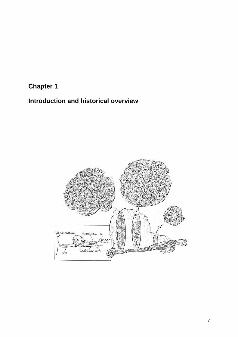
Chapter 1
Introduction and historical overview
8
Semantics
Ever since Eduard Sandifort of Leyden (1742-1814) first described this pathological entity in
1777, controversy has existed and still exists as to the origin, histopathology, growth patterns
and above all the surgical treatment of this benign tumor, commonly called “acoustic
neuroma”.
The term “neuroma” was first used by Verocay in 1910 and is not generally accepted. Stout
therefore suggested in 1935 to use the term neurilemmoma. Young also rejects this term, for
the neurilemma is the innermost layer of endoneurium, also called the Plenk-Laidlaw sheath,
that serves as basal membrane for the Schwann cells (1). Friedmann (2) finally, suggests
that the term schwannoma should be preferred, for in his opinion; the tumor originates from
the Schwann cells and not from the endoneurium in its narrowest meaning.
The term “acoustic neuroma” is therefore a misnomer; even more so because the tumor
usually arises from the vestibular nerve and not the cochlear nerve. Vestibular schwannoma
is therefore a more appropriate denomination, although not quite uniformly adopted.
This does not end the discussion, however, for Penfield et al. suggested as early as in 1932
that the tumor is of fibroblastic-mesodermal origin and they suggested it be called “perineural
fibroblastoma”. Contemporary literature still is not unanimous on this matter. One of the
problems is the uncertainty about the exact origin of the Schwann cells. There are, however,
indications as to a mesodermal origin and there also are arguments as to an ectodermal
origin.
These discussions notwithstanding, the terms vestibular schwannoma, neuroma and
neurinoma are still being used interchangeably, even in reference textbooks.
Epidemiology
The tumors originating from the eighth cranial nerve account for about 6 to 8% of all
intracranial tumors and comprise approximately 78% of all cerebellopontine angle tumors (3)
According to Mattox (4), acoustic neuromas are found in about 1.5% of all routine autopsies.
Hardy & Crowe (5) have reported back in 1936 a very high number of 2.5%. Their series has
been reviewed, however, and four cases have been reclassified, yielding a more acceptable
number of 0.8%. According to House and Hitselberger (6), only 0.1% of all these tumors
eventually become symptomatic.
9
Interesting data, spanning no less than 19.5 years, were recently published by Tos et al. (7),
offering a very reliable survey of the Danish situation (Table 1).
Number of new cases Incidence per millionJune 1976 – June 1983 278 7.8July 1983 – June 1990 337 9.4July 1990 – December 1995 355 12.4
Table 1: the incidence of acoustic neuromas in Denmark according to Tos et al. (7)
The authors observed a significant increase in the incidence of the acoustic neuroma during
the last two periods, but this can be easily explained by the more refined radiological
techniques allowing for earlier detection of smaller tumors. Indeed, the increase mainly
consists of small and intracanalicular tumors (7).
According to House & Hitselberger (6) the mean age at diagnosis is 47.3 years with 51% of
the patients being male and 49% female. In our own modest series, the mean age was 59.2
years and the male/female ratio was 0.46 (chapter 3).
Clinical presentation
In the early days, an acoustic neuroma was usually only diagnosed after it had started
compressing cranial nerves, such as the facial, trigeminal and vagus nerves. In a further
stage the cerebellum, brainstem and the other cranial nerves in the vicinity, such as the VIth,
Xth, XIth and XIIth eventually were compressed too and a life threatening condition with
elevated intracranial pressure arose. Succinct clinical descriptions of what it means to die of
an untreated acoustic neuroma were published by Sir Charles Bell (8) in 1830.
It was Toynbee (9) who first recognized an early acoustic neuroma confined to the internal
auditory canal. His description was presented in 1853.
Not until the end of the nineteenth century however was the diagnosis made on the basis of
clinical symptoms before death. Indeed, little was known of audiology before 1900. Julius
Lempert (10) introduced the concepts of perceptive and conductive hearing loss in 1938.
The majority of patients present with a unilateral, slowly progressive sensorineural hearing
loss as the principal symptom. There is, however, no such thing as a “typical” audiogram.
As demonstrated in William House’s monograph I (11), four pure tone loss patterns can be
roughly distinguished. Interestingly, 67% of all cases had a high tone loss type, defined as a
perceptive hearing loss that sloped from low frequencies to high frequencies with a loss of at
10
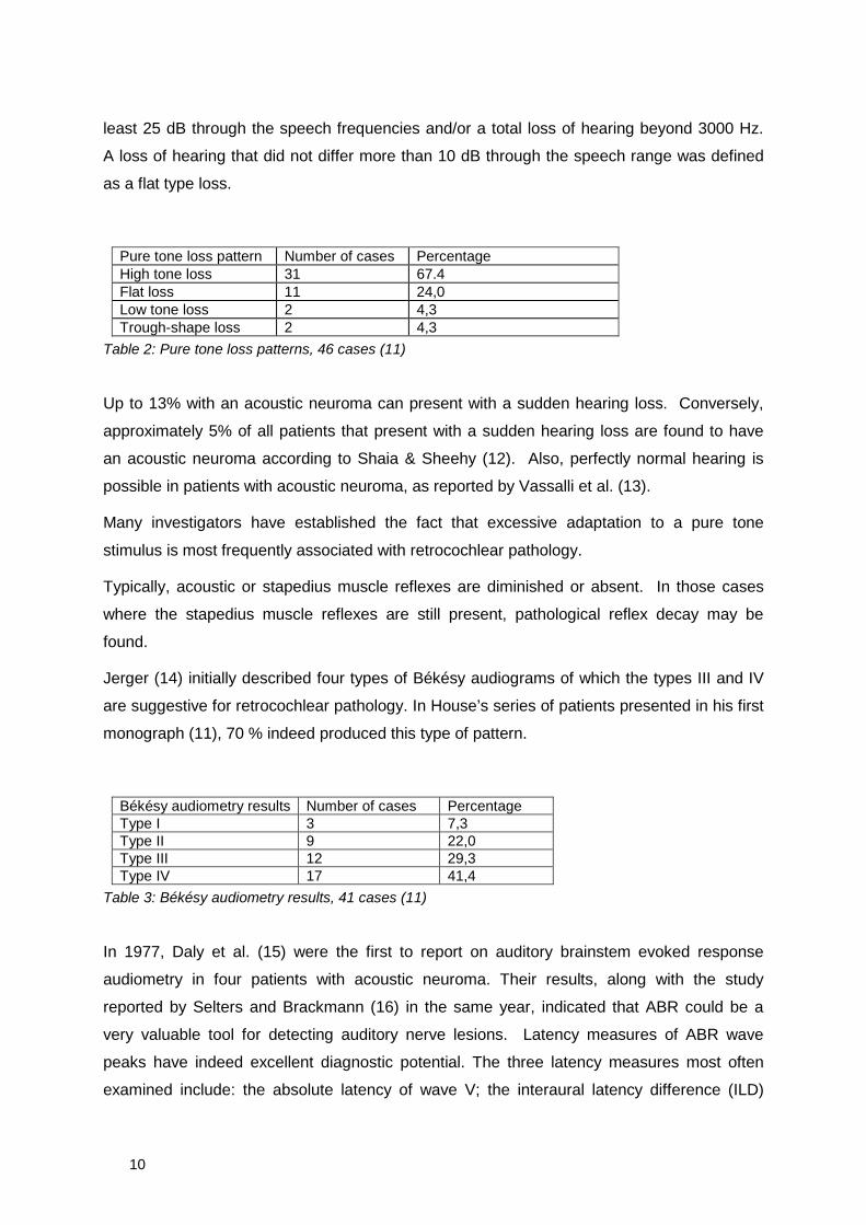
least 25 dB through the speech frequencies and/or a total loss of hearing beyond 3000 Hz.
A loss of hearing that did not differ more than 10 dB through the speech range was defined
as a flat type loss.
Pure tone loss pattern Number of cases PercentageHigh tone loss 31 67.4Flat loss 11 24,0Low tone loss 2 4,3Trough-shape loss 2 4,3
Table 2: Pure tone loss patterns, 46 cases (11)
Up to 13% with an acoustic neuroma can present with a sudden hearing loss. Conversely,
approximately 5% of all patients that present with a sudden hearing loss are found to have
an acoustic neuroma according to Shaia & Sheehy (12). Also, perfectly normal hearing is
possible in patients with acoustic neuroma, as reported by Vassalli et al. (13).
Many investigators have established the fact that excessive adaptation to a pure tone
stimulus is most frequently associated with retrocochlear pathology.
Typically, acoustic or stapedius muscle reflexes are diminished or absent. In those cases
where the stapedius muscle reflexes are still present, pathological reflex decay may be
found.
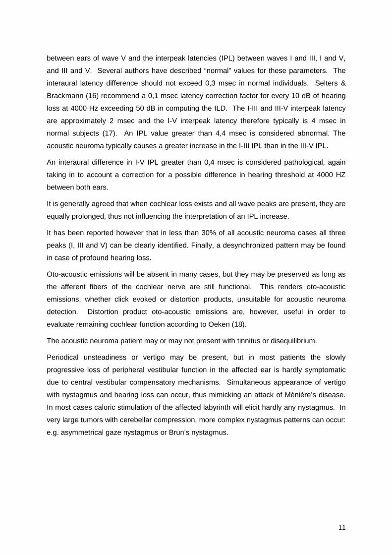
Jerger (14) initially described four types of Békésy audiograms of which the types III and IV
are suggestive for retrocochlear pathology. In House’s series of patients presented in his first
monograph (11), 70 % indeed produced this type of pattern.
Békésy audiometry results Number of cases PercentageType I 3 7,3Type II 9 22,0Type III 12 29,3Type IV 17 41,4
Table 3: Békésy audiometry results, 41 cases (11)
In 1977, Daly et al. (15) were the first to report on auditory brainstem evoked response
audiometry in four patients with acoustic neuroma. Their results, along with the study
reported by Selters and Brackmann (16) in the same year, indicated that ABR could be a
very valuable tool for detecting auditory nerve lesions. Latency measures of ABR wave
peaks have indeed excellent diagnostic potential. The three latency measures most often
examined include: the absolute latency of wave V; the interaural latency difference (ILD)
11
between ears of wave V and the interpeak latencies (IPL) between waves I and III, I and V,
and III and V. Several authors have described “normal” values for these parameters. The
interaural latency difference should not exceed 0,3 msec in normal individuals. Selters &
Brackmann (16) recommend a 0,1 msec latency correction factor for every 10 dB of hearing
loss at 4000 Hz exceeding 50 dB in computing the ILD. The I-III and III-V interpeak latency
are approximately 2 msec and the I-V interpeak latency therefore typically is 4 msec in
normal subjects (17). An IPL value greater than 4,4 msec is considered abnormal. The
acoustic neuroma typically causes a greater increase in the I-III IPL than in the III-V IPL.
An interaural difference in I-V IPL greater than 0,4 msec is considered pathological, again
taking in to account a correction for a possible difference in hearing threshold at 4000 HZ
between both ears.
It is generally agreed that when cochlear loss exists and all wave peaks are present, they are
equally prolonged, thus not influencing the interpretation of an IPL increase.
It has been reported however that in less than 30% of all acoustic neuroma cases all three
peaks (I, III and V) can be clearly identified. Finally, a desynchronized pattern may be found
in case of profound hearing loss.
Oto-acoustic emissions will be absent in many cases, but they may be preserved as long as
the afferent fibers of the cochlear nerve are still functional. This renders oto-acoustic
emissions, whether click evoked or distortion products, unsuitable for acoustic neuroma
detection. Distortion product oto-acoustic emissions are, however, useful in order to
evaluate remaining cochlear function according to Oeken (18).
The acoustic neuroma patient may or may not present with tinnitus or disequilibrium.
Periodical unsteadiness or vertigo may be present, but in most patients the slowly
progressive loss of peripheral vestibular function in the affected ear is hardly symptomatic
due to central vestibular compensatory mechanisms. Simultaneous appearance of vertigo
with nystagmus and hearing loss can occur, thus mimicking an attack of Ménière’s disease.
In most cases caloric stimulation of the affected labyrinth will elicit hardly any nystagmus. In
very large tumors with cerebellar compression, more complex nystagmus patterns can occur:
e.g. asymmetrical gaze nystagmus or Brun’s nystagmus.
12
Imaging
The first report on acoustic neuroma imaging was from the hand of Folke Henschen from
Stockholm (19) who wrote, back in 1912: “diese Akustikustumoren den inneren Gehörgang
erheblich erweitern und dass diese Erweiterung radiographisch darstellbar sein kann”.
Indeed, the acoustic neuroma tends to enlarge the internal auditory canal and this is one of
the most important radiological signs.
However, detailed radiological examination of the internal auditory canal started when
radiologist like Stenvers, Guillen and Schuller published their methods. The main problem
was avoiding the superimposition of the mid-face and the skull base. Hypocycloidal
tomography together with iodocysternography were the techniques of choice from the 1950’s
to the late 1970’s when CT-scan became the most accurate technique to study the inner ear.
Several generations of CT-scanners followed one another, the latest generation being able to
visualize very small anatomical structures and tumoral lesions. Typically, the acoustic
neuroma is a tumor that is centered on the internal auditory canal and it shows marked
enhancement after contrast administration. The enhancement usually extends into the
internal auditory canal. The canal itself is usually widened with erosion of the posterior ridge.
Also the crista falciformis shows marked erosion. An exception to this rule is, of course, the
so-called “medial acoustic neuroma” as described by Tos et al. (20). This tumor is defined as
an acoustic neuroma without tumor tissue located laterally in the internal auditory meatus. It
is a relatively rare variant since it comprises about 2 – 3% of all acoustic neuromas. Tos et
al. (20) found that these medial tumors were generally larger than the non-medial ones. The
smallest medial tumor they found had an extrameatal diameter of 15 mm. Clinically, there
seems to be no significant difference in symptoms or duration of symptoms between the two
entities. Surgically, Tos et al. (20) found that the involvement of brainstem and cerebellum
were significantly higher than is the case with non-medial acoustic neuromas.
The next important evolution was the introduction of magnetic resonance imaging in the
1980’s. Again, several generations of MRI scanners followed one another and more
sophisticated sequences and software protocols are being developed in rapid succession.
Clinical studies were subsequently carried out to assess the sensitivity of MRI. One of these
studies was a joint effort from the departments of radiology, neurosurgery and
otorhinolaryngolgy of the University Hospital of Antwerp and will be discussed in chapter 3.
After contrast administration, all tumors showed intense and greater than average
enhancement (up to 300 %) on the T1-weighted images, except for one very small
intracanalicular tumor that did not enhance. The enhancement was homogenous in 67% and
13
slightly inhomogeneous in 10%. Heterogeneous enhancement was encountered in cases
with cystic degeneration (22%). The absence of enhancement after contrast administration
in a pathologically proven acoustic neuroma is highly unusual, and as far as we could
establish, our one patient is the only reported case. T2-weighted images may show mild to
moderate peritumoral edema.
Today, visualization of the contents of the labyrinth is a reality. The 3DFT-CISS sequences
allow us to detect extremely small lesions, such as intracochlear or intravestibular neuromas.
A clinical report of a very small intralabyrinthine schwannoma of only 3.5 mm diameter that
had been preoperatively diagnosed by means of MRI was published in the Annals of
Otology, Rhinology and Laryngology (22) and the report is included in this volume (chapter
4). Until today, this case still is the smallest preoperatively diagnosed intralabyrinthine tumor
in the literature.
A major contribution in this field has been made by the Flemish neuroradiologist from
Bruges, J.W. Casselman, as demonstrated in his magnificent doctoral thesis (23). Recently,
MRI imaging technique has evolved dramatically and now allows us to actually assess the
cochlea’s candidacy for hearing preservation surgery. Somers et al. (24) from Antwerp have
recently demonstrated an astonishing correlation (p < 0.05) between a normal
intralabyrinthine signal on 3DFT-CISS MRI images (being an iso-intense signal compared to
the contralateral unaffected ear) and successful hearing preservation surgery. In their series
of twenty six patients with an acoustic tumor, all being candidates for hearing preservation
surgery, hearing was preserved in 50% of the ears where the tumor did not extend into the
fundus (77% of the cases), but only in 33% of the cases where the fundus was obliterated by
tumor (23% of the cases). Moreover, hearing preservation succeeded in 82% of the cases
where a normal intralabyrinthine signal on 3DFT-CISS images was obtained, whereas in
cases where the intralabyrinthine signal intensity was low, hearing was preserved in only
20%. In two cases with low pre-operative intralabyrinthine signal intensity but successful
hearing preservation surgery, the post-operative control MRI showed that the
intralabyrinthine signal intensity had returned to normal. The cause of the lower signal
intensity is not exactly known, but the authors postulate that vascular compression due to
mechanical obstruction by the tumor in the internal auditory canal might be responsible for
this phenomenon. It is a well known fact that the presence of an acoustic neuroma causes
significant biochemical alterations in the fluids of the inner ear, as described by Dix &
Hallpike (25), Schuknecht (26), O’Conner et al. (27), Silverstein et al. (28) and others. The
perilymph of acoustic neuroma patients appears to have a high protein level, which gives rise
to altered light microscopic staining characteristics: Acidophilic staining precipitates are seen
14
in the perilymphatic spaces. Jahnke (29) studied the inner ear in case of an acoustic
neuroma using electron microscopy. He describes high amounts of fibrous long-spacing
collagen, significant thickening of the capillary basement membrane and even doubling of the
capillary basement membrane, mucoid degeneration in the subepithelial space and severe
degeneration of the sensory and non-sensory epithelia. The high protein content is ascribed
to impaired terminal blood supply to the labyrinth secondary to vascular compression or even
invasion of arteries in the internal auditory canal. The radiological finding of a signal
decrease of the cerebrospinal fluid trapped in the fundus and of the intralabyrinthine fluid is in
concordance with the histological findings. The study of the intralabyrinthine fluids by means
of the 3DFT-CISS MRI sequence will yet increase our ability to select the best possible
treatment for any given acoustic neuroma case.
Histology
Harold F. Schuknecht (3) describes the macroscopical appearance of the acoustic
schwannoma as follows: cochleovestibular schwannomas are usually firm, circumscribed,
and encapsulated, and when small, they are round or ovoid in shape. Larger tumors tend to
become lobulated, and then protrude from the internal auditory canal into the
cerebellopontine angle. As the tumor grows, adjacent nerve roots are stretched over the
surface of the mass or are incorporated into it. The internal auditory canal becomes
enlarged and funnel shaped as the result of pressure erosion of the bone. Thus, the
characteristic image of the “police whistle” or “champagne cork” arises.
Microscopically, the acoustic schwannoma is a generally cellular tumor with zones of low
density alternating with zones of high nuclear density.
Antoni, in a 500-page monograph published in 1920, divided these tumors in two histologic
types. Type A is composed of compact tissue of compact and interwoven bundles of long or
oval shaped cells. In some areas, the cells are arranged in whorls, while in other areas there
is a parallel alignment of the rather large nuclei, referred to as “palisading”. Streaks of
collagen fibers are amply present. At times, the arrangement of nuclei and fibers creates
formations resembling the tactile corpuscles of Meissner, known as “Verocay bodies”. Type
B is a degenerate form, which often is intermingled with type A. It is characterized by loose
texture and polymorphism of tumor cells. Nager (30) divided the type B in two subtypes.
Subgroup 1 shows fatty degeneration leading to a honeycombed appearance of large pale
tumor cells with small pyknotic nuclei. Subgroup 2 shows transformation of tumor tissue into

15
hyaline masses in which case the cell content is often reduced to a few stellate tumor cells
embedded in an amorphous hyaline substance. All these types may coexist in one single
tumor.
The typical whorls of collagen, sometimes forming so-called Luse-bodies, are of a particular
kind: it is collagen type IV and V with a characteristic 130 µm band pattern, therefore called
“long-spacing collagen”.
Although several authors have described the presence of the S-100 protein both in the
normal Schwann cell and in the acoustic neuroma (Harkin (31), Clark et al. (32)), this
observation does not necessarily indicate that the schwannoma obligatory has to originate
from the Schwann cells. Gould et al. (33) have described the abundant presence of
vimentine in the cytoplasm of the tumor cells. Vimentine is a so-called intermediate filament
and is typically considered as a mesenchymal marker protein. Peltonen et al. (34) reported
on the presence of vimentine in perineural cells, thus indicating that these cells or not
derivatives of epithelial or endothelial cells, but that they are of mesenchymal origin instead.
Most specimens are also neuron specific enolase (NSE) positive.
One of the more recent developments is immunohistochemical staining using the monoclonal
mouse antibody MIB-1 to demonstrate the Ki-67 nuclear antigen. This antigen is only
expressed by nuclei that are not in the G0 resting phase of the cell cycle and is therefore a
marker of cell proliferation or tumor growth. Several authors have reported on this issue
(table 4).
Lesser et al.(35)
Charabi et al. (36) Chen et al. (37) Yokoyama et al. (38)
Proliferativecell fraction
0.36 – 3.15 % 0.4 – 38.0% 1.16 – 3.40% 0.37% - 6.61 %
Table 4: proliferative cell fraction (percentage of Ki-67 antigen positive cells compared to all cells).
In the aforementioned studies, no correlation was reported between cell proliferation rates,
and patient age, duration of symptoms or tumor volume. Clinical correlation in these studies
suggests that lower proliferation rates are indicative of slower growth rates, but they are not
conclusive.
Erlandson and Woodruff (39) have used electron microscopy to demonstrate that the tumor
cells of the solitary schwannoma are provided with well-structured external laminae that form
some kind of basement membrane for the tumor cells resembling the Plenk-Laidlaw sheath.
16
The cells have spindle shaped nuclei with one or two nucleoli. According to these authors,
these findings indicated clearly that these cells are in fact differentiated Schwann cells.
These last observations are illustrative for the discussion and the speculations as to the true
origin of the acoustic neuroma cells.
Treatment
Surgical treatment
The very early period
The first successful removal of an acoustic neuroma was reported on in 1894 (40) and was
performed by way of a suboccipital approach by the English otologist Sir Charles Ballance on
November 19th, 1894 on a 49-year-old woman. This patient was reported to be alive and
well in 1906. She had a right facial palsy and she had lost function of the right trigeminal
nerve. Furthermore, she had lost the right eye due to a corneal ulcer. In 1903, the German
brain surgeon Krause (41) reported on a unilateral subocciptal approach for the removal of
acoustic tumors. Unfortunately, bleeding was uncontrollable when it occurred in the
cerebellopontine angle and the mortality rate was reported to be 85%. In 1904 the German
otologist Panse (42) decided that the most straightforward way to remove an acoustic
neuroma had to be through the labyrinth and he was thus the first to perform the
translabyrinthine approach to the cerebellopontine angle. Due to lack of instruments and the
limited exposure, mortality again was high. The first transsigmoid approach was devised and
performed by Borchardt in 1905 (43), but again bleeding, this time from the lateral sinus, was
a major problem and after a few attempts, this approach was abandoned.
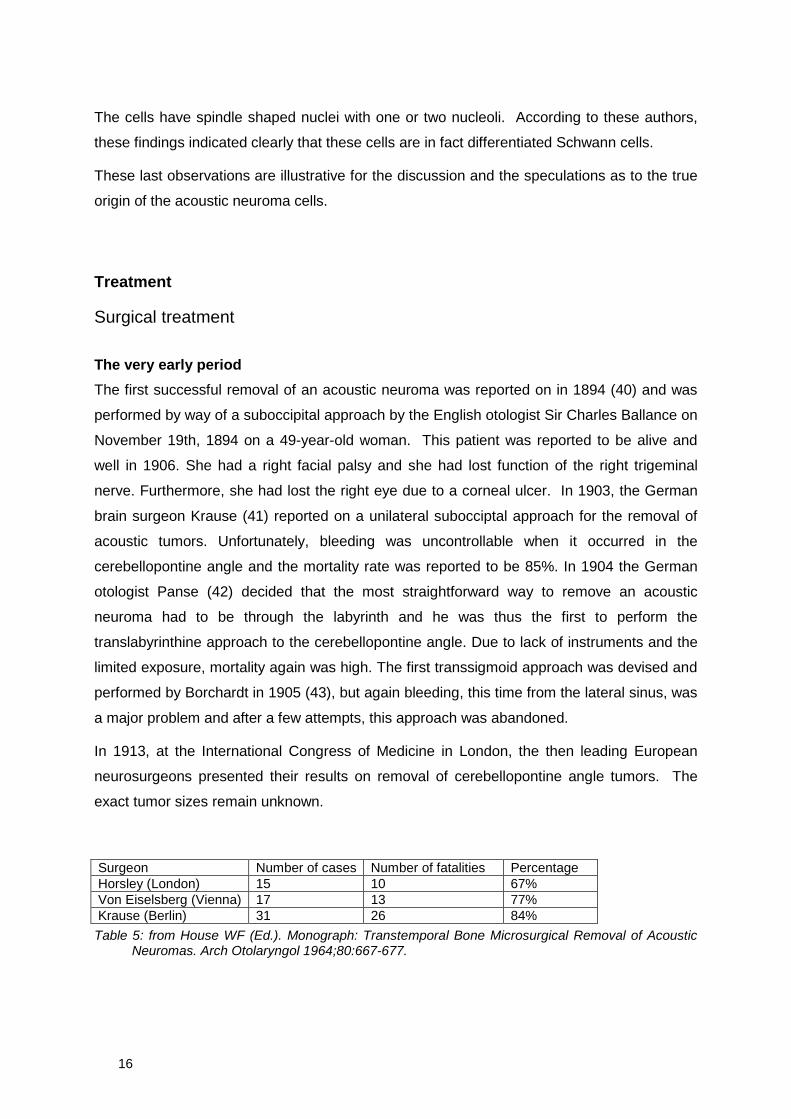
In 1913, at the International Congress of Medicine in London, the then leading European
neurosurgeons presented their results on removal of cerebellopontine angle tumors. The
exact tumor sizes remain unknown.
Surgeon Number of cases Number of fatalities PercentageHorsley (London) 15 10 67%Von Eiselsberg (Vienna) 17 13 77%Krause (Berlin) 31 26 84%
Table 5: from House WF (Ed.). Monograph: Transtemporal Bone Microsurgical Removal of AcousticNeuromas. Arch Otolaryngol 1964;80:667-677.
17
As illustrated above, although brave attempts were made to surgically treat cerebellopontine
angle tumors, morbidity and mortality were formidable.
The Cushing era (1917 – 1935)
This situation was unacceptable to Harvey Cushing (44), who in 1917 published his
monograph “Tumors of the Nervus Acusticus and the Syndrome of the Cerebellopontine
Angel”, a milestone in the surgery of cerebellopontine angle tumors. He described 30 cases
in great detail, 25 of which had tinnitus and hearing loss as their first symptom with the
tinnitus preceding the hearing loss with several months. Of course, most of Cushing’s
patients had very large tumors and had ataxia, advance papilledema or even total blindness
due to severely elevated intracranial pressure. He was the first to think of partial tumor
removal via a wide bilateral suboccipital approach. The approach had to be bilateral, for in
many cases they did not know which side the tumor was preoperatively. Thus, by performing
intracapsular subtotal removal, he was able to dramatically lower operative mortality from
80% to 20%. By 1931, Cushing was able to report an operative mortality rate of only 4%!
Some patients survived long enough to present with recurrence of symptoms due to regrowth
of the tumor remnant. Cushing soon found out that the mortality rate was much higher with
the second operation.
The Dandy era (1922 – 1940)
While Cushing continued to advocate subtotal tumor removal, other neurosurgeons, among
which the famous Walter Dandy, started to pursue total tumor removal. The latter argued
that a higher initial mortality rate was acceptable if better long-term survival was obtained. In
1940 Dandy reported (45) on 40 cases with a mortality rate of 10%. He also abandoned the
bilateral approach and started doing a unilateral suboccipital approach. At that time, the
clinical signs and symptoms were recognized in an earlier stage and the x-ray examination of
the temporal bone usually indicated the site of lesion. Dandy dealt with smaller tumors than
Cushing, was better equipped and realized that he could lower intracranial pressure by doing
a ventricular or cisterna magna tap. While his exposure was still limited, he could gain better
access to the cerebellopontine angle by resecting part of the lateral cerebellar hemisphere.
In addition, anesthesia improved, blood replacement techniques became available and the
electrocautery was invented.
18
The pre-House era (1940 – 1961)
In the post-Dandy era, the discussion on total or subtotal removal continued, but for three
decades no major advances in clinical diagnosis or surgical treatment of the acoustic
neuroma were made, in spite of improved equipment and patient care. More and more
surgeons started doing this kind of surgery, which resulted in a higher cranial nerve deficit
rate, while the mortality rate remained 20%. It is rather illustrative to note that during this
span of time only one surgeon was actually attempting to save the facial nerve. In 1941,
Olivecrona (46) reported on a series of patients in which he had been able to save the facial
nerve in 65%.
Since Balance and Panse, and until well into the 1950’s the otolaryngologist did not have an
active part in the treatment of cerebellopontine angle tumors. If a patient presented with a
unilateral progressive sensorineural hearing loss with tinnitus and cerebellar ataxia, the
diagnosis would of course be considered, but an early diagnosis was unlikely. And even if a
small tumor were diagnosed, the neurosurgeon would not operate until the tumor grew and
became life threatening for the morbidity and mortality was still high.
The House era (1961 - ?)
The situation remained unchanged until the American otologist William House took up the
challenge in 1961 when he presented his candidate’s thesis to the American Laryngological,
Rhinological and Otological Society. His work was called “Surgical Exposure of the Internal
Auditory Canal and its Contents through the Middle Cranial Fossa” (47) and was the first
presentation of a combined otologic-neurosurgical procedure designed for vestibular nerve
sectioning and decompression. In 1964 he published his transtemporal technique (48) for
total removal of acoustic neuromas, No longer were acoustic neuromas referred for the
classic suboccipital approach, but operated by his oto-neurosurgical team via the middle
fossa approach at first and subsequently via the re-instated translabyrinthine technique.
House’s modified translabyrinthine approach was revolutionary as it made use of all newly
invented instruments, such as the operating microscope and diamond burrs (both introduced
in the field of otology by George Shambaugh Jr.), and it allowed early visualization of the
facial nerve. As refined audiologic techniques emerged and iodocysternography was
introduced in the same year, earlier diagnosis was made possible and still smaller tumors
were diagnosed and subsequently operated.
By December 1964, House published his first monograph presenting no less than 54 cases.
The era of microsurgery of the cerebellopontine angle had begun. The co-operation of
19
House, an otologist, and Hitselberger, a neurosurgeon, inspired many teams all over the
world.
Modern microsurgery of the cerebellopontine angle: hearing preservation surgery(early 1970’s)
In the late 1960’s and early 1970’s, otolaryngologists throughout Western Europe, were
inspired by House and started treating acoustic neuromas themselves instead of referring
these patients to the neurosurgeon. We have tried to acquire some data from these
pioneers.
One of the first was Ugo Fisch of Zürich, who did his first translabyrinthine and middle fossa
approaches at the end of 1967 (49).
In the United Kingdom, Andrew Morrison from London, was the first to carry out
translabyrinthine acoustic tumor surgery in 1968 (personal letter from A. Morrison). Except
for the smaller tumors, these operations were performed in conjunction with T. King, a
neurosurgeon. They started doing hearing preservation surgery in 1977, using the
retrosigmoid route. In 1978 Richard Ramsden from Manchester started his impressive series
of 778 cases (2000), using the retrosigmoid approach and the translabyrinthine approach
fairly equally during the first 150 cases (personal letter from R. Ramsden). Later on, most of
the tumors were operated via the translabyrinthine approach.
Jean-Marc Sterkers from Paris operated his first acoustic neuroma by means of the
translabyrinthine approach on March 7th, 1966 (personal letter from J.-M. Sterkers). He
started using the suspetrous and the retrosigmoid approach in order to save hearing from
1972 on. In 1967, Michel Portmann of Bordeaux, another famous French pioneer, operated
his first acoustic neuroma case using the middle fossa approach (personal letter from M.
Portmann). He reported on his experience with the middle fossa approach in 1972 (50).
In 1972, Ed Marres started to do acoustic neuroma surgery at the University Hospital of
Nijmegen, The Netherlands (personal communication).
In Germany, Malte Erik Wigand was a pioneer who performed his first acoustic tumor
removal in 1973. He used the translabyrinthine approach for large tumors and chose the
middle fossa approach for small and intracanalicular tumors. From 1973 on, he tried to
preserve hearing in all cases up to 3 cm irrespective of the preoperative hearing! In all these
cases he used the middle fossa approach (personal letter from M.E. Wigand)
20
In Italy, Mario Sanna from Piacenza performed a first translabyrinthine removal of an
acoustic neuroma on June 29th, 1975. From 1978 on, the Piacenza-group also started using
the retro-sigmoid and middle fossa approaches in order to save hearing (personal letter from
M. Sanna)
Mirko Tos and Jens Thomsen introduced the translabyrinthine approach in Denmark in May
1976 (51, personal letter from J. Thomsen). The first middle fossa case was in 1989.
In Belgium, the late Jean Marquet of Antwerp was the first to adopt House’s technique for
translabyrinthine removal of acoustic neuroma in the early 1970’s. He too advocated total
tumor removal whenever feasible, for he was convinced that the tumor, although benign,
actively grew into the adjacent cochlear nerve. Therefore, the cochlear nerve was to be
removed in all cases. This premise was the impetus for this thesis: to prove the actual
ingrowth of the schwannoma into the cochlear nerve, thus rendering hearing conservation
surgery a futile exercise, for recurrences were bound to occur according to our late head of
department. This fact explains why in our department, hearing preservation started only in
1990.
Histological evaluation of tumoral ingrowth: the controversies
The first spin-off of this histological research was a report on staining techniques to
differentiate cartilage from bone in human fetal temporal bones and is included in this paper.
As we will demonstrate in subsequent chapters, there exists no cleavage plane whatsoever
between the cochlear nerve and the tumor whenever adherences are present.
Consequently, in case of adherences to the facial and/or cochlear nerve, a variable amount
of tumor cells invisible to the surgeon will inevitably be left behind. Hearing preservation
surgery in the same way is prone to leaving potentially viable tumor cells behind. In most
cases of acoustic neuroma surgery, there are adherences between the tumor and the facial
nerve. Therefore, in all cases, even without hearing preservation, potentially viable tumor
cells are left behind on the facial nerve as demonstrated by Luetje et al. (52)
The discovery of tumoral ingrowth into the cochlear nerve in 1989 – 1990 at first lead us to
believe that attempts to preserve hearing was a misconception for tumor recurrence was
bound to occur, as Prof. Marquet had predicted. Therefore, the translabyrinthine approach
combined with total tumor removal was advocated as the only correct way if dealing with an
acoustic neuroma. The aforementioned statement to prefer the translabyrinthine approach in
all cases was based on our histological findings and was made about ten years ago. Since
21
attempts to preserve social hearing continued elsewhere, a review of these attempts with
special reference to tumor recurrence rate is presented.
However, as surgical techniques got better and still smaller acoustic neuromas were
diagnosed in an earlier stage with hardly any loss of hearing, the idea of hearing preservation
surgery gradually took form. Our research data, though providing ample evidence as to
tumoral ingrowth in to the cochlear nerve, should therefore be interpreted in a different light.
As oto-neurosurgical teams in the late eighties and early nineties gradually mastered the
three main approaches to the cerebellopontine angle (middle fossa approach, retrosigmoid
approach and translabyrinthine approach), controversies and discussions as to which
approach was best, flared. As always, the truth lies in the middle and each case has to be
considered separately and then the most appropriate approach is to be chosen.
The goal of hearing preservation surgery
The goal of hearing preservation surgery is obviously maximal or total tumor removal, while
at the same time avoiding major neurological sequellae, preserving facial nerve function and
hearing in the affected ear. To achieve preservation of hearing, the labyrinth has to be left
untouched, the cochlear nerve has to be dissected from the tumor and kept morphologically
and functionally intact. This requires an intact blood supply to the labyrinth and the cochlear
nerve. Thanks to the work of many distinguished otologic surgeons and neurosurgeons, we
know now for a fact that preservation of social hearing is only an attainable goal in very well
selected cases. An upper limit of tumor size of 15 mm extrameatal diameter in the
cerebellopontine angle is generally accepted (53). Shelton et al (53) confirm that the rate of
socially serviceable hearing preservation is much better for small tumors. A preoperative
speech reception threshold better than 30 dB and a speech discrimination score better than
70 dB are broadly accepted values in order to consider hearing preservation surgery. The
latter rule is also in accordance with George Browning’s Glasgow Benefit Plot (54). Since this
rule was originally devised to evaluate postoperative binaural hearing after middle ear
surgery, it can easily be adapted to the evaluation of binaural hearing after hearing
conservation surgery of an acoustic neuroma.
In the literature, a distinction is made between total (or near total) tumor resection and a
deliberate partial removal.
Silverstein et al. (55) report on reoperations due to planned subtotal resection in patients
over 65 years. All but two patients were operated via the translabyrinthine approach. The
follow-up period ranged from at least one year to 13 years with an average follow-up time of
22
3.6 years. 50% had regrowth of their tumor at varying rates (0.09 to 0.76 cm/year) and 23%
started to have symptoms due to their tumor regrowth. 14% of all cases had to have a
second procedure. Olivecrona (56) reported a recurrence rate of 21% after subtotal resection
and House (57) reported a recurrence rate of 23% after subtotal removal. Van Leeuwen et al.
(58) report on a series of 106 patients operated on via a subocciptal approach of which 24
(23%) had total removal, 9 (8%) had near-total removal and 73 (69%) had a deliberate
subtotal removal. The duration of follow-up ranged from 12 to 156 months. In the original
group of 73 patients who had subtotal removal 19 patients (26%) developed tumor
recurrence. In the group with near-total removal 2 of the 9 patients (22%) developed tumor
recurrence. There were no recurrences in the total tumor removal group.
Though rather scarce, some reports exist that mention a second intervention due to tumor
recurrence after incomplete tumor removal (59- 64).
Beatty et al. (59) report on 22 patients who needed a secondary procedure due to tumor
recurrence from 1976 to 1985. All but three patients presented with symptoms and most of
these with several symptoms. The primary procedure had consisted of a retrosigmoid
approach in 21 patients and a middle fossa approach in one. Seven of these patients were
known to have had incomplete tumor removal, while in 11 patients gross tumor removal was
thought to have been accomplished. According to the authors, the interval between the
primary and the secondary procedure ranged from 3.3 to 17 years with a median of 78
months. They advocate the translabyrinthine approach in case of re-intervention in those
cases where a retrosigmoid approach has been used. They found that the translabyrinthine
approach to the cerebellopontine angle through previously untouched tissues allow early
definition of the facial nerve, delineation of the lateral extent of the tumor and access to a
face of the tumor that often has less scarring.
Robertson et al. (60) report on 35 recurrent tumors after primary suboccipital resections.
They all had a secondary procedure via the translabyrinthine approach. The interval
between primary and secondary intervention ranged from 6 months to as long as 24 years,
the mean being 69 months. The average size of the recurrent tumor was 2.6 cm, the largest
measuring 6 cm. Interestingly, in only 3 of the 22 available patient records, the surgeon
reported incomplete tumor removal. In all other cases, the surgeon believed to have
accomplished total tumor removal.
Ohta et al. (61) on the other hand, report on a series of 81 patients treated for acoustic
neuromas. In 8 of these cases, a thin layer of tumor was left overlying the facial nerve in
order to preserve the nerve. Only one case showed regrowth 3.5 years after the operation.
23
In the other seven cases, the tumor remnants remained dormant during an observation
period ranging from 4.5 to 8.5 years.
Cerullo et al. (62) have found tumor recurrence in 18 cases out of their series of 116
consecutive vestibular schwannoma cases. Their primary goal was preservation of facial
nerve and cochlear nerve function, even if it meant leaving small fragments of tumor in situ.
The authors found no correlation between recurrence and age, residual coagulated morsels
of tumor, preoperative tumor size or opening of the internal auditory canal. The interval
between primary surgery and detection of the recurrent tumor ranged from 6 to 148 months.
Gormley et al (63), report on 179 patients with acoustic neuromas, of which 84% were
operated on using the retrosigmoid approach. They claim to have accomplished total tumor
removal in 99% of their cases and they have not yet discovered tumor recurrence in this
group in an observation period ranging from 3 tot 171 months. In one case, tumor removal
was incomplete and this patient eventually required a secondary procedure due to
symptomatic tumor regrowth.
In their retrospective series of 155 patients, Pace-Balzan et al (64) 143 (i.e. 92%) had
“macroscopic” total tumor removal. However, in 21 of these cases, tiny fragments of tumor
had to be left behind in order to preserve vital structures, the facial or the cochlear nerve. Of
these 21 patients, 14 agreed to participate in a long-term follow-up using MRI. In 7 cases, a
recurrence was found in an interval ranging from 6 to 150 months (mean 70 months).
All aforementioned authors agree that a very long surveillance period, some say lifelong, is
mandatory. As hearing can deteriorate for a number of reasons, even after successful
hearing preservation surgery at first, hearing loss does not necessarily equals tumor
recurrence. Conversely, however, as a large number of operated acoustic neuroma patients
are nearly always already deaf, hearing loss is no longer a useful clinical sign. Routine
imaging is therefore the only safe method and, as recurrences will be small in the beginning,
gadolinium enhanced MRI will be the method of choice due to its superior specificity.
A survey of more recent literature did not reveal new facts or new reports on large series.
The facial nerve
One of the main reasons to revert to subtotal tumor removal is to safeguard the facial nerve.
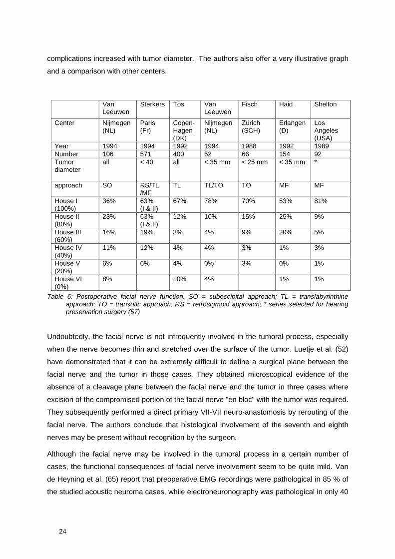
A very nice review of postoperative facial nerve function (suboccipital approach) is offered by
Van Leeuwen et al (58). They conclude that, in this series, the initial tumor diameter had very
little influence on the postoperative facial nerve function. However, the number of
24
complications increased with tumor diameter. The authors also offer a very illustrative graph
and a comparison with other centers.
VanLeeuwen
Sterkers Tos VanLeeuwen
Fisch Haid Shelton
Center Nijmegen(NL)
Paris(Fr)
Copen-Hagen(DK)
Nijmegen(NL)
Zürich(SCH)
Erlangen(D)
LosAngeles(USA)
Year 1994 1994 1992 1994 1988 1992 1989Number 106 571 400 52 66 154 92Tumordiameter
all < 40 all < 35 mm < 25 mm < 35 mm *
approach SO RS/TL/MF
TL TL/TO TO MF MF
House I(100%)
36% 63%(I & II)
67% 78% 70% 53% 81%
House II(80%)
23% 63%(I & II)
12% 10% 15% 25% 9%
House III(60%)
16% 19% 3% 4% 9% 20% 5%
House IV(40%)
11% 12% 4% 4% 3% 1% 3%
House V(20%)
6% 6% 4% 0% 3% 0% 1%
House VI(0%)
8% 10% 4% 1% 1%
Table 6: Postoperative facial nerve function. SO = suboccipital approach; TL = translabyrinthineapproach; TO = transotic approach; RS = retrosigmoid approach; * series selected for hearingpreservation surgery (57)
Undoubtedly, the facial nerve is not infrequently involved in the tumoral process, especially
when the nerve becomes thin and stretched over the surface of the tumor. Luetje et al. (52)
have demonstrated that it can be extremely difficult to define a surgical plane between the
facial nerve and the tumor in those cases. They obtained microscopical evidence of the
absence of a cleavage plane between the facial nerve and the tumor in three cases where
excision of the compromised portion of the facial nerve "en bloc" with the tumor was required.
They subsequently performed a direct primary VII-VII neuro-anastomosis by rerouting of the
facial nerve. The authors conclude that histological involvement of the seventh and eighth
nerves may be present without recognition by the surgeon.
Although the facial nerve may be involved in the tumoral process in a certain number of
cases, the functional consequences of facial nerve involvement seem to be quite mild. Van
de Heyning et al. (65) report that preoperative EMG recordings were pathological in 85 % of
the studied acoustic neuroma cases, while electroneuronography was pathological in only 40
25
% of the cases. However, the progressive involvement of the facial nerve, as evidenced by
the EMG recordings, only rarely provoked clinical signs. These findings may be explained by
the phenomenon of collateral innervation, which reduces the loss of muscle power. Given the
logarithmical relationship between mimic muscle power and facial movement, as described
by Burres (66), one can readily understand that a fairly major reduction of functional facial
nerve fibers is required before facial paralysis occurs.
Another interesting observation concerns the spatial relations between the intermediate
nerve (Wrisberg) and the vestibular nerve. Although the intermediate nerve is considered to
be a part of the facial nerve, it is more related to the vestibular nerve during its embryological
development (67, 68). Involvement of the facial nerve by acoustic neuroma will therefore
cause a loss of afferent functions before any clinical sign of facial paresis appears. An
example is Hitselberger's sign.
There is also an important anatomical difference between the facial nerve and the acoustic
nerve. The myelin sheath of the former extends far more medially than the myelin sheath of
the cochlear nerve. The junction between the glial sheath and the myelin sheath therefore
lies more medial to the brainstem. The well-developed myelin sheath of the facial nerve may
also be partly responsible for the sparing of the facial nerve motor function.
These reflections are well illustrated by the clinical results (Table 4). As mentioned before,
we cannot rule out the possibility of leaving tumor behind when preserving the facial nerve.
Tumor-biological considerations
Contemporary data clearly show that not all residual tumors grow, especially not if the
morsels of tumor that are originally left behind are small and, according to some authors (61,
62) are cauterized. Some propose that there must be at least a certain volume of residual
tumor, a “critical mass”, with its own blood supply and trophic factors in order to have a
growing tumor again. Filipo et al. (69) have discovered the expression of the Transforming
Growth Factor-β1 in the majority (83.87%) of the acoustic neuromas they studied. The
expression was more important in Antoni A regions than in Antoni B regions. There was also
a marked presence of TGF- β1 at the edges of the tumor. TGF-β1 is a multifunctional
peptide that modulates cell proliferation and differentiation, embryonic development and
maintenance of adult tissues. It is also involved in the development and function of the
nervous system. Animal studies have shown that TGF-β1 is a major and potent mitogen for
normal Schwann cells. Also, it is involved in the development of Schwann cell tumors with a
different mechanism from GGF (glial growth factor). This growth factor could therefore
26
participate in the biological behavior of the acoustic neuroma and its recurrence after
incomplete removal.
Radiation therapy
The first “gamma-knife” in the world was installed at the Karolinska Institute in Sweden and
was applied for acoustic neuroma treatment as early as 1969. The first unit in the United
States was installed in Pittsburgh on August 4th, 1987. The latter unit has published their
most recent results. In 1996, they treated 323 patients with an acoustic neuroma (70). The
authors state optimistically that, although radiation therapy has until recently been reserved
for patients who were either unwilling or unable to undergo microsurgery, they expect
radiation therapy to replace surgery as method of choice by the year 2020. Although they
present a nice survey of all possible hazards and complications regarding surgery, they fail to
provide hard evidence as to the alleged superiority of radiation therapy and more important,
they only assume that long-term tumor control will be good. Complications of radiation
therapy are not discussed. A tumor growth control of 88% has indeed been reported by
Maire et al (70) after no less than 45 Gy fractionated external beam radiotherapy. Norén et
al (72) report on 110 patients: growth control was obtained in 90%, meaning that in 60% the
tumor stopped growing and remained unaltered, while in 30% the tumor actually shrunk.
Hearing was unaltered at first, but subsequently started to deteriorate. Only 30% of the
patients maintained hearing at their preoperative levels. In a more recent paper, Thomassin
et al. (73) report on their experience with 138 vestibular schwannomas treated with gamma
knife surgery from 1992 to 1994, of which 104 patients were evaluated 3 years after
irradiation. 70% of the patients who had normal hearing retained their normal hearing, while
50% of those patients with serviceable hearing kept their serviceable hearing
Stereotactic radiosurgery therefore is an alternative to offer patients who decline surgery or
who are in a poor general condition. One has to keep in mind however, that it is not surgery
at all and that the tumor, albeit irradiated, stays in place.
Recently, an intracranial sarcoma after gamma knife surgery for a vestibular schwannoma in
a patient with neurofibromatosis was reported by Thomsen et al. (74). It is common
knowledge that irradiation of the central nervous system is also capable of inducing tumors,
especially when the dose exceeds 10 Gy (74).
27
Serious MRI studies as to the long-term results of radiation therapy have to be carried out
and their results should be compared to the observed growth rates in the “wait-and-see”
policy group.
The “wait and see” policy
While in the eighties, a wait-and-see policy was restricted to those patients refusing any kind
of treatment for their condition, the first results of this policy have led to a change in the
management of a certain subgroup of acoustic neuroma patients.
Many authors have tried to establish a mean growth rate and came up with rather variable
data. One of the issues at hand is how tumor growth is to be measured. The most accurate
measurement, of course, would be the assessment of the tumor volume. Unfortunately, this
kind of accurate measurements has only recently become available, again thanks to MRI.
Before that, measurements were carried out on axial and coronal CT-scan images. Usually,
the maximal diameter of the extrameatal portion of the tumor was measured. When
comparing these data, one should keep in mind that the volume of a roughly spherical
shaped object is directly related to the third power of its radius. Therefore, we have tried to
seek correlations between two, and whenever available, three-dimensional measurements of
the tumor, calculations of its volume and all available audiological and clinical data. These
results are presented in a subsequent chapter. The same thought applies to the following
data: growth rates are discussed, but it is not always clear how the authors measured
growth.
Strassnick et al. (75) found a mean annual growth rate of 1,1 mm (ranging from 0 tot 11 mm
per year). Rosenberg et al (76) compared nonsurgical patients to patients who underwent
subtotal resection. They found the growth in the nonsurgical group to vary from 0 to 17.1
mm per year, with a mean growth rate of 0.6 mm/year. The follow-up period ranged from 0.7
to 9.2 years with an average follow-up period of 4.3 years. Some patients eventually did
require surgery due to rapid tumor growth. In the patient group that had subtotal tumor
removal, the average growth rate post-operatively was 0.7 mm/year. The authors state that
tumor growth rate can be readily monitored using MRI and that one can predict if and when
problems, such as headaches, elevated intracranial pressure or cerebellar problems are to
be expected. Surgery can therefore be postponed until it becomes really necessary,
according to these authors.
28
We have to re-emphasize at this point, however, an observed growth rate based on the
measurement of tumor diameter (e.g. by means of CT scan) does not accurately reflect the
actual increase in tumor volume and its effect on the neighboring structures.
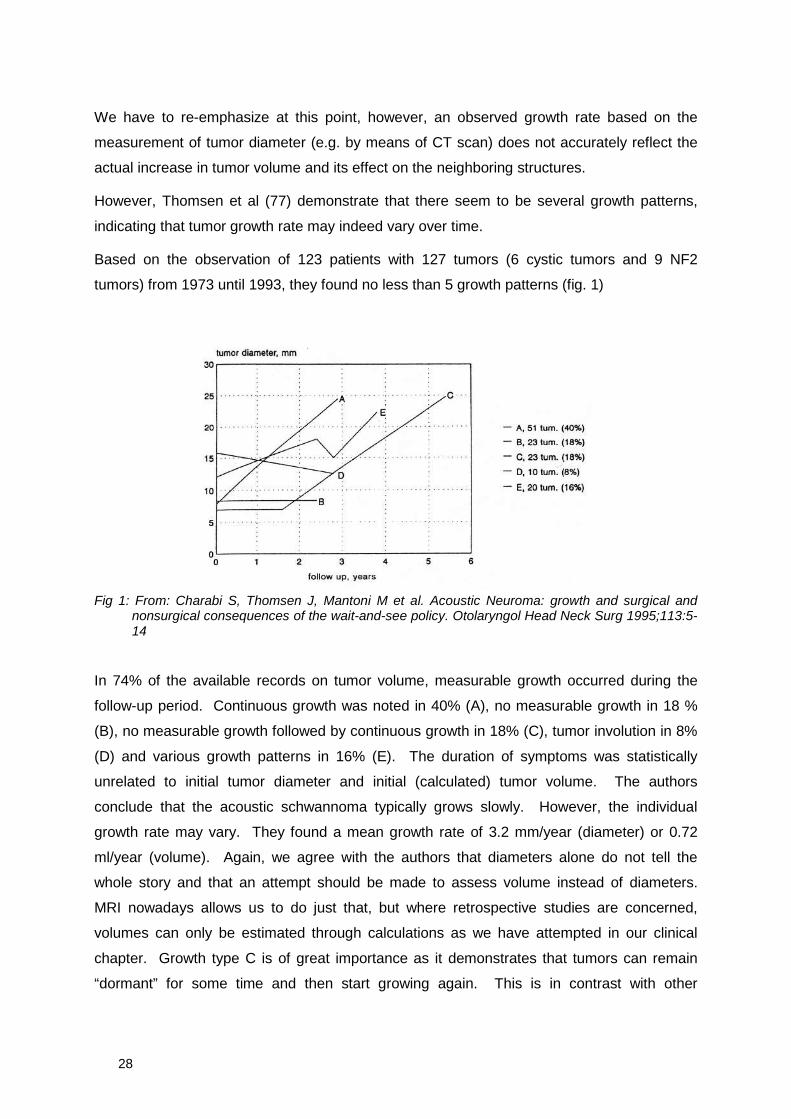
However, Thomsen et al (77) demonstrate that there seem to be several growth patterns,
indicating that tumor growth rate may indeed vary over time.
Based on the observation of 123 patients with 127 tumors (6 cystic tumors and 9 NF2
tumors) from 1973 until 1993, they found no less than 5 growth patterns (fig. 1)
Fig 1: From: Charabi S, Thomsen J, Mantoni M et al. Acoustic Neuroma: growth and surgical andnonsurgical consequences of the wait-and-see policy. Otolaryngol Head Neck Surg 1995;113:5-14
In 74% of the available records on tumor volume, measurable growth occurred during the
follow-up period. Continuous growth was noted in 40% (A), no measurable growth in 18 %
(B), no measurable growth followed by continuous growth in 18% (C), tumor involution in 8%
(D) and various growth patterns in 16% (E). The duration of symptoms was statistically
unrelated to initial tumor diameter and initial (calculated) tumor volume. The authors
conclude that the acoustic schwannoma typically grows slowly. However, the individual
growth rate may vary. They found a mean growth rate of 3.2 mm/year (diameter) or 0.72
ml/year (volume). Again, we agree with the authors that diameters alone do not tell the
whole story and that an attempt should be made to assess volume instead of diameters.
MRI nowadays allows us to do just that, but where retrospective studies are concerned,
volumes can only be estimated through calculations as we have attempted in our clinical
chapter. Growth type C is of great importance as it demonstrates that tumors can remain
“dormant” for some time and then start growing again. This is in contrast with other
29
publications, such as Rosenberg et al (76), where the authors state that the growth pattern
and therefore the need for surgery can be estimated within a short evaluation period during
which radiological assessment is carried out. Thomsen et al (77) submit this question to the
reader: “How many tumors in group B will start growing during a longer observation period?”
Tumor involution or negative growth is not as reassuring as it seems either, for some tumors
in group E first shrunk and afterwards started growing again: a strange tumorbiologic
phenomenon for sure. The authors conclude that tumors in patients with a short duration of
symptoms prior to diagnosis grew faster than tumors in patients with a long prediagnostic
duration of symptoms. In addition, cystic tumors tended to grow faster. The initials
neuroradiological architecture of the tumor therefore has a prognostic value. Growth
appeared no to be statistically related to age.
Finally, the authors state that before adapting a wait-and-see policy, duration of symptoms
and the neuroradiological architecture should be studied, for a number of patients in their
series lost their “candidacy” for hearing preservation surgery due to waiting too long.
Therefore, patients who are considered candidates for hearing preservation surgery from the
beginning, should decide from the start whether they want or do not want a maximal chance
for hearing preserve and therefore choose for early surgery. Together with Nedzelski et al.
(78), but contrary to other authors, Thomsen et al. (76) do not consider old age in itself a
contra-indication for surgery.
Nedzelski et al. (78) concur that a wait-and-see policy is only to be suggested to selected
individuals and that it implies strict follow-up on tumor growth by means of a high-definition
CT or preferably MRI twice a year. If the tumor remains unchanged for three years,
evaluation is carried out on a yearly basis. A growth rate exceeding 2 mm per year is an
indication for surgery as is, of course, any tumor becoming too big and life threatening.
Shelton and Hitselberger (79) in 1991 presented a review of the results on no less than 2520
acoustic neuromas removed by members of the House Ear Clinic from 1961 until 1989.
They compare surgery to radiotherapy and doing nothing (i.e. wait-and-see) and they
concluded then that their surgical results were superior than the 22 % post-treatment total
hearing loss obtained with radiation therapy and better than the 30% unaltered hearing
mentioned by Norén (72). They point out the difficulties of saving the facial nerve when
operating on an irradiated neuroma after it has started growing again. As for wait-and-see:
they argue that their data confirm their intuition: the smaller the tumor, the easier it is to
remove.
30
Quality of life
Over the last five years, much attention has quite rightfully been paid to the quality of life
after acoustic neuroma surgery. Questionnaires were developed and sent to the patients in
an effort to evaluate how this kind of surgery had interfered with their lives. Andersson et al;
(80) have evaluated the Swedish situation in 1997. They questioned 141 patients who
underwent surgery via the translabyrinthine approach and concluded that normal to
moderately impaired facial function (House & Brackmann I-III) was evident in 85.2% of
patients. Most patients (80%) considered their hearing to be worse after surgery, 60%
complained of tinnitus and 45% had balance problems. Headaches remained a problem in
22%. Work ability was affected in 23% and 37% reported a continued need for medical
consultations, mainly due to facial problems or pain.
Van Leeuwen et al (81) in 1995, report on a survey of 134 patients of whom 108 underwent a
suboccipital approach and 66 had a translabyrinthine or transotic approach. The patients
operated via the suboccipital approach appeared to have more emotional problems
afterwards than the translabyrinthine/transotic group. Irrespective of tumor diameter, more
patients from the suboccipital group complained of headaches as compared to the
translabyrinthine/transotic group. Overall, the quality of life of the patients was influenced to a
limited extent by the surgical approach and whether or not a reoperation had been
necessary. Suboccipital surgery led significantly more often to being declared unfit to work
than translabyrinthine/transotic surgery. The main reasons were vertigo and hearing
impairment. The surgical approach and the tumor size did not have any effect on hearing and
tinnitus . A large portion of patients without vertigo preoperatively complained of vertigo after
translabyrinthine/transotic surgery. Some patients with preoperative vertigo reported an
improvement after suboccipital surgery. The most favorable facial nerve function was
obtained after translabyrinthine surgery for a small tumor and the worst result after
suboccipital surgery for a larger tumor. Recovery after the operation was always a slow
process and recovery was not always complete. Patients had a poorer general state of
health than their healthy peers, according to Van Leeuwen et al. (81).
31
Development and aims of this thesis
In a first stage, staring in 1988, we have tried to select and adapt suitable histological
staining techniques for our research project. The general idea was to demonstrate ingrowth
of the schwannoma into the cochlear nerve. A first spin-off was the application of the Luxol
fast blue MBS staining technique on human fetal temporal bone sections. This resulted in a
first publication with F. Declau, using the Luxol fast blue staining technique in order to
differentiate cartilage from bone in human fetal temporal bones. This paper comprises the
first part of chapter two.
Parts two and three of the second chapter summarize our own histological research on the
acoustic neuroma at the Experimental ENT laboratory of the University of Antwerp. We have
applied a score of routine staining techniques and the Luxol Fast Blue staining technique to a
series of intact small and medium sized acoustic neuromas that had been removed via the
translabyrinthine approach by the late Prof. Marquet. The latter staining technique, however,
did not help us to attain our ultimate goal: the identification of tumor ingrowth in the cochlear
nerve. Therefore, we sought and found yet another technique: Verhoeff’s old elastin staining
technique. This time, we were able to clearly identify the nerve fibers and we could study the
interface between tumor and nerve. This study was published in 1989 and comprises the
second part of this chapter. This also explains why this study only deals with small and
medium sized acoustic neuromas. The larger tumors could not be preserved as a whole
without endangering the facial nerve or other vital structures.
In a following stage, immunohistochemical technology was applied. This time, we have tried
to obtain microscopical sections that would prove beyond any doubt that there is actually no
cleavage plane between the compromised nerves and the tumor, whenever macroscopically
visible adherences were present between the nerve and the tumor. This study was published
in 1990 and comprises the third part of chapter two.
In the third chapter, we offer clinical data of the subjects involved and we demonstrate some
correlations. Also, we try to position our group of subjects in the general population of
acoustic neuroma patients. In a second part, MRI data are discussed and the MRI
characteristics of the acoustic neuroma are summarized.
Finally, atypical presentations of the schwannoma in the temporal bone are presented in the
fourth chapter by means of case reports: one on facial nerve neuromas and one on a very
small intralabyrinthine neuroma. A case report of a cerebellar astrocytoma was also
included, because it perfectly mimicked an acoustic neuroma.
32
References
1. Schefter RP, Harner SG. Histologic study of the vestibulocochlear nerve. Ann Otol RhinolLaryngol 1986;95:146-150.
2. Friedmann I. Pathology of the ear. Oxford, London, Edinburgh, Melbourne: Blackwell ScientificPublications, 1974:211-224.
3. Schuknecht HF. Pathology of the Ear. 2nd Ed. Lea & Febiger, Philadelphia, 1993. p. 461.
4. Mattox DE. Vestibular schwannomas. Otolaryngologic Clinics of North America 1987;20:149-160.
5. Hardy M, Crowe SJ. Early asymptomatic acoustic tumor. Arch Surg 1936;32:292-301.
6. House WF, Hitselberger WE. The neuro-otologist's view of the surgical management ofacoustic neuromas. Clin Neurosurg 1985;32:214-222.
7. Tos M, Charabi S, Thomsen J. Incidence of vestibular schwannomas. Laryngoscope1999;109(5):736-40.
8. Bell C. The Nervous System of the Human Body. London 1830, Appendix of cases 112-114.
9. Toynbee J. Neuroma of the Auditory Nerve. Trans Path Soc Lond 1853;4:259-260.
10. Lempert J. Improvement of hearing in cases of otosclerosis: new one stage surgical technic.Arch Otolaryngol 1938;28:42-97.
11. House WF (Ed.). Monograph: Transtemporal Bone Microsurgical Removal of AcousticNeuromas. Arch Otolaryngol 1964;80:667-677.
12. Shaia FT, Sheehy JL. Sudden sensorineural hearing impairment; a report of 1220 cases.Laryngoscope 1976;86:389-398.
13. Vassalli L, Landolfi M, Taibah A, Russo A, Pasanisi E, Shaan M, Sanna M. Atypicalpresentations of acoustic neuroma. In: Proceedings of the First International Conference onAcoustic Neuroma, Copenhagen,1991. Tos & Thomsen Eds. Kugler Publications,Amsterdam/New York, 1992:23-30.
14. Jerger J. Békésy Audiometry in Analysis of Auditory Disorders. J Speech Hearing Res1960;3:275-287.
15. Daly DM, Roeser R, Aung M, Daly DD. Early evoked potentials in patients with acousticneuroma. Electroencephalogr Clin Neurophysiol 1977;43:151-159.
16. Selters W, Brackmann DE. Acoustic tumor detection with brainstem electric responseaudiometry. Arch Otolaryngol 1977;103:181-187.
17. Katz J (Ed.) Handbook of Clinical Audiology. 4th edition. Williams & Wilkins, Baltimore 1994.pp 351-374.
18. Oeken J. Assessment of cochlear function with distortion products of otoacoustic emissions inacoustic neuroma. HNO 1996;44(12):677-684.
19. Appel L. Honouring the pioneers. Belgisch Tijdschr Radiol 1980;63:v-vi.
20. Tos M, Drozdziewicz D, Thomsen J. The medial acoustic neuroma: a new clinical subgroup.In: Acoustic Neuroma. Proceedings of the First International Conference on AcousticNeuroma. Tos M & Thomsen J (Eds.) Copenhagen 1991. Kugler Publications. Amsterdam –New York. 1991: 211
21. Mulkens TH, Parizel PM, Martin J.-J., Degryse HR, Van de Heyning PH, Forton GE,De Schepper AM. Acoustic Schwannoma: MR Findings in 84 Tumors (Pictorial essay) .Am JRoentgenol 1993; 160:395-398.
33
22. Forton GEJ, Somers Th, Hermans R, Baert AL, Offeciers FE. Preoperatively diagnosedutricular neuroma treated by selective partial labyrinthectomy. Ann Otol Rhinol Laryngol1994;103: 885-888
23. Casselman JW. Magnetic Resonance Imaging of the Inner Ear. Doctoral thesis R.U.G.Vanmelle, Gent
24. Somers Th, Casselman J, de Ceulaer G, Govaerts P, Offeciers E. Prognostic value of MRIfindings in hearing preservation surgery for vestibular schwannoma. Am J Otol: submitted forpublication.
25. Dix M, Hallpike C. Observations on the pathological mechanism of conductive deafness incertain cases of neuroma of the VIIIth nerve. Laryngoscope 1950;64:658.
26. SchuknechtHF. The pathophysiology of angle tumors. In: The vestibular system and itsdiseases. Wolfson RJ, ed. University of Pennsylvania Press, Philadelphia. 1966:428-438.
27. O’Conner F, Luxon L, Shortman RC, Thompson E, Morrison A. Electrophoretic separation andidentification of perilymph proteins in cases of acoustic neuroma. Acta otolaryngol.1982;93:195-200.
28. Silverstein H, Schuknecht H. Biochemical studies of inner ear fluid in man: changes inotosclerosis, Ménière’s disease and acoustic neuroma. Arch Otolaryngol 1966;84:395.
29. Jahnke K, Neumann TA. The fine structure of vestibular end organs in acoustic neuromapatients. In: Proceedings of the First International Conference on Acoustic Neuroma,Copenhagen,1991. Tos & Thomsen Eds. Kugler Publications, Amsterdam/New York,1992:203-207.
30. Nager GT. Acoustic neurinomas pathology and differential diagnosis. Arch Otolaryng1969;89:252-279.
31. Harkin JC. Pathology of nerve sheath tumors. Annals New York Academy of Science1986;147-54.
32. Clark HB, Minesky JJ, Agrawal D, Agrawal HC. Myelin basic protein and P2 protein are notimmunohisto-chemical markers for Schwann cell neoplasms. Am J Pathol 1985;121:96-101.
33. Gould VE, Moll R, Moll I, Lee I, Schwechheimer K, Franke WW. The intermediate filamentcomplement of the spectrum of nerve sheath neoplasms. Lab Invest 1986;55:463-474.
34. Peltonen J, Foidart JM, Aho HJ. Type IV and V collagens in Von Recklinghausen'sneurofibromas. Virchows Archiv (Cell Pathol) 1984;47:291-301
35. Lesser T, Janzer R, Kleihues P. Growth rate of acoustic schwannomas. Skull Base Surg1991;1(1):11-15.
36. Charabi S, Engels P, Charabi B, Jacobsen GK, Overgaard J, Thomsen J, Tos M. Growth ofvestibular schwannomas: in situ model employing the monoclonal antibody Ki-67 and DNAflow cytometry. Am J Otol 1996;17(2):301-306.
37. Chen JM, Houle S, Ang LC, Commins D, Allan K, Nedzelski J, Rowed D. A study of vestibularschwannomas using positron emission tomography and monoclonal antibody Ki-67. Am J Otol1998;19(6):840-845.
38. Yokoyama M, Matsuda M, Nakasu S, Nakajima M, Handa J. Clinical Significance of Ki-67staining index in acoustic neurinoma. Neurol Med Chir 1996;36(10):698-702
39. Erlandson RA, Woodruff JM. Peripheral nerve sheath tumors. Cancer 1982;49:273-87.
40. Ballance CA. Some points in the surgery of the brain and its membranes. London. MacMillan& Co. 1907.
41. Krause F. Zur Freilegung der hinteren Felsenbeinflache und des Kleinhirns. Beitr Klin Chir1903;37:728-764.
42. Panse R. Ein gliom des Akustikus. Arch Ohr 1904;61:251-255.
34
43. Borchardt M. Zur Operation der Tumoren des Kleinhirnbrückenwinkels. Berl Klin Wchnscher19905;42:1033-1035.
44. Cushing H. Tumors of the Nervus Acusticus and Syndrome of the Cerebellopontine Angle.W.B. Saunders Company, Philadelphia, 1917: p. 277.
45. Dandy WE. Operation for total removal of cerebellopontine (acoustic) tumors. Surg GynecObstet 1925;41:129-148.
46. Pool JL, Pava AA. The early diagnosis and treatment of acoustic nerve tumors. Charles CThomas Publishers. Springfield (Ill.) 1957. p 110.
47. House WF. Surgical exposure of the internal auditory canal and its contents through themiddle cranial fossa. Laryngoscope 1961:1363-1385
48. House WF. Transtemporal bone microsurgical removal of acoustic neuromas. ArchOtolaryngol 1964;80:597.
49. Fisch U, Mattox D. Microsurgery of the skull base. Thieme Verlag. Stuttgart 1988. p. V
50. Portmann M, Bébéar JM, Bisch X, Boussens J, Boyer M, Caille JM, Guillen P, Koehler R,Lacaze JL, Lenoir JL, Martin PL, Piton J. La chirurgie du conduit auditif interne. Cahiers d’ORL(Montpellier) 1972;7:2.
51. Tos M, Thomsen J, Harmsen A. Ti års translabyrintær acusticusneurinomkirurgi I Danmark.Ugeskrift for Læger 1987;149:2901-2905.
52. Luetje CM, Whittaker CK, Callaway LA, Veraga G. Histological acoustic tumor involvement ofthe VIIth nerve and multicentric origin in the VIIIth nerve. Laryngoscope. 1983;93:1133-1139.
53. Shelton C, Brackmann DE, House WF, Hitselberger WE. Acoustic tumor surgery. ArchOtolaryngol Head Neck Surg 1989;115:1213-1216.
54. Browning GG. Reporting the benefits from middle ear surgery using the Glasgow Benefit Plot.Am J Otol 1993;14(2):135-140.
55. Silverstein H, Rosenberg SI, Flanzer JM, Wanamaker HH, Seidman MD. An algorithm for themanagement of acoustic neuromas regarding age, hearing, tumor size and symptoms.Otolaryngol Head Neck Surg. 1993;108(1):1-10.
56. Olivecrona H. Acoustic tumors. J Neurosurg. 1967;86:6-13
57. House W. Partial tumor removal and recurrence in acoustic tumor surgery. Arch Otolaryngol.1968;88:96-106.
58. Van Leeuwen JPPM, Meijer E, Grotenhuis JA, Thijssen HOM, Cremers CWRJ. Subocciptalsurgery for acoustic neuroma. Clin Otolaryngol 1995
59. Beatty CW, Ebersold MJ, Harner SG. Residual and recurrent acoustic neuromas.Laryngoscope. 1987;97:1168-1171.
60. Robertson JB, Brackmann DE, Hitselberger WE. Acoustic neuroma recurrence aftersuboccipital resection: management with translabyrinthine resection. Am J Otol.1996;17(2):307-311.
61. Ohta S, Yokoyama T, Nishizawa S, Uemura K. Regrowth of residual tumor after acousticneuroma surgery. Br J Neurosurg. 1998;12(5):419-422.
62. Cerullo L, Grutsch J, Osterdock R. Recurrence of vestibular schwannomas in surgical patientswhere preservation of facial and cochlear nerve is the priority. Br J Neurosurg 1998;12(6):547-552.
63. Gormley WB, Sekhar LN, Wright DC, Kamerer D, Schessel D. Acoustic neuromas: results ofcurrent surgical management. Neurosurgery 1997;41(1):50-58.
64. Pace-Balzan A, Lye R, Ramsden RT, Chandler C, Gillespie JE, Dutton JM. Growthcharacteristics of acoustic neuromas with particular reference to the fate of capsule fragmentsremaining after tumor removal: implications for patient management. In: Proceedings of the
35
First International Conference on Acoustic Neuroma, Copenhagen,1991. Tos & Thomsen Eds.Kugler Publications, Amsterdam/New York, 1992:657-659.
65. Van de Heyning P, Valcke H, Claes J, Delaporte C. L'atteinte infraclinique du nerf facial. In:Capella, ed. Nervi Craniales Praeter Octavum. Barcelona: Uriach 1988:289-291.
66. Burres J. Facial biomechanical properties. In: Portmann M, ed. The facial nerve. Paris:Editions Masson, 1985:188-190
67. Declau F. Embryology of the cranial nerves. In: Marquet J, Van de Heyning P, Claes J, eds.The cranial nerves in ORL. Acta Otorhinolaryngol Belg 1986;40:27-49
68. Gasser RF. The development of the facial nerve in man. Ann Otol 1967;76:1
69. Cardillo MR, Filipo R, Monini S, Aliotta N, Barbara M. Transforming growth factor beta-1expression in human acoustic neuroma. Am J Otol. 1999;20(1):65-68.
70. Lunsford LD, Flickinger J, Lindner G, Maitz A. Stereotactic radiosurgery of the brain using thefirst United States 201 Cobalt-60 source gamma knife. Neurosurgery. 1989;24(2):151-159.
71. Maire JP, Caudry M, Darroutet V, Guerin J, Trouette R, Bebear JP. Fractionated radiationtherapy in the treatment of stage III and IV cerebellopontine angle neurinomas: long termresults in 24 cases. Int J Radiat Oncol Biol Phys. 1995;32:1137-1143
72. Norén G, Arndt J, Hindmarsch T, Hirsch A. Stereotactic radiosurgical treatment of acousticneurinomas. In: Modern Stereotactic Neurosurgery. Lunsford LD, ed. Martinus Nijhoff, Boston,1988: 481-489.
73. Thomassin JM, Epron JP, Régis J, Delsanti C, Sarabian A, Peragut JC, Pellet W. Preservationof hearing in acoustic neuromas treated by gamma knife surgery. Stereotact Funct Neurosurg.1998;70, suppl1:74-79.
74. Thomsen J, Mirz F, Wetke R, Astrup J, Bojsen-Møller, Nielsen E. Intracranial sarcoma in apatient wih neurofibromatosis type 2 treated with gamma knife neurosurgery for vestibularschwannoma. Am J Otol 2000;21(3):364-370.
75. StrassnickB, Glasscock III ME, Haynes D, McMenor SO, Minor LB. The natural history ofuntreated acoustic neuromas. Laryngoscope 1994;104:1115-1119.
76. Rosenberg SI, Silverstein H, Gordon MA, Flanzer JM, Willcox TO, Silverstein J. A comparisonof growth rates of acoustic neuromas: nonsurgical patients versus subtotal resection.Otolaryngol Head Neck Surg. 1993; 109:482-487.
77. Charabi S, Thomsen J, Mantoni M, Charabi B, Jørgensen B, Børgesen SE, Gyldensted C, TosM. Acoustic neuroma: growth and surgical and nonsurgical consequencesof the wait-and-seepolicy. Otolaryngol Head Neck Surg. 1995;113(1):5-14
78. Nedzelski JM, Schessel DA, Pfleiderer A, Kassel EE, Rowed DW. Conservative managementof acoustic neuromas. Otolaryngol Clin North Am. 1992;25(3):691-705.
79. Shelton C, Hitselberger WE. The treatment of small acoustic tumors: now or later?Laryngoscope 1991;101:925-928.
80. Andersson G, Ekvall L, Kinnefors A, Nyberg G, Rask-Andersen H. Evaluation of quality of lifeand symptoms after translabyrinthine acoustic neuroma surgery. Am J Otol 1997;18(4):421-426.
81. Van Leeuwen JP, Braspenning JC, Meijer H, Cremers CW. Quality of life after acousticneuroma surgery. Ann Otol Rhinol Laryngol 1996;105(6):423-30.

36
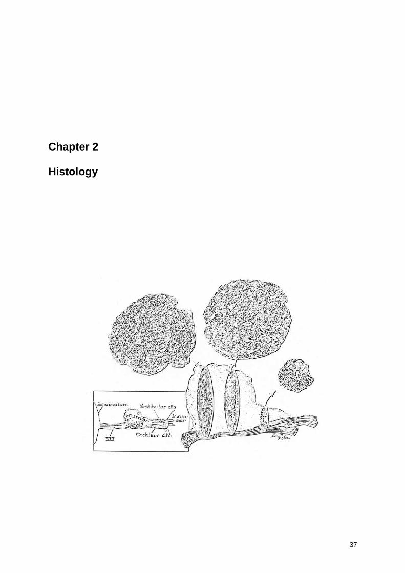
37
Chapter 2
Histology

38
39
Chapter 2a
Differentiation of cartilage and bone in human fetaltemporal bones with Luxol fast blue stain
F. Declau, MD
L. Moeneclaey
G. Forton, MD
J. Marquet†, MD
ENT Department, University of Antwerp, Wilrijk, BelgiumArch Otorhinolaryngol 1988;245:218-220
40
Samenvatting
Een gemodificeerde Luxol fast blue techniek werd op punt gesteld om de ontwikkeling van
het os temporale te kunnen bestuderen. De gemodificeerde kleuringstechniek laat toe een
duidelijk onderscheid te maken tussen het aanwezige primitieve kraakbeen en het
nieuwgevormde bot. Alhoewel beide weefsels een grote hoeveelheid collageen bevatten,
zijn hun kleuringseigenschappen na de Luxol fast blue kleuring duidelijk verschillend en dit
door de verschillende fysicochemische eigenschappen van de respectievelijke
collageentypes in beide weefsels. De differentiatie van het mesenchymateuze weefsel tot
ligamenten en gewrichten kan fraai in beeld worden gebracht met deze techniek. Ook bij de
studie van de endochondrale botvorming binnen het otisch kapsel en het middenoor geeft de
gemodificeerde Luxol fast blue kleuring complementaire informatie bij de standaard
kleuringsmethoden.
Summary
A modified Luxol fast blue technique was used to study the development of the temporal
bone. This staining method makes it possible to make a clear distinction between the
primitive cartilage present and the new forming bone. Although these tissues both contain a
significant amount of collagen, their staining properties with the Luxol dyes are widely
dissimilar, due to the different physicochemical properties of the collagen types involved in
these tissues. The differentiation of mesenchymatous tissue into ligaments and joints can
also be very clearly demonstrated with this technique. In studying the endochondral
ossification process of the otic capsule and middle ear, the modified Luxol fast blue stain is a
valuable technique that is complementary to more conventional staining methods.
Introduction
The Luxol dye technique was developed by Klüver and Barrera (4) in 1953 for staining the
myelin sheath of nerve fibers. The staining properties of the Luxol dyes are based upon their
chemical structure. Luxol fast blue is a diarylguanidine salt of an acidic dye. The specific
structure of the latter is not responsible for the staining reaction produced (2). In their early
publication, Klüver and Barrera (5) used the du Pont product, Luxol fast blue MBS, a
phtalocyanine dye, but similar results were obtained with azo-dyes: Luxol fast blue type ARN
and type G (9, 10). Following its discovery in 1953, the Klüver and Barrera method was
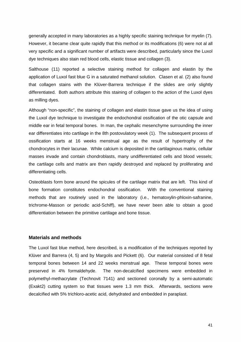
41
generally accepted in many laboratories as a highly specific staining technique for myelin (7).
However, it became clear quite rapidly that this method or its modifications (6) were not al all
very specific and a significant number of artifacts were described, particularly since the Luxol
dye techniques also stain red blood cells, elastic tissue and collagen (3).
Salthouse (11) reported a selective staining method for collagen and elastin by the
application of Luxol fast blue G in a saturated methanol solution. Clasen et al. (2) also found
that collagen stains with the Klüver-Barrera technique if the slides are only slightly
differentiated. Both authors attribute this staining of collagen to the action of the Luxol dyes
as milling dyes.
Although “non-specific”, the staining of collagen and elastin tissue gave us the idea of using
the Luxol dye technique to investigate the endochondral ossification of the otic capsule and
middle ear in fetal temporal bones. In man, the cephalic mesenchyme surrounding the inner
ear differentiates into cartilage in the 8th postovulatory week (1). The subsequent process of
ossification starts at 16 weeks menstrual age as the result of hypertrophy of the
chondrocytes in their lacunae. While calcium is deposited in the cartilaginous matrix, cellular
masses invade and contain chondroblasts, many undifferentiated cells and blood vessels;
the cartilage cells and matrix are then rapidly destroyed and replaced by proliferating and
differentiating cells.
Osteoblasts form bone around the spicules of the cartilage matrix that are left. This kind of
bone formation constitutes endochondral ossification. With the conventional staining
methods that are routinely used in the laboratory (i.e., hematoxylin-phloxin-safranine,
trichrome-Masson or periodic acid-Schiff), we have never been able to obtain a good
differentiation between the primitive cartilage and bone tissue.
Materials and methods
The Luxol fast blue method, here described, is a modification of the techniques reported by
Klüver and Barrera (4, 5) and by Margolis and Pickett (6). Our material consisted of 8 fetal
temporal bones between 14 and 22 weeks menstrual age. These temporal bones were
preserved in 4% formaldehyde. The non-decalcified specimens were embedded in
polymethyl-methacrylate (Technovit 7141) and sectioned coronally by a semi-automatic
(Exakt2) cutting system so that tissues were 1.3 mm thick. Afterwards, sections were
decalcified with 5% trichloro-acetic acid, dehydrated and embedded in paraplast.
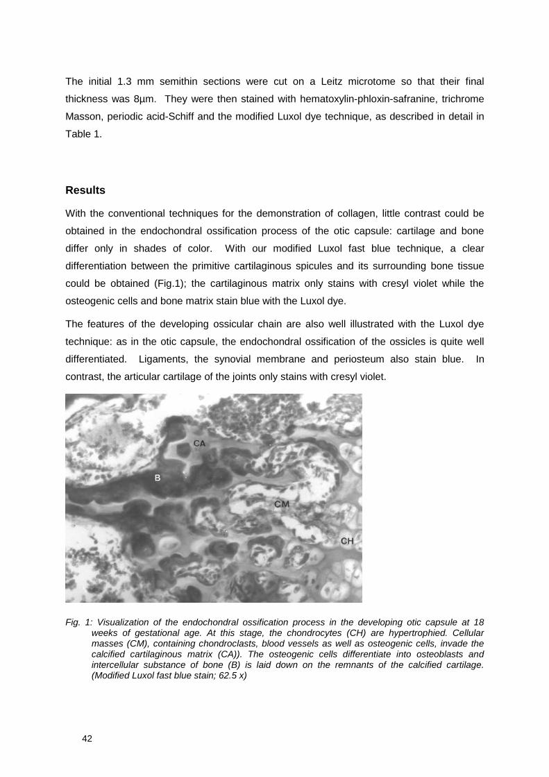
42
The initial 1.3 mm semithin sections were cut on a Leitz microtome so that their final
thickness was 8µm. They were then stained with hematoxylin-phloxin-safranine, trichrome
Masson, periodic acid-Schiff and the modified Luxol dye technique, as described in detail in
Table 1.
Results
With the conventional techniques for the demonstration of collagen, little contrast could be
obtained in the endochondral ossification process of the otic capsule: cartilage and bone
differ only in shades of color. With our modified Luxol fast blue technique, a clear
differentiation between the primitive cartilaginous spicules and its surrounding bone tissue
could be obtained (Fig.1); the cartilaginous matrix only stains with cresyl violet while the
osteogenic cells and bone matrix stain blue with the Luxol dye.
The features of the developing ossicular chain are also well illustrated with the Luxol dye
technique: as in the otic capsule, the endochondral ossification of the ossicles is quite well
differentiated. Ligaments, the synovial membrane and periosteum also stain blue. In
contrast, the articular cartilage of the joints only stains with cresyl violet.
Fig. 1: Visualization of the endochondral ossification process in the developing otic capsule at 18weeks of gestational age. At this stage, the chondrocytes (CH) are hypertrophied. Cellularmasses (CM), containing chondroclasts, blood vessels as well as osteogenic cells, invade thecalcified cartilaginous matrix (CA)). The osteogenic cells differentiate into osteoblasts andintercellular substance of bone (B) is laid down on the remnants of the calcified cartilage.(Modified Luxol fast blue stain; 62.5 x)
43
Discussion
Although cartilage and bone both contain collagen, they differ widely in their staining
properties. The exact staining mechanism of the Luxol dyes is not quite clear. Investigations
on the molecular organization of the myelin sheath have revealed the existence of a
glycoprotein and glycolipid coat in the extra-cellular space between adjacent cellular lipid
layers (12).
Clasen et al. (2) reported diminished staining properties of myelin with the Klüver-Barrera
method when tissue sections were first subjected to alkaline hydrolysis. However, such
hydrolysis leads to disrupted proteins and bound lipids.
Salthouse (11) described a reduction in collagen staining after tanning. Tannic acid affects
the properties of collagen as a polymer and also blocks keto-imide groups known to be
important for this type of dye binding. Clasen et al. (2) and also Salthouse (11) have
suggested that the staining of the myelin sheath and of collagen and elastin is due to the
presence of hydrophobic sites (non-polar amino acids) in the fixed substrate. The same
hypothesis can explain our results with the modified Luxol fast blue-cresyl violet method.
Our findings show that this technique will not stain all types of collagen, but only those fibers
with hydrophobic properties. Indeed, cartilage and bone contain different types of collagen:
cartilage contains chiefly type II and bone type I collagen.
Type I collagen is more glycosylated then type II. The glycosylation of the collagen may
greatly modify their physicochemical properties, including hydrophobicity. Type II collagen
contains twice as much water as type I, due to quite large amounts of hydrophobic
carbohydrates.
Elastin also stains properly with the modified Luxol dye technique and is a very hydrophobic
protein, due to non-polar amino acid groups (8). As a consequence, elastic cartilage also
stains with the Luxol dye in contrast to hyaline cartilage. The mucinous matrix of the
mesenchymatous tissue which contains highly hydrophylic proteoglycans, is not al all stained
by the Luxol dye.
44
Staining solutions
Make a 1% solution of Luxol fast blue MBS by dissolving 1 g in 100 cm³ 95% ethanol. Add 0.5 cm³ of10% acetic acid. Filter before using.
Make a 0.1% aqueous solution of cresyl violet.Cresyl violet differentiator: add 90 cm³ 95% ethanol to 10 cm³ chloroform and use 3 drops acetic acid.
Method for staining paraffin sections
Immerse fetal temporal bone sections in 95% ethanol for 2 min.Rinse with 95% ethanolStain overnight in solution A at 37° C.Immerse in 95% ethanol for 10 sec.Wash in distilled water for 10 sec.Begin to differentiate with 0.05% aqueous solution of lithium carbonate for 5 sec.Continue differentiation in 70% ethanol for 3 sec.Wash thoroughly in distilled waterStain with solution B for 2 min.Wash briefly in distilled waterRinse in 70% ethanolDifferentiate with solution C by washing the slides in 95% ethanolDehydrate in the usual manner
Table 1: Technique for the preparation of modified Luxol fast blue MBS and cresyl violet solutions.
45
References
1. Anson BJ, Donaldson JA (1973) Surgery anatomy of the temporal bone and ear. Saunders,Philadelphia
2. Clasen RA, Simon RG, Scott R, Pandolfi S, Laing I, Lesak A (1973) The staining of the myelinsheath by Luxol dye technique. J Neuropathol Exp Neurol 32:271-283
3. Gustavson KH (1956) The chemistry and reactivity of collagen. Academic Press, New York
4. Klüver H, Barrera E (1953) A method for combined staining of cells and fibers in the nervoussystem. J Neuropathol Exp Neurol 12:400-403
5. Klüver H, Barrera E (1954) On the use of azaporphyrin derivates (phtalocyanines) in stainingnervous tissue. J Psychol 37:199-223
6. Margolis G, Pickett JP (1956) New applications of the Luxol fast blue myelin stain. Lab Invest5:459
7. Neely JG (1981) Gross and microscopic anatomy of the eight cranial nerve in relationship tothe solitary schwannoma. Laryngoscope 91:1512-1531
8. Piez KA, Reddy AM (1984) Extracellular matrix biochemistry. Elsevier, New York
9. Salthouse TN (1962) Luxol fast blue ARN: a new solvent azodye with improved stainingqualities for myelin and phospholipids. Stain Technol 37:313-316
10. Salthouse TN (1964) Luxol fast blue G as a myelin stain. Stain Technol 39:123
11. Salthouse TN (1965) Selective staining of collagen and elastin by Luxol fast blue G inmethanol: a histochemical study. J Histochem Cytochem 13:133-140
12. Schmitt FO (1970) Molecular neurobiology: an interpretative survey. In: Schmitt FO (ed) Theneurosciences. Rockefeller University Press, New York, pp 867-877
Acknowledgements
The authors wish to thank Mr Ludo Van Hove for his assistance in the preparation of the
manuscript and Mr Dirk De Saegher for the preparation of the illustrations.

46
47
Chapter 2b
The involvement of the cochlear nerve in neurinomas of theeighth cranial nerve
G.E.J. Forton, MD
L.L.M. Moeneclaey
F. Declau, MD
J.F.E. Marquet†, MD
ENT Department, University of Antwerp, Wilrijk, BelgiumArch Otorhinolaryngol 1989;246(3):156-160
48
Samenvatting
In dit hoofdstuk wordt enerzijds de ontwikkeling van een geschikt histologisch
kleuringprotocol besproken en anderzijds worden de microscopische bevindingen ter hoogte
van de grenszone tussen tumor en nervus cochlearis beschreven. Onze bevindingen
hebben geleid tot twee publicaties, die hier weergegeven worden.
Als kleuringstechnieken werden de klassieke trichroom-Masson kleuring, de Luxol Fast Blue
techniek van Klüver en Barrera, de elastinekleuring volgens Verhoeff en een
immunohistochemische techniek op basis van monoclonale muis-antilichamen tegen
humane neurofilamenten gebruikt. Samengevat kwamen wij aldus tot de volgende
conclusies:
De Trichroom-Masson kleuring is een waardevolle routinekleuring voor
acousticusneurinomen en laat een mooie differentiatie tussen de myelineschede en het
omringende perineurale bindweefsel toe.
De Luxol fast blue kleuring is geenszins een specifieke myelinekleuring: zowel myeline als
collageen kleuren op vergelijkbare wijze. Deze kleuring is dan ook van weinig of geen nut
wanneer men geconfronteerd wordt met een specimen dat beiden bevat, zoals
acousticusneurinoma's.
De elastinekleuring volgens Verhoeff levert een duidelijk beeld op, waarin eventueel
aanwezige zenuwen voldoende opvallen. Er ontstaat voldoende contrast tussen het tumorale
stroma enerzijds, en het collageen bindweefsel, waaronder het perineurale bindweefsel,
anderzijds. De myelineschede wordt echter niet duidelijk gekleurd.
Een specifieke immunohistochemische techniek is noodzakelijk wanneer men de
zenuwvezels met absolute zekerheid wil definiëren of wanneer men het verloop van
zenuwvezels in de tumormassa wil bestuderen. Met behulp van deze techniek konden wij
tumorale ingroei in de Nervus Cochlearis aantonen, tenminste op die plaatsen waar ook
macroscopisch zichtbare vergroeiingen bestonden tussen de tumor en de zenuw. Wij troffen
typische schwannoom cellen aan temidden van zenuwvezels van de Nervus Cochlearis,
terwijl er omgekeerd ook zenuwvezels werden gevonden omgeven door manifest
schwannoom. Een duidelijk klievingsvlak is niet aanwezig.
Op histopathologische basis kan bijgevolg gesteld worden dat de N. Cochlearis ten minste
gedeeltelijk betrokken is in het tumorale proces in die gevallen waar macroscopische
adherenties waarneembaar zijn. Het microscopisch correlaat is de afwezigheid van enig
klievingsvlak tussen tumor en zenuw
49
Summary
In view of recent controversies concerning the preservation of hearing in acoustic neurinoma
surgery, we examined the courses of the vestibular and cochlear nerve fibers in 12 intact
acoustic neurinomas studied in our department. Due to its lack of specificity, the Luxol fast
blue stain was found to be inadequate for our study of the nerve fibers. In contrast, Verhoeff's
stain proved to be satisfactory when combined with a highly specific immunohistochemical
technique. There were macroscopically visible adherences between the tumor and the
cochlear nerve in 9 out of the 12 specimens. From those specimens, histological sections
were obtained in which both the cochlear nerve and tumor could be clearly identified. In
these specimens the cochlear nerve was involved in the tumoral process and there was no
clear cleavage plane between the nerve and the tumor. However, all these patients suffered
only from minimal losses of hearing as a result of their tumors.
Introduction
It is well known clinically that the neurinoma of the acoustic nerve usually arises from the
inferior branch of the vestibular nerve. The differentiation between cochlear and vestibular
fibers, however, remains a matter of debate. To seek evidence as to the involvement of the
cochlear nerve in the tumoral process, we initiated an investigation to determine the courses
of both the cochlear and vestibular nerve fibers in acoustic neurinomas, particularly since
there have been recent tendencies for surgeons to spare the cochlear nerve during removal
of acoustic neurinomas. Indeed, from a histological point of view, the surgical concept of
hearing preservation in cases of cochlear involvement is doubtful. The functional implications
of such surgery and the fate of the facial nerve have been omitted from the present study
and will be the subject of a separate report.
A number of authors seem to have different opinions about whether the cochlear and
vestibular nerve fibers are myelinated or not. Kimura et al. (4) found that 92% of all the
human spiral ganglion cells they had examined were of the unmyelinated or partly
myelinated type, with more unmyelinated neurons (i.e. 99%) under the age of 40. Neely (10),
on the other hand, reported that a vast majority of the cochlear fibers is myelinated. One can
find analogous controversies concerning the vestibular nerve. For example, Perre et al (11)
wrote that there are no myelin sheaths around the vestibular ganglion cells. Ylikoski (17)
confirmed that the vestibular ganglion cells are ensheathed by a single layer of Schwann cell
cytoplasm and that there is no real myelin sheath. In contrast, Ballantyne and Engström (1)
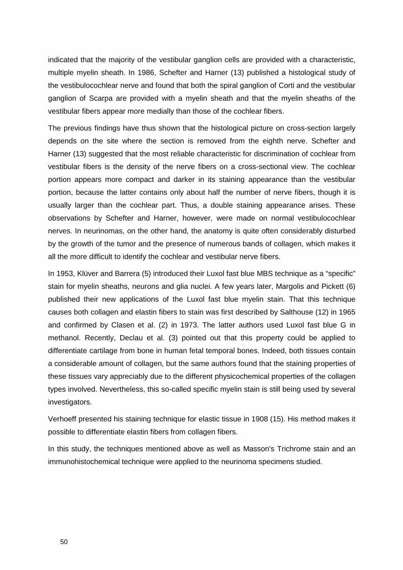
50
indicated that the majority of the vestibular ganglion cells are provided with a characteristic,
multiple myelin sheath. In 1986, Schefter and Harner (13) published a histological study of
the vestibulocochlear nerve and found that both the spiral ganglion of Corti and the vestibular
ganglion of Scarpa are provided with a myelin sheath and that the myelin sheaths of the
vestibular fibers appear more medially than those of the cochlear fibers.
The previous findings have thus shown that the histological picture on cross-section largely
depends on the site where the section is removed from the eighth nerve. Schefter and
Harner (13) suggested that the most reliable characteristic for discrimination of cochlear from
vestibular fibers is the density of the nerve fibers on a cross-sectional view. The cochlear
portion appears more compact and darker in its staining appearance than the vestibular
portion, because the latter contains only about half the number of nerve fibers, though it is
usually larger than the cochlear part. Thus, a double staining appearance arises. These
observations by Schefter and Harner, however, were made on normal vestibulocochlear
nerves. In neurinomas, on the other hand, the anatomy is quite often considerably disturbed
by the growth of the tumor and the presence of numerous bands of collagen, which makes it
all the more difficult to identify the cochlear and vestibular nerve fibers.
In 1953, Klüver and Barrera (5) introduced their Luxol fast blue MBS technique as a “specific”
stain for myelin sheaths, neurons and glia nuclei. A few years later, Margolis and Pickett (6)
published their new applications of the Luxol fast blue myelin stain. That this technique
causes both collagen and elastin fibers to stain was first described by Salthouse (12) in 1965
and confirmed by Clasen et al. (2) in 1973. The latter authors used Luxol fast blue G in
methanol. Recently, Declau et al. (3) pointed out that this property could be applied to
differentiate cartilage from bone in human fetal temporal bones. Indeed, both tissues contain
a considerable amount of collagen, but the same authors found that the staining properties of
these tissues vary appreciably due to the different physicochemical properties of the collagen
types involved. Nevertheless, this so-called specific myelin stain is still being used by several
investigators.
Verhoeff presented his staining technique for elastic tissue in 1908 (15). His method makes it
possible to differentiate elastin fibers from collagen fibers.
In this study, the techniques mentioned above as well as Masson's Trichrome stain and an
immunohistochemical technique were applied to the neurinoma specimens studied.
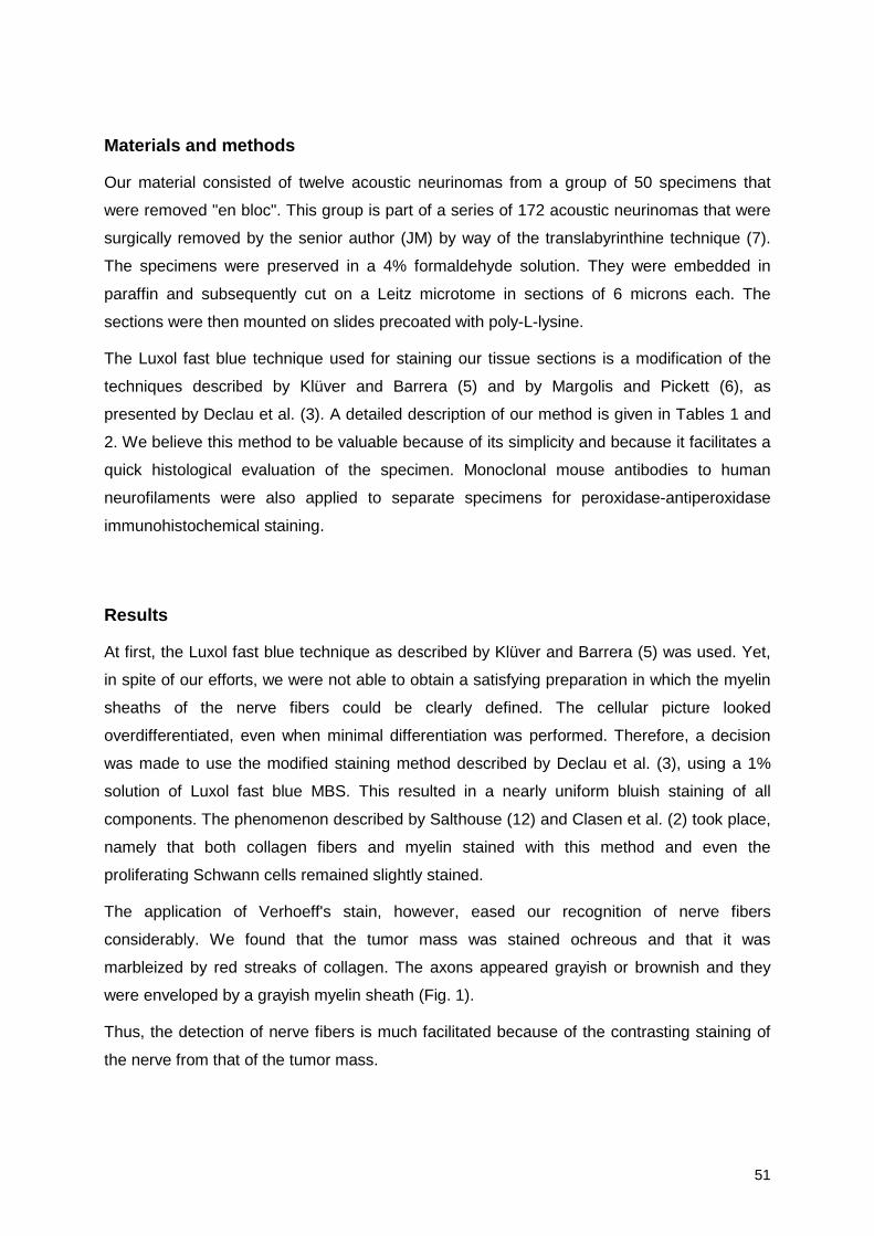
51
Materials and methods
Our material consisted of twelve acoustic neurinomas from a group of 50 specimens that
were removed "en bloc". This group is part of a series of 172 acoustic neurinomas that were
surgically removed by the senior author (JM) by way of the translabyrinthine technique (7).
The specimens were preserved in a 4% formaldehyde solution. They were embedded in
paraffin and subsequently cut on a Leitz microtome in sections of 6 microns each. The
sections were then mounted on slides precoated with poly-L-lysine.
The Luxol fast blue technique used for staining our tissue sections is a modification of the
techniques described by Klüver and Barrera (5) and by Margolis and Pickett (6), as
presented by Declau et al. (3). A detailed description of our method is given in Tables 1 and
2. We believe this method to be valuable because of its simplicity and because it facilitates a
quick histological evaluation of the specimen. Monoclonal mouse antibodies to human
neurofilaments were also applied to separate specimens for peroxidase-antiperoxidase
immunohistochemical staining.
Results
At first, the Luxol fast blue technique as described by Klüver and Barrera (5) was used. Yet,
in spite of our efforts, we were not able to obtain a satisfying preparation in which the myelin
sheaths of the nerve fibers could be clearly defined. The cellular picture looked
overdifferentiated, even when minimal differentiation was performed. Therefore, a decision
was made to use the modified staining method described by Declau et al. (3), using a 1%
solution of Luxol fast blue MBS. This resulted in a nearly uniform bluish staining of all
components. The phenomenon described by Salthouse (12) and Clasen et al. (2) took place,
namely that both collagen fibers and myelin stained with this method and even the
proliferating Schwann cells remained slightly stained.
The application of Verhoeff's stain, however, eased our recognition of nerve fibers
considerably. We found that the tumor mass was stained ochreous and that it was
marbleized by red streaks of collagen. The axons appeared grayish or brownish and they
were enveloped by a grayish myelin sheath (Fig. 1).
Thus, the detection of nerve fibers is much facilitated because of the contrasting staining of
the nerve from that of the tumor mass.
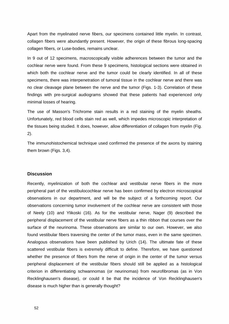
52
Apart from the myelinated nerve fibers, our specimens contained little myelin. In contrast,
collagen fibers were abundantly present. However, the origin of these fibrous long-spacing
collagen fibers, or Luse-bodies, remains unclear.
In 9 out of 12 specimens, macroscopically visible adherences between the tumor and the
cochlear nerve were found. From these 9 specimens, histological sections were obtained in
which both the cochlear nerve and the tumor could be clearly identified. In all of these
specimens, there was interpenetration of tumoral tissue in the cochlear nerve and there was
no clear cleavage plane between the nerve and the tumor (Figs. 1-3). Correlation of these
findings with pre-surgical audiograms showed that these patients had experienced only
minimal losses of hearing.
The use of Masson's Trichrome stain results in a red staining of the myelin sheaths.
Unfortunately, red blood cells stain red as well, which impedes microscopic interpretation of
the tissues being studied. It does, however, allow differentiation of collagen from myelin (Fig.
2).
The immunohistochemical technique used confirmed the presence of the axons by staining
them brown (Figs. 3,4).
Discussion
Recently, myelinization of both the cochlear and vestibular nerve fibers in the more
peripheral part of the vestibulocochlear nerve has been confirmed by electron microscopical
observations in our department, and will be the subject of a forthcoming report. Our
observations concerning tumor involvement of the cochlear nerve are consistent with those
of Neely (10) and Ylikoski (16). As for the vestibular nerve, Nager (9) described the
peripheral displacement of the vestibular nerve fibers as a thin ribbon that courses over the
surface of the neurinoma. These observations are similar to our own. However, we also
found vestibular fibers traversing the center of the tumor mass, even in the same specimen.
Analogous observations have been published by Urich (14). The ultimate fate of these
scattered vestibular fibers is extremely difficult to define. Therefore, we have questioned
whether the presence of fibers from the nerve of origin in the center of the tumor versus
peripheral displacement of the vestibular fibers should still be applied as a histological
criterion in differentiating schwannomas (or neurinomas) from neurofibromas (as in Von
Recklinghausen's disease), or could it be that the incidence of Von Recklinghausen's
disease is much higher than is generally thought?
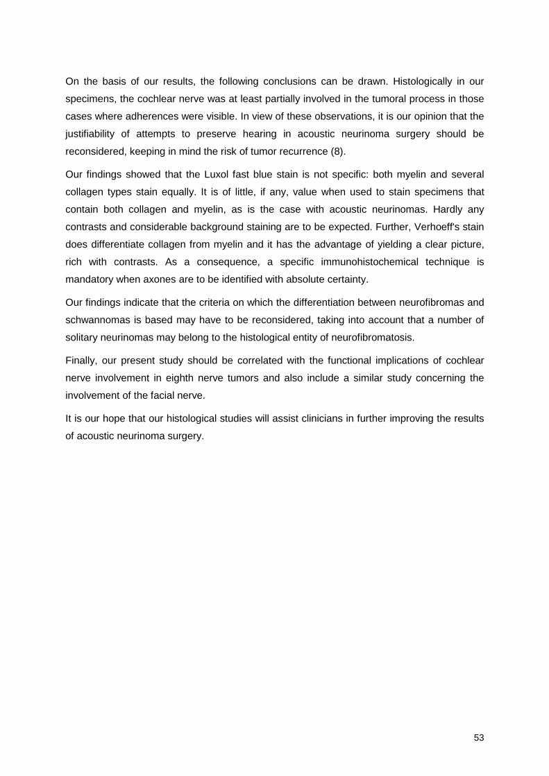
53
On the basis of our results, the following conclusions can be drawn. Histologically in our
specimens, the cochlear nerve was at least partially involved in the tumoral process in those
cases where adherences were visible. In view of these observations, it is our opinion that the
justifiability of attempts to preserve hearing in acoustic neurinoma surgery should be
reconsidered, keeping in mind the risk of tumor recurrence (8).
Our findings showed that the Luxol fast blue stain is not specific: both myelin and several
collagen types stain equally. It is of little, if any, value when used to stain specimens that
contain both collagen and myelin, as is the case with acoustic neurinomas. Hardly any
contrasts and considerable background staining are to be expected. Further, Verhoeff's stain
does differentiate collagen from myelin and it has the advantage of yielding a clear picture,
rich with contrasts. As a consequence, a specific immunohistochemical technique is
mandatory when axones are to be identified with absolute certainty.
Our findings indicate that the criteria on which the differentiation between neurofibromas and
schwannomas is based may have to be reconsidered, taking into account that a number of
solitary neurinomas may belong to the histological entity of neurofibromatosis.
Finally, our present study should be correlated with the functional implications of cochlear
nerve involvement in eighth nerve tumors and also include a similar study concerning the
involvement of the facial nerve.
It is our hope that our histological studies will assist clinicians in further improving the results
of acoustic neurinoma surgery.
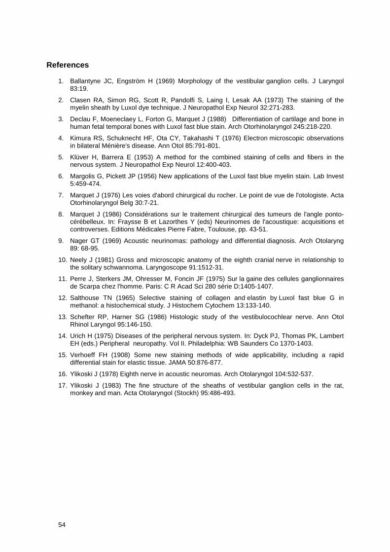
54
References
1. Ballantyne JC, Engström H (1969) Morphology of the vestibular ganglion cells. J Laryngol83:19.
2. Clasen RA, Simon RG, Scott R, Pandolfi S, Laing I, Lesak AA (1973) The staining of themyelin sheath by Luxol dye technique. J Neuropathol Exp Neurol 32:271-283.
3. Declau F, Moeneclaey L, Forton G, Marquet J (1988) Differentiation of cartilage and bone inhuman fetal temporal bones with Luxol fast blue stain. Arch Otorhinolaryngol 245:218-220.
4. Kimura RS, Schuknecht HF, Ota CY, Takahashi T (1976) Electron microscopic observationsin bilateral Ménière's disease. Ann Otol 85:791-801.
5. Klüver H, Barrera E (1953) A method for the combined staining of cells and fibers in thenervous system. J Neuropathol Exp Neurol 12:400-403.
6. Margolis G, Pickett JP (1956) New applications of the Luxol fast blue myelin stain. Lab Invest5:459-474.
7. Marquet J (1976) Les voies d'abord chirurgical du rocher. Le point de vue de l'otologiste. ActaOtorhinolaryngol Belg 30:7-21.
8. Marquet J (1986) Considérations sur le traitement chirurgical des tumeurs de l'angle ponto-cérébelleux. In: Fraysse B et Lazorthes Y (eds) Neurinomes de l'acoustique: acquisitions etcontroverses. Editions Médicales Pierre Fabre, Toulouse, pp. 43-51.
9. Nager GT (1969) Acoustic neurinomas: pathology and differential diagnosis. Arch Otolaryng89: 68-95.
10. Neely J (1981) Gross and microscopic anatomy of the eighth cranial nerve in relationship tothe solitary schwannoma. Laryngoscope 91:1512-31.
11. Perre J, Sterkers JM, Ohresser M, Foncin JF (1975) Sur la gaine des cellules ganglionnairesde Scarpa chez l'homme. Paris: C R Acad Sci 280 série D:1405-1407.
12. Salthouse TN (1965) Selective staining of collagen and elastin by Luxol fast blue G inmethanol: a histochemical study. J Histochem Cytochem 13:133-140.
13. Schefter RP, Harner SG (1986) Histologic study of the vestibulocochlear nerve. Ann OtolRhinol Laryngol 95:146-150.
14. Urich H (1975) Diseases of the peripheral nervous system. In: Dyck PJ, Thomas PK, LambertEH (eds.) Peripheral neuropathy. Vol II. Philadelphia: WB Saunders Co 1370-1403.
15. Verhoeff FH (1908) Some new staining methods of wide applicability, including a rapiddifferential stain for elastic tissue. JAMA 50:876-877.
16. Ylikoski J (1978) Eighth nerve in acoustic neuromas. Arch Otolaryngol 104:532-537.
17. Ylikoski J (1983) The fine structure of the sheaths of vestibular ganglion cells in the rat,monkey and man. Acta Otolaryngol (Stockh) 95:486-493.
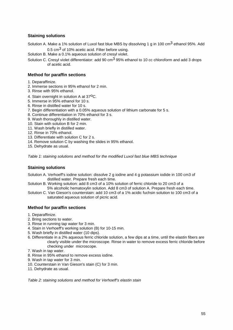
55
Staining solutions
Solution A. Make a 1% solution of Luxol fast blue MBS by dissolving 1 g in 100 cm3 ethanol 95%. Add
0.5 cm3 of 10% acetic acid. Filter before using.Solution B. Make a 0.1% aqueous solution of cresyl violet.
Solution C. Cresyl violet differentiator: add 90 cm3 95% ethanol to 10 cc chloroform and add 3 dropsof acetic acid.
Method for paraffin sections
1. Deparaffinize.2. Immerse sections in 95% ethanol for 2 min.3. Rinse with 95% ethanol.
4. Stain overnight in solution A at 37oC.5. Immerse in 95% ethanol for 10 s.6. Rinse in distilled water for 10 s.7. Begin differentiation with a 0.05% aqueous solution of lithium carbonate for 5 s.8. Continue differentiation in 70% ethanol for 3 s.9. Wash thoroughly in distilled water.10. Stain with solution B for 2 min.11. Wash briefly in distilled water.12. Rinse in 70% ethanol.13. Differentiate with solution C for 2 s.14. Remove solution C by washing the slides in 95% ethanol.15. Dehydrate as usual.
Table 1: staining solutions and method for the modified Luxol fast blue MBS technique
Staining solutions
Solution A. Verhoeff's iodine solution: dissolve 2 g iodine and 4 g potassium iodide in 100 cm3 ofdistilled water. Prepare fresh each time.
Solution B. Working solution: add 8 cm3 of a 10% solution of ferric chloride to 20 cm3 of a5% alcoholic hematoxylin solution. Add 8 cm3 of solution A. Prepare fresh each time.
Solution C. Van Gieson's counterstain: add 10 cm3 of a 1% acidic fuchsin solution to 100 cm3 of asaturated aqueous solution of picric acid.
Method for paraffin sections
1. Deparaffinize.2. Bring sections to water.3. Rinse in running tap water for 3 min.4. Stain in Verhoeff's working solution (B) for 10-15 min.5. Wash briefly in distilled water (10 dips).6. Differentiate in a 2% aqueous ferric chloride solution, a few dips at a time, until the elastin fibers are
clearly visible under the microscope. Rinse in water to remove excess ferric chloride beforechecking under microscope.
7. Wash in tap water.8. Rinse in 95% ethanol to remove excess iodine.9. Wash in tap water for 3 min.10. Counterstain in Van Gieson's stain (C) for 3 min.11. Dehydrate as usual.
Table 2: staining solutions and method for Verhoeff’s elastin stain
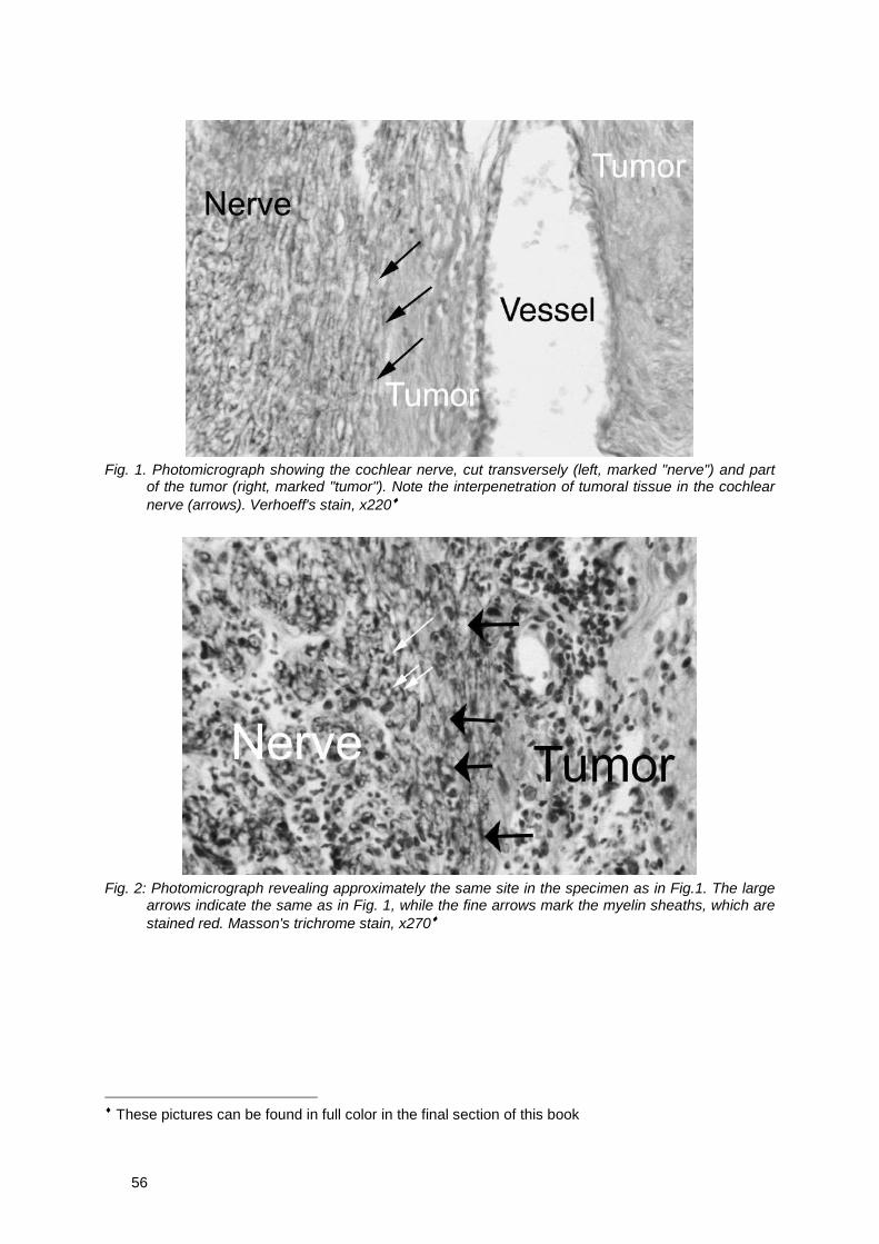
56
Fig. 1. Photomicrograph showing the cochlear nerve, cut transversely (left, marked "nerve") and partof the tumor (right, marked "tumor"). Note the interpenetration of tumoral tissue in the cochlearnerve (arrows). Verhoeff's stain, x220♦
Fig. 2: Photomicrograph revealing approximately the same site in the specimen as in Fig.1. The largearrows indicate the same as in Fig. 1, while the fine arrows mark the myelin sheaths, which arestained red. Masson's trichrome stain, x270♦
♦ These pictures can be found in full color in the final section of this book
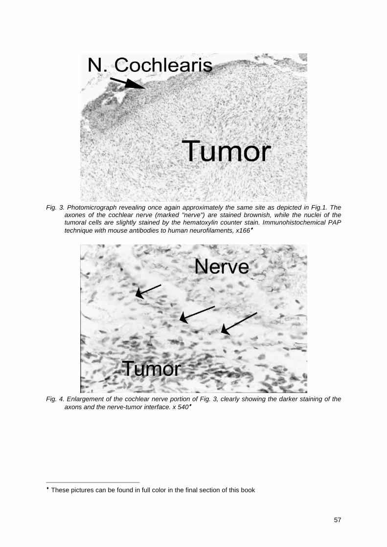
57
Fig. 3. Photomicrograph revealing once again approximately the same site as depicted in Fig.1. Theaxones of the cochlear nerve (marked "nerve") are stained brownish, while the nuclei of thetumoral cells are slightly stained by the hematoxylin counter stain. Immunohistochemical PAPtechnique with mouse antibodies to human neurofilaments, x166♦
Fig. 4. Enlargement of the cochlear nerve portion of Fig. 3, clearly showing the darker staining of theaxons and the nerve-tumor interface. x 540♦
♦ These pictures can be found in full color in the final section of this book

58
59
Chapter 2c
The solitary schwannoma of the eighth cranial nerve:
An immuno-histochemical study of the cochlear nerve -tumor interface
J.F.E. Marquet†, MD
G.E.J. Forton, MD
F.E. Offeciers, MD
L.L.M. Moeneclaey
ENT Department, University of Antwerp, Wilrijk, BelgiumArch Otolaryngol Head Neck Surg 1990;116(9):1023-1025
60
Samenvatting
Met behulp van de in het vorige hoofdstuk beschreven kleuringtechnieken, worden tien
intacte acousticusneurinomen, waarbij macroscopisch zichtbare vergroeiingen tussen tumor
en Nervus Cochlearis bleken te bestaan, bestudeerd. In het bijzonder wordt de grenszone
tussen tumor en zenuw in detail bestudeerd. Er blijkt een duidelijke ingroei van tumorale
cellen in de zenuw te bestaan. Dit blijkt zowel uit de aanwezigheid van manifeste
schwannoomcellen tussen de zenuwvezels, als uit het voorkomen van cochleaire
zenuwvezels tot voorbij deze grenszone. In de bespreking wordt opnieuw het concept van
gehoorsparende chirurgie in vraag gesteld, al wordt hier reeds gewag gemaakt van een op
imaging gebaseerde “wait and see” houding.
Abstract
Given recent controversy concerning hearing preservation surgery of the acoustic neuroma,
an immunohistochemical study was undertaken in order to investigate the cochlear nerve -
tumor interface. Ten intact medium-size acoustic neurinomas were studied by means of
classical staining procedures, as well as an immunohistochemical technique using
monoclonal mouse antibodies to human neurofilaments. Our observations indicate that the
cochlear nerve is histologically involved in the tumoral process in those cases where
macroscopically visible adherences between the cochlear nerve and the tumor are present.
We were not able to discern a clear cleavage plane. In six out of the ten specimens did we
objectivate tumoral invasion of the cochlear nerve. In view of these observations and
international literature, several therapeutic attitudes are discussed. In conclusion, the
principle of hearing preservation surgery is rejected in favor of total tumor removal in every
case where surgery is indicated.
61
Introduction
Facing the increasing number of papers dedicated to hearing conservation surgery of the
acoustic neuroma, we initiated a histopathological study on the interface between the
cochlear nerve and the tumor. Indeed, histopathological evidence of cochlear nerve
involvement or invasion is, in our opinion, a very serious argument against hearing
conservation surgery.
As mentioned by Marquet [1] in 1986, several reflections have to be considered when
discussing acoustic neuroma surgery. Recent advances in medical imaging techniques have
enabled us to visualize very small acoustic tumors with unprecedented accuracy. The neuro-
otologist is therefore offered two options when confronted with a newly diagnosed acoustic
neuroma: to operate or not to operate. This attitude is especially indicated with elderly people
having a slowly growing tumor, in case of bilateral acoustic neurinomas (as observed in
some particular cases and more commonly in neurofibromatosis type II) and in patients with
a single hearing ear.
Should the surgeon decide not to perform surgery, a meticulous follow-up stratagem is to be
carried out to monitor the growth of the tumor. Fortunately, Gadolinium-pentetic acid
enhanced magnetic resonance imaging allows excellent visualization of small intracanalicular
tumors. Surgery can thus be postponed and whatever may be left of useful hearing on the
affected side be saved until surgery becomes indicated.
However, should the surgeon decide to operate at once, there are several points worth
considering: the surgical inaccessibility of the fundus of the internal auditory meatus by way
of the suboccipital approach; the biochemical alterations that occur in the perilymph fluid in
the course of tumor development, as demonstrated by Silverstein and Schuknecht [2]; and
the vascular lesions that may be caused either by tumor growth or during surgery. House
and Hitselberger [3] consider these vascular lesions to be one of the major causes of post-
operative deafness, in spite of the anatomical integrity of the cochlear nerve. Also worth
considering are the histopathological aspects of acoustic neurinomas in relation to the
cochlear nerve. Although benign, the schwannoma, which usually originates from the inferior
branch of the vestibular nerve, actually tends to destroy adjacent nerves, first by crushing
them and later by invading them. Invasion of the cochlear nerve by the acoustic neurinoma
has been described by Neely and associates [4,5], Ylikoski et al. [6,7] and Forton et al. [8].
Even facial nerve involvement has been described by Luetje et al. [9]. Our observations have
shown that recurrent acoustic neurinomas have a mean growth rate of 4.2 mm/y in every
dimension [1], a fact that is frequently overlooked by several authors. These figures are
62
comparable with those of Wazen et al. [10], who have found a mean growth rate of 3.8 mm/y.
In this study, we are particularly interested in the nerve-tumor interface, in order to establish
any cochlear nerve involvement or invasion.
Materials and methods
We have studied 10 intact medium-sized acoustic neurinomas that have been removed via
the translabyrinthine approach. These tumors were removed by the senior authors (J.F.E.M.
and F.E.O.) en bloc with the cochlear nerve, allowing not only the identification of all the
nervous structures involved, but also the serial sectioning of the specimens. In all 10 cases,
macroscopically visible adherences between the cochlear nerve and the tumor were present.
The specimens were fixed in a buffered 4% formaldehyde solution and embedded in paraffin.
Subsequently, they were cut on a microtome (Leitz) in 6 µm-thick sections. These were
mounted on slides precoated with poly-L-lysine.
Three different staining procedures were performed. First, Masson's trichrome stain was
applied, because it is a quick and easy routine stain and because it stains myelin red, which
is a useful characteristic. Second, Verhoeff's elastin stain was applied, because it yields a
clear picture, rich with contrasts: the characteristic streaks of collagen, which are colored red
are readily identifiable in the tumor mass, which is stained ochreous. The nerve fibers,
however, are not particularly stained. Only perineural connective tissue is colored red by this
method. Thus, any connective tissue septum that might exist between the cochlear nerve
and the tumor should be visualised in this manner. Last, we devised an
immunohistochemical protocol using monoclonal mouse antibodies to human neurofilament
protein (2F11) (Dakopatts M762) as primary antibodies, rabbit immunoglobulins to mouse
immunoglobulins (Dakopatts Z109) as secondary antibodies and peroxidase-antiperoxidase
monoclonal mouse antibody complexes (Dakopatts P850). A 3,3-diaminobenzidine
tetrahydrochloride solution was used as chromogenic agent. Thus, any present bundle of
nerve fibers was stained brown. A discrete Harris' hematoxylin stain was used as
counterstain. The details of our staining methods are discussed in a previous article [8].
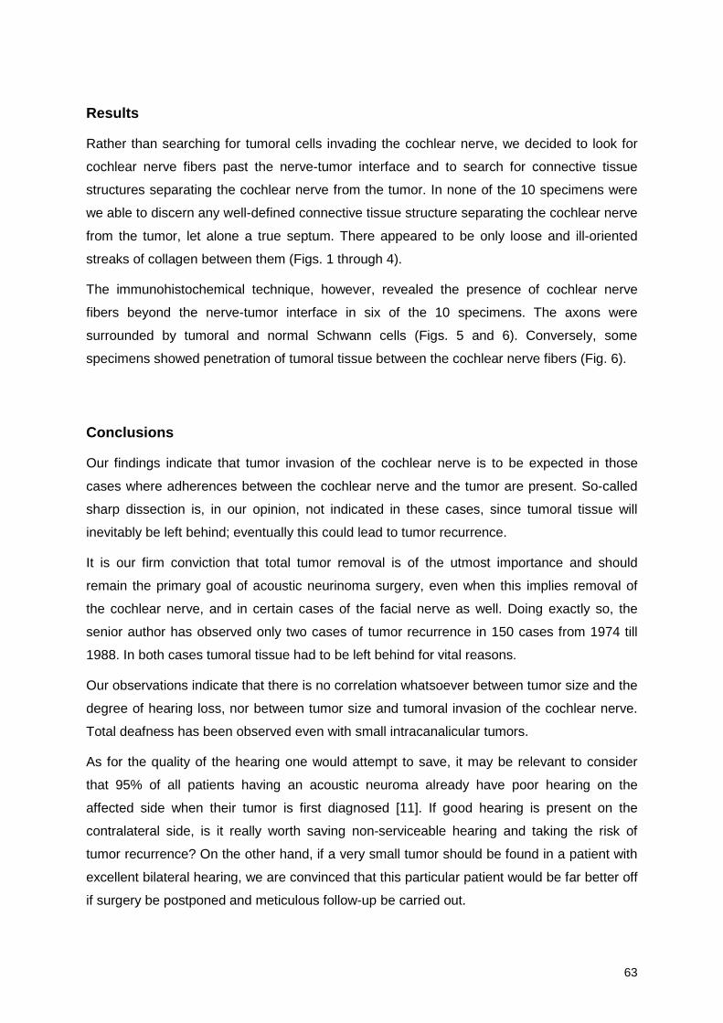
63
Results
Rather than searching for tumoral cells invading the cochlear nerve, we decided to look for
cochlear nerve fibers past the nerve-tumor interface and to search for connective tissue
structures separating the cochlear nerve from the tumor. In none of the 10 specimens were
we able to discern any well-defined connective tissue structure separating the cochlear nerve
from the tumor, let alone a true septum. There appeared to be only loose and ill-oriented
streaks of collagen between them (Figs. 1 through 4).
The immunohistochemical technique, however, revealed the presence of cochlear nerve
fibers beyond the nerve-tumor interface in six of the 10 specimens. The axons were
surrounded by tumoral and normal Schwann cells (Figs. 5 and 6). Conversely, some
specimens showed penetration of tumoral tissue between the cochlear nerve fibers (Fig. 6).
Conclusions
Our findings indicate that tumor invasion of the cochlear nerve is to be expected in those
cases where adherences between the cochlear nerve and the tumor are present. So-called
sharp dissection is, in our opinion, not indicated in these cases, since tumoral tissue will
inevitably be left behind; eventually this could lead to tumor recurrence.
It is our firm conviction that total tumor removal is of the utmost importance and should
remain the primary goal of acoustic neurinoma surgery, even when this implies removal of
the cochlear nerve, and in certain cases of the facial nerve as well. Doing exactly so, the
senior author has observed only two cases of tumor recurrence in 150 cases from 1974 till
1988. In both cases tumoral tissue had to be left behind for vital reasons.
Our observations indicate that there is no correlation whatsoever between tumor size and the
degree of hearing loss, nor between tumor size and tumoral invasion of the cochlear nerve.
Total deafness has been observed even with small intracanalicular tumors.
As for the quality of the hearing one would attempt to save, it may be relevant to consider
that 95% of all patients having an acoustic neuroma already have poor hearing on the
affected side when their tumor is first diagnosed [11]. If good hearing is present on the
contralateral side, is it really worth saving non-serviceable hearing and taking the risk of
tumor recurrence? On the other hand, if a very small tumor should be found in a patient with
excellent bilateral hearing, we are convinced that this particular patient would be far better off
if surgery be postponed and meticulous follow-up be carried out.
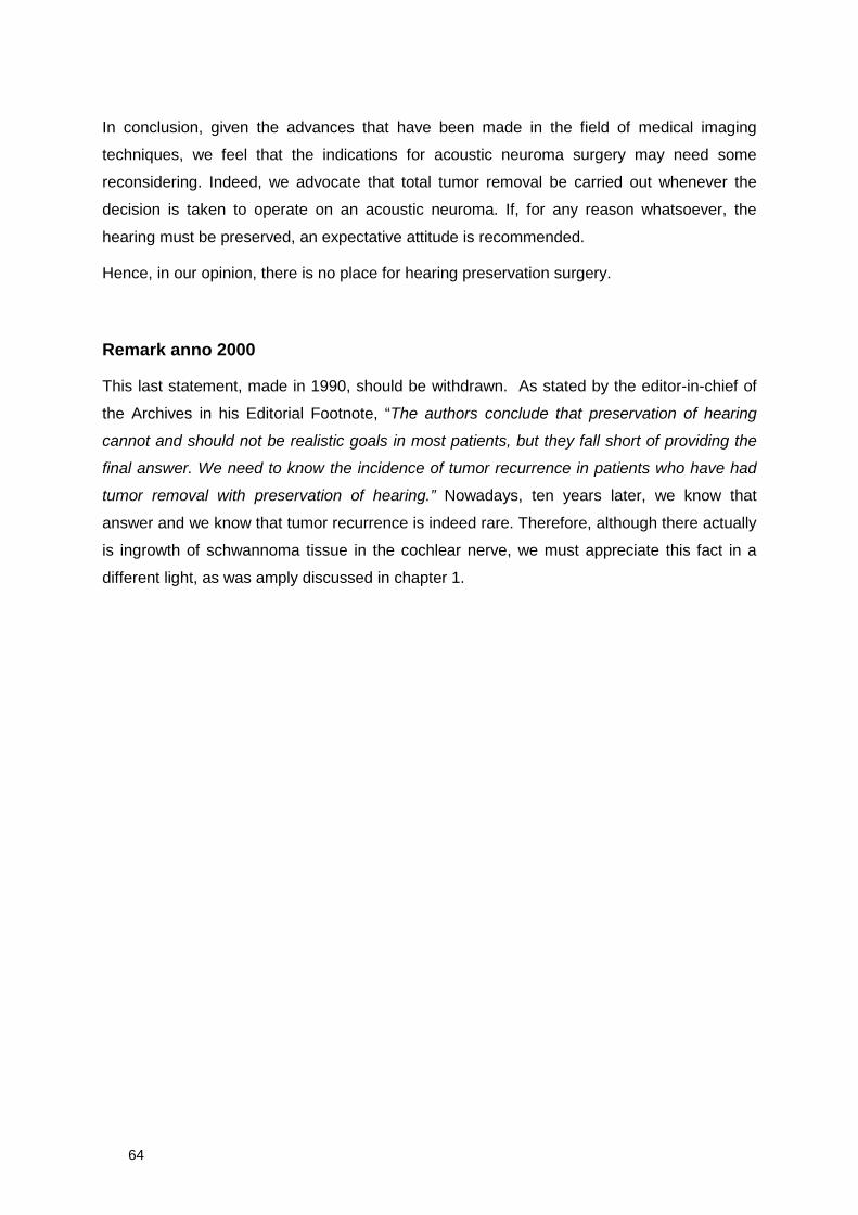
64
In conclusion, given the advances that have been made in the field of medical imaging
techniques, we feel that the indications for acoustic neuroma surgery may need some
reconsidering. Indeed, we advocate that total tumor removal be carried out whenever the
decision is taken to operate on an acoustic neuroma. If, for any reason whatsoever, the
hearing must be preserved, an expectative attitude is recommended.
Hence, in our opinion, there is no place for hearing preservation surgery.
Remark anno 2000
This last statement, made in 1990, should be withdrawn. As stated by the editor-in-chief of
the Archives in his Editorial Footnote, “The authors conclude that preservation of hearing
cannot and should not be realistic goals in most patients, but they fall short of providing the
final answer. We need to know the incidence of tumor recurrence in patients who have had
tumor removal with preservation of hearing.” Nowadays, ten years later, we know that
answer and we know that tumor recurrence is indeed rare. Therefore, although there actually
is ingrowth of schwannoma tissue in the cochlear nerve, we must appreciate this fact in a
different light, as was amply discussed in chapter 1.
65
References
1. Marquet J. Considérations sur le traitement chirurgical des tumeurs de l'angle ponto-cérébelleux. In: Fraysse B, Lazorthes Y, eds. Neurinomes de l'acoustique: acquisitions etcontroverses. Toulouse: Editions Médicales Pierre Fabre, 1986:43-51.
2. Silverstein H, Schuknecht H. Biochemical studies of inner ear fluid in man: Changes inotosclerosis, Ménière's disease and acoustic neuroma. Arch Otolaryngol 1966;84:395.
3. House WF, Hitselberger WE. The neuro-otologist's view of surgical management of acousticneuromas. Clin Neurosurg 1985;32:214-222.
4. Neely JG, Hough J. Histologic findings in two very small intracanicular solitary schwannomasof the eighth nerve. Ann Otol Rhinol Laryngol 1986;95:460-465.
5. Neely JG. Gross and microscopic anatomy of the eighth cranial nerve in relationship to thesolitary schwannoma. Laryngoscope 1981;91:1512-1531.
6. Ylikoski J, Collan Y, Palva T, Jauhiainen T. Cochlear nerve in neurilemomas. Arch Otolaryngol1978;104:679-684.
7. Ylikoski J, Palva T, Collan Y. Eighth nerve in acoustic neuromas. Arch Otolaryngol1978;104:532-537.
8. Forton G, Moeneclaey L, Declau F, Marquet J. The involvement of the cochlear nerve inneurinomas of the eighth cranial nerve. Arch Otorhinolaryngol 1989;246:156-160.
9. Luetje CM, Whittaker CK, Callaway LA, Veraga G. Histological acoustic tumor involvement ofthe VIIth nerve and multicentric origin in the VIIIth nerve. Laryngoscope 1983;93:1133-1139.
10. Wazen J, Silverstein H, Norrel H, Besse B. Preoperative and postoperative growth rates inacoustic neuromas documented with CT scanning? Otolaryngol Head Neck Surg1985;93(2):151-155.
11. Van de Heyning P, Marquet J, Declau F. The preservation of facial function in acousticneuroma surgery. In: Proceedings of the VIth International Symposium on the Facial Nerve.Rio de Janeiro, 1988.
66
Fig. 1: The cochlear nerve – tumor interface (Masson trichrome stain, x 166). Note the closerelationship that exists between the cochlear nerve and the tumor. A cleavage plane cannot beidentified♦
Fig 2: Detail of the cochlear nerve – tumor interface (Masson trichrome stain, x 646). The tumor massis stained green, which is partly due to the large amounts of collagen that are present. Note thered myelin sheaths (fine arrows)), surrounded by perineural connective tissue (green). Note,again, the absence of a clear cleavage plane between the nerve and the neuroma♦
♦ These pictures can be found in full color in the final section of this book
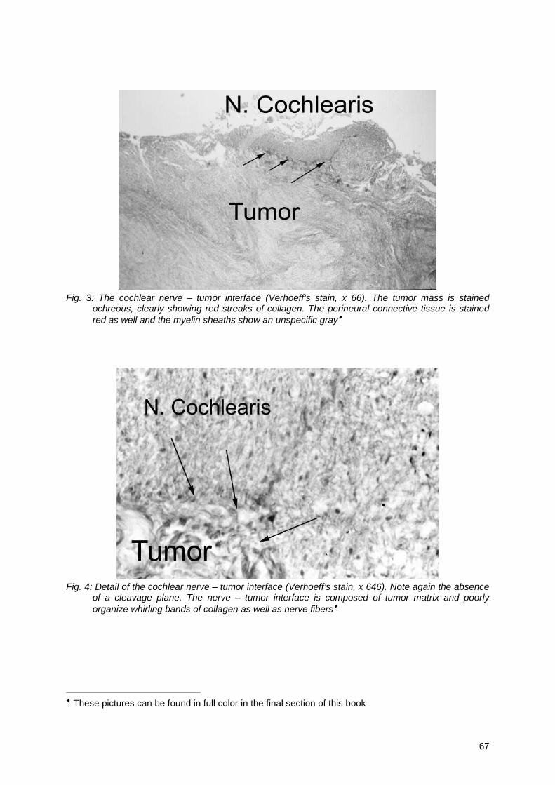
67
Fig. 3: The cochlear nerve – tumor interface (Verhoeff’s stain, x 66). The tumor mass is stainedochreous, clearly showing red streaks of collagen. The perineural connective tissue is stainedred as well and the myelin sheaths show an unspecific gray♦
Fig. 4: Detail of the cochlear nerve – tumor interface (Verhoeff’s stain, x 646). Note again the absenceof a cleavage plane. The nerve – tumor interface is composed of tumor matrix and poorlyorganize whirling bands of collagen as well as nerve fibers♦
♦ These pictures can be found in full color in the final section of this book
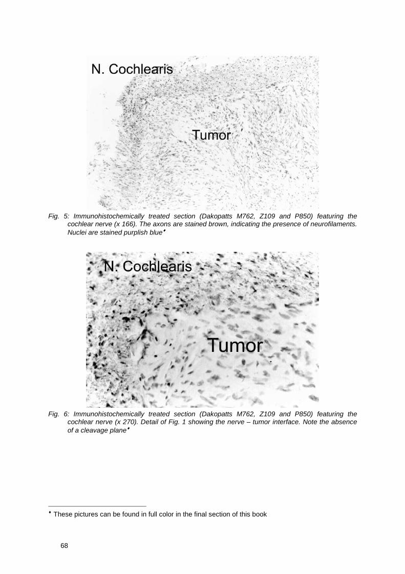
68
Fig. 5: Immunohistochemically treated section (Dakopatts M762, Z109 and P850) featuring thecochlear nerve (x 166). The axons are stained brown, indicating the presence of neurofilaments.Nuclei are stained purplish blue♦
Fig. 6: Immunohistochemically treated section (Dakopatts M762, Z109 and P850) featuring thecochlear nerve (x 270). Detail of Fig. 1 showing the nerve – tumor interface. Note the absenceof a cleavage plane♦
♦ These pictures can be found in full color in the final section of this book
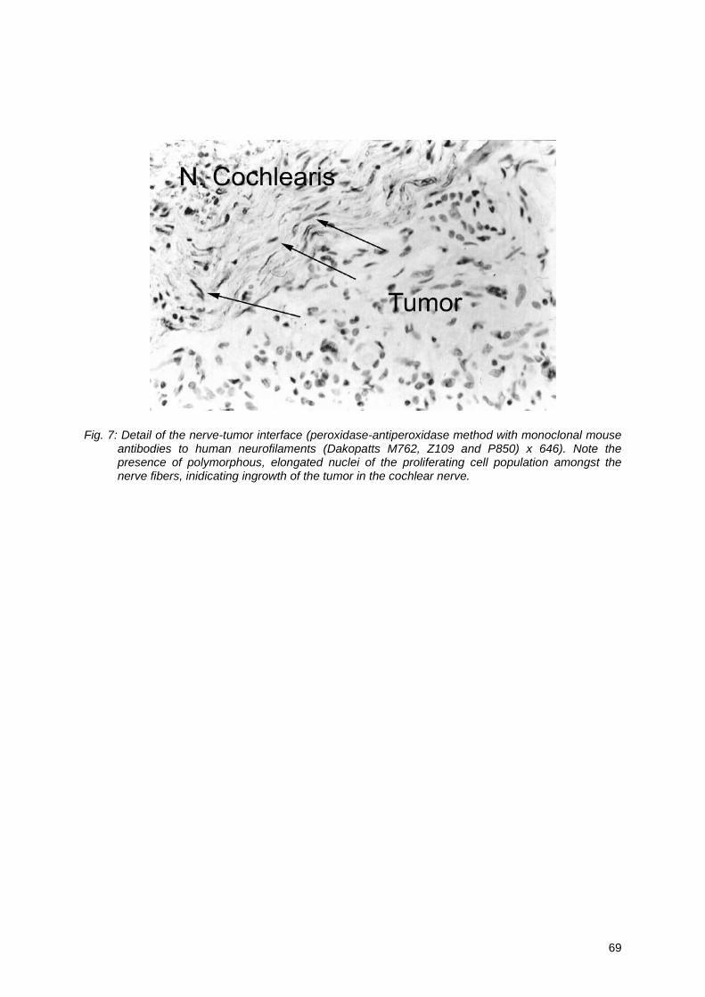
69
Fig. 7: Detail of the nerve-tumor interface (peroxidase-antiperoxidase method with monoclonal mouseantibodies to human neurofilaments (Dakopatts M762, Z109 and P850) x 646). Note thepresence of polymorphous, elongated nuclei of the proliferating cell population amongst thenerve fibers, inidicating ingrowth of the tumor in the cochlear nerve.

70
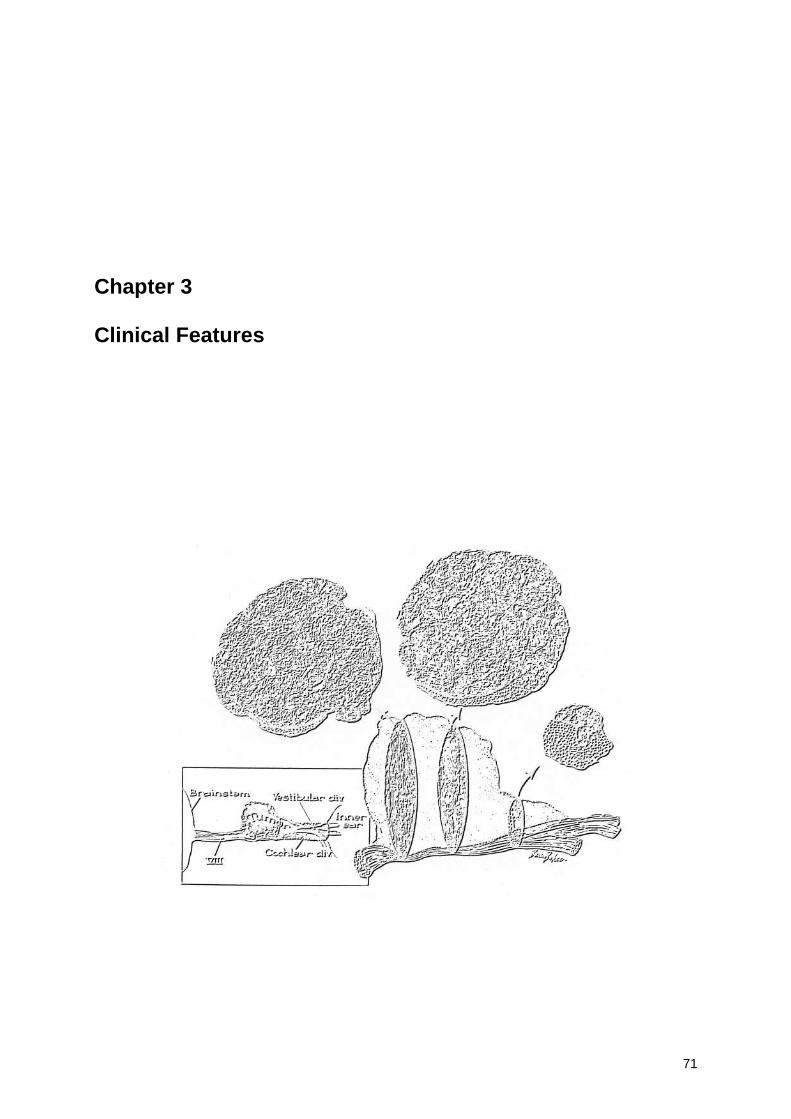
71
Chapter 3
Clinical Features

72
73
Chapter 3a
Acoustic neuroma ingrowth in the cochlear nerve:
An analysis of clinical data
Glen E.J. Forton
Submitted for publication
74
Samenvatting
In de vorige hoofdstukken werd aangetoond hoe de Nervus Cochlearis histologisch
aantoonbaar betrokken is bij het tumorale proces. De afwezigheid van enig duidelijk
klievingsvlak werd bewezen. In dit hoofdstuk trachten we te achterhalen of deze histologisch
onderzochte neurinomen op enigerlei wijze verschillen van de doorsnee populatie. Met
andere woorden: is er in onze groep met bewezen invasie ook een groter aantal zware
gehoorverliezen? Tot de onderzochte parameters behoren de tonale audiometrische
gegevens, tumorafmetingen zoals bepaald door beeldvorming en B.E.R.A.-gegevens. Deze
door de gekozen selectiecriteria beperkte reeks (N = 13) wordt vervolgens getoetst aan
enkele grotere gepubliceerde reeksen.
Introduction
Given the fact that microscopical evidence as to the involvement of the cochlear nerve has
been rendered by several authors including our own research (1, 2), we thought it interesting
to assess whether these tumors differ in any way from the mean population of small (1 – 25
mm) to medium-sized (26 – 40 mm) acoustic neuromas. Is there a higher-than-average
number of cases with severe hearing loss or deafness in this series? Therefore, we
executed a concise statistical analysis on the retrieved data of the microscopically studied
cases with proven invasion (such as tumor size, audiometric findings, onset of symptoms)
and compared it to some of the large published series.
Materials and methods
Over the years, we have microscopically studied a fair amount of intact acoustic neuromas in
order to evaluate the ingrowth of the tumor in the cochlear nerve. The early cases were
operated by the late head of department, Jean Marquet, while the second series was
composed of specimens removed by the late Jean Marquet and Erwin Offeciers, both from
the Sint-Augistinus Medical Institute of Antwerp (Belgium). The histological findings from both
series were published separately (1,2) and are discussed in Chapter 2. A third, more recent,
series was provided by Cor W.R.J. Cremers at the University Hospital Nijmegen (The
Netherlands).
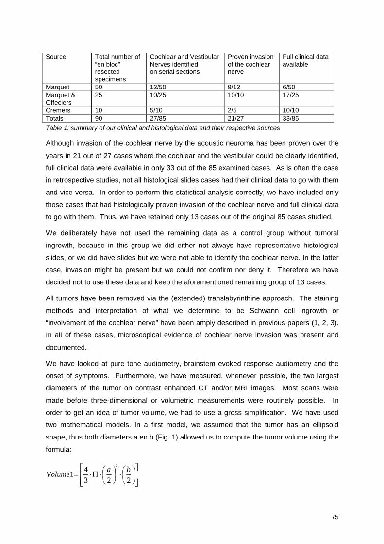
75
Source Total number of“en bloc”resectedspecimens
Cochlear and VestibularNerves identifiedon serial sections
Proven invasionof the cochlearnerve
Full clinical dataavailable
Marquet 50 12/50 9/12 6/50Marquet &Offeciers
25 10/25 10/10 17/25
Cremers 10 5/10 2/5 10/10Totals 90 27/85 21/27 33/85
Table 1: summary of our clinical and histological data and their respective sources
Although invasion of the cochlear nerve by the acoustic neuroma has been proven over the
years in 21 out of 27 cases where the cochlear and the vestibular could be clearly identified,
full clinical data were available in only 33 out of the 85 examined cases. As is often the case
in retrospective studies, not all histological slides cases had their clinical data to go with them
and vice versa. In order to perform this statistical analysis correctly, we have included only
those cases that had histologically proven invasion of the cochlear nerve and full clinical data
to go with them. Thus, we have retained only 13 cases out of the original 85 cases studied.
We deliberately have not used the remaining data as a control group without tumoral
ingrowth, because in this group we did either not always have representative histological
slides, or we did have slides but we were not able to identify the cochlear nerve. In the latter
case, invasion might be present but we could not confirm nor deny it. Therefore we have
decided not to use these data and keep the aforementioned remaining group of 13 cases.
All tumors have been removed via the (extended) translabyrinthine approach. The staining
methods and interpretation of what we determine to be Schwann cell ingrowth or
“involvement of the cochlear nerve” have been amply described in previous papers (1, 2, 3).
In all of these cases, microscopical evidence of cochlear nerve invasion was present and
documented.
We have looked at pure tone audiometry, brainstem evoked response audiometry and the
onset of symptoms. Furthermore, we have measured, whenever possible, the two largest
diameters of the tumor on contrast enhanced CT and/or MRI images. Most scans were
made before three-dimensional or volumetric measurements were routinely possible. In
order to get an idea of tumor volume, we had to use a gross simplification. We have used
two mathematical models. In a first model, we assumed that the tumor has an ellipsoid
shape, thus both diameters a en b (Fig. 1) allowed us to compute the tumor volume using the
formula:
⋅
⋅Π⋅=
2234
12
baVolume
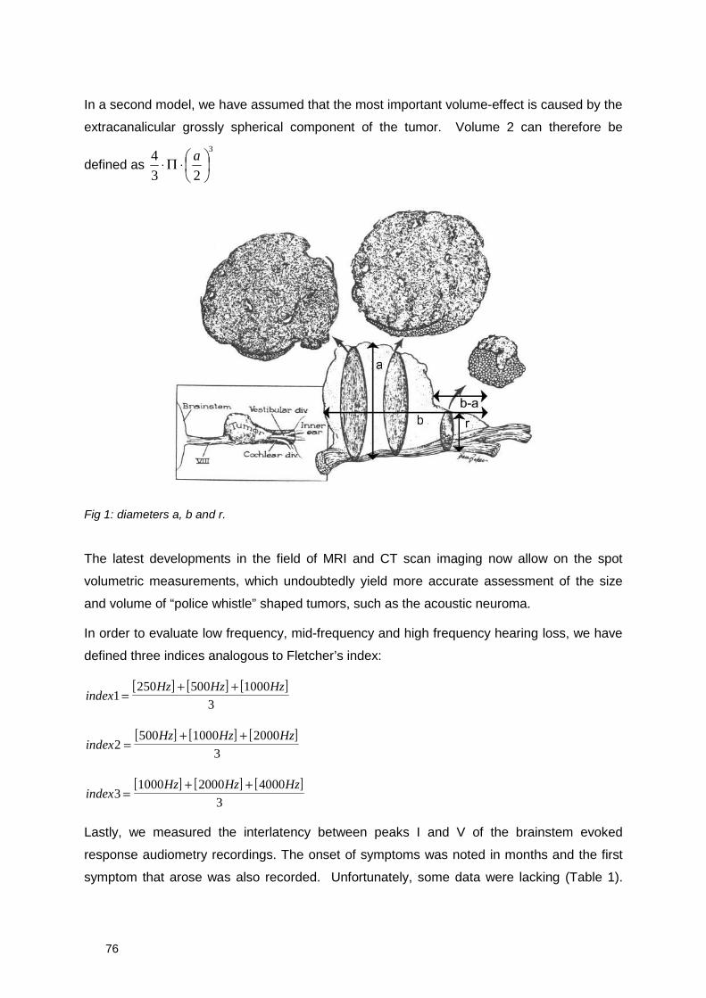
76
In a second model, we have assumed that the most important volume-effect is caused by the
extracanalicular grossly spherical component of the tumor. Volume 2 can therefore be
defined as3
23
4
⋅Π⋅ a
Fig 1: diameters a, b and r.
The latest developments in the field of MRI and CT scan imaging now allow on the spot
volumetric measurements, which undoubtedly yield more accurate assessment of the size
and volume of “police whistle” shaped tumors, such as the acoustic neuroma.
In order to evaluate low frequency, mid-frequency and high frequency hearing loss, we have
defined three indices analogous to Fletcher’s index:
[ ] [ ] [ ]index
Hz Hz Hz1
250 500 1000
3=
+ +
[ ] [ ] [ ]index
Hz Hz Hz2
500 1000 2000
3=
+ +
[ ] [ ] [ ]index
Hz Hz Hz3
1000 2000 4000
3=
+ +
Lastly, we measured the interlatency between peaks I and V of the brainstem evoked
response audiometry recordings. The onset of symptoms was noted in months and the first
symptom that arose was also recorded. Unfortunately, some data were lacking (Table 1).
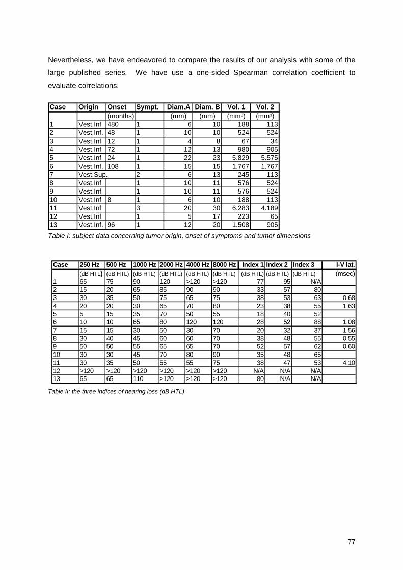
77
Nevertheless, we have endeavored to compare the results of our analysis with some of the
large published series. We have use a one-sided Spearman correlation coefficient to
evaluate correlations.
Table I: subject data concerning tumor origin, onset of symptoms and tumor dimensions
Table II: the three indices of hearing loss (dB HTL)
Case Origin Onset Sympt. Diam.A Diam. B Vol. 1 Vol. 2(months) (mm) (mm) (mm³) (mm³)
1 Vest.Inf 480 1 6 10 188 1132 Vest.Inf. 48 1 10 10 524 5243 Vest.Inf 12 1 4 8 67 344 Vest.Inf 72 1 12 13 980 9055 Vest.Inf 24 1 22 23 5.829 5.5756 Vest.Inf. 108 1 15 15 1.767 1.7677 Vest.Sup. 2 6 13 245 1138 Vest.Inf 1 10 11 576 5249 Vest.Inf 1 10 11 576 52410 Vest.Inf 8 1 6 10 188 11311 Vest.Inf 3 20 30 6.283 4.18912 Vest.Inf 1 5 17 223 6513 Vest.Inf. 96 1 12 20 1.508 905
Case 250 Hz 500 Hz 1000 Hz 2000 Hz 4000 Hz 8000 Hz Index 1 Index 2 Index 3 I-V lat.(dB HTL) (dB HTL) (dB HTL) (dB HTL) (dB HTL) (dB HTL) (dB HTL) (dB HTL) (dB HTL) (msec)
1 65 75 90 120 >120 >120 77 95 N/A2 15 20 65 85 90 90 33 57 803 30 35 50 75 65 75 38 53 63 0,684 20 20 30 65 70 80 23 38 55 1,635 5 15 35 70 50 55 18 40 526 10 10 65 80 120 120 28 52 88 1,087 15 15 30 50 30 70 20 32 37 1,568 30 40 45 60 60 70 38 48 55 0,559 50 50 55 65 65 70 52 57 62 0,6010 30 30 45 70 80 90 35 48 6511 30 35 50 55 55 75 38 47 53 4,1012 >120 >120 >120 >120 >120 >120 N/A N/A N/A13 65 65 110 >120 >120 >120 80 N/A N/A
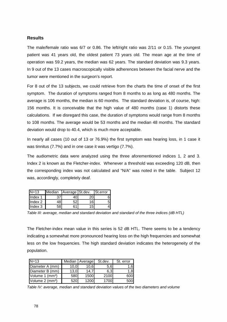
78
Results
The male/female ratio was 6/7 or 0.86. The left/right ratio was 2/11 or 0.15. The youngest
patient was 41 years old, the oldest patient 73 years old. The mean age at the time of
operation was 59.2 years, the median was 62 years. The standard deviation was 9.3 years.
In 9 out of the 13 cases macroscopically visible adherences between the facial nerve and the
tumor were mentioned in the surgeon’s report.
For 8 out of the 13 subjects, we could retrieve from the charts the time of onset of the first
symptom. The duration of symptoms ranged from 8 months to as long as 480 months. The
average is 106 months, the median is 60 months. The standard deviation is, of course, high:
156 months. It is conceivable that the high value of 480 months (case 1) distorts these
calculations. If we disregard this case, the duration of symptoms would range from 8 months
to 108 months. The average would be 53 months and the median 48 months. The standard
deviation would drop to 40.4, which is much more acceptable.
In nearly all cases (10 out of 13 or 76.9%) the first symptom was hearing loss, in 1 case it
was tinnitus (7.7%) and in one case it was vertigo (7.7%).
The audiometric data were analyzed using the three aforementioned indices 1, 2 and 3.
Index 2 is known as the Fletcher-index. Whenever a threshold was exceeding 120 dB, then
the corresponding index was not calculated and “N/A” was noted in the table. Subject 12
was, accordingly, completely deaf.
Table III: average, median and standard deviation and standard of the three indices (dB HTL)
The Fletcher-index mean value in this series is 52 dB HTL. There seems to be a tendency
indicating a somewhat more pronounced hearing loss on the high frequencies and somewhat
less on the low frequencies. The high standard deviation indicates the heterogeneity of the
population.
Table IV: average, median and standard deviation values of the two diameters and volume
N=13 Median Average St.dev. St. errorDiameter A (mm) 10,0 10,6 5,6 1,6Diameter B (mm) 13,0 14,7 6,3 1,8Volume 1 (mm³) 580 1500 2100 600Volume 2 (mm³) 520 1200 1700 500
N=13 Median Average St.dev. St.errorIndex 1 37 40 20 6Index 2 48 52 16 5Index 3 58 61 15 4
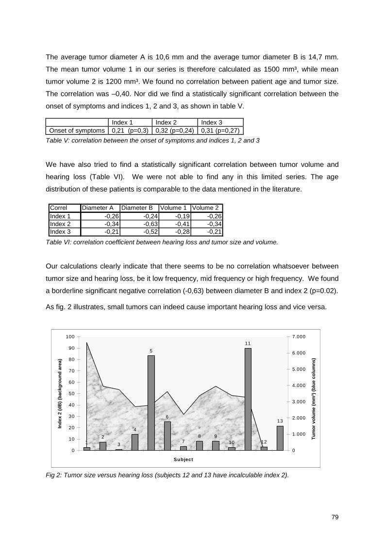
79
The average tumor diameter A is 10,6 mm and the average tumor diameter B is 14,7 mm.
The mean tumor volume 1 in our series is therefore calculated as 1500 mm³, while mean
tumor volume 2 is 1200 mm³. We found no correlation between patient age and tumor size.
The correlation was –0,40. Nor did we find a statistically significant correlation between the
onset of symptoms and indices 1, 2 and 3, as shown in table V.
Index 1 Index 2 Index 3Onset of symptoms 0,21 (p=0,3) 0,32 (p=0,24) 0,31 (p=0,27)
Table V: correlation between the onset of symptoms and indices 1, 2 and 3
We have also tried to find a statistically significant correlation between tumor volume and
hearing loss (Table VI). We were not able to find any in this limited series. The age
distribution of these patients is comparable to the data mentioned in the literature.
Table VI: correlation coefficient between hearing loss and tumor size and volume.
Our calculations clearly indicate that there seems to be no correlation whatsoever between
tumor size and hearing loss, be it low frequency, mid frequency or high frequency. We found
a borderline significant negative correlation (-0,63) between diameter B and index 2 (p=0.02).
As fig. 2 illustrates, small tumors can indeed cause important hearing loss and vice versa.
Fig 2: Tumor size versus hearing loss (subjects 12 and 13 have incalculable index 2).
12
3
4
5
6
78 9
10
11
12
13
0
10
20
30
40
50
60
70
80
90
100
Subject
Ind
ex2
(dB
)(b
ackg
rou
nd
area
)
0
1.000
2.000
3.000
4.000
5.000
6.000
7.000
Tu
mo
rvo
lum
e(m
m³)
(blu
eco
lum
ns)
Correl Diameter A Diameter B Volume 1 Volume 2Index 1 -0,26 -0,24 -0,19 -0,26Index 2 -0,34 -0,63 -0,41 -0,34Index 3 -0,21 -0,52 -0,28 -0,21
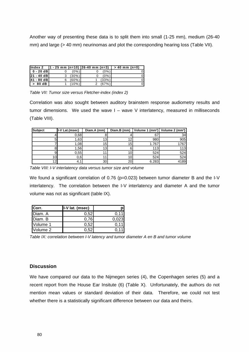
80
Another way of presenting these data is to split them into small (1-25 mm), medium (26-40
mm) and large (> 40 mm) neurinomas and plot the corresponding hearing loss (Table VII).
Index 2 1 - 25 mm (n=10) 26-40 mm (n=3) > 40 mm (n=0)0 - 20 dB 0 (0%) 0 (0%) 0
21 - 40 dB 3 (30%) 0 (0%) 041 - 80 dB 6 (60%) 1 (33%) 0
> 80 dB 1 (10%) 2 (67%) 0
Table VII: Tumor size versus Fletcher-index (index 2)
Correlation was also sought between auditory brainstem response audiometry results and
tumor dimensions. We used the wave I – wave V interlatency, measured in milliseconds
(Table VIII).
Table VIII: I-V interlatency data versus tumor size and volume
We found a significant correlation of 0.76 (p=0.023) between tumor diameter B and the I-V
interlatency. The correlation between the I-V interlatency and diameter A and the tumor
volume was not as significant (table IX).
Table IX: correlation between I-V latency and tumor diameter A en B and tumor volume
Discussion
We have compared our data to the Nijmegen series (4), the Copenhagen series (5) and a
recent report from the House Ear Insitute (6) (Table X). Unfortunately, the authors do not
mention mean values or standard deviation of their data. Therefore, we could not test
whether there is a statistically significant difference between our data and theirs.
Subject I-V Lat.(msec) Diam.A (mm) Diam.B (mm) Volume 1 (mm³) Volume 2 (mm³)
4 0,68 8 4 67 345 1,63 13 12 980 9057 1,08 15 15 1.767 17678 1,56 13 6 113 1139 0,55 11 10 524 524
10 0,6 11 10 524 52412 4,1 30 20 6.283 4189
Corr. I-V lat. (msec) pDiam. A 0,52 0,11Diam. B 0,76 0,023Volume 1 0,52 0,11Volume 2 0,52 0,11
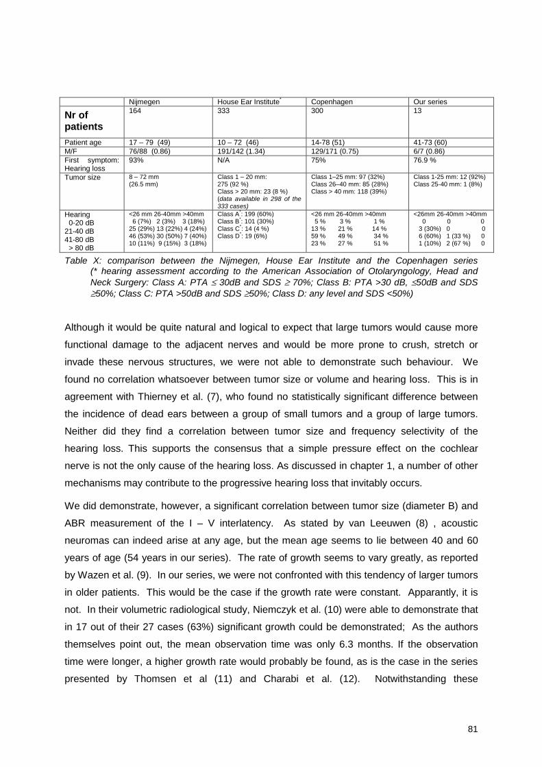
81
Nijmegen House Ear Institute* Copenhagen Our series
Nr ofpatients
164 333 300 13
Patient age 17 – 79 (49) 10 – 72 (46) 14-78 (51) 41-73 (60)M/F 76/88 (0.86) 191/142 (1.34) 129/171 (0.75) 6/7 (0.86)First symptom:Hearing loss
93% N/A 75% 76.9 %
Tumor size 8 – 72 mm(26.5 mm)
Class 1 – 20 mm:275 (92 %)Class > 20 mm: 23 (8 %)(data available in 298 of the333 cases)
Class 1–25 mm: 97 (32%)Class 26–40 mm: 85 (28%)Class > 40 mm: 118 (39%)
Class 1-25 mm: 12 (92%)Class 25-40 mm: 1 (8%)
Hearing0-20 dB
21-40 dB41-80 dB
> 80 dB
<26 mm 26-40mm >40mm6 (7%) 2 (3%) 3 (18%)
25 (29%) 13 (22%) 4 (24%)46 (53%) 30 (50%) 7 (40%)10 (11%) 9 (15%) 3 (18%)
Class A*: 199 (60%)Class B*: 101 (30%)Class C*: 14 (4 %)Class D*: 19 (6%)
<26 mm 26-40mm >40mm5 % 3 % 1 %
13 % 21 % 14 %59 % 49 % 34 %23 % 27 % 51 %
<26mm 26-40mm >40mm0 0 0
3 (30%) 0 06 (60%) 1 (33 %) 01 (10%) 2 (67 %) 0
Table X: comparison between the Nijmegen, House Ear Institute and the Copenhagen series(* hearing assessment according to the American Association of Otolaryngology, Head andNeck Surgery: Class A: PTA ≤ 30dB and SDS ≥ 70%; Class B: PTA >30 dB, ≤50dB and SDS≥50%; Class C: PTA >50dB and SDS ≥50%; Class D: any level and SDS <50%)
Although it would be quite natural and logical to expect that large tumors would cause more
functional damage to the adjacent nerves and would be more prone to crush, stretch or
invade these nervous structures, we were not able to demonstrate such behaviour. We
found no correlation whatsoever between tumor size or volume and hearing loss. This is in
agreement with Thierney et al. (7), who found no statistically significant difference between
the incidence of dead ears between a group of small tumors and a group of large tumors.
Neither did they find a correlation between tumor size and frequency selectivity of the
hearing loss. This supports the consensus that a simple pressure effect on the cochlear
nerve is not the only cause of the hearing loss. As discussed in chapter 1, a number of other
mechanisms may contribute to the progressive hearing loss that invitably occurs.
We did demonstrate, however, a significant correlation between tumor size (diameter B) and
ABR measurement of the I – V interlatency. As stated by van Leeuwen (8) , acoustic
neuromas can indeed arise at any age, but the mean age seems to lie between 40 and 60
years of age (54 years in our series). The rate of growth seems to vary greatly, as reported
by Wazen et al. (9). In our series, we were not confronted with this tendency of larger tumors
in older patients. This would be the case if the growth rate were constant. Apparantly, it is
not. In their volumetric radiological study, Niemczyk et al. (10) were able to demonstrate that
in 17 out of their 27 cases (63%) significant growth could be demonstrated; As the authors
themselves point out, the mean observation time was only 6.3 months. If the observation
time were longer, a higher growth rate would probably be found, as is the case in the series
presented by Thomsen et al (11) and Charabi et al. (12). Notwithstanding these
82
observations, correlations with either age or onset of symptoms were not reported by these
authors.
The majority of our patients presented with hearing loss, although two presented themselves
with tinnitus or vertigo. We were not, however, able to confirm van Leeuwen’s observation
that impaired hearing as the main symptom decreases as tumor size increases (8). Neither
could we confirm a correlation between tumor size and time of onset of symptoms. This is in
contrast to Thomsen and Tos’ Copenhagen study (13). We could not demonstrate a relation
between tumor size and hearing loss, we nor did we find a relation between hearing loss and
onset of symptoms. Stipkovits (14) did find significant correlations between duration of
symptoms and pure tone thresholds. The average tumor size, however, was significantly
larger than in our series. Stipkovits did not find significant correlations between tumor size
and pure tone thresholds either (14).
Although our series is small (N = 13) and composed of selected cases, there is no clear
difference between our series and the quoted Nijmegen and Copenhagen series, as far as
the small (1-25 mm) and medium size (16 – 40 mm) acoustic tumors are concerned. We do
see a small difference with the House Ear Institute series, which could be explained by their
selection criteria. As they are discussing prognostic factors for hearing preservation surgery
by way of the middle fossa approach, their series is mainly composed of small tumors with
serviceable hearing.
83
References
1. Forton GEJ, Moeneclaey LLM, Declau F, Marquet JFE. The involvement of the cochlear nervein neurinomas of the eighth cranial nerve. Arch Otorhinolaryngol; 1989:246:156-160.
2. Marquet JFE, Forton GEJ, Offeciers FE, Moeneclaey LLM. The solitary schwannoma of theeighth cranial nerve. Arch Otolaryngol Head Neck Surg 1990;116:1023-1025.
3. Declau F, Moeneclaey LLM, Forton GEJ, Marquet JFE. Differentiation of cartilage and bone inhuman fetal temporal bones with Luxol fast blue stain. Arch Otorhinolaryngol 1988;245:218-220.
4. Van Leeuwen J. Acoustic Neuromas: Correlation among tumor size, symptoms and patientage. Proefschrift ter verkrijging van de graad van doctor aan de Katholieke UniversiteitNijmegen. Nijmegen,1995, ISBN 90-9008907-1
5. Thomsen J, Tos M. Diagnostic Strategies in Search for Acoustic Neuromas. Acta Otolaryngol(Stockh) 1988;Suppl.452:16-25
6. Brackmann DE, Owens RM, Friedman RA, Hitselberger WE, De la Cruz A, House JW, NelsonRA, Luxford WM, Slattery III WM, Fayad J. Prognostic factors for Hearing Preservation inVestibular Schwannoma Surgery. AJO 2000;21:417-424.
7. Thierney PA, Chitnavis BP, Sherriff M, Strong AJ, Gleeson MJ. The relationship between puretone thresholds and the radiological dimensions of acoustic neuromas. Skull Base Surg1998;;8(3):149-151.
8. Van Leeuwen J. Acoustic Neuromas: diagnostic aspects and results of surgery. Proefschriftter verkrijging van de graad van doctor aan de Katholieke Universiteit Nijmegen.Nijmegen,1995, ISBN 90-9008907-1
9. Wazen J, Silverstein H, Norrel H, Besse B. Preoperative and postoperative growth rates inacoustic neuromas documented with CT scanning. Otol Head Neck Surg 1985;93:151-155
10. Niemczyk K, Vaneecloo FM, Lemaitre L et al. The growth of acoustic neuromas in volumetricradiologic assessment. Am J Otology 1999;20(2):244-248
11. Thomsen J, Charabi S, Tos M et al. Vestibular schwannoma growth: a never ending story.Abstracts, 3rd European Skull Base Congress, London, 9-11/4/1997. Skull Base Surg1997;7(suppl2):54
12. Charabi S, Thomsen J, Mantoni M, et al. Acoustic Neuroma (vestibular schwannoma): growthand surgical and nonsurgical consequences of the wait-and-see policy. Otolaryngol HeadNeck Surg 1995:113:5-14
13. Thomsen J, Tos M. Acoustic neuroma: clinical aspects, audiovestibular assessment,diagnostic delay and growth rate. Am J Otology 1990;11:12-19
14. Stipkovits E.M. Vestibular Schwannoma: aspects of biological behavior. Proefschrift terverkrijging van de graad van doctor aan de Universiteit Utrecht. Utrecht 2000. ISBN 90-393-2578-2

84
85
Chapter 3b
Acoustic Schwannoma: MR Findings in 84 Tumors
T.H. Mulkens1
P.M. Parizel1
J.-J. Martin3
H.R. Degryse1
P.H. Van de Heyning2
G.E.J Forton2
A.M. De Schepper1
1 Department of Radiology2 Department of Ear, Nose and Throat3 Department of Neurology
University Hospital Antwerp, Wilrijkstraat 10, 2650 Edegem, BelgiumAmerican Journal of Roentgenology 1993;160:395-398
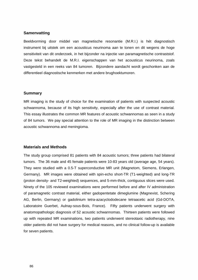
86
Samenvatting
Beeldvorming door middel van magnetische resonantie (M.R.I.) is hét diagnostisch
instrument bij uitstek om een acousticus neurinoma aan te tonen en dit wegens de hoge
sensitiviteit van dit onderzoek, in het bijzonder na injectie van paramagnetische contraststof.
Deze tekst behandelt de M.R.I. eigenschappen van het acousticus neurinoma, zoals
vastgesteld in een reeks van 84 tumoren. Bijzondere aandacht wordt geschonken aan de
differentieel diagnostische kenmerken met andere brughoektumoren.
Summary
MR imaging is the study of choice for the examination of patients with suspected acoustic
schwannoma, because of its high sensitivity, especially after the use of contrast material.
This essay illustrates the common MR features of acoustic schwannomas as seen in a study
of 84 tumors. We pay special attention to the role of MR imaging in the distinction between
acoustic schwannoma and meningioma.
Materials and Methods
The study group comprised 81 patients with 84 acoustic tumors; three patients had bilateral
tumors. The 36 male and 45 female patients were 10-83 years old (average age, 54 years).
They were studied with a 0.5-T superconductive MR unit (Magnetom, Siemens, Erlangen,
Germany). MR images were obtained with spin-echo short-TR (T1-weighted) and long-TR
(proton density- and T2-weighted) sequences, and 5-mm-thick, contiguous slices were used.
Ninety of the 105 reviewed examinations were performed before and after IV administration
of paramagnetic contrast material, either gadopentetate dimeglumine (Magnevist, Schering
AG, Berlin, Germany) or gadolinium tetra-azacyclododecane tetraacetic acid (Gd-DOTA,
Laboratoire Guerbet, Aulnay-sous-Bois, France). Fifty patients underwent surgery with
anatomopathologic diagnosis of 52 acoustic schwannomas. Thirteen patients were followed
up with repeated MR examinations, two patients underwent stereotaxic radiotherapy, nine
older patients did not have surgery for medical reasons, and no clinical follow-up is available
for seven patients.
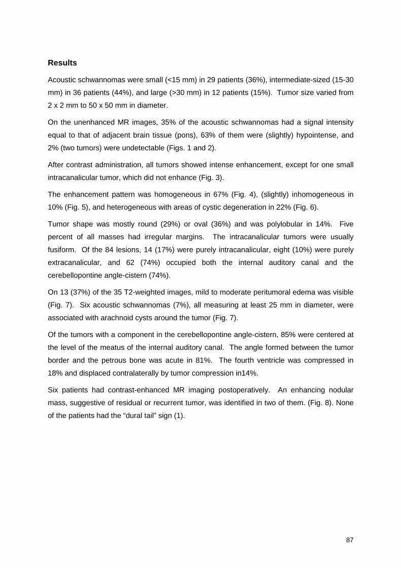
87
Results
Acoustic schwannomas were small (<15 mm) in 29 patients (36%), intermediate-sized (15-30
mm) in 36 patients (44%), and large (>30 mm) in 12 patients (15%). Tumor size varied from
2 x 2 mm to 50 x 50 mm in diameter.
On the unenhanced MR images, 35% of the acoustic schwannomas had a signal intensity
equal to that of adjacent brain tissue (pons), 63% of them were (slightly) hypointense, and
2% (two tumors) were undetectable (Figs. 1 and 2).
After contrast administration, all tumors showed intense enhancement, except for one small
intracanalicular tumor, which did not enhance (Fig. 3).
The enhancement pattern was homogeneous in 67% (Fig. 4), (slightly) inhomogeneous in
10% (Fig. 5), and heterogeneous with areas of cystic degeneration in 22% (Fig. 6).
Tumor shape was mostly round (29%) or oval (36%) and was polylobular in 14%. Five
percent of all masses had irregular margins. The intracanalicular tumors were usually
fusiform. Of the 84 lesions, 14 (17%) were purely intracanalicular, eight (10%) were purely
extracanalicular, and 62 (74%) occupied both the internal auditory canal and the
cerebellopontine angle-cistern (74%).
On 13 (37%) of the 35 T2-weighted images, mild to moderate peritumoral edema was visible
(Fig. 7). Six acoustic schwannomas (7%), all measuring at least 25 mm in diameter, were
associated with arachnoid cysts around the tumor (Fig. 7).
Of the tumors with a component in the cerebellopontine angle-cistern, 85% were centered at
the level of the meatus of the internal auditory canal. The angle formed between the tumor
border and the petrous bone was acute in 81%. The fourth ventricle was compressed in
18% and displaced contralaterally by tumor compression in14%.
Six patients had contrast-enhanced MR imaging postoperatively. An enhancing nodular
mass, suggestive of residual or recurrent tumor, was identified in two of them. (Fig. 8). None
of the patients had the “dural tail” sign (1).
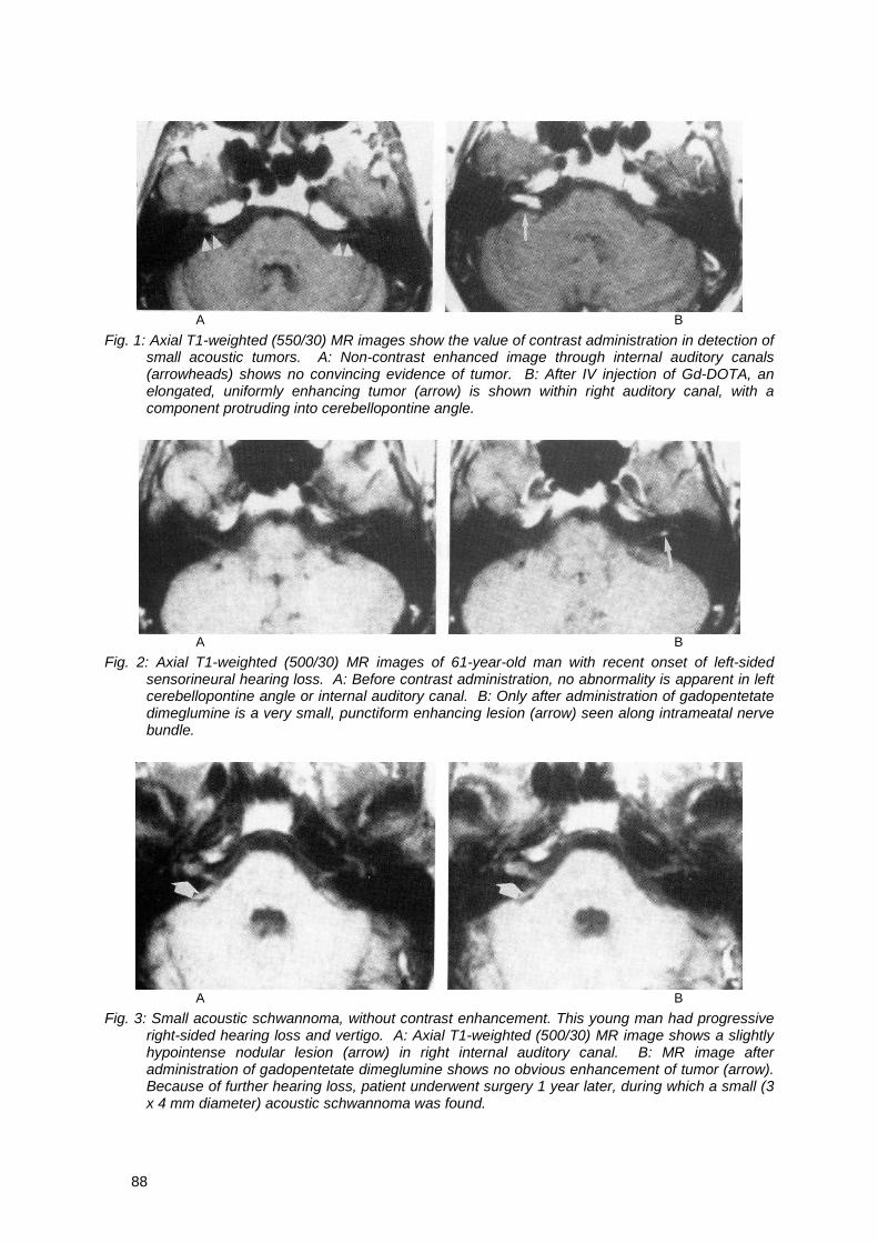
88
A B
Fig. 1: Axial T1-weighted (550/30) MR images show the value of contrast administration in detection ofsmall acoustic tumors. A: Non-contrast enhanced image through internal auditory canals(arrowheads) shows no convincing evidence of tumor. B: After IV injection of Gd-DOTA, anelongated, uniformly enhancing tumor (arrow) is shown within right auditory canal, with acomponent protruding into cerebellopontine angle.
A B
Fig. 2: Axial T1-weighted (500/30) MR images of 61-year-old man with recent onset of left-sidedsensorineural hearing loss. A: Before contrast administration, no abnormality is apparent in leftcerebellopontine angle or internal auditory canal. B: Only after administration of gadopentetatedimeglumine is a very small, punctiform enhancing lesion (arrow) seen along intrameatal nervebundle.
A B
Fig. 3: Small acoustic schwannoma, without contrast enhancement. This young man had progressiveright-sided hearing loss and vertigo. A: Axial T1-weighted (500/30) MR image shows a slightlyhypointense nodular lesion (arrow) in right internal auditory canal. B: MR image afteradministration of gadopentetate dimeglumine shows no obvious enhancement of tumor (arrow).Because of further hearing loss, patient underwent surgery 1 year later, during which a small (3x 4 mm diameter) acoustic schwannoma was found.
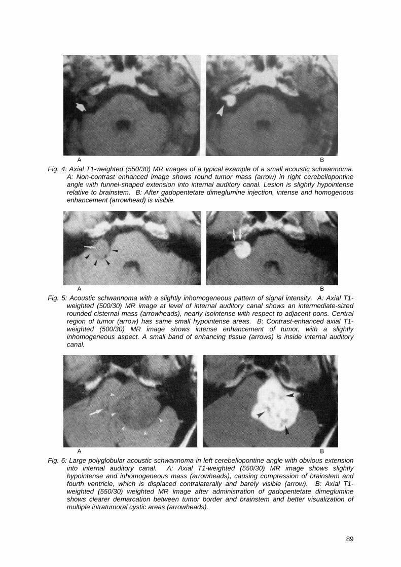
89
A B
Fig. 4: Axial T1-weighted (550/30) MR images of a typical example of a small acoustic schwannoma.A: Non-contrast enhanced image shows round tumor mass (arrow) in right cerebellopontineangle with funnel-shaped extension into internal auditory canal. Lesion is slightly hypointenserelative to brainstem. B: After gadopentetate dimeglumine injection, intense and homogenousenhancement (arrowhead) is visible.
A B
Fig. 5: Acoustic schwannoma with a slightly inhomogeneous pattern of signal intensity. A: Axial T1-weighted (500/30) MR image at level of internal auditory canal shows an intermediate-sizedrounded cisternal mass (arrowheads), nearly isointense with respect to adjacent pons. Centralregion of tumor (arrow) has same small hypointense areas. B: Contrast-enhanced axial T1-weighted (500/30) MR image shows intense enhancement of tumor, with a slightlyinhomogeneous aspect. A small band of enhancing tissue (arrows) is inside internal auditorycanal.
A B
Fig. 6: Large polyglobular acoustic schwannoma in left cerebellopontine angle with obvious extensioninto internal auditory canal. A: Axial T1-weighted (550/30) MR image shows slightlyhypointense and inhomogeneous mass (arrowheads), causing compression of brainstem andfourth ventricle, which is displaced contralaterally and barely visible (arrow). B: Axial T1-weighted (550/30) weighted MR image after administration of gadopentetate dimeglumineshows clearer demarcation between tumor border and brainstem and better visualization ofmultiple intratumoral cystic areas (arrowheads).
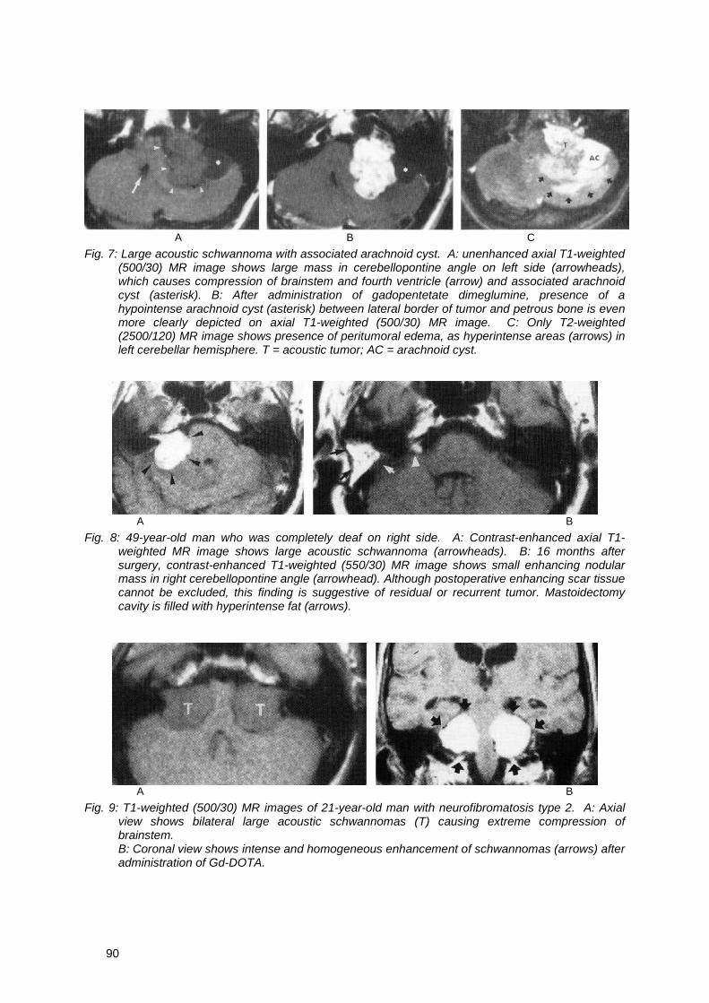
90
A B C
Fig. 7: Large acoustic schwannoma with associated arachnoid cyst. A: unenhanced axial T1-weighted(500/30) MR image shows large mass in cerebellopontine angle on left side (arrowheads),which causes compression of brainstem and fourth ventricle (arrow) and associated arachnoidcyst (asterisk). B: After administration of gadopentetate dimeglumine, presence of ahypointense arachnoid cyst (asterisk) between lateral border of tumor and petrous bone is evenmore clearly depicted on axial T1-weighted (500/30) MR image. C: Only T2-weighted(2500/120) MR image shows presence of peritumoral edema, as hyperintense areas (arrows) inleft cerebellar hemisphere. T = acoustic tumor; AC = arachnoid cyst.
A B
Fig. 8: 49-year-old man who was completely deaf on right side. A: Contrast-enhanced axial T1-weighted MR image shows large acoustic schwannoma (arrowheads). B: 16 months aftersurgery, contrast-enhanced T1-weighted (550/30) MR image shows small enhancing nodularmass in right cerebellopontine angle (arrowhead). Although postoperative enhancing scar tissuecannot be excluded, this finding is suggestive of residual or recurrent tumor. Mastoidectomycavity is filled with hyperintense fat (arrows).
A B
Fig. 9: T1-weighted (500/30) MR images of 21-year-old man with neurofibromatosis type 2. A: Axialview shows bilateral large acoustic schwannomas (T) causing extreme compression ofbrainstem.B: Coronal view shows intense and homogeneous enhancement of schwannomas (arrows) afteradministration of Gd-DOTA.
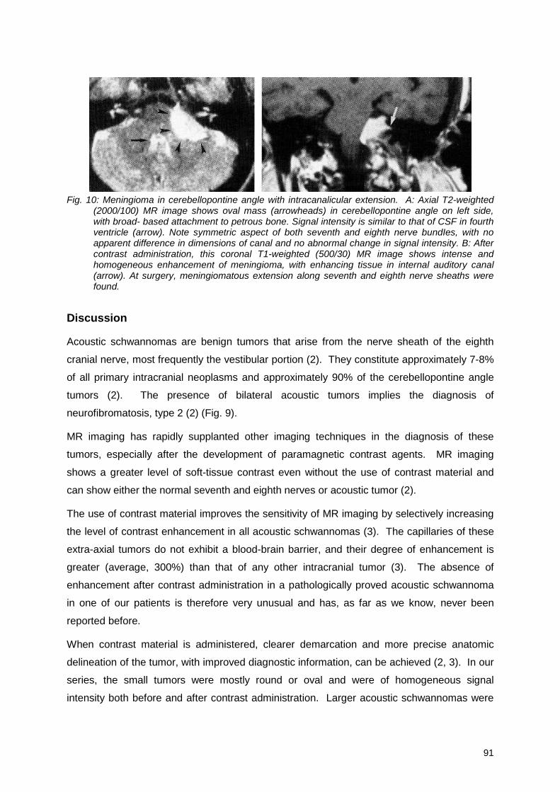
91
Fig. 10: Meningioma in cerebellopontine angle with intracanalicular extension. A: Axial T2-weighted(2000/100) MR image shows oval mass (arrowheads) in cerebellopontine angle on left side,with broad- based attachment to petrous bone. Signal intensity is similar to that of CSF in fourthventricle (arrow). Note symmetric aspect of both seventh and eighth nerve bundIes, with noapparent difference in dimensions of canal and no abnormal change in signal intensity. B: Aftercontrast administration, this coronal T1-weighted (500/30) MR image shows intense andhomogeneous enhancement of meningioma, with enhancing tissue in internal auditory canal(arrow). At surgery, meningiomatous extension along seventh and eighth nerve sheaths werefound.
Discussion
Acoustic schwannomas are benign tumors that arise from the nerve sheath of the eighth
cranial nerve, most frequently the vestibular portion (2). They constitute approximately 7-8%
of all primary intracranial neoplasms and approximately 90% of the cerebellopontine angle
tumors (2). The presence of bilateral acoustic tumors implies the diagnosis of
neurofibromatosis, type 2 (2) (Fig. 9).
MR imaging has rapidly supplanted other imaging techniques in the diagnosis of these
tumors, especially after the development of paramagnetic contrast agents. MR imaging
shows a greater level of soft-tissue contrast even without the use of contrast material and
can show either the normal seventh and eighth nerves or acoustic tumor (2).
The use of contrast material improves the sensitivity of MR imaging by selectively increasing
the level of contrast enhancement in all acoustic schwannomas (3). The capillaries of these
extra-axial tumors do not exhibit a blood-brain barrier, and their degree of enhancement is
greater (average, 300%) than that of any other intracranial tumor (3). The absence of
enhancement after contrast administration in a pathologically proved acoustic schwannoma
in one of our patients is therefore very unusual and has, as far as we know, never been
reported before.
When contrast material is administered, clearer demarcation and more precise anatomic
delineation of the tumor, with improved diagnostic information, can be achieved (2, 3). In our
series, the small tumors were mostly round or oval and were of homogeneous signal
intensity both before and after contrast administration. Larger acoustic schwannomas were
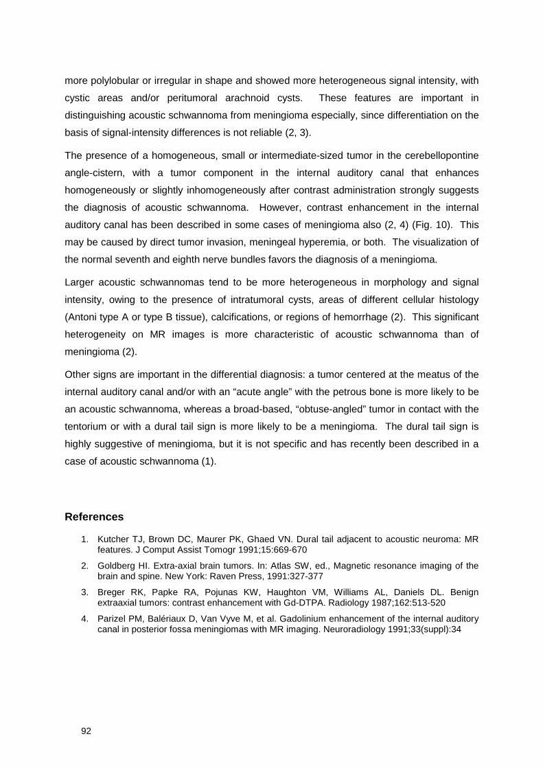
92
more polylobular or irregular in shape and showed more heterogeneous signal intensity, with
cystic areas and/or peritumoral arachnoid cysts. These features are important in
distinguishing acoustic schwannoma from meningioma especially, since differentiation on the
basis of signal-intensity differences is not reliable (2, 3).
The presence of a homogeneous, small or intermediate-sized tumor in the cerebellopontine
angle-cistern, with a tumor component in the internal auditory canal that enhances
homogeneously or slightly inhomogeneously after contrast administration strongly suggests
the diagnosis of acoustic schwannoma. However, contrast enhancement in the internal
auditory canal has been described in some cases of meningioma also (2, 4) (Fig. 10). This
may be caused by direct tumor invasion, meningeal hyperemia, or both. The visualization of
the normal seventh and eighth nerve bundles favors the diagnosis of a meningioma.
Larger acoustic schwannomas tend to be more heterogeneous in morphology and signal
intensity, owing to the presence of intratumoral cysts, areas of different cellular histology
(Antoni type A or type B tissue), calcifications, or regions of hemorrhage (2). This significant
heterogeneity on MR images is more characteristic of acoustic schwannoma than of
meningioma (2).
Other signs are important in the differential diagnosis: a tumor centered at the meatus of the
internal auditory canal and/or with an “acute angle” with the petrous bone is more likely to be
an acoustic schwannoma, whereas a broad-based, “obtuse-angled” tumor in contact with the
tentorium or with a dural tail sign is more likely to be a meningioma. The dural tail sign is
highly suggestive of meningioma, but it is not specific and has recently been described in a
case of acoustic schwannoma (1).
References
1. Kutcher TJ, Brown DC, Maurer PK, Ghaed VN. Dural tail adjacent to acoustic neuroma: MRfeatures. J Comput Assist Tomogr 1991;15:669-670
2. Goldberg HI. Extra-axial brain tumors. In: Atlas SW, ed., Magnetic resonance imaging of thebrain and spine. New York: Raven Press, 1991:327-377
3. Breger RK, Papke RA, Pojunas KW, Haughton VM, Williams AL, Daniels DL. Benignextraaxial tumors: contrast enhancement with Gd-DTPA. Radiology 1987;162:513-520
4. Parizel PM, Balériaux D, Van Vyve M, et al. Gadolinium enhancement of the internal auditorycanal in posterior fossa meningiomas with MR imaging. Neuroradiology 1991;33(suppl):34
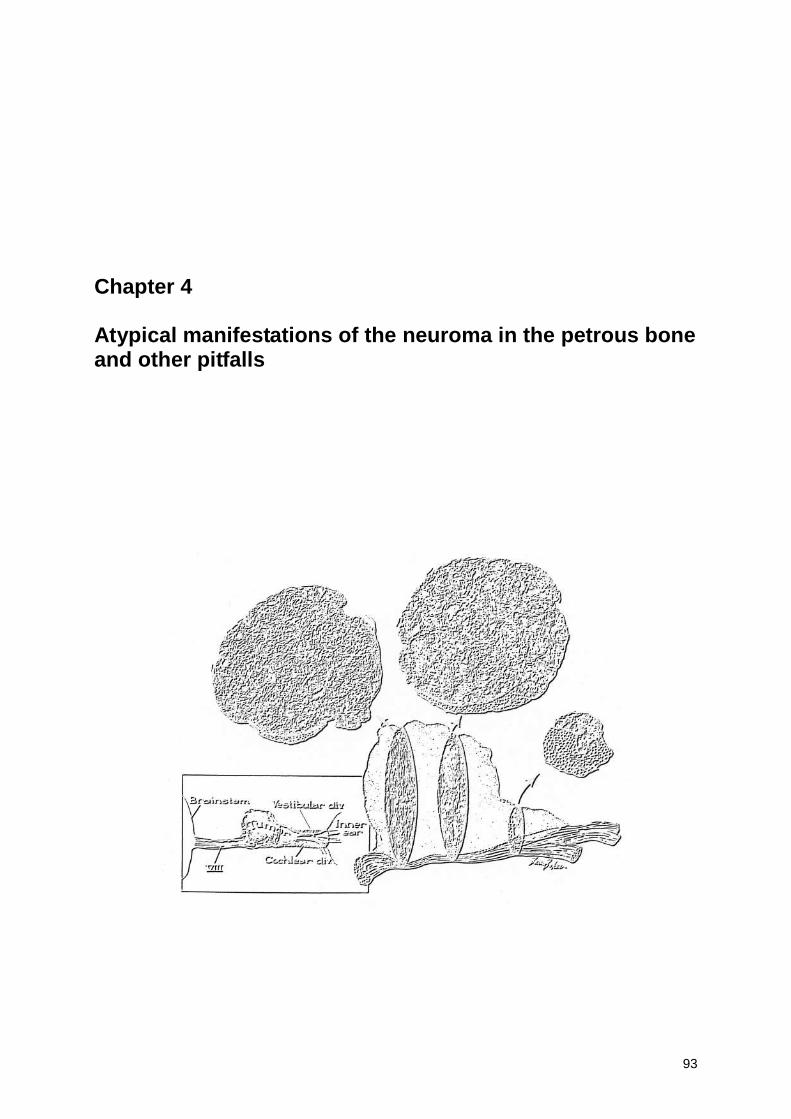
93
Chapter 4
Atypical manifestations of the neuroma in the petrous boneand other pitfalls

94
95
Chapter 4a
Facial Nerve Neuroma: Report of Two Cases IncludingHistologic and Radiological Imaging Studies
G.E.J. Forton, MD
L.L.M. Moeneclaey
F.E. Offeciers, MD PhD
ENT Department, University of Antwerp, Wilrijk, BelgiumEur Arch Otorhinolaryngol 1994;251:17-22
96
Samenvatting
In tegenstelling tot het acousticusneurinoma is het neurinoma van de Nervus Facialis een vrij
zeldzame vondst, zelfs in tertiaire centra. Wij presenteren twee gevallen met daarbij
bijzondere aandacht voor de typische beeldvorming en de histologische kleuringstechnieken
zoals deze in de twee vorige hoofdstukken werden beschreven. De heelkundige
behandeling wordt besproken en getoetst aan de literatuur.
Summary
Unlike the acoustic neuroma, the facial nerve neuroma is an uncommon finding, even in
referral clinical centers. Two cases of facial nerve neuromas are presented, with special
focus on the importance of adequate radiological imaging techniques and on the histological
characteristics of tumor specimens. Surgical modalities regarding tumor removal as well as
facial nerve repair are discussed and the current world literature reviewed.
Introduction
Unlike the acoustic neuroma, a neuroma of the facial nerve is an uncommon clinical finding,
even in referral medical centers. The world literature indicates that only about 180 cases
have been reported. In the review of Shambaugh and Clemis [31] neoplastic involvement of
the facial nerve accounted for no more than 5% of all lower motor neuron palsies. Most
frequently, in these cases, the mastoid segment is involved (58.1% of all cases according to
Isamat et al. [13]). In 23.4% of all cases, the tumor originates from the labyrinthine segment,
while the tympanic portion of the nerve is involved in only 10.3% of all cases.
Nine percent of all facial nerve neuromas are located extracranially and they usually appear
as asymptomatic parotid tumors [2]. Even more rare, facial nerve neuromas can present as
middle fossa or pontocerebellar angle tumors [3, 5, 9, 16, 17, 20, 24, 25, 27, 30, 34]
Malignant facial nerve neuromas are even less common. To our knowledge, only six cases
have been published [4, 10, 15, 23], one of them being a tumor localized in the parotid gland
of an 8-year old child [33]. To the best of our knowledge, bilateral facial nerve tumors have
only been described in central neurofibromatosis (type II) [1, 17].
About 50% of all facial nerve neuromas present as a lower motor neuron paralysis and are
frequently misdiagnosed as being an idiopathic Bell's palsy. Indeed, the clinical symptoms of
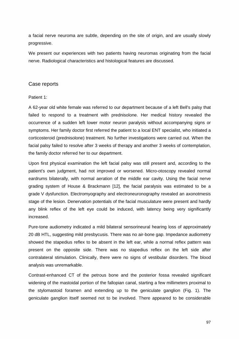
97
a facial nerve neuroma are subtle, depending on the site of origin, and are usually slowly
progressive.
We present our experiences with two patients having neuromas originating from the facial
nerve. Radiological characteristics and histological features are discussed.
Case reports
Patient 1:
A 62-year old white female was referred to our department because of a left Bell's palsy that
failed to respond to a treatment with prednisolone. Her medical history revealed the
occurrence of a sudden left lower motor neuron paralysis without accompanying signs or
symptoms. Her family doctor first referred the patient to a local ENT specialist, who initiated a
corticosteroid (prednisolone) treatment. No further investigations were carried out. When the
facial palsy failed to resolve after 3 weeks of therapy and another 3 weeks of contemplation,
the family doctor referred her to our department.
Upon first physical examination the left facial palsy was still present and, according to the
patient's own judgment, had not improved or worsened. Micro-otoscopy revealed normal
eardrums bilaterally, with normal aeration of the middle ear cavity. Using the facial nerve
grading system of House & Brackmann [12], the facial paralysis was estimated to be a
grade V dysfunction. Electromyography and electroneuronography revealed an axonotmesis
stage of the lesion. Denervation potentials of the facial musculature were present and hardly
any blink reflex of the left eye could be induced, with latency being very significantly
increased.
Pure-tone audiometry indicated a mild bilateral sensorineural hearing loss of approximately
20 dB HTL, suggesting mild presbycusis. There was no air-bone gap. Impedance audiometry
showed the stapedius reflex to be absent in the left ear, while a normal reflex pattern was
present on the opposite side. There was no stapedius reflex on the left side after
contralateral stimulation. Clinically, there were no signs of vestibular disorders. The blood
analysis was unremarkable.
Contrast-enhanced CT of the petrous bone and the posterior fossa revealed significant
widening of the mastoidal portion of the fallopian canal, starting a few millimeters proximal to
the stylomastoid foramen and extending up to the geniculate ganglion (Fig. 1). The
geniculate ganglion itself seemed not to be involved. There appeared to be considerable
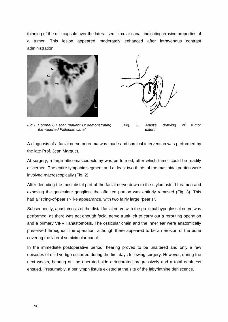
98
thinning of the otic capsule over the lateral semicircular canal, indicating erosive properties of
a tumor. This lesion appeared moderately enhanced after intravenous contrast
administration.
Fig 1. Coronal CT scan (patient 1): demonstrating Fig. 2: Artist's drawing of tumorthe widened Fallopian canal extent
A diagnosis of a facial nerve neuroma was made and surgical intervention was performed by
the late Prof. Jean Marquet.
At surgery, a large atticomastoidectomy was performed, after which tumor could be readily
discerned. The entire tympanic segment and at least two-thirds of the mastoidal portion were
involved macroscopically (Fig. 2)
After denuding the most distal part of the facial nerve down to the stylomastoid foramen and
exposing the geniculate ganglion, the affected portion was entirely removed (Fig. 3). This
had a "string-of-pearls"-like appearance, with two fairly large "pearls".
Subsequently, anastomosis of the distal facial nerve with the proximal hypoglossal nerve was
performed, as there was not enough facial nerve trunk left to carry out a rerouting operation
and a primary VII-VII anastomosis. The ossicular chain and the inner ear were anatomically
preserved throughout the operation, although there appeared to be an erosion of the bone
covering the lateral semicircular canal.
In the immediate postoperative period, hearing proved to be unaltered and only a few
episodes of mild vertigo occurred during the first days following surgery. However, during the
next weeks, hearing on the operated side deteriorated progressively and a total deafness
ensued. Presumably, a perilymph fistula existed at the site of the labyrinthine dehiscence.
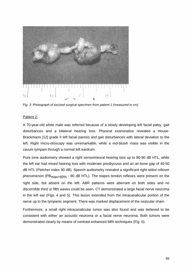
99
Fig. 3: Photograph of excised surgical specimen from patient 1 (measured in cm)
Patient 2:
A 70-year-old white male was referred because of a slowly developing left facial palsy, gait
disturbances and a bilateral hearing loss. Physical examination revealed a House-
Brackmann [12] grade II left facial paresis and gait disturbances with lateral deviation to the
left. Right micro-otoscopy was unremarkable, while a red-bluish mass was visible in the
cavum tympani through a normal left eardrum.
Pure tone audiometry showed a right sensorineural hearing loss up to 80-90 dB HTL, while
the left ear had mixed hearing loss with moderate presbycusis and an air-bone gap of 40-50
dB HTL (Fletcher index 90 dB). Speech audiometry revealed a significant right-sided rollover
phenomenon (PBmax=60% : 80 dB HTL). The stapes tendon reflexes were present on the
right side, but absent on the left. ABR patterns were aberrant on both sides and no
discernible third or fifth waves could be seen. CT demonstrated a large facial nerve neuroma
in the left ear (Figs. 4 and 5). This lesion extended from the intracanalicular portion of the
nerve up to the tympanic segment. There was marked displacement of the ossicular chain.
Furthermore, a small right intracanalicular tumor was also found and was believed to be
consistent with either an acoustic neuroma or a facial nerve neuroma. Both tumors were
demonstrated clearly by means of contrast-enhanced MRI techniques (Fig. 6).
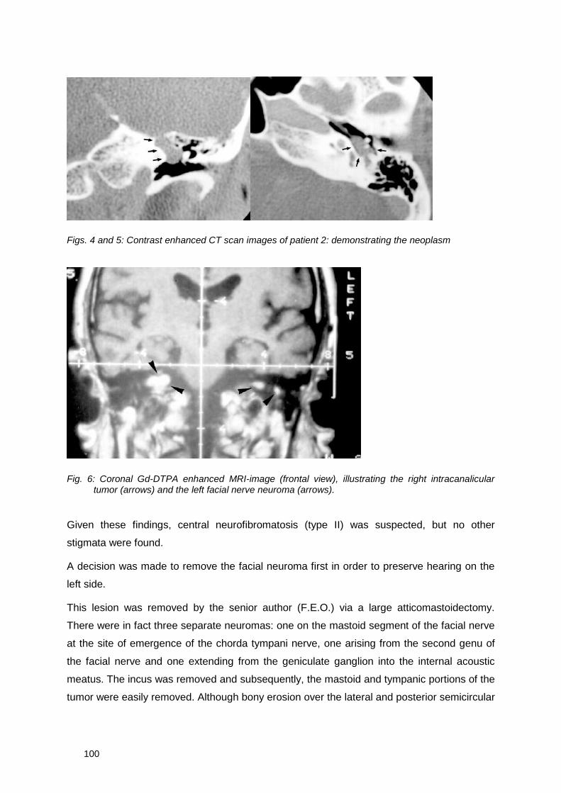
100
Figs. 4 and 5: Contrast enhanced CT scan images of patient 2: demonstrating the neoplasm
Fig. 6: Coronal Gd-DTPA enhanced MRI-image (frontal view), illustrating the right intracanaliculartumor (arrows) and the left facial nerve neuroma (arrows).
Given these findings, central neurofibromatosis (type II) was suspected, but no other
stigmata were found.
A decision was made to remove the facial neuroma first in order to preserve hearing on the
left side.
This lesion was removed by the senior author (F.E.O.) via a large atticomastoidectomy.
There were in fact three separate neuromas: one on the mastoid segment of the facial nerve
at the site of emergence of the chorda tympani nerve, one arising from the second genu of
the facial nerve and one extending from the geniculate ganglion into the internal acoustic
meatus. The incus was removed and subsequently, the mastoid and tympanic portions of the
tumor were easily removed. Although bony erosion over the lateral and posterior semicircular
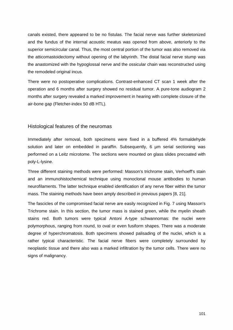
101
canals existed, there appeared to be no fistulas. The facial nerve was further skeletonized
and the fundus of the internal acoustic meatus was opened from above, anteriorly to the
superior semicircular canal. Thus, the most central portion of the tumor was also removed via
the atticomastoidectomy without opening of the labyrinth. The distal facial nerve stump was
the anastomized with the hypoglossal nerve and the ossicular chain was reconstructed using
the remodeled original incus.
There were no postoperative complications. Contrast-enhanced CT scan 1 week after the
operation and 6 months after surgery showed no residual tumor. A pure-tone audiogram 2
months after surgery revealed a marked improvement in hearing with complete closure of the
air-bone gap (Fletcher-index 50 dB HTL).
Histological features of the neuromas
Immediately after removal, both specimens were fixed in a buffered 4% formaldehyde
solution and later on embedded in paraffin. Subsequently, 6 µm serial sectioning was
performed on a Leitz microtome. The sections were mounted on glass slides precoated with
poly-L-lysine.
Three different staining methods were performed: Masson's trichrome stain, Verhoeff's stain
and an immunohistochemical technique using monoclonal mouse antibodies to human
neurofilaments. The latter technique enabled identification of any nerve fiber within the tumor
mass. The staining methods have been amply described in previous papers [8, 21].
The fascicles of the compromised facial nerve are easily recognized in Fig. 7 using Masson's
Trichrome stain. In this section, the tumor mass is stained green, while the myelin sheath
stains red. Both tumors were typical Antoni A-type schwannomas: the nuclei were
polymorphous, ranging from round, to oval or even fusiform shapes. There was a moderate
degree of hyperchromatosis. Both specimens showed palisading of the nuclei, which is a
rather typical characteristic. The facial nerve fibers were completely surrounded by
neoplastic tissue and there also was a marked infiltration by the tumor cells. There were no
signs of malignancy.
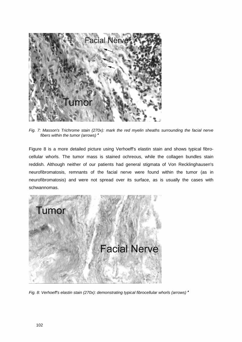
102
Fig. 7: Masson's Trichrome stain (270x): mark the red myelin sheaths surrounding the facial nervefibers within the tumor (arrows) ♦
Figure 8 is a more detailed picture using Verhoeff's elastin stain and shows typical fibro-
cellular whorls. The tumor mass is stained ochreous, while the collagen bundles stain
reddish. Although neither of our patients had general stigmata of Von Recklinghausen's
neurofibromatosis, remnants of the facial nerve were found within the tumor (as in
neurofibromatosis) and were not spread over its surface, as is usually the cases with
schwannomas.
Fig. 8: Verhoeff's elastin stain (270x): demonstrating typical fibrocellular whorls (arrows) ♦
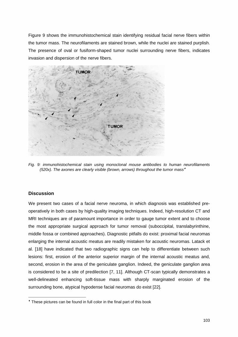
103
Figure 9 shows the immunohistochemical stain identifying residual facial nerve fibers within
the tumor mass. The neurofilaments are stained brown, while the nuclei are stained purplish.
The presence of oval or fusiform-shaped tumor nuclei surrounding nerve fibers, indicates
invasion and dispersion of the nerve fibers.
Fig. 9: immunohistochemical stain using monoclonal mouse antibodies to human neurofilaments(520x). The axones are clearly visible (brown, arrows) throughout the tumor mass♦
Discussion
We present two cases of a facial nerve neuroma, in which diagnosis was established pre-
operatively in both cases by high-quality imaging techniques. Indeed, high-resolution CT and
MRI techniques are of paramount importance in order to gauge tumor extent and to choose
the most appropriate surgical approach for tumor removal (suboccipital, translabyrinthine,
middle fossa or combined approaches). Diagnostic pitfalls do exist: proximal facial neuromas
enlarging the internal acoustic meatus are readily mistaken for acoustic neuromas. Latack et
al. [18] have indicated that two radiographic signs can help to differentiate between such
lesions: first, erosion of the anterior superior margin of the internal acoustic meatus and,
second, erosion in the area of the geniculate ganglion. Indeed, the geniculate ganglion area
is considered to be a site of predilection [7, 11]. Although CT-scan typically demonstrates a
well-delineated enhancing soft-tissue mass with sharply marginated erosion of the
surrounding bone, atypical hypodense facial neuromas do exist [22].
♦ These pictures can be found in full color in the final part of this book
104
Recent work emphasizes the need for MRI in patients with facial nerve palsy. Even with
non-enhanced MRI, detection of an intracanalicular tumor is feasible in most cases, although
small intracanalicular facial nerve neuromas can be inconspicuous [6]. Also,
non-homogeneous signals of uncertain origin and without peroperative surgical correlative
can render interpretation cumbersome [6]. Contrast-enhanced MRI, however, dramatically
improves sensitivity and allows significant accuracy for detecting small acoustic or facial
nerve tumors. Even in cases of genuine Bell's palsy, Ramsey-Hunt syndrome and radiation-
induced facial palsy, diffuse homogeneous enhancement of the facial nerve can be observed
[32]. The true nature of this enhancement is still not fully understood.
Martin et al. [22] have described MRI findings in four facial nerve neuromas. All tumors were
slightly heterogeneous lesions isointense to brain on T1 as well as T2-weighted images.
Martin's group also stressed the superiority of MRI over CT in detecting and delineating
extensions of intrapetrous lesions. These qualities are improved even more after intravenous
injection of gadolinium-DTPA or gadopentetate-dimeglumine, since facial nerve neuromas
typically present as distinct, intensely enhanced masses. However, Lidov et al. [19] have
described a facial nerve neuroma that clinically and radiographically mimicked a cystic
posterior skull base lesion, which extrinsically compressed the facial nerve. Even detailed
contrast-enhanced MRI studies were not able to discover preoperatively the true nature of
this peculiar neuroma. Only after transmastoid exploration and frozen section examination
was a final diagnosis established.
In general, neuromas and hemangiomas of the facial nerve customarily occur near or in the
geniculate ganglion and typically result in enlargement of the nerve with adjacent bone
erosion [14]. Our first patient, however, did not present any evidence for geniculate ganglion
involvement, while our second patient had apparent multicentric neuromas, including
geniculate ganglion involvement. Neoplastic bridging of these lobulations has been
demonstrated elsewhere, contradicting the multicentric origin hypothesis [7, 11]. Our findings
agree with these observations, since we were able to demonstrate tumor cells penetrating
nerve fascicles and thus pursuing the natural course of the facial nerve. Such a growth
pattern readily explains the classical "dumbbell tumor".
Both of our cases illustrate the need for thorough investigation of all so-called "Bell's palsies"
that do not resolve spontaneously within 6 weeks. Facial nerve neuromas can indeed appear
in several forms, including vague pain without facial paralysis, simulating acoustic tumors,
appearing as parotid masses with or without paralysis, and causing sudden onset of facial
paralysis to simulate Bell's palsy [26]. In these latter cases, electrical excitability can be
105
maintained (as in neuropraxia) or it can recover clinically or electromyographically. A slowly
progressive facial palsy is, of course, very suggestive of neoplasm.
In our opinion, adequate removal of facial neuromas not extending medially beyond the
fundus of the internal acoustic meatus, can be performed via a large atticomastoidectomy
and with extensive denuding of the facial nerve. Whenever necessary, a middle fossa
approach ensures complete removal of those tumors that extend more medially. An
immediate reapproximation of nerve ends with primary end-to-end anastomosis should be
accomplished whenever feasible. Unfortunately, the nerve segment that needs to be
removed is often too large to allow such anastomosis to be done. Whenever this is the case,
we prefer a primary anastomosis with the hypoglossal nerve. Interpositional (sural) nerve
grafting is also a reliable method if and when the proximal stump is readily accessible. Both
methods will in time provide acceptable facial tone and allow voluntary facial movement in
most cases.
References
1. Baldwin D, King TT, Chevretton E, Morrison AW (1991) Bilateral cerebellopontine angletumors in neurofibromatosis type 2. J Neurosurg 74:910-915
2. Balle VH, Greisen O (1984) Neurilemmomas of the facial nerve presenting as parotid tumors.Ann Otol Rhinol Laryngol 93:70-72
3. Confavreux C, Noël E, Ventre JJ, Aimard G, Devic M (1986) Paralysie faciale isolée parneurinome facial intrapétreux. Presse Méd 15:888-889
4. Conley J, Janecka I (1974) Schwann cell tumors of the facial nerve. Laryngoscope 84:958-962
5. Curtin JM, Lanigan JP (1964) Neuilemmoma of the seventh nerve with intra-cranial extension.J Laryngol 78:212-219
6. Daniels D, Millen SJ, Meyer GA, Pojunas KW, Kilgore DP, Shaffer KA, Williams AL, HaughtonVM (1987) MR Detection of tumor in the internal auditory canal. AJNR 148:1219-1222
7. Fisch U, Ruttner J (1977) Pathology of intratemporal tumors involving the facial nerve. In:Fisch U (ed) Facial nerve surgery. Aesculapius, Birmingham, pp 448-456
8. Forton G, Moeneclaey L, Declau F, Marquet J (1989) The involvement of the cochlear nerve inneurinomas of the eighth cranial nerve. Arch Otorhinolaryngol 246:156-160
9. Furlow LT (1960) The neurosurgical aspects of seventh nerve neurilemmoma. J Neurosurg17:712-735
10. Gutmann MR, Simon MV (1952) Neurofibrosarcoma of the facial nerve involving thetympanomastoid. Arch Otolaryngol 54:162-166
11. Horn KL, Crumley RL, Schindler RA (1981) Facial neurilemmomas. Laryngoscope 91:1326-1331
12. House JW, Brackmann DE (1985) Facial nerve grading system. Otolaryngol Head Neck Surg93:146-147
106
13. Isamat F, Bartameus F, Miranda AM, Prat J, Pons LC (1975) Neurinomas of the facial nerve. JNeurosurg 43:608-613
14. Janecka IP, Conley J (1987) Primary neoplasms of the facial nerve. Plast Reconstr Surg79:177-185
15. Kettel K (1950) Intratemporal sarcoma of the facial nerve. Arch Otolaryngol 52:778-781
16. Kienzle GD, Goldenberg MH, Just NWM, Arbit E (1986) Facial nerve neurinoma presenting asmiddle cranial fossa mass: CT appearance. J Comput Assist Tomogr 10:391-394
17. King TT, Morrison AW (1990) Primary facial nerve tumors within the skull. J Neurosurg 72:1-8
18. Latack JT, Gabrielsen TO, Knake JE, Kemink JL, Graham MD, Gebarski SS, Yang PJ (1983)Facial nerve neuromas: radiologic evaluation. Radiology 149:731-739
19. Lidov M, Som PM, Stacy C, Catalano P (1991) Eccentric cystic facial schwannoma: CT andMR features. J Comput Assist Tomogr 15:1065-1067
20. Liliequist B (1978) Neurinomas of the labyrinthine portion of the facial nerve canal. Adv Oto-rhino-laryngol 24:58-67
21. Marquet JFE, Forton GEJ, Offeciers FE, Moeneclaey LLM (1990) The solitary schwannoma ofthe eighth cranial nerve. Arch Otolaryngol Head Neck Surg 116:1023-1025
22. Martin N, Sterkers O, Mompoint D, Nahum H (1992) Facial nerve neuromas: MR imaging.Neuroradiology 34:62-67
23. Muhlbauer MS, Clark WC, Robertson JH, Gardner LG, Dohan Jr. FC (1987) Malignant nervesheath tumor of the Facial Nerve: case report and discussion. Neurosurg 21:68-73
24. Murata T, Hakuba A, Okumura T, Mori K (1985) Intrapetrous neurinomas of the facial nerve.Surg Neurol 23:507-512
25. Nedzelski J, Tator C (1982) Other cerebellopontine angle (non-acoustic neurinoma) tumors.J Otolaryngol 11:248-252
26. Neely JG, Alford BR (1974) Facial nerve neuromas. Arch Otolaryngol 100:298-301
27. O'Donoghue GM, Brackmann DE, House JW, Jackler RK (1989) Neuromas of the facialnerve. Am J Otol 10:49-54
28. Rocchi G, Artico M, Lunardi P, Gagliardi FM (1991) Intracranial schwannoma of the facialnerve: report of two cases and review of the literature. Neurochirurgia 34:180-183
29. Rosenblum B, Davis R, Camins M (1987) Middle fossa facial schwannoma removed via theintracranial extradural approach: case report and review of the literature. Neurosurgery21:739-741
30. Schnegg JF, Tribolet N de (1984) Neurinoma of the facial nerve associated with a parietalmeningeoma. Surg Neurol 21:19-22
31. Shambaugh GE Jr., Clemis JD (1973) Facial nerve paralysis. In: Paparella MM, Shumrick DA(eds) Otolaryngology, vol 2. Saunders, Philadelphia, pp 275
32. Tien R, Dillon WP, Jackler RK (1990) Contrast-enhanced MR imaging of the facial nerve in 11patients with Bell's palsy. AJR 155:573-579
33. Ul Hasan N, Kazi T (1986) Malignant schwannoma of the facial nerve. J Pediatr Surg 21:926-928
34. Wiet RJ, Pyle GM, Schramm DR (1991) Middle fossa and intratemporal facial nerveneuromas. Otolaryngol Head Neck Surg 104:141-142
107
Chapter 4b
Preoperatively diagnosed utricular neuroma treated byselective partial labyrinthectomy
G.E.J. Forton, MD1
Th. Somers, MD1
R. Hermans, MD2
A.L. Baert, MD PhD2
F.E. Offeciers, MD PhD1
1 University ENT Department St.-Augustinus Hospital Antwerp, University of Antwerp, Antwerp,Belgium
2 Department of Radiology, University Hospitals of the Catholic University of Louvain, Louvain,
Belgium
Ann Otol Rhinol Laryngol 1994;103:885-888
108
Samenvatting
In tegenstelling tot het acousticusneurinoma dat doorgaans de inwendige gehoorgang opvult
en zich vervolgens in de brughoek uitstrekt, is het échte intralabyrinthaire neurinoma een
zeldzaamheid. De meerderheid der beschreven gevallen werden ofwel aangetroffen bij
autopsie, ofwel toevallig ontdekt ter gelegenheid van een destructieve labyrinthectomie voor
een medicamenteus oncontroleerbare ziekte van Ménière. Voor zover wij konden
achterhalen, is dit het kleinste tot op heden gepubliceerde intra-utriculaire neurinoma waarbij
reeds pre-operatief deze diagnose bekend was. Gedetailleerde MRI beelden lieten ons toe
deze kleine tumor van slechts 3,5 mm diameter te detecteren. De tumor werd via een ruime
atticomastoïdectomie en een partiële labyrinthectomie verwijderd. De diagnostische
oppuntstelling, radiologische kenmerken en de heelkundige behandeling worden besproken
en getoetst aan de literatuur.
Summary
Unlike the acoustic neuroma that occupies the internal acoustic meatus and extends into the
cerebellopontine angle, the true intralabyrinthine neuroma is an extremely uncommon
finding. The majority of the published cases were found at autopsy or during destructive
labyrinthectomy for uncontrollable Ménière’s syndrome. To the best of our knowledge, we
present the smallest exclusively intrautricular neuroma that has ever been diagnosed
preoperatively. Detailed magnetic resonance imaging studies allowed the detection of this
tumor, measuring only 3.5 mm in diameter. The tumor was removed via
atticomastoidectomy and partial labyrinthectomy. The diagnostic workup, radiographic
characteristics and surgical treatment are discussed, together with a review of the literature.
109
Introduction
Acoustic neuromas are benign tumors that originate from the Schwann cell portion of the
acoustic nerve, with a predilection for the vestibular nerve in the internal acoustic meatus.
Acoustic neuromas penetrating the cribrose area and thus penetrating into the labyrinth have
been described several times since Henschen (1) first reported on this topic in 1923. True
intralabyrinthine neuromas are very rare: to the best of our knowledge, only 35 cases have
been reported in the English-language world literature. Twenty of these tumors (2-18) were
primary intracochlear neuromas. Several of these cases, among them the first case ever
described (2), were associated with von Recklinghausen’s disease. The intracochlear
neuroma seems to have a predilection for the modiolus and the scala tympani of the basal
turn of the cochlea.
The other cases are vestibular schwannomas, arising from the macula utriculi (19-28) or,
even more rarely, from the crista of the ampulla of one of the semicircular canals (29-31).
Among these is a peculiar case worth mentioning: Hallpike (19) described in 1963 a case of
neurofibromatosis (NF-1) with an intralabyrinthine schwannoma that impinged on the stapes
footplate, thus causing conductive deafness.
The majority of all described intralabyrinthine neuromas presented as “Ménière-like”
syndromes with severe sensorineural hearing loss and poor discrimination, and there was no
prominent fluctuation in 83 % of all cases (31). Spells of vertigo or unsteadiness are the
second most important symptom (57% of all cases [31]). Tinnitus is reported in 45 % (31).
The mean age at the time of diagnosis is 61 years. In contrast to the “classic” acoustic
neuroma, vertigo seems to persist or even worsen during the natural course of the disease.
The destruction of the vestibular neurons by the intracanalicular neuroma is a progressive
but slow process that allows for central vestibular compensation. Therefore, the vestibular
function is usually significantly diminished before auditory deficits are noted (23).
Intralabyrinthine tumors are probably capable of eliciting inappropriate activity of the residual
vestibular sensory epithelium. These abnormal discharges are then transmitted via the intact
vestibular neurons (23,31). Stewart et al (29), indeed, describe a temporal bone harboring a
neuroma originating from the utricular macula with intact vestibular sense organs and only a
slight loss of superior vestibular nerve fibers. Several theories exist as to the Ménière-like
sensorineural hearing loss. DeLozier et al (23) and Miyamoto et al (24) suggest that 1)
release of proteins or potassium ions by the tumor, 2) compression of the organ of Corti by
extension of the tumor into the basal turn of the cochlea, and 3) endolymphatic hydrops
secondary to tumor compression of the ductus reuniens and sacculus may be responsible for
110
the hearing loss. The third possibility might explain the sometimes fluctuating hearing
threshold mimicking true Ménière’s disease.
All but 4 tumors were discovered during destructive labyrinthectomy for Ménière’s disease
with in tractable vertigo or at postmortem temporal bone study. Only 3 vestibular neuromas
were discovered preoperatively: 1 by conventional polytomograms (21) and 2 by means of
the combination of computed tomography and magnetic resonance imaging (MRI) (27). All 3
tumors were protruding from the vestibule into the middle ear cavity. One large intracochlear
neuroma was discovered radiographically before the intervention took place (15). The latter
tumor partially filed the middle ear cavity and was even visible through the tympanic
membrane. Only 1 small intracochlear schwannoma has been of MRI (32).
These facts illustrate the devious symptomatology of these tumors, for they usually grow
slowly and inconspicuously. Frequently, these cases are misdiagnosed as “uncontrollable”
Ménière’s disease. Only recent MRI techniques seem to be a helpful tool in discovering such
small lesions.
We present a very small intrautricular schwannoma that was discovered preoperatively by
means of detailed, high –resolution MRI studies.
A 49-years-old woman was referred to our department because of a progressive left-sided
hearing loss with short episodes of tinnitus. Over a period of only 18 months, the hearing loss
deteriorated until total deafness ensued. There never was any vertigo. The patient was an
experienced diver and most positively denied having had any trouble with equilibrium.
Otoscopic examination yielded no remarkable findings. There were no neurologic signs. Pure
tone audiometry revealed normal hearing on the right side (Fletcher-index, 10 dB hearing
threshold level) and a left-sided deafness.
When the hearing started to worse rapidly, contrast-enhanced computed tomography of the
petrous bone and the posterior fossa was performed elsewhere. There were no enhancing
lesions to be found in the internal acoustic meatus, nor in the labyrinth.
Given the fact that eventually deafness supervened, an MRI study of the brain was
performed at the Department of Radiology of the University Hospitals of the Catholic
University of Louvain. The examination was done on a Magnetom SP (Siemens, Erlangen,
Germany), operating at 1 T, using the standard head coil as an antenna. Axial T2- weighted
images, proton-density images, and frontal T1 weighted sequences were obtained before
and after intravenous administration of gadolinium. These showed normal brain parenchyma
and brain stem. Both internal acoustic meati were quite symmetric and free from disease. In
the labyrinth, however, a small enhancing focus was noted. Subsequently, axial 3-mm-thick
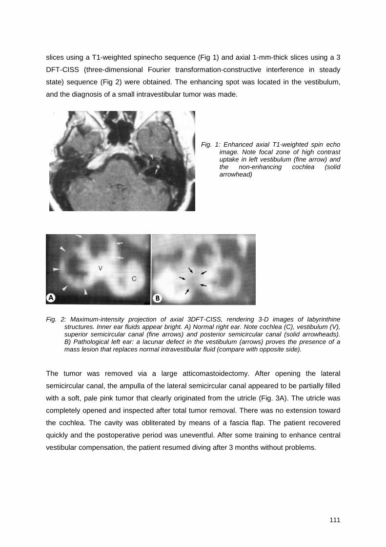
111
slices using a T1-weighted spinecho sequence (Fig 1) and axial 1-mm-thick slices using a 3
DFT-CISS (three-dimensional Fourier transformation-constructive interference in steady
state) sequence (Fig 2) were obtained. The enhancing spot was located in the vestibulum,
and the diagnosis of a small intravestibular tumor was made.
Fig. 1: Enhanced axial T1-weighted spin echoimage. Note focal zone of high contrastuptake in left vestibulum (fine arrow) andthe non-enhancing cochlea (solidarrowhead)
Fig. 2: Maximum-intensity projection of axial 3DFT-CISS, rendering 3-D images of labyrinthinestructures. Inner ear fluids appear bright. A) Normal right ear. Note cochlea (C), vestibulum (V),superior semicircular canal (fine arrows) and posterior semicircular canal (solid arrowheads).B) Pathological left ear: a lacunar defect in the vestibulum (arrows) proves the presence of amass lesion that replaces normal intravestibular fluid (compare with opposite side).
The tumor was removed via a large atticomastoidectomy. After opening the lateral
semicircular canal, the ampulla of the lateral semicircular canal appeared to be partially filled
with a soft, pale pink tumor that clearly originated from the utricle (Fig. 3A). The utricle was
completely opened and inspected after total tumor removal. There was no extension toward
the cochlea. The cavity was obliterated by means of a fascia flap. The patient recovered
quickly and the postoperative period was uneventful. After some training to enhance central
vestibular compensation, the patient resumed diving after 3 months without problems.
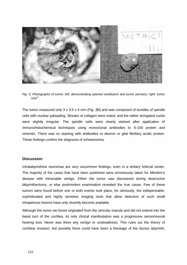
112
Fig. 3: Photographs of tumor: left: demonstrating opened vestibulum and tumor (arrows); right: tumorsize♦
The tumor measured only 3 x 3.5 x 4 mm (Fig. 3B) and was composed of bundles of spindle
cells with nuclear palisading. Streaks of collagen were noted, and the rather elongated nuclei
were slightly irregular. The spindle cells were clearly stained after application of
immunohistochemical techniques using monoclonal antibodies to S-100 protein and
vimentin. There was no staining with antibodies to desmin or glial fibrillary acidic protein.
These findings confirm the diagnosis of schwannoma.
Discussion
Intralabyrinthine neuromas are very uncommon findings, even in a tertiary referral center.
The majority of the cases that have been published were erroneously taken for Ménière’s
disease with intractable vertigo. Either the tumor was discovered during destructive
labyrinthectomy, or else postmortem examination revealed the true cause. Few of these
tumors were found before one or both events took place, for obviously, the indispensable,
sophisticated and highly sensitive imaging tools that allow detection of such small
intrapetrous lesions have only recently become available.
Although the tumor we found originated from the utricular macula and did not extend into the
basal turn of the cochlea, its only clinical manifestation was a progressive sensorineural
hearing loss. Never was there any vertigo or unsteadiness. This rules out the theory of
cochlear invasion, but possibly there could have been a blockage of the ductus labyrinth,
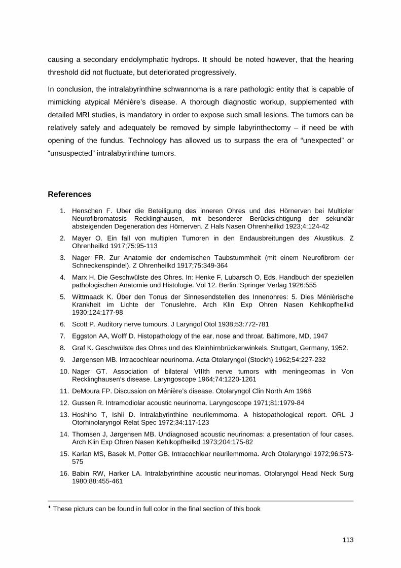
113
causing a secondary endolymphatic hydrops. It should be noted however, that the hearing
threshold did not fluctuate, but deteriorated progressively.
In conclusion, the intralabyrinthine schwannoma is a rare pathologic entity that is capable of
mimicking atypical Ménière’s disease. A thorough diagnostic workup, supplemented with
detailed MRI studies, is mandatory in order to expose such small lesions. The tumors can be
relatively safely and adequately be removed by simple labyrinthectomy – if need be with
opening of the fundus. Technology has allowed us to surpass the era of “unexpected” or
“unsuspected” intralabyrinthine tumors.
References
1. Henschen F. Uber die Beteiligung des inneren Ohres und des Hörnerven bei MultiplerNeurofibromatosis Recklinghausen, mit besonderer Berücksichtigung der sekundärabsteigenden Degeneration des Hörnerven. Z Hals Nasen Ohrenheilkd 1923;4:124-42
2. Mayer O. Ein fall von multiplen Tumoren in den Endausbreitungen des Akustikus. ZOhrenheilkd 1917;75:95-113
3. Nager FR. Zur Anatomie der endemischen Taubstummheit (mit einem Neurofibrom derSchneckenspindel). Z Ohrenheilkd 1917;75:349-364
4. Marx H. Die Geschwülste des Ohres. In: Henke F, Lubarsch O, Eds. Handbuch der speziellenpathologischen Anatomie und Histologie. Vol 12. Berlin: Springer Verlag 1926:555
5. Wittmaack K. Über den Tonus der Sinnesendstellen des Innenohres: 5. Dies MénièrischeKrankheit im Lichte der Tonuslehre. Arch Klin Exp Ohren Nasen Kehlkopfheilkd1930;124:177-98
6. Scott P. Auditory nerve tumours. J Laryngol Otol 1938;53:772-781
7. Eggston AA, Wolff D. Histopathology of the ear, nose and throat. Baltimore, MD, 1947
8. Graf K. Geschwülste des Ohres und des Kleinhirnbrückenwinkels. Stuttgart, Germany, 1952.
9. Jørgensen MB. Intracochlear neurinoma. Acta Otolaryngol (Stockh) 1962;54:227-232
10. Nager GT. Association of bilateral VIIIth nerve tumors with meningeomas in VonRecklinghausen’s disease. Laryngoscope 1964;74:1220-1261
11. DeMoura FP. Discussion on Ménière’s disease. Otolaryngol Clin North Am 1968
12. Gussen R. Intramodiolar acoustic neurinoma. Laryngoscope 1971;81:1979-84
13. Hoshino T, Ishii D. Intralabyrinthine neurilemmoma. A histopathological report. ORL JOtorhinolaryngol Relat Spec 1972;34:117-123
14. Thomsen J, Jørgensen MB. Undiagnosed acoustic neurinomas: a presentation of four cases.Arch Klin Exp Ohren Nasen Kehlkopfheilkd 1973;204:175-82
15. Karlan MS, Basek M, Potter GB. Intracochlear neurilemmoma. Arch Otolaryngol 1972;96:573-575
16. Babin RW, Harker LA. Intralabyrinthine acoustic neurinomas. Otolaryngol Head Neck Surg1980;88:455-461
♦ These picturs can be found in full color in the final section of this book
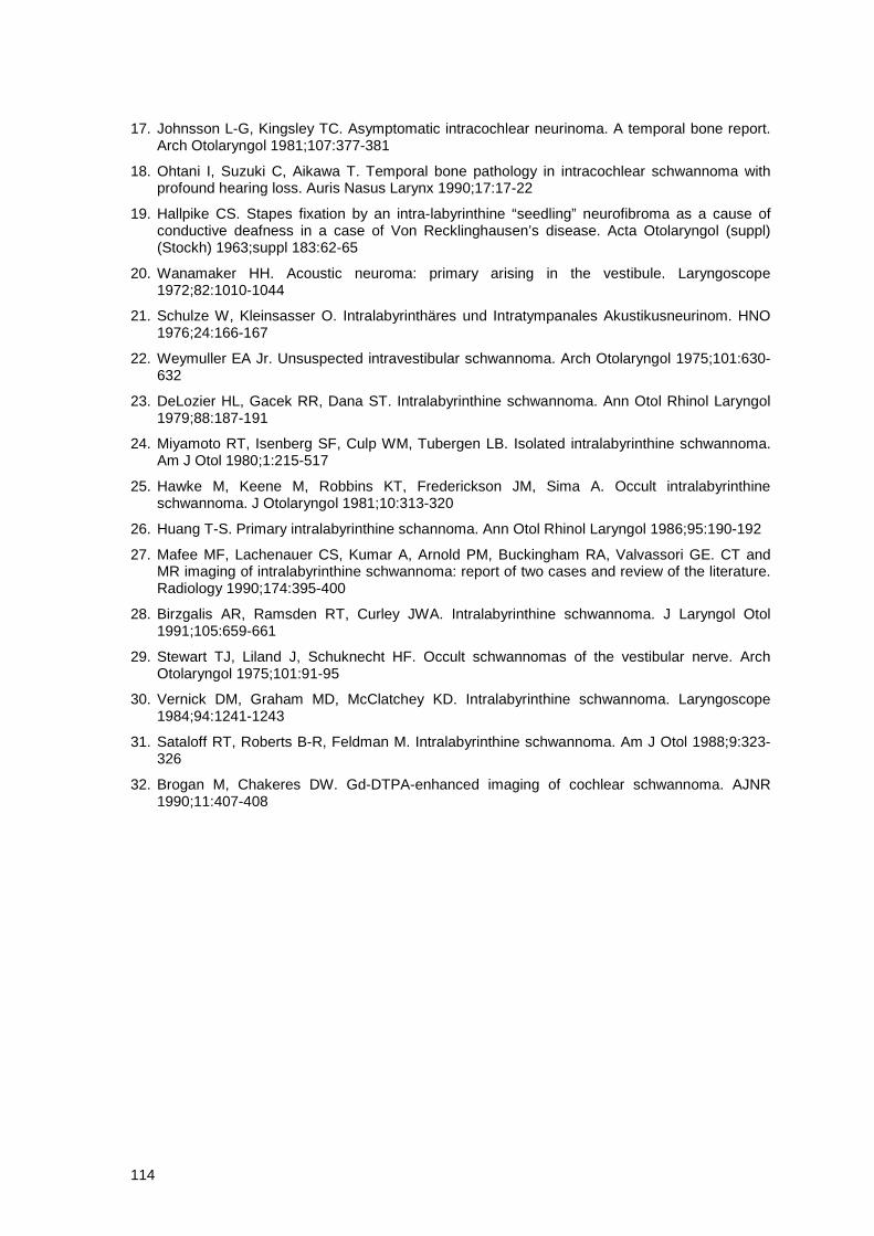
114
17. Johnsson L-G, Kingsley TC. Asymptomatic intracochlear neurinoma. A temporal bone report.Arch Otolaryngol 1981;107:377-381
18. Ohtani I, Suzuki C, Aikawa T. Temporal bone pathology in intracochlear schwannoma withprofound hearing loss. Auris Nasus Larynx 1990;17:17-22
19. Hallpike CS. Stapes fixation by an intra-labyrinthine “seedling” neurofibroma as a cause ofconductive deafness in a case of Von Recklinghausen’s disease. Acta Otolaryngol (suppl)(Stockh) 1963;suppl 183:62-65
20. Wanamaker HH. Acoustic neuroma: primary arising in the vestibule. Laryngoscope1972;82:1010-1044
21. Schulze W, Kleinsasser O. Intralabyrinthäres und Intratympanales Akustikusneurinom. HNO1976;24:166-167
22. Weymuller EA Jr. Unsuspected intravestibular schwannoma. Arch Otolaryngol 1975;101:630-632
23. DeLozier HL, Gacek RR, Dana ST. Intralabyrinthine schwannoma. Ann Otol Rhinol Laryngol1979;88:187-191
24. Miyamoto RT, Isenberg SF, Culp WM, Tubergen LB. Isolated intralabyrinthine schwannoma.Am J Otol 1980;1:215-517
25. Hawke M, Keene M, Robbins KT, Frederickson JM, Sima A. Occult intralabyrinthineschwannoma. J Otolaryngol 1981;10:313-320
26. Huang T-S. Primary intralabyrinthine schannoma. Ann Otol Rhinol Laryngol 1986;95:190-192
27. Mafee MF, Lachenauer CS, Kumar A, Arnold PM, Buckingham RA, Valvassori GE. CT andMR imaging of intralabyrinthine schwannoma: report of two cases and review of the literature.Radiology 1990;174:395-400
28. Birzgalis AR, Ramsden RT, Curley JWA. Intralabyrinthine schwannoma. J Laryngol Otol1991;105:659-661
29. Stewart TJ, Liland J, Schuknecht HF. Occult schwannomas of the vestibular nerve. ArchOtolaryngol 1975;101:91-95
30. Vernick DM, Graham MD, McClatchey KD. Intralabyrinthine schwannoma. Laryngoscope1984;94:1241-1243
31. Sataloff RT, Roberts B-R, Feldman M. Intralabyrinthine schwannoma. Am J Otol 1988;9:323-326
32. Brogan M, Chakeres DW. Gd-DTPA-enhanced imaging of cochlear schwannoma. AJNR1990;11:407-408
115
Chapter 4c
Problems with flute playing: an otological problem?Case report of a peculiar cerebellar astrocytoma
Problematisch dwarsfluitspel: een otologisch probleem?Case report van een bijzonder cerebellair astrocytoma
G. Forton, MD1
J. Verlooy, MD. PhD2
P. Cras, MD3
P. Parizel, MD4
P. Van de Heyning, MD PhD1
1 Dienst Neus-, Keel- & Oorziekten, Hoofd- en Halsheelkunde2 Dienst Neurochirurgie3 Dienst Neurologie4 Dienst RadiologieUniversitair Ziekenhuis Antwerpen, Edegem, BelgiëActa Otorhinolaryngol Belg 1993;47(4):389-96
116
Summary
A rare case of cerebellar astrocytoma presenting as a cerebellopontine angle tumour is
discussed. A 35-year old woman noticed a bizarre twitching and fatigability of the left upper
lip while playing the flute. There was also a mild hearing loss on the left side and she
sometimes felt unsure of herself when walking. A thorough examination by means of speech
audiometry, electronystagmography, ABR, CT-scan and MRI revealed a large, partly
calcified mass occupying the cerebellopontine angle. Only the histological examination of the
surgical specimen revealed the true nature of the tumor. The special characteristics of this
tumor and the unusual clinical course are discussed. The importance of a good histological
diagnosis is stressed.
Samenvatting
Een zeldzame casus van een cerebellair astrocytoma, dat zich presenteerde als een
brughoektumor wordt besproken. Een 35-jarige dame kreeg progressief last van een
abnormale vermoeibaarheid en trillen van de linker bovenlip tijdens het dwarsfluit spleen. Er
bestond ook een gering gehoorverlies links en een transiënte instabiliteit bij het stappen. Een
grondige oppuntstelling met behulp van tonale en spraakaudiometrie, elektronystagmografie,
BERA, CT-scan en MRI wezen op een grote, gedeeltelijk gecalcificeerde brughoektumor
links. Enkel na histologisch onderzoek kwam de ware aard van deze tumor aan het licht. De
bijzondere kenmerken en het ongewone klinisch verloop worden besproken, met nadruk op
een adequaat histologisch onderzoek.
Gevalstudie
Ziektegeschiedenis
Een 35-jarige patiënte bood zich aan op de raadpleging wegens trekkingen van de linker
bovenliphelft, gepaard gaande met krachtverlies van deze linker bovenliphelft en dit na een
tiental minuten dwarsfluit spelen. Dit probleem bestond reeds enkele weken. Bij verdere
anamnese bleek dat patiënte sinds haar kinderjaren aan de linkerkant minder scherp hoorde,
echter zonder tinnitus. Intermittent vertoonde zij de neiging naar links af te wijken. Tenslotte
werd er nog melding gemaakt van frequente, drukkende bifrontale hoofdpijn.
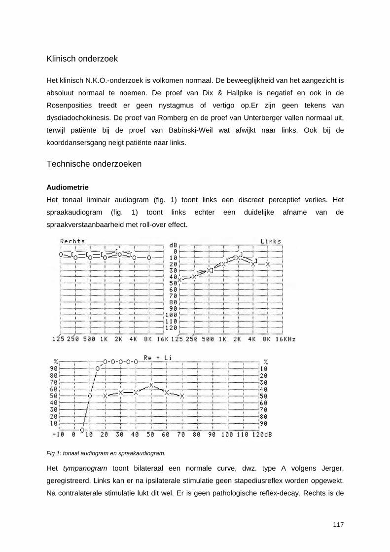
117
Klinisch onderzoek
Het klinisch N.K.O.-onderzoek is volkomen normaal. De beweeglijkheid van het aangezicht is
absoluut normaal te noemen. De proef van Dix & Hallpike is negatief en ook in de
Rosenposities treedt er geen nystagmus of vertigo op.Er zijn geen tekens van
dysdiadochokinesis. De proef van Romberg en de proef van Unterberger vallen normaal uit,
terwijl patiënte bij de proef van Babínski-Weil wat afwijkt naar links. Ook bij de
koorddansersgang neigt patiënte naar links.
Technische onderzoeken
Audiometrie
Het tonaal liminair audiogram (fig. 1) toont links een discreet perceptief verlies. Het
spraakaudiogram (fig. 1) toont links echter een duidelijke afname van de
spraakverstaanbaarheid met roll-over effect.
Fig 1: tonaal audiogram en spraakaudiogram.
Het tympanogram toont bilateraal een normale curve, dwz. type A volgens Jerger,
geregistreerd. Links kan er na ipsilaterale stimulatie geen stapediusreflex worden opgewekt.
Na contralaterale stimulatie lukt dit wel. Er is geen pathologische reflex-decay. Rechts is de
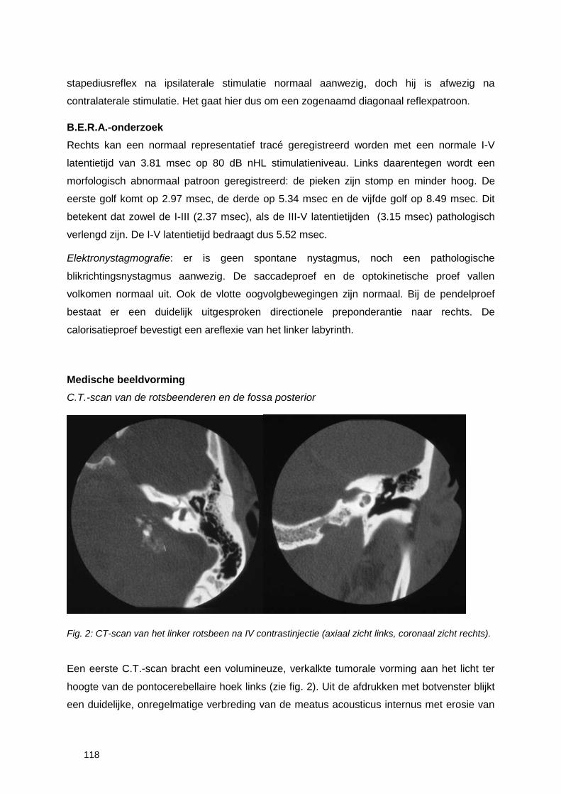
118
stapediusreflex na ipsilaterale stimulatie normaal aanwezig, doch hij is afwezig na
contralaterale stimulatie. Het gaat hier dus om een zogenaamd diagonaal reflexpatroon.
B.E.R.A.-onderzoek
Rechts kan een normaal representatief tracé geregistreerd worden met een normale I-V
latentietijd van 3.81 msec op 80 dB nHL stimulatieniveau. Links daarentegen wordt een
morfologisch abnormaal patroon geregistreerd: de pieken zijn stomp en minder hoog. De
eerste golf komt op 2.97 msec, de derde op 5.34 msec en de vijfde golf op 8.49 msec. Dit
betekent dat zowel de I-III (2.37 msec), als de III-V latentietijden (3.15 msec) pathologisch
verlengd zijn. De I-V latentietijd bedraagt dus 5.52 msec.
Elektronystagmografie: er is geen spontane nystagmus, noch een pathologische
blikrichtingsnystagmus aanwezig. De saccadeproef en de optokinetische proef vallen
volkomen normaal uit. Ook de vlotte oogvolgbewegingen zijn normaal. Bij de pendelproef
bestaat er een duidelijk uitgesproken directionele preponderantie naar rechts. De
calorisatieproef bevestigt een areflexie van het linker labyrinth.
Medische beeldvorming
C.T.-scan van de rotsbeenderen en de fossa posterior
Fig. 2: CT-scan van het linker rotsbeen na IV contrastinjectie (axiaal zicht links, coronaal zicht rechts).
Een eerste C.T.-scan bracht een volumineuze, verkalkte tumorale vorming aan het licht ter
hoogte van de pontocerebellaire hoek links (zie fig. 2). Uit de afdrukken met botvenster blijkt
een duidelijke, onregelmatige verbreding van de meatus acousticus internus met erosie van

119
de crista falciformis. De tumor is ietwat posterior van de meatus gecentreerd. De lokalisatie
van de tumor en de aanwezigheid van calcificaties doen in eerste instantie denken aan een
meningeoom. Anderzijds is de verbreding van de meatus acousticus internus en de erosie
van de crista een argument dat wijst in de richting van een acousticusneurinoma. Tegen
beide hypothesen pleit dan weer de eerder povere aanverving van de tumor na
contrastinjectie. Aanvullende investigatie met M.R.I. is dus alleszins geïndiceerd!
M.R.I.-onderzoek van de fossa posterior
Fig. 3: T1-gewogen M.R.I.-beelden na IV Gd-DTPA injectie (axiaal zicht links, coronaal rechts)
Een eerste M.R.I. van de brughoekregio werd uitgevoerd, zowel zonder als met intraveneuze
toediening van Gd-DTPA. Dit onderzoek bevestigt de aanwezigheid van een vrij groot
ruimte-innemend proces in de linker brughoek (zie fig. 3), dat gekenmerkt wordt door
heterogene signaalintensiteiten, zowel op de T1- als op de T2-gewogen opnamen. Deze
zones met lage signaalintensiteit corresponderen met de inliggende calcificaties. De tumor
reikt tot aan de middellijn in de prepontiene cisterna en is moeilijk aflijnbaar ten opzichte van
de linker cerebellaire hemisfeer. De tumor ligt eveneens in nauw contact met de hersenstam,
doch hier is een scheidingsvlak merkbaar. Caudaalwaarts reikt de tumor tot aan het foramen
magnum. Na intraveneuze injectie van Gd-DTPA is er een discrete aanverving van deze
massa, wat een sterk argument is tegen de acousticusneurinoma-hypothese. Ook in het
geval van een partieel verkalkt meningeoom zou men een sterke contrastcaptatie
verwachten. Er is doorbraak van de bloed-hersenbarrière ter hoogte van het cerebellum. De
mogelijkheid van een epidermoïdtumor wordt geopperd, gezien de toch wel vrij hoge
signaalintensiteit op de sterk T2-gewogen sequenties. Ook een hamartoma of een teratoma
120
kunnen worden opgenomen in de differentiële diagnose. De middellijnstructuren blijven
alleszins gerespecteerd en er is geen uitzetting van het ventriculair systeem.
Evolutie
Gezien de eerder geringe subjectieve last en het vrij goede gehoor enerzijds, en gezien de
majeure implicaties van een chirurgische interventie anderzijds, werd geopteerd voorlopig
een afwachtende houding aan te nemen en de evolutie nauwlettend te volgen.
Bij een controle 6 en 9 maanden na de eerste raadpleging bleken de klachten subjectief
ongewijzigd te zijn gebleven en toonden noch het zuivere toonaudiogram, noch het
spraakaudiogram enige noemenswaardige evolutie. Bij een volgende raadpleging 1 maand
later waren de klachten toegenomen: de valneiging naar links was meer uitgesproken, terwijl
de tics en de vermoeibaarheid van de linker bovenliphelft waren toegenomen. Klinisch was
er geen facialisparese merkbaar. Patiënte vertoonde echter wel tekenen van cerebellaire
ataxie, vooral dan ter hoogte van de linker arm. Er werd ook melding gemaakt van een
discrete dysarthrie bij vermoeidheid. Het zuivere toonaudiogram was ongewijzigd, doch het
spraakaudiogram toonde een duidelijke achteruitgang van de spraakdiscriminatie. Een
tweede M.R.I.-onderzoek van de fossa posterior toonde geen noemenswaardige toename
van de tumorale vorming ten opzichte van de vorige opnamen. Er waren echter wel tekens
van cerebellair oedeem. De vierde ventrikel was nog steeds gerespecteerd.
Gezien de ongunstige klinische evolutie werd door het multidisciplinair otoneurochirurgisch
team een fossa posterior benadering van de tumor gepland. Een in het kader van de pre-
operatieve oppuntstelling uitgevoerde angiografie van het linker vertebrobasilair systeem
toonde een discrete nodulaire blush in de pontocerebellaire hoek zonder duidelijke
pathologische bloedvaten.
Ingreep
Na spinale drainage werd een retrosigmoïdale fossa posterior benadering uitgevoerd. Na
ecarteren van het cerebellum werd de zeer heterogeen uitziende tumor in het licht gesteld.
De tumor bleek sterk verkleefd te zijn met het cerebellum. De Nn. VII en VIII waren nog
moeilijk te identificeren en dienden als verloren te worden beschouwd. De andere
omliggende craniale zenuwen, in het bijzonder de Nn. V, VI, X en XII waren verdrongen en
dun uitgewaaierd. Zij bleven echter macroscopisch gespaard. Na debulking kon dan de
tumor vrijgedisseceerd worden van de hersenstam en kon een macroscopisch volledige
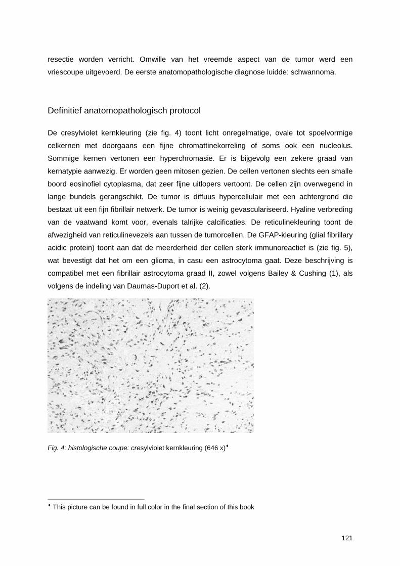
121
resectie worden verricht. Omwille van het vreemde aspect van de tumor werd een
vriescoupe uitgevoerd. De eerste anatomopathologische diagnose luidde: schwannoma.
Definitief anatomopathologisch protocol
De cresylviolet kernkleuring (zie fig. 4) toont licht onregelmatige, ovale tot spoelvormige
celkernen met doorgaans een fijne chromattinekorreling of soms ook een nucleolus.
Sommige kernen vertonen een hyperchromasie. Er is bijgevolg een zekere graad van
kernatypie aanwezig. Er worden geen mitosen gezien. De cellen vertonen slechts een smalle
boord eosinofiel cytoplasma, dat zeer fijne uitlopers vertoont. De cellen zijn overwegend in
lange bundels gerangschikt. De tumor is diffuus hypercellulair met een achtergrond die
bestaat uit een fijn fibrillair netwerk. De tumor is weinig gevasculariseerd. Hyaline verbreding
van de vaatwand komt voor, evenals talrijke calcificaties. De reticulinekleuring toont de
afwezigheid van reticulinevezels aan tussen de tumorcellen. De GFAP-kleuring (glial fibrillary
acidic protein) toont aan dat de meerderheid der cellen sterk immunoreactief is (zie fig. 5),
wat bevestigt dat het om een glioma, in casu een astrocytoma gaat. Deze beschrijving is
compatibel met een fibrillair astrocytoma graad II, zowel volgens Bailey & Cushing (1), als
volgens de indeling van Daumas-Duport et al. (2).
Fig. 4: histologische coupe: cresylviolet kernkleuring (646 x)♦
♦ This picture can be found in full color in the final section of this book
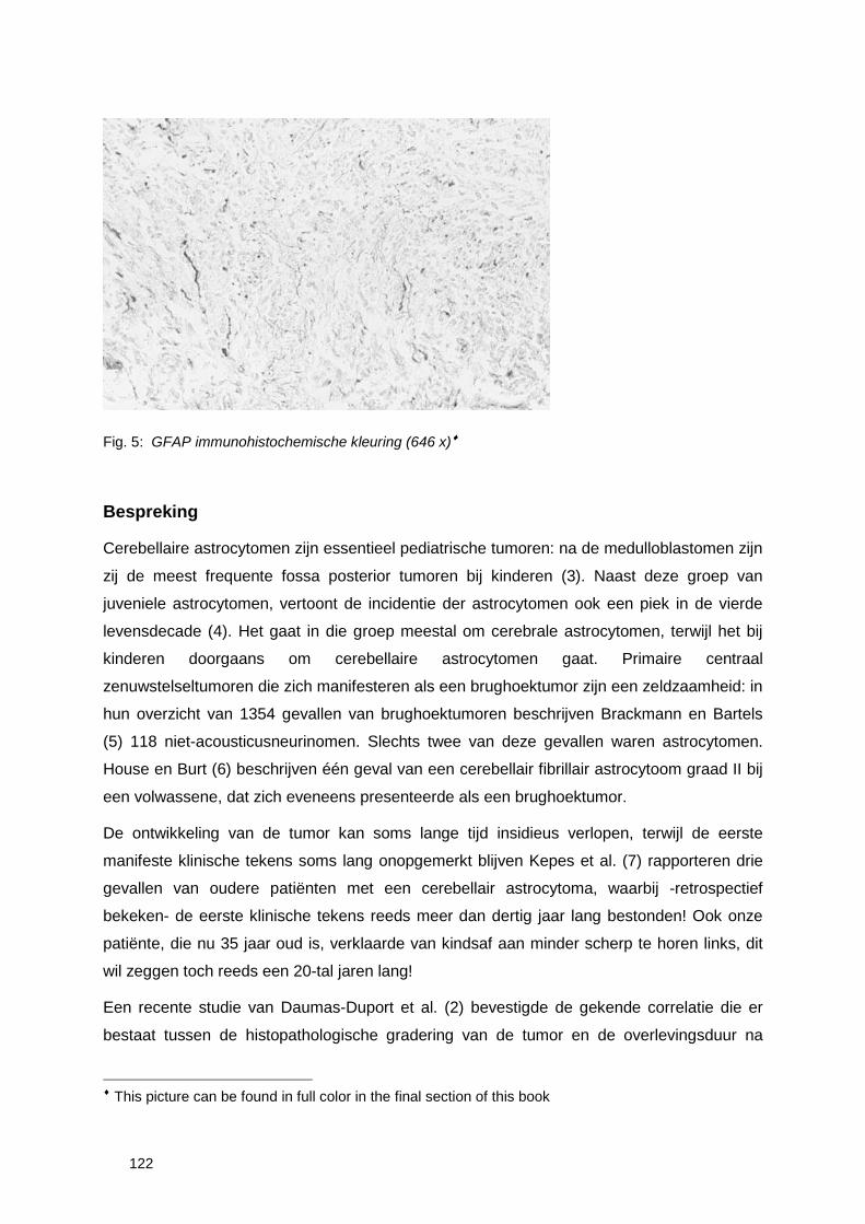
122
Fig. 5: GFAP immunohistochemische kleuring (646 x)♦
Bespreking
Cerebellaire astrocytomen zijn essentieel pediatrische tumoren: na de medulloblastomen zijn
zij de meest frequente fossa posterior tumoren bij kinderen (3). Naast deze groep van
juveniele astrocytomen, vertoont de incidentie der astrocytomen ook een piek in de vierde
levensdecade (4). Het gaat in die groep meestal om cerebrale astrocytomen, terwijl het bij
kinderen doorgaans om cerebellaire astrocytomen gaat. Primaire centraal
zenuwstelseltumoren die zich manifesteren als een brughoektumor zijn een zeldzaamheid: in
hun overzicht van 1354 gevallen van brughoektumoren beschrijven Brackmann en Bartels
(5) 118 niet-acousticusneurinomen. Slechts twee van deze gevallen waren astrocytomen.
House en Burt (6) beschrijven één geval van een cerebellair fibrillair astrocytoom graad II bij
een volwassene, dat zich eveneens presenteerde als een brughoektumor.
De ontwikkeling van de tumor kan soms lange tijd insidieus verlopen, terwijl de eerste
manifeste klinische tekens soms lang onopgemerkt blijven Kepes et al. (7) rapporteren drie
gevallen van oudere patiënten met een cerebellair astrocytoma, waarbij -retrospectief
bekeken- de eerste klinische tekens reeds meer dan dertig jaar lang bestonden! Ook onze
patiënte, die nu 35 jaar oud is, verklaarde van kindsaf aan minder scherp te horen links, dit
wil zeggen toch reeds een 20-tal jaren lang!
Een recente studie van Daumas-Duport et al. (2) bevestigde de gekende correlatie die er
bestaat tussen de histopathologische gradering van de tumor en de overlevingsduur na
♦ This picture can be found in full color in the final section of this book
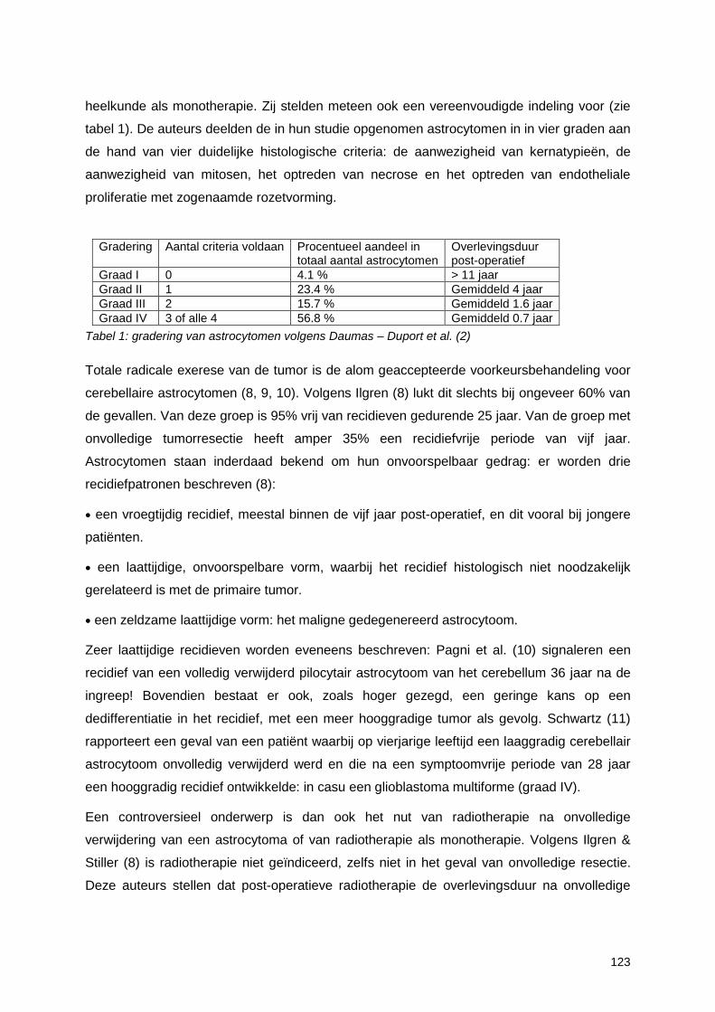
123
heelkunde als monotherapie. Zij stelden meteen ook een vereenvoudigde indeling voor (zie
tabel 1). De auteurs deelden de in hun studie opgenomen astrocytomen in in vier graden aan
de hand van vier duidelijke histologische criteria: de aanwezigheid van kernatypieën, de
aanwezigheid van mitosen, het optreden van necrose en het optreden van endotheliale
proliferatie met zogenaamde rozetvorming.
Gradering Aantal criteria voldaan Procentueel aandeel intotaal aantal astrocytomen
Overlevingsduurpost-operatief
Graad I 0 4.1 % > 11 jaarGraad II 1 23.4 % Gemiddeld 4 jaarGraad III 2 15.7 % Gemiddeld 1.6 jaarGraad IV 3 of alle 4 56.8 % Gemiddeld 0.7 jaar
Tabel 1: gradering van astrocytomen volgens Daumas – Duport et al. (2)
Totale radicale exerese van de tumor is de alom geaccepteerde voorkeursbehandeling voor
cerebellaire astrocytomen (8, 9, 10). Volgens Ilgren (8) lukt dit slechts bij ongeveer 60% van
de gevallen. Van deze groep is 95% vrij van recidieven gedurende 25 jaar. Van de groep met
onvolledige tumorresectie heeft amper 35% een recidiefvrije periode van vijf jaar.
Astrocytomen staan inderdaad bekend om hun onvoorspelbaar gedrag: er worden drie
recidiefpatronen beschreven (8):
• een vroegtijdig recidief, meestal binnen de vijf jaar post-operatief, en dit vooral bij jongere
patiënten.
• een laattijdige, onvoorspelbare vorm, waarbij het recidief histologisch niet noodzakelijk
gerelateerd is met de primaire tumor.
• een zeldzame laattijdige vorm: het maligne gedegenereerd astrocytoom.
Zeer laattijdige recidieven worden eveneens beschreven: Pagni et al. (10) signaleren een
recidief van een volledig verwijderd pilocytair astrocytoom van het cerebellum 36 jaar na de
ingreep! Bovendien bestaat er ook, zoals hoger gezegd, een geringe kans op een
dedifferentiatie in het recidief, met een meer hooggradige tumor als gevolg. Schwartz (11)
rapporteert een geval van een patiënt waarbij op vierjarige leeftijd een laaggradig cerebellair
astrocytoom onvolledig verwijderd werd en die na een symptoomvrije periode van 28 jaar
een hooggradig recidief ontwikkelde: in casu een glioblastoma multiforme (graad IV).
Een controversieel onderwerp is dan ook het nut van radiotherapie na onvolledige
verwijdering van een astrocytoma of van radiotherapie als monotherapie. Volgens Ilgren &
Stiller (8) is radiotherapie niet geïndiceerd, zelfs niet in het geval van onvolledige resectie.
Deze auteurs stellen dat post-operatieve radiotherapie de overlevingsduur na onvolledige
124
resectie niet verlengt, noch het aantal recidieven doet afnemen. Niettemin geven deze
auteurs toe dat radiotherapie het risico op locale tumorspreiding via de subarachnoïdale
ruimte zou doen afnemen. Andere auteurs, zoals Garcia et al (11), Fazekas (12) en Leibel et
al. (13) poneren daarentegen dat post-operatieve radiotherapie wel degelijk nuttig is, zowel
voor wat de overleviungsduur betreft, als voor wat de recidiefkans betreft. Er werd dan ook
besloten onze patiënte 56 Gy toe te dienen ter hoogte van de ponto-cerebellaire hoek.
Besluit
Deze case report benadrukt de noodzaak van een grondige investigatie van elk
asymmetrisch perceptieverlies en van elk geval van Nervus Facialisprikkeling of -parese.
Vooral indien er een combinatie van beiden bestaat, dient de brughoekregio aan een
grondige oppuntstelling onderworpen. Alhoewel zeldzaam, kunnen andere tumoren dan
acousticusneurinomen, meningeomen of epidermoïdtumoren zich als een klassieke
brughoektumor presenteren. Een grondig histopathologisch onderzoek van het specimen
kan in dergelijke gevallen in belangrijke mate mede bepalend zijn voor het verdere beleid en
de prognose.
Bibliografie
1. Bailey P, Cushing H. A classification of the tumors of the glioma group on a histogenetic basiswith a correlated study of prognosis. J.B. Lippincott Co., Philadelphia, 1926.
2. Daumas-Duport C, Scheithauer B, O'Fallon J, Kelly P. Grading of astrocytomas. A simple andreproducible method. Cancer, 62:2152-2165, 1988
3. Duffrer PK, Cohen ME, Meyers MH, Heise HW. Survival of children with brain tumors: SEERprogram, 1973-1980. Neurology, 36:597-601, 1986.
4. Davis RL. Astrocytomas. In: Pathology of the Nervous System, Minckler J, Bailey OT, Feigin I,Jervis G, Lindenberg R & Neubuerger KT (Eds.), McGraw-Hill Book Company, New York, Vol2, 2007-2026, 1971.
5. Brackmann DE, Bartels LJ. Rare tumors of the cerebellopontine angle. Otolaryngol HeadNeck Surg, 88:555-559, 1980.
6. House JL, Burt MR. Primary CNS tumors presenting as cerebellopontine angle tumors. Am JOtol, (suppl):147-153, 1985.
7. Kepes JJ, Whittaker CK, Watson K, Morantz RA, Millett R, Clough CA, Oxley DK. Cerebellarastrocytomas in elderly patients with very long preoperative histories: report of three cases.Neurosurgery, 25(2):258-264, 1989.
8. Ilgren EB, Stiller CA. Cerebellar astrocytomas: therapeutic management. Acta neurochir(Wien), 81:11-26, 1986.
125
9. Garcia DM, Marks JE, Latifi HR, Kliefoth AB. C hildhood Cerebellar astrocytomas: is there arole for postoperative irradiation? Int J Rad Oncol Biol Phys, 18:815-818, 1990.
10. Pagni CA, Giordana MT, Canavero S. Benign recurrence of a pilocytic cerebellar astrocytoma36 years after radical removal: case report. Neurosurgery, 28(4):606-609, 1991.
11. Schwartz AM, Ghatak NR. Malignant transformation of benign cerebellar astrocytoma.Cancer, 65(2):333-6, 1990.
12. Fazekas J. Treatment of grades 1 and 2 brain astrocytomas. The role of radiotherapy. Int JRad Oncol Bio Phys, 2:661-666, 1977.
13. Leibel S, Sheline G, Wara W, Boldrey E, Neilson S. The role of radiation therapy in thetreatment of astrocytomas. Cancer, 35:1551-1557, 1975.

126
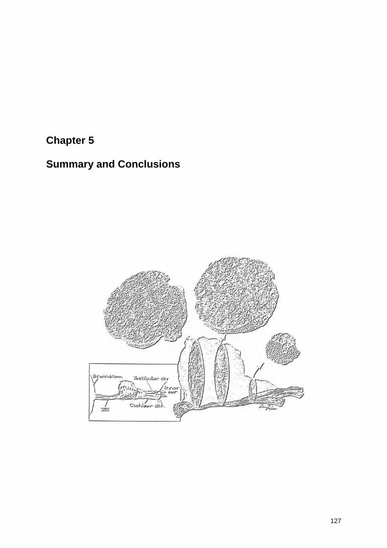
127
Chapter 5
Summary and Conclusions
128
Over the last decennia, the diagnostic regimen and treatment of the acoustic neuroma has
changed dramatically. As always during a period of changes, questions arise that need be
answered on a sound scientific basis. We have tried to formulate and answer to some of
these questions in this thesis.
In chapter 1, we provide a historical overview regarding important advances in diagnostics
and treatment of the acoustic neuroma. One of the most dramatic diagnostic acquirements
is undoubtedly the fast evolution of magnetic resonance imaging. New techniques allow for
long-term follow-up of acoustic tumors, which already yields new insights on the natural
course of the acoustic neuroma. A wait-and-see policy has therefore become a valid
alternative for immediate surgery when the acoustic neuroma still is in an early stage. MRI
also enables us to study the changes in the inner ear fluids that are quite possibly linked to
inner ear deterioration and thus allows in a timely fashion to choose the best moment for
surgery and the suitable surgical access to the cerebellopontine angle.
Another most important feature of MRI is early detection and follow-up of tumor recurrence
and/or monitoring of residual tumor. Since we have provided irrefutable histological evidence
of tumor ingrowth in the cochlear nerve in chapter 2, it is quite thinkable that more than once
tumor remnants that are indeed invisible to the surgeon at the time of surgery, but
nonetheless present on the microscopical level, are left behind after successful removal of
the tumor. MRI provides us and will continue to provide us the necessary data as to the fate
and growing potentials of these remnants. The same logic holds true for the facial nerve,
which is almost always preserved.
Up till now, CT and MRI follow-up studies do not indicate that sharp dissection of the facial
nerve, in order to maximize nerve function preservation, yields a higher risk of tumor
recurrence due to tumor remnants that are inevitably left behind.
In chapter 3, the clinical data of 13 selected cases are presented. In all of these cases, the
cochlear nerve was positively identified in the microscopical specimen and tumor ingrowth
was established. Moreover, all required clinical data of these subjects could be retrieved
from the patient files. Comparison of these data with other series in contemporary literature
teaches us that there is no significant difference between these selected cases and similar
cases in the literature.
As MRI was introduced in the clinic and was rapidly recognized as a very powerful diagnostic
tool, more data were required as to the sensitivity and specificity of MRI in the differential
diagnosis of cerebellopontine angle tumors. In the second part of chapter 3, a pictorial essay
on the characteristics of the acoustic neuroma is presented. Meanwhile, daily practice has


