Embed Size (px)
Citation preview

Available online at www.sciencedirect.com
1773 (2007) 1664–1671www.elsevier.com/locate/bbamcr
Biochimica et Biophysica Acta
The anti-apoptotic MAP kinase pathway is inhibited in NIH3T3 fibroblastswith increased expression of phosphatidylinositol transfer protein β
Martijn Schenning ⁎, Claudia M. van Tiel, Karel W.A. Wirtz, Gerry T. Snoek
Bijvoet Center, Department of Biochemistry of Lipids, Institute of Biomembranes, Utrecht University, Padualaan 8, 3584 CH Utrecht, The Netherlands
Received 20 March 2007; received in revised form 18 June 2007; accepted 19 June 2007Available online 26 June 2007
Abstract
Mouse NIH3T3 fibroblast cells overexpressing phosphatidylinositol transfer protein β (PI-TPβ, SPIβ cells) demonstrate a low rate ofproliferation and a high sensitivity towards UV-induced apoptosis when compared with wtNIH3T3 cells. In contrast, SPIβS262A cells over-expressing a mutant PI-TPβ that lacks the protein kinase C-dependent phosphorylation site Ser-262, demonstrate a phenotype comparable withwtNIH3T3 cells. This suggests that the phosphorylation of Ser-262 in PI-TPβ is involved in the regulation of apoptosis. Conditioned medium (CM)fromwtNIH3T3 cells contains bioactive factors, presumably arachidonic acid metabolites [H. Bunte, et al., 2006; M. Schenning, et al., 2004] that areable to protect SPIβ cells against UV-induced apoptosis. CM from SPIβ cells lacks this protective activity. However, after heat denaturation CM fromSPIβ cells regains a protective activity comparable with that of wtNIH3T3 cells. This indicates that CM from SPIβ cells contains an antagonisticfactor interfering with the anti-apoptotic activity present. SPIβS262A cells do not produce the antagonist suggesting that phosphorylation of Ser-262is required. Moreover, in line with the apparent lack of anti-apoptotic activity, CM from SPIβ cells does not induce the expression of COX-2 or theactivation of p42/p44 MAP kinase in SPIβ cells. In contrast, CM from wtNIH3T3 and SPIβS262A cells or heat-treated CM from SPIβ cells doesinduce these anti-apoptotic markers. Since we have previously shown that some of the arachidonic acid metabolites present in CM from wtNIH3T3cells are prostaglandin (PG) E2 and PGF2α, we investigated the effect of these PGs on cell survival. Although PGE2 and PGF2αwere found to protectwtNIH3T3 and SPIβS262A cells against UV-induced apoptosis, these PGs failed to rescue SPIβ cells. The fact that the concentrations of PGE2 andPGF2α in the CM from SPIβ cells and wtNIH3T3 cells were found to be comparable suggests that the failure of these PGs to protect SPIβ cells couldrender these cells more apoptosis sensitive. Concomitantly, upon incubation with PGE2 and PGF2α, an increased expression of COX-2 and activationof p42/p44 MAP kinase were observed in wtNIH3T3 and SPIβS262A cells but not in SPIβ cells. Hence, it appears that specific mechanisms of cellsurvival are impaired in SPIβ cells.© 2007 Elsevier B.V. All rights reserved.
Keywords: Phospholipid transfer protein; Sphingomyelin metabolism; Eicosanoids; Phosphatidylinositol metabolism
Abbreviations: PI-TP, phosphatidylinositol transfer protein; PI, phosphati-dylinositol; PC, phosphatidylcholine; SM, sphingomyelin; PKC, protein kinaseC; COX-1, cyclooxygenase-1; COX-2, cyclooxygenase-2; DMEM, Dulbecco'smodified Eagle medium; CM, conditioned medium; NCS, newborn calf serum;PBS, phosphate buffered saline; DBB, DMEM containing 0.1% bovine serumalbumin; PGE2, prostaglandin E2; PGF2α, prostaglandin F2α; MAPK, mitogen-activated protein kinase; MKK, MAP kinase kinase; GPCR, G protein-coupledreceptor⁎ Corresponding author. Division of Molecular Physiology, College of Life
Sciences, University of Dundee, Dundee DD1 5EH, UK. Tel.: +44 1382 386259;fax: +44 1382 385507.
E-mail address: [email protected] (M. Schenning).
0167-4889/$ - see front matter © 2007 Elsevier B.V. All rights reserved.doi:10.1016/j.bbamcr.2007.06.004
1. Introduction
The mammalian phosphatidylinositol transfer protein β (PI-TPβ) is a highly conserved protein with up to 99% sequenceidentity between species [1,2]. It shares 77% sequence identitywith its isoform PI-TPα. PI-TPβ is mainly associated with theGolgi system and may be the functional analogue of Sec14p, themajor yeast PI-TP [3,4]. Although its physiological function hasnot yet been established, its importance follows from the obser-vation that gene ablation of PI-TPβ in murine embryonic stemcells prevents embryonic development [5]. Murine embryoslacking PI-TPα develop normally but die within 2 weeks afterbirth [6]. This clearly shows that these two isoforms servedifferent functions in the cell.

1665M. Schenning et al. / Biochimica et Biophysica Acta 1773 (2007) 1664–1671
In previous studies on wtNIH3T3 cells that have a 10-foldincrease in PI-TPβ levels (SPIβ cells) we observed that thesecells display a decreased growth rate relative to the wtNIH3T3cells [7]. In addition, under conditions where sphingomyelin(SM) in the plasma membrane was hydrolyzed to ceramide byexogenous sphingomyelinase, SPIβ cells in contrast towtNIH3T3 cells maintained the steady-state levels of SM inthe plasma membrane suggesting that PI-TPβ was involved inthis process [7]. This is in agreement with the finding that invitro PI-TPβ binds and transfers SM in addition to PI and PCwhile PI-TPα lacks the ability to transfer SM [8]. Unlike SPIβcells, mutant SPIβS262A cells were unable to instantaneouslyreplenish SM after its degradation by exogenous sphingomye-linase, strongly suggesting that the phosphorylation of Ser-262was required [9,10]. If SM replenishment is linked to membranevesicle flow it could be that PI-TPβ plays a role in the buddingprocess, an activity analogous to Sec14p function in yeast [11].
In contrast to SPIβ cells, cells with an increased expression ofPI-TPα (SPIα cells) demonstrate a highly increased rate ofproliferation as well as an increased survival upon induction ofapoptosis [12,13]. Furthermore, it was shown that SPIα cellsproduce a PI-TPα/COX-2-dependent mitogenic and anti-apop-totic factor. Upon secretion, this factor is able to stimulategrowth and to promote survival in wtNIH3T3 and SPIβ cells[13]. In agreement with the increased resistance of these cellstowards UV-induced apoptosis, the PI-TPα-dependent survivalfactor induced COX-2 expression in both cell lines. In addition ithad been reported that this survival factor is able to preventapoptosis in rat motor neurons, suggesting a vital role in thecentral nervous system [14].
In earlier studies we have shown that SPIβ cells, whencompared with wtNIH3T3 cells, are very susceptible towardsUV-induced apoptosis, whereas the SPIβS262A cells behavesimilar to wtNIH3T3 cells [15]. Here we report that two abun-dant COX-1,-2 dependent arachidonic acid metabolites, PGE2
and PGF2α do not protect SPIβ cells against apoptosis whereaswtNIH3T3 and SPIβS262A cells are protected. In addition, wereport that conditioned medium (CM) from SPIβ cells contains aheat-labile antagonist masking the survival factor present. Thisantagonist prevents the activation of the anti-apoptotic p42/p44MAP kinase pathway and the upregulation of COX-2.
2. Materials and methods
2.1. Materials
Prostaglandins PGE2 and PGF2α were obtained from Sigma, polyclonalantibodies against COX-1/COX-2 from Cayman; anti-p42/p44 MAP kinaseantibodies where obtained from Cell Signaling technology, DMEM and NCSfrom Invitrogen; PGE2 ELISA kit was obtained from Cayman.
2.2. Cell culture
All cells were cultured in Dulbecco's modified Eagles medium (DMEM)containing 10% newborn calf serum (NCS) and buffered with 44 mM NaHCO3.Cells were maintained at 7.5% CO2 at 37 °C in a humidified atmosphere. Wildtype cells used were designated ATCC CRL 1658 NIH3T3 cells. The SPIα cellswere obtained as described in Snoek et al. [12]. The SPIβ cells and SPIβS262Acells were obtained as described in van Tiel et al. [7] and van Tiel et al. [9].
2.3. Preparation of conditioned medium
Cells were grown to 90% confluency in 150 cm2 dishes. After washing thecells twice with PBS, the mediumwas replaced with 13 ml of DMEM containing0.1% bovine serum albumin (DBB medium). After 24 h the medium wascollected and centrifuged (10 min at 1000 rpm) to remove floating cells. Thesupernatant is the conditioned medium (CM). Neutral lipids were extracted fromCM with two 39 ml portions of ethyl acetate (after adjusting to pH 2.0 withformic acid) [16]. CM was heat-denatured by incubation for 20 min at 80 °C,then centrifuged for 10 min at 17,500×g and the supernatant used forexperiments. Under standard conditions 90% confluent cells were incubatedwith CM derived from an identical surface of cells (i.e. 9.5 cm2 of cells per wellof a six-well dish was incubated with the amount of CM or neutral lipid extractderived from 9.5 cm2 of cells).
2.4. Induction of apoptosis
Induction of apoptosis by serum starvation: Cells were seeded in 6-wellplates and grown under standard conditions for 48 h until ca 85% confluency.Growth medium was removed and the cells were washed with PBS. The cellswere incubated with DMEM containing 0.1% bovine serum albumin (DBBmedium) for 16 h at 37 °C and the percentage of apoptotic cells wasmorphologically determined as the percentage of cells that was in the process ofblebbing.
Induction of apoptosis by tumor necrosis factor α (TNFα): Cells wereseeded in 6-well plates and grown under standard conditions for 48 h until ca85% confluency. Growth medium was removed and cells were washed withPBS. The cells were incubated with DBB medium containing cycloheximide(2.5 μg/ml) and TNFα (10 ng/ml) and the cells were incubated for 7 h at 37 °Cand the percentage of apoptotic cells was morphologically determined as thepercentage of cells that was in the process of blebbing.
Induction of apoptosis by UV irradiation: Cells were seeded in 6-well platesand grown for 48 h until ca 85% confluency. Growth medium was removed andcells were washed with PBS. Before UV treatment, the cells were incubated for4 h in DBB medium. To investigate the effects on the sensitivity to apoptosis,CM or prostaglandins were added to this DBB during the incubation prior to UVirradiation. The medium was removed and the cells were given a UV dose(200 J/m2) using a Stratalinker (Stratagene). After UV irradiation, the cells wereincubated with DBB at 37 °C. At the indicated time points cell death wasmorphologically determined as described above.
2.5. Determination of COX-1 and COX-2 levels
Cells were grown in 21 cm2 dishes to 80–90% confluency. Cells werewashed twice with PBS and lysed in 20 mM Tris–HCl pH 7.5 containing0.1% (v/v) NP40. The cell lysate was centrifuged at 17,500×g for 10 min at4 °C and the protein content of the supernatant fraction determined using theBradford assay [17]. Equal amounts of supernatant proteins (50 μg) weresubjected to SDS-PAGE on a 12% gel and Western blot analysis wasperformed using an antibody specific for COX-1 or COX-2. To determinewhether equal amounts of protein were analyzed, blots were stained withponceau S and scanned. The levels of COX-1 or -2 on the immunoblot werequantified using a Bio-Rad GS700 imaging densitometer equipped with anintegrating program. In some experiments prior to harvesting the cells wereincubated with CM for 5 h.
2.6. Determination of p42/44 MAP kinase levels
Cells were grown in 21 cm2 dishes to 80–90%confluency. Cells werewashedtwice with PBS and lysed in 20 mM Tris–HCl pH 7.5 containing 0.1% (v/v)NP40, 10 mM β-glycerophosphate, 1 mMNa3VO4, 50 mMNaF, 1 mM aprotininand 1 mM PMSF. Sample preparation was performed as described above.Western blot analysis was performed using an antibody specific for p42/44 MAPkinase and immunoreactive bands were quantified. To determine whether equalamounts of protein were analyzed, blots were stained with ponceau S andscanned. In some experiments prior to harvesting the cells were incubated withCM for 10 min.

1666 M. Schenning et al. / Biochimica et Biophysica Acta 1773 (2007) 1664–1671
2.7. Measurement of ceramide and sphingomyelin levels
Cells were grown to 80% confluency and total lipids were extracted by themethod of Bligh and Dyer [18]. Ceramide levels were determined using theEscherichia coli diacylglycerol kinase assay as described [19]. Briefly, thelipids were incubated at room temperature for 30 min in the presence of β-octylglucoside/dioleoyl-phosphatidyl glycerol micelles, 2 mM dithiothreitol,5 μg of proteins from the diacylglycerol kinase membranes, and 2 mM ATP(mixed with [γ-32P]ATP) in a final volume of 100 μl. After Bligh and Dyerextraction the lipids were separated by thin layer chromatography (TLC) inchloroform:acetone:methanol:acetic acid:H2O (50:20:15:10:5, by vol.) and theradioactivity associated with ceramide-phosphate was measured. Ceramidelevels were quantified using external standards and were normalized tophosphate. SM levels were determined as described previously [7].
3. Results
3.1. Apoptosis sensitivity of wtNIH3T3, SPIβ and SPIβS262Acells
Previously we have shown that SPIβ cells are sensitivetowards UV-induced apoptosis [15]. Exposure of the
Fig. 1. (A) Survival of wtNIH3T3, SPIβ and SPIβS262A cells upon induction of apoconfluency. Serum starvation: growth medium was replaced by DMEM/Bic/0.1% bovinduced apoptosis: growth medium was replaced by DBB containing cycloheximideUV induced apoptosis: growth medium was replaced by DBB and cells were incubatefresh DBBwas added to the cells and incubated for 3 h at 37 °C. The number of apoptomean values of at least three independent experiments. p-values were calculated relatiof blebbing wtNIH3T3, SPIβ and SPIβS262A cells. Cells were grown to 85% conflucells were irradiated with 200 J/m2 and fresh DBB was added. Images were taken 90wtNIH3T3 cells; middle panel, SPIβ cells; right panel, SPIβS262A cells. (C) Long agrown to 80% confluency and total lipids were extracted by the method of Bligh andexperiments. p-values were calculated relative to the level of long chain SM in wtN
wtNIH3T3, SPIβ and SPIβS262A cells to other apoptoticstimuli (i.e. 10 ng/ml TNFα, 2.5 μg/ml; serum starvation)showed that the increased apoptotic response of the SPIβcells was not restricted to UV irradiation. Under all conditionstested, SPIβ cells showed a significantly higher extent ofapoptosis than wtNIH3T3 and SPIβS262A cells (Fig. 1A).This is illustrated by Fig. 1B, which shows the extent ofblebbing 90 min after UV irradiation. These images, at alltime points shown, have been visually analyzed to determinethe extent of apoptosis.
Because SM metabolism may play a role in apoptosis[20–23] , we analyzed SM and ceramide in SPIβ,SPIβS262A and wtNIH3T3 cells. In agreement with previousstudies [7], SM levels were comparable in the three cell lines,(i.e. 59 pmol/nmol of total lipid). Similar to other studies, weobserved that SM separated in two bands by TLC [24,25].The lower band represents short-chain (C16:0) and the upperband long-chain (C24:0/1) SM species. However, we noticedthat the relative proportion of the two classes of SM specieswas different in the three cell lines (Fig. 1C). Specifically,
ptosis by UV irradiation, TNFα or serum deprivation. Cells were grown to 90%ine serum albumin (DBB) and the cells were incubated for 16 h at 37 °C. TNFα(2.5 μg/ml) and TNFα (10 ng/ml) and the cells were incubated for 7 h at 37 °C.d for 4 h at 37 °C. After removal of DBB the cells were irradiated with 200 J/m2,tic cells (blebbing) was determined by visual analysis. Results±SD represent theve to the survival of wtNIH3T3 cells (⁎⁎pb0.01; ⁎⁎⁎pb0.001). (B) Photographsency. Cells were incubated for 4 h at 37 °C with DBB. After removal of DBB themin after UV irradiation on a Nikon TE2000 U inverted microscope. Left panel,nd short chain SM levels in wtNIH3T3, SPIβ and SPIβS262A cells. Cells wereDyer [18]. Results±SD represent the mean values of at least three independentIH3T3 cells (⁎⁎⁎pb0.001).
1667M. Schenning et al. / Biochimica et Biophysica Acta 1773 (2007) 1664–1671
wtNIH3T3 and SPIβS262A cells have relatively more shortchain than long chain SM species (ratio of long chain over shortchain of 0.7), whereas SPIβ cells have relatively less short chainthan long chain SM species (ratio of 1.25). Ceramide analysis ofwtNIH3T3 and SPIβ cells showed that the species compositionwas similar to that of SM (data not shown).
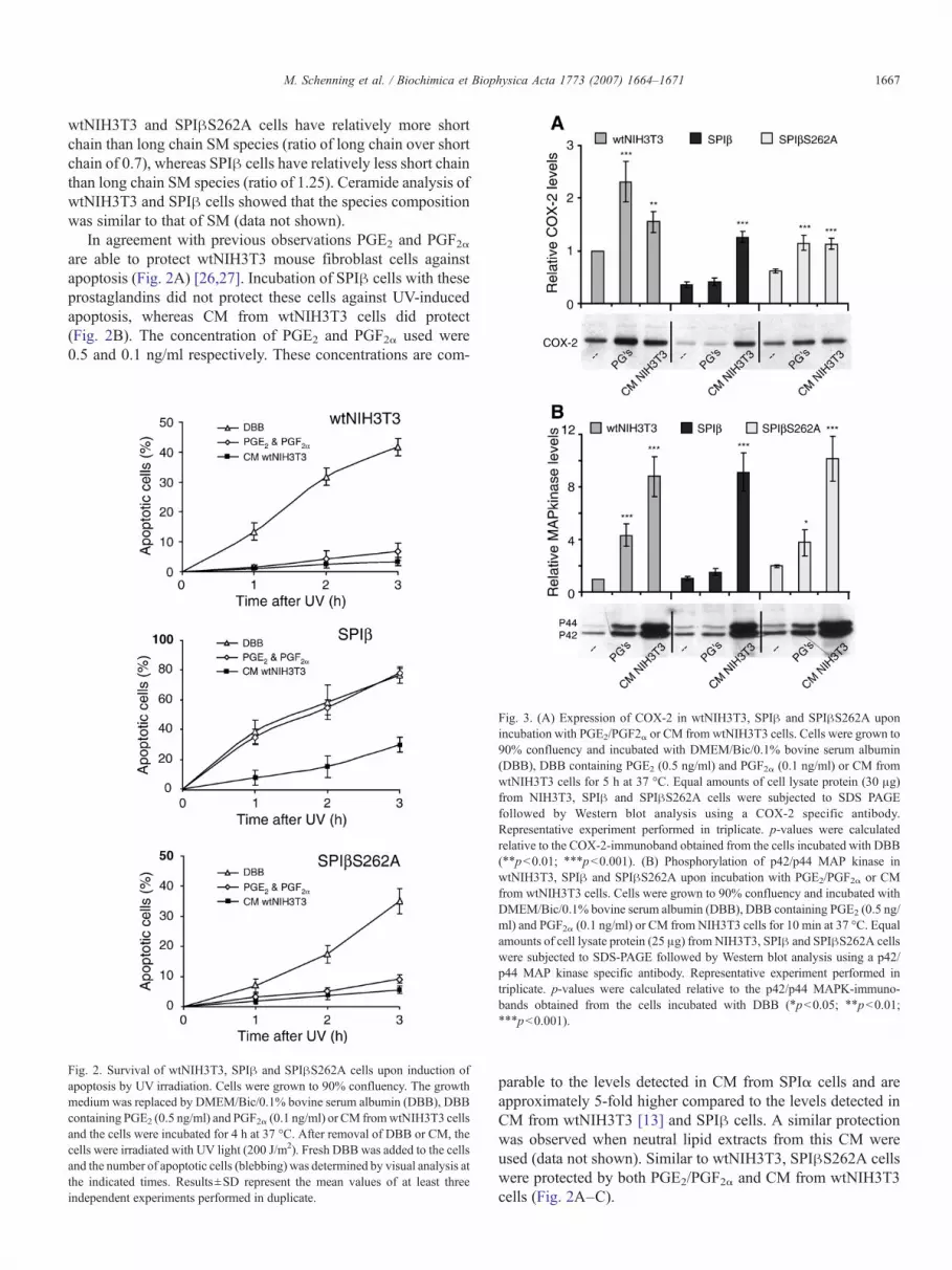
In agreement with previous observations PGE2 and PGF2αare able to protect wtNIH3T3 mouse fibroblast cells againstapoptosis (Fig. 2A) [26,27]. Incubation of SPIβ cells with theseprostaglandins did not protect these cells against UV-inducedapoptosis, whereas CM from wtNIH3T3 cells did protect(Fig. 2B). The concentration of PGE2 and PGF2α used were0.5 and 0.1 ng/ml respectively. These concentrations are com-
Fig. 2. Survival of wtNIH3T3, SPIβ and SPIβS262A cells upon induction ofapoptosis by UV irradiation. Cells were grown to 90% confluency. The growthmedium was replaced by DMEM/Bic/0.1% bovine serum albumin (DBB), DBBcontaining PGE2 (0.5 ng/ml) and PGF2α (0.1 ng/ml) or CM fromwtNIH3T3 cellsand the cells were incubated for 4 h at 37 °C. After removal of DBB or CM, thecells were irradiated with UV light (200 J/m2). Fresh DBB was added to the cellsand the number of apoptotic cells (blebbing) was determined by visual analysis atthe indicated times. Results±SD represent the mean values of at least threeindependent experiments performed in duplicate.
Fig. 3. (A) Expression of COX-2 in wtNIH3T3, SPIβ and SPIβS262A uponincubation with PGE2/PGF2α or CM from wtNIH3T3 cells. Cells were grown to90% confluency and incubated with DMEM/Bic/0.1% bovine serum albumin(DBB), DBB containing PGE2 (0.5 ng/ml) and PGF2α (0.1 ng/ml) or CM fromwtNIH3T3 cells for 5 h at 37 °C. Equal amounts of cell lysate protein (30 μg)from NIH3T3, SPIβ and SPIβS262A cells were subjected to SDS PAGEfollowed by Western blot analysis using a COX-2 specific antibody.Representative experiment performed in triplicate. p-values were calculatedrelative to the COX-2-immunoband obtained from the cells incubated with DBB(⁎⁎pb0.01; ⁎⁎⁎pb0.001). (B) Phosphorylation of p42/p44 MAP kinase inwtNIH3T3, SPIβ and SPIβS262A upon incubation with PGE2/PGF2α or CMfrom wtNIH3T3 cells. Cells were grown to 90% confluency and incubated withDMEM/Bic/0.1% bovine serum albumin (DBB), DBB containing PGE2 (0.5 ng/ml) and PGF2α (0.1 ng/ml) or CM from NIH3T3 cells for 10 min at 37 °C. Equalamounts of cell lysate protein (25 μg) from NIH3T3, SPIβ and SPIβS262A cellswere subjected to SDS-PAGE followed by Western blot analysis using a p42/p44 MAP kinase specific antibody. Representative experiment performed intriplicate. p-values were calculated relative to the p42/p44 MAPK-immuno-bands obtained from the cells incubated with DBB (⁎pb0.05; ⁎⁎pb0.01;⁎⁎⁎pb0.001).
parable to the levels detected in CM from SPIα cells and areapproximately 5-fold higher compared to the levels detected inCM from wtNIH3T3 [13] and SPIβ cells. A similar protectionwas observed when neutral lipid extracts from this CM wereused (data not shown). Similar to wtNIH3T3, SPIβS262A cellswere protected by both PGE2/PGF2α and CM from wtNIH3T3cells (Fig. 2A–C).

Fig. 4. Survival of SPIβ cells upon induction of apoptosis by UV irradiation afterpreincubation with CM from wtNIH3T3, SPIβ and SPIβS262A cells with orwithout heat denaturation. SPIβ cells were grown to 90% confluency. Thegrowth medium was replaced by DMEM/Bic/0.1% bovine serum albumin(DBB) or by CM and the cells were incubated for 4 h at 37 °C. After removal ofDBB the cells were irradiated with UV light (200 J/m2), fresh DBB was added tothe cells and incubated for 3 h at 37 °C. The number of apoptotic cells (blebbing)was determined by visual analysis. Results±SD represent the mean values of atleast three experiments performed in duplicate. p-values were calculated relativeto the survival of SPIβ cells incubated with the untreated conditioned medium(⁎⁎⁎pb0.001).
Fig. 5. (A) Expression of COX-2 in SPIβ cells upon incubation with CM fromwtNIH3T3, SPIβ and SPIβS262Awith or without heat denaturation of the CM.Cells were grown to 90% confluency and incubated for 5 h at 37 °C with CMfrom NIH3T3, SPIβ or SPIβS262A cells, either heat denatured or not. Equalamounts of cell lysate protein (30 μg) from NIH3T3, SPIβ and SPIβS262A cellswere subjected to SDS PAGE followed by Western blot analysis using a COX-2specific antibody. Representative experiment performed in triplicate. p-valueswere calculated relative to the COX-2-immunoband obtained from the SPIβincubated with the untreated conditioned medium (⁎⁎⁎pb0.001). (B)Phosphorylation of p42/p44 MAP kinase in SPIβ cells upon incubation withCM fromwtNIH3T3, SPIβ and SPIβS262Awith or without heat denaturation ofthe CM. Cells were grown to 90% confluency and incubated for 10 min at 37 °Cwith CM from wtNIH3T3, SPIβ or SPIβS262A cells, either heat denatured ornot. Equal amounts of cell lysate protein (25 μg) from NIH3T3, SPIβ andSPIβS262A cells were subjected to SDS PAGE followed by Western blotanalysis using a p42/p44 MAP kinase specific antibody. Representativeexperiment performed in triplicate. p-values were calculated relative to thep42/p44 MAPK-immunobands obtained from the SPIβ incubated with theuntreated conditioned medium (⁎pb .05; ⁎⁎pb0.01; ⁎⁎⁎pb0.001).
1668 M. Schenning et al. / Biochimica et Biophysica Acta 1773 (2007) 1664–1671
3.2. Expression of cyclooxygenase-1 and -2
The anti-apoptotic activity of CM from wtNIH3T3 andSPIα cells is caused by the presence of eicosanoids, thesynthesis of which is (partially) dependent on COX-2 activity[13]. As shown by Western blot analysis, the level of COX-2in SPIβ cells is reduced when compared to wtNIH3T3 cells(Fig. 3A; lanes 1 and 4). Again the SPIβS262A cellsresembled the wild type cells as the levels of COX-2 werecomparable in both cell lines (Fig. 3A; lanes 1 and 7).Arachidonic acid metabolites produced by COX-2 canstimulate the expression of this enzyme via an autocrinepathway [28,29]. By using an ELISA kit we showed that theamount of PGE2 secreted by the SPIβ cells (0.16 ng/ml) issimilar to that of wtNIH3T3 cells (0.1 ng/ml). Therefore thereduced level of COX-2 in SPIβ cells is not linked to adecreased level of PGE2. When SPIβ cells were incubated for5 h with PGE2 in combination with PGF2α, COX-2 levelsremained the same (Fig. 3A; lanes 4 and 5), whereas the COX-2 levels of wtNIH3T3 and SPIβS262A cells were increased(Fig. 3A; lanes 2 and 8). This strongly suggests that theupregulation of COX-2 by PGE2/PGF2α is inhibited in SPIβcells. On the other hand, incubation of SPIβ cells with CMfrom wtNIH3T3 cells did increase the COX-2 levels (Fig. 3A;cf. lanes 4 and 6). A similar upregulation of COX-2 wasobserved for wtNIH3T3 cells (Fig. 3A; cf. lanes 1 and 3) andSPIβS262A cells (Fig. 3A; cf. lanes 7 and 9). This suggeststhat the upregulation of COX-2 in SPIβ cells by CM fromwtNIH3T3 cells may be linked to the increased survival uponUV irradiation under the same conditions. Lack of COX-2induction by PGE2 and PGF2α in SPIβ cells agrees with thefailure to protect these cells (Fig. 2B). In contrast to COX-2,expression levels of COX-1 were similar in all three cell linesand did not change upon incubation with PGs or CM fromwtNIH3T3 cells (data not shown).
3.3. p42/p44-MAP kinase activation
Activation of p42/p44 MAP kinase is commonly observedafter hormone or polypeptide growth factor induced prolifera-tion or cell survival [30–33]. Incubation of wtNIH3T3, SPIβand SPIβS262A cells with CM from wtNIH3T3 cells induceda rapid activation of p42/p44 MAP kinase (Fig. 3B; cf. lanes 3,6, 9). Upon incubation with PGE2 and PGF2α, p42/p44 MAPkinase was activated in wtNIH3T3 and SPIβS262A cells butnot in SPIβ cells (Fig. 3B; cf. lanes 2, 5, 8). These data

Fig. 6. Effect of prostaglandins and conditioned medium on the survival of wtNIH3T3 and SPIβ cells. Prostaglandin E2 and prostaglandin F2α protect wtNIH3T3 andSPIβS262A cells against UV-induced apoptosis but fail to do so with SPIβ cells. The anti-apoptotic activity present in CM from SPIβ cells is masked by the presenceof an antagonist, which is inactivated upon heat treatment. Heat treatment of CM from wtNIH3T3 and SPIβS262A cells has no effect on the anti-apoptotic activity.This suggests that the expression and phosphorylation of PI-TPβ is responsible for the production of the antagonist.
1669M. Schenning et al. / Biochimica et Biophysica Acta 1773 (2007) 1664–1671
support the finding that PGE2 and PGF2α do protectwtNIH3T3 and SPIβS262A cells against apoptosis but failto protect SPIβ cells, indicating that in SPIβ cells this signalpathway for stimulation of proliferation and cell survival isinhibited.
3.4. Secretion of an antagonist of anti-apoptotic activity
SPIα cells secrete a highly potent anti-apoptotic andmitogenic factor(s) [12,13]. Although to a lesser extent,wtNIH3T3 cells also produce bioactive factors. Indeed, whenprior to UV irradiation SPIβ cells were incubated with CM fromwtNIH3T3 cells instead of DBB, the extent of apoptosis wasdecreased by 45% (Fig. 4). Incubation of SPIβ cells with CMfrom SPIβ cells did not prevent apoptosis. However, upon heattreatment (20 min at 80 °C) CM from SPIβ cells expressed ananti-apoptotic activity comparable to that from wtNIH3T3 cells(Fig. 4). As a control, heat treatment of CM from wtNIH3T3and SPIβS262A cells had little effect on the anti-apoptoticactivity indicating that this activity was heat stable. Thisstrongly suggests that CM from SPIβ cells contains a heat-labilefactor (antagonist) that interferes with the anti-apoptotic activitypresent. The addition of CM from SPIβ cells containing theantagonist did not increase the rate of apoptosis in the wild typecells and apoptosis resistant SPIα cells, which suggests that theantagonist has no pro-apoptotic activity. CM from SPIβS262Acells resembled that from wtNIH3T3 cells indicating thatphosphorylation of PI-TPβ is required for the production of theantagonist.
In agreement with the observations for cell survival, CM fromSPIβ cells after heat-treatment (20min 80 °C) induced COX-2 in
SPIβ cells (Fig. 5A; cf. lanes 2 and 6) to the same extent as CMfrom wtNIH3T3 cells (Fig. 5A; cf. lanes 4 and 6) andSPIβS262A cells (Fig. 5A; lanes 6 and 8) whereas the untreatedCM from SPIβ cells had no effect on COX-2 expression (Fig.5A; lane 5). Similarly, the untreated CM from SPIβ has no effecton the level of p42/p44 MAP kinase, whereas the heat-treatedCM from SPIβ cells is able to activate MAPK (Fig. 5B; cf. lanes5 and 6). For comparison, the untreated CM fromwtNIH3T3 andSPIβS262A cells are able to activate MAPK in SPIβ cells (Fig.5B; cf. lanes 3, 4, 7 and 8), resulting in protection of these cellsagainst UV induced apoptosis (Fig. 4).
The effect of PGE2/PGF2α and conditioned media on thesurvival of wtNIH3T3 and SPIβ cells are summarized in Fig. 6.
4. Discussion
Previously we have shown that a ten-fold increase of PI-TPβin wtNIH3T3 mouse fibroblast cells (SPIβ cells) significantlyincreases the sensitivity towards apoptosis induced by UV-radiation [15]. Here we show that SPIβ cells incubated withTNFα and serum starvation are much more prone to apoptosisindicating that the increased apoptotic response was notrestricted to UV irradiation. A role of PI-TPβ in cell survivalwas also indicated by the finding that initially, using anotherexpression vector (pSG5) than the one currently used (pBK-CMV) we failed to obtain stable SPIβ cell lines. Due to highlevels of PI-TPβ we routinely observed that these cells diedafter 4–5 passages. Previously we have shown that PI-TPβ ismainly associated with the Golgi system and that this asso-ciation requires the PKC-dependent phosphorylation of Ser-262as the mutant PI-TPβ(S262A) is present throughout the cell

1670 M. Schenning et al. / Biochimica et Biophysica Acta 1773 (2007) 1664–1671
[3,9,10]. By overexpressing PI-TPβ(S262A) to a level com-parable to that of PI-TPβ (9.0 and 10.6 ng per 100 μg cytosolicprotein, respectively) the ensuing SPIβS262A cells have asensitivity towards apoptosis comparable to that of wtNIH3T3cells [15]. This strongly suggests that there is a relationshipbetween the phosphorylation of PI-TPβ and the sensitivitytowards apoptosis.
On the other hand, recent studies concluded that the Golgilocalization of PI-TPβ is not dependent on the phosphorylationof Ser-262 [34,35]. We do not have an explanation for thisdiscrepancy. However, we do find that increased levels of PI-TPβS262A have no effect on the apoptosis sensitivity and thus,regardless of Golgi localization, it is clear that phosphorylationof PI-TPβ is required to affect apoptosis.
Two important parameters linked to apoptosis are activationof p42/p44 MAP kinase and expression of COX-2 [36–38]. Ingeneral, inhibition of COX-2 expression enhances apoptosis andmore specifically reduces the incidence and progression oftumors in animal models [39–42]. Phosphorylation of p42/p44MAP kinase through the RasNRafNMAP kinase kinase (MKK)cascade is associated with proliferation, protection againstapoptosis and angiogenesis [30–33]. Here we show that theprostaglandins PGE2/PGF2α, although present in CM from SPIβcells, are unable to upregulate COX-2 in SPIβ cells, but are ableto do so in wtNIH3T3 and SPIβS262A cells, showing that thepathway of upregulation is blocked in SPIβ cells (Fig. 3A). Thisresults in a lower basal level of COX-2 in SPIβ cells andprevents prostaglandins (PGs) from protecting SPIβ cellsagainst apoptosis. In addition we show that these PGs cannotactivate p42/p44 MAP kinase in SPIβ cells whereas thispathway is activated in wtNIH3T3 and SPIβS262A cells (Fig.3B). These observations may explain why SPIβ cells are moreprone to apoptosis and why PGE2 and PGF2α are unable toinduce survival in SPIβ cells. CM from SPIβ cells appeared tolack the survival activity present in the CM from NIH3T3 cell.Interestingly, CM from SPIβ cells acquired survival activityupon heating, indicating that the expression of PI-TPβ isresponsible for the production and secretion of a component thatinterferes with the action of the intrinsic survival factors present.Attempts to gain insight into the nature of the PI-TPβ-dependent‘antagonist’ were inconclusive. Experiments using proteinsynthesis inhibitors and analysis of [14C]serine-labeled SMmetabolites secreted by SPIβ cells revealed no significantdifferences compared with control cells.
The relationship between SM metabolism and apoptosis hasbeen investigated extensively. B-cell receptor-triggered apopto-sis is associated with an early rise of C16 ceramide leading to thesubsequent formation of long chain C24 ceramide via activationof effector caspases [43]. Apoptotic stimuli including TNFα[44], ionizing radiation [45] and B-cell receptor cross-linking[43] can generate ceramide by the induction of SM hydrolysisthrough the action of sphingomyelinases or the de novo pathway.In contrast to SPIβS262A and wtNIH3T3 cells, SPIβ cellsmaintain the total level of SM under conditions where SM isdegraded in the plasma membrane to ceramide by exogenoussphingomyelinase [7,9]. Although the mechanism by which PI-TPβ regulates the rapid conversion of ceramide to SM is not
known, the data again strongly suggest that PI-TPβ must bephosphorylated at Ser-262 in order to maintain the cellular SMlevels. Although increased levels of PI-TPβ appear to berequired for SPIβ cells to rapidly convert ceramide to SM [7], wefound that steady-state levels of ceramide in SPIβ andwtNIH3T3 cells are similar. However, we did observe that themolecular species of the fatty acids of SM (Fig. 1B) and cera-mide are different with SPIβ cells having relatively less shortchain (C16) and more long chain species (C24:1/0) comparedwith wtNIH3T3 cells. To date it is not known whether thesensitivity towards apoptosis of SPIβ cells is related to this shiftfrom short chain to long chain ceramide/SM. However, it couldbe that the relative enrichment of long chain SM species in SPIβcells has an effect on the plasma membrane and, thereby, affectthe properties of membrane proteins [46].
We propose that as a consequence of an antagonist blockingof the autocrine action of the survival factor present in CM fromSPIβ cells, the SPIβ cells are more sensitive towards inducedapoptosis (summarized in Fig. 6). In addition, the failure ofPGE2/PGF2α to both activate p42/p44 MAP kinase and to up-regulate COX-2 levels may also explain why SPIβ cells are moreprone to apoptosis (summarized in Fig. 6). At this point our datasuggest that since an increased expression of PI-TPβ promotesapoptosis, the deletion of PI-TPβ may have a prohibitive effecton apoptosis. Since apoptosis is an essential event during earlyembryonic development, the proposed role of PI-TPβ inapoptosis may explain why the generation of a PI-TPβ knock-out mouse has failed [6]. Understanding why and how ex-pression of a single protein decreases the rate of proliferation aswell as survival of cells might be of interest for research onmethods to decrease the growth of rapidly proliferating tumorcells that have gained resistance against induction of apoptosis.
References
[1] S. Tanaka, S. Yamashita, K. Hosaka, Cloning and expression of humancDNA encoding phosphatidylinositol transfer protein beta, Biochim.Biophys. Acta 1259 (1995) 199–202.
[2] K.W. Wirtz, Phospholipid transfer proteins revisited, Biochem. J. 324(Pt 2) (1997) 353–360.
[3] K.J. De Vries, J. Westerman, P.I. Bastiaens, T.M. Jovin, K.W. Wirtz, G.T.Snoek, Fluorescently labeled phosphatidylinositol transfer protein iso-forms (alpha and beta), microinjected into fetal bovine heart endothelialcells, are targeted to distinct intracellular sites, Exp. Cell Res. 227 (1996)33–39.
[4] V.A. Bankaitis, Cell biology. Slick recruitment to the Golgi, Science 295(2002) 290–291.
[5] J.G. Alb Jr., J.D. Cortese, S.E. Phillips, R.L. Albin, T.R. Nagy, B.A.Hamilton, Mice lacking phosphatidylinositol transfer protein-alpha exhibitspinocerebellar degeneration, intestinal and hepatic steatosis, andhypoglycemia, J. Biol. Chem. 278 (2003) 33501–33518.
[6] J.G. Alb Jr., S.E. Phillips, K. Rostand, X. Cui, J. Pinxteren, L. Cotlin, T.Manning, S. Guo, J.D. York, H. Sontheimer, J.F. Collawn, V.A. Bankaitis,Genetic ablation of phosphatidylinositol transfer protein function inmurine embryonic stem cells, Mol. Biol. Cell 13 (2002) 739–754.
[7] C.M. Van Tiel, C. Luberto, G.T. Snoek, Y.A. Hannun, K.W. Wirtz, Rapidreplenishment of sphingomyelin in the plasma membrane upon degrada-tion by sphingomyelinase in NIH3T3 cells overexpressing the phospha-tidylinositol transfer protein beta, Biochem. J. 346 (Pt 2) (2000) 537–543.
[8] K.J. de Vries, A.A. Heinrichs, E. Cunningham, F. Brunink, J. Westerman,P.J. Somerharju, S. Cockcroft, K.W. Wirtz, G.T. Snoek, An isoform of the
1671M. Schenning et al. / Biochimica et Biophysica Acta 1773 (2007) 1664–1671
phosphatidylinositol-transfer protein transfers sphingomyelin and isassociated with the Golgi system, Biochem. J. 310 (Pt 2) (1995) 643–649.
[9] C.M. van Tiel, J. Westerman, M.A. Paasman, M.M. Hoebens, K.W. Wirtz,G.T. Snoek, The Golgi localization of phosphatidylinositol transfer proteinbeta requires the protein kinase C-dependent phosphorylation of serine 262and is essential for maintaining plasma membrane sphingomyelin levels,J. Biol. Chem. 277 (2002) 22447–22452.
[10] G.T. Snoek, C.M. Van Tiel, M.R. Egmond, Structure–function relation-ships of phosphatidylinositol transfer proteins: involvement of phosphory-lation sites, Biochimie 86 (2004) 857–864.
[11] V.A. Bankaitis, J.R. Aitken, A.E. Cleves, W. Dowhan, An essential role fora phospholipid transfer protein in yeast Golgi function, Nature 347 (1990)561–562.
[12] G.T. Snoek, C.P. Berrie, T.B. Geijtenbeek, H.A. van der Helm, J.A. Cadee,C. Iurisci, D. Corda, K.W. Wirtz, Overexpression of phosphatidylinositoltransfer protein alpha in NIH3T3 cells activates a phospholipase A, J. Biol.Chem. 274 (1999) 35393–35399.
[13] M. Schenning, C.M. van Tiel, D. Van Manen, J.C. Stam, B.M. Gadella,K.W. Wirtz, G.T. Snoek, Phosphatidylinositol transfer protein alpha regu-lates growth and apoptosis of NIH3T3 cells: involvement of a cannabinoid1-like receptor, J. Lipid Res. 45 (2004) 1555–1564.
[14] H. Bunte, M. Schenning, P. Sodaar, D.P. Bar, K.W. Wirtz, F.L. vanMuiswinkel, G.T. Snoek, A phosphatidylinositol transfer protein alpha-dependent survival factor protects cultured primary neurons against serumdeprivation-induced cell death, J. Neurochem. 97 (2006) 707–715.
[15] C.M. van Tiel, M. Schenning, G.T. Snoek, K.W. Wirtz, Overexpression ofphosphatidylinositol transfer protein beta in NIH3T3 cells has astimulatory effect on sphingomyelin synthesis and apoptosis, Biochim.Biophys. Acta 1636 (2004) 151–158.
[16] H.H. Tai, C.L. Tai, C.S. Hollander, Biosynthesis of prostaglandins in rabbitkidney medulla. Properties of prostaglandin synthase, Biochem. J. 154(1976) 257–264.
[17] M.M. Bradford, A rapid and sensitive method for the quantitation ofmicrogram quantities of protein utilizing the principle of protein-dyebinding, Anal. Biochem. 72 (1976) 248–254.
[18] E.G. Bligh, W.J. Dyer, A rapid method of total lipid extraction andpurification, Can. J. Biochem. Physiol. 37 (1959) 911–917.
[19] C. Luberto, Y.A. Hannun, Sphingomyelin synthase, a potential regulatorof intracellular levels of ceramide and diacylglycerol during SV40 trans-formation. Does sphingomyelin synthase account for the putativephosphatidylcholine-specific phospholipase C? J. Biol. Chem. 273(1998) 14550–14559.
[20] L. Colombaioni, M. Garcia-Gil, Sphingolipid metabolites in neuralsignalling and function, Brain Res. Brain Res. Rev. 46 (2004) 328–355.
[21] B. Ogretmen, Y.A. Hannun, Biologically active sphingolipids in cancerpathogenesis and treatment, Nat. Rev., Cancer 4 (2004) 604–616.
[22] Y.A. Hannun, L.M. Obeid, Mechanisms of ceramide-mediated apoptosis,Adv. Exp. Med. Biol. 407 (1997) 145–149.
[23] W.J. van Blitterswijk, A.H. van der Luit, R.J. Veldman, M. Verheij, J.Borst, Ceramide: second messenger or modulator of membrane structureand dynamics? Biochem. J. 369 (2003) 199–211.
[24] B. Ramstedt, J.P. Slotte, Separation and purification of sphingomyelindiastereomers by high-performance liquid chromatography, Anal. Bio-chem. 282 (2000) 245–249.
[25] B. Ramstedt, J.P. Slotte, Membrane properties of sphingomyelins, FEBSLett. 531 (2002) 33–37.
[26] H. Sugiura, X. Liu, S. Togo, T. Kobayashi, L. Shen, S. Kawasaki, K.Kamio, X.Q. Wang, L.J. Mao, S.I. Rennard, Prostaglandin E(2) protectshuman lung fibroblasts from cigarette smoke extract-induced apoptosis viaEP(2) receptor activation, J. Cell. Physiol. 210 (2007) 99–110.
[27] K.M. Leahy, R.L. Ornberg, Y. Wang, B.S. Zweifel, A.T. Koki, J.L.Masferrer, Cyclooxygenase-2 inhibition by celecoxib reduces proliferationand induces apoptosis in angiogenic endothelial cells in vivo, Cancer Res.62 (2002) 625–631.
[28] M.A. Iniguez, A. Rodriguez, O.V. Volpert, M. Fresno, J.M. Redondo,Cyclooxygenase-2: a therapeutic target in angiogenesis, Trends Mol. Med.9 (2003) 73–78.
[29] D. Parolaro, P. Massi, T. Rubino, E. Monti, Endocannabinoids in theimmune system and cancer, Prostaglandins Leukot. Essent. Fat. Acids 66(2002) 319–332.
[30] J.N. Lavoie, N. Rivard, G. L'Allemain, J. Pouyssegur, A temporal andbiochemical link between growth factor-activated MAP kinases, cyclin D1induction and cell cycle entry, Prog. Cell Cycle Res. 2 (1996) 49–58.
[31] M. Le Gall, J.C. Chambard, J.P. Breittmayer, D. Grall, J. Pouyssegur, E.Van Obberghen-Schilling, The p42/p44 MAP kinase pathway preventsapoptosis induced by anchorage and serum removal, Mol. Biol. Cell 11(2000) 1103–1112.
[32] E. Berra, J. Milanini, D.E. Richard, M. Le Gall, F. Vinals, E. Gothie, D.Roux, G. Pages, J. Pouyssegur, Signaling angiogenesis via p42/p44 MAPkinase and hypoxia, Biochem. Pharmacol. 60 (2000) 1171–1178.
[33] G. Pages, J. Milanini, D.E. Richard, E. Berra, E. Gothie, F. Vinals, J.Pouyssegur, Signaling angiogenesis via p42/p44 MAP kinase cascade,Ann. N. Y. Acad. Sci. 902 (2000) 187–200.
[34] S.E. Phillips, K.E. Ile, M. Boukhelifa, R.P. Huijbregts, V.A. Bankaitis,Specific and nonspecific membrane-binding determinants cooperate intargeting phosphatidylinositol transfer protein beta-isoform to themammalian trans-Golgi network, Mol. Biol. Cell 17 (2006) 2498–2512.
[35] C.P. Morgan, V. Allen-Baume, M. Radulovic, M. Li, A. Skippen, S.Cockcroft, Differential expression of a C-terminal splice variant ofphosphatidylinositol transfer protein beta lacking the constitutive-phos-phorylated Ser262 that localizes to the Golgi compartment, Biochem. J.398 (2006) 411–421.
[36] M. Hetman, K. Kanning, J.E. Cavanaugh, Z. Xia, Neuroprotection bybrain-derived neurotrophic factor is mediated by extracellular signal-regulated kinase and phosphatidylinositol 3-kinase, J. Biol. Chem. 274(1999) 22569–22580.
[37] L.J. Crofford, COX-1 and COX-2 tissue expression: implications andpredictions, J. Rheumatol., Suppl. 49 (1997) 15–19.
[38] A. Hara, N. Yoshimi, M. Niwa, N. Ino, H. Mori, Apoptosis induced by NS-398, a selective cyclooxygenase-2 inhibitor, in human colorectal cancercell lines, Jpn. J. Cancer Res. 88 (1997) 600–604.
[39] Y. Cao, S.M. Prescott, Many actions of cyclooxygenase-2 in cellulardynamics and in cancer, J. Cell. Physiol. 190 (2002) 279–286.
[40] X.Z. Ding, W.G. Tong, T.E. Adrian, Blockade of cyclooxygenase-2inhibits proliferation and induces apoptosis in human pancreatic cancercells, Anticancer Res. 20 (2000) 2625–2631.
[41] T. Hida, K. Kozaki, H. Muramatsu, A. Masuda, S. Shimizu, T.Mitsudomi, T. Sugiura, M. Ogawa, T. Takahashi, Cyclooxygenase-2inhibitor induces apoptosis and enhances cytotoxicity of variousanticancer agents in non-small cell lung cancer cell lines, Clin. CancerRes. 6 (2000) 2006–20011.
[42] A.L. Hsu, T.T. Ching, D.S. Wang, X. Song, V.M. Rangnekar, C.S. Chen,The cyclooxygenase-2 inhibitor celecoxib induces apoptosis by blockingAkt activation in human prostate cancer cells independently of Bcl-2, J.Biol. Chem. 275 (2000) 11397–11403.
[43] B.J. Kroesen, S. Jacobs, B.J. Pettus, H. Sietsma, J.W. Kok, Y.A. Hannun,L.F. de Leij, BcR-induced apoptosis involves differential regulation of C16and C24-ceramide formation and sphingolipid-dependent activation of theproteasome, J. Biol. Chem. 278 (2003) 14723–14731.
[44] L.M. Obeid, C.M. Linardic, L.A. Karolak, Y.A. Hannun, Programmed celldeath induced by ceramide, Science 259 (1993) 1769–1771.
[45] P. Santana, L.A. Pena, A. Haimovitz-Friedman, S. Martin, D. Green, M.McLoughlin, C. Cordon-Cardo, E.H. Schuchman, Z. Fuks, R. Kolesnick,Acid sphingomyelinase-deficient human lymphoblasts and mice aredefective in radiation-induced apoptosis, Cell 86 (1996) 189–199.
[46] P.S. Niemela, M.T. Hyvonen, I. Vattulainen, Influence of chain lengthand unsaturation on sphingomyelin bilayers, Biophys. J. 90 (2006)851–863.


