Embed Size (px)
Citation preview
Exp Brain Res (1994) 101: 216-230 �9 Springer-Verlag 1994
Istv/m Benedeczky - Elek Molnfir �9 P6ter Somogyi
The cisternal organelle as a Ca2+-storing compartment associated with GABAergic synapses in the axon initial segment of hippocampal pyramidal neurones
Received: 2 March 1994 / Accepted: 13 June 1994
Abstract The axon initial segment of cortical principal neurones contains an organelle consisting of two to four stacks of flat, membrane-delineated cisternae alternat- ing with electron-dense, fibrillar material. These cister- nal organelles are situated predominantly close to the synaptic junctions of GABAergic axo-axonic cell termi- nals. To examine the possibility that the cisternal or- ganelle is involved in Ca 2+ sequestration, we tested for the presence of CaZ+-ATPase in the cisternal organelles of pyramidal cell axons in the CA1 and CA3 regions of the hippocampus. Electron microscopic immunocyto- chemistry using antibodies to muscle sarcoplasmic reticulum ATPase revealed immunoreactivity associat- ed with cisternal organelle membranes. The localisation of CaZ+-ATPase in cisternal organelles was also con- firmed by enzyme cytochemistry, which produced reac- tion product in the lumen of the cisternae. These exper- iments provide evidence for the presence of a Ca 2+ pump in the cisternal organelle membrane, which may play a role in the sequestration and release of C a 2+. Cisternal organelles are very closely aligned to the axolemma and the outermost cisternal membrane is connected to the plasma membrane by periodic elec- tron-dense bridges as detected in electron micrographs. It is suggested that the interface acts as a voltage sensor, r e l eas ing C a 2+ from cisternal organelles upon depolari- sation of the axon initial segment, in a manner similar to the sarcoplasmic reticulum of skeletal muscle. The in- crease in intra-axonal C a 2+ may regulate the G A B A A receptors associated with the axo-axonic cell synapses, and could affect the excitability of pyramidal cells.
Key words Calcium �9 Calcium-activated ATP-ase GABA. Immunocytochemistry �9 Synapse
I. Benedeczky - E. Molnfir �9 P. Somogyi ([El) Medical Research Council, Anatomical Neuropharmacology Unit, Oxford University, Mansfield Road, Oxford OX1 3TH, UK; FAX no: +44-865-271647
Introduction
The axon initial segment (AIS) of cortical and hippo- campal principal neurones contains a structure called the cisternal organelle, consisting of stacks of mem- brane-delineated flat stacks interspersed with an elec- tron-dense granular material (Kosaka 1980; Palay et al. 1968; Peters et al. 1968; Sloper and Powell 1979; Som- ogyi et al. 1983b). The axon initial segment and these organelles can be recognised in the electron microscope on the basis of their fine structural characteristics (Pe- ters et al. 1991). The cisternal organelle is similar struc- turally, and perhaps also functionally, to the spine ap- paratus which has been shown to accumulate and store calcium (Andrews et al. 1988), and has inositol 1,4,5- trisphosphate receptor-activated Ca 2§ channels in its membrane (Satoh et al. 1990; Takei et al. 1992). The spine apparatus therefore is thought to be involved in the sequestration of calcium entering the cell and/or in the release of endogenous calcium mediated by second messengers (see review, Rossier and Putney 1992). Noth- ing is known about the chemistry of the analogous or- ganelle in the axon initial segment of pyramidal cells, which have another feature unique to cortex; they re- ceive dense GABAergic input from the so called axo-ax- onic cells (Somogyi 1977; Somogyi et al. 1983a, b; 1985).
Is it possible that the two unique features of pyrami- dal cell AISs, namely the presence of cisternal organelles and the dense GABAergic input, are related ? The iden- tification of the molecular structure of the cisternal or- ganelle and the AIS would help to answer this question. As a first step, we began testing the cisternal organelle for the presence of markers indicating Ca 2§ sequestra- tion, such as the enzyme Ca2+-ATPase, which is respon- sible for pumping calcium across membranes against the electrochemical gradient (see Grover and Khan 1992). Electron microscopic immunocytochemistry and enzyme histochemistry were used to localise Ca 2+- ATPase.

Materials and methods
Immunocytochemistry
Preparation of animals and tissue
Six adult female or male Wistar rats (100~400 g) were deeply anaesthetised with sodium pentobarbital (150 mg/kg, i.p.). They were perfused through the aorta for 10-30 rain with NaC1 solution (0.9% for 1 min) followed by ice-cold fixative containing 4% paraformaldehyde, 0.025% glutaraldehyde and approximately 0.2% picric acid (Somogyi and Takagi 1982) made up in 0.1 M phosphate buffer (PB, pH 7.2).
Immunoreaction
The rabbit polyclonal antiserum (designated EM-3) was raised against sarcoplasmic reticulum Ca2+-ATPase, which was purified from rat gastrocnemius muscle (Molnfir et al. 1990). Previous re- ports on this antiserum established its specificity to isoenzymes of several sarcoplasmic and endoplasmic Ca2+-ATPase, but it has not been found to react with the plasma membrane, calcium pump proteins (Molnfir et al. 1990, 1992; Sarkadi et al. 1988). Par- tial proteolysis and immunoblot analysis revealed antiserum reac- tion with all major fragments of the sarcoplasmic reticulum Ca 2§ ATPase (Molnfir et al. 1990). Affinity purified antibodies were also used in some immunochemical experiments, and they gave identi- cal and very specific results with the appropriately diluted full serum (E. Moln~r, unpublished observations). The antiserum was specific for Ca 2 § and dit not react with Na +, K § or H +, K +-ATPase (Molnfir et al. 1992). The immunoreactivity of Ca 2+-ATPase was preserved after fixation of tissue samples (Kre- nfics et al. 1989).
Immunocytochemical procedures were carried out as de- scribed earlier (Molnfir et al. 1993). Antiserum to the sarcoplasmic reticulum Ca ;+-ATPase was used at a final dilution of 1 : 1 000. Primary antibodies were localised by immunoperoxidase reaction using the avidin biotinylated horseradish peroxidase complex method. Sections were incubated for 1 h in a mixture of biotinylat- ed goat anti-rabbit IgG (diluted 1 : 50, Vector Labs, Peterborough, England), followed by washing and incubation in avidin-biotiny- lated horseradish peroxidase complex solution (Vector). Peroxi- dase was revealed using 3,3' diaminobenzidine tetrahydrochloride (DAB, 0.015%) dissolved in 50 mM Tris-HC1 (pH 7.2) and H202 (0.01%). Sections were routinely processed either for light or elec- tron microscopic examination (Moln/tr et al. 1993).
In control incubations, the primary antiserum was replaced by normal rabbit serum at the same dilution. Selective distribution of labelling was not observed in the controls, indicating that the reactions described in this paper are due to the primary antiserum to Caa+-ATPase.
Enzyme cytochemistry for Ca2+-ATPase
Five Wistar rats were perfused via the heart with 4% paraformaldehyde solution in 0.1 M cacodylate buffer (pH 7.2) for 40 min at room temperature. After removing the brain, 70-gm- thick sections were cut from the hippocampus and cerebellum using a vibratome. The sections were washed overnight in cold 0.1 M cacodylate buffer, followed by 30 rain washing in 250 mM glycine-NaOH buffer (pH 9.0). Incubation for Ca2+-ATPase was carried out in a water bath at 37 ~ C for 3040 rain according to Ando et al. (1981). The incubation medium consisted of 250 mM glycine-NaOH buffer (pH 9.0), 3 mM ATP (Sigma), 10 mM CaC12, 2 mM lead citrate and 10 mM levamisole (L[-]-2,3,5,6,-Tetrahy- dro-6-phenylimidazo[2,1-b]-thiazole, Sigma) for the inhibition of non-specific alkaline phosph~itase. After incubation, the sections were washed for 5 min in 0.1 M cacodylate buffer (pH 7.2). Post- fixation was carried out in 2% OsO4 buffered with 0.1 M cacody- late buffer to pH 7.2. Staining with 1% ura@l acetate was per-
217
formed for 30 rain during dehydration in the 70% alcohol stage. Samples were embedded in epoxy resin (Durcupan ACM, Fluka).
Controls included omitting (a) ATP, (b) Ca 2+ or (c) both ATP and Ca 2 § from the incubation medium. Under control conditions no significant electron dense deposit was observed in the tissue indicating that the electron-dense deposit obtained with the full medium is due to Ca2+-ATPase. In order to increase the tissue ATP content, in some experiments, pre-incubation was applied in 3 mM ATP-Na dissolved in 250 mM glycine-NaOH buffer (pH 9.0) prior to changing to the full incubation medium. No consis- tent improvement was observed in the reaction in intracellular organelles. In some experiments, 0.5% or 5% DMSO was added to the pre-incubation solution to increase the penetration of ATP, but no significant improvement was observed.
Results
Fine structural organisat ion of the cisternal organelle in the IS of pyramida l neurones
Axon initial segments are recognised on the basis of the electron-dense, m e m b r a n e undercoat ing on the cyto- plasmic face of the axonal m e m b r a n e (Fig. 1A) and by the presence of fasciculated microtubules (Peters et al. 1991). As described in earlier studies, the axon initial segments of cortical principal cells, i.e. pyramida l cells, spiny stellate cells and dentate granule cells also fre- quently contain cisternal organelles (Kosaka 1980; Palay et al. 1968; Peters et al. 1968; Sloper and Powell 1979; Somogyi et al. 1983b). In convent ional electron microscopic material , cisternal organelles consist of two to four stacks of flat, membrane-de l inea ted cisternae al- ternating with electron-dense fibrillar material which is separated f rom the cisternal m e m b r a n e by an electron- lucent gap on bo th sides (Fig. 1). The lumen of individu- al cisternae is variable and can be as nar row as abou t 5-10 nm (Fig. 1B).
Cisternal organelles are usually situated near GABAerg ic synapses at the per iphery of the axoplasm, but they extend beyond the area of any given synapse. Since cisternal organelles are larger structures than synapses, the two may not be present in all single elec- t ron microscopic sections, even if they were associated. Since AISs in the rat h ippocampus are densely, but un- evenly, covered by synapses, complete three dimension- al reconstruct ion would be necessary to establish quan- titatively the associat ion between cisternal organelles and synaptic junctions. This was not carried out in the present study, nevertheless it emerged that it is much easier to find cisternal organelles near synapses than in non-synapt ic regions of the AIS. The ou te rmos t cister- nal m e m b r a n e forms close associations with the p lasma membrane . The postsynapt ic junct ional area of the ax- onal m e m b r a n e is not contacted by the cisternal or- ganelles which often lie just to the side of the synapse (Fig. 1). In longitudinal sections of the axon, when cisternal organelles can be seen along their long axis the junct ions with the p lasma m e m b r a n e are found periodi- cally (Fig. 1A). The distance between the ou te rmos t cisternal m e m b r a n e and the axo lemma is abou t 10- 20 nm and, in some planes of sectioning, periodic elec-

218
Fig. 1 Cisternal organelles (co) shown in longitudinal (A) and cross-sections (B) in the axon initial segment (ais) of hippocampal pyramidal neurones in the CA1 area. In A the organelle consists of three cisternae (1 3) and two intervening electron dense fibrillar bands. Notice repeated close association (double arrows) of the cisternal organelle in A with the plasma membrane in the vicinity of synaptic junctions (asterisks). Periodic bridges (vertical arrows in B) connect the outermost cisternal membrane to the plasma membrane. In this example, the two membranes of one (1) of two cisternae (1, 2) are partly fused. The intercisternal, electron-dense band is indicated by double arrow in B. The axon initial segment can be identified by the presence of the electron-dense membrane undercoating (e.g. between single arrows in A). Scale bars A 0.5 gm, B 0.1 gm
tron-dense bridges can be seen joining the two mem- branes (Fig. 1B). In contrast to cisternal stacks in the somata and dendrites of Purkinje cells (Rusakov et al. 1993), the bridges in the initial segment of pyramidal cells are not present between cisternae instead, as men- t ioned above, they are separated by electron-dense fibrillar material.
Cisternal organelles are almost always present in the axonal spines which are found frequently on hippocam- pal principal neurones (Figs. 3, 4). In contrast to den- dritic spines, in axonal spines the cisternal organelle membrane forms close association with the plasma membrane (Somogyi et al. 1983 b, Fig. 8A).
Electron microscopic localization of Ca2+-ATPase immunoreact ivi ty in s tratum pyramidale of rat h ippocampus
Immunoreact ive sites were identified by the electron- dense, immunoperoxidase reaction product. Immunore- activity to Ca2+-ATPase was evident in somata, den- drites, dendritic spines and axon initial segments of hippocampal neurones, and also in some axon terminals (Figs. 2-4). The immunoperoxidase product was always associated with endomembranes in these structures; such as the Golgi apparatus and the endoplasmic reticu- lum. Some interneurones displayed particularly strong
219
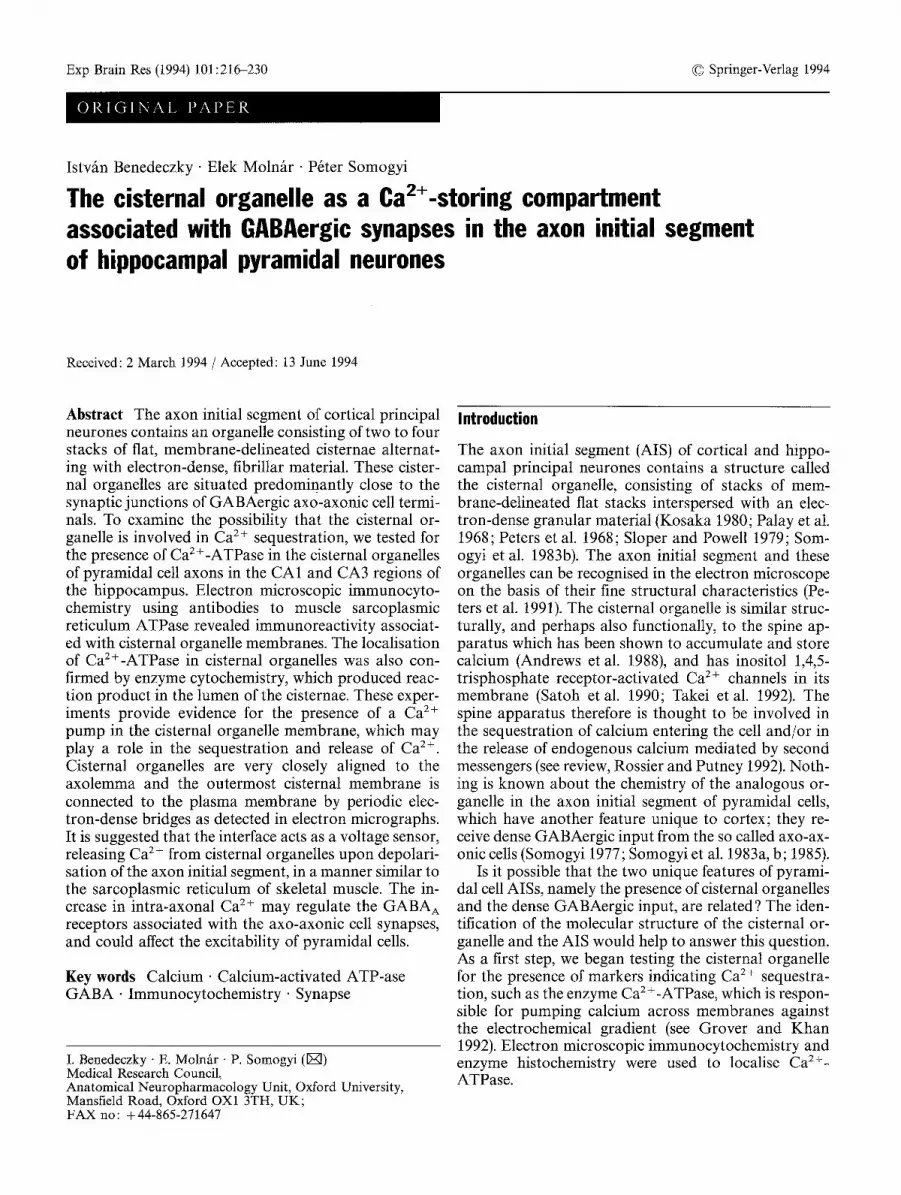
Fig. 2A-C Immunoreactivity for Ca2 +-ATPase is associated with cisternal organelles (co) in axon initial segments (ais) as demon- strated by immunoperoxidase reaction. A Immunolabelling oc- curs either in the lumen (col) or in the pericisternal space (co2). B The close association of col with a synaptic junction (asterisk) is
shown at higher magnification. Only the inner cisternal lumen contains reaction endproduct. C An axonal protrusion containing an immunopositive cisternal organelle (co) receives two synaptic junctions (asterisks). Scale bars A 0.5 gm, B 0.1 gm, C 0.2 gm

220
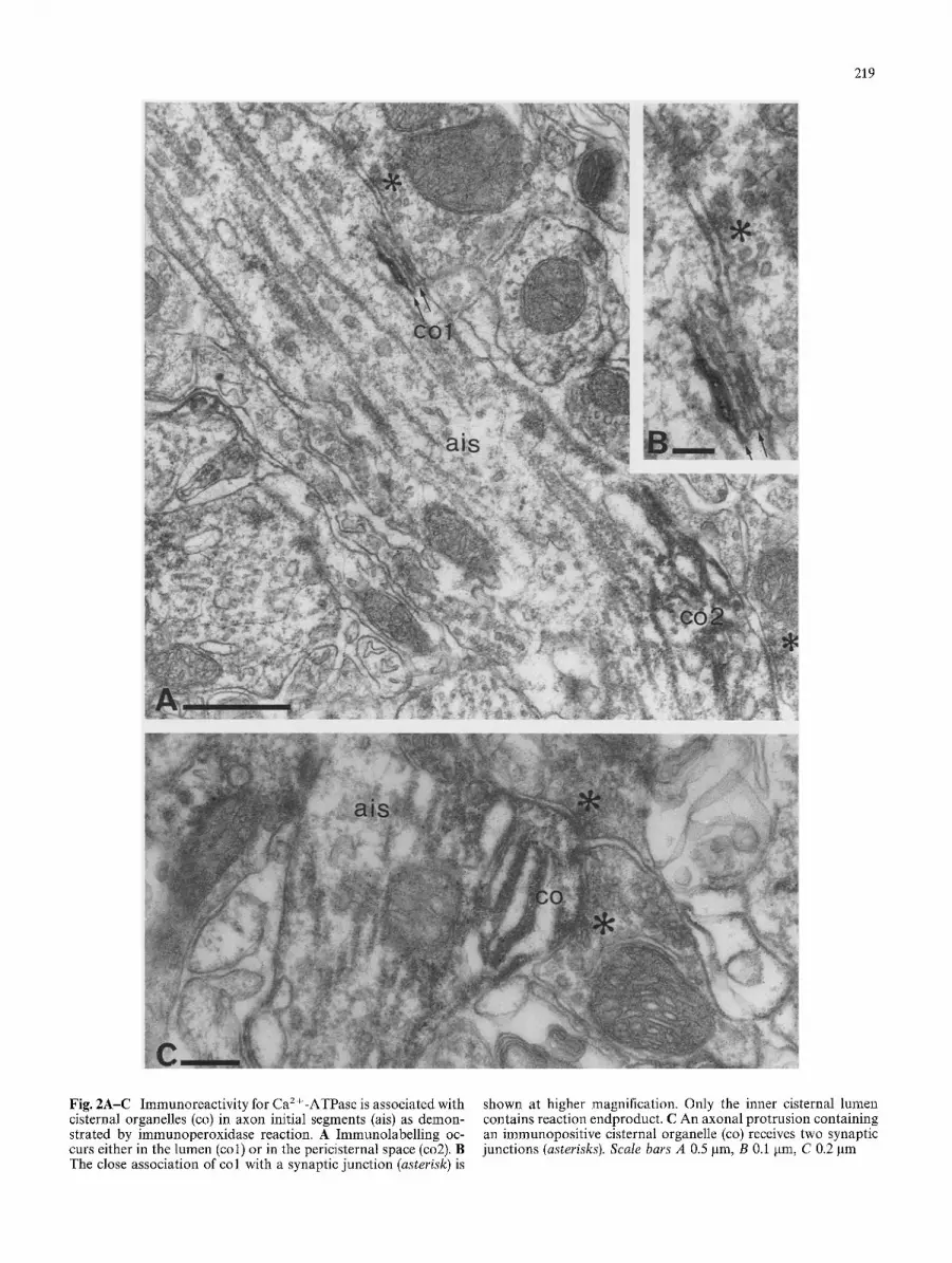
Fig. 3 Immunocytochemical localization of Ca2+-ATPase in the axon initial segments (ais) of pyramidal neurones. A Pericisternal immunolabelling is prominent in two cisternal organelles (co), one of them adjacent to a synaptic bouton (asterisk). B Immunola- belling in an axon initial segment emitting spines (s). Framed area is shown in Fig. 4. at higher magnification. Labelled processes are also found in the neuropil. Scale bars 0.5 ~tm
immunoreact ivi ty (not illustrated). As one of the posi- tive controls, cerebellar Purkinje cells were also analysed in the electron microscope, since the localisa- tion of Ca2+-ATPase has been studied extensively in these cells (e.g. Plessers et al. 1991; Takei et al. 1992). Strong immunoreact ivi ty was associated with the endo- plasmic reticulum (not illustrated), supporting the suit- ability of this antiserum for the localisation of vesicular CaZ+-ATPase.
221
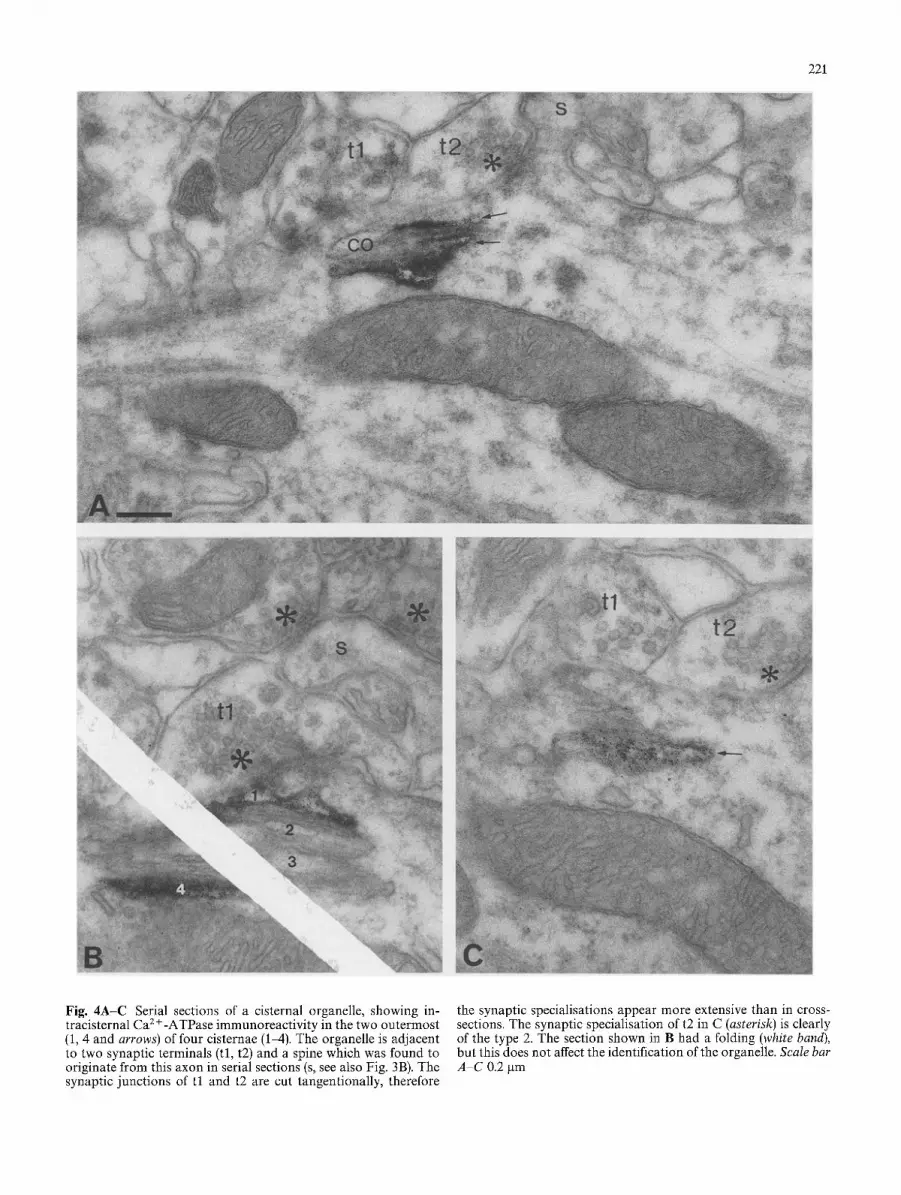
Fig. 4A-C Serial sections of a cisternal organelle, showing in- tracisternal Ca 2 +-ATPase immunoreactivity in the two outermost (1, 4 and arrows) of four cisternae (1-4). The organelle is adjacent to two synaptic terminals (tl, t2) and a spine which was found to originate from this axon in serial sections (s, see also Fig. 3B). The synaptic junctions of t l and t2 are cut tangentionally, therefore
the synaptic specialisations appear more extensive than in cross- sections. The synaptic specialisation of t2 in C (asterisk) is clearly of the type 2. The section shown in B had a folding (white band), but this does not affect the identification of the organelle. Scale bar A-C 0.2 gm

222
In the following, we focus on the axon initial seg- ments of pyramidal cells, which are the main objects of this study. Therefore, reactivity in other structures is not illustrated separately and is only mentioned when it helps to interpret reaction in the AIS. No difference was observed between the CA1 and CA3 regions, therefore the results are described together. Immunoreactivity was most frequently localised in association with cister- nal organelles (Figs. 2 4). The lumen of cisternae were wider in immunoreacted material than in tissue pro- cessed without immunoreaction. In most cases, im- munoreaction end-product was distributed between the cisternae leaving the lumen free (Figs. 2A, C, 3A). Since HRP reaction product does not spread through the membrane, this reaction predicts epitope(s) on the cyto- plasmic side of the cisternal membrane. In some cases, the lumen of the cisternae contained the reaction product indicating epitope(s) on the luminal side of the cisternal membrane (Figs. 2A, B, 3B, 4). The two types of immunoreactivity were present in the same sections and could also be seen in the same axon (Fig. 2A), and occasionally at the same cisternal organelle. The locali- sation of the endproduct on both sides of the membrane was also detected at sites other than the axon initial segment, and the distribution of labelling is in line with the properties of the polyclonal antiserum used in the present study, which recognises all major proteolytic peptide fragments of the enzyme (Molnfir et al. 1990; Sarkadi et al. 1988). Thus, it is likely that several epi- topes, present at both sides of the cisternal membrane, contributed to the electron microscopically detected, immunoreaction product. When the reaction product was located in the cisternal lumen, usually only the out- ermost cisternae - those closest to and/or furthest from the plasma membrane - showed reaction as detected in serial sections (Fig. 4). Some immunoreactivity was also detected along the microtubules of axons (Fig. 3B).
In the vicinity of all immunoreactive cisternal or- ganelles, the axon received synaptic contacts (Figs. 2-4). These synaptic contacts resembled those type 2 junc- tions reported earlier (Kosaka 1980; Somogyi etal. 1983a) and shown to be provided by boutons im- munopositive for glutamate decarboxylase (Somogyi et al. 1983b) or originate from GABA immunopositive neurones (Somogyi et al. 1985). Boutons on AISs stud- ied in conventional material, with few exemptions (Kosaka 1980), usually contain pleomorphic vesicles. The shape of synaptic vesicles in freeze-thawed, peroxi- dase-reacted material can be different from those ob- served in conventional electron microscopic material and boutons containing largely round vesicles were also seen in our sections.
Enzyme cytochemical localisation of Ca2+-ATPase in the stratum pyramidale of rat hippocampus
The method of Ando et al. (1981) has been used exten- sively at the electron microscopic level in many tissues, including the brain, for the localisation of Ca2+-depen - dent, ATPase activity (Cohen and Kriho 1991; Maggio et al. 1991; Mata and Fink, 1989; Maxwell et al. 1991; Nasu and Inomata 1990; Soji et al. 1991). The reaction product of the Ca2+-dependent ATP hydrolysis is a highly electron-dense precipitate of lead phosphate which was found both extra- and intracellularly in the hippocampus. The reaction varied greatly with depth in the incubated thick sections, indicating limited penetra- tion of the reagents. The reaction product present in the extracellular space generally consisted of larger clumps than the intracellular product, which was usually finely granulated (Figs. 5-7). It has been suggested that the large amount of reaction product precipitated in extra- cellular spaces is produced by ecto-ATPase, and not by the plasma membrane Ca 2+ pump (Kortje et al. 1990), but this possibility should not affect the intracellular Ca2+-ATPase localisation. Confusion with other ATPase activities, not depending on the presence of Ca 2+, is unlikely under our reaction conditions (Ando et al. 1981); for example, Na +, K+-ATPase is not acti- vated in the absence of K--.
Readily detectable reaction product appeared in the perikarya, as well as in the processes of neurones. Only a limited number of pyramidal cells showed intracellu- lar reactivity for Ca2+-ATPase probably due to the lim- ited penetration of the substrate. The intracellular en- zyme reaction product was localised to membrane de- lineated spaces of the Golgi apparatus and endoplasmic reticulum (Fig. 5A). Some interneurones and glial cells showed very heavy deposition of a coarse reaction product which filled their cytoplasm and processes, in- cluding interneurone dendrites (Fig. 5B). The synaptic vesicles and smooth endoplasmic reticulum tubules showed fine granular endproduct of the Ca2+-depen - dent ATPase reaction (Fig. 6, 7). Dense, homogeneous reaction product was often observed in synaptic clefts formed by dendritic spines (Fig. 7B). Fine granulated reaction product may also occur in some mitochondria, both in the neuronal perikarya (Fig. 5A) and neuronal processes (Figs. 5-7).
The initial segment of pyramidal axons showed ATPase reaction in tubules of the smooth ER and in cisternal organelles (Figs. 6, 7A). Similar to our im- munocytochemical results, not every cistern was evenly positive for Ca 2 § ATPase in a given cisternal organelle (Fig. 6, 7A). Sometimes only the outer cister- na contained reaction product (Figs. 7A). The ATPase enzyme reactivity in cisternal organelles was similar to that in the spine apparatus of dendritic spines (Figs. 7B). The lack of reaction in some cisternae or in whole cister- nal organelles for Ca2+-ATPase may be due to the poor penetration of ATP through cell membranes.

223
Fig. 5A, B Enzyme cytochemical demonstration of Ca2 +-ATPase in the stratum pyramidale. The electron-dense, enzyme reaction end-product is lead phosphate. A Strong, patchy reaction product is found in the extracellular space (e.g. thick arrows). Fine granu- lated reaction product occurs in the Golgi saccules (Gs) and vesi- cles (v), as well as in some endoplasmic reticulum cisternae (er). B
In the neuropil, reaction product is strong along plasma mem- branes. Some interneuron dendrites (id), axon terminals (t) and glial processes (g) show very strong deposit intracellularly. Axon initial segments (ais) contain enzyme reaction product in smooth surfaced vesicles and tubules (see framed area in Fig. 6). Scale bars A 0.5 gm, B 1 gm

224
Fig. 6A Fine granulated reaction product is seen in smooth endo- plasmic reticulum tubules (ser) and also in the cisternae of a cister- nal organelle (arrows, co). This axon initial segment (ais) is shown also in Fig. 5. A tubule (ser) is labelled in an axo-axonic cell termi- nal (asterisk). B Cisternal organelles (co) contain fine grains as do
synaptic vesicles (sv). The cisternal organelle to the left is cut tangentially and is near synaptic junctions (asterisks). Ca 2+- ATPase enzyme activity associated with the plasma membranes results in a coarser reaction end-product (e.g. open arrows). Scale bars 0.2 gm

225
Fig. 7A Axon initial segment (ais) with a cisternal organelle (co) showing enzyme activity only in the outer cisterna (arrow). The smooth endoplasmic reticulum is also stained (ser). B Ca 2§ ATPase enzyme cytochemical reaction in the neuropil of stratum pyramidale. Reaction product in the spine apparatus (sa), synaptic
vesicles (sv) and dendritic (den) smooth endoplasmic reticulum (ser) is similar to that in the axonal cisternal organelles. Plasma membrane associated reaction products e.g. in a synaptic cleft (sc) is more dense and deposited in larger clumps. Scale bars 0.5 I.tm

226
Similarities and differences between immunocytochemical and enzyme cytochemical detection of CaZ+-ATPase
Cisternal organelles, positive for Ca2+-ATPase, were found with both methods. Certain types of interneu- rones also showed very strong labelling with antibodies, as well as with the enzyme reaction. However, the im- munoreacted material was uneven at any given depth of the thick sections, which were recut for electron mi- croscopy. Antibodies do not penetrate evenly into tissue and at the same depth both immunopositive and im- munonegative cisternal organelles could be found. The lack of immunoreactivity could reflect the absence of Ca2+-ATPase, but since using the pre-embedding method apparent negative reactions are encountered more frequently in the depth of the sections with anti- bodies to tissue constituents (e.g. GABA), which are known to be present in certain elements throughout the tissue, a more likely explanation is that immunonega- tive cisternal organelles were not reached by antibodies. Using the enzyme cytochemical method, there was also an uneven penetration of reagents; the surface was over- reacted with heavy deposit in many processes, whereas at deeper levels there was no reaction product. Never- theless, with this method, at a given level within the thick section, more cisternal organelles were positive for Ca2+-ATPase reaction than with the immunocyto- chemical method. The same unevenness applies to other organelles and membranes that were positive for Ca 2+- ATPase.
A major difference between the two methods was in the frequency of labelling throughout the tissue, the im- munocytochemical reaction giving much sparser la- belling. Furthermore, no clear immunolabelling of the plasma membrane was detected, but dense reaction product delineated plasma membranes following Ca 2+- ATPase enzyme reaction. Immunolabelling was very rare in synaptic terminals, but most synaptic vesicles usually showed fine granular CaZ+-ATPase enzyme re- action. These and other differences are probably due to the differences in the amino acid sequence of different Ca2+-ATPase proteins, rendering some of them un- recognisable by our antibodies, whereas the enzyme re- action would reveal most if not all Ca2+-ATPase en- zyme activity.
Discussion
Calcium storing organelles in neurones
The results demonstrate that the cisternal organelle membrane contains one element of calcium sequestra- tion, a Ca 2 + pump, which is probably of the vacuolar or SERCA2b type ATPase (Grover and Khan 1992; Plessers et al. 1991). In this respect, the cisternal or- ganelle is similar to the endoplasmic reticulum in the somata and dendrites and to the spine apparatus in den-
dritic spines (Takei et al. 1992). The latter is thought to be involved in sequestering and releasing calcium in re- lation to synaptic input (Andrews et al. 1988; Mignery et al. 1989; Satoh et al. 1990). Cisternal organelles in the axon initial segment are a highly specialised derivative of the smooth surfaced, endoplasmic reticulum, which is generally accepted as a Ca 2+ storing and releasing cell compartment (Berridge and Irvine 1989; Ross et al. 1989; Rossier and Putney 1991; Satoh et al. 1990). In addition, mitochondria, the rough endoplasmic reticu- lure and so called 'calciosomes', derived from the endo- plasmic reticulum, may also function a s C a 2+ regulating subcellular components in nerve cells (Hashimoto et al. 1988; Takei et al. 1992; Treves et al. 1990; Volpe et al. 1988; see review, Rossier and Putney 1991).
The special feature of the cisternal organelle is that it is closely associated with the plasma membrane and it is connected to it through bridges as demonstrated in the present study. The close association with the plasma membrane is reminiscent of a similar association of the so-called sub-surface cisternae in the somata and den- drites of various neurones (Henkart et al. 1976; Rosen- bluth 1962). However, subsurface cisternae rarely form stacks and, when they do, no filamentous electron-dense bands are present between the cisternae. Subsurface cisternae have been shown to contain immunoreactive material resembling the gap junction proteins, connex- ins, and it has been suggested that these channels play a role in the entry and release of Ca 2+ (Yamamoto et al. 1990; 1991). Interestingly, a population of nerve termi- nals - the so called C terminals - on spinal motoneu- rones is always associated with subsurface cisternae, similar to the axo-axonic cell terminals which are asso- ciated with cisternal organelles.
Cisternal organelles, sometimes called lamellar bod- ies, have also been demonstrated in dendrites of thala- too-recipient cortical spiny stellate cells in the monkey visual cortex (Freund et al. 1989), and in other cortical neurones (Somogyi and Cowey 1981). However, in den- drites, the organelle does not seem to be in close contact with the plasma membrane, resembling in this respect the spine apparatus.
Calcium release from intracellular stores
As in other parts of the neurone, calcium may enter the axon initial segment upon depolarisation through voltage-gated, calcium channels activated by the gener- ation of action potentials. This calcium is extruded by the plasma membrane calcium pump and/or is taken up by intracellular organelles. Calcium uptake not only helps to keep the cytoplasmic calcium concentration low, but also provides a pool of calcium for internal signalling (see review, Henzi and MacDermott 1992). The selective enrichment of the axon initial segment of cortical pyramidal cells in cisternal organelles suggests that the accumulated calcium serves a potentially re- leasable pool. There are at least three possible calcium

release mechanisms that might operate in the initial seg- ment:
1. Activation of phospholipase C, resulting either from the entry of calcium through voltage-gated Ca 2+ chan- nels (Audigier etal. 1988; Linden and Routtenberg 1989), or from the activation of receptors by a neuroac- tive peptide co-released with GABA from axo-axonic cell terminals (Lewis and Lund 1990) could lead to an increase in inositol 1,4,5-trisphosphate (IP3) and diacyl- ~lycerol (for review, see Berridge 1993; Tsunoda, 1993). The former, together with calcium, could activate IP3 receptors in the cisternal membrane resulting in calcium release. Inositol phosphate activated calcium channels have been demonstrated in the endomembranes of many neurones, including hippocampal pyramidal cells (Maeda etal. 1989; Mignery etal. 1989; Ross etal. 1989; Sato.h et al. 1990; Sharp et al. 1993; Theibert et al. 1987), therefore, this hypothesis can be tested by estab- lishing whether IP3 receptors are present in cisternal organelles. 2. Entry of calcium into the axon could directly activate cardiac type ryanodine receptors in neurones (Kuwaji- ma et al. 1992; Lai et al. 1992) and lead to calcium- evoked calcium release from cisternal organelles. Ryanodine binding (Padua et al. 1992) and immunore- active ryanodine receptors have been demonstrated in hippocampal pyramidal cells (Nakanishi et al. 1992), but the resolution is not yet high enough to see their presence in axon initial segments. Ryanodine-receptor- mediated calcium release is also regulated by cyclic ADP ribose (Galione 1992), and this mechanism could also operate in hippocampal pyramidal cells, provided the signal that leads to a rise in cyclic ADP ribose con- centration was present.
The problem with the two above hypotheses is that they do not explain the close association of the cisternal organelle with the plasma membrane. Both IP3 and ryanodine-receptor-mediated calcium release operate throughout the cells, not only along the plasma mem- brane. 3. A calcium release mechanism that requires close asso- ciation of the storage compartment in the cell with the plasma membrane has been studied in detail in skeletal muscle. The muscle type of ryanodine receptor, calcium release channel is present in the sarcoplasmic reticulum membrane at the triad structure. The large cytoplasmic end-foot of the receptor/channel molecule forms a bridge to the T-tubules of the plasma membrane (Inui et al. 1987; Saito et al. 1988; Takeshima et al. 1989). The end-foot is thought to be associated with a dihydrbpy- ridine sensitive calcium channel, which acts as a voltage sensor detecting the depolarization of the plasma mem- brane (for review, see McPherson and Campbell 1993). Conformational change mediated by the end-foot has been suggested to act as the signal to evoke the release of calcium from the sarcoplasmic reticulum.
The skeletal muscle type ryanodine receptor is ex- pressed in the brain, particularly in the cerebellum
227
(Kuwajima et al. 1992). In the hippocampus, antibodies did not reveal skeletal muscle type ryanodine receptors in immunoblots (Kuwajima et al. 1992). However, if the protein was present only in the axon initial segments, as the structural characteristics suggest, it may not be suffi- cient for detection by this technique. Further high reso- lution immunocytochemical examination is required to examine the molecular composition of cisternal or- ganelles. On the basis of the structural features, we sug- gest that cisternal organelles may release calcium through a mechanism similar to that found in skeletal muscle sarcoplasmic reticulum, i.e. by direct activation of a ryanodine receptor type voltage sensor/calcium channel interface in the cisternal membrane, following action potential evoked depolarisation.
The role of the cisternal organelle in regulating principal cell excitability
The possibility that cisternal organelles release C a 2+
upon the generation of action potentials raises the ques- tion-what is the functional significance of the compart- mentalised rise in Ca 2+ within the initial segment of pyramidal axons? The membrane of the axon initial segment contains high density of voltage-sensitive sodi- um channels, which are known to be regulated by Ca2+/ phospholipid-dependent protein kinase (PKC) requir- ing Ca 2+ for activation (Numann et al. 1991). Thus, C a 2+ released from cisternal organelles might contribute to the modulation of sodium channels. Although this mechanism may well operate in cortical cells, if an in- trinsic calcium store was required for sodium channel modulation in the axon initial segment, cisternal or- ganelles would be expected to be widespread in neu- rones of other parts of the brain. The same applies to the possibility that cisternal organelles are mainly involved i n C a 2+ uptake to keep its level low for the normal functioning of this part of the neurone.
As far as it is known, cisternal organelles in axon initial segments are unique to cortical principal cells (i.e. pyramidal cells, spiny stellate cells, dentate granule cells), and previous systematic studies in other neurones have not reported similar structures (Conradi 1969; de Zeeuw et al. 1990; Somogyi and Hamori 1976; Westrum 1993). Therefore, it is reasonable to suggest that their presence in the AIS of cortical principal cells is related to the unique source of GABAergic input to the axon from axo-axonic cells (Somogyi 1977; Somogyi et al. 1983a; 1985). Using paired intracellular recording of identified neurones, Buhl et al. (1993, 1994a) recently demonstrated that axo-axonic cells produce fast IPSPs in hippocampal principal cells and this response is blocked by the specific GABA A receptor antagonist bicuculline. The GABA A receptor complex is known to be regulated through phosphorylation by both protein kinase A, activated by cyclic AMP, and PKC, activated by diacylglycerol and C a 2+ (Leidenheimer et al. 1991; Raymond et al. 1993). An increase in intra-axonal C a 2+
228
released from cisternal organelles could therefore mod- ulate GABAA receptor function, as suggested for many different cells (see below) using diverse sources of calci- um.
Increased intracellular calcium has generally been shown to down-regulate GABA A receptors in many cells (Inoue et al. 1986; Leidenheimer et al. 1991; Stelzer 1992; Whiting et al. 1990) including hippocampal pyra- midal neurones (Pitler and Alger 1992). If this was the case also in the axon initial segment, repetitive firing of principal cells and the consequent increase in Ca 2§ would lead to a down-regulation of axo-axonic cell synaptic efficacy and enhanced responsiveness of princi- pal cells. Increased responsiveness might be adaptive once a neurone reaches threshold of firing upon receiv- ing strong and consistent stimuli. However, the same mechanism might also lead to excessive firing due to a weakening of inhibitory control at the action potential generation site (Stuart and Sakmann 1994). A general reduction of inhibition in the cortical network leads to non-adaptive epileptic states.
In contrast to the widely observed down-regulation of GABAA receptor-mediated responses, an increase in intracellular Ca 2§ through voltage gated calcium chan- nels has been shown to enhance GABAa-receptor-me- diated chloride currents in cerebellar Purkinje cells (Llano et al. 1991). In the axon initial segment, GABA A receptors may be regulated in a similar manner by Ca2+; thus the release of calcium from cisternal or- ganelles could strengthen inhibition by axo-axonic cells. This mechanism would tend to reduce the probability of firing in proportion to the firing rate of the principal cell, and might provide a fine tuning for negative feed- back on cortical cells exerted by axo-axonic cells. The recognition that the axo-axonal GABAergic synapses probably have a Ca2+-mediated regulatory mechanism different from other GABAergic synapses on principal cells, which are not associated with cisternal organelles, is testable experimentally, by studying the effect of intra- cellular calcium levels on the inhibitory signals pro- duced by identified types of presynaptic neurones (Buhl et al. 1994a, b).
Acknowledgements The authors are grateful to Mr J. David B. Roberts and Miss Diane Latawiec for technical assistance, to Mr Frank Kennedy and Mr Paul Jays for photographic assistance and to Mrs Laura Lyford for secretarial help.
References
Ando T, Fujimoto K, Mayahara H, Miyajima H, Ogawa K (1981) A new one-step method for the histochemistry and cytochem- istry of Ca2+-ATPase activity. Acta Histochem Cytochem 14:705-726
Andrews SB, Leapman RD, Landis DMD, Reese TS (1988) Activ- ity-dependent accumulation of calcium in Purkinje cell den- dritic spines. Proc Nat Acad Sci USA 85:1682-1685
Audigier SMP, Wang JKT, Greengard P (1988) Membrane depo- larization and carbamoylcholine stimulate phosphatidylinosi- tol turnover in intact nerve terminals. Proc Natl Acad Sci USA 85:2859-2863
Berridge MJ (1993) Inositol trisphosphate and calcium signalling. Nature 361:315-325
Berridge MJ, Irvine RF (1989) Inositol phosphates and cell sig- nalling. Nature 341 : 197-205
Buhl EH, Halasy K, Somogyi P (1993) Hippocampal unitary IP- SPs: identified sources and number of release sites. Eur J Neu- rosci [Suppl 6]: 225
Buhl EH, Halasy K, Somogyi P (1994a) Hippocampal unitary inhibitory postsynaptic potentials: diverse sources and num- ber of release sites. Nature 368: 823-828
Buhl EH, Han Z.-S., Lorinczi Z, Stezhka VV, Karnup SV, Somogyi P (1994b) Physiological properties of anatomically identified axo-axonic cells in the rat hippocampus. J Neurophysiol 71:1289 1307
Cohen RS, Kriho V (1991) Localization of ATPase activity in dendritic spines of the cerebral cortex. J Neurocytol 20:703- 715
Conradi S (1969) Observations on the ultrastructure of the axon hillock and intial axon segment of lumbosacral motoneurones in the cat. Acta Physiol Scand [Supp. 332]: 65-84
de Zeeuw CI, Ruigrok TJH, Holstege JC, Schalekamp MPA, Voogd, J (1990) Intracellular labeling of neurons in the medial accessory olive of the cat: III. Ultrastructure of axon hillock and initial segment and their GABAergic innervation. J Comp Neurol 300:495-510
Freund TF, Martin KAC, Soltesz I, Somogyi P, Whitteridge D (1989) Arborisation pattern and postsynaptic targets of physi- ologically identified thalamocortical afferents in the monkey striate cortex. J Comp Neurol 289:315-336
Galione A (1992) Ca2+-induced Ca 2+ release and its modulation by cyclic ADP-ribose. Trends Pharmacol Sci 13:304-306
Grover AK, Khan I (1992) Calcium pump isoforms: diversity, selectivity and plasticity. Cell Calcium 13:9-17
Hashimoto J, Bruno B, Lew DP, Pozzan T, Volpe P, Meldolesi J (1988) Immunocytochemistry of calciosomes in liver and pan- creas. J Cell Biol 107:2523-2531
Henkart M, Landis DMD, Reese TS (1976) Similarity of junctions between plasma membranes and endoplasmic reticulum in muscle and neurons. J Cell Biol 70:338-347
Henzi V, MacDermott AB (1992) Characteristics and function of Ca 2+- and inositol 1,4,5-trisphosphate-releasable stores of Ca 2+ in neurons. Neuroscience 46:251-273
Inoue M, Oomura Y, Yakushiji T, Akaike N (1986) Intracellular calcium ions decrease the affinity of the GABA receptor. Na- ture 324:156-158
Inui M, Saito A, Fleischer S (1987) Purification of the ryanodine receptor and identity with feet structures of junctional termi- nal cisternae of sarcoplasmic reticulum from fast skeletal mus- cle. J Biol Chem 262:1740-1747
Kortje KH, Freihofer D, Rahmann H (1990) Cytochemical local- ization of high-affinity Ca2+-ATPase activity in synaptic ter- minals. J Histochem Cytochem 38:895 900
Kosaka T (1980) The axon initial segment as a synaptic site: ultra- structure and synaptology of the initial segment of the pyrami- dal cell in the rat hippocampus (CA3 region). J Neurocytol 9:861 882
Krenfics T, Moln/tr E, Dob6 E, Dux L (1989) Fibre typing using sarcoplasmic reticulum Ca2+-ATPase and myoglobin im- munohistochemistry in rat gastrocnemius muscle. Histochem J 21:145-155
Kuwajima G, Futatsugi A, Niinobe M, Nakanishi S, Mikoshiba K (1992) Two types of ryanodine receptors in mouse brain: skele- tal muscle type exclusively in Purkinje cells and cardiac muscle type in various neurons. Neuron 9:1133-1142
Lai FA, Dent M, Wickenden C, Xu L, Kumari G, Misra M, Lee HB, Sar M, Meissner G (1992) Expression of a cardiac Ca 2+- ATPase channel isoform in mammalian brain. Biochem J 288:553-564
Leidenheimer NJ, Browning MD, Harris RA (1991) GABA A re- ceptor phosphorylation: multiple sites, actions and artifacts. Trends Pharmacol Sci 12:84-87

229
Lewis DA, Lund JS (1990) Heterogeneity of chandelier neurons in monkey neocortex: corticotropin-releasing factor- and parval- bumin- immunoreactive populations. J Comp Neurol 293:599-615
Linden DJ, Routtenberg A (1989) The role of protein kinase C in long-term potentiation: a testable model. Brain Res Rev 14: 279-296
Llano I, Leresche N, Marty A (1991) Calcium entry increases the sensitivity of cerebellar Purkinje cells to applied GABA and decreases inhibitory synaptic currents. Neuron 6:565-574
Maeda N, Niinobe M, Inoue Y, Mikoshiba K (1989) Developmen- tal expression and intracellular location of P400 protein, char- acteristic of Purkinje cells in the mouse cerebellum. Dev Biol 133 : 67-76
Maggio K, Watrin A, Keicher E, Nicaise G, Hernandez-Nicaise M-L (1991) Ca2+-ATPase and Mg2+-ATPase in Aplysia glial and interstitial cells: an EM cytochemical study. J Histochem Cytochem 39:1645-1658
Mata M, Fink DJ (1989) Ca § +-ATPase in the central nervous system: an EM cytochemical study. J Histochem Cytochem 37:971-980
Maxwell WL, Watt C, Pediani JD, Graham DI, Adams JH, Gen- narelli TA (1991) Localisation of calcium ions and calcium- ATPase activity within myelinated nerve fibres of the adult guinea-pig optic nerve. J Anat 176 : 71-79
McPherson PS, Campbell KP (1993) The ryanodine receptor/ Ca a+ release channel. J Biol Chem 268:13765-13768
Mignery GA, Sudhof TC, Takei K, De Camilli P (1989) Putative receptor for inositol 1,4,5-trisphosphate similar to ryanodine receptor. Nature 342:192-195
Moln~ir E, Seidler NW, Jona I, Martonosi AN (1990) The binding of monoclonal and polyclonal antibodies to the Ca2+-ATPase of sarcoplasmic reticulum: effects on interactions between ATPase molecules. Biochem Biophys Acta 1023:147-167
Molnfir E, Varga S, J6na I, Seidler NW, Martonosi A (1992) Im- munological relatedness of the sarcoplasmic reticulum Ca 2+- ATPase and the Na +, K+-ATPase. Biochem Biophys Acta 1103: 281-295
Molnfir E, Baude A, Richmond SA, Patel PB, Somogyi P, McI1- hinney RAJ (1993) Biochemical and immunocytochemical characterization of antipeptide antibodies to a cloned GIuR1 glutamate receptor subunit: cellular and subcellular distribu- tion in the rat forebrain. Neuroscience 53:307-326
Nakanishi S, Kuwajima G, Mikoshiba K (1992) Immunohisto- chemical localisation of ryanodine receptors in mouse central nervous system. Neurosci Res 15:130-142
Nasu F, Inomata K (1990) Ultracytochemical demonstration of Ca2+-ATPase activity in the rat saphenous artery and its in- nervated nerve terminal. J Electron Microsc (Tokyo) 39:487- 491
Numann R, Catterall WA, Scheuer T (1991) Functional modula- tion of brain sodium channels by protein kinase C phosphory- lation. Science 254:115 118
Padua RA, Yamamoto T, Fyda D, Sawchuk MA, Geiger JD, Nagy JI (1992) Autoradiographic analysis of [3H]ryanodine binding sites in rat brain: regional distribution and the effects of lesions on sites in the hippocampus. J Chem Neuroanat 5: 63-73
Palay SL, Sotelo C, Peters A, Orkand PM (1968) The axon hillock and the initial segment. J Cell Biol 38:193-201
Peters A, Proskauer CC, Kaiserman-Abramof IR (1968) The small pyramidal neuron of the rat cerebral cortex. The axon hillock and initial segment. J Cell Biol 39:604-619
Peters A, Palay SL, Webster H (1991) The fine structure of the nervous system, 3rd edn. Oxford University Press, New York
Pitler TA, Alger BE (1992) Postsynaptic spike firing reduces synaptic GABA a responses in hippocampal pyramidal cells. J Neurosci 12:4122-4132
Plessers L, Eggermont JA, Wuytack F, Casteels R (1991) A study of the organellar Ca2+-transport ATPase isozymes in pig cere- bellar Purkinje cells. J Neurosci 11:650-656
Raymond LA, Blackstone CD, Huganir RL (1993) Phosphoryla- tion of amino acid neurotransmitter receptors in synaptic plas- ticity. Trends Neurosci 16:147-153
Rosenbluth J (1962) Subsurface cisterns and their relationship to the neuronal plasma membrane. J Cell Biol 13:405-421
Ross CA, Meldolesi J, Milner TA, Satoh T, Supattapone S, Snyder SH (1989) Inositol 1,4,5-trisphosphate receptor localized to endoplasmic reticulum in cerebellar Purkinje neurons. Nature 339:468-470
Rossier MF, Putney JW (1991) The identity of the calcium-stor- ing, inositol 1,4,5-trisphosphate-sensitive organelle in non- muscle cells: calciosome, endoplasmic reticulum...or both? Trends Neurosci 14:310-314
Rusakov DA, Podini P, Villa A, Meldolesi J (1993) Tridimensional organization of Purkinje neuron cisternal stacks, a specialized endoplasmic reticulum subcompartment rich in inositol 1,4,5- trisphosphate receptors. J Neurocytol 22:273-282
Saito A, Inui M, Radermacher M, Frank J, Fleischer S (1988) Ultrastructure of the calcium release channel of sarcoplasmic reticulum. J Cell Biol 107:211-219
Sarkadi B, Enyedi A, Penniston JT, Verma AK, Dux L, Moln/tr E, Gardos G (1988) Characterization of membrane calcium pumps by simultaneous immunoblotting and 32p radiography. Biochem Biophys Acta 939:40-46
Satoh T, Ross CA, Villa A, Supattapone S, Pozzan T, Snyder SH, Meldolesi J (1990) The inositol 1,4,5-trisphosphate receptor in cerebellar Purkinje cells: quantitative immunogold labeling re- veals concentration in an ER subcompartment. J Cell Biol 111:615-624
Sharp AH, McPherson PS, Dawson TM, Aoki C, Campbell KP, Snyder SH (1993) Differential immunohistochemical localiza- tion of inositol 1,4,5-trisphosphate- and ryanodine-sensitive Ca 2+ release channels in rat brain. J Neurosci 13:3051-3063
Sloper JJ, Powell TPS (1979) A study of the axon initial segment and proximal axon of neurons in the primate motor and so- matic sensory cortices. Phil Trans R Soc Lond B 285:173-197
Soji T, Nishizono H, Yashiro T, Herbert DC (1991) Cytochemistry of Ca2+-dependent adenosine triphosphatase (Ca-ATPase) in rat anterior pituitary cells. Tissue Cell 23:1-6
Somogyi P (1977) A specific 'axo-axonal' interneuron in the visual cortex of the rat. Brain Res 136:345-350
Somogyi P, Hamori J (1976) A quantitative electron microscopic study of the Purkinje cell axon initial segment. Neuroscience 1:361-366
Somogyi P, Cowey A (1981) Combined Golgi and electron micro- scopic study on the synapses formed by double bouquet cells in the visual cortex of the cat and monkey. J Comp Neurol 195: 547-566
Somogyi P, Takagi H (1982) A note on the use of picric acid- paraformaldehyde-glutaraldehyde fixative for correlated light and electron microscopic immunocytochemistry. Neuro- science 7:1779-1783
Somogyi P, Nunzi MG, Gorio A, Smith AD (1983a) A new type of specific interneuron in the monkey hippocampus forming synapses exclusively with the axon initial segments of pyrami- dal cells. Brain Res 259:137-142
Somogyi P, Smith AD, Nunzi MG, Gorio A, Takagi H, Wu J-Y (1983b) Glutamate decarboxylase immunoreactivity in the hippocampus of the cat. Distribution of immuiaoreactive synaptic terminals with special reference to the axon initial segment of pyramidal neurons. J Neurosci 3:1450-1468
Somogyi P, Freund TF, Hodgson AJ, Somogyi J, Beroukas D, Chubb IW (1985) Identified axo-axonic cells are immunoreac- tive for GABA in the hippocampus and visual cortex of the cat. Brain Res 332:143-149
Stelzer A (1992) Intracellular regulation of GABAA-receptor func- tion. In: Narahashi T (ed) Ion Channels, vol 3. Plenum Press, New York, pp 83-136
Stuart GJ, Sakmann B (1994) Active propagation of somatic ac- tion potentials into neocortical pyramidal cell dendrites. Na- ture 367:69-72

230
Takei K, Stukenbrok H, Metcalf A, Mignery GA, Sudhof TC, Volpe P, De Camilli P (1992) Ca 2+ stores in Purkinje neurons: endoplasmic reticulum subcompartments demonstrated by the heterogenous distribution of the InsP3 receptor, Ca 2+- ATPase, and calsequestrin. J Neurosci 12:489-505
Takeshima H, Nishimura S, Matsumoto T, Ishida H, Kangawa K, Minamino N, Matsuo H, Ueda M, Hanaoka M, Hirose T, Numa S (1989) Primary structure and expression from comple- mentary DNA of skeletal muscle ryanodine receptor. Nature 339:439445
Theibert AB, Supattapone S, Worley PF, Baraban JM, Meek JL, Snyder SH (1987) Demonstration of inositol 1,3,4,5-tetrak- isphosphate receptor binding. Biochem Biophys Res Commun 148:1283-1289
Treves S, De Mattei M, Landfredi M, Villa A, Green NM, MacLennan DH, Meldolesi J, Pozzan T (1990) Calreticulin is a candidate for a calsequestrin-like function in Ca 2+ storage compartments (calciosomes) of liver and brain. Biochem J 271:473480
Tsunoda Y (1993) Receptor-operated Ca 2+ signaling and crosstalk in stimulus secretion coupling. Biochem Biophys Ac- ta 1154:105-156
Volpe P, Krause K-H, Hashimoto S, Zorzato F, Pozzan T, Mel- dolesi J, Lew DP (1988) 'Calciosome,' a cytoplasmic organelle: The inositol 1,4,5-trisphosphate-sensitive Ca 2+ store of non- muscle cells? Proc Natl Acad Sci USA 85:1091-1095
Westrum LE (1993) Axon hillocks and initial segments in spinal trigeminal nucleus with emphasis on synapses including axo- axo-axonic contacts. J Neurocytol 22:793-803
Whiting P, McKernan RM, Iversen LL (1990) Another mecha- nism for creating diversity in ~-aminobutyrate type A recep- tors: RNA splicing directs expression of two forms of y2 sub- unit, one of which contains a protein kinase C phosphoryla- tion site. Proc Natl Acad Sci USA 87:9966-9970
Yamamoto T, Hertzberg EL, Nagy JI (1990) Epitopes of gap junc- tional proteins localized to neuronal subsurface cisterns. Brain Res 527:135-139
Yamamoto T, Hertzberg EL, Nagy JI (1991) Subsurface cisterns in e-motoneurons of the rat and cat: immunohistochemical de- tection with antibodies against connexin 32. Synapse 8:119- 136


