Embed Size (px)
Citation preview

THE CORTICAL REPRESENTATION OF SHADOWS CASTBY RETINAL BLOOD VESSELS*
BYJonathan C. Horton, MD, PhD, AND Daniel L. Adamns, PhD (BY INV'ITATION)
ABSTRACT
Purpose: We inquired whether the representation of angioscotomas could be detected in the primary (striate) visualcortex.
Methods: In 12 normal squirrel monkeys, the ocular fundi were photographed and retinal vascular landmarks were pro-jected onto a tangent screen for calibration. Each animal then underwent monocular enucleation under general anes-thesia. Animals were perfused after 8 to 10 days, and flat-mounted sections of striate cortex were processed for themetabolic enzyme cytochrome oxidase (CO).
Results: In each animal, the cortical region corresponding to the blind spot appeared as a 3 x 2 mm oval in the COstaining pattern. It stood out because it received input from only 1 eye. In 9 of 12 animals, the representation of themajor retinal vessels was also visible, for the same reason. In our best examples, CO sections showed about 10 thinlines radiating from the blind spot representation. Some could be traced for 15 mm, all the way to the vertical merid-ian. Vessels only 12 minutes of arc in diameter were represented in the cortex. Each angioscotoma representation inthe cortex could be matched with its corresponding retinal vessel in the fundus.
Conclusions: Our findings show that (1) the visual field map in layer IVc is more precise than indicated by physiolog-ical studies, and (2) visual experience must refine the final pattern of geniculocortical projections, given that the reti-nal vessels can produce a shadow only after birth.
Tr Am Ophth Soc 2000;98:33-39
INTRODUCTION
In the primary visual cortex of many mammalian species,the inputs serving each eye are segregated into discretezones called ocular dominance columns.' These columnsare present throughout the striate cortex, except in therepresentation of 2 monocular regions: the temporal cres-cent and the blind spot. Many techniques have been usedsuccessfully to map the cortical representation of the tem-poral crescent and the blind spot. Perhaps the simplestapproach is to stain the cortex for a mitochondrialenzyme, cytochrome oxidase (CO), after enucleation ofone eye.23 Because CO activity is regulated by metabolicdemands, a prompt reduction in staining occurs withinocular dominance columns serving the missing eye(Fig 1). The temporal crescent and the blind spot repre-sentations are outlined as monocular zones embedded ina mosaic of alternating light (enucleated eye) and dark(remaining eye) ocular dominance columns.
TFrom the Beckman Nision Center, University of California, SanFrancisco, School of Medicine. Stupported by grant RO1-EY10217 fromthe National Eye Instittute, core grant EY02162, That Man Max See, andResearch to Prevent Blindness, Inc. The Californiia Primate Center issupported by base grant RR00169 from the National Institutes ofHealth.
A third monocular compartment in the visual field,usually ignored by ophthalmologists, arises from the shad-ows of retinal blood vessels. Mapping his blind spot,
FIGURE 1
Drawing of ocular dominance columns in right striate cortex of amacaque monkey. Columns were labelled by cytochrome oxidase (CO)staining after enucleation of left eye. Dark oval represents blind spot ofleft eye, xvhich stains darkly because it receives input from intact righteve. By contrast, monocular crescent (MC) representation of left eye ispale. Octular dominance coluimns in macaque are crisply segregated,fracturinig retinotopic map in layer IV'c into 2 copies, 1 for each eye.(Horton and Hocking, unpublished data).
Helmholtz was able to trace the proximal portions of themajor blood vessels emanating from the optic disc4(Fig 2). He correctly inferred that these blood vessels cre-
Tr. Am. Ophth. Soc. Vol. 98, 2000 33

Horton et al
FIGURE 2
Angioscotomiias were discovered by Helinboltz,' sown in this drawNing ofhis right eyeis blind spot with stumps of three large retinal vessels.
ate scotomas in the visual field because the opaque ery-throcytes prevent light from reaching the photoreceptors.In 1926, Evans' coined the term "angioscotoma" to referto parts of the visual field obscured by retinal blood ves-sels. He published an extensive plot of the angioscotomasin the visual field (Fig 3) obtained with a stereocampime-ter." Proxided the subject can maintain steadv fixation,angioscotomas canl be demonstrated easily with use of awide variety of perimnetric techniques. 'l w
FIGURE 3
Diagr-amn of angioscotomnas of right eve, by Evans."
In the present stud(y, we report that representationsof angioscotomas are revealed in monkey visual cortexwhen 1 eye is removed and the tissue is stained for COactivity. Discovery of the representation of the angiosco-tomas indicates that the retinotopic map in the cortex ismore finely detailed than realized previously. It also pro-vides direct evidence that patterns of connections to thecortex are remodeled early in life by visual experience,since the retinal vessels cannot begin to cast a shadowuntil after birth.
METHODS
Primate Research Center, Davis. All experimental proce-dures were approved by the Committee on AnimalResearch at the University of California, San Francisco.Animals were placed in a stereotaxic frame after inductionof general endotracheal anesthesia with 2% isoflurane.They were paralyzed with succinylcholine to abolish eyemovements and artificially respirated. The pupils weredilated with 1% cyclopentolate hydrochloride and 10%phenylephrine hydrochloride. The retinas were pho-tographed with a Topcon fundus camera. The funduscamera was then used as a reversing ophthalmoscope byplacing a mirror over the objective lens to project thepositions of retinal landmarks onto a tangent screen forcalibration. Finally, 1 eye was enucleated to differentiatemonocular and binocular zones in striate cortex with COhistochemistry. After the experiment, animals were givenbuprenorphine hydrochloride (0.02 mg/kg every 8 hours),a potent analgesic, until they had recovered from surgery.
After a survival time of 8 to 10 days, monkeys wereeuthanized by injection of pentobarbital (150 mg/kg) andperfused through the heart with 11 of saline followed by0.5 1 of 2% paraformaldehyde in 0.1 M phosphate buffer.Visual cortex was dissected from each occipital lobe in asingle piece and flattened gently by squeezing it betweena glass slide and a sponge. After postfixation overnight in1.33% paraformnaldehyde plus 30% sucrose, the tissue wassectioned tangential to the pial surface at 35 pm with afreezing microtome. Sections were mounted on glassslides, air-dried, and reacted for CO activity.'5 In somecases, the eyes were postfixed with osmium tetroxide,embedded in epon-araldite, sectioned at 1 plm, and exam-ined in the light microscope.
Flat-mounted cortical sections were imaged at 600dots per inch on an Agfa Arcus II scanner fitted with atransparency adapter. Images of individual sections wereimported into Photoshop 5.0 and montaged to provide acomplete reconstruction of CO activity in layer IVc, theprincipal site of geniculate input to striate cortex.
RESULTS
The pattern ofCO activity in squirrel monkeys with normalvision in both eyes appears completely homogeneous inlayer IVc of striate cortex. Results from such control ani-mals have been described previously.'6 In the presentstudy, we report that following monocular enucleation, therepresentation of angioscotomas became visible in 9 of 12animals. A typical example is illustrated in Figs 4 through11. Figures 4 and 5 show the right and left retinas, respec-tively, in this animal. The left eye was enucleated.
In the macaque, a sharply demarcated pattern ofcolumns resembling zebra stripes always emerges aftermonocular enucleation (Fig 1). By contrast, in the squir-
34
Twelve normal adult squirrel monkeys (Saimirti sciureus)were used from colonies at the California Regional

The Cortical Representatiotn of Shadow Cast by Retitnal Blood Vessels
Left Eye
FIGURE 4
Right fundois, s(quiirrel monkey. Arrow; marks vessel shown in Fig 12.
rel monkey, the ocular dominance columns are often quiterudimentary.'6'-2 In Figs 6 and 7, the level of CO activityappears nearly uniform, reflecting diffuse, poorly segre-
gated input from the remaining right eye. At higherpower, hints of ocular dominance columns could be seen
in some regions of the cortex, but they are not visible inthis low-magnification picture.
In Fig 6, a pale oval is visible in the left striate cortex,corresponding to the representation of the blind spot ofthe right eye. Parenthetically, we point out that it is mis-leading to refer to the "representation of the blind spot,"because a scotoma does not need to be represented.Rather, the pale oval is a region receiving input exclusive-ly from the ipsilateral eye, because the contralateral eyecontributes nothing. We refer colloquially to this regionas the blind spot "representation" simply because it corre-sponds to the retinotopic coordinates of the blind spot ofthe contralateral eye. It is not actually representing theblind spot. The same caveat applies to our use of the term
FIGURE 5
Left fundus, squiirrel mnonkey.
angioscotoma "representation."CO levels declined throughout most of the left striate
cortex during the 8 to 10 days following enucleation of theleft eye, but they remained at an intermediate levelbecause of innervation provided by the remaining righteye. In the blind spot representation of the right eye,
however, all innervation was lost after removal of the lefteye. Consequently, CO activity fell more than in sur-
rounding binocular cortex, as evinced by the emergence
of a pale oval silhouetted against a darker background ofenzyme staining.
Looking carefully at Fig 6, one can see thin, pale,threadlike structures emanating from the blind spot rep-
resentation. Their optical density is similar to that of theblind spot representation, suggesting that they also corre-
spond to regions of cortex innervated exclusively by theleft eye. They represent the angioscotomas of the majorblood vessels exiting the right optic disc.
Figure 7 shows the right cortex from the same animal.
FIGURE 6
CO montage of layer IVc from squirrel monkey after enuicleationi of lefteve. Unlike macaque, ocular dominance columns are halrdly xisible.Note pale optic disc representation with radiating angioscotomas.
FIGURE 7
CO miontage of striate cortex in the other hemisphere. Oni this side,optic dlisc representation and angioscotomnas are dlark, whereas monocu-
lar crescent (MC) representation is pale.
35
Right Eye

Horton et al
On this side, the blind spot representation is dark becauseit has remained normally innervated by the intact righteye. In the far periphery, a pale triangular region is visi-ble. This is the monocular crescent representation. Itappears pale because all input has been cut off after enu-cleation of the left eye. The rest of the cortex has anintermediate optical density, reflecting partial loss ofinnervation after removal of one eye.
As in the other hemisphere, thin lines radiate fromthe blind spot representation in Fig 7. They are dark, likethe blind spot representation, because only the intactright eye projects to them. They represent the angiosco-tomas of the missing left eye.
Figures 8 through 11 show the match between theretinal vessels in each eye and their representation in thecontralateral cortex. The perimeter of striate cortex cor-responds to the vertical meridian, providing a fiduciarypoint as each major vessel crosses the vertical meridian inthe retina. In addition, occasional branch points are visi-
Right Eye
FIGURE 8
Drawing of retina in Fig 4, indicating in color vessels whose shadowr isvisible in left cortex. Star indicates fovea.
L\'LY * 5mmJ|FIGURE 10
DraNwing of cortex in Fig 6, showing represented angioscotomas in color,corresponding to colored vessels in figure above. MC, monocular cres-cent; V12, second visual area.
ble, which aid in assigning each retinal vessel to its corti-cal image.
The largest veins in Figs 4 and 5 are 80 pm wide, cor-responding to about 0.50 degrees in this squirrel monkey.Figure 12 shows the inferior temporal arteriole in Fig 4 inan epon section cut parallel to the horizontal meridian.The vessel is situated in the nerve fiber layer, which isgreatly thickened because of its proximity to the opticdisc. It has an internal diameter of 40 pm. In this part ofthe retina, the vessel casts a shadow approximately 4 coneswide. Some of the smallest vessels represented in the cor-tex-for example, those coded pink and yellow in Fig 9-areonly 30 pm wide, corresponding to 12 minutes of arc. Inepon sections, the pink vessel was found to cover about 4to 5 cones. Although it is much smaller than the vessel inFig 12, it approaches the macula, where the concentrationof cones is higher. By contrast, the yellow vessel headingtoward the periphery covers only a few cones.
FIGURE 9
Drawring of retina in Fig 5, indicating in color vessels whose shadowT isvisible in right cortex. Star indicates fovea.
Right Cortex
... ....... .. ../ : .: : :::: ::: ::\ V2 ',t.... .;.:.... ;. .V.
.... :..-....:...:..
.... .. ...... .. . .... .....~~~~~~~~~~~~~~~~~~~~~~~~~~~~~~. ;. .;..t. ..... ... ............. ...... .. ......
:.:..: M ....................~~~~~. ... ....
.:
.....~~~~~~~~~~~~.......: ::..~~~~~~~~~~~~~~~~~~~~~~~~~~~~~~~~~. .. .. .....;... . .... ..w;~~~~~~~~~~~~~~~~~~~~~. .. :.. Xrs ~~~~~~~~~~~~~~~~~~~~~~~~~~~~~.::..:.:. .. .......... i
~~,, ........~~~~~~~~~~~~~~~~~~~~~~~~~~~~.. .. ...V2
5mm
FIGURE 11
DraNving of cortex in Fig 7, showing color-coded angioscotomas fromretina pictured above. MC, monocular crescent; V2, second visual area.
36
The Cortical Representation of Shadow Cast by Retinal Blood Vessels
FIGURE 12
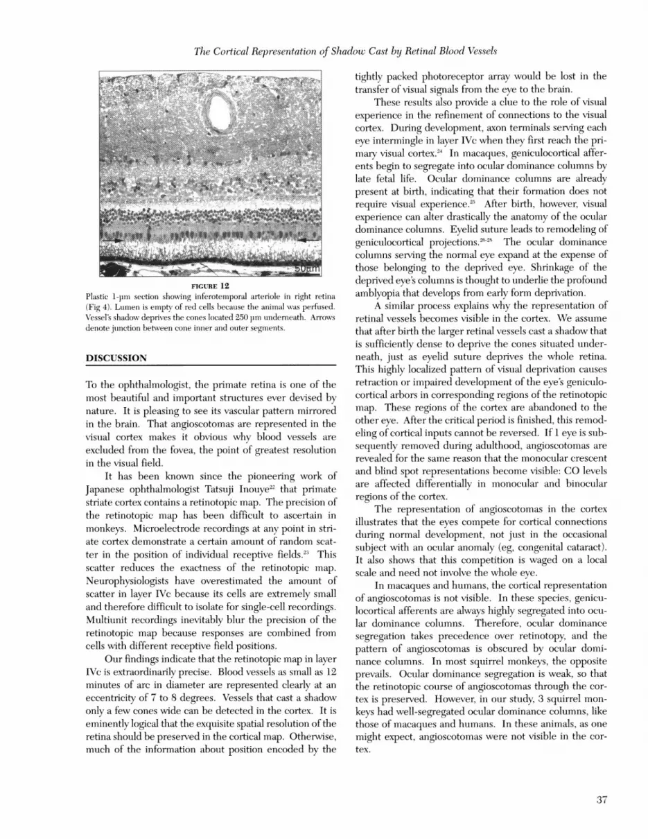
Plastic 1-pim section showing inferotemporal arteriole in right retina(Fig 4). Lumen is empty of red cells because the animal was perfused.Vessel's shadow deprives the cones located 250 pm underneath. Arrowsdenote junction between cone inner and ouiter segments.
DISCUSSION
To the ophthalmologist, the primate retina is one of themost beautiful and important structures ever devised bynature. It is pleasing to see its vascular pattern mirroredin the brain. That angioscotomas are represented in thevisual cortex makes it obvious why blood vessels areexcluded from the fovea, the point of greatest resolutionin the visual field.
It has been known since the pioneering work ofJapanese ophthalmologist Tatsuji Inouye22 that primatestriate cortex contains a retinotopic map. The precision ofthe retinotopic map has been difficult to ascertain inmonkeys. Microelectrode recordings at any point in stri-ate cortex demonstrate a certain amount of random scat-ter in the position of individual receptive fields.23 Thisscatter reduces the exactness of the retinotopic map.Neurophysiologists have overestimated the amount ofscatter in layer IVc because its cells are extremely smalland therefore difficult to isolate for single-cell recordings.Multiunit recordings inevitably blur the precision of theretinotopic map because responses are combined fromcells with different receptive field positions.
Our findings indicate that the retinotopic map in layerIVc is extraordinarily precise. Blood vessels as small as 12minutes of arc in diameter are represented clearly at aneccentricity of 7 to 8 degrees. Vessels that cast a shadowonly a few cones wide can be detected in the cortex. It iseminently logical that the exquisite spatial resolution of theretina should be preserved in the cortical map. Otherwise,much of the information about position encoded by the
tightly packed photoreceptor array would be lost in thetransfer of visual signals from the eye to the brain.
These results also provide a clue to the role of visualexperience in the refinement of connections to the visualcortex. During development, axon terminals serving eacheye intermingle in layer IVc when they first reach the pri-mary visual cortex.24 In macaques, geniculocortical affer-ents begin to segregate into ocular dominance columns bylate fetal life. Ocular dominance columns are alreadypresent at birth, indicating that their formation does notrequire visual experience.25 After birth, however, visualexperience can alter drastically the anatomy of the oculardominance columns. Eyelid suture leads to remodeling ofgeniculocortical projections.sE28 The ocular dominancecolumns serving the normal eye expand at the expense ofthose belonging to the deprived eye. Shrinkage of thedeprived eye's columns is thought to underlie the profoundamblyopia that develops from early form deprivation.
A similar process explains why the representation ofretinal vessels becomes visible in the cortex. We assumethat after birth the larger retinal vessels cast a shadow thatis sufficiently dense to deprive the cones situated under-neath, just as eyelid suture deprives the whole retina.This highly localized pattern of visual deprivation causesretraction or impaired development of the eye's geniculo-cortical arbors in corresponding regions of the retinotopicmap. These regions of the cortex are abandoned to theother eye. After the critical period is finished, this remod-eling of cortical inputs cannot be reversed. If 1 eye is sub-sequently removed during adulthood, angioscotomas arerevealed for the same reason that the monocular crescentand blind spot representations become visible: CO levelsare affected differentially in monocular and binocularregions of the cortex.
The representation of angioscotomas in the cortexillustrates that the eyes compete for cortical connectionsduring normal development, not just in the occasionalsubject with an ocular anomaly (eg, congenital cataract).It also shows that this competition is waged on a localscale and need not involve the whole eye.
In macaques and humans, the cortical representationof angioscotomas is not visible. In these species, genicu-locortical afferents are always highly segregated into ocu-lar dominance columns. Therefore, ocular dominancesegregation takes precedence over retinotopy, and thepattern of angioscotomas is obscured by ocular domi-nance columns. In most squirrel monkeys, the oppositeprevails. Ocular dominance segregation is weak, so thatthe retinotopic course of angioscotomas through the cor-tex is preserved. However, in our study, 3 squirrel mon-keys had well-segregated ocular dominance columns, likethose of macaques and humans. In these animals, as onemight expect, angioscotomas were not visible in the cor-tex.
37

Hortoni et al
REFERENCES
1. Iltlbel DH, \Vtiesel TN. Ftonictionial arcrhitecttore of mllacaqoie m11oo1-key visoal cortex. Pr-oc R Soc Lood(1 [Biol] 1977;198:1-59.
2. Ioirtooi JC. Cvtoclhromiie oxi(lase patches: A olew'J cvtoarchitectonicfeatture of moikey visual coItex. Phlilos Tratis R Soc Louid [Biol]1984;304:199-253.
3. Hor-toni JC, Hockineg DR. Initrinisic variability of ocuilar (luomiinancecoltumniii per-iodicity in nior-miial imacaqicue monkeys. J i\VCro.sci1996;16: 7228- 239.
4. Southll iJPC. Hclmholt/zs Treati.s oo Phlysiologica/ Optics. V0ol II. TheSensa-tionis of XVi-sioi.i Menasha, Wis: George Banta Puiblishing; 1924.
5. Evans JN. Ancgioscometrv. AmJ Oplithlalol 1926;9:489.6. Evans JN. Ani Ititi-odtictio) to Clinical Scotomcti,y. Ne\w Haven,
Cornii: Yale Unix irsity Press; 1938.7. Dashevskv Al. Cliniical ancgioscotomet-atr-a niewT method wNith the
;e of differenit conitrast test objects. Arc/i Ophltlialmol19:38; 19:334-353.
8. Welt NI. Etuide stir ]es rappoIts enltre les (liment'isioins (Ie la tacheavetugle de Nlariotte et des anigioscotomiles. et la tenisioni ariteieller6tinieniie. Ophlthalmologica 1945;1(0)9:137-158.
9. Goldmann H. Beitrag zuir AIsgioskotoissetrie. Op/tlthalmologic'a1947:114:147 -158.
10. Dubois-Poulsen A, Franc'ois P, Tihi A, et al. Lc C/ItiaImp Visicil.loporap/hic iio r'mi/rle et p)atlio/ogiquc de/ sc's ssibi/itcs. Paris:NIlasson; 1952:257-308.
11. Abe T. Study onI angioscotomas in glauconiatous eves. Actci Soc'Op/it/halmo1 jflu 1968.
12. Zuilauif NI. QtiaistificatioIn of angioscotomas. Oplit/talinol/ogica1990;200):203-209.
13. Safrain ABR Ilalfoni A, Saflran E, et al. Anclioscotoimiata a(Il mor'pho-logical features of related vessels in auitomlated perimetiy. Br- JOp/it/talmol 1995;79:118-124.
14. Schiefer U. Benda N. Dietricls Tj. et al. Angioscotomaa detectioinwitlh flllnlllis-ori.ente(d perimnetrv. A study wNrith dark aiil lbrigltt stinti-ohi of differenit sizes. Vi.siomi Res 1999;39:1897-190)9.
15. \W\ong-Riley NI. Changes in the visual system of' imtonocularlvsuturl-ed or enucleate(l kitten.s demonstrable wxith cvtochrome oxi-dclase histochemistry. BBrai Rce,s 1979J171:11-28.
16. Hortoit JC. Hockini,g DR. Ainatomlical demiionistrationi of ocular(dlminance columniiiis in striate cortex of the squiirrel mssonkev. JNcYorosci 1996;16:5510-5522.
17. Huibel DII. \Niesel TN. LeVav S. Funiictionial ai-clhitectuire of area17 in niorissal and maonocularlv dcprivedi maca(qute monkeys. ColdSpi-icg Hcarb1) Spynpi Qucmntit Biol 1976:40:581-589.
18. Tigges J. Tigges NI. Peraclhio AA. Complementary lansiiiiar terilli-inationis of aff'renits to ar'a 17 originating in area 18 amid iin the lat-eral gaemiculate nuticleuis in sqciirrel monkey. J Cooilp Ncuri^ol1977;176:87-100).
19. Hendclricksoni AE, Wilson JRl? Og(gen NIP. The nieulroatnatomicalorganiization of' pathways between the dorsal lateral gcnicimlatenut'cleuls ani(I visual cortex in old world and nw\v world primiiates. JComo;,) Nc'mrol 19718-:182:12:3-136.
20. RoeTC NIH. Benevento LA. Rczak NI. Soiitc ob)serivatioins oii thepatterins of segregated getniculate iilputs to the \isual cortex in newworld primiiates: Ani autoradiographic study. Brc,iit Rfecs1978: 159:371-3,78.
21. Fitzpatrick D? Itoh K. I)iamondl IT. The laminiiiar organization of'the lateral ceniciulate b(ody anid thie striate cortex in the squiirr-elimtoinkey (Sainiriii sci ureus). J Nctirosci 1983;3:673-702.
22. Inouive T. Die Sc/h.storunmgcit /)c'i .Sc/csrhrlc't ngc'it c/eu kortikcmlciSe/lvs/icacrc. Leipzig: Vi' Emorelmaim; 19(19.
23. Hubel DH, WNiesel TN. tJniformitv of monkey striate c-ortex: A par-allel rtelationislhip betx\vTeen fielo size, scatter-, anidI maunification flac-tor. J Cotli) MNmuiu-ol 1974;158:295.
24. Rakic P. Preniatal (levelopment of the visual system in rhesus ioon-kev. Philos Tran)-s R Soc Louid [Biol] 1977;278:245-260.
25. Ilortoin JC hlockinig DR. An adult-like pattern of ocuilar domi-n1an'ce coluiminls in striate cortex of newborn monkeys prior to visti-al experienice. J Nei\eosci 1996; 16: 1791-1807.
26. Huibel DH, Wiesel TN, LeVav S. Plasticity of ocuilar (lomiiinancecoliiiumns in monkey striate cortex. Philos Tratns R Soc Loand [Biol]1977;278:377-409.
27. Shatz CJ, Strvker MP. Ocuilar dominanice in layer IN' of the cat'svisuial cortex and the effects of imoniocular deprivation. J Physiol(Loand) 1978;281:267-283.
28. Hortoin JC' Hockinig DR. Timiinig of the critical period for plastici-tv of ocuilar (lomliinance columniiiis in mnacaquie striate co-tex. J.\e iri (osci 1997; 17:3684-33709.
DISCUSSION
DR ALFREDO A. SADUN. The primate visual cortex isorganized in several respects. First, there is a retinotopicmlap so that each point in the visuial field via the retinaprojects a map onto the primary visuial cortex (V-I cortex).Joonathon Horton has previouisly helped elucidate onrunderstanding that this retinotopic map is far from linear,as the mactila is vastly over-represented. A second organ-ization of V-1 cortex is the segregation of ocular inpuitsinto alternate bands from each eye, which are termed ocu-lar dominance columns.
Drs Horton and Adams have revisited the techniqueof cytochrome oxidase (CO) staining of ocular dominancecolumns to investigate features in the V-1 area of visualcortex. In particular, they have demonstrated the mani-festation of regions of vascular patterns in layer IVc thatrepresent the retinal vasculatuire. Or, more precisely, theyhave deimonstrated those areas in the visuial cortex that, byreceiving afferents of input by those photoreceptors lyingin the shadow beneath retinal blood vessels, are not rep-resented in the visual cortex. They refer to these areas asangioscoto-mas.
SimilarlK, when the authors show us a pale oval (theirFig 10) in the striate cortex, it is a negative representationof the blind spot. Negative, in the sense that this regiononly receives input from the (right) eye whose blind spotis not being illustrated. This monocular dependent area,reflecting the right blind spot, shows up in relief when theauithors remloved the fellow left eye and then, 8-10 dayslater, sacrificed the animal for CO staining. The CO onlystains the areas that the intact right eye inpuits, which, ofcouirse, does not inclutde the blind spot and angioscotomasof the right eye.
Horton and Adams have done much more than showuis the power of the CO technique and the beauty of theretinal vasculature inversely reflecte(d on the visuial cortex.They deemonstrated angioscotomas that radiated andbranched from the representation of the blind spot as faras 15 mnmn. Some of the angioscotomas were only12 miinutes of arc in diamneter! This is extraordinary if one
38

The Cortical Representation of Shadowc Cast by Retin2al Blood Vessels
considers that 20/20 vision mneans the resolution of 60 sec-onds, or 1 minute of arc. This is certainly a more preciseretinotopic map in INVc of V-1 than was previously consid-ered.
How can we explain the resolution of 12 arciminutesdiameter so far eccentric? Twelve arc minuites is about20/200 buit ouir vision at 8 degrees is a bit worse.
One explanation may lie in the fact that vernier ctI-ity is mutch better than Snellen acuity. Indeed, not witlh-standing the 30 arc second packing of cones (which trans-lates to a limitation of 60 arc seconds resolution = 20/20),and the wavelength of green light (wvhich creates tliffrac-tion problems limiting vision to abouit 20/20), hyperacuitvhas indeed been deimonstrated to exist to the point of 7arc seconds (=20/3). This probably7 depends on corticalprocessing which may even be analogouis to a FourierTransform.
An important take-home message proxided by thepresent work is that visuial experience must refine the pat-tern of these projections to N-LI cortex. It is known that inmonkeys the genicuilate input to IV'c begins as a courseand largely branching tree. Over time, this gets refined.WVhile the ocuilar dominance column organization is pres-ent by birth, only post-natal visuial experienice could, byfurther refinemnent, create the inverse reflection ofangioscotomas whose existence in the retina depends onliglht. This is also consistent kith the results of many otherstudlies of experimental amblyopia.
It is interesting that in macaquie and hulmtlans, suclangioscotomas are not visible on the cortex. The authorsexplain that this is because in these species ocular domi-nance takes precedence. Retinotopv is lost in favxor ofocular dominance. Yet, in squiirrel monkeys, retinotopv ismore clearly seen, as the ocular dominance segregation ismuich weaker. Are the trade-offs between retinotopv acndocular dominance of a fuindamental significance, or arethey juist epiphenomnenia of different developmental pat-terns? I eagerly await fuirther work in this area to provideus a better insight on the meaning of the precise andIbeauitiful organization of the visua-Il cortex.
REFERENCES
1. Hortoni J.C. Hoyt \VF. The represenitationi of the vistial fielcl inloumiiani striate cortex. Arch Oplithlalonol 1991;1(0)9:816-824.
2. Hubel DH. Wiesel TN. Laminar anid coltumniiiar distribntion ofgellicillocortical fibers in the imiacaqie imonley. J Comnpj) Nclurol1972; 14f5:421-45()
3. We-stlieimner G. The Prenitice Lectiure, \isuial acuiity anid hvlpleracut-it\: Resolution, localizatioIn, forimi. Amner J Optooiland P1hysJolOptics 1987;65:567.
4. Rakic P. PreiIatal developimeint of tlhe visual svstein in rhesuis miioo1-key. Philos Tramis R Soc Lould [Bioll 1977;278:245-260.
5. Au.tonin1i A, Strvker NIP. Rapi(d rem11o(leliug of axoial arlbors in thevisuial c-ortex. Science 1993;206: 1819-1821.
6. Shatz (CJ Stryker NIP. Ocular domliinaice in layer IN' of thle cat'svisual cortex anid the effects of monocular deprivation. J Phlopsiol(Lonidoni). 1978;281:267-283.
7. Hortoni JC, Hocking DR. Til1iming of the cr-itical period for plastic-itxN of ocular doiIsiniance columniiiis iu iluacaquie striate cortex. JNcurosci 1997;d7:3684-3709.
DR JONATHAN C. HORTON. Tlhank von Dr Sadun for thosevaluable remarks.
It wvould be nice to correlate visual acuity wvith theresolution of the cortical retinotopic imap. \We are hamn-pered in our efforts by the fact that within about the ceni-tral 30 we do not see the angioscotomas in the coitex. Youmav havxe noticed that the cortical angioscotomias peterout as they approaclh the foveal representation. \Webelieve that when the retincal vessels drop belowv the sizeand thickness required to deprive the photoreceptors,they become invisible in the cortex.
Amblvopia is a major clinical problem of interest to alarge segment of the membership of this society. \Vhy isthe cortex v-ulnerable to this disease? \Why not silmply,freeze things before birth so that the visual system isimpervious to deprivation of 1 eve (lurinig early7 child-hood? Our studies demonstrate that visual stimulationearly in life is critical for the maturation of the cortex. Thedevelopmnent of the angioscotoma representations in tllecortex provides direct evidlence for the role for visuialexperience in the form-lationi of the cortical architecture.
Finallh, I would like to address the issue of whyangioscotomas have never been seeni previouslv in thecortex. It's truie that they're not present in the human or inthe macaque. The reason is that these species have highlydeveloped ocular dominance columns. This means thatthe angioscotomnas are uniable to connect across thecolumiins to create a continuous line that your eye can seeas anl angioscotomna. Instead, the angioscotomuas are frac-tured into short segments, caamouflaged if von-i ^vill, by thepattern of the ocular dominance stripes. It is a whim ofnature that squirrel monkeys have ratlher rudementarvcolumns, so visuotopv trimllphs over ocular doimiinancecolumn segregation, andl one can see the cor-tical repre-sention of the angiscotomi-ias. Why one primlate speciesshould organize its ocular inpuits in a strictly segregatedfashion, wvhereas anotlher animal allows them to interdigi-tate rather promiscuouslx, I cannot v7et say. Thalnk y7ou.
39


