Embed Size (px)
Citation preview
hr. J. Devl. Neuroscience. Vol. 3. No. 4. pp. 341-348. 1985. Printed in Great Britain.
0736-.574&!85 w3.oo+o.w Pergamon Press Ltd.
@ 1985 ISDN
THE EFFECT OF EXOGENOUS GANGLIOSIDES ON NEURONS IN CULTURE:
A MORPHOMETRIC ANALYSIS
R. MASSARELLI. B. FERRET,* A. GORIO, M. DURAND and H. DREYFUS
Centre de Neurochimie du CNRS and U44 de I’INSERM. 5. rue Blaise Pascal. 67W4 Strasbourg Cedex. France: *Fidia Research Laboratories, Abano Terme, Italy
(Accepted 4 December 1984)
AbstractXultures of isolated neurons have been treated with a purified preparation of gangliosides (IW’M and IW’M) added to the cell growth medium at the 3rd day in culture and a morphometric analysis of the cells was performed with an image analyzer after I and 4 days of treatment. The number of cells and the area of the cell bodies were increased following the treatment. The results indicate as well the ‘sprouting’ effect of the glycolipids on the number of secondary neuronal processes and an increase in the length of the primary neurites. The present data and other biochemical evidence (Dreyfus et al., 1984.1. Neurosci. Res.) suggest that the addition of exogenous gangliosides may have a trophic effect on neurons, greatly enhances the number of cell to cell contacts. and, possibly, stimulates cell proliferation and differentiation.
Key words: Gangliosides, Neuronal primary cultures. Morphometry. Neuronal sprouting.
Much experimental evidence indicates that glycolipids may participate in the transmission of membrane mediated signals and suggests a possible role for these molecules in neuronal differ- entiation 7-18~24 The addition of exogenous gangliosides facilitates the processes of reinnervation in vivo, I’ the formation of synapses in vitro2o and stimulates nerve regeneration by an increased sprouting of nerve fibers. ‘*Jo Glycolipids are inserted in the neuronal membrane by their lipid moiety while the sugar chain is protruding towards the extracellular environment. The variety in the composition of the sugar chain and in the distribution of its single components raises the hypothesis that gangliosides may act as ‘coding’ agents for some steps in the process of cell to cell recognition or may be responsible for some ‘linking’ properties which maintain contacts among nerve cells.
To study the possible effects of the addition of exogenous gangliosides in the establishment of neuronal contacts and differentiation we have chosen a simplified model of the nervous system represented by isolated neurons in culture from chick embryo’s cerebral hemispheres. The basic characteristics (morphological and biochemical) of the model have already been described.17 Particularly the ganglioside metabolism has been studied and found to be similar to the one present in vivo or in isolated neuronal preparations from mammalian or avian brains.“-7
It will be shown that the addition of a purified preparation of gangliosides on isolated neurons in culture induced a net sprouting effect, and it possibly increased the proliferation and the differentiation of neurons.
MATERIALS AND METHODS
Exclusively neuronal cell cultures from 8-day-old chick embryo’s cerebral hemispheres have been obtained in 60 or 100 mm diameter plastic Petri dishes as previously described.17 The amount of cells added per Petri dish was however reduced by 75% in order to have the possibility to analyse the morphometry of isolated neurons. The day after the seeding, the culture medium was substituted with a modified Bottenstein-Sato defined medium using Dulbecco’s modified Eagle’s medium supplemented with insuline, transferrin, progesterone, putrescine, selenium (as reported in ref. 1) and also by lo-‘*M oestradiol. Two days after (at 3 days in culture: 3 d.i.c.) different concentrations of a mixture of purified gangliosides kindly provided by FIDIA Research Laboratories (GMl: 19.8%; GD3: 5.2%; GDla: 39.6%; GDlb: 14.6%; GTl: 17.6%; GQl: 3.2%) the purity and the composition of the mixture was controlled by methods described else- where7) were added to the cultures and pictures were taken the next day (at 4 d.i.c.). The
341

342 R. Massarelli et al.
medium was changed at 5 d.i.c., the same concentrations of gangliosides were again added to the new medium and pictures were taken at 7 d.i.c. The following parameters have been measured:
(a) the number of cells per microscopic field, in order to know whether gangliosides had an effect on the multiplication and/or the fixation of the cells to the support of polylysine; (b) the area of
the cell bodies; (c) the number and (d) the length of primary and secondary processes per neuron. The analysis of all these parameters has been performed by taking random micrographs out of randomly chosen Petri dishes (the researcher who was taking the pictures was not aware of the nature of the cells) using a Leitz ASM image analyzer, connected with a Hewlett-Packard micro-
processor. The analysis was performed on pictures whose nature was not known by the operator. The data have been analyzed statistically using the tests of Student to obtain any information
regarding the deviation of the means. Considering the large population under analysis and the possible non-Gaussian distribution. the statistical significance was measured by the tests of Wilcoxon and of Mann-Whitney.
RESULTS
Different concentrations of gangliosides were used to analyse the effects of the exogenously added gangliosides upon neuronal morphology ranging from 10e4M (which is lethal, cells de- generate in 48 h) to lo- ‘“M. It was chosen to utilize 10p5M and 10e9M concentrations which
supposedly are above and below the critical micellar concentration of gangliosides, respec- tively.“’ Furthermore, the gangliosides were added at 3 d.i.c. (a time which corresponds to the
beginning of neuronal maturation and synaptogenesis and lasts up to the 5th d.i.c.) to observe their effects upon these parameters.
The morphological aspect of the cultures after 1 or 4 days treatment with 10d9M and 10-5M gangliosides is shown in Figs 1 and 2, respectively. The cells treated with gangliosides (Figs lb, c and 2b. c) appeared to have more processes and the number of cells per microscopic field seemed
more important upon treatment. This has been confirmed by the morphometric analysis which has been resumed in Tables 1 and 2. The number of cells per microscopic field (per photographic
pictures) after 1 day of treatment (Table 1) increased from 21.29 + 0.85 (mean + S.E.M.) cells per photo to 27.62 * 1.74 and 31.64 2 1.70 after ganglioside treatment (10e5M and 10e9M, respectively). The area of the cell bodies instead did not vary significantly from 29.05 ? 1.16 t.r,rn*
in the controls to 30.10 + 4.62 and 31.15 ? 1.16 km2 in the treated cultures. The same was also observed in the number of primary processes per neuron (3.23 ? 0.33) in the controls (2.83 or: 0.13
Table 1. Morphometric analysis of neurons grown for 1 day in the presence of exogenous gangliosides
1 dayoftreatment(3Ad.i.c.)
Control Gangliosides
(lo-‘M) Gangliosides
(W9 M)
21.20 2 0.85
(52) I!c).os_t I.16
(100) 3.23 2 0.33
(55) 5.31 LO.36
(73)
‘7.62 i 1.74*’
(45) 30.10 t J.62*
( 100) 2.iS3to.13*
(54) 12.331 1.22**
(54)
31.645 1.70**
(22) 31.lSk l.lh*
(100) 3.06*0.13*
(731 11.29+0.67**
(74)
The values represent the mean-f S.E.M. of two independent experiments of five Petri dishes each.
d.i.c. = days in culture.
NC = number of cells per microscopic field. S = surface area of cell bodies. Npp = number of primary processes per neuron. Nsp = number of secondary processes per neuron. @I = number of photos analyzed. H = number of determina- t1ons.
* Statistically not significant. ‘* Statistically significant by at least P<O.O2 (two sided)
wng Student’s and Wilcoxon‘s tests.

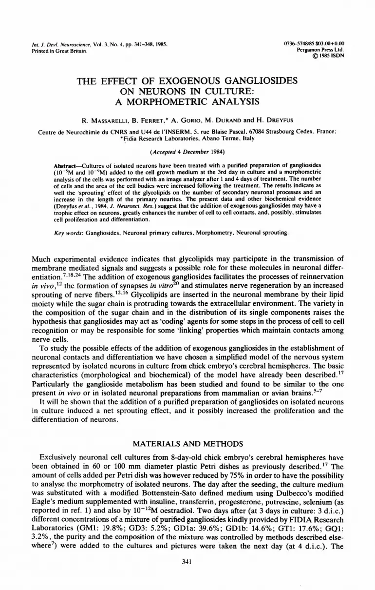
Fig.
The effect of exogenous gangliosides on neurons in culture: a morphometric analysis 343
1. E : tak
Iffect of 4 days of treatment with gangliosides on isolated neurons. The photographic en at 7 days in culture. (A) Control neurons, (B) neurons after the addition of IO-‘M
lo-‘M of a mixture of gangliosides. Phase contrast microscopy. x 280.
pictures and (C)

K. Massarelli et ctl
Fig picl the
ttfect of 1 day of treatment with gangliosides on isolated neurons in culture. The phot( :s were taken at 4 days in culture. Ganglioside treatment and cultures were formed as desc aterial and Methods section. (A) Control neurons. (B) neurons after the addition of 10
(C) 10~ ‘M of a mixture of ganyliosides. Phase contrast microxopy ‘- 2X0
Dgraphlc xibed in “M and

The effect of exogenous gangliosides on neurons in culture: a morphometric analysis 345
Table 2. Morphometric analysis of neurons grown for 4 days in the presence of exogenous gangliosides
4daysoftreatment(>‘ld.i.c.)
Control Gangliosides
(IO-‘M) Gangliosides
(IO-‘M)
EN S6.d (n)
22.6 + 0.80
(5% 24.34 2 0.58
(390)
NPP 2.53kO.IS
(n) (72)
NSP 6.29 + 0.37
(n) (72)
hppb.4
(n) Asp (km) (n)
46.03 t 1.79 (149)
17.15 + 0.86 (lot))
29.79rO.72**
(52) 31.48?9.74**
(3(N)
4.28~O.lh**
(54) 9.76 k o.s4**
(S4)
40.35 f 2.36: (232)
22.23 ” 7.31’
(224)
27.41 ‘O.gl**
(73) 34.24 + 0.70**
(300)
3.43kO.25”
(69) 12.23 + O.S4**
(0)
77.402 s.t59** (153)
17.93 t 1.0s*
(loo)
Values represent the mean + S.E.M. of two independent ex- periments of five Petri dishes each. Abbreviations (NC, S. Npp,
Nsp, nph. n) as in Table I. App = length of primary processes per neuron. A sp = length
of secondary processes per neuron. * Statistically not significant.
** Statistically significant by at least P ~0.02 (two sided)
using Student’s and Wilcoxon‘s tests.
and 3.06 + 0.13 in the treated cultures). Conversely a large significant difference was observed in the number of the secondary processes which increased by more than 100% after the addition of exogenous gangliosides without any difference between the two treatments, the increase being as effective whether cells were grown with lo-‘M or 10W9M gangliosides.
After 4 days of treatment (Table 2) the increase obtained in the number of cells per photo observed after 1 day of treatment was maintained with small variations, statistically not significant (for example, the decrease with 10e9M at 4 days of treatment when compared to the same treat- ment at 1 day was not significant). The surface of the cell bodies increased significantly in treated cells essentially because the area of the controls (24.34k0.58 pm2) decreased at 7 d.i.c. when compared to the controls (29.05 + 1.16 km’) at 4 d.i.c. (2P<O.O01, Tables 2 and 1, respectively). The surface area of the neurons treated for 4 days with gangliosides did not differ from that of neurons treated for 1 day. The change in the morphology of neurons during their development was further shown in the number of main processes which at 7 d.i.c. decreased considerably when compared to 4 d.i.c. (2.53 it 0.15 and 3.23 + 0.33, respectively, 2P<O.O01).
The length of primary processes per cell was increased after treatment with 10m9M gangliosides but no effect was observed with 10W5M. No effect was observed upon the length of secondary processes with either concentration.
The reason for the variability in the surface area and in the number of primary and secondary processes observed between cells treated with gangliosides for 1 and 4 days and between controls might be found in analyzing the ratio of these values (Table 3). Firstly, as it would be expected and on the basis of previous observations, the ratio of secondary over primary processes in- creased after 1 day of treatment with lo-‘M and 10m9M gangliosides. After 4 days an increase was observed only with 10e9M (but an increase was also observed in the control). Secondly, the ratio of the number of primary processes to the surface area does not change during the culture nor during the treatment remaining, in the average, close to 0.1. The ratio of the number of secondary processes to the surface area increased instead after treatment (but note the increase in the 7-day-old controls).
DISCUSSION
Several experimental data have pointed out a possible role for gangliosides in the nervous system and more particularly in neurons because of the presence of high amounts of gangliosides,

346 K. Massarelli 6’1 nl.
Table 3. Ratio of some morphometric parameters m control and gangiioslde treated neurons
‘Treatment days I (S4d.i.c.) 4(?--:,I ,.c ,
Gangliosides Gangliosides (iangliosides Gangliosides Ratio Control (IO ‘M) (IO “M) Control (IO ‘M) (10 “M)
NSPJNPP 1 .h4 +- 0. I1 4.37 IO.87 3.6Y kO.43 2.4Y-tO.lX 2.2XZO.32’ 3.57-tO.lY NpplS 0.1 I -t 0.02 0.09 -c 0.001* 0. lot O.(H)7” 0. IO 2 0.021 0. I4? 0.005* 0. 10 IT 0.01*
NsplS 0.18 -t 0.01 0.41 2 0.015 O.%t- 0.04 0.26 t 0.01 0.31 r 0.02* 0.36 5 0.02*
Values represent the mean + S.E.M. Abbreviations as in Table I. The ratios have been calcu-
lated by using the values presented in Tables I and 2. * Statistically not significant compared to control values. t Statistically not significant compared to control of day I of treatment. All other values were
statistically significant by at least ZP<O.O.S
with a high percentage of polysialogangliosides, ” in neuronal membranes. The glycolipids have been suggested to act as and to be essential constituents of receptors for some neurotransmitters, hormones, and toxins.2,‘” They have been suggested to be relevant in cell to cell interactions and mord particularly as active sites for recognition among nerve cells.2,y Their molecular roles as
regulatory mechanisms for certain membrane phenomena such as fluxes and release of solutes and neurotransmitters have also been suggested.“,’ Another function of gangliosides seems to be correlated with regeneration phenomena and several experimental data point out the important role that these compounds play in this respect. “,22
The present data suggest that gangliosides may have a function on the development of neurons in culture. The increase in the number of cells per microscopic field may indicate an increase in
proliferation thus a stimulation of the proliferation phenomena in the cells or increased attach- ment of the cells to the support of polylysine. An increase in proliferation would imply that some- how the insertion of sialoglycolipids into the neuronal plasma membrane may act on the nuclear machinery of the cell and there is some evidence in favour of such an interpretation.‘,8*‘5,21 On the other side, gangliosides possess a negative charge on the sialic acid terminals and the polylysine film on which cells are grown is actually a layer of positive charges. Thus the attach- ment of cells treated with gangliosides should actually be increased.
The analysis of the surface areas and of the number of primary and secondary processes in- dicates that the following two phenomena must be taken into consideration: the maturation, differentiation of neurons during the culture and the effect of gangliosides. Isolated neurons from chick embryos dissociated cerebral hemispheres, when seeded on Petri dishes, attach rapidly to the support and begin to proliferate rapidly. At the 3rd d.i.c. electron microscopy has shown’ the
appearance of the first synapses and by the 5th d.i.c. synaptogenesis appears to have reached an advanced stage of maturation which is complete at the 7th d.i.c. as much biochemical evidence would also suggest.’
In this respect, the present data show that the morphology of neurons changed from the 4th to the 7th d.i.c. in decreasing the size of the cell bodies and the number of primary branches while increasing the number of secondary branches. The phenomenon might be correlated to the maturation of the cells and shows that some of the primary branches observed in the early stages of the culture may become secondary processes at maturation.
What seems particularly interesting is the finding that the ratio NpplS (Table 3) does not change during growth nor during treatment; thus, irrespective of the size of the cell body, a
neuron has, on the average, one primary process per 10 km 2. Is this due to a structural property of the chick neurons or is it the index of a cellular selection? (The latter does not seem to be the
case when observing the morphology of these cell cultures.)’ The addition of gangliosides stimulates greatly the amount of secondary processes confirming
their role in the ‘sprouting’ of nerve fibers. Quite unexpectedly, however, gangliosides act as well on the length of primary fibers (Table 2) and this was observed with a low concentration of the glycolipids.
The different effects observed between the treatments with the two concentrations, especially after 4 days of treatment (Table 2), may be the reflection of the different status of solubility of

The effect of exogenous gangliosides on neurons in culture: a morphometric analysis 347
gangliosides at the two concentrations, the critical micellar concentration (CMC) ranging bet- ween 10e5M and 10-9M.‘o Above the CMC gangliosides aggregate to form stable, high mole- cular weight micelles and below the CMC they are present in solution as monomers. l9 It has been shown that the binding of a GM1 purified preparation to rat brain membranes results in a biphasic interaction over the range from 10 nM up to 500 FM. 23 This may be explained by the different micellar status of gangliosides in solution. This, in turn, might also explain the differential effect of the two concentrations used in this study.
Finally, the increase in the surface of the cells after ganglioside treatment as well as in the number of branches may be correlated to the increase in fluxes movements which has been observed for choline, GABA and dopamine as well as to the overall increase of the metabolism of endogenous gangliosides. 3V4 From this point of view, it may be suggested that gangliosides may act as trophic factors in favouring exchanges of solutes and metabolites across nerve cell mem- branes. The increase in the number of branches indicates, at the same time, that more cell to cell contacts have been established. Preliminary results on the effects of gangliosides on the biochemistry of neurotransmitters and on the endogenous metabolism of gangliosides confirm the conclusions which have been drawn on the possible action of these glycolipids on neurons.3,4
Acknowledgements-The secretarial assistance of MS F. Herth is gratefully acknowledged. Statistical analysis has been performed by Dr C. Nanopoulos (Groupe de Statistique, FacultC de MathCmatique, Universitt Louis Pasteur, Strasbourg), whom we warmly thank. This work was partly supported by INSERM (CRL No. 81.60.38).
REFERENCES
1.
2.
7 _
4.
5.
6.
7.
8.
9. 10.
Bottenstein J. E. and Sato G. (1979) Growth of rat neuroblastoma cell line in serum-free supplemented medium. Proc. natn. Acad. Sci., U.S.A. 76, 514-517. Critchley D. R. and Wicker M. G. (1977) Glycolipid as membrane receptors important in growth regulation and cell- cell interaction. In Dynamic Aspects of Cell Surface Organization (eds Poste G. and Nicolson G. L.), pp. 307-370. Elsevier North Holland Biomedical Press, Amsterdam. Dreyfus H.. Ferret B., Harth S.. Gorio A.. Durand M.. Freysz L. and Massarelli R. (1984) Metabolism and function of gangliosides in developing neurons. J. Neurosci. Res. 12. 31 l-322. Dreyfus H., Ferret B., Harth S., Gorio A., Freysz L. and Massarelli R. (1984) Effect of exogenous gangliosides on the morphology and biochemistry of cultured neurons. In Ganglioside Structure, Funcrion and Biomedical Potential (eds Ledeen R. W., Yu R. K., Rapport M. M. and Suzuki K.), pp. 513-524. Plenum Publishing Corporation, New York. Dreyfus H., Harth S., Massarelli R. and Louis J. C. (1981) Mechanisms of differentiation in cultured neurons: involvement of gangliosides. In Gangliosides in Neurological and Neuromuscular Function, Development and Repair (eds Rapport M. M. and Gorio A.), pp. 151-170. Raven Press, New York. Dreyfus H., Louis J. C., Harth S., Durand M. and Massarelli R. (1983) Role of sialoglycoconjugates in the transport of neurotransmitters. In Neural Transmission, Learning and Memory (eds Caputto R. and Aimone Marsan C.), pp. 21-23. Raven Press, New York. Dreyfus H., Louis J. C., Harth S. and Mandel P. (1980) Gangliosides in cultured neurons. Neuroscience 5, 1647- 1655. Dreyfus H., Massarelli R., Lombard D., Gorio A., Freysz L. and Durand M. (1984) Effects of exogenous gangliosides on the morphology and biochemistry of neurons. In Developmental Neuroscience: Physiological, Phar- macological and Clinical Aspecfs (eds Caciagli F., Giacobini E. and Paoletti R.). pp. 27-32. Elsevier Science Publishers. Amsterdam. Fishman P. H. and Brady R. 0. (1976) Biosynthesis and function of gangliosides. Science, Wash. 194, 906-915. Formisano S., Johnson M. L., Lee G., Aloj S. M. and Edelhoch H. (1979) Critical micelle concentrations of gangliosides. Biochemistry 18, 1119-1124. -.. -
11. tiorlo A., Carmlgnoto ti., Facci L. and Finesso M. (1980) Motor nerve sprouting induced by ganglioside treatment. Possible implications for gangliosides on neuronal growth. Brain Res. 197. 236-241.
12. Gorio A., Marini P. and %an&i R. (1983) Muscle reinnervation. III. MO&neuron sprouting capacity, enhancement by exogenous gangliosides. Neuroscience 8, 417-429.
13. Ledeen R. W. and Yu R. K. (1976) Gangliosides of the nervous system. In Glycolipid Methodology (ed. Witting L. A.), pp. 187-214. American Oil Chemist’s Society, Champaign, Illinois, U.S.A.
14. Lee G., Aloj S. M. and Kohn L. D. (1977) The structure and function of glycoprotein hormone receptors: ganglioside interactions with luteinizing hormone. Biochim. Biophys. Res. Commun. 77, 434-441.
15. Massarelli R., Durand M., Gorio A. and Dreyfus H. (1984) Role CL glycoconjugates in neuron to neuron inter- actions. In Regulation of Neurotransmitter Function: Basic and Clinical Aspects (eds Vizi E. S. and Magyar K.), pp. 405-408. Akademiai Kiado, Budapest.
16. Obata K., Oide M. and Handa S. (1977) Effects of glycolipids on in vitro development of neuromuscular function. Nature, Lond. 266, 369-371.
17. Pettmann B., Louis J. C. and Sensenbrenner M. (1979) Morphological and biological maturation of neurons cultured in the absence of rrlial cells. Nature. Lond. 281. 378-380.
18. Purpura D. P. (1578) Ectopic dendritic growth in mature pyramidal neurons in feline GM1 ganglioside storage disease. Nature, Lond. 266, 553-5.54.

348 R. Massarelli VI al.
19.
20.
21.
22.
23.
24.
Rauvaia H. (1979) Monomer-micelle transition of the ganglioside GM1 and the hydrolysis by Cfosrridium perfringetu neuraminidase. Eur. .I. Biochem. 97, 555-564. Roisen F. J., Bartfeld H., Nagele R. and Yorke G. (1981) Gangliosidc stimulntton of axonal sprouting 111 r’rtro. Science, Wash. 214, 577-579. Rybak S., Ginzburg 1. and Yavin E. (1983) Gangliosides stimulate neurite outgrowth and Induce tubulin mKNA accumulation in neural cells. Biochem. Biophys. Res. Commun. 116, 97&980. Sparrow J. R. and Grafstein B. (1982) Sciatic nerve regeneration in ganglioside-treated rats. .Qf>. Neural. 77, 2% 235. T’offano G., Benvegnu D., Bonetti A. C., Facci L.. Leon A.. Orlando P., Ghidoni R. and Tettamanti (i ( IWO) Interactions of GM1 ganglioside with crude rat brain neuronal membranes. J. Neurochem. 35, 861-866. Willinger M. and Schachner M. (1980) GM1 ganglioside as a marker for neuronal differentiation in mouse cere- bellum. Devl. Biol. 74, 101-117.


