Embed Size (px)
Citation preview

BioMed Central
Reproductive Biology and Endocrinology
ss
Open AcceResearchThe expression of tumor necrosis factor-alpha, its receptors and steroidogenic acute regulatory protein during corpus luteum regressionMichael Abdo, Susan Hisheh, Frank Arfuso and Arun Dharmarajan*Address: School of Anatomy and Human Biology, The University of Western Australia, 35 Stirling Highway, Crawley, Western Australia 6009, Australia
Email: Michael Abdo - [email protected]; Susan Hisheh - [email protected]; Frank Arfuso - [email protected]; Arun Dharmarajan* - [email protected]
* Corresponding author
AbstractBackground: Corpus luteum (CL) regression is known to occur as two parts; functionalregression when steroidogenesis declines and structural regression when apoptosis is induced.Previous studies suggest this process occurs by the production of luteolytic factors, such as tumournecrosis factor-alpha (TNF-alpha).
Methods: We examined TNF-alpha, TNF-alpha receptors (TNFR1 and 2) and steroidogenic acuteregulatory (StAR) protein expression during CL regression in albino Wistar rats. CL from Days 16and 22 of pregnancy and Day 3 post-partum were examined, in addition CL from Day 16 ofpregnancy were cultured in vitro to induce apoptosis. mRNA was quantitated by kinetic RT-PCRand protein expression examined by immunohistochemistry and Western blot analyses.
Results: TNF-alpha mRNA increased on Day 3 post-partum. TNFR were immunolocalized toluteal cells, and an increase in TNFR2 mRNA observed on Day 3 post-partum whilst no change wasdetected in TNFR1 mRNA relative to Day 16. StAR protein decreased on Day 3 post-partum andfollowing trophic withdrawal but no change was observed following exogenous TNF-alphatreatment. StAR mRNA decreased on Day 3 post-partum; however, it increased following trophicwithdrawal and TNF-alpha treatment in vitro.
Conclusion: These results demonstrate the existence of TNFR1 and TNFR2 in rat CL and suggestthe involvement of TNF-alpha in rat CL regression following parturition. Furthermore, decreasedStAR expression over the same time points was consistent with the functional regression of the CL.
BackgroundThe demise of the corpus luteum (CL) is characterized bya decrease in progesterone synthesis [1] and an increase inapoptotic cell death [2]. Whilst a temporal pattern is wellestablished, the factors regulating both the functional and
structural regression of the rat CL remain poorly under-stood.
Whilst progesterone is synthesized by the ovary, the adre-nal and the placenta, the CL of pregnancy are the major
Published: 7 November 2008
Reproductive Biology and Endocrinology 2008, 6:50 doi:10.1186/1477-7827-6-50
Received: 25 June 2008Accepted: 7 November 2008
This article is available from: http://www.rbej.com/content/6/1/50
© 2008 Abdo et al; licensee BioMed Central Ltd. This is an Open Access article distributed under the terms of the Creative Commons Attribution License (http://creativecommons.org/licenses/by/2.0), which permits unrestricted use, distribution, and reproduction in any medium, provided the original work is properly cited.
Page 1 of 11(page number not for citation purposes)

Reproductive Biology and Endocrinology 2008, 6:50 http://www.rbej.com/content/6/1/50
source of progesterone in the rat [3-5]. Small and largeluteal cells within the rat CL of pregnancy retain ster-oidogenic potential though large luteal cells predominate[6]. During pregnancy total progestin synthesis (proges-terone and 20α-hydroxypregn-4-en-3-one (20α-OHP))declines from a high on Day 16 to the morning of Day 22prior to an increase in the afternoon on Day 22 [1]. Thisobserved pattern in total progestin production in rats hasbeen demonstrated to be a product of decreased synthesisof progesterone toward term [7] and increased synthesisof 20α-OHP [1].
Total progestin production is dependent on the transportof cholesterol to the mitochondria and then from theouter to the inner mitochondrial membrane which ismediated by the steroidogenic acute regulatory (StAR)protein [8]. StAR protein has been reported in the ovary ofthe mouse [9], rat [10], rabbit [11] and human [12] andcorrelated with the functional state of the CL [11,13,14].As such StAR expression has been proposed as a reliable"marker" of CL function [15].
Several publications have reported the participation of theimmune system in ovarian events [16], suggesting a rolefor the cytokine tumor necrosis factor – alpha (TNFα) inCL regression. Luteal cells and endothelial cells are capa-ble of TNFα synthesis though macrophages remain theprimary ovarian source [17,18]. TNFα expression in theovary is coordinated between the infiltration and activa-tion of macrophages and the hormonal regulation of theCL [19-21]. We have recently reported TNFα protein local-ization in the rat CL on Day 16 and Day 22 of pregnancyand Day 3 post-partum [22]. Furthermore, we have dem-onstrated the induction of luteal cell apoptosis followingtreatment with recombinant TNFα in a dose- and time-dependent manner [22].
Associated with the TNFα ligand are two similar, thoughdistinct receptors, TNFα receptor 1 (TNFR1) and TNFαreceptor 2 (TNFR2). The lack of homology between thetwo cytoplasmic domains [23,24] is thought to contributeto the different outcomes of TNFα. Involved in a variety ofbiological processes, TNFα is implicated in both cell pro-liferation and cell death; TNFR1 is generally associatedwith TNFα-induced cell death and TNFR2 with cell prolif-eration [25]. TNFR binding sites have been demonstratedwithin the bovine [18,26], porcine [27] and rat [28,29] CLunder various experimental conditions. TNFR are presenton nearly all cell types with few exceptions [24] and thesubtypes are often co-expressed by the same cells [30].
The aims of this study are to examine the role of TNFαduring the structural regression of the CL by analysis ofTNFR expression, and to determine the role of TNFα in
the functional regression of the CL through regulation ofStAR protein expression.
MethodsAnimalsThe animals used were mature (12–20 week old) nullipa-rous albino Wistar rats obtained from the AnimalResources Center (Murdoch, WA, Australia). Animalswere housed in a controlled environment and mated over-night. Day 1 of pregnancy was designated as the morningon which spermatozoa were present in a vaginal smear.Litters were born on Day 23 of pregnancy. All procedureswere approved by The University of Western AustraliaAnimal Ethics Committee.
Experimental tissue collectionAll tissues were collected under aseptic conditions withlight anesthesia using a mix of 0.2 L/min O2, 0.8 L/minNO and 5% Halothane. CL were collected on Day 16 andDay 22 of pregnancy and Day 3 post-partum. Four ratsfrom each stage of pregnancy and post-partum were used.One ovary from each animal (alternating left or right) wasused for immunohistochemistry, the contralateral ovarywas used for Western blot and mRNA analyses (n = 4).Ovaries were trimmed of adhering tissues and the CL ofpregnancy were selected and dissected as previouslydescribed [2]. In addition to the above, in vitro studieswere conducted using CL collected on Day 16 of preg-nancy. Three pairs each from a different animal (n = 3)were collected for each treatment group. Dissected CLwere cultured in MEM either without trophic support for8 h or with minimal trophic support supplemented with37.5 ng/ml of recombinant rat TNFα (R&D Systems, USA)for 6 h as described previously [22]. Following incubationprotein or mRNA was extracted from each CL pair asdescribed under each experimental method.
ImmunohistochemistryCL collected for immunohistochemistry were fixed andprocessed as previously described [22,31]. Sections weretreated for 10 min with 3% hydrogen peroxide in metha-nol, washed in PBS (pH 7.4) and treated with 10% fetalbovine serum (FBS; Sigma Chemical Co., St Louis, MO,USA) (TNFR) or 0.1% bovine serum albumin (BSA; SigmaChemical Co.) (StAR) for 1 h at room temperature. Sec-tions were incubated with either 1:50 polyclonal goatanti-rat TNFR1 antibody (Santa Cruz Biotechnology,Santa Cruz, CA, USA), 1:100 polyclonal goat anti-ratTNFR2 (Santa Cruz Biotechnology) or 1:100 polyclonalrabbit anti-mouse StAR antibody (supplied by ProfessorDoug Stocco). TNFR antibodies were diluted in PBS pH7.4 whilst the StAR antibody was diluted in PBS pH 7.4,1% BSA, 0.1% Triton X. Sections were incubated for 2 h at37°C (TNFR) or overnight at 4°C (StAR). Following this,sections were incubated for 1 h at 37°C with a 1:200 don-
Page 2 of 11(page number not for citation purposes)

Reproductive Biology and Endocrinology 2008, 6:50 http://www.rbej.com/content/6/1/50
key anti-goat HRP (Santa Cruz Biotechnology) secondaryantibody (TNFR) or 1:200 biotinylated goat anti-rabbitIgG (Santa Cruz Biotechnology) secondary antibody(StAR). The sections were then incubated with AvidinBiotin Enzyme Reagent (Vector Laboratories, Burlingame,CA, USA) for 1 h at room temperature (StAR) and the reac-tion visualized by the addition of 3,3'-diaminobenzidinetetrahydrochloride (DAB; 1.2 mg/ml). The immunohisto-chemical procedures described were repeated for each ani-mal group (n = 4).
Western blot analysisCL collected for Western blot analyses were snap frozen inliquid nitrogen and stored at -70°C until use. Total pro-tein was extracted by homogenization in RIPA buffer (150mM NaCl, 50 mM Tris-HCl, pH7.5, 1% Triton X, 0.5% Nadeoxycholate, 1 mM PMSF) as described previously [32].Protein concentration of homogenates was measured [33]and 30 μg resolved by 12% SDS-PAGE and transferred tonitrocellulose membranes (MSI, Westboro, MA, USA).
Membranes were blocked in 5% non-fat milk for 1 h atroom temperature and probed with polyclonal rabbitanti-mouse StAR antibody diluted 1:5000 in Tris-bufferedsaline/0.1% Tween-20 (TBST). Following this membraneswere incubated with biotinylated goat anti-rabbit IgG for1 h at room temperature (diluted 1:10,000 in TBST).Membranes were then incubated with Avidin BiotinEnzyme Reagent for 1 h at room temperature and proteinsignals detected by enhanced chemiluminescence (Super-signal West Pico ECL substrate, Pierce, Rockford, IL, USA)and quantitated by densitometry.
A common tissue sample was included on each gel toallow for standardization of chemiluminescence levelsand exposure times. Staining of each gel (post transfer)and membrane with Coomassie Brilliant Blue (SigmaChemical Co.) assessed the accuracy of sample loadingand the efficiency of protein transfer. This procedure wasrepeated for each animal/experimental group (n = 3).
Kinetic RT-PCRTNFα, TNFR1, TNFR2, L19 and StAR mRNA expressionwere quantitated through kinetic reverse transcription(RT) – polymerase chain reaction (PCR) using the Bio RadiCycler (Bio Rad Laboratories, Hercules, CA, USA). All tis-sue collected was snap frozen in liquid nitrogen andstored at -70°C until use. Total RNA was extracted usingRNAzol B (Tel-test, Friendswood, TX, USA) and 5 μgreverse transcribed using SuperScript II reverse tran-scriptase (Invitrogen, Life Technologies, Melbourne, Aus-tralia) as per manufacturer's instructions. cDNA sampleswere purified using an UltraClean PCR kit (Mo Bio Labo-ratories, Solana Beach CA, USA), concentrations meas-ured by spectrophotometry and samples stored at -20°Cuntil use.
Kinetic PCR and melt curve analyses were performedusing the Qiagen Quantitect PCR SYBR Green I kit (Qia-gen, Clifton Hill, Victoria, Australia) according to manu-facturer's instructions with the addition of 100 nMfluorescein (Bio Rad Laboratories). 2.5 μl of each RT sam-ple (cDNA) was added to the 1× PCR master mix in a 25μl final volume. Primers used for each target (0.5 μM)were based on published rat sequences (Table 1). EachPCR cycle included an initial denaturation at 95°C for 15min (including 90 sec at 95°C for automated well factorcollection) followed by 45 cycles of 95°C for 30 sec, 52–57°C (depending on target) for 30 sec, and 72°C for 60sec. The annealing temperatures used for each target were;TNFα 56°C, TNFR1 52°C, TNFR2 54°C, L19 56°C andStAR 52°C. A fluorescence measurement was performedduring the extension step of each cycle. In addition, meltcurve analysis was performed with continuous fluores-cence measurement between 55–95°C in 0.5°C incre-ments.
External standards for each target were generated byextraction of the RT-PCR product following agarose gelelectrophoresis using the QIAquick PCR Purification Kit(Qiagen) as per manufacturer's instructions. Sampleswere quantified by spectrophotometry, and then used to
Table 1: Primer sequences used for individual targets
Target Forward Primer Reverse Primer
TNFα Clontech Laboratories Inc, CA, USA
5'-TAC TGC ACT TCG GGG TGA TTG GTC C-3' 5'-CAG CCT TGT CCC TTG AAG AGA ACC-3'
TNFR1 [28] 5'-CCA GCC CCA ATG GGG GAG TG-3' 5'-CGG TGT TCT GTT TCT CCT TA-3'
TNFR2 [28] 5'-TTC GGA GTG GCC CGT TCA AGA-3' 5'-GCT GTG GTC AAT AGG TGC TGC-3'
L19 [36] 5'-CTGAAGGTCAAAGGGAATGTG-3' 5'-GGACAGAGTCTTGATGATCTC-3'
StAR [32] 5'-GCA GCA GGC AAC CTG GTG-3' 5'-TGA TTG TCT TCG GCA GCC-3'
Page 3 of 11(page number not for citation purposes)
Reproductive Biology and Endocrinology 2008, 6:50 http://www.rbej.com/content/6/1/50
generate a standard curve via serial dilutions. Fluorescencedata were analyzed and a standard curve generated usingthe Bio Rad iCycler software (3.0 beta) (Bio Rad Labora-tories).
The potential for genomic DNA contamination wasassessed by amplification of a DNA sample and RT con-trols (no RNA template). To confirm reproducibility,repeats (n = 3) for each time-point of interest were ampli-fied in duplicate, the external standards were amplified induplicate simultaneously. To avoid competition, targetand L19 cDNAs were amplified in 2 separate PCR reac-tions. At the completion of each PCR reaction the startingquantity of each sample was calculated against the stand-ard curve (constructed by software) using the appropriatethreshold cycle (CT) value. Samples were given a relativemeasure against their starting cDNA concentration [34]and this value normalized against corresponding L19value [35,36].
Statistical analysesAll experiments were conducted using a minimum ofthree animals per time point/treatment. Variation amonggroups was analyzed by one-way ANOVA or t-test whereappropriate. Where significant differences (P < 0.05)among groups were detected, specific group comparisonswere made by least significant difference (LSD) tests [37].Associations between parameters were measured by Pear-son correlation.
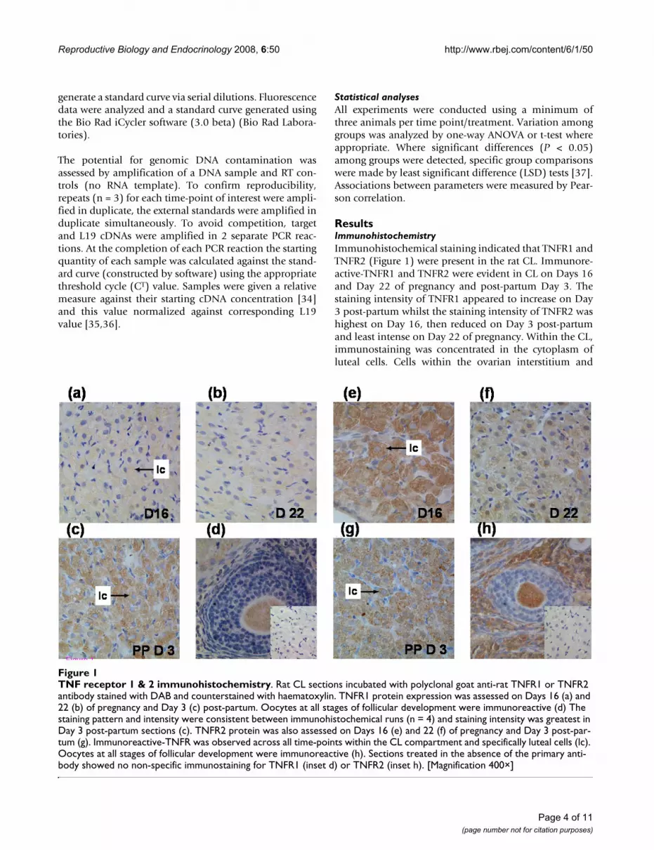
ResultsImmunohistochemistryImmunohistochemical staining indicated that TNFR1 andTNFR2 (Figure 1) were present in the rat CL. Immunore-active-TNFR1 and TNFR2 were evident in CL on Days 16and Day 22 of pregnancy and post-partum Day 3. Thestaining intensity of TNFR1 appeared to increase on Day3 post-partum whilst the staining intensity of TNFR2 washighest on Day 16, then reduced on Day 3 post-partumand least intense on Day 22 of pregnancy. Within the CL,immunostaining was concentrated in the cytoplasm ofluteal cells. Cells within the ovarian interstitium and
TNF receptor 1 & 2 immunohistochemistryFigure 1TNF receptor 1 & 2 immunohistochemistry. Rat CL sections incubated with polyclonal goat anti-rat TNFR1 or TNFR2 antibody stained with DAB and counterstained with haematoxylin. TNFR1 protein expression was assessed on Days 16 (a) and 22 (b) of pregnancy and Day 3 (c) post-partum. Oocytes at all stages of follicular development were immunoreactive (d) The staining pattern and intensity were consistent between immunohistochemical runs (n = 4) and staining intensity was greatest in Day 3 post-partum sections (c). TNFR2 protein was also assessed on Days 16 (e) and 22 (f) of pregnancy and Day 3 post-par-tum (g). Immunoreactive-TNFR was observed across all time-points within the CL compartment and specifically luteal cells (lc). Oocytes at all stages of follicular development were immunoreactive (h). Sections treated in the absence of the primary anti-body showed no non-specific immunostaining for TNFR1 (inset d) or TNFR2 (inset h). [Magnification 400×]
Page 4 of 11(page number not for citation purposes)

Reproductive Biology and Endocrinology 2008, 6:50 http://www.rbej.com/content/6/1/50
Page 5 of 11(page number not for citation purposes)
StAR protein localizationFigure 2StAR protein localization. StAR protein expression was analyzed using a polyclonal rabbit anti-mouse StAR antibody on Day 16 (a) and 22 (b) of pregnancy and Day 3 post-partum (c). Immunoreactive-StAR was observed across all days in the CL compartment, specifically in luteal cells (lc). Sections treated without the primary antibody showed no immunostaining (d). The staining pattern and intensity were consistent between immunohistochemical runs (n = 4). [Magnification 400×]

Reproductive Biology and Endocrinology 2008, 6:50 http://www.rbej.com/content/6/1/50
oocytes at all stages of follicular development were immu-noreactive for TNFR1 and TNFR2. Sections treated in theabsence of the primary antibody showed no immunos-taining.
Immunoreactive-StAR was evident in CL at all stages ofpregnancy and post-partum examined (Figure 2). StARprotein was localized within the cytoplasm of luteal cellson Days 16 and Day 22 of pregnancy and post-partumDay 3. Staining intensity appeared to decrease toward Day3 post-partum. Negative control sections incubated in theabsence of the primary antibody showed no immunos-taining. The staining pattern and intensity for all proteintargets were consistent between immunohistochemicalruns (n = 4).
Western blot analysisWestern blot analysis revealed a single immunoreactiveband of approximately 30 kDa consistent with that previ-ously reported [38] (Figure 3a). StAR protein expressiondecreased significantly (P < 0.05) between Day 16 and theother time points examined. StAR protein expressiondecreased significantly following in vitro incubation with-out trophic support for 8 h (Figure 3b). There was no sig-
nificant change in StAR protein expression followingtreatment with recombinant TNFα (37.5 ng/ml) for 6 h(Figure 3b).
Kinetic RT-PCRTNFα mRNA RT-PCR product was detected in ovariansamples on all days/time-points (Figure 4). Approxi-mately 25–30 amplification cycles were needed to reachthe threshold, the threshold cycle (CT) vs. log (startingconcentration) plot or standard curve was linear with astrong correlation coefficient (r = 0.999) (data notshown). The relative amount of TNFα increased signifi-cantly on Day 3 post-partum (P < 0.001) compared to Day16 of pregnancy, though there was no significant differ-ence between Day 16 and Day 22 of pregnancy (Figure 4).Melt curve analysis revealed the amplification of a singleproduct (295 bp) with a denaturation temperature (Tm)of 87°C (data not shown).
A single RT-PCR product corresponding to both TNFR1and TNFR2 on Day 16 and Day 22 of pregnancy and Day3 post-partum. Approximately 21–27 (TNFR1) and 23–26(TNFR2) amplification cycles were needed to reach thethreshold and the standard curves generated were linear
Western blot analysis of StAR protein in vivo and in vitroFigure 3Western blot analysis of StAR protein in vivo and in vitro. Western blot analysis of StAR protein on Day 16 and 22 of pregnancy, and Day 3 post-partum. A representative autoradiogram showing a single immunoreactive band (a) and corre-sponding statistical analysis (b). There was a significant change between Day 16 of pregnancy and Day 3 post-partum (P < 0.05; one-way ANOVA). Values without shared notations differ at P < 0.05 (LSD test). Western blot analysis of StAR protein on Day 16 of pregnancy following incubation without trophic support for 8 h or following treatment with recombinant rat TNFα (37.5 ng/ml). A representative autoradiogram showing a single immunoreactive band (c) and corresponding statistical analysis (d). Values are expressed in arbitrary density units and show mean ± SEM for all groups (n = 3). Asterisk denotes a significant dif-ference between control and 8 h group (P < 0.05; t-test).
Page 6 of 11(page number not for citation purposes)

Reproductive Biology and Endocrinology 2008, 6:50 http://www.rbej.com/content/6/1/50
with strong correlation coefficients (r = 0.999 and 0.989respectively) (data not shown). There was a slight butinsignificant change in TNFR1 mRNA levels between Day16 and Day 22 of pregnancy (Figure 5a), but levelsincreased significantly (P < 0.05) from Day 22 of preg-nancy to Day 3 post-partum. TNFR2 mRNA levelsincreased significantly (P < 0.05) on Day 3 post-partumcompared to Day 16 and Day 22 of pregnancy (Figure 5b).There was no significant difference in TNFR2 mRNA levelsbetween Day 16 and Day 22 of pregnancy. Followingamplification, samples were subjected to melt curve anal-yses which demonstrated a single product of 536 bp witha Tm of 88°C (TNFR1) and 527 bp with a Tm 86°C(TNFR2) (data not shown).
StAR mRNA expression was assessed by kinetic RT-PCR onthe same days of pregnancy and post-partum (Figure 6a).The amplification of StAR required approximately 20–25cycles to reach the threshold and the standard curve gen-erated was linear with a strong correlation coefficient (r =0.985) (data not shown). The relative amount of StARmRNA increased significantly from Day 16 to Day 22 ofpregnancy then decreased to levels below Day 16 on Day3 post-partum (P < 0.05) (Figure 6a). StAR mRNA expres-sion was further examined following in vitro incubationwithout trophic support for 8 h and treatment withrecombinant TNFα (37.5 ng/ml) for 6 h (Figure 6b). Therelative amount of StAR mRNA increased significantly fol-lowing incubation without trophic support and followingtreatment with recombinant TNFα (P < 0.05). Melt curve
analysis revealed the amplification of a single product of246 bp, with a Tm of 87°C (data not shown).
DiscussionOur work to date has focused on elucidating the mecha-nisms of CL regression, particularly those associated withstructural regression of the CL [22,31,39,40]. We have pre-viously demonstrated TNFα expression during the struc-tural regression of the CL both in vivo and in vitro throughimmunohistochemistry and Western blot analyses [22],concluding that TNFα is a potential luteolytic factor atDay 22 of pregnancy and Day 3 post-partum. The analysisof TNFα mRNA expression in this study supports theseearlier findings and suggests that the involvement ofTNFα in CL regression is active (requiring transcription)rather than passive.
Critical to the effectiveness of cytokine-mediated apopto-sis, is receptor-ligand binding. The immunohistochemicaldata in this study demonstrate for the first time the pres-ence of the TNFR in adult rat CL during pregnancy andpost-partum. Since the TNFR is an essential element in theTNFα cell death pathway, our findings further support arole for TNFα during CL regression. TNFR immunostain-ing appeared to be confined to rat luteal cells, despite pub-lished evidence of endothelial cell expression within theporcine and bovine CL [18,27,41]. Furthermore, immu-nostaining was not solely localized to the cell membrane(as expected) but also the cytoplasm of luteal cells; for thisreason quantitative analysis of protein expression was notundertaken since the results, whilst supporting the pres-ence of TNFR in the CL, do not definitively define it's cel-lular compartmentalization. Importantly, this finding issupported by the manufacturer's (Santa Cruz Biotechnol-ogy) disclosure stating the presence of both membraneand cytoplasmic staining for either antibody. Indeed pho-tographs presented in cited publications [42,43] clearlyshow both cytoplasmic and membrane-bound localiza-tion of TNFR1 and TNFR2. Whilst the significance of thesefindings is not discussed it is possible that it reflects eitherlatent TNFR protein expression or is the result of thehomology between the TNFR death domain and the deathdomain of adapter proteins localized within the cyto-plasm.
TNFR mRNA was expressed in the rat CL at all stagesexamined during pregnancy and post-partum. One of theunanswered questions in TNFα biology is what types ofsignals are mediated through either TNFR. The hypothesisof this study was that CL fate might be regulated at thelevel of the TNFR; should only one receptor type beexpressed during CL regression. There was no change inTNFR1 mRNA levels when compared to Day 16 of preg-nancy; however, there was a significant increase in TNFR1mRNA levels from Day 22 of pregnancy to Day 3 post-par-
TNFα mRNA expressionFigure 4TNFα mRNA expression. TNFα mRNA expression was assessed through kinetic RT-PCR on Day 16 and 22 of preg-nancy and Day 3 post-partum. mRNA levels were normalized against L19 and are shown as mean ± SEM for all groups (n = 3). There was a significant difference between pregnancy and post-partum time-points (P < 0.05; one-way ANOVA). Val-ues without shared notations differ at P < 0.001 (LSD test).
Page 7 of 11(page number not for citation purposes)

Reproductive Biology and Endocrinology 2008, 6:50 http://www.rbej.com/content/6/1/50
Page 8 of 11(page number not for citation purposes)
TNFR1and TNFR2 mRNA analysisFigure 5TNFR1and TNFR2 mRNA analysis. (a) Kinetic RT-PCR measurements of TNFR1 mRNA expression on Day 16 and 22 of pregnancy and Day 3 post-partum. There was a significant difference between Day 22 of pregnancy and Day 3 post-partum groups (P < 0.05; one-way ANOVA). Values without shared notations differ at P < 0.05 (LSD test). (b) TNFR2 mRNA expres-sion was assessed on Day 16 and 22 of pregnancy and Day 3 post-partum by kinetic RT-PCR. mRNA levels were normalized against L19 and shown as mean ± SEM for all groups (n = 3). There was significant variation between pregnancy and post-par-tum (P < 0.05; one-way ANOVA). Values without shared notations differ at P < 0.05 (LSD test).
StAR mRNA analysis in vivo and in vitroFigure 6StAR mRNA analysis in vivo and in vitro. StAR mRNA expression measured by kinetic RT-PCR on Day 16 and 22 of pregnancy and Day 3 post-partum. mRNA levels were normalized against L19 and are shown as the mean ± SEM for all groups (n = 3). There was a significant difference among groups (P < 0.05; one-way ANOVA). Values without shared notations differ at P < 0.05 (LSD test). (b) mRNA expression measured by kinetic RT-PCR on Day 16 of pregnancy following incubation without trophic support for 8 h or following treatment with recombinant rat TNFα (37.5 ng/ml). mRNA levels were normalized against L19 and are shown as mean ± SEM for all groups (n = 3). Asterisk denotes significant difference between control and treatment groups (P < 0.05; t-test).

Reproductive Biology and Endocrinology 2008, 6:50 http://www.rbej.com/content/6/1/50
tum. This finding supports the association of TNFR1 withapoptosis [25] and also its association with the rapidluteal regression seen following parturition. TNFR2mRNA was more abundant on Day 3 post-partum, aperiod when active growth of new follicles is occurring,and this finding further supports the cell proliferative roleof TNFR2 [25]. Whilst mRNA expression cannot beextrapolated to protein expression, the observed changesare intriguing.
Despite its association with cell survival, TNFR2 expres-sion may also contribute to the apoptotic signal of TNFα.Anti-TNFR2 antibodies, although not directly cytotoxic,can antagonize the cytotoxic action of TNFα [44]. Tar-taglia et al., [45] hypothesized that the auxiliary functionof TNFR2 was to cooperate in the binding of TNFα toTNFR1 (the ligand-passing hypothesis). An alternatehypothesis is that TNFR2 may be responsible for the DNAfragmentation activity associated with TNFα-inducedapoptosis [46]. Although separation between the func-tioning of the two receptors has been demonstrated, over-expression of TNFR2 can result in apoptosis [47]. Thus theincreased levels of TNFR2 mRNA observed on Day 3 post-partum may possibly contribute to TNFα-induced apop-tosis [48]. However, if none of these hypotheses hold true,these data do not diminish the role of TNFα during CLapoptosis since, associated with receptor expression, is areported increase in the binding affinity of TNFα duringthe oestrus cycle and pregnancy [26,28,49]. A quantitativestudy of TNFR binding affinity and protein expression inthe rat CL is required before such conclusions can bemade.
The role of TNFα in the functional regression of the CLwas assessed through analysis of StAR protein expression.Localized to luteal cells, StAR protein and mRNA expres-sion decreased on Day 3 post-partum. StAR expressioncorrelated with the reported changes in total progestinsynthesis [1] and the structural regression of the CL[40,50,51]. Whilst StAR expression was significantlyreduced on Day 3 post-partum, the observed increase inmRNA expression on Day 22 of pregnancy is consistentwith the observed synthesis of 20α-OHP [1]. Importantly,the decline in StAR expression post-partum was inverselycorrelated with TNFα expression as reported in earlierstudies [15]. However, following treatment with recom-binant TNFα, StAR mRNA expression increased whilstprotein expression remained unchanged. A similar effectwas observed following the withdrawal of trophic supportfor 8 hours. We have previously demonstrated the patternof apoptosis occurring both during pregnancy and follow-ing in vitro organ culture [2,22] and the decline in proges-terone synthesis during the structural regression of the CLis well documented [3]. It is possible that the elevation inStAR mRNA corresponds to the attempted compensation
by remaining healthy luteal cells in a similar manner tothat observed following unilaterally ovariectomized rats[52]. As such the in vitro data present confounding evi-dence for the role of TNFα in the functional regression ofthe CL.
ConclusionThe results of the present study indicate the local produc-tion of TNFα and the presence of both TNFR in rat CLthroughout pregnancy, and further support the role ofTNFα in the structural regression of the rat CL. The datafurther demonstrate the relationship between StARexpression and the functional state of the rat CL and sug-gest that TNFα is associated with its functional regression.This work forms the basis from which further investiga-tions around TNFα systems' biology may be undertakenand may ultimately lead to the ability to manipulate CLregression.
Competing interestsThe authors declare that they have no competing interests.
Authors' contributionsThis is part of MA's Ph.D work. MA carried out most of theexperiments with the help of SH. FA participated in revis-ing the manuscript for important intellectual content. ADwas involved in the design of the experiments. All authorscontributed to drafting the manuscripts. All authors readand approved the final manuscript.
AcknowledgementsThe authors wish to thank Professor Douglas Stocco (Department of Cell Biology and Biochemistry, Texas Tech University Health Sciences Center, Lubbock, Texas, USA) for providing the StAR protein antibody, and Ms Nicky Buller (Department of Agriculture Western Australia, South Perth, Western Australia, Australia) for use of the Bio Rad iCycler.
This work was supported by grants received from The National Health and Medical Research Council of Australia (AD), Australian Research Council (AD, The West Australian Institute of Medical Research, and the Raine Foundation (AD).
References1. Waddell BJ, Bruce NW, Dharmarajan AM: Changes in ovarian
blood flow and secretion of progesterone and 20 alpha-hydroxypregn-4-en-3-one on day 16 and the morning andafternoon of day 22 of pregnancy in the rat. Biol Reprod 1989,41:990-996.
2. Bruce NW, Hisheh S, Dharmarajan AM: Patterns of apoptosis inthe corpora lutea of the rat during the oestrous cycle, preg-nancy and in vitro culture. Reprod Fertil Dev 2001, 13:105-109.
3. Chan SW, Leathem JH: Placental steroidogenesis in the rat:progesterone production by tissue of the basal zone. Endo-crinology 1975, 96:298-303.
4. Macdonald GJ, Matt DW: Adrenal and placental steroid secre-tion during pregnancy in the rat. Endocrinology 1984,114:2068-2073.
5. Bruce NW, Meyer GT, Dharmarajan AM: Rate of blood flow andgrowth of the corpora lutea of pregnancy and of previouscycles throughout pregnancy in the rat. J Reprod Fertil 1984,71:445-452.
Page 9 of 11(page number not for citation purposes)

Reproductive Biology and Endocrinology 2008, 6:50 http://www.rbej.com/content/6/1/50
6. Nelson SE, McLean MP, Jayatilak PG, Gibori G: Isolation, charac-terization, and culture of cell subpopulations forming thepregnant rat corpus luteum. Endocrinology 1992, 130:954-966.
7. Uchida K, Kadowaki M, Nomura Y, Miyata K, Miyake T: Relation-ship between ovarian progestin secretion and corpora luteafunction in pregnant rats. Endocrinol Jpn 1970, 17(6):499-507.
8. Clark BJ, Wells J, King SR, Stocco DM: The purification, cloning,and expression of a novel luteinizing hormone-induced mito-chondrial protein in MA-10 mouse Leydig tumor cells. Char-acterization of the steroidogenic acute regulatory protein(StAR). J Biol Chem 1994, 269:28314-28322.
9. Clark BJ, Stocco DM: Expression of the steroidogenic acuteregulatory (StAR) protein: a novel LH-induced mitochon-drial protein required for the acute regulation of steroido-genesis in mouse Leydig tumor cells. Endocr Res 1995,21:243-257.
10. Sandhoff TW, Hales DB, Hales KH, McLean MP: Transcriptionalregulation of the rat steroidogenic acute regulatory proteingene by steroidogenic factor 1. Endocrinology 1998,139:4820-4831.
11. Townson DH, Wang XJ, Keyes PL, Kostyo JL, Stocco DM: Expres-sion of the steroidogenic acute regulatory protein in the cor-pus luteum of the rabbit: dependence upon the luteotropichormone, estradiol-17 beta. Biol Reprod 1996, 55:868-874.
12. Sugawara T, Lin D, Holt JA: Structure of the human steroidog-enic acute regulatory protein (StAR) gene: StAR stimulatesmitochondrial cholesterol 27-hydroxylase activity. Biochemis-try 1995, 34:12506-12512.
13. Pescador N, Soumano K, Stocco DM, Price CA, Murphy BD: Ster-oidogenic acute regulatory protein in bovine corpora lutea.Biol Reprod 1996, 55:485-491.
14. Fiedler EP, Plouffe L Jr, Hales DB, Hales KH, Khan I: ProstaglandinF(2alpha) induces a rapid decline in progesterone produc-tion and steroidogenic acute regulatory protein expressionin isolated rat corpus luteum without altering messengerribonucleic acid expression. Biol Reprod 1999, 61:643-650.
15. Chen YJ, Feng Q, Liu YX: Expression of the steroidogenic acuteregulatory protein and luteinizing hormone receptor andtheir regulation by tumor necrosis factor alpha in rat cor-pora lutea. Biol Reprod 1999, 60:419-427.
16. Lea RG, Riley SC, Antipatis C, Hannah L, Ashworth CJ, Clark DA,Critchley HO: Cytokines and the regulation of apoptosis inreproductive tissues: a review. Am J Reprod Immunol 1999,42:100-109.
17. Zhao Y, Burbach JA, Roby KF, Terranova PF, Brannian JD: Macro-phages are the major source of tumor necrosis factor alphain the porcine corpus luteum. Biol Reprod 1998, 59:1385-1391.
18. Friedman A, Weiss S, Levy N, Meidan R: Role of tumor necrosisfactor alpha and its type I receptor in luteal regression:induction of programmed cell death in bovine corpusluteum-derived endothelial cells. Biol Reprod 2000,63:1905-1912.
19. Fukuoka M, Yasuda K, Fujiwara H, Kanzaki H, Mori T: Interactionsbetween interferon gamma, tumour necrosis factor alpha,and interleukin-1 in modulating progesterone and oestradiolproduction by human luteinized granulosa cells in culture.Hum Reprod 1992, 7(10):1361-1364.
20. Brannstrom M, Norman RJ, Seamark RF, Robertson SA: Rat ovaryproduces cytokines during ovulation. Biol Reprod 1994,50:88-94.
21. Vinatier D, Dufour P, Tordjeman-Rizzi N, Prolongeau JF, Depret-Moser S, Monnier JC: Immunological aspects of ovarian func-tion: role of the cytokines. Euro J Obstet Gynecol Reprod Biol 1995,63:155-168.
22. Abdo MA, Hisheh S, Dharmarajan AM: The Role of Tumor Necro-sis Factor – alpha during CL Apoptosis and the ModulatingEffect of the Caspases. Biol Reprod 2003, 68:1241-1248.
23. White K, Tahaoglu E, Steller H: Cell killing by the Drosophilagene reaper. Science 1996, 271:805-807.
24. Fiers W: Tumor necrosis factor. Characterization at themolecular, cellular and in vivo level. FEBS Letters 1991,285:199-212.
25. Beutler B, van Huffel C: Unraveling function in the TNF ligandand receptor families. Science 1994, 264:667-668.
26. Sakumoto R, Berisha B, Kawate N, Schams D, Okuda K: Tumornecrosis factor-alpha and its receptor in bovine corpus
luteum throughout the estrous cycle. Biol Reprod 2000,62:192-199.
27. Richards RG, Almond GW: Identification and distribution oftumor necrosis factor alpha receptors in pig corpora lutea.Biol Reprod 1994, 51:1285-1291.
28. Balchak SK, Marcinkiewicz JL: Evidence for the presence oftumor necrosis factor alpha receptors during ovarian devel-opment in the rat. Biol Reprod 1999, 61:1506-1512.
29. Morrison LJ, Marcinkiewicz JL: Tumor necrosis factor alphaenhances oocyte/follicle apoptosis in the neonatal rat ovary.Biol Reprod 2002, 66:450-457.
30. Vassalli P: The pathophysiology of tumor necrosis factors.Annual Rev of Immunol 1992, 10:411-452.
31. Roughton SA, Lareu RR, Bittles AH, Dharmarajan AM: Fas and Fasligand messenger ribonucleic acid and protein expression inthe rat corpus luteum during apoptosis-mediated luteolysis.Biol Reprod 1999, 60:797-804.
32. Ronen-Fuhrmann T, Timberg R, King SR, Hales KH, Hales DB, StoccoDM, Orly J: Spatio-temporal expression patterns of steroidog-enic acute regulatory protein (StAR) during follicular devel-opment in the rat ovary. Endocrinology 1998, 139:303-315.
33. Bradford MM: A rapid and sensitive method for the quantita-tion of microgram quantities of protein utilizing the princi-ple of protein-dye binding. Anal Biochem 1976, 72:248-254.
34. Seeber RM, Smith JT, Waddell BJ: Plasma leptin-binding activityand hypothalamic leptin receptor expression during preg-nancy and lactation in the rat. Biol Reprod 2002, 66:1762-1767.
35. Camp TA, Rahal JO, Mayo KE: Cellular localization and hormo-nal regulation of follicle-stimulating hormone and luteinizinghormone receptor messenger RNAs in the rat ovary. MolEndocrinol 1991, 5:1405-1417.
36. Chan YL, Lin A, McNally J, Peleg D, Meyuhas O, Wool IG: The pri-mary structure of rat ribosomal protein L19. A determina-tion from the sequence of nucleotides in a cDNA and fromthe sequence of amino acids in the protein. J Biol Chem 1987,262:1111-1115.
37. Snedecor GW, Cochran WG: Statistical Methods Ames: Iowa StateUniversity Press; 1989.
38. Stocco DM, Clark BJ, Reinhart AJ, Williams SC, Dyson M, Dassi B,Walsh LP, Manna PR, Wang XJ, Zeleznik AJ, Orly J: Elementsinvolved in the regulation of the StAR gene. Mol Cell Endocrinol2001, 177:55-59.
39. Abdo MA, Richards A, Atiya N, Singh B, Parkinson S, Hisheh S, Dhar-marajan AM: Inhibitors of caspase homologues suppress anapoptotic phenotype in cultured rabbit corpora lutea. ReprodFertil Dev 2001, 13:395-403.
40. Dharmarajan AM, Goodman SB, Tilly KI, Tilly JL: Apoptosis duringfunctional corpus luteum regression: evidence of a role forchorionic gonadotropin in promoting luteal cell survival.Endocrine J 1994, 2:295-303.
41. Kull FC, Jacobs S, Cuatrecasas P: Cellular receptor for 125I-labeled tumor necrosis factor: specific binding, affinity labe-ling, and relationship to sensitivity. Proc Natl Acad Sci USA 1985,82:5756-5760.
42. Skoff AM, Lisak RP, Bealmear B, Benjamins JA: TNF-alpha andTGF-beta act synergistically to kill Schwann cells. J NeurosciRes 1998, 53:747-756.
43. Kuebler WM, Parthasarathi K, Wang PM, Bhattacharya J: A novelsignaling mechanism between gas and blood compartmentsof the lung. J Clin Invest 2000, 105:905-913.
44. Ware CF, VanArsdale S, VanArsdale TL: Apoptosis mediated bythe TNF-related cytokine and receptor families. J Cell Biochem1996, 60:47-55.
45. Tartaglia LA, Pennica D, Goeddel DV: Ligand passing: the 75-kDatumor necrosis factor (TNF) receptor recruits TNF for sign-aling by the 55-kDa TNF receptor. J Biol Chem 1993,268:18542-18548.
46. Higuchi M, Aggarwal BB: Differential roles of two types of theTNF receptor in TNF-induced cytotoxicity, DNA fragmen-tation, and differentiation. J Immunol 1994, 152:4017-4025.
47. Haridas V, Darnay BG, Natarajan K, Heller R, Aggarwal BB: Overex-pression of the p80 TNF receptor leads to TNF-dependentapoptosis, nuclear factor-kappa B activation, and c-Junkinase activation. J Immunol 1998, 160:3152-3162.
Page 10 of 11(page number not for citation purposes)

Reproductive Biology and Endocrinology 2008, 6:50 http://www.rbej.com/content/6/1/50
Publish with BioMed Central and every scientist can read your work free of charge
"BioMed Central will be the most significant development for disseminating the results of biomedical research in our lifetime."
Sir Paul Nurse, Cancer Research UK
Your research papers will be:
available free of charge to the entire biomedical community
peer reviewed and published immediately upon acceptance
cited in PubMed and archived on PubMed Central
yours — you keep the copyright
Submit your manuscript here:http://www.biomedcentral.com/info/publishing_adv.asp
BioMedcentral
48. Grell M, Zimmermann G, Hulser D, Pfizenmaier K, Scheurich P: TNFreceptors TR60 and TR80 can mediate apoptosis via induc-tion of distinct signal pathways. J Immunol 1994, 153:1963-1972.
49. Okuda K, Sakumoto R, Uenoyama Y, Berisha B, Miyamoto A, SchamsD: Tumor necrosis factor alpha receptors in microvascularendothelial cells from bovine corpus luteum. Biol Reprod 1999,61:1017-1022.
50. Bacci ML, Barazzoni AM, Forni M, Costerbosa GL: In situ detectionof apoptosis in regressing corpus luteum of pregnant sow:evidence of an early presence of DNA fragmentation. DomestAnim Endocrinol 1996, 13:361-372.
51. Shikone T, Yamoto M, Kokawa K, Yamashita K, Nishimori K, NakanoR: Apoptosis of human corpora lutea during cyclic lutealregression and early pregnancy. J Clinical Endocrin Metabol 1996,81:2376-2380.
52. Meyer GT, Bruce NW: Corpus luteum growth and plasma pro-gesterone levels in unilaterally ovariectomized pregnantrats. Anat Rec 1979, 195:311-316.
Page 11 of 11(page number not for citation purposes)


