Embed Size (px)
Citation preview

lIHIJE IFU§IHIW!lJEICUJB)§ Of AmIDC!
Family
Trichodactylidaeand Supplement
to the FamilyPseudothelphusidae
GILBERTORODRiGUEZ
CR3L~Editions

1111111111111111111111111111111111111111111111I

THE FRESHWATERCRABS OF AMERICA
Conception, realisation I design and production control: Martine LACOMME
Maquette de couverture / cover page: Pierre LOPEZ
Dessins I drawings: Gilberto RODlUGUEZ, Iliana RODRfGUEZ-DfAZ
Legende de couverture / legendfor the crab represented in the cover: Syluiacarcinus piriformis (Pretzrnann, 1968), youngmale from the Maracaibo Lake basin, carapace breadth 36 mm
La loi du 11 mars 1957 n'autorisant, aux tennes des alineas 2 et 3 de l'article 41, d'une part, que les "copies ou reproductions strictement reservees it l'usageprive du copiste et non destinees it une utilisation collective" et, d'autre part, que les analyses et les courtes citations dans un but d'exemple et d'illustration,"toute representation ou reproduction integrale, ou partielle, faite sans le consentement de l'auteur ou de ses ayants droit DU ayants cause, est illicite" (alinea lerde l'article 40),
Cette representation ou reproduction, par queique precede que ce soit, constituerait done une contrefacon sanctionnee par les articles 425 et suivants duCode penal.
ISSN 0152-674-X
© ORSTOM 1992
ISBN 2-7099-1093-4
THE FRESHWATERCRABS OF AMERICA
FamilyTrichodactylidae
and Supplementto the Family
Pseudothelphusidae
GILBERTOi'
RODRIGUEZ
Editions de I'Orstom
INSTITUT FRAN~AIS,DERECHERCHE POUR LE
DEVELOPPEMENT EN COOPERATION
Collection Faune tropicale n? XXXI
Paris 1992

1111111111111111111111111111111111111111111111I
CONTENTS
Preface 7
Resume 9
Introduction 13
The taxonomic structure of the family 13List of species 14Collecting localities 15Repositories and abbreviations 15Aknowledgements 15
I - Cladistic analysis of the Family TRICHODACTYLIDAE 17
The systematic position of the Trichodactylidae 17Characters examined 19
Carapace 19Structures related to respiration 21Reproductive structures 23Pereiopods 27
Data Analysis 27
11 - Systematic Study 41
Subfamily Trichodactylinae 42Subfamily Dilocarcininae 69
Tribe Holthuisiini 69Tribe Valdiviini 85Tribe Dilocarcinini 100
III - Morphometric relationships 135
IV - Biogeography 137
Area1distribution of the species 137
Disjunctions 143
Literature 147
Appendix 153
I - Generic groupings by BOTT (1969) and PRETZMANN (l968b) 153II - A gazetteer of collection localities 155
Index 171
Supplement to the Family Pseudothelphusidae (G. RODRIGUEZ, 1982)............................................................................................................................................................................ 183
En presentant, en 1982, le memo ire du Dr Gilberto RODRieuEz sur les Pseudothelphusidae, je soulignais
l' interet, l' utilite et la qualite de cet ouvrage traitant de l' une des deux familles de crabes propres aux eaux
douces neotropicales et largement distribuees dans ce milieu. Offrant, comme beaucoup de Decapodes d' eau
douce, une forte variabilite morphologique, ces crustaces etaient d'une identification souvent malaisee : si de
nombreux genres, sous-genres, especes et sous-especes en avaient ere decrits, le groupe n' avait jamais fait
l' objet d' une etude systematique d' ensemble comportant une recherche serieuse des synonymies et l' enonce des
caracteres prop res achaque taxon reconnu comme valide.
Ce premier memoire constituait une excellente remise en ordre de lafamille des Pseudothelphusidae. Fonde
sur l' examen d' un materiel considerable, incluant en particulier les collections du Museum national d'Histoire
naturelle etudiees par Mary RATHBUN dans sa grande monographie parue en 1904-1905 sous le titre «Les
crabes d' eaux douce», il est apparu a l' usage comme un instrument d'identification desormais indispensable
dans toute recherche portant sur cettefamille.
Le Dr G. RODRieuEz complete ici son oeuvre en nous proposant une revision des Trichodactylidae, dont la
distribution et l' ecologie different notablement de celles des Pseudothelphusidae. En ejfet, alors que ceux-ci
sont cantonnes dans les regions montagneuses au nord de l' Amazone, avec une large extension a travers
l' Amerique centrale, jusqu' au nord du Mexique, les Trichodactylidae habitent principalement les plaines
cotieres et les grands bassins fluviaux sud-americains, jusqu'a l' Uruguay au sud, toujours a basse altitude.
Moins diversifiee, comptant moins de formes decrites, mais posant les memes problemes d'identification, cette
seconde famille est ici traitee comme la premiere, avec, outre des remarques generales sur le choix et la
signification des caracteres morphologiques retenus, une revision taxonomique complete et detaillee, incluant la
description de plusieurs especes nouvelles. On trouvera ainsi dans le present ouvrage, et pour chaque taxon
reconnu, une liste des synonymies et des principales references, une diagnose ou une description substantielle,
en meme temps que des remarques sur les variations, les affinites et la distribution. Pour chaque espece, la liste
du materiel examine et les donnees relatives aux types sont egalement fournies. Enfin, aux differents niveaux
taxonomiques, les identifications sont [acilitees par des clefs dichotomiques bien construites et par une
excellente illustration, l' utilisateur etant de surcroit aide par une importante bibliographie et par l' index.
Si la systematique des Pseudothelphusidae repose en grande partie sur la conformation des appendices
sexuels males, les gonopodes, celle des Trichodactylidae fait aussi appel a d' autres caracteres somatiques,
nombreux, cl valeur diagnostique. G. RODRfcUEZ a pu, de ce fait, recourir cl une analyse cladistique qui I'a
oriente vers une classification originale, laquelle s' ecarte sensiblement de celles, elles-memes diverses,
proposees dans le passe. Sur le plan pratique, ce nouvel arrangement a l' avantage de la simplicite, puisque les
sous-genres et sous-especes se trouvent, soit eleves respectivement aux rangs de genres et d' especes, soit places
en synonymie. La nomenclature du groupe, devenue tri-ou quadrinominale par l' action de certains specialistes
des crabes d' eau douce, reprend ainsi une forme strictement binominale.
On notera encore que, en dehors de son utilisation taxonomique, l' analyse cladistique conduit l' auteur cl
separer tres nettement les Trichodactylidae des Pseudothelphusidae sur le plan phyletique et cl les rapprocher
au contraire dune famille de crabes marins, celle des Portunidae. De meme, apres avoir distingue plusieurs
groupements geographiques, c' est encore cl la lumiere des relations cladistiques qu'il interprete les
particularites et l' origine de la distribution actuelle des differents genres et especes.
Au cours des dix dernieres annees, beaucoup de chercheurs se sont interesses aux crabes d' eau douce
americains. En ce qui concerne les Pseudothelphusidae, de nombreuses especes et plusieurs genres ont ete
decrits comme nouveaux et se sont ajoutes cl ceux recenses en 1982. Aussi, dans un souci d' actualisation,
G. RODRfcUEZ a-toil rassemble l' essentiel des donnees recemment acquises sur cette famille en un supplement
insere cl la fin du volume qu' il publie aujourd'hui.
Pour sa nouvelle et importante contribution cl la connaissance des eaux douces du Nouveau Continent, le
Dr G. RODRfcUEZ a droit cl nos felicitations et cl nos remerciements. Mais il convient en meme temps de rendre
hommage cl l' organisme qui assure l' edition du memoire : en l' accueillant dans la serie Faune tropicale,
l'Institut francais de recherche pour le developpement en cooperation (Orstom) manifeste une fois de plus son
interet pour des travaux d'ordre systematique, conscient que leur realisation est un prealable au developpement
des recherches biologiques, tout specialement dans le domaine de l' ecologie.
Jacques FOREST
Professeur au Museum national d' Histoire naturelle de Paris
I I
RESUME
LES CRABES D'EAU DOUCE D'AMERIQUE : FAMILLE DES TRICHODACTYLIDAE, ET SUPPLEMENT A LA
FAMILLE DES PSEUDOTHELPHUSIDAE
Les crabes de la famille des Trichodactylidae sont un element important de la faune des grandes rivieres et
lacs de basse plaine dans les plus grands bassins continentaux al'est des Andes, c'est-a-dire ceux de l'Amazone,
de I'Orenoque, du Magdalena, et du Paraguay-Parana. On trouve aussi deux genres, isoles de cette region princi
pale, au Mexique, pres de l'isthme de Tehuantepec ; parmi les cinq especes mexicaines, trois sont cavernicoles,
une est epigee, et la derniere se rencontre dans les deux milieux.
Ces crabes, comme toutes les autres especes d'eau douce groupees auparavant dans la famille des Potamidae
Ortmann, 1896, appartiennent ala section des Heterotremata Guinot, 1977, regroupant les brachyoures chez les
quels l'orifice genital de la femelle est sternal, l'orifice du male pouvant etre coxal ou coxo-sternal. Chez toutes
les especes d' eau douce, I'orifice est coxal, mais le penis est lege dans une gaine peniale de position variable
selon la familJe. Chez les Pseudothelphusidae la gaine peniale se situe entre le bord posterieur du 7' episternite
et le bord anterieur du 8e sternite, suggerant une etape primitive, conduisant aux types d'orifices localises dans
la suture 7/8 des Ocypodidae et Pinnoteridae.
Chez les Trichodactylidae et quelques crabes d'eau douce asiatiques, la gaine peniale se situe au milieu du 8'
sternite, entouree par le 7' episternite et un lobe sternal rudimentaire. Une localisation similaire, et les memesstructures auxiliaires, s'observent chez les crabes de la famille des Portunidae. Les deux familles partagent aussi
les caracteres suivants dans leur carapace et leurs appendices: presence d'un «lobe portunien» sur l'angle inter
ne de l'endopodite du premier maxillipede qui, chez les Trichodactylidae, forme une expansion laterale ou
s' enroule pour former une projection ovale; presence d'une suture orbitaire ; orbites entieres ; antennules pliees
transversalement ; carpe du troisieme maxillipede articule pres de l'angle antero-lateral du merus ; pattes ambu
latoires comprimees, depourvues d'epines, avec le propode et le dactyle franges de soies. On peut ajouter aussi
la forme du premier gonopode male de quelques Trichodactylidae, qui est simple, allonge, avec la portion dis
tale s'amincissant regulierement jusqu'a une fine pointe couverte de fortes epines coniques et avec l'orifice api
cal (gonopore) en forme de V, similaire au gonopode de Carcinus maenas et d'autres Carcininae.
La sous-famille des Carcininae est utili see comme groupe exteme (out-group) pour la polarisation des carac
teres dans l'analyse cladistique parce que c'est, parmi les Portunoidea, celle qui presente le plus d'affinite avec
les Trichodactylidae. Cette analyse s'appuie sur les caracteres de la carapace et du plastron, les structures respi
ratoires annexes (orifice superieur des canaux respiratoires, l'endopodite du premier maxillipede, le troisieme
maxillipede, les epines peristomiales, les orbites et l'article basal de 1'antenne), les structures reproductrices
(gaine peniale, abdomen du male, premier et second gonopodes), et les pereiopodes.
Le cladogramme resultant de l'analyse cladistique est a la base de la classification adoptee dans cet ouvrage.
Deux groupes sont separes au niveau des sous-familles : les Trichodactylinae H. Milne Edwards, 1853 (genres
Trichodactylus Latreille, 1828, Mikrotrichodactylus Pretzmann, 1968, Rodriguezia Bott, 1969,
Avotrichodactylus Pretzmann, 1968), et les Dilocarcininae Pretzmann, 1978. Cette demiere sous-famille est
divisee en tribus : Holthuisiini Pretzmann, 1978 (genre Sylviocarcinus H. Milne Edwards, 1853), Valdiviini
Pretzmann, 1978 (genres Valdivia White, 1847, et Forsteria Bott, 1969), Dilocarcinini Pretzmann, 1978 (genres
Zilchiopsis Bott, 1969, Fredilocarcinus.Pretzmann, 1978, et Dilocarcinus H. Milne Edwards, 1853). Cette clas
sification diverge cependant du cladogramme par la validation du genre Zilchiopsis bien que les especes du
genre soient partagees entre les tribus des Holthuisiini et des Dilocarcinini dans le cladogramme.
Dans cet arrangement taxonomique, lequel differe des classifications employees par d'autres auteurs, tels que
RATHBUN (1906), PRETZMANN (1968b) et BOTT (1969) (Appendice 1),41 especes sont reconnues dans la famille,
parmi lesquelles 5 sont nouvelles pour la science: Trichodactylus kensleyi, Avotrichodactylus oaxensis,
Dilocarcinus truncatus, D. bulbifer et une espece de Sylviocarcinus classifiee au niveau generique seulement.
La partie systematique comprend des descriptions detaillees et des illustrations de toutes les especes de la famille,
aussi bien que les donnees morphometriques pour chacune. Les distributions geographiques detaillees sont enre
gistrees, avec un repertoire de localites a l'appui.
Les aires de repartition des especes sudamericaines correspondent a 3 types: (1) deux especes, Valdivia ser
rata et Sylviocarcinus devillei, possedent de larges aires centrees dans l' Amazone, (2) trois especes couvrent de
larges aires comprenant les bass ins de l' Amazone, du Madeira et du Parana, (3) quelques especes sont res
treintes a de petits territoires dans le cours superieur de l' Amazone et ses tributaires, (4) un dernier groupe est
endernique des bassins peripheriques a I' Amazone et aI'Orenoque,
Toutes les especes des cours inferieur et moyen de l' Amazone, de I'Orenoque, et du Parana ne vivent qu'a de
basses altitudes, mais un petit groupe atteint des hauteurs moderees, 350-550 m, dans les Guyanes, les flancs des
Andes, la vallee du Magdalena et quelques autres bassins interieurs, Il y a peu de barrieres effectives pour les
crabes de ces basses altitudes. En outre, dans le cas de plusieurs rivieres de basses terres, une nappe d'eau
couvre les plaines pendant la saison pluvieuse. Dans ces conditions, la haute «porosite» des barrieres est respon
sable des aires du type (1) et (2) deja mentionnees, et meme des aires les plus grandes des especes endemiques
des bassins peripheriques. Cependant, les especes du type (3) n'ont pas ete capables d'etendre leur distribution
aux cours moyen et inferieur de ces rivieres.
A une echelle plus grande, I'Amazone et d'autres bass ins majeurs communiquent entre eux par un labyrinthe
de canaux et de plaines d'inondation. Ainsi, l' Amazone communique avec l'Orenoque par la bifurcation du
Cassiquiare, avec les bass ins guyanais par le Rio Branco, et avec le systerne des rivieres Paraguay-Parana par la
plaine marecageuse du Madeira superieur. Ainsi la distribution des especes de type (2) deja mentionnees corres-
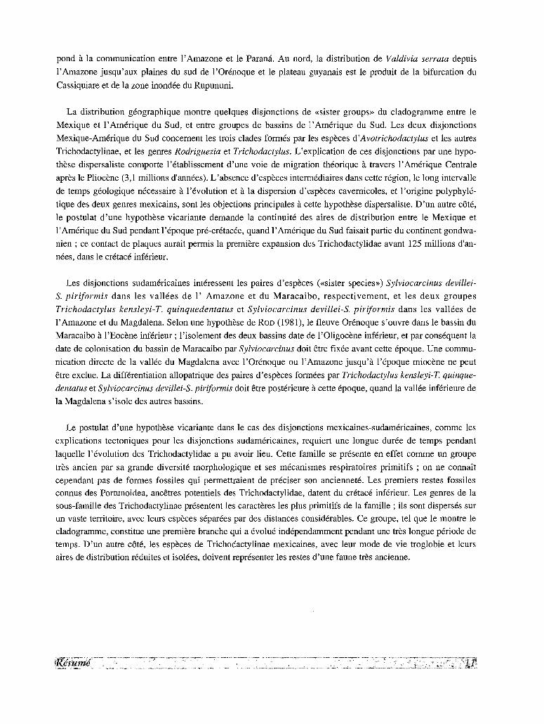
pond a la communication entre 1'Amazone et le Parana. Au nord, la distribution de Valdivia serrata depuis
I'Amazone jusqu'aux plaines du sud de I'Orenoque et le plateau guyanais est le produit de la bifurcation du
Cassiquiare et de la zone inondee du Rupununi.
La distribution geographique montre quelques disjonctions de «sister groups» du cladogramme entre le
Mexique et I' Amerique du Sud, et entre groupes de bassins de I' Amerique du Sud. Les deux disjonctions
Mexique-Amerique du Sud concement les trois clades formes par les especes d'Avotrichodactylus et les autres
Trichodactylinae, et les genres Rodriguezia et Trichodactylus. L'explication de ces disjonctions par une hypo
these dispersaliste comporte l'etablissement d'une voie de migration theorique a travers l'Amerique Centrale
apres le Pliocene (3,1 millions d'annees), L'absence d'especes intermediaires dans cette region, le long intervalle
de temps geologique necessaire a revolution et a la dispersion d'especes cavemicoles, et 1'origine polyphyle
tique des deux genres mexicains, sont les objections principales acette hypothese dispersaliste. D'un autre cote,
le postulat d 'une hypothese vicariante demande la continuite des aires de distribution entre le Mexique et
I'Amerique du Sud pendant l'epoque pre-cretacee, quand l'Amerique du Sud faisait partie du continent gondwa
nien ; ce contact de plaques aurait permis la premiere expansion des Trichodactylidae avant 125 millions d'an
nees, dans le cretace inferieur.
Les disjonctions sudamericaines interessent les paires d' especes (<<sister species») Sylviocarcinus devillei-
S. piriformis dans les vallees de I' Amazone et du Maracaibo, respectivement, et les deux groupes
Trichodactylus kensleyi-T. quinquedentatus et Sylviocarcinus devillei-S. piriformis dans les vallees de
l'Amazone et du Magdalena. Selon une hypothese de ROD (1981), le fleuve Orenoque s' ouvre dans le bassin du
Maracaibo aI'Eocene inferieur ; 1'isolement des deux bassins date de l'Oligocene inferieur, et par consequent la
date de colonisation du bassin de Maracaibo par Sylviocarcinus doit etre fixee avant cette epoque. Une commu
nication directe de la vallee du Magdalena avec I'Orenoque ou l'Amazone jusqu'a l'epoque miocene ne peut
etre exclue. La differentiation allopatrique des paires d'especes formees par Trichodactylus kensleyi-T. quinque
dentatus et Sylviocarcinus devillei-S. piriformis doit etre posterieure acette epoque, quand la vallee inferieure de
la Magdalena s'isole des autres bassins.
Le postulat d'une hypothese vicariante dans le cas des disjonctions mexicaines-sudamericaines, comme les
explications tectoniques pour les disjonctions sudamericaines, requiert une longue duree de temps pendant
laquelle revolution des Trichodactylidae a pu avoir lieu. Cette famille se presente en effet comme un groupe
tres ancien par sa grande diversite morphologique et ses mecanismes respiratoires primitifs ; on ne connait
cependant pas de formes fossiles qui permettraient de preciser son anciennete. Les premiers restes fossiles
connus des Portunoidea, ancetres potentiels des Trichodactylidae, datent du cretace inferieur, Les genres de la
sous-famille des Trichodactylinae presentent les caracteres les plus primitifs de la famille ; ils sont disperses sur
un vaste territoire, avec leurs especes separees par des distances considerables, Ce groupe, tel que le montre le
c1adogramme, constitue une premiere branche qui a evolue independarnment pendant une tres longue periode de
temps. D'un autre cote, les especes de Trichodactylinae mexicaines, avec 1eur mode de vie troglobie et leurs
aires de distribution reduites et isolees, doivent representer les restes d'une faune tres ancienne.

INTRODUCTION
The freshwater crabs are an important faunal
element in the inland waters of tropical America.
These crabs belong to two different families: (1) the
Pseudothelphusidae are found, with few exceptions,
in mountainous streams up to an altitude of 3,000
meters, within a geographical range which extends
from the State of Sonora in Northern Mexico, to
Central Peru. With three exceptions, they do not
extend south of the Amazon River. (2) The
Trichodactylidae inhabit the large rivers and lakes of
the low lands in the major continental basins of
South America east of the Andes, that is, the
Amazonas, Orinoco, Magdalena, Paraguay-Parana
and the smaller basins of the Guianese and Brazilian
coastal plains. Isolated from this main area, there are
also 5 species in the states of Tabasco, Veracruz,
Chiapas and Oaxaca in southern Mexico. With the
exception of one Colombian species reported from
Nicaragua, and of one mainland species which
extends into Trinidad, the Trichodactylidae are
absent from Central America and the Antilles.
The present monograph deals with the cladistics,
systematics and biogeography of the family
Trichodactylidae as part of a revision of the
freshwater crabs of America, published under the
auspices of the Orstom (Ins tit ut francais de
recherche scientifique pour le developpement en
cooperation); the Pseudothelphusidae were already
treated in a previous monograph of the series Faune
tropicale (ROORIGUEZ, 1982).
The taxonomic structure of the familyThe first description of a species of
Trichodactylidae, Cancer orbicularis, was published
by MEUSCHEN (1781). But since his Index
Zoophylacium Gronovianum is not accepted as a
precedent in binomial nomenclature, its junior
synonym, Cancer septemdentatus Herbst, 1783, is
considered the first valid name proposed for a
species of this family. From 1825 to 1901,
LATREILLE, Henri MILNE EOWAROS, RANOALL,
Evnoux & SOULEYET, GERSTA.KER, WHITE,
Alphonse MILNE EOWAROS, VON MARTENS,
KINGSLEY, GOLDI, STIMPSON, RATHBUN, ORTMANN,
NOBILI, and MOREIRA, added 26 new species.
RATHBUN'S monograph of 1906 included 29 species,
9 of which were new species. During the first half of
the twentieth century only three new species were
described by PEARSE (1911), MOREIRA (1912) and
PARISI (1923), respectively, but from 1966 there
have been a relatively copious literature. Thirteen
new species have been published in the
contributions by BOTT, COTTARELLI and ARGANO,
PRETZMANN, PRETZMANN and SCHMITT, PRETZMANN
and MAYTA, ROORIGUEZ and MANRIQUE, and
SMALLEY and ROORIGUEZ, bringing the total number
of published specific names to 52.
From this large number of taxa, probably no
more than 41 are good species. As a result, and
notwithstanding the relatively modest dimensions of
the family, its systematic is encumbered with a large
remnant of synonyms. In addition, BOTT and
PRETZMANN have described 7 and 15 subspecies
each. At least two of PRETzMANN'S subspecies are in
fact distinct species, but others cannot be
differentiated from the typical forms. This situation
is further complicated by the existence of different
competing systems of genera.
The taxonomic arrangement used by RATHBUN
(1906) for the family reflected the work done by
previous workers during the nineteenth century.
LATREILLE (1828), WHITE (1847a) and H. MILNE
EDWARDS (1853), respectively, had erected
Trichodactylus, Valdtvia, Sylviocarcinus, and
Dilocarcinus as separate genera. RATHBUN (1906)
gave generic status only to Trichodactylus, reduced
Valdivia and Dilocarcinus to subgenera of the first,
and discarded Sylviocarcinus. This simple
arrangement was used by all latter authors until
PRETZMANN (1968b) and BOTT (1969) proposed two
alternative systems, with little in common with
RATHBUN'S and between themselves (Appendix 1).
The differences in rank between these systems
imply also considerable differences in the grouping
of the species. For example, the species
Sylviocarcinus pictus H. Milne Edwards, 1853, is
placed by RATHBUN (1906) in the genus
Dilocarcinus, by BOTT (1969) in the genus
Sylviocarcinus and by PRETZMANN (1968b) as the
type species for his new genus Holthuisisia.
List of species
The list that follows indicates the classification
employed herein, based on the cladistic analysis
presented in the following section, and shows the
species numbers refered to in the appendices.
Family TRICHODACTYLIDAE H. Milne Edwards, 1853
Subfamily TRICHODACTYLINAE H. Milne Edwards, 1853
Genus Trichodactylus Latreille, 1828
1. Trichodactylus fluviatilis Latreille, 1828
2. Trichodactylus maytai Pretzmann, 1978
3. Trichodactylus kensleyi, new species
4. Trichodactylus petropolitanus (G61di, 1886)
5. Trichodactylus quinquedentatus Rathbun, 1893
6. Trichodactylus ehrhardti (Bott, 1969)
Genus Mikrotrichodactylus Pretzmann 1968
7. Mikrotrichodactylus borellianus Nobili, 1896
8. Mikrotrichodactylus panoplus (van Martens, 1869)
Genus Rodriguezia Bott, 1969
9. Rodriguezia mensabak (Cottarelli & Argano, 1977)
10. Rodriguezia villalobosi (Rodrfguez & Manrique,
1967)
Genus A votrichodactylus Pretzmann, 1968
11. Avotrichodactylus bidens (Bott, 1969)
12. Avotrichodactylus constrictus (Pearse, 1911)
13. Avotrichodactylus oaxensis, new species
Subfamily DILOCARCININAE Pretzmann, 1978
Tribe HOLTHUISIINI Pretzmann, 1978
Genus Sylviocarcinus H. Milne Edwards, 1853
14. Sylviocarcinus devillei H. Milne Edwards, 1853
15. Sylviocarcinus maldonadoensis (Pretzmann, 1978)
16. Sylviocarcinus pictus (H. Milne Edwards, 1853)
17. Sylviocarcinus piriformis (Pretzmann, 1968)
18. Sylviocarcinus sp.
Tribe VALDIVIINI Pretzmann, 1978
Genus Valdivia White, 1847
19. Valdivia camerani (Nobili, 1896)
20. Valdivia gila Pretzmann, 1978
21. Valdivia harttii (Rathbun, 1906)
22. Valdivia latidens (A. Milne Edwards, 1869)
23. Valdivia serrata White, 1847
Genus Forsteria Bott, 1969
24. Forsteria venezuelensis (Rathbun, 1906)
Tribe DlLOCARCININI Pretzmann, 1978
Genus Zilchiopsis Bott, 1969
25. Zilchiopsis chacei (Pretzrnann, 1968)
26. Zilchiopsis cryptodus (Ortmann, 1893)
27. Zilchiopsis emarginatus (H. Milne Edwards, 1853)
28. Zilchiopsis sattleri Bott, 1969
Genus Dilocarcinus H. Milne Edwards, 1853
29. Dilocarcinus argentinianus (Rathbun, 1906)
30. Dilocarcinus truncatus, new species
31. Dilocarcinus bulbifer, new species
32. Dilocarcinus castelnaui H. Milne Edwards, 1853
33. Dilocarcinus dentatus (Randall, 1839)34. Dilocarcinus laevijrons Moreira, 1901
35. Dilocarcinus medemi Smalley& Rodriguez, 197236. Dilocarcinus niceforei (Schmitt & Pretzmann,
1968)37. Dilocarcinus pagei Stimpson, 186138. Dilocarcinus septemdentatus (Herbst, 1783)39. Dilocarcinus spinifer H. MilneEdwards, 1853
GenusFredilocarcinus Pretzmann, 197840. Fredilocarcinus raddai (Pretzmann, 1978)41. Fredilocarcinus musmuschiae (Pretzmann &
Mayta, 1980)
species incertae sedis
42. Trichodactylus (Dilocarcinus) gurupensis Rathbun,1906
43. Trichodactylus petropolitanus paranensis Bott,1969
44. Trichodactylus (Valdivia)/axoni Rathbun, 1906
Collecting localities
The exact location of collecting places is
indispensable for the delimitation of the ranges of
the species. This task, however, is fraught with
difficulties in such a vast area as the plains of South
America, overall when the collector only gives the
name of a small village, a stream or even the nearest
farm. For these reasons I have appended a gazetteer
(Appendix 2) of all the localities mentioned in the
text or found in the literature, compiled with the
help of many sources, too numerous to be listed.
The Columbia Limpicott Gazetteer of the World
(1951) was used in many instances. The location of
several collecting stations recorded by BOTT (1969)
was provided by Dr Harald SIOLI through the
courtesy of Dr Hans KLINGE.
Repositories and abbreviationsThe materials reported herein are deposited in the
following institutions: Instituto Venezolano de
Investigaciones Cientificas, Caracas (Ivic); Museo
de Biologia, Universidad Central de Venezuela,
Caracas (MB); Museo de la Sociedad de Ciencias
Naturales La Salle, Caracas (LS) ; Museo de Historia
Natural De La Salle, Bogota (LSB); Museo de
Historia Natural, Universidad Nacional de Bogota
(ICN-MHN); Museo de Historia Natural, Universidad
Nacional de San Marcos, Lima (ML); Museo de la
Universidad de Santa Ursula, Recife (Usu); Museo de
Biologia, Universidad Nacional Autonoma, Mexico
(Unam); Museum national d'histoire naturelle, Paris
(MP); US National Museum, Washington (USNM);
Zoologisches Museum, Hamburg (MH); Zoologische
Staatssammlung, Munich (ZSM); Rijksmuseum van
Naturlijke Historie, Leiden (RNH). Other
abbreviations employed are: Disca = Division de
Investigaciones Sobre Contaminacion Ambiental,
Venezuela; cl = carapace length; cb = carapace
breadth; alt = altitude of collecting localities.
Aknowledgements
I wish to express my appreciation to the
following curators and specialists for making
material available to me: Marta CAMPOS (lCN
MHN), Jorge LAMAS, and Enrique DEL SOLAR (ML),
Marcos SIQUEIRA TAVARES (Usu), Jorge VILLALOBOS
IRIART (Unam), Jacques FOREST, Alain CROSNIER,
and Daniele GUINOT (MP), Raymond MANNING,
Horton H. HOBBS jr, and Bryan KENSLEY (USNM),
Horst WILKENS (MH), Ludwig TIEFENBACHER and
Ernst J. FITKAU (ZSM), and C. H. M. FRANSEN
(RNH).
Pierre LE L<EUFF, antenne Orstom, centre Ifremer,
Nantes, has kindly made all arrangements for the
publication of the present work in the collection
Faune tropicale.
I thank GuidoPEREIRA for his invaluable help
with the cladistic analysis, and Elfas RODRIGUEZ for
the adaptations of the computer programs used in
this analysis. Vicente CALLEJAS and Iliana
RODRIGUEZ executed most of the illustrations.
Hector SUAREZ gave invaluable help at different
stages of the work.

I· CLADISTIC ANALYSIS OF THE FAMILY TRICHODACTYLIDAE
The systematic position of the TRICHODACTYLIDAE
As has being pointed out by GUINOT (1978), the position of the sexual openings is a character of fundamental
phylogenetical significance in the Brachyura. All the freshwater crabs formerly grouped in the family Potamidae
belong in the section Heterotremata Guinot, 1977, i.e. brachyuran crabs in which the female opening is sternal,
but the male opening could be coxal or coxo-sternal. The heterotrematous condition suggests an evolutionary
process begining with the location of the penis in the coxa of the fifth pereiopod; in successive stages the penis
is lodged in a sternal groove which latter forms a channel due to the disposition of the sternites in this area;
finally 7th and 8th sternites completely cover the channel and the penis is implanted in the sternum, although the
male duct still reaches the coxa.
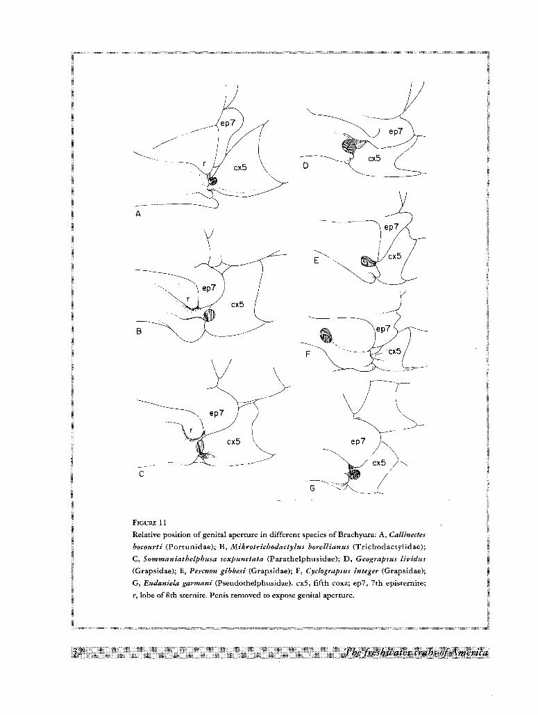
In the Trichodactylidae, as well as in some Asiatic freshwater crabs like Sommaniathelphusa sexpunctata
(Fig. llC), the penial groove is located along the 8th sternite; this disposition suggests a primitive condition
which could have led to the situation found in the Grapsidae. In some species of this latter family the penis is
implanted near the lateral margin of the 8th sternite (Fig. llD, E), while in others it is located away from the
margin, but leaving a slight furrow which suggests a progressive migration of the appendage along the mid-line
of the sternite (Fig. lIF). From this point of view, the penial groove of the Grapsidae could be considered as the
apomorphic state in relation to the one found in the Trichodactylidae and thus unavailable as an out-group for
the present cladistic analysis.The development of the penial groove followed a different path in other freshwater crabs. Thus, in the
Pseudothelphusidae, the penial groove is located between the posterior margin of the 7th episternite and the
anterior margin of the 8th sternite (Fig. llG). This is probably a primitive stage in the process leading to the
orifices located near the 7/8 sutures in the Ocypodidae and Pinnotheridae, as illustrated by GUINOT (1978,
Fig. 3H). Consequently, any close relationship between the two families of neotropical freshwater crabs should
be ruled out.
According to the heterotrematous condition of the Trichodactylidae, their closest phylogenetic affinities
should be looked for in other members of the Section in which the penial groove is not only rudimentary, but
also centrally located along the 8th sternite. Some members of the superfamily Portunoidea satisfy both
conditions (Fig.IIA) since their penial grooves form a very shallow depression along the 8th sternite. The
primitive condition of this groove is reflected also in the 7th episternite which do not overlap the 5th coxa. On
meso-
frontal---- ,),repi_ "-' ,proto-
A
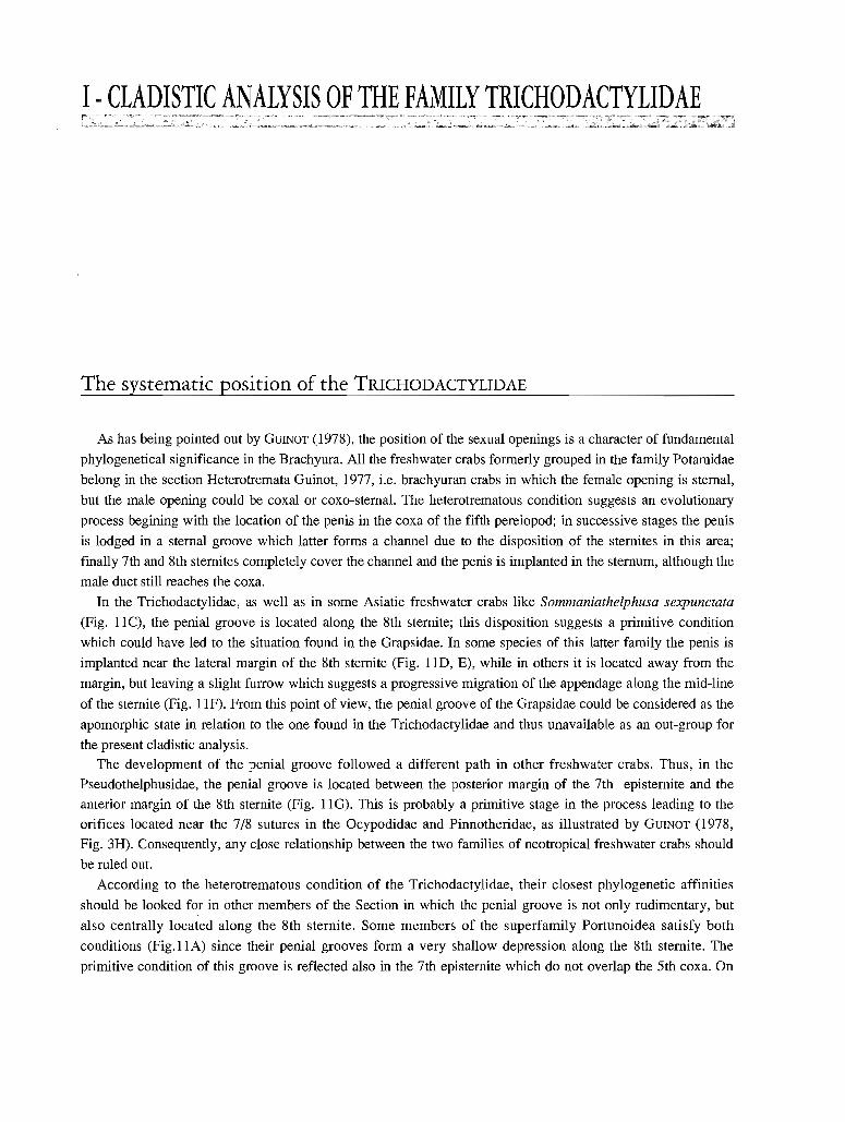
FIGURE 1
A, Carapace of a Trichodactylidae; B, carapace of Dilocarcinus pagei with horizontal and
vertical axes of symetry; C, plastron of Trichodactylus quinquedentatus, male, with abdomen
and left gonopods removed; D, buccal frame and endostome in Trichodactylus fluviatilis;
E, left posterior corner of plastron in T. fluviatilis; F-H, fourth epipodites (between 1st and
2nd pereiopods) in Valdivia serrata, Forsteria venezuelensis, and Dilocarcinus dentatus. as3,
3rd abdominal segment; as4, 4th abdominal segment; beg, branchio-cardiac groove; br,
branchial ridge; bug, branchio-urogastric groove; cig, cardio-intestinal groove; cx4, 4th coxa;
cx5, 5th coxa; el, elevated line; ep7, 7th episternite; gl, first male gonopod; hr, hepatic ridge;
on, orbital notch; p, penis; pp, postgastric pits; s7, 7th sternite; s8, 8th sternite.
the other hand, in some members of the Portunoidea the penial groove is supplemented by a rudimentary sternal
lobe (Fig. IIA; r), which is also present and more developed in the Trichodactylidae (Fig. llB, C; r). This
sternallobe should be considered as an autapomorphy shared by both groups.
Another possible autapomorphy shared by the Trichodactylidae and Portunoidea is the small lobe on the inner
angle of the endopodite of the 1st maxilliped ("portunid lobe", Fig. SA). In the Trichodactylidae it forms a
lateral expansion (Fig. 50-I) or rolls over to form an oval-shaped projection located on the cephalic surface of
the endopodite (Fig. 5B-F, K-N).
Finally, the reduced orbital suture present in the Trichodactylidae could be considered the apomorphic state
of the well developed suture found at least in some species of Portunidae.
Other characters shared by both groups are the following: (1) orbits complete, (2) antennules folding
slantwise or transversely, (3) carpus of third maxilliped articulating at or near the antero-Iateral angle of the
merus, (4) walking legs compressed, without spines, propodus and dactylus with upper and lower rows of setae.
Most species of Trichodactylidae fit into the definition of the subfamily Carcininae given by STEPHENSON
& CAMPBELL (1960): "legs stout and long, at least one pair as long as chelipeds, last pair with lanceolate
dactylus, but otherwise similar to the 3 other pairs. Carapace not broad, antero-lateral borders cut into 4 or 5
teeth. Basal joint of second antenna fixed, longer than broad, lying in longitudinal axis of carapace". Further,
the first male gonopod in many Trichodactylidae is simple, long, with the neck tapering evenly to a fine tip, with
the apex provided with stout spines and the apical opening (gonopore) V-shaped. A similar morphology is found
in Carcinus maenas (see STEPHENSON & CAMPBELL, 1960, Fig. lA, 2A) and other Carcininae.
According to the preceeding considerations, the Carcininae can be considered as the most likely sister group
of the Trichodactylidae and will be used as an out-group for the polarization of characters in the cladistic
analysis discussed below.
Characters examined
CARAPACE
The outline of carapace in the Trichodactylidae could be either hexagonal or suborbicular, but always it is
slightly wider than long. The hexagonal outline is related to the outline found in some Carcininae, like Carcinus
maenas, and thus should be considered as the plesiomorphic state of the character. The relative position of the
widest part of carapace on the longitudinal axis varies in different species from 36 % of the carapace length in
Zilchiopsis emarginatus to 57 % in Mikrotrichodactylus borellianus. The values show a normal unimodal
distribution around a mean of 43.0, with a strong skewness to the left (0.65). The slight bilateral asymetry found
in some specimens (Fig. lB) is associated with a strong development of the left cheliped.
The progressive smoothness of the upper surface is a character often accompanied by the progressive
convexity from front to back and in frontal view.
In figure lA is presented the nomenclature for the regions, grooves, etc., of carapace, used in the description
of the species. A more pronounced delimitation of the regions and the presence of ridges and grooves is closer
to the condition found in the Carcininae. This is the case of the transbranchial ridge present in several species,
the crescent shaped triangular prominence on each side of the mesogastric region in the species of Valdivia, and
cF
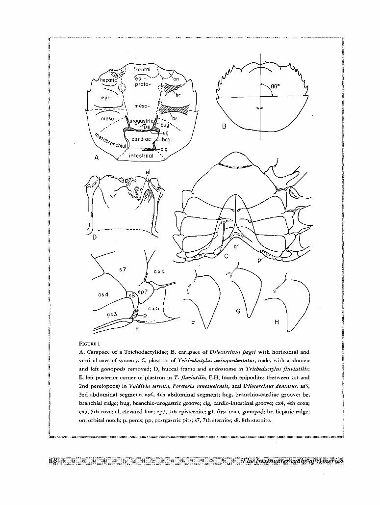
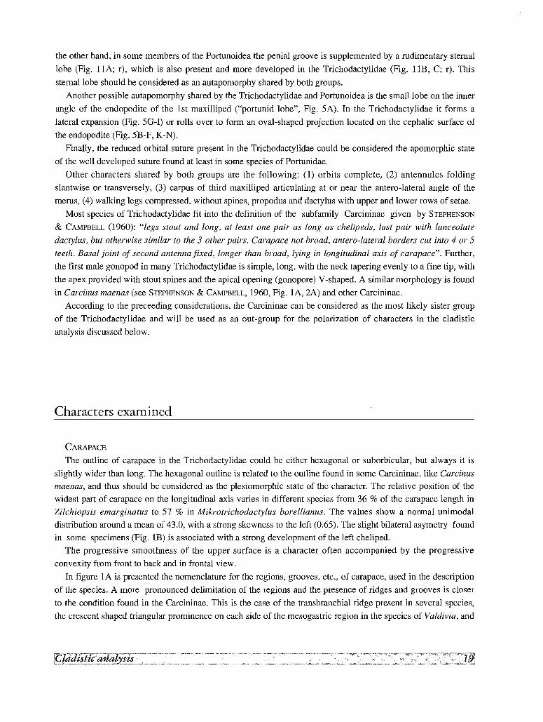
FIGURE 2
Orbital area: A, Avotrichodactylus constrictus; B, Mikrotrichodactylus borellianus; C, Valdivia camerani;
D, Dilocarcinus dentatus; E, D. bulbifer; F, Fredilocarcinus musmuschiae. ac, antennal cavity; ec, endostomial
cavity; oa, internal orbital angle; ot, occlusive tooth; ya, buccal angle.
~..............D - - - --- --'
~----
~
~-~~/ r -::~\~F . _
J K
FIGURE 3
Aperture of left efferent channel: A, 'Tricbodactylus fluviatilis; B, T. quinquedentatus; C, Avotrichodactylus oaxensis;
D, T. kensleyi; E, Mikrotrichodactylus borellianus; F, M. panoplies; G, Zllcblopsis emarginatus; H, Valdivia serrata;
I, Sylviocarcinus piriformis; J, Z. sattleri; K, Dilocarcinus truncatus, y, lateral yugallobe.
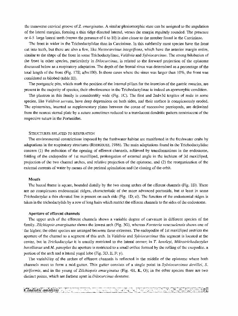
the transverse cervical groove of Z. emarginatus. A similar plesiomorphic state can be assigned to the angulation
of the lateral margins, forming a thin ridge directed laterad, versus the margin regularly rounded. The presence
or 4-5 large lateral teeth (versus the presence of 6 to 10) is also closer to the number found in the Carcininae.
The front is wider in the Trichodactylidae than in Carcininae. In this subfamily most species have the front
cut into teeth, but there are also a few, like Nectocarcinus integrifrons, which have the anterior margin entire,
similar to the shape of the front in some Trichodactylinae, Valdivia and Sylviocarcinus. The strong bilobation of
the front in other species, particularly in Dilocarcinus, is related to the forward projection of the epistome
discussed below as a respiratory adaptation. The depth of the frontal sinus was determined as a percentage of the
total length of the front (Fig. 17E; a/bx100). In those cases where the sinus was larger than 10%, the front was
considered as bilobed (table III).
The postgastric pits, which mark the position of the internal pillars for the insertion of the gastric muscles, are
present in the majority of species; their obsolescence in the Trichodactylinae is indeed an apomorphic condition.
The plastron in this family is considerably wide (Fig. lC). The first and 2nd+3d tergites of male in some
species, like Valdivia serrata, have deep depressions on both sides, and their surface is conspicuously eroded.
The episternites, inserted as supplementary plates between the coxae of successive pereiopods, are delimited
from the nearest sternal plate by a suture sometimes reduced to a translucent dendritic pattern reminiscent of the
respective suture in the Portunidae.
STRUCTURES RELATED TO RESPIRATION
The environmental constrictions imposed by the freshwater habitat are manifested in the freshwater crabs by
adaptations in the respiratory structures (RODRIGUEZ, 1986). The main adaptations found in the Trichodactylidae
concern (1) the reduction of the opening of efferent channels, achieved by transformations in the endostome,
folding of the endopodite of l st maxilliped, prolongation of external angle in the ischium of 3d maxilliped,
projection of the two channel arches, and relative projection of the epistome, and (2) the reorganization of the
external currents of water by means of the perioral spinulation and the closing of the orbit.
Mouth
The buccal frame is square, bounded distally by the two strong arches of the efferent channels (Fig. ID). There
are no conspicuous endostomial ridges, characteristic of the more advanced portunids, but at least in some
Trichodactylus a thin elevated line is present on each side (Fig. ID; el). The function of the endostomial ridges is
taken in the trichodactylids by a row of long hairs which restrict the efferent channels to the sides of the endostome.
Aperture of efferent channels
The upper arch of the efferent channels shows a variable degree of curvature in different species of the
family. Zilchiopsis emarginatus shows the lowest arch (Fig. 3G), whereas Forsteria venezuelensis shows one of
. the higher; the other species are arranged between these extremes. The endopodite of l st maxilliped restricts the
aperture of the channel to a segment of this arch. In Valdivia and Sylviocarcinus this segment is located at the
center, but in Trichodactylus it is usually restricted to the lateral corner; in T. kensleyi, Mikrotrichodactylus
borellianus and M. panoplus the aperture is restricted to a small orifice formed by the rolling of the exopodite, a
portion of the arch and a lateral yugallobe (Fig. 3D, E, F; y).
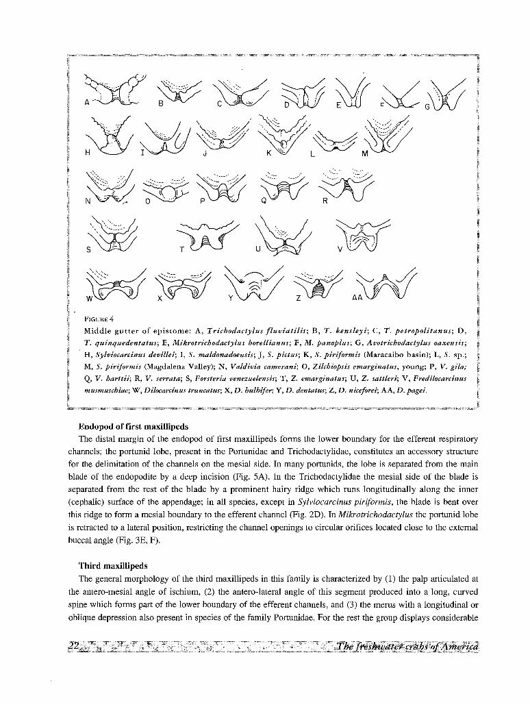
The variability of the arches of efferent channels is reflected in the middle of the epistome where both
channels meet to form a mid-gutter. This gutter consists of a single point in Svlviocarcinus devillei, S.
piriformis, and in the young of Zilchiopsis emarginatus (Fig. 4H, K, 0); in the other species there are two
distinct points, which are farthest apart in Dilocarcinus dentatus.
~~:~V' .
FIGURE 4
Middle gutter of epistome: A, Triehodaetylus flu·viatilis; B, T. kensleyi; C, T. petropolitanus; D,
T. quinquedentatus; E, Mikrotriehodaetylus borellianus; F, M. panoplus; G, Avotriehodaetylus oaxensis;
. H, Sylvioeareinus devillei; I, S. maldonadoensis; J, S. pietus; K, S. piriformis (Maracaibo basin); L, S.
M, S. piriformis (Magdalena Valley); N, Valdivia eamerani; 0, Zilehiopsis emarginatus, young; P, V.
Q, V. harttii; R, V. serrata; S, Forsteria venezuelensis; T, Z. emarginatus; U, Z. sattleri; V, Freaitocarcinus
musmuscbiae; W, Diloeareinus truneatus; X, D. bulbifer; Y, D. dentatus; Z, D. nieeforei; AA, D. pagei.
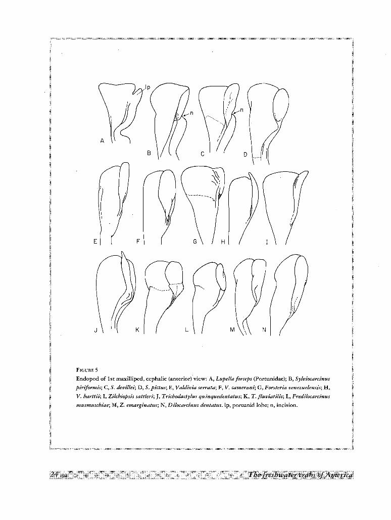
Endopod of first maxillipedsThe distal margin of the endopod of first maxillipeds forms the lower boundary for the efferent respiratory
channels; the portunid lobe, present in the Portunidae and Trichodactylidae, constitutes an accessory structure
for the delimitation of the channels on the mesial side. In many portunids, the lobe is separated from the main
blade of the endopodite by a deep incision (Fig. SA). In the Trichodactylidae the mesial side of the blade is
separated from the rest of the blade by a prominent hairy ridge which runs longitudinally along the inner
(cephalic) surface of the appendage; in all species, except in Sylviocarcinus piriformis, the blade is bent over
this ridge to form a mesial boundary to the efferent channel (Fig. 2D). In Mikrotrichodactylus the portunid lobe
is retracted to a lateral position, restricting the channel openings to circular orifices located close to the external
buccal angle (Fig. 3£, F).
Third maxillipeds
The general morphology of the third maxillipeds in this family is characterized by (1) the palp articulated at
the antero-mesial angle of ischium, (2) the antero-lateral angle of this segment produced into a long, curved
spine which forms part of the lower boundary of the efferent channels, and (3) the merus with a longitudinal or
oblique depression also present in species of the family Portunidae. For the rest the group displays considerable
specific variability, as follows. (A) The merus is trapezoidal in all species of Trichodactylinae and in some
species of other genera; in two species of this group, Dilocarcinus truncatus and D. bulbifer, the antero-mesial
angle is produced into a triangular tooth located near the articulation of the palp (Fig. 80, H; a); the distal
external spine is considerably reduced (r), particularly in the Trichodactylinae (Fig. 6B, D, E), but also in
Forsteria venezuelensis (Fig. 8A) and in some species of Sylviocarcinus (Fig. 7A). (B) The merus is
conspicuously narrow in some species of Valdivia, Sylviocarcinus, and Dilocarcinus; this reduction of the merus
is accompanied in some species of Dilocarcinus by a conspicuous slenderness of the exognath. (4) The ischium
is unusually wide in Zilchiopsis emarginatus.
Peristomial spinulation, orbits and basal antennal articles
Some freshwater crabs, in particular the Pseudothelphusidae, are capable of aerial respiration (DIAZ &
RODRIGUEZ, 1977), but, in the Trichodactylidae, respiration takes place only under water and the orientation of
the water coming through the efferent channels, away from the inhaling orifices at the base of the chelipeds, is
achieved by several spinuous borders, ridges and hairy areas. The peristomial spinulation in Trichodactylidae
comprises the lower orbital margin and the buccal angle, and both varies from smooth ridges, as in
Avotrichodactylus constrictus (Fig. 2A), to ridges provided with strong hooked spines, as in Dilocarcinus
dentatus and Fredilocarcinus musmuschiae (Fig. 2D, F).
In most species of Portunidae and Trichodactylidae the access of the water currents is kept away from the
orbits by means of a lateral expansion of the antennal basal article; this expansion is usually interpreted as the
exopod of this appendage. In the Portunidae the expansion is well developed and directly in contact with the
lower orbital margin (Fig. 9A); in the Trichodactylidae an occlusor tooth is interposed between the expansion
and the orbital margin, but both antennal expansion and occlusive tooth display considerable variability.
In the genera Sylviocarcinus (Fig. 9B-0) and Zilchiopsis (Fig. 91,J) the lobe is conspicuously developed. All
the species of Valdivia present a lobe moderately reduced, but the species of Trichodactylinae could be arranged
in a series (Fig. lOD-I) which displays a progressive reduction of this lobe, beginning with Trichodactylus
fluviatilis and T. petropolitanus, and ending in T. kensleyi. In Dilocarcinus and Fredilocarcinus the lobe is
completely absent (Fig. lOJ-L).
The reduction of the basal antennal expansion is accompanied by an increase in size of the occlusor tooth. In
Sylviocarcinus devillei and S. piriformis (Fig. lOB, C) the tooth is fused to the external orbital angle. This tooth
is progressively detached in the species of Sylviocarcinus and other genera; it becomes obsolescent in
Zilchiopsis sattleri (Fig. 9J), and even disappears in Trichodactylus petropolitanus (Fig. lOE).
The internal orbital angle is the third element closing the orbit on its inner angle. In most species it is well
developed, either spiniform or blunt, and directed upwards. However, in Fredilocarcinus musmuschiae and in
some Dilocarcinus (Fig. 2D-F), in addition to the reduction of the occlusive orbital tooth and the basal antennal
expansion, the outer orbital angle and the spines following it laterally are bent downwards, and thus the floor of
the orbital cavity is continuous with the epistome. In this particular case the orbits are very large in the vertical
direction and eyes very small, disproportionate to orbital cavity.
REPRODUCTIVE STRUCTURES
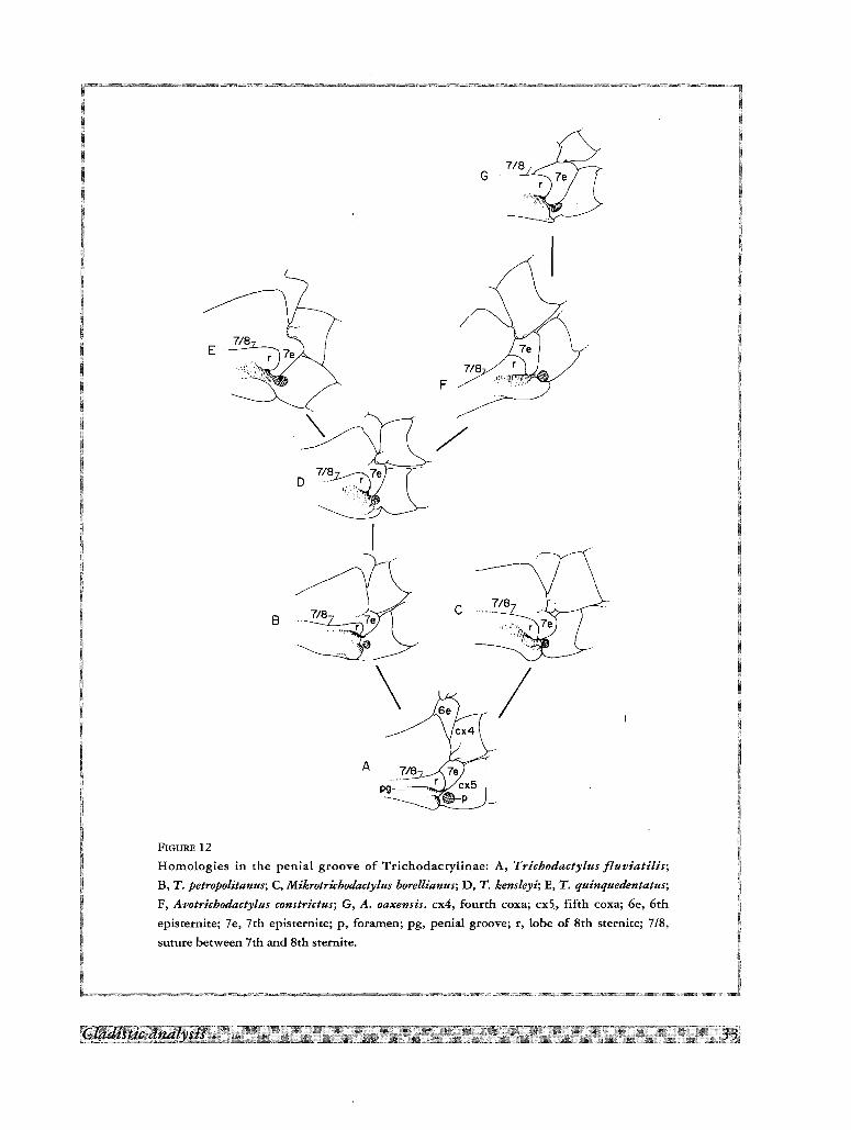
Penial groove
As discussed above, the male opening of Trichodactylidae is located in the coxa of the 5th pereiopod,
reaching the base of the first gonopod through a penial groove. This groove, however, does not show the same
degree of development in all species. In the most rudimentary condition found in Sylviocarcinus (Fig. 13) the
E
J L
FIGURE 5
Endopod of 1st maxilliped, cephalic (anterior) view: A, Lupella forceps (Portunidae); B, Sy/viocarcinus
piriformis; C, S. devillei; D, S. pictus; E, Va/divia serrata; F, V. carnerani; G, Forsteria venezue/ensis; H,
V. harttii; I, Zilcbiopsis satt/eri; J, Trichodacty/us quinquedentatus; K, T. jluviati/is; L, Fredi/ocarcinus
musmuscbiae; M, Z. emarglnatus; N, Di/ocarcinus dentatus. lp, portunid lobe; n, incision.
penial groove is a shallow depression and the surface of the sternite is not folded over it; consequently the
sternallobe is rudimentary. The 7th episternite can be elongated and narrow (Fig. l3A-F), or short and wide
(Fig. l3G-J), but it is not projected over the groove.
In the species of Trichodactylus, Mikrotrichodactylus and Avotrichodactylus (Fig. 12), the caudal portion of
the 7th episternite is narrow and displaced backwards, forming a spur which, together with the sternallobe (r),
partially covers the penial groove. These species can be serialized according to the progressive elongation of the
7th episternite, from the condition found in Trichodactylus fluviatilis and T. kensleyi (Fig. 12A, C) in which the
caudal spur is narrow and almost straight, to the spur strongly bent inwards of T. quinquedentatus (E), and
widened as in Avotrichodactylus oaxensis (G).
Male abdomen
A narrowing of the abdomen in many groups of Brachyura is associated with the displacement of the male
opening from a coxal to a sternal position. In the Trichodactylidae, although the male apertures are always
coxal, this trend is manifest in several species. Mikrotrichodactylus has a triangular abdomen, very short and
wide; but in Trichodactylus some species have a similar triangular abdomen, while others, like T. fluviatilis,
have a trapezoidal, narrower one, with the margins concave. Some species of Dilocarcinus possess a wide,
rounded abdomen. In all other species the abdomen is more or less trapezoidal.
In many groups of Brachyura there is a trend to the stenosis of the abdominal segments, which eventually
may end in the obsolescence of the abdominal sutures. Within the Trichodactylidae, only in the members of the
subfamily Trichodactylinae all abdominal sutures are clearly visible. In Sylviocarcinus piriformis all sutures are
partially visible. In S. devillei, S. maldonadoensis, the genus Avotrichodactylus, and the species of Valdivia, only
the sutures 6/7 and 7/8 are visible. In S. pictus, S. sp. and all the species of other genera only the 6/7 suture is
visible and mobile.
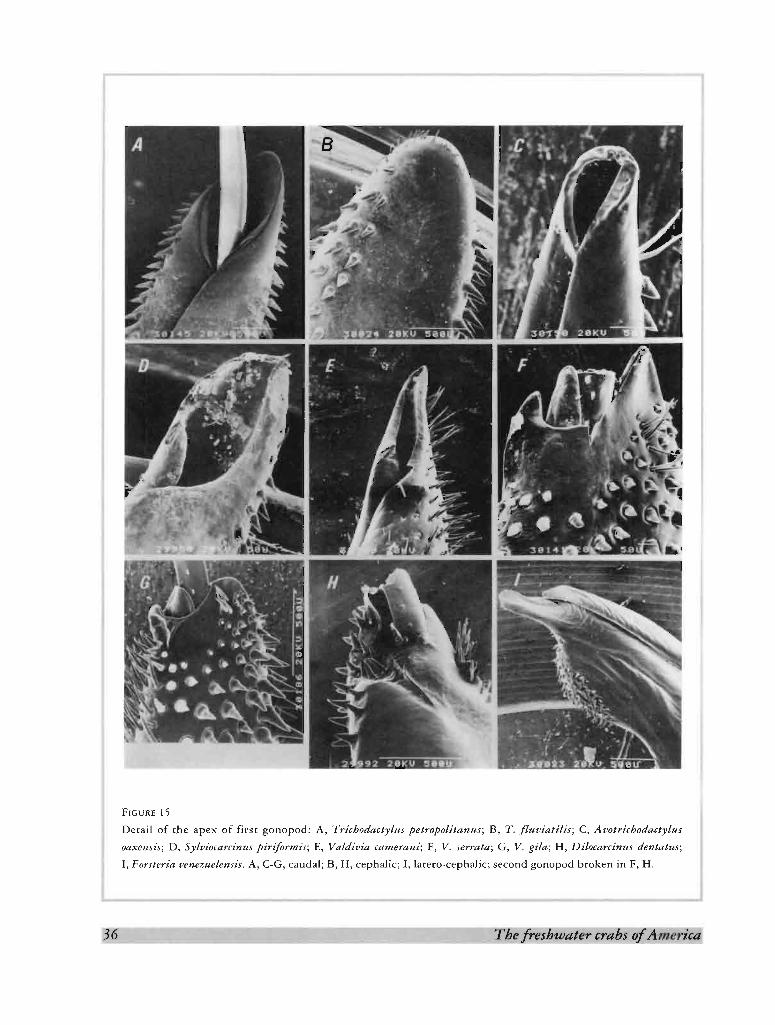
First male gonopod
As stated before, the conical shape of the first male gonopod is a plesiomorphic state. The Trichodactylinae
depart from this form and, in all of them, there are angular lobes on the mesial and lateral sides of the first
gonopod, giving to this appendage a flask-shaped appearance. On the other hand, the V-shaped apical opening
(gonopore) found also in the Trichodactylinae and in Sylviocarcinus, is a plesiomorphic state. All other species
have a terminal, slit-like gonopore, usually flanked by corneous processes (Fig. 15). The apical setae present in
some Trichodactylinae (with the exception of Avotrichodactylus, Fig. 15C) are small, scattered on the cephalic
surface (Fig. 15B) whereas in other species they are very long, conspicuous and grouped as a brush in the lateral
surface (Fig. 15).
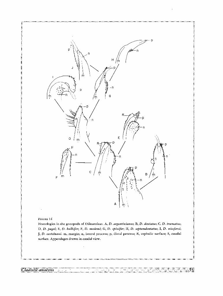
Notwithstanding the diversity of form displayed by the gonopods inside each genus, it is possible to dispose
them in series according to postulated homologies, as shown for Dilocarcinus (Fig. 14). The gonopod which
depart most from the general morphology found in the family is that of Fredilocarcinus musmuschiae, but even
here it is possible to place it in the last stages of the series of Dilocarcinus.
Second male gonopodThree types of male second gonopod are found within the Trichodactylidae, (1) very short, (2) S-shaped, of
equal length or slightly longer than first, and (3) very long, rolled up as a crosier. The second type possibly
corresponds to the apomorphic state found in the Carcininae. The Trichodactylidae do not possess the
specializations found in some species of Liocarcininae, particularly in Macropipus and other genera, in which
the apex of the second gonopod ends in a pseudochela due to the reduction of the terminal flagellum
Ac
II
I
I
I
I
I
~
II
I
I
I
I
I
I
I
!,
G
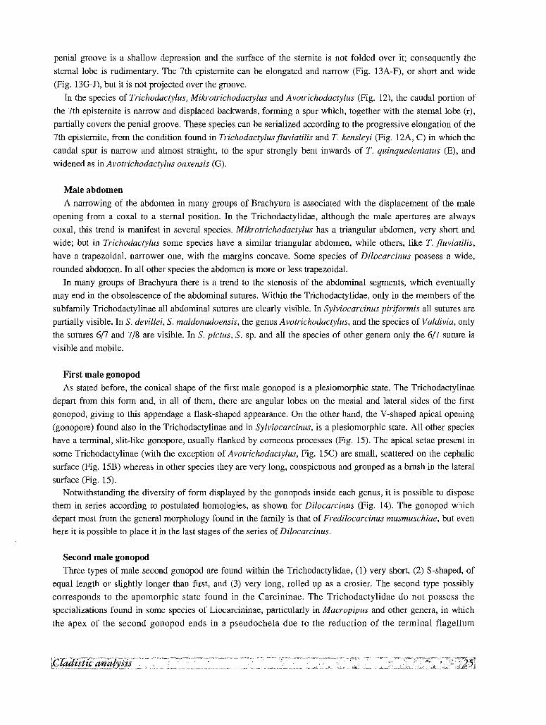
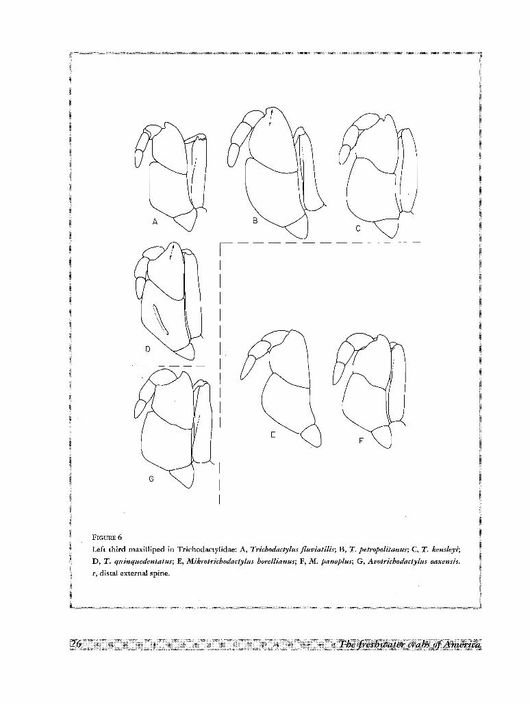
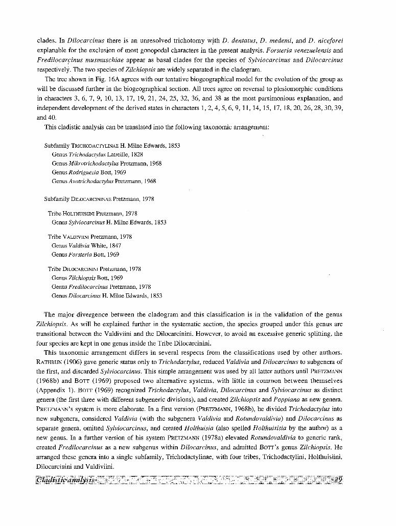
FIGURE 6
Left third maxilliped in Trichodacrylidae: A, Tricbodactyius fluviatilis; B, T. petropolitanus; C, T. kensleyi;
D, T. quinquedentatus; E, Mikrotrichodactylus horellianus; F, M. panoplus; G, Avotrichodactylus oaxensis.
r, distal external spine.
(ZARIQUIEY, 1968, Fig. 124, 126), but in the very short appendage of Rodriguezia and Avotrichodactylus, a
similar reduction is achieved (Fig. 23E, H; 24E).
PEREIOPODS
In the generalized condition the walking legs are stout, with the propodus widened (length/width == 1.2-1.8),
the dactylus lanceolate, and the lower margin of dactylus and propodus densely covered by long setae. In a few
species the legs are more slender (length/width of propodus == 2, or more; length/ width of merus >4). The two
cave species found in the family have, of course, extremely long pereiopods. The setation is also different in
some Trichodactylinae: the long setae are replaced by sparse short hairs, or are totally absent, and the sides of
the dactylus are covered by a felt-like tomentum (Fig. 171; Fig. 221).
In most species the chelipeds are stout and conspicuously unequal in size, but in Valdivia gila this unequality
reaches its maximum, with a cheliped considerably larger than the carapace. In many species the chelipeds
possess longitudinal grooves along the external surfaces of the fingers. This condition is reminiscent of the
grooved chelipeds of Portunidae, and is more manifest in the smaller cheliped. In a few species, the grooves are
obsolescent.
Data Analysis
In contrast with the Pseudothelphusidae in which most reliable characters for the cladistic analysis are found
in the first gonopod (RoDRIGUEZ & CAMPOS, 1989), in the Trichodactylidae there are many somatic characters
available. Of all the characters discussed before, 40 were selected for the cladistic analysis (table I). Several
were rejected because of their continuous distribution and others were not used because of their intraspecific
variability. Only two characters from those of the first male gonopod were selected due to the difficulties
encountered in homologating the characters in conical and flask-shaped types of gonopods. The species included
in the phylogenetic analysis are listed in table Il. Dilocarcinus castelnaui, D. argentinianus and D. spinifer were
excluded because the carapace morphology in the genus Dilocarcinus is rather uniform and the excluded species
did not contribute new information to the analysis. A few other species in other genera were also excluded
because some of the morphological data required for the matrix of characters were not available.The phylogenetic programs used were Phylip (Phylogeny Inference Program) 3.0 (FELSENSTEIN, 1984)
routings MIX, and PAUP (Phylogenetic Analysis Using Parsimony) 2.2 routing BAND B that guarantee the
finding of all the most parsimonious trees. A strict consensus was obtained by using the program CONTREE
included in PAUP.
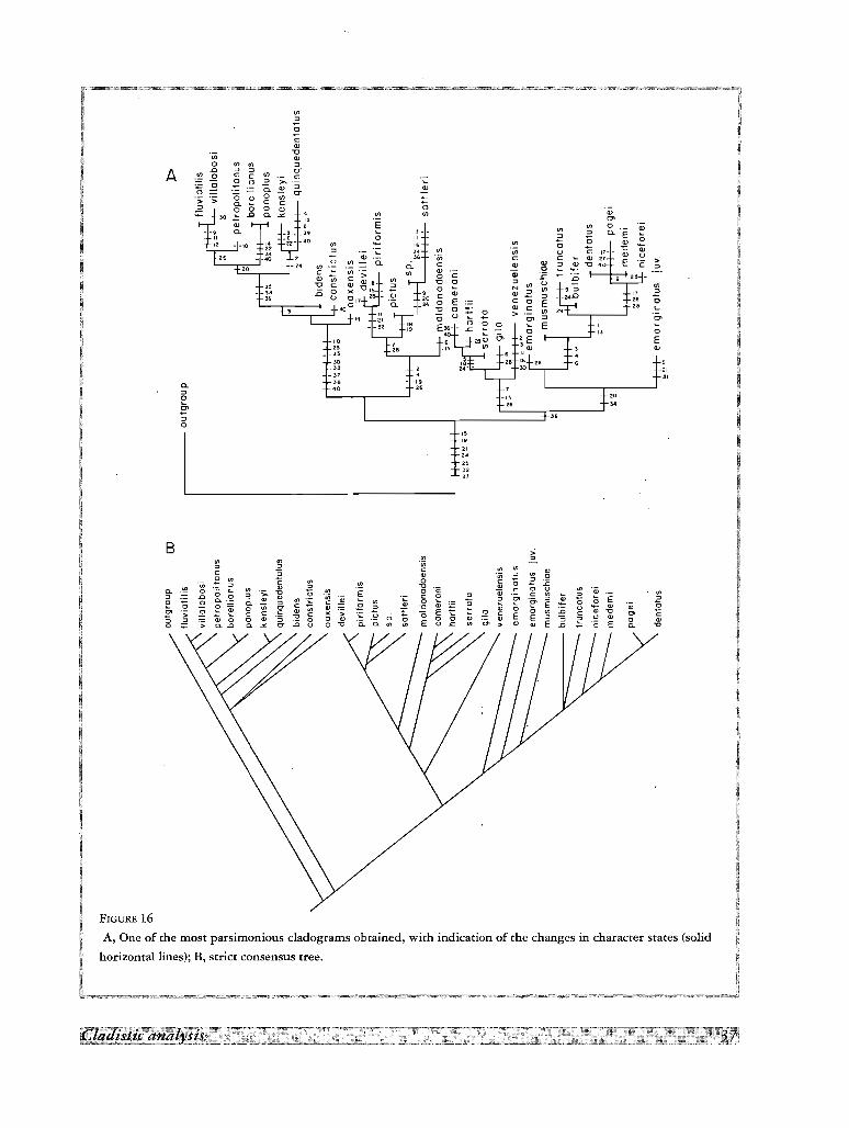
Five and 18 more parsimonious trees with 108 steps and Consistency Index of 0.37 were found with
programs Phylip and PAUP, respectively. An individual tree with character changes is shown in Fig. 16A. The
strict consensus tree in Fig. 16B summarizes the point of agreement in all trees. The species of Trichodactylus,
Valdivia and Sylviocarcinus forms well-defined monophyletic groups. Rodriguezia appears nested in
Trichodactylus and the three species of Avotrichodactylus form three distinct clades closely associated, but A.
constrictus and A. oaxensis forms a dichotomy in the strict consensus tree which is readily resolved by the
diagnostic characters of both species. The species Sylviocarcinus and Valdivia form two well differentiated
D
II
I
I,
I
I
I
IG
c
F
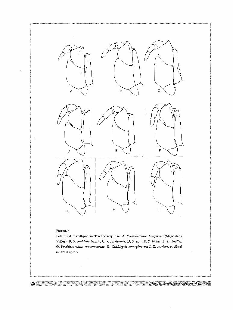
FIGURE 7
Left third maxilliped in Trichodacrylidae: A, Sylviocarcinus piriformis (Magdalena
Valley); B, S. maldonadoensis; C, S. piriformis; D, S. sp. ; E, S. pictus; F, S. devillei;
G, Fredilocarcinus musmuschiae; H, Zilchiopsis emarginatus; I, Z. sattleri. r, distal
external spine.
clades. In Dilocarcinus there is an unresolved trichotomy with D. dentatus, D. medemi, and D. niceforei
explanable for the exclusion of most gonopodal characters in the present analysis. Forsteria venezuelensis and
Fredilocarcinus musmuschiae appear as basal clades for the species of Sylviocarcinus and Dilocarcinus
respectively. The two species of Zilchiopsis are widely separated in the cladogram.
The tree shown in Fig. l6A agrees with our tentative biogeographical model for the evolution of the group as
will be discussed further in the biogeographical section. All trees agree on reversal to plesiomorphic conditions
in characters 3, 6, 7, 9, 10, 13, 17, 19, 21, 24, 25, 32, 36, and 38 as the most parsimonious explanation, and
independent development of the derived states in characters 1,2,4,5,6,9, 11, 14, 15, 17, 18,20,26,28,30,39,
and 40.
This cladistic analysis can be translated into the following taxonomic arrangement:
Subfamily TRICHODACTYLINAE H. Milne Edwards, 1853
Genus Trichodactylus Latreille, 1828
Genus Mikrotrichodactylus Pretzmann, 1968
Genus Rodriguezia Bott, 1969
Genus Avotrichodactylus Pretzmann, 1968
Subfamily DILOCARCININAE Pretzmann, 1978
Tribe HOLTHUISIINI Pretzmann, 1978
Genus Sylviocarcinus H. Milne Edwards, 1853
Tribe VALDIVIINI Pretzmann, 1978
Genus Valdivia White, 1847
Genus Forsteria Bott, 1969
Tribe DlLOCARCININI Pretzmann, 1978
Genus Zilchiopsis Bott, 1969
Genus Fredilocarcinus Pretzmann, 1978
Genus Dilocarcinus H. Milne Edwards, 1853
The major divergence between the cladogram and this classification is in the validation of the genus
Zilchiopsis. As will be explained further in the systematic section, the species grouped under this genus are
transitional between the Valdiviini and the Dilocarcinini. However, to avoid an excessive generic splitting, the
four species are kept in one genus inside the Tribe Dilocarcinini.This taxonomic arrangement differs in several respects from the classifications used by other authors.
RATHBUN (1906) gave generic status only to Trichodactylus, reduced Valdivia and Dilocarcinus to subgenera of
the first, and discarded Sylviocarcinus. This simple arrangement was used by all latter authors until PRETZMANN
(1968b) and BOTT (1969) proposed two alternative systems, with little in common between themselves
(Appendix 1). BOTT (1969) recognized Trichodactylus, Valdivia, Dilocarcinus and Sylviocarcinus as distinct
genera (the first three with different subgeneric divisions), and created Zilchiopsis and Poppiana as new genera.
PRETZMANN'S system is more elaborate. In a first version (PRETZMANN, 1968b), he divided Trichodactylus into
new subgenera, considered Valdivia (with the subgenera Valdivia and Rotundovaldivia) and Dilocarcinus as
separate genera, omitted Sylviocarcinus, and created Holthuisia (also spelled Holthuisisia by the author) as a
new genus. In a further version of his system PRETZMANN (1978a) elevated Rotundovaldivia to generic rank,
created Fredilocarcinus as a new subgenus within Dilocarcinus, and admitted BOTT'S genus Zilchiopsis. He
arranged these genera into a single subfamily, Trichodactylinae, with four tribes, Trichodactylini, Holthuisiini,
Dilocarcinini and Valdiviini.
IIIIII
I
I
III~
B
H
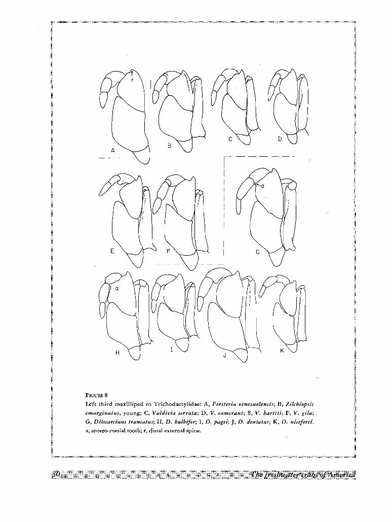
FIGURE 8
Left third maxilliped in Trichodactylidae: A, Forsteria venezuelensis; B, Zilchiopsis
emarginatus, young; C, Valdivia serrata; D, V. camerani; E, V. harttii; F, V. gila;
G, Dilocarcinus truncatus; H, D. bulbifer; I, D. pagei; ], D. dentatus; K, D. niceforei.
a, antero-mesial tooth; r, distal external spine.
.~~t~I
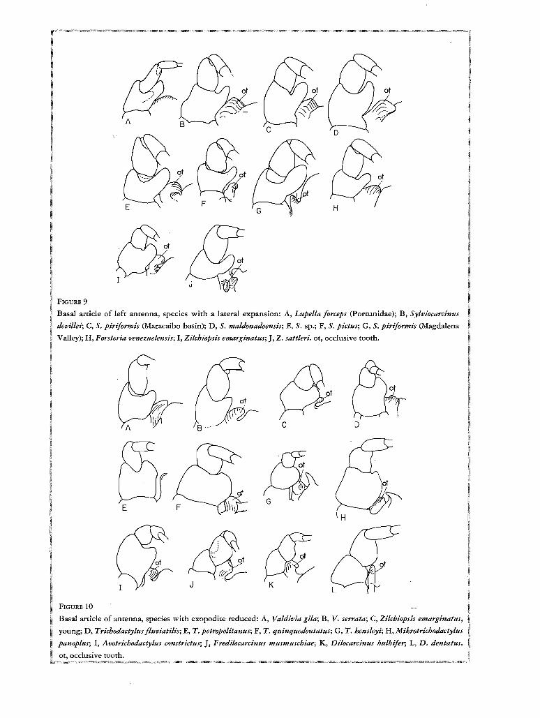
FIGURE 9
Basal article of left antenna, species with a lateral expansion: A, Lupe//a forceps (Portunidae); B, Syluiocarcinns
devillei; C, S. piriformis (Maracaibo basin); D, S. maldonadoensis; E, S. sp.; F, S. pictus; G, S. piriformis (Magdalena
Valley); H, Forsteria venezuelensis; I, Zilcbiopsis emarginatus;], Z. sattleri. ot, occlusive tooth.
c D
$~J K
FIGURE 10
Basal article of antenna, species with exopodire reduced: A, Valdivia gila; B, V. serrata; C, Zilcbiopsis emarginatus,
young; D, Trichodactylus f/uviatilis; E, T. petropolitanus; F, T. quinquedentatus; G, T. kensleyi; H, Ml;kr,otrieh'oaactyhes
panoplus; I, Avotrichodactyltts constrictus; ], Fredilocarcinus musmuscbiae; K, Dilocarcinus bulbifer; L, D. dentatus.
ot, occlusive tooth.
re,(/(/ :~cx5
.... ~.....
A
'---------
ep7
•~~ CX5...., /
G--------<·····_~
F
c
B
FIGURE 11
Relative position of genital aperture in different species ·of Brachyura: A, Callinectes
bocourti (Porrunidae), B, Mikrotrichodactylus borellianus (Trichodactylidae);
C, Sommaniathelphusa sexpunctata (Parathelphusidae); D, Geograpsus lividus
(Grapsidae); E, Percnon gibbesi (Grapsidae); F, Cyclograpsus integer (Grapsidae);
G, Eudaniela garmani (Pseudothelphusidae). cxS, fifth coxa; ep7, 7th episrernite;
r, lobe of 8th sternire, Penis removed to expose genital aperture.
G
E
D
../
F
/
B 7/8
A
\
c
/
FIGURE 12
Homo1ogies in the penial groove of Trichodactylinae: A, Trichodactylus fluviatilis;
B, T. petropolitanus; C, MikrotrichoiUutylus borellianus; D, T. kensleyi; E, T. quinquedentatus;
F, Avotrichodactylus constrictus; G, A. oaxensis, cx4, fourth coxa; cxfi, fifth coxa; 6e, 6th
episrernite; 7e, 7th episternite; p, foramen; pg, penial groove; r, lobe of 8th sternite; 7/8,
suture between 7th and 8th sternite.
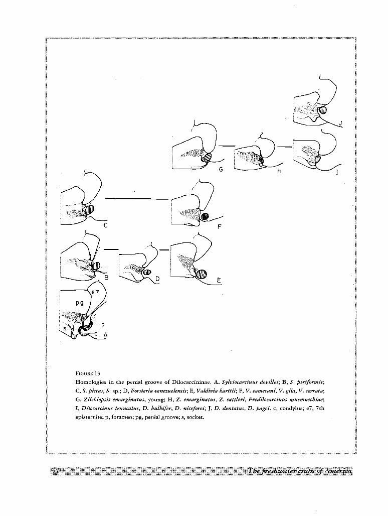
FIGURE 13
Homologies in the penial groove of Dilocarcininae. A. Sylvioeareinus devillei; B, S. piriformis;
C, S. pictus, S. sp.; D, Forsteria venezuelensis; E, Valdivia harttii; F, V. eamerani, V. gila, V. serrata;
G, Zilehiopsis emarginatus, young; H, Z. emarginatus, Z. sattleri, Frediloeareinus musmusehiae;
I, Diloeareinus truncatus, D. bulbifer, D. niceforei;], D. dentatus, D. pagei. c, condylus; e7, 7th
episrernite; p, foramen; pg, penial groove; s, socket.
F
\A m', "
FIGURE 14
Homo1ogies in the gonopods of Dilocarcinus. A, D. argentinianus; B, D. dentatus; C, D. truncatus;
D, D. pagei; E, D. bulbifer; F, D. medemi; G, D. spinifer; H, D. septemdentatus; I, D. niceforei;
J, D. castelnaui. m, margin; n, lateral process; p, dista1 process; R, cephalic surface; S, caudal
surface. Appendages drawn in caudal view.
36
FIGUR E 15
Detail of th e apex o f fir st go no po d: A, T richodactylus petropolitanus; B, T. [lnuiatilis; C, Acotricboda cty lu s
oaxensis ; D, Syloiocarcinus p irif orm is; E, Va ldi via cameraru; F, V. serra ta; G, V . gi /a ; H, Dilocarcinus dentatus;
I, Forsteriaoen ezuelen sis, A, C-G, caud al; B, H , cep hal ic; I, larero-ceph alic; second go no po d brok en in F, H .
The freshwater crabs o[America
~
Cl>
0Cl>V> V> ry
E V>0 Cl>
7 - - a.0 " ~ (;- " 0 Q;~ " V> U
Cl> se c: c: u :>Cl> :J :J
c: Cl>
~:J
V>NCl> ~c: 0Cl> c:>
'0
"-"'0"__ 31
se40
V>
~0
cCl>
V>-0Cl>
0 V> on :J
A .0 :J :J er~ .2 c: c c:
.2 .2 0:Jer
's
""ar24
""at
B :>on on on "" :J "in
V> V>c: 0 c: "<1> "in " V> 0~ V> C V> V> 0 c 0 Z E"in " <1> " "0 <1>
0- V> 0 c; V> "0
~V> § 0 "c a; c: 0 :;: V> "0; V>
" .D 0 0 ~,..
<1> "in 'c c e 0
" 0>c
" " E "e "0 0 0- 0.<1> :J V> c; ~ V> 0
~~
.~ E s "0 .2 0~ ~ 0 V> 0- c;
V> ~.E " s "0 <1>
~ <1> 0 :0 o <1> <1>0> 0 c: c; <1>
~ E ~ c 0 on c: <1> "0 0> C> c "0 c: > 'c~
0 E :J U <1>-; " Q; 0 0 <1> "3 0 0 <1> 0. 0 0 <1> '" E :;~ 0 <1>
0 ;;:: ";; 0. .D 0. '" 0- :0 o 0 "0 0. 0. V> E o s: V> 0> > <1> <1> E .D c E 0. "0
FIGURE 16A, One of the most parsimonious cladograms obtained, with indication of the changes in character states (solid
horizontal lines); B, strict consensus tree.
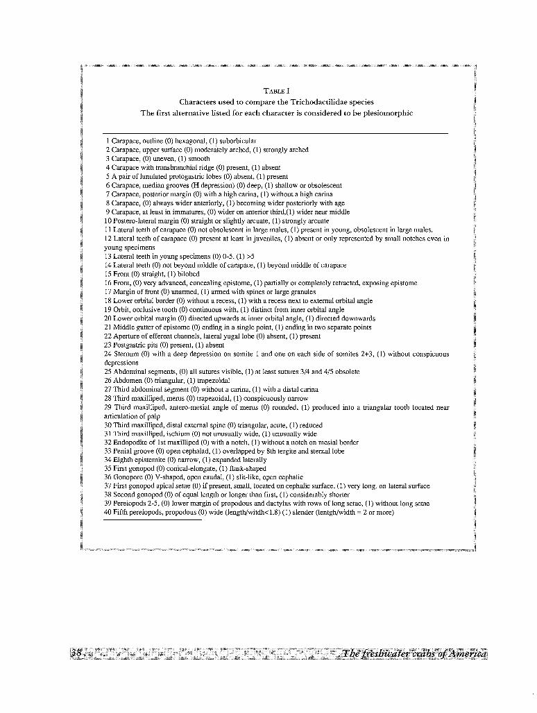
TABLE I
Characters used to compare the Trichodactilidae species
The first alternative listed for each character is considered to be plesiomorphic
I Carapace, outline (0) hexagonal, (1) suborbicular2 Carapace, upper surface (0) moderately arched, (1) strongly arched3 Carapace, (0) uneven, (1) smooth4 Carapace with transbranchial ridge (0) present, (1) absent5 A pair of lunulated protogastric lobes (0) absent, (1) present6 Carapace, median grooves (H depression) (0) deep, (I) shallow or obsolescent7 Carapace, posterior margin (0) with a high carina, (1) without a high carina8 Carapace, (0) always wider anteriorly, (1) becoming wider posteriorly with age9 Carapace, at least in immatures, (0) wider on anterior third,(1) wider near middlela Postero-Iateral margin (0) straight or slightly arcuate, (1) strongly arcuateII Lateral teeth of carapace (0) not obsolescent in large males, (1) present in young, obsolescent in large males.12 Lateral teeth of carapace (0) present at least in juveniles, (1) absent or only represented by small notches even inyoung specimens13 Lateral teeth in young specimens (0) 0-5, (1) >514 Lateral teeth (0) not beyond middle of carapace, (1) beyond middle of carapace15 Front (0) straight, (1) bilobed16 Front, (0) very advanced, concealing epistome, (1) partially or completely retracted, exposing epistome17 Margin of front (0) unarmed, (1) armed with spines or large granules18 Lower orbital border (0) without a recess, (1) with a recess next to external orbital angle19 Orbit, occlusive tooth (0) continuous with, (1) distinct from inner orbital angle20 Lower orbital margin (0) directed upwards at inner orbital angle, (1) directed downwards21 Middle gutter of epistome (0) ending in a single point, (1) ending in two separate points22 Aperture of efferent channels, lateral yugallobe (0) absent, (1) present23 Postgastric pits (0) present, (1) absent24 Sternum (0) with a deep depression on somite 1 and one on each side of somites 2+3, (1) without conspicuousdepressions25 Abdominal segments, (0) all sutures visible, (1) at least sutures 3/4 and 4/5 obsolete26 Abdomen (0) triangular, (1) trapezoidal27 Third abdominal segment (0) without a carina, (1) with a distal carina28 Third maxilliped, merus (0) trapezoidal, (1) conspicuously narrow29 Third maxilliped, antero-mesial angle of merus (0) rounded, (l) produced into a triangular tooth located neararticulation of palp30 Third maxilliped, distal external spine (0) triangular, acute, (1) reduced31 Third maxilliped, ischium (0) not unusually wide, (1) unusually wide32 Endopodite of 1st maxilliped (0) with a notch, (1) without a notch on mesial border33 Penial groove (0) open cephalad, (1) overlapped by 8th tergite and sternallobe34 Eighth episternite (0) narrow, (1) expanded laterally35 First gonopod (0) conical-elongate, (1) flask-shaped36 Gonopore (0) V-shaped, open caudal, (1) slit-like, open cephalic37 First gonopod apical setae (0) if present, small, located on cephalic surface, (1) very long, on lateral surface38 Second gonopod (0) of equal length or longer than first, (1) considerably shorter39 Pereiopods 2-5, (0) lower margin of propodous and dactylus with rows of long setae, (1) without long setae40 Fifth pereiopods, propodous (0) wide (length/witdh<1.8) (1) slender (lentgh/width =2 or more)
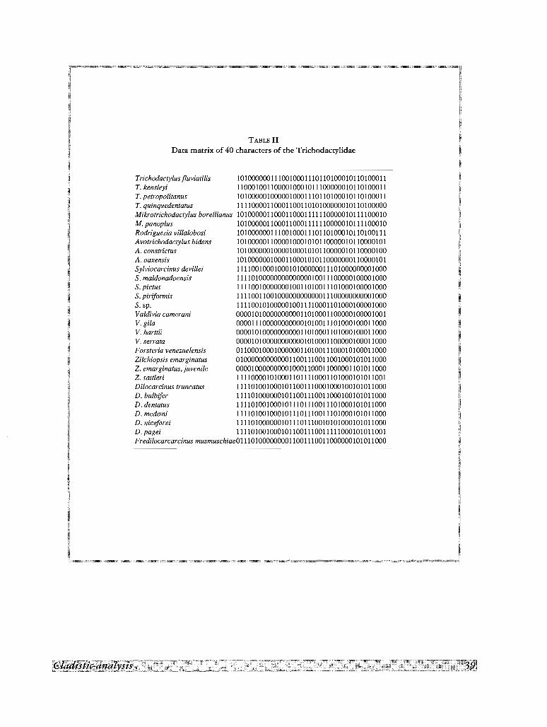
TABLE 11Data matrix of 40 characters of the Trichodacrylidae
Trichodactylusfluviatilis 1010000001110010001110110100010110100011T. kensleyi 1100010011000010001011100000010110100011T. petropolitanus 1010000010000010001110110100010110100011T. quinquedentatus 1111000011000110011010100000010110100000Mikrotrichodactylus borellianus 1010000011000110001111110000010111100010M. panoplus 1010000011000110001111110000010111100010Rodriguezia villalobosi 1010000001110010001110110100010110100111Avotrichodactylus bidens 1010000011000010001010110000010110000101A.cons~ictus 1010000001000010001010110000010110000100A.oaxens~ 1010000001000110001010110000000110000101Sylviocarcinus devillei 1111001000100010100000011101000000001000S. maldonadoensis 1111010000000000000010011100000100001000S.pictus 1111001000000010011010011101000100001000S. piriformis 1111001100100000000000011100000000001000S.sp. 1111001010000010011110001101000100001000Valdivia camerani 0000101000000000011010001100000100001001V. gila 0000111000000000001010011101000100011000V. harttii 0000101000000000011010001101000100011000V. serrata 00001010000000000010100011 00000100011000Forsteria venezuelensis 0110001000100000011010011100010100011000Zilchiopsis emarginatus 0100000000000011001110011001000101011000Z. emarginatus,juvenile 0000100000000010001100011000001101011000Z. sattleri 1111000010100011011110001101000101011001Dilocarcinus truncatus 1111010010001011001110001000100101011000D. bulbifer 1111010000001011001110011000100101011000D. dentatus 1111010010001011101110011101000101011000D. medemi 1111010010001011101110011101000101011000D. niceforei 1111010000001011101110010101000101011000D.pagei 1111010010001011001110011111000101011001Fredilocarcarcinus musmuschiae0111010000000011001110011000000101011000

jjjjjjjjjjjjjjjjjjjjjjjjjjjjjjjjjjjjjjjjjjjjjjjjjjjjjjjjjjjj
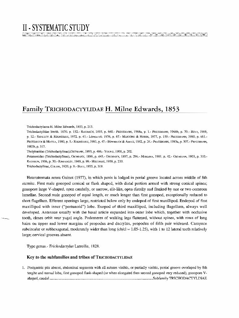
11· SYSTEMATIC STUDY
Family TRICHODACTYLIDAE H. Milne Edwards, 1853
Trichodactylacea H. Milne Edwards, 1853, p. 213.
Trichodactylidae Smith, 1870, p. 152.- RATHBUN, 1893, p. 660.- PRETZMANN, 1968a, p. 1.- PRETZMANN, 1968b, p. 70.- BOTT, 1969,
p. 12.- SMALLEY & RODRlGUEZ, 1972, p. 41.- LOPRETTO, 1976, p. 67.- MANNING & HOBBS, 1977, p. 159.- PRETZMANN, 1980, p. 661.
PRETZMANN & MAYTA, 1980, p. 5.- RODRiGUEZ, 1981, p. 47.- BOWMANN & ABELE, 1982, p. 24.- PRETZMANN, 1983a, p. 307.- PRETZMANN,
1983b, p. 317.
Thelphusidae (Trichodactylinae),ORTMANN, 1893, p. 486.- YOUNG, 1900, p. 202.
Potamonidae (Trichodactylinae), ORTMANN, 1896, p. 445.- ORTMANN, 1897, p. 296.- MOREIRA, 1901, p. 42.- ORTMANN, 1903, p. 310.
RATHBUN, 1906, p. 30.- RINGUELET, 1949, p. 99.- HOLTHUIS, 1959, p. 210.
Trichodacty1inae,COLOSI, 1920, p. 9.- BOTT, 1955, p. 319.
Heterotremata sensu Guinot (1977), in which penis is lodged in penial groove located across middle of 8th
sternite. First male gonopod conical or flask shaped, with distal portion armed with strong conical spines;
gonopore large V-shaped, open caudally, or narrow, slit-like, open distally and flanked by one or two corneous
lamellae. Second male gonopod of equal length, or much longer than first gonopod, exceptionally reduced to
short flagellum. Efferent openings large, restricted below only by endopod of first maxilliped. Endopod of first
maxilliped with inner ("portunoid") lobe. Exopod of third maxilliped, including flagellum, always well
developed. Antennae usually with the basal article expanded into outer lobe which, together with occlusive
_____. tooth, closes orbit near yugal angle. Podomeres of walking legs flattened, without spines, with rows of long
hairs on upper and lower margins of propodus and dactylus, propodus of fifth pair widened. Carapace
subcircular or subhexagonal, moderately wider than long (cb/cl == 1.05-1.25), with 1 to 12 lateral teeth relatively
large; cervical grooves absent.
Type genus.- Trichodactylus Latreille, 1828.
Key to the subfamilies and tribes of TRICHODACTYLIDAE
1. Postgastric pits absent, abdominal segments with all sutures visible, or partially visible, penial grooveoverlaped by 8thtergiteand stemallobe, first gonopod flask-shaped (or when elongated then secondgonopod veryreduced), gonopore V-shaped, caudal Subfamily TRICHODACTYLINAE
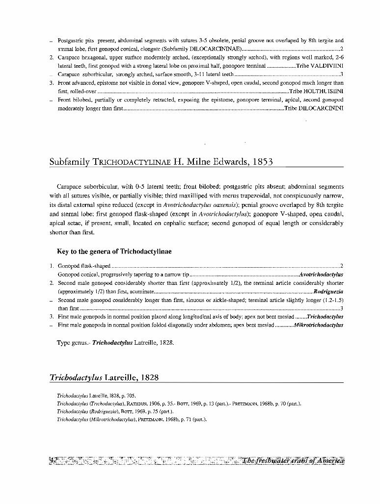
- Postgastric pits present, abdominal segments with sutures 3-5 obsolete, penial groove not overlaped by 8th tergite and
stemallobe, first gonopod conical, elongate (Subfamily DILOCARCININAE) 2
2. Carapace hexagonal, upper surface moderately arched, (exceptionally strongly arched), with regions well marked, 2-6
lateral teeth, first gonopod with a strong lateral lobe on proximal half, gonopore terminal Tribe VALDNIINI
- Carapace suborbicular, strongly arched, surface smooth, 3-11 lateral teeth 3
3. Front advanced, epistome not visible in dorsal view, gonopore V-shaped, open caudal, second gonopod much longer than
first, rolled-over Tribe HOLTHUISIlNl
- Front bilobed, partially or completely retracted, exposing the epistome, gonopore terminal, apical, second gonopod
moderately longer than first Tribe DILOCARCININI
Subfamily TRICHODACTYLINAE H. Milne Edwards, 1853
Carapace suborbicular, with 0-5 lateral teeth; front bilobed; postgastric pits absent; abdominal segments
with all sutures visible, or partially visible; third maxilliped with merus trapezoidal, not conspicuously narrow,
its distal external spine reduced (except in Avotrichodactylus oaxensis); penial groove overlaped by 8th tergite
and sternallobe; first gonopod flask-shaped (except in Avotrichodactylus); gonopore V-shaped, open caudal,
apical setae, if present, small, located on cephalic surface; second gonopod of equal length or considerably
shorter than first.
Key to the genera of Trichodactylinae
1. Gonopod flask-shaped 2
- Gonopod conical, progressively tapering to a narrow tip : Avotrichodactylus
2. Second male gonopod considerably shorter than first (approximately 1/2), the terminal article considerably shorter
(approximately 1/2) than first, acuminate Rodriguezia
- Second male gonopod considerably longer than first, sinuous or sickle-shaped; terminal article slightly longer (1.2-1.5)
than first 3
3. First male gonopods in normal position placed along longitudinal axis of body; apex not bent mesiad Trichodactylus
- First male gonopods in normal position folded diagonally under abdomen; apex bent mesiad Mikrotrichodactylus
Type genus.- Trichodactylus Latreille, 1828.
Trichodactylus Latreille, 1828
Trichodactylus Latreille,1828, p. 705.
Trichodactylus (Trichodactylus), RATHBUN, 1906,p. 35.- BOIT, 1969,p. 13 (part.).- PRElZMANN, 1968b, p. 70 (part.).
Trichodactylus (Rodriguezia), BOIT, 1969,p. 25 (part.).
Trichodactylus (Mikrotrichodactylus), PRETZMANN, 1968b,p. 71 (part.).
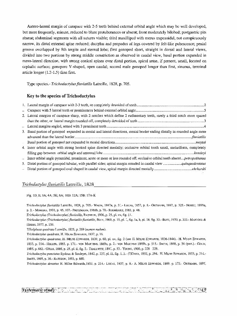
Antero-lateral margin of carapace with 2-5 teeth behind external orbital angle which may be well developed,
but more frequently, minute, reduced to blunt protuberances or absent; front moderately bilobed; postgastric pits
absent; abdominal segments with all sutures visible; third maxilliped with merus trapezoidal, not conspicuously
narrow, its distal external spine reduced; dactylus and propodus of legs covered by felt-like pubescence; penial
groove overlapped by 8th tergite and sternal lobe; first gonopod short, straight in dorsal and lateral views,
divided into two portions by strong middle constriction as observed in caudal view, basal portion expanded in
meso-lateral direction, with strong conical spines over distal portion, apical setae, if present, small, located on
cephalic surface; gonopore V-shaped, open caudal; second male gonopod longer than first, sinuous, terminal
article longer (1.2-1.5) than first.
Type species.- Trichodactylusfluviatilis Latreille, 1828, p. 705.
Key to the species of Trichodactylus
1. Lateral margin of carapace with 2-3 teeth, or completely devoided of teeth 2
- Carapace with 5 lateral teeth or prominences behind external orbital angle 5
2. Lateral margins of carapace sharp, with 2 notches which define 2 rudimentary teeth, rarely a third notch more spaced
than the other, or lateral margin rounded off, completely devoided of teeth 3
- Lateral margins angled, armed with 3 prominent teeth .4
3. Basal portion of gonopod expanded in mesial and lateral directions, mesial border ending distally in rounded angle more
advanced than the lateral border fluviatilis
- Basal portion of gonopod not expanded in mesial directions maytai
4. Inner orbital angle with strong hooked spine directed mesially; occlusive orbital tooth small, molariform, completely
filling gap between orbital angle and antennallobe kensleyi
- Inner orbital angle pyramidal, prominent, acute or more or less rounded off; occlusive orbital tooth absent...,petropolitanus
5. Distal portion of gonopod tubular, with parallel sides; apical margin rounded in caudal view quinquedentatus
- Distal portion of gonopod oval shaped in caudal view, apical margin directed mesially ehrhardti
Trichodactylus fluviatilis Latreille, 1828
Fig. in, E; 3A; 4A; 5K; 6A; !OD; 12A; 15B; 17A-K
Trichodactylus fluviatilis Latreille, 1828, p. 705.- WIllTE, 1847a, p. 31.- LUCAS, 1857, p. 8.- ORTMANN, 1897, p. 325.- NOBILI, 1899a,
p. 2. - MORElRA, 1901, p. 45,107.- PRE1ZMANN. 1968b, p. 70.- RODRiGUEZ, 1981, p. 48.
Trichodactylus (Trichodactylus) fluviatilis, RATHBUN, 1906, p. 25, pI. xv, fig. 11.
Trichodactylus (Trichodactylus)fluviatilisfluviatilis, BOIT, 1969, p. 15, pI. 1, fig. la, b, pI. 18, fig. 33.- BOIT, 1970, p. 333.- MANNING &
HOBBS, 1977,p. 159.
?Thelphusa quadrata Latreille, 1825, p. 269 (nomen nudum).
Trichodactylus quadrata, H. MILNE EDWARDS, 1837, p. 16.
Trichodactylus quadratus, H. MILNE EDWARDS, 1839, p. 60, pI. xv, fig. 2 (see H. MILNEEDWARDS, 1836-1844).- H. MILNE EDWARDS,
1853, p. 214.- HELLER, 1865, p. 171.- VON MARTENS, 1869a, p. 2.- VON MARTENS 1869b, p. 515.- SMITH, 1869, p. 36 (part.).- GOLD!,
1885, p. 662.- GOLDI, 1886, p. 25, pI. ii, fig. 2.- THALLWI1Z, 1891, p. 53.- YOUNG, 1900, p. 228 - 229.
Trichodactylus punctatus Eydoux & Sou1eyet, 1842, p. 237, pI. iii, fig. 1,2.- (?)DANA, 1852, p. 294.- H. MILNEEDWARDS, 1853, p. 214.
SMITH, 1869, p. 36.- RATHBUN, 1893, p. 660.
Trichodactylus dentatus H. Mi1ne Edwards,1853, p. 214.- LUCAS, 1857, p. 8.- A. MILNE EDWARDS, 1869, p. 173.- ORTMANN, 1897,
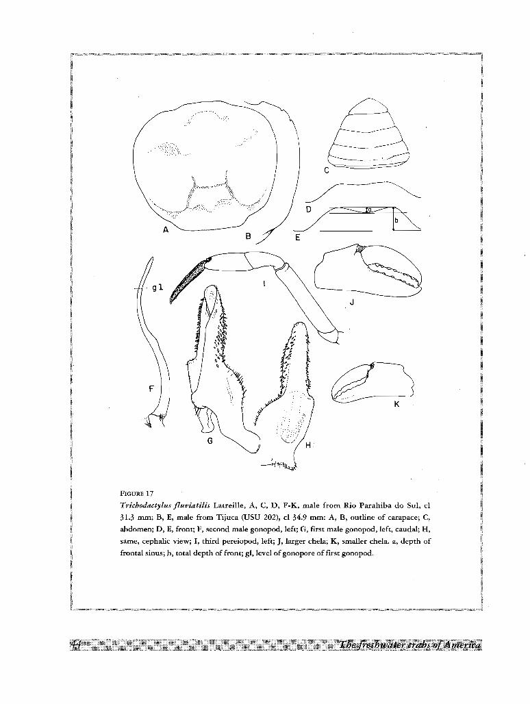
AB E
FIGURE 17
Trichodactylus fluviatilis Latreille, A, C, D, F-K, male from Rio Parahiba do SuI, cl
31.3 mm; B, E, male from Tijuca (USU 202), cl 34.9 mm: A, B, outline of carapace; C,
abdomen; D, E, front; F, second male gonopod, left; G, first male gonopod, left, caudal; H,
same, cephalic view; I, third pereiopod, left; J, larger chela; K, smaller chela. a, depth of
frontal sinus; b, total depth of front; gl, level of gonopore of first gonopod.
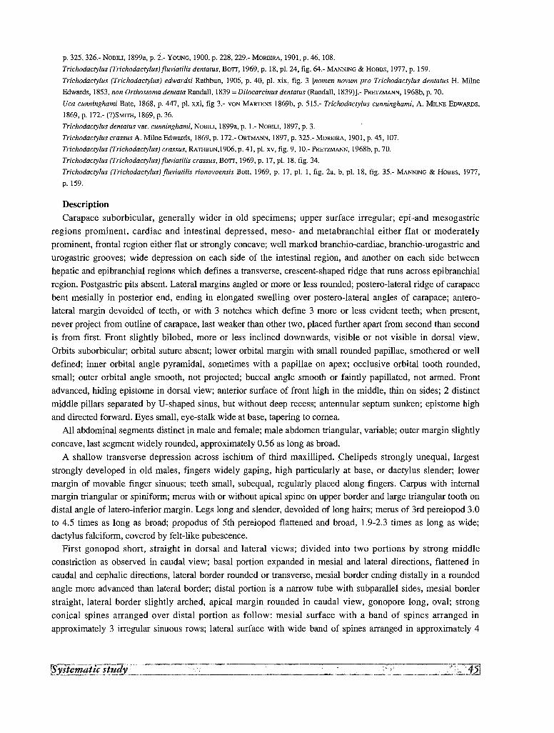
p. 325, 326.- NOBILI, 1899a, p. 2.- YOUNG, 1900, p. 228, 229.- MOREIRA, 1901, p. 46, 108.
Trichodactylus (Trichodactylus) fluviatilis dentatus, BOTT, 1969, p. 18, pI. 24, fig. 64.- MANNING & HOBBs, 1977, p. 159.
Trichodactylus (Trichodactylus) edwardsi Rathbun, 1906, p. 40, pI. xix, fig. 3 [nomen novum pro Trichodactylus dentatus H. Milne
Edwards, 1853, non Orthostoma dentata Randall, 1839 =Dilocarcinus dentatus (Randall, 1839)].- PRETZMANN, 1968b, p. 70.
Uca cunninghami Bate, 1868, p. 447, pI. xxi, fig 3.- VaN MARTENS 1869b, p. 515.- Trichodactylus cunninghami, A. MILNE EDWARDS,
1869, p. 172.- (?)SMITH, 1869, p. 36.
Trichodactylus dentatus var. cunninghami, NOBILI, 1899a, p. 1.- NOBILI, 1897, p. 3.
Trichodactylus crassus A. Milne Edwards, 1869, p. 172.- ORTMANN, 1897, p. 325.- MOREIRA, 1901, p. 45, 107.
Trichodactylus (Trichodactylus) crassus, RATHBUN,1906, p. 41, pI. xv, fig. 9, 10.- l'RETZMANN, 1968b, p. 70.
Trichodactylus (Trichodactylusi fluviatilis crassus, BOTT, 1969, p. 17, pI. 18, fig. 34.
Trichodactylus (Trichodactylus) fluviatilis rionovoensis Bott, 1969, p. 17, pI. I, fig. 2a, b, pI. 18, fig. 35.- MANNING & HOBBs, 1977,
p.159.
Description
Carapace suborbicular, generally wider in old specimens; upper surface irregular; epi-and mesogastric
regions prominent, cardiac and intestinal depressed, meso- and metabranchial either flat or moderately
prominent, frontal region either flat or strongly concave; well marked branchio-cardiac, branchio-urogastric and
urogastric grooves; wide depression on each side of the intestinal region, and another on each side between
hepatic and epibranchial regions which defines a transverse, crescent-shaped ridge that runs across epibranchial
region. Postgastric pits absent. Lateral margins angled or more or less rounded; postero-lateral ridge of carapace
bent mesially in posterior end, ending in elongated swelling over postero-lateral angles of carapace; antero
lateral margin devoided of teeth, or with 3 notches which define 3 more or less evident teeth; when present,
never project from outline of carapace, last weaker than other two, placed further apart from second than second
is from first. Front slightly bilobed, more or less inclined downwards, visible or not visible in dorsal view.
Orbits suborbicular; orbital suture absent; lower orbital margin with small rounded papillae, smothered or well
defined; inner orbital angle pyramidal, sometimes with a papillae on apex; occlusive orbital tooth rounded,
small; outer orbital angle smooth, not projected; buccal angle smooth or faintly papillated, not armed. Front
advanced, hiding epistome in dorsal view; anterior surface of front high in the middle, thin on sides; 2 distinct
middle pillars separated by U-shaped sinus, but without deep recess; antennular septum sunken; epistome high
and directed forward. Eyes small, eye-stalk wide at base, tapering to cornea.
All abdominal segments distinct in male and female; male abdomen triangular, variable; outer margin slightly
concave, last segment widely rounded, approximately 0.56 as long as broad.
A shallow transverse depression across ischium of third maxilliped. Chelipeds strongly unequal, largest
strongly developed in old males, fingers widely gaping, high particularly at base, or dactylus slender; lower
margin of movable finger sinuous; teeth small, subequal, regularly placed along fingers. Carpus with internal
margin triangular or spiniforrn; merus with or without apical spine on upper border and large triangular tooth on
distal angle of latero-inferior margin. Legs long and slender, devoided of long hairs; merus of 3rd pereiopod 3.0
to 4.5 times as long as broad; propodus of 5th pereiopod flattened and broad, 1.9-2.3 times as long as wide;
dactylus falciforrn, covered by felt-like pubescence.
First gonopod short, straight in dorsal and lateral views; divided into two portions by strong middle
constriction as observed in caudal view; basal portion expanded in mesial and lateral directions, flattened in
caudal and cephalic directions, lateral border rounded or transverse, mesial border ending distally in a rounded
angle more advanced than lateral border; distal portion is a narrow tube with subparallel sides, mesial border
straight, lateral border slightly arched, apical margin rounded in caudal view, gonopore long, oval; strong
conical spines arranged over distal portion as follow: mesial surface with a band of spines arranged in
approximately 3 irregular sinuous rows; lateral surface with wide band of spines arranged in approximately 4
irregular rows directed transversely towards base and towards caudal surface, and second narrower band of
spines, more cephalad and parallel to former, arranged in approximately 2 irregular rows and continued over
lateral expansion of base by slenderer spines; caudal and cephalic surfaces devoided of spines; patch of spinules
over basal mesial angle. Second gonopod considerably longer than first, sinuous or broadly S-shaped; terminal
article slightly longer (1.2) than first, basal incurved laterally, terminal incurved mesially; apex flat and
acuminate.
Material examinedBrazil. Floresta do Horto, Rio de Janeiro State; 8 June 1984; L. de O. SALLE; 1 male, 3 females (USU 125).
Rio dos Macacos, Serra da Carioca, Rio de Janeiro State; 21 November 1984; M. A. TAVARES; 4 males, 1 female
(USU 229). Serra da Carioca, Covanca, Tijuca, Rio de Janeiro State; 3 males, 4 females (USU 202). Rio
Parahiba do SuI, Valenca, Rio de Janeiro State; D. A. MACHADO FILHO; 1 male (USU 138).
Type and distribution
Type locality Brazil. Type not extant. Aside from doubtful records, i. e. Guiana (possibly French Guiana
since it is one of LEPRIEUR'S collections), Tefe (Central Brazil), Rio Negro (the affluent of the Amazon where
the Thayer Expedition made collections, not Rio Negro, between the states of Parana and Santa Catarina, near
the area of distribution of the species, but draining to the Rio Iguassu), Chile and "Sandwich islands" (RATHBUN,
1906), and Peru (NoBILl, 1899a), the identifiable localities mentioned in the literature are the following: Brazil.
Rio de Janeiro State: Rio de Janeiro (RATHBUN, 1906; BOTT, 1969; GOLDI, 1886); Rio de Janeiro (edwardsi,
RATHBUN, 1906); Tijuca (BATE, 1868; MOREIRA, 1901); Teresopolis (BOTT, 1969); Nova Friburgo (BOTT, 1969).
Minas Geraes State: Rio Angu, tributary of Rio Parahiba (GOLDI, 1886); Sacutenga (BOTT, 1969); Serra da Bisca
(Bicas?) (GOLDI, 1886). Sao Paulo State: Ilha do Puzios (BOTT, 1969); Sao Paulo, 1,000 malt; Sao Sebastian
(dentatus, BOTT, 1969). Santa Catarina State: Rio Negro (RATHBUN, 1906); Joinville, between Rio Nova y Rio
Humboldt (BOTT, 1969); Rio Itapoca, Jaragua District (BOTT, 1969); Punta do Pharol, Sao Francisco do SuI
(MOREIRA, 1901); Rio Novo basin (type of rionovoensis, BOTT, 1969). Rio Grande do SuI State (GOLDI, 1886).
Bahia State: Bahia (type of crassus, A. MlLNE EDwARDs, 1869; RATHBUN, 1906); Bahia (Salvador) (BOTT,
1970). Espiritu Santo State: Santa Cruz (=Aracruz, dentatus, RATHBUN, 1906).
Remarks
Specimens can be separated into two distinct groups, as was first pointed out by GOLDI (1885, 1886) who
divided them into two separate species, T. quadrata and T. cunninghami, and by RATHBUN (1906) who included
all specimens under Ti fluviatilis.
Group a. The carapace more square; lateral margins sharp, with 2 notches which define 2 rudimentary teeth,
rarely a third notch more spaced than the other 2; frontal margin visible in dorsal view; lower orbital margin
with distinct granules; carapace surface more flattened and irregular; the branchial area flattened or concave.
Group b. Carapace more orbicular; lateral margins rounded off and smooth, devoided of teeth; frontal margin
sharply inclined downwards and consequently not visible in dorsal view; papillae of lower orbital margin
relatively smaller, less conspicuous; carapace surface less irregular, branchial region clearly convex.
In several species of Trichodactylidae the lateral spines become worn out with age, and the lateral margin
correspondingly rounded off. In T.fluviatilis mature and immature specimens are found in both groups.
All specimens recorded under Material examined belong to group a, with the exception of the male specimen
from Rio Parahiba (VSV 138).
Further variability is observed within group a. RATHBUN (1906) already recorded specimens with I to 3
lateral teeth and with margins either rounded or acute. Two of the subspecies proposed by BUIT (1969) partly
reflect this intra-group variability, as follows:
(1) T. f crassus. Carapace strongly arched, but still the frontal margin visible in dorsal view; 1-2 lateral teeth.
All specimens under this name are from Bahia (Sao Salvador) (LATREILLE, 1828; RATHBUN, 1906).
(2) T. f. dentatus. Carapace convex in the anterior posterior direction; grooves scarcely marked; front not
visible dorsally, margins acute; third lateral tooth not rudimentary, developed, sharp, slightly bent outwards; first
interdental space shorter (0.50-0.75) than second space, sinus in front of teeth V-shaped; the type locality is
undetermined; additional material from Rio de Janeiro (RATHBUN, 1906) and Sao Paulo (BOTT, 1969).
(3) T. f rionovoensis. Smaller than fiuviatilis sensu stricto (cb 20 vs 40 mm), carapace convex, only the H
depression marked, no teeth or notches on margin of carapace; basal portion of first gonopod not very expanded.
All specimens come from Rio Novo, Santa Catarina State (near Joinville).
After examining supplementary material of crassus from the type locality, BOTT (1970) synonymized it with
[luviatilis sensu stricto. The distribution of the 2 remaining subspecies do not make sense from a
biogeographical point of view, since their localities are nested inside the range of fiuviatilis sensu stricto. It is
almost certain than dentatus is a variation of group a and rionovoensis young specimens of group b.
Trichodactylus maytai Pretzmann, 1978
Trichodactylus (Trichodactylus) maytai Pretzmann, 1978a, p. 165, fig. 8.- PRETZMANN, 1983a, p. 307, pl, I, fig. I, pI. 2, fig. 7, pl, 3, fig.
11, pl. 4, fig. IS, pl. 5, fig. 19.- PRETlMANN, 1983b, p. 320.
Description
Carapace suborbicular; upper surface completely smooth; central grooves of carapace only slightly marked.
Lateral margins angled, completely devoided of teeth; exorbital angle with depression behind it, followed by
inconspicuous papillae. Front moderately bilobed, the margin somewhat irregular; lower orbital margin with a
lobe followed by small granules; inner orbital angle with blunt spine.
All abdominal segments distinct in male (unknown in female); male abdomen subtriangular, wide, outer
margin straight; last segment wide, triangular-rounded, the proximal margin narrower than distal margin of
penultimate segment; distal outer angles of penultimate segment projected forwards.
Chelipeds strongly unequal; larger chela swollen, fingers moderately gaping, teeth only slightly defined;
smaller chela with longitudinal carinae on the external surface; carpus with a long, acute spine. P2-P5 long and
slender.
First gonopod with basal portion expanded in the meso-lateral direction, distal portion a narrow straight tube,
slightly reduced distally, with strong and numerous conical spines.
The species is well differentiated by its unarmed lateral margin. The preceeding description is based on
PRETZMANN'S (1978a, 1983a) description of the holotype and only specimen known.
Type and distribution
The species is only known from the type specimen, a small male cl 16.1 mm from Tingo Maria, Huanuco
Department, Peru (PRETZMANN, 1978a).
Trichodactylus kensleyi, new species
Fig. 3D; 4B; 100; 12D; 18A-0
Description
Carapace suborbicular; upper surface irregular; protogastric region more prominent than rest; hepatic,
epibranchial and intestinal depressed; meso- and metabranchial moderately prominent; frontal region concave;
branchio-cardiac and branchio-urogastric grooves represented by deep and wide depressions; urogastric grooves
less conspicuous; area between mesobranchial, meso- and urogastric regions elevated, forming subtriangular or
Y-shaped lobe. Postgastric pits absent. Lateral margins angled, with 3 prominent acute spines behind external
orbital angle; first and second closer than second and third; third more prominent, directed transversely laterad;
postero-lateral ridge of carapace bent mesially in posterior end, ending in elongated swelling over postero-lateral
angles of carapace. Front moderately bilobed, margin somewhat irregular, slightly convex in frontal view. Orbits
small, suborbicular, orbital suture absent; lower orbital margin smooth; inner orbital angle with strong hooked
spine directed mesially; occlusive orbital tooth small, molariform, completely filling gap between inner orbital
angle and lateral lobe of antenna; outer orbital angle rounded, not projected; buccal angle rounded, smooth or
with minute papillae. Front advanced, hiding epistome in dorsal view; anterior surface of front high in middle,
moderately thin on sides; middle pillars distinct; antennular septum sunken; epistome high and directed
backwards, with thin transverse ridge in middle portion. Eyes small, but filling orbital cavity, eyestalk wide at
base, tapering to cornea.
All abdominal segments distinct in male and female, but ankylosed, except for 7th in male which is movable;
male abdomen subtriangular, wide, outer margin slightly concave, last segment triangular-rounded,
approximately 0.58 as long as broad, with proximal margin narrower than distal margin of penultimate segment.
Shallow transverse depression across ischium of 3rd maxilliped. Chelipeds moderately unequal; lower margin
of fixed finger sinuous, teeth small, subequal, spaced and regularly placed over fingers; a longitudinal carina on
external surface of each finger. Carpus with strong hooked spine on inner margin. Legs long and slender, with
scattered hairs; merus of 3rd pereiopod 3.2 times as long as wide; propodus of 5th pereiopod narrow,
approximately 3.8 times as long as wide; dactylus falciform, long and narrow, covered by felt-like pubescence
and short hairs irregularly placed.
First gonopod short, straight in dorsal and lateral views; divided into two portions by strong middleconstriction as observed in caudal view; basal portion expanded in meso-lateral direction, flattened in caudal
cephalic direction, lateral border expanded into sinuous lobe, mesial border convex; distal portion a narrow tube
with mesial border almost straight, lateral border slightly more convex, apical margin rounded in caudal view,
gonopore long, oval; strong conical spines arranged over distal portion as follow: mesial surface with a band of
spines arranged in approximately 3 irregular sinuous rows;lateral surface with a wide band of spines arranged in
approximately 4 irregular rows directed transversely towards base and towards caudal surface, and second
narrower band of spines, more cephalad and parallel to former, arranged in approximately 2 irregular rows and
continued over lateral expansion of base by slenderer spines; caudal and cephalic surfaces devoided of spines;
few long subapical spinules on cephalic surface. Second gonopod slightly longer than first, sinuous or broadly
S-shaped; terminal article longer (1.5) than first, basal incurved laterally, terminal incurved mesially; apex flat
and acuminate.
Material examined
Paraguay. Canendiyu, Rio Carapa at bridge on dirt highway 45.1 km WSW of Salto del Guaira; Rio Parana
drainage; 14 July 1979; J. N. TAYLOR & T. W. GRIMSHAW; Lat/Long: 24Q 10' 54" S - 54Q 41' 42" W; Field
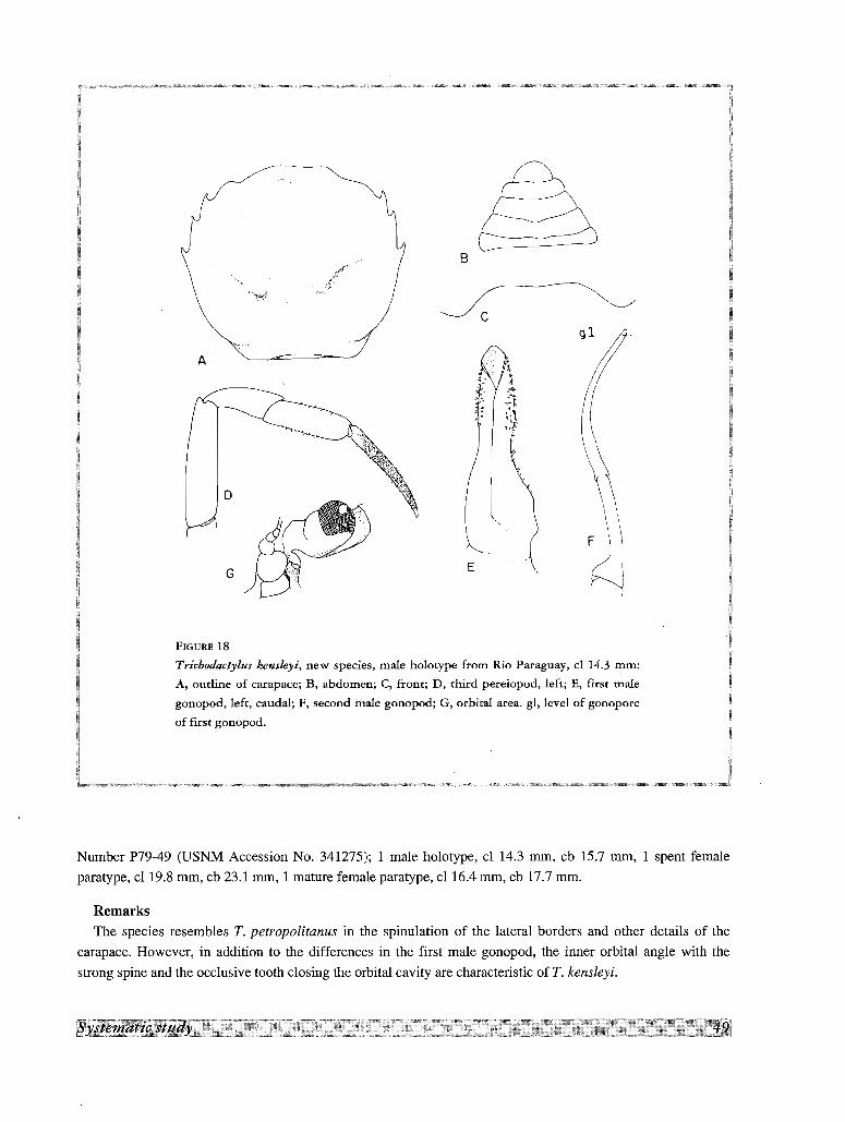
B
A [).........•...
f:Y :~r .~{1- •
J: ;1"l,
E
FIGURE 18
Trichodactylus kensleyi, new species, male holotype from Rio Paraguay, cl 14.3 mm:
A, outline of carapace; B, abdomen; C, from; D, third pereiopod, left; E, first male
gonopod, left, caudal; F, second male gonopod; G, orbital area. gl, level of gonopore
of first gonopod.
Number P79-49 (USNM Accession No. 341275); 1 male holotype, cl 14.3 mm, cb 15.7 mm, 1 spent female
paratype, cl 19.8 mm, cb 23.1 mm, 1 mature female paratype, cl 16.4 mm, cb 17.7 mm.
Remarks
The species resembles T. petropolitanus in the spinulation of the lateral borders and other details of the
carapace. However, in addition to the differences in the first male gonopod, the inner orbital angle with the
strong spine and the occlusive tooth closing the orbital cavity are characteristic of T. kensleyi.
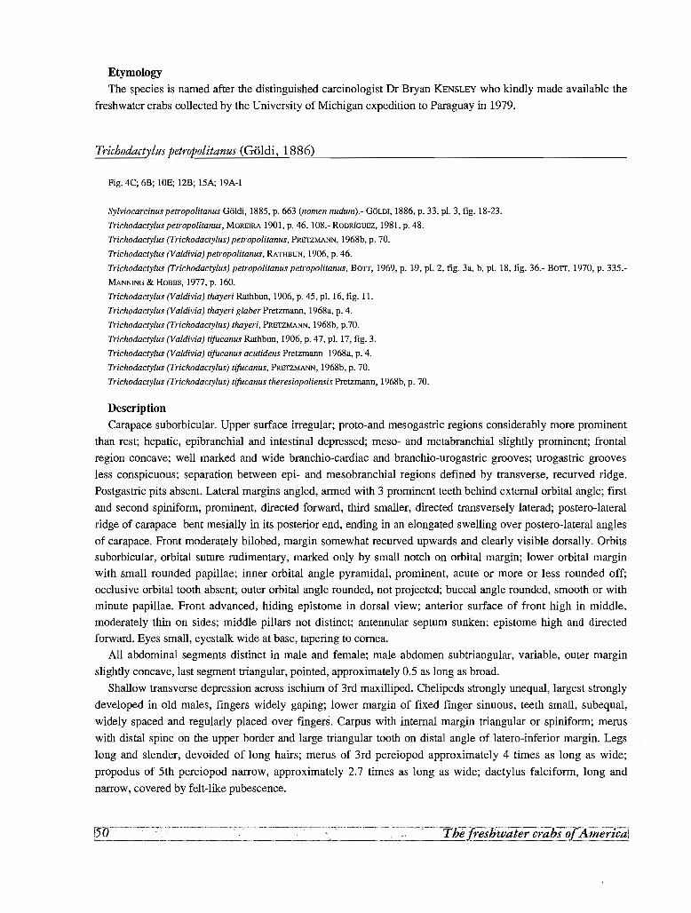
Etymology
The species is named after the distinguished carcinologist Or Bryan KENSLEY who kindly made available the
freshwater crabs collected by the University of Michigan expedition to Paraguay in 1979.
Trichodactylus petropolitanus (G61di, 1886)
Fig. 4C; 6B; tOE; 12B; 15A; 19A-I
Sylviocarcinus petropolitanus G61di, 1885, p. 663 (nomen nudum).- GOLD!, 1886, p. 33, pI. 3, fig. 18-23.
Trichodactylus petropolitanus, MOREIRA 1901, p. 46, 108.- RODRiGUEZ, 1981, p. 48.
Trichodactylus (Trichodactylus) petropolitanus, PRETZMANN, 1968b, p. 70.
Trichodactylus (Yaldivia) petropolitanus, RATHBUN, 1906, p. 46.
Trichodactylus (Trichodactylus) petropolitanus petropolitanus, BOTT, 1969, p. W. pI. 2, fig. 3a, b, pI. 18, fig. 36.- BOTT. 1970, p. 335.
MANNING & HOBBS, 1977, p. 160.
Trichodactylus (Valdivia) thayeri Rathbun, 1906, p. 45. pI. 16, fig. 11.
Trichodactylus (Valdivia) thayeri glaber Pretzmann, 1968a, p. 4.
Trichodactylus (Trichodactylus) thayeri, PRETZMANN. 1968b, p.70.
Trichodactylus (Valdivia) tifucanus Rathbun, 1906, p. 47, pI. 17, fig. 3.
Trichodactylus (Valdivia) tifucanus acutidens Pretzmann 1968a, p. 4.
Trichodactylus (Trichodactylus) tifucanus, PRETZMANN, 1968b, p. 70.
Trichodactylus (Trichodactylus) tifucanus theresiopoliensis Pretzmann, 1968b, p. 70.
Description
Carapace suborbicular. Upper surface irregular; proto-and mesogastric regions considerably more prominent
than rest; hepatic, epibranchial and intestinal depressed; meso- and metabranchial slightly prominent; frontal
region concave; well marked and wide branchio-cardiac and branchio-urogastric grooves; urogastric grooves
less conspicuous; separation between epi- and mesobranchial regions defined by transverse, recurved ridge.
Postgastric pits absent. Lateral margins angled, armed with 3 prominent teeth behind external orbital angle; first
and second spiniform, prominent, directed forward, third smaller, directed transversely laterad; postero-lateral
ridge of carapace bent mesially in its posterior end, ending in an elongated swelling over postero-lateral angles
of carapace. Front moderately bilobed, margin somewhat recurved upwards and clearly visible dorsally. Orbitssuborbicular, orbital suture rudimentary, marked only by small notch on orbital margin; lower orbital margin
with small rounded papillae; inner orbital angle pyramidal, prominent, acute or more or less rounded off;
occlusive orbital tooth absent; outer orbital angle rounded, not projected; buccal angle rounded, smooth or with
minute papillae. Front advanced, hiding epistome in dorsal view; anterior surface of front high in middle,
moderately thin on sides; middle pillars not distinct; antennular septum sunken; epistome high and directedforward. Eyes small, eyestalk wide at base, tapering to cornea.
All abdominal segments distinct in male and female; male abdomen subtriangular, variable, outer margin
slightly concave, last segment triangular, pointed, approximately 0.5 as long as broad.
Shallow transverse depression across ischium of 3rd maxilliped. Chelipeds strongly unequal, largest strongly
developed in old males, fingers widely gaping; lower margin of fixed finger sinuous, teeth small, subequal,
widely spaced and regularly placed over fingers. Carpus with internal margin triangular or spiniform; merus
with distal spine on the upper border and large triangular tooth on distal angle of latero-inferior margin. Legs
long and slender, devoided of long hairs; merus of 3rd pereiopod approximately 4 times as long as wide;
propodus of 5th pereiopod narrow, approximately 2.7 times as long as wide; dactylus falciform, long and
narrow, covered by felt-like pubescence.
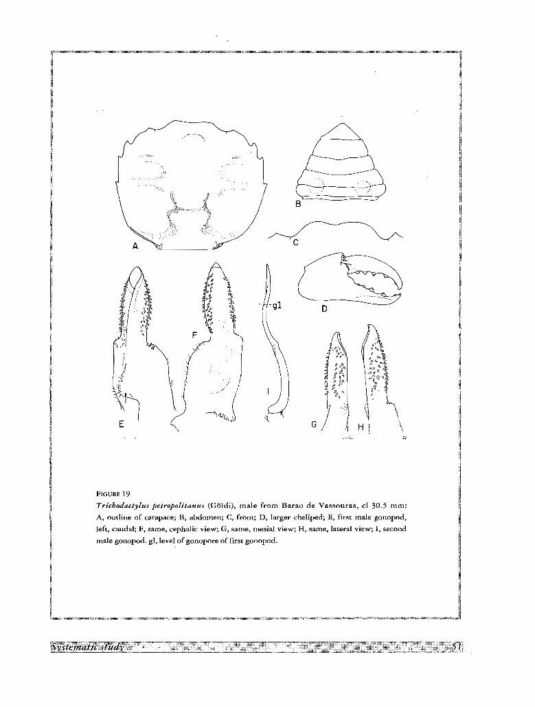
.... ,.
B
c
D
..~; ge-
p ""
o ,"gll".~
P 04 ;~~ ~
YI: P q~ Q
, ", q?, "9()~ I:l. ~ '1
"A "',. "<1 \.J
G )1 H..• ,':A' .~~
FIGURE 19
Trichodactylus petropolitanus (Goldi), male from Barao de Vassouras, cl 30.5 mm:
A, outline of carapace; B, abdomen; C, front; D, larger cheliped; E, first male gonopod,
left, caudal; F, same, cephalic view; G, same, mesial view; H, same, lateral view; I, second
male gonopod. gl, level ofgonopore of first gonopod.
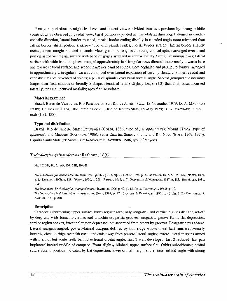
First gonopod short, straight in dorsal and lateral views; divided into two portions by strong middle
constriction as observed in caudal view; basal portion expanded in meso-lateral direction, flattened in caudal
cephalic direction, lateral border rounded, mesial border ending distally in rounded angle more advanced than
lateral border; distal portion a narrow tube with parallel sides, mesial border straight, lateral border slightly
arched, apical margin rounded in caudal view, gonopore long, oval; strong conical spines arranged over distal
portion as follow: mesial surface with band of spines arranged in approximately 3 irregular sinuous rows; lateral
surface with wide band of spines arranged approximately in 4 irregular rows directed transversely towards base
and towards caudal surface, and second narrower band of spines, more cephalad and parallel to former, arranged
in approximately 2 irregular rows and continued over lateral expansion of base by slenderer spines; caudal and
cephalic surfaces devoided of spines; a patch of spinules over basal mesial angle. Second gonopod considerably
longer than first, sinuous or broadly S-shaped; terminal article slightly longer (1.3) than first, basal incurved
laterally, terminal incurved mesially; apex flat, acuminate.
Material examined
Brazil. Barao de Vassouras, Rio Parahiba do SuI, Rio de Janeiro State; 13 November 1979; D. A. MACHADO
FILHO; 1 male (USU 134). Rio Parahiba do SuI, Rio de Janeiro State; 15 May 1979; D. A. MACHADO FILHO; 1
male (USU 138).-
Type and distribution
Brazil. Rio de Janeiro State: Petropolis (GOLD!, 1886, type of petropolitanus); Mount Tijuca (type of
tifucanus), and Macacos (RATHBUN, 1906). Santa Catarina State: Joinville and Rio Novo (BOTT, 1969, 1970).
Espiritu Santo State (?): Santa Cruz (=Aracruz?, RATHBUN, 1906, type of thayeri).
Trichodactylus quinquedentatus Rathbun, 1893
Fig. 1C; 3B; 4C; SI; 6D; !OF; 12E; 20A-H
Trichodactylus quinquedentatus Rathbun, 1893, p. 660, pl, 77, fig. 7.- NOBIll, 1896, p. 2.- ORTMANN, 1897, p. 325, 326.- NOBILI, 1899,
p. 1.- DOFLEIN, 1899b, p. 188.- YOUNG, 1900, p. 228.- ZIMMER, 1912, p. 7.- RODRIGUEZ & MANR1QUE, 1967, p. 183.- RODRIGUEZ, 1981,
p.47.
Trichodactylus (Trichodactylus) quinquedentatus, RATHBUN, 1906, p. 42, pl. 15, fig. 3.- PRETZMANN, 1968b, p. 70.
Trichodactylus (Rodriguczia) quinquedentatus, BOTT, 1969, p. 27.- SMALLEY & RODRIGUEZ, 1972, p. 43, fig. 1, 2.- COTTARELLI &
ARGANO,1977,p.210.
DescriptionCarapace suborbicular; upper surface forms regular arch; only urogastric and cardiac regions distinct, set-off
by deep and wide branchio-cardiac and branchio-urogastric grooves; urogastric groove forms flat depression;
cardiac region convex, intestinal region depressed, not separated from others by grooves. Postgastric pits absent.
Lateral margins angled; postero-lateral margins defined by thin ridge whose distal half runs transversely
inwards, close to ridge over 5th coxa, and ends away from postero-lateral angles; .antero-lateral margins armed
with 5 small but acute teeth behind external orbital angle, first 3 well developed, last 2 reduced, last pair
implanted behind middle of carapace. Front slightly bilobed, upper surface flat. Orbits suborbicular; orbital
suture absent, position indicated by flat depression; lower orbital margin entire; inner orbital angle with strong
[52'--'''-~''''-''-~- •.• ';•.~_..- __._--_...... . -_.~ - - - ---- - --~..-'---_._-------~.,_._----------_._--
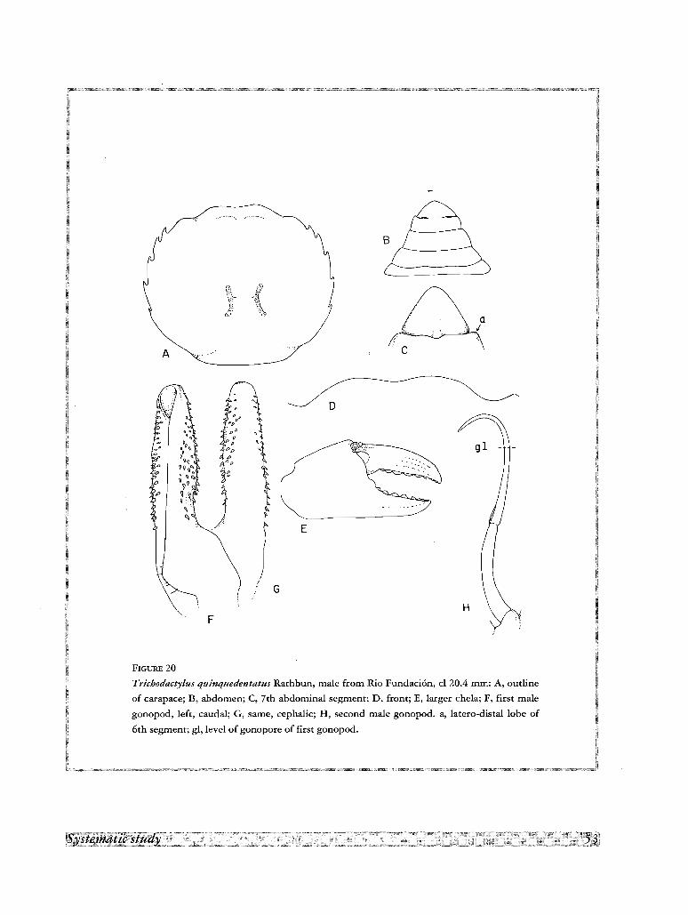
A
B
c
" ..F
G
E
D
FIGURE 20
Trichodactylus quinquedentatus Rathbun, male from Rio Fundaci6n, cl 20.4 mm: A, outline
of carapace; B, abdomen; C, 7th abdominal segment; D, front; E, larger chela; F, first male
gonopod, left, caudal; G, same, cephalic; H, second male gonopod. a, Iatero-drstal
6th segment; gl, level of gonopore of first gonopod.
sharp spine, sometimes followed laterally by recess and second spine or strong tubercle; external orbital angle
not armed, in some specimens lateral margin not in contact with orbit in this area, but separated from it by small
recess; buccal angle smooth, not armed. Front little advanced, epistome partially visible in dorsal view; anterior
surface of front not high, with a thin ridge over each antennular fossa; 2 middle pillars separated by U'-shaped
recess; interantennular septum moderately sunken, epistome not very high, directed forwards. Eyes normal.
All abdominal segments distinct in male and female; male abdomen triangular, wide at base; outer margin
slightly concave; last segment widely rounded, approximately 0.7 as long as broad, its base conspicuously
narrower than margin of the last segment.
Conspicuous transverse groove across ischium of 3rd maxilliped. Chelipeds strongly unequal, largest strongly
developed in males, fingers moderately gaping; lower margin of fixed finger slightly sinuous; teeth small
subequal in proximal half, diminishing in size towards tip on distal half, widely spaced and regularly placed
over cutting surfaces. Carpus with strong hooked spine on internal margin; merus with small sharp spine on
upper border and larger one at middle of each lower margin, distal angle of latero-inferior margin not strongly
produced. Legs long and slender, lower and upper margins of dactylus and lower margin of propodus covered
with long hairs, lateral sides of dactylus and propodus, and distal part of carpus covered by a felt-like
pubescence; long hairs scattered elsewhere over the leg; merus of third pereiopod approximately 4 times as long
as broad; propodus of 5th pereiopod broad, approximately 1.6 as long as wide; dactylus falciform, moderately
long.
First gonopod short, straight in dorsal and lateral views, divided into two portions as observed in caudal view,
basal portion with lateral border expanded, sinuous, mesial border straight, not advanced; distal portion forming
narrow tube, with parallel sides, mesial border straight, lateral border slightly arched, apical margin rounded in
caudal view, gonopore subtriangular; strong conical spines arranged over distal portion as follows: mesial
surface with band of spines arranged in approximately 3 irregular rows; lateral surface with band of spines
arranged approximately in 6 irregular rows directed transversely towards base and towards caudal surface;
caudal and cephalic surfaces devoided of spines. Second gonopod considerably longer than first, sinuous, apical
article 1.5 longer than basal, basal incurved laterally, terminal bent mesially; apex rounded, acuminate.
Material examined
Colombia. Rio Fundacion, near Santa Marta, 15 November 1967; A. ZAMORA; 1 male, 1 female (Ivic), Rio
Gaira, near Santa Marta; 15 November 1967; A. Zamora; 1 male, 1 female (Ivic). Rio Sevilla, near Santa Marta;
15 November 1967; A. ZAMORA; 1 male, 2 females (Ivic). Rio Aracataca, near Santa Marta; 15 November
1967; A. ZAMORA; 1 male, 4 females (Ivic). Rio Cesar, 10 km south ofValledupar; 2 January 1968; A. ZAMORA;
4 males, 3 females (Ivic).
Type and distributionNicaragua. Rio Escondido, 50 miles from Bluefields (type locality, RATHBUN, 1906). Colombia. Magdalena
Department: Santa Marta (PEARSE, 1915) and rivers up to 80 km south of this city (SMALLEY & RODRIGUEZ,
1972); Cesar Department: 10 km South of Valledupar (SMALLEY & RODRIGUEZ, 1972); Atlantico Department:
Barranquilla (ZIMMER, 1912). Bolivar Department: Cartagena (SMALLEY & RODRfGUEZ, 1972). Of the disjunct
distribution of this species in Nicaragua and Colombia, SMALLEY & RODRIGUEZ (1972), said: it "is unusual when
compared with distribution of other species of the family. Unfortunately, the only known Nicaraguan specimen
is a female; however, the suborbital margin is very distinctive in this species, and in our opinion the similarity
of the Nicaraguan holotype to the Colombian specimens is sufficiently close to confirm the conclusion of
previous workers that they are the same species".
Trichodactylus ehrhardti (Bott, 1969)
Trichodactylus (Trichodactylus) panoplus ehrhardti Bott, 1969, p. 22, pl. 4, fig. 7a, b, pl. 18, fig. 38.
Type and distribution
The male holotype, with a cb of 9.5 mm, a female paratype, and several other specimens, come from Lago
Manacapuni, near Manaus, Brazil. BOIT(1969) also reports specimens from Rio Negro, 80 km from Manaus.
Remarks
This species resembles Trichodactylus (Valdivia] faxoni Rathbun, 1906, also reported from the Amazon (see
under "Species incertae sedis"), in the reduction of the teeth, particularly the last two which are "very small,
blunt, near each other, difficult to see" (RATHBUN, 1906).
Mikrotrichodactylus Pretzmann, 1968
Trichodactylus (Mikrotrichodactylus) Pretzmann, 1968b, p. 71
Very small species, carapace length usually under 20 mm at maturity, front bilobed, lateral margin with a
series of 5 lateral teeth behind external orbital angle, which reach level of cardiac region; postgastric pits absent;
all abdominal segments distinct in male arid female, male abdomen very wide; third maxilliped with merus
trapezoidal, not conspicuously narrow, distal external spine reduced; dactylus and propodus of l~gs covered by
felt-like pubescence; first male gonopods in normal position folded diagonally under abdomen; basal portion
expanded laterally into wide thin lobe, distal portion with bulbous expansion, apex awl-shaped, strongly bent
mesiad; gonopore V-shaped, large, open caudally; second male gonopod considerably longer than first, sickle
shaped; terminal article longer (1.35) than first.
Type species.- Trichodactylus borellianus Nobili, 1896
The lateral margin with 5 spines reaching postero-lateral margin of carapace is a character only found outside
this group in Trichodactylus quinquedentatus and T. ehrhardti. These species also present the small size and
several morphological traits (i. e., shape of chela) of Mikrotrichodactylus, or characters intermediate between
both genera (i. e. length of second male gonopod), but with the first gonopod typical of Trichodactylus. The
small body size of the species of Mikrotrichodactylus is found also in Trichodactylus maytai, T. kensleyi and the
species of Avotrichodactylus and Rodriguezia within the family. Notwithstanding this overlap of characters, M.
panoplus and M. borellianus possess a very distinctive first gonopod which justifies the erection of
Mikrotrichodactylus Pretzmann, 1968b, as a separate taxa, to receive them.
Key to the species of Mikrotrichodactylus
1. Carapace convex, with regions slightly marked. Lateral teeth subequal, equidistant. Last abdominal article of male
triangular, length/width = 0.5. Bulbous expansion of distal portion of first gonopod with a single lobe on lateral side
.................................................................................................................................................................. ..................panoplus
- Carapace flattened, with regions more marked and covered by gibbosities. Last abdominal article of male suboval,
length/width = 0.3. Bulbous expansion of distal portion of gonopod formed by two longitudinally subparallellobes
.................................................................................................................................................................. ..............borellianus
Mikrotrichodactylus borellianus (Nobili, 1896)
Fig. 2B; 3E; 4E; 6E; lIB; 12C; 2IA-H
Trichodactylus borellianus Nobili, 1896, p. 2.- NOBlLI, 1898, p. 12.- NOBILI, 1899a, p. 3.- NOBlLI, 1901, p. 11.- RODmGUEZ, 1981, p. 48.
Trichodactylus tvaldivia} borellianus, RATHBUN, 1906, p. 5, pI. 17, fig. 6, text-fig. lIS.
Trichodactylus (Dilocarcinus) borellianus, RINGUELET, 1949, p. 104, pI. 8, fig. 2.
Trichodactylus (Trichodactylus) borellianus, BOTT,1969, p. 23, pI. 5, fig. 8a, b, pI. 18, fig. 39.- LOPRETTO, 1976, p. 74, fig. 6-9.
MANNING & HOBBS, 1977, p. 159.
Trichodactylus tMikrotrichodactylus) borellianus brasiliensis Pretzrnann, 1968b, p. 71.
Tlrichodactylus (Trichodactylus) parvus Moreira, 1912, p. 151, pI. 6, fig. 12, 13, text-fig. 2, 3.
Description
Carapace suborbicular; upper surface moderately convex, irregular; protogastric region vaulted and, together
with mesobranchial, more elevated than others; frontal, hepatic and epibranchial regions depressed, flat, or
slightly concave; area between cardiac and intestinal regions forming concave depression; branchio-urogastric
groove deep and thin, well marked, forming semicircle. Postgastric pits absent. Lateral margins angled,
irregular; postero-lateral ridge of carapace not bent abruptly inwards, but ends near margin of carapace above
5th pereiopod; spinulation of lateral margin consists of strong sinuous postorbital lobe, followed by two tiny
spines placed very close to each other on hepatic margin, these followed by concavity of margin and then three
large equidistant hooked spines, last placed at level of cardiac region. Front very wide, in the form of spatulated
lamella, squarish due to right-angled orientation of inner orbital margin; surface of these margins arched; frontal
lobes widely spaced by shallow notch. Orbits rounded, small, shallow, eyes completely fill orbits; orbital suture
absent; lower orbital margin smooth; inner orbital angle with strong blunt spine directed mesially; occlusive
orbital tooth small, attached to external orbital angle; expanded lobe of antenna, occlusive tooth and inner orbital
angle applied to each other to form continuous closure of orbit near yugal angle; outer orbital angle smooth, not
projected, with a conspicuous constriction behind it; buccal angle smooth, with frontal angle retracted. Front
advanced, hiding epistome in dorsal view; anterior surface of front retracted, very high in middle, oblique
laterally; 2 distinct but very close middle pillars, without deep recess; antennular septum sunk; epistome high
and directed backwards, with very faint transverse ridge across it.
All abdominal sutures distinct in both male and female, but only last segment freely movable, segments 3-5
ankylosed; male abdomen wide, triangular; lateral margins of segments 3-6 form lobes over general outline of
abdomen; last segment widely rounded, approximately 0.3 as long as broad.
Thin transverse depression across ischium of 3rd maxilliped. Chelipeds strongly unequal, largest strongly
developed in mature males, fingers short and curved, widely gaping, lower margin of movable finger sinuous;
teeth small, subequal, regularly placed along fingers; palm and fingers smooth, smaller chela with longitudinal
ridge on each finger and covered by felt-like pubescence. Carpus with strong hooked spine; merus without
spines. Legs long and slender; dactyli long, falciform, with short felt-like pubescence on sides and short hairs on
upper and lower margins, claws short without conspicuous carinae; lower margin of propodus with patches of
short hairs.
First male gonopods in normal position fold diagonally under abdomen. Basal portion expanded laterally into
wide thin lobe, with border turned cephalad, mesial border thin, straight; distal portion with bulbous expansion,
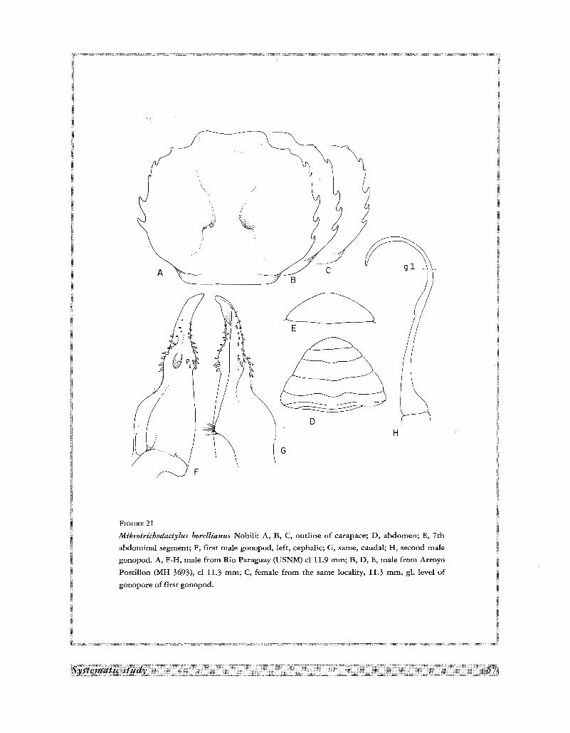
HD
~E
Bc
G
A
F1GURE 21
Mikrotrichodactylus borellianus Nobili: A, B, C, outline of carapace; D, abdomen; E, 7th
abdominal segment; F, first male gonopod, left, cephalic; G, same, caudal; H, second male
gonopod. A, F-H, male from Rio Paraguay (USNM) cl 11.9 mm; B, D, E, male from Arroyo
Postillon (MH 3693), cl 11.3 mm; C, female from the same locality, 11.3 mm. gl, level of
gonopore of first gonopod.
lateral border formed by two longitudinal subparallel lobes; apex linguiform, strongly bent mesially; strong
spines on mesial and lateral surfaces of distal expansion, diminishing in size distally. Gonopore large, opening
caudal. Second male pleopod longer than first, sickle-shaped.
Material examined
Paraguay. Rio Paraguay, Puerto Max, Arroyo Postillon; 3 males, 2 females (MH 3693). Rio Paraguay,
between Puerto Casado and Puerto Sastre; KRIEG, Nr 1931; 2 females (ZSM 1097/2). Villarrica; 1901; SPEYER;
2 males (ZSM 1097/3). Partially flooded shipyard (San Isidro) on Rio Paraguay in NW Asuncion; Rio La Plata
drainage; 1 June 1979; 1. N. TAYLOR & T.W. GRIMSHAW; Lat/Long: 25Q IT S - 57Q 39' W; Field NumberP79
11A (USNM Accession No 341275); 3 specimens. Arroyo Gaguaresau at bridge on route 2, approximately
1.5 km E of toll booth (just beyond km 39) and approximately 13.5 km W of Caacupe; 6 June 1979; R. BAILEY
& 1. N. TAYLOR; Lat/Long: 25 Q 22' 42" S - 57Q 15' 18" W; Field Number P79-21 (USNM Accession No
341275); 15 specimens. Paraguari, Arroyo Caanabe; 19 June 1979; R. BAILEY, J. N. TAYLOR & T.W.
GRIMSHAW; Lat/Long: 25Q 45' 18" S - 57Q 11' 42" W; Field Number P79-24 (USNM Accession No 341275); 5
specimens. Caaguazu, Arroyo Tobatiry at bridge on dirt highway, near 22 km N of junction with route 2 in
Coronel Oviedo; 29 July 1979; 1. N. TAYLOR & T.W. GRIMSHAW; Lat/Long: 25Q 16' 54" S - 56Q 24' W; Field
Number P79-69 (USNM Accession No 341275); 5 specimens. President Hayes Department, Rio Pilcomayo,
overflow pools at bridge (= Puerto Falcon) to Argentina, approximately 12 km WSW Chaco-i; Rio Paraguay; 29
August 1979; J. N. TAYLOR, B. SMITH, E. KOON & R. MYERS; Lat/Long: 26Q 24' 0" S - 57Q 4' 48" W; Field
Number P79-98 (USNM Accession No 341275); 4 specimens. San Pedro/Caaguazu Departments, Arroyo Pindo
at bridge on dirt highway (route 3) approximately 9 km N of Mbutuy and approximately 67 km N of Coronel
Oviedo; Rio Manduvira; 29 July 1979; 1. N. TAYLOR, T. W. GRIMSHAW & 1. K. CREIGHTON; Lat/Long: 24Q 52'
54" S - 56Q 19' 48" W; Field Number P79-68 (USNM Accession No 341275); 3 specimens. Concepcion
Department, Rio Paraguay, flood pools at municipal airport, approximately 4 km S of Concepcion; Rio de la
Plata drainage; 9 October 1979; 1. N. TAYLOR & T. W. GRIMSHAW; Lat/Long: 23Q 21' 18" S - 57Q 27' 00" W;
Field Number P79-109 (USNM Accession No 341275); 2 specimens. President Hayes Department, small stream
approximately 33.7 km NW of toll booth on Puente Remanso bridge, Rio Confuso; 8 November 1979; J. N.
TAYLOR, T. W. GRIMSHAW & B. SMITH; Lat/Long: 25Q 04' 54" - 57Q 26' W Field Number P79-79 (USNM
Accession No 341275); 16 specimens. Argentina? Parana, near Romallo; July 1913; ELLENRIDER; 10 males, 17
females (ZSM 1097/1).
Type and distribution
The syntypes were collected by Alfredo BORELLI at the Colonia Risso, Rio Apa in the high Paraguay River
basin, Paraguay. RATHBUN (1906) mentions cotypes in the USNM and MP. It is recorded in the literature from
the following localities. Paraguay. Concepcion Department: Puerto Max. Boqueron Department: Puerto Casado
and Puerto Sastre. Guaira Department: Villarrica (BOTT, 1969). Argentina. Santa Fe Province: Las Garzas, near
Ocampo; Reconquista (RATHBUN, 1906); Rio Parana Mini (RINGUELET, 1949); Romallo (BOTT, 1969).
Corrientes Province: Goya (RINGUELET, 1949). Chaco Province: Resistencia. Misiones Province: Posadas
(RATHBUN, 1906). Brazil. Para State: Pindobal; Lago do Tostao; Rio Tapajos. Arnazonas State: Igarape das 3
Casas (BOTT, 1969).
Remarks
In immature males the two lobes on the lateral surface of the distal bulbous expansion of gonopod are not
clearly distinct, but even these specimens can be separated from M. panoplus by the shape of the apex which is
more recurved mesially, and by the mesial border of the bulbous expansion which is more rounded.
The species display considerable variations in the anterior teeth of carapace. Thus, in 6 specimens examined
from Rio Apa, Paraguay, all have the 1st tooth as a tiny spine, but 2nd tooth is present as a blunt projection, a
lobe, or it is altogether absent (Fig. 21A-C).
Trichodactylus (Mikrotrichodactylus) borellianus brasiliensis, described by PRETZMANN (1968b) from Brazil
(but precise locality unknown), is separated by this author from the typical borellianus because its carapace is
more convex and less sculptured, the lateral teeth is not prominent, the 3rd not reduced (characters of panoplus),
the lateral margin is more straight and the transversal groove of ischium of 3rd maxilliped is less transversely
directed.
The small female reported by MOREIRA (1912) from Puerto Espiridiao, Rio Jaunis, Mato Grosso State, as
Trichodactylus (Trichodactylus) parvus may be a specimen of Mikrotrichodactylus borellianus in which all
lateral teeth, with the exception of the third, have become angled lobes. Reduction of the teeth occurs in both
M. borellianus and M. panoplus. As will be mentioned below, the specimens of M. panoplus from Buenos Aires
(Figure 22D, and the aberratio of panoplus reported by RINGUELET, 1949) also approach the condition found in
T.parvus.
Mikrotrichodactylus panoplus (von Martens, 1869)
Fig. 3F; 4F; 6F; lOH; 22A-L
Sylviocarcinus panoplus van Martens, 1869a, p. 3, pl. 1, fig. 1.- GOLDI, 1886, p. 34.- NOBILI, 1896, p. 3.
Orthostoma panoplus, NOBlLI, 1898, p. 12 (part.).- ORTMANN, 1897, p. 326.
Dilocarcinus panoplus, ORTMANN, 1893, p. 492.- NOBILI, 1899a, p. 3.- ORTMANN, 1903, p. 311.
Trichodactylus (Valdivia) panoplus, RATIlBUN, 1906, p. 52, pl. 17, fig. 5, text-fig. 114.- RINGUELET, 1949, p. 103, pl, 8, fig. 3, pl. 9,
fig. 1.
Trichodactylus (Trichodactylus) panoplus, GARCIA, 1973, p. 97, fig. 1-5.- LOPRETTO, 1976, p. 79, fig. 10-13.
Dilocarcinus panoplus var. marmorata Nobili, 1901, p. 11.
Trichodactylus (Trichodactylus) panoplus panoplus, BOTT, 1969, p. 21, pl, 4, fig. 6a, b, pl. 18, fig. 37.- MANNING & HOBBS, 1977, p. 160.
Trichodactylus panoplus, RODIUGUEZ, 1981, p. 48.
Dilocarcinus armatus A. Milne Edwards, 1869, p. 177.
Trichodactylus (Valdivia) spec. [aberratio Trichodactylus (V.) panoplus (van Martens, 1869)], RINGUELET, 1949, p. 103, pl. 8, fig. 3,
pl. 9, fig. 1.
Description
Carapace suborbicular; upper surface regularly convex; regions not delimited; wide depressions on
protogastric, epibranchiaJ and metabranchial regions, and on each side of protogastric regions. Branchio-cardiac,
branchio-urogastric and urogastric grooves form wide depressions. Postgastric pits absent. Lateral margins
angled; postero-lateral ridge of carapace bent mesially in posterior end, ending in elongated swelling over
postero-lateral angles of carapace; spinulation of lateral margin variable (see under Remarks), but in more
generalized condition margin has 5 spiniform teeth behind external orbital angle, first two placed closed
together, second smaller than first, other teeth widely spaced, last one at level of cardiac region. Front bilobed
more or less inclined downwards. Orbits small, suborbicular, eyes large and completely filling orbits; orbital
suture absent; lower orbital margin smooth; inner orbital angle with strong acute spine directed mesially;
occlusive orbital tooth small, attached to external orbital angle; outer orbital angle smooth, not projected; buccal
angle smooth or faintly papillated. Front advanced, hiding epistome in dorsal view; anterior surface of front high
in middle, thin on sides; 2 distinct middle pillars separated by U-shaped sinus, but without deep recess;
antennular septum sunken; epistome high, directed forward, with thin transverse ridge across it.
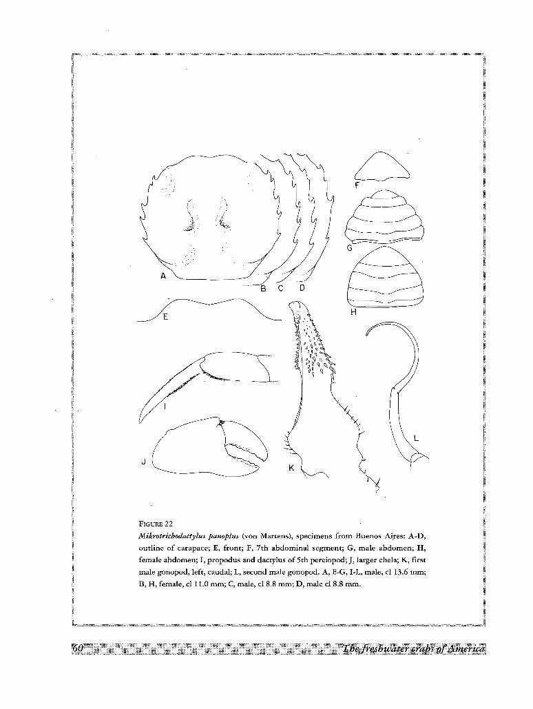
\.~.Jl).::
A
BeD
.:F
'c::::::::=::::----==~G
FIGURE 22
Mikrotrichodactylus panoplus (von Martens), specimens from Buenos Aires: A-D,
outline of carapace; E, front; F, 7th abdominal segment; G, male abdomen; H,
female abdomen; I, propodus and dactylus of 5th pereiopod; J, larger chela; K, first
male gonopod, left, caudal; L, second male gonopod. A, E-G, I-L, male, cl 13.6 mm;
B, H, female, cl 11.0 mm; C, male, cl 8.8 mm; D, male cl 8.8 mm.
All abdominal sutures distinct in both male and female, but only last segment freely movable, segments 3-5
ankylosed; male abdomen wide, triangular; lateral margins of segments 3-6 form conspicuous lobes over general
outline of abdomen; last segment widely rounded, approximately 0.55 as long as broad.
Shallow transverse depression across ischium of 3rd maxilliped. Chelipeds strongly unequal, largest strongly
developed in mature males, fingers short and curved, widely gaping lower margin of movable finger sinuous;
teeth small, subequal, regularly placed along the fingers; palm and fingers smooth, smaller chela with
longitudinal ridge on each finger. Carpus with strong hooked spine; merus with small apical spine on upper
border and large submedian spine on lateral margin. Legs long and slender; dactyli long, falciform, with short
felt-like pubescence on sides and rows of short hairs on upper and lower margins, claws short without
conspicuous carinae; lower margin of propodus with short hairs on distal quarter in 2nd and 3rd pair of
pereiopods, in distal half in 5th pair, and absent in 4th pair.
First male gonopods in normal position folded diagonally under abdomen; gonopod divided in two portions
by strong middle constriction; basal portion expanded in mesial and lateral directions, flattened in caudal and
cephalic directions, lateral border strongly sinuous, mesial border slightly concave; distal portion forms narrow
acuminate tube, expanded at base; apex awl-shaped, bent mesiad; gonopore long, oval, open mesocaudally;
strong conical spines arranged in irregular rows over mesial and latero-caudal surfaces. Second male gonopod
longer than first, sickle-shaped.
Material examined
Brazil. Rio Grande do SuI; 1 male, 4 mature females, 1 immature female (VON MARTENS Nr 1130/254; ZSM
1100/1, ex. Museum Liibeck).- Argentina. Buenos Aires; 9 April 1920; Dr FRANK; 2 males, 1 female (MH K
5251).
Type and distribution
The type material was collected by Reinhardt HENSEL at Rio Cadea and Santa Cruz, above Rio Pardo,
Guahyba, near Porto Alegre, Rio Grande do SuI, Brazil. Dr Reinhardt HENSEL (1826-1881), was a German
zoologist and mammalogist, "ordentlicher Professor" of Zoology at the Academy of Proskau, Schleswig (1867
1881), and member of the Kaiserlich Leopold-Karolus Deutsche Akademie der Naturforschung. Under the
patronage of the academy he undertook zoological studies in Southern Brazil, and from 1863-1866 he collected
in Rio Grande do SuI, specially Porto Alegre and the German colonies to the north of that city (PAPAVERO,
1972). The specimens from Brazil mentioned above perhaps were part of the type material. RATHBUN (1906)
mentions a male cotype in the collections of the USNM. Other records from the literature are the following.
Brazil. Rio Grande do SuI State (NOBILI, l899a; MOREIRA, 1901; BOTT, 1969): Guahyba, near Porto Alegre
(type, VON MARTENS, 1869a); Sao Lourenco (ORTMANN, 1893). Rio de Janeiro State: near Rio de Janeiro
(A. MILNE EDWARDS, 1869). Argentina. Buenos Aires Province (RATHBUN, 1906): Ensenada, Rio de la Plata
(ORTMANN, 1893); Tigre, Rio de la Plata (type of Dilocarcinus panoplus var. marmorata Nobili, 1901).
Uruguay. Paysandu Department: Santa Rita. Rio Negro Department: Arroyo Salsipuedes Grande. Treinta y tres
Department: Rio Olimar; Laguna Merin. Durazno Department: Paso de la Cruz. Tucuarembo Department:
Arroyo Yaguaron 29 km S of Ansina (GARCIA, 1973).
Remarks
The specimens I examined and those reported in the literature display a trend towards a reduction of the
lateral spines which affects the first and second one, and secondarily the last two. Thus in the specimens from
Rio Grande do SuI and Uruguay (GARCIA, 1973) the teeth are not reduced in number and only the last of the
series is reduced in size. Fig. 22A-D shows some of the variations observed in a lot of 6 specimens from Buenos
Aires. In this case, the 2nd spine is smaller in all specimens, in two it is very minute and in one it is replaced by
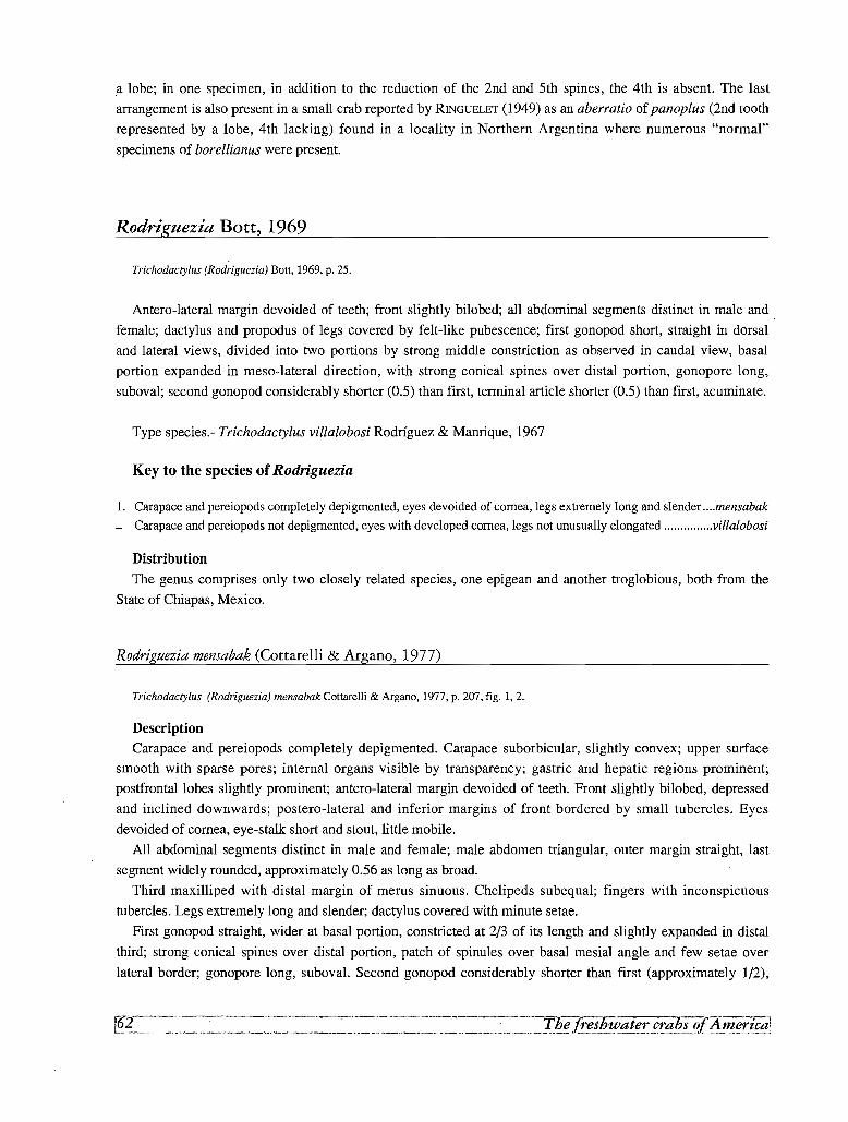
a lobe; in one specimen, in addition to the reduction of the 2nd and 5th spines, the 4th is absent. The last
arrangement is also present in a small crab reported by RINGUELET (1949) as an aberratio of panoplus (2nd tooth
represented by a lobe, 4th lacking) found in a locality in Northern Argentina where numerous "normal"
specimens of borellianus were present.
Rodriguezia Bott, 1969
Trichodactylus (Rodriguezia) Bott, 1969, p. 25.
Antero-lateral margin devoided of teeth; front slightly bilobed; all abdominal segments distinct in male and .
female; dactylus and propodus of legs covered by felt-like pubescence; first gonopod short, straight in dorsal
and lateral views, divided into two portions by strong middle constriction as observed in caudal view, basal
portion expanded in meso-lateral direction, with strong conical spines over distal portion, gonopore long,
suboval; second gonopod considerably shorter (0.5) than first, terminal article shorter (0.5) than first, acuminate.
Type species.- Trichodactylus villalobosi Rodrfguez & Manrique, 1967
Key to the species of Rodriguezia
1. Carapace and pereiopods completely depigmented, eyes devoided of cornea, legs extremely long and slender mensabak
- Carapace and pereiopods not depigmented, eyes with developed cornea, legs not unusually elongated villalobosi
Distribution
The genus comprises only two closely related species, one epigean and another troglobious, both from the
State of Chiapas, Mexico.
Rodriguezia mensabak (Cottarelli & Argano, 1977)
Trichodactylus (Rodriguezia) mensabak Cottarelli & Argaoo, 1977, p. 207, fig. 1,2.
Description
Carapace and pereiopods completely depigmented. Carapace suborbicular, slightly convex; upper surface
smooth with sparse pores; internal organs visible by transparency; gastric and hepatic regions prominent;
postfrontallobes slightly prominent; antero-lateral margin devoided of teeth. Front slightly bilobed, depressed
and inclined downwards; postero-lateral and inferior margins of front bordered by small tubercles. Eyes
devoided of cornea, eye-stalk short and stout, little mobile.
All abdominal segments distinct in male and female; male abdomen triangular, outer margin straight, last
segment widely rounded, approximately 0.56 as long as broad.
Third maxilliped with distal margin of merus sinuous. Chelipeds subequal; fingers with inconspicuous
tubercles. Legs extremely long and slender; dactylus covered with minute setae.
First gonopod straight, wider at basal portion, constricted at 2/3 of its length and slightly expanded in distal
third; strong conical spines over distal portion, patch of spinules over basal mesial angle and few setae over
lateral border; gonopore long, suboval. Second gonopod considerably shorter than first (approximately 1/2),
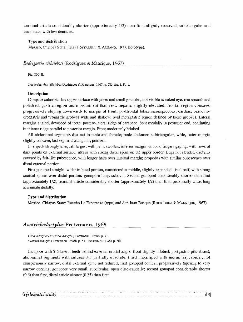
terminal article considerably shorter (approximately 1/2) than first, slightly recurved, subtriangular and
acuminate, with few denticles.
Type and distribution
Mexico, Chiapas State: Tila (COTTARELLI & ARGANO, 1977, holotype).
Rodriguezia villalobosi (Rodrfguez & Manrigue, 1967)
Fig. 23G-H.
Trichodactylus villalobosi Rodrfguez & Manrique, 1967, p. 183, fig. 1, PI. 1.
Description
Carapace suborbicular; upper surface with pores and small granules, not visible to naked eye, rest smooth and
polished; gastric region more prominent than rest, hepatic slightly elevated; frontal region concave,
progressively sloping downwards to margin of front; postfrontal lobes inconspicuous; cardiac, branchio
urogastric and urogastric grooves wide and shallow; oval metagastric region defined by these grooves. Lateral
margins angled, devoided of teeth; postero-lateral ridge of carapace bent mesially in posterior end, continuing
in thinner ridge parallel to posterior margin. Front moderately bilobed.
All abdominal segments distinct in male and female; male abdomen subtriangular, wide, outer margin
slightly concave, last segment triangular, pointed.
Chelipeds strongly unequal, largest with palm swollen, inferior margin sinuous; fingers gaping, with rows of
dark points on external surface; merus with strong distal spine on the upper border. Legs not slender, dactylus
covered by felt-like pubescence, with longer hairs over internal margin; propodus with similar pubescence over
distal external portion.
First gonopod straight, wider in basal portion, constricted at middle, slightly expanded distal half, with strong
conical spines over distal portion; gonopore long, suboval. Second gonopod considerably shorter than first
(approximately 1/2), terminal article considerably shorter (approximately 1/2) than first, proximally wide, long
acuminate distally.
Type and distribution
Mexico. Chiapas State: Rancho La Esperanza (type) and San Juan Bosque (RODRfGUEZ & MANRIQUE, 1967).
Avotrichodactylus Pretzmann, 1968
Trichodactylus (Avotrichodactylus) Pretzmann, 1968b, p. 71.
Avotrichodactylus Pretzmann, 1978b, p. 54.- PRETZMANN, 1980, p. 661.
Carapace with 2-5 lateral teeth behind external orbital angle; front slightly bilobed; postgastric pits absent;
abdominal segments with sutures 3-5 partially obsolete; third maxilliped with merus trapezoidal, not
conspicuously narrow, distal external spine not reduced; first gonopod conical, progressively tapering to very
narrow opening; gonopore very small, subcircular, open disto-caudally; second gonopod considerably shorter
(0.6) than first, distal article shorter (0.25) than first.
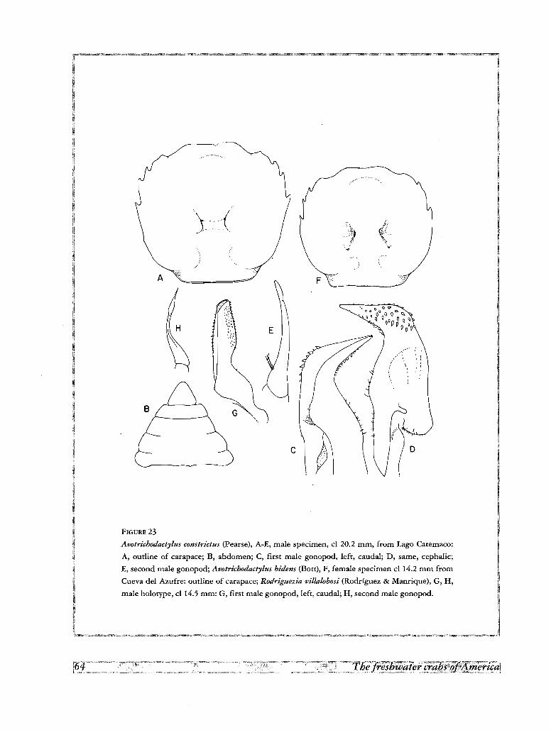
(
.. - ~,
B
A
-.
F
..,
c~~
FIGURE 23
Avotrichodactylus constrictus (Pearse), A-E, male specimen, cl 20.2 mm, from Lago Carernaco:
A, outline of carapace; B, abdomen; C, first male gonopod, left, caudal; D, same, cephalic;
E, second male gonopod; Avotrichodactylus bidens (Bott), F, female specimen cl 14.2 mm from
Cueva del Azufre: outline of carapace; Rodriguezia villalobosi (Rodriguez & Manrique), G, H,
male holotype, cl 14.5 mm: G, first male gonopod, lefe, caudal; H, second male gonopod,
Type species.- Trichodactylus constrictus Pearse, 1911.
Distribution
Southern Mexico, rivers in the States of Veracruz, Oaxaca, Tabasco and Chiapas draining into the Gulf of
Mexico.
Key to the species of Avotrichodactylus
1. First male gonopod sinuous, only slightly bent caudo-Iaterad; gonopore caudal; 4-5 spines on lateral side oaxensis
- First male gonopod with distal half bill-like, strongly bent laterad and forming a "knee" covered by short, stout spines;
gonopore terminal 2
2. Antero-Iateral margin with 3 spines constrictus
- Antero-Iateral margin with 2 spines bidens
Avotrichodactylus bidens (Bott, 1969)
Fig.23F
Trichodactylus bidens Bott. 1969, p. 25, pI. 24, fig. 68, 69.- COITARELLI & ARGANO, 1977, p. 207, fig. 3,4.
Description
Carapace suborbicular; upper surface moderately irregular; in frontal view regularly arched; frontal and
orbital regions excavated; lobes distinct, not prominent; branchio-urogastric and anterior portion of branchio
cardiac grooves deep, well marked, urogastric and posterior part of branchio-cardiac groove indicated by flat
depressions. Postgastric pits absent. Antero-Iateral margin with 2 small, spiniform teeth of approximately equal
size behind external orbital angle; interdental space approximately twice space between outer orbital angle and
1st tooth; postero-lateral ridge of carapace, not bent mesially in posterior end as in other species, but parallel to
lower ridge located over 5th coxa, ends away from postero-lateral angles of carapace. Margin of front slightly
concave. Orbits suborbicular; orbital suture absent or represented by small depression; lower orbital margin
papillated; inner orbital angle blunt; occlusive orbital tooth rounded, small, located close to inner orbital angle,
but not continuous with it; outer orbital not prominent, its border in contact with orbital margin; margin between
outer orbital angle and first tooth of carapace forms rounded lobe; buccal angle smooth. Front advanced, hiding
epistome in dorsal view; anterior surface of front sunk, low in middle, thin on sides; margin over each.
antennular fossa more or less straight; antennular septum sunken; epistome moderately high, inclined forwards.
Third to fifth abdominal segments fused in both male and female.
Basal article of antenna with outer lobe prominent and narrow; ischium of third maxilliped with deep groove.
Chelipeds strongly unequal; larger chela of male with upper border of hand strongly arched and lower sinuous,
fingers with elongated gap between them, teeth small, regularly placed along cutting edges; carpus with inner
margin produced in sharp hooked spine; merus with apical spine on upper border, another at distal angle of
latero-inferior margin and another at inner margin. Legs with long coarse hairs on lower margin of dactylus;
similar hairs cover all the lower margin of propodus in 5th, 3/4 in 2nd, 1/2 in third and only distal angle in 4th
pereiopod; upper margin of propodus and dactylus with shorter sparse hairs, rest of surface covered by felt-like
pubescence; claws of dactyli very short, with indistinct longitudinal carinae, 1 upper, 2 lateral and 2 inferior.
First and second gonopods, as illustrated by COTTARELLI & ARGANO (1977), similar to those of
Avotrichodactylus constrictus.
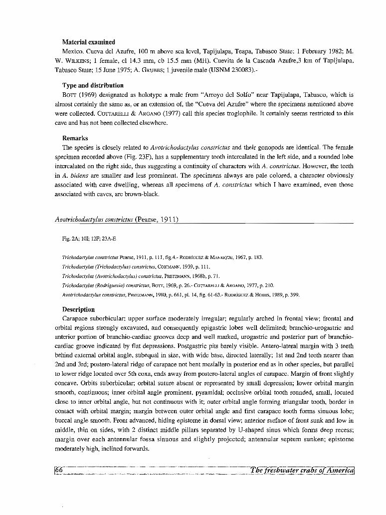
Material examined
Mexico. Cueva del Azufre, 100 m above sea level, Tapijulapa, Teapa, Tabasco State; 1 February 1982; M.
W. WILKENS; 1 female, cl 14.3 mm, cb 15.5 mm (MH). Cuevita de la Cascada Azufre,3 km of Tapijulapa,
Tabasco State; 15 June 1975; A. GRUBBS; 1 juvenile male (USNM 230083).-
Type and distribution
BOTT (1969) designated as holotype a male from "Arroyo del Solfo" near Tapijulapa, Tabasco, which is
almost certainly the same as, or an extension of, the "Cueva del Azufre" where the specimens mentioned above
were collected. COTTARELLI & ARGANO (1977) call this species troglophile. It certainly seems restricted to this
cave and has not been collected elsewhere.
Remarks
The species is closely related to Avotrichodactylus constrictus and their gonopods are identical. The female
specimen recorded above (Fig. 23F), has a supplementary tooth intercalated in the left side, and a rounded lobe
intercalated on the right side, thus suggesting a continuity of characters with A. constrictus. However, the teeth
in A. bidens are smaller and less prominent. The specimens always are pale colored, a character obviously
associated with cave dwelling, whereas all specimens of A. constrictus which I have examined, even those
associated with caves, are brown-black.
Avotrichodactylus constrictus (Pearse, 1911)
Fig. 2A; tor, 12F; 23A-E
Trichodactylus constrictus Pearse, 1911, p. 11I, figA.- RODRloUEZ & MANRIQUE, 1967, p. 183.
Trichodactylus (Trichodactylus) constrictus, COlFMANN, 1939, p. 111.
Trichodactylus (Avotrichodactylus) constrictus, PRETZMANN, 1968b, p. 71.
Trichodactylus (Rodriguezia) constrictus, BOTT, 1969, p. 26.- COTTARELLI & AROANO, 1977, p. 210.
Avotrichodactylus constrictus, PRETZMANN, 1980, p. 661, pl. 14, fig. 61-63.- RODRloUEZ & HOBBS, 1989, p. 399.
DescriptionCarapace suborbicular; upper surface moderately irregular; regularly arched in frontal view; frontal and
orbital regions strongly excavated, and consequently epigastric lobes well delimited; branchio-urogastric and
anterior portion of branchio-cardiac grooves deep and well marked, urogastric and posterior part of branchio
cardiac groove indicated by flat depressions. Postgastric pits barely visible. Antero-lateral margin with 3 teeth
behind external orbital angle, subequal in size, with wide base, directed laterally; l st and 2nd teeth nearer than
2nd and 3rd; postero-lateral ridge of carapace not bent mesially in posterior end as in other species, but parallel
to lower ridge located over 5th coxa, ends away from postero-lateral angles of carapace. Margin of front slightly
concave. Orbits suborbicular; orbital suture absent or represented by small depression; lower orbital margin
smooth, continuous; inner orbital angle prominent, pyramidal; occlusive orbital tooth rounded, small, located
close to inner orbital angle, but not continuous with it; outer orbital angle forming triangular tooth, border in
contact with orbital margin; margin between outer orbital angle and first carapace tooth forms sinuous lobe;
buccal angle smooth. Front advanced, hiding epistome in dorsal view; anterior surface of front sunk and low in
middle, thin on sides, with 2 distinct middle pillars separated by U-shaped sinus which forms deep recess;
margin over each antennular fossa sinuous and slightly projected; antennular septum sunken; epistome
moderately high, inclined forwards.
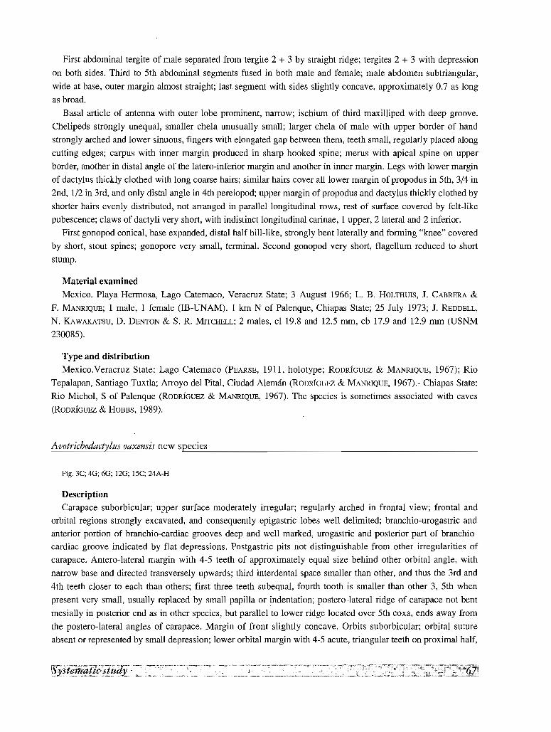
First abdominal tergite of male separated from tergite 2 + 3 by straight ridge; tergites 2 + 3 with depression
on both sides. Third to 5th abdominal segments fused in both male and female; male abdomen subtriangular,
wide at base, outer margin almost straight; last segment with sides slightly concave, approximately 0.7 as long
as broad.
Basal article of antenna with outer lobe prominent, narrow; ischium of third maxilliped with deep groove.
Chelipeds strongly unequal, smaller chela unusually small; larger chela of male with upper border of hand
strongly arched and lower sinuous, fingers with elongated gap between them, teeth small, regularly placed along
cutting edges; carpus with inner margin produced in sharp hooked spine; merus with apical spine on upper
border, another in distal angle of the latero-inferior margin and another in inner margin. Legs with lower margin
of dactylus thickly clothed with long coarse hairs; similar hairs cover all lower margin of propodus in 5th, 3/4 in
2nd, 1/2 in 3rd, and only distal angle in 4th pereiopod; upper margin of prop odus and dactylus thickly clothed by
shorter hairs evenly distributed, not arranged in parallel longitudinal rows, rest of surface covered by felt-like
pubescence; claws of dactyli very short, with indistinct longitudinal carinae, 1 upper, 2 lateral and 2 inferior.
First gonopod conical, base expanded, distal half bill-like, strongly bent laterally and forming "knee" covered
by short, stout spines; gonopore very small, terminal. Second gonopod very short, flagellum reduced to short
stump.
Material examined
Mexico. Playa Hermosa, Lago Catemaco, Veracruz State; 3 August 1966; L. B. HOLTHUIS, J. CABRERA &
F. MANRIQUE; 1 male, I female (IB-UNAM). I km N of Palenque, Chiapas State; 25 July 1973; 1. REDDELL,
N. KAWAKATSU, D. DENTON & S. R. MITCHELL; 2 males, cl 19.8 and 12.5 mm, cb 17.9 and 12.9 mm (USNM
230085).
Type and distributionMexico.Veracruz State: Lago Catemaco (PEARSE, 1911, holotype; RODRIGUEZ & MANRIQUE, 1967); Rio
Tepalapan, Santiago Tuxtla; Arroyo del Pital, Ciudad Aleman (RODRIGUEZ & MANRIQUE, 1967).- Chiapas State:
Rio Michol, S of Palenque (RoDRIGUEZ & MANRIQUE, 1967). The species is sometimes associated with caves
(RoDRIGUEZ & HOBBS, 1989).
Avotrichodactylus oaxensis new species
Fig. 3C; 4G; 6G; 12G; 15C; 24A-H
DescriptionCarapace suborbicular; upper surface moderately irregular; regularly arched in frontal view; frontal and
orbital regions strongly excavated, and consequently epigastric lobes well delimited; branchio-urogastric and
anterior portion of branchio-cardiac grooves deep and well marked, urogastric and posterior part of branchio
cardiac groove indicated by flat depressions. Postgastric pits not distinguishable from other irregularities of
carapace. Antero-lateral margin with 4-5 teeth of approximately equal size behind other orbital angle, with
narrow base and directed transversely upwards; third interdental space smaller than other, and thus the 3rd and
4th teeth closer to each than others; first three teeth subequal, fourth tooth is smaller than other 3, 5th when
present very small, usually replaced by small papilla or indentation; postero-lateral ridge of carapace not bent
mesially in posterior end as in other species, but parallel to lower ridge located over 5th coxa, ends away from
the postero-lateral angles of carapace. Margin of front slightly concave. Orbits suborbicular; orbital suture
absent or represented by small depression; lower orbital margin with 4-5 acute, triangular teeth on proximal half,
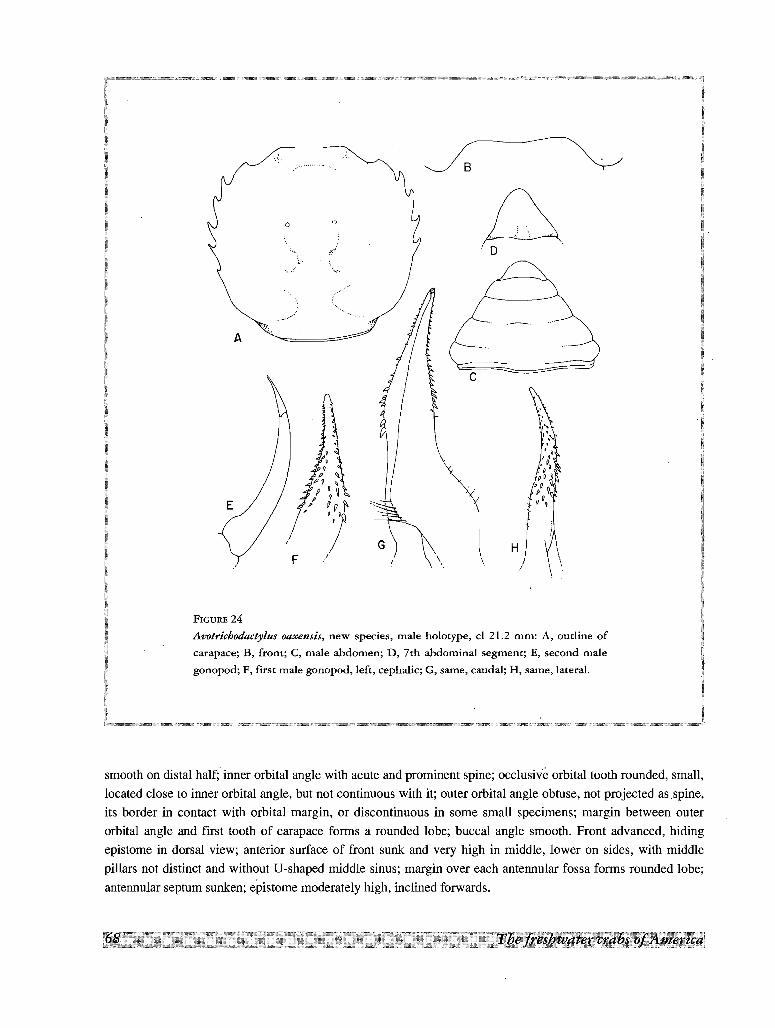
~., 0 .
c
B
~G) ~\
o
A
FIGURE 24
Avotrichodactylus oaxensis, new species, male holotype, cl 21.2 mm: A, outline of
carapace; B, from; C, male abdomen; D, 7th abdominal segment; E, second male
gonopod; F, first male gonopod, left, cephalic; G, same, caudal; H, same, lateral.
smooth on distal half; inner orbital angle with acute and prominent spine; occlusive orbital tooth rounded, small,
located close to inner orbital angle, but not continuous with it; outer orbital angle obtuse, not projected as spine,
its border in contact with orbital margin, or discontinuous in some small specimens; margin between outer
orbital angle and first tooth of carapace forms a rounded lobe; buccal angle smooth. Front advanced, hiding
epistome in dorsal view; anterior surface of front sunk and very high in middle, lower on sides, with middle
pillars not distinct and without If-shaped middle sinus; margin over each antennular fossa forms rounded lobe;
antennular septum sunken; epistome moderately high, inclined forwards.
First abdominal tergite of male separated from tergite 2 + 3 by straight ridge; tergites 2 + 3 with depression on
both sides. Third to 5th abdominal segments fused in both male and female; male abdomen subtriangular, wide at
base, outer margin slightly concave; last segment with sides slightly sinuous, approximately 0.7 as long as broad.
Basal article of antenna with outer lobe prominent and narrow; ischium of 3rd maxilliped with deep groove.
Chelipeds strongly unequal, smaller chela unusually small; larger chela of male with upper border of hand
strongly arched, lower sinuous; teeth small, regularly placed along cutting edges; carpus with inner margin
produced in sharp hooked spine; merus with apical spine on upper border, another at distal angle of latero
inferior margin and another at inner margin. Legs with lower margin of dactylus thickly clothed with long
coarse hairs; similar hairs cover all lower margin of propodus in 5th, 3/4 in 2nd, 1/2 in 3rd and only distal angle
in 4th; upper margin of propodus and dactylus thickly clothed by shorter hairs evenly distributed, not arranged
in parallel longitudinal rows; rest of surface of dactylus and propodus covered by felt-like pubescence; claw of
dactylus very short, with indistinct longitudinal carinae, I upper, 2 lateral and 2 inferior.
First gonopod slender, progressively tapering to very narrow tip; distal part moderately bent cephalo-Iaterad;
in caudal view narrow triangular, slightly widened at middle; in lateral view narrow and sinuous; distal third of
cephalic surface covered by spines which strongly diminish in size distally and tend to form four rows
proximally, leaving naked band over mid-line of appendage; gonopore very small, open on caudal surface.
Second gonopod very short, flagellum reduced to short, acute stump.
Material examined
Mexico. Canada Acatlan, Rio San Antonio, Oaxaca State; 2 February 1988; H. WILKENS; 1 male holotype,
cl 21.2 mm, cb 22.8 mm, 5 males paratypes, cl 18.0, 16.8, 16.5, 11.9, 11.3 mm, cb 19.0, 17.2, 18.3, 12.3 and
12.0 mm, 1 immature female paratype, cl 11.4 mm, cb 12.4 mm (MP).
Subfamily DILOCARCININAE Pretzmann, 1978
Postgastric pits present; abdominal segments with sutures 3-5 obsolete; penial groove not overlapped by 8th
tergite and sternal lobe; first gonopod conical, elongate, apical setae long, conspicuous; second gonopod
moderately or considerably longer than first.
Tribe HOLTHUISIINI
Carapace suborbicular, strongly arched, surface smooth, 3-5 lateral teeth behind external orbital angle
(sometimes obsolescent in large males), median grooves usually deep; abdomen trapezoidal; first gonopod with
basal portion expanded laterally, or with middle lateral lobe provided with conical spines; gonopore V-shaped,
open caudal; second gonopod much longer than first, rolled-over.
Type and only genus.- Sylviocarcinus H. Milne Edwards, 1853
Sylviocarcinus H. Milne Edwards, 1853
Sylviocarcinus H. Milne Edwards, 1853, p. 215.
Holthuisisia Pretzmann, 1968b, p. 74.
Holthuisia Pretzmann, 1968b, p. 74; PRETZMANN, 1983b, p. 321.
Carapace suborbicular, strongly arched, surface smooth, with the regions ill-defined and the grooves shallow
or obsolescent, but median grooves usually deep; 3-5 lateral spines (sometimes obsolescent in large males), in
addition to spiniform external orbital angle; front bilobed; buccal area usually with yugal and orbital
prominences as blunt teeth, exceptionally spiniform; first gonopod with the basal portion expanded laterally, or
with a middle lateral lobe provided with conical spines; gonopore V-shaped, open caudal; second male gonopod
considerably longer than first, usually twisted as a question mark (except in Sylviocarcinus sp.).
Type species.- Sylviocarcinus devillei H. Milne Edwards, 1853.
PRETZMANN (1968b, p.74) erected the genus Holthuisisia, which in the same page of that publication and in
PRETZMANN (1983b) he spelled Holthuisia, with Dilocarcinus pictus H. Milne Edwards, 1853, as the type
species. Later (PRETZMANN, 1983b) he gave as his reason to discard the genus Sylviocarcinus and replace it with
Holthuisia (=Holthuisisia) the fact that the type species of the former genus, S. devillei, is a female specimen.
However, S. devillei is easily recognized by the somatic characters, without recourse to male gonopods (see
Remarks under S. devillei).
Distribution
The genus Sylviocarcinus is widely distributed in the Amazon River and its Andean affluents, Magdalena
River basin, Lake Maracaibo basin and Paraguay River basin. It is occasionally present in the Guiana basins, but
absent from the Orinoco and the Atlantic basins of Brazil, except for the record of S. pictus from the Poty river
(RATHBUN, 1906). As here understood it is divided into 6 taxa, well defined geographically as follows. (1)
S. pictus (H. Milne Edwards, 1853) (syn. pardalinus Gerstacker, 1856), widely distributed in the Amazon basin
and its tributaries, as well as in the nearby basin of the Paraguay, is easily distinguished from the other species
by the acute spines surrounding the mouth area. (2) S. maldonadoensis, known only from Puerto Maldonado,Peru, is almost identical with S. pictus, except for the absence of the 2 buccal spines. (3) S. piriformis
Pretzmann, 1968, with a disjunct distribution in the Maracaibo basin and the Magdalena valley, show a
characteristic growth pattern, with a progressive obsolescence of the lateral spines and an increase in width of
the posterior section of carapace. Another species, (4) S. devillei H. Milne Edwards, 1853, characterized by the
spinulation of the rostral lobes, is distributed in the Amazon and its affluents of Colombia and northern Peru.
The species has been recorded in the literature under several synonyms, the most recent of them S. gigas
Smalley & Rodriguez, 1972. (5) S. sp. superficially resembles S. pictus, but in fact differs from all other species
of the genus in its short second gonopod. The records of this species include specimens from the Amazon and
northern Argentina.
Key to the species of Sylviocarcinus
1. Two acute hooked spines on buccal angle, 1 on inner orbital angle and 2 or 3 similar one on the lower orbital margin
.................................................................................................................................................................. .......................pictus
- Buccal angle with a large spine or tubercle, followed by several smaller tubercles or papillae. Inner orbital angle formed
by a pyramidal prominence, followed on the lower orbital margin by a series of papillae 2
2. Second male gonopod slightly longer than first, S-shaped sp.
- Second male gonopod considerably longer than first, usually twisted as a question mark 3
3. First gonopod not wide basally, without a well developed lateral lobe on the proximal half.. .4
- First gonopod very wide basally, with a conspicuous lateral lobe on the proximal half. Margin of front without large
granules piriformis
4. Gonopod with a conspicuous lateral "hump" on distal quarter. Margin of front often with large granules or spines ..devillei
- Gonopod without a conspicuous lateral "hump" on distal quarter. Margin of front without large granules or spines
.................................................................................................................................................................. ......maldonadoensis
Syloiocarcinus devillei H. Milne Edwards, 1853
Fig. 4H; 5C; 7F; 9B; 13A; 25A-F
Sylviocarcinus devillei H. Milne Edwards, 1853, p. 215.- H. MILNE EDWARDS, 1854, p. 176, pl. 14, fig. la-2.- LUCAS, 1857, p. 6, pl, 2,
fig. 1.- SMITH, 1869, p. 36.- A. MILNE EDWARDS, 1869, p. 174.- GOLD!, 1886, p. 33.- NOBILI, 1896, p. 2.- YOUNG, 1900, p. 231.- BOIT,
1969, p. 28 (part.).- RODRIGUEZ, 1981, p. 48.
Trichodactylus (Valdivia) devillei, RATHBUN, 1906, p. 51, pl, 17, fig. 2.
Sylviocarcinus peruvianus A. Milne Edwards, 1869, p. 174.-NoBILI, 1896, p. 3.- MORElRA, 1901, p. 44.
Trichodactylus (Valdivia) peruvianus, RATHBUN, 1906, p. 51, pI. 17, fig. 1.- DEL SOLAR et al., 1970, p. 31.
Orthostoma peruvianum, ORTMANN, 1897, p. 327.
Holthuisia peruviana peruviana, PRETZMANN, 1983b, p. 323, pl, 4, fig. 7, 8, pl, 5, fig. 9, 10, 11.
Dilocarcinus spinifrons Kingsley, 1880, p. 35.
Dilocarcinus margaritifrons Ortmann, 1893, p. 492, pl. 17, fig. 11.- YOUNG, 1900, p. 231.
Orthostoma margaritifrons, ORTMANN, 1897, p. 327.
Trichodactylus (Valdivia] margaritifrons, RATHBUN, 1906, p. 44.- PRETZMANN, 1968b, p. 71.- DEL SOLAR etal., 1970, p. 31.
Holthuisia peruviana margaritifrons, PRETZMANN & MAYTA, 1980, p. 141, fig. 9, 10.
Sylviocarcinus gigas Sma11ey & Rodriguez, 1972, p. 48, fig. 6-7, 21-22.-RoDRfGUEZ,.1981, p. 48.- CAMPOS, 1985, p. 270.
Description
Carapace suborbicular; upper surface slightly irregular; in frontal view forms irregular arch; gastric region
slightly more prominent than adjacent epibranchial regions; metabranchial region slightly more prominent than
adjacent cardiac and intestinal; epigastric lobes very prominent and well delimited anteriorly; frontal region
strongly concave in frontal view; branchio-urogastric and urogastric grooves wide and shallow, branchio-cardiac
groove almost absent. Postgastric pits lunulate, placed across urogastric groove. Lateral margins angled and
directed obliquely upwards, except in large males where they become rounded; postero-lateral ridge of carapace
bent mesially in posterior end, ending in elongated swelling over postero-lateral angles of carapace; antero
lateral margin with 3-4 prominent, evenly spaced, acute spines; last spines smaller; lateral teeth becomes
obsolescent in large males. Margin of front bilobed, with 13 to 19 spines which become papilliform and
obsolescent with age. Orbits oblong, orbital suture absent or indicated by inconspicuous depression; lower
orbital margin with 7-9 papillae; inner orbital angle prominent, pyramidal, topped by prominent and acute spine
or rounded tubercle; occlusive orbital tooth rounded, small, fused to inner orbital angle; outer orbital angle
spiniform, prominent particularly in females and young specimens, represented by stump in old males; buccal
angle with large acute spine followed laterally by smaller one, or large tubercle followed by one or two smaller
ones, Front advanced, hiding epistome in dorsal view; anterior surface of front inclined backwards, moderately
high in the middle, lower on sides, middle pillars short and thick, widely separated, sometimes not distinct,
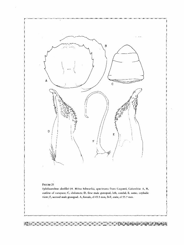
Q
p 0 v'l.: p ~
;<>o/Pop,§r, 0p Q
V~S~Ij! Q
Jff '\(
"E
c
F
A
FIGURE 25
Sylviocarcinus devillei (H. Milne Edwards), specimens from Caqueta, Colombia: A, B,
outline of carapace; C, abdomen; D, first male gonopod, left, caudal; E, same, cephalic
view; F, second male gonopod. A, female, cl 45.5 mm, B-F, male, cl 55.7 mm.
margin over each antennular fossa slightly thickened, but not projected, antennular septum sunken; epistome
high, inclined forwards; mid-point of epistome acute, in ventral view appearing as a triangular spine.
First abdominal tergite of male separated from tergite 2 + 3 by suture or deep depression, tergite 2 + 3 with
very shallow depression on both sides. Third to 5th abdominal segments fused in both male and female; suture
5/6 very well marked, suture 4/5 only as a thin line and suture 3/4 visible only near lateral margins and on mid
line; 7th segment freely movable; male abdomen elongated, narrow at base, outer margin almost straight, last
segment with sides straight, approximately 0.5 as long as broad.
Basal article of antenna with outer lobe prominent and narrow; ischium with shallow longitudinal depression.
Chelipeds unequal; upper border of chela strongly arched, lower almost straight or slightly concave; teeth
alternatively large and small; acute spine on upper distal angle of hand, above articulation of finger; inner
margin of carpus produced in sharp hooked spine; apical spines on upper border and latero-inferior margin of
merus. Legs with lower margin of dactylus and lower margin of propodus of 5th pereiopod thickly clothed with
band of coarse hairs; these pubescence absent from other pereiopods, except for inferior distal angle of 2nd;
upper margin of propodus and dactylus thickly clothed with shorter hairs arranged more or less in 2 parallel
longitudinal rows, more extensive in 2nd and 3rd; claws of dactyli with 5 longitudinal carinae, 1 upper, 2 lateral
and 2 inferior.
First gonopod sinuous, tapering strongly to narrow apex, which is directed mesiad; lateral margin of
appendage with strong constriction at middle and conspicuous "hump" on distal half; lateral lobe undivided;
field of terminal spines extends proximally along posterior margin, lateral margin ("hump") and cephalo-mesial
surface, forming 3 distinct patches; brush of long setae on lateral side of apex; gonopore slit-like, open on caudal
side. Second gonopod considerably longer than first, bent mesially in form of question mark.
Color
The carapace is mottled with red specks, which in some specimens are restricted to the distal half, the anterior
portion being uniformly reddish.
Size
The largest male on record of this species, cl 88.2 mm (SMALLEY & RODRIGUEZ, 1972) is also the largest
known specimen of Trichodactylidae.
Material examined
Colombia. Florencia, Caqueta Department; H. Nicsroao MARIA; 1 male, I female (Ivic).- Peru. Upper Rio
Marafion, between Jaen and San Ignacio; 15 November 1979; Ministry of Fisheries of Peru; 1 male (Ivic). Puerto
Maldonado; 7 November 1972; E. DEL SOLAR; 1 male (MHNL).
Type and distributionThe type is a small (cl 32.8 mm) female (MP), collected by the count Francis de CASTELNAU and Mr Emile
DEvILLE, this last an employee of the Paris Museum, at Salinas, on the Rio Crixas Acu, affluent of the Rio
Araguaia, where they stayed from May 14 to June 10, 1844, during their expedition across South America
(PAPAVERo,1971).
The other records of the species in the literature are the following. Brazil. Amazonas State: Lago Manacapuru
near Manaus (BOTT, 1969); ?Rio Madeira (MOREIRA, 1901). Colombia. Caqueta Department: Rio Orteguaza,
near Venecia (type S. gigas). Putumayo Department: Rio Putumayo, Puerto Asis (SMALLEY & RODRfGUEZ,
1972). Ecuador. Rio Bobonaza-Pastaza (PRETZMANN, 1983b, as Holthuisia peruviana peruviana). Peru. Rio
Ucayali (type of Dilocarcinus margaritifrons Ortmann, 1893). Guyallaga (type of S. peruvianus A. Milne
Edwards, 1869). Upper Rio Marafion (present record).
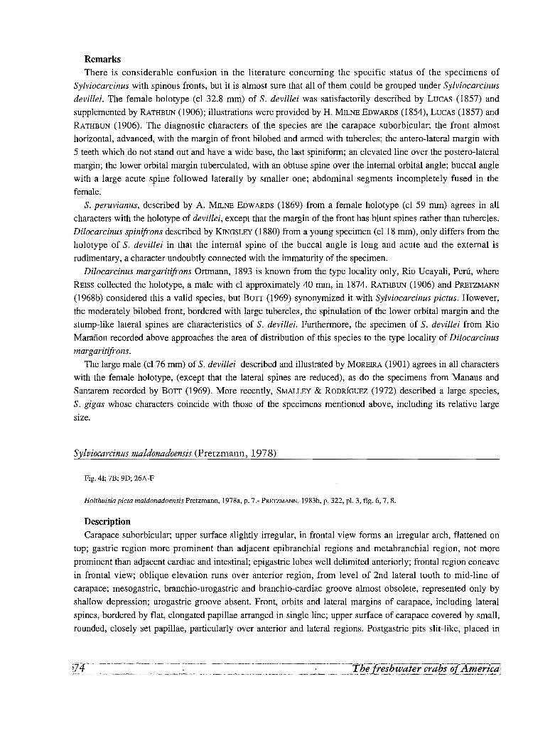
RemarksThere is considerable confusion in the literature concerning the specific status of the specimens of
Sylviocarcinus with spinous fronts, but it is almost sure that all of them could be grouped under Sylviocarcinus
devillei. The female holotype (cl 32.8 mm) of S. devillei was satisfactorily described by LUCAS (1857) and
supplemented by RATHBUN (1906); illustrations were provided by H. MILNE EOWAROS (1854), LUCAS (1857) and
RATHBUN (1906). The diagnostic characters of the species are the carapace suborbicular; the front almost
horizontal, advanced, with the margin of front bilobed and armed with tubercles; the antero-lateral margin with
5 teeth which do not stand out and have a wide base, the last spiniform; an elevated line over the postero-lateral
margin; the lower orbital margin tuberculated, with an obtuse spine over the internal orbital angle; buccal angle
with a large acute spine followed laterally by smaller one; abdominal segments incompletely fused in the
female.
S. peruvianus, described by A. MILNE EOWAROS (1869) from a female holotype (cl 59 mm) agrees in all
characters with the holotype of devillei, except that the margin of the front has blunt spines rather than tubercles.
Dilocarcinus spinifrons described by KINGSLEY (1880) from a young specimen (cl 18 mm), only differs from the
holotype of S. devillei in that the internal spine of the buccal angle is long and acute and the external is
rudimentary, a character undoubtly connected with the immaturity of the specimen.
Dilocarcinus margaritifrons Ortmann, 1893 is known from the type locality only, Rio Ucayali, Peru, where
REISS collected the holotype, a male with cl approximately 40 mm, in 1874. RATHBUN (1906) and PRETZMANN
(1968b) considered this a valid species, but BOTT (1969) synonymized it with Sylviocarcinus pictus. However,
the moderately bilobed front, bordered with large tubercles, the spinulation of the lower orbital margin and the
stump-like lateral spines are characteristics of S. devillei. Furthermore, the specimen of S. devillei from Rio
Marafion recorded above approaches the area of distribution of this species to the type locality of Dilocarcinus
margaritifrons.
The large male (cl 76 mm) of S. devillei described and illustrated by MOREIRA (1901) agrees in all characters
with the female holotype, (except that the lateral spines are reduced), as do the specimens from Manaus and
Santarem recorded by BOTT (1969). More recently, SMALLEY & ROORIGUEZ (1972) described a large species,
S. gigas whose characters coincide with those of the specimens mentioned above, including its relative large
size.
Sylviocarcinus maldonadoensis (Pretzmann, 1978)
Fig. 41; 7B; 9D; 26A-F
Holthuisia picta maldonadoensis Pretzmann, 1978a, p. 7.- PRETZMANN, 1983b, p. 322, pI. 3, fig. 6, 7, 8.
Description
Carapace suborbicular; upper surface slightly irregular, in frontal view forms an irregular arch, flattened on
top; gastric region more prominent than adjacent epibranchial regions and metabranchial region, not more
prominent than adjacent cardiac and intestinal; epigastric lobes well delimited anteriorly; frontal region concave
in frontal view; oblique elevation runs over anterior region, from level of 2nd lateral tooth to mid-line of
carapace; mesogastric, branchio-urogastric and branchio-cardiac groove almost obsolete, represented only by
shallow depression; urogastric groove absent. Front, orbits and lateral margins of carapace, including lateral
spines, bordered by flat, elongated papillae arranged in single line; upper surface of carapace covered by small,
rounded, closely set papillae, particularly over anterior and lateral regions. Postgastric pits slit-like, placed in
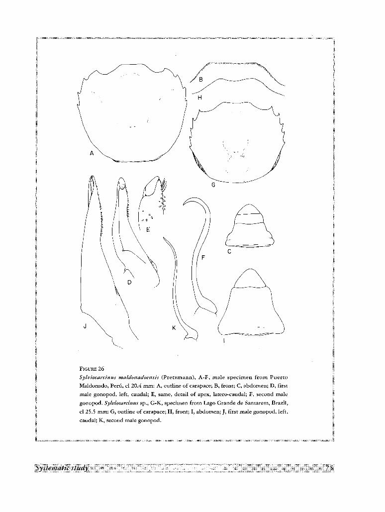
r>:I
/8
H
A
J
G
c
FIGURE 26
Sylviocarcinus maldonadoensis (Prerzrnann), A-F, male specimen from Puerto
Maldonado, Peru, cl 20.4 mm: A, outline of carapace; B, from; C, abdomen; D, first
male gonopod, left, caudal; E, same, detail of apex, latero-caudal; F, second male
gonopod. Sylviocarcinus sp., G-K, specimen from Lago Grande de Santarern, Brazil,
cl 25.5 mm: G, outline of carapace; H, front; I, abdomen; J, first male gonopod, left,
caudal; K, second male gonopod.
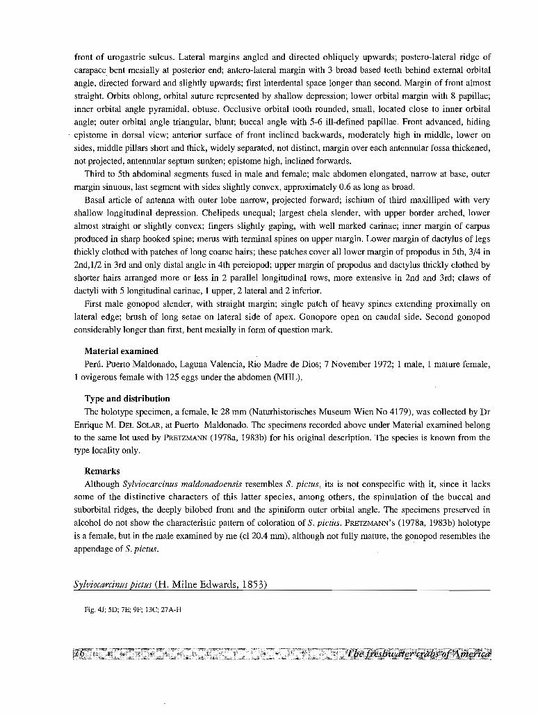
front of urogastric sulcus. Lateral margins angled and directed obliquely upwards; postero-lateral ridge of
carapace bent mesially at posterior end; antero-lateral margin with 3 broad based teeth behind external orbital
angle, directed forward and slightly upwards; first interdental space longer than second. Margin of front almost
straight. Orbits oblong, orbital suture represented by shallow depression; lower orbital margin with 8 papillae;
inner orbital angle pyramidal, obtuse. Occlusive orbital tooth rounded, small, located close to inner orbital
angle; outer orbital angle triangular, blunt; buccal angle with 5-6 ill-defined papillae. Front advanced, hiding
. epistome in dorsal view; anterior surface of front inclined backwards, moderately high in middle, lower on
sides, middle pillars short and thick, widely separated, not distinct, margin over each antennular fossa thickened,
not projected, antennular septum sunken; epistome high, inclined forwards.
Third to 5th abdominal segments fused in male and female; male abdomen elongated, narrow at base, outer
margin sinuous, last segment with sides slightly convex, approximately 0.6 as long as broad.
Basal article of antenna with outer lobe narrow, projected forward; ischium of third maxilliped with very
shallow longitudinal depression. Chelipeds unequal; largest chela slender, with upper border arched, lower
almost straight or slightly convex; fingers slightly gaping, with well marked carinae; inner margin of carpus
produced in sharp hooked spine; merus with terminal spines on upper margin. Lower margin of dactylus of legs
thickly clothed with patches of long coarse hairs; these patches cover all lower margin of propodus in 5th, 3/4 in
2nd,l/2 in 3rd and only distal angle in 4th pereiopod; upper margin of propodus and dactylus thickly clothed by
shorter hairs arranged more or less in 2 parallel longitudinal rows, more extensive in 2nd and 3rd; claws of
dactyli with 5 longitudinal carinae, 1 upper, 2 lateral and 2 inferior.
First male gonopod slender, with straight margin; single patch of heavy spines extending proximally on
lateral edge; brush of long setae on lateral side of apex. Gonopore open on caudal side. Second gonopod
considerably longer than first, bent mesially in form of question mark.
Material examined
Peru. Puerto Maldonado, Laguna Valencia, Rio Madre de Dios; 7 November 1972; 1 male, 1 mature female,
1 ovigerous female with 125 eggs under the abdomen (MHL).
Type and distribution
The holotype specimen, a female, le 28 mm (Naturhistorisches Museum Wien No 4179), was collected by Dr
Enrique M. DEL SOLAR, at Puerto Maldonado. The specimens recorded above under Material examined belongto the same lot used by PRETZMANN (1978a, 1983b) for his original description. The species is known from the
type locality only.
Remarks
Although Sylviocarcinus maldonadoensis resembles S. pictus, its is not conspecific with it, since it lacks
some of the distinctive characters of this latter species, among others, the spinulation of the buccal and
suborbital ridges, the deeply bilobed front and the spiniform outer orbital angle. The specimens preserved in
alcohol do not show the characteristic pattern of coloration of S. pictus. PRETZMANN'S (l978a, 1983b) holotype
is a female, but in the male examined by me (cl 2004 mm), although not fully mature, the gonopod resembles the
appendage of S. pictus.
Sylviocarcinus pictus (H. Milne Edwards, 1853)
Fig. 4J; 5D; 7E; 9F; 13C; 27A-H
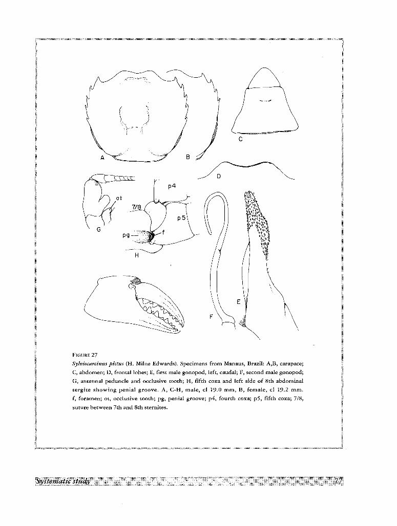
c
'~~--,:..A
E
D
p4
H
at
FIGURE 27
Sylviocarcinus pictus (H. Milne Edwards). Specimens from Manaus, Brazil: A,B, carapace;
C, abdomen; D, frontal lobes; E, first male gonopod, left, caudal; F, second male gonopod;
G, antennal peduncle and occlusive tooth; H, fifth coxa and left side of 8th abdominal
tergite showing penial groove. A, C-H, male, cl 19.0 mm, B, female, cl 19.2 mm.
f, foramen; ot, occlusive tooth; pg, penial groove; p4, fourth coxa; p5, fifth coxa; 7/8,
suture between 7th and 8th srernites.
Dilocarcinus pictus H. Milne Edwards, 1853, p. 216.- H. MILNE EDWARDS, 1854, p. 181, pI. 14, fig. 2.- GERSTACKER, 1856, p. 148.
LUCAS, 1857, p. 7, p1.2, fig. 3.- A. MILNE EDWARDS, 1869, p.I77.- SMITH, 1870, p. 36.- NOBlLI, 1896, p. 1.- YOUNG, 1900, p. 231.
MOREIRA, 1901, p. 44.- MOREIRA, 1913, p. 19, pI. 6, fig. 1-3.- COLDSI, 1920, p. 16.- RINGUELET, 1949, p. 102, pI. 7.
Dilocarcinus pieta, STIMPSON, 1861, p. 373.
Trichodactylus (Dilocarcinus) pictus, RATHBUN, 1906, p. 62, pI. 19, fig. 9.- MOREIRA, 1912, p. 150, pI. 6, fig. 9-11.- RiNGUELET, 1949,
p. 102, pI. 7.
Orthostoma pictum, ORTMANN 1897, p. 327.- NOBILI, 1898, p. 11.
Sylviocarcinus pictus pictus, BOTT, 1969, p. 31, pI. 12, fig. 22a, b, pI. 21, fig. 53.
Sylviocarcinus pictus, SMALLEY & RODRiGUEZ, 1972, p. 48, fig. 8.- RODRiGUEZ, 1981, p. 48.- CAMPOS, 1985, p. 270.
Holthuisia picta pieta, PRETZMANN, 1983b, p. 321, pI. I, fig. 1,2, pI. 2, fig. 3-5.
Holthuisia pieta rionegrensis Pretzmann, 1968b, p. 74.
Holthuisia pieta collastinensis Pretzmann, 1968b, p. 74.
Dilocarcinus pardalinus Gerstacker, 1856, p. 148.- A. MILNE EDWARDS, 1869, p. 177.- KINGSLEY, 1880, p. 35.- NOBILl, 1896, p. 1.
MORElRA, 1901, p. 44.- RODRiGUEZ, 1981, p. 48.
Orthostoma pardalinus, ORTMANN, 1897, p. 327.
Trichodactylus (Yaldivia) pardalinus, RATHBUN, 1906, p. 46.
Sylviocarcinus pictus pardalinus, BOTT, 1969, p. 13, p. 32, fig. 23a, b, pI. 21, fig. 54.
Description
Carapace suborbicular; upper surface slightly irregular, in frontal view forms irregular arch, flattened on top;
gastric region not more prominent than adjacent epibranchial regions, and metabranchial region slightly more
prominent than adjacent cardiac and intestinal; epigastric lobes well delimited anteriorly; frontal region slightly
concave in frontal view; branchio-urogastric and branchio-cardiac grooves very shallow, urogastric groove
almost absent. Postgastric pits slit-like, placed in front of urogastric sulcus. Lateral margins angled and directed
obliquely upwards; postero-lateral ridge of carapace bent mesially in posterior end; antero-lateral margin with 4
equally spaced prominent acute teeth behind external orbital angle (see below under Remarks), directed forward
and slightly upwards, and sometimes fifth smaller one placed farthest from rest. Margin of front deeply bilobed
covered with minute papillae. Orbits oblong, orbital suture absent; lower orbital margin with 2-3 spines directed
mesially and decreasing in size laterally, followed by 3-4 papillae; inner orbital angle has sharp, hooked well
developed spine directed ventrally. Occlusive orbital tooth rounded, small, fused to inner orbital angle; outer
orbital angle spiniform; buccal angle with 2 well-developed hooked spines. Front advanced, but leaving exposed
the lower angle of epistome in dorsal view; anterior surface of front vertical, moderately high in middle, lower
on sides, middle pillars short and triangular, widely separated, sometimes not distinct, margin over each
antennular fossa thickened, not projected, antennular septum sunken; epistome high, inclined forward.
First abdominal tergite of male separated from tergite 2 + 3 by shallow depression, tergite 2 + 3 with very
shallow depression on both sides. Third to 6th abdominal segments fused in both male and female; male
abdomen elongated, narrow at base, outer margin concave, last segment with sides straight or slightly convex,
approximately 0.6 as long as broad.
Basal article of antenna with outer lobe prominent and narrow; ischium of third maxilliped with very shallow
longitudinal depression. Chelipeds unequal; largest chela slender, with upper border arched, lower almost
straight or slightly convex; fingers not gaping, with well marked carinae at least in young specimens, teeth
alternating large and small; inner margin of carpus produced in sharp hooked spine; merus with terminal spines
on upper and inner margins; spine on the latero-inferior margin of this segment in largest chela and two in
smaller one. Lower margin of dactylus of legs thickly clothed with patches of long coarse hairs; these patches
cover all lower margin of propodus in 5th, 3/4 in 2nd, 1/2 in 3rd and only distal angle in 4th; upper margin of
propodus and dactylus thickly clothed by shorter hairs arranged more or less in 2 parallel longitudinal rows,
more extensive in Znd and 3rd; claws of dactyli with 5 longitudinal carinae, 1 upper, 2 lateral and 2 inferior.
First male gonopod slender, with straight margin; lateral edge in caudal view has curve at 4/5 length of
gonopod from base, forming slight constriction, followed by strong outward curve, and ending in slender tip;
single patch of heavy spines, extending proximally on lateral edge; brush of long setae on lateral side of apex.
Gonopore slit-like and open on caudal side. Second gonopod considerably longer than first, bent mesially in
form of question mark.
Colour
Carapace light brown, with scattered red spots persistent in alcohol.
Material examined
Colombia. Leticia, on the Amazon River, Department of Amazonas; H. NICEFORO MARIA; 1 male (LSB).
Brazil. Sistema Janavaca, South of Manaus, Amazon River; July 1978; 2 males, 2 females (Ivic).- Venezuela.
Cafio Sin Nombre, left bank of Rio Cuyuni, near Isla Jacobo, Bolivar State; 26 January 1977; F. MAGO; 1 soft
shell male (Ivic).- Paraguay. Concepcion, Rio lpare at ferry crossing, approximately 2.0 km S by dirt road of
Belen (which is 18 km ESE of Concepcion); Rio Paraguay drainage; 9 October 1979; J. N. TAYLOR, B. SMITH,
E. KOON & R.MYERS; Lat/Long: 23227' 18" S - 57 2 27' W; Field Number P79-11O (USNM Accession No
341275); 1 specimen. Canendiyu, Rio Jejui-Guazii, bridge approximately 14 km S of Ygatimi on dirt road and
approximately 29.5 km N of Curuguaty; Rio Paraguay drainage; 7 July 1979; R.M. BAILEY, J. N. TAYLOR &
T. W. GRIMSHAW; Lat/Long: 242 14' 12" S - 552 37' 30" W; Field Number P79-43 (USNM Accession No
341275); I specimen. Amambay Department, Rio Apaca approximately 0.5 km upstream (==E) of bridge
between Brazil & Paraguay in Bella Vista, Paraguay; Rio Paraguay drainage; 27 July 1979; J. N. TAYLOR, T. W.
GRIMSHAW & R. MYERS; Lat/Long: 22Q 6' 30" S - 56 Q 30' 36" W; Field Number P79-65A (USNM Accession No
341275); 1 specimen.
Type and distribution
The types are two females (MP) collected by the count Francis de CASTELNAU and Mr Emile DEVILLE at
Loreto, Colombia, where they stayed from December 25 to 28, 1846, on their way back from their expedition
across South America (PAPAVERO, 1971). The other records of the species in the literature are the following.
Peru. Nauta (RATHBUN, 1906); Rio Siamiria (BOIT,1969); 25 km W ofIquitos (PRETZMANN, 1983b). Colombia.
Rio Arara, Leticia, on the Amazon River (SMALLEY & RODRfGUEz, 1972). Brazil. Manaus; Villa Bella, 600 km
upriver Amazonas; Poty, Piauhy State (RATHBuN, 1906); Solimoes, Fonte Boa; Rio Negro, Ponta Arara; Rio
Tapajos; Rio Mulata (BOTT, 1969). Bolivia. Rio Chimpire, Rio Chapare (BOTT, 1969). Paraguay. Rio Paraguay,
near Puerto Max, North Paraguay (BOTT, 1969); Colonia Risso, Rio Apa (RATHBuN, 1906). Argentina. Buenos
Aires (probably Buenos Aires Province); Candelaria, Misiones (RATHBuN, 1906). French Guiana. Riviere
Camopi; Haut Carsevenne (RATHBUN, 1906). The specimens from Rio Parana Mini, in front of La Invemada
island, Reconquista Department, Santa Fe Province, Argentina reported by RINGUELET (1949) probably belong
to the Sylviocarcinus sp., described below. As Sylviocarcinus pictus pardalinus, it has been recorded from the
following localities in Brazil. Peixe Boi near Para; entrance of the Igarape into the Rio Tapajos; Ducke Reserve;
Rio Guama at Ourem; Rio Aripuana at Beneficiente; Rio Cupari at Goiabal; Rio Irapiri; Rio Marauia at
Cachoeira; all these records by BOTT (1969) come from the lower Amazon and its affluents. As Dilocarcinus
pardalinus it has been recorded from Paraguay, Rio Apa (NOBILI, 1896).
Remarks
As shown in Fig. 48, the area of distribution of the species covers a very large territory comprising the basins
of the Amazon and its tributaries, the Parana-Paraguay basin, and the Atlantic basins of the Guianas. This wide,
scattered distribution rises doubts about the cohesion of the species and have led BOTT (1969) and PRETZMANN
(1968b, 1977a) to segregate some specimens into distinct subspecies.
The type specimen of Diloearcinus pardalinus is a female from an unknown locality. The species was later
recorded from Rio Apa, Upper Paraguay by NOBILI (1896). BOTT (1969) considered this a subspecies of
Sylviocarcinus pietus and recorded it as such from the lower Amazon. The only character with diagnostic value
given by BOTT (1969) to separate it from the typical form S. pictus, besides the form of the red dots of the
carapace concentrated "as in the leopard" (GERSTACKER, 1856), is the presence of 3 lateral teeth instead of 4,
behind the spiniform external orbital angle. This character, however, is variable as shown by specimens
collected in the same locality. Thus, in 4 specimens from Manaus, 3 males had 4 lateral teeth and 1 female had 3
(Fig. 27a, b); in three females from Paraguay River, 1 specimen had 3 teeth on each side, 1 had 3 teeth on each
side and an indentation on the right side, and 1 specimen 4 teeth on each side. On the other hand the specimens
from the Paraguay River also present some variability in the characteristic pair of buccal spines which in this
case could be present or replaced by a large and a smaller one, or even by a single spine.
PRETZMANN (1968b, 1978a) described three subspecies of Sylvioeareinus pictus as follows.
(1) Holthuisia pieta rionegrensis Pretzmann, 1968, based on an immature male (cl 20.2 mm), from the mouth
of Rio Negro (i. e. near Manaus). The specimens I have examined from Manaus (see above), cannot be
differentiated from typical S. pietus.
(2) Holthuisia picta eollastinensis Pretzmann, 1968, from Collastine, based on a female from Santa Fe.
SMALLEY & RODRIGUEZ (1972), suggest that collastinensis refers to the city of Clatine, formerly called Collatina,
50 km north of Vitoria, Spirito Santo State, Brazil, but this locality lies outside the area of distribution of
S. pietus. Another possibility is that Santa Fe refers to the Santa Fe Province in Argentina where RINGUELET
(1949) has recorded S. pietus.
(3) Holthuisia picta maldonadoensis Pretzmann, 1978. As stated before, the characters displayed by this
taxon are enough to give it specific rank.
Sylviocarcinus piriformis (Pretzmann, 1968)
Fig. 31; 4K, M; 5B; 7A, C; 9C, G; BB; 15D; 28A-G; 29A-F
Yaldivia (Valdivia) piriformis Pretzmann, 1968b, p. 73.- SCHMITT, 1969, p. 98, fig. 3, a-e, f-i.
Sylviocarcinus piriformis, SMALLEY & RODRiGUEZ, 1972, p. 45, fig. 5.- RODRiGUEZ, 1980, p. 340, fig. 97.- RODRiGUEZ, 1981, p. 48.- VON
PRAHL, 1982, p. 23, fig. 1.- VON PRAHL, 1988, p. 13.
Yaldivia (valdivia) torresi Pretzmann, 1968b, p. 72.
Sylviocarcinus torresi, SMALLEY & RODRfGUEZ, 1972, p. 44, fig. 3, 4.- RODRiGUEZ, 1981, p. 48.
Description
Carapace suborbicular in smaller specimens, widest between fourth lateral teeth; in larger specimens
becoming subquadrate to piriform, widest behind 4th lateral teeth due to swelling of lateral wall of branchial
chamber; finally in largest specimens becoming orbicular with posterior lateral walls considerably swollen.
Upper surface slightly irregular, in frontal view forms irregular arch, gastric region slightly more prominent than
adjacent epibranchial regions; metabranchial region slightly more prominent than adjacent cardiac and
intestinal; epigastric lobes well delimited anteriorly; frontal region concave in frontal view; branchio-urogastric
and branchio-cardiac grooves wide and shallow, urogastric groove almost absent. Postgastric pits lunulate,
placed in front of urogastric sulcus. Lateral margins angled and directed obliquely upwards, except in large
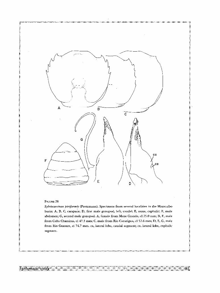
ce
ca
c
....~
BA
F
FIGURE 28
Sylviocarcinus piriformis (Pretzmann). Specimens from several localities in the Maracaibo
basin: A, B, C, carapace; D, first male gonopod, left, caudal; E, same, cephalic; F, male
abdomen; G, second male gonopod. A, female from Mene Grande, cl 25.0 mm; B, F, male
from Cafio Chamiras, cl 47.3 mm; C, male from Rio Curarigua, cl 52.6 mm; D, E, G, male
from Rio Guasare, cl 74.7 mm. ea, lateral lobe, caudal segment; ce, lateral lobe, cephalic
segment.
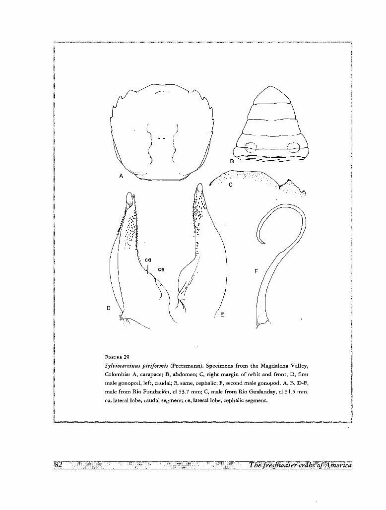
/ .
.... ; ..... :.;,- .; .:.,' ..
c
./ E
/
\j
"
)\.
co
' ..... ~..
A
FIGURE 29
Sylviocarcinus piriformis (Pretzmann). Specimens from the Magdalena Valley,
Colombia: A, carapace; B, abdomen; C, right margin of orbit and front; D, first
male gonopod, left, caudal; E, same, cephalic; F, second male gonopod. A, B, D-F,
male from Rio Fundaci6n, cl 53.7 mm; C, male from Rio Gualanday, cl 51.5 mm.
ca, lateral lobe, caudal segment; ce, lateral lobe, cephalic segment.
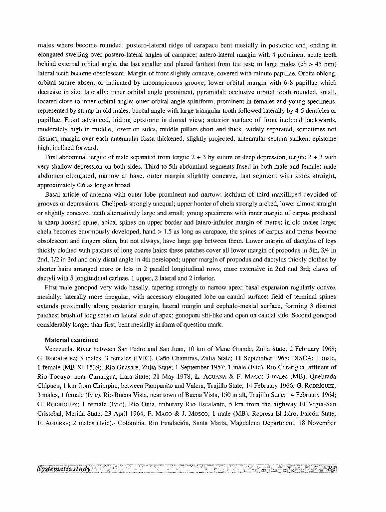
males where become rounded; postero-lateral ridge of carapace bent mesially in posterior end, ending in
elongated swelling over postero-lateral angles of carapace; antero-lateral margin with 4 prominent acute teeth
behind external orbital angle, the last smaller and placed farthest from the rest; in large males (cb> 45 mm)
lateral teeth become obsolescent. Margin of front slightly concave, covered with minute papillae. Orbits oblong,
orbital suture absent or indicated by inconspicuous groove; lower orbital margin with 6-8 papillae which
decrease in size laterally; inner orbital angle prominent, pyramidal; occlusive orbital tooth rounded, small,
located close to inner orbital angle; outer orbital angle spiniform, prominent in females and young specimens,
represented by stump in old males; buccal angle with large triangular tooth followed laterally by 4-5 denticles or
papillae. Front advanced, hiding epistome in dorsal view; anterior surface of front inclined backwards,
moderately high in middle, lower on sides, middle pillars short and thick, widely separated, sometimes not
distinct, margin over each antennular fossa thickened, slightly projected, antennular septum sunken; epistome
high, inclined forward.
First abdominal tergite of male separated from tergite 2 + 3 by suture or deep depression, tergite 2 + 3 with
very shallow depression on both sides. Third to 5th abdomina1 segments fused in both male and fema1e; male
abdomen elongated, narrow at base, outer margin slightly concave, last segment with sides straight,
approximately 0.6 as long as broad.
Basal article of antenna with outer lobe prominent and narrow; ischium of third maxilliped devoided of
grooves or depressions. Chelipeds strongly unequal; upper border of chela strongly arched, lower almost straight
or slightly concave; teeth alternatively large and small; young specimens with inner margin of carpus produced
in sharp hooked spine; apical spines on upper border and latero-inferior margin of merus; in old males larger
chela becomes enormously developed, hand> 1.5 as long as carapace, the spines of carpus and merus become
obsolescent and fingers often, but not always, have large gap between them. Lower margin of dactylus of legs
thickly clothed with patches of long coarse hairs; these patches cover all lower margin of propodus in 5th, 3/4 in
2nd, 1/2 in 3rd and only distal angle in 4th pereiopod; upper margin of propodus and dactylus thickly clothed by
shorter hairs arranged more or less in 2 parallel longitudinal rows, more extensive in 2nd and 3rd; claws of
dactyli with 5 longitudinal carinae, 1 upper, 2 lateral and 2 inferior.
First ma1e gonopod very wide basally, tapering strongly to narrow apex; basal expansion regularly convex
mesially; laterally more irregular, with accessory elongated lobe on caudal surface; field of terminal spines
extends proximally along posterior margin, lateral margin and cephalo-mesial surface, forming 3 distinct
patches; brush of long setae on lateral side of apex; gonopore slit-like and open on caudal side. Second gonopod
considerably longer than first, bent mesially in form of question mark.
Material examinedVenezuela. River between San Pedro and San Juan, 10 km of Mene Grande, Zulia State; 2 February 1968;
G. RODRIGUEZ; 3 males, 3 females (IVIC). Cafio Chamiras, Zulia State; 11 September 1968; DISCA; 1 ma1e,
1 fema1e (ME XI 1539). Rio Guasare, Zulia State; 1 September 1957; 1 male (Ivic). Rio Curarigua, affluent of
Rio Tocuyo, near Curarigua, Lara State; 21 May 1978; L. AGUANA & F. MAGO; 3 males (MB). Quebrada
Chipuen, 1 km from Chimpire, between Pampanito and Valera, Trujillo State; 14 February 1966; G. RODRIGUEZ;
3 males, 1 female (Ivic). Rio Buena Vista, near town of Buena Vista, 150 malt, Trujillo State; 14 February 1964;
G. RODRIGUEZ; 1 female (Ivic). Rio Onia, tributary Rio Escalante, 5 km from the highway El Vigia-San
Cristobal, Merida State; 23 April 1964; F. MAGO & J. Mosco; 1 male (MB). Represa El Isiro, Fa1c6n State;
F. AGUIRRE; 2 males (Ivic).- Colombia. Rio Fundaci6n, Santa Marta, Magdalena Department; 18 November
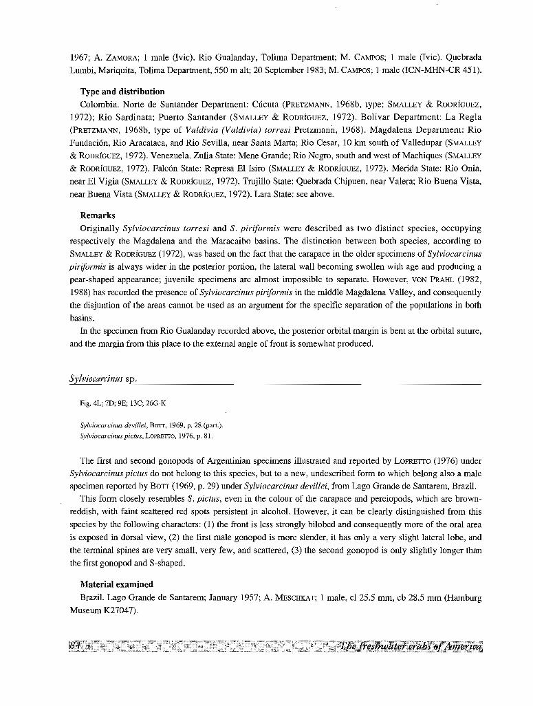
1967; A. ZAMORA; 1 male (Ivic). Rio Gualanday, Tolima Department; M. CAMPOS; 1 male (Ivic). Quebrada
Lumbi, Mariquita, Tolima Department, 550 malt; 20 September 1983; M. CAMPOS; 1 male (ICN-MHN-CR 451).
Type and distributionColombia. Norte de Santander Department: Ciicuta (PRETZMANN, 1968b, type; SMALLEY & RODRlouEz,
1972); Rio Sardinata; Puerto Santander (SMALLEY & RODRlouEz, 1972). Bolivar Department: La Regla
(PRETZMANN, 1968b, type of Valdivia (Valdivia) torresi Pretzmann, 1968). Magdalena Department: Rio
Fundacion. Rio Aracataca, and Rio Sevilla, near Santa Marta; Rio Cesar, 10 km south of Valledupar (SMALLEY
& RODRlOUEZ, 1972). Venezuela. Zulia State: Mene Grande; Rio Negro, south and west of Machiques (SMALLEY
& RODRlouEz, 1972). Falcon State: Represa El Isiro (SMALLEY & RODRlOUEZ, 1972). Merida State: Rio Onia,
near El Vigia (SMALLEY & RODRloUEZ, 1972). Trujillo State: Quebrada Chipuen, near Valera; Rio Buena Vista,
near Buena Vista (SMALLEY & RODRloUEz, 1972). Lara State: see above.
Remarks
Originally Sylviocarcinus torresi and S. piriformis were described as two distinct species, occupying
respectively the Magdalena and the Maracaibo basins. The distinction between both species, according to
SMALLEY & RODRloUEz (1972), was based on the fact that the carapace in the older specimens of Sylviocarcinus
piriformis is always wider in the posterior portion, the lateral wall becoming swollen with age and producing a
pear-shaped appearance; juvenile specimens are almost impossible to separate. However, VON PRAHL (1982,
1988) has recorded the presence of Sylviocarcinus piriformis in the middle Magdalena Valley, and consequently
the disjuntion of the areas cannot be used as an argument for the specific separation of the populations in both
basins.
In the specimen from Rio Gualanday recorded above, the posterior orbital margin is bent at the orbital suture,
and the margin from this place to the external angle of front is somewhat produced.
Sylviocarcinus sp.
Fig. 4L; 7D; 9E; l3C; 260-K
Sylviocarcinus devillei, BaIT, 1969, p. 28 (part.).
Sylviocarcinus pictus, LOPREITO, 1976, p. 81.
The first and second gonopods of Argentinian specimens illustrated and reported by LOPRETTO (1976) under
Sylviocarcinus pictus do not belong to this species, but to a new, undescribed form to which belong also a male
specimen reported by BOTT (1969, p. 29) under Sylviocarcinus devillei, from Lago Grande de Santarem, Brazil.
This form closely resembles S. pictus, even in the colour of the carapace and pereiopods, which are brown
reddish, with faint scattered red spots persistent in alcohol. However, it can be clearly distinguished from this
species by the following characters: (1) the front is less strongly bilobed and consequently more of the oral area
is exposed in dorsal view, (2) the first male gonopod is more slender, it has only a very slight lateral lobe, and
the terminal spines are very small, very few, and scattered, (3) the second gonopod is only slightly longer than
the first gonopod and Svshaped.
Material examined
Brazil. Lago Grande de Santarem; January 1957; A. MESCHKAT; I male, cl 25.5 mm, cb 28.5 mm (Hamburg
Museum K27047).
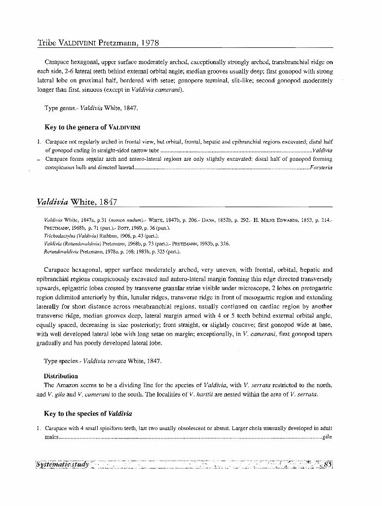
Tribe VALDIVIINI Pretzmann, 1978
Carapace hexagonal, upper surface moderately arched, exceptionally strongly arched, transbranchial ridge on
each side, 2-6 lateral teeth behind external orbital angle; median grooves usually deep; first gonopod with strong
lateral lobe on proximal half, bordered with setae; gonopore terminal, slit-like; second gonopod moderately
longer than first, sinuous (except in Valdivia camerani).
Type genus.- Valdivia White, 1847.
Key to the genera of VALDIVIINI
1. Carapace not regularly arched in frontal view, but orbital, frontal, hepatic and epibranchial regions excavated; distal half
of gonopod ending in straight-sided narrow tube Valdivia
- Carapace forms regular arch and antero-lateral regions are only slightly excavated; distal half of gonopod forming
conspicuous bulb and directed laterad .Forsteria
Valdivia White, 1847
Valdivia White, 1847a, p.31 (nomen nudum).- WHITE, 1847b, p. 206.- DANA, 1852b, p. 292.- H. M1LNE EDWARDS, 1853, p. 214.
PRETlMANN, 1968b, p. 71 (part.).- BOTT, 1969, p. 36 (part.).
Trichodactylus (Valdivia) Rathbun, 1906, p. 43 (part.).
Valdivia (Rotundovaldivia) Pretzmann, 1968b, p. 73 (part.).- PRETlMANN, 1983b, p. 326.
Rotundovaldivia Pretzmann, 1978a, p. 168; 1983b, p. 325 (part.).
Carapace hexagonal, upper surface moderately arched, very uneven, with frontal, orbital, hepatic and
epibranchial regions conspicuously excavated and antero-lateral margin forming thin edge directed transversely
upwards, epigastric lobes crossed by transverse granular striae visible under microscope, 2 lobes on protogastric
region delimited anteriorly by thin, lunular ridges, transverse ridge in front of mesogastric region and extending
laterally for short distance across metabranchial regions, usually continued on cardiac region by another
transverse ridge, median grooves deep, lateral margin armed with 4 or 5 teeth behind external orbital angle,
equally spaced, decreasing in size posteriorly; front straight, or slightly concave; first gonopod wide at base,
with well developed lateral lobe with long setae on margin; exceptionally, in V. camerani, first gonopod tapers
gradually and has poorly developed lateral lobe.
Type species.- Valdivia serrata White, 1847.
DistributionThe Amazon seems to be a dividing line for the species of Valdivia, with V. serrata restricted to the north,
and V. gila and V. camerani to the south. The localities of V. harttii are nested within the area of V. serrata.
Key to the species of Valdivia
1. Carapace with 4 small spiniform teeth, last two usually obsolescent or absent. Larger chela unusually developed in adult
males gila
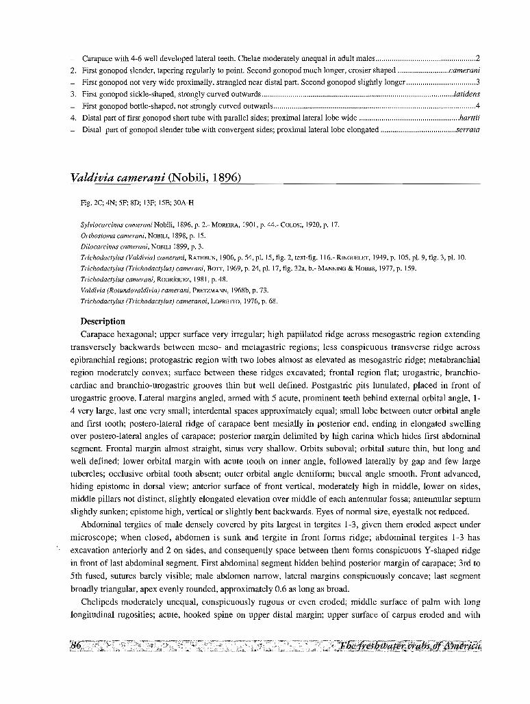
- Carapace with 4-6 well developed lateral teeth. Chelae moderately unequal in adult males 2
2. First gonopod slender, tapering regularly to point. Second gonopod much longer, crosier shaped camerani
- First gonopod not very wide proximally, strangled near distal part. Second gonopod slightly longer 3
3. First gonopod sickle-shaped, stron~ly curved outwards latidens
- First gonopod bottle-shaped, not strongly curved outwards ..4
4. Distal part of first gonopod short tube with parallel sides; proximal lateral lobe wide harttii
- Distal part of gonopod slender tube with convergent sides; proximal lateral lobe elongated serrata
Valdivia camerani (Nobili, 1896)
Fig. 2C; 4N; SF; 8D; 13F; 15E; 30A-H
Sylviocarcinus cameraniNobili, 1896, p. 2.- MORElRA, 1901, p. 44.- COLDSI, 1920, p. 17.
Orthostoma camerani, NOBILI, 1898. p. 15.
Dilocarcinus camerani, NOBILI 1899, p. 3.
Trichodactylus (Valdivia) camerani, RATHBUN, 1906, p. 54, pl, IS, fig. 2, text-fig. 116.- RINGUELET, 1949, p. 105, pI. 9, fig. 3, pI. 10.
Trichodactylus (Trichodactylus) camerani, BOTT, 1969, p. 24, pl. 17, fig. 32a, b.- MANNING & HOBBS, 1977, p. 159.
Trichodactylus camerani, RODRfGUEZ, 1981, p. 48.
Valdivia (Rotundovaldivia) camerani, PRETZMANN, 1968b, p. 73.
Trichodactylus (Trichodactylus) cameranoi, LOPRETTO, 1976, p. 68.
Description
Carapace hexagonal; upper surface very irregular; high papillated ridge across mesogastric region extending
transversely backwards between meso- and metagastric regions; less conspicuous transverse ridge across
epibranchial regions; protogastric region with two lobes almost as elevated as mesogastric ridge; metabranchial
region moderately convex; surface between these ridges excavated; frontal region flat; urogastric, branchio
cardiac and branchio-urogastric grooves thin but well defined. Postgastric pits lunulated, placed in front of
urogastric groove. Lateral margins angled, armed with 5 acute, prominent teeth behind external orbital angle, 1
4 very large, last one very small; interdental spaces approximately equal; small lobe between outer orbital angle
and first tooth; postero-lateral ridge of carapace bent mesially in posterior end, ending in elongated swelling
over postero-lateral angles of carapace; posterior margin delimited by high carina which hides first abdominal
segment. Frontal margin almost straight, sinus very shallow. Orbits suboval; orbital suture thin, but long and
well defined; lower orbital margin with acute tooth on inner angle, followed laterally by gap and few large
tubercles; occlusive orbital tooth absent; outer orbital angle dentiform; buccal angle smooth. Front advanced,
hiding epistome in dorsal view; anterior surface of front vertical, moderately high in middle, lower on sides,
middle pillars not distinct, slightly elongated elevation over middle of each antennular fossa; antennular septum
slightly sunken; epistome high, vertical or slightly bent backwards. Eyes of normal size, eyestalk not reduced.
Abdominal tergites of male densely covered by pits largest in tergites 1-3, given them eroded aspect under
microscope; when closed, abdomen is sunk and tergite in front forms ridge; abdominal tergites 1-3 has
excavation anteriorly and 2 on sides, and consequently space between them forms conspicuous Y-shaped ridge
in front of last abdominal segment. First abdominal segment hidden behind posterior margin of carapace; 3rd to
5th fused, sutures barely visible; male abdomen narrow, lateral margins conspicuously concave; last segment
broadly triangular, apex evenly rounded, approximately 0.6 as long as broad.
Chelipeds moderately unequal, conspicuously rugous or even eroded; middle surface of palm with long
longitudinal rugosities; acute, hooked spine on upper distal margin; upper surface of carpus eroded and with
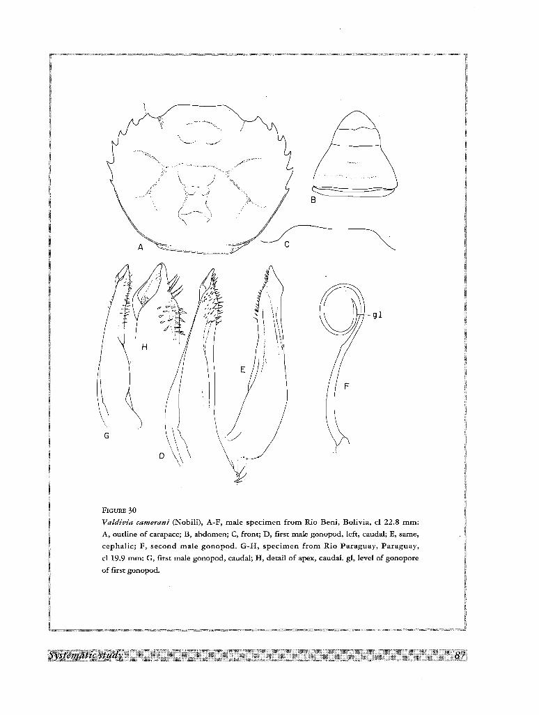
B
0 9 1
,;
'\E
\ ~i
G \,J
0\ (
\"~
FIGURE 30
Valdivia camerani (Nobili), A-F, male specimen from Rio Beni, Bolivia, cl 22.8 mm:
A, outline of carapace; B, abdomen; C, front; D, first male gonopod, left, caudal; E, same,
cephalic; F, second male gonopod. G-H, specimen from Rio Paraguay, Paraguay,
cl 19.9 mm: G, first male gonopod, caudal; H, detail of apex, caudal. gl, level of gonopore
of first gonopod.
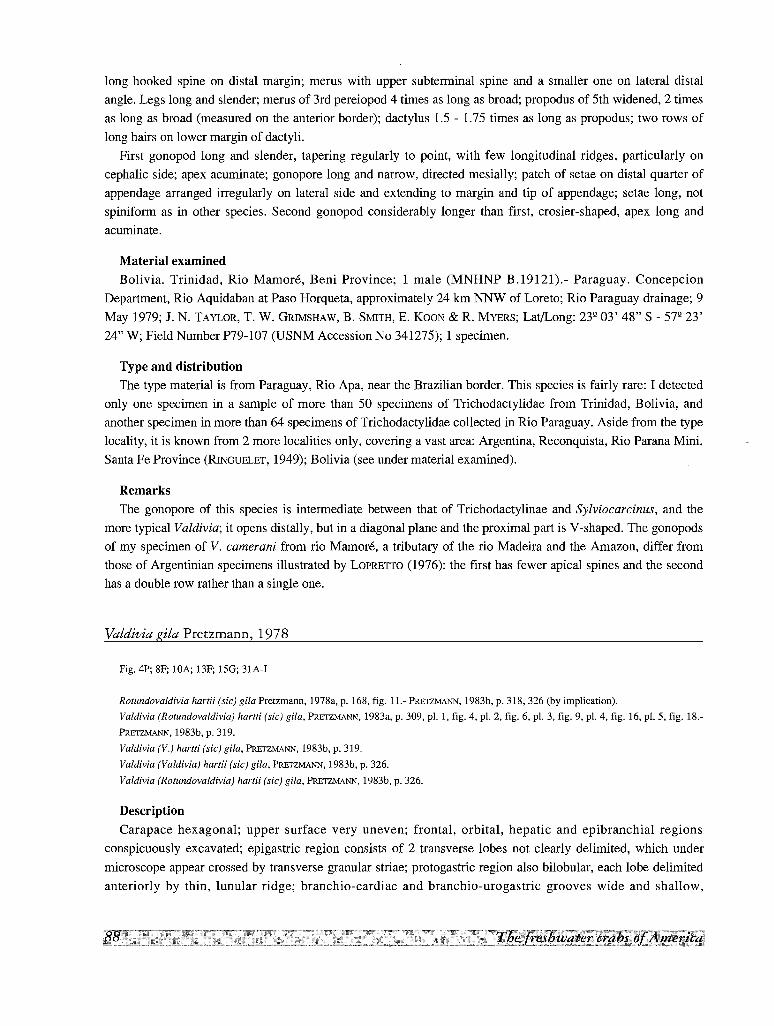
long hooked spine on distal margin; merus with upper subterminal spine and a smaller one on lateral distal
angle. Legs long and slender; merus of 3rd pereiopod 4 times as long as broad; propodus of 5th widened, 2 times
as long as broad (measured on the anterior border); dactylus 1.5 - 1.75 times as long as propodus; two rows of
long hairs on lower margin of dactyli.
First gonopod long and slender, tapering regularly to point, with few longitudinal ridges, particularly on
cephalic side; apex acuminate; gonopore long and narrow, directed mesially; patch of setae on distal quarter of
appendage arranged irregularly on lateral side and extending to margin and tip of appendage; setae long, not
spiniform as in other species. Second gonopod considerably longer than first, crosier-shaped, apex long and
acuminate.
Material examined
Bolivia. Trinidad, Rio Mamore, Beni Province; 1 male (MNHNP B.19121).- Paraguay. Concepcion
Department, Rio Aquidaban at Paso Horqueta, approximately 24 km NNW of Loreto; Rio Paraguay drainage; 9
May 1979; J. N. TAYLOR, T. W. GRIMSHAW, B. SMITH, E. KOON & R. MYERS; Lat/Long: 23Q 03' 48" S - 57Q 23'
24" W; Field Number P79-107 (USNM Accession No 341275); 1 specimen.
Type and distribution
The type material is from Paraguay, Rio Apa, near the Brazilian border. This species is fairly rare: I detected
only one specimen in a sample of more than 50 specimens of Trichodactylidae from Trinidad, Bolivia, and
another specimen in more than 64 specimens of Trichodactylidae collected in Rio Paraguay. Aside from the type
locality, it is known from 2 more localities only, covering a vast area: Argentina, Reconquista, Rio Parana Mini,
Santa Fe Province (RINGUELET, 1949); Bolivia (see under material examined).
Remarks
The gonopore of this species is intermediate between that of Trichodactylinae and Sylviocarcinus, and the
more typical Valdivia; it opens distally, but in a diagonal plane and the proximal part is V-shaped. The gonopods
of my specimen of V. camerani from rio Mamore, a tributary of the rio Madeira and the Amazon, differ from
those of Argentinian specimens illustrated by LOPRETTO (1976): the first has fewer apical spines and the second
has a double row rather than a single one.
Valdivia gila Pretzmann, 1978
Fig. 4P; 8F; lOA; 13F; 15G; 31A-I
Rotundovaldivia hartii (sic) gila Pretzmann, 1978a, p. 168, fig. 11.- PRETZMANN, 1983b, p. 318, 326 (by implication).
Valdivia (Rotundovaldivia) hartti (sic) gila, PRETZMANN, 1983a, p. 309, pI. 1, fig. 4, pI. 2, fig. 6, pI. 3, fig. 9, pI. 4, fig. 16, pI. 5, fig. 18.
PRETZMANN, 1983b, p. 319.
Valdivia (V.) hartti (sic) gila, PRETZMANN, 1983b, p. 319.
Valdivia (Valdivia) hartii (sic) gila, PRETZMANN, 1983b, p. 326.
Valdivia (Rotundovaldivia) hartii (sic) gila, PRETZMANN, 1983b, p. 326.
Description
Carapace hexagonal; upper surface very uneven; frontal, orbital, hepatic and epibranchial regions
conspicuously excavated; epigastric region consists of 2 transverse lobes not clearly delimited, which under
microscope appear crossed by transverse granular striae; protogastric region also bilobular, each lobe delimited
anteriorly by thin, lunular ridge; branchio-cardiac and branchio-urogastric grooves wide and shallow,
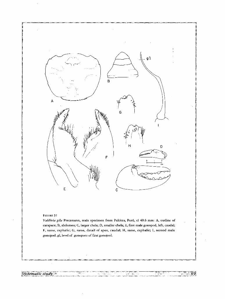
c
B
F
E
\l\~,y~ r~.:,w~ ~i ~ \;~ ..~~ l- \ ~,
J~ . I J41j~. ,\~/~r~ ~ /"/i
1\J /1
/ -::
V //
-;
A
FIGURE 31
Valdivia gila Pretzmann, male specimen from Pakitza, Peru, cl 40.6 mm: A, outline of
carapace; B, abdomen; C, larger chela; D, smaller chela; E, first male gonopod, left, caudal;
F, same, cephalic; G, same, detail of apex, caudal; H, same, cephalic; I, second male
gonopod. gl, level of gonopore of first gonopod.
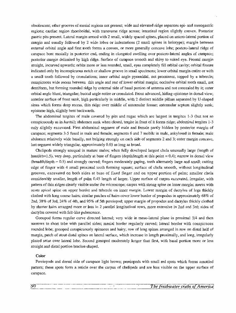
obsolescent; other grooves of mesial regions not present; wide and elevated ridge separates epi- and mesogastric
regions; cardiac region rhomboidal, with transverse ridge across; intestinal region slightly convex. Posterior
gastric pits present. Lateral margin armed with 2 small, widely spaced spines, placed on antero-lateral portion of
margin and usually followed by 2 wide lobes or indentations (2 small spines in holotype); margin between
external orbital angle and first tooth forms a convex, or more generally concave lobe; postero-lateral ridge of
carapace bent mesially in posterior end, ending in elongated swelling over postero-lateral angles of carapace;
posterior margin delimited by high ridge. Surface of carapace smooth and shiny to naked eye. Frontal margin
straight, incurved upwards; orbits more or less rounded, small, eyes completely fill orbital cavity; orbital fissure
indicated only by inconspicuous notch or shallow groove in small specimens; lower orbital margin entire or with
a small tooth followed by crenulations; inner orbital angle pyramidal, not prominent, topped by a tubercle;
conspicuous wide recess between this angle and rest of lower orbital margin; occlusive orbital tooth small, not
dentiform, but forming rounded ridge by external side of basal portion of antenna and not concealed by it; outer
orbital angle blunt, triangular; buccal angle entire or crenulated. Front advanced, hiding epistome in dorsal view;
anterior surface of front sunk, high particularly in middle, with 2 distinct middle pillars separated by U'-shaped
sinus which forms deep recess, thin ridge over middle of antennular fossae; antennular septum slightly sunk;
epistome high, slightly bent backwards.
The abdominal tergites of male covered by pits and rugae which are largest in tergites 1-3 (but not so
conspicuously as in harttii); abdomen sunk when closed, tergite in front of it forms ridge; abdominal tergites 1-3
only slightly excavated. First abdominal segment of male and female partly hidden by posterior margin of
carapace; segments 3-5 fused in male and female; segments 6 and 7 mobile in male, ankylosed in female; male
abdomen relatively wide basally, not bulging strongly on each side of segments 2 and 3; outer margin concave,
last segment widely triangular, approximately 0.65 as long as broad.
Chelipeds strongly unequal in mature males; when fully developed largest chela unusually large (length of
hand/cb=1.5), very deep, particularly at base of fingers (depth/length at this point = 0.4); narrow in dorsal view
(breadth/depth = 0.5) and strongly curved; fingers moderately gaping, teeth alternately large and small; cutting
edge of finger with 4 small proximal teeth forming square; surface of chela smooth, without longitudinal
grooves, excavated on both sides at base of fixed finger and on upper portion of palm; smaller chela
considerably smaller, length of palm 0.45 length of larger. Upper surface of carpus excavated, irregular, with
pattern of thin ridges clearly visible under the microscope; carpus with strong spine on inner margin; merus withacute apical spine on upper border and tubercle on inner margin. Lower margin of dactylus of legs thickly
clothed with long coarse hairs; similar patches of hairs cover lower border of propodus in approximately 68% of
2nd, 38% of 3rd, 24% of 4th, and 95% of 5th pereiopod; upper margin of propodus and dactylus thickly clothed
by shorter hairs arranged more or less in 2 parallel longitudinal rows, more extensive in 2nd and 3rd; sides of
dactylus covered with felt-like pubescence.
Gonopod forms regular curve directed laterad; very wide in meso-lateral plane in proximal 3/4 and then
narrows to short tube with parallel sides; mesial border regularly curved; lateral border with conspicuous
rounded lobe; gonopod conspicuously spinuous and hairy; row of long spines arranged in row on distal half of
margin, patch of stout distal spines on lateral surface, which increase in length proximally, and long, irregularly
placed setae over lateral lobe. Second gonopod moderately longer than first, with basal portion more or less
straight and distal portion bracket-shaped.
Color
Pereiopods and dorsal side of carapace light brown; pereiopods with small red spots which forms amottled
pattern; these spots form a reticle over the carpus of chelipeds and are less visible on the upper surface of
carapace.
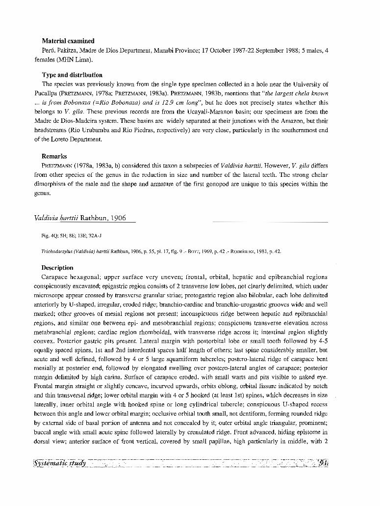
Material examined
Peru. Pakitza, Madre de Dios Department, Manabi Province; 17 October 1987-22 September 1988; 5 males, 4
females (MHN Lima).
Type and distribution
The species was previously known from the single type specimen collected in a hole near the University of
Pucallpa (PRETZMANN, 1978a; PRETZMANN, 1983a). PRETZMANN, 1983b, mentions that "the largest chela known
... is from Bobonaza (=Rio Bobanaza) and is 12.9 cm long", but he does not precisely states whether this
belongs to V. gila. These previous records are from the Ucayali-Maranon basin; our specimens are from the
Madre de Dios-Madeira system. These basins are widely separated at their junctions with the Amazon, but their
headstreams (Rio Urubamba and Rio Piedras, respectively) are very close, particularly in the southernmost end
of the Loreto Department.
Remarks
PRETZMANN (l978a, 1983a, b) considered this taxon a subspecies of Valdivia harttii. However, V. gila differs
from other species of the genus in the reduction in size and number of the lateral teeth. The strong chelar
dimorphism of the male and the shape and armature of the first gonopod are unique to this species within the
genus.
Valdivia harttii Rathbun, 1906
Fig. 4Q; 5H; 8E; BE; 32A-J
Trichodactylus (Va/divia) harttii Rathbun, 1906, p. 55, pI. 17, fig. 9.- BaIT, 1969, p. 42.- RODlUGUEZ, 1981, p. 42.
Description
Carapace hexagonal; upper surface very uneven; frontal, orbital, hepatic and epibranchial regions
conspicuously excavated; epigastric region consists of 2 transverse low lobes, not clearly delimited, which under
microscope appear crossed by transverse granular striae; protogastric region also bilobular, each lobe delimited
anteriorly by U-shaped, irregular, eroded ridge; branchio-cardiac and branchio-urcgastric grooves wide and well
marked; other grooves of mesial regions not present; inconspicuous ridge between hepatic and epibranchial
regions, and similar one between epi- and mesobranchial regions; conspicuous transverse elevation across
metabranchial regions; cardiac region rhomboidal, with transverse ridge across it; intestinal region slightlyconvex. Posterior gastric pits present. Lateral margin with postorbital lobe or small tooth followed by 4-5
equally spaced spines, 1st and 2nd interdental spaces half length of others; last spine considerably smaller, but
acute and well defined, followed by 4 or 5 large squamiform tubercles; postero-lateral ridge of carapace bent
mesially at posterior end, followed by elongated swelling over postero-lateral angles of carapace; posterior
margin delimited by high carina. Surface of carapace eroded, with small warts and pits visible to naked eye.
Frontal margin straight or slightly concave, incurved upwards, orbits oblong, orbital fissure indicated by notch
and thin transversal ridge; lower orbital margin with 4 or 5 hooked (at least 1st) spines, which decreases in size
laterally, inner orbital angle with hooked spine or long cylindrical tubercle; conspicuous U-shaped recess
between this angle and lower orbital margin; occlusive orbital tooth small, not dentiform, forming rounded ridge
by external side of basal portion of antenna and not concealed by it; outer orbital angle triangular, prominent;
buccal angle with small acute spine followed laterally by crenulated ridge. Front advanced, hiding epistome in
dorsal view; anterior surface of front vertical, covered by small papillae, high particularly in middle, with 2

A ..~. c
"'--,D ........ ~
. o: ..
; !
, '.I
E
FIGURE 32
Valdivia harttii Rathbun, male from San Carlos de Rio Negro, Venezuela, cl 39.8 mm:
A, outline of carapace; B, abdomen; C, outline of front; D, first male gonopod, left, caudal;
E, same, cephalic; G, same, detail of apex, caudal; H, same, cephalic; I, same,
F, second male gonopod. gl, level of gonopore of first gonopod.
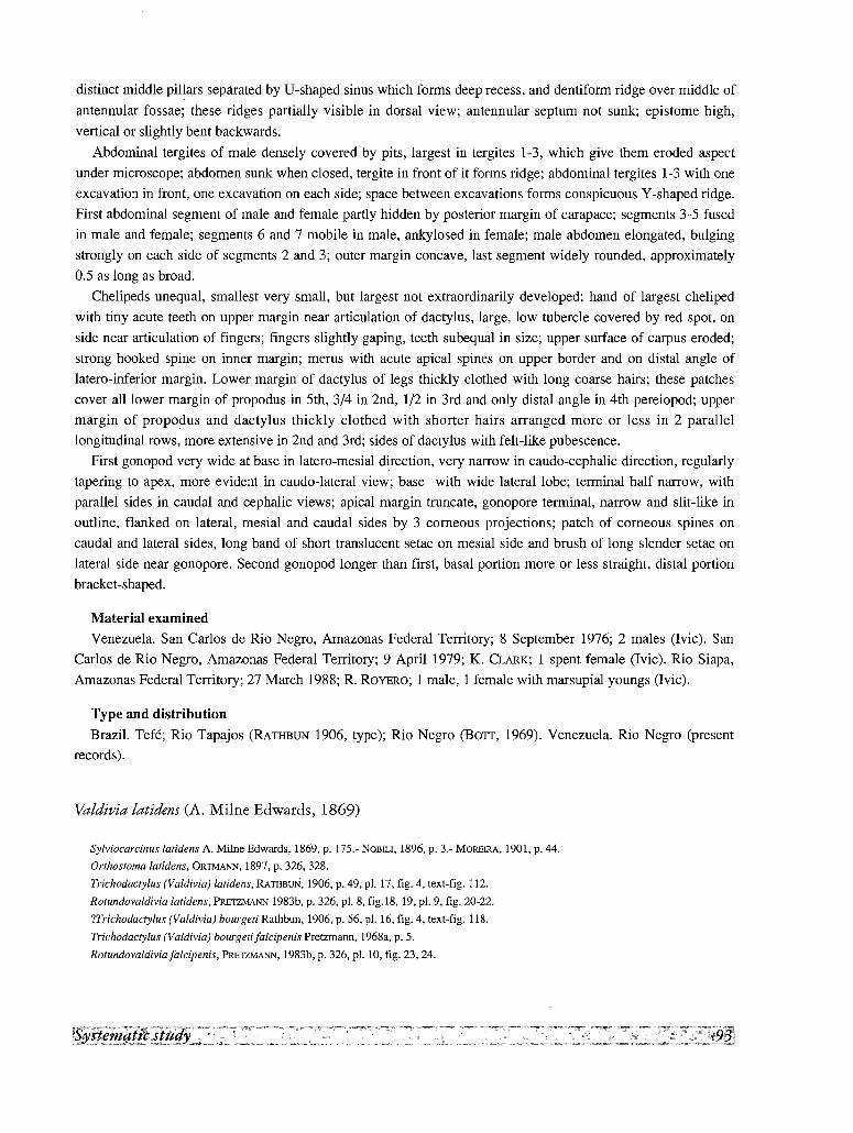
distinct middle pil~ars separated by If-shaped sinus which forms deep recess, and dentiform ridge over middle of
antennular fossae; these ridges partially visible in dorsal view; antennular septum not sunk; epistome high,
vertical or slightly bent backwards.
Abdominal tergites of male densely covered by pits, largest in tergites 1-3, which give them eroded aspect
under microscope; abdomen sunk when closed, tergite in front of it forms ridge; abdominal tergites 1-3 with one
excavation in front, one excavation on each side; space between excavations forms conspicuous Y -shaped ridge.
First abdominal segment of male and female partly hidden by posterior margin of carapace; segments 3-5 fused
in male and female; segments 6 and 7 mobile in male, ankylosed in female; male abdomen elongated, bulging
strongly on each side of segments 2 and 3; outer margin concave, last segment widely rounded, approximately
0.5 as long as broad.
Chelipeds unequal, smallest very small, but largest not extraordinarily developed; hand of largest cheliped
with tiny acute teeth on upper margin near articulation of dactylus, large, low tubercle covered by red spot, on
side near articulation of fingers; fingers slightly gaping, teeth subequal in size; upper surface of carpus eroded;
strong hooked spine on inner margin; merus with acute apical spines on upper border and on distal angle of
latero-inferior margin. Lower margin of dactylus of legs thickly clothed with long coarse hairs; these patches
cover all lower margin of propodus in 5th, 3/4 in 2nd, 1/2 in 3rd and only distal angle in 4th pereiopod; upper
margin of propodus and dactylus thickly clothed with shorter hairs arranged more or less in 2 parallel
longitudinal rows, more extensive in 2nd and 3rd; sides of dactylus with felt-like pubescence.
First gonopod very wide at base in latero-rnesial direction, very narrow in caudo-cephalic direction, regularly
tapering to apex, more evident in caudo-lateral view; base with wide lateral lobe; terminal half narrow, with
parallel sides in caudal and cephalic views; apical margin truncate, gonopore terminal, narrow and slit-like in
outline, flanked on lateral, mesial and caudal sides by 3 corneous projections; patch of corneous spines on
caudal and lateral sides, long band of short translucent setae on mesial side and brush of long slender setae on
lateral side near gonopore. Second gonopod longer than first, basal portion more or less straight, distal portion
bracket-shaped,
Material examined
Venezuela. San Carlos de Rio Negro, Amazonas Federal Territory; 8 September 1976; 2 males (Ivic). San
Carlos de Rio Negro, Amazonas Federal Territory; 9 April 1979; K. QARK; 1 spent female (Ivic). Rio Siapa,
Amazonas Federal Territory; 27 March 1988; R. ROYERO; 1 male, 1 female with marsupial youngs (Ivic).
Type and distribution
Brazil. Tefe; Rio Tapajos (RATHBUN 1906, type); Rio Negro (BOTT, 1969). Venezuela. Rio Negro (present
records).
Valdivia latidens (A. Milne Edwards, 1869)
Sylviocarcinus latidens A. Milne Edwards, 1869, p. 175.- NOBILI, 1896, p. 3.- MOREIRA, 1901, p. 44.
Orthostoma latidens, ORTMANN, 1897, p. 326, 328.
Trichodactylus (Valdivia) latidens, RATHBUN, 1906, p. 49, pI. 17, fig. 4, text-fig. 112.
Rotundovaldivia latidens, PRETZMANN 1983b, p. 326, pI. 8, fig.18, 19, pI. 9, fig. 20-22.
Tl'richodactylus (Valdivia) bourgeti Rathbun, 1906, p. 56, pI. 16, fig. 4, text-fig. 118.
Trichodactylus (Valdivia) bourgeti falcipenis Pretzmann, 1968a, p. 5.
Rotundovaldivia falcipenis, PRETZMANN, 1983b, p. 326, pI. 10, fig. 23, 24.
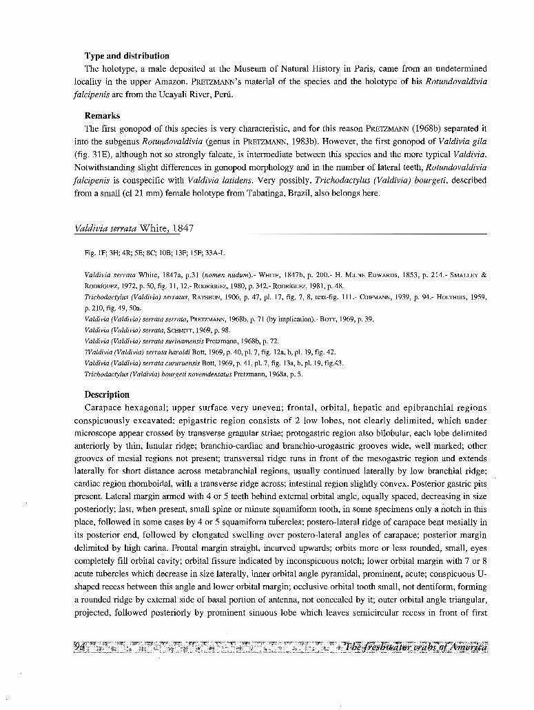
Type and distribution
The holotype, a male deposited at the Museum of Natural History in Paris, came from an undetermined
locality in the upper Amazon. PRETZMANN'S material of the species and the holotype of his Rotundovaldivia
[alcipenis are from the Ucayali River, Peru.
Remarks
The first gonopod of this species is very characteristic, and for this reason PRETZMANN (l968b) separated it
into the subgenus Rotundovaldivia (genus in PRETZMANN, 1983b). However, the first gonopod of Valdivia gila
(fig. 31E), although not so strongly falcate, is intermediate between this species and the more typical Valdivia.
Notwithstanding slight differences in gonopod morphology and in the number of lateral teeth, Rotundovaldivia
[alcipenis is conspecific with Yaldivia latidens. Very possibly, Trichodactylus (Valdivia) bourgeti, described
from a small (cl 21 mm) female holotype from Tabatinga, Brazil, also belongs here.
Valdivia serrata White, 1847
Fig. IF; 3H; 4R; SE; 8C; lOB; 13F; 15F; 33A-L
Yaldivia serrata White, 1847a, p.31 (nomen nudumi.: WHITE, 1847b, p. 200.- H. MILNE EDWARDS, 1853, p. 214.- SMALLEY &
RODRIGUEZ, 1972, p. 50, fig. 11, 12.- RODRIGUEZ, 1980, p. 342.- RODRlGUEZ, 1981, p. 48.
Trichodactylus (Valdivia) serratus, RATHBUN, 1906, p. 47, pI. 17, fig. 7, 8, text-fig. 111.- COIFMANN, 1939, p. 94.- HOLTHUIS, 1959,
p. 210, fig. 49, 50a.
Valdivia (Valdivia) serrata serrata, PRETZMANN, 1968b, p. 71 (by implication).- BOTT, 1969, p. 39.
Yaldivia (Valdivia) serrata, SCHMITT, 1969, p. 98.
Valdivia (Valdivia) serrata surinamensis Pretzmann, 1968b, p. 72.
?Valdivia (Valdivia) serrata haraldi Bott, 1969, p. 40, pI. 7, fig. 12a, b, pI. 19, fig. 42.
Valdivia (Valdivia) serrata cururuensis Bott, 1969, p. 41, pI. 7, fig. Ba, b, pI. 19, fig.43.
Trichodactylus (Valdivia) bourgeti novemdentatus Pretzmann, 1968a, p. 5.
Description
Carapace hexagonal; upper surface very uneven; frontal, orbital, hepatic and epibranchia1 regions
conspicuously excavated; epigastric region consists of 2 low lobes, not clearly delimited, which under
microscope appear crossed by transverse granular striae; protogastric region also bilobular, each lobe delimited
anteriorly by thin, lunular ridge; branchio-cardiac and branchio-urogastric grooves wide, well marked; other
grooves of mesial regions not present; transversal ridge runs in front of the mesogastric region and extends
laterally for short distance across metabranchial regions, usually continued laterally by low branchial ridge;
cardiac region rhomboidal, with a transverse ridge across; intestinal region slightly convex. Posterior gastric pits
present. Lateral margin armed with 4 or 5 teeth behind external orbital angle, equally spaced, decreasing in size
posteriorly; last, when present, small spine or minute squamiform tooth, in some specimens only a notch in this
place, followed in some cases by 4 or 5 squamiform tubercles; postero-lateral ridge of carapace bent mesially in
its posterior end, followed by elongated swelling over postero-lateral angles of carapace; posterior margin
delimited by high carina. Frontal margin straight, incurved upwards; orbits more or less rounded, small, eyes
completely fill orbital cavity; orbital fissure indicated by inconspicuous notch; lower orbital margin with 7 or 8
acute tubercles which decrease in size laterally, inner orbital angle pyramidal, prominent, acute; conspicuous U
shaped recess between this angle and lower orbital margin; occlusive orbital tooth small, not dentiform, forming
a rounded ridge by external side of basal portion of antenna, not concealed by it; outer orbital angle triangular,
projected, followed posteriorly by prominent sinuous lobe which leaves semicircular recess in front of first
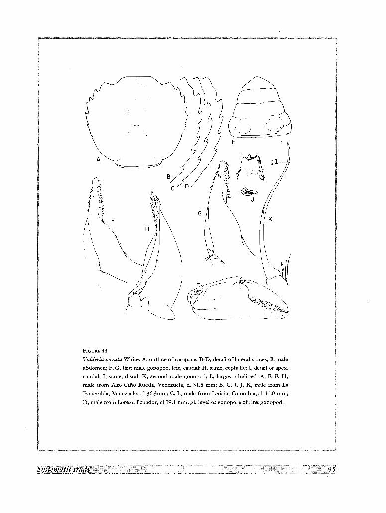
A
c
G
£~
~-_.. ------
FIGURE 33
Valdivia serrata White: A, outline of carapace; B-D, detail of lateral spines; E, male
abdomen; F, G, first male gonopod, left, caudal; H, same, cephalic; I, detail of apex,
caudal; J, same, distal; K, second male gonopod; L, largest cheliped. A, E, F, H,
male from Alto Cafio Rueda, Venezuela, cl 31.8 mm; B, G, I, J, K, male from La
Esrneralda, Venezuela, cl 36.3mm; C, L, male from Leticia, Colombia, cl 41.0 mm;
D, male from Loreto, Ecuador, cl 39.1 mm. gl, level of gonopore of first gonopod.
lateral tooth; buccal angle crenulated or with 4 ill-defined tubercles. Front advanced, hiding epistome in dorsal
view; anterior surface of front vertical, high particularly in middle, with 2 distinct middle pillars separated by U
shaped sinus which forms deep recess, and dentiform ridge over middle of antennular fossae; antennular septum
not sunk; epistome high, vertical or slightly bent backwards.
Abdominal tergites of male densely covered by pits, largest in tergites 1-3, giving them eroded aspect under
microscope; abdomen sunk when closed, tergite in front of it forms ridge; abdominal tergites 1-3 with one
excavation in front, one excavation on each side; space between excavations forms conspicuous Y-shaped ridge.
First abdominal segment of male and female partly hidden by posterior margin of carapace; segments 3-5 fused
in male and female; segments 6 and 7 mobile in male, ankylosed in female; male abdomen elongated, bulging
strongly on each side of segments 2 and 3; outer margin concave, last segment widely rounded, approximately
0.5 as long as broad.
Chelipeds unequal, smallest very small, largest not extraordinarily developed; hand with longitudinal carina
in middle of external surface, surface between this carina and upper margin excavated, particularly in smaller
chela; acute teeth on upper margin near articulation of dactylus (absent or reduced in some large specimens),
and large low tubercle often covered by red spots, on external side near articulation of fingers; fingers slightly
gaping, teeth large, subequal, interspaced with smaller ones; upper surface of carpus eroded, with pattern of thin
ridges clearly visible under microscope; strong hooked spine on inner margin; merus with acute apical spine on
upper border and another on distal angle of latero-inferior margin. Lower margin of dactylus in legs thickly
clothed with long coarse hairs; these patches cover all lower margin of propodus in 5th, 3/4 in 2nd, 1/2 in 3rd
and only the distal angle in 4th pereiopod; upper margin of propodus and dactylus thickly clothed by shorter
hairs arranged more or less in 2 parallel longitudinal rows, more extensive in 2nd and 3rd; sides of dactylus with
felt-like pubescence.
First gonopod very wide at base in latero-mesial direction, very narrow in caudo-cephalic direction, regularly
tapering to apex, more evident in caudo-lateral view; base with elongated lateral lobe; terminal half narrow, with
convergent sides in caudal and cephalic views, bulbiform in latero-cephalic view; apical margin truncate,
gonopore terminal, narrow and slit-like in outline, flanked on lateral, mesial and caudal sides by 3 corneous
projections; patch of corneous spines on caudal and lateral sides, long band of short translucent setae on mesial
side and brush of long slender setae on lateral side near gonopore. Second gonopod longer than first, basal
portion more or less straight, distal portion bracket-shaped.
ColorPereiopods and dorsal side of carapace covered with red spots, which sometimes are elongated and thin,
forming a symmetrical pattern; these spots form a reticle over the carpus of cheliped. The background color
varies from light to dark brown, reddish or even black.
Material examined
Venezuela. Alto Cafio Rueda, Atures, 22 km from Puerto Ayacucho, Amazonas Federal Territory; 30
October 1965; P. ANDUZE; 1 male (Ivic). Carinagua, Puerto Ayacucho, Amazonas Federal Territory; 5
December 1977; 2 juvenile females (Ivic). Id.; 29 November 1978; 3 immature females, 1 juvenile male (Ivic).
La Esmeralda, Cafio Iguapo, Amazonas Federal Territory; 19 February 1966; J. PULIDO & L. DUQuE; 5 males,
2 females (MB XI-0671). Id.; 450 malt; 1 March 1968; J. A. RIVERO; 1 female (Ivic), San Pedro de Cataniapo,
100 malt, Amazonas Federal Territory; 23 August 1981; O. PACHANO; 1 female (LS 1120). Same locality;
25 August 1981; R. FEo; 1 male (LS 1045). Rio Tucuragua, affluent of the Orinoco, between Cuchivero and
Caura rivers, Bolivar State; 7 April 1981; G. RODRIGUEZ; 1 juvenile male (Ivic). Rio Tauca, affluent of the Rio
Caura, Bolivar State; 7 May 1981; G. RODRIGUEZ; 1 juvenile male (Ivic). Cafio Charro de Agua, Las Bateas,
Los Pijiguaos, Bolivar State; 27 April 1988; C. LAsso & G. COLONNELLO; 1 male (LS 1143). Quebrada
Trapichote, road to El Jobal, Los Pijiguaos, Bolivar State; 28 April 1988; C. LAsso & G. COLONNELLO; 2
immature females (LS 1137). La Solanera, Los Pijiguaos, Bolivar State; 26 April 1988; C. LAsso & G.
COLONNELLO; 1 female (LS 1136).- Colombia. Leticia, Amazonas Department; 1 April 1957; H. NICEFORO
MARIA; 1 male (Ivic). Rio Hacha, Florencia, Caqueta Department; 4 January 1954; H. NICEFORO MARIA; 1 male
(Ivic). Quebrada La Yuca, Florencia, Caqueta Department, 10 March 1954; H. NICEFORO MARIA; 1 female
(Ivic). San Juan de Arama, Meta Department, 500 malt; 20 September 1987; M. R. CAMPOS; 1 male (ICN
MHN-CR 808).- Ecuador. Loreto, slopes of Monte Sumaco, 450 malt, Napo Province; 1 June 1968; M.
OLALLA; 2 males (Ivic).
Type and distribution
The type specimen is a female and the type locality is unknown. The species occupies an extensive area
between the Orinoco and the Amazon, but it does not extend north of the first river neither south of the second.
The records in the literature are the following: Brazil. Tabatinga; Tefe (RATHBUN, 1906); Brazilian Guiana,
Serra de Tumucumaque, Rio Pani and Rio Pani Superior; Quatipuru, near Rio Toboa; Rio Pani do Oeste; Rio
Marauia, Cachoeira San Antonio, type of Valdivia (Valdivia) serrata haraldi Bott, 1969, and near Tupuruquara,
at the junction of Rio Marauia and Rio Negro; at the junction of the Rio Kenebiit-Tabiri and the Rio Curuni,
type of Yaldivia (Valdivia) serrata cururuensis Bott, 1969. Venezuela. See records above. Colombia. Caqueta
Department: Rio Orteguaza, near Venecia; Putumayo Department: Puerto Limon, Rio Caqueta (SMALLEY &
RODRIGUEZ, 1972); Puerto Asis (SCHMITT, 1969); Amazonas Department: Rio Arara. See other records above.
Ecuador. Napo Province: Loreto, foothills of Mount Sumaco, 450 m alt (SMALLEY & RODRIGUEZ, 1972).
Suriname. Paramaribo; near Republiek; Zanderdij; Sectie 0, 70 km S of Paramaribo; Litani River, upper
reaches of the Marowijne River basin (HOLTHUIS, 1959); Paramaribo, type of Valdivia (Valdivia) serrata
surinamensis Pretzmann, 1968.- Guyana. Demerara River (COIFMANN, 1939).
RemarksThe species displays considerable variations in the number and development of the lateral teeth of carapace,
as can be observed in specimens from Venezuela, Colombia and Ecuador (fig. 33A, B, C, D). The gonopod also
presents variations in the shape of the lateral lobe, thickness of the distal tube and convexity of the mesial
outline, which can be observed even in specimens from localities close to each other (fig. 33G, H). Most of the
subspecies of Valdivia serrata described by PRETZMANN (1968a, b) and BOTT (1969) are based on these
characters of the carapace and gonopod. Valdivia (Valdivia) serrata surinamensis Pretzmann, 1968, andValdivia (Valdivia) serrata cururuensis Bott, 1969 cannot be distinguished from the more typical Valdivia
serrata. On the other hand the inclusion of Valdivia (Valdivia) serrata haraldi under this species is doubtful. In
this subspecies the lateral teeth of carapace are identical to those of V. serrata, but the first gonopod is more
recurved laterad, resembling more that of Yaldivia gila than the one of V. serrata.
Trichodactylus (Valdivia) bourgeti novemdentatus Pretzmann, 1968, from the Rio Negro, probably should be
grouped under Valdivia serrata and not under V. bourgeti (=V. latidens), since the dentition of the carapace falls
within the variation displayed by the former species.
Valdivia (Valdivia) serrata oronensis Pretzmann, 1968 (see PRETZMANN, 1968b, p. 72), described from male
and female types from Rio Paraguay, R. d. Oro [Rio de Oro, Chaco province, Argentina], according to his
diagnosis (front deeply bilobed, carapace strongly arched, without sculpturing, gonopod without lateral lobe),
does not belong to the genus Valdivia but to the genus Zilchiopsis.
Forsteria Bott, 1969
Yaldivia (Forsteria) Bott, 1969, p. 37.
Carapace hexagonal, upper surface strongly arched, smooth, frontal and orbital regions excavated, but regions
not delimited, antero-lateral margin angled, exceptionally rounded in all males, median grooves not deeply
marked, 3 lateral teeth behind external orbital angle, obsolescent in old males, front straight; first gonopod
widened at base, with long setae on lateral margin, distal half strongly bent laterad, apex bulbous.
Forsteria venezuelensis (Rarhbun, 1906)
Fig. IG; 4S; 5G; 8A; 9H; 13D; 151; 34A-H
Trichodactylus (Valdivia) venezuelensis Rathbun, 1906, p. 47, pI. 17, fig. 10.- COIFMANN, 1939, p. 112.
Valdivia venezuelensis, SMALLEY and RODRIGUEZ, 1972, p. 50, fig. 13,14.- RODRiGUEZ, 1980, p. 343, fig. 99.- RODRIGUEZ, 1981, p. 48.
Valdivia (Forsteria) venezuelensis venezuelensis, BOlT, 1969, p. 37, pI. 5, fig. 9a, b.
Valdivia (Forsteria) venezuelensis edentata Bott, 1969, p. 38, pI. 6, fig. lOa, b, pI. 19, fig. 40.
Holthuisisia venezuelensis, PRETlMANN, 1968b, p. 74.
Trichodactylus (Valdivia) ornatifrons Pretzmann, 1968a, p. 3.
Valdivia (Valdivia) ornatifrons, PRETlMANN, 1968b, p. 71.
Description
Carapace hexagonal; upper surface slightly irregular, in frontal view forms regular arch, only metabranchial
region slightly more prominent than adjacent cardiac and intestinal regions; frontal and orbital regions
excavated, epigastric lobes well delimited; branchio-urogastric groove weakly marked, urogastric groove almost
absent, branchio-cardiac groove indicated by flat depressions. Lateral margins angled, except in largest males
where it becomes rounded; postero-lateral ridge of carapace bent mesially in posterior end, followed by
elongated swelling over postero-lateral angles of carapace; antero-lateral margin with 3 teeth behind external
orbital angle which decrease in size posteriorly; 2nd and 3rd teeth nearer than 1st and 2nd; in the largest males
(cb=>40 mm) lateral teeth become obsolete, or only first remains, represented by small tubercle. Margin of frontstraight, covered with small tubercles. Orbits suborbicular; orbital suture absent, although position sometimes
indicated by small depression; lower orbital margin with blunt teeth or papillated tubercles; inner orbital angle
prominent, pyramidal; occlusive orbital tooth rounded, small; outer orbital angle forming triangular tooth,
border in contact with orbital margin; buccal angle papillated, not armed. Front advanced, hiding epistome in
dorsal view; anterior surface of front vertical, moderately high in middle, lower on sides, middle pillars not
distinct, margin over each antennular fossae sinuous, slightly projected; antennular septum slightly sunk;
epistome high, vertical or slightly bent backwards.
First abdominal tergite of male separated from tergite 2 + 3 by suture or deep depression; tergites 2 + 3 with
very shallow depression on both sides. Third to 5th abdominal segments fused in both male and female; male
. abdomen elongated, narrow at base, outer margin slightly concave; last segment with sides concave,
approximately 0.5 as long as broad.
Basal article of antenna with outer lobe prominent and narrow; ischium of third maxilliped devoided of
grooves or depressions. Chelipeds strongly unequal in both male and female; upper border of chela strongly
arched and lower almost straight, fingers do not gap; teeth small, regularly placed along cutting edges and
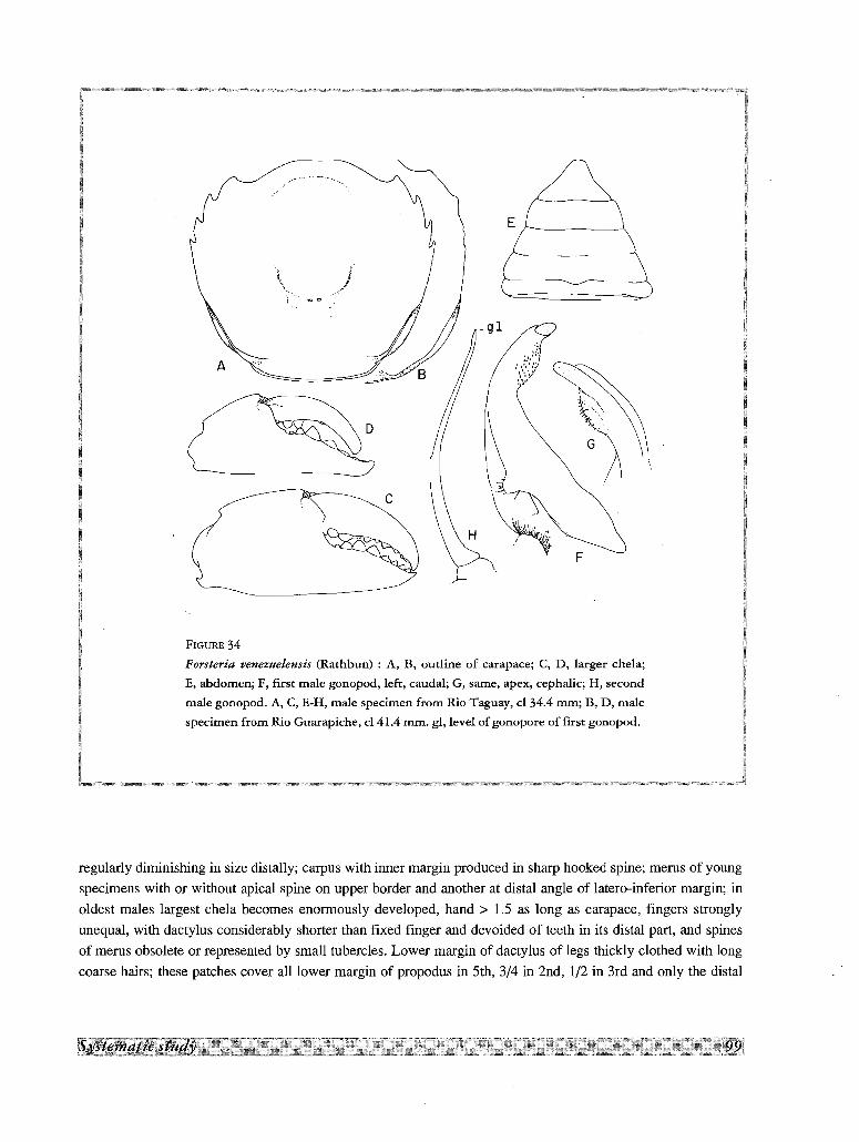
Er- _
-(19....
:'i:'..;)~.',\'1"'..,,
~ f\) ..~ \,',.3.c
I"CiW ..
\~\ ..
FIGURE 34
Forsteria cenezuelensis (Rathbun) : A, B, outline of carapace; C, D, larger chela;
E, abdomen; F, first male gonopod, left, caudal; G, same, apex, cephalic; H, second
male gonopod. A, C, E-H, male specimen from Rio Taguay, cl 34.4 mm; B, D, male
specimen from Rio Guarapiche, cl 41.4 mm. gl, level of gonopore of first gonopod.
regularly diminishing in size distally; carpus with inner margin produced in sharp hooked spine; merus of young
specimens with or without apical spine on upper border and another at distal angle of latero-inferior margin; in
oldest males largest chela becomes enormously developed, hand> 1.5 as long as carapace, fingers strongly
unequal, with dactylus considerably shorter than fixed finger and devoided of teeth in its distal part, and spines
of merus obsolete or represented by small tubercles. Lower margin of dactylus of legs thickly clothed with long
coarse hairs; these patches cover all lower margin of propodus in 5th, 3/4 in 2nd, 1/2 in 3rd and only the distal
angle in 4th pereiopod; upper margin of propodus and dactylus thickly clothed by shorter hairs arranged more or
less in 2 parallel longitudinal rows, more extensive in 2nd and 3rd; claws of dactylus with 5 longitudinal carinae,
1 upper, 2 lateral and 2 inferior.
First gonopod with proximal half conical, strongly widened at base and directed mesially; distal half strongly
bent laterad, lateral globular expansion covered by short thick setae, arranged in several parallel rows; gonopore
overreached by distal corneous expansion. Second gonopod of equal length than first, bracket-shaped, apex flat,
lanceolate.
Material examined
Venezuela. Rio Taguay, Aragua State; 12 December 1967; J. PULIDO; 3 males, 2 females (Ivic). Paraima,
Guarico State; 31 March 1950; 1 female (Ivic). Caicara, Rio Guarapiche, Monagas State; 8 September 1968;
1 male, 1 female (lvic). Rio Chiviripa, between La Urbana and Caicara, Bolivar State; 1 April 1958; G. MEDINA;
1 female (lvic).- Venezuela, without data; 1 female (Ivic).
Type and distribution
Type specimens are 2 females from the Orinoco River (MHNP). The species has been recorded from the
Orinoco and many of the rivers draining to it: Ciudad Bolivar (BOTT, 1969); Rio Orinoco (SMALLEY &
RODRlouEz, 1972); affluent of the Rio Apure (RATHBUN, 1906);,Rio Cura and Rio Taguay, both affluents of Rio
Guarico (SMALLEY & RODRlouEz, 1972); Rio Chiviripa, draining on the right margin of the Orinoco, near
Caicara (SMALLEY & RODRlouEz,1972); Rio Guarapiche, draining to Rio San Juan and the Gulf of Paria
(SMALLEY & RODRIOUEZ, 1972).
Remarks
The species have been described under several synonyms in the literature (SMALLEY & RODRloUEz, 1972).
The characters given for Trichodactylus (Valdivia) meekei Pretzmann, 1968, and moreover the illustrations of
the carapace, chela, pereiopods and first gonopod given in PRETZMANN (l983a), closely correspond with the
characters of Forsteria venezuelensis, but I have not examined the holotype of the first species.
Tribe DILOCARCININI
Carapace suborbicular, wider anteriorly, upper surface strongly arched, smooth, front bilobed, partially or
completely retracted, exposing epistome; first gonopod expanded laterally at base, with long plumose setae on
lateral margin, apex bulbiform; gonopore slit-like, apical; second gonopod moderately longer than first, sinuous,
exceptionally much longer and bent as question mark.
Type genus.- Dilocarcinus H. Milne Edwards, 1853
Key to the genera of Dilocarcinini
1. 3-4 teeth on lateral margin Zilchiopsis
- 6-10 teeth on lateral margin 2
2. Distal part of first gonopod rounded or bulbiform .Dilocarcinus
- Distal part of first gonopod strongly twisted sinistrally, apex with very few small conical spines Fredilocarcinus
Zilchiopsis Bott, 1969
Carapace hexagonal or suborbicular, upper surface strongly arched, median grooves deeply marked, front
bilobed, advanced, exposing epistome in dorsal view, lower orbital margin directed downwards at inner orbital
angle, 3-4 teeth on lateral margin, often minute or obsolescent; abdomen triangular-rounded or trapezoidal, third
maxilliped with merus conspicuously narrow; first gonopod wide basally, strongly reduced in distal half.
Type species.- Zilchiopsis sattleri Bott, 1969.
Distribution
The four species of the genus are distributed in the tributaries of the Amazon, in four successive and non
overlapping areas, through an extensive territory on the west and south sides of South America: (a) Venezuela,
Colombia and Ecuador (Z. emarginatus); (b) Ecuador (Z. chacei); (c) Peru (Z. cryptodus); and (d) Bolivia
(Z. sattleri). An exception to this distribution is the record of Z. sattleri in the Rio Paraguay (BOTT, 1969), which
is not an affluent of the Amazon.
Remarks
The carapace morphology of the species grouped under this genus, although transitional between the
Valdiviini and the Dilocarcinini, displays the same basic characters in all the species. On the other hand, there is
considerable variability in gonopodal morphology. The simple first gonopod of Z. chacei resembles that
appendage in some Sylviocarcinus, particularly S. pictus and S. sp., whereas in Z. emarginatus it is more akin to
Valdivia. The morphology of this appendage in Z. sattleri and Z. cryptodus departs from any of the types found
in other genera. However, to avoid an excessive generic splitting, the four species are kept in one genus inside
the Tribe Dilocarcinini. To this genus probably belongs also Trichodactylus (Valdivia) boliviensis a species
from Misiones Molletones, Bolivia, not examined by me.
Key to the species of Zilchiopsis
1. First gonopod almost straight, regularly tapering to apex chacei
- First gonopod regularly bent laterad, strongly constricted at middle 2
2. A well defined basal lobe on mesial side of first gonopod; apical tube relatively wide emarginatus
- Basal portion of first gonopod widened but not forming defined lateral lobe; apical tube narrow 3
3. Apical tube regularly tapering to terminal gonopore cryptodus
- Distal portion of apical tube reduced to a filament sattleri
Zilchiopsis chacei (Pretzmann, 1968)
Trichodactylus (Trichodactylus) chacei Pretzmann, 1968a, p. 3.
Zilchiopsis chacei, PRETZMANN, 1983b, p. 327, pl. 11, fig. 25, 26, pl, 12, fig. 27, 28.
Zilchiopsis chacei ecuadoroides Pretzmann, 1978b, p. 7.-PRETZMANN, 1983a, p. 310, pl, 6, fig. 21-24.- PRETZMANN 1983b, p. 327, not
pl. 6, fig. 21, 22.
As described and illustrated by PRETZMANN (1968a, 1983b), this species has in common with the other three
members of the genus the strongly convex carapace, with rounded lateral margins provided with small, obsolescent
teeth, and the bilobed front, but it is well differentiated from them by its simple almost smooth gonopod,
moderately widened at base and regularly tapering from the middle, with its lateral side slightly sinuous.
BOTT (1969) synonymized this species with Zilchiopsis cryptodus. The carapace of both are almost identical,
except for the front less deeply bilobed in Z. chacei. However the gonopod of both species are different.
Type and distribution
The holotype of Trichodactylus (Trichodactylus) chacei is a male specimen, cl 30.4 mm, from Chichirota,
Pastaza Province, east Ecuador, and the holotype of Zilchiopsis chacei ecuadoroides a male, cl 30.1 mm, from
Sevilla del Oro, between Mendez and Paute, Ecuador. Thus the two taxa come from different drainage basins:
Bobonaza-Pastaza rivers and Santiago-Marafion rivers; these two localities are approximately 230 km apart
(Appendix 2).
Zilchiopsis cryptodus (Ortmann, 1893)
Dilocarcinus cryptodus Ortmann, 1893, p. 493.
Zilchiopsis cryptodus, BOTT, 1969, p. 35, pl. 25a, b, pI. 22, fig. 75.- RODRfGUEZ, 1981, p. 48.- PRETlMANN, 1983b, p. 327.
Trichodactylus (Dilocarcinus) emarginatus, RATHBUN, 1906, p. 69 (part.).-
This species, as redescribed and illustrated from the holotype specimen in the Strassburg Museum by BOTT
(1969), is well characterized by the suborbicular strongly convex carapace, with the upper surface smooth, the
lateral margin with 2-3 minute, blunt teeth behind external orbital angle, and the triangular gonopod densely
covered by long setae on the lateral margin, with the apex very thin and directed laterad.
Type and distribution
Zilchiopsis cryptodus was described from 1 male specimen, cl 27 mm, from Rio Ucayali, Peru (ORTMANN,
1893; BOTT, 1969).
Zilchiopsis emarginatus (H. Milne Edwards, 1853)
Fig. 3G; 40 T; 5M; 7H; 8B; 91; 10C; 13G, H; 35A-E; 36A-H
'Dilocarcinus emarginatus H. Milne Edwards, 1853, p. 216.- H. MILNEEDWARDS, 1854, p. 181, pl. 14, fig. 4.- LUCAS, 1857, p. 7, pl. 2,
fig. 3.- A. MILNEEDWARDS, 1869, p. 176, 178.- SMITH, 1869, p. 36.- YOUNG, 1900, p. 231, 232.- MORElRA, 1901, p. 44.
Trichodactylus (Dilocarcinus] emarginatus, RATHBUN, 1906, p. 64, pl. 18, fig. 2.
Orthostoma emarginatus, ORTMANN, 1897, p. 326, 328.
Zilchiopsis emarginatus, BOTT, 1969, p. 35, pl. 21, fig. 56.
Valdivia ecuadoriensis Pretzmann, 1968a, p. 3.
Yaldivia ecuadorensis (sic), PRETlMANN, 1968b, p. 71.
Zilchiopsis ecuadoriensis, SMALLEY & RODRiGUEZ, 1972, p. 49, fig. 9, 10.-PRErZMANN, 1983b, p. 328, pI. 13, fig. 29, 30, pl. 14, fig. 31,
32.
Zilchiopsis ecuadorensis (sic), RODRiGUEZ, 1981, p. 48.
Description
Carapace hexagonal at least in larger specimens (cl > 30 mm); upper surface irregular; strongly convex from
front to back, almost flat in frontal view; mesogastric region less prominent than epibranchial; hepatic region
excavated; frontal region flat on middle, convex laterally; triangular prominence on each side, in area of contact
of epi-, mesobranchial and mesogastric regions, with transverse cervical groove in front of each prominence;
metabranchial regions flattened, not more prominent than cardiac and intestinal; branchio-urogastric and
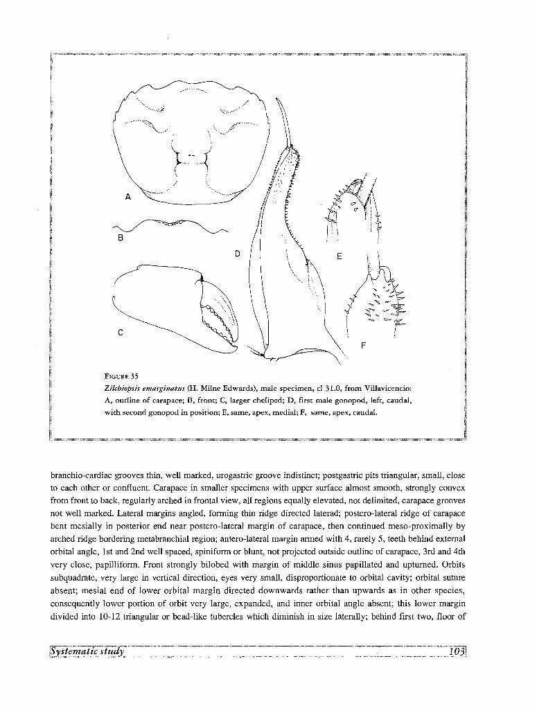
<', \~.
r1"/ '}? ,..
; .'.', j
li f/J
D
..... ,./;r'-· .... ·-~···.~ ... '
...........A
c
B
FIGURE 35
Zi/chiopsis emarginatus (H. Milne Edwards), male specimen, cl 31.0, from Villavicencio:
A, outline of carapace; B, front; C, larger cheliped; D, first male gonopod, left, caudal,
with second gonopod in position; E, same, apex, medial; F, same, apex, caudal.
branchio-cardiac grooves thin, well marked, urogastric groove indistinct; postgastric pits triangular, small, close
to each other or confluent. Carapace in smaller specimens with upper surface almost smooth, strongly convex
from front to back, regularly arched in frontal view, all regions equally elevated, not delimited, carapace grooves
not well marked. Lateral margins angled, forming thin ridge directed laterad; postero-lateral ridge of carapace
bent mesially in posterior end near postero-lateral margin of carapace, then continued meso-proximally by
arched ridge bordering metabranchial region; antero-lateral margin armed with 4, rarely 5, teeth behind external
orbital angle, 1st and 2nd well spaced, spiniform or blunt, not projected outside outline of carapace, 3rd and 4th
very close, papilliform. Front strongly bilobed with margin of middle sinus papillated and upturned. Orbits
subquadrate , very large in vertical direction, eyes very small, disproportionate to orbital cavity; orbital suture
absent; mesial end of lower orbital margin directed downwards rather than upwards as in other species,
consequently lower portion of orbit very large, expanded, and inner orbital angle absent; this lower margin
divided into 10-12 triangular or bead-like tubercles which diminish in size laterally; behind first two, floor of
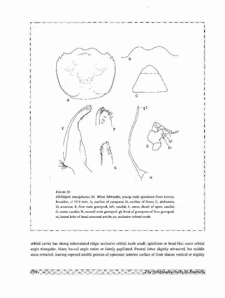
B
) \
'--------~c
A
-- -- 9 1
F
H
FIGURE 36
Zilchiopsis emarginatus (H. Milne Edwards), young male specimen from Lorero,
Ecuador, cl 19.9 mm: A, outline of carapace; B, outline of front; C, abdomen;
D, antenna; E, first male gonopod, left, caudal; F, same, detail of apex, caudal;
G, same, caudal; H, second male gonopod. gl, level of gonopore of first gonopod;
01, lateral lobe of basal antennal article; or, occlusive orbital tooth.
orbital cavity has strong tuberculated ridge; occlusive orbital tooth small, spiniforrn or bead-like; outer orbital
angle triangular, blunt; buccal angle entire or faintly papillated. Frontal lobes slightly advanced, but middle
sinus retracted, leaving exposed middle portion of epistome; anterior surface of front almost vertical or slightly
inclined forward, moderately high in middle, lower on sides, middle pillars short and thick, widely separated,
margin over each antennular fossa slightly thickened, not projected, antennular septum not sunk; epistome high,
divided by transverse middle suture into larger upper portion, almost vertical, and lower small portion, projected
forward and ending in single mid-point, this continued into buccal cavity by 2 divergent ridges.
First and 2nd + 3d abdominal tergites of male without deep depressions. Third to 6th abdominal segments
fused in both male and female; male abdomen very wide, outer margin convex, last segment with sides rounded,
approximately 0.57 as long as broad.
Basal article of antenna with outer lobe prominent and narrow; ischium of third maxilliped broad. Chelipeds
unequal; larger male cheliped with fingers short, narrow, widely gaping, with small, closely set teeth, fingers
with longitudinal ridges and upper distal angle of palm projected or with spine, more conspicuous in smaller
male chela and in both female chelae; inner margin of carpus with sharp, hooked spine; merus of male without
spines; merus of female with sharp spines on distal angle of upper and inner borders. 2-4 smaller spines on
lower margin. Lower margin of dactylus of legs thickly clothed with rows of long hairs, on lower margin of
propodus these patches cover 84% of 2nd, 62% of 3rd, 60% of 4th, and 45% of 5th pereiopod; upper margin of
dactylus, propodus and carpus with a thick row of short hairs.
First male gonopod slender, small as compared to size of species, wide basally, very narrow distally after
middle constriction; basal expansion regularly convex mesially, laterally more irregular, with accessory
elongated lobe on cephalic surface; distal portion with subparallel sides, strongly bent laterad; terminal spines
small, arranged as thick band on lateral side and diminishing in thickness proximally, forming a single row on
mesial side; apical portion rounded on lateral side, straight on mesial side where forms thin lamella which
surpasses gonopore; gonopore terminal, small, slit-like. Second male gonopod sinuous, moderately longer than
first.
Material examined
Colombia. Tres Esquinas, Rio Orteguaza, Caqueta Department; May 1954; H. NICEFORO MARIA; 1 male,
1 female (LSB 44-45). Vereda Vanguardia, Villavicencio, 500m alt, Meta Department; 29 March 1984;
M. CAMPOS; 1 male (ICN-MHN-CR0566).- Venezuela. Cafio Carinagua, near Puerto Ayacucho, Amazonas
Federal Territory; 29 November 1978; 2 females (Ivic). Alto Rio Cuao, Amazonas Federal Territory; November
1986; S. ZENOT; 1 male, 2 females (Ivic). Las Pavas, Rio Cataniapo, Amazonas Federal Territory; 28 September
1981; R. ROYERO; 2 females (MB XI-1640).- Ecuador. Loreto, in the foothills of mount Sumaco, Napo
Province, 450 m alt; June 1968; M. OLALLA; 2 males, 1 spent female (Ivic).
Type and distributionColombia. Loreto, upper Amazon (H. MILNE EOWAROS, 1853, type). Peru. Rio Ucayali (ORTMANN, 1893).
Ecuador. Napo Province (PRETZMANN, 1968a, type of Valdivia ecuadoriensisy. The present records extend the
distribution of the species towards the north to the Colombian llanos, towards the west to the foothills of the
Andes (Loreto, in Napo Province, not Loreto in Colombia, see Appendix), and towards the east to the
Venezuelan Guiana.
Remarks
The species presents considerable variability in relation to the sculpturing and convexity of the upper surface
of the carapace. In the young specimen from Ecuador (Fig. 36) and Colombia it is more flattened, excavated
near the margins, with the grooves well marked, thus resembling the species of Valdivia. In the young
specimens of the Venezuelan Guiana, it is smooth and more evenly arched, approaching the characters of
Dilocarcinus.
Zilchiopsis sattleri Bott, 1969
Fig. 3J; 4U; 51; 71; 9J; BR; 37A-R
Zilchiopsis sattleri Bott, 1969, p. 34, pl. 13, fig. 24a, b, pl. 21, fig. 55.- MANNING & ROBBS, 1977, p. 160.- RODRiGUEZ, 1981, p. 48.
DescriptionCarapace suborbicular, wider than long; upper surface slightly irregular; in frontal view forms an irregular
arch; gastric region slightly more prominent than adjacent epibranchial regions; metabranchial region slightly
more prominent than adjacent cardiac and intestinal; epigastric lobes very prominent, well delimited anteriorly;
frontal region slightly concave or almost flat in frontal view; branchio-urogastric, branchio-cardiac and
branchio-intestinal grooves wide and deep, urogastric groove may be absent or present. Postgastric pits slit-like,
placed in front of urogastric groove. Lateral margins angled and directed obliquely upwards, except in large
males where become rounded; postero-lateral ridge of carapace bent mesially in posterior end, ending in
elongated swelling over postero-lateral angles of carapace; antero-lateral margin with 4 spines behind external
orbital angle, prominent, evenly spaced, acute; last spines smaller; in large males lateral teeth becomes
obsolescent. Margin of front bilobed. Orbits oblong, orbital suture absent or indicated by inconspicuous
depression; eyes completely fill orbits; lower orbital margin in young specimens with 3 acute, widely spaced
spines (including the one at inner orbital angle), followed laterally by approximately 7 small papillae; inner
orbital angle prominent, pyramidal, topped by prominent and acute spine or rounded tubercle; occlusive orbital
tooth rounded, small, located close to inner orbital angle; outer orbital angle spinifonn, prominent in females
and young specimens, represented by stump in old males; buccal angle with large acute spine followed laterally
by smaller one, or large tubercle followed by one or two smaller ones. Front advanced, hiding epistome in dorsal
view; anterior surface of front inclined almost vertical or slightly inclined forward, moderately high in middle,
lower on sides, middle pillars short and thick, widely separated, margin over each antennular fossa slightly
thickened, but not projected, antennular septum not sunk; epistome high, inclined forward.
First abdominal tergite of male separated from tergite 2 + 3 by deep depression, tergite 2 + 3 with similar
depression on both sides. Third to 6th abdominal segments fused in both male and female; male abdomen
elongated, narrow at base, outer margin concave, last segment with sides concave, approximately 0.65 as long as
broad.Basal article of antenna with outer lobe prominent and narrow; ischium of third maxilliped with shallow
longitudinal depression. Chelipeds unequal; upper border of chela strongly arched, lower almost straight or
slightly concave; teeth alternatively large and small; acute spine on upper distal angle of hand, above
articulation of finger; inner margin of carpus produced in sharp hooked spine, apical spines on upper border and
latero-inferior margin of merus. Lower margin of dactylus of legs thickly clothed with patches of long coarse
hairs; these patches cover all lower margin of propodus in 5th, 3/4 in 2nd, 1/2 in 3rd and only distal angle in 4th;
upper margin of propodus and dactylus thickly clothed by shorter hairs arranged in 2 parallel longitudinal rows,
more extensive in 2nd and 3rd; claws of dactylus with 5 longitudinal carinae, 1 upper, 2 lateral and 2 inferior.
First gonopod very wide basally on latero-mesial plane, strongly constricted on distal third, tapering to very
thin apex directed slightly cephalad; well developed elongated lobe on latero-cephalic surface on proximal 3/4
of appendage; lateral and mesial surfaces of apex covered by very small spines, forming 2 distinct patches;
gonopore slit-like and open distally, major axis directed along cephalo-caudal plane. Second gonopod slightly
longer than first, strongly arched, but terminal portion straight.
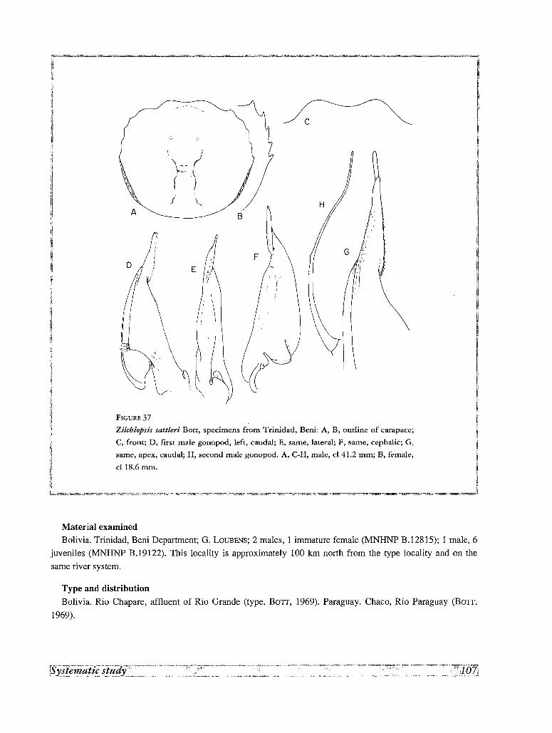
.........•
c
E
r./ i
I •
/ .; ~
I
A
FIGURE 37
Zilcbiopsis sattleri Bott, specimens from Trinidad, Beni: A, B, outline of carapace;
C, front; D, first male gonopod, left, caudal; E, same, lateral; F, same, cephalic; G,
same, apex, caudal; H, second male gonopod. A, C-H, male, cl 41.2 mm; B, female,
cl 18.6 mm.
Material examinedBolivia. Trinidad, Beni Department; G. LOUBENS; 2 males, I immature female (MNHNP B.128IS); I male, 6
juveniles (MNHNP B.19122). This locality is approximately 100 km north from the type locality and on the
same river system.
Type and distribution
Bolivia. Rio Chapare, affluent of Rio Grande (type, BOTT, 1969). Paraguay. Chaco, Rio Paraguay (BOTT,
1969).
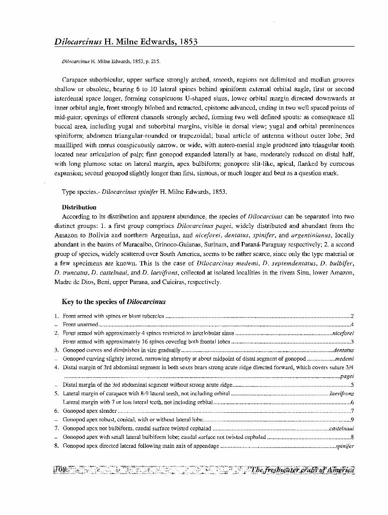
Dilocarcinus H. Milne Edwards, 1853
Dilocarcinus H. MilneEdwards, 1853,p. 215.
Carapace suborbicular, upper surface strongly arched, smooth, regions not delimited and median grooves
shallow or obsolete, bearing 6 to 10 lateral spines behind spiniform external orbital angle, first or second
interdental space longer, forming conspicuous U'-shaped sinus, lower orbital margin directed downwards at
inner orbital angle, front strongly bilobed and retracted, epistome advanced, ending in two well spaced points of
mid-guter; openings of efferent channels strongly arched, forming two well defined spouts: as consequence all
buccal area, including yugal and suborbital margins, visible in dorsal view; yugal and orbital prominences
spiniform; abdomen triangular-rounded or trapezoidal; basal article of antenna without outer lobe; 3rd
maxilliped with merus conspicuously narrow, or wide, with antero-mesial angle produced into triangular tooth
located near articulation of palp; first gonopod expanded laterally at base, moderately reduced on distal half,
with long plumose setae on lateral margin, apex bulbiform; gonopore slit-like, apical, flanked by corneous
expansion; second gonopod slightly longer than first, sinuous, or much longer and bent as a question mark.
Type species.- Dilocarcinus spinifer H. Milne Edwards, 1853.
Distribution
According to its distribution and apparent abundance, the species of Dilocarcinus can be separated into two
distinct groups: 1. a first group comprises Dilocarcinus pagei, widely distributed and abundant from the
Amazon to Bolivia and northern Argentina, and niceforei, dentatus, spin ifer, and argentinianus, locally
abundant in the basins of Maracaibo, Orinoco-Guianas, Surinam, and Parana-Paraguay respectively; 2. a second
group of species, widely scattered over South America, seems to be rather scarce, since only the type material or
a few specimens are known. This is the case of Dilocarcinus medemi, D. septemdentatus, D. bulbifer,
D. truncatus, D. castelnaui, and D. laevifrons, collected at isolated localities in the rivers Sinu, lower Amazon,
Madre de Dios, Beni, upper Parana, and Cuieiras, respectively.
Key to the species of Dilocarcinus
1. Front armed with spines or blunt tubercles 2
- Front unarmed .4
2. Front armed with approximately 4 spines restricted to interlobular sinus niceforei
- Front armed with approximately 16 spines covering both frontal lobes 3
3. Gonopod curves and diminishes in size gradually dentatus
- Gonopod curving slightly laterad, narrowing abruptly at about midpoint of distal segment of gonopod medemi
4. Distal margin of 3rd abdominal segment in both sexes bears strong acute ridge directed forward, which covers suture 3/4
.................................................................................................................................................................. .......................pagei
- Distal margin of the 3rd abdominal segment without strong acute ridge S
S. Lateral margin of carapace with 8-9 lateral teeth, not including orbital laevifrons
- Lateral margin with 7 or less lateral teeth, not including orbital.. 6
6. Gonopod apex slender 7
- Gonopod apex robust, conical, with or without lateral lobe 9
7. Gonopod apex not bulbiform, caudal surface twisted cephalad castelnaui
- Gonopod apex with small lateral bulbiform lobe; caudal surface not twisted cephalad 8
8. Gonopod apex directed laterad following main axis of appendage spinifer
- Gonopod apexgeniculate due to stronglaterad bent.. septemdentatus
9. Gonopod apex simple, without conspicuous lateral lobe argentinianus
- Gonopod apex withstrongrounded lateral lobe 11
10. Outline of apical lobecontinuous withgonopore truncatus
- Outline of apical lobe separated from gonopore by deepcleft or Ll-shaped sinus bulbifer
: Dilocarcinus argentiniamts Rathbun, 1906
Fig. 140; 38A-F
Trichodactylus (Trichodactylus) argentinianus Rathbun, 1906, p. 60, pI. 18, fig. 5, 6, text-fig. 120.
Dilocarcinus argentinianus apaluensis Pretzmann, 1968b, p. 75.
Poppiana argentinianus, BOlT, 1969, p. 52 (part.), pl, 12, fig. 21a, b, pI. 21, fig. 52.- RODRiGUEZ, 1981, p. 48.
Poppiana argentiniana, MANNING & HOBBs, 1977, p. 159.-PRETZMANN, 1979, p. 592, pI. 3, fig. 6-10.
Dilocarcinus (Poppiana) argentinianus, LOPRElTO, 1976, p. 88, fig. 30-33.
Dilocarcinus septemdentatus, NOBILl, 1896, p. 1 (part.).
Trichodactylus (Dilocarcinus) bachmayeri Pretzmann, J968a, p. 4.
Dilocarcinus bachmayeri, PRETZMANN, J968b, p. 75.
Poppiana bachmayeri, PRETZMANN J979, p.591, pI. 1, fig. 1, 2, p1.2, fig. 3-5.
Description
Carapace suborbicular; upper surface very convex, the convexity more pronounced along antero-posterior
axis, forming regular arch; regions not differentiated; epigastric lobes obsolescent, delimited anteriorIy only by
depression of frontal surface; front bilobed, inclined downwards; urogastric, branchio-urogastric, branchio
cardiac and branchio-intestinal grooves faintly indicated; dorsal surface of carapace smooth, polished, covered
by small papillae barely visible to naked eye. Postgastric pits lunulated, well marked. Anterolateral margin with
6 subtriangular teeth or acute spines behind external orbital angle, directed anteriorIy, approximately of equal
size, last spine smaller; interdental spaces of equal length except first which is longer, sinuous; ridge in postero
lateral margin starts at lateral side of last lateral spine, curves inwards at middle and ends at some distance of
ridge on the postero-lateral angle of carapace. Orbits subquadrate in frontal view, large, and eyes relatively
small, leaving ample empty space in orbital cavity; orbital suture obsolete or indicated by slight groove. Lower
orbital margin with 5 acute spines curved inwards followed by 5 Iow papillae, innermost spine larger and
forming inner orbital angle. Occlusive orbital tooth rounded and small, implanted inside orbit, away from inner
orbital angle and partially concealed by basal antennal article; floor of orbit is elevated behind outer orbital
angle, forming, with basal antennal article and occlusive tooth, a shallow channel; outer orbital angle with
subtriangular spine directed forward, similar to other on antero-lateral margin of carapace; buccal angle armed
with approximately 5 conical small spines or Iow papillae. Front retracted leaving epistome completely exposed
in dorsal view; anterior margin of frontal lobes lamellar, not forming distinct surface; epistome strongly
advanced, points of the mid-gutter well spaced; opening of efferent channels strongly arched.
Abdominal segments 3-6 fused in both sexes, but suture 3/4 still slightly visible in males; male abdomen
triangular, wide at base; outer margins straight; last segment widely triangular with outer margin straight,
approximately 0.5 as long as broad, proximal margin slightly shorter than distal margin of penultimate segment.
Basal article of antenna without outer lobe. Shallow depression along ischium of 3rd maxilliped. Chelipeds
strongly unequal in male, subequal in female; larger chela of male high and short, with upper border strongly
arched, lower border almost straight, with conspicuous swelling on external surface at base of fingers; fingers
very high and thick, strongly gaping; larger teeth of cutting edges interspaced by 1 or 2 smaller; carpus with

E
(:' <-\\... J
".'."".,
B
FIGURE 38
Dilocarcinus argentinianus (Rathbun), male specimen, cl 20.0 mm, from Rio Paraguay: A,
outline of carapace; B, front; C, abdomen; D, basal article of antenna; E, larger chela; F,
first male gonopod, left, caudal. ot, occlusive tooth.
large hooked spine on inner margin, merus with indistinct large tubercles on lateral distal, upper border and
middle of latero-inferior margins. Propodus and dactylus of legs devoided of long hairs on lower and upper
margins; claws of dactylus with 5 longitudinal carinae, I upper, 2 lateral and 2 inferior.
First gonopod simple, conical, regularly tapering to apex in caudal view, bracket-shaped, bent laterad; apex
with slight caudo-lateral swelling, with few short conical spines, but no conspicuous lateral lobe. Second
gonopod slightly longer than first, sinuous, terminal article incurved mesiad.
Material examined
Paraguay. Rio Paraguay; 1 male, cl 20.0 mm, cb 24.8 mm (ZSM 1091/1).
Type and distribution
Argentina. Las Garzas, 25 km from Ocampo, Santa Fe (type). Chaco: Resistencia (RATHBUN, 1906). Buenos
Aires. Paraguay. Puerto Max (BOTT, 1969). The type locality of Trichodactylus (Dilocarcinus) bachmayeri
PRETZMANN, 1968, Ignavi (=Ingavi), is uncertain, since PRETZMANN (1968a) located it in the Chaco, but latter
changed its position to llQS - 67QW, (PRETZMANN, 1979). These coordinates correspond to Ignavi, in the Pando
Province, Bolivia, on the Rio Orton, affluent of the Rio Beni, but there is, however, an homonymous locality
(Fortin Ingavi) in northern Paraguay (19Q55' 00' S - 61Q45'00' W), in the Rio Paraguay basin.
Remarks
The first gonopods from the Paraguayan specimens closely correspond with those of Argentinian specimens
illustrated by LOPRETTO (1976). The spinulation of the suborbital border resembles that of Dilocarcinus bulbifer
and Fredilocarcinus musmuschiae, but the spines are conspicuously smaller (Fig. 2E, F)
Dilocarcinus truncatus, new species
Fig. 3K; 8G; 131; 14C; 39A-E
Poppiana argentinianus, BOTT, 1969, p. 52 (part.).
Description
Carapace suborbicular; upper surface moderately convex, convexity more pronounced along antero-posterior
axis, forming an arch slightly flattened on top, with regions not differentiated, epigastric lobes slightly indicated;
frontal region flattened; front strongly bilobed inclined downwards; hepato-epibranchial, branchio-urogastric,
branchio-cardiac and branchio-intestinal grooves indicated by faint depressions; urogastric groove absent.
Dorsal surface of carapace smooth and polished, covered by small papillae not visible to naked eye. Postgastric
pits confluent in middle. Anterolateral margin with 6 acute spines behind external orbital angle, directed
anteriorly, except first two closer, other equally spaced; last spine smaller; postero-lateral margin marked by
well defined ridge throughout, which begins at lateral side of last lateral spine, straight in middle and parallel to
ridge on postero-lateral angle of carapace. Orbits subquadrate in frontal view; orbital suture indicated by very
faint line; orbits large and eyes relatively small, leaving ample empty space in orbital cavity. Lower orbital
margin with 3 or 4 acute spines curved inwards, followed by 8-10 papillae or crenulations; innermost spine
fused at base with spine of inner orbital angle. Occlusive orbital tooth reduced to small triangular projection
directed laterad, implanted inside orbit, away from inner orbital angle, partially concealed by basal antennal
article; notwithstanding reduction of inner orbital angle, a slight elevation behind it forms deep channel with
basal antennal article; outer orbital angle with hooked spine directed forward, similar to other on antero-lateral
margin of carapace; rounded lobe between outer orbital angle and first lateral angle; buccal angle armed with 4
small spines. Front retracted leaving epistome completely exposed in dorsal view; the anterior margin of the
front does not form distinct surface, but rather rounded-off regularly, with the exception of the semicircular
middle pillars; epistome strongly advanced, points of the mid-gutter well separated; opening of efferent channels
strongly arched, forming two well defined spouts delimited below by second maxillipeds; surface delimiting the
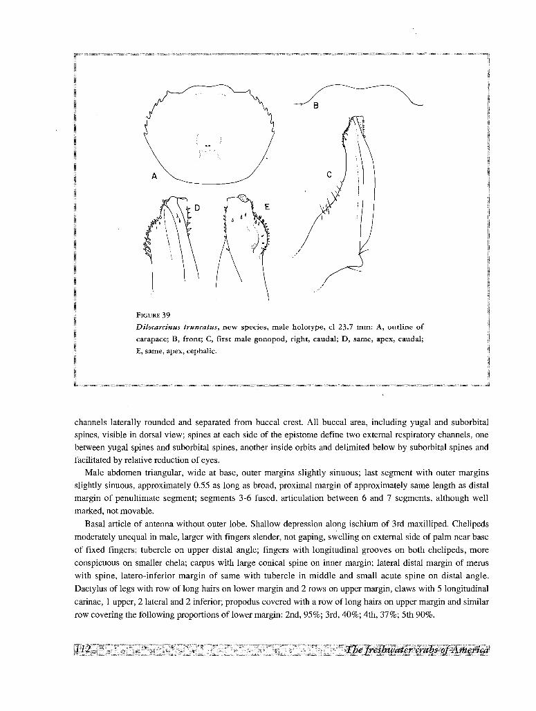
A
B
FIGURE 39
Dilocarcinus truncatus, new species, male holorype, cl 23.7 mm: A, outline of
carapace; B, front; C, first male gonopod, right, caudal; D, same, apex, caudal;
apex, cephalic.
channels laterally rounded and separated from buccal crest. All buccal area, including yugal and suborbitalspines, visible in dorsal view; spines at each side of the epistome define two external respiratory channels, one
between yugal spines and suborbital spines, another inside orbits and delimited below by suborbital spines and
facilitated by relative reduction of eyes.
Male abdomen triangular, wide at base, outer margins slightly sinuous; last segment with outer margins
slightly sinuous, approximately 0.55 as long as broad, proximal margin of approximately same length as distal
margin of penultimate segment; segments 3-6 fused, articulation between 6 and 7 segments, although well
marked, not movable.
Basal article of antenna without outer lobe. Shallow depression along ischium of 3rd maxilliped. Chelipeds
moderately unequal in male, larger with fingers slender, not gaping, swelling on external side of palm near base
of fixed fingers; tubercle on upper distal angle; fingers with longitudinal grooves on both chelipeds, more
conspicuous on smaller chela; carpus with large conical spine on inner margin; lateral distal margin of merus
with spine, latero-inferior margin of same with tubercle in middle and small acute spine on distal angle.
Dactylus of legs with row of long hairs on lower margin and 2 rows on upper margin, claws with 5 longitudinal
carinae, I upper, 2 lateral and 2 inferior; propodus covered with a row of long hairs on upper margin and similar
row covering the following proportions of lower margin: 2nd, 95%; 3rd, 40%; 4th, 37%; 5th 90%.
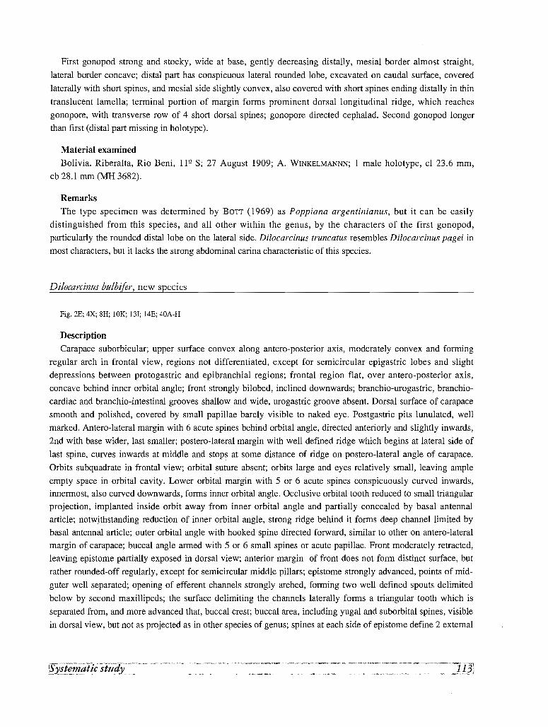
First gonopod strong and stocky, wide at base, gently decreasing distally, mesial border almost straight,
lateral border concave; distal part has conspicuous lateral rounded lobe, excavated on caudal surface, covered
laterally with short spines, and mesial side slightly convex, also covered with short spines ending distally in thin
translucent lamella; terminal portion of margin forms prominent dorsal longitudinal ridge, which reaches
gonopore, with transverse row of 4 short dorsal spines; gonopore directed cephalad. Second gonopod longer
than first (distal part missing in holotype).
Material examined
Bolivia. Riberalta, Rio Beni, 11Q S; 27 August 1909; A. WINKELMANNN; 1 male holotype, cl 23.6 mm,
cb 28.1 mm (MH 3682).
Remarks
The type specimen was determined by BOTT (1969) as Poppiana argentinianus, but it can be easily
distinguished from this species, and all other within the genus, by the characters of the first gonopod,
particularly the rounded distallobe on the lateral side. Dilocarcinus truncatus resembles Dilocarcinus pagei in
most characters, but it lacks the strong abdominal carina characteristic of this species.
Dilocarcinus bulbifer, new species
Fig. 2E; 4X; 8H; 10K; 131; 14E; 40A-H
Description
Carapace suborbicular; upper surface convex along antero-posterior axis, moderately convex and forming
regular arch in frontal view, regions not differentiated, except for semicircular epigastric lobes and slight
depressions between protogastric and epibranchial regions; frontal region flat, over antero-posterior axis,
concave behind inner orbital angle; front strongly bilobed, inclined downwards; branchio-urogastric, branchio
cardiac and branchio-intestinal grooves shallow and wide, urogastric groove absent. Dorsal surface of carapace
smooth and polished, covered by small papillae barely visible to naked eye. Postgastric pits lunulated, well
marked. Antero-Iateral margin with 6 acute spines behind orbital angle, directed anteriorly and slightly inwards,
2nd with base wider, last smaller; postero-Iateral margin with well defined ridge which begins at lateral side of
last spine, curves inwards at middle and stops at some distance of ridge on postero-Iateral angle of carapace.
Orbits subquadrate in frontal view; orbital suture absent; orbits large and eyes relatively small, leaving ample
empty space in orbital cavity. Lower orbital margin with 5 or 6 acute spines conspicuously curved inwards,innermost, also curved downwards, forms inner orbital angle. Occlusive orbital tooth reduced to small triangular
projection, implanted inside orbit away from inner orbital angle and partially concealed by basal antennal
article; notwithstanding reduction of inner orbital angle, strong ridge behind it forms deep channel limited by
basal antennal article; outer orbital angle with hooked spine directed forward, similar to other on antero-lateral
margin of carapace; buccal angle armed with 5 or 6 small spines or acute papillae. Front moderately retracted,
leaving epistome partially exposed in dorsal view; anterior margin of front does not form distinct surface, but
rather rounded-off regularly, except for semicircular middle pillars; epistome strongly advanced, points of mid
guter well separated; opening of efferent channels strongly arched, forming two well defined spouts delimited
below by second maxillipeds; the surface delimiting the channels laterally forms a triangular tooth which is
separated from, and more advanced that, buccal crest; buccal area, including yugal and suborbital spines, visible
in dorsal view, but not as projected as in other species of genus; spines at each side of epistome define 2 external
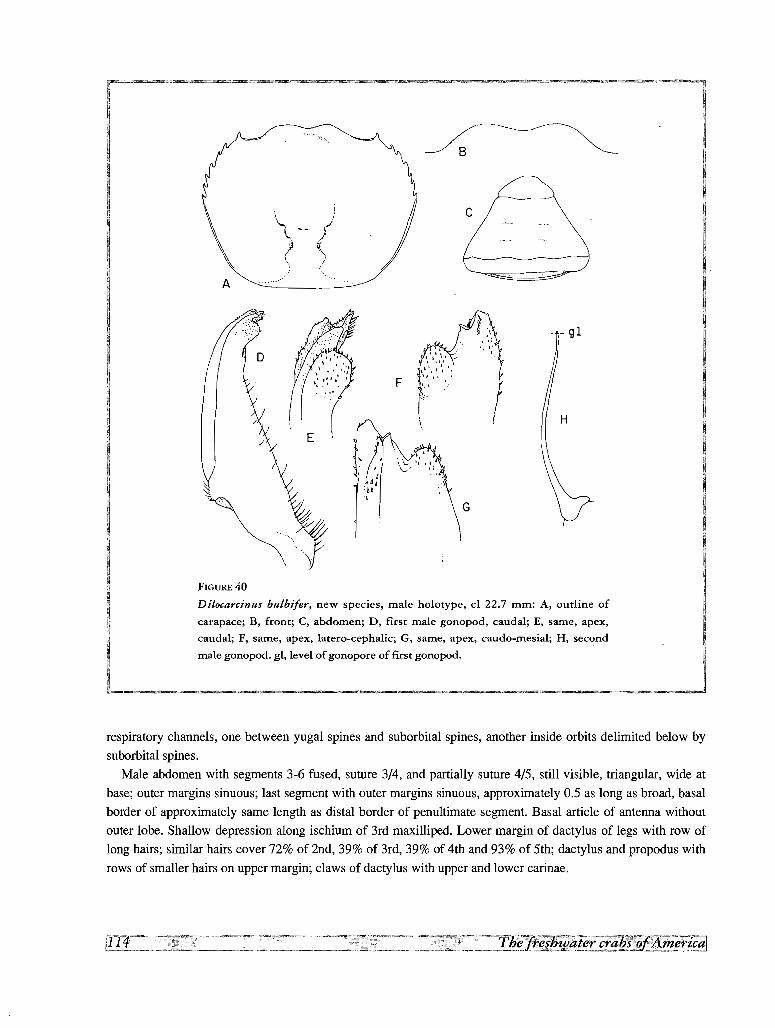
\.., j~ )/~ ~.•.
(
~-------~
c
A
fM
: :', .....'I " ..I "I
\ ~'I I ., '.
,..... .1. 1 .
~'::"
\
F
, I
; :~ d AdCb'.\
FIGURE 40
Dilocarcinus bulbi/er, new species, male holocype, cl 22.7 mm: A, outline of
carapace; B, front; C, abdomen; D, first male gonopod, caudal; E, same, apex,
caudal; F, same, apex, latero-cephalic; G, same, apex, caudo-mesial; H, second
male gonopod. gl, level of gonopore of first gonopod.
respiratory channels, one between yugal spines and suborbital spines, another inside orbits delimited below by
suborbital spines.
Male abdomen with segments 3-6 fused, suture 3/4, and partially suture 4/5, still visible, triangular, wide at
base; outer margins sinuous; last segment with outer margins sinuous, approximately 0.5 as long as broad, basal
border of approximately same length as distal border of penultimate segment. Basal article of antenna without
outer lobe. Shallow depression along ischium of 3rd maxilliped. Lower margin of dactylus of legs with row of
long hairs; similar hairs cover 72% of 2nd, 39% of 3rd, 39% of 4th and 93% of 5th; dactylus and propodus with
rows of smaller hairs on upper margin; claws of dactylus with upper and lower carinae.
First male gonopod wide at base, strongly narrowing distally, apex incurved laterad, mesial border straight,
lateral border transverse, provided with row of long plumose setae along proximal 3/4; apical portion consists of
conspicuous bulb covered by rows of small conical spinules, marginal process with spinules irregularly arranged
on its surface, and corneous awl shaped lamella overreaching other apical processes, provided with row of long
setae on caudal surface and small conical spinules on mesial border. Second male gonopod slightly sinuous, of
equal length, or slightly longer than first.
Material examined
Peru. Aguajal, Manu Province, Madre de Dios Department; 9 September 1988; H. ORTEGA; I male holotype,
cl 22.7 mm, cb 26.8 mm, I male paratype, cl 20.0 mm, cb 23.5 mm (MHN Lima).
Remarks
Dilocarcinus bulbifer is well characterized by the shape of the gonopod, and in particular by the conspicuous
subapical bulb. The dilocarcinian characters are not well developed in this species: the carapace is only
moderately convex, the front not strongly retracted, the epistome not completely exposed. The abdomen is wider
than in other species of the genus. Our type specimens lacks both chelipeds.
Etymology
The specific name is from the Latin bulbus (bulb) and fero (I bear) in reference to the conspicuous apical
lobe.
Dilocarcinus castelnaui H. Milne Edwards, 1853
Fig.40A-J
Dilocarcinus castelnaui H. Milne Edwards, 1853, p. 216.- H. MILNE EDWARDS, 1854, p. 182, pI. XIV, fig. 5, (not fig. 4, fide BOIT,
1969).- LUCAS, 1857, p. 8, pI. 11, fig. 4.- A. MILNE EDWARDS, 1869, p. 176,178.- SMITH, 1869, p. 36.
Dilocarcinus septemdentatus, GERSTACKER, 1856, p. 148 (part.).- GOLDI, 1886, p. 28, (part.).- MOREIRA, 1901, p. 44,49, 109 (part.).
Orthostoma septemdentatum,ORTMANN, 1897, p. 326, 327 (part.).- NOBILI, 1898, p. 9 (part.),
Trichodactylus (Dilocarcinus) castelnaui, RATHBUN, 1906, p. 61, pI. 18, fig. 9, 10.
Dilocarcinus (Goyazana) castelnaui, BOIT, 1969, p. 48, pI. 10, fig. 18a, b, C, pI. 20, fig. 49.
Dilocarcinus castelmani (sic), YOUNG, 1900, p. 231, 233.
DescriptionCarapace suborbicular; upper surface very convex, more pronounced along the antero-posterior axis,
regularly arched; regions not differentiated; epigastric lobes forming semicircular raised surface, delimited
anteriorly by conspicuous step; surface of frontal region flat, continuous with surface of protogastric region;
front strongly bilobed, inclined downwards; branchio-urogastric, branchio-cardiac and branchio-intestinal
grooves defined by few discontinuous depressions; branchio-intestinal grooves indicated in larger specimens by
discontinuous line of punctae; urogastric groove absent; dorsal surface of carapace covered by small papillae
barely visible to naked eye, and large punctae which form reticular pattern at middle of carapace. Postgastric
pits lunulated, well marked. Antero-lateral margin with 6 acute spines behind external orbital angle, directed
anteriorly and approximately of equal size, last spine smaller; these spines replaced by blunt large tubercles in
old males; interdental spaces approximately equal, first one forming sinuous or angled lobe; postero-lateral ridge
from side of last lateral spine, curves inwards at middle, ends at some distance of ridge on the postero-Iateral
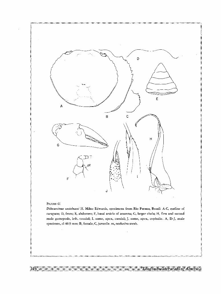
\A
G
F
B c
E
FIGURE 41
Dilocarcinus caste/naui H. Milne Edwards, specimens from Rio Parana, Brazil: A-C, outline of
carapace; D, front; E, abdomen; F, basal article of antenna; G, larger chela; H, first and second
male gonopods, left, caudal; I, same, apex, caudal; ], same, apex, cephalic. A, D-], male
specimen, cl 40.9 mm; B, female; C, juvenile. ot, occlusive tooth.
angle of carapace. Orbits subquadrate in frontal view, very large in vertical direction, eyes small, leaving ample
space in orbital cavity; orbital suture obsolete or indicated by slight groove. Lower orbital margin with 9 acute
spines directed inwards and downwards, diminishing in size laterally; innermost long, forms inner orbital angle;
last three of series papilliform. Occlusive orbital tooth consists of large ovoid blade, extending anteriorly beyond
distal margin of basal antennal article. Orbital floor elevated behind outer orbital angle, forming shallow channel
with basal antennal article and occlusive tooth; outer orbital angle with acute spine directed forward, similar to
other on antero-lateral margin of carapace, often broken and worn out; buccal angle armed with 4-5 hooked
spines. Front retracted leaving epistome completely exposed in dorsal view; anterior margin of frontal lobes
lamellar, not forming distinct surface; epistome strongly advanced, points of the mid-gutter well spaced;
opening of efferent channels strongly arched, forming 2 well defined spouts delimited below by 2nd
maxillipeds; surface delimiting channels laterally forms rounded lobe; all buccal area, including yugal and
suborbital spines, visible in dorsal view; spines at each side of epistome define 2 external respiratory channels,
one between yugal spines and suborbital spines, another inside orbits, delimited below by suborbital spines.
Sutures betwen abdominal segments 3-6 clearly visible in both sexes; male abdomen triangular, lateral
margins of 3rd and 4th segments rounded and expanded, general outline of the abdomen straight or slightly
concave; last segment with outer margin slightly concave, approximately 0.6 as long as broad, proximal margin
shorter than distal margin of penultimate segment.
Basal article of antenna without outer lobe. Shallow depression along ischium of 3rd maxilliped.Chelipeds
moderately unequal; larger chela elongated with upper border moderately convex, lower border almost straight;
a large flat tubercle on external surface, at base of fingers; fingers slightly gaping; lower margin of merus of
female and small males with 3 median and 1 terminal spine; internal margin with median spine; upper margin
with distal spine; carpus with large spine on internal margin; smaller spines on external and upper margins near
articulation of palm; legs with row of long hairs on lower margin of propodus and dactylus, and inner and outer
distal margin of carpus; two rows of smaller hairs on upper margin of dactylus and similar internal row on
propodus; claws of dactylus with 5 longitudinal carinae, 1 upper, 2 lateral and 2 inferior.
First gonopod simple, slender, slightly sinuous, progressively tapering to apex, devoided of distal lateral lobe;
distal portion of margin bent laterad, shorter than cephalic surface; gonopore large, opening laterad; apical
spines very small, forming continuous patch over lateral, cephalic and mesial surfaces. Second gonopod
considerable longer than first, bent mesially in form of question mark.
Material examined
Brazil. Parana River, lagoon near Fundao, 32 km upriver from Puerto Tiberica [Porto Tibirica]; December
1938; SCHINDLER; 1 male, cl 40.9 mm, cb 46.5 mm, 1 female, cl 37.7 mm, cb 41.5 mm, with 180 marsupialyoungs (ZSM 1088-1). Highlands of Goias, near Brasilia; 10 August 1965; SCHULTZ; 1 young male, cl 10.8 mm,
cb 18.7 mm (ZSM 1088-2).
Type and distributionThe types are two small males (largest cl 16.8 mm) and 1 female (MP), collected by the count Francis de
CASTELNAU and Mr Emile DEVILLE, at Salinas, on the Rio Crixas Acu, affluent of the Rio Araguaia, where they
stayed from May 14 to June 10, 1844, during their expedition across South America (PAPAVERO, 1971).
RATHBUN (1906) excluded from the type material the smallest male, which she placed under D. spinifer, but
BOTT (1969) kept all the type specimens under D. castelnaui. BOTT (1969) recorded 3 males and 3 females from
the Xingu river basin, and 1 male and 1 female from the Parana River (see above under material examined).
Thus the area of distribution of the species is located on both sides of the South American water divide formed
by the Serra das Divisoes.
rsy.stemailcstttay··----- .---..---.--.-.--- _. '---'--'" ".-".-..-.,.,,'.-._ -..- '-.._-,-._....~ ..--~- ·,11.7]1L __•__"., . _... ... .. . . __ ..,. . . _ . __ .. .., _ .. _, _
Dilocarcinus dentatus (Randall, 1839)
Fig. IH; 2D; 4Y; 5N; 8J; IOL; 13J; 14B; ISH; 42A-I
Orthostoma dentata Randall, 1839, p. 122, p1.5,fig. 1-3.- H. MILNE EDWARDS, 1853, p. 215.- ORTMANN, 1897, p. 326.
Dilocarcinus dentatus, YOUNG, 1900, p. 231, pJ. 5, 6.- MORElRA, 1901, p. 44.- PRETZMANN, 1968b, p. 75.- SMALLEY & RODRiGUEZ, 1972,
p.52, fig. 17,18.- RODRfGUEZ, 1980, p. 344, fig. 100.- RODRiGUEZ, 1981, p. 48.
Trichodactylus (Dilacarcinus] dentatus, RATHBUN 1906, p. 65, pI. 18, fig. 4.- HOLTHUIS, 1959, p. 214, fig. SOb, 51.- CHACE & HOBBS,
1969, p. 152, fig. 44.
Poppiana dentata, BOTT, 1969, p. 50, pJ. 11, fig. 19a, b, pI. 20, fig. 50, text-fig. 2.
Dilocarcinus dentatus cayennensis Pretzmann, 1968b, p. 75.
Dilocarcinus dentatus trinidadensis Pretzmann, 1968b, p. 76.
Dilocarcinus multidentatus van Martens, 1869a, p. 5, pI. 1, fig. 2.
DescriptionCarapace suborbicular; upper surface very convex, the convexity more pronounced along antero-posterior
axis, forming regular arch; regions not differentiated; epigastric lobes semicircular, not well delimited; surface
of frontal region continuous with surface of protogastric region; front moderately bilobed, inclined downwards,
with 16 to 21 acute triangular spines on its margin; branchio-urogastric, branchio-cardiac and branchio-intestinal
grooves faintly indicated; urogastric groove absent; dorsal surface of carapace smooth, polished, covered by
small papillae barely visible to naked eye. Postgastric pits lunulated, well marked. Antero-lateral margin with 8
10 acute spines behind external orbital angle, directed anteriorly, approximately of equal size, last spine smaller;
teeth replaced by semicircular lobes in some specimens; interdental spaces slightly increase posteriorly, 2nd
longest, forming conspicuous U'-shaped sinus; ridge in postero-Iateral margin starts at lateral side of last lateral
spine, curves inwards at middle and ends at some distance of ridge on postero-lateral angle of carapace. Orbits
subquadrate in frontal view; orbital suture obsolete or indicated by slight groove. Lower orbital margin with 7 to
11, usually 9, acute spines, innermost forming inner orbital angle. Occlusive orbital tooth reduced to slender
spine implanted inside orbit, away from inner orbital angle; floor of orbit elevated behind outer orbital angle,
forming with basal antennal article and occlusive tooth a shallow channel; outer orbital angle with hooked spine
directed forward, similar to other on antero-lateral margin of carapace; buccal angle armed with 5 to 8, usually
6, small spines. Front retracted leaving epistome completely exposed in dorsalview; anterior margin of frontallobes lamellar, not forming distinct surface; epistome strongly advanced, points of mid-gutter well spaced;
opening of efferent channels strongly arched, forming 2 well defined spouts limited below by 2nd maxillipeds;
all buccal area, including yugal and suborbital spines, visible in dorsal view; spines at each side of epistome
define 2 external respiratory channels, one between yugal spines and suborbital spines, another inside orbits,
delimited below by suborbital spines.
Abdominal segments 3-6 fused in both sexes, but suture 3/4 still visible in males; male abdomen triangular,
wide at base; outer margins slightly concave; last segment with outer margin sinuous, approximately 0.6 as long
as broad, margin of approximately same length as distal margin of penultimate segment.
Basal article of antenna without outer lobe. Shallow depression along ischium of 3rd maxilliped. Chelipeds
strongly unequal in male, subequal in female; chela of male with upper border strongly arched, lower border
slightly sinuous; fingers very high and thick, not gaping; larger teeth of cutting edges interspaced by I or 2
smaller; chelae in both sexes with swelling on external surface at base of fingers; carpus with large conical spine
on inner margin, merus with spine on latero-distal margin, another on distal half of upper border and sometimes
in middle of latero-inferior margin; female chelae and smaller chela of male with small acute spine on distal
upper angle of palm, distal angle of carpus and infero-distal margin of ischium, respectively; 2- 4 spines on the
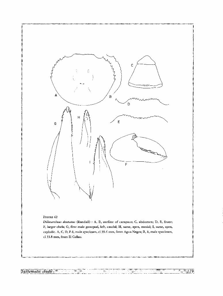
B
o
F
E
II J
/JvtI \ H
1G
FIGURE 42
D'llocarcinus dentatus (Rand all) : A, B, outline of carapace; C, abdomen; D, E, front;
F, larger chela; G, first male gonopod, left, caudal; H, same, apex, mesial; I, same, apex,
cephalic. A, C, D, F-I, male specimen, cl 39.4 mm, from Agua Negra; B, E, male specimen,
cl 33.8 mm, from El Cal1ao.
lower margin of merus. Propodus and dactylus of legs with row of long hairs on lower margin, upper margin
with 2 rows of shorter hairs on dactylus and similar internal row on propodus; claws of dactylus with 5
longitudinal carinae, 1 upper, 2 lateral and 2 inferior.
First gonopod curved, directed laterad, progressively tapering to apex; distal portion of margin is bent
mesially, emerging on anterior surface; basal lateral prominence poorly developed, with long, conspicuous
lateral surface, implanted in small pit; gonopore U-shaped, open distally, with thin corneous ridge on lateral
side. Second gonopod longer than first, curved mesiad as incomplete semicircle.
Color
In life the color appears to be brown or cream colored (SMALLEY and RODRIGUEZ, 1972). Specimens
preserved in alcohol are very variable, more commonly reddish brown, but sometimes grey or pale brown.
Material examined
Venezuela. Agua Negra, 80 malt, Yaracuy State; 3 January 1971; O. CHACON; 3 males, 2 females (LS 1044).
Agua Negra, Yaracuy State; 23 February 1971; B. ROMAN, 2 males, 3 females (LS 1037). Laguna de Tacarigua,
Falc6n State; 26 June 1967; E. VALLADARES (LS). Hato La Marrerefia, Las Mercedes, Guarico State, 220 malt;
15 January 1971; D. LOUEIRO (LS). Finca Vuelta Larga, 100 km by road SE of Guararinos, Sucre State; 14
August 1987; H. CASTELLANOS, 1 female (LS 1039). Parque Cachamay, Puerto Ordaz, Rio Caroni, Bolivar
State; 4 March 1978; 1. CLINE, 1 male (Ivic). Cafio Onoto, El Callao, Bolivar State; 1 male (Ivic). Hato Terecay,
near El Manteco, Bolivar State; 7 June 1977; S. GORZULA; 1 male, 2 females (females captured while walking
on gravel road under heavy rain) (Ivic). 70 km SSE from El Manteco, Bolivar State, 250 malt; 1 female (Ivic).
Lago de Guri, Bolivar State; June 1985; 1 male (LS 1043). Quebrada Trapichote, at railway crossing, Los
Pijiguaos, Bolivar State; 3 May 1988; C. LASSO & G. COLONNELLO; 1 ovigerous female (LS 1140). Road to El
Dorado, Santa Elena, km 88, Bolivar State; 25 February 1988; M. LENTINO; 1 female, 3 males, 3 juveniles (LS
871).- Colombia. Between Cartagena andBayunca, Bolivar Department; 1 September 1972; P. CALA; 1 male,
1 female (ICN MHN CR 50). Confluence of the Rio Merguia and Rio Cobug6n, southern extremity of the
Norte de Santander Department; October 1968; B. NICEFORO MARIA; 1 female (LSB).- Trinidad. Mitan; 27
March 1980; A. E. ESTEvEs; 28 males, 1 female (Ivic).- Other specimens from Venezuela examined by me are
recorded in SMALLEY & RODRIGUEZ (1972).
Type and distributionSuriname. Paramaribo (type locality by designation, HOLTHUIS, 1959); Corantijn and Coppename Rivers
(HOLTHUIS, 1959). Guyana. Berbice (YOUNG, 1900). French Guiana (RATHBUN, 1906). Venezuela. Very
common in the llanos, in rivers discharging into the Orinoco, in the states of Portuguesa, Cojedes, Calabozo,
Apure, Guarico, Aragua, Monagas, Bolivar and Delta Amacuro (SMALLEY & RODRIGUEZ 1972). Trinidad
(HOLTHUIS, 1959).
Remarks
PRETZMANN, 1968b, p. 75-76 described two subspecies, Dilocarcinus dentatus trinidadensis based on a male
cl 35.6 mm, from Trinidad and D. dentatus cayennensis on a male specimen cl 44.3, from French Guiana. The
characters given for the first subspecies (carapace narrower, flatter, 10 blunt lateral teeth, finger of larger chela
gap,ing, with shorter dactylus, gonopod wider, more recurved, color dark brown) are all very variable in the large
series of specimens examined by me from Trinidad, and are also very variable in the specimens which I have
examined from the continental part of the range of the species. A similar situation occurs with the second
subspecies.
The specimen from Cartagena reported above is completely isolated from the area of distribution of the
species. However, it coincides in all characters with those from the Orinoco basin.
Dilocarcinus laevifrons Moreira, 1901
Dilocarcinus laevifrons Moreira, 1901, p. 48, pI. I, fig. 2.
Trichodactylus (Dilocarcinus) laevifrons, RATHBUN, 1906, p.66.
Poppiana laevifrons, BOIT, 1969, p. 51, pI. 11, fig. 20a, b, pI. 21, fig. 51.- RODRiGUEZ, 1981, p. 48.
Type and distribution
This species was described from a single female specimen whose typical locality is not well established ("we
found it in a jar with several crustaceans coming from Pernambuco; we believe accordingly that this species
lives in the rivers of that state", MOREIRA, 1901, p. 49). Borr (1969) assigned to this species one small male
(cl l? mm) from Rio Cuieiras.
Dilocarcinus medemi Smalley & Rodrfguez, 1972
Fig. 43A-F
Dilocarcinus medemi Smalley & Rodrfguez, 1972, p. 53, fig. 19,20,23.- RODRiGUEZ, 1981, p. 48.
Description
Carapace suborbicular; upper surface very convex, convexity more pronounced along antero-posterior axis,
forming regular arch; regions not differentiated; semicircular epigastric lobes not well delimited; frontal and
postorbital regions depressed; front moderately bilobed, inclined downwards, with 13 acute triangular spines on
its margin; branchio-urogastric, branchio-cardiac and branchio-intestinal grooves faintly indicated; urogastric
groove absent; dorsal surface of carapace smooth and polished, covered by small papillae barely visible to naked
eye. Postgastric pits lunulated, well marked. Anterolateral margin with 7 spines behind external orbital margin,
sharp, conical, directed anteriorly and approximately of equal size; 1st interdental space longest, forming
conspicuous U-shaped sinus; postero-lateral margin, with well defined ridge from lateral side of last lateral
spine, curves inwards at middle and stops at some distance of ridge on postero-lateral angle of carapace. Orbits
subquadrate in frontal view; orbital suture obsolete or indicated by slight groove. Lower orbital margin with 6 or
7 acute spines of unequal size, innermost forming inner orbital angle. Occlusive orbital tooth reduced to roundedlobe implanted inside orbit, away from inner orbital angle; orbital floor elevated behind outer orbital angle,
forming shallow channel with basal antennal article and occlusive tooth; outer orbital angle with hooked spine
directed forward, similar to other on antero-lateral margin of carapace; buccal angle armed with 5 small spines.
Front retracted leaving epistome completely exposed in dorsal view; anterior margin of frontal lobes lamellar,
not forming distinct surface; epistome strongly advanced, points of mid-gutter well spaced; opening of efferent
channels strongly arched, forming well defined spouts delimited below by 2nd maxillipeds; surface delimiting
channels laterally forming rounded lobe; all buccal area, including yugal and suborbital spines, visible in dorsal
view; spines at each side of epistome define 2 external respiratory channels, one between yugal spines and
suborbital spines, another inside orbits and delimited below by suborbital spines.
Abdominal segments with all sutures distinct at least in males; male abdomen trapezoidal, wide at base; outer
margins concave; last segment with outer margin conspicuously concave.
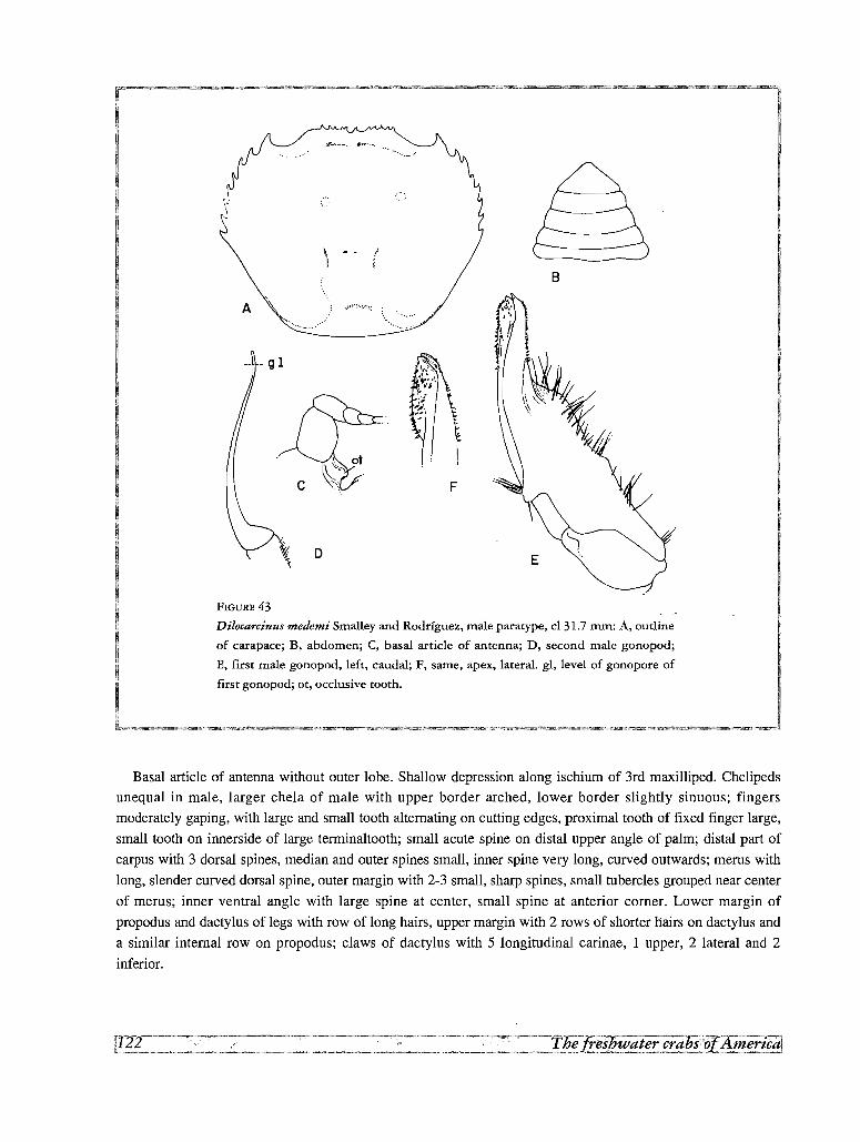
Ii
A
F
o
B
FIGURE 43
Dilocarcinus medemi Smalley and Rodrfguez, male paratype, cl 31.7 mm: A, outline
of carapace; B, abdomen; C, basal article of antenna; D, second male gonopod;
E, first male gonopod, left, caudal; F, same, apex, lateral. gl, level of gonopore of
first gonopod; ot, occlusive tooth.
Basal article of antenna without outer lobe. Shallow depression along ischium of 3rd maxilliped. Chelipeds
unequal in male, larger chela of male with upper border arched, lower border slightly sinuous; fingers
moderately gaping, with large and small tooth alternating on cutting edges, proximal tooth of fixed finger large,
small tooth on innerside of large terminaltooth; small acute spine on distal upper angle of palm; distal part of
carpus with 3 dorsal spines, median and outer spines small, inner spine very long, curved outwards; merus with
long, slender curved dorsal spine, outer margin with 2-3 small, sharp spines, small tubercles grouped near center
of merus; inner ventral angle with large spine at center, small spine at anterior corner. Lower margin of
propodus and dactylus of legs with row of long hairs, upper margin with 2 rows of shorter hairs on dactylus and
a similar internal rowan propodus; claws of dactylus with 5 longitudinal carinae, I upper, 2 lateral and 2
inferior.
First gonopod with proximal half wide in meso-caudal view, mesial border curved laterad, lateral border
forming sinuous, lamelliform lobe provided with long, stiff setae; basal lobe prominent, subtriangular, bearing
long stiff marginal setae; at distal quarter gonopod becomes abruptly narrowed and bent laterad; sides of distal
part nearly parallel, with slight expansion at tip; lateral process hook-shaped at tip, slightly exceeding mesial
process; distal part of gonopod bears lateral and mesial patches of small conical spines; no apical tuft of setae.
Second gonopod sinuous, slightly longer than first.
Material examined
Colombia. Quebrada Tinaj6n, Rio Sirni drainage, near Monterfa, C6rdoba Department; April
1962; C. A. VELAzQUEZ and R. CAMACHO; 1 male paratype, cl 31.7 mm, cb 38.7 mm (USNM 139123, ex Tulane
University Collections 4870).
Type and distribution
The species is known only from the two type specimens (SMALLEY and RODRIGUEZ, 1972).
Remarks
The species closely resembles Dilocarcinus dentatus, but could be clearly differentiated from it by the shape
of the gonopod.
Dilocarcinus niceforei (Schmitt & Pretzmann, 1968)
Fig.4Z; 8K; 131; 141; 44A-C
Trichodactylus (Valdivia) niceforei Schmitt and Pretzmann, 1968, p. 6.
Valdivia (Rotundovaldivia) niceforei, PRETZMANN, 1968b, p. 73.- SCHMlTI, 1969, p. 93, fig. 1.
Yaldivia (Rotundovaldivia) niceforei cucutensis Pretzmann, 1968b, p. 73.
Dilocarcinus (Dilocarcinus) niceforei, SMALLEY & RODRiGUEZ, 1972, p. 51, fig. 15, 16.
Dilocarcinus niceforei, RODRioUEZ, 1981, p. 48.
Description
Carapace suborbicular; upper surface very convex, more pronounced along the antero-posterior axis,
regularly arched; regions not differentiated; semicircular epigastric lobes not well delimited; surface of frontal
region continuous with surface of protogastric region; front strongly bilobed, inclined downwards, with
interlobular space bearing 2 to 5 teeth (8 in cucutensis) with additional tubercles or papillae; branchio
urogastric, branchio-cardiac and branchio-intestinal grooves defined by thin continuous line, partially
obsolescent in small specimens; branchio-intestinal groove indicated in larger specimens by 2 parallel sulci;
urogastric groove absent; dorsal surface of carapace covered by small papillae barely visible to naked eye, also
large punctae. Postgastric pits lunulated, well marked. Antero-lateral margin with 7 acute spines behind external
orbital angle, directed anteriorly and approximately of equal size, last spine smaller; interdental spaces
approximately equal, first one longer, forming conspicuous U-shaped sinus; postero-lateral ridge, from side of
last lateral spine, curves inwards at middle, ends at some distance of ridge on the postero-lateral angle of
carapace. Orbits subquadrate in frontal view; orbital suture obsolete or indicated by slight groove. Lower orbital
margin with 5 to 8 acute spines which diminish in size laterally, innermost forming inner orbital angle.
Occlusive orbital tooth reduced to low lobe implanted inside orbit, away from inner orbital angle; orbital floor
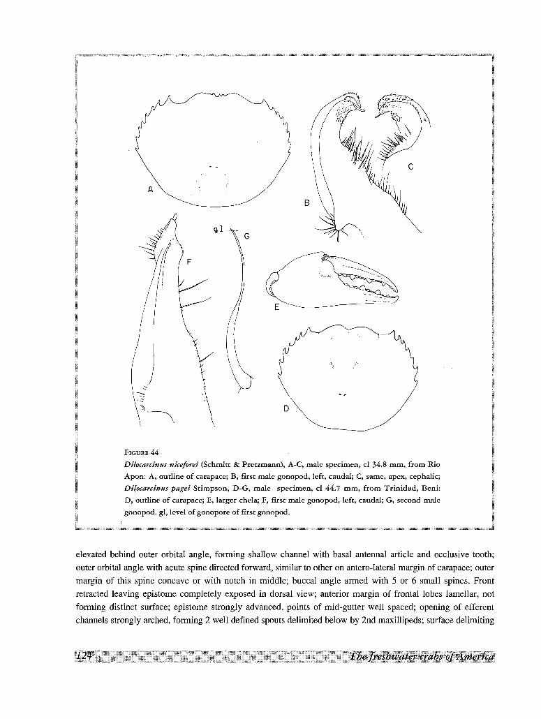
D
E
A
FIGURE 44
Dilocarcinus niceforei (Schmitt & Pretzmann), A-C, male specimen, cl 34.8 mm, from
Apon: A, outline of carapace; B, first male gonopod, left, caudal; C, same, apex, cephalic;
Di(ocarcinus pagei Stimpson, D-G, male specimen, cl 44.7 mm, from Trinidad,
D, outline of carapace; E, larger chela; F, first male gonopod, left, caudal; G, second
gonopod. gl, level of gonopore of first gonopod.
elevated behind outer orbital angle, forming shallow channel with basal antennal article and occlusive tooth;
outer orbital angle with acute spine directed forward, similar to other on antero-lateral margin of carapace; outer
margin of this spine concave or with notch in middle; buccal angle armed with 5 or 6 small spines. Front
retracted leaving epistome completely exposed in dorsal view; anterior margin of frontal lobes lamellar, not
forming distinct surface; epistome strongly advanced, points of mid-gutter well spaced; opening of efferent
channels strongly arched, forming 2 well defined spouts delimited below by 2nd maxillipeds; surface delimiting
channels laterally forms rounded lobe; all buccal area, including yugal and suborbital spines, visible in dorsal
view; spines at each side of epistome define 2 external respiratory channels, one between yugal spines and
suborbital spines, another inside orbits, delimited below by suborbital spines.
Sutures betwen abdominal segments 3-6 clearly visible in both sexes; male abdomen triangular, lateral
margins of 3rd and 4th segments rounded and expanded, general outline of abdomen conspicuously concave;
last segment with outer margin concave, approximately 0.6 as long as broad, margin of approximately same
length as distal margin of penultimate segment.
Basal article of antenna without outer lobe. Shallow depression along ischium of 3rd maxilliped. Chelipeds
moderately unequal; lower margin of merus with 3 median and 1 terminal spine; internal margin with median
spine; upper margin with distal spine; carpus with large spine on internal margin; smaller spines on external and
upper margins near articulation of palm; lower margin of propodus and dactylus of legs with row of long hairs,
upper margin with 2 rows of smaller hairs on dactylus and similar internal row on propodus; claws of dactylus
with 5 longitudinal carinae, 1 upper, 2 lateral and 2 inferior.
First gonopod with proximal 3/4 wide in meso-caudal view, mesial border slightly curved laterad, lateral
border sinuous, provided with long, stiff setae, at least some plumose, projected in lateral view as a sinuous
ridge; prominent basal lobe bearing long stiff marginal setae, subtriangular; gonopod becomes abruptly
narrowed and strongly bent laterad at distal quarter; apical portion strongly recurved, margin twisted to proximal
position and mesial border to distal position, forming two rounded lobes with gonopore in between, directed
laterad; distal part of gonopod bears lateral and mesial patches of prominent spines, recurved at different angles,
irregular in appearance; apical tuft of setae absent. Second gonopod slightly longer than first, extruded from
gonopore as small tongue-like appendage.
Material examined
Venezuela. Rio El Quebrad6n, near Aguas Calientes, Zulia State; 7 May 1965; captured with Pronox-Fish,
bottom sand-gravel, water temperature 28.6Q C; F. MAGO; 1 male (MB). Swamp near Bobures, Zulia State;
captured with Pronox-Fish, bottom mud with decomposing organic matter, water temperature 30.8Q C; 6 June
196~; F. MAGo; 1 male, 1 immature female (MB).- Rio Apon, near Machiques, Zulia State, 90 malt; 17 August
1978; L. GAMBA; 1 male (Ivic).
Type and distribution
The holotype is a male from Pamplona, Colombia (SCHMITT and PRETZMANN, 1968), collected by
H. NICEFORO MARIA; SCHMITT (1969) fixed the holotype designation to a male, cl 34 mm, in the USNM 112117.
The holotype and only specimen of Valdivia (Rotundovaldivia) niceforei cucutensis is a male from Ciicuta,
Colombia, in the Basel Museum, N° 763-b (PRETZMANN, 1968b). Both localities are 50 km apart, in the same
river basin (SMALLEY & RODRIGUEZ, 1972). In addition to the type material, the species have been reported by
SMALLEY & RODRIGUEZ (1972), RODRIGUEZ (1980) & CAMPOS (1985) from many localities in the rivers of the
Lago Maracaibo drainage in Venezuela and Colombia.
Dilocarcinus pagei Stimpson, 1861
Fig. lE; 4AA; 81; 13J; 14D; 44D-G
Dilocarcinus pagei Stimpson, 1861, p. 373.- RODRiGUEZ, 1981, p. 48.
Dilocarcinus (Dilocarcinus) pagei, LOPREITO, 1981, p. 23.
Dilocarcinus (Dilocarcinus) pagei pagei, BOIT, 1969. p.46, pI. 9, fig. 16a, b, pI. 20, fig. 47.- LOPREITO, 1976, p. 84, fig. 26-29.-
MANNING & HOBBS, 1977, p. 159.- LOPREITO, 1981, p. 22.
Dilocarcinus pagei cristatus Bott, 1969, p. 47, pl. 9, fig. 17a, b, pl. 20, fig. 48.- LOPRETTO, 1976, p. 86, fig. 14, 17.- LOPREITO, 1981, p. 22.
Dilocarcinus (Dilocarcinus) pagei enriquei Pretzmann, 1978a, p. 168; PRETZMANN, 1983b, p. 324.
Dilocarcinus (Dilocarcinus) pagei enricei [sic], PRETZMANN, 1983b, p. 318.
Trichodactylus (Dilocarcinus) orbicularis, RATHBUN, 1906, p. 58, pl, 18, fig. 6, text-fig. 119 (part.).- MORElRA, 1913, p. 19, pl. 7, fig. 1,
2.- BALss, 1914, p. 409.- RINGUELET, 1949, p. 101, pl, 9, fig. 2.
Trichodactylus (Dilocarcinus) septemdentatus, HOLTHUIS, 1959, p. 218 (part.).
Orthostoma septemdentatum, NOBILI, 1898, p. 9 (part.).
Description
Carapace suborbicular; upper surface very convex, convexity more pronounced along antero-posterior axis,
regularly arched, with regions not differentiated, except for semicircular epigastric lobes and slight depressions
between protogastric and epibranchial regions; frontal region slightly convex over antero-posterior axis, concave
behind inner orbital angle; front strongly bilobed, inclined downwards; hepato-epibranchial, branchio
urogastric, branchio-cardiac and branchio-intestinal grooves indicated by thin, obsolescent lines; urogastric
groove absent; a short incision in middle of epigastric lobes. Dorsal surface of carapace smooth and polished,
covered by small papillae barely visible to naked eye. Postgastric pits lunulated, well marked. Anterolateral
margin with 6 acute spines behind external orbital angle, directed anteriorly and slightly inwards; interdental
spaces slightly increasing posteriorly; last spine smaller; postero-lateral margin marked by well defined ridge
throughout, which begins at lateral side of last lateral spine, curves slightly inwards in middle and stops at some
distance of ridge on postero-lateral angle of carapace in female, running parallel to it in male. Orbits
subquadrate in frontal view; orbital suture indicated by well marked groove; orbits large and eyes relatively
small,leaving ample empty space in orbital cavity. Lower orbital margin with 6 or 7 acute spines curved
inwards, innermost pair fused at base to form inner orbital angle. Occlusive orbital tooth reduced to small
triangular projection, implanted inside orbit away from inner orbital angle, partially concealed by basal antennal
article; strong ridge behind inner orbital angle forms deep channel limited by basal antennal article; outer orbital
angle with hooked spine directed forward, similar to other on antero-lateral margin of carapace; buccal angle
armed with 5 or 6 small spines or acute papillae. Front retracted leaving epistome completely exposed in dorsal
view; anterior margin of front not forming distinct surface, regularly rounded-off; epistome strongly advanced,
points of the mid-gutter well separated; opening of efferent channels strongly arched, forming two well defined
spouts delimited below by 2nd maxillipeds; surface delimiting channels laterally forms triangular toothseparated from, and more advanced that, buccal crest; yugal and suborbital spines visible in dorsal view; spines
at each side of epistome define 2 external respiratory channels, one between yugal spines and suborbital spines,
another inside orbits, limited below by suborbital spines.
Abdominal segments 3-6 fused in both sexes; distal margin of 3rd abdominal segment with strong acute ridge
directed forward and covering suture 3/4; male abdomen triangular, wide at base; outer margins slightly
concave; last segment with outer margin sinuous, approximately 0.6 as long as broad, margin of same length as
distal margin of penultimate segment.
Basal article of antenna without outer lobe. Shallow depression along ischium of 3rd maxilliped. Chelipeds
strongly unequal in male, subequal in female; chela of male with upper border strongly arched, lower border
slightly sinuous; fingers strongly gaping, each cutting edge with approximately 3 larger teeth interspaced by
groups of 3 smaller; both chelae in both sexes with large, flat tubercle or swelling on external surface, at base of
fingers; carpus with large conical spine on inner margin, merus with spines on distal margin, distal half of upper
border and middle of latero-inferior margin; female chelae and smaller chela of male with small acute spine on
distal upper angle, distal angle of carpus, infero-distal margin of ischium, respectively; 4 spines on lower margin
of merus forming continuous row with ischial and infero-distal spine of merus. Propodus of legs with row of
long hairs on lower margin except on proximal quart, another row on upper margin, dactylus with row on lower
margin and 2 rows on upper margin; claws of dactylus with 5 longitudinal carinae, 1 upper, 2 lateral and 2
inferior.
First gonopod with basal portion flattened and expanded on meso-lateral plane; apex directed cephalad, with
mesial margin rounded and provided with long hairs, lateral side oblique, with short spines; awl-shaped terminal
portion bearing dorsal longitudinal carina, overreaching gonopore; gonopore directed cephalad. Second gonopod
slightly longer than first, curved in proximal half, straight or more or less sinuous in distal half.
Material examined
Brazil. Sistema de Janavaca, Amazonas; 19 males, 1 female (Ivic). San Luis de Caceres, Mato Grosso; 27
June 1947; J. JANSEN; 3 males, 1 female (USU 203). Fazenda Nhumirim, Nhecolandia, Pantanal do Mato
Grosso; 24 July1948; F. FERREIRA LOURIVAL; 1 male, 1 female (USU 201). Rio Cuiba, Varzea Grande, Mato
Grosso; 15 July 1982; M. CATARINO; 1 male (USU 160).- Peru. Rio Tombopata, Madre de Dios Department; 27
October 1972; H. ORTEGA; 1 male (MHNL).-Bolivia. Trinidad, Beni Department; March 1982 - July 1984;
G. LOUBENS; 32 specimens (MNHNP B.12813, 12813, 12814, 12816, 12817, 19119, 19120). Arroyo San Juan,
Trinidad; 9 January 1982; collected by Orstom (MP B-19116). Laguna Suarez, near the University, Trinidad;
19 October 1981; collected by Orstom (MP B-19118).- Paraguay. Bridge over stream 7 km E of Luque on dirt
road to Aregua; Lago Ypacarai; 17 May 1979; J. N. TAYLOR, T. W. GRIMSHAW & J. K. CREIGHTON; Lat/Long:
25218' 30" S - 572 22' 48" W; Field Number P79-1 (USNM Accession No 341275); 3 females. Overflow inlet
along E shore of Rio Paraguay approximately 1 km (=down stream) from Puente Remanso bridge; Rio La Plata
drainage; 21 May 1979; J. N. TAYLOR, T. W. GRIMSHAW & J. K. CREIGHTON; Lat/Long: 25212' 30" S - 572 33'
W; Field Number P79-4 (USNM Accession No 341275); 1 female. Flooded pastures and river banks along Eshore of Rio Paraguay approximately 1 km S of Puente Remanso bridge; La Plata drainage; 6 November 1979;
R. BAILEY & J. N. TAYLOR; Lat/Long: 252 12' 30" S - 572 33' 00' W; Field Number P79-19B & P79-19C
(USNM Accession No 341275); 1 male, 1 female, 1 juvenile. Overflow areas adjacent to Rio Salado (near
mouth) at bridges 5.4 and 5.85 km N of Limpio via dirt road, Rio Paraguay; 16 June 1979; R. BAILEY & J. N.
TAYLOR; Lat/Long: 25208' 42" S - 572 25' 12" W; Field Number P79-22B (USNM Accession No 341275); 3
females and 2 juveniles. 32.4 km W of turn off to Curuguaty; 21 July 1979; J. N. TAYLOR & T. W. GRIMSHAW;
Lat/Long: 24222' 54" S - 55256' 24" W; Field Number P79-55 (USNM Accession No 341275 ); 2 females and
2 juveniles. Caaguazu, Arroyo Tobatiry at bridge on dirt highway approximately 22 km N of junction with route
2 in Coronel Oviedo; Rio Manduvira drainage; 29 July 1979; J. N. TAYLOR & T. W. GRIMSHAW; Lat/Long: 252
16' 54" S - 562 24' 00' W; Field Number P79-69 (USNM Accession No 341275). Presidente Hayes department;
large lagoon above dam (and adjacent small pools below dam) approximately 34.8 km NW toll booth at Puente
Remanso bridge; 8 July 1979; G. R. SMITH & J. N. TAYLOR; Lat/Long: 25204' 54" S - 572 36' 00' W; Field
Number P79-77A (USNM Accession No 341275); 1 male.- Presidente Hayes Department, Rio Pilcomayo and
adjacent overflow pools at bridges (=Puerto Falcon) to Argentina, approximately 12 km WSW Chaco-i; Rio
Paraguay; 29 August 1979; J. N. TAYLOR, T. W. GRIMSHAW & B. SMITH; Lat/Long: 252 15' 48" S - 57242' 36"
W; Field Number P79-98 (USNM Accession No.341275).
Type and distribution
The holotype is a female collected by Captain PAGE in Paraguay (USNM), Rio Paraguay (STIMPSON, 1861,
type; BOTT, 1969). The species have been frequently reported from Brazil, Bolivia, Paraguay and Argentina
(RATHBUN,1906; BOTT, 1969; LOPRETTO, 1981; RINGUELET, 1949). To this species probably belong the
specimens recorded under Orthostoma septemdentatum by NOBILI (1898) from Beni, Bolivia and Alto Rio
Paraguay, under Trichodactylus (Dilocarcinus) orbicularis by RATHBUN (1906) from Paraguay, and Moreira
· (1913) from Rio Paraguay, Mato Grosso, Brazil and the male specimen recorded under Trichodactylus
(Dilocarcinus) septemdentatus by HOLTHUIS (1959), from Rio Yacuma, Near Espiritu Santo, Bolivia.
The status of the subspecies of pagei are highly dubious. LoPRETTO (1981) has convincingly shown that the
characters used by BOTT (1969) to diagnose his subspecies Dilocarcinus pagei cristatus (type Careiro, near
Manaus, Brazil; and other specimens from the lower Amazon, Brazil, and the Beni Department in Bolivia,
BOTT, 1969) and those of the typical subspecies, are shared by sympatric specimens from a locality in northern
Argentina, possibly due to polyphenic variation of the species. Dilocarcinus (Dilocarcinus) pagei enriquei,
described by PRETZMANN (1978a) from a single male specimen from Rio Ucayali, Pucallpa, Peru, is also
separated from the typical form on highly variable characters: chelae longer,slender; carapace shorter; front
narrower, strongly bilobed. Commenting on this subspecies, PRETzMANN (1983b) adds: "pagei perhaps is only a
subspecies of septemdentatus. This question could only be resolved with more study material". If this is true,
enriquei would be a subspecies of a subspecies.
Dilocarcinus septemdentatus (Herbst, 1783)
Cancer orbicularis Meuschen,1781, p. 957 (name not available according to Opinion 260-261, International Commission of Zoological
Nomenclature, 5: 265-269, 1954).
Cancer septemdentatus Herbst, 1783, p. 155.
Dilocarcinus septemdentatus, GERSTAcKER, 1856, p. 148 (part.).- GOLD!, 1885, p. 662.- GOLD!, 1886, p. 28, pl, 2, fig. 3-17.- NOBILI,
1896, p. 1.- NOBILI, 1899a, p. 5 (part.).- MOREIRA, 1901, p. 44 (part.).
Dilocarcinus (Dilocarcinus) septemdentatus, BOTT, 1969, p. 44, pl. 8, fig. 14a, b, pl. 20, fig. 45.
Trichodactylus (Dilocarcinus] septemdentatus, HOLTHUIS, 1959, p. 218 (part.).
Orthostoma septemdentatum, ORTMANN, 1897, p. 326.- NOBILI, 1898, p. 9 (part.).
Type and distribution
According to HERBST (1783) the type specimen of Cancer septemdentatus came from the "American shores";
the type specimen is not extant. All subsequent authors, with the exception of GOLD! (1886) and BOTT (1969),
mentioned the species but did not recorded any specimens. GOLD! (1886) recorded and illustrated a female from
the Island of Marajo. RATHBUN (1906) revived MEUSCHEN name for the species, but the specimens she described
and illustrated belonged to Dilocarcinus pagei. BOTT (1969) assigned and properly described a male from
territory Quatipuru, Campo Cumaru in the road to Santarem, Brazil, which he considered conspecific with
HERBST's and GOLDI'S specimens. BOTT'S specimen (1969) clearly differs from other species of Dilocarcinus:
the carapace is wider (cb/cl = 1.30); the first gonopod has the apex slender, slightly bulbiform, strongly bent
laterad, the dorsal surface extending over the spinuous ventral surface.
Dilocarcinusspinifer H. Milne Edwards, 1853
Fig. 45A-I
Dilocarcinus spinifer H. Milne Edwards, 1853, p. 215.- H. MILNE EDWARDS, 1854, p. 178, p1.l4, fig. 3-3e.- A. MILNE EDWARDS, 1869,
p.178.- YOUNG, 1900, p. 234.- MOREIRA, 1901, p. 49.- RODRiGUEZ, 1981, p. 48.
Trichodactylus (Dilocarcinus) spinifer, RATHBUN, 1906, p. 60 (part.), pl. 18, fig. 1 (not fig. 121).- HOLTHUIS, 1959, p. 210, fig. 50c, 52.
Dilocarcinus (Dilocarcinust spinifer, BOTT, 1969, p. 45, pl. 8, fig. 15a, b, pl. 20, fig. 46.
Orthostoma spiniferum, ORTMANN, 1897, p. 327.
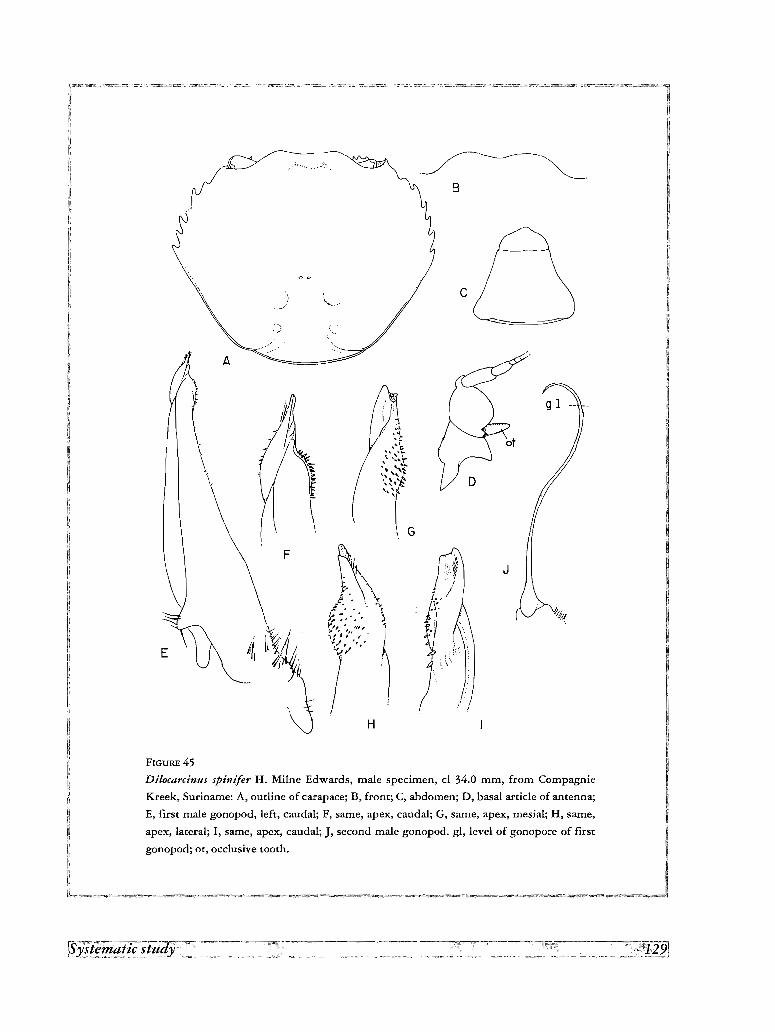
J
c
H
F
A~.. <
{~
"\\
FIGURE 45
Dilocarcinus spinifer H. Milne Edwards, male specimen, cl 34.0 mm, from Compagnie
Kreek, Suriname: A, outline of carapace; B, front; C, abdomen; D, basal article of antenna;
E, first male gonopod, left, caudal; F, same, apex, caudal; G, same, apex, mesial; H, same,
apex, lateral; I, same, apex, caudal; J, second male gonopod. gl, level of gonopore of first
gonopod; or, occlusive tooth.
Description
Carapace suborbicular; upper surface very convex, convexity more pronounced along antero-posterior axis,
regularly arched, with regions not differentiated, except for elongated depression on intestinal region; postorbital
area depressed; postfrontal lobes obsolescent, only indicated by eroded surface in this area; frontal region
slightly concave; front strongly bilobed, inclined downwards; dorsal grooves of carapace absent. Dorsal surface
of carapace smooth and polished, covered by small papillae barely visible to naked eye. Postgastric pits
lunulated, well marked. Antero-lateral margin with 6 acute spines behind external orbital angle, directed
anteriorly and slightly inwards; last spine smaller; third interdental space longest, other subequal in length;
rounded lobe behind external orbital angle; postero-lateral margin marked by well defined ridge throughout,
which begins at lateral side of last lateral spine, runs parallel to and bends inwards on postero-lateral angle of
carapace. Orbits large, subquadrate in frontal view; orbital suture absent or indicated by slight depression.
Lower orbital margin with 4 large acute spines curved inwards, followed by 4 smaller. Occlusive orbital tooth
reduced to small finger-like spine, implanted perpendicular to basal antennal article; strong ridge behind inner
orbital angle forms deep channel limited by basal antennal article; outer orbital angle with hooked spine directed
forward, similar to other on antero-lateral margin of carapace; buccal angle armed with 5 acute recurved spines.
Front retracted leaving epistome completely exposed in dorsal view; anterior margin of front not forming
distinct surface, regularly rounded-off; epistome strongly advanced, points of the mid-gutter well separated;
opening of efferent channels strongly arched, forming two well defined spouts delimited below by 2nd
maxillipeds; surface delimiting channels laterally forms rounded lobe tooth separated from, and more advanced
that buccal crest; yugal and suborbital spines visible in dorsal view; spines at each side of epistome define 2
external respiratory channels, one between yugal spines and suborbital spines, another inside orbits, limited
below by suborbital spines.
Abdominal segments 3-6 fused in both sexes; male abdomen trapezoidal, wide at base; outer margins slightly
concave; last segment with its outer margin concave, approximately 0.5 as long as broad, margin of same length
as distal margin of penultimate segment.
Basal article of antenna without outer lobe. Shallow depression along ischium of 3rd maxilliped. Chelipeds
moderately unequal in both male and female; chela of male with upper border strongly arched, lower border
slightly sinuous; fingers not gaping, each cutting edge with each larger tooth interspaced by 1 or 2 smaller;
external surface of palm with small papillae forming obscure reticulated pattern; carpus with large hooked spine
on inner margin, merus with hooked spine on distal margin, distal half of upper border and middle of latero
inferior margin, but no conspicuous row of spines on lower margin of merus. Propodus of legs with row of long
hairs on lower margin except on proximal quarter, and 2 rows on upper margin, dactylus with row on lower
margin and 2 rows on upper margin; rest of propodus and dactylus covered by felt-like pubescence formingreticulated pattern.
First gonopod slender, curved outwards, with small bulbiform apex ending in acute point directed laterad.
Second gonopod sinuous, moderately longer than first.
Material examined
Compagnie Kreek, left bank of Suriname River, near Brokopondo, Suriname; 13 April 1965; G. F. MEES;
4 males, 2 females (RNH 21247).
Type and distribution
This species was described from 2 males collected in Cayenne (French Guiana). HOLTHUIS (1959) and BaIT
(1969) give numerous records from Suriname. So far, the species is restricted to these two countries.
Fredilocarcinus PRETZMANN, 1978
Dilocarcinus (Fredilocarcinus) Pretzrnann, 1978a, p. 168.
Carapace hexagonal, constricted on posterior half, upper surface strongly arched, smooth, front bilobed,
advanced, exposing epistome in dorsal view, lower orbital margin directed downwards at inner orbital angle, 6-8
lateral teeth behind external orbital angle; abdomen triangular-rounded; merus of third maxilliped trapezoidal;
first male gonopod moderately widened at base, with irregular mesial lobe and long setae on lateral margin; apex
flattened, strongly twisted sinistrally, forming flattened projection which overreaches bulbous expansion;
gonopore opening on caudal surface of apex; apex with very few small conical spines and conspicuous row of
stiff setae on mesial side of apex; second gonopod of equal length than first,
Type speciesDilocarcinus (Fredilocarcinus) raddai Pretzmann, 1978.
DistributionThe two species of the genus have been collected only in the Ucayali River and the nearby Amazon, from
Pucallpa to Iquitos.
Key to the species of Fredilocarcinus
1. Subapical bulbous expansion of first gonopod small, simple; contour of apex awl-shaped in caudal view; mesial lobe
geniculate raddai
- Subapical bulbous expansion large 0/4 of gonopod total length), with longitudinal folds and ridges; contour of apex
subtriangular, pointed in caudal view; mesial lobe depressed musmuschiae
Fredilocarcinus raddai (Prerzmann, 1978)
Dilocarcinus (Fredilocarcinus) raddai Pretzmann, 1978a, p. 168, fig. 9.- PRETZMANN, 1983a, p. 308, pI. 1. fig. 2, pI. 2, fig. 5, pl. 3,
fig.lO, pIA, fig. 13, pI. 5, fig. 17.-PRETZMANN, 1983b, p. 319, 324.
As described and illustrated by PRETZMANN (1978a, 1983a), the first gonopod of the species is clearly related
to that of F. musmuschiae, but can be distinguish from it by the shape of the apical processes and the lateral
lobe. Both species resemble the species of Dilocarcinus in carapace morphology, however the gonopod departs
considerably from the general type found in this later genus.
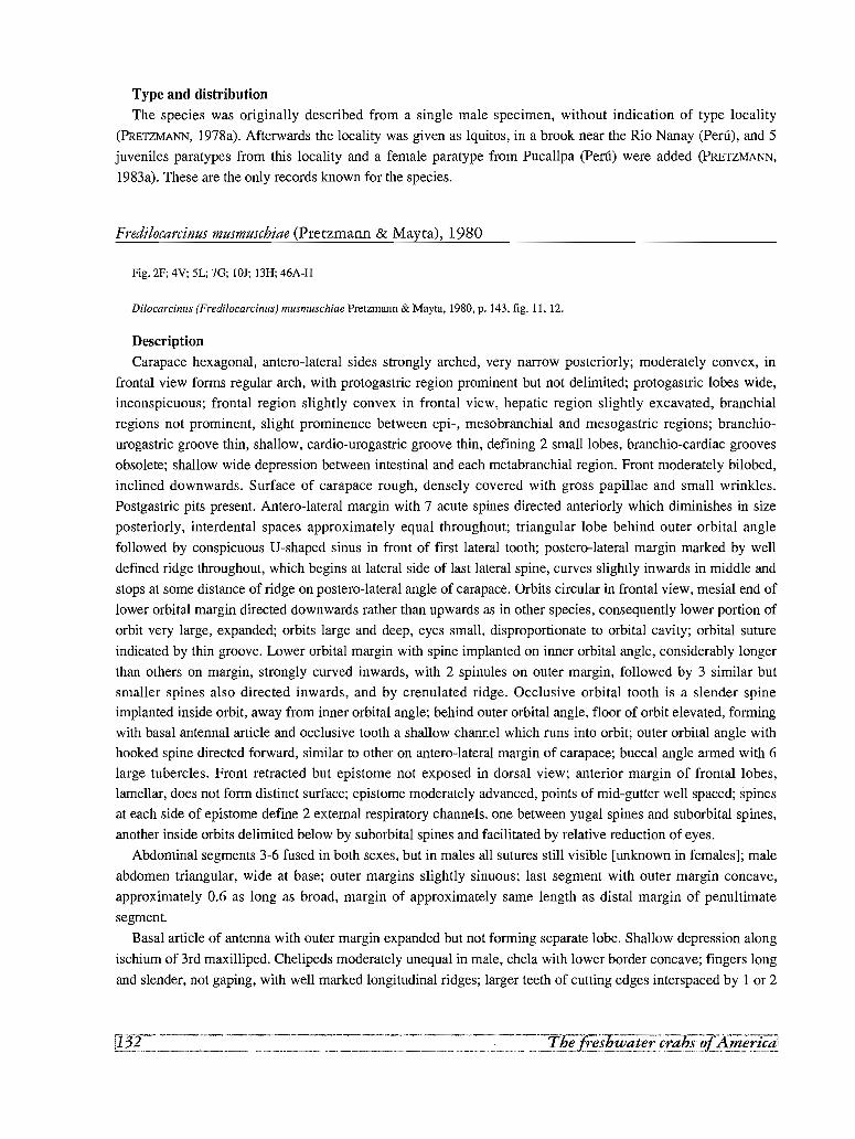
Type and distribution
The species was originally described from a single male specimen, without indication of type locality
(PRETZMANN, 1978a). Afterwards the locality was given as Iquitos, in a brook near the Rio Nanay (Peru), and 5
juveniles paratypes from this locality and a female paratype from Pucallpa (Peru) were added (PRETZMANN,
1983a). These are the only records known for the species.
Fredilocarcinus musmuschiae (Pretzmann & Mayta), 1980
Fig. 2F; 4V; 5L; 7G; IOJ; 13H; 46A-H
Dilocarcinus (Fredilocarcinus) musmuschiae Pretzmann & Mayta, 1980, p. 143, fig. 11,12.
Description
Carapace hexagonal, antero-lateral sides strongly arched, very narrow posteriorly; moderately convex, in
frontal view forms regular arch, with protogastric region prominent but not delimited; protogastric lobes wide,
inconspicuous; frontal region slightly convex in frontal view, hepatic region slightly excavated, branchial
regions not prominent, slight prominence between epi-, mesobranchial and mesogastric regions; branchio
urogastric groove thin, shallow, cardio-urogastric groove thin, defining 2 small lobes, branchio-cardiac grooves
obsolete; shallow wide depression between intestinal and each metabranchial region. Front moderately bilobed,
inclined downwards. Surface of carapace rough, densely covered with gross papillae and small wrinkles.
Postgastric pits present. Antero-lateral margin with 7 acute spines directed anteriorly which diminishes in size
posteriorly, interdental spaces approximately equal throughout; triangular lobe behind outer orbital angle
followed by conspicuous U'-shaped sinus in front of first lateral tooth; postero-lateral margin marked by well
defined ridge throughout, which begins at lateral side of last lateral spine, curves slightly inwards in middle and
stops at some distance of ridge on postero-lateral angle of carapace. Orbits circular in frontal view, mesial end of
lower orbital margin directed downwards rather than upwards as in other species, consequently lower portion of
orbit very large, expanded; orbits large and deep, eyes small, disproportionate to orbital cavity; orbital suture
indicated by thin groove. Lower orbital margin with spine implanted on inner orbital angle, considerably longer
than others on margin, strongly curved inwards, with 2 spinules on outer margin, followed by 3 similar but
smaller spines also directed inwards, and by crenulated ridge. Occlusive orbital tooth is a slender spine
implanted inside orbit, away from inner orbital angle; behind outer orbital angle, floor of orbit elevated, forming
with basal antennal article and occlusive tooth a shallow channel which runs into orbit; outer orbital angle with
hooked spine directed forward, similar to other on antero-lateral margin of carapace; buccal angle armed with 6
large tubercles. Front retracted but epistome not exposed in dorsal view; anterior margin of frontal lobes,
lamellar, does not form distinct surface; epistome moderately advanced, points of mid-gutter well spaced; spines
at each side of epistome define 2 external respiratory channels, one between yugal spines and suborbital spines,
another inside orbits delimited below by suborbital spines and facilitated by relative reduction of eyes.
Abdominal segments 3-6 fused in both sexes, but in males all sutures still visible [unknown in females]; male
abdomen triangular, wide at base; outer margins slightly sinuous; last segment with outer margin concave,
approximately 0.6 as long as broad, margin of approximately same length as distal margin of penultimate
segment.
Basal article of antenna with outer margin expanded but not forming separate lobe. Shallow depression along
ischium of 3rd maxilliped. Chelipeds moderately unequal in male, chela with lower border concave; fingers long
and slender, not gaping, with well marked longitudinal ridges; larger teeth of cutting edges interspaced by 1 or 2
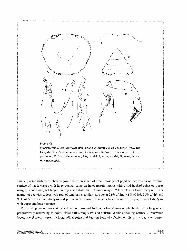
B
c
o
H :.;
("\
FIGURE 46
Fredilocarcinus musmuschiae (Pretzmann & Mayta), male specimen from Rio
Ucayali, cl 30.5 mm: A, outline of carapace; B, front; C, abdomen; D, 5th
pereiopod; E, first male gonopod, left, caudal; F, same, caudal; G, same, lateral;
H, same, mesial.
I1
.·o.~~=-~·~>: J
smaller; outer surface of chela rugose due to presence of small closely set papillae; depression on external
surface of hand; carpus with large conical spine on inner margin, merus with distal hooked spine on upper
margin; similar one, but larger, on upper and distal half of inner margin, 2 tubercles on lower margin. Lower
margin of dactylus of legs with row of long hairs; similar hairs cover 28% of 2nd, 48% of 3rd, 51% of 4th and
98% of 5th pereiopod; dactylus and propodus with rows of smaller hairs on upper margin; claws of dactylus
with upper and lower carinae.
First male gonopod moderately widened on proximal half, with lateral narrow lobe bordered by long setae,
progressively narrowing to point; distal half strongly twisted sinistrally; this spiralling defines 2 transverse
lobes, one shorter, crossed by longitudinal striae and bearing band of spinules on distal margin, other larger,
surrounding first, extending into apex, provided with lateral row of tiny spinules; apex awl-shaped, strongly
twisted mesially, with lateral row of long setae on caudal surface, ending in corneous lamella which overreaches
slit-like gonopore. First and second male gonopods of equal length.
Material examined
Peru. Ucayali, Pucallpa, Aguaitia, Rio Huacamayo, km 155, road to Basadre; 24 November 1983; H. ORTEGA
& G. CONTRERAS; 1 male (MHN Lima).
Type and distribution
The type and only specimen previously known is a male from the Rio Aguaitia, in the basin of the Rio
Ucayali. The specimen recorded above also come from the Rio Aguaitia.
Species incertae sedis
1.Trichodactylus (Dilocarcinus) gurupensis Rathbun, 1906, p. 64, pl. 18, fig. 7.
The holotype of this species was collected by Louis AGASSIZ in Gurupa, which he visited on the 22 August
1865 and again on the 31 January 1866, during his trip up the Amazon and back to Rio de Janeiro, respectively.
Mrs AGASSIZ states on the first occasion: "In the afternoon we stopped at Gurupa and went on shore; but just as
we landed a violent thunderstorm burst upon us...and we saw little of the town ...Mr Agassiz obtained a most
valuable collection of 'forest fishes'". On the second occasion she wrote: "This little town stands on a low cliff
some thirty feet above the river [Amazon]. ..Here we received some valuable specimens, collected, since our
previous visit by the subdelegado and one or two other residents" (AGASSIZ & AGASSIZ, 1868). The holotype,
deposited at the Museum of Comparative Zoology, No 4950, is a small (cl 21 mm) but adult female. RATHBUN
(1906) considered it close to Sylviocarcinus pictus, and BOTT (1969) as a synonym of S. devillei. The external
characters do not permit its allocation to either of these species, or its separation as a valid one.
2. Trichodactylus petropolitanus paranensis Bott, 1969, p. 20, pl. 2, fig 4a, b.
Described after several female types deposited in the Museum d'Histoire naturelle de Paris, "from the shores
of the Parana, Seyn, Cuare". The spinulation of the antero-Iateral margins corresponds rather with that of
Trichodactylus fluviatilis, whereas other characters, like the tuberculation of the front and the setation of the
pereiopods, point to a species different from both T. fluviatilis and T. petropolitanus. However, the absence of
males in the type material does not even allow a precise generic allocation.
3. Trichodactylus (Valdivia)faxoni Rathbun, 1906, p. 49, pl. 16, fig. 10.
Described from 1 male and 5 females collected in Tabatinga, Brazil, by the Thayer Expedition in 1865, and
originally deposited in the Museum of Comparative Zoology. According to the characters given in the original
description, this species resembles Trichodactylus ehrhardti (Bott, 1969) in the possession of five lateral teeth
on each side of carapace, the last two very small, difficult to distinguish.
III . MORPHOMETRIC RELATIONSHIPS
The available data on the maximum size attained by each species, the proportions of the carapace, the relative
position of its widest part, the relative depth of the frontal sinus, and the proportions of merus of the third
pereiopods and propodus of the fifth pereiopods, are presented in table Ill.
The measurements taken in the carapace, with the respective abbreviations used in table Ill, are as follows.
Length of carapace (d) from the frontal sinus to the articulation of the abdomen, breadth (cb) in its widest part
and including the lateral spines, the depth of the frontal sinus and the total depth of the front measured from a
tangent to the frontal lobes, to the dorsal margin of the orbits. From these measurements, the relationship
carapace breadth/carapace length (cb/cl), the percentage depth of the frontal sinus (s %) (Fig. 17E) and the
position of the widest part of the carapace (w %), were calculated. The first column of the table (cl max.)
indicates the maximum carapace length known for each species. These values have been taken from the largest
specimen recorded under "Material examined", except in those cases where the record of a larger specimen is
available in the literature. According to the information available, 39 % of the species are among the smallest
(cl < 25 mm) in the family. This group includes Trichodactylus maytai, T. kensleyi, the species of
Avotrichodactylus and Rodriguezia, and Valdivia camerani. The largest species belong in the genus
Sylviocarcinus. The absolute record is the male specimen of Sylviocarcinus devillei mentioned by SMALLEY &
RODRIGUEZ (1972) (cl 88.2 mm).
The overall mean of the ratio cb/cl for all species of Trichodactylidae is 1.15 +/- 0.06 SD, with values ranging
from 1.05 to 1.30. A clear increase in carapace breadth is found in Dilocarcinus, where the species with the
widest carapace, Dilocarcinus septemdentatus, is found. On the other hand, the species of Sylviocarcinus
possess a relatively narrow carapace (cb/d1.05-1.14).
In table III are also presented the relationship length/width in the merus of the third pair of pereiopods (m3),
and the relationship length/width in the propodous of the fifth pair (pS).

TABLE IIIMorphometric relationships
clmax cb/cl w% m3 p5 s% Nmm
Trichodactylus fluviatilis 42.8 1.16 42 4.37 2.21 18 28T. maytai 16.1*T. kensleyi 19.8 1.11 45 3.90 3.18 11 3T. petropolitanus 31.5 1.13 50 3.42 2.68 15 2T. quinquedentatus 20.4 1.16 47 4.13 1.60 21 19Mikrotrichodactylus borellianus 16.7 1.13 57 3.28 1.67 17 4M.panoplus 16.0 1.14 47 3.28 1.67 20 3Rodriguezia mensabak 15.0* 1.08*R. villalobosi 14.5* 1.17*Avotrichodactylus bidens 15.0 1.08 51 12 1A. constrictus 20.5 1.11 38 3.26 1.45 14 2A.oaxensis 21.2 1.07 44 5.18 2.00 12 7Sylviocarcinus devillei 88.2 1.14 40 3.19 1.58 31 4S. maldonadoensis 29.3 1.10 40 3.45 1.46 2 3S. pictus 39.0 1.05 37 3.39 1.38 27 7S. piriformis 78.8 1.05 39 3.39 1.38 1 37S. sp. 25.5 1.12 50 3.66 1.63 16 1Valdivia camerani 19.9 1.16 42 4.07 2.00 8 2V.gila 50.2 1.16 40 3.38 1.75 1 9V. harttii 39.8 1.18 37 6 3V. serrata 44.4 1.15 43 4.10 1.83 1 24Forsteria venezuelensis 45.4 1.11 37 3.46 1.51 1 10Zilchiopsis cryptodus 27.0*Z. emarginatus 34.2 1.20 35 3.15 1.35 30 8Z. emarginatus, juvenile 21.0 1.08 42 2.72 1.22 35 1Z. sattleri 49.0 1.Hi 48 3.32 2.00 20 6Dilocarcinus argentinianus 24.9 1.24 43 2.00 1.44 21 1D. bulbifer 22.3 1.19 42 2.29 1.49 25 2D. castelnaui 40.9 1.19 41 3.14 1.80 21 3D. dentatus 45.8 1.22 45 2.67 1.64 26D. laevifrons 27.0*D. medemi 31.0* 1.26*D. niceforei 34.8 1.28 40 3.29 1.57 45 2D.pagei 44.7 1.21 47 2.86 2.08 44 22D. septemdentatus 40.0* 1.30*D. spinifer 38.0 1.16 49 3.16 1.50 40 2D. truncatus 23.7 1.18 46 2.89 1.33 24 1Fredilocarcinus musmuschiae 30.5 1.15 40 2.75 1.20 20 1
Abbreviations for the measurements taken are given in Chapter 3.,. indicates records from the literature, N =number of specimens measured.
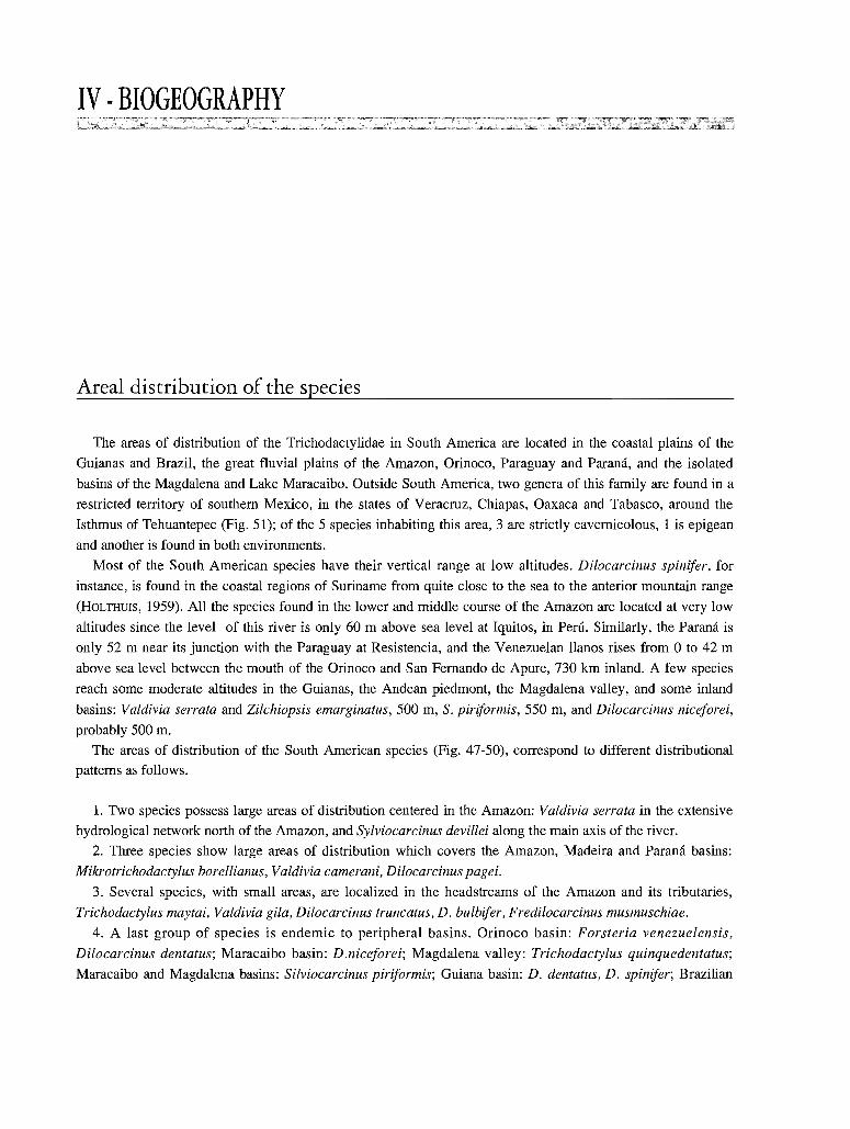
IV . BIOGEOGRAPHY
Areal distribution of the species
The areas of distribution of the Trichodactylidae in South America are located in the coastal plains of the
Guianas and Brazil, the great fluvial plains of the Amazon, Orinoco, Paraguay and Parana, and the isolated
basins of the Magdalena and Lake Maracaibo. Outside South America, two genera of this family are found in a
restricted territory of southern Mexico, in the states of Veracruz, Chiapas, Oaxaca and Tabasco, around the
Isthmus of Tehuantepec (Fig. 51); of the 5 species inhabiting this area, 3 are strictly cavernicolous, 1 is epigean
and another is found in both environments.
Most of the South American species have their vertical range at low altitudes. Dilocarcinus spinifer, for
instance, is found in the coastal regions of Suriname from quite close to the sea to the anterior mountain range
(HOLTHUIS, 1959). All the species found in the lower and middle course of the Amazon are located at very low
altitudes since the level of this river is only 60 m above sea level at Iquitos, in Peru. Similarly, the Parana is
only 52 m near its junction with the Paraguay at Resistencia, and the Venezuelan llanos rises from 0 to 42 m
above sea level between the mouth of the Orinoco and San Fernando de Apure, 730 km inland. A few species
reach some moderate altitudes in the Guianas, the Andean piedmont, the Magdalena valley, and some inland
basins: Valdivia serrata and Zilchiopsis emarginatus, 500 m, S. piriformis, 550 m, and Dilocarcinus niceforei,
probably 500 m.
The areas of distribution of the South American species (Fig. 47-50), correspond to different distributional
patterns as follows.
1. Two species possess large areas of distribution centered in the Amazon: Valdivia serrata in the extensive
hydrological network north of the Amazon, and Sylviocarcinus devillei along the main axis of the river.
2. Three species show large areas of distribution which covers the Amazon, Madeira and Parana basins:
Mikrotrichodactylus borellianus, Valdivia camerani, Dilocarcinus pagei.
3. Several species, with small areas, are localized in the headstreams of the Amazon and its tributaries,
Trichodactylus maytai, Valdivia gila, Dilocarcinus truncatus, D. bulbifer, Fredilocarcinus musmuschiae.
4. A last group of species is endemic to peripheral basins. Orinoco basin: Forsteria venezuelensis,
Dilocarcinus dentatus; Maracaibo basin: D.niceforei; Magdalena valley: Trichodactylus quinquedentatus;
Maracaibo and Magdalena basins: Silviocarcinus piriformis; Guiana basin: D. dentatus, D. spinifer; Brazilian
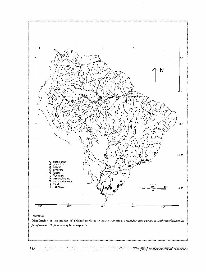
80·
o borellianus• panoplus.. parvus-e- erhardti<I> faxoni
-i- fluviatilisT petropolitanus~ quinquedentatus~ maytaiX kensleyi
70· 60· 50·
SCALEo 500~ ±oo:a --+-±z..'
Km
1000!
40·
10·
O·
10·
20·
30·
FIGURE 47
Distribution of the species of Trichodactylinae in South America. 'Tricbodactylus parvus (?=Mikrotrichodactylus
panoplus) and T. faxoni may be conspecific.
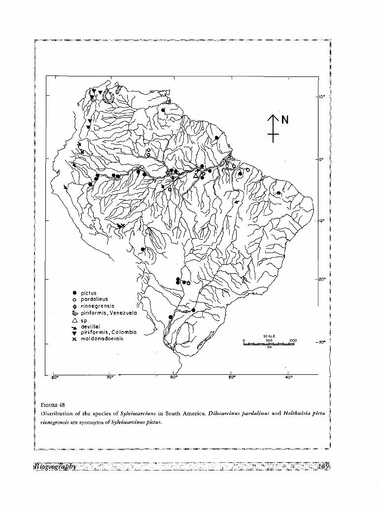
80·
• pictuso pardalinuscl> rionegrensis~... piriformis, Venezuelac: sp.~ devillei... piriformis, ColombiaX maldonadoensis
70· 60· 50·
o!
SCALE500
! !
Km
1000, !
40·
10·
o-
IQ·
,0·
30·
FIGURE 48
Distribution of the species of Sylviocarcinus in South America. Dilocarcinus pardalinus and Holthuisia picta
rionegrensis are synonyms of Sylviocarcinus pictus.
90·
X ecuadorensis.." camerani~ gila• harttiio serrata,~ venezuelensis
70· 50·
SCALEo 500
! !;! !
Km
1000!
40·
10·
O·
10·
200·
FIGURE 49
Distribution of the species of Valdiviini in South America.
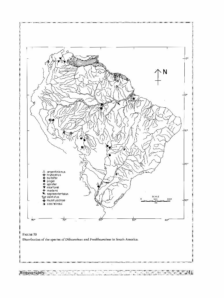
80'
£::,. argentinianus-& truncatus
et> bulbifer• pageio spiniferT niceforei+ medemi" septem dentotus~ dentatus.. musmuschioe
ca costelnaui
70· 60' 50'
o!
se ALE500
t ± !
"m1000
!
40'
10'
o-
20'
30·
FIGURE 50
Distribution of the species of Di/ocarcinus and Fredilocarcinus in South America.
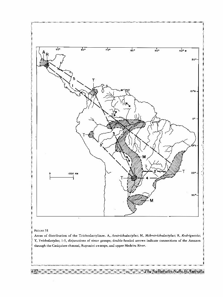
30°
T
1000 KM
1-------1fo
I
FIGURE 51
disrriburion of the Trichodactylinae. A, Avotrichodactylus; M, Mikrotriehodactylus; R, Rodriguezia;
'Tricbodactytus: 1-5, disjunctions of sister groups; double-headed arrows indicate connections of the Amazon
Casiquiare channel, Rupunini swamps, and upper Madeira River.
coastal basins: T. fluviatilis, T. petropolitanus ; Paraguay-Parana basin: T. kensleyi, Mikrotrichodactylus
panoplus, D. argentinianus.
The trichodactylid crabs are restricted by their mode of respiration to live submerged in water (DIAZ &
RODRIGUEZ, 1977), but there is evidence that, like the Pseudothelphusidae, they are able to migrate overland
during heavy rains (see Material examined of Dilocarcinus dentatus). On the other hand, as in all freshwater
crabs, migration is facilitated by the direct development, since any gravid female is able to transport a propagule
with her to a newly colonized territory. Then, aside from paleogeographic considerations, which will be dealt
with further, the differences in size and extension of the areas of distribution largely depend on the existence of
barriers for the dispersion. From this point of view, at the low elevations covered by these areas of distribution,
there are few effective geographical barriers for these crabs inside each drainage basin. In many flatland rivers,
flooding of the riverine plains during the rainy season covers the main trunk channel and tributary channels with
a continuous sheet of water. Even under dry season conditions, in various basins water drains from the land not
in distinct streams and rivers, but in interlaced channels; an example of this geomorphologic setting is the
Caronf River in Venezuela, studied by GARNER (1966). Under these conditions, the high "porosity" of the
barriers (RApOPORT, 1985) is responsible for the types of areas 1 and 2 described above, and even for the large
areas of the species endemic to peripheral basins (type 4). At the other extreme, the species of type 3 are
restricted to the upper tributaries and the headstreams of the Amazon and have not being able to extend their
areas of distribution to the middle and lower reaches of this river.
On a larger scale, the water divides between the Amazon and other major drainage basins are bridged in
certain areas by an interconnected labyrinth of waterways and flood spillways. This situation is attributed
(GARNER, 1975) to desert erosion that occurred in the tropics during Quaternary, in synchrony with the glacial
interglacial fluctuations at higher latitudes, that deposited gravels in areas now occupied by rain forest and
savannah ecosystems. The disruption involved deposition of alluvium in stream channels and valleys,
sometimes to the point of actual alluvial burial of these depressions. Resumption of humid conditions in more
recent times has led to chaotic channel networks due to alluvial clogged flow routes and to diversions plus
inland "deltas' where relict riverine deposits impede flow at tributary junctures now situated along former desert
margins. Major rivers affected in this manner in South America include the Amazon, Orinoco, and Paraguay.
Thus, the Amazon (Fig. 51) communicates with the Orinoco through the Casiquiare channel between the
Orinoco and the Rio Negro (GRABEN, 1977), with the Guianas basin through the Rio Branco (LoWE-McCoNNEL,
1964) and with the Paru-Paraguay system through the swamp areas of the upper Rio Madeira (GARNER, 1975).
The distribution of the species mentioned above for the second type of area responds to the communication
between the Amazon and Parana. In the North, the distribution of Valdivia serrata from the Amazon to thesouthern Orinoco llanos and the Guianas plateau has being achieved through the Casiquiare channel and the
Rupununi swamps.
Disjunctions
The geographical distribution, together with the cladogram of relationships presented in figure 16A, show
several disjunctions of sister groups between Mexico and South America, and between different drainage basins
in South America.
The Mexico-South America disjunctions concern (Fig. 51) (a) the three clades formed by the species
Avotrichodactylus with respect to the rest of the Trichodactylinae, and (b) Rodriguezia villalobosi (and
presumably the other 2 species of the genus) with respect to its sister species, Trichodactylus fluviatilis.
The solution of these disjunctions by means of a dispersalist hypothesis involves the postulation of a
theoretical path across Central America after the isthmian bridge was established in the Pliocene, an event dated
at 3.1 million years ago by KEIGWIN (1978) on foraminiferal biogeography and paleoceanographic evidence. The
absence of intervening species in this area (except for the very problematic record of Trichodactylus
quinquedentatus in Nicaragua (SMALLEY & RODRIGUEZ, 1972), the short geological time involved for the
evolution of these relatively complex troglobic species, and the polyphyletic origin of the two Mexican genera,
are the main objections to this hypothesis.
The establishment of a vicariant hypothesis concerns the continuity of the areas of distribution in the
respective parts of South America and Mexico to account for the disjunct distribution of these species, and the
synchronization of the paleogeographic evidence available with the branching events postulated in the
cladogram.
The only time in which a continuity of the areas can be established is in pre-Cretaceous time when South
America was part of Gondwana, together with the other two circum-Atlantic continents of Africa and North
America. In the contact zone between North and South America (PINDELL & DEWEY, 1982; PINDELL, 1985) the
Caribbean did not exist and the Yucatan block, comprising the northern part of the present Yucatan Peninsula,
completely closed the space between North and South America; the Chortis block, comprising the present
territories of Honduras and Nicaragua, formed a southern extension of Mexico and was attached to the western
side of South America. This configuration existed up to middle Jurassic time, with little tectonic activity or
relative motions between the individual constituents. The eastward migration of the Mexican block and the
rotation of Yucatan by 140 Ma, led to the juxtaposition of Yucatan and southern Mexico in the Tehuantepec
area. By about 125 Ma the continued spreading between North America and Gondwana produced an oceanic
seaway, isolating South America from North America. Thus, within a vicariant hypothesis a first radiation of the
Trichodactylidae before 125 Ma, in early Cretaceous times, should be postulated. The possible synchrony of
these events with the development of the Portunoidea, potential relatives of the Trichodactylidae, have been
recently reinforced by BISHOP'S (1991) postulation of the species of Xanthosia as the possible ancestral roots of
the Portunoidea, since the fossil record of Xanthosia goes back to early Cretaceous times (Aptian).
The South American disjunctions concern the sister species Sylviocarcinus devillei-S, piriformis, in the
Amazon and Maracaibo basins, Trichodactylus kensleyi-T. quinquedentatus, and S. devillei-S, piriformis, in the
Amazon, Maracaibo and Magdalena basins. The isolation of these basins from the adjacent Amazon and
Orinoco began in Tertiary times. According to an hypothesis (RoD, 1981), in early Eocene times the Merida
Andes has not yet emerged so that the Orinoco River was flowing to the northwest into the area of the
Maracaibo basin of today. But during the late Eocene the Merida Andes started to rise and caused the gradual
migration of the channels of the Orinoco into a depression parallel to the emerging Cordillera; further orogenic
activities forced the waters to flow towards east into the Atlantic. Thus isolation of the Maracaibo basin was
completed during early Oligocene time by low ridges building a barrier to the northwest, and consequently the
colonization by Sylviocarcinus (which nearest species S. devillei is in the Amazon) could not have occurred after
this time.
Regarding the Magdalena valley, WELLMANN (1970) has shown, by the study of paleocurrents during the
deposition of Miocene sediments, that the rivers flowed to the east in lower and middle reaches of this valley,
and consequently a direct communication with the Orinoco or the Amazon up to this time cannot be ruled out.
The allopatric events leading to the disjunct distribution of the sister pairs Trichodactylus kensleyi-
T. quinquedentatus and Sylviocarcinus devillei-S. piriformis could be dated after this time when the isolation of
the lower Magdalena valley was completed, but at least in the first case, the long distances involved point to a
much earlier date(l).
Thus, the temporal congruence of the cladogram of relationships with the paleogeographic events discussed
above requires a first radiation of the Trichodactylinae at an unknown date, and speciation events within the
South American genera, dating back to early Tertiary time.
The Trichodactylidae appear as a very old group, due to (a) the high morphological diversity, which is
reflected in the multiplicity of sister groups above species level present in the cladogram, and (b) the relatively
primitive respiratory adaptations, which constrast with the highly developed efferent channels and pseudolungs
of other freshwater crabs (DIAZ & RODRIGUEZ, 1977; RODRIGUEZ, 1986). Within the family, the Trichodactylinae
is perhaps the most ancient group. This subfamily is divided into several genera, with their areas of distribution
fragmented over a vast territory (Fig. 51), and the species separated from each other by long distances. For this
reason, and notwithstanding the presence of several apomorphic characters, they should be considered as a first
branching of the family, which evolved independently for a long period of time. This postulate is supported also
by the cladogram. Within the Trichodactylinae, the troglobious habit (HOLSINGER, 1988), isolation, reduced
areas of distribution and scarcity of the Mexican species, suggest that these are relicts of an antique fauna. The
presumed permanence of these old forms is supported by the stasis observed in some species of Portunoidea.
Carcinus maenas, for instance, is known since Eocene times (GLAESSNER, 1969).
(1) Sylviocarcinus piriformis has been recently recorded from Miocene fossiliferous outcrops of the Honda Group in the Upper MagdalenaValley; Age of these sediments is 13.0 ±0.88 Ma by fission track (RODRfGUEZ, ill press).

LITERATURE
AGASSIZ (L.) & AGASSIZ (E.), 1868. - A journey in Brazil. Ticknor and Fields. Boston
BALss (H.), 1914. - Potamoniden-Studien, Zoologische Jarbucher (Systematik), 37: 401-410, fig. 1-6, pl. 1-15.
BATE (c. S.), 1868. - Carcinological gleanings. NQ 3. Annales and Magazine ofNatural History, (4) 1: 442-448, pl. XXI.
BISHOP (G. A.), 1991. - Xanthosia occidentalis Bishop, 1985, and Xanthosia spinosa, new species, two Late Cretaceous
crabs from the Pierre Shale of the Western Interior. Journal ofCrustacean Biology, 11: 305-314.
BOIT (R.), 1955. - Die Susswasserkrabben von Afrika und ihre Stammesgeschichte. Annales du Musee du Congo beige,
(3,3),3 (1): 209-352, pl, 16-21.
BOIT (R.), 1969. - Die Stisswasserkrabben Siid-Amerikas und ihre Stammesgeschichte. Eine Revision der Trichodactylidae
und der Pseudothelphusidae ostlisch der Anden (Crustacea, Decapoda). Abhandlungen der Senckenbergischen
Naturforschenden Gesellschaft (Frankfurt am Main), 518: 1-94, fig. 1-6, pI. 1-24,4 maps.
BOIT (R), 1970. - Betrachtungen iiber die Entwicklungsgeschichte und Verbreitung der Stisswasserkrabben nach der
Sammlung des Naturhistorischen Museums in Genf/Schweiz. Revue Suisse de Zoologie, 77 (2): 327-244.
BowMANN (T. E.) & ABELE (L. G.) 1982. - Classification of the recent Crustacea. In: Bliss, D. E., The Biology of Crustacea,
I: 1-27.
CAMPOS (M. R.), 1985. - Decapodos de agua duke del Suborden Brachyura reportados para Colombia. Caldasia (Bogota),
14 (67): 265-284.
CHACE (F. A.) & HOBBs (H. H.) Jr., 1969. - The freshwater and terrestrial decapod crustaceans of the West Indies, with
special reference to Dominica. Bulletin of the U.S. National Museum, 292: 1-258, fig. 1-76.'
COIFMAN (I.) 1939. - Potamonidi della Guiana Inglesi. Archivo Zoologico Italiano, 27: 93-116.
COLaSI (G.), 1920. - I Potamonidi del R. Museo Zoologico di Torino. Bolletino Museo Zoologico Anatomico di Torino, 35
(734): 1-39.
COITARELLI (V.) & ARGANO (R.) 1977. - Trichodactylus (Rodriguezia) mensabak n. sp. (Crustacea, Decapoda, Brachyura),
granchio cieco delle acque sotterranee del Chiapas (Messico). Guaderni Problemi Attuali di Scienza e di Cultura,
Accademia Nazionali dei Lincei, 171 (3): 207-212.
COWAN (c. F.) 1976. - On the disciples' edition of Cuvier's Regne Animal. Journal of the Society for the Bibliography of
natural History, 6: 32-64.
CUNNINGHAM (R. 0.), 1871. - Notes on the Reptiles, Amphibia, Fishes, Mollusca, and Crustacea obtained during the voyage
of H. M. S."Nassau" in the years 1866-69, 1871. Transactions of the Linnean Society ofLondon, 27: 465-502. pl. 58, 59.
DANA (J. D.) 1851. - On the classification of the Cancroidea. American Journal ofScience, 12 (2): 121-131.
DANA (J. D.), 1852a. - On the genus Orthostoma. American Journal ofScience, 13 (2): 123-124.
DANA (J. D.), 1852b. - Crustacea. United States Exploring Expedition during the years 1838, 1839, 1840, 1841, 1842, under
the command of Charles Wilkes, U. S. N., 13: 1-1620.
DEL SOLAR (E. M.), BLANCAS (p.) & MAYfA (R.) 1970. - Catalogo de 10sCrustaceos del Peril. Imprenta D. Miranda, Lima, 54 p.
DIAZ (H.) & RODRfoUEZ (G.), 1977. - The branchial chamber in terrestrial crabs: a comparative study. Biological Bulletin,
153: 485-504.
DOFLEIN (F.), 1899. - Amerikanische Dekapoden der k. bayerischen Staatssamm1ungen. Sitzungberichte dermathematisch
physische Klasse del' bayerischen Akademie del' Wissenschaften, Mtinchen, 29: 177-195.
EYDOUX (p.) & SOULEYET (L. F. A.), 1841 (1842). - Crustaces. Voyage autour du Monde execute pendant 1es annees 1836 et
1837 sur la corvette la Bonite, commandee par M. Vaillant, Capitaine de Vaisseau. Histoire naturelle, Zoologie, 1: 219
272, pl, 5.
FELSENSTEIN (J.), 1984. - The statistical approach to inferring evolutionary trees and what it tells us about parsimony and
compatibility. In: Duncan, T. and T. F. Atussy, eds, C1adistics: Perspectives in the reconstruction of evolutionary history.
Columbia University Press, New York: 169-191.
GARCIA (J. E.), 1973. - Trichodactylus (Trichodactylus) panoplus (von Martens 1869) (Decapoda Brachyura) nueva especie
de la fauna Uruguaya. Trabajos del Quinto Congreso Latinoamericano de Zoologia (Montevideo), 1: 97-103, fig. 1-5.
GARNER (H. F.), 1966. - Derangement of the Rio Caroni, Venezuela. Revue de Geomorphologie dynamique, 2: 50-83.
GARNER (H. F.), 1975. - Rain forests, deserts and evolution. Annales de la Academia brasileira de Ciencias, 47 (Supl.):
127-133.
GERSTACKER (A.), 1856. - Carcinologische Beitrage. Archiv fur Naturgesellschaft, 22 (1): 101-162, pI. 4-6.
GOLD! (E. A.), 1885. - Studien iiber neue und wenig bekannte Podophtha1men Brasiliens (Vorlaufige Notiz). Zoologische
Anzeiger, 8: 662-663.
GOLD!, (E. A.), 1886. - Studien Iiber neue und weniger bekannte Podophtha1men Brasiliens. Archiv fur Naturgesellschaft, 52
(1): 19-46, pl. IT-Ill.
GRABERT (H.), 1977. - Orinoco und Amazonas. Eine Betrachtung zum Alter beider Stromsysteme. Sonderveroffentlichungen
des Geologischen Institutes Universitiit Koln, 33: 179-191.
GRONOVIUS (L. T.), 1764. - Zoophy1acii Gronoviani Fasciculus secundus exhibens Enumerationem Insectorum quae in
Museo suo adservat, examini subjecit, systematice disposuit atque descripsit. Cancer, 957, p. 222.
GUINOT (D.), 1977. - Propositions pour une nouvelle classification des Crustaces Decapodes Brachyoures. Comptes Rendus
de l'Academic des Sciences, Paris, D, 285: 1049-1052.
GUINOT (D.), 1978. - Principes d'une classification evolutive des Crustaces Decapodes Brachyoures. Bulletin Biologique de
la France et de la Belgique, 112: 211-292
HELLER (C.), 1865. - Crustaceen. In: Reise der Osterreichischen Fregatte Novara urn die Erde in den Jahren 1857, 1858,
1859 unter der Befeh1en des Commodore B. von Wtillerstorf-Urbair. Teil 2, Abt. 3: 1-280, p1.1-25.
HERBST (J. F. W.), 1782-1790. - Versuch einer Naturgeschichte der Krabben und Krebse, nebst einer Systematischen
Beschreibung ihrer verschiedenen Arten, 1: 1-274, textfig. A, pI. 1-21.
HERKLOTS (L A.), 1861. - Symbo1ae Carcinologicae, 1. Catalogue des Crustaces qui ont servi de base au systeme
carcinologique de M. W. de Haan, redige d'apres la collection du Musee des Pays-Bas et 1es Crustaces de la Faune du
Japon. Tijdschrift voor Entomologie, 4: 116-156.
HOLSINOER (J. R.), 1988. - Troglobites: The Evolution of cave-dwelling organisms. American Scientist, 76: 147-153.
HOLTHUIS (L. B.), 1959. - The Crustacea Decapoda of Suriname (Dutch Guiana). Zoologische Verhandelingen (Leiden), 44:
1-296, fig. 1-68, pI. 1-16, 1 map.
KEIOWIN (L. D.), 1978. - Pliocene closing of the Isthmus of Panama, based on biostratigraphic evidence from nearby Pacific
Ocean and Caribbean Sea cores. Geology, 6: 630-634.
KINGSLEY (J. S.), 1880. - Carcinological Notes. 1. Proceedings of the Academy of Natural Sciences of Philadelphia, 1880:
34-37.
LATREILLE (P. A), 1825. - Familles naturelles du regne animal, exposees succinctement et dans un ordre analytique avec
l' indication de leur genre. Paris, [vi] + 570 p.
LATREILLE (P. A), 1828. - Telphuse et Trichodactyle. In: Encyclopedie methodique, Histoire naturelle, Entomologie, 10:
561-564,705.
LOPRETIO (E. C.), 1976. - Morfologia comparada de los ple6podos sexuales masculinos de los Trichodactylidae de la
Argentina (Decapoda, Brachyura). Limnobios (Buenos Aires), 1: 67-94, fig. 1-30.
LOPRETIO (E. C.), 1981. - Discusi6n sobre las presuntas subespecies de Dilocarcinus (D). pagei (Crustacea Brachyura
Trichodactylidae). Redescripci6n y referencia a su polifenismo. Physis (Buenos Aires), B 39 (97): 21-31.
LOWE-McCONNELL (R. H.), 1964. - The fishes of the Rupununi Savanna district of British Guiana. Journal of the Linnean
Society (Zoology), 45: 103-144.
LUCAS (H.), 1857. - Crustaces. In: Animaux nouveaux ou rares recueillis pendant I'expedition dans les parties centrales de
l'Amerique du Sud, de Rio de Janeiro aLima, et de Lima au Para, executee par ordre du Gouvernement francais pendant
les annees 1843 a1847, sous la direction du comte Francois de Castelnau. Paris, (7) 3: 1-13, pl. 1,2.
MARTENS (E. von.), 1869a. - Siidbrasilische Suss-und Brackwasser-Crustaceen nach Sammlungen des Dr Reinh. Hensel.
Archiv far Naturgesellschaft, 35 (1): 1-37, pI. 1-2.
MARTENS (E. von.), 1869b. - Crustacea, In: Zoological Record, 1868: 515.
MANNING (R. G.) & HOBBS (H. H.) Jr., 1977. - Decapoda. In: Hurlbert D. H. (ed.), Biota acuatica de Sudamerica austral. San
Diego State University, San Diego, California: 157-162.
MEUSCHEN (F. C.), 1781. - Index continens Nomina Generica Specierum propia, Trivalia ut et Synonyma. In: Gronovius
L. T., Zoophylacium Gronovianum, exhibens Animalia Quadrupeda, Amphibia, Pisces, Insecta, Vermes, Mollusca,
Testacea, et Zoophyta, quae in Museu suo adservavit, examini subjecit, systematice disposuit atque descripsit, 19 p.
MILNE EOWAROS (A), 1869. - Revision des Genres Trichodactylus, Sylviocarcinus et Dilocarcinus et description de quelques
especes nouvelles qui s'y rattachent. Annales de la Societe entomologique de France, (4) 9: 170-178.
MILNE EOWARDS (H.), 1837. - Histoire Naturelle des Crustaces; comprenant l'anatomie, la physiologie et la classification de
ces animaux, 2: 1-532, atlas pl. 1-42.
MILNE EOWAROS (H.), 1836-1844. - Les Crustaces. In: Cuvier G., Le regne animal, distribue d'apres son organisation, pour
servir de base a l'histoire naturelle des animaux, et d'introduction a l'anatomie comparee, 278 p., Atlas pl. 1-80.
Paris.[According to Cowan (1976), p. 60 and pl. 15 on which Trichodactylus quadrata is recorded were published in
1839 and 1842, respectively]
MILNE EOWAROS (H.), 1853. - Observations sur les affinites zoologiques et la classification naturelle des crustaces, Annales
de Sciences Naturelles, Zoologie, 20 (3): 163-228, pl. 6-11.
MILNE EOWAROS (H.), 1854. - Notes sur que1ques Crustaces nouveaux ou peu connus conserves dans la collection du
Museum d'Histoire naturelle. Archives du Museum national d' histoire naturelle, 7: 145-192, pl, 9-16.
MOREIRA (C.), 1901. - Contribuicoes para 0 conhecimento da Fauna Brazileira. Crustaceos Thoracostraceos. Archivos do
Museu Nacional de Rio de Janeiro, 11: i-iv, 1-151, pl. 1-5.
MOREIRA (C.), 1903. - Crustaceos da Ponta do Pharo1 em S. Francisco do SuI no Estado de Santa Catharina. Archivos do
Museu Nacional de Rio de Janeiro, 12: 119-123.
MORElRA (C.), 1912. - Crustaces du Bresil. Memoires de la Societe Zoologique de France, 25: 145-155.
MOREIRA (C.), 1913. - Crustaceos. In: Commissao de Linhas Telegraficas Estrategicas de Mato Grosso ao Amazonas. Anexo
5, Historia Natural, Zoologia: 1-21, fig. 1-2, pl. 1-7.
MULLER (F.), 1880. - Palaemon Potiuna. Ein Beispiel abgekiirzter Verwandlung. Zoologischer Anzeiger, 3: 152-157.
MULLER (p.), 1887. - Zur Crustaceenfauna von Trincomali. Verhandlung Naturforschenden Gesellschaft, Basel, 8: 470-485.
MULLER (F.), 1892. - Trichodactylus, siri de agua doce, sem metamorphose. Archivo do Museu Nacional de Rio de Janeiro,
8: 125-133, pl. 5-6.
NICOLET (H.), 1849. - Crustaceos, In: Gay, c.,Historia Ffsica y Polftica de Chile, Zoologia, 3: 115-318.
NOBILI (G.), 1896. - Viaggio del Dott. A. Borelli nella Republica Argentina e nel Paraguay, 19. Crostacei Decapodi.
Bolletino Musei di Zoologia ed Anatomia Comparata della R. Universita di Torino, 11 (222): 1-4.
NOBILI (G.), 1898. - Sopra alcuni decapodi terrestre e de acqua dolce dell'America meridionale. Annales dei Musei Civico et
di Storia Naturali di Genova, 19 (2): 9-14.
NOBILI (G.), 1899a. - Intorno ad alcuni Crostacei Decapodi del Brasile. Bolletino Musei di Zoologia ed Anatomia Comparata
della R. Universita di Torino, 14 (355): 1-6.
NOBILI (G.), 1899b. - Observazioni sui Trichodactylus quinquedentatus, Rathb. Bolletino dei Musei di Zoologia ed Anatomia
Comparata della R. Universita di Torino, 14 (365): 1-3.
NOBILI (G.), 1901. - Decapodi raccolti dal Dr. Filippo Silvestri nell'America meridionale. Bolletino dei Musei di Zoologia ed
Anatomia Comparata della R. Universita di Torino, 16 (402): 1-16.
ORTMANN (A. E.), 1893. - Die-Decapoden-Krebse des Strassburger Museum, mit besonderer Berticksichtigung der von
Herrn Dr Doderlein bei Japan und bei den Liu-Kiu-Inseln gesammelten und zur Zeit im Strassburger Museum
aufbewahrten Formen. 7. Theil. Zoologische Jahrberichter (Systematische), 7: 411-495, pl. 1-17.
ORTMANN (A. E.), 1896. - Das System der Dekapodeen-Krebse. Zoologische Jahrberichter (Systematische), 9: 409-453.
ORTMANN (A. E.), 1897. - Carcinologische Studien. Zoologische Jahrberichter (Systematische), 10: 258-372, pI. XVII.
ORTMANN (A. E.), 1903. - The geographical distribution of freshwater decapods and its bearing upon ancient geography.
Proceedings of the American Philosophical Society, 41: 267-400.
PAPAVERO (N.), 1971-1972. - Essays on the history of Neotropical Dipterology with special reference to collectors. 1 (1971):
i-vii, 1-216; 2 (1972): i-iii, 217-446.
PARISI (R), 1923. - Un nuovo Potamonide Americano. Annali di Museo Civico di Storia Naturale (Genova), 51: 29-30,
fig. 1-2.
PEARSE (A. S.), 1911. - Report on the crustacea collected by the University of Michigan-Walker expedition in the state of
Vera Cruz, Mexico. Reports of the Michigan Academy ofSciences, 13: 108-113.
PEARSE (A. S.), 1915. - An account of the crustacea collected by the Walker Expedition to Santa Marta, Colombia.
Proceedings of the U. S. National Museum, 49: 531-556, pl. 70-73.
PINDELL (J.), 1985. - Alleghenian reconstruction and subsequent evolution of the Gulf of Mexico, Bahamas, and Proto
Caribbean. Tectonics, 4: 1-39.
P!NDELL (J.) & (J. F.) DEWEY, 1982. - Permo-Triassic reconstruction of Western Pangea and the evolution of the Gulf of
Mexico/Caribbean region. Tectonics, 1: 179-212.
PRAHL (H. von), 1982. - Notas sobre Sylviocarcinus piriformis (Pretzmann, 1968) (Crustacea: Brachyura: Trichodactylidae)
con enfasis en su zoogeografia. Actualidades biologicas, 10: 22-25.
PRAHL (H. von), 1988. - Cangrejos de agua dulce (Crustacea, Brachyura, Pseudothelphusidae y Trichodactylidae) capturados
en el Departamento de Antioquia, Colombia. Boletin Ecotropica: Ecologia Tropical, 18: 3-17.
PRETZMANN (G.), 1968a. - Weitere neue stidamerikanische Siisswasserkrabben. Entomologische Nachrichtenblatt (Vienna),
15 (2): 1-6.
PRETZMANN (G.), 1968b. - Die Familie Trichodactylidae (Milne Edwards 1853) Smith (vorlaufige Mitteilung).
Entomologische Nachrichtenblatt (Vienna), 15 (7-8): 70-76.
PRETZMANN (G.), 1977. - Notizen zur Biologie der Susswasserkraben. Sitzungberichten der mathematisch
naturwissenschaftlichen Klasse der Osterreichischen Akademie der Wissenschaften (Vienna), 1, 186 (7): 87-89.
PRETZMANN (G.), 1978a. - Neue Siisswasserkrabben aus den Anden. Sitzungberichten der mathematisch
naturwissenschaftlichen Klasse der Osterreichischen Akademie der Wissenschaften (Vienna), 1, 187 (6): 163-170, fig. 1-13.
PRETZMANN (G.), 1978b. - Neue Potamocarcini, Poglayen-Neuwald leg. 1975 (vorlaufige Mitteilung). Sitzungberichten der
mathematisch-naturwissenschaftlichen Klasse der Osterreichischen Akademie der Wissenschaften (Vienna), 1, 187 (2):
51-54.
PRETZMANN (G.), 1979. - Poppiana bachmayeri novo comb. Annalen des Naturhistorichen Museums in Wien, 82: 591-593,
pl. 1-3.
PRETZMANN (G.), 1980. - Von Dr Ivo Poglayen-Neuwadl 1975 in Mittelamerika gesammelte Krabben. Annalen des
Naturhistorichen Museums in Wien, 83: 651-666, pl, 1-18.
PRETZMANN (G.), 1983a. - Ergebnisse einiger Sammelreisen in Peru und Ecuador 1976/77. Annalen des Naturhistorichen
Museums in Wien, 84/B: 307-311, pI. 1-66.
PRETZMANN (G.), 1983b. - Die Trichodactylidae von Peru und Ecuador. Annalen des Naturhistorichen Museums in Wien,
84/B: 317-330, pI. 1-14.
PRETZMANN (G.) & MAYTA (R.), 1980. - Uber einige Siisswasserkrabben aus Peru. Sitzungberichten der mathematisch
naturwissen-schaftlichen Klasse der Osterreichischen Akademie der Wissen-schaften (Vienna), 1, 189 (9): 137-144,
fig. 1-14.
RANDALL (J. W.), 1839 (1840). - Catalogue of the Crustacea brought by Thomas Nuttall and J. K. Townsend, from the West
Coast of North America and the Sandwich Islands, with descriptions of such species as are apparently new, among which
are included several species of different localities, previously existing in the collection of the Academy. Journal of the
Academy ofNatural Sciences ofPhiladelphia, 8: 106-147, pl. 3-7.
RAPOPORT (E.), 1985. - Areology: Geographical strategies of the species. Pergamon Press, 250 p.
RATHBUN (M. J.), 1893. - Descriptions of new species of American Fresh-Water Crabs. Proceedings of the U.S. National
Museum, 16: 649-661, pI. 73-77.
RATHBUN (M. J.), 1906. - Les crabes d'eau douce (Potamonidae). Nouvelles archives du Museum national d' Histoire
naturelle, (4) 8: 33-122.
RINGUELET (R.), 1949. - Camarones y cangrejos de la zona de Goya (Sergestidae, Palemonidae y Trichodactylinae). Notas
del Museo de la Plata, Zoologia, 24 (119): 79-109, pl. I-X.
ROD (E.), 1981. - Notes on the shifting course of the Ancient Rio Orinoco from Late Cretaceous to Oligocene time. Geos,
26: 54-56.
RODRfGUEZ (G.), 1980. - Los crustaceos decapodos de Venezuela. Instituto Venezolano de Investigaciones Cientfficas,
Caracas. 450 p.
RODRfGUEZ (G.), 1981. - Decapoda. In: Hurlbert, S. H., G. Rodrfguez & N. D. Dos Santos, eds. Aquatic Biota of Tropical
South America, part 1: Arthropoda. San Diego State University, San Diego, California: 41-50.
RODRlGUEZ(G.), 1982. - Les crabes d'eau douce d'Amerique. Famille des Pseudothelphusidae. Faune trop. XXII, Orstom,
Paris, 224 p.
RODRfGUEZ (G.), 1986. - Centers of radiation of freshwater crabs in the Neotropics. In: Gore, R. H. and K. L. Heck, eds.,
Crustacean Biogeography. A. A. Balkema, Rotterdam: 51-67.
RODRfGUEZ (G.), in press. - Trichodactylid crabs from the Miocene Honda Group of Colombia. In: R. Kay, R. Madden, R.
Cifelli and 1. Flyn, eds. A History of Neotropical Fauna: Vertebrate Paleobiology of the Miocene of Tropical South America.
RODRfGUEZ (G.), & CAMPOS (M.), 1989. - Cladistic relationships of the freshwater crabs of the tribe Strengerianini
(Crustacea, Decapoda, Pseudothelphusidae) from the northern Andes, with comments on their biogeography and
descriptions of new species. Journal of Crustacean Biology, 9: 141-156.
RODRlGUEZ (G.), & HOBBS (H. H.) Jr., 1989. - Freshwater crabs associated with caves in southern Mexico and Belize, with
descriptions of three new species (Crustacea: Decapoda). Proceedings of the Biological Society ofWashington, 102: 394-400.
RODRlGUEZ (G.), & MANRlQUE (F.), 1967. - El genero Trichodactylus en Mexico (Brachyura, Potamonidae). Anales del
Instituto de Biologia (Mexico), 37: 183-186, fig. 1, pI. 1.
SCHMITI (W. L.), 1969. - Colombian freshwater crabs notes. Proceedings of the biological Society of Washington, 82: 93
112, fig. 1-7.
SCHMITT (W. L.) & PRETZMANN (G.), 1968. - Eine neue Trichodactylus Art aus Kolumbien. Vorlaufige Mitteilung.
Entomologische Nachrichtenblatt (Vienna), 15 (2): 6.
SMALLEY (W. L.) & RODRIGUEZ (G.), 1972. - Trichodactylidae from Venezuela, Colombia and Ecuador (Crustacea,
Brachyura). Tulane Studies in Zoology and Botany, 17: 41-55, fig. 1-23.
SMITH (S. I.), 1869. - Notice of the crustacea collected by Prof. C. F. Hartt on the coast of Brazil, in 1867, together with a list
of the described species of Brazilian Podophthalmia. Transactions of the Connecticut Academy ofArts and Sciences, 2:
1-41, pl. 1.
SMITH (S. I.), 1870. - Notes on American Crustacea. 1. Ocypodoidea. Transactions of the Connecticut Academy of Arts and
Sciences, 2: 113-176, pl. 2-5.
STEPHENSON (W.) & CAMPBELL (B.), 1960. - The Australian Portunids (Crustacea: Portunidae). IV. Remaining genera.
Australian Journal of Marine and Freshwater Research, 2: 73-122, fig. 1-3, pl. 1-6.
STlMPSON (W.), 1861. - Notes on certain Decapod Crustacea. Proceedings of the Academy of Natural Sciences of
Philadelphia, 13: 372-373.
THALLWITZ (J.), 1891. - Decapoden-Studien, insbesondere basirt auf A. B. Meyer's Samm1ungen im Ostindischen Archipel,
nebst einer Aufzahlung der Decapoden und Stomatopoden des Dresdener Museums. Abhandlungen des Koniglichen
Zoologischen Museum, Dresden, 1890-91,3: 1-55, pl. 1.
WELLMAN (S. S.), 1970. - Stratigraphy and petrology of the nonmarine Honda Group (Miocene), Upper Magdalena Valley,
Colombia. Geological Society ofAmerica Bulletin, 81: 2353-2374.
WHITE (A.), 1847a. - List of the specimens of Crustacea in the collection of the British Museum. Printed by order of the
trustees, Edward Newman, London, i-viii, 1-143.
WHITE (A.), 1847b. - Short descriptions of some new species of Crustacea in the Collection of the British Museum. Annals
and Magazine ofNatural History, 20: 205-207. (Also in Proceedings of the Zoological Society ofLondon, 15: 84-86).
YOUNG (c. G.), 1900. - The Stalk-eyed Crustacea of British Guiana, West Indies and Bermuda. John M. Watkins, London: i
xix, 1-514,7 pl.
ZARIQUIEY ALVAREZ (R.), 1968. - Crustaceos decapodos ibericos.lnvestigaci6n Pesquera, 32: i-xv, 1-510.
ZIMMER (G.), 1912. - Beitrage zur Kentniss der Susswasserdekapoden Kolumbiens.ln: Ftihrmann, O. and E. Mayor, Voyage
d'exploration scientifique en Colombie. Memoires de la Societe neuchateloise des Sciences nature lies, 5: 1-8, fig. 1-15,
pl. 1.
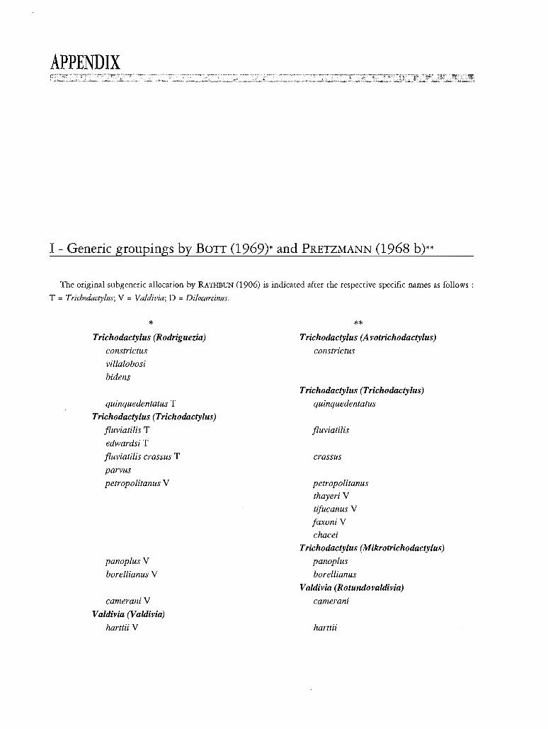
APPENDIX
I - Generic groupings by BOTT (1969)* and PRETZMANN (1968 b)**
The original subgeneric allocation by RATHBUN (1906) is indicated after the respective specific names as follows:
T = Trichodactylus; V = Valdivia; D = Dilocarcinus.
*Trichodactylus (Rodriguezia)
constrictus
villalobosi
bidens
quinquedentatus T
Trichodactylus (Trichodactylus)
fluviatilis T
edwardsi T
fluviatilis crassus T
parvus
petropolitanus V
panoplus V
borellianus V
cameraniV
Valdivia (Valdivia)
harttii V
**Trichodactylus (A votrichodactylus)
constrictus
Trichodactylus (Trichodactylus)
quinquedentatus
fluviatilis
crassus
petropolitanus
thayeri V
tifucanus V
faxoniV
chacei
Trichodactylus (Mikrotrichodactylus)
panoplus
borellianus
Valdivia (Rotundovaldivia)
camerani
harttii
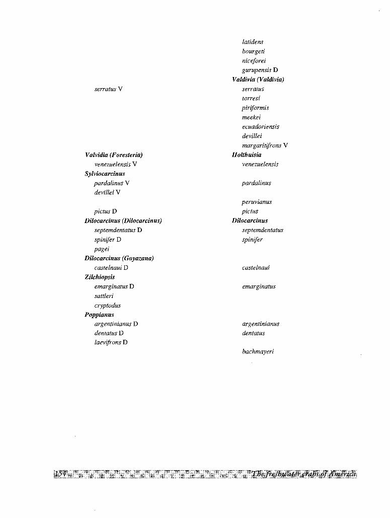
serratus V
Valvidia (Foresteria)
venezuelensis V
Sylviocarcinus
pardalinus V
devillei V
pictus D
Dilocarcinus (Dilocarcinus)
septemdentatus D
spinifer D
pagei
Dilocarcinus (Goyazana)
castelnaui D
Zilchiopsis
emarginatus D
sattleri
cryptodus
Poppianusargentinianus D
dentatus D
laevifrons D
latidens
bourgeti
niceforei
gurupensis D
Valdivia (Valdivia)
serratus
torresi
piriformis
meekei
ecuadoriensis
devillei
margaritifrons V
Holthuisia
venezuelensis
pardalinus
peruvianus
pictus
Dilocarcinus
septemdentatus
spinifer
castelnaui
emarginatus
argentinianus
dentatus
bachmayeri
II - A gazetteer of collection localities
Under every heading are given the localities name, the country, the political division (province, state, etc.), a
short description and the approximate coordinates (degrees, minutes) of the localities. The number given in
parentheses at the end refers to the order assigned to each species in the list presented in the introduction.
The political division of each country are named as follows:
Mexico, Venezuela, Brazil: states
Bolivia, Colombia, Paraguay, Peru, Uruguay: departments
Argentina, Ecuador: provinces
Suriname, Guyana: districts
- Aguaitia, Peru, Loreto, see San Juan (41).
- Aguajal, Peru, Madre de Dios, locality on Rio Manu, approximately l2Q N - 71Q W (31).
- Agua Negra, Venezuela, Yaracuy, small village in Rio Yaracuy basin, lOQ 25' N - 68Q 30' W (33).
- Aguas Calientes, Venezuela, Zulia, unidentified locality in Lago Maracaibo basin (36).
- Alto Cafio Rueda, see Atures (23).
- Amazonas, Brazil, name of a state.
- Amazonas, Colombia, name of a department.
- Amambay, Paraguay, name of a department.
- Ansina, Uruguay, Tucuarembo, not identified (8).
- Aparicion, Venezuela, Portuguesa, town on Rio Aro, 9Q 30' N - 69Q 20' W (33).
- Apartaderos, Venezuela, Cojedes, town, 9Q 40' N - 68Q 55' W (33)
- Apure, Venezuela, name of a state.
- Aracruz, Brazil, Espirito Santo, city on Atlantic, 50 km NNE ofVitoria (1).
- Aragua, Venezuela, name of a state.
- Aregua, Paraguay Central, town on Lago Ypacarai 25Q 18' S - 57Q 22' W (37).
- Arroyo Caanabe, Paraguay, Paraguari, stream near town of Paraguari, 25Q 45' S - 57Q 11' W (7).
- Arroyo del Pital, Mexico, Veracruz, creek near Ciudad Aleman, l8 Q 10' N - 96Q 08' W (11).
- Arroyo del Solfo, Mexico, Tabasco, same as, or an extension of, Cueva del Azufre (10).
- Arroyo Gaguaresau, Paraguay, San Pedro, stream near town of Caacupe, 25Q 22' S - 57Q 15' W (7).
- Arroyo Pindo, Paraguay, stream on San Pedro-Caaguazti Departments border, 24Q 52' N - 56Q 19' W (7).- Arroyo Postilion, see Puerto Max (7).
- Arroyo Salsipuedes Grande, Uruguay, Rio Negro, stream joining left side of Rio Uruguay, approximately
32Q 30' S - 58Q W (8).
- Arroyo San Juan, Beni, Bolivia, stream in the vicinity of the city of Trinidad (37).
- Arroyo Tobatiry, Paraguay, Caaguazii, stream near town of Caaguazri, 25Q 16' S - 56Q 24' W (7, 37).
- Arroyo Yaguaron, Uruguay, Tucuarembo, small river joining Rio Negro through mouth of Rio Tucuarembo
at 32Q 25' 30' S - 55Q 50' W (8).
- Asuncion, Paraguay, Central, capital city on Rio Paraguay, 25Q 16' S - 57Q 41' W (7).
- Atlantico, Colombia, name of a department.
- Atures, Venezuela, Amazonas Federal Territory, rapids of Rio Orinoco, 22 km S of Puerto Ayacucho (23).
- Azuay, Ecuador, name of a province.
- Bahia, see Salvador (1).
- Barao de Vassouras, see Vassouras (4).
- Barranquilla, Colombia, Atlantico, port on Caribbean Sea, on left bank of Rio Magdalena, lOQ 58' N - 74Q
46' W (5).
- Basadre, see Pucallpa (41).
- Bayunca, Colombia, Bolivar, town 20 km NE of Cartagena (33).
- Belen, Paraguay, Concepci6n, town 18 km ESE of Concepci6n (16).
- Bella Vista, Paraguay, Amambay, at Paraguay-Brazil border, 22Q lO' S - 36Q 25' W (16).
- Beneficiente, Brazil, Amazonas, town on Rio Aripuana (16).
- Beni, Bolivia, name of a department.
- Berbice, Guyana, a county bounded by Suriname (E) and Brazil (S), drained by Berbice and Corantijn rivers
(33).
- Bluefields, Nicaragua, city near Caribbean Sea, at mouth of Rio Escondido, 12Q 01' N - 83Q 46' W (5).
- Bobures, Venezuela, Zulia, town on SE shore of Lago Maracaibo, 9Q 13' N -71Q lO' W (36).
- Boquer6n, Paraguay, name of a department.
- Bolivar, Colombia, name of a department.
- Bolivar, Venezuela, name of a state.
- Brokopondo, Suriname, town on Suriname River, 5Q 01' N - 56Q 02' W (39).
- Buena Vista, see Rio Buena Vista (17).
- Buenos Aires, Argentina (probably Buenos Aires province) (7, 16,29).
- Caacupe, locality of Paraguay, see Arroyo Gaguaresau (7).
- Caaguazii, Paraguay, name of a department
- Caaguazti, see Arroyo Tobatiri (7)
- Cacaual Grande, unidentified locality in Brazil (37).
- Cachoeira, Brazil, Amazonas, unidentified locality on Rio Marauia (16).
- Cachoeira San Antonio, Brazil, Amazonas, unidentified locality on Rio Marauia (23).
- Caicara, Venezuela, Bolivar, city on right bank of Rio Orinoco 7Q 40' N - 66Q lO' W (24).
- Caicara, Venezuela, Monagas, town on Rio Guarapiche, 9Q 45' N - 63Q 40' W (24,33).
- Cajamarca, Peru, name of a department.
- Calabozo, Venezuela, Guarico, a city on Rio Guarico, 8Q 56' N - 67Q 26' (33).
- Candelaria, Argentina, Misiones, small town on left bank of Rio Parana, 27Q 30' S - 57Q 50' W (16).
- Canendiyu, Paraguay, Caaguazti, town on Rio Jejui-Guazti 24Q 14' S - 55Q 37' W (3, 16).
- Cafiada Acatlan, Mexico, Oaxaca, creek of Rio San Antonio 18Q 30' N - 96Q 30' W (13).
- Cafio Carinagua, Venezuela, Amazonas Federal Territory, creek S of Puerto Ayacucho (27).
- Cafio Chamiras, Venezuela, Zulia, unidentified locality (17).
- Cafio Chorro de Agua, see Los Pijiguaos (23).
- Cafio Iguapo, Venezuela, Amazonas Federal Territory, small river draining E slopes of Mount Duida and S
slopes of Mountain Marahuaca, joins Rio Orinoco at 3Q 05' N - 65Q 28' W; collecting locality at 450 malt
(23).
- Cafio Onoto, see El Callao (33).
- Cafio Sin Nombre, Venezuela, Bolivar, creek on left bank of Rio Cuyuni, near Isla Jacobo, 6Q 45' N - 61Q
05' W (16).
- Caqueta, Colombia, name of a department.
- Carabobo, Venezuela, name of a state.
- Careiro (or Carrero), Brazil, Amazonas, town on island in Rio Amazonas, 50 km ESE of Manaus (37).
- Carinagua, Venezuela, Amazonas Federal Territory, locality near Puerto Ayacucho, 5Q 40' N - 67Q 38' W
(23).
- Cartagena, Colombia, Bolivar, port on Caribbean Sea, connected by natural waterway to Rio Magdalena,
lOQ 25' N - 75Q31' W (5, 33).
- Cayenne, French Guiana, capital city, 4Q 56' N - 52Q 20' W (39).
- Central, Paraguay, name of a department.
- Cesar, Colombia, name of a department.
- Chaco, Argentina, name of a province.
- Chaco, extensive lowland plain of S Central South America, divided among Bolivia, Paraguay and
Argentina (7, 28).
- Chaco-i, Paraguay, President Hayes, undetermined locality, near Puerto Falc6n (7).
- Chiapas, Mexico, name of a state.
- Chichirota, Ecuador, Pastaza, locality on Rio Bobonaza, left affluent of Rio Pastaza, 2Q 48' S - 76Q 32' W
(25).
- Chimpire, Venezuela, Trujillo, locality on road between Valera and Pampanito (17).
- Ciudad Aleman, Mexico, Veracruz, town, 18Q 12' N - 95Q 15' W (12).
- Ciudad Bolivar, Venezuela, Bolivar, city on S bank of Rio Orinoco, 8Q 09' N - 63Q 33' W (24, 37).
- Clatine, see Collastine (16).
- Cojedes, Venezuela, name of a state.
- Collatina, see Collastine (16).
- Collastine, Santa Fe. RODRIGUEZ & SMALLEY (1972), suggest that this refers to the city of Clatine, formerly
called Collatina, 50 km N of Victoria, Espirito Santo State, Brazil. Another possibility is that Santa Fe refers
to Santa Fe Province in Argentina (16).
- Colonia Risso, Paraguay, Concepci6n, locality on Rio Apa (7,16).
- Compagnie Kreek, see Brokopondo (39).
- Conception, Paraguay, name of a department.
- Concepci6n, Paraguay, Concepci6n, port on Rio Paraguay, 23Q 24' S - 57Q 28' W (7).
- Coppename River, Suriname, rises in Wilhelmina mountains at about 3Q 45' N and flows 400 km to Atlantic
just SW of the Saramacca River mouth at 5Q 50' N - 56Q W (33).
- Corantijn River, Suriname, river along Guyana-Surinam border, rises in Serra Acarai on Brazil border,
flows about 700 km to the Atlantic, 8 km of Nieuw Nickerie (33).
- Coronel Oviedo, Paraguay, Caaguazii, town, 25Q 25' S - 56Q 28' W (7).
- Corrientes, Argentina, name of a province.
- Covanca, Brazil, Rio de Janeiro. Locality in Tijuca (1).
- Cuare, unidentified locality on Rio Parana (43).
- Cticuta, Colombia, Norte de Santander, city 4 km from Venezuelan border; drainage of waters is towards
Lago Maracaibo, 7Q 53' N - 72Q 30' W (17, 36).
- Cueva del Azufre, Mexico, Tabasco, cave about 100 malt, 3 km of Tapijulapa, about 21 km SE of Teapa,
17Q 25' N - 92Q 45' W (10).
- Cuevita de la Cascada Azufre, see Cueva del Azufre (lO).
- Curarigua, Venezuela, Lara, town, 9Q 58' N - 69Q 35' W (17).
- Curuguaty, Paraguay, Caaguazii, town 24Q 22' N - 55Q 56' W (16, 37).
- Demerara River, Guyana, rises at 4Q 42' N - 58Q 21' Wand flows generally N to Atlantic at Georgetown (23).
- Delta Amacuro, Venezuela, name of a territory.
- Ducke Reserve, Brazil, Amazonas, forestry reserve approximately 60 km NW of Manaus (16).
- Durazno, Uruguay, name of a department.
- El Callao, Venezuela, Bolivar, town, 7Q 20' N - 61Q 50' W (33).
- El Dorado, Venezuela, Bolivar, town on headwaters of Rio Cuyuni 6Q 33 N - 6l Q 38' W (33).
- El Jobal, see Los Pijiguaos (23).
- El Manteco, Venezuela, Bolivar, town, 7Q 20' N - 62Q 30' W (33).
- El Vigia, Venezuela, Merida, city, 8Q 38' N - 71Q 39' W (17).
- Ensenada, Argentina, Buenos Aires, suburban port of La Plata (10 km N), on Rio La Plata, 34Q 55' S- 57Q
58' W (8).
- Espino, Venezuela, Guarico, village in llanos 125 km N of Rio Orinoco, 8Q 30' N - 66Q W (33).
- Espirito Santo, Brazil, name of a state.
- Espiritu, Bolivia, Beni, unidentified locality, not Espiritu Santo in Cochabamba Department (37).
- Falcon, Venezuela, name of a state.
- Fazenda Nhumirim, see Nhecolandia (37).
- Finca Vuelta Larga, Venezuela, Sucre, ranch 100 km SE of Los Guaraunos (33).
- Florencia, Colombia, Caqueta, city on Rio Orteguaza, upper Rio Caqueta, 450 malt, IQ 36' N - 73Q37' W
(14,23).
- Floresta do Horto (Horto Florestal), Brazil, Rio de Janeiro, locality S of Rio de Janeiro city, part of
Botanical Garden (1).
- Fonte Boa, Amazonas, Brazil, town on right bank of Rio Solimoes, approximately 3Q S - 66Q W (16).
- Fortin Ingavi, Paraguay, Boquer6n, town, 19Q 55' S - 61Q W (29).
- Fundao, Brazil, Sao Paulo, locality 32 km from Puerto Tiberica (32).
- Goiabal, Brazil, Para, locality on Rio Cupari (16).
- Goias, Brazil, name of a state.
- Goya, Argentina, Corrientes, town on Rio Parana, 29Q 10' S - 59Q 15' W (7).
- Guahyba (Guaiba), Brazil, Rio Grande do SuI, city on right bank of Rio Guahyba, inlet of Lagoa dos Patos,
diagonally opposite Porto Alegre, 30Q 01' S - 51Q Il'W (8).
- Guaira, Paraguay, name of a department.
- Guaraunos, Venezuela, Sucre, village, lOQ 33' N - 63Q 07' W (33).
- Guarico, Venezuela, name of a state.
- Gulf of Paria, inlet of Caribbean, between Venezuela and Trinidad (24).
- Gurupa, Brazil, Para, town on the head of Rio Amazonas delta, NE ofPorto do Moz (42).
- Guyallaga, Peru, unidentified locality, perhaps a misspelling of Rio Huallaga, a main left affluent of Rio
Marafion (14).
- Hato La Marrerefia, see Las Mercedes (33).
- Hato Terecay, see El Manteco (33).
- Haut Carsevenne, French Guiana, not identified (16).
- Huanuco, Peru, name of a department.
- Igarape Agua Preta, undetermined locality in Brazil (7).
- Igarape das Tres Casas, Brazil, Amazonas, natural channel linking right bank of Rio Madeira with small
lake, between Humaita and Borba (H. SIOLI, in litt.), approximately 7Q S - 62Q 30'W (7, 37).
- Ignavi (Ingavi), Bolivia, Pando, town on Rio Orton, 19Q 55' S - 6lQ 45' W (29).
- Ilha do Puzios, Brazil, Sao Paulo, Buzios Island in Atlantic, 23Q 40' S - 45Q 05' W (1).
- Iquitos, Peru, Loreto, port on left bank of upper Rio Amazonas, at Rio Nanay mouth, 3Q45' S - 72Q15' W
(16,40).
- Isla Chivera, Venezuela, Delta Amacuro, undetermined island in Rio Orinoco Delta (33).
- Isla Jacobo, Venezuela, Bolivar, small island in Rio Cuyuni, near Cafio sin Nombre (16).
- Jaen, Peru, Cajamarca, town in western Cordillera, at 800 malt, 5Q21' S - 78Q28' W. Collecting station is
on Rio Marafion, at approximately 400 m alt, between this town and San Ignacio (14).
- Jaragua, (Jaragua do SuI), Brazil, Santa Catarina, city 30 km SW of Joinville (1).
- Jg. Takana, Para, Brazil, possibly near Tome-assii, on Rio Acara Miri, 150 km S of Belem, approximately
3QS - 48Q30' (16).
- Joinville, Brazil, Santa Catarina, city, 26QIQ' S - 48Q55' W (1, 4).
- La Esmeralda, Venezuela, Amazonas Federal Territory, airstrip and village at base of Mount Duida, 3Q08'
N - 65Q32' W (23).
- Laginho, Brazil, Amazonas, locality near Santarem (37).
- Lago Catemaco, Mexico, Veracruz, lake 13 km long, 8 km wide, at SE foot of Tuxtla Volcano, 13 km of
San Andres Tuxtla, l8Q25' N - 95Q05' W (11).
- Lago de Guri (Represa Raul Leoni), Venezuela, Bolivar, hydroelectric dam on Rio Caronf, 7Q30'N
62Q 40' W (33).
- Lago do Tostao, Brazil, Para, "varzea" lake, on left bank of lower Rio Amazonas, N of mouth of Rio
Tapajos, between Obidos and Alenquer (H. SroLI, in lit.) IQ 55' N - 55QW (7).
- Lago Grande de Santarem, Brazil, Amazonas, a large embayment of Rio Tapajos, at its connection with the
Rio Amazonas, near the city of Santarem (18).
- Lago Manacapuni (or Lago Grande de Manacapuni), Brazil, Amazonas, formed by small affluent of left
bank of Rio Amazonas which joins it in town of Manacapuni, 60 km E of Manaus (6, 14).
- Lago Maracaibo, E Venezuela, large brackish lake, 12000 km2, in free communication with the sea in the N
(36).
- Lago Ypacarai, see Aregua (37).
- Laguna de Tacarigua, Venezuela, Falcon, small lagoon near coast, 11QN - 68Q25' W, not to be confused
with the larger coastal lagoon in E Venezuela (33).
- Laguna Merin, Uruguay, Treinta y Tres, lake near littoral, Atlantic watershed, at Brazilian border (8).
- Laguna Suarez, Beni, Bolivia, pool in the vicinity of the city of Trinidad (37).
- Laguna Valencia, Peru, Madre de Dios, an inlet of Rio Madre de Dios, near Puerto Maldonado (15).
- La Invernada, Argentina, Santa Fe, small island on Rio Parana Mini (16).
- La Plata, Argentina and Uruguay, great estuary of combined Rio Parana and Rio Uruguay (37).
- Lara, Venezuela, name of a state.
- La Regla, Colombia, Bolivar, unidentified locality (19).
- Las Bateas, see Los Pijiguaos (23).
- Las Garzas, Argentina, Santa Fe, town 25 km S from Ocampo (Villa Ocampo), 50 km NNE from
Resistencia, near Rio Amores, stream parallel to Rio Parana Mini and affluent of right side of Parana, 28Q50' S
- 58Q32' W (7, 29).
- Las Mercedes, Venezuela, Guarico, town, 9Q08' N - 66Q25' W (33).
- La Solanera, see Los Pijiguaos (23).
- Las Pavas (Pava), Venezuela, Amazonas Federal Territory, village on creek draining to Rio Cataniapo, right
affluent of Rio Orinoco, 5Q35' N - 67Q25' W (27).
- La Urbana, Venezuela, Bolivar, town on left bank of Rio Orinoco, 7Q08' N - 66Q55' W (24).
- Leticia, Colombia, Amazonas, town on upper Rio Amazonas, at Peru and Brazil borders, 4210' S - 692 55' W
(23, 16).
- Limpio, Paraguay, Central, town 19 km NE of Asunci6n (37).
- Litani River, Surinam, Marowijne, one of the headstreams of Marowijne River, joining it at 32 10' N - 542
10' W (23).
- Loreto, Colombia, Amazonas, town on left bank of upper Rio Amazonas, 80 km NW from Leticia, 40230' S
70230' W (16, 27).
- Loreto, Ecuador, Napo, locality at 450 m alt on E foothills of Mount Sumaco, on Rio Suno, small affluent of
Rio Napo (23, 27).
- Loreto, Paraguay, Concepci6n, town 50 km NE of Concepci6n (19).
- Loreto, Peru, name of a department.
- Los Guaraunos, Venezuela, Sucre, locality.
- Los Pijiguaos, Venezuela, Estado Bolivar, town SE of Rio Orinoco, 6235' N - 66215 W (24, 34).
- Luque, Paraguay, Central, city 12 km E ofAsuncion, 25215' S - 57240' W (37).
- Macacos, see Rio dos Macacos (4).
- Machiques, Venezuela, Zulia, town near upper reaches of Rio Apon, 10205' N -72230' W (36).
- Madre de Dios, Peru, name of a department.
- Magdalena, Colombia, name of a department.
- Manabf, Peru, province in Madre de Dios department.
- Manaus, Brazil, Amazonas, town in confluence of Rio Negro and Rio Amazonas, 3205' S - 602W (16).
- Manu, Peru, a province of Madre de Dios department.
- Maracaibo, Venezuela, Zulia, city on Lago Maracaibo, 10240' N - 71236' W (17).
. - Maracay, Venezuela, Aragua,city, 102 15' N - 67236' N (33).
- Maraj6, Brazil, Para, large island near mouth of Rio Amazonas, 22S - 49230' W (38).
- Mariquita, Colombia, Tolima, city on Magdalena Valley, 52 12' N - 74254' W (19).
- Marowijne River (Maroni River), Surinam and French Guiana, rises in the mountains of French Guiana and
flows 720 km N along French Guiana-Surinam border to Atlantic at Galibi Point (23).
- Mato Grosso, Brazil, name of a state.
. - Mbutuy, see Arroyo Pindo (7),
- Mendez, Ecuador, Morona Santiago, town, 2243' N -78215' W (25).
- Mene Grande, Venezuela, Zulia, city on E coast of Lago Maracaibo, 9240' N - 69250' W (17).
- Merida, Venezuela, name of a state.
- Meta, Colombia, name of a department.
- Minas Geraes, Brazil, name of a state.
- Misiones, Argentina, name of a province.
- Mitan, Trinidad, village in central-eastern part of island, near Nariva swamp, 10225' N - 612 W (33).
- Monagas, Venezuela, name of a state.
- Monterfa, Colombia, C6rdoba, inland port on Rio Sirni, 8240' N - 75250' W (35).
- Monte Sumaco, Ecuador, Napo, mountain 3900 m alt, approximately 0235' S - 77240 W. Nearby localities
are Loreto and Payamino (24, 28).
- Morona Santiago, Ecuador, name of a province.
- Mount Tifuca, see Tijuca (1, 4).
- Naguanagua, Venezuela, Carabobo, town in suburbs of Valencia, 10220' N - 68205' W (33).
- Napo, Ecuador, name of a province.
- Nauta, Peru, Loreto, town on Rio Marafion near its confluence with Rio Ucayali, 4Q 37' S - 73Q 34' W (16).
- Nhecolandia, Mato Grosso, Brazil, town in Pantanal do Rio Negro, 19Q S - 57Q W (37).
- Norte de Santander, Colombia, name of a department.
- Nova Friburgo, Brazil, Rio de Janeiro, city in Serra dos Orgaos, 840 malt, 22Q 25' S - 42Q 30' W (1).
- Oaxaca, Mexico, name of a state.
- Ocampo (Villa Ocampo), see Las Garzas (7).
- Ourern, Brazil, see Rio Guama (16).
- Pakitza, Peru, Madre de Dios, undetermined locality in Rio Madre de Dios basin (20).
- Palenque, Mexico, Chiapas, town, near important archeological site, 16Q30' N - IQ 58' W (11).
- Pampanito, Venezuela, Trujillo, town, 9Q 24' N -70Q 29' W (17).
- Pamplona, Colombia, Norte de Santander, town on W bank of Rio Pamplonita, 7Q 25' N - n Q 40' W (36).
- Pando, Bolivia, name of a department.
- Pantanal do Mato Grosso, Brazil, swampy area in the headwaters of Rio Paraguay, near Bolivia-Paraguay-
Brazil borders (37).
- Para, Brazil, name of a state and also old name for the city of Belem in Rio Amazonas delta, IQ 27' S - 48Q
30' W (16).
- Paraguari, Paraguay, name of a department.
- Paraguari, see Arroyo Caanabe (7).
- Paraima, Venezuela, Guarico, unidentified locality (24).
- Paramaribo, capital city of Surinam, on left bank of Suriname River, 24 km upstream from its mouth on
Atlantic, 5Q 50'N - 55Q 13' W (23, 33).
- Parana, Brazil, name of a state.
- Pardillal, Venezuela, Guarico, undetermined locality (33).
- Parque Cachamay, Venezuela, Bolivar, National Park near Puerto Ordaz (Ciudad Guayana), in rapids of Rio
Caroni, 7Q 20' N - 62Q 40' W (33).
- Paso de la Cruz, Uruguay, Durazno, locality on left bank of Rio Negro (8).
- Paso Horqueta, Paraguay, Concepci6n, locality on Rio Aquidaban (19).
- Pastaza, Ecuador, name of a province.
- Paute, Ecuador, Azuay, town, 2Q 45' N - 78Q 45 W (25).
- Payamino (San Jose de Payamino), Ecuador, Napo, town on foothills of Monte Sumaco, on Rio Payamino,
small affluent on left side of Rio Napo, OQ 30' S - 77Q18' W (27).
- Paysandu, Uruguay, name of a department.
- Peixe Boi, Brazil, Para, locality near Para, S coast of Isla Maraj6, 2Q S - 49 Q 30' W (16).
- Pernambuco, Brazil, name of a state.
- Petr6polis, Brazil, Rio de Janeiro, city on N slope of Serra da Estrela, 22Q 30' N - 42Q 50' W (4).
- Piauhy, Brazil, name of a state.
- Pindobal, Brazil, Para, port for Belterra (old Ford Plantations) on right bank of Rio Tapaj6s, 40 km from
Santarem. River here is about 15 km wide (Baia de Boca) and flow is very slow (H. SroLI, in lit.), 2Q 35' S
55Q 30' W (7).
- Playa Hermosa, Mexico, Tabasco, locality in Lago Catemaco (12).
- Ponta Arara, Brazil, Amazonas, a "ponta" on left bank of lower Rio Negro, before Manaus, 3Q 05' S - 60Q W
(16).
- Porto Alegre, Brazil, Rio Grande do SuI, on N end of Lagoa dos Patos, 30Q 01' S - 51Q 11' (8).
- Porto Espiridiao, Brazil, Mato Grosso, port on Rio Jaurus, 16Q S - 58 Q 50' W (7).
- Portuguesa, Venezuela, name of a state.
- Posadas, Argentina, Misiones, city on upper Rio Parana, 27Q 27' S - 55Q 54' W (7).
- Poty, Brazil, Piauhy, possibly Rio Poti, S of Teresina and affluent of Rio Itapecuru; this last river drains into
Atlantic independently of Rio Amazonas, 4Q 30' S - 42Q 25' W (16).
- President Hayes, Paraguay, name of a department.
- Pucallpa, Peru, Loreto, town on Rio Ucayali, 8Q 25' S -74Q 35' W (20, 37,40).
- Puente Remanso, Paraguay, President Hayes, locality on Rio Confuso, on E shore of Rio Paraguay, 25Q 40' S
- 57Q 36' W (7, 37).
- Puerto Asis, Colombia, Putumayo, on upper Rio Putumayo, near boundary with Ecuador, OQ 30' N - 75Q 20' W
(23, 14).
- Puerto Ayacucho, Venezuela, Amazonas Federal Territory, city on left bank of Rio Orinoco, 5Q 35' N - 67Q
41' W (23, 27).
- Puerto Casado, Paraguay, Boquer6n, port on Rio Paraguay 20 km S of mouth of Rio Apa, 22Q 35' S - 57Q
40' W (7).
- Puerto Falc6n, Paraguay, President Hayes, port on Rio Pilcomayo at Argentina-Paraguay border (7,37).
- Puerto Lim6n, Colombia, Putumayo, town on upper Rio Caqueta, 1Q 00" N - 76Q 35' W (23).
- Puerto Maldonado, Peru, Madre de Dios, small town on Sbank of Rio Madre de Dios, 12Q 30' S - 73Q 30' W
(14).
- Puerto Max, Paraguay, Concepci6n, village on Rio Paraguay, near Puerto Pinasco, 100 km NW of
Concepci6n; collection locality for (7) is on nearby Arroyo Postill6n, 21Q 12' S - 57Q 50' W (7, 15,28).
- Puerto Ordaz, Venezuela, Bolivar, city, 8Q 28' N - 62Q 43' W (33).
- Puerto Santander, Colombia, Norte de Santander, village near Venezuelan border, on Rio Sardinata, this last
draining to Lago Maracaibo through Rio Catatumbo, 8Q 20' N - 72Q 30' W (17).
- Puerto Sastre, Paraguay, Boquer6n, port on Rio Paraguay, near mouth of Rio Apa; collection locality of (16)
is between this port and Puerto Casado, 35 km S, 22Q S - 53Q W (7, 15).
- Puerto Tiberica (Porto Tibirica), Brazil, Sao Paulo, boat landing on Rio Parana 5 km NE of Presidente
Epitacio, 21Q 42' S - 52Q 12' W (32).
- Punta do Pharol, Brazil, Santa Catarina. Locality near Sao Francisco do SuI (1).
- Putumayo, Colombia, name of a department.
- Quatipuru, Brazil, Para, town 180 km NE of Belem, near Atlantic coast, on Rio Toboa basin, OQ 55' N - 47Q
30' W (22, 38).
- Quebrada Barbacoas, Venezuela, Aragua, creek near town of same name, 9Q 25' N - 67Q 00' 34).
- Quebrada Chipuen, Venezuela, Trujillo, creek 1 km from Chimpire, between Pampanito and Valera (17).
- Quebrada La Yuca, creek near Florencia (23).
- Quebrada Lumbi, see Mariquita (19).
- Quebrada Tinaj6n, see Monteria (35).
- Quebrada Trapichote, see Los Pijiguaos (23).
- Rancho La Esperanza, Mexico, Chiapas, undetermined locality (10).
- R. d. 0., see Rio de Oro (23).
- Reconquista, Argentina, Santa Fe, town 40 km W of Goya across Rio Parana, 29Q 10' S - 59Q 40' W (7, 16,
19,37).
- Represa El Isiro, Venezuela, Falc6n, dam providing water for nearby city of Coro, 11Q 20' N - 69Q 40' W (17).
- Republiek, Suriname, city 30 km S ofParamaribo, 5Q 35' N - 55Q 15' W (23).
- Resistencia, Argentina, Chaco, city on Rio Parana, 27Q 28' S .;58Q 58' W (7, 28).
- Riberalta, Bolivia, Beni, city at confluence of Rio Beni and Rio Madre de Dios, 11Q 30' S - 66Q 10' W (30).
- Rio Aguaitia, Peru, Loreto, left affluent of Rio Ucayali, with headwaters in Tingo Maria (41).
- Rio Angu, Brazil, Minas Geraes, left tributary of lower Rio Parahiba do SuI (1).
- Rio Apa, Paraguay, river draining into Rio Paraguay, at Paraguay-Brazil border, 22Q 30' S - 58Q 30' W (7,
16, 19).
- Rio Apaca, Paraguay, Amambay, small river near Bella Vista (16).
- Rio Apon, see Machiques (36).
- Rio Apure, Venezuela, formed by headstreams rising in Andean Cordillera Oriental of Colombia, flows NE
and E for 560 km through Venezuelan llanos to Rio Orinoco, 120 km ESE of San Femando (24).
- Rio Aquidaban, N Paraguay, rises near Brazil border and flows 240 km SW and W to join Rio Paraguay at
Concepcion (19).
- Rio Aracataca, Colombia, Magdalena, river draining W slopes of Sierra Nevada de Santa Marta, joining
Cienega Grande 62 km S of Santa Marta, lOQ 35' N -74Q 10 W (5,18).
- Rio Araguaia, Brazil, rises on interior plateau near l8Q S - 53Q W, flows NNW for 2400 km to Rio Tocantins
(14,32).
- Rio Arara, Colombia, Amazonas, small river near Leticia, joins Rio Amazonas at approximately 4Q S - 70Q W
(22, 15).
- Rio Aripuana, Brazil, affluent on right bank of lower Rio Madeira, 5Q 35' S - 60Q 30' W (16).
- Rio Beni, NW Bolivia, formed at Huachi, flows 96 km past Puerto Pando, Concepcion and Riberalta,
joining Rio Marnore at Villa Bella to form Rio Madeira (29,30).
- Rio Bobonaza, Ecuador, Napo-Pastaza, rises at E slopes of Andes, E of Puyo, flows 180 km SE to Rio
Pastaza at Peru border (21, 13).
- Rio Buena Vista, Venezuela, Trujillo, stream draining into S shores of Lago Maracaibo, lOQ 20' N - 71Q 30' W
(17).
- Rio Cadea, Brazil, Rio Grande do sul, stream draining to Lago dos Patos, near Guahyba (8).
- Rio Caqueta, S Colombia, rises in Andes in Cauca Department and flows ESE to Brazilian border where it
becomes Rio Japura (23).
- Rio Carapa, Paraguay, Alto Parana, small affluent of Rio Parana, near Salto del Guaira (3).
- Rio Caroni, Venezuela, Bolivar, rises at Monte Roraima in Guiana highlands, flows 690 km Wand N to Rio
Orinoco near Puerto Ordaz (33).
- Rio Cataniapo, Venezuela, Amazonas Federal Territory, rigth affluent of Rio Orinoco, S of Puerto
Ayacucho, joining it at lOQ 20' N - 67Q 10' W (27).
- Rio Caura, Venezuela, Bolivar, rises in Guiana Highlands near Brazil border and flows 740 km NNW to
Rio Orinoco at 7Q 38' N - 64Q 53' W (23).
- Rio Cesar, Colombia, Cesar, runs SW in valley between Sierra Nevada de Santa Marta and Sierra de Perija;
joins Rio Magdalena through system of swamps at 9Q N - 75 Q 50 W; altitude at Valledupar is 138 m (5, 19).
- Rio Chapare, Cochabamba, Bolivia, joins Rio Mamore 130 km S of Trinidad (28).
- Rio Chimpire, Bolivia, Cochabamba, a lesser affluent of Rio Chapare, 16Q 30' S - 65Q 35' W (16, 37).
- Rio Chiviripa, Venezuela, Bolivar, small right affluent of Rio Orinoco, 42 km SW of Caicara (24).
- Rio Cobugon, see Rio Merguia (34).
- Rio Confuso, Paraguay, President Hayes, runs parallel to Rio Pilcomayo and joins Rio Parana at town of
Villa Hayes (7).
- Rio Crixas Acu, Brazil, Goias, it minor (approximately 200 km long) rigth affluent of upper Rio Araguaia,
at 12Q 50' N - 50Q 30' W (14, 32).
- Rio Cuao, Venezuela, Amazonas Federal Territory, right affluent of Rio Orinoco, joining it at 5Q N ~ 67Q 50' W
(27).
- Rio Cuchivero, Venezuela, Bolivar, right affluent of Rio Orinoco, 20 km E of Caicara (23).
- Rio Cuiba (Varzea Grande), Brazil, Mato Grosso, main river in Pantanal de San Lourenco, joins Rio
Paraguay at 18Q 35' S - 57Q 30' W (37).
- Rio Cuieiras, Brazil, Amazonas, left affluent of Rio Negro, joining it 60 km NW of Manaus (34).
- Rio Cupari, Brazil, right affluent of Rio Tapaj6s, 3Q 30' S - 55Q 30' W (16).
- Rio Cura, Venezuela, Aragua, one of upper affluents of Rio Guarico, 10Q 05' N - 67Q 30' W (24).
- Rio Curarigua, Venezuela, Lara, left affluent of upper Rio Tocuyo, lOQ 30'N - 70Q 50' W (17).
- Rio Curuni, see Rio Kenebiit-Tabiri (23).
- Rio Cuyuni, Guyana and Venezuela, rises in Venezuela in Guianas Highland near 6Q N - 61Q 30' Wand
flows along border and into Guyana, to Mazaruni River just before it joins Essequibo River (16).
- Rio de Janeiro, Brazil, Rio de Janeiro, main city, 22Q 55' S - 43Q 10' W (1,8).
- Rio de Oro, Argentina, Chaco, river running parallel to Rio Bermejo and joining Rio Paraguay at
approximately 40 km N of Resistencia, 27Q S - 56Q 30' W (23).
- Rio dos Macacos (Macacu), Brazil, Rio de Janeiro, rises in Serra dos Orgaos S of Nova Friburgo and flows
100 km SW to Baia de Guanabara, 22Q 45' S - 43Q 30' W (1, 4).
- Rio El Quebrad6n, Venezuela, Zulia, unidentified stream draining to Lago Maracaibo (36).
- Rio Escalante, Venezuela, Zulia, flows 110 km N through upper Maracaibo basin, to Lago Maracaibo, 16
km S of Rio Catatumbo mouth (17).
- Rio Escondido, Nicaragua, flows 100 km to Bluefields Bay on Caribbean Sea (5).
- Rio Fundaci6n, Colombia, Magdalena, river draining W slopes of Sierra Nevada de Santa Marta, joining
Cienega Grande 75 km S of Santa Marta, lOQ 30' N - 74Q 10' W (5, 19).
- Rio Gaira, Colombia, Magdalena, river draining W slopes of Sierra Nevada de Santa Marta, joining
Caribbean Sea 10 km S of Santa Marta (5).
- Rio Grande, Bolivia, Santa Cruz, joins Rio Chapare to form Rio Marnore, at 15Q 48' S - 64Q 18' W (28).
- Rio Grande do SuI, Brazil, name of a state.
- Rio Grande do SuI, Brazil, Rio Grande do SuI, city on outlet of Lagoa dos Patos, 32Q 31' S - 52Q 06' W (7).
- Rio Gualanday, Colombia, Tolima, small left affluent of upper Rio Magdalena, joining it 20 km from
Girardot, 4Q 20' N - 75Q W (19).
- Rio Guama, Brazil, discharges near Belem do Para; Ourem is a city in banks of this river, approximately
100 km from Belem, IQ 30' S - 47Q 35' W (16).
- Rio Guarapiche, Venezuela, Monagas, rises in coastal range SW of San Antonio, flows 160 km generally in
a E semicircle, joins Rio San Juan near its mouth on Gulf of Paria (24).
- Rio Guarico, N and Central Venezuela, rises in coastal range N of Rio Orinoco and flows 300 km through
llanos to Rio Apure (24, 33).
- Rio Guasare, Venezuela, Zulia, affluent on NE corner of Lago Maracaibo, approximately 11Q N - 72Q W
(17).
- Rio Hacha, small river near Florencia (23).
- Rio Huacamayo, see Pucallpa (41).
- Rio Humboldt, Brazil, Santa Catarina, near Joinville (1)
- Rio Iguassii, Brazil, Parana, Rigth tributary of Rio Parana, at Argentina-Brazil-Paraguay border (1).
- Rio Ipare, Paraguay, Concepci6n, small left tributary of Rio Paraguay, joining this river near Concepci6n (16).
- Rio Irapiri, Brazil, affluent on left side of Rio Marauia (16).
- Rio Itapoca, Brazil, Santa Catarina, small river near Jaragua do SuI, 30 km SW of Joinville (1).
- Rio Jaunis, Brazil, Mato Grosso, rises in Serra dos Parecis and flows 280 km to join upper Rio Paraguay at
Caceres, 16Q 10' S - 57 Q 35' W (7).
- Rio Jejui-Guazii, Paraguay, left tributary of Rio Paraguay, near 24Q 20' S - 57 Q 10' W (16).
- Rio Kenebiit-Tabiri, Brazil, Para, affluent of Rio Curuni, right affluent of Rio Tapaj6s (23).
- Rio Madeira, Brazil, most important affluent of Rio Amazonas, formed by Rio Mamore and Rio Beni at
Villa Bella (Bolivia); flows N 100 km along Brazil-Bolivia border, then NE through Guapore territory and
Amazonas, past Porto Velho, Humaita and Borba to Rio Amazonas 140 km E of Manaus (14).
- Rio Madre de Dios, Peru and Bolivia, rises in Cuzco Department, S Peru, flows 1100 km to Beni, at
Riberalta (15).
- Rio Magdalena, Colombia, rises in the Andes, near 2Q N - 76 Q 30' Wand flows N for 1600 km to Caribbean,
near Barranquilla (19).
- Rio Mamore, N Central Bolivia, formed by confluence of Rio Chapare and Rio Ichilo, flows 1920 km to
Villa Bella, where it is joined by Rio Beni to form Rio Madeira (19).
- Rio Manduvira, Paraguay, Caaguazii, left tributary of Rio Paraguay, 50 km N of Asunci6n (37).
- Rio Marafi6n, one of Rio Amazonas main headstreams in Peru, rises in the Andes from a series of small
lakes, flows NNE along high Andean ranges, turns NE to break through Pongo de Manseriche gorge into Rio
Amazonas basin, flowing E past Nauta, joins Rio Ucayali to form Rio Amazonas 90 km SSW of Iquitos at 4Q
30' S - 73Q 27' W (14).
- Rio Marauia, Brazil, Amazonas, left affluent of middle Rio Negro, entering near Tapurucuara (23, 16).
- Rio Merguia, Colombia, river joining Rio Cobug6n at 7Q 05' N - n Q 05' W, in S extremity of Department
Norte de Santander, in upper reaches of Rio Arauca (33).
- Rio Michol, Mexico, Chiapas, small river S of Palenque (11).
- Rio Mulata, Para, Brazil, small river in N bank of lower Rio Amazonas, near Monte Alegre, approximately
2Q S - 54Q W (16).
- Rio Nanay, Peru, Loreto, rises near 2Q 45' S - 75Q W, flows 320 km to Rio Amazonas at Iquitos (40).
- Rio Negro, Brazil, Venezuela, left tributary of Rio Amazonas, known as Guainia from its sources in Vaupes
(Colombia) to junction with Casiquiare on Colombia-Venezuela border; enters Brazil at Cocuy and flows
generally SE to join Rio Amazonas 20 km below Manaus; communicates with Rio Orinoco system via Brazo
Casiquiare (7,16,21).
- Rio Negro, Brazil, Santa Catarina, rises in Sierra do Mar, near Curitiba and flows W to Rio Iguazii (1).
- Rio Negro, Uruguay, rises just over the border with Brazil (Rio Grande do SuI State), flows WSW through
center of Uruguay to Rio Uruguay, forming main inland watershed (7).
- Rio Negro, Venezuela, Zulia, affluent on western shores of Lago Maracaibo, lOQ 35' N - n Q 30' W (17).
- Rio Novo basin, Brazil, Santa Catarina, near Joinville (1, 4).
- Rio Olimar, Uruguay, Treinta y Tres, small river near town of Santa Clara del Olimar, drains to Rio Negro
at 32Q 05' S - 54Q 58' W (8).
- Rio Onia, Merida, Venezuela, affluent of Rio Escalante, lOQ 30' N - 71Q 50' W (17).
- Rio Orinoco, Venezuela, rises in Guiana Highlands on Brazil border at 2Q 18' N - 63Q 15' W, winds in large
semicircle along Colombia border, then through center of Venezuela, running NNE to mouth of Rio Apure,
and finally E to Atlantic in wide delta S of Trinidad (24).
- Rio Orteguaza, Colombia, Caqueta, see Venecia (23, 25).
- Rio Orton, see Ignavi (29).
- Rio Paraguay, rises in Brazil in central Mato Grosso, on SE slopes of Serra dos Parecis, and flows S past
Caceres (Sao Luiz de Caceres), Corumba and Coimbra; between Caceres and Paraguay border it crosses over
marshy flood plain, locally called "Pantanal", inundated November-April; forms Brazil-Bolivia line for 40
km, then Brazil-Paraguay line; enters Paraguay at influx of Rio Apa, divides that country into Chaco and E
Paraguay, flows past Concepci6n and Asunci6n, then forms Paraguay-Argentina border until its influx into
Rio Parana just above Corrientes (Argentina); collecting locality for (37) is at Puente Remanso, bridge on
Trans-Chaco highway, approximately 20 km NE of Asuncion (3, 14,28,37).
- Rio Parahiba do SuI, Brazil, Rio de Janeiro, chief stream of Rio de Janeiro State, rises at Serra da Bocaina
(Sao Paulo), flows SW to Guararema (64 km from Sao Paulo), then turns sharply NE between Serra da
Mantequeira (N) and Serra do Mar (S) to narrow rocky gorge below Tres Rios; lower valley begins below
influx of Rio Pomba; enters Atlantic below Campos, 21Q 40'S - 4lQ 30' W (1, 4).
- Rio Parana, Brazil, Goias, rises near Formosa, flows 400 km NNW to Rio Tocantins below Parana city (32).
-Rio Parana Mini, Argentina, Santa Fe, river W of Rio Parana, joining Rio Correntoso at 29Q S - 59Q 30' (7,
19, 16).
- Rio Pani, Brazil, Para, rises on S slopes of Serra de Tumucumaque and flows SSE to Rio Trombetas and
Rio Amazonas left bank at IQ 35' S - 56Q 30' W (23).
- Rio Pani do Oeste, see Rio Pani (23).
- Rio Pardo, Brazil, Rio Grande do SuI, city, 100 km W of Porto Alegre (8).
- Rio Pastaza, see Rio Bobonaza (25).
- Rio Piedras, Peru, headstream of Rio Madre de Dios (20).
- Rio Pilcomayo, Paraguay, President Hayes, chief right affluent of Rio Paraguay, 1100 km long. Its lower
course forms Argentina-Paraguay border (7, 37).
- Rio Pindo (Pindoyacu), Ecuador, Pastaza, headstream of Rio Tigre joining this at 2Q 10' S - 76Q 5' W (27) .
. Rio Putumayo, called lea in its lower course in Brazil; rises in Colombian Andes E of Pasto, flows SE along
Ecuador-Colombia and Colombia-Peru borders into Brazil, to left bank of Rio Amazonas (14).
- Rio Salado, Paraguay, Central, left affluent of Rio Paraguay, 25 km NE of Asuncion (37).
- Rio San Antonio, see Cafiada Acatlan (13).
- Rio San Juan, NE Venezuela, rises in coastal range and flows 120 km SE to Gulf of Pari a (24).
- Rio Santiago, Ecuador-Peru, formed by Rios Paute and Zamora near 3Q S, flows 240 km to Rio Marafi6n
(25).
- Rio Sardinata, Colombia, Norte de Santander, one of the headstreams of Rio Catatumbo, near Sardinata, 8Q
05' N -72Q 45' W (17).
- Rio Sevilla, Colombia, Magdalena, river draining W slopes of Sierra Nevada de Santa Marta, joining
Cienega Grande 45 km S of Santa Marta, lOQ 50' N- 74Q 10' W (5, 18).
- Rio Siamiria, Peru, Loreto, small right affluent of Rio Marafion, approximately 100 km before its union
with Rio Ucayali, 5Q S - 74Q 30' W (16).
- Rio Siapa, Venezuela, Territorio Amazonas, right affluent of Rio Orinoco and Rio Negro through Brazo
Casiquiare, near Venezuela-Brazil border (21).
- Rio Simi, Colombia, Bolivar, rises at N foot of Cordillera Occidental and flows 400 km N to Gulf of
Morrosquillo in Caribbean Sea (35).
- Rio Solimoes, Brazil, Amazonas, a sector of Rio Amazonas between Tabatinga and Manaus (16).
- Rio Taguay, Venezuela, Aragua, one of the upper affluents of Rio Guarico, 9Q 45' N - 66Q 35' W (24).
- Rio Tapajos, Brazil, Para, right tributary of Rio Amazonas at 2Q 30' S - 55Q 35' W, just above Santarem (7,
21, 16).
- Rio Tauca, Venezuela, Bolivar, small right affluent of Rio Caura, 7Q 33' N - 64Q 58' W (23).
- Rio Tepalapan, Mexico, Veracruz, small river near Santiago Tuxtla, 18Q28' N - 95Q22' W (11).
- Rio Tocuyo, NW Venezuela, rises in Andean locality of Humocaro Alto and flows for 3200 km to
Caribbean (17).
- Rio Tombopata, Peru, Madre de Dios, right affluent of Rio Madre de Dios, joining it at Puerto Maldonado
(37).
- Rio Tucuragua, Venezuela, Bolivar, right affluent of Rio Orinoco, between Rio Cuchivero and Rio Caura,
7Q50' N - 65Q20' W (23).
- Rio Ucayali, Peru, one of Rio Amazonas main headstreams, formed by union of Rio Tambo (Apurimac) and
Rio Urubamba at 11Q17' S - 73Q47' W, flows 1600 km joining Rio Marafi6n to form Rio Amazonas 90 km
SW oflquitos at 4Q30' S - 73Q27' W (14,22,26,37).
- Rio Urubamba, Peru, headstream of Rio Ucayali (20).
- Rio Xingti, N Central Brazil, large right tributary of Rio Amazonas, rises in Mato Grosso and flows N into
Para, entering Rio Amazonas at head of its delta (32).
- Rio Yacuma (written Yacoma by BOTT, 1969, p. 47), Bolivia, Beni, left affluent of Rio Mamore, joining it
at 13Q30' S - 65Q30' W (37).
- Riviere Camopi, French Guiana, river draining into left side of Oyapock River, 3Q30' N - 53Q25' W (16).
- Romallo, unidentified locality on Rio Parana, perhaps Romang, Argentina, Santa Fe, 12 km inland from
right bank of Rio Parana, 40 km S of Reconquista, 29Q35' S - 59Q45' W (7).
- Sacutenga (probably Santa Rita do Jacutinga), Brazil, Minas Geraes, town on a left affluent of Rio Parahiba
do SuI, 40 km NW of Valenca, 22Q18' S - 44Q 15' W; not to confused with nearby Jacutinga, on Rio Grande
watershed, 22Q20'S - 46Q35' W (1).
- Salinas, Brazil, Goias, a locality W of Crixas, on Rio Crixas Acu, tributary of Rio Araguaia, 14Q20' S - 49Q
45' W (14, 32).
- Salto del Guaira (Guaira Falls), Paraguay, Alto Parana, cataract on Rio Parana, at Brazil-Paraguay border (3).
- Salvador (formerly Bahia or Sao Salvador), Brazil, Bahia, city, 13QS - 38Q31' W (1).
- San Carlos de Rio Negro, Venezuela, Amazonas Federal Territory, town, IQ 55' N - 67Q02' W (21).
- San Cristobal, Venezuela, Tachira, city, 7Q46' N - 72Q14' W (17).
- San Femando de Apure, Venezuela, Apure, city in Venezuelan llanos, 7Q53' N - 67Q28' W (33).
- San Ignacio, see Jaen (14).
- San Isidro, Paraguay, Central. Shipyard NW of Asunci6n (7).
- San Juan, Peru, Loreto, locality near town of Aguaitia, 9Q10' S - 76Q20' W (41).
- San Juan, Venezuela, Zulia, town, 15 km SE of Mene Grande (17).
- San Juan Bosque, Mexico, Chiapas, undetermined locality (10).
- San Juan de Arama, Colombia, Meta, town, 500 malt. 3Q25' N - 73Q55' W (23).
- San Pedro, Paraguay, name of a department.
- San Pedro, Venezuela, Zulia, town, 5 km SE of Mene Grande (17).
- San Pedro de Cataniapo, Venezuela, Amazonas Federal Territory, town on Rio Cataniapo (23).
- Santa Catarina, Brazil, name of a state.
- Santa Cruz (=Aracruz), Brazil, Espirito Santo, town 50 km NNE of Vitoria, 20QS - 40Q55' W (1, 4).
- Santa Cruz (do SuI), Brazil, Rio Grande do SuI, town 30 km N of Rio Para (8).
- Santa Elena, Venezuela, Bolivar, town in Gran Sabana Plateau, 4Q40' N - 61Q 05' W (33).
- Santa Fe, Argentina, name of a province.
- Santa Marta, Colombia, Magdalena, port on Caribbean Sea, 11Q15' N - 74Q13' W (5).
- Santa Rita, Uruguay, Paysandu, unidentified locality between Rio Daymon and Rio Negro (8).
- Santiago Tuxtla, Mexico, Veracruz, town, 18Q 28' N - 95Q 20' N (12).
- Sao Francisco do SuI, Brazil, Santa Catarina, city and Atlantic port, 20 km ENE from Joinville (1).
- Sao Luiz de Caceres (since 1939 called Caceres), Brazil, Mato Grosso, port on upper reaches of Rio
Paraguay, 16Q 10' S - 57Q 30' W (37).
- Sao Lourenco (do Sui), Brazil, Rio Grande do Sui, city on W shore of Lagoa dos Patos, 31Q 28' S - 50Q W (8).
- Sao Sebastian, Brazil, Sao Paulo, city 100 km ENE of Santos, 23Q 40' S - 45Q 20' W (1).
- Sectie 0, Suriname, locality 70 km S ofParamaribo, on railway, 5Q 16' N - 55Q 17' W (23).
- Serra da Bisca (Bicas?), Brazil, Minas Geraes, Bicas is a town near other localities reported by GOLD!
(1886), 30 km E of Juiz de Fora, 21Q 40' S - 44Q 55' W (1).
- Serra da Carioca, Brazil, Rio de Janeiro, a part of Serra do Mar, a great scarpment overlooking Guanabara
Bay (1).
- Serra das Divisoes, S and Central Brazil, a part of South American water divide which separate Rio
Amazonas basin from Paraguay-Parana basin (32).
- Serra de Tumucumaque, Brazil, Para and Amapa, montanous range of Brazilian Guiana, near Suriname and
French Guiana borders, dividing Guianas Atlantic basins from Rio Amazonas basin (23).
- Sevilla del Oro, Ecuador, Azuay, locality 12 km E of Paute, on headstreams of Rio Santiago, 2Q 48' S - 78Q
32' W (25).
- Seyn, unidentified locality on Rio Parana (43).
- Sistema de Janavaca, Brazil, Amazonas, S of Manaus, Rio Amazonas (16, 37).
- Sucre, Venezuela, name of a state.
- Suriname River, Suriname, rises in Guiana Highlands at about 3Q 20' N - 56Q 10' W, flows 480 km past
Paramaribo to Atlantic (39).
- Tabasco, Mexico, name of a state.
- Tabatinga (also Sapurara), Brazil, Amazonas, village on right bank of Rio Amazonas at Peru-Colombia
border, 5 km SSE of Leticia, 4Q 20' S - 69Q 55' W (7, 22, 44).
- Tachira, Venezuela, name of a state.
- Tapijulapa, Mexico, Tabasco, town, 17Q 29' N - 92Q 47' W (11).
- Teapa, Mexico, Tabasco, city, 17Q 35' N - 92Q 57' W (11).
- Tefe, Brazil, Amazonas, city on Rio Tefe, just above its influx into Rio Amazonas (also name of a nearby
lake) 3Q 30' S - 64Q 30' W (21).
- Teres6polis, Brazil, Rio de Janeiro, city near crest of Serra dos Orgaos, 880 malt, 22Q 30' S - 43Q 35' W (1).
- Tijuca (Pico da Tijuca), Brazil, Rio de Janeiro, mountain 1000 malt, 12 km WSW of center of Rio de
Janeiro (1, 4).
- Tigre, Argentina, Buenos Aires, city at confluence of Rio Las Conchas and Rio Lujan, affluents of Parana
delta, 30 km NW of Buenos Aires, 34Q 26' S- 58Q 35' W (8).
- Tila, Mexico, Chiapas, small village in N foothills of Chiapas plateau, 17Q 27' N - 92Q 30' W (9).
- Tingo Maria, Peru, Huanuco, town at confluence of Rio Monz6n and Rio Huallaga, headstream of Rio
Aguaitia, 9Q 05' S - 76Q W (2).
- Tolima, Colombia, name of a department.
- Treinta y tres, Uruguay, name of a department.
- Tres Esquinas, Colombia, Caqueta, village on junction of Rio Orteguaza and Rio Caqueta, OQ 40' N - 75Q
15' W (27).
- Trinidad, Bolivia, Beni, city near Rio Mamore, 14Q 47' S - 64Q 38' W (19, 28, 37).
- Trujillo, Venezuela, name of a state.
- Tucuarembo, Uruguay, name of a department.
- Tupuranga, Brazil, Amazonas, small river joining Rio Negro, near Tupurucuara, 02 25' S - 652 W (23).
- Tupurucuara, see Tupuranga (23).
- Valenca (Marques de Valenca), Brazil, Rio de Janeiro, city 80 km NW of Rio de Janeiro on Rio Parahiba do
SuI, 22230' S - 43240' W (1).
- Valera, Venezuela, Trujillo, city, 92 19' N -702 36' W (17).
- Valledupar, Colombia, Cesar, town on foothills of Sierra Nevada de Santa Marta, on affluent of Rio Cesar,
10225' N - 732 20' W (5).
- Vanguardia, locality near Villavicencio (27).
- Varzea Grande, see Rio Cuiba (37).
- Vassouras, Brazil, Rio de Janeiro, city near Rio Parahiba do SuI, 222 25' S - 43 2 40' W (4).
- Venecia, Colombia, Caqueta, town on Rio Orteguaza, 20 km NE of Florencia, 02 30' N - 75 Q 38' W (23,
14).
- Veracruz, Mexico, name of a state.
- Vereda Vanguardia, see Villavicencio (27).
- Villa Bella, Brazil, Para, a locality 600 miles up Rio Amazonas (16).
- Villarrica, Paraguay, Guaira, city, 25Q 44' S - 56 2 27' W (7).
- Villavicencio, Colombia, Meta, town on Rio Guiquitia, 4Q 08' N - 73 2 37' W (27).
- Vitoria, Brazil, Espirito Santo, city, 20Q 18' S - 40 Q 20' W (16).
- Yaracuy, Venezuela, name of a state.
- Ygatimi, Paraguay, Caaguazii, town 24Q N - 55Q 30' W (16).
- Zanderdij, Surinam, city 40 km from Paramaribo, 5Q 25' N - 55Q 15' W (23).
- Zulia, Venezuela, name of a state.

INDEX
The names used in the systematic part for the species are printed in bold-face. Numbers in bold-face denote
the pages where an illustration is given.
argentiniana, Poppiana, 109
argentinianus, Dilocarcinus, 14,27,35,108,109,110,136,141,143,154
Poppiana, 109,111,113Poppianus, 154
Dilocarcinus (Poppiana), 109
Trichodactylus (Trichodactylus) 109
apaluensis, Dilocarcinus, 109
armatus, Dilocarcinus, 59
Avotrichodactylus, 10, 11, 14,25,27,29,42,55,63,65, 135,142,144bidens, 14,64,65,66,136constrictus, 14,20,23,27,31,33,37,39,64,65,66, 136oaxens~, 10, 14,20,22,25,26,27,33,36,37,39,42,65,67,68,136
bachmayeri, Dilocarcinus, 109, 154Poppiana, 109
Trichodactylus (Dilocarcinus), 109, 111bidens.Avotrichodactylus, 14,37,39,64,65,66,136
Trichodactylus, 65
Trichodactylus (Rodriguezia), 153
bocourtii, Callinectes, 32
boliviensis, Trichodactylus (Valdivia), 101boreUianus,~ikrotrichodactylus, 14, 19,20,21,22,26,32,33,37,39,56,57,59, 136, 137,138
Trichodactylus, 56
Trichodactylus (Dilocarcinus), 56
Trichodactylus (Mikrotrichodactylus), 153
Trichodactylus (Mikrotrichodactylus) borellianus brasiliensis, 56, 59Trichodactylus (Trichodactylus), 56, 153Trichodactylus (Valdivia), 56
bourgeti, Trichodactylus (Valdivia), 93, 94Valdivia, 97Valdivia (Rotundovaldivia), 153
falcipenis, Trichodactylus (Valdivia), 93
novemdentatus, Trichodactylus (Valdivia), 94, 97
bulb{fer,l)ilocarcinus, 10, 14,20,22,23,30,31,34,35,37,39, 108, 109,111,113,114,115,136,137,141
Callinectes bocourtii, 32
camerani, Dilocarcinus, 86
Orthostoma, 86
Sylviocarcinus, 86
Trichodactylus, 86
Trichodactylus (Trichodactylus), 86, 153
Trichodactylus (Yaldivia), 116
Valdivia, 14,20,22,24,30,34,36,37,39,85,86,87, 135, 136, 137, 140
Valdivia (Rotundovaldivia), 86, 153
cameranoi, Trichodactylus (Trichodactylus), 86
Cancer orbicularis, 13, 128
septemdentatus, 13, 128
Carcinus maenas, 9,19,145
castelmani, Dilocarcinus, 115
castelnaui, Dilocarcinus, 14,27,35,108,115,116,117,136,141,154
Dilocarcinus (Goyazana), 115, 154
Trichodactylus (Dilocarcinus), 115
chacei, Trichodactylus (Trichodactylus), 101, 102, 153
Zilchiopsis, 14, 101, 102
ecuadoroides, Zilchiopsis, 101, 102
constrictus,Avotrichodactylus, 14,20,23,27,31,33,37,39,64,65,66, 136
Trichodactylus, 65, 66
Trichodactylus (Trichodactylus), 45, 53, 66
Trichodactylus (Avotrichodactylus), 66, 153
Trichodactylus (Rodriguezia}, 66, 153
Crassus, Trichodactylus, 45, 46, 47
cryptodus, Dilocarcinus, 102
Zilchiopsis, 14, 101, 102, 136, 154
cunninghami, Trichodactylus, 43, 46Uca,43
Cyclograpsus integer, 32
dentata, Orthostoma, 118
Poppiana, 118
dentatus, Dilocarcinus, 15, 18, 20, 21, 22, 23, 24, 29, 30, 31, 34, 35, 36, 37, 39,45, 108, 118, 119, 123, 136, 137, 141,
143, 154
Poppianus, 154
Trichodactylus, 43, 46
Trichodactylus (Dilocarcinus), 118
cayennensis, Dilocarcinus, 118, 120
cunninghami, Trichodactylus, 45
fluviatilis, Trichodactylus, 47
fluviatilis, Trichodactylus (Trichodactylus}, 45, 47
trinidadensis, 118, 120
devillei,Sylviocarcinus, 10, 11, 14,21,22,23,24,25,28,31,34,37,39,70,71,72,74,84, 134, 135, 136, 137, 139, 144,
145, 154
Trichodactylus (Valdivia), 71
Dilocarcinus, 10, 14,21,23,25,27,29, 100, 105, 108, 128, 131, 135, 141, 153, 154
argentinianus, 14,27,35,108,109,110,136,141,143,154
argentinianus apaluensis, 109
armatus, 59
bachmayeri, 109, 154
bulbifer, 10,14,20,22,23,30,31,34,35,37,39,108,109,111,113,114, 115, 136, 137, 141
camerani, 86
castelmani, 115
castelnaui, 14,27,35,108,115,116,117,136,141,154
cryptodus, 102
dentatus, 15, 18,20,21,22,23,24,29,30,31,34,35,36,37,39,45, 108, 118,119, 123, 136, 137, 141, 143, 154
dentatus cayennensis, 118, 120
dentatus trinidadensis, 118, 120
(Dilocarcinus), 154
(Dilocarcinus) niceforei, 123
(Dilocarcinus) pagei, 125, 154
(Dilocarcinus) pagei cristatus, 126, 128
(Dilocarcinus) pagei enricei, 126
(Dilocarcinus) pagei enriquei, 126, 128
(Dilocarcinus) pagei pagei, 125
(Dilocarcinus) septemdentatus, 128, 154
(Dilocarcinus) spinifer, 128, 154
emarginatus, 102, 154
(Fredilocarcinus) , 131
(Fredilocarcinus) musmuschiae, 132
(Fredilocarcinus) raddai, 131
(Goyazana) , 154
(Goyazana) castelnaui, 115, 154
laevifrons, 15, 108, 121, 136
margaritifrons, 71,73,74
Inedelni, 15,29,35,37,39, 108, 121,122, 136, 141
multidentatus, 118
niceforei, 15,22,29,30,34,35,37,39,108,123,124,136,137, 141
pagei, 15,18,22,30,34,35,37,39, 108, 113,124, 125, 128, 136, 137, 141
panoplus, 59
panoplus marmorata, 59, 61
pardalinus, 70, 78, 79, 80, 139
picta, 78
pictus, 70, 78, 80
(Poppiana) argentinianus, 109
septemdentatus, 15,35,108,109,115,128,135,136,141, 154
spinifer, 15,27,35, 108, 117,128,129,136,137,141,154
spinifrons, 71, 74
truncatus, 10, 14,20,22,23,30,34,35,37,39, 108, 109, 111,112, 113, 136, 137, 141
ecuadorensis, Valdivia, 102, 140
Zilchiopsis, 102
ecuadoriensis, Valdivia, 102, 105
Zilchiopsis, 102edentata, Valdivia (Forsteria) venezuelensis, 98
edwardsi, Trichodactylus (Trichodactylus), 45, 153ehrhardti, Trichodactylus, 14,43,55, 134, 138
panoplus, Trichodactylus (Trichodactylus}, 55
emarginatus, Dilocarcinus, 102, 154Orthostoma, 102
Trichodactylus (Dilocarcinus), 102Zilchiops~, 14,19,20,21,22,23,24,28,30,31,34,37,39, 101, 102,103,104, 136, 137, 154
Eudaniela garmani, 32 .falcipenis, Rotundovaldivia, 93, 94
bourgeti, Trichodactylus (Valdivia), 93
[axoni, Trichodactylus, 138Trichodactylus (Trichodactylus), 153
Trichodactylus (Yaldivia), 15,55, 134jluviatilis, Trichodactylus, 14,18,20,22,23,24,25,26,31,33,36,37,39,43,44,46,47, 134, 136, 138, 143, 144
Trichodactylus (Trichodactylus], 43, 153crassus, 43, 47, 153dentatus, 45fluviatilis, 43rionovoensis, 45, 46,47
forceps, lupella, 24, 31Forsteria, 10, 14,29,85,98
venezuelens~, 14,18,21,22,23,24,29,30,31,34,36,37,39,98,99, 100, 136,137,140Fredilocarcinus, 10, 15,23,29,100, 131, 141
musmuschiae, 15,20,22,23,24,25,28,29,31,34,37,39, 111, 131, 132,133, 136, 137, 141raddai, 15
garmani, Eudaniela, 32geograpsuslividus, 32gibbesi, Percnon, 32gigas, Sylviocarcinus, 70, 71, 73gila, Valdivia, 14,22,27,30,31,34,36,37,39,85,88,89,91,94,97,136, 137, 140
hartii gila, Rotundovaldivia, 88hartti gila, Valdivia (Rotundovaldivia), 88hartii gila, Valdivia (Valdivia), 88hartii gila, Valdivia (Rotundovaldivia), 88Valdivia (Rotundovaldivia), 154
gurupensis, Trichodactylus (Dilocarcinus), 15, 134Valdivia (Rotundovaldivia), 154
hartii, Valdivia (Rotundovaldivia), 88gila, Rotundovaldivia, 88gila, Valdivia (Valdivia), 88gila, Valdivia (Rotundovaldivia), 88
hartti, Valdivia (Rotundovaldivia), 88Valdivia (Valdivia), 88gila, Valdivia (Valdivia), 88gila, Valdivia (Rotundovaldivia), 88
harttii, Trichodactylus (Valdivia), 91
Valdivia, 14,22,24,30,34,37,39,85,86,91,92, 136,140
Valdivia (Rotundovaldivia), 153
Valdivia (Valdivia), 153
Holthuisia, 29, 70, 154
Pardalinus, 154
peruviana, 71
peruviana margaritifrons, 71
peruviana peruviana, 71, 73
peruvianus, 154
picta collastinensis, 78, 80
picta maldonadoensis, 74, 80
picta picta, 78
picta rionegrensis, 78, 80, 139
pictus,154
venezuelensis, 154
Holthuisisia, 14,29,70
venezuelensis, 98
integer, Cyclograpsus, 32
integrifons, Nectocarcinus, 21
kensleyi, Trichodactylus, 10, 11, 14,20,21,22,23,25,26,31,33,37,39,43,48,49,55,135, 136, 138,143, 144
laevifrons, Dilocarcinus, 15, 108, 121, 136
Poppiana, 121
Poppianus,154
Trichodactylus (Dilocarcinus), 121
latidens, Orthostoma, 93
Rotundovaldivia, 93
Sylviocarcinus, 93
Trichodactylus (Valdivia), 93
Valdivia,14,86,93,94,97
Valdivia (Rotundovaldivia), 154
lividus, Geograpsus, 32
Lupellaforceps, 24, 31
Macropipus, 25
maenas, Carcinus, 145
maldonadoensis, Holthuisia, 97
Sywiocarcinus, 14,25,28,31,37,39,70,71,74,75,76,136,139
margaritifrons, Dilocarcinus, 71, 73, 74
Orthostoma, 71
Trichodactylus (Valdivia), 71
Valdivia (Valdivia), 154
peruviana margaritifrons, Holthuisia, 71
maytai, Trichodactylus, 14,43,47,55,135,136,137, 138
Trichodactylus (Trichodactylus), 47
medemi, Dilocarcinus, 15,29,35,37,39,108,121,122,136,141
meekei, Trichodactylus (Valdivia), 100
Valdivia (Valdivia), 154
mensabak, Rodriguezia, 14,62, 136
Trichodactylus (Rodriguezia), 62
hlikrohichodacrylus, 10,14,22,25,29,42,55, 142boreUianus, 14,19,20,21,22,26,32,33,37,39,56,57,59, 136,137,138panoplus, 14,20,21,22,26,31,37,39,55,58,59,60, 136, 138, 143
multidentatus, Dilocarcinus, 118musmuschiae, Dilocarcinus (Fredilocarcinus), 132
Fredilocarcinus, 15,20,22,23,24,25,28,29,31,34,37,39,111, 131, 132,133,136,137,141Nectocarcinus integrifons, 21
niceforei, Dilocarcinus, 15,22,29,30,34,35,37,39,108,123,124,136,137,141(Dilocarcinus) Dilocarcinus, 123
Trichodactylus (Valdivia), 123
Yuldivia (Rotundovaldivia}, 123, 154cucutensis, Valdivia (Rotundovaldivia), 123, 125
oaxensis, Avotrichodactylus, 10, 14,20,22,25,26,27,33,36,37,39,42,65,67,68,136orbicularis, Cancer, 13, 128
Trichodactylus (Dilocarcinus), 126,127ornatifrons, Trichodactylus (Valdivia), 98
Valdivia (Valdivia), 98
Orthostoma, camerani, 86
dentata, 45, 118emarginatus, 102
latidens, 93
margaritifrons, 71panoplus, 59
pardalinus.Ts
peruvianum.Ti
pictum, 78septemdentatum, 115, 126, 127, 128spiniferum, 128
pagei,Dilocarcinus, 15,18,22,30,34,35,37,39, 108, 113,124, 125, 128,136, 137, 141Dilocarcinus (Dilocarcinus), 125, 154cristatus, Dilocarcinus, 126, 128enricei, Dilocarcinus (Dilocarcinus), 126
enriquei, Dilocarcinus (Dilocarcinus), 126,128pagei, Dilocarcinus (Dilocarcinus), 125
panoplus, Dilocarcinus, 59
hlikrohichodacrylus, 14,20,21,22,26,31,37,39,55,58,59, 136,138,143Orthostoma, 59Sylviocarcinus, 59
Trichodactylus, 59
Trichodactylus (Mikrotrichodactylus), 153
Trichodactylus (Trichodactylus), 59, 153Trichodactylus (Valdivia), 59
ehrhardti, Trichodactylus (Trichodactylus), 64
marmorata, Dilocarcinus, 59, 61pardalinus, Dilocarcinus, 70, 78, 79, 80, 139
Holthuisia, 154
Orthostoma, 78Sylviocarcinus, 154
Trichodactylus (Valdivia), 78
patvus, Trichodactylus, 59,138
Trichodactylus (Trichodactylus], 56, 59, 153
Percnon gibbesi, 32
peruviana, Holthuisia, 71
peruviana, 71, 73
peruvianum, Orthostoma, 71
Holthuisia, 154
peruvianus, Sylviocarcinus, 71, 73, 74
Trichodactylus (Valdivia), 71
petropolitanus, Sylviocarcinus, 50
Trichodactylus, 14,22,23,26,31,33,36,37,39,43,49,50,51,52, 134, 136, 138, 143
Trichodactylus (Trichodactylus), 50, 153
Trichodactylus (Valdivia), 50
paranensis, Trichodactylus, 15, 134
petropolitanus, Tricliodactylus (Trichodactylus), 50
picta, Dilocarcinus, 78
collastinensis, Holthuisia, 78, 80
maldonadoensis, Holthuisia, 74, 80
picta, Holthllisia, 78
rionegrensis, Holthuisia, 78,80, 139
pictum, Orthostoma, 78
pictus, Dilocarcinus, 70, 78
Holthuisia, 154
Sylviocarcinus, 14,22,24,25,28,31,34,37,39,70,74,76,77,78,80,84, 101, 134, 136, 139, 154
Trichodactylus (Dilocarcinus), 78
pardalinus, Sylviocarcinus, 78, 79
pictus, 78
piriformis, Sylviocarcinus, 11, 14,20,21,22,23,24,25,28,31,34,36,37,39,70,71,80, 81, 82, 84, 136, 137, 139,
144, 145
Valdivia (Valdivia), 80
Poppiana, 29
argentiniana, 109
argentinianus, 109, Ill, 113
bachmayeri, 109
dentata, 118
laevifrons, 121
Poppianus, 154
argentianus, 154
dentatus, 154
laevifrons, 154
punctatus, Trichodactylus, 43
quadrata, Thelphusa 43
Trichodactylus, 43, 46
quadratus, Trichodactylus, 43
quinquedentatus, Trichodactylus, 11, 14, 18, 20, 22,24, 25, 26, 31, 33, 37,39,43, 52, 53, 55, 136, 137, 138, 144, 145
Trichodactylus (Rodriguezia), 52, 153
Trichodactylus (Trichodactylus}, 52, 153
raddai, Dilocarcinus (Fredilocarcinus), 131
Fredilocarcinus, 15, 131Rodriguezia, 10,11,14,27,29,42,55,62,135,142
mensabak, 14,62,136
villalobosi, 14,37,39,62,63,64,136, 144Rotundovaldivia, 29, 85, 94
falcipenis, 93, 94hartii gila, 88latidens, 93
sauleri,Zilchiops~, 14,20,22,23,24,28,31,34,37,39, 101, 106, 107,136, 154septemdentatum, Orthostoma, 126, 127, 128septemdentatus, Cancer, 13, 128
Dilocarcinus, 15,35,108, 109, 115, 128, 135, 136, 141, 154Dilocarcinus (Dilocarcinus), 128, 154
Trichodactylus (Dilocarcinus), 126, 128serrata, Valdivia, 10, 11, 14,18,20,21,22,24,30,31,34,36,37,39,85,86,94,95,97, 136, 137, 140, 143
Valdivia (Valdivia), 94
cururuensis, Valdivia (Valdivia), 94, 97haraldi, Valdivia (Valdivia}, 94, 97oronensis, Valdivia (Valdivia), 97
serrata, Valdivia (Valdivia), 94
surinamensis, Valdivia (Valdivia), 94, 97serratus, Trichodactylus (Valdivia), 94
Valdivia (Yaldivia), 154
sexpunctata, Sommaniathelphusa, 17,32Sommaniathelphusa sexpunctata, 17,32species (sp.), Sylviocarcinus, 14,22,25,28,34,37,39,70,71,79,84, 136, 139
spinifer, Dilocarcinus, 15,27,35, 108, 117,128,129, 136, 137, 141, 154Dilocarcinus (Dilocarcinus), 128, 154Trichodactylus (Dilocarcinus), 128
spiniferum, Orthostoma, 128
spinifrons, Dilocarcinus, 71, 74Sylviocarcinus, 10, 11, 14,21,23,25,27,29,69,70,74,88,101,135,139,144,154
camerani, 86
deviUei, 10, 11, 14,21,22,23,24,25,28,31,34,37,39,70,71,72,74,84, 134, 135, 136, 137, 139, 144, 145, 154gigas, 70, 71, 72, 74latidens, 93
maldonadoens~, 14,25,28,31,37,39,70,71,74,75,76,136,139panoplus, 59
pardalinus, 154
peruvianus, 71, 73, 74petropolitanus, 50
pktus, 14,22,24,25,28,31,34,37,39,70,74,76,77,78,80,84, 101, 134, 136, 139, 154
pictus pictus, 78
pictus pardalinus, 78, 79
piriform~, 11, 14,20,21,22,23,24,25,28,31,34,36,37,39, 70, 71,80,81,82,84,136,137,139,144,145species (sp.), 14,22,25,28,34,37,39,70,71,79,84,136,139
torresi, 80, 84
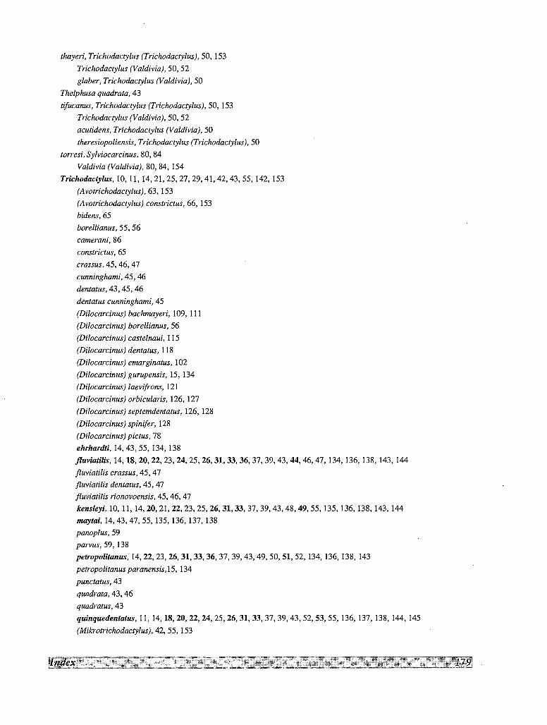
thayeri, Trichodactylus (Trichodactylus), 50, 153Trichodactylus (Valdivia), 50, 52glaber, Trichodactylus (Valdivia), 50
Thelphusa quadrata, 43
tifucanus, Trichodactylus (Trichodactylus), 50, 153Trichodactylus (Yaldivia), 50,52acutidens, Trichodactylus (Valdivia), 50
theresiopoliensis, Trichodactylus (Trichodactylus], 50
torresi, Sylviocarcinus, 80, 84Valdivia (Valdivia), 80, 84, 154
Trichodactylus, 10, 11, 14,21,25,27,29,41,42,43,55, 142,153(Avotrichodactylus), 63, 153(Avotrichodactylus) constrictus, 66, 153bidens, 65borellianus, 55, 56camerani, 86
constrictus, 65crassus, 45, 46, 47cunninghami, 45, 46dentatus, 43, 45, 46dentatus cunninghami, 45
(Dilocarcinus) bachmayeri, 109, 111(Dilocarcinus) borellianus, 56
(Dilocarcinus) castelnaui, 115
(Dilocarcinus) dentatus, 118(Dilocarcinus) emarginatus, 102
(Dilocarcinus) gurupensis, 15, 134(Dilocarcinus) laevifrons, 121(Dilocarcinus) orbicularis, 126,127(Dilocarcinus) septemdentatus, 126,128(Dilocarcinus) spinifer, 128(Dilocarcinus) pictus, 78ehrhardti, 14,43,55, 134, 138jluviatilis, 14,18,20,22,23,24,25,26,31,33,36,37,39,43,44,46,47, 134,136,138, 143, 144fluviatilis crassus, 45, 47fluviatilis dentatus, 45, 47fluviatilis rionovoensis, 45,46, 47kensleyi, 10, 11, 14,20,21,22,23,25,26,31,33,37,39,43,48,49,55, 135, 136, 138,143, 144maytai, 14,43,47,55, 135, 136,137,138panoplus, 59
parvus, 59, 138peuopolitanus, 14,22,23,26,31,33,36,37,39,43,49,50,51,52, 134, 136, 138, 143petropolitanus paranensis, 15, 134punctatus, 43quadrata, 43, 46quadratus, 43
quinquedentatus, 11,14,18,20,22,24,25,26,31,33,37,39,43,52,53,55, 136, 137, 138,144,145(Mikrotrichodactylus), 42, 55, 153
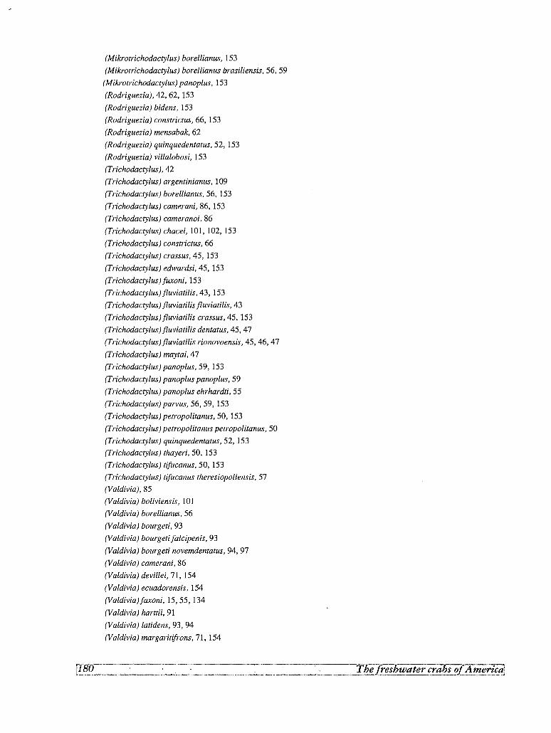
(Mikrotrichodactylus) borellianus, 153
(Mikrotrichodactylus) borellianus brasiliensis, 56, 59
(Mikrotrichodactylus) panoplus, 153
(Rodriguezia), 42, 62, 153
(Rodriguezia) bidens, 153
(Rodriguezia) constrictus, 66, 153
(Rodriguezia) mensabak, 62
(Rodriguezia) quinquedentatus, 52, 153
(Rodriguezia) villalobosi, 153
(Trichodactylus),42
(Trichodactylus) argentinianus, 109
(Trichodactylus) borellianus, 56, 153
(Trichodactylus) camerani, 86, 153
(Trichodactylus) cameranoi, 86
(Trichodactylus) chacei, 101, 102, 153
(Trichodactylus) constrictus, 66
(Trichodactylus) crass us, 45, 153
(Trichodactylus) edwardsi, 45, 153
(Trichodactylus)fuxoni,153
(Trichodactylusi fluviatilis, 43,153
(Trichodactylus) fluviatilis fluviatilis, 43
(Trichodactylust fluviatilis crass us, 45,153
(Trichodactylus) fluviatilis dentatus, 45, 47
(Trichodactylusi fluviatilis rionovoensis, 45, 46, 47
(Trichodactylus) maytai, 47
(Trichodactylus) panoplus, 59, 153
(Trichodactylus) panoplus panoplus, 59
(Trichodactylus) panoplus ehrhardti, 55
(Trichodactylus) parvus, 56, 59, 153
(Trichodactylus) petropolitanus, 50, 153
(Trichodactylus) petropolitanus petropolitanus, 50
(Trichodactylus) quinquedentatus, 52, 153
(Trichodactylus) thayeri, 50, 153
(Trichodactylus) tifucanus, 50, 153
(Trichodactylus) tifucanus theresiopoliensis, 57
(Valdivia),85
(Valdivia) boliviensis, 101
(Valdivia) borellianus, 56
(Valdivia) bourgeti, 93
(Valdivia) bourgeti jalcipenis, 93
(Valdivia) bourgeti novemdentatus, 94, 97
(Valdivia) camerani, 86
(Valdivia) devillei, 71, 154
(Valdivia) ecuadorensis, 154
(Valdiviai faxoni, 15,55, 134
(Valdivia) harttii, 91
(Valdivia) latidens, 93, 94
(Valdivia) margaritifrons, 71, 154
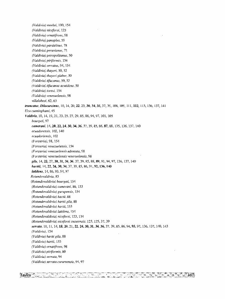
(Valdivia) meekei, 100, 154
(Valdivia) niceforei, 123
(Valdivia) ornatifrons, 98
(Valdivia) panoplus, 59
(Valdivia) pardalinus, 78(Valdivia) peruvianus, 71(Valdivia) petropolitanus, 50
(Valdivia) piriformis, 154
(Valdivia) serratus, 94, 154(Valdivia) thayeri, 50, 52(Valdivia) thayeri glaber, 50
(Valdivia) tifucanus, 50, 52(Valdivia) tifucanus acutidens, 50
(Valdivia) toresi, 154
(Valdivia) venezuelensis, 98
villalobosi, 62, 63truncatus, Dilocarcinus, 10, 14,20,22,23,30,34,35,37,39, 108, 109, 111,112, 113, 136, 137, 141Uca cunninghami, 45
Valdivia, 10, 14, 19,21,23,25,27,29,85,88,94,97, 101, 105bourgeti, 97
camerani, 14,20,22,24,30,34,36,37,39,85,86,87,88, 135, 136,137,140ecuadorensis, 102, 140ecuadoriensis, 102
(Forsteria}, 98, 154(Forsteria) venezuelensis, 154
(Forsteria) venezuelensis edentata, 98
(Forsteria) venezuelensis venezuelensis, 98
gila, 14,22,27,30,31,34,36,37,39,85,88,89,91,94,97,136, 137, 140harUii,14,22,24,30,34,37,39,85,86,91,92,136,140
latidens,14,86,93,94,97
Rotundovaldivia, 85
(Rotundovaldivia) bourgeti, 154
(Rotundovaldivia) camerani, 86, 153(Rotundovaldivia) gurupensis, 154
(Rotundovaldivia) hartii, 88(Rotundovaldivia) hartii gila, 88(Rotundovaldivia) hartii, 153
(Rotundovaldivia) latidens, 154
(Rotundovaldivia) niceforei, 123, 154(Rotundovaldivia) niceforei cucutensis, 123, 125,37,39serrata, 10,11, 14,18,20,21,22,24,30,31,34,36,37,39,85,86,94,95,97, 136, 137, 140, 143(Valdivia), 154
(Valdivia) hartti gila, 88
(Valdivia) hartii, 153
(Valdivia) ornatifrons, 98
(Valdivia) piriformis, 80
(Valdivia) serrata, 94
(Valdivia) serrata cururuensis, 94, 97
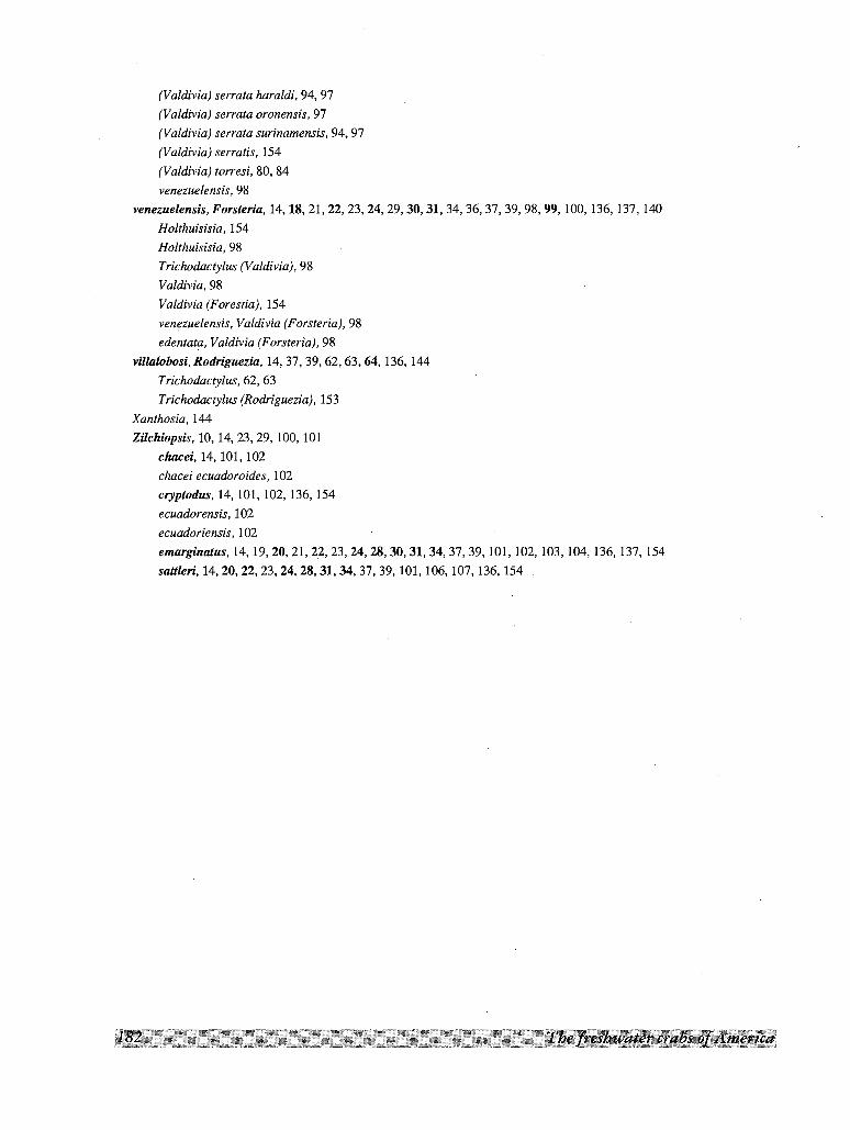
(Valdivia) serrata haraldi, 94, 97(Valdivia) serrata oronensis, 97
(Valdivia) serrata surinamensis, 94,97(Valdivia) serratis, 154
(Valdivia) torresi, 80, 84venezuelensis, 98
venezuelensis, Forsteria, 14,18,21,22,23,24,29,30,31,34,36,37,39,98,99, 100, 136, 137,140Holthuisisia, 154
Holthuisisia, 98
Trichodactylus (Yaldivia), 98
Yaldivia, 98Valdivia (Forestia), 154
venezuelensis, Valdivia (Forsteria), 98
edentata, Valdivia (Forsteria), 98
villalobosi, Rodriguezia, 14,37,39,62,63,64,136,144
Trichodactylus, 62, 63Trichodactylus (Rodriguezia), 153
Xanthosia, 144
ZilchiopsM, 10,14,23,29, 100,101chacei, 14, 101, 102chaceiecuadoroides,102
cryptodus, 14,101,102,136, 154ecuadorensis, 102
ecuadoriensis, 102emarginatus, 14,19,20,21,22,23,24,28,30,31,34,37,39, 101,102,103,104,136,137,154saUleri, 14,20,22,23,24,28,31,34,37,39, 101,106,107,136, 154
SUPPLEMENT TO THE FAMILY PSEUDOTHELPHUSIDAE
Since the publication of the first part of this
monograph on the freshwater crabs of America,
concerning the family Pseudothelphusidae, in 1982,
a number of additional species have been added to
the family list and new information published
regarding several others. It therefore seemed
desirable to prepare a supplement to that work to
cover the additional species, correct errors and
omissions, record changes in nomenclature, and
update the bibliography on different aspects of
pseudothelphusid biology, including works omitted
in 1982. This article is to be regarded as purely
supplementary to my work of 1982 named above.
Bibliographic references already given in that work
are omitted from the literature below.
New taxa described after 1982Since 1982, 37 new species have been described
and 5 new genera have been erected. These new taxaare listed below, arranged in a systematic order. The
list includes also Ptychophallus costaricensisVillalobos, 1974, omitted in the first part.
Tribe STRENGERIANINI Rodnguez, 1982
Chaceus Pretzmann, 1965
Chaceus caecus Rodriguez &Bosque. 1989.Venezuela.
Chaceus davidi Campos& Rodriguez, 1984.Colombia.Chaceus cesarensis Rodriguez & Vilosia, 1992. Colombia
Strengeriana Pretzmann, 1971
Strengeriana antioquensis Prah1, 1987. Colombia.
Strengeriana bolivarensis Rodrfguez & Campos, 1989.
Colombia.
Strengeriana chaparralensis Campos & Rodrfguez,
1984. Colombia.
Strengeriana huilensis Rodriguez & Campos, 1989.
Colombia.
Strengeriana risaraldensis Rodrfguez & Campos,
1989. Colombia.
Strengeriana taironae Rodrfguez & Campos, 1989.
Colombia.
Tribe HYPOLOBOCERINI Pretzmann, 1971
Hypolobocera Ortmann, 1897
Hypolobocera alata Campos, 1989. Colombia.
Hypolobocera cajambrensis Prahl, 1988. Colombia.
Hypolobocera dentata Prah1, 1987. Colombia.Hypolobocera gorgonensis Prah1, 1983. Colombia.
Hypolobocera lloroensis Campos, 1989. Colombia.Hypolobocera malagueiia Prah1, 1988. Colombia.Hypolobocera meineli Prah1, 1988. Colombia.
Hypolobocera merenbergersis Prah1 & Gira1do, 1985.Colombia.
Hypolobocera mutisi Prah1, 1988.Colombia.
Moritschus Pretzmann, 1965
Moritschus narinnensis Campos & Rodrfguez, 1988.
Colombia.
Neostrengeria Pretzmann, 1965
Neostrengeria charalensis Campos & Rodriguez,
1985. Colombia.
Neostrengeria sketi Rodriguez, 1985. Colombia.
Tribe POTAMOCARCININI Ortmann, 1897
Odontothelphusa Rodriguez, 1982
Odontothelphusa monodontis Rodriguez & Hobbs,
1989. Mexico.
Odontothelphusa toninae Alvarez & Villalobos, 1991.
Mexico.
Potamocarcinus H. Milne Edwards, 1853
Potamocarcinus colombiensis Prahl & Ramos, 1987.
Colombia.
Potamocarcinus leptomelus Rodriguez & Hobbs, 1989.
Colombia.
Ptychophallus Smalley, 1964
Ptychophallus costaricensis Villalobos, 1974. Costa
Rica.
Stygothelphusa Alvarez & Villalobos, 1991
Stygothelphusa lopezformenti Alvarez & Villobos,
1991. Mexico (type species).
Typhlopseudothelphusa Rioja, 1952
Typhlopseudothelphusa acanthochela Hobbs, 1986.
Belize.
Typhlopseudothelphusa hyba Rodriguez & Hobbs,
1989. Mexico.
Zilchia Pretzmann 1968
Zilchiafalcata Rodriguez & Hobbs, 1989. Guatemala.
Tribe PSEUDOTHELPHUSINI Ortmann, 1897
Disparithelphusa Smalley & Adkinson, 1984
Disparithelphusa pecki Smalley & Adkinson, 1984.
Mexico (type species).
Eidocamptophallus Rodriguez & Hobbs, 1989
Potamocarcinus (Potamocarcinus) chacei Pretzmann,
1967 (type species).
Pseudothelphusa de Saussure, 1857
Pseudothelphusa galloi Alvarez & Villalobos, 1990.
Mexico.
Pseudothelphusa mexicana Alvarez-Noguera, 1987.
Mexico.
Pseudothelphusa parabelliana Alvarez, 1989. Mexico.
Pseudothelphusa puntarenas Hobbs, 1991. Costa Rica.
Smalleyus Alvarez, 1989
Smalleyus tricristatus Alvarez, 1989. Mexico (type
species).
Tribe KINGSLEYINI Bott, 1970
Brasiliothelphusa Magalhaes & Turkay, 1986.
Brasiliothelphusa tapajoense Magalhaes & Ttirkay,
1986. Brazil (type species).
Kingsleya Ortmann, 1897
Kingsleya besti Magalhaes, 1990. Brazil.
Kingsleya ytupora Magalhaes, 1986. Brazil.
Fredius Pretzmann, 1967
Fredius adpress us Rodriguez & Pereira, 1992.
Venezuela.
Fredius adpressus piaroensis Rodriguez & Pereira,
1992. Venezuela.
Fredius platyacanthus Rodrfguez & Pereira, 1992.
Venezuela.
Fredius estevisi siapensis Rodriguez & Pereira, 1992.
Venezuela.
Faunistic lists and taxonomic modificationsRODRIGUEZ (1981) gave a general listing of the
South American species and CAMPOS (1985)
included several new records of Pseudothelphusidae
in his list of freshwater decapods of Colombia.
PRETZMANN (1983a, 1983b, 1983c) amplified and
illustrated the species he had already described from
Ecuador and Peru (PRETZMANN 1977b, 1978a) (see
Addendum to RODRIGUEZ, 1982); PRETZMANN &
MAYTA (1980) also insist on these species.
The species of Hypolobocera and Potamocarcinus
of the Pacific drainage of Colombia, have been
reviewed and adequately illustrated by VON PRAHL
(1988). This author (VON PRAHL 1987d, 1988) also
redescribed Hypolobocera buenaventurensis,
previously included in my list of species incertae
sedis.
Based on the possible homologies found in the
gonopods of the species of Potamocarcinini,
ROORfGUEZ & HOBBS (1989b) have reviewed the
structure of this Tribe, giving new diagnosis of the
genera, erecting the new genus Eidocamptophallus
to receive Potamocarcinus chacei Pretzmann, 1967,
and excluding Pseudothelphusa pittieri Rathbun,
1898, and Potamocarcinus garthi Pretzmann, 1971
from this Tribe.
SMALLEY & AOKINSON (1984) have reviewed the
gonopod terminology and the use of female genital
openings as a taxonomic character. Through the use
of scanning electron microscope these authors have
determined that the setae at the distal terminus of the
sperm channel of the male gonopod of their new
species Disparithelphusa pecki and other
pseudothelphusids, are not pointed, but have a large
terminal pore; they have called these peculiar setae
"terminal pore setae".
SMALLEY & AOKINSON (1984) have also
determined that terminal pore setae are present on
the inner surface of the merus of the third maxilliped
in the Tribes Pseudothelphusini, Potamocarcinini
(sensu Pretzmann, 1972) and Kingsleyini, but
neither in the Tribe Hypolobocerini (sensu
Pretzmann, 1972), nor in the subfamily
Epilobocerinae. In view of these results (as well as
other criteria based on gonopod morphology), it
would be advisable to keep PRETZMANN (1972)
arrangement of genera for the Potamocarcinini and
Hypolobocerini rather than which I proposed in
1982.
Biogeography and phylogeneticsMany of the taxonomic studies published since
1982 have materially added to a more detailed
knowledge of the distribution of the family. An
important contribution have been that of
MAGALHAES (1986) who have shown an extensive
distribution of the Pseudothelphusidae, represented
by the species of Fredius, troughout the Amazon
basin, particularly south of this river. The origin of
the species of Fredius have been studied by
ROORfGUEZ & PEREIRA (1992) using area
cladograms.
The increase in our knowledge of the taxonomy
and distribution of the species has also affected our
ideas on the biogeography and possible evolution of
this family, and have considerably altered my
tentative scheme of 1982 for the origin and radiation
of the family. Concerning the origin of the
Pseudothelphusidae, ROORfGUEZ (1986) have shown
that an analysis of the synapomorphies found in the
respiratory structures of the Old- and New World
freshwater crabs, suggests a monophyletic origin for
the species with reduced exognaths in America,
Africa and India. This distribution is evidence of a
former Gondwanan distribution and shows at the
same time that the transformation of buccal
appendages had already began in Mid-cretaceous
times in basic groups whose modern representatives
are the American Pseudothelphusidae and the
African Gecarcinucidae. The Trichodactylidae, in
which this buccal reaccomodation does not appear,
derive from a separate basic group and are related to
the Eupotamonea ofWestern Africa.
Regarding the separation of the two subfamilies of
Pseudothelphusidae, and through the analysis of
chorological series and other criteria, ROORfGUEZ
(1986), to explain the disjunct distribution of both
groups in the Antilles and South America, postulates
the origin of the main phyletic trunks of
Pseudothelphusinae of Middle and South America,
from a common ancestor in northern South America,
represented today by the Strengerianini. Concerning
the phylogenetics of this last Tribe, ROORfGUEZ &
CAMPOS (1989) have postulated by means of a
cladistic analysis, that this group was widespread in
the area of north-western South America in Miocene
times. Later the uplifting and displacement of the
Santa Marta Massif led to disruption of the ancestral
Strengerianini stock to form the actual pattern of
distribution and diversification.
Cavernicolous species
The adaptation of some pseudothelphusid crabs
to cave life is a well known fact ever since RIOJA
(1955) described the strictly stygobiont
Potamocarcinus (Typhlopseudothelphusa) mocinoi
from a cave in Mexico. However, the assignment of
other species found in subterranean waters to the
categories stygobionts, stygophiles and stygoxenes
is not an easy task. HOBBS et al. (1977) list as
troglobitic only RIOJA'S species, but added 5 s~fc:iesfrom Mexico and Central America, and 3 species
from the Greater Antilles as associated with caves.
REDDELL (1981) cited as troglobious RIOJA'S species
and the two other Typhlopseudothelphusa described
by DELAMARE-DEBOUTEVILLE (1976), and include
six others that are probably troglophile from
Mexico, Guatemala and Belize. HOLTHUIS' list
(1986) included the 3 troglobious cited by REDDELL,
and as stygophiles or stygoxenes 4 species from the
Antilles, 5 from Mexico and Central America and 1
from Venezuela. All the species listed by these
authors are also included my 1982 work. GUINOT
(1988) have given a useful discussion of these
species, and three other recorded by RODRfouEz
(1985).
The following troglobious species were described
after 1982.
Chaceus caecus Rodrfguez & Bosque, 1989. CuevaPunto Fijo, Rio Guasare, Zulia, Venezuela. Pereiopodsvery slender, corneae absent, carapace depigmented.
Neostrengeria sketi Rodrfguez, 1985. Hoyo del Aire,La Paz, Santander, Colombia. Pereiopods very slender,corneae reduced, carapace partly depigmented.
Potamocarcinus leptomelus Rodrfguez & Hobbs, 1989.Cueva del Tunel, Veracruz, Mexico. Eyes small, butdistinctly faceted and pigmented, walking legs very long.
Zilchia falcata Rodrfguez & Hobbs, 1989. AltaVerapaz, Guatemala. Slender pereiopods, but eyes normal.
Stygothelphusa lopezformenti Alvarez & Villalobos,1991. Cueva de los brujos, Valle Nacional, Oaxaca,Mexico. Elongated pereiopods and carapace notpigmented, but eyes normal.
Typhlopseudothelphusa acanthochela Hobbs, 1986.Blind crab cave, Cayo District, Belize. Degenerate eyes,elongated pereiopods and carapace not pigmented.
Typhlopseudothelphusa hyba Rodriguez & Hobbs,1989. Cueva de Los Llanos, Chiapas, Mexico. Degenerateeyes, elongated pereiopods and carapace not pigmented.
Odontothelphusa monodontis Rodriguez & Hobbs,1989. Grutas del Cocona, Tabasco, Mexico. Dorsalsurface of carapace pale and walking legs moderatelyslender; the eyes however, are well pigmented.
Pseudothelphusa mexicana Alvarez-Noguera, 1987. LaJolla Cave, Guerrero State, Mexico. The species exhibitsno obvious adaptations to cave environment, but seemsrestricted to the deeper part of the cave, in completedarkness.
Pseudothelphusa puntarenas Villalobos, 1991. EmusCave, Costa Rica. This crab lacks any obvious externaltroglomorphic modifications, but is not yet known fromepigean waters.
Additionally, Neostrengeria charalensis Campos andRodrfguez 1985, and Neostrengeria niceforoi (Schmitt,1969) were reported as troglophile from the Cueva de LosIndios and the nearby Cueva del Paramo, respectively, LaPaz, Santander Department, Colombia (RoDRIouEz, 1985).
Corrections
Several errors have been detected after publication
of the first part of this monograph. The following
were kindly pointed out to me by Dr Alfred SMALLEY:
(a) The species Ptychophallus costaricensis
Villalobos (1974) was not included. (b) In Figure 26d
the apical view of Hypolobocera orientalis is not
referenced in the caption text. (c) The specific name
of Potamocarcinus aspoeckorum should be spelled
with a "c", as in PRETZMANN'S original description,
since it is derived from the name of Ulrike and Horst
ASPOCK; the author subsequently omitted the "c" in
other papers, but on pages 16-19 of his monograph
(PRETZMANN, 1972) he spelled Aspoeckia correctly.
(d) On page 44, in the key just before Phrygiopilus,
"Costa Rica" should be "Guatemala". (e) In Figure 2,
VII should be VIb.
Additionally, the caption text and illustrations
under Figure 39 should be interchanged with the
caption text and illustrations under figure 71.
LITERATURE
ALVAREZ-NOGUERA (F.), 1987. - Pseudothelphusa mexicana, a new freshwater crab from the State of Guerrero, Mexico
(Brachyura: Pseudothelphusidae). Proceedings of the Biological Society ofWashington, 100: 1-3.
ALVAREZ (F.), 1989. - Smal/eyus tricristatus, new genus, new species, and Pseudothelphusa parabelliana, new species
(Brachyura: Pseudothelphusidae) from Los Tuxtlas, Veracruz, Mexico. Proceedings of the Biological Society of
Washington, 102: 45-49.
ALVAREZ (F.) & VILLALOBOS (J. L.), 1990. - Pseudothelphusa gal/oi, a new species of freshwater crab (Crustacea:
Brachyura: Pseudothelphusidae) from southwestern Mexico. Proceedings of the Biological Society of Washington, 103:
103-105.
ALVAREZ (F.) & VILLALOBOS (J. L.), 1991. - A new genus and two new species of freshwater crabs from Mexico,
Odontothelphusa toninae and Stygocephalus lopezformenti (Custacea: Brachyura: Pseudothelphusidae). Proceedings of
the Biological Society ofWashington, 104: 288-294.
CAMPOS (M. R), 1985. - Decapodos de agua du1ce del Suborden Brachyura reportados para Colombia. Caldasia, 14 (67):
265-284.
CAMPOS (M. R), 1989. - Nuevas especies de cangrejos de agua du1ce del Genero Hypolobocera (Crustacea, Decapoda,
Pseudothelphusidae) para Colombia. Trianea (Acta Cientifica y Tecnica de Inderena), 3: 143-147.
CAMPOS (M. R) & PARRA-CASTELLANOS (H.), 1986. - Estudio morfometrico para discriminar segun el sexo, cangrejos adultos
de la subespecie Hypolobocera bouvieri (Rathbun, 1898) (Decapoda: Pseudothelphusidae). Caldasia, 15 (71-75): 733-742.
CAMPOS (M. R) & RODIUGUEZ (G.), 1984. New species of freshwater crabs (Crustacea, Decapoda, Pseudothelphusidae) from
Colombia. Proceedings of the Biological Society ofWashington, 97: 583-543.
CAMPOS (M. R) & RODRfGUEZ (G.), 1985. - A new species of Neostrengeria (Crustacea, Decapoda, Pseudothelphusidae) with
notes on the geographical distribution of the genus. Proceedings of the Biological Society ofWashington, 98: 718-727.
CAMPOS (M. R.) & RODRfGUEZ (G.), 1988. - Notes on the freshwater crabs of the genus Moritschus Pretzmann, 1965
(Crustacea: Decapoda: Pseudothe1phusidae) with description of M. narinnensis from Southern Colombia. Proceedings of
the Biological Society ofWashington, 101: 640-643.
GUINOT (D.), 1988. - Les crabes cavernicoles du monde. Memoires de Biospeleogie, 15: 3-40.
HOBBS (H. H.) Jr., 1986. - A new Troglobitic Crab (Crustacea: Decapoda: Pseudothelphusidae) from Belize. Texas Memorial
Museum, Speleological Monographs, 1: 1-4.
HOBBs (H. H.) Jr., HOBBs (H. H.) III & DANIEL (M. A.), 1977. - A review of the troglobitic decapod crustaceans of the
Americas. Smithsonian Contributions to Zoology, 244: v + 183 p.
HOBBS (H. H.) Ill, 1991. - A new pseudothelphusid crab from a cave in southern Costa Rica. Proceedings of the Biological
Society ofWashington, 104: 295-298.
HOLTHUIS (L. B.), 1986. - Decapoda, In: Botosaneanu, L. (ed.), Stygofauna mundi, E. J. Brill, Leiden, 9: 589-615.
MAGALHAES (C), 1986. - Revisao taxonomica dos caranguejos de agua doce brasileiros da familia Pseudothelphusidae
(Crustacea, Decapoda). Amazoniana, 8: 609-636.
MAGALHAES (C), 1987. - Notes on some Pseudothelphusidae crabs from Venezuela, Ecuador and Mexico found in the
collection of the Museu de Zoologia da Universidade de Sao Paulo, Brazil. Revista Brasileira de Zoologia, 4: 55-58.
MAGALHAES (C), 1990. - A new species of the genus Kingsleya from Amazonia, with a modified key for the Brazilian
Pseudothelphusidae. Zoologische Mededelingen, 63 (21): 275-281.
MAGALHAES (C), & TURKAY (M.), 1986. - Brasiliothelphusa, a new Brazilian freshwater-crab genus (Crustacea: Decapoda:
Pseudothe1phusidae). Senckenbergiana biologica, 66: 371-376.
MAGALHAES (C.), DE OLIVEIRA MALTA (J. C.), ROBERTSON (B.) & VARELLA (A.), 1988. - A catalogue of type specimens of
Crustacea in the Invertebrate Collection of the Instituto Nacional de Pesquisas da Amazonia, Manaus, Brazil.
Amazoniana, 10: 267-282.
PRAHL (H. von), 1983. - Hypolobocera gorgonensis sp. novo (Crustacea: Brachyura: Pseudothelphusidae): un nuevo cangrejo
de agua duke de la Isla de Gorgona, Colombia. Cespedesia, 12: 45-46.
PRAHL (H. von), 1985. - Distribucion del cangrejo de agua duke Hypolobocera beiri Pretzmann, 1968 y anatomia de su
gonopodo. Actualidades Biologicas, 14 (52): 43-47.
PRAHL (H. von), 1987a. - Strengeriana antioquensis sp. novo (Crustacea: Pseudothelphusidae): a new freshwater crab of
Colombia. Caribbean Journal ofScience, 23: 244-246.
PRAHL (H. von), 1987b. - Hypolobocera dentata sp. nov.: a new freshwater crab (Crustacea: Brachyura: Pseudothelphusidae)
from the Cordillera Occidental, Colombia. Revista de Biologia Tropical, 35: 93-95.
PRAHL (H. von), 1987c. - Potamocarcinus colombiensis sp. nov.: un nuevo cangrejo de agua duke (Decapoda: Brachyura:
Pseudothelphusidae) de la Serrania Costera del Baudo, Colombia. Revista de Biologia Tropical, 35: 131-133.
PRAHL (H. von), 1987d. - Redescripcion de Hypolobocera buenaventurensis, un cangrejo de agua duke (Decapoda,
Pseudothelphusidae) del Pacifico Colombiano. Boletin Ecotropica, 16: 59-64.
PRAHL (H. von), 1988. - Fresh-water crabs (Crustacea: Decapoda: Pseudothelphusidae) of the Pacific drainage of Colombia.
Zoologische Jahrberichter (Systematische), 115: 171-186.
PRAHL (H. von) & GlRALDO (J.), 1985. - Un nuevo cangrejo de agua duke de la Cordillera Central de Colombia. Lozania,
49: 101-104.
PRAHL (H. von) & RAMOS (G. R.), 1987. - Potamocarcinus colombiensis sp. nov.: Un nuevo cangrejo de agua duke
(Decapoda: Brachyura: Pseudothelphusidae) de la Serrania Costera del Baudo, Colombia. Revista de Biologia Tropical,
35: 131-133, 1 fig.
PRETZMANN (G.), 1983a. - Ergebnisse einiger Sammelreisen in Sudamerika. 1. Teil: Neue Pseudothelphusidae. Annales
Naturhistorische Museum Wien, 84/B: 301-305, pl. 1-14.
. PRETZMANN (G.), 1983b. - Die Pseudothelphusidae Perus. Annales Naturhistorische Museum Wien, 84/B: 331-345, pl. 1-10.
PRETZMANN (G.), 1983c. - Die Pseudothelphusidae von Ecuador. Annales Naturhistorische Museum Wien, 84/B: 301-305,
pl. 1-14.
PRETZMANN (G.) & Mayta (R.), 1980. - Uber einige Susswasserkrabben aus Peru. Sitzungberichten del' mathematisch
naturwissenschaftlichen Klasse del' Osterreichischen Akademie del' Wissenschaften (Vienna), 9: 137-144, fig. 1-14.
REDDELL (1. R.), 1981. - A review of the cavernico1e fauna of Mexico, Guatemala and Belize. Bulletin of the Texas
Memorial Museum, 27: 1-327.
RODRIGUEZ (G), 1981. - Decapoda. In: Hurlbert, H., G. Rodrfguez & N. D. dos Santos, Aquatic Biota of Tropical South
America, I: 41-51.
RODRIGUEZ (G.), 1982. - Les crabes d'eau douce d'Amerique, Famille des Pseudothe1phusidae. Faune trop. XXII, Orstom,
Paris, 224 p.
RODRlGUEZ (G), 1985. - A new cavernicolous crab (Crustacea, Decapoda, Pseudothelphusidae) from Colombia. Bioloski
vestnik (Ljubljana), 33 (2): 73-80, fig. 1-3.
RODRIGUEZ (G), 1986. - Centers of Distribution of Neotropical Freshwater Crabs, In: Gore, R. H. & K. L. Heck (eds),
Biogeography of the Crustacea. Crustacean Issues, 4: 51-67.
RODRfGUEZ (G), & Bosous (C.), 1989. - A stygobiont crab, Chaceus caecus n. sp., and its related stygophile species,
Chaceus moti/oni Rodrfguez, 1980, (Crustacea, Decapoda, Pseudothelphusidae) from a cave in the Cordillera de Perija,
Venezuela. Memoires de Biospeleogie, 17: 127-134.
RODRlGUEZ (G), & CAMPOS (M. R.), 1989. - The cladistic relationships of the freshwater crabs of the Tribe Strengerianini
(Crustacea, Decapoda, Pseudothelphusidae) from the northern Andes, with comments on their biogeography and
descriptions of new species. Journal ofCrustacean Biology, 9: 141-156.
RODRioUEZ (G.) & HOBBS (H. H.) Jr., 1989a. - Freshwater crabs associated with caves in Southern Mexico and Belize, with
descriptions of three new species. Proceedings of the Biological Society of Washington, 102: 394-400.
RODRioUEZ (G.) & HOBBS (H. H.) Jr., 1989b. - A new cavernicolous crab from Guatemala, with notes on the genera of
Potamocarcinini (Crustacea, Decapoda, Pseudothelphusidae), Bulletin du Museum national d' histoire naturelle, Paris, 4e
ser., 11 (sect. A, 1): 183-189.
RODRioUEZ (G.) & PEREIRA (G.), 1992. - New species, cladistic relationships and biogeography of the genus Fredius
(Decapoda: Brachyura: Pseudothe1phusidae) from South America. Journal ofCrustacean Biology, 12: 298-311.
RODRioUEZ (G.) & VILORIA (A. L.), 1992. - Chaceus cesarensis, a new species of fresh-water crab (crustacea: Decapoda:
Pseudothelphusidae) from Colombia with a key to the genus. Proceedings ofbiological Society ofWashington, 105: 77-80.
SMALLEY (A. E.) & ADKINSON (D. L.), 1984. - Disparithelphusa pecki, a new genus and species of fresh-water crab from
Mexico (Brachyura: Pseudothe1phusidae). Journal ofCrustacean Biology, 4: 127-133.
VILLALOBOS (C. R.), 1974. - Ptychophallus costaricensis, a new freshwater crab from Costa Rica. Revista de Biologia
Tropical, 21: 197-203.
Ouvrage realise en PAOFlashage : ACIR, Montpellier
Acheve d'imprimer sur Jes presses de I'imprimerie PUBLlCEP, Montpellier
Depot legal: decernbre 1992
I I ,'u.\1<1' ''''.
l ( x.tcur (, ill rro I O DRiGl EZest rnem b re de ('Inst il to 1, ' n '1. I l.1Il o
(It In 'cs 'gal ,ones C icnr uicas.
s -, p re m icrs rruv.u
sur les Ud )C' , d 'cau dOLI d ' l1leriYlle
clatenr des unnccs so ixanre.
Acr ucl lcm r nr con , ide r': cornrne
l'un d{ m eillcurs spec i.ilist dc et" groupe,
il s'est ar tache. tour -n en rir tiissaru
l'in enraire IXlr la dcocr ipt io/ld l' nouvclles l'sjJ<:n:s. :, simpli fic·r
I classificati on ;i l'inrerieur de-s C ilk s.
11 Ill: doi aussi "' l llt e rC", ~ ' nt ,: '> r,'lIt" ('IlS
sur la ph) I )~t-n ll et 1., I i ll ,l'; e()~Ta l' h ic
dc c ; CfllsTac':s
de lit r; I . 2 1 , re i a Fayc t rc,
• 11 P i" c:(;J x 10.
O,fllI. i 11 " 7_, r )UP d ' i ulnay,~3 I ·i j Boridv ccdcx.
11 IS 1- 71)t ') - 1( V - ,j


