Embed Size (px)
Citation preview
E U R O P E A N J O U R N A L O F C A N C E R 4 7 ( 2 0 1 1 ) 1 1 2 7 – 1 1 3 7
. sc iencedi rec t . com
ava i lab le a t wwwjournal homepage: www.ejconl ine.com
Review
The oncogenic and tumour suppressive roles of microRNAsin cancer and apoptosis
Sadegh Babashah a, Masoud Soleimani b,*
a Department of Molecular Genetics, Faculty of Biological Sciences, Tarbiat Modares University, Tehran, Iranb Department of Hematology, School of Medical Sciences, Tarbiat Modares University, Tehran, Iran
A R T I C L E I N F O
Article history:
Received 25 December 2010
Accepted 14 February 2011
Available online 12 March 2011
Keywords:
MicroRNA
Cancer
Apoptosis
Oncogene
Tumour suppressor
0959-8049/$ - see front matter � 2011 Elsevidoi:10.1016/j.ejca.2011.02.008
* Corresponding author: Address: DepartmenTehran, Iran. Tel./fax: +98 21 82884508.
E-mail addresses: sadegh.babashah@gma
A B S T R A C T
MicroRNAs (miRNAs) are small, non-coding, endogenous RNAs that regulate gene expres-
sion at the post-transcriptional level. MiRNAs play important roles in regulating a variety of
biological process such as proliferation, differentiation and apoptosis. It has been demon-
strated that miRNAs have a crucial function in oncogenesis by regulating cell proliferation
and apoptosis as oncogenes or tumour suppressors. As several reports have underlined the
possible contribution of miRNAs to promote or evade apoptosis, it seems that the dysreg-
ulation of miRNAs involved in apoptosis may provide a mechanism for cancer develop-
ment. Given emerging evidence that points to oncogenic and tumour suppressive roles
of miRNAs in cancer and apoptosis, it is thought that manipulating miRNA expression level
may be a potential therapeutic strategy for curing cancer.
� 2011 Elsevier Ltd. All rights reserved.
1. Introduction to microRNA; biogenesis,processing and function
MicroRNAs (miRNAs), small RNA molecules of approximately
22 nucleotides, are a novel class of endogenously encoded
non-coding RNAs that control gene expression by targeting
specific mRNAs bearing partially complementary target se-
quences for degradation and/or translational repression.1,2
Since the discovery of small non-coding RNAs lin-4 and let-7
(now known to be miRNAs) in Caenorhabditis elegans,3,4 hun-
dreds of miRNA sequences have been so far identified in a
wide range of organisms from plants to humans. In the hu-
man genome, it is currently estimated that there may be
1000 miRNA genes which could account for approximately
1% of the genome and up to one third of human genome
may be regulated by miRNAs. Each miRNA can target approx-
er Ltd. All rights reserved
t of Hematology, School o
il.com (S. Babashah), sole
imately 200 transcripts directly or indirectly, whereas a single
protein coding gene target could be regulated by more than
one miRNA (http://www.sanger.ac.uk/Software/Rfam/mirna/).
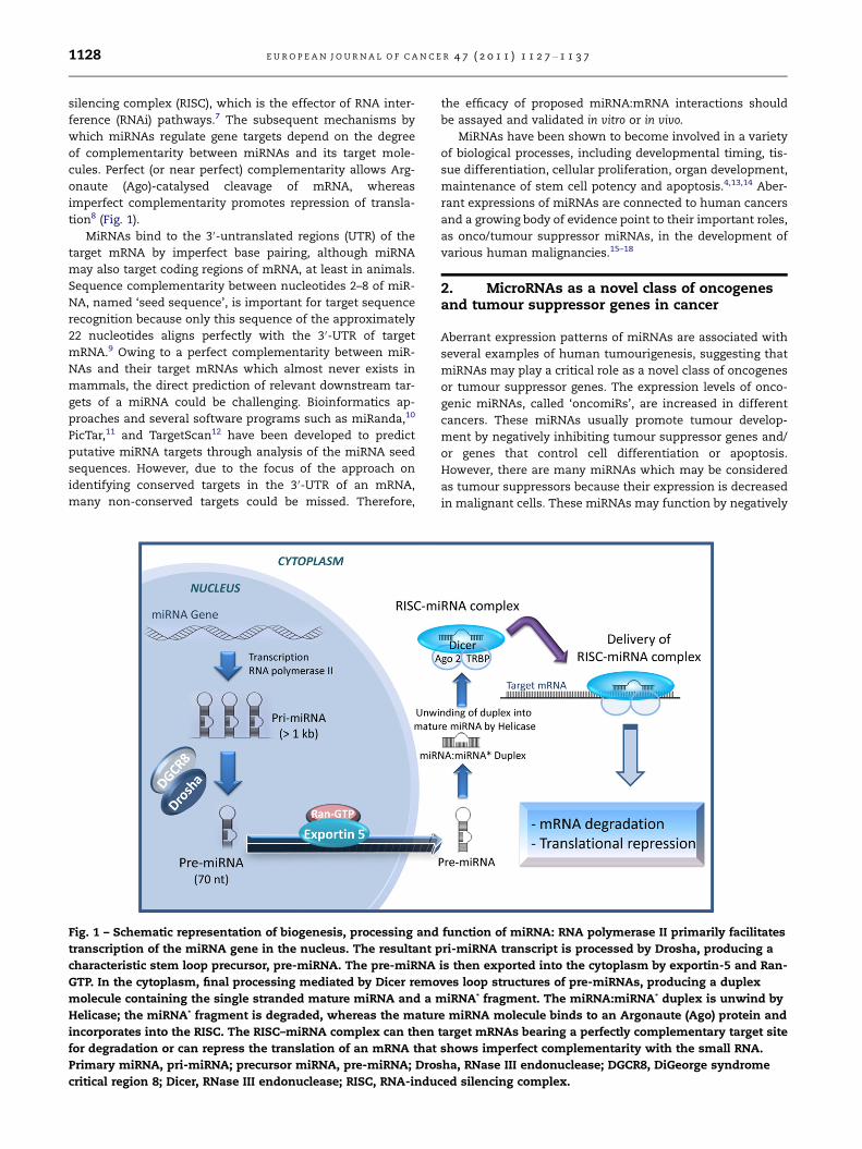
MiRNAs are transcribed by RNA polymerase II or III as
short RNA hairpin structures which are subsequently pro-
cessed by the nuclear and cytoplasmic RNase III-type en-
zymes. Primary microRNA transcripts (pri-miRNA) are
processed in the nucleus by the RNase III endonuclease
Drosha and DGCR8 (the ‘microprocessor complex’) to form
intermediate stem-loop structures approximately 70 nucleo-
tides long called ‘precursor miRNAs’ (pre-miRNAs).5,6 The
process is followed by exportin-5-mediated export of pre-
miRNA to the cytoplasm. Further processing facilitated by
the cytoplasmic RNase III endonuclease Dicer complex
appears coupled to the assembly of the mature single strand
miRNA (often designated miR) into the RNA-induced
.
f Medical Sciences, Tarbiat Modares University, P.O. Box 14115-331,
[email protected] (M. Soleimani).
1128 E U R O P E A N J O U R N A L O F C A N C E R 4 7 ( 2 0 1 1 ) 1 1 2 7 – 1 1 3 7
silencing complex (RISC), which is the effector of RNA inter-
ference (RNAi) pathways.7 The subsequent mechanisms by
which miRNAs regulate gene targets depend on the degree
of complementarity between miRNAs and its target mole-
cules. Perfect (or near perfect) complementarity allows Arg-
onaute (Ago)-catalysed cleavage of mRNA, whereas
imperfect complementarity promotes repression of transla-
tion8 (Fig. 1).
MiRNAs bind to the 3 0-untranslated regions (UTR) of the
target mRNA by imperfect base pairing, although miRNA
may also target coding regions of mRNA, at least in animals.
Sequence complementarity between nucleotides 2–8 of miR-
NA, named ‘seed sequence’, is important for target sequence
recognition because only this sequence of the approximately
22 nucleotides aligns perfectly with the 3 0-UTR of target
mRNA.9 Owing to a perfect complementarity between miR-
NAs and their target mRNAs which almost never exists in
mammals, the direct prediction of relevant downstream tar-
gets of a miRNA could be challenging. Bioinformatics ap-
proaches and several software programs such as miRanda,10
PicTar,11 and TargetScan12 have been developed to predict
putative miRNA targets through analysis of the miRNA seed
sequences. However, due to the focus of the approach on
identifying conserved targets in the 3 0-UTR of an mRNA,
many non-conserved targets could be missed. Therefore,
Fig. 1 – Schematic representation of biogenesis, processing and
transcription of the miRNA gene in the nucleus. The resultant p
characteristic stem loop precursor, pre-miRNA. The pre-miRNA
GTP. In the cytoplasm, final processing mediated by Dicer remo
molecule containing the single stranded mature miRNA and a m
Helicase; the miRNA* fragment is degraded, whereas the matur
incorporates into the RISC. The RISC–miRNA complex can then
for degradation or can repress the translation of an mRNA that
Primary miRNA, pri-miRNA; precursor miRNA, pre-miRNA; Dros
critical region 8; Dicer, RNase III endonuclease; RISC, RNA-indu
the efficacy of proposed miRNA:mRNA interactions should
be assayed and validated in vitro or in vivo.
MiRNAs have been shown to become involved in a variety
of biological processes, including developmental timing, tis-
sue differentiation, cellular proliferation, organ development,
maintenance of stem cell potency and apoptosis.4,13,14 Aber-
rant expressions of miRNAs are connected to human cancers
and a growing body of evidence point to their important roles,
as onco/tumour suppressor miRNAs, in the development of
various human malignancies.15–18
2. MicroRNAs as a novel class of oncogenesand tumour suppressor genes in cancer
Aberrant expression patterns of miRNAs are associated with
several examples of human tumourigenesis, suggesting that
miRNAs may play a critical role as a novel class of oncogenes
or tumour suppressor genes. The expression levels of onco-
genic miRNAs, called ‘oncomiRs’, are increased in different
cancers. These miRNAs usually promote tumour develop-
ment by negatively inhibiting tumour suppressor genes and/
or genes that control cell differentiation or apoptosis.
However, there are many miRNAs which may be considered
as tumour suppressors because their expression is decreased
in malignant cells. These miRNAs may function by negatively
function of miRNA: RNA polymerase II primarily facilitates
ri-miRNA transcript is processed by Drosha, producing a
is then exported into the cytoplasm by exportin-5 and Ran-
ves loop structures of pre-miRNAs, producing a duplex
iRNA* fragment. The miRNA:miRNA* duplex is unwind by
e miRNA molecule binds to an Argonaute (Ago) protein and
target mRNAs bearing a perfectly complementary target site
shows imperfect complementarity with the small RNA.
ha, RNase III endonuclease; DGCR8, DiGeorge syndrome
ced silencing complex.
E U R O P E A N J O U R N A L O F C A N C E R 4 7 ( 2 0 1 1 ) 1 1 2 7 – 1 1 3 7 1129
inhibiting oncogenes and/or genes that inhibit cell differenti-
ation or apoptosis.15–17
MiRNAs functioning as tumour suppressor genes such as
the let-7, which negatively regulates Ras19 and high mobility
group A2 (HMGA2);20,21 mir-15a and mir-16-1, which nega-
tively regulate BCL2;22 as well as the miR-34, that is induced
by DNA damage and oncogenic stress in a p53-dependent
manner which leads to apoptosis or cellular senescence.23
MiRNAs functioning as oncogenes such as miR-21, targets
the tumour suppressors tropomyosin 124 and programmed
cell death 4 (PDCD4) in breast cancer cells25 and also phos-
phatase and tensin homologue detected on chromosome
ten (PTEN) in hepatocellular carcinomas.26,27 The miR-17-92
cluster can be regarded as a family of oncogenes, directly tar-
geting many genes involved in apoptotic pathways.28 Thus,
miRNAs can act both as oncogenes and tumour suppressors,
depending on the particular miRNA and the cell type.
2.1. MiR-17-92 cluster, a family of oncogenic miRNAs
One of the first and well-studied oncogenic miRNAs identified
is the miR-17-92 cluster, containing seven homologous miR-
NAs (miR-17-3p, miR-17-5p, miR-18a, miR-20a, miR-19a,
miR-19b-1 and miR-92a-1), with genomic positions on chro-
mosome X, 7 and 13. The cluster located on chromosome 13
seems to be frequently overexpressed in a range of haemato-
poietic malignancies, particularly B-cell lymphomas.29 Be-
cause the miR-17-92 cluster targets many genes involved in
apoptotic pathways, it seems that the combination of
suppressing many target mRNAs is responsible for the anti-
apoptotic effect.28,30 The proto-oncogene c-Myc is a helix-
loop-helix leucin zipper transcription factor that regulates
cell proliferation, growth and apoptosis by targeting about
10–15% of the human genes.31 The proto-oncogene c-Myc in-
duces the expression of miR-17-92 cluster through direct
binding to the locus at chromosome 13q31. This binding has
been confirmed via chromatin immunoprecipitation assays
(CHIPs).32 The miR-17-92 cluster decrease Myc-induced apop-
tosis, possibly by targeting apoptotic factors activated in re-
sponse to Myc overexpression.28 Interaction between the
cluster and c-Myc modulate the expression of the E2F tran-
scription factor family members (E2F1, 2 and 3).32 The E2F1,
a transcription factor promoting cell cycle progression, is in-
duced by c-Myc and creates a reciprocal positive feedback
loop by inducing c-Myc expression. Owing to the miR-17-5p
and miR-20a which directly target (the 3 0UTR of) the E2F1 in
a negative feedback loop of transcriptional regulation, it
seems that c-Myc simultaneously promotes E2F1 transcrip-
tion and represses following translation, indicating a tightly
controlled cell cycle progressive signal. The miR-17-92 cluster
represses the activity of E2F1, thus reducing Myc-induced cell
proliferation. Given that excessive E2F1 induces apoptosis,
the cluster might be activated by Myc to counter the apoptotic
activity of E2F1, allowing Myc-mediated proliferation. This
finding would suggest a tumour suppressor role for the
miR-17-92 cluster, which contrasts with the hypothesised
oncogenic role seen in B-cell lymphoma.32
The oncogenic activity of mir-17-92 cluster in malignant
lymphomas was further investigated in vivo, in the Eu-Myc
transgenic mouse model of human B cell lymphoma. In this
model, transgenic mouse contain immunoglobulin heavy
chain enhancer (Eu) driven c-Myc oncogene (Eu-Myc) and
overexpression of the c-Myc oncogene leads to lymphoma
development.28 He et al.28 demonstrated that overexpression
of the miR-17-92 cluster not only accelerates c-Myc-induced
lymphoma development, but also resulted in a more aggres-
sive tumour in these lymphoma models, indicating oncogenic
roles of miR-17-92 in B-cell cancer development and progres-
sion. Tumours resulting from combined c-Myc and miR-17-92
expression showed a low degree of apoptosis when compared
to tumours lacking the cluster, suggesting a mechanism for
evasion of apoptosis.
On the basis of bioinformatics studies, there are a wide
variety of targets for miRNA members of the cluster: more
than 600 for miR-19a and miR-20, two members of miR-17-
92 cluster.11,33 Yu et al.34 showed the ability of cyclin D1 to in-
duce expression of miR-17-5p and miR-20a. Cyclin D1 can
bind to promoter regulatory region of the miR-17-92 cluster
and induce expression of miR-17-5p and miR-20a, in turn
the miRNAs bind to the complementary site (in the 3 0UTR)
of cyclin D1 mRNA, leading to inhibition of proliferation in
breast cancer cells. The possible role of miR-17-92 cluster
for evading normal apoptotic responses has been further
strengthened by the validation of the pro-apoptotic gene
Bim as a direct target. The pro-apoptotic gene Bim is a crucial
regulator of B-cell survival and a tumour suppressor in the
Eu-Myc model of B-cell lymphoma. Negative regulation of
Bim by the miR-17-92 cluster may provide a mechanism for
evasion of apoptosis. The postulated mechanism is that
miR-17-92 may have anti-apoptotic function in B cells and
thus abrogates Myc-induced apoptosis in this lymphoma
model.35
Another two predicted targets of the miR-17-92 cluster
seems to be the tumour suppressor genes PTEN and RB2.12
PTEN promotes apoptosis through the P13K-Akt-PKB path-
way.36,37 Akt, as a major cell survival pathway which plays a
key role in resistance to apoptosis, was confirmed to be the
target of PTEN.38 MiR-19 has been demonstrated to down-reg-
ulate the tumour suppressor PTEN and thereby would in-
crease flux through the PI3K-Akt signalling pathway and
promote cell survival.36
Inomata et al.39 revealed that silencing of two miR-17-92
encoded miRNAs (miR-17 and miR-20a) in mantle cell lym-
phoma cells leads to up-regulation of cyclin-dependent ki-
nase (CDK) inhibitor p21, suggesting that p21 is an essential
target of miR-17-92 during B cell lymphomagenesis.
Consequently, up-regulation of p21 results in G1–S arrest
and decreased cell growth. It appears that miR-17-92 cluster
down-regulates expression of distinct targets in different B-
cell lymphoma subtypes.
2.2. The let-7 family of miRNAs, a family of tumoursuppressive miRNAs
The human let-7 family of miRNAs is a highly conserved
group comprising 12 closely related members (let-7-a-1, a-2,
a-3, b, c, d, e, f-1, f-2, g, i and miR-98) organised in eight dis-
tinct clusters.40 These 12 family members represent nine dis-
tinct let-7 sequences with identical seed sequences and,
probably, overlapping sets of targets. The let-7 genes are lo-
1130 E U R O P E A N J O U R N A L O F C A N C E R 4 7 ( 2 0 1 1 ) 1 1 2 7 – 1 1 3 7
cated at fragile sites associated with human cancers, suggest-
ing a possible role in human cancer.41 This is further
strengthened by the validation of a common tumour suppres-
sor role for let-7 in a variety of human tissues, particularly in
the lung, by negatively regulating the expression of multiple
oncogenes including RAS and MYC as well as other cell cycle
progression genes.19,42
Johnson et al.19 showed that lung tumour tissues of pa-
tients with both adenocarcinoma and squamous cell carci-
noma exhibit significantly decreased levels of let-7 and
significantly increased levels of RAS protein relative to normal
lung tissue, suggesting that let-7 regulation of RAS is a possi-
ble mechanism for let-7 to function as a tumour suppressor
gene in lung oncogenesis. Let-7 is complementary to multiple
sites in the 3 0UTR of human RAS genes, allowing let-7 to re-
press the expression of K-RAS and N-RAS in tissue culture.
In lung squamous cell carcinoma, down-regulation of let-7
miRNA in association with over-expression of RAS oncogene
has been reported,19 consistent with let-7 negatively regulat-
ing RAS protein levels in vivo. The inverse correlation between
the expression of let-7 and RAS protein level in the lung tu-
mour suggests that the level of expression of the miRNA
might be an important factor in limiting oncogenesis. Fur-
thermore, poor expression level of let-7 may be a powerful
diagnostic and even prognostic marker for lung tumour.43
Let-7 directly targets a few key cell cycle proto-oncogenes
such as CDC25A and CDK6 in addition to RAS, thus directly re-
presses cell proliferation by reducing flux through the path-
ways promoting the G1 to S transition. Johnson et al.44
analysed the expression of two cell cycle regulators, CDK6
and CDC25A, in cells transfected with pre-miRs. They found
that protein levels of both CDK6 and CDC25A decrease in cells
transfected with pre-let-7 compared with cells transfected
with a control pre-miRNA. Pre-let-7 transfection resulted in
approximately a 50% reduction of protein compared with
the normal levels of CDK6 and CDC25A. Owing to close corre-
lation between CDK6 and cyclin D to promote the G1 to S tran-
sition and the fact that CCND2 (encoding cyclin D) is the
highest scoring cell cycle gene predicted as a let-7 target by
PicTar,45 the CCND2 3 0UTR was also tested in the same assay.
Interestingly, a similar result to CDK6 was found, implying
that CCND2 is also a direct target of let-7. It seems that the
poor let-7 expression and/or aberrant let-7-mediated regula-
tion of key cell cycle progression proteins (i.e. Ras, CDC25a,
CDK6, cyclin D and CCND2) which are frequently observed
in cancers, allows for the up-regulation of these oncogenic
proteins, resulting in unregulated cell cycle progression and
ultimately transformation.46
High mobility group A2 (HMGA2) oncogene, which is fre-
quently mutated in several cancers, is also another target of
let-7.20,21 The HMGA2 3 0UTR was shown to harbour seven
let-7 binding sites and disruption of these sites reduce miR-
NA-mediated HMGA2 regulation and consequently enhances
oncogenic transformation.20,21 Importantly, overexpression
of HMGA2 devoid of let-7 binding sites decreased the tumour
suppressor function of let-7 in lung cancer cells. Finally, there
seems to be an inverse correlation between let-7 and HMGA2
expression and ectopic overexpression of HMGA2 promotes
cellular proliferation in the presence of let-7.20 Although the
above findings propose a tumour suppressor role for the let-
7 family of miRNAs, there are no convincing evidences that
let-7 miRNA can suppress tumourigenesis in vivo. Moreover,
the regulation of individual let-7 targets on tumourigenesis
in vivo needs to be further investigated.47
Apart from the tumour suppressor role of let-7 miRNA, it
also modulates cell proliferation which is frequently dysreg-
ulated in cancer cells.44 Meng et al.48 has been reported that
let-7a, one of the let-7 family miRNAs, modulates interleu-
kin-6-dependent STAT-3 survival signalling in human malig-
nant cholangiocytes by targeting the tumour suppressor
gene NF2. In this study, let-7a was confirmed to be involved
in the signalling for cancer survival. Given that the increased
expression of interleukin-6 is associated with poor outcomes
in cancer therapy was commonly observed, it is thought that
let-7a may directly regulate the cellular response to thera-
peutic drugs. It seems that the outcome of cancer therapy
by using chemotherapeutic drugs is determined by
apoptosis.49
Tsang et al.49 revealed that let-7a regulates the drug-in-
duced apoptosis in human cancer cells by targeting cas-
pase-3, a major executioner caspase in apoptosis. This
study demonstrated that caspase-3 is a direct target of let-
7a as ectopic expression of let-7a reduced the luciferase
activity of a reporter construct containing the 3 0UTR of cas-
pase-3 and at the same time repressed the enzyme expres-
sion in human squamous carcinoma A431 cells and
hepatocellular carcinoma HepG2 cells. By targeting caspase
3, Let-7a is confirmed to be an important factor in the reg-
ulation of the drug-induced apoptosis in A431 cells and
HepG2 cells. This is supported as down-regulation of let-7a
through the anti-let-7a inhibitor increased the drug-induced
apoptosis in A431 parent cells, A10A cells (the drug resistant
subline of A431 cells) and HepG2 cells while the increase
was suppressed by caspase-3 inhibitor. Both anti-let-7a
inhibitor and caspase-3 inhibitor, however, failed to affect
the drug-induced apoptosis in human breast cancer MCF7
cells, the cells that do not express caspase-3. Taken to-
gether, caspase-3 was confirmed to be the target of let-7a
and the miRNA plays a regulatory role in therapeutic
drug-induced apoptosis of the cancer cells expressing cas-
pase-3. MiRNA-mediated regulation of apoptosis is dis-
cussed below.
3. MicroRNA-mediated regulation of apoptosis
Apoptosis is programmed cell death, generally characterised
by distinct morphological/cellular characteristics and en-
ergy-dependent biochemical mechanisms. Apoptosis is
essential for normal development and homeostasis and
serves to remove excess, damaged or harmful cells in multi-
cellular organism(s). Apoptotic cell death is triggered by the
extrinsic and the intrinsic (regulated by mitochondria) signal-
ling pathways, that are closely related via protein interac-
tions.50 Deregulated apoptotic pathways have been shown
to be involved in the pathogenesis of cancer.51 The roles of
miRNAs in apoptotic signalling pathways have not yet com-
pletely determined; however, a number of studies have high-
lighted pivotal regulatory roles of miRNAs in this
programmed process.
E U R O P E A N J O U R N A L O F C A N C E R 4 7 ( 2 0 1 1 ) 1 1 2 7 – 1 1 3 7 1131
3.1. MiR-21, an miRNA with proliferative and anti-apoptotic functions, targets a network of tumour suppressivepathways
In a number of studies, miR-21 was confirmed to be a regula-
tory miRNA that has been linked to tumour aggression and
carcinogenesis, in part, by preventing apoptosis. MiR-21 is
overexpressed in multiple malignancies such as lung,43
breast,52 pancreatic,53,54 oesophageal,55 cervical56 and colon57
cancers.
The highly malignant human brain tumour, glioblastoma,
robustly overexpresses a specific miRNA, miR-21. Chan et al.13
observed that miR-21 was strongly overexpressed (5- to 100-
fold) in highly malignant human glioblastoma tumour tis-
sues, early-passage glioblastoma cultures, and in six estab-
lished glioblastoma cell lines compared with its expression
in non-neoplastic controls. Their results also indicated that
knockdown of miR-21 in cultured glioblastoma cells triggers
activation of caspases and leads to increased apoptotic cell
death, indication a role in down-regulation of apoptosis-re-
lated genes. On the base of the observations, they suggested
that miR-21 is an anti-apoptotic factor in human glioblastoma
and its aberrant expression may contribute to the malignant
phenotype through blocking expression of critical apoptosis-
related genes. Paradoxically, knockdown of miR-21 led to in-
creased cell growth in HeLa cells,14 indicating the opposite
roles of an miRNA, as a tumour suppressor or an oncogene,
in the control of cell proliferation in distinct cancers. This
function discrepancy might attribute to the type of individual
miR-21 targets driving tumourigenesis and the differences in
the expression pattern of them. However, some methodolog-
ical differences between the contradictory reports could not
be ruled out.58
Papagiannakopoulos et al.59 were able to show that miR-21
targets a network of p53, transforming growth factor-b (TGF-
b) and mitochondrial apoptosis tumour suppressor genes in
glioblastoma cells. They suggested that miR-21 up-regulation
may be a key step leading to oncogenesis in glioblastoma.
This study also demonstrated that down-regulation of miR-
21 in glioblastoma cells led to derepression of the p53 path-
way. Derepression of the p53 pathway in response to miR-21
down-regulation may assist in the derepression of the cyto-
static response of TGF-b signalling, leading to repression of
growth, increased apoptosis and cell cycle arrest. The pheno-
typic effects observed upon down-regulation of miR-21 in two
established glioblastoma cell lines, U251 and U87, reflect the
significant repression of multiple components of the p53,
TGF-b and apoptotic pathways by miR-21. Furthermore, the
results showed that down-regulation of miR-21 was accompa-
nied by up-regulation of p21, a protein that is known to regu-
late cell cycle checkpoints in response to its transactivation
by p53. The functional relationship between miR-21 targets
suggests that altered expression levels of miR-21 modulate
the robustness of a highly interconnected tumour suppres-
sive network. This consequently leads to global regulation
or dysregulation of the network functions.
One study reported that inhibition of miR-21 in MCF-7
breast cancer cells caused reduced cell growth.25 The expres-
sion levels of miR-21 have been shown to link with the p53 tu-
mour suppressor protein. Apart from previously identified
targets for miR-21 which include the tumour suppressors
tropomyosin 1 in breast cancer cells24 and PTEN in hepatocel-
lular carcinomas,26,27 this study showed that the tumour sup-
pressor PDCD4 was regulated by miR-21 in breast cancer
cells.25 Since PDCD4 seems to be involved in inhibiting AP-
1-mediated trans-activation60 and inducing expression of
the CDK inhibitor p21,61 down-regulation of PDCD4 confers
growth advantages to the cells. Consequently, this increased
growth capacity, which is achieved by several means, facili-
tates the development of cancer.25
The miRNA regulation of PDCD4 was demonstrated to oc-
cur at the translational level and to be mediated through di-
rect interaction at the seed region in MCF-7 breast cancer
cells. On the basis of the study, PDCD4 which is a tumour sup-
pressor known to be up-regulated during apoptosis62 and
down-regulated in several cancer forms63–65 could be an
important mediator of the biological effects of miR-21 in
breast cancer cells.25 It seems the dysregulation of miR-21
could be responsible for cancer initiation and progression in
breast cancer through the miRNA regulation of PDCD4.
3.2. Down-regulation of BCL-2 expression by the miR-15/16 cluster induce intrinsic pathway of apoptosis
The Bcl-2 family of apoptotic proteins contains pro-apoptotic
and anti-apoptotic members, all of which function as crucial
regulators of the intrinsic apoptotic pathway.66 The anti-
apoptotic BCL-2 which frequently overexpressed in a number
of human cancers such as breast,67 Hodgkin’s lymphoma68
and B-cell lymphoma,69 blocks the mitochondrial release of
cytochrome c and inhibits the activation of caspase 9 through
imperfectly sequence complementary to the target mRNA.
The miR-15a and miR-16-1 are clustered on human chromo-
some 13q14 which is frequently lost or down-regulated in B-
cell chronic lymphocytic leukaemia (CLL) and several solid tu-
mours. This indicates that the anti-apoptotic BCL-2 overex-
pression in CLL might be due to the loss or down-regulation
of the miR-15/16 cluster.70
It was proposed that both miR-15a and miR-16-1 promote
the normal apoptotic response by direct targeting the anti-
apoptotic gene BCL-2, indicating the probable tumour sup-
pressive function of these two miRNAs in tumourigenesis.22,46
In fact, the 3 0UTR region of BCL2 contains one potential bind-
ing site for both miRNAs. The interaction between miR-15a
and miR-16-1 and anti-apoptotic gene BCL2 leads to cleavage
of pro-caspase 9 and poly ADP-ribose polymerase (PARP), and
activation of the intrinsic apoptosis pathway. Therefore, miR-
15 and miR-16 are natural antisense Bcl2 interactors that
could be used for therapy of Bcl2-overexpressing tumours.22
Furthermore, it was recently revealed that in non-small
cell lung carcinoma (NSCLC) cells, cyclin D1, cyclin D2 and cy-
clin E1 are directly regulated by physiological concentrations
of miR-15a and miR-16. MiRNA-mediated down-regulation
of these cyclins, which control the progression of cells
through the cell cycle by activating CDK enzymes, leads to
inhibition of cell proliferation.71 In the study led by Bandi
et al.72, G1 cyclins were confirmed to be the major targets of
miR-15a/miR-16 in NSCLC. They showed that overexpression
of this couple of miRNAs induced cell cycle arrest in an Rb-
dependent manner in G1–G0.
1132 E U R O P E A N J O U R N A L O F C A N C E R 4 7 ( 2 0 1 1 ) 1 1 2 7 – 1 1 3 7
3.3. MiR-29 regulates Mcl-1 protein expression andapoptosis
Mcl-1 is a potent multi-domain anti-apoptotic protein of the
Bcl-2 family which specifically binds to pro-apoptotic mem-
bers Bim and Bid preventing TRAIL (tumour necrosis factor-
related apoptosis-inducing ligand)-induced cell death.73 Mcl-
1 overexpression is frequently observed in cancers including
cholangiocarcinoma.74,75 In cholangiocarcinoma, overexpres-
sion of Mcl-1 prevents TRAIL-induced apoptosis, whereas its
suppression sensitises cells to apoptosis.75 The study led by
Mott et al.76 showed the ability of miR-29 to regulate Mcl-1
protein expression. In silico analysis identified a putative tar-
get site in the 3 0UTR of Mcl-1 mRNA for the miR-29. Inverse
correlation between miR-29b and Mcl-1 expression was dem-
onstrated in malignant KMCH cholangiocarcinoma and non-
malignant H69 cholangiocytes cell lines. Non-malignant H69
cholangiocytes cell line showed abundant expression of
miR-29 accompanying reduced Mcl-1 protein levels, while
miR-29 expression was reduced in cholangiocarcinoma cell
line which strongly expressed Mcl-1 protein. Owing to KMCH
cholangiocarcinoma cells are resistant to TRAIL-induced
apoptosis compared to non-malignant H69 cells due to Mcl-
1 expression, overexpression of miR-29 sensitised the cancer
cells to TRAIL cytotoxicity, consistent with miR-29 negatively
regulating Mcl-1 protein levels.
Garzon et al.77 recently showed that miR-29 expression re-
duced cell growth and induced apoptosis in cell lines and pri-
mary acute myeloid leukaemia (AML) samples. The analysis
of primary AML samples revealed an inverse correlation be-
tween miR-29b and Mcl-1 expression. Another recent work
led by this author has shown that over-expressing miR-29b
in AML cell lines and primary AML blasts down-regulates
the expression of DNA methyltransferases isoforms; DNMT1,
DNMT3A and 3B. MiR-29b induces global DNA hypomethyla-
tion and re-expression of tumour suppressor genes such as
the CDK inhibitor p15INK4b and the oestrogen receptor ESR1
genes by targeting directly DNMT3A and 3B and indirectly
DNMT1 through its activator Sp1.78 Nevertheless, overexpres-
sion of DNA methyltransferases isoforms may mediate aber-
rant hypermethylation and epigenetic silencing of tumour
suppressor genes.79
MiR-29 is also down-regulated in other malignancies such
as CLL,80 colon81 and breast52 cancers. On the basis of these
studies, reduced expression of mir-29b helps cells evade
apoptosis, a common characteristic of cancer cells. Therefore,
approaches to enhance miR-29 expression would be of value
to reduce Mcl-1 protein levels and induce TRAIL-mediated
apoptosis. These findings strongly point towards a potential
role of miR-29 in cancer therapy.
3.4. Mir-34 and the p53 tumour suppressor network
P53 is the most broadly studied tumour suppressor with
pleiotropic functions. It operates as a transcription factor in
response to diverse cellular stresses, regulating target genes
involved in DNA repair machinery, cell cycle arrest at G1/S
checkpoint and apoptosis.82 The p53 serves as a transactiva-
tor or transrepressor for many different downstream genes
to trigger apoptotic response. The p53-mediated transactiva-
tion of apoptosis-related genes includes pro-apoptotic Bcl-2
family members (e.g. Bax, Puma, Noxa and BH3-only member
Bid) leading to the intrinsic apoptotic pathway; apoptotic pro-
tease activating factor-1 (APAF-1); and Fas/CD95, death recep-
tor 4 (DR4) and DR5, components of the extrinsic apoptotic
pathways.82,83 The p53 can also function by repressing anti-
apoptotic proteins such as Bcl-2 and Bcl-xL, leading to the re-
lease of cytochrome C from mitochondria, the initiation of
caspase cascade, and resultant DNA fragmentation. Dysfunc-
tion of the p53 pathway is a common characteristic of most
human cancers.84
In mammalians, the miR-34 family of miRNAs (miR-34a, b
and c) comprises three processed miRNAs that are encoded
by two different genes. MiR-34a is encoded by its own primary
transcript, whereas miR-34b and miR-34c share a common
primary transcript.23 It is demonstrated that the miR-34 fam-
ily as direct, conserved p53 target genes, presumably induce
apoptosis, cell cycle arrest and senescence.85,86 P53 occupied
an evolutionarily conserved binding site proximal to the first
non-coding exon of miR-34a. Several studies have identified
that the highly conserved miR-34 family to be involved in
p53 mediated cell apoptosis; however, miR-34a is also regu-
lated independent of p53 during oncogene-induced senes-
cence.87 He et al.88 have highlighted that overexpression of
miR-34 leads to G1 cell-cycle arrest and apoptosis in various
cancer cell lines, whereas reduction of miR-34 expression
attenuates p53-mediated apoptosis showing that miRNAs
affecting tumour suppressor pathways can suppress tumour
cell proliferation.
Chang et al.86 were able to show that p53-induced transac-
tivation of miR-34 promotes apoptosis and leads to dramatic
reprogramming of gene expression, in particular the genes
regulating the cell cycle progression, apoptosis, DNA repair
and angiogenesis. Apoptotic cell death measurement follow-
ing transient transfection of miR-34a into HCT116 p53WT
and p53–/– cells revealed low levels of apoptosis in cells trans-
fected with control oligonucleotide (p53WT, 6.6 ± 3.4% apopto-
tic cells; p53–/–, 4.1 ± 1.6% apoptotic cells). On the contrary,
transfection of p53WT cells with synthetic miR-34a potently
induced apoptosis (24.2 ± 3.8% apoptotic cells). Interestingly,
apoptosis was considerably decreased but not completely
stopped following transfection of miR-34a into p53–/– cells
(9.3 ± 2.5% apoptotic cells), suggesting both p53-dependent
and p53-independent mechanisms of miR-34a-induced apop-
tosis. These data give more emphasis to the significance of
the miR-34a expression to promote apoptosis triggered by
p53 activation. They also reported that pancreatic cancer cells
which frequently exhibit p53 loss of function, showed a
reduction in miR-34a expression as compared to the expres-
sion in HPNE and HPDE cells, two non-transformed pancre-
atic ductal epithelial cell lines. In general, the genomic
region encompassing miR-34a was found to be lost in several
human cancers, in particular pancreatic cancer. Taken to-
gether, these findings suggest an important role for miR-34a
in mediating p53 tumour suppressor function.
A genome-wide screening for p53-regulated miRNAs con-
ducted by Tarasov et al.89 show that the most pronounced in-
crease in miRNA abundance after p53 activation was
observed for miR-34a. Further analysis of this miRNA in this
study showed that the addition of the DNA-damaging agent
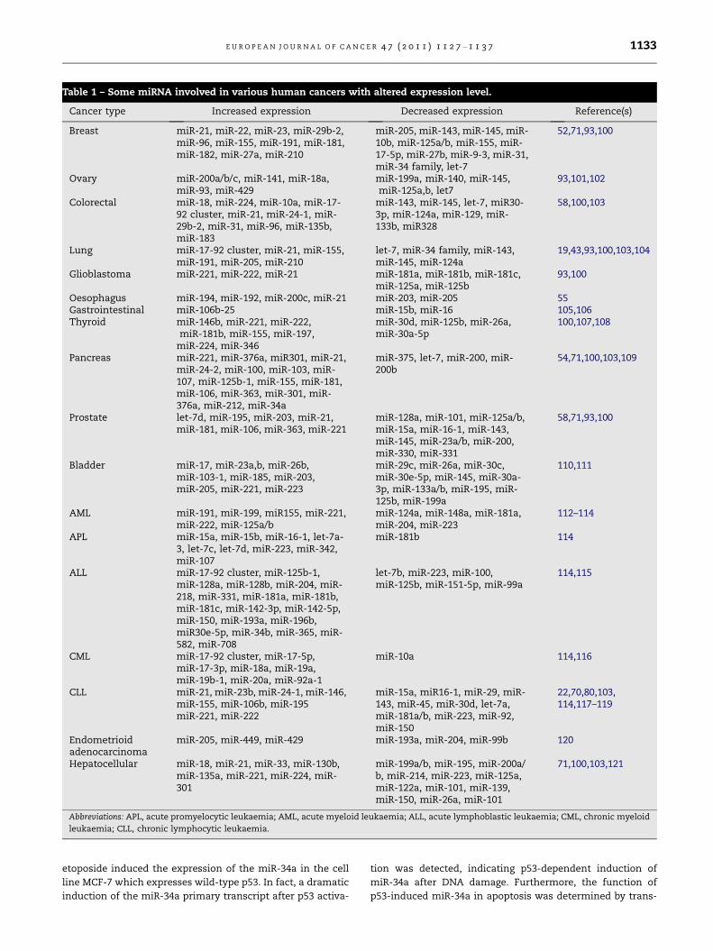
Table 1 – Some miRNA involved in various human cancers with altered expression level.
Cancer type Increased expression Decreased expression Reference(s)
Breast miR-21, miR-22, miR-23, miR-29b-2,miR-96, miR-155, miR-191, miR-181,miR-182, miR-27a, miR-210
miR-205, miR-143, miR-145, miR-10b, miR-125a/b, miR-155, miR-17-5p, miR-27b, miR-9-3, miR-31,miR-34 family, let-7
52,71,93,100
Ovary miR-200a/b/c, miR-141, miR-18a,miR-93, miR-429
miR-199a, miR-140, miR-145,miR-125a,b, let7
93,101,102
Colorectal miR-18, miR-224, miR-10a, miR-17-92 cluster, miR-21, miR-24-1, miR-29b-2, miR-31, miR-96, miR-135b,miR-183
miR-143, miR-145, let-7, miR30-3p, miR-124a, miR-129, miR-133b, miR328
58,100,103
Lung miR-17-92 cluster, miR-21, miR-155,miR-191, miR-205, miR-210
let-7, miR-34 family, miR-143,miR-145, miR-124a
19,43,93,100,103,104
Glioblastoma miR-221, miR-222, miR-21 miR-181a, miR-181b, miR-181c,miR-125a, miR-125b
93,100
Oesophagus miR-194, miR-192, miR-200c, miR-21 miR-203, miR-205 55Gastrointestinal miR-106b-25 miR-15b, miR-16 105,106Thyroid miR-146b, miR-221, miR-222,
miR-181b, miR-155, miR-197,miR-224, miR-346
miR-30d, miR-125b, miR-26a,miR-30a-5p
100,107,108
Pancreas miR-221, miR-376a, miR301, miR-21,miR-24-2, miR-100, miR-103, miR-107, miR-125b-1, miR-155, miR-181,miR-106, miR-363, miR-301, miR-376a, miR-212, miR-34a
miR-375, let-7, miR-200, miR-200b
54,71,100,103,109
Prostate let-7d, miR-195, miR-203, miR-21,miR-181, miR-106, miR-363, miR-221
miR-128a, miR-101, miR-125a/b,miR-15a, miR-16-1, miR-143,miR-145, miR-23a/b, miR-200,miR-330, miR-331
58,71,93,100
Bladder miR-17, miR-23a,b, miR-26b,miR-103-1, miR-185, miR-203,miR-205, miR-221, miR-223
miR-29c, miR-26a, miR-30c,miR-30e-5p, miR-145, miR-30a-3p, miR-133a/b, miR-195, miR-125b, miR-199a
110,111
AML miR-191, miR-199, miR155, miR-221,miR-222, miR-125a/b
miR-124a, miR-148a, miR-181a,miR-204, miR-223
112–114
APL miR-15a, miR-15b, miR-16-1, let-7a-3, let-7c, let-7d, miR-223, miR-342,miR-107
miR-181b 114
ALL miR-17-92 cluster, miR-125b-1,miR-128a, miR-128b, miR-204, miR-218, miR-331, miR-181a, miR-181b,miR-181c, miR-142-3p, miR-142-5p,miR-150, miR-193a, miR-196b,miR30e-5p, miR-34b, miR-365, miR-582, miR-708
let-7b, miR-223, miR-100,miR-125b, miR-151-5p, miR-99a
114,115
CML miR-17-92 cluster, miR-17-5p,miR-17-3p, miR-18a, miR-19a,miR-19b-1, miR-20a, miR-92a-1
miR-10a 114,116
CLL miR-21, miR-23b, miR-24-1, miR-146,miR-155, miR-106b, miR-195miR-221, miR-222
miR-15a, miR16-1, miR-29, miR-143, miR-45, miR-30d, let-7a,miR-181a/b, miR-223, miR-92,miR-150
22,70,80,103,114,117–119
Endometrioidadenocarcinoma
miR-205, miR-449, miR-429 miR-193a, miR-204, miR-99b 120
Hepatocellular miR-18, miR-21, miR-33, miR-130b,miR-135a, miR-221, miR-224, miR-301
miR-199a/b, miR-195, miR-200a/b, miR-214, miR-223, miR-125a,miR-122a, miR-101, miR-139,miR-150, miR-26a, miR-101
71,100,103,121
Abbreviations: APL, acute promyelocytic leukaemia; AML, acute myeloid leukaemia; ALL, acute lymphoblastic leukaemia; CML, chronic myeloid
leukaemia; CLL, chronic lymphocytic leukaemia.
E U R O P E A N J O U R N A L O F C A N C E R 4 7 ( 2 0 1 1 ) 1 1 2 7 – 1 1 3 7 1133
etoposide induced the expression of the miR-34a in the cell
line MCF-7 which expresses wild-type p53. In fact, a dramatic
induction of the miR-34a primary transcript after p53 activa-
tion was detected, indicating p53-dependent induction of
miR-34a after DNA damage. Furthermore, the function of
p53-induced miR-34a in apoptosis was determined by trans-
1134 E U R O P E A N J O U R N A L O F C A N C E R 4 7 ( 2 0 1 1 ) 1 1 2 7 – 1 1 3 7
fection of H1299 cells with duplex siRNAs corresponding to
processed miR-34a. DNA content analysis by flow cytometry
revealed an increase in the fractions of cells with a sub-G1
DNA content, which is indicative of apoptosis, whereas the
number of cells in S-phase and G2/M-phase was decreased.
Therefore, this study proposed that activation of miR-34a by
p53 may contribute to induction of apoptosis.
The anti-apoptotic proto-oncogene Bcl2 is down-regulated
by miR-15a and miR-16, two other p53-induced microRNAs
with tumour suppressive activity.22 Interestingly, the overex-
pression of miR-34 leads to a mild decrease in Bcl2 protein le-
vel.90 It is thought that miR-34a can act together with other
miRNAs such as miR-15 and miR-1622 to suppress efficiently
anti-apoptotic Bcl2. MiR-34a is a potent suppressor of cell pro-
liferation through modulation of E2F signalling pathway.
Thus, expression of miR-34a causes reprogramming of genes
involved in p53 mediated cell apoptosis. In addition, ectopic
expression of miR-34a reduces the levels of E2F3 by targeting
its mRNA.89,91 Other targets of the p53-induced miRNAs are
the NOTCH1 receptor and its ligand DLL1, which were con-
firmed to be the targets for down-regulation by miR-34a.12
4. Potential use of microRNAs in cancertherapy
As stated above, it has been demonstrated that miRNAs are
differentially expressed in human cancers and contribute to
cancer development (Table 1). However, it remains to be fur-
ther elucidated whether this altered expression of miRNAs
occurs as a consequence of the pathological state of cancer
or whether the cancer is a direct cause of this altered expres-
sion. Nonetheless, given that cancer cells often exhibit a dis-
tinctive pattern of miRNA expression and many functionality
validated miRNA targets are oncogenes and tumour suppres-
sors,92,93 it is thought that manipulating of miRNA expression
levels could be a potential therapeutic strategy for developing
efficient therapies against cancer. The feasibility of manipu-
lating miRNA expression levels have been demonstrated by
antisense oligonucleotide targeting experiments. For onco-
genic miRNAs, potential anti-miRNA therapeutics include
the locked nucleic acid (LNA)-modified oligonucleotides,94
the anti-miRNA oligonucleotides (AMOs)95 and the ‘antagom-
irs’.96 For miRNAs which function as tumour suppressor,
restoring the expression levels of these miRNAs via overex-
pression of them, in the purpose of consequent miRNA-med-
iated targeting of cancer oncogene, may be a potential
therapeutic strategy.27,92 However, the efficacy of the anti-
sense oligonucleotide targeting strategy needs to be further
evaluated by future in vivo studies of miRNA transgenics
and knockouts.92 Two examples about the potential use of
miRNA in cancer therapy are briefly described below.
Galardi et al.97 showed that ectopic overexpression of miR-
221 and miR-222 enhanced the growth potential of slowly
growing prostate carcinoma LNCaP cells and induced a pro-
gression to the S phase of cell cycle by targeting CDK inhibitor
p27, a dosage-dependent tumour suppressor in prostate can-
cer. This induced increase of the colony-forming potential of
LNCaP cells in soft agar. MiR-221 and miR-222 knockdown
through LNA antisense oligonucleotides increases the levels
of CDK inhibitor p27Kip1 in aggressive prostate carcinoma
PC3 cells, and considerably reduces in vitro clonogenicity fea-
ture of the cells. This study suggests that the overexpression
of miR-221 and miR-222 may contribute to the oncogenesis
and progression of prostate carcinoma, at least in part
through down-regulation of the tumour suppressor p27Kip1.
Therefore, miR-221 and miR-222 silencing based strategy
would be of value against prostate cancer.
Kefas et al.98 demonstrated that miR-326, a neuronally-ex-
pressed microRNA, acts in a feedback loop with Notch signal-
ling pathway in glioblastomas. Given the finding that Notch
and miR-326 each suppress the other, they speculate that this
Notch/miR-326 axis is shifted towards high Notch activity and
low miR-326 activity. Their results showed that efficient deliv-
ery of miR-326 has therapeutic potential against both glioma
stem-like cells and established glioma lines.
5. Conclusion and perspectives
Emerging evidence suggests that miRNAs play important
roles in human cancers and act as onco/tumour suppressor
miRNAs. MiRNAs may be involved in cancer development by
controlling cell differentiation and apoptosis or targeting can-
cer oncogenes and/or tumour suppressors. Intriguingly, due
to significantly altered expression profile of miRNA in differ-
ent types of cancer, miRNA profiling could be used for classi-
fication of human tumours,16 indicating the diagnostic and
even prognostic value of miRNA profiling.99 There is no doubt
that uncovering the crucial roles of miRNAs in cancer and the
potential of miRNA-based therapeutics opens up new oppor-
tunities in the future of cancer therapy. However, the basic
biology of cancer/miRNA pathways is not completely eluci-
dated and further studies, establishing the oncogenic or tu-
mour suppressive roles of miRNAs using in vivo
experimental models, are needed to gain more insights into
basic mechanisms of miRNA functions in cancer.
Conflict of interest statement
None declared.
Acknowledgements
We would like to thank all authors responsible for the insights
that we attempted to summarise. We apologise to the col-
leagues whose work could not be cited owing to space
limitations.
R E F E R E N C E S
1. Cullen BR. Derivation and function of small interfering RNAsand microRNAs. Virus Res 2004;102(1):3–9.
2. Liu X, Fortin K, Mourelatos Z. MicroRNAs: biogenesis andmolecular functions. Brain Pathol 2008;18(1):113–21.
3. Lee RC, Feinbaum RL, Ambros V. The C. elegans heterochronicgene lin-4 encodes small RNAs with antisensecomplementarity to lin-14. Cell 1993;75(5):843–54.
E U R O P E A N J O U R N A L O F C A N C E R 4 7 ( 2 0 1 1 ) 1 1 2 7 – 1 1 3 7 1135
4. Reinhart BJ, Slack FJ, Basson M, et al. The 21-nucleotide let-7RNA regulates developmental timing in Caenorhabditiselegans. Nature 2000;403(6772):901–6.
5. Lee Y, Kim M, Han J, et al. MicroRNA genes are transcribed byRNA polymerase II. EMBO J 2004;23(20):4051–60.
6. Borchert GM, Lanier W, Davidson BL. RNA polymerase IIItranscribes human microRNAs. Nat Struct Mol Biol2006;13(12):1097–101.
7. Lee Y, Jeon K, Lee JT, Kim S, Kim VN. MicroRNA maturation:stepwise processing and subcellular localization. EMBO J2002;21(17):4663–70.
8. Hutvagner G, Zamore PD. A microRNA in a multiple-turnoverRNAi enzyme complex. Science 2002;297(5589):2056–60.
9. Brennecke J, Stark A, Russell RB, Cohen SM. Principles ofmicroRNA-target recognition. PLoS Biol 2005;3(3):e85.
10. John B, Enright AJ, Aravin A, et al. Human microRNA targets.PLoS Biol 2004;2(11):e363.
11. Krek A, Grun D, Poy MN, et al. Combinatorial microRNAtarget predictions. Nat Genet 2005;37(5):495–500.
12. Lewis BP, Shih IH, Jones-Rhoades MW, Bartel DP, Burge CB.Prediction of mammalian microRNA targets. Cell2003;115(7):787–98.
13. Chan JA, Krichevsky AM, Kosik KS. MicroRNA-21 is anantiapoptotic factor in human glioblastoma cells. Cancer Res2005;65(14):6029–33.
14. Cheng AM, Byrom MW, Shelton J, Ford LP. Antisenseinhibition of human miRNAs and indications for aninvolvement of miRNA in cell growth and apoptosis. NucleicAcids Res 2005;33(4):1290–7.
[15]. Bandyopadhyay S, Mitra R, Maulik U, Zhang MQ.Development of the human cancer microRNA network.Silence 2010;1(1):6.
16. Lu J, Getz G, Miska EA, et al. MicroRNA expression profilesclassify human cancers. Nature 2005;435(7043):834–8.
17. Esquela-Kerscher A, Slack FJ. Oncomirs – microRNAs with arole in cancer. Nat Rev Cancer 2006;6(4):259–69.
18. Volinia S, Calin GA, Liu CG, et al. A microRNA expressionsignature of human solid tumors defines cancer genetargets. Proc Natl Acad Sci USA 2006;103(7):2257–61.
19. Johnson SM, Grosshans H, Shingara J, et al. RAS is regulatedby the let-7 microRNA family. Cell 2005;120(5):635–47.
20. Lee YS, Dutta A. The tumor suppressor microRNA let-7represses the HMGA2 oncogene. Genes Dev2007;21(9):1025–30.
21. Mayr C, Hemann MT, Bartel DP. Disrupting the pairingbetween let-7 and Hmga2 enhances oncogenictransformation. Science 2007;315(5818):1576–9.
22. Cimmino A, Calin GA, Fabbri M, et al. miR-15 and miR-16induce apoptosis by targeting BCL2. Proc Natl Acad Sci USA2005;102(39):13944–9.
23. Hermeking H. P53 enters the microRNA world. Cancer Cell2007;12(5):414–8.
24. Zhu S, Si ML, Wu H, Mo YY. MicroRNA-21 targets the tumorsuppressor gene tropomyosin 1 (TPM1). J Biol Chem2007;282(19):14328–36.
25. Frankel LB, Christoffersen NR, Jacobsen A, et al.Programmed cell death 4 (PDCD4) is an important functionaltarget of the microRNA miR-21 in breast cancer cells. J BiolChem 2008;283(2):1026–33.
26. Meng F, Henson R, Wehbe-Janek H, et al. MicroRNA-21regulates expression of the PTEN tumor suppressor gene inhuman hepatocellular cancer. Gastroenterology2007;133(2):647–58.
27. Meng F, Henson R, Lang M, et al. Involvement of humanmicro-RNA in growth and response to chemotherapy inhuman cholangiocarcinoma cell lines. Gastroenterology2006;130(7):2113–29.
28. He L, Thomson JM, Hemann MT, et al. A microRNApolycistron as a potential human oncogene. Nature2005;435(7043):828–33.
29. Ota A, Tagawa H, Karnan S, et al. Identification andcharacterization of a novel gene, C13orf25, as a target for13q31–q32 amplification in malignant lymphoma. Cancer Res2004;64(9):3087–95.
30. Li C, Feng Y, Coukos G, Zhang L. Therapeutic microRNAstrategies in human cancer. AAPS J 2009;11(4):747–57.
31. Fernandez PC, Frank SR, Wang L, et al. Genomic targets ofthe human c-Myc protein. Genes Dev 2003;17(9):1115–29.
32. O’Donnell KA, Wentzel EA, Zeller KI, Dang CV, Mendell JT. c-Myc-regulated microRNAs modulate E2F1 expression. Nature2005;435(7043):839–43.
33. Lewis BP, Burge CB, Bartel DP. Conserved seed pairing, oftenflanked by adenosines, indicates that thousands of humangenes are microRNA targets. Cell 2005;120(1):15–20.
34. Yu Z, Wang C, Wang M, et al. A cyclin D1/microRNA 17/20regulatory feedback loop in control of breast cancer cellproliferation. J Cell Biol 2008;182(3):509–17.
35. Xiao C, Srinivasan L, Calado DP, et al. Lymphoproliferativedisease and autoimmunity in mice with increased miR-17-92expression in lymphocytes. Nat Immunol 2008;9(4):405–14.
36. Hammond SM. MicroRNAs as oncogenes. Curr Opin Genet Dev2006;16(1):4–9.
37. Hammond SM. MicroRNAs as tumor suppressors. Nat Genet2007;39(5):582–3.
38. Altomare DA, Wang HQ, Skele KL, et al. AKT and mTORphosphorylation is frequently detected in ovarian cancerand can be targeted to disrupt ovarian tumor cell growth.Oncogene 2004;23(34):5853–7.
39. Inomata M, Tagawa H, Guo YM, et al. MicroRNA-17-92down-regulates expression of distinct targets in different B-cell lymphoma subtypes. Blood 2009;113(2):396–402.
40. Park SM, Shell S, Radjabi AR, et al. Let-7 prevents earlycancer progression by suppressing expression of theembryonic gene HMGA2. Cell Cycle 2007;6(21):2585–90.
41. Calin GA, Sevignani C, Dumitru CD, et al. Human microRNAgenes are frequently located at fragile sites and genomicregions involved in cancers. Proc Natl Acad Sci USA2004;101(9):2999–3004.
42. Bhat-Nakshatri P, Wang G, Collins NR, et al. Estradiol-regulated microRNAs control estradiol response in breastcancer cells. Nucleic Acids Res 2009;37(14):4850–61.
43. Yanaihara N, Caplen N, Bowman E, et al. Unique microRNAmolecular profiles in lung cancer diagnosis and prognosis.Cancer Cell 2006;9(3):189–98.
44. Johnson CD, Esquela-Kerscher A, Stefani G, et al. The let-7microRNA represses cell proliferation pathways in humancells. Cancer Res 2007;67(16):7713–22.
45. Lall S, Grun D, Krek A, et al. A genome-wide map ofconserved microRNA targets in C. elegans. Curr Biol2006;16(5):460–71.
46. Lynam-Lennon N, Maher SG, Reynolds JV. The roles ofmicroRNA in cancer and apoptosis. Biol Rev Camb Philos Soc2009;84(1):55–71.
47. Kumar MS, Erkeland SJ, Pester RE, et al. Suppression of non-small cell lung tumor development by the let-7 microRNAfamily. Proc Natl Acad Sci USA 2008;105(10):3903–8.
48. Meng F, Henson R, Wehbe-Janek H, et al. The microRNA let-7a modulates interleukin-6-dependent STAT-3 survivalsignaling in malignant human cholangiocytes. J Biol Chem2007;282(11):8256–64.
49. Tsang WP, Kwok TT. Let-7a microRNA suppressestherapeutics-induced cancer cell death by targeting caspase-3. Apoptosis 2008;13(10):1215–22.
50. Danial NN. BCL-2 family proteins: critical checkpoints ofapoptotic cell death. Clin Cancer Res 2007;13(24):7254–63.
1136 E U R O P E A N J O U R N A L O F C A N C E R 4 7 ( 2 0 1 1 ) 1 1 2 7 – 1 1 3 7
51. Hanahan D, Weinberg RA. The hallmarks of cancer. Cell2000;100(1):57–70.
52. Iorio MV, Ferracin M, Liu CG, et al. MicroRNA geneexpression deregulation in human breast cancer. Cancer Res2005;65(16):7065–70.
53. Bloomston M, Frankel WL, Petrocca F, et al. MicroRNAexpression patterns to differentiate pancreaticadenocarcinoma from normal pancreas and chronicpancreatitis. JAMA 2007;297(17):1901–8.
54. Lee EJ, Gusev Y, Jiang J, et al. Expression profiling identifiesmicroRNA signature in pancreatic cancer. Int J Cancer2007;120(5):1046–54.
55. Feber A, Xi L, Luketich JD, et al. MicroRNA expressionprofiles of esophageal cancer. J Thorac Cardiovasc Surg2008;135(2):255–60. discussion 260.
56. Lui WO, Pourmand N, Patterson BK, Fire A. Patterns of knownand novel small RNAs in human cervical cancer. Cancer Res2007;67(13):6031–43.
57. Schetter AJ, Leung SY, Sohn JJ, et al. MicroRNA expressionprofiles associated with prognosis and therapeutic outcomein colon adenocarcinoma. JAMA 2008;299(4):425–36.
58. Gartel AL, Kandel ES. miRNAs: little known mediators ofoncogenesis. Semin Cancer Biol 2008;18(2):103–10.
59. Papagiannakopoulos T, Shapiro A, Kosik KS. MicroRNA-21targets a network of key tumor-suppressive pathways inglioblastoma cells. Cancer Res 2008;68(19):8164–72.
60. Yang HS, Jansen AP, Nair R, et al. A novel transformationsuppressor, Pdcd4, inhibits AP-1 transactivation but not NF-kappaB or ODC transactivation. Oncogene 2001;20(6):669–76.
61. Goke R, Barth P, Schmidt A, Samans B, Lankat-Buttgereit B.Programmed cell death protein 4 suppresses CDK1/cdc2 viainduction of p21(Waf1/Cip1). Am J Physiol Cell Physiol2004;287(6):C1541–6.
62. Yang HS, Knies JL, Stark C, Colburn NH. Pdcd4 suppressestumor phenotype in JB6 cells by inhibiting AP-1transactivation. Oncogene 2003;22(24):3712–20.
63. Chen Y, Knosel T, Kristiansen G, et al. Loss of PDCD4expression in human lung cancer correlates with tumourprogression and prognosis. J Pathol 2003;200(5):640–6.
64. Gao F, Zhang P, Zhou C, et al. Frequent loss of PDCD4expression in human glioma: possible role in thetumorigenesis of glioma. Oncol Rep 2007;17(1):123–8.
65. Zhang H, Ozaki I, Mizuta T, et al. Involvement ofprogrammed cell death 4 in transforming growth factor-beta1-induced apoptosis in human hepatocellularcarcinoma. Oncogene 2006;25(45):6101–12.
66. Cory S, Adams JM. The Bcl2 family: regulators of the cellularlife-or-death switch. Nat Rev Cancer 2002;2(9):647–56.
67. Krajewski S, Krajewska M, Turner BC, et al. Prognosticsignificance of apoptosis regulators in breast cancer. EndocrRelat Cancer 1999;6(1):29–40.
68. Rassidakis GZ, Medeiros LJ, Vassilakopoulos TP, et al. BCL-2expression in Hodgkin and Reed-Sternberg cells of classicalHodgkin disease predicts a poorer prognosis in patientstreated with ABVD or equivalent regimens. Blood2002;100(12):3935–41.
69. Sanchez-Beato M, Sanchez-Aguilera A, Piris MA. Cell cyclederegulation in B-cell lymphomas. Blood 2003;101(4):1220–35.
70. Calin GA, Dumitru CD, Shimizu M, et al. Frequent deletionsand down-regulation of micro- RNA genes miR15 and miR16at 13q14 in chronic lymphocytic leukemia. Proc Natl Acad SciUSA 2002;99(24):15524–9.
71. Cho WC. MicroRNAs in cancer – from research to therapy.Biochim Biophys Acta 2010;1805(2):209–17.
72. Bandi N, Zbinden S, Gugger M, et al. miR-15a and miR-16 areimplicated in cell cycle regulation in a Rb-dependentmanner and are frequently deleted or down-regulated innon-small cell lung cancer. Cancer Res 2009;69(13):5553–9.
73. Chen L, Willis SN, Wei A, et al. Differential targeting ofprosurvival Bcl-2 proteins by their BH3-only ligands allowscomplementary apoptotic function. Mol Cell2005;17(3):393–403.
74. Kobayashi S, Werneburg NW, Bronk SF, Kaufmann SH, GoresGJ. Interleukin-6 contributes to Mcl-1 up-regulation andTRAIL resistance via an Akt-signaling pathway incholangiocarcinoma cells. Gastroenterology2005;128(7):2054–65.
75. Taniai M, Grambihler A, Higuchi H, et al. Mcl-1 mediatestumor necrosis factor-related apoptosis-inducing ligandresistance in human cholangiocarcinoma cells. Cancer Res2004;64(10):3517–24.
76. Mott JL, Kobayashi S, Bronk SF, Gores GJ. Mir-29 regulatesMcl-1 protein expression and apoptosis. Oncogene2007;26(42):6133–40.
77. Garzon R, Heaphy CE, Havelange V, et al. MicroRNA 29bfunctions in acute myeloid leukemia. Blood2009;114(26):5331–41.
78. Garzon R, Liu S, Fabbri M, et al. MicroRNA-29b inducesglobal DNA hypomethylation and tumor suppressor genereexpression in acute myeloid leukemia by targeting directlyDNMT3A and 3B and indirectly DNMT1. Blood2009;113(25):6411–8.
79. Marcucci G, Mrozek K, Radmacher MD, Bloomfield CD, CroceCM. MicroRNA expression profiling in acute myeloid andchronic lymphocytic leukaemias. Best Pract Res Clin Haematol2009;22(2):239–48.
80. Calin GA, Ferracin M, Cimmino A, et al. A microRNAsignature associated with prognosis and progression inchronic lymphocytic leukemia. N Engl J Med2005;353(17):1793–801.
81. Cummins JM, He Y, Leary RJ, et al. The colorectalmicroRNAome. Proc Natl Acad Sci USA 2006;103(10):3687–92.
82. Shu KX, Li B, Wu LX. The p53 network: p53 and itsdownstream genes. Colloids Surf B Biointerfaces2007;55(1):10–8.
83. Haupt S, Berger M, Goldberg Z, Haupt Y. Apoptosis – the p53network. J Cell Sci 2003;116(Pt 20):4077–85.
84. Mihara M, Erster S, Zaika A, et al. p53 has a directapoptogenic role at the mitochondria. Mol Cell2003;11(3):577–90.
85. Bommer GT, Gerin I, Feng Y, et al. p53-mediated activationof miRNA34 candidate tumor-suppressor genes. Curr Biol2007;17(15):1298–307.
86. Chang TC, Wentzel EA, Kent OA, et al. Transactivation ofmiR-34a by p53 broadly influences gene expression andpromotes apoptosis. Mol Cell 2007;26(5):745–52.
87. Christoffersen NR, Shalgi R, Frankel LB, et al. p53-independent upregulation of miR-34a during oncogene-induced senescence represses MYC. Cell Death Differ2010;17(2):236–45.
88. He X, He L, Hannon GJ. The guardian’s little helper:microRNAs in the p53 tumor suppressor network. Cancer Res2007;67(23):11099–101.
89. Tarasov V, Jung P, Verdoodt B, et al. Differential regulation ofmicroRNAs by p53 revealed by massively parallelsequencing: miR-34a is a p53 target that induces apoptosisand G1-arrest. Cell Cycle 2007;6(13):1586–93.
90. Raver-Shapira N, Marciano E, Meiri E, et al. Transcriptionalactivation of miR-34a contributes to p53-mediatedapoptosis. Mol Cell 2007;26(5):731–43.
91. Welch C, Chen Y, Stallings RL. MicroRNA-34a functions as apotential tumor suppressor by inducing apoptosis inneuroblastoma cells. Oncogene 2007;26(34):5017–22.
92. Calin GA, Croce CM. MicroRNA signatures in human cancers.Nat Rev Cancer 2006;6(11):857–66.
E U R O P E A N J O U R N A L O F C A N C E R 4 7 ( 2 0 1 1 ) 1 1 2 7 – 1 1 3 7 1137
93. Spizzo R, Nicoloso MS, Croce CM, Calin GA. SnapShot:microRNAs in cancer. Cell 2009;137(3):586. e.1.
94. Orom UA, Kauppinen S, Lund AH. LNA-modifiedoligonucleotides mediate specific inhibition of microRNAfunction. Gene 2006;372:137–41.
95. Weiler J, Hunziker J, Hall J. Anti-miRNA oligonucleotides(AMOs): ammunition to target miRNAs implicated in humandisease? Gene Ther 2006;13(6):496–502.
96. Krutzfeldt J, Rajewsky N, Braich R, et al. Silencing ofmicroRNAs in vivo with ‘antagomirs’. Nature2005;438(7068):685–9.
97. Galardi S, Mercatelli N, Giorda E, et al. miR-221 and miR-222expression affects the proliferation potential of humanprostate carcinoma cell lines by targeting p27Kip1. J BiolChem 2007;282(32):23716–24.
98. Kefas B, Comeau L, Floyd DH, et al. The neuronal microRNAmiR-326 acts in a feedback loop with notch and hastherapeutic potential against brain tumors. J Neurosci2009;29(48):15161–8.
99. Cho WC. MicroRNAs: potential biomarkers for cancerdiagnosis, prognosis and targets for therapy. Int J Biochem CellBiol 2010;42(8):1273–81.
100. Lowery AJ, Miller N, McNeill RE, Kerin MJ. MicroRNAs asprognostic indicators and therapeutic targets: potentialeffect on breast cancer management. Clin Cancer Res2008;14(2):360–5.
101. Iorio MV, Visone R, Di Leva G, et al. MicroRNA signatures inhuman ovarian cancer. Cancer Res 2007;67(18):8699–707.
102. Nam EJ, Yoon H, Kim SW, et al. MicroRNA expression profilesin serous ovarian carcinoma. Clin Cancer Res2008;14(9):2690–5.
103. Barbarotto E, Schmittgen TD, Calin GA. MicroRNAs andcancer: profile, profile, profile. Int J Cancer 2008;122(5):969–77.
104. Hayashita Y, Osada H, Tatematsu Y, et al. A polycistronicmicroRNA cluster, miR-17-92, is overexpressed in humanlung cancers and enhances cell proliferation. Cancer Res2005;65(21):9628–32.
105. Xia L, Zhang D, Du R, et al. miR-15b and miR-16 modulatemultidrug resistance by targeting BCL2 in human gastriccancer cells. Int J Cancer 2008;123(2):372–9.
106. Petrocca F, Visone R, Onelli MR, et al. E2F1-regulatedmicroRNAs impair TGFbeta-dependent cell-cycle arrest andapoptosis in gastric cancer. Cancer Cell 2008;13(3):272–86.
107. Nikiforova MN, Tseng GC, Steward D, Diorio D, Nikiforov YE.MicroRNA expression profiling of thyroid tumors: biologicalsignificance and diagnostic utility. J Clin Endocrinol Metab2008;93(5):1600–8.
108. Visone R, Pallante P, Vecchione A, et al. Specific microRNAsare downregulated in human thyroid anaplastic carcinomas.Oncogene 2007;26(54):7590–5.
109. Dutta KK, Zhong Y, Liu YT, et al. Association of microRNA-34a overexpression with proliferation is cell type-dependent.Cancer Sci 2007;98(12):1845–52.
110. Ichimi T, Enokida H, Okuno Y, et al. Identification of novelmicroRNA targets based on microRNA signatures in bladdercancer. Int J Cancer 2009;125(2):345–52.
111. Gottardo F, Liu CG, Ferracin M, et al. Micro-RNA profiling inkidney and bladder cancers. Urol Oncol 2007;25(5):387–92.
112. Garzon R, Garofalo M, Martelli MP, et al. DistinctivemicroRNA signature of acute myeloid leukemia bearingcytoplasmic mutated nucleophosmin. Proc Natl Acad Sci USA2008;105(10):3945–50.
113. O’Connell RM, Rao DS, Chaudhuri AA, et al. Sustainedexpression of microRNA-155 in hematopoietic stem cellscauses a myeloproliferative disorder. J Exp Med2008;205(3):585–94.
114. Zhao H, Wang D, Du W, Gu D, Yang R. MicroRNA andleukemia: tiny molecule, great function. Crit Rev OncolHematol 2010;74(3):149–55.
115. Zanette DL, Rivadavia F, Molfetta GA, et al. miRNAexpression profiles in chronic lymphocytic and acutelymphocytic leukemia. Braz J Med Biol Res2007;40(11):1435–40.
116. Venturini L, Battmer K, Castoldi M, et al. Expression of themiR-17-92 polycistron in chronic myeloid leukemia (CML)CD34+ cells. Blood 2007;109(10):4399–405.
117. Akao Y, Nakagawa Y, Kitade Y, Kinoshita T, Naoe T.Downregulation of microRNAs-143 and -145 in B-cellmalignancies. Cancer Sci 2007;98(12):1914–20.
118. Marton S, Garcia MR, Robello C, et al. Small RNAs analysis inCLL reveals a deregulation of miRNA expression and novelmiRNA candidates of putative relevance in CLLpathogenesis. Leukemia 2008;22(2):330–8.
119. Pekarsky Y, Santanam U, Cimmino A, et al. Tcl1 expressionin chronic lymphocytic leukemia is regulated by miR-29 andmiR-181. Cancer Res 2006;66(24):11590–3.
120. Wu W, Lin Z, Zhuang Z, Liang X. Expression profile ofmammalian microRNAs in endometrioid adenocarcinoma.Eur J Cancer Prev 2009;18(1):50–5.
121. Jiang J, Gusev Y, Aderca I, et al. Association of microRNAexpression in hepatocellular carcinomas with hepatitisinfection, cirrhosis, and patient survival. Clin Cancer Res2008;14(2):419–27.