Embed Size (px)
Citation preview

Journal of Cellular Biochemistry 89:133–143 (2003)
AIDS-Related Kaposi’s Sarcoma Cells Rapidly InternalizeEndostatin, Which Co-Localizes to TropomysinMicrofilaments and Inhibits Cytokine-MediatedMigration and Invasion
Susan R. Mallery,1,3* Mark A. Morse,2,3 Ralph F. Wilson,1 Ping Pei,1 Gregory M. Ness,1
Jennifer E. Bradburn,1 Robert J. Renner,1 David E. Schuller,2,4 and Fredika M. Robertson3,5
1Departments of Oral Maxillofacial Surgery and Pathology and Periodontology, College of Dentistry,Ohio State University, Columbus, Ohio2Division of Environmental Health Sciences, School of Public Health, College of Medicine,Ohio State University, Columbus, Ohio3The Ohio State University Comprehensive Cancer Center, Ohio State University, Columbus, OH4James Cancer Hospital and Solove Research Institute, Ohio State University, Columbus, OH5Department of Molecular Virology, Immunology, and Medical Genetics, College of Medicine,Ohio State University, Columbus, Ohio
Abstract AIDS-related Kaposi’s sarcoma (KS) is the most common HIV-related malignancy. In some respects, KS isanalogous to other angioproliferative diseases, in that KS lesions are highly vascularized and promoted by inflammatorycytokines. However, unlike other cancers or inflammatory mediated vascular diseases, KS is unique in that the KS lesionalcells both express and respond to the complete angiogenic cytokines vascular endothelial growth factor (VEGF) and basicfibroblast growth factor (bFGF). Therefore, the angiogenic phenotype, which is crucial for cancer progression, is inherentto KS tumor cells. Due to the recognized importance of angiogenesis in cancer progression, numerous angiostatic agentsare being investigated as potential therapeutic agents. One such agent is endostatin, which is a 20-kDa carboxyl-terminalfragment of collagen XVIII that has demonstrated potent angiostatic activities at both the in vivo and in vitro levels. Sinceendostatin is recognized as a potent angiostatic agent, the majority of in vitro endostatin studies have evaluated its effectson endothelial cells. Although KS cells are speculated to arise from endothelial cell precursors and KS lesions are highlyvascularized, no previous studies have investigated endostatin–KS cell interactions. This present study evaluatedendostatin’s effects on KS tumor cell: (i) signal transduction (endostatin internalization and transcription factor activation),and (ii) migration and invasion (functional activity assays and tropomysin co-localization). Our results show that KScells rapidly internalize endostatin and that endostatin initiates activation of the transcription activating factors nuclearfactor-kB (NF-kB) and activating protein 1 (AP-1). Our data also show that internalized endostatin co-localizes totropomysin microfilaments and acts to inhibit KS cell migration and invasion in response to the clinically relevantangiogenic cytokines VEGF and bFGF. As a consequence of its combined angiostatic and antitumorigenic activities,endostatin could provide dual therapeutic benefits for patients with mucocutaneous KS. J. Cell. Biochem. 89: 133–143,2003. Published 2003 Wiley-Liss, Inc.y
Key words: angiostatic agents; Kaposi’s sarcoma; angiogenesis; migration; invasion; tumorigenesis
Published 2003 Wiley-Liss, Inc. yThis article is a US Government work,and as such, is in the public domain of the United States of America.
*Correspondence to: Dr. Susan R. Mallery, Departmentof Oral Maxillofacial Surgery and Pathology, 305 W 12thAve., P.O. Box 182357, Columbus, Ohio 43218-2357.E-mail: [email protected]
Received 3 January 2003; Accepted 6 January 2003
DOI 10.1002/jcb.10489
Ralph F. Wilson is the recipient of the Ohio Division of theAmerican Cancer Society Research Fellowship.
David E. Schuller is the Director of James Cancer Hospitaland Solove Research Institute.
Grant sponsor: NIH/NIDCR; Grant numbers: PO1 DE12704, UO1 CA 66531, and P30 CA 16058.
*Correspondence to: Dr. Susan R. Mallery, Departmentof Oral Maxillofacial Surgery and Pathology, 305 W 12th

Both clinical and experimental evidenceimply that AIDS-related Kaposi’s sarcoma(KS) is initially an angioproliferative hyperplasia that employs autocrine and paracrinegrowth loops to facilitate progression to a highlyangiogenic sarcoma [Miles, 1994; Jacobson andArnemian, 1995]. Analogous to other angiopro-liferative diseases such as rheumatoid arthritis,KS lesions are highly vascularized and pro-moted by inflammatory cytokines. However,in contrast to other cancers or inflammatorymediated diseases, KS is unique in that the KSlesional spindle cells both express and re-spond to the potent and complete angio-genic cytokines basic fibroblast growth factor(bFGF) and vascular endothelial growth factor(VEGF) [Miles et al., 1990; Masood et al., 1997;Samaniego et al., 1998]. Consequently, neovas-cularization is intimately associated with KSprogression. During angiogenesis, endothelialcells invade and infiltrate surrounding tissues,where they undergo division and differentiationinto microtubules [Folkman and Shing, 1992].Similar to their putative endothelial originprogenitor cells [Rutger et al., 1986], KS cellspossess migratory and invasive properties thatfacilitate KS angiogenesis and tumorigenesis.
Due to the recognized importance of angio-genesis in cancer progression, numerous angio-static agents are currently being investigated aspotential therapeutic agents [Folkman, 1995;Gasparini, 1999]. One such agent is endostatin,which is a 20-kDa carboxyl-terminal fragmentof collagen XVIII discovered by O’Reilly et al.[1997]. Although the mechanism(s) of action ofendostatin has not been completely delineated,several studies have provided mechanisticinsights. While immobilized endostatin servesas an integrin agonist that facilitates cellmigration, soluble endostatin perturbs integ-rin–substrate interactions, and inhibits cellattachment and migration [Rehn et al., 2001].Studies that investigated endostatin’s effects onintracellular signaling showed soluble endo-statin initiated pathways that reduced expres-sion of a bank of growth-associated genes in awide range of endothelial lineage cells [Shichiriand Hirata, 2001]. Additional investigations byMacDonald et al. [2001], which demonstratedthat internalized endostatin bound tropomysin-containing microfilaments of endothelial cells,provided a plausible mechanism for endosta-tin’s inhibition of endothelial cell migration andinvasion. Further, Lee et al. [2002] have shown
a potential mechanism whereby endostatin sup-presses cell invasion as their data show endo-statin directly binds to the catalytic domain ofmatrix metalloproteinase-2 (MMP-2), therebysuppressing MMP-2’s catalytic function. Cumu-latively, these studies show endostatin’s pleio-trophic effects are the consequence of diversemechanisms that modulate numerous cellularpathways. Further clarification of these cellularmechanisms and identification of endostatin-responsive cell types could extend therapeuticapplications of endostatin beyond inhibition ofangiogenesis.
Because endostatin is recognized as a potentangiostatic agent, the vast majority of endosta-tin cell-based studies have employed endo-thelial cells. In contrast, this present studyinvestigated whether endostatin modulatesfunctional activities of KS tumor cells. We re-port, for the first time, that KS cells rapidlyinternalize endostatin and that endostatininitiates activation of the transcription activat-ing factors nuclear factor-kB (NF-kB) andactivating protein 1 (AP-1). We also show thatinternalized endostatin co-localizes to tropomy-sin microfilaments and acts to inhibit KS cellmigration and invasion in response to theclinically relevant complete angiogenic cyto-kines bFGF and VEGF.
MATERIALS AND METHODS
Isolation and Characterization ofAIDS-KS Cells
AIDS-KS cells were isolated from biopsyconfirmed tumors of AIDS-KS as previouslydescribed [Mallery et al., 2000a]. These AIDS-KS strains have been shown to possess a normalXY karyotype and show aspects of a trans-formed phenotype including: high production of‘‘KS’’ related cytokines such as IL-6, TNF-a, andbFGF, capacity to undergo multiple populationdoublings (>30) in reduced serum (0.5%) med-ium, and loss of anchorage dependence [Baileret al., 1995]. Furthermore, relative to matched,nonlesional cells from the AIDS-KS donors, theKS spindle cells possess unique biochemicalfeatures including reduced cytoprotective en-zyme function, increased responsiveness to pro-inflammatory cytokines, and reduced toleranceto chemotherapeutic agents that function byredox cycling [Mallery et al., 1999, 2000b]. TheAIDS-KS cells were cultured at 378C, 5% CO2 in‘‘complete’’ medium, which consisted of: M-199
134 Mallery et al.

(GIBCO, Grand Island, NY), supplementedwith 15 mM HEPES, 0.23 mg/ml L-glutamine,11 mg/ml sodium pyruvate, sodium heparin(Sigma, St. Louis, MO, 90 mg/ml), 10% heatinactivated fetal bovine serum (Hyclone, Logan,UT).
To reduce nonspecific integrin and matrixmetalloproteinase expression due to the variouscytokines and growth factors present in serumand provide a baseline from which to assessendostatin and/or cytokineresponsiveness, cellswere cultured in serum-free M-199 (base)medium for 72 h prior to conduction of eithermigration, invasion, and electromobility shiftassays. While serum deprivation reduced mito-tic indices and growth rates, cell viabilityremained comparable (>95%) to log growthconditions. KS cell strains (ks.1, ks.2, ks.3)isolated from three nodular phase intraoral KStumors were used for these studies. Thesestrains were selected due to their high histologicgrade and aggressive clinical behavior, i.e.,destruction of underlying alveolar bone andprimary tumors of �1 cm.
Assessment of KS Cell Capacity to InternalizeBiotinylated Endostatin
A total of 1� 104 KS cells were plated on Lab-Tek chamber slides coated with 0.5 ml of poly-L-lysine (at a concentration of 0.1 mg/ml) andcultured in serum-free M-199 base medium for24 h, followed by a 24 h incubation in basemediumþ 50 ng/ml bFGF. Endostatin (Calbio-chem, La Jolla, CA) was biotin labeled using theImmunoprobeTM Biotinylation Kit (Sigma),with a final biotin:protein ratio of 2.2:1. TheKS cells were then incubated with 10 mg/ml ofbiotinylated endostatin for specific periods (0, 5,30, 90, 150 min) at 378C, 5% CO2. At thedesignated harvest points, slides were fixed(methanol and acetone washes), followed by theaddition of Alex 488-conjugated avidin (Mole-cular Probes, Eugene, OR, final concentration1 mg/ml in PBS) and ProLong Antifade mount-ing medium (Molecular Probes). Cell-associatedendostatin was then determined by fluores-cence microscopy.
Determination of Endostatin’s Effects onActivation Status of NF-kB and AP-1 by
Electromobility Shift Assays (EMSAs)
EMSAs were conducted to determine whetheror not endostatin challenge activated KS cellmembranes and initiated a signal transduction
cascade. The experimental groups for theseexperiments were: (i) log growth, (ii) 72 h seradeprived (negative control), (iii) sera deprived,treated for 30 min with endostatin (10 mg/ml),(iv) sera deprived, treated for 30 min with TNF-a (100 U/ml, positive control). Nuclear andcytosolic extracts were prepared from trypsi-nized cells using a commercially available celllysis buffer (NE-PER #78833, Pierce, Rockford,IL) which contained a protease inhibitor cock-tail that was added to the lysis buffer just priorto use (Halt Protease inhibitor cocktail kit#78410, Pierce).
Every EMSA run included positive nuclearextracts [HeLa (AP-1 and NF-kB) from Pro-mega, Madison, WI] as well as negative controlsconsisting of reagents without nuclear extracts.EMSAs were conducted using the LightShiftChemiluminescent EMSA kit (Pierce #20148).This method employs a nonisotopic method todetect DNA–protein interaction, and uses bio-tin end-labeled DNA that is detected using astreptavidin–horseradish peroxidase conjugateand a chemiluminescent substrate. EMSAsconducted using LightShift assays requirebiotin-labeled double-stranded DNA, with end-labeling of both complimentary oligonucleotidesseparately, followed by annealing at room tem-perature. The consensus oligonucleotide se-quences (50 to 30) used were: ACT TGA GGGGAC TTT CCC AGG C (NF-kB, forward), GCCTGG GAA AGT CCC CTC AAC T (NF-kB,reverse); CGC TTG ATG AGT CAG CCG GAA(AP-1, forward), TTC CGG CTG ACT CAT CAAGCG (AP-1, reverse). Three assay controls,which consisted of biotin–EBNA control DNAonly, biotin–EBNA control DNA and EBNAextract, and biotin–EBNA control DNA, EBNAextract, and a 200-fold molar excess of unla-beled EBNA DNA, were included in everyEMSA conducted. A fourth KS strain (ks.4),which was also isolated from a patient with anodular phase intraoral KS tumor, was in-cluded for the EMSA analyses.
Fluorescent Staining of KS Cells
KS cells (1� 104) were plated on Lab-Tekchamber slides (Naperville, IL) coated withhuman plasma fibronectin (25mg/ml), incubatedovernight in proliferative growth medium (M-199 supplemented with 10% heat inactivatedFBS), and then fixed in 10% neutral formalin,followed by a methanol wash in accordance withthe methods of MacDonald et al., 2001. The
KS Cells Internalize and Respond to Endostatin 135
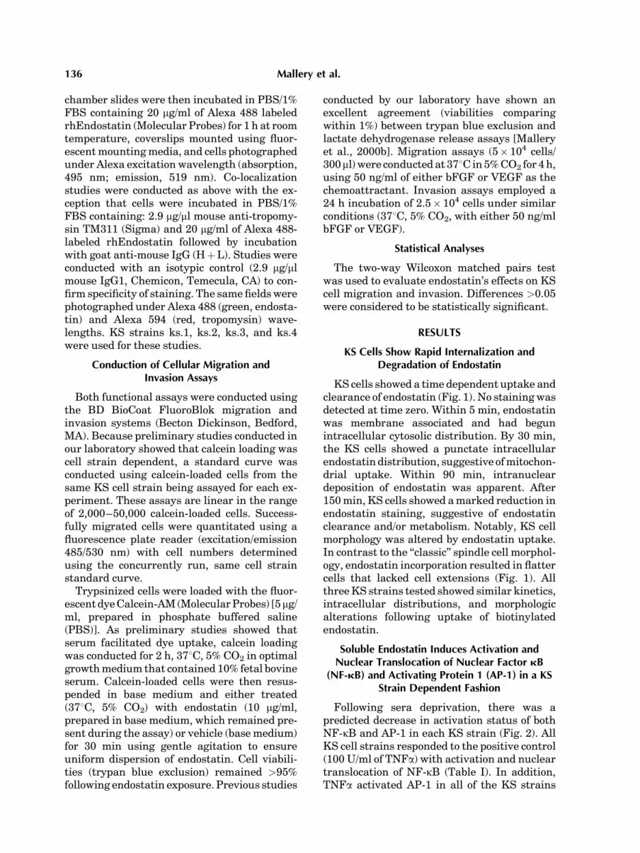
chamber slides were then incubated in PBS/1%FBS containing 20 mg/ml of Alexa 488 labeledrhEndostatin (Molecular Probes) for 1 h at roomtemperature, coverslips mounted using fluor-escent mounting media, and cells photographedunder Alexa excitation wavelength (absorption,495 nm; emission, 519 nm). Co-localizationstudies were conducted as above with the ex-ception that cells were incubated in PBS/1%FBS containing: 2.9 mg/ml mouse anti-tropomy-sin TM311 (Sigma) and 20 mg/ml of Alexa 488-labeled rhEndostatin followed by incubationwith goat anti-mouse IgG (HþL). Studies wereconducted with an isotypic control (2.9 mg/mlmouse IgG1, Chemicon, Temecula, CA) to con-firm specificity of staining. The same fields werephotographed under Alexa 488 (green, endosta-tin) and Alexa 594 (red, tropomysin) wave-lengths. KS strains ks.1, ks.2, ks.3, and ks.4were used for these studies.
Conduction of Cellular Migration andInvasion Assays
Both functional assays were conducted usingthe BD BioCoat FluoroBlok migration andinvasion systems (Becton Dickinson, Bedford,MA). Because preliminary studies conducted inour laboratory showed that calcein loading wascell strain dependent, a standard curve wasconducted using calcein-loaded cells from thesame KS cell strain being assayed for each ex-periment. These assays are linear in the rangeof 2,000–50,000 calcein-loaded cells. Success-fully migrated cells were quantitated using afluorescence plate reader (excitation/emission485/530 nm) with cell numbers determinedusing the concurrently run, same cell strainstandard curve.
Trypsinized cells were loaded with the fluor-escent dye Calcein-AM (Molecular Probes) [5mg/ml, prepared in phosphate buffered saline(PBS)]. As preliminary studies showed thatserum facilitated dye uptake, calcein loadingwas conducted for 2 h, 378C, 5% CO2 in optimalgrowth medium that contained 10% fetal bovineserum. Calcein-loaded cells were then resus-pended in base medium and either treated(378C, 5% CO2) with endostatin (10 mg/ml,prepared in base medium, which remained pre-sent during the assay) or vehicle (base medium)for 30 min using gentle agitation to ensureuniform dispersion of endostatin. Cell viabili-ties (trypan blue exclusion) remained >95%following endostatin exposure. Previous studies
conducted by our laboratory have shown anexcellent agreement (viabilities comparingwithin 1%) between trypan blue exclusion andlactate dehydrogenase release assays [Malleryet al., 2000b]. Migration assays (5� 104 cells/300ml) were conducted at 378C in 5% CO2 for 4 h,using 50 ng/ml of either bFGF or VEGF as thechemoattractant. Invasion assays employed a24 h incubation of 2.5� 104 cells under similarconditions (378C, 5% CO2, with either 50 ng/mlbFGF or VEGF).
Statistical Analyses
The two-way Wilcoxon matched pairs testwas used to evaluate endostatin’s effects on KScell migration and invasion. Differences >0.05were considered to be statistically significant.
RESULTS
KS Cells Show Rapid Internalization andDegradation of Endostatin
KS cells showed a time dependent uptake andclearance of endostatin (Fig. 1). No staining wasdetected at time zero. Within 5 min, endostatinwas membrane associated and had begunintracellular cytosolic distribution. By 30 min,the KS cells showed a punctate intracellularendostatin distribution, suggestive of mitochon-drial uptake. Within 90 min, intranucleardeposition of endostatin was apparent. After150 min, KS cells showed a marked reduction inendostatin staining, suggestive of endostatinclearance and/or metabolism. Notably, KS cellmorphology was altered by endostatin uptake.In contrast to the ‘‘classic’’ spindle cell morphol-ogy, endostatin incorporation resulted in flattercells that lacked cell extensions (Fig. 1). Allthree KS strains tested showed similar kinetics,intracellular distributions, and morphologicalterations following uptake of biotinylatedendostatin.
Soluble Endostatin Induces Activation andNuclear Translocation of Nuclear Factor kB
(NF-kB) and Activating Protein 1 (AP-1) in a KSStrain Dependent Fashion
Following sera deprivation, there was apredicted decrease in activation status of bothNF-kB and AP-1 in each KS strain (Fig. 2). AllKS cell strains responded to the positive control(100 U/ml of TNFa) with activation and nucleartranslocation of NF-kB (Table I). In addition,TNFa activated AP-1 in all of the KS strains
136 Mallery et al.

with the exception of ks.4, albeit at lower levelsrelative to NF-kB. Endostatin challenge re-sulted in activation and nuclear translocationof NF-kB in each KS strain except ks.2. Whileendostatin exposure elicited AP-1 activation inks.1 and ks.3 cultures, strain ks.2 was againrefractory to endostatin challenge. The ks.4
strain failed to activate AP-1 following chal-lenge with either TNFa or endostatin.
Recombinant Human Endostatin and TropomysinCo-Localize to KS Cell Microfilaments
Co-localization studies showed that KS cellslocalized Alexa 488-labeled endostatin (green)
Fig. 1. Kaposi’s sarcoma cells show rapid internalization anddegradation of endostatin. A total of 1� 104 KS cells were platedon Lab-Tek chamber slides coated with 0.5 ml of poly-L-lysine ata concentration of 0.1 mg/ml, and cultured in serum-free M-199base medium for 24 h, followed by a 24 h incubation in basemediumþ50 ng/ml bFGF. The KS cells were then incubated with10 mg/ml of biotinylated endostatin for specific periods (0, 5, 30,90, 150 min) at 378C, 5% CO2. At the designated harvest points,slides were fixed (methanol and acetone washes), followed bythe addition of Alex 488-conjugated avidin and ProLong Antifademounting medium. Cell-associated endostatin was then deter-
mined by fluorescence microscopy. Our results show a lack ofstaining at 0 min (A), and that by 5 min endostatin is bothmembrane associated and beginning intracellular cytosolicdistribution (B). By 30 min (C), KS cells show a punctate, intra-cellular distribution of endostatin, suggestive of mitochondrialuptake. By 90 min (D), punctate, intranuclear distribution ofendostatin is apparent. At the 150-min time point (E), KS cellsshow a marked reduction in staining, indicating either degrada-tion and/or clearing of endostatin. All photomicrographs areshown at 400� image scale, ks.1 strain.
KS Cells Internalize and Respond to Endostatin 137

within cytosolic microfilaments and within thenucleus (Fig. 3). Evaluation of these same fieldsunder Alexa 594 wavelength (red), showed tro-pomysin staining of the same cytosolic micro-filaments that had stained positively with theAlexa 488 (green) wavelength. Complete ab-sence of staining was noted in the isotypiccontrol slides (data not shown). Co-localizationwas confirmed by photography under bothAlexa 488 and 594 wavelengths. Similar to thebiotinylated endostatin internalization studies,all three KS cell strains evaluated showed co-localization of endostatin to tropomysin micro-filaments.
Data summaries for the electromobility shift,endostatin internalization, and endostatin co-localization assays are presented in Table I.
Soluble Endostatin Significantly Inhibits Migrationand Invasion of KS to bFGF and VEGF
Our results showed that endostatin signifi-cantly inhibited KS cellular migration and in-vasion in response to the KS relevant, completeangiogenic growth factors, bFGF and VEGF(Table IIA,B). Our studies also showed thatbFGF elicited greater KS cell functional re-sponses relative to VEGF for both the migra-tion and invasion assays. Stain dependent
Fig. 2. Soluble endostatin causes nuclear translocation ofnuclear factor kB (NF-kB) and activating protein 1 (AP-1) in KScells. Cells were cultured for 72 h in serum free M-199 basemedium to provide a baseline from which to assess endostatin’seffects on transcription activating factors. The experimentalgroups for the nuclear extract preparations were as follows: (i) loggrowth, (ii) 72 h sera deprived (negative control), (iii) seradeprived, treated for 30 min with endostatin (10 mg/ml), (iv) seradeprived, treated for 30 min with 100 U/ml TNFa (positivecontrol) (ks.1 strain). Nuclear and cytosolic extracts were pre-pared from trypsinized cells using a commercially available celllysis buffer (NE-PER #78833, Pierce) which contained a protease
inhibitor cocktail that was added to the lysis buffer just prior touse (Halt Protease inhibitor cocktail kit #78410, Pierce). Threeassay controls, which consisted of biotin–EBNA control DNAonly, biotin–EBNA control DNA and EBNA extract, and biotin-EBNA control DNA, EBNA extract, and a 200-fold molar excessof unlabeled EBNA DNA, were included in every EMSAconducted. EMSA lanes for both NF-kB and AP-1 were: 1, loggrowth; 2, sera deprivation; 3, endostatin treatment; 4, TNFachallenge; 5, no extract. Challenge with TNFa resulted in nucleartranslocation of both NF-kB and AP-1. Endostatin challengeresulted in comparable levels of NF-kB and AP-1 activation andnuclear translocation.
TABLE I. Data Summaries for the Electromobility Shift, Endostatin Internalization, andEndostatin Co-Localization Assays
Cellstrain
EMSANF-kB TNFa
EMSANF-kB endostatin
EMSA AP-1TNFa
EMSA AP-1endostatin
Endostatininternalization
Endostatintropomysin
co-localization
ks.1 Nucleartranslocation
Nucleartranslocation
Nucleartranslocation
Nucleartranslocation
Yes (strong) Yes
ks.2 Nucleartranslocation
No response Nucleartranslocation
No response Yes (moderate) Yes
ks.3 Nucleartranslocation
Nucleartranslocation
Nucleartranslocation
Nucleartranslocation
Yes (moderate) Yes
ks.4 Nucleartranslocation
Nucleartranslocation
No response No response Not conducted Not conducted
138 Mallery et al.

differences were apparent as the ks.1 straindemonstrated the lowest migration and inva-sion responses to either VEGF or bFGF, regard-less of experimental conditions (Table IIA,B,Fig. 4).
Migration assays showed that 10 mg/ml ofsoluble endostatin inhibited KS cellular migra-tion to VEGF (P< 0.001) and bFGF (P< 0.001)(Table IIA and Fig. 4A). Inclusion of endostatinsignificantly decreased KS cell migration by
Fig. 3. Recombinant human endostatin binding and tropomy-sin co-localize to the microfilaments of KS cells. A total of1� 104 KS cells were plated on Lab-Tek chamber slides coatedwith human plasma fibronectin (25 mg/ml), incubated overnightin proliferative growth medium (M-199 supplemented with 10%heat inactivated FBS), and then fixed in 10% neutral formalin,followed by a methanol wash. The chamber slides were thenincubated in PBS/1% FBS containing 20 mg/ml of Alexa 488labeled rhEndostatin (Molecular Probes) for 1 h at room tem-perature, coverslips mounted using fluorescent mounting media,and cells photographed under Alexa excitation wavelength(absorption, 495 nm; emission, 519 nm). Co-localization studieswere conducted as above with the exception that cells wereincubated in PBS/1% FBS containing: 2.9 mg/ml mouse anti-tropomysin TM311 (Sigma) and 20 mg/ml of Alexa 488-labeledrhEndostatin followed by incubation with goat anti-mouse IgG
(H&L). Studies were conducted with an isotypiccontrol (2.9mg/mlmouse IgG1, Chemicon) to confirm specificity of staining. Thesame fields were photographed under Alexa 488 (green,endostatin) and Alexa 594 (red, tropomysin) wavelengths. KScells from two different donors were used for these studies (donorks.1 shown in Fig. 2A,C,E; donor ks.2 shown in Fig. 2B,D,F). Allphotomicrographs are shown at a 400� image scale. KS cells(A, B, C) localize Alexa 488-labeled endostatin (green) withincytosolic microfilaments and within the nucleus. The same fields(D, E, F), photographed under Alexa 594 wavelength (red), showtropomysin staining of the cytosolic microfilaments. Depicted inG, H, and I are the same fields photographed under both Alexa488 and Alexa 594 wavelengths. Complete absence of stainingwas noted in the isotypic control slides (data not shown) [strainks.1(A, D, G), ks.2 (B, E, H), ks.3 (C, F, I)].
KS Cells Internalize and Respond to Endostatin 139

28% (VEGF, P< 0.001) and 24% (bFGF,P< 0.001). The individual cell line migrationdata are shown in Table IIA.
The uniform layer of Matrigel BasementMembrane Matrix contained in the BD Bio-CoatTM FluoroBlokTM Invasion System pro-vides cells with conditions that simulate anintact basement membrane that provides a truebarrier to noninvasive cells. Therefore, this
assay permits a quantifiable and reproduciblein vitro assessment of cellular invasive proper-ties. Soluble endostatin significantly inhibitedKS cellular invasion to response to VEGF(P< 0.001) and in response to bFGF (P< 0.01),Figure 4B. Inclusion of endostatin decreased KSinvasion by 35% (VEGF) and 40% (bFGF). Theindividual cell line invasion data are shown inTable IIB.
TABLE II. Individual KS Cell Strain Results of Functional Migration and Invasion Assays
Cell strainControlþ
VEGFEndostatinþ
VEGFPercent
change (%)Controlþ
bFGFEndostatinþ
bFGFPercent
change (%)
Migration results (A)ks.1 10,678þ7,468 6,122þ 2,146 �43 19,452þ3,100 10,420þ 2,121 �46
n¼ 5 VEGFn¼ 5 bFGF
ks.2 19,261þ4,430 16,310þ 1,792 �15 36,351þ3,209 28,850þ 3,227 �20n¼ 4 VEGFn¼ 5 bFGF
ks.3 29,809þ2,325 20,775þ 2,525 �30 47,698þ2,001 38,962þ 3,861 �18n¼ 4 VEGFn¼ 6 bFGF
Invasion results (B)ks.1 9,711þ 1,475 7,888þ 1,957 �19 8,777þ1,567 7,607þ 2,179 �13
n¼ 4 VEGFn¼ 3 bFGF
ks.2 15,557þ2,930 9,951þ 3,648 �36 18,155þ1,820 9,988þ 3,651 �45n¼ 4 VEGFn¼ 3 bFGF
ks.3 14,182þ1,695 6,467þ 1,899 �54 19,378þ5,238 10,378þ 5,238 �46n¼ 3 VEGFn¼ 4 bFGF
Functional assays were conducted using BD BioCoat FluoroBlok migration and invasion systems as described in Materials andMethods. Data are expressed as means of cell numbersþSEM. Endostatin inhibited migration in response to VEGF (P<0.001), andbFGF (P< 0.001), as well as invasion in response to VEGF (P<0.001) and bFGF (P<0.01).
Fig. 4. Endostatin significantly inhibits migration and invasionof Kaposi’s sarcoma (KS) cells to both basic fibroblast growthfactor (bFGF) and vascular endothelial growth factor (VEGF).Both the migration and invasion assays were conducted using theBD BioCoat systems. The BD Invasion System simulates an intactbasement membrane, thereby providing a true barrier to non-invasive cells. Migration assays (5.0� 104 cells/300 ml, A) wereconductedat 378C, 5% CO2 for 4 h, using 50 ng/ml of either bFGF
or VEGF as the chemoattractant. Invasion assays (2.5� 104 cells/300 ml, B) were conducted under similar conditions for 24 h.Successfully migrated or invasive cells were quantitated using afluorescence plate reader with cells numbers determined usingthe concurrently run, same cell strain standard curve (n¼ 13VEGF migration, n¼ 16 bFGF migration, n¼11 VEGF invasion,n¼10 bFGF invasion, n¼ 10 bFGF invasion, *¼P<0.001,#¼P<0.01).
140 Mallery et al.

DISCUSSION
Nodular KS lesions, which are the mostadvanced tumor stage, are histologically sarco-mas that behave in a clinically aggressivefashion [McGarvey et al., 1998]. Our laboratoryhas previously shown that KS stains isolatedfrom nodular KS tumors retain many featuresof a transformed phenotype such as sustainedproliferation in serum-deficient medium andhigh autologous production of KS associatedgrowth factors [Bailer et al., 1995]. This presentstudy used cells isolated from nodular KStumors in order to assess endostatin’s effectson KS strains that possess the most malignantphenotype.
Results of our biotinylated endostatin studiesshowed that vital KS cells rapidly internalizedendostatin. There are two potential mechan-isms, which are not mutually exclusive, whichcould account for these findings. First, asendostatin is a naturally occurring substance,it is predictable that cells associated withangiogenesis such as endothelial lineage cells,possess an endostatin specific receptor. Alter-natively, endostatin may be internalized viaphagocytosis. This latter possibility is sup-ported by previous studies from our laboratorythat show KS cells are actively phagocytic[Mallery et al., 2000b]. The kinetic data showKS intracellular endostatin levels dramaticallydecreased within 150 min; implying eitherendostatin metabolism and/or export by KScells. Our biotinylated endostatin intracellulardistribution and kinetic data compare favorablywith the work of Dixelius et al. [2000], whoevaluated endostatin internalization and clear-ance in murine brain endothelial cells.
NF-kB and AP-1 are both pleiotrophic tran-scription factors that are activated by numerousstimuli including changes in the cell redoxpotential and cell membrane activation [Stallet al., 1994; Janssen-Heininger et al., 2000].Activation of the integrin-linked kinase (ILK) isalso known to activate AP-1 [Yoganathan et al.,2000]. Relevant to the ILK pathway, our labora-tory has confirmed expression of the ILK as-sociated integrin subunits b1 and b3 in each KScell strain used in this study (data not shown).Our EMSA data show endostatin activated NF-kB and AP-1 in a cell-strain dependent fashion.These findings imply endostatin–integrin and/or endostatin–membrane interactions activatesignaling pathways such as the ILK and suggest
that integrin subunit expression may be a criti-cal factor in determining whether or not endo-statin challenge initiates signal transduction.Our results, which show endostatin initiatestranscription factor activation, may initiallyappear contradictory to the findings of Shichiriand Hirata [2001], who showed that endostatinexposure down-regulated growth-associatedgenes in endothelial cells. Notably, both ourbiotinylated and fluorescent labeling studiesshowed endostatin intranuclear localization.We speculate that intranuclear endostatinimpedes transcription factor binding to itsDNA cognate binding site, potentially via sterichindrance, thereby reducing transcription fac-tor mediated gene expression.
The co-localization of internalized endostatinto tropomysin microfilaments that was observ-ed in all three KS cell strains examined suggeststhat this may be the underlying mechanism forthe inhibitory activity of endostatin on KS cellmigration and invasion. Our fluorescent stain-ing results agree favorably with the findings ofMacDonald et al., 2001, who identified hTM3 asan endostatin-binding tropomysin epitope in avariety of large and small vessel derived endo-thelial cells. Since tropomysin fulfills crucialrole in actin stabilization and reorganizationof the cytoskeleton [Lin et al., 1997], it is notsurprising that tropomysin-binding agentscould suppress cell motility.
Although cell migration and invasion fre-quently occur concurrently or sequentially,these cellular functions are related but distinctprocesses. While migration entails integrinupregulation and specific interactions withextracellular substrates, invasion also requiresupregulation and activation of proteolyticenzymes [Murphy and Bavrilovic, 1999; Sta-menkovic, 2000]. Our data show that short-termendostatin exposure inhibited both of thesefunctions in KS cells isolated from three dif-ferent donors. Our results also show that re-lative to VEGF, bFGF initiated higher levelsof migration and invasion in all KS strains,suggesting increased bFGF receptor expressionand/or bFGF is superior as a KS cell chemoat-tractant. Differences were also apparent amongthe KS strains with regard to baseline mobilityas well as endostatin responsiveness. Thesedata likely reflect a variety of KS interstraincellular differences including numbers of VEGFand bFGF receptors, cellular capacity to in-crease expression of pro-migratory integrins
KS Cells Internalize and Respond to Endostatin 141

such as avb6, and ability to activate basementmembrane degrading proteolytic enzymes suchas MMP-2 and MMP-9. Because modulation ofthe patterns of integrin expression alters theability of cells to interact with the extracellularmatrix [Parise et al., 2000], we surmise thatendostatin’s abilities to interact with integrinsacts in conjunction with endostatin–tropomy-sin interactions to inhibit KS cell migration andinvasion.
Our functional assay data are in agreementwith the findings of Rehn et al. [2001], whichshowed soluble endostatin served as an integrinantagonist for endothelial origin cells. In addi-tion, the endostatin concentration [10 mg/ml(4.71� 10�7 M)] used in our KS cell studies iscomparable or lower than endostatin concen-trations that have demonstrated efficacy duringin vitro endothelial cell studies [Dhanabal et al.,1999; Dixelius et al., 2000; Rehn et al., 2001;Shichiri and Hirata, 2001]. This dose con-sistency implies that KS cells have similarthresholds of endostatin responsiveness asnontransformed endothelial cells.
There are several reasons why endostatin isan attractive agent for AIDS-related KS ther-apy. KS lesional cells both produce and respondto the complete angiogenic cytokines VEGF andbFGF. Therefore, the ‘‘proangiogenic pheno-type’’ is inherent to KS tumor cells and alsocrucial for KS lesional progression. In addition,our data show endostatin directly inhibits KScell migration and invasion. As a consequence ofits combined angiostatic and antitumorigenicactivities, endostatin could provide dual ther-apeutic benefits for KS.
Although human clinical trials with intrave-nously administered endostatin have not pro-ven highly successful, subsequently conductedanimal studies that used osmotic pumps thatprovided sustained endostatin drug levelsdemonstrated greater efficacies [Kisker et al.,2001]. These data suggest that intravenousdelivery may not achieve and sustain optimaltherapeutic endostatin concentrations, therebyreducing endostatin’s efficacy. Notably, KSlesions often occur at visibly accessible muco-cutaneous sites, which are amenable to use oflocally injectable, biodegradable, controlled-release drug delivery systems. Controlled-release delivery vehicles provide sustaineddosing rates and high intralesional drug con-centrations without systemic side effects. Theseproperties convey a pharmacologic advantage
by increasing the drug’s therapeutic index re-lative to systemic delivery [Toguchi, 1995].While it is established that sustained con-trolled-release of bioactive proteins and pep-tides is technically challenging, our laboratorieshave demonstrated the ability to stabilize andprovide sustained controlled-release of bioac-tive proteins from biodegradable polylactide-co-glycolide (PLGA) delivery vehicles [Zhu et al.,2000].
Currently advocated therapies for AIDS-associated malignancies therapies are designedto limit disease progression without exacerbat-ing the underlying immune suppression [Shahet al., 2002]. We propose that locally injectablePLGA controlled-release endostatin deliverysystems, which would combine endostatin’sangiostatic and antitumorigenic effects withoutinducing deleterious systemic effects, couldprovide an effective and novel treatment formucocutaneous AIDS-KS.
REFERENCES
Bailer RT, Lazo A, Ng-Bautista CL, Hout BL, Ness GM,Hegtvedt AK, Blakeslee JR, Stephens RE, Mallery SR.1995. Correlation between AIDS-related Kaposi sarcomahistologic grade and in vitro behavior: Reduced exogen-ous growth factor requirements for isolates from highgrade lesions. Lymphology 28:473–483.
Dhanabal M, Ramchandran R, Waterman MJF,Knebelmann B, Segal M, Sukhatme VP. 1999. Endosta-tin induces endothelial cell apoptosis. J Biol Chem 274:11721–11726.
Dixelius J, Larsson H, Saski T, Holmqvist K, Lu L. 2000.Endostatin-induced tyrosine kinase signaling throughthe Shb adaptor protein regulates endothelial cell apop-tosis. Blood 95:3403–3411.
Folkman J. 1995. Clinical application of research onangiogenesis. New Engl J Med 333:1757–1763.
Folkman J, Shing Y. 1992. Angiogenesis J Biol Chem 267:10931–10934.
Gasparini G. 1999. The rationale and future potential ofangiogenesis inhibitors in neoplasia. Drugs 58:17–38.
Jacobson LP, Arnemian HK. 1995. An integrated approachto the epidemiology of Kaposi’s sarcoma. Curr Opin Oncol7:450–455.
Janssen-Heininger YMW, Poynter ME, Baeuerle PA. 2000.Recent advances towards understanding redox mechan-isms in the activation of nuclear factor kB. Free RadicBiol Med 28:1317–1327.
Kisker O, Becker CM, Prox D, Fannon M, D’Amato R,Flynn E, Fogler WE, Sim BKL, Allred EN, Pirie-Shepherd SR, Folkman J. 2001. Continuous administra-tion of endostatin by intraperitoneally implanted osmoticpump improve the efficacy and potency of therapy in amouse xenograft tumor model. Cancer Res 61:7669–7674.
Lee S-J, Jang J-W, Kim Y-M, Lee HI, Jeon JY, Kwon Y-G,Lee S-T. 2002. Endostatin binds to the catalytic domain
142 Mallery et al.

of matrix metalloproteinase-2. FEBS Letts 519:147–152.
Lin JJ, Warren KS, Wanboldt DD, Want T, Lin JL. 1997.Tropomysin isoforms in nonmuscle cells. Int Rev Cytol170:1–38.
MacDonald NJ, Shivers WY, Narum DL, Plum SM,Wingard JN, Fuhrmann SR, Liang H, Holland-Linn J,Chen DHT, Sim BKL. 2001. Endostatin binds tropomy-sin: A potential modulator of the antitumor activity ofendostatin. J Biol Chem 276:25190–25196.
Mallery SR, Clark YM, Ness GM, Minahawi OM, Pei P,Hohl CM. 1999. Thiol redox modulation of doxorubicinmediated cytotoxicity in cultured AIDS-related Kaposi’ssarcoma cells. J Cell Biochem 73:259–277.
Mallery SR, Pei P, Kang J, Zhu G, Ness GM, SchwendemanSP. 2000a. Sustained angiogenesis enables in vivotransplantation of mucocutaneous derived AIDS-relatedKaposi’s sarcoma cells in murine hosts. Carcinogenesis21:1647–1653.
Mallery SR, Pei P, Kang J, Ness GM, Ortiz R, TouhaliskyJE, Schwendeman SP. 2000b. Controlled release ofdoxorubicin from poly(lactide-co-glycolide) microspheressignificantly enhances cytotoxicity against culturedAIDS-related Kaposi’s sarcoma cells. Anticancer Res 20:2817–2826.
Masood R, Cai J, Zheng T, Smith D, Naidu Y, Gill PS. 1997.Vascular endothelial growth factor/vascular permeabil-ity factor is an autocrine growth factor for AIDS-Kaposisarcoma. Proc Natl Acad Sci USA 94:979–984.
McGarvey ME, Tulpule A, Cai J, Zheng T, Masood R,Espina B, Arora N, Smith DL, Gill PS. 1998. Emergingtreatments for epidemic (AIDS-related) Kaposi’s sar-coma. Curr Opin Oncol 10:413–421.
Miles D. 1994. Pathogenesis of HIV-related Kaposi’ssarcoma. Curr Opin Oncol 6:497–502.
Miles S, Rezai A, Salazar-Gonzalez J, Van der Meyden M,Stevens R, Logan D, Mitsuqasu R, Laza L, Hirano L,Kishimoto L, Martinez-Nazo O. 1990. AIDS Kaposi’ssarcoma-derived cells produce and respond to IL-6. ProcNatl Acad Sci USA 87:4068–4071.
Murphy G, Bavrilovic J. 1999. Proteolysis and cell migra-tion: Creating a path? Curr Opin Cell Biol 11:614–621.
O’Reilly MS, Boehm T, Shing Y, Fukai N, Vasios G, LaneWS, Flynn E, Birkhead JR, Olsen BR, Folkman J. 1997.
Endostatin: An endogenous inhibitor of angiogenesis andtumor growth. Cell 88:277–285.
Parise LV, Lee JW, Juliano RL. 2000. New aspectsof integrin signaling in cancer. Cancer Biol 10:407–414.
Rehn M, Veikkola T, Valdre-Kukk E, Nakamura H,Ilmonen M, Lombardo DR, Pihlajaniemi T, Alitalo K,Vuori K. 2001. Interaction of endostatin with integrinsimplicated in angiogenesis. Proc Natl Acad Sci USA 98:1024–1029.
Rutger JL, Wieczorek R, Bintti F, Kaplan KL, Posnett DN,Friedman-Kein AE, Knowles DM. 1986. The expressionof endothelial cell surface antigens by AIDS-associatedKaposi’s sarcoma: Evidence for a vascular endothelialcell origin. Am J Pathol 122:493–499.
Samaniego F, Markhan PD, Gendelman R, Watanabe Y,Kao V, Kowalsek K, Sonnabend JA, Pintus A, Gallo RC,Ensoli B. 1998. Vascular endothelial growth factor andbasic fibroblast growth factor present in Kaposi’s sar-coma are induced by inflammatory cytokines and sy-nergize to promote vascular permeability and KS lesiondevelopment. Am J Pathol 152:1433–1443.
Shah MH, Porcu P, Mallery SR, Caligiuri MA. 2002.Chapter 31: AIDS-associated malignancies. In: GiacconeG, Schilsky R, Sondel P, editors. Cancer chemotherapyand biological response modifiers, Elsevier Science B.V.p 633–664.
Shichiri M, Hirata Y. 2001. Antiangiogenesis signals byendostatin. FASEB 15:1044–1053.
Stall FJT, Anderson MT, Staal GEJ, Herzenberg LA, GitlerC, Herzenberg LA. 1994. Redox regulation signal trans-duction: Tyrosine phosphorylation and calcium influx.Proc Natl Acad Sci USA 91:3619–3622.
Stamenkovic I. 2000. Matrix metalloproteinases in tumorinvasion and metastasis. Cancer Biol 10:415–4333.
Toguchi H. 1995. Biodegradable microspheres in drugdelivery. Crit Rev Ther Drug Carr Sys 12:1–99.
Yoganathan TN, Costello P, Chen X, Jabali M, Yan J,Leung D, Zhang Z, Yee A, Dedhar S, Sanghera J. 2000.Integrin-linked kinase (ILK): A ‘‘hot’’ therapeutic target.Biochem Pharmacol 60:1115–1119.
Zhu G, Mallery SR, Schwendeman SP. 2000. Stabilizationof proteins encapsulated in poly(lactide-co-glycolide). NatBiotechnol 18:52–57.
KS Cells Internalize and Respond to Endostatin 143


