Embed Size (px)
Citation preview

NRC Publications Archive (NPArC)Archives des publications du CNRC (NPArC)
Publisher’s version / la version de l'éditeur: Apoptosis, 16, 3, pp. 256-271, 2010-03-01
The ribonucleotide reductase R1 subunits of herpes simplex virus types 1 and 2 protect cells against TNFα- and FasL-induced apoptosis by interacting with caspase-8Dufour, Florent; Sasseville, A. Marie-Josée; Chabaud, Stéphane; Massie, Bernard; Siegel, Richard M.; Langelier, Yves
Contact us / Contactez nous: [email protected].
http://nparc.cisti-icist.nrc-cnrc.gc.ca/npsi/jsp/nparc_cp.jsp?lang=frL’accès à ce site Web et l’utilisation de son contenu sont assujettis aux conditions présentées dans le site
Web page / page Webhttp://dx.doi.org/10.1007/s10495-010-0560-2http://nparc.cisti-icist.nrc-cnrc.gc.ca/npsi/ctrl?action=rtdoc&an=16616680&lang=enhttp://nparc.cisti-icist.nrc-cnrc.gc.ca/npsi/ctrl?action=rtdoc&an=16616680&lang=fr
LISEZ CES CONDITIONS ATTENTIVEMENT AVANT D’UTILISER CE SITE WEB.
READ THESE TERMS AND CONDITIONS CAREFULLY BEFORE USING THIS WEBSITE.
Access and use of this website and the material on it are subject to the Terms and Conditions set forth athttp://nparc.cisti-icist.nrc-cnrc.gc.ca/npsi/jsp/nparc_cp.jsp?lang=en

ORIGINAL PAPER
The ribonucleotide reductase R1 subunits of herpes simplex virustypes 1 and 2 protect cells against TNFa- and FasL-inducedapoptosis by interacting with caspase-8
Florent Dufour • A. Marie-Josee Sasseville •
Stephane Chabaud • Bernard Massie •
Richard M. Siegel • Yves Langelier
� Springer Science+Business Media, LLC 2010
Abstract We previously reported that HSV-2 R1, the R1
subunit (ICP10; UL39) of herpes simplex virus type-2
ribonucleotide reductase, protects cells against apoptosis
induced by the death receptor (DR) ligands tumor necrosis
factor-alpha- (TNFa) and Fas ligand (FasL) by interrupting
DR-mediated signaling at, or upstream of, caspase-8 acti-
vation. Further investigation of the molecular mechanism
underlying HSV-2 R1 protection showed that extracellular-
regulated kinase 1/2 (ERK1/2), phosphatidylinositol
3-kinase (PI3-K)/Akt, NF-jB and JNK survival pathways
do not play a major role in this antiapoptotic function.
Interaction studies revealed that HSV-2 R1 interacted
constitutively with caspase-8. The HSV-2 R1 deletion
mutant R1(1-834)-GFP and Epstein–Barr virus (EBV) R1,
which did not protect against apoptosis induced by DR
ligands, did not interact with caspase-8, indicating that
interaction is required for protection. HSV-2 R1 impaired
caspase-8 activation induced by caspase-8 over-expression,
suggesting that interaction between the two proteins pre-
vents caspase-8 dimerization/activation. HSV-2 R1 bound
to caspase-8 directly through its prodomain but did not
interact with either its caspase domain or Fas-associated
death domain protein (FADD). Interaction between HSV-2
R1 and caspase-8 disrupted FADD-caspase-8 binding. We
further demonstrated that individually expressed HSV-1 R1
(ICP6) shares, with HSV-2 R1, the ability to bind caspase-8
and to protect cells against DR-induced apoptosis. Finally,
as the long-lived Fas protein remained stable during the
early period of infection, experiments with the HSV-1
UL39 deletion mutant ICP6D showed that HSV-1 R1 could
be essential for the protection of HSV-1-infected cells
against FasL.
Keywords FasL � ICP6 � ICP10 � Viral inhibitor
of apoptosis � Caspase-8Electronic supplementary material The online version of thisarticle (doi:10.1007/s10495-010-0560-2) contains supplementarymaterial, which is available to authorized users.
F. Dufour � A. M.-J. Sasseville � S. Chabaud � Y. Langelier (&)
Centre de recherche du Centre hospitalier de l’Universite de
Montreal (CRCHUM) and Institut du cancer de Montreal,
Hopital Notre-Dame, 1560 Sherbrooke Est, Montreal,
QC H2L 4M1, Canada
e-mail: [email protected]
B. Massie � Y. Langelier
Departement de microbiologie et immunologie,
Universite de Montreal, Montreal, QC, Canada
B. Massie
Institut de recherche en biotechnologie, Montreal, QC, Canada
B. Massie
Institut national de la recherche scientifique-Institut Armand-
Frappier, Universite du Quebec, Laval, QC, Canada
R. M. Siegel
Immunoregulation Unit, Autoimmunity Branch, National
Institute of Arthritis and Musculoskeletal and Skin Diseases,
National Institutes of Health, Bethesda, MD, USA
Y. Langelier
Departement de medecine, Universite de Montreal,
Montreal, QC, Canada
Present Address:
S. Chabaud
Laboratoire d’Organogenese Experimentale (LOEX),
Universite Laval, Quebec, QC, Canada
123
Apoptosis
DOI 10.1007/s10495-010-0560-2

Introduction
The tumor necrosis factor (TNF) superfamily of ligands,
acting through cognate death receptors (DRs), plays a
critical role in innate and adaptive immune responses to
viruses. The antiviral activity of several of these ligands
often correlates with their ability to induce apoptosis ini-
tiated by the triggering of DRs [1–3]. TNFa, which acts
mainly through binding to TNF receptor 1 (TNFR1), has
been reported to have antiviral activity against herpes
simplex viruses (HSVs) [4, 5]. HSV-1-infected mice
knocked-out for TNFa develop HSV encephalitis due to an
impaired immune response which fails to clear the virus
from the brain [6]. Fas ligand (FasL), expressed on cyto-
toxic T lymphocytes and natural killer cells, is also
important in the elimination of infected cells. Recently,
Fas-mediated apoptosis of HSV-2-infected cells involving
CD4? cytotoxic T lymphocytes was shown to be crucial for
the defense against lethal infection in mice [3]. The
importance for HSVs to counteract DR activation is fur-
thermore evidenced by the diverse strategies used by these
viruses to block DR signaling pathways. At least two viral
proteins, the ribonucleotide reductase (RR) R1 subunit of
HSV-2 (HSV-2 R1) and the virion host shutoff protein
(vhs), have been hypothesized to interfere with these
pathways [7, 8].
Ligand binding to DRs triggers the recruitment of death
domain (DD)-containing adaptor proteins to the DD of
receptors, resulting in the formation of membrane-bound
signaling complexes. Important differences exist between
Fas and TNFR1 activation, not only in terms of signaling
complexes but also function, Fas exhibiting mainly pro-
apoptotic behavior, and TNFR1, mostly proinflammatory
activity. Upon FasL-Fas binding, Fas-induced apoptosis
involves recruitment of the Fas-associated death domain
protein FADD to Fas. Receptor-bound FADD forms a
death-inducing signaling complex (DISC) with caspase-8.
Aggregation of proteins in DISC stimulates autocata-
lytic processing of caspase 8 and triggers the caspase
cascade [reviewed in 9]. Cellular FLICE-inhibitory protein
(c-FLIP), by inhibiting the recruitment of caspase-8 to
DISCs, negatively modulates the caspase cascade [10, 11].
FLIP was originally identified as a viral gene, viral FLIP
(v-FLIP) being present in c-herpes viruses such as equine
herpes virus-2, herpes virus saimiri and human herpes
virus-8 (HHV-8) [reviewed in 12].
TNFa-induced signaling involves the formation of two
sequential signaling complexes [13, 14]. Upon TNFa
binding to TNFR1, a first complex consisting of TNFa
receptor-associated death domain protein (TRADD), Ser/
Thr kinase receptor-interacting protein 1 (RIP1) and TNFa
receptor-associated factor 2 (TRAF2) is formed [reviewed
in 15]. This membrane-associated complex (complex I)
rapidly leads to the activation of nuclear factor-jB
(NF-jB), mitogen-activated protein kinase (MAPK), c-Jun
NH2-terminal kinase (JNK), p38 MAPK and ERK1/2.
Activation of these pathways stimulates the transcription of
genes that regulate proliferation, inflammation or apopto-
sis, including the key antiapoptotic cellular inhibitor of
apoptosis (cIAPs) and c-FLIP [reviewed in 16, 17]. In a
second step, TRADD and RIP1 form an intracellular
complex (complex II) with FADD. FADD recruits caspase-
8 by death-effector domain (DED) interaction, allowing the
dimerization and activation of caspase-8 that initiates the
apoptotic process [13, 14]. The outcome of TNFR1 stim-
ulation depends on the balance between signals triggered
by both complexes, the activation of NF-jB by complex I
playing an essential role in protecting cells against apop-
tosis [18]. With several cell lines, such as HeLa cells,
induction of apoptosis by TNFa critically depends on the
presence of cycloheximide (CHX), which, by drastically
reducing the levels of the short-lived proteins c-FLIPL and
c-FLIPS, increases caspase-8–FADD interaction [10].
HSV RRs convert ribonucleotide diphosphates to cor-
responding deoxyribonucleotides, allowing virus replica-
tion in non-dividing cells [19]. The association of two
homodimeric subunits, denoted as R1 and R2, forms the
holoenzyme. HSV R1s carry the RR domain and a unique
NH2 domain of about 400 amino acids dispensable for RR
activity [20, 21]. Several non-RR related functions have
been ascribed to HSV R1s. (i) Numerous reports indicate
that a protein kinase activity could be intrinsic to the NH2
domain of HSV-2 R1, but not to that of HSV-1 R1 [22].
However, extensive biochemical studies on HSV-1 R1 and
HSV-2 R1 provided strong evidence that both proteins are
devoid of intrinsic kinase activity [23, 24]. (ii) A stretch
exhibiting weak similarity with the a-crystallin domain of
the small heat shock proteins (sHsps) has been detected in
the HSV R1s N terminus [25, 26], leading to the obser-
vation that purified HSV-2 R1 exhibits a chaperone activity
similar to Hsp27 in vitro [26]. (iii) HSV-1 R1 has been
associated with the promotion of protein translation in
growth-arrested cells by stimulating assembly of the
translation initiation complex eIF4F through its binding to
eIF4G [27]. (iv) Finally, antiapoptotic properties have been
associated to HSV R1s, most often to HSV-2 R1 [7,
reviewed in 25].
The Aurelian group has provided evidence that HSV-2
R1 could impair apoptosis induced by the mitochondrial
pathway through its ability to activate the MEK/ERK1/2
and PI3-K/Akt pathways [reviewed in 25]. Our group has
shown that the individually expressed HSV-2 R1 blocks
DR-induced apoptosis but does not impair apoptosis trig-
gered via the mitochondrial pathway [7]. The RR domain
of HSV-2 R1, but not the NH2 domain, is essential for
protection against TNFa-induced apoptosis [28]. HSV-2
Apoptosis
123

R1, which does not act as an enzymatic inhibitor of active
caspase-8, interrupts DR-mediated signaling at, or
upstream of, caspase-8 activation [7]. As two HSV-1 UL39
deletion mutants, ICP6D and hrR3, exhibited a 50%
reduction in protection from TNFa, we had proposed that
HSV-1 R1 could be important for protecting HSV-infected
cells against this DR ligand. However, as 50% of cells
infected with R1 deletion mutants were resistant to TNFa,
we also postulated that other viral protein(s) contribute(s)
to protection. Since deletion of the vhs gene reduced
antiapoptotic potential by 30%, we hypothesized that vhs
protein, by decreasing the amount of DRs at the cell sur-
face, could play a significant role in resistance [7]. Our
hypothesis was later substantiated by a report showing that,
owing to the very short half-life of TNFR1 protein, vhs
causes a rapid reduction of TNFR1 early after HSV-1
infection [8].
Here, our efforts to better understand how HSV-2 R1
behave at the molecular level to prevent TNFa-induced
apoptosis led us to show that HSV-2 R1 interacts consti-
tutively with caspase-8 through its prodomain. Evidence is
provided that the interaction between the two proteins
impairs caspase-8 dimerization/activation and disrupts
FADD-caspase-8 binding. Extending our study to HSV-1
R1, we demonstrated that individually expressed HSV-1
R1 shares with HSV-2 R1 the ability to protect cells against
TNFa- and FasL-induced apoptosis and to bind to caspase-
8. Moreover, we provided evidence that HSV-1 R1 could
play an essential role in the protection of HSV-1-infected
cells against FasL. These results suggest that HSV R1s are
viral caspase-8 inhibitors that are functionally similar to
viral inhibitor of caspase-8 activation (vICA), the UL36
gene product of cytomegalovirus (CMV).
Materials and methods
Cell lines
The conditions for culture of HeLa, A549-tTA and A549-
tTA-HSV-R1-GFP cells have been described [7, 28]. When
cultured in the absence of anhydrotetracyclin (Tet), A549-
tTA-HSV-R1-GFP cells express HSV-2 R1 fused to green
fluorescent protein (GFP) [28].
Transfection and immunofluorescence
HeLa cells were seeded one day before transfection in
6-well plates at a concentration of 2 9 105 per well or in
100-mm dishes at 1.5 9 106 per dish. The calcium phos-
phate technique [7] was used to transfect 10 lg per
well and 50 lg per dish of the plasmids pAdCMV5
(empty), pAdCMV5-R1 (HSV-2 R1); pLBPF1-GST (GST),
pLBPF1-GST-R1 (GST-HSV-1 R1) (kindly provided by
A. Pearson); pcDNA3 (empty), pcDNA3-HA-EBV R1
(HA-EBV R1) [29]; pEGFP C1 (GFP) and plasmids
encoding for caspase-8 GFP (casp-8 GFP), caspase-8 C360S
GFP (casp-8 C360S GFP), caspase-8 DED-AB (1-209)
GFP (casp-8 DED-AB GFP), caspase-8 CD (210-479) GFP
(casp-8 CD GFP) and FADD YFP [30]. Immunofluores-
cence was performed on cells fixed with paraformaldehyde
as previously described [31].
Infection
The previously-described adenovirus (Ad) recombinants
AdCMV5-GFP, AdCMV5-R1, AdTR5-GFP, AdTR5-R1-
GFP and AdTR5-R1(1-834)-GFP express GFP, HSV-2 R1,
GFP, HSV-2 R1-GFP and HSV-2 R1(1-834)-GFP,
respectively [7]. HeLa cells (2 9 105 cells/well, 2.5 9 106
cells/100-mm dish) were infected at 15 plaque forming
units (PFU)/cell with AdCMV5-R1 or AdCMV5-GFP.
A549-tTA cells (2.5 9 106 cells/100-mm dish) were
infected with AdTR5-R1-GFP (10 PFU/cell), AdTR5-
R1(1-834)-GFP (50 PFU/cell) or AdTR5-GFP (25 PFU/
cell). For infection followed by transfection, HeLa cells
(2 9 105 cells/well) were infected with AdCMV5-R1 or
AdTR5-CuO (10 PFU/cell) for 8 h before transfection. For
HSV infections, HeLa and A549-tTA cells (2.5 9 106
cells/100-mm dish) were infected at 10 PFU/cell with the
HSV-1 R1 null mutant ICP6D [19] or its parental wild type
(WT) HSV-1 strain KOS.
Apoptosis and caspase assays
Apoptosis was induced by adding to the cellular medium
either CHX (15 lg/ml) plus human recombinant TNFa
(2.5 ng/ml; Sigma) or CHX (15 lg/ml; Sigma) alone as
control, or hexameric FasL (Fc:FasL, a human recombinant
FasL fused at the C terminus of the Fc domain of IgG1)
[32]. After 6–8 h of treatment, the percentage of apoptotic
cells was scored by observation under an inverted micro-
scope in at least five randomly-selected fields, as described
previously [7]. Apoptotic cells showed several of the fol-
lowing morphological features: cytoplasmic shrinkage,
membrane blebbing, rounding up, ballooning, acquisition
of refringence, detachment from the substratum and loss
of membrane integrity. Attached and detached cells were
collected, washed twice with PBS and lysed in buffer
appropriate for subsequent assays. Caspase-3/7 activity
(DEVD-AFC cleavage) was measured as described [7].
Immunoprecipitation
After infection, transfection and/or treatment, attached and
detached cells were collected, washed twice with PBS, and
Apoptosis
123

lysed at 4�C for 15 min in a buffer containing 0.5% Non-
idet-P40, 0.5% Triton X-100, 50 mM Hepes (pH 7.5),
1 mM EDTA, 150 mM NaCl, 10% glycerol, protease
inhibitor cocktail and phosphatase inhibitor cocktail
(Roche), followed by centrifugation (10 min at 10,0009g
at 4�C). Lysates were pre-cleared by incubation with pro-
tein G Sepharose 4B beads (Amersham Biosciences) for
2 h at 4�C, followed by centrifugation. Anti-GFP antiserum
or caspase-8 monoclonal antibody (mAb) 1C15 (kindly
provided by Peter and co-workers [33]) was incubated with
protein G Sepharose 4B beads for 2 h at 4�C. Pre-cleared
lysates were incubated with antibody-coated beads for 2 h
at 4�C. The beads were recovered by centrifugation,
washed five times with lysis buffer, and immunoprecipi-
tated. Proteins were eluted by boiling in sodium dodecyl
sulphate (SDS) sample-loading buffer. As controls, pre-
cleared lysates were incubated for 2 h at 4�C with protein
G Sepharose beads alone.
In vitro interaction assays
Glutathione-S-transferase (GST), GST caspase-8 (GST
casp-8) [34], GST caspase-8 DED-AB (amino acids 1–180)
(GST casp-8 DED-AB) and GST caspase-8 CD (amino
acids 181–478) (GST casp-8 CD) [35] were produced in
Escherichia coli BL21DE3 after induction by isopropyl
b-D-1-thiogalactopyranoside (0.1 mM) at 37�C. Recom-
binant GST fusion proteins were purified by affinity
absorption with glutathione Sepharose 4B beads (Amer-
sham). GST fusion proteins pre-coupled to glutathione
Sepharose 4B beads (Amersham Biosciences) were incu-
bated at 4�C for 1 h with 1 lg of HSV-2 R1 purified by
peptidoaffinity from production in bacteria (pET-R1) or in
human 293S cells (BM5-R1) [24]. The beads were recov-
ered by centrifugation and washed five times. Bound pro-
teins were eluted by boiling in SDS sample-loading buffer
and immunoblotted with anti-R1 serum 168R1.
Protein extraction and immunoblot analysis
Whole cell extracts were prepared by lysis in SDS buffer
(2% SDS, protease inhibitor cocktail and phosphatase
inhibitor cocktail), followed by brief sonication and cen-
trifugation. Conditions for cytosolic and nuclear protein
fraction extraction have been described previously [36].
Protein content was analyzed by SDS–polyacrylamide gel
electrophoresis and immunoblotting [37]. mAbs against
caspase-8 (1C12) and phospho-p42/44 ERK1/2 (Thr202/
Tyr204; E10), and polyclonal antibodies against p42/44
ERK1/2, Akt, phospho-Akt (Thr308), JNK and phospho-
JNK (Thr183/Tyr185), were from Cell Signaling Tech-
nology. mAbs directed against FADD (A66-2), TRADD
(clone 37) and RIP1 (G322-2) were from BD Biosciences.
Anti-TNFR1 (H-5), a-tubulin, IjB-a (C-21), NF-jB p65
(C-21) and GFP (B-2) mAbs were from Santa Cruz Bio-
technologies. mAb AC15 against b-actin was from Abcam,
and c-FLIP (NF6), from Alexis Biochemicals. Polyclonal
anti-R1 serum 168R1 and polyclonal anti-R2 serum P9
served for HSV R1s and HSV R2s detection, respectively
[7, 38]. Polyclonal antiserum against GFP was used for
HSV-2 R1-GFP and GFP detection [28]. Immunoblotting
was quantified with Quantity One software (Bio-Rad).
Results
The ERK, Akt, NF-jB and JNK survival pathways do not
play a major role in protection from TNFa-induced
apoptosis.
ERK1/2 and Akt
The role of the ERK1/2 and PI3-K/Akt signaling pathways
in controlling apoptosis is well known [reviewed in 39, 40],
and it has been shown that ERK1/2 or PI3-K/Akt activation
can confer resistance to ligands of the TNF superfamily
[41, 42]. Moreover, it has been reported that cells consti-
tutively expressing HSV-2 R1 exhibit activation of the
ERK1/2 signaling pathway [43] and that such activation
could be involved in HSV-2 R1 antiapoptotic activity [22].
To study the effect of HSV-2 R1 on ERK1/2 and Akt
phosphorylation, we first took advantage of our A549-tTA-
HSV-R1-GFP cell line, in which expression of the chimeric
protein HSV-2 R1-GFP is controlled by TR5, a Tet-respon-
sive promoter. When these cells were kept in the OFF state in
the presence of Tet, HSV-2 R1-GFP expression was shut
down and apoptotic morphology, as scored by microscopic
observation, was induced in more than 90% of the cells by
CHX ? TNFa treatment. Switching ON HSV-2 R1-GFP
expression upon Tet removal for 24 h induced HSV-2 R1-
GFP expression and more than 90% of the cells exhibited
healthy morphology as previously described [28]. ERK1/2
and Akt activationwasmeasured with antibodies recognizing
the activated/phosphorylated forms of Thr202/Tyr204-
ERK1/2 and Ser473-Akt in A549-tTA-HSV-R1-GFP cell
extracts at 24 h after switching on HSV-2 R1-GFP expres-
sion. As depicted in Fig. 1a (lanes 1 and 2), HSV-2 R1-GFP
expression did not significantly affect the level of either
phospho-ERK1/2 or phospho-Akt. This was confirmed by
densitometric analyses of immunoblots fromfive experiments
(graph bars, right panel). As this result was contradictory to
the above-mentioned report ofERK1/2 activation in 293 cells,
we examined the phospho-ERK1/2 level in 293 cells induci-
bly expressing HSV-2 R1 (293-rtTA-HSV-2-R1 cells). Once
again, we did not detect significant ERK1/2 activation after
switching on HSV-2 R1 expression (data not shown).
Apoptosis
123

TNFa is known to induce transient cell type-dependent
activation of ERK1/2 [44]. To determine whether HSV-2 R1
could influence this transient activation, we investigated the
effect of switching on HSV-2 R1-GFP expression for 48 h in
A549-tTA-HSV-R1-GFP cells on the time course of TNFa-
induced ERK1/2 phosphorylation. As expected, in cells kept
in theOFF state, CHX ? TNFa elicited an increase inERK1/
2 phosphorylation that peaked between 15 and 30 min after
treatment (Fig. 1b, lanes 5–7). HSV-2 R1-GFP-expressing
cells treated with CHX ? TNFa exhibited a pattern similar
to that of control cells in terms of level and time course of
ERK1/2 phosphorylation (Fig. 1b, lanes 9–11).
To further assess the involvement of the ERK1/2 path-
way in HSV-2 R1 protection from TNFa-induced apoptosis,
we examined whether PD98059, a specific pharmacological
inhibitor of mitogenic-extracellular signal-regulated kinase
1/2 (MEK1/2) [45] would decrease the HSV-2 R1 protec-
tive effect. PD98059 did not increase the low level of
apoptosis (\10%) seen in CHX ? TNFa treated cells when
HSV-2 R1-GFP was expressed. Measurements of caspase-
3/7 activity showed that the presence of PD98059 during
CHX ? TNFa treatment did not increase the low level of
activity seen in the presence of HSV-2 R1-GFP (Fig. 1c,
graph bars). These results indicated that the MEK1/2
inhibitor did not alter the protection afforded by HSV-2 R1-
GFP expression. The efficiency of the inhibitor was ascer-
tained by showing that it caused the disappearance of basal
phospho-ERK1/2 level in serum-starved cells (Fig. 1a,
lanes 3 and 4) and a strong reduction of ERK1/2 phos-
phorylation induced by serum addition (Fig. 1c, upper
panel). Similar experiments with LY294002, a specific
inhibitor of PI3-K [46], did not disclose any effect on
(a) (b) (c)
(d) (e)
ON
Time (min)
OFF
R1-GFP
P-ERK1/2
ERK1/2
0 3015 60 3015 60 0 3015 60 3015 60
CHX+
TNFαCHXctl
CHX+
TNFα CHXctl
1 2 3 4 5 6 7 8 9 10 11 12 13 14
ON
Time (min)
OFF
P-JNK p46
P-JNK p54
R1-GFP
0 3015 60 3015 60 0 3015 60 3015 60
CHX+
TNFαCHXctl
CHX+
TNFα CHXctl
JNK p46
JNK p54
1 2 3 4 5 6 7 8 9 10 11 12 13 14
TNFα
R1-GFP
- - + +- + - +R1-GFP
p65
IκB-α
nuclearcytosolic
α-tubulin
β-actin
- - + +- + - +
1 2 3 4 5 6 7 8
3PD98059
Akt
P-ERK1/2 variation
OFF
ON
R1-GFP
P-ERK1/2
ERK1/2
P-Akt
R1-GFPP-Akt variation
OFF
ON
0 0.2 0.4 0.6 0.8 1.0 1.2+- +--- ++
1 2 4
0 0.2 0.4 0.6 0.8 1.0 1.2
Cas
p-3
/7 a
ctiv
ity
(pm
ole
/min
/mg p
rote
in)
0
100
200
300
400
+
---
--
++
++
+ ++
+
+
++
-
-
-
--
--
R1-GFP
PD98059CHX + TNFα
P-ERK1/2
1 2 3 4 5 6
---
--
++
++
+ ++
-
-
-
-+
+R1-GFP
FBSPD98059
Fig. 1 ERK1/2, Akt, NF-jB and JNK survival pathways do not play
a major role in HSV-2 R1 protection against TNFa-induced apoptosis.
a A549-tTA-HSV-R1-GFP cells, grown for 5 h in medium containing
(OFF) or not (ON) Tet and 10% FBS, were cultured in 0.5% FBS for
18 h before being treated or not with PD98059 for 90 min. Total
protein extracts were analyzed by immunoblotting for HSV-2 R1-
GFP, phospho-ERK1/2 (Thr202/Tyr204, P-ERK1/2), phospho-Akt
(Ser473, P-Akt), Akt and ERK1/2. P-ERK1/2 and P-Akt variations
(mean ± SE, n = 10) in ON samples versus OFF samples were
quantified by densitometric analyses of immunoblots from five
experiments performed in duplicate. b Cells were cultured as in
(a) and treated with CHX or CHX ? TNFa for the indicated periods.
Total protein extracts were analyzed by immunoblotting for HSV-2
R1-GFP, phospho-ERK1/2 (Thr202/Tyr204, P-ERK1/2) and ERK1/2.
Immunoblotting is representative of three experiments performed in
duplicate. c Cells were incubated or not with PD98059 for 90 min as
in (a) and treated with CHX ? TNFa for 5 h before harvesting for
caspase-3/7 activity determination. The inserted immunoblot shows
the efficacy of PD98059 in inhibiting ERK1/2 phosphorylation
induced by 7 min treatment with medium containing 10% FBS.
Immunoblots and caspase assays (mean ± SE, n = 6) are represen-
tative of three experiments performed in duplicate. d Cells were
grown as in (a) before being treated or not with TNFa for 30 min.
Cytosolic and nuclear fractions were prepared and analyzed by
immunoblotting for HSV-2 R1-GFP, NF-jB p65 (p65) and IjB-a.
Protein loading was assessed by probing for b-actin, and cytosolic
contamination of nuclear fraction, by probing for a-tubulin. e Cell
lysates prepared as in (b) were analyzed by immunoblotting for
HSV-2 R1-GFP, phospho-JNK (Thr183/Tyr185, P-JNK) and JNK.
The immunoblots are representative of three experiments performed
in duplicate
Apoptosis
123
HSV-2 R1 protection against TNFa-induced apoptosis (data
not shown). Altogether, these results suggested that the
MEK/ERK1/2 and PI3-K/Akt pathways do not play a major
role in HSV-2 R1 protection against TNFa.
NF-jB and JNK
As HHV-8 v-FLIP/K13 protein, which can interfere with
TNFa-induced apoptosis, exhibits the ability to constitu-
tively activate NF-jB by interacting with IjB kinase
complex (IKK) [12], we sought to determine whether
HSV-2 R1 would similarly activate NF-jB signaling.
Moreover, as complex I formation after TNFa stimulation
is known to lead to the rapid degradation of IjB proteins
and to the phosphorylation of NF-jB dimeric transcription
factor, which translocates to the nucleus to activate many
antiapoptotic genes [reviewed in 9], it was also interesting
to study the effect of HSV-2 R1 on NF-jB activation by
TNFa. To this end, we evaluated the impact of turning on
HSV-2 R1-GFP expression in our A549-tTA-HSV-R1-GFP
cells on IjB-a degradation (Fig. 1d) and phosphorylation
of the major NF-jB subunit p65 on Ser536 (not shown).
Nuclear translocation of p65 was also examined both by
cellular fractionation (Fig. 1d, lanes 1–8) and immunoflu-
orescence staining (Fig. S1). HSV-2 R1-GFP did not affect
basal levels of IjB-a and p65 (Fig. 1d, lanes 1 and 2), and
p65 cytosolic localization (Fig. 1d, lanes 5 and 6). IkB-a
degradation induced by TNFa (Fig. 1d, lanes 3 and 4) and
consequent phosphorylation (data not shown), and nuclear
translocation of NF-jB p65 occurred also normally in the
presence of HSV-2 R1-GFP (Figs. 1d, lanes 7 and 8; S1).
These results showed that HSV-2 R1-GFP expression did
not either constitutively activate NF-jB or affect NF-jB
activation by TNFa.
After TNFa treatment, activation of the JNK pathway by
complex I has been described as being either anti- or
proapoptotic, depending on the cellular context [47]. Fig-
ure 1e illustrates that the TNFa-induced increase in
JNK(Thr183/Tyr185) phosphorylation, which peaked at
30 min, was not influenced by HSV-2 R1-GFP expression
(compare lanes 5–7 to lanes 9–11). Altogether, these
results suggested that the protection against TNFa-induced
apoptosis afforded by HSV-2 R1 does not involve alter-
ation in the NF-jB and JNK signaling pathways.
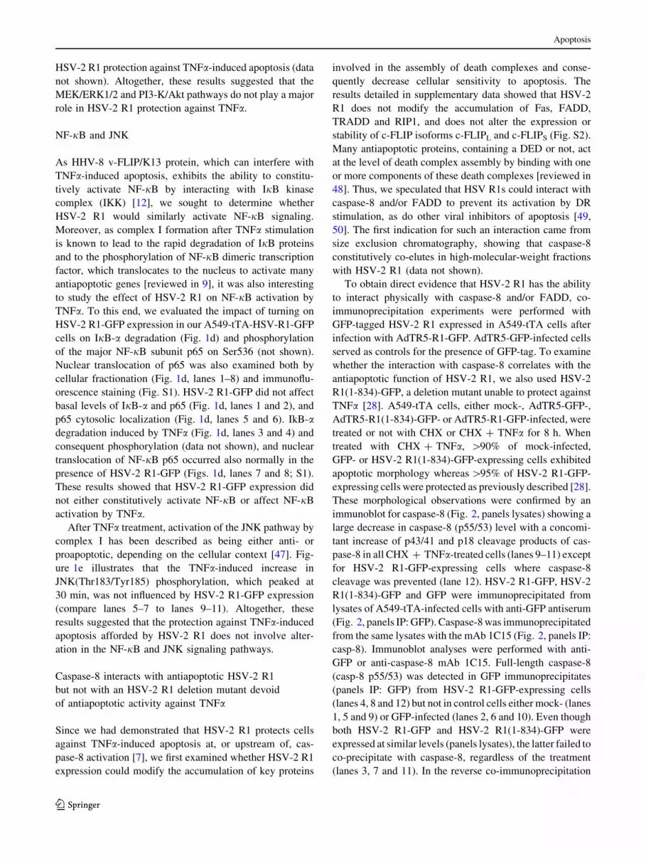
Caspase-8 interacts with antiapoptotic HSV-2 R1
but not with an HSV-2 R1 deletion mutant devoid
of antiapoptotic activity against TNFa
Since we had demonstrated that HSV-2 R1 protects cells
against TNFa-induced apoptosis at, or upstream of, cas-
pase-8 activation [7], we first examined whether HSV-2 R1
expression could modify the accumulation of key proteins
involved in the assembly of death complexes and conse-
quently decrease cellular sensitivity to apoptosis. The
results detailed in supplementary data showed that HSV-2
R1 does not modify the accumulation of Fas, FADD,
TRADD and RIP1, and does not alter the expression or
stability of c-FLIP isoforms c-FLIPL and c-FLIPS (Fig. S2).
Many antiapoptotic proteins, containing a DED or not, act
at the level of death complex assembly by binding with one
or more components of these death complexes [reviewed in
48]. Thus, we speculated that HSV R1s could interact with
caspase-8 and/or FADD to prevent its activation by DR
stimulation, as do other viral inhibitors of apoptosis [49,
50]. The first indication for such an interaction came from
size exclusion chromatography, showing that caspase-8
constitutively co-elutes in high-molecular-weight fractions
with HSV-2 R1 (data not shown).
To obtain direct evidence that HSV-2 R1 has the ability
to interact physically with caspase-8 and/or FADD, co-
immunoprecipitation experiments were performed with
GFP-tagged HSV-2 R1 expressed in A549-tTA cells after
infection with AdTR5-R1-GFP. AdTR5-GFP-infected cells
served as controls for the presence of GFP-tag. To examine
whether the interaction with caspase-8 correlates with the
antiapoptotic function of HSV-2 R1, we also used HSV-2
R1(1-834)-GFP, a deletion mutant unable to protect against
TNFa [28]. A549-tTA cells, either mock-, AdTR5-GFP-,
AdTR5-R1(1-834)-GFP- or AdTR5-R1-GFP-infected, were
treated or not with CHX or CHX ? TNFa for 8 h. When
treated with CHX ? TNFa, [90% of mock-infected,
GFP- or HSV-2 R1(1-834)-GFP-expressing cells exhibited
apoptotic morphology whereas[95% of HSV-2 R1-GFP-
expressing cells were protected as previously described [28].
These morphological observations were confirmed by an
immunoblot for caspase-8 (Fig. 2, panels lysates) showing a
large decrease in caspase-8 (p55/53) level with a concomi-
tant increase of p43/41 and p18 cleavage products of cas-
pase-8 in all CHX ? TNFa-treated cells (lanes 9–11) except
for HSV-2 R1-GFP-expressing cells where caspase-8
cleavage was prevented (lane 12). HSV-2 R1-GFP, HSV-2
R1(1-834)-GFP and GFP were immunoprecipitated from
lysates of A549-tTA-infected cells with anti-GFP antiserum
(Fig. 2, panels IP: GFP). Caspase-8was immunoprecipitated
from the same lysates with the mAb 1C15 (Fig. 2, panels IP:
casp-8). Immunoblot analyses were performed with anti-
GFP or anti-caspase-8 mAb 1C15. Full-length caspase-8
(casp-8 p55/53) was detected in GFP immunoprecipitates
(panels IP: GFP) from HSV-2 R1-GFP-expressing cells
(lanes 4, 8 and 12) but not in control cells either mock- (lanes
1, 5 and 9) or GFP-infected (lanes 2, 6 and 10). Even though
both HSV-2 R1-GFP and HSV-2 R1(1-834)-GFP were
expressed at similar levels (panels lysates), the latter failed to
co-precipitate with caspase-8, regardless of the treatment
(lanes 3, 7 and 11). In the reverse co-immunoprecipitation
Apoptosis
123

experiment (panels IP: casp-8), HSV-2 R1-GFP (lanes 4, 8
and 12) but not HSV-2 R1(1-834)-GFP (lanes 3, 7 and 11)
was specifically detected in caspase-8 immunoprecipitates,
the low amount of HSV-2 R1(1-834)-GFP seen in all
R1(1-834)-GFP samples being due to the unspecific pre-
cipitation of mutant protein with beads (see ctl lanes 13 and
14). Similar co-immunoprecipitation experiments with
A549-tTA-HSV-R1-GFP cells confirmed the interaction
between HSV-2 R1 and caspase-8 (data not shown).
HSV-2 R1 impairs apoptosis induced by caspase-8
over-expression
It is well known that caspase-8 over-expression induces
apoptosis [51]. In this context, apoptosis is thought to be the
outcome of caspase-8 dimerization through catalytic domain
interaction, as single point mutants in the dimer interface,
which are unable to undergo dimerization, exhibit greatly
reduced apoptosis induction [52]. To determine whether
HSV-2 R1 could prevent apoptosis induced by caspase-8
over-expression, we transfected into HeLa cells a GFP-
tagged version of caspase-8 (casp-8 GFP depicted in
Fig. 3a) known to potently induce apoptosis in these cells
[30]. Observations by fluorescence microscopy at 24 h
following casp-8 GFP transfection showed that all the
GFP-positive cells in control dishes (mock-infected or pre-
infected with an empty control Ad recombinant) were
rounded up and floating in the medium (Fig. 3b). In these
detached cells, the fluorescence was seen as diffuse
throughout apoptotic bodies and blebs, as well as concen-
trated in large aggregates (Fig. 3b, photographs). In sharp
contrast, when cells had been infected with AdCMV5-R1
prior to transfection, more than 40% of GFP-positive cells
remained adherent with healthy morphology (Fig. 3b). In
these adherent cells, fluorescence appeared diffuse
throughout the cytoplasm, as reported in conditions where
apoptosis induced by casp-8 GFP was inhibited by z-VAD-
fmk [30]. Immunoblotting with anti-GFP antibody showed
that casp-8 GFP (*85 kDa) was nearly completely absent in
control cells, which accumulated the processed casp-8 p10
GFP product (*42 kDa) (Fig. 3b, lanes 1 and 2). Strikingly,
cells expressing HSV-2 R1 accumulated high levels of
unprocessed casp-8 GFP (Fig. 3b, lane 3), indicating that
HSV-2 R1 can impair casp-8 GFP activation. To determine
whether casp-8 GFP interacted with HSV-2 R1 as did
endogenous caspase-8, cells were infected with AdCMV5-
R1 and 12 h later transfected with a plasmid encoding cat-
alytically-inactive caspase-8 C360S GFP (casp-8 C360S
GFP) (depicted in Fig. 3a) to avoid caspase-8 autoprocess-
ing. Immunoprecipitation with anti-GFP antiserum showed
that HSV-2 R1 co-immunoprecipitated with casp-8 C360S
GFP, as expected (Fig. 3c, lane 2). The results demonstrat-
ing that HSV-2 R1 impairs caspase-8 activation induced by
its over-expression suggested that the interaction between
the two proteins impedes caspase-8 dimerization.
HSV-2 R1 interacts directly with caspase-8
and the DEDs of caspase-8 are essential
for this interaction
As purified HSV-2 R1 does not inhibit active recombinant
human caspase-8, which consists of the processed caspase
domain [7], we hypothesized that it interacts with the
DEDs of caspase-8. To ascertain that, two caspase-8
deletion mutants C-terminally fused to GFP (casp-8 DED-
AB GFP and casp-8 CD GFP depicted in Fig. 3a) were
tested. In addition, to determine whether the interaction
was specific to caspase-8 DEDs, FADD fused to YFP
R1(1-834)-GFPR1-GFP
WB : GFP
lysates
Casp-8
p55/53
R1(1-834)-GFP
R1-GFP
-- +
+- - -
- - - +- -- +
+- - -
- - - +- -- +
+- - -
- - - +- -
- -
+ -+
GFP
untreated CHX CHX+TNFα ctl
R1(1-834)-GFPR1-GFP
WB : GFP
R1(1-834)-GFPR1-GFP
WB : GFP
Casp-8 p55/53WB : Casp-8
(1C15)
Casp-8 p55/53
Casp-8 p55/53
p43/41
p18
WB : Casp-8
(1C15)
IP : G
FP
IP : C
asp-8
(1C
15
)
WB : Casp-8
(1C15)
WB : Casp-8
(1C12)
-+ - - -+ - - -+ - - - -mock
1 2 3 4 5 6 7 8 9 10 11 13 1412
1 2 3 4 5 6 7 8 9 10 11 12
Fig. 2 HSV-2 R1, but not the inactive mutant R1(1-834)-GFP,
co-immunoprecipitates with caspase-8. A549-tTA cells were infected
with AdTR5-R1-GFP (R1-GFP), AdTR5-R1(1-834)-GFP (R1(1-834)-
GFP) or AdTR5-GFP (GFP) and, 24 h later, were treated or not
(untreated) with CHX or CHX ? TNFa. After 8 h, the cells were
harvested, and GFP-tagged proteins or caspase-8 were immunopre-
cipitated with anti-GFP antibody or anti-caspase-8 mAb 1C15. As
precipitation control (ctl), pre-cleared lysates were incubated with
G-Sepharose beads without antibody. Immunoprecipitates (panels IP)
and cell lysates (panels lysates) were analyzed by immunoblotting for
GFP-tagged proteins and caspase-8. The immunoblots are represen-
tative of three experiments
Apoptosis
123

(a)
(e)
(d)
p18
Prodomain Caspase domain
GFP
DED-A DED-B p10
D210 D216 D374 D384
DED DDYFP
GFPC360S 479
GFP209
GFP210 479
GST478
GST180
GST181 478
Casp-8 GFP
Casp-8 C360S GFP
Casp-8 DED-AB GFP
Casp-8 CD GFP
FADD YFP
GST Casp-8
GST Casp-8 DED-AB
GST Casp-8 CD
C360
*
*
*
*
(b)TR5 + Casp-8 GFP R1 + Casp-8 GFP
-R1 - +TR5 +- -mock -+ -
50
40
20
10
30
Adher
ent
cell
s
(%
GF
P+
)
Casp-8 GFP ++ +
R1
-R1 - +TR5 +- -
Casp-8 GFP
β-actin
*
mock -+ -
Casp-8 p10 GFP
Casp-8 GFP ++ +
1 2 3
IP : GFP
Casp-8 DED-AB GFP + R1 - -- + -
+GFP + R1 - -- -
Casp-8 C360S GFP + R1 + -- --
lysates ctl
Casp-8 CD GFP + R1
FADD YFP + R1
- +- - -
- -- - +
- -- + -
+ - -- -+ -- --
- +- - -
- -- - +
- -- + -
+ - -- -+ -- --
- +- - -
- -- - +
R1
Casp-8 C360S GFP
Casp-8 DED-AB GFP
Casp-8 CD GFP
FADD YFP
(c)
1 2 3 4 5 6 7 8 9 10 11 12 13 14 15
GST
GST C
asp-
8
GST C
asp-
8 D
ED-A
B
GST C
asp-
8 CD
Bea
ds
R1
(100
ng)
GST
GST C
asp-
8
GST C
asp-
8 D
ED-A
B
GST C
asp-
8 CD
Bea
ds
R1
(100
ng)
R1 (pET-R1) R1 (BM5-R1)
R1
1 2 3 4 5 6 7 8 9 10 11 12
IP : GFP
GFP - -+ +
+TR5 + --R1 - ++-
lysates ctl
Casp-8 C360S GFP + +- -
- -+ +
+ + --- ++-
+ +- -
- -+ +
+ + --- ++-
+ +- -
R1
Casp-8 C360S GFP
FADD
1 2 3 4 5 6 7 8 9 10 11 12
Fig. 3 HSV-2 R1 interacts directly with caspase-8 through its two
tandem DEDs. a Schematic representation of the fusion proteins used
in this study. Death-effector domains are indicated by DED, death
domains by DD, and caspase domain by CD. The aspartic residue
processed during caspase-8 maturation and the active site cysteine
(C360) are shown. b HSV-2 R1 protects cells against caspase-8 GFP-
induced apoptosis. HeLa cells were mock-infected or infected with
AdCMV5-R1 (R1) or AdTR5CuO (TR5). After 8 h, they were
transfected with plasmid encoding for casp-8 GFP. Twenty-four hours
later, GFP-positive cells were observed under fluorescence micros-
copy, and the percentages (mean ± SE, n = 6) of GFP-positive
apoptotic cells were determined. Cell lysates were analyzed by
immunoblotting for HSV-2 R1, GFP-tagged proteins and b-actin. The
data are representative of three independent experiments performed in
duplicate. The asterisk indicates a non-specific band. c HSV-2 R1
interacts with caspase-8 through its DEDs but not with FADD. HeLa
cells were infected with AdCMV5-R1 (R1) and transfected 12 h later
with the plasmid encoding GFP, casp-8 C360S GFP, casp-8 DED-AB
GFP, casp-8 CD GFP or FADD YFP and harvested 30 h later. GFP-
tagged proteins were immunoprecipitated with polyclonal anti-GFP
antibody. As immunoprecipitation control (ctl), pre-cleared lysates
were incubated with G-Sepharose beads without antibody. Immuno-
precipitates (IP) and cell lysates (lysates) were analyzed by immu-
noblotting for HSV-2 R1 and GFP-tagged proteins. Since the
anti-GFP mAb B-2 has low affinity for YFP, FADD YFP was
revealed with the anti-FADD mAb A66-2. d HSV-2 R1 directly
interacts with caspase-8 through its DEDs but not with its caspase
domain. Beads coupled to GST, GST casp-8, GST casp-8 DED-AB or
GST casp-8 CD were incubated with 1 lg of purified HSV-2 R1
(pET-R1 or BM5-R1), and proteins bound to beads were analyzed by
immunoblotting with the anti-R1 serum 168R1. As control for
unspecific binding, 1 lg of each purified R1 was incubated with
uncoupled beads (Beads) or GST-coupled beads (GST). R1 was
quantified by loading 100 ng of each R1. e The interaction between
HSV-2 R1 and caspase-8 disrupts FADD-caspase-8 binding. HeLa
cells were infected as in (b) and transfected to express GFP or casp-8
C360S GFP. GFP-tagged proteins were immunoprecipitated as in (c).
Immunoprecipitates (IP) and cell lysates (lysates) were analyzed by
immunoblotting for HSV-2 R1, FADD and GFP-tagged proteins. The
immunoblots are representative of two experiments
Apoptosis
123

(FADD YFP depicted in Fig. 3a) was included in the
analysis. Immunoprecipitation with anti-GFP antiserum
(Fig. 3c) showed that HSV-2 R1 co-immunoprecipitated
with casp-8 DED-AB GFP (lane 3), but not with casp-8 CD
GFP (lane 4) or with FADD YFP (lane 5). Consistent with
these results, we also observed that HSV-2 R1 could block
the formation of previously-described [30] cytoplasmic
filaments in casp-8 DED-AB GFP-expressing cells but not
those seen in FADD YFP-expressing cells (Fig. S3). Our
findings suggested that HSV-2 R1 could interact with
DED-AB of caspase-8 in a way that inhibits the formation
of filaments but not with the DED of FADD or with the
caspase domain of caspase-8.
To better establish that HSV-2 R1 interacted with casp-8
DED-AB, GST pull-down experiments (Fig. 3d) were
performed with GST-tagged deletion mutants of caspase-8
produced in E. coli and with purified HSV-2 R1 produced
in either bacteria (pET-R1, lanes 1–6) or human cells
(BM5-R1, lanes 7–12). Purified HSV-2 R1, whether pro-
duced in bacteria or in mammalian cells, could be effi-
ciently pulled-down with either GST casp-8 (lanes 3 and 9)
or GST casp-8 DED-AB (lanes 4 and 10) but not with GST
casp-8 CD (lanes 5 and 11). These results demonstrated not
only that HSV-2 R1 directly interacts with DED-AB of
caspase-8, without requiring additional cofactors, but also
that eukaryotic post-translational modifications of HSV-2
R1 are not required for the interaction.
Binding of HSV-2 R1 to caspase-8 disrupts FADD–
caspase-8 interaction
To gain additional insight into the mechanism by which
HSV R1 interaction with caspase-8 prevented its activa-
tion, we investigated whether HSV-2 R1 could affect the
interaction between caspase-8 and FADD. To this end,
HeLa cells were infected with AdCMV5-R1 or an empty
Ad recombinant for 16 h before transfecting GFP or casp-8
C360S GFP to avoid caspase-8 autoprocessing (Fig. 3e).
As expected, when immunoprecipitation was performed
with anti-GFP antiserum, FADD co-precipitated with casp-
8 C360S GFP (lane 2) but not with GFP (lane 1). When
cells were infected with AdCMV5-R1 prior to transfection
(lanes 3 and 4), the interaction between FADD and casp-8
C360S GFP was greatly reduced (compare lanes 2 and 4)
and HSV-2 R1 was present in the immunoprecipitate (lane
4). This result indicated that HSV-2 R1 binding to caspase-
8 disrupts FADD–caspase-8 interaction.
Individually expressed HSV-1 R1 protects cells against
TNFa- and FasL-induced apoptosis
Our previous observation that HSV-1 UL39 deletion
mutants exhibited half of the WT virus antiapoptotic
potential against TNFa had provided indirect evidence that
HSV-1 R1 could be antiapoptotic against DR ligands [7]. To
firmly establish that it is the case and also to determine
whether HSV-1 R1 could, like HSV-2 R1, interact with
caspase-8, HeLa cells were transfected with an expression
plasmid encoding HSV-1 R1 fused to GST or, for compar-
ison, with an HSV-2 R1 expression plasmid. In these
experiments a plasmid encoding EBV R1 (an RR active
subunit) was also tested. Treatments with CHX ? TNFa or
highly cytotoxic hexameric FasL for 6 h (Fc:FasL, [32])
induced apoptotic morphology in more than 80% of control
HeLa cells (Fig. 4a) as previously reported [7, 53]. Both
HSV R1s reduced by more than 5-fold the % of morpho-
logically apoptotic cells, but HA-EBV R1 was without sig-
nificant effect. To confirm the morphological observations,
caspase-8 activation was assessed by immunoblotting
(Fig. 4b), whereas caspase-3/7 activation was evaluated by
in vitro enzymatic assay with DEVD-AFC as substrate
(Fig. 4a). Both HSV-1 R1 and HSV-2 R1, but not EBV-R1,
greatly reduced caspase-8 (lanes 6, 8; 14, 16; 22, 24) and
caspase-3/7 activation produced by DR triggering (lanes 5,
7; 13, 15; 21, 23). Note that the 168R1 serum, which was
raised against purified HSV-2 R1, recognized less efficiently
HSV-1 R1 than HSV-2 R1 (C. Guilbault and Y. Langelier,
unpublished observations). As immunofluorescence stain-
ing revealed that[75% of cells were positive for the HA tag,
it is unlikely that a poor transfection efficiency would be the
cause of the lack of protection by the HA tagged EBV R1.
Altogether these results indicated that the R1s of both types
of HSVs protect HeLa cells against TNFa and FasL whereas
that of EBV does not exhibit this property.
To assess interaction with caspase-8, immunoprecipita-
tion was performed with the mAb 1C15. Figure 4c showed
that, like HSV-2 R1 (lane 7), HSV-1 R1 (lane 2) copre-
cipitated with caspase-8 but not EBV R1 (lane 12). These
results demonstrated that HSV-1 R1 and HSV-2 R1 but not
EBV R1 interact with caspase-8.
The R1 null mutant ICP6D does not protect cells
against FasL
Determining the importance of HSV-1 R1 in protecting
HSV-infected cells from TNFa was complicated by the vhs
effect on the short-lived TNFR1. As Fas has been described
to be much more stable than TNFR1 [54] and given the
antiapoptotic potential of individually expressed HSV-1 R1
against DR-induced apoptosis, we thought that more con-
clusive data could be obtained for FasL-induced apoptosis.
Because the effect of HSV infection on Fas level had never
been studied, we first compared the fate of Fas and TNFR1
in total lysates of HeLa cells infected with WT HSV-1
KOS or its R1 null mutant ICP6D for up to 8 h. As seen in
Fig. 5a for KOS (lanes 2–6) and ICP6D (lanes 7–11)
Apoptosis
123

infection, Fas levels remained almost unaltered during this
period as expected from the report on Fas stability [54].
TNFR1 decreased steadily from 2 h post-infection as pre-
viously reported [8] but, even at 8 h, it was still detectable,
albeit at a very low level (lanes 6 and 11). The rate of
TNFR1 disappearance was similar for both viruses, indi-
cating that HSV-1 R1 does not affect the rate of TNFR1
reduction. The experiments were repeated for HSV-2
infection with similar results (data not shown). Two con-
clusions can be drawn from these observations. First,
protein stability appears to be a key determinant of the fate
of DRs during HSV infection. Second, even if TNFR1
protein level decreases drastically after HSV infection, this
DR is still present at 8 h after infection.
As Fas levels remained almost unaltered during the first
8 h of infection, the effect of deleting the HSV-1 R1 gene
(a)
(b)
GST-HSV-1 R1 HSV-2 R1 HA-EBV R1
(c)IP
pCMVpHSV-2 R1
+ -- + +
- + -- +
ltc setasyl
HSV-2 R1
Casp-8 p55/53
6 7 8 9 10
pGSTpGST-HSV-1 R1
+ -- + +
- + -- +
ltc setasylPI
GST-HSV-1 R1
Casp-8 p55/53
1 2 3 4 5
pcDNA3pHA-EBV R1
+ -- + +
- + -- +
ltc setasylPI
HA-EBV R1
Casp-8 p55/53
11 12 13 14 15
700
800
900
600
500
400
300
200
100
0
100
80
60
40
20
0 Cas
p-3
/7 a
ctiv
ity
(p
mo
le/m
in/m
g)
pcDNA3pHA-EBV R1
+-
+- +
-+-
ctl
CH
X
CH
X +
TN
Fα
Fc:Fas
L
Ap
op
toti
c ce
lls
(%)
+-
+- +
-+- +
-+- +
-+- +
-+- +
-+-
600
500
400
300
200
100
0
100
80
60
40
20
0 Cas
p-3
/7 a
ctiv
ity
(p
mo
le/m
in/m
g)
pGSTpGST-HSV-1 R1
+-
+- +
-+-
ctl
CH
X
CH
X +
TN
Fα
Fc:Fas
L
Ap
op
toti
c ce
lls
(%)
+-
+- +
-+- +
-+- +
-+- +
-+- +
-+-
600
500
400
300
200
100
0
100
80
60
40
20
0 Cas
p-3
/7 a
ctiv
ity
(p
mo
le/m
in/m
g)
pCMVpHSV-2 R1
+-
+- +
-+-
ctl
CH
X
CH
X +
TN
Fα
Fc:Fas
L
Ap
op
toti
c ce
lls
(%)
+-
+- +
-+- +
-+- +
-+- +
-+- +
-+-
GST-HSV-1 R1
+--- ++pGST +
-+++ ---pGST-HSV-1 R1
-
+
ctl
CH
XCH
X +
TN
Fα
Fc:Fas
L
p43/41
Casp-8
p55/53
p18
1 2 3 4 5 6 7 8
β-actin
+--- ++pCMV +
-+++ ---
HSV-2 R1
pHSV-2 R1
-
+
ctl
CH
XCH
X +
TN
Fα
Fc:Fas
L
p43/41
Casp-8
p55/53
p18
9 10 11 12 13 14 15 16
β-actin
HA-EBV R1
+--- ++pcDNA3 +
-+++ ---pHA-EBV R1
-
+
ctl
CH
XCH
X +
TN
Fα
Fc:Fas
L
Casp-8
p55/53
p43/41
p18
17 18 19 20 21 22 23 24
β-actin
Fig. 4 HSV-1 R1 inhibits DR-induced apoptosis and interacts with
caspase-8. a, b GST-HSV-1 R1 and HSV-2 R1 inhibit TNFa- and
FasL-induced apoptosis but not HA-EBV R1. HeLa cells were
transfected with the plasmid pAdCMV5 (pCMV) or pAdCMV5-R1
(pHSV-2 R1) (left panel); pLBPF1-GST (pGST) or pLBPF1-GST-R1
(pGST-HSV-1 R1) (middle panel); pcDNA3 or pcDNA3-HA-EBV
R1 (pHA-EBV R1) (right panel). After 48 h, the cells were untreated
(ctl) or treated with CHX (15 lg/ml), CHX ? TNFa (2.5 ng/ml) or
Fc:FasL (10 ng/ml) for 8 h. Percentages of apoptotic cells were
determined by microscopic observation (a). Cytoplasmic cell lysates
were analyzed by immunoblotting for HSV R1s, EBV R1, caspase-8
(1C12) and b-actin protein content (b) and tested for caspase-3/7
activity (a). The immunoblots and caspase activities (mean ± SE,
n = 4) are representative of at least two experiments performed in
duplicate. c HSV-1 R1 and HSV-2 R1 co-immunoprecipitate with
caspase-8 but not EBV R1. Caspase-8 was immunoprecipitated with
anti-caspase-8 mAb 1C15 from ctl extracts prepared in (a, b).
Immunoprecipitates (IP) and cell lysates (lysates) were analyzed by
immunoblotting for HSV R1s, HA-EBV R1 and caspase-8 (1C15). As
precipitation control (ctl), pre-cleared lysates were incubated with
G-Sepharose beads without antibody. The immunoblots are repre-
sentative of two experiments
Apoptosis
123

on protection from FasL was studied by infecting HeLa
cells for 8 h with either WT KOS or ICP6D and treating
them for 6 h with 50 lg/ml Fc:FasL. KOS infection
reduced the % of morphologically apoptotic cells from
[90% in mock-infected samples to\5%. In sharp contrast,
ICP6D infection did not alter the % of apoptosis induced by
Fc:FasL (\90%). Our morphological observations were
corroborated by analyses of caspase-8 and caspase-3/7
activation. Indeed, KOS infection completely prevented
caspase-8 cleavage (Fig. 5b, lane 4 vs. lane 2) and caspase-
3/7 activation (Fig. 5c), whereas ICP6D infection did not
affect either caspase-8 (Fig. 5b, lane 6 vs. lane 2) or cas-
pase-3/7 activation (Fig. 5c). These results showed that the
protection afforded by HSV against Fc:FasL is completely
lost by deleting R1. For all the experiments involving
ICP6D, exponentially growing cells were used to prevent
the inhibition of translation of viral mRNAs, which would
occur if the cells were growth-arrested [27]. Evidence that
the synthesis of viral proteins was not impaired in our
ICP6D-infected cells was obtained from the observation
that the R2 subunit of HSV RR (Fig. 5a) and the infected
cell polypeptide 0 (data not shown) accumulated at similar
rate in cells infected with either KOS or ICP6D. This made
unlikely the possibility that the antiapoptotic defect of
ICP6D against FasL is related to a lower ability of the cells
to support the synthesis of viral polypeptides. Together,
these results suggested that HSV-1 R1 could play an
essential role in the protection against FasL.
HSV-1 R1 and HSV-2 R1 interact with caspase-8
in the context of HSV infection
We next determined whether HSV R1s could interact with
caspase-8 in the context of HSV infection. Our previous
work had revealed that protection from TNFa-induced
apoptosis plateaued around 8 h of infection with both types
of HSV [7]. Hence, HeLa and A549-tTA cells were
infected for 8 h with either HSV-1 or HSV-2, and caspase-
8 was immunoprecipitated with the anti-caspase-8 mAb
1C15. Immunoblot analyses with anti-R1 serum 168R1
(Fig. 6) revealed that both HSV-1 R1 (lanes 2 and 5) and
HSV-2 R1 (lanes 3 and 6) interacted with caspase-8 in the
two cell lines tested. Altogether, these results indicate that
HSV R1s can physiologically interact with caspase-8.
Discussion
Apoptosis triggered by stimulation of DRs is important in
the host response to viral infection. To counteract host
(a) (c)(b)
400
300
200
100
0
Cas
p-3
/7 a
ctiv
ity
(pm
ole
/min
/mg
)
Fc:FasLctl
mock KOS ICP6∆
Casp-8
p55/53
ß-actin
R1
- -+ + +-Fc:FasL
mock KOS ICP6∆
R2
p43/41
p18
1 2 3 4 5 6
Fas
1 40 2 6 8Time P.I. (h)
R1
TNFR1
ß-actin
R2
KOS
1 42 6 8
ICP6∆
1 2 3 4 5 6 7 8 9 10 11
Fig. 5 HSV-1 R1 plays a key role for protection against FasL-
induced apoptosis. a HeLa cells were infected with the parental
HSV-1 strain (KOS) or the R1 null mutant strain (ICP6D) and
harvested at the indicated times post-infection (PI). Whole cell extracts
were analyzed by immunoblotting for TNFR1, Fas, HSV-1 R1, HSV-1
R2 and b-actin. b, c HeLa cells were mock-infected (mock) or infected
as in (a) for 8 h and then treated with Fc:FasL (50 ng/ml) or not (ctl).
After 6 h, the cells were harvested and cytoplasmic cell lysates were
analyzed by immunoblotting for HSV-1 R1, HSV-1 R2, caspase-8
(1C12) and b-actin protein content (b) and tested for caspase-3/7
activity (c). The immunoblots and caspase activities (mean ± SE,
n = 4) are representative of two experiments performed in duplicate
R1
R1
HSV-1HSV-2
- +- - +
- - +- - +
- -+-
+
HeLa A549tTa ctl
WB : R1
WB : R1
Casp-8 p55/53
Casp-8 p55/53WB : Casp-8
(1C15)
WB : Casp-8
(1C15)
IPly
sates
mock + - - + - - --
1 2 3 4 5 6 7 8
1 2 3 4 5 6
Fig. 6 HSV-1 R1 and HSV-2 R1 co-immunoprecipitate with
caspase-8 in the context of HSV infection. HeLa and A549-tTA
cells were infected with HSV-1 (strain KOS) or HSV-2 (strain HG-
52). After 8 h, the cells were harvested, and caspase-8 was
immunoprecipitated with anti-caspase-8 mAb 1C15. Immunoprecip-
itates (panels IP) and cell lysates (panels lysates) were analyzed by
immunoblotting for HSV R1s and caspase-8 (1C15). As precipitation
control (ctl), pre-cleared lysates were incubated with G-Sepharose
beads without antibody. The immunoblots are representative of two
experiments
Apoptosis
123

defense mechanisms, viruses have evolved several molec-
ular strategies to prevent caspase-8 activation after DR
stimulation through the acquisition of proteins that act on
different targets in this pathway [50, 55–57]. Several
studies have indicated that HSVs can efficiently block
apoptosis induced by DR activation [reviewed in 58]. In
this regard, two HSV proteins were proposed to play
important roles: the R1 subunit of HSV-2 RR which,
expressed on its own, protects epithelial cells against
TNFa- and FasL-induced apoptosis by interrupting DR-
mediated signaling at, or upstream of, caspase-8 activation
[7, 28], and vhs which, by destroying TNFR1 mRNA,
impairs the replenishment of short-half-life TNFR1 [8].
Here, we report the following main findings on the anti-
apoptotic activity of HSV R1s against DR ligands. HSV-2
R1, which does not stimulate the major antiapoptotic sur-
vival pathways ERK, Akt, NF-jB and JNK, interacts with
the prodomain of caspase-8 in a way that inhibits its
dimerization/activation. The interaction between HSV-2
R1 and caspase-8 also disrupts FADD-caspase-8 binding.
Individually expressed HSV-1 R1, like HSV-2 R1, has the
ability to protect cells against TNFa and FasL and to bind
to caspase-8. Strikingly, HSV-1 R1 could play an essential
role for protecting infected cells against FasL.
Since some viral antiapoptotic proteins, including
HSV-2 R1 [22], had been found to target the ERK1/2 and
Akt signaling pathways, it was important to evaluate
whether they could be involved in the antiapoptotic activity
of HSV-2 R1. Three lines of evidence strongly argue
against any effect of HSV-2 R1 on ERK1/2 and PI3-K/Akt
pathways in the protection from TNFa. Firstly, HSV-2 R1
expression at a level sufficient to confer full protection
against TNFa-induced apoptosis did not significantly affect
ERK1/2 or Akt phosphorylation. Secondly, HSV-2 R1 did
not modify the transient activation of ERK1/2 induced by
TNFa. Thirdly, suppression of the ERK1/2 or Akt path-
ways with specific biochemical inhibitors did not influence
HSV-2 R1 protection against TNFa-induced apoptosis. Our
observation that the inducible expression of HSV-2 R1 did
not increase ERK1/2 phosphorylation contrasts with pre-
vious reports that 293 or PC12 cells stably expressing
HSV-2 R1 exhibit a higher level of ERK1/2 phosphoryla-
tion than their parental counterparts [43, 59]. As we have
been unable to detect any increase in ERK1/2 phosphory-
lation upon inducible HSV-2 R1 expression in 293 cells,
we have ruled out, as an explanation for this discrepancy,
that the effect could be cell type-specific. It is possible that
the higher constitutive ERK1/2 phosphorylation, previ-
ously observed in 293 cells stably expressing HSV-2 R1,
could be due to clonal variation. Indeed, it is well-known
that several established cell lines exhibit high constitutive
ERK1/2 phosphorylation whereas others do not [60].
Furthermore, HSV-2 R1 does not constitutively activate
NF-jB and JNK or alter the activation of NF-jB and JNK
by TNFa. Such observations not only indicate that these
pathways are not involved in protection from TNFa but
also strongly suggest that HSV-2 R1 does not interfere with
the TNFa-induced formation of complex I, since assembly
of the complex is required for early NF-jB, JNK or ERK1/
2 phosphorylation. In this respect, HSV-2 R1 differs from
HHV-8 v-FLIP/K13 that constitutively activates the
canonical NF-jB pathway by interacting with IKKc [12].
HSV-2 R1 protection against TNFa-induced apoptosis
differs from viral strategies affecting the expression or
stability of critical proteins implicated in DR signaling
[57, 61–63]. Indeed, HSV-2 R1 does not affect either the
expression of c-FLIP isoforms or their stability. HSV-2
R1 does not modify the expression of TNFR1, TRADD,
FADD, RIP1 and caspase-8. Even if sequences of HSV
R1s do not present similarities with c- or v-FLIP, we
hypothesized that HSV R1s could interact with caspase-8
to prevent its activation by DR stimulation, as do other
viral inhibitors of apoptosis [49, 50]. Immunoprecipita-
tion experiments confirmed that both individually
expressed HSV-1 and HSV-2 R1 interact with caspase-8
with or without treatment with TNFa, demonstrating that
the interaction is constitutive. Moreover, interaction
between HSV R1s and caspase-8 could be detected with
extracts of HSV-1- and HSV-2-infected cells, supporting
a role for this interaction in the resistance of HSV-
infected cells to DR-induced apoptosis [7, 64]. HSV-2 R1
binding to caspase-8 appears indispensable for the anti-
apoptotic activity of HSV-2 R1 since the deletion mutant
HSV-2 R1(1-834)-GFP, which is devoid of antiapoptotic
activity [28], does not interact with caspase-8. Moreover,
EBV R1, which does not show protection from DR-
ligands in our experimental system, does not interact with
caspase-8.
The data, obtained with two series of caspase-8 deletion
mutants, showed that HSV-2 R1 interacts with the two
tandem DED-containing prodomain of caspase-8 but not
with the caspase domain of caspase-8. This is in agreement
with the observation that HSV-2 R1 does not protect cells
against caspase-8 CD GFP-induced apoptosis (F. Dufour
and Y. Langelier, unpublished observations) and with our
previous finding that purified HSV-2 R1 does not inhibit
the protease activity of processed recombinant caspase-8,
which contains only the caspase domain [7]. GST pull-
down experiments with purified proteins demonstrated that
HSV-2 R1 interaction with either full-length caspase-8 or
caspase-8 DED-AB is direct and does not require addi-
tional molecules. Also, as the interaction still occurred
when both proteins were produced and purified from bac-
teria, it can be concluded that post-translational modifica-
tions specific to mammalian cells are not necessary for
binding. Altogether, these data firmly established that
Apoptosis
123
HSV-2 R1, by interacting constitutively and directly with
caspase-8, prevents its activation. A similar mechanism of
action has been described for vICA, the UL36 gene product
of human CMV that is conserved in all CMV genomes
sequenced to date [65]. Like HSV R1s, vICA suppresses
TNFa- and FasL-induced apoptosis but appears to mar-
ginally inhibit cell death induced by cytotoxic drugs acti-
vating the mitochondrial apoptosis pathway [7, 50]. Even
though HSV-2 R1 and vICA do not exhibit sequence
similarity, both proteins associate with caspase-8 in the
absence of DR activation and interact with the caspase-8
prodomain.
Since dimerization is a precondition for self-processing
and caspase-8 activity [66], we speculated that HSV-2 R1
could interact with caspase-8 in a way that inhibits this
event. Supporting our hypothesis, we found that HSV-2 R1
protects cells against apoptosis induced by over-expression
of GFP-tagged caspase-8 that is known to be triggered by
dimerization/activation of the over-expressed zymogen
[51]. In the case of TNFa- or FasL-induced apoptosis,
complex II or DISC drives dimerization and activation of
monomeric caspase-8 via FADD [13, 14]. Since on one
hand HSV-2 R1 interacts with the DEDs of caspase-8 and
on the other hand FADD contains a DED involved in both
FADD self-association and caspase-8 interaction [67], we
investigated putative interaction between FADD and
HSV-2 R1. We have been unable to detect, by co-immu-
noprecipitation, any interaction between HSV-2 R1 and
endogenous or over-expressed FADD, indicating that the
interaction is specific to the tandem DEDs contained in the
caspase-8 prodomain. A recent report identified the cas-
pase-8-specific binding surface on FADD and suggested
preferential interaction of caspase-8 DED-B with FADD
DED [67]. Since HSV-2 R1 interacts with the caspase-8
prodomain, it is conceivable that oligomerized HSV-2 R1
could sterically hinder the site of caspase-8 interaction with
FADD DED. Supporting this hypothesis, we found that the
interaction between over-expressed casp-8 C360S GFP and
endogenous FADD is inhibited when HSV-2 R1 is
expressed. It remains to be determined whether HSV R1s
inhibit the recruitment of caspase-8 to the assembled
complex II or DISC.
Even if most of our data were obtained with HSV-2 R1,
we believe that the majority of our conclusions on the
mechanism of action of this protein can be extrapolated to
HSV-1 R1. Indeed, both proteins interact with caspase-8
and our previous mapping of the antiapoptotic domain of
HSV-2 R1 indicates that this domain is highly conserved
between both types of HSV (95% similarity [28]). We also
speculate that the deficiency of EBV R1 in protection from
TNFa and FasL would be related to the absence of the
putative a-crystallin domain in the shorter EBV R1, a
domain shown to be important for HSV-2 R1 antiapoptotic
activity [26]. It is noteworthy that until now protection
from TNFa has been reported only for another herpes virus
R1, that of murine ß-herpes virus CMV, also named M45
[reviewed in 68].
Extensive studies have uncovered several antiapoptotic
proteins in the herpes viruses family, several of them being
FLIP or Bcl-2 homologues [reviewed in 69]. HSV R1s
differ structurally and functionally from these inhibitors.
Indeed, HSV R1s belong with vICA to a new class of viral
antiapoptotic proteins that interact directly and constitu-
tively with caspase-8 in a way that inhibits its activation. It
is highly attractive to hypothesize that, in addition to
counteracting DR-induced apoptosis, HSV R1s could pre-
vent apoptosis induced by other signals that trigger cas-
pase-8 activation during HSV infection. This function
could be particularly important in certain types of cells as it
has been shown recently that both c-FLIPL and c-FLIPS are
degraded during HSV infection [70].
Our previous experiments with two HSV-1 R1 deletion
mutants (hrR3 and ICP6D) had suggested that the R1 of
HSV-1 could play an important role in the prevention of
TNFa-induced apoptosis during the early period of infec-
tion [7, 28]. However, during the same period, there is a
rapid decrease of TNFR1 from HSV-1- and HSV-2-infec-
ted cells (Fig. 5a) [8], which should also contribute to
TNFa resistance. It could even seem paradoxical that at 8 h
post infection *50% of infected cells are still sensitive to
TNFa while they exhibit barely detectable levels of
unstable TNFR1. One can hypothesize that low levels of
TNFR1 are sufficient to trigger apoptosis or that TNFa
signals through TNFR2 or through a third, unknown
TNFR. The existence of such an unknown TNF receptor
was invoked to explain how TNFa could protect against
fatal HSV-1 encephalitis in TNFR1-/- TNFR2-/- mice
[71]. A much clearer conclusion on the importance of
HSV-1 R1 in counteracting DR-induced apoptosis was
obtained here with respect to the Fas pathway. Indeed,
from the observation that HSV infection does not affect Fas
levels, we were able to show with the R1 deletion mutant
ICP6D that HSV-1 R1 could play an essential role in the
protection of HSV-infected cells from FasL, a more
definitive conclusion awaiting an experiment with a virus
in which the deleted sequences would be restored. This
finding is important not only because of the magnitude of
the effect seen with the R1 deletion mutant but also
because Fc:FasL was used without CHX, a more physio-
logical treatment than CHX ? TNFa. The importance of
protection against DR ligands in HSV pathogenesis
remains to be determined, but a recent report showing that
Fas-FasL signaling is important in protecting mice against
HSV-2 lethality suggests that HSV R1 could contribute to
increase virus replication by counteracting this immune
system mechanism in vivo [3].
Apoptosis
123

Acknowledgments This work was supported by Canadian Institutes
of Health Research Grant NRF #67052. We thank Angela Pearson,
Andrew D. Badley, Marcus E. Peter and Joseph S. Pagano for their
kind gifts of plasmids and antibodies, Sandra Weller for the generous
donation of ICP6D, Pascal Schneider for its kind gift of Fc:FasL, and
W. Edward C. Bradley as well as Richard Bertrand for critical
manuscript editing. This is National Research Council of Canada
publication No. 52767.
Conflict of interest The authors declare that they have no conflict
of interest.
References
1. Wong GH, Tartaglia LA, Lee MS, Goeddel DV (1992) Antiviral
activity of tumor necrosis factor is signaled through the 55-kDa
type I TNF receptor [corrected]. J Immunol 149:3350–3353
2. Chawla-Sarkar M, Lindner DJ, Liu YF, Williams BR, Sen GC,
Silverman RH, Borden EC (2003) Apoptosis and interferons: role
of interferon-stimulated genes as mediators of apoptosis. Apop-
tosis 8:237–249
3. Ishikawa T, Yamada H, Oyamada A, Goshima F, Nishiyama Y,
Yoshikai Y (2009) Protective role of Fas-FasL signaling in lethal
infection with herpes simplex virus type 2 in mice. J Virol
83:11777–11783
4. Lokensgard JR, Hu S, Sheng W, van Oijen M, Cox D, Cheeran
MC, Peterson PK (2001) Robust expression of TNF-alpha,
IL-1beta, RANTES, and IP-10 by human microglial cells during
nonproductive infection with herpes simplex virus. J Neurovirol
7:208–219
5. Chen SH, Oakes JE, Lausch RN (1994) Synergistic anti-herpes
effect of TNF-alpha and IFN-gamma in human corneal epithelial
cells compared with that in corneal fibroblasts. Antiviral Res
25:201–213
6. Sergerie Y, Rivest S, Boivin G (2007) Tumor necrosis factor-
alpha and interleukin-1 beta play a critical role in the resistance
against lethal herpes simplex virus encephalitis. J Infect Dis
196:853–860
7. Langelier Y, Bergeron S, Chabaud S, Lippens J, Guilbault C,
Sasseville AM, Denis S, Mosser DD, Massie B (2002) The R1
subunit of herpes simplex virus ribonucleotide reductase protects
cells against apoptosis at, or upstream of, caspase-8 activation.
J Gen Virol 83:2779–2789
8. Liang L, Roizman B (2006) Herpes simplex virus 1 precludes
replenishment of the short-lived receptor of tumor necrosis factor
alpha by virion host shutoff-dependent degradation of its mRNA.
J Virol 80:7756–7759
9. Wilson NS, Dixit V, Ashkenazi A (2009) Death receptor signal
transducers: nodes of coordination in immune signaling networks.
Nat Immunol 10:348–355
10. Kreuz S, Siegmund D, Scheurich P, Wajant H (2001) NF-kappaB
inducers upregulate cFLIP, a cycloheximide-sensitive inhibitor of
death receptor signaling. Mol Cell Biol 21:3964–3973
11. Krueger A, Schmitz I, Baumann S, Krammer PH, Kirchhoff S
(2001) Cellular FLICE-inhibitory protein splice variants inhibit
different steps of caspase-8 activation at the CD95 death-inducing
signaling complex. J Biol Chem 276:20633–20640
12. Bagneris C, Ageichik AV, Cronin N, Wallace B, Collins M,
Boshoff C, Waksman G, Barrett T (2008) Crystal structure of a
vFlip-IKKgamma complex: insights into viral activation of the
IKK signalosome. Mol Cell 30:620–631
13. Micheau O, Tschopp J (2003) Induction of TNF receptor
I-mediated apoptosis via two sequential signaling complexes.
Cell 114:181–190
14. Harper N, Hughes M, MacFarlane M, Cohen GM (2003) Fas-
associated death domain protein and caspase-8 are not recruited
to the tumor necrosis factor receptor 1 signaling complex during
tumor necrosis factor-induced apoptosis. J Biol Chem 278:
25534–25541
15. Thorburn A (2004) Death receptor-induced cell killing. Cell
Signal 16:139–144
16. Wajant H, Pfizenmaier K, Scheurich P (2003) Tumor necrosis
factor signaling. Cell Death Differ 10:45–65
17. Muppidi JR, Tschopp J, Siegel RM (2004) Life and death deci-
sions: secondary complexes and lipid rafts in TNF receptor
family signal transduction. Immunity 21:461–465
18. Wang CY, Mayo MW, Korneluk RG, Goeddel DV, Baldwin AS
Jr (1998) NF-kappaB antiapoptosis: induction of TRAF1 and
TRAF2 and c-IAP1 and c-IAP2 to suppress caspase-8 activation.
Science 281:1680–1683
19. Goldstein DJ, Weller SK (1988) Factor(s) present in herpes
simplex virus type 1-infected cells can compensate for the loss of
the large subunit of the viral ribonucleotide reductase: charac-
terization of an ICP6 deletion mutant. Virology 166:41–51
20. Ingemarson R, Lankinen H (1987) The herpes simplex virus type
1 ribonucleotide reductase is a tight complex of the type alpha 2
beta 2 composed of 40 K and 140 K proteins, of which the latter
shows multiple forms due to proteolysis. Virology 156:417–422
21. Swain MA, Galloway DA (1986) Herpes simplex virus specifies
two subunits of ribonucleotide reductase encoded by 30-coter-
minal transcripts. J Virol 57:802–808
22. Perkins D, Pereira EF, Gober M, Yarowsky PJ, Aurelian L (2002)
The herpes simplex virus type 2 R1 protein kinase (ICP10 PK)
blocks apoptosis in hippocampal neurons, involving activation of
the MEK/MAPK survival pathway. J Virol 76:1435–1449
23. Conner J (1999) The unique N terminus of herpes simplex virus
type 1 ribonucleotide reductase large subunit is phosphorylated
by casein kinase 2, which may have a homologue in Escherichia
coli. J Gen Virol 80(Pt 6):1471–1476
24. Langelier Y, Champoux L, Hamel M, Guilbault C, Lamarche N,
Gaudreau P, Massie B (1998) The R1 subunit of herpes simplex
virus ribonucleotide reductase is a good substrate for host cell
protein kinases but is not itself a protein kinase. J Biol Chem
273:1435–1443
25. Gober MD, Wales SQ, Aurelian L (2005) Herpes simplex virus
type 2 encodes a heat shock protein homologue with apoptosis
regulatory functions. Front Biosci 10:2788–2803
26. Chabaud S, Lambert H, Sasseville AM, Lavoie H, Guilbault C,
Massie B, Landry J, Langelier Y (2003) The R1 subunit of herpes
simplex virus ribonucleotide reductase has chaperone-like activ-
ity similar to Hsp27. FEBS Lett 545:213–218
27. Walsh D, Mohr I (2006) Assembly of an active translation ini-
tiation factor complex by a viral protein. Genes Dev 20:461–472
28. Chabaud S, Sasseville AM, Elahi SM, Caron A, Dufour F, Massie
B, Langelier Y (2007) The ribonucleotide reductase domain of
the R1 subunit of herpes simplex virus type 2 ribonucleotide
reductase is essential for R1 antiapoptotic function. J Gen Virol
88:384–394
29. Whitehurst CB, Ning S, Bentz GL, Dufour F, Gershburg E,
Shackelford J, Langelier Y, Pagano JS (2009) The EBV de-
ubiquitinating enzyme, BPLF1, reduces EBV ribonucleotide
reductase activity. J Virol 83:4345–4353
30. Siegel RM, Martin DA, Zheng L, Ng SY, Bertin J, Cohen J,
Lenardo MJ (1998) Death-effector filaments: novel cytoplasmic
structures that recruit caspases and trigger apoptosis. J Cell Biol
141:1243–1253
Apoptosis
123

31. Laramee M, Chabot C, Cloutier M, Stenne R, Holgado-Madruga
M, Wong AJ, Royal I (2007) The scaffolding adapter Gab1
mediates vascular endothelial growth factor signaling and is
required for endothelial cell migration and capillary formation.
J Biol Chem 282:7758–7769
32. Holler N, Tardivel A, Kovacsovics-Bankowski M, Hertig S,
Gaide O, Martinon F, Tinel A, Deperthes D, Calderara S,
Schulthess T, Engel J, Schneider P, Tschopp J (2003) Two
adjacent trimeric Fas ligands are required for Fas signaling and
formation of a death-inducing signaling complex. Mol Cell Biol
23:1428–1440
33. Scaffidi C, Medema JP, Krammer PH, Peter ME (1997) FLICE is
predominantly expressed as two functionally active isoforms,
caspase-8/a and caspase-8/b. J Biol Chem 272:26953–26958
34. Nie Z, Phenix BN, Lum JJ, Alam A, Lynch DH, Beckett B,
Krammer PH, Sekaly RP, Badley AD (2002) HIV-1 protease
processes procaspase 8 to cause mitochondrial release of cyto-
chrome c, caspase cleavage and nuclear fragmentation. Cell
Death Differ 9:1172–1184
35. Medema JP, Scaffidi C, Krammer PH, Peter ME (1998) Bcl-xL
acts downstream of caspase-8 activation by the CD95 death-
inducing signaling complex. J Biol Chem 273:3388–3393
36. Peant B, Diallo JS, Dufour F, Le Page C, Delvoye N, Saad F,
Mes-Masson AM (2009) Over-expression of IkappaB-kinase-
epsilon (IKKepsilon/IKKi) induces secretion of inflammatory
cytokines in prostate cancer cell lines. Prostate 69:706–718
37. Lamarche N, Matton G, Massie B, Fontecave M, Atta M, Dumas
F, Gaudreau P, Langelier Y (1996) Production of the R2 subunit
of ribonucleotide reductase from herpes simplex virus with pro-
karyotic and eukaryotic expression systems: higher activity of R2
produced by eukaryotic cells related to higher iron-binding
capacity. Biochem J 320(Pt 1):129–135
38. Cohen EA, Gaudreau P, Brazeau P, Langelier Y (1986) Neu-
tralization of herpes simplex virus ribonucleotide reductase
activity by an oligopeptide-induced antiserum directed against
subunit H2. J Virol 60:1130–1133
39. Kyriakis JM, Avruch J (2001) Mammalian mitogen-activated
protein kinase signal transduction pathways activated by stress
and inflammation. Physiol Rev 81:807–869
40. Osaki M, Oshimura M, Ito H (2004) PI3K-Akt pathway: its
functions and alterations in human cancer. Apoptosis 9:667–676
41. Dida F, Li Y, Iwao A, Deguchi T, Azuma E, Komada Y (2008)
Resistance to TRAIL-induced apoptosis caused by constitutional
phosphorylation of Akt and PTEN in acute lymphoblastic leu-
kemia cells. Exp Hematol 36:1343–1353
42. Pucci B, Indelicato M, Paradisi V, Reali V, Pellegrini L, Aven-
taggiato M, Karpinich NO, Fini M, Russo MA, Farber JL, Tafani
M (2009) ERK-1 MAP kinase prevents TNF-induced apoptosis
through Bad phosphorylation and inhibition of Bax translocation
in HeLa Cells. J Cell Biochem 108:1166–1174
43. Hunter JC, Smith CC, Bose D, Kulka M, Broderick R, Aurelian L
(1995) Intracellular internalization and signaling pathways trig-
gered by the large subunit of HSV-2 ribonucleotide reductase
(ICP10). Virology 210:345–360
44. Luschen S, Falk M, Scherer G, Ussat S, Paulsen M, Adam-Klages
S (2005) The Fas-associated death domain protein/caspase-8/c-
FLIP signaling pathway is involved in TNF-induced activation of
ERK. Exp Cell Res 310:33–42
45. Alessi DR, Cuenda A, Cohen P, Dudley DT, Saltiel AR (1995)
PD 098059 is a specific inhibitor of the activation of mitogen-
activated protein kinase kinase in vitro and in vivo. J Biol Chem
270:27489–27494
46. Vlahos CJ, Matter WF, Hui KY, Brown RF (1994) A specific
inhibitor of phosphatidylinositol 3-kinase, 2-(4-morpholinyl)-8-
phenyl-4H-1-benzopyran-4-one (LY294002). J Biol Chem 269:
5241–5248
47. Liu J, Lin A (2005) Role of JNK activation in apoptosis: a
double-edged sword. Cell Res 15:36–42
48. Hay S, Kannourakis G (2002) A time to kill: viral manipulation
of the cell death program. J Gen Virol 83:1547–1564
49. Bertin J, Armstrong RC, Ottilie S, Martin DA, Wang Y, Banks S,
Wang GH, Senkevich TG, Alnemri ES, Moss B, Lenardo MJ,
Tomaselli KJ, Cohen JI (1997) Death effector domain-containing
herpesvirus and poxvirus proteins inhibit both Fas- and TNFR1-
induced apoptosis. Proc Natl Acad Sci USA 94:1172–1176
50. Skaletskaya A, Bartle LM, Chittenden T, McCormick AL, Mo-
carski ES, Goldmacher VS (2001) A cytomegalovirus-encoded
inhibitor of apoptosis that suppresses caspase-8 activation. Proc
Natl Acad Sci USA 98:7829–7834
51. Fernandes-Alnemri T, Armstrong RC, Krebs J, Srinivasula SM,
Wang L, Bullrich F, Fritz LC, Trapani JA, Tomaselli KJ, Litwack
G, Alnemri ES (1996) In vitro activation of CPP32 and Mch3 by
Mch4, a novel human apoptotic cysteine protease containing two
FADD-like domains. Proc Natl Acad Sci USA 93:7464–7469
52. Chen M, Orozco A, Spencer DM, Wang J (2002) Activation of
initiator caspases through a stable dimeric intermediate. J Biol
Chem 277:50761–50767
53. Iordanov MS, Ryabinina OP, Schneider P, Magun BE (2005)
Two mechanisms of caspase 9 processing in double-stranded
RNA- and virus-triggered apoptosis. Apoptosis 10:153–166
54. Neznanov N, Chumakov KP, Ullrich A, Agol VI, Gudkov AV
(2002) Unstable receptors disappear from cell surface during
poliovirus infection. Med Sci Monit 8:BR391–BR396
55. Tewari M, Dixit VM (1995) Fas- and tumor necrosis factor-
induced apoptosis is inhibited by the poxvirus crmA gene prod-
uct. J Biol Chem 270:3255–3260
56. Thome M, Schneider P, Hofmann K, Fickenscher H, Meinl E,
Neipel F, Mattmann C, Burns K, Bodmer JL, Schroter M, Scaffidi
C, Krammer PH, Peter ME, Tschopp J (1997) Viral FLICE-
inhibitory proteins (FLIPs) prevent apoptosis induced by death
receptors. Nature 386:517–521
57. Saito K, Meyer K, Warner R, Basu A, Ray RB, Ray R (2006)
Hepatitis C virus core protein inhibits tumor necrosis factor
alpha-mediated apoptosis by a protective effect involving cellular
FLICE inhibitory protein. J Virol 80:4372–4379
58. Goodkin ML, Morton ER, Blaho JA (2004) Herpes simplex virus
infection and apoptosis. Int Rev Immunol 23:141–172
59. Wales SQ, Li B, Laing JM, Aurelian L (2007) The herpes
simplex virus type 2 gene ICP10PK protects from apoptosis
caused by nerve growth factor deprivation through inhibition of
caspase-3 activation and XIAP up-regulation. J Neurochem
103:365–379
60. Hardy S, St-Onge GG, Joly E, Langelier Y, Prentki M (2005)
Oleate promotes the proliferation of breast cancer cells via the G
protein-coupled receptor GPR40. J Biol Chem 280:13285–13291
61. Filippova M, Johnson MM, Bautista M, Filippov V, Fodor N,
Tungteakkhun SS, Williams K, Duerksen-Hughes PJ (2007) The
large and small isoforms of human papillomavirus type 16 E6
bind to and differentially affect procaspase 8 stability and
activity. J Virol 81:4116–4129
62. Filippova M, Parkhurst L, Duerksen-Hughes PJ (2004) The
human papillomavirus 16 E6 protein binds to Fas-associated
death domain and protects cells from Fas-triggered apoptosis.
J Biol Chem 279:25729–25744
63. Chiou SH, Yang YP, Lin JC, Hsu CH, Jhang HC, Yang YT, Lee
CH, Ho LL, Hsu WM, Ku HH, Chen SJ, Chen SS, Chang MD,
Wu CW, Juan LJ (2006) The immediate early 2 protein of human
cytomegalovirus (HCMV) mediates the apoptotic control in
HCMV retinitis through up-regulation of the cellular FLICE-
inhibitory protein expression. J Immunol 177:6199–6206
64. Galvan V, Roizman B (1998) Herpes simplex virus 1 induces and
blocks apoptosis at multiple steps during infection and protects
Apoptosis
123

cells from exogenous inducers in a cell-type-dependent manner.
Proc Natl Acad Sci USA 95:3931–3936
65. McCormick AL, Skaletskaya A, Barry PA, Mocarski ES,
Goldmacher VS (2003) Differential function and expression of
the viral inhibitor of caspase 8-induced apoptosis (vICA) and the
viral mitochondria-localized inhibitor of apoptosis (vMIA) cell
death suppressors conserved in primate and rodent cytomegal-
oviruses. Virology 316:221–233
66. Keller N, Mares J, Zerbe O, Grutter MG (2009) Structural and
biochemical studies on procaspase-8: new insights on initiator
caspase activation. Structure 17:438–448
67. Carrington PE, Sandu C, Wei Y, Hill JM, Morisawa G, Huang T,
Gavathiotis E, Wei Y, Werner MH (2006) The structure of FADD
and its mode of interaction with procaspase-8. Mol Cell
22:599–610
68. Lembo D, Brune W (2009) Tinkering with a viral ribonucleotide
reductase. Trends Biochem Sci 34:25–32
69. Galluzzi L, Kepp O, Morselli E, Vitale I, Senovilla L, Pinti M,
Zitvogel L, Kroemer G (2010) Viral strategies for the evasion of
immunogenic cell death. J Intern Med 267:526–542
70. Kather A, Raftery MJ, Devi-Rao G, Lippmann J, Giese T, Sandri-
Goldin RM, Schonrich G (2010) Herpes simplex virus type 1
(HSV-1)-induced apoptosis in human dendritic cells as a result of
downregulation of cellular FLICE-inhibitory protein and reduced
expression of HSV-1 antiapoptotic latency-associated transcript
sequences. J Virol 84:1034–1046
71. Lundberg P, Welander PV, Edwards CK 3rd, van Rooijen N,
Cantin E (2007) Tumor necrosis factor (TNF) protects resistant
C57BL/6 mice against herpes simplex virus-induced encephalitis
independently of signaling via TNF receptor 1 or 2. J Virol
81:1451–1460
Apoptosis
123


