Embed Size (px)
Citation preview
J. gen. Virol. (1983), 64, 1327-1335. Printed in Great Britain
Key words: ribonucleotide reductase/HSV enzymes/reductase purification
1327
Herpes Simplex Virus-induced Ribonucleotide Reductase: Development of Antibodies Specific for the Enzyme
By D. H U S Z A R , S. B E H A R R Y AND S. B A C C H E T T I *
Department o f Pathology, McMaster University, Hamilton, Ontario, Canada
(Accepted 19 January 1983)
S U M M A R Y
We have previously reported on the characterization of a novel ribonucleotide reductase induced in herpes simplex virus type 2 (HSV-2)-infected mammalian cells. The virus-induced enzyme was partially purified, free of the constitutive cell isozyme, by fractionation of infected cell extracts with ammonium sulphate. In this report we describe a further purification of the virus-induced enzyme and the development of a rabbit antiserum capable of specifically inhibiting its activity. Enzymically active salt fractions from infected cell extracts were sedimented through glycerol gradients; the virus-induced enzyme was found to sediment approximately 2.5 times faster than t h e constitutive, or control, enzyme and was separated from the majority of the protein in the sample. Immunization of rabbits with the partially purified enzyme preparation recovered from gradients resulted in the synthesis of antibodies which completely and specifically inhibited the virus-inducod reductase, and precipitated one major polypeptide and a few minor species from both HSV-1- and HSV-2-infected cells. The antibodies, however, exhibited much greater affinity for the HSV-2 than the HSV-1 antigen. These results demonstrate that the virus-induced enzyme differs antigenically, as well as biochemically, from the constitutive cell isozyme and lend further support to the hypothesis that the enzyme is virus-codod.
INTRODUCTION
Ribonucleotide reductase (EC 1.17.4.1) catalyses the synthesis of four deoxyribonucleotides and its activity is closely correlated to the regulation of D N A synthesis (Thelander & Reichard, 1979; Holmgren, 1981). Several members of the herpesvirus group (Epstein-Barr virus, equine herpesvirus, pseudorabies virus and herpes simplex virus) have been reported to induce the synthesis of a novel ribonucleotide reductase in infected cells (Cohen et al., 1977; Ponce de Leon et al., 1977; Henry et al., 1978; Langelier et al., 1978; Huszar & Bacchetti, t981 ; Lankinen et aL, 1982). The activity of the herpesvirus-induced enzymes can be distinguished from that of the host isozyme on the basis of properties such as altered regulatory control, lack of requirement for exogenous Mg 2÷ and resistance to inhibition by hydroxyurea, depending on the virus being studied. It has not yet been determined for any of the herpesviruses whether the novel ribonucleotide reductase is of virus or cellular origin.
Recently, we have confirmed and characterized the induction of ribonucleotide reductase in herpes simplex virus (HSV)-infected cells (Huszar & Bacchetti, 1981). In that report we demonstrated that the induced enzyme could be purified essentially free of the constitutive, or control, enzyme by fractionation of infected cell extracts with ammonium sulphate, the induced enzyme precipitating at 0 to 27 ~ salt saturation, the control enzyme at 27 to 41 ~ salt saturation. The partially purified enzymes were found to differ further in their resistance to inhibition by dTTP and dATP, as reported also by others (Ponce de Leon et al., 1977; Langelier et al., 1978).
The approach we have chosen to follow in our studies on the HSV-2-induced reductase was to develop antibodies against this enzyme. This paper describes the further purification of the virus-induced reductase and the characterization of an immune rabbit serum raised against this enzyme preparation. From our data we conclude that the HSV-2-induced reductase, already
0022-1317/83/0000-5482 $02.00 © 1983
1328 D. HUSZAR, S. BEHARRY AND S. BACCHETTI
shown to be biochemically distinct from the cellular isozyme, can also be differentiated from it on the basis of its antigenic properties.
METHODS
Cells and virus. BHK-21 clone 13 (C1.13) and Vero cells (Flow Laboratories) were grown as monolayers in Dulbecco's modified and F15-modified minimal essential media respectively; BHK-21 C1.13 cells modified for suspension culture (Banerjee & Rhodes, 1973) were grown in Joklik's modified minimal essential medium. All media were supplemented with 5 ~ heat-inactivated calf serum and antibiotics. The preparation of virus stocks [HSV-2 strains 219 and 333 (Seth et al., 1974) and HSV-1 strain KOS] and infection of cell cultures were carried out as described previously (Huszar & Bacchetti, 1981).
Preparation o f crude extracts andpartialpurifieation o f enzymes. The manner in which cell extracts were prepared and enzymes partially purified by ammonium sulphate fractionation has been described in detail elsewhere (Huszar & Bacchetti, 1981). In the preceding paper, incorrect ammonium sulphate saturation levels were given: the actual values are 27~ for the viral enzyme and 41 ~ for the cellular enzyme, not 35~ and 55~ (Huszar & Bacchetti, 1981). Enzymically active salt fractions were further purified by sedimentation through 5 to 25~o glycerol gradients, prepared in 20 mM-HEPES pH 7.2, 1 mM-dithiothreitol (buffer A) and 6 raM-magnesium acetate. Each gradient was loaded with approximately 800 to 1600p.g protein and 1000 to 2500 units of ribonucleotide reductase activity and centrifugation was carried out in the Beckman SW40 Ti rotor at 4 °C and 81000 g for 17 h. Gradient fractions were assayed for reductase activity under standard conditions (see below), except that the induced enzyme, like the control one, was assayed in the presence of 3 mM- rather than I mM-ATP (Huszar & Bacchetti, 1981). As indicated in Results, 30 mM-ammonium sulphate was added to some of the assays. In parallel experiments the purification protocol was carried out on extracts of infected cells labelled with [35S]methionine as described below. The partially purified fractions were then analysed by gel electrophoresis, again as described below.
Enzyme assays. Ribonucleotide reductase was assayed by monitoring the conversion of CDP to dCDP, as outlined previously (Huszar & Bacchetti, 1981). The standard reaction mixture contained 50 mM-HEPES pH 7.2, 0.06 mM-FeCI3, 6 mM-magnesium acetate, 8"3 mM-NaF, 6.2 mM-dithioerythritol, 1 mM- or 3 mM-ATP (for assays of the induced or control enzyme respectively), 0-1 mM-CDP, 2.5 ~tCi [3 H]CDP (sp. act. 21 to 26 Ci/mmol; New England Nuclear) and approximately 300 pig of partially purified enzyme in a total reaction vol. of 160 ktl. Incubation was at 37 °C for 30 min, after which HC104 was added to a final concentration of 1 ~ and samples were boiled for 15 min to convert nucleotides to the monophosphate form. Samples were then cooled and neutralized with KOH, the resulting precipitate removed by centrifugation, and aliquots of the supernatant were analysed by descending chromatography on Whatman 3MM paper using EDTA (250 mg)-ammonium acetate (5 M)-sodium tetraborate (saturated) ethanol (1:20:80:220, by vol.) as solvent (Reichard, 1958). One unit of ribonucleotide reductase activity is defined as the amount of enzyme which catalyses the formation of 1 pmol dCMP in 30 min at 37 °C.
HSV-specific alkaline DNase was assayed essentially by the procedure of Morrison & Keir (1968), except that we measured the amount of trichloroacetic acid (TCA)-precipitable material remaining after enzymic digestion of DNA. HSV DNA polymerase was assayed as described by Keir et al. (1966) in the presence of 100 mM-ammonium sulphate to inhibit selectively cellular DNA polymerase. Thymidine kinase (TK) activity was assayed according to Munyon et al. (1972). All enzymes were assayed under linear reaction conditions. The total enzymic units within each ammonium sulphate fraction were calculated on the basis of the number of units of activity measured in the assayed aliquots and the total amount of protein present in the salt fraction.
Immunization of rabbits. Each of two adult male New Zealand rabbits was injected (subcutaneously and intramuscularly) with 150 ~tg of antigen (prepared by sedimentation of the partially purified induced enzyme as described in the text) in Freund's complete adjuvant. Each animal received three subsequent intravenous boosts at 2 l-day intervals with 70 ~tg of the same antigen and was bled 1 week after each boost. Later boosts were delivered at monthly intervals using 120 to 150 ~tg of the 0 to 27~ ammonium sulphate fraction from infected cells and the animals were bled at approximately 6, l0 and 14 days after each injection. Preimmune serum was obtained from both rabbits prior to immunization.
Initial screening of the sera on mock-infected or HSV-2-infected Vero cells was carried out by indirect immunofluorescence as described by Killington et al. (1981).
Radiolabelting o f cells, immunoprecipitation and gel etectrophoresis. Monolayers of BHK-21 CI. 13 or Vero cells, mock-infected or infected at a multiplicity of infection of 20 with HSV-1 (KOS) or HSV-2 (333), were labelled between 0 and 7 h after infection with 100 ~tCi [3sS]methionine (Amersham International) in 5 ml methionine-free 199 medium (Gibco) supplemented with 5 ~o dialysed foetal bovine serum. Cell lysates were prepared as described by Balachandran et al. (1981). For immunoprecipitation, 500 ~tl of each lysate was incubated with 30/~1 of undiluted rabbit serum in the presence of 135 ~tl of Protein A-Sepharose CL-4B beads (i.e. approx. 19 mg of beads; Pharmacia) for 2 h at 4 °C under constant mixing. Immunoprecipitates were then collected by centrifugation, washed with RIPA buffer (0-05 M-Tris-HC1 pH 7.2, 0.15 M-NaC1, 0-1 ~ SDS, 1 ~ sodium deoxycholate, 1 ~ Triton
Antibodies to H S V ribonucleotide reductase 1329
x- 100) and solubilized in sample buffer (0.37 M-Tris-HC1 pH 6.8, 10 ~ glycerol, 5 ~ 2-mercaptoethanol, 10 ~ SDS; Laemmli, 1970). Electrophoresis, whether of immunoprecipitates or of purified fractions from extracts of labelled cells (see above) was carried out on 9~ polyacrylamide, 0.24~ N,N'-methylenebisacrylamide, 0.1 ~ SDS gels which were run at constant voltage. The gels were stained with Coomassie Brilliant Blue, destained, infused with 2,5-diphenyloxazole, dried on filter paper, and exposed to Kodak X-Omat film at - 70 °C.
Antibody purification. Purification of IgG was carried out on columns of Protein A-Sepharose CL-4B beads. Two ml of rabbit serum were diluted with an equal volume of phosphate-buffered saline (PBS) and loaded on a 5 ml column. The column was washed with 10 ml PBS and the IgG was subsequently eluted with 4.5 ml 0.1 M- glycine pH 2.5. The glycine eluate was collected in tubes containing 1 M-Tris-HC1 pH 7.5 to neutralize the solution, dialysed against PBS and concentrated by Aquacide to a protein concentration of approximately 30 mg/ml.
Antibody inhibition of enzyme activity. Aliquots of partially purified enzyme preparations were mixed with 40 ~tl Protein A-Sepharose beads (500 mg beads in 3.5 ml buffer A), and with dilutions of serum or purified IgG in buffer A, as specified in Results. Following incubation at 4 °C for 1-5 to 2 h with constant mixing, the beads were pelleted and the supernatant tested for residual reductase activity under standard assay conditions. For successive serum adsorption of the supernatants, the procedure outlined above was repeated.
R E S U L T S
Enzyme purification
As reported previously (Huszar & Bacchetti , 1981), our procedure for the par t ia l purification of ribonucleotide reductase involved removal of nucleic acids from crude cell extracts followed by fractionation of protein with ammonium sulphate. In the case of the induced enzyme, this procedure resulted in approximately a threefold increase in specific act ivi ty and a fivefold purification in terms of protein. Since this degree of purity was not considered sufficient for immunizat ion purposes, further purification of the induced enzyme was under taken by sedimenting the salt fraction through glycerol gradients. The choice of this method was based largely on our previous success in recovering control enzyme activi ty from these gradients (Huszar & Bacchetti , 1981); in addition, others have reported an inabi l i ty to purify further the enzyme by other techniques (Ponce de Leon et al., 1977).
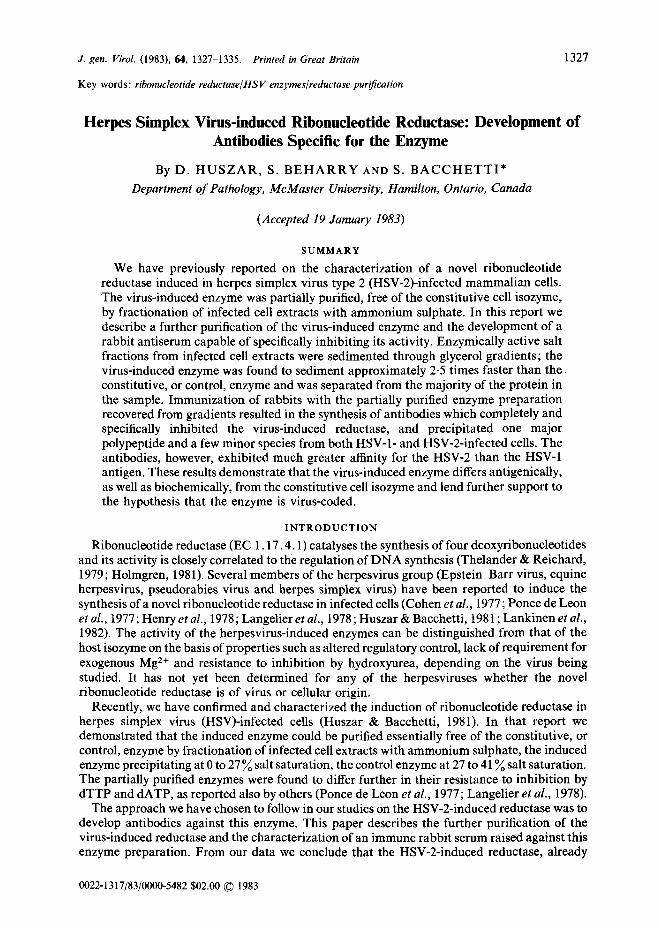
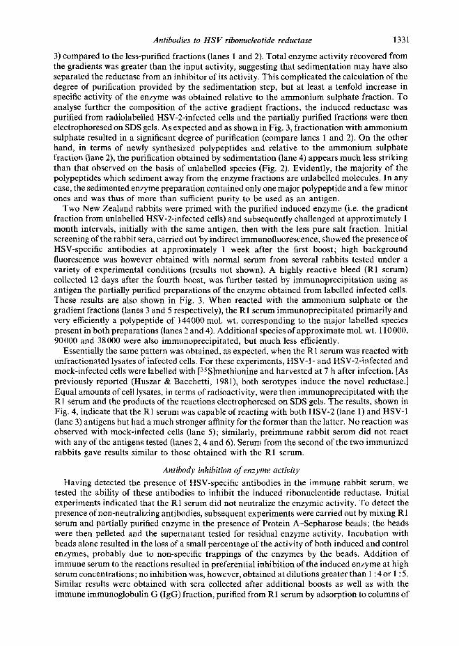
The sedimentat ion properties of the part ial ly purified control enzyme have already been described (Huszar & Bacchetti, 1981). Essentially, this enzyme sedimented in 5 to 25 ~o glycerol gradients as a symmetrical peak and its recovery depended upon the concentrat ion of Mg 2÷ in the gradient; approximately 5 0 ~ of the activi ty was recovered in the presence of 6 mM-Mg 2÷ whereas no activity could be detected in the absence of the ion. Induced enzyme activity, on the other hand, could not be detected, whether in the presence or absence of Mg z÷, under identical sedimentat ion conditions (I 30000 g). Subsequent experiments indicated that this loss of act ivi ty resulted from the induced reductase being pelleted during centrifugation. As shown in Fig. 1, by decreasing the sedimentat ion force to 81000g both enzymes could be recovered as discrete peaks within the gradient, the induced activity sedimenting in the bot tom half of the gradient (Fig. 1 a) and the control one near the top (Fig. 1 b). Enzymic act ivi ty of gradient fractions was assayed both in the standard reaction mixture and in the presence of 30 mM-ammonium sulphate since, in aggreement with others (Langelier & Buttin, 1981), we found this salt concentrat ion inhibitory for the control, but not for the induced enzyme. A similar differential response was obtained with concentrations of Triton X-100 up to 0 .05~ (results not shown).
From the results of a reconstruction experiment (Fig. 1 c), in which par t ia l ly purified preparat ions of the two enzymes were loaded on the same gradient , we est imated that the induced reductase activity sediments 2.5 times faster than the control enzyme. Since the mammal ian reductase is itself a large protein of at least 200 000 mol. wt. (Chang & Cheng, 1979; Engstrom et al., 1979) it seems likely that the induced enzyme might sediment as par t of a high molecular weight complex resulting from self-aggregation or aggregation with other molecules. This putat ive complex does not appear to contain cell membranes since sedimentat ion in the presence of 0 -05~ Triton X-100 did not alter the mobil i ty of the enzyme, nor was the mobil i ty altered by extensive dialysis or solubilization with detergent and salt prior to sedimentat ion. Prompted by reports in the l i terature of replicat ion complexes consisting of enzymes involved in nucleotide and D N A metabolism (Holmgren, 1981), we also examined the possibi l i ty that the
1330
X
O
D . H U S Z A R , S . B E H A R R Y A N D S. B A C C H E T T I
/ /q , 8 ~- (a) t I 200K---~
,' A', 6 - 150K--~
93K--~
: i ' ~ 69K---~ (b) \
6 --
4 -
2 ~ - , 3. o
I _ _ ~ - e
6, - ~ - 30K---~
5 10 15 Fraction number
Fig. 1 Fig. 2
Fig. 1. Sedimentation of partially purified preparations of ribonucleotide reductase on 5 to 25~ glycerol gradients. The enzymes were centrifuged as described in the text and assayed under standard conditions (except for the ATP concentration which was raised to 3 mM for assay of the induced enzyme) in the presence (©) or absence (O) of 30 mM-ammonium sulphate. (a) Virus-induced enzyme; (b) control enzyme; (c) a mixture of induced and control enzymes. Sedimentation is from right to left.
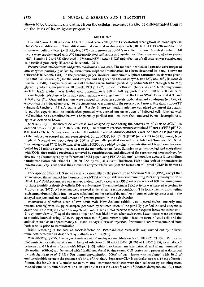
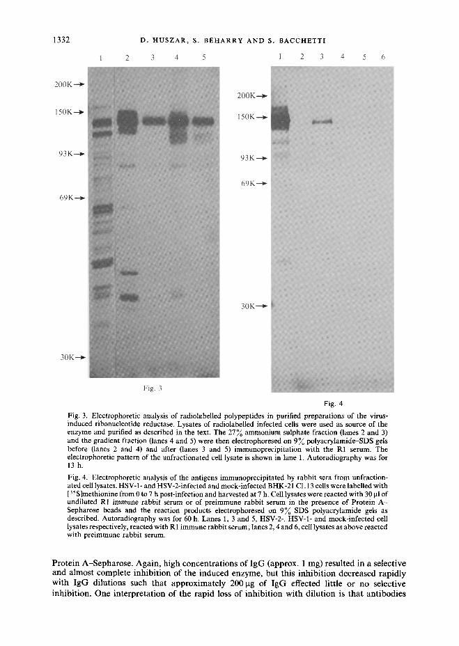
Fig. 2. Electrophoretic analysis of purified preparations of the virus-induced ribonucleotide reductase. Lysates of infected cells were used as source of enzyme and purified as described in the text. Aliquots from each fraction (corresponding to equivalent amounts of protein) were then electrophoresed on 9 polyacrylamide-SDS gels. The gels were stained with Coomassie Brilliant Blue, destained and photographed. Lane 1, crude extract; lane 2, 0 to 27~ ammonium sulphate fraction; lane 3, gradient fraction. The positions of molecular weight standards are shown at the left.
induced reductase may be complexed with some or all of the known HSV-coded enzymes involved in D N A synthesis (i.e. thymidine kinase, D N A polymerase, alkaline DNase). However, none of these enzymes was found to co-purify in significant amounts with the induced reductase in the 0 to 27yo ammonium sulphate fraction, nor could their activity be detected in the gradient fractions which contained the reductase (data not shown).
Characterization of the antigen and preparation of antisera As previously mentioned, sedimentation of the virus-induced reductase through glycerol
gradients was undertaken in order to obtain an antigen preparation of sufficient purity for immunization purposes. Determination of the distribution of protein following sedimentation demonstrated that the majority of the protein remained near the top of the gradient and suggested that further purification had been obtained. This was indeed confirmed by Coomassie Brilliant Blue staining of enzyme samples electrophoresed on SDS-polyacrylamide gels (Fig. 2) which indicated that significantly fewer polypeptides were present in the gradient fraction (lane
Antibodies to HS V ribonucleotide reductase 1331
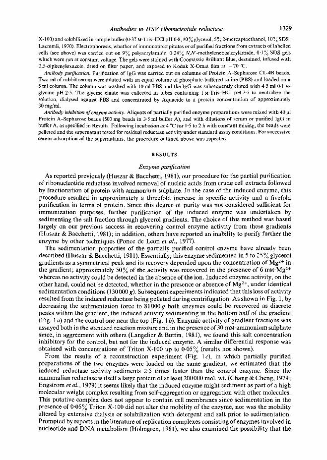
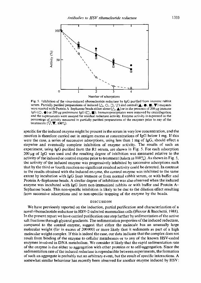
3) compared to the less-purified fractions (lanes 1 and 2). Total enzyme activity recovered from the gradients was greater than the input activity, suggesting that sedimentation may have also separated the reductase from an inhibitor of its activity. This complicated the calculation of the degree of purification provided by the sedimentation step, but at least a tenfold increase in specific activity of the enzyme was obtained relative to the ammonium sulphate fraction. To analyse further the composition of the active gradient fractions, the induced reductase was purified from radiolabelled HSV-2-infected cells and the partially purified fractions were then electrophoresed on SDS gels. As expected and as shown in Fig. 3, fractionation with ammonium sulphate resulted in a significant degree of purification (compare lanes 1 and 2). On the other hand, in terms of newly synthesized polypeptides and relative to the ammonium sulphate fraction (lane 2), the purification obtained by sedimentation (lane 4) appears much less striking than that observed on the basis of unlabelled species (Fig. 2). Evidently, the majority of the polypeptides which sediment away from the enzyme fractions are unlabelled molecules. In any case, the sedimented enzyme preparation contained only one major polypeptide and a few minor ones and was thus of more than sufficient purity to be used as an antigen.
Two New Zealand rabbits were primed with the purified induced enzyme (i.e. the gradient fraction from unlabelled HSV-2-infected cells) and subsequently challenged at approximately 1 month intervals, initially with the same antigen, then with the less pure salt fraction. Initial screening of the rabbit sera, carried out by indirect immunofluorescence, showed the presence of HSV-specific antibodies at approximately 1 week after the first boost; high background fluorescence was however obtained with normal serum from several rabbits tested under a variety of experimental conditions (results not shown). A highly reactive bleed (R1 serum) collected 12 days after the fourth boost, was further tested by immunoprecipitation using as antigen the partially purified preparations of the enzyme obtained from labelled infected cells. These results are also shown in Fig. 3. When reacted with the ammonium sulphate or the gradient fractions (lanes 3 and 5 respectively), the R1 serum immunoprecipitated primarily and very efficiently a polypeptide of 144000 mol. wt. corresponding to the major labelled species present in both preparations (lanes 2 and 4). Additional species of approximate mol. wt. 110000, 90000 and 38000 were also immunoprecipitated, but much less efficiently.
Essentially the same pattern was obtained, as expected, when the R1 serum was reacted with unfractionated lysates of infected cells. For these experiments, HSV-1- and HSV-2-infected and mock-infected cells were labelled with [35 S]methionine and harvested at 7 h after infection. [As previously reported (Huszar & Bacchetti, 1981), both serotypes induce the novel reductase.] Equal amounts of cell lysates, in terms of radioactivity, were then immunoprecipitated with the R 1 serum and the products of the reactions etectrophoresed on SDS gels. The results, shown in Fig. 4, indicate that the R1 serum was capable of reacting with both HSV-2 (lane 1) and HSV-1 (lane 3) antigens but had a much stronger affinity for the former than the latter. No reaction was observed with mock-infected cells (lane 5); similarly, preimmune rabbit serum did not react with any of the antigens tested (lanes 2, 4 and 6). Serum from the second of the two immunized rabbits gave results similar to those obtained with the R1 serum.
Antibody inhibition of enzyme activity
Having detected the presence of HSV-specific antibodies in the immune rabbit serum, we tested the ability of these antibodies to inhibit the induced ribonucleotide reductase. Initial experiments indicated that the R1 serum did not neutralize the enzymic activity. To detect the presence of non-neutralizing antibodies, subsequent experiments were carried out by mixing R 1 serum and partially purified enzyme in the presence of Protein A-Sepharose beads; the beads were then pelleted and the supernatant tested for residual enzyme activity. Incubation with beads alone resulted in the loss of a small percentage of the activity of both induced and control enzymes, probably due to non-specific trappings of the enzymes by the beads. Addition of immune serum to the reactions resulted in preferential inhibition of the induced enzyme at high serum concentrations; no inhibition was, however, obtained at dilutions greater than 1 : 4 or 1 : 5. Similar results were obtained with sera collected after additional boosts as well as with the immune immunoglobulin G (IgG) fraction, purified from R1 serum by adsorption to columns of
1332 D. HUSZAR, S. BEHARRY AND S. BACCHETT!
1 2 3 4 5 1 2 3 4 5 6
200K-"~
150K--~
93K---~
69K---~
30K.--.~
200K--.~
150K-.-.~
9 3 K - - ~
69K--.~
30K---.~
Fig. 3
Fig. 4
Fig. 3. Electrophoretic analysis of radiolabelled polypeptides in purified preparations of the virus- induced ribonucleotide reductase. Lysates of radiolabelled infected cells were used as source of the enzyme and purified as described in the text. The 27~ ammonium sulphate fraction (lanes 2 and 3) and the gradient fraction (lanes 4 and 5) were then electrophoresed on 9 ~ polyacrylamide-SDS gels before (lanes 2 and 4) and after (lanes 3 and 5) immunoprecipitation with the R1 serum. The electrophoretic pattern of the unfractionated cell lysate is shown in lane 1. Autoradiography was for 13h.
Fig. 4. Electrophoretic analysis of the antigens immunoprecipitated by rabbit sera from unfraction- ated cell lysates. HSV-1- and HSV-2-infected and mock-infected BHK-21 C1.13 ceils were labelled with [35S]methionine from 0 to 7 h post-infection and harvested at 7 h. Cell lysates were reacted with 30 p.1 of undiluted RI immune rabbit serum or of preimmune rabbit serum in the presence of Protein A - Sepharose beads and the reaction products electrophoresed on 9% SDS-polyacrylamide gels as described. Autoradiography was for 60 h. Lanes 1, 3 and 5, HSV-2-, HSV-1- and mock-infected cell lysates respectively, reacted with R1 immune rabbit serum; lanes 2, 4 and 6, cell lysates as above reacted with preimmune rabbit serum.
P r o t e i n A - S e p h a r o s e . A g a i n , h i g h c o n c e n t r a t i o n s o f I g G (approx . 1 mg) r e su l t ed in a se lec t ive a n d a lmos t comple t e i n h i b i t i o n o f t he i n d u c e d enzyme , b u t th i s i n h i b i t i o n d e c r e a s e d r ap i d l y w i t h I g G d i lu t ions such t h a t a p p r o x i m a t e l y 200 ktg of I g G effected l i t t le or n o se lec t ive i nh ib i t i on . O n e i n t e r p r e t a t i o n o f the r a p i d loss of i n h i b i t i o n w i t h d i l u t i o n is t h a t a n t i b o d i e s
Antibodies to H S V ribonucleotide reductase 1333
I I f I
1 0 0 ~
"~" 501 " q ~ q ,
X \ r't
I I "~" . . . . 0 0 1 2 3 4
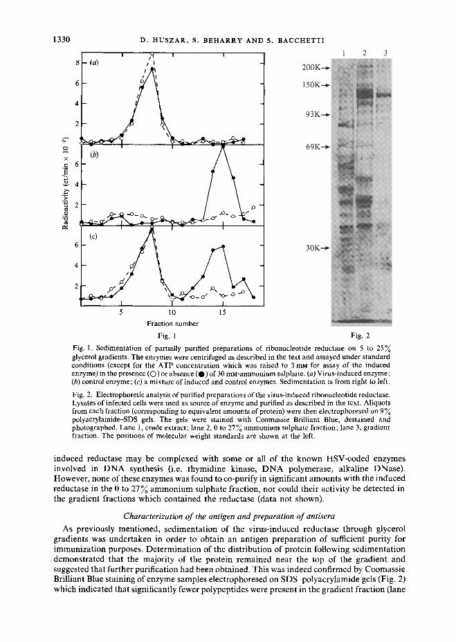
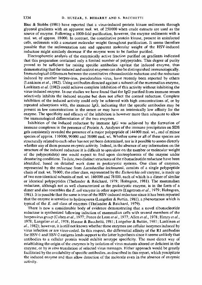
Number of adsorptions Fig. 5. Inhibition of the virus-induced ribonucleotide reductase by IgG purified from immune rabbit serum. Partially purified preparations of induced (A, O, [3, V) and control CA, O, l , V) enzymes were reacted with Protein A-Sepharose beads either alone (A, A) or in the presence of 200 ~tg immune IgG (O, 0) or 200 txg preimmune IgG (I-q, 1). Immunoprecipitates were removed by centrifugation and the supernatants were assayed for residual reductase activity. Enzyme activity is expressed as the percentage of activity measured in partially purified preparations of the enzymes prior to any of the treatments (V,V, 100%).
specific for the induced enzyme might be present in the serum in very low concentration, and the reaction is therefore carried out in antigen excess at concentrations of IgG below 1 mg. If this were the case, a series of successive adsorptions, using less than 1 mg of IgG, should effect a stepwise and eventually complete inhibition of enzyme activity. The results of such an experiment, using IgG purified from the R1 serum, are shown in Fig. 5. For each adsorption 200 gg of IgG was used and the resulting degree of inhibition was measured relative to the activity of the induced or control enzyme prior to treatment (taken as 100%). As shown in Fig. 5, the activity of the induced enzyme was progressively inhibited by successive adsorptions such that by the third or fourth reaction no significant residual activity could be detected. In contrast to the results obtained with the induced enzyme, the control enzyme was inhibited to the same extent by incubation with IgG from immune or from normal rabbit serum, or with buffer and Protein A-Sepharose beads. A similar degree of inhibition was also observed when the induced enzyme was incubated with IgG from non-immunized rabbits or with buffer and Protein A - Sepharose beads. This non-specific inhibition is likely to be due to the dilution effect resulting from successive adsorptions and to non-specific trapping of the enzyme by the beads.
DISCUSSION
We have previously reported on the induction, partial purification and characterization of a novel ribonucleotide reductase in HSV-2-infected mammalian cells (Huszar & Bacchetti, 1981). In the present report we have carried purification one step further by sedimentation of the active salt fractions through glycerol gradients. The sedimentation properties of the induced reductase, compared to the control enzyme, suggest that either the molecule has an unusually large molecular weight (far in excess of 200000) or more likely that it sediments as part of a high molecular weight complex. If this is indeed the case, our data indicate that the complex does not result from binding of the enzyme to cellular membranes or to any of the known HSV-coded enzymes involved in DNA metabolism. We consider it likely that the rapid sedimentation rate of the enzyme is due either to aggregation with other proteins or to self-aggregation. Since the sedimentation rate of the induced reductase is reproducible between experiments, the formation of such an aggregate is probably not an arbitrary event, but the result of specific interactions. A somewhat similar behaviour has recently been observed for another enzyme induced by HSV:
1334 D. H U S Z A R , S. B E H A R R Y AND S. B A C C H E T T I
Blue & Stobbs (1981) have reported that a virus-induced protein kinase sediments through glycerol gradients with an apparent mol. wt. of 250000 when crude extracts are used as the source of enzyme. Following a 1000-fold purification, however, the enzyme sediments with a mol. wt. of approx. 18000. In contrast, the constitutive protein kinase, present in uninfected cells, sediments with a constant molecular weight throughout purification. It seems therefore possible that the sedimentation rate and apparent molecular weight of the HSV-induced reductase might similarly decrease if the enzyme were to be further purified.
Electrophoretic analysis of the enzymically active fraction purified on gradients indicated that this preparation contained only a limited number of polypeptides. This degree of purity proved to be sufficient for raising specific antibodies against the induced enzyme, thus demonstrating that the induced and control enzymes can also be distinguished immunologically. Immunological differences between the constitutive ribonucleotide reductase and the reductase induced by another herpesvirus, pseudorabies virus, have recently been reported by others (Lankinen et al., 1982). Using antibodies directed against a subunit of the mammalian enzyme, Lankinen et al. (1982) could achieve complete inhibition of this activity without inhibiting the virus-induced enzyme. In our studies we have found that the IgG purified from immune serum selectively inhibits the induced enzyme but does not affect the control reductase. Complete inhibition of the induced activity could only be achieved with high concentrations of, or by repeated adsorptions with, the immune IgG, indicating that the specific antibodies may be present in low concentrations in the serum or may have an intrinsically low affinity for the enzyme. The specificity and efficacy of the inhibition is however more than adequate to allow the immunological differentiation of the two enzymes.
Inhibition of the induced reductase by immune IgG was achieved by the formation of immune complexes in the presence of Protein A. Analyses of the immune precipitates on SDS gels consistently revealed the presence of a major polypeptide of 144 000 mol. wt., and of minor species of approx. 110000, 90000 and 38000 mol. wt. Whether some or all of these species are structurally related to each other has not yet been determined, nor is it possible to assess directly whether any of them possess enzymic activity. Indeed, in the absence of any information on the structure of the induced reductase it is difficult to speculate on the number or molecular weight of the polypeptide(s) one would expect to find upon electrophoresis of the enzyme under denaturing conditions. To date, two distinct structures of the ribonucleotide reductase have been identified, based on detailed work done in prokaryotic systems. One class of enzymes, represented by the reductase from Lactobacillus leichmannii, consists of a single polypeptide chain of mol. wt. 76 000; the other class, represented by the Escherichia coli enzyme, is made up of two non-identical subunits of mol. wt. 160000 and 78000, each of which is a dimer of similar or identical polypeptides (Thelander & Reichard, 1979; Holmgren, 1981). The mammalian reductase, although not as well characterized as the prokaryotic enzyme, is in the form of a dimer and also resembles the E. coli enzyme in other aspects (Engstrom et al., 1979; Holmgren, 1981). It is possible that the same is true of the HSV-induced reductase since it has been reported that the enzyme is sensitive to hydroxyurea (Langelier & Buttin, 1981), a phenomenon which is typical of the E. coli class of enzymes (Thelander & Reichard, 1979).
There is now a considerable body of evidence demonstrating that a novel ribonucleotide reductase is synthesized following infection of mammalian cells with several members of the herpesvirus group (Cohen et al., 1977; Ponce de Leon et at., 1977; Allen et al., 1978; Henry et al., 1978; Langelier et al., 1978; Huszar & Bacchetti, 1981 ; Langelier & Buttin, 1981 ; Lankinen et al., 1982); however, it is still not known whether these enzymes are cellular isozymes induced by virus infection or are virus-coded. In this respect, the differential affinity of the R1 antibodies for HSV-1 and HSV-2 antigens lends support to the latter hypothesis since it seems unlikely that antibodies to a cellular protein would exhibit serotype specificity. The most direct way of establishing the origin of the enzymes is by isolation of virus mutants altered or deficient in the enzyme, or by in vitro translation of selected virus messages. Either approach would be greatly facilitated by the availability of specific antibodies, as described in this report, which precipitate the induced enzyme and thus allow detection of the molecule even in the absence of enzymic activity.
Antibodies to H S V ribonucleotide reductase 1335
We are grateful to Claudio Sartori for truly excellent technical assistance and to J. R. Smiley and W. E. Rawls for reading the manuscript. This work was supported by the National Cancer Institute of Canada. D.H. is a recipient of a NCIC Research studentship and S.B. is a Research Associate of the NCIC.
R E F E R E N C E S
ALLEN, G. P., COHEN, J. C., RANDALL, C. C. & O'CALLAGHAN, D. J. (1978). Replication of equine herpesvirus type 1 and type 2: resistance to hydroxyurea and thymidine. Intervirology 9, 276-285.
BALACHANDRAN, N., HARNISH, D., KILLINGTON, R. A., BACCHETTI, S. & RAWLS, W. E. (1981). Monoclonal antibodies to two glycoproteins of herpes simplex virus type 2. Journal of Virology 39, 438~146.
BANERJEE, A. K. & RHODES, D. l'. (1973). In vitro.synthesis of RNA that contains polyadenylate by virion-associated RNA polymerase of vesicular stomatitis virus. Proceedings of the National Academy of Sciences, U.S.A. 70, 3566-3570.
BLUE, W. Y. & STOBBS, D. G. (1981). Isolation of a protein kinase induced by herpes simplex virus type 1. Journal of Virology 38, 383-388.
CHANG, C.-H. & CHEN6, Y.-C. (1979). Demonstration of two components and association of adenosine diphosphate- cytidine diphosphate reductase from cultured human lymphoblast cells (molt-4F). Cancer Research 39, 436- 442.
COHEN, J. C., HENRY, B. E., RANDALL, C. C. & O'CALLAGHAN, D. J. (1977). Ribonucleotide reductase activity in hydroxyurea resistant herpesvirus replication. Proceedings of the Society for Experimental Biology and Medicine 155, 395-399.
ENGSTROM, V., ERIKSSON, S., THELANDER, L. & AKERMAN, M. (1979). Ribonucleotide reductase from calf thymus. Purification and properties. Biochemistry 18, 2941-2948.
HENRY, B. E., GLASER, R., HEWETSON, J. & O'CALLAGHAN, D. J. (1978). Expression of altered ribonucleotide reductase activity associated with the replication of the Epstein-Barr virus. Virology 89, 262-271.
HOLMGREN, A. (1981). Regulation of ribonucleotide reductase. Current Topics in Cellular Regulation 19, 47-76. HUSZAR, D. & BACCHEYrI, S. (1981). Partial purification and characterization of the ribonucleotide reductase
induced by herpes simplex virus infection of mammalian cells. Journal of Virology 37, 580-588. KEIR, H. M., SUBAK-SHARPE, n., SHEDDEN, W. I. H., WATSON, D. H. & WILDY, P. (1966). Immunological evidence for a
specific DNA polymerase produced after infection by herpes simplex virus. Virology 30, 154-157. KILLINGTON, R. A., NEWHOOK, L., BALACHANDRAN, N., RAWLS, W. E. & BACCHETTI, S. (1981). Production of hybrid
cell lines secreting antibodies to herpes simplex virus type 2. Journal of Virological Methods 2, 223-236. LAEMMLI, U. K. (1970). Cleavage of structural proteins during the assembly of the head of bacteriophage T4.
Nature, London 227, 680 685. LANGELIER, Y. & BUTTIN, G. ( 1981). Characterization of ribonucleotide reductase induction in BHK-21/C 13 Syrian
hamster cell line upon infection by herpes simplex virus (HSV). Journal of General Virology 57, 21-31. LANGELIER, Y., DECHAMPS, M. & BUTTIN, G. (t978). Analysis of dCMP deaminase and CDP reductase levels in
hamster cells infected with herpes simplex virus. Journal of Virology 26, 547-553. LANKINEN, H., GRASLUND, A. & THELANDER, L. (1982). Induction of a new ribonucleotide reductase after infection of
mouse L cells with pseudorabies virus. Journal of Virology 41, 893-900. MORRISON, J. M. & KEIR, H. M. (1968). A new DNA-exonuclease in cells infected with herpes virus: partial
purification and properties of the enzyme. Journal of General Virology 3, 337-374. MUNYON, W., BUCHSBAUM, R., PAOLETTI, E., MANN, J., KRAISELBURD, E. & DAVIS, D. (1972). Electrophoresis of
thymidine kinase activity synthesized by cells transformed by herpes simplex virus. Virology 49, 683-689. PONCE DE LEON, M., EISENBERG, R. J. & COHEN, G. H. (1977). Ribonucleotide reductase from herpes simplex virus
(types 1 and 2) infected and uninfected KB cells: properties of the partially purified enzymes. Journal of General Virology 36, 163-173.
REICHARD, P. (1958). Chromatographic separations of cytosine containing compounds. Acta chemica scandinavica 12, 2048.
SETH, P., RAWLS, W. E., DUFF, R., RAPP, F., ADAM, E. & MELNICK, J. L. (1974). Antigenic differences between isolates of herpesvirus type 2. Intervirology 3, 1-14.
THELANDER, L. & REICHARD, P. (1979). Reduction of ribonucleotides. Annual Review of Biochemistry 48, 133-158.
(Received 19 October 1982)


