Embed Size (px)
Citation preview
of July 18, 2016.This information is current as
Progression in LeishmaniasisThe Role of IL-10 in Promoting Disease
Margaret Mentink Kane and David M. Mosser
http://www.jimmunol.org/content/166/2/1141doi: 10.4049/jimmunol.166.2.1141
2001; 166:1141-1147; ;J Immunol
Referenceshttp://www.jimmunol.org/content/166/2/1141.full#ref-list-1
, 17 of which you can access for free at: cites 31 articlesThis article
Subscriptionshttp://jimmunol.org/subscriptions
is online at: The Journal of ImmunologyInformation about subscribing to
Permissionshttp://www.aai.org/ji/copyright.htmlSubmit copyright permission requests at:
Email Alertshttp://jimmunol.org/cgi/alerts/etocReceive free email-alerts when new articles cite this article. Sign up at:
Print ISSN: 0022-1767 Online ISSN: 1550-6606. Immunologists All rights reserved.Copyright © 2001 by The American Association of9650 Rockville Pike, Bethesda, MD 20814-3994.The American Association of Immunologists, Inc.,
is published twice each month byThe Journal of Immunology
by guest on July 18, 2016http://w
ww
.jimm
unol.org/D
ownloaded from
by guest on July 18, 2016
http://ww
w.jim
munol.org/
Dow
nloaded from
The Role of IL-10 in Promoting Disease Progression inLeishmaniasis1
Margaret Mentink Kane and David M. Mosser2
To determine the role of IL-10 in cutaneous leishmaniasis, we examined lesion development followingLeishmania majorinfectionof genetically susceptible BALB/c mice lacking IL-10. Whereas normal BALB/c mice developed progressive nonhealing lesionswith numerous parasites within them, IL-102/2 BALB/c mice controlled disease progression, and had relatively small lesions with1000-fold fewer parasites within them by the fifth week of infection. We also examined a mechanism wherebyLeishmaniainducedthe production of IL-10 from macrophages. We show that surface IgG onLeishmania amastigotes allows them to ligate Fcgreceptors on inflammatory macrophages to preferentially induce the production of high amounts of IL-10. The IL-10 producedby infected macrophages prevented macrophage activation and diminished their production of IL-12 and TNF-a. In vitro survivalassays confirmed the importance of IL-10 in preventing parasite killing by activated macrophages. Pretreatment of monolayerswith either rIL-10 or supernatants from amastigote-infected macrophages resulted in a dramatic enhancement in parasite intra-cellular survival. These studies indicate that amastigotes ofLeishmaniause an unusual and unexpected virulence factor, host IgG.This IgG allows amastigotes to exploit the antiinflammatory effects of FcgR ligation to induce the production of IL-10, whichrenders macrophages refractory to the activating effects of IFN-g. The Journal of Immunology,2001, 166: 1141–1147.
L eishmaniaare intracellular parasites that reside primarilywithin host tissue macrophages. The immunological re-sponse toLeishmaniahas been extensively characterized,
and the importance of the activated macrophage in resolving in-fection has been unequivocally established (1, 2). In theLeishma-nia major model of cutaneous leishmaniasis, genetically inbredstrains of mice exhibit polarized immune responses that can resultin dramatic differences in the clinical outcome of infection.BALB/c mice mount an inappropriate Th2 response and succumbto progressive disease. In contrast, other strains such as C3H orC57BL/6 mice mount a Th1 response and control infections (3).There are, however, several species ofLeishmaniaand many mod-els of clinical leishmaniasis in which this immune deviation is nota true predictor of disease progression. In both humans and mice,for example, ample IFN-g is produced during visceral leishmani-asis caused byLeishmania donovani(4, 5). Despite the presence ofhigh levels of IFN-g, infected hosts generally fail to control theinfection and resolve their disease. In fact in humans, the severityof visceral leishmaniasis has been most closely associated withincreased levels of IL-10 (5–7). IL-10 production also correlatedwith lesion progression in patients with cutaneous leishmaniasis(8). A similar correlation has recently been made in IL-10-trans-genic mice, which are susceptible to progressiveL. major diseasedespite producing IFN-g (9). These and other studies point to animportant role for IL-10 in regulating immune responses to thisintracellular pathogen.
There are two developmental forms ofLeishmania: the promas-tigote and the amastigote (10). The promastigote is introduced into
the mammalian host when an infected sandfly takes a bloodmeal.This form is taken up by phagocytic cells and rapidly transforms intothe amastigote form. Amastigotes replicate intracellularly withinmononuclear phagocytes and are the only form found within themammalian host following infection. The unexpected observationwas made several years ago thatLeishmaniaamastigotes have host-derived IgG on their surface (11, 12). This observation was recentlyconfirmed, and the role of IgG as an opsonin for enhanced parasiteadhesion to macrophages was proposed (13). We have previouslyshown thatLeishmaniaamastigotes bind avidly to mammalian cellproteoglycans (14), and do not require opsonization for parasite ad-hesion to macrophages. We therefore began to look for alternativefunctions for Ig on the amastigote surface to explain the enhancedvirulence of IgG-opsonized amastigotes.
We have recently demonstrated that the ligation of phagocyticreceptors on macrophages can alter their cytokine profile whenthese cells are exposed to a variety of inflammatory stimuli (15,16). We showed that the ligation of the FcgR by immune com-plexes was a particularly potent way to prevent the production ofproinflammatory cytokines. The ligation of this receptor class notonly inhibited the production of IL-12 (15), but unlike complementreceptor ligation, FcgR ligation also induced the synthesis andsecretion of IL-10 (16). IL-10 production occurred only in cellscontaining a functional FcRg-chain, indicating that FcgR signal-ing through theg-chain was required for IL-10 production. Weproposed that this antiinflammatory cytokine milieu would havethe potential to inhibit the production of a type 1 immune responseand prevent macrophage activation. Consistent with this hypothe-sis is the observation by others that the administration of immunecomplexes to mice prevented effective cellular responses toListe-ria monocytogenesand diminished bacterial clearance (17).
In the present study, we examined cytokine production by mac-rophages following their interaction withLeishmaniaamastigotes.We show that lesion-derived amastigotes induce the robust pro-duction of IL-10 from stimulated macrophages. The molecule re-sponsible for this induction is host IgG on the amastigote surface,which ligates macrophage FcgRs. The IL-10 that is produced bythis mechanism inhibits macrophage activation and contributes to
Department of Microbiology and Immunology, Temple University School of Medi-cine, Philadelphia, PA 19140
Received for publication May 9, 2000. Accepted for publication October 13, 2000.
The costs of publication of this article were defrayed in part by the payment of pagecharges. This article must therefore be hereby markedadvertisementin accordancewith 18 U.S.C. Section 1734 solely to indicate this fact.1 This work was supported by National Institutes of Health Grants AI-24313 andAI-46805.2 Address correspondence and reprint requests to Dr. David M. Mosser, Departmentof Cell Biology and Molecular Genetics, University of Maryland, College Park, MD20742. E-mail address: [email protected]
Copyright © 2001 by The American Association of Immunologists 0022-1767/01/$02.00
by guest on July 18, 2016http://w
ww
.jimm
unol.org/D
ownloaded from
parasite growth in lesions. Thus, we have identified an unexpectedLeishmaniavirulence factor: host IgG.
Materials and MethodsAnimals
C57BL/6, C3H/HeJ, and BALB/c mice were obtained from The JacksonLaboratory (Bar Harbor, ME). IL-102/2 mice on a BALB/c backgroundwere kindly provided by Donna Rennick, DNAX (Palo Alto, CA). IL-102/2 mice were maintained under germfree conditions in the Barrier An-imal Facility of Temple University in MicroFlow System ventilated cages(Allentown Caging Equipment, Allentown, PA). Breeding pairs of FcRg-chain knockout mice (g2/2) (18) were purchased from Taconic Farms(Germantown, NY).
Parasites
A clone ofL. major (WHO MHOM/IL/80/Friedlin) and the Josefa isolateof Leishmania mexicana amazonensis(14) were used for these studies.Promastigotes were grown in Schneider’s insect cell culture medium (LifeTechnologies, Grand Island, NY) supplemented with 20% heat-inactivatedFBS, 2 mM glutamine, 100 U/ml penicillin G, and 100mg/ml streptomy-cin. AxenicL. mexicana amazonensisamastigotes were grown at 32°C, aspreviously described (19). Lesion-derived amastigotes were isolated fromBALB/c mice infected 6–8 wk before as described previously (20).
Macrophages
Bone marrow-derived macrophages (BMMf)3 were established as previ-ously described (15). Murine peritoneal macrophages were washed fromthe peritoneal cavity of either C57BL/6 or BALB/c mice as described else-where (21). Cells were cultivated in DMEM containing 10% heat-inacti-vated FBS, 2 mM glutamine, 100 U/ml penicillin G, and 100mg/ml strep-tomycin (complete medium) (D-10).
Macrophage stimulation and receptor ligation
BMMf were used to measure the production of cytokines. Cells wereseeded overnight in 24-well plates in complete medium at a density of 23105 cells/well. Cells were washed once with complete medium, and thenstimuli were added to induce cytokine production. Lesion amastigotes andaxenic amastigotes were added at a ratio of 10 amastigotes per macro-phage. Amastigotes were added either alone or simultaneously with eitherLPS (Escherichia coli0128.B12; Sigma, St. Louis, MO) or low molecularweight hyaluronic acid (HA; ICN Biomedicals, Costa Mesa, CA) at con-centrations indicated in the figure legends.
Macrophage activation in vitro
BMMf were activated by pretreating them overnight with 250 U/ml IFN-g(R&D Systems, Minneapolis, MN) and 100 ng/ml LPS. For in vitro leish-manicidal assays, peritoneal macrophages were pretreated with either 10ng/ml rIL-10 (R&D Systems) or supernatants from stimulated macro-phages infected withLeishmaniaamastigotes (infected macrophage super-natants) 3 h before activation with IFN-g. Three hours later,L. majoramastigotes were added to macrophage monolayers at a 3:1 (parasite:mac-rophage) ratio for 72 h at 35°C. Nonphagocytozed amastigotes werewashed from the cultures at 24 h postinfection, and fresh medium wasadded to each well with the appropriate cytokine conditions for an addi-tional 48 h. At the termination of the incubation period, the wells werewashed once with complete medium, then fixed with 100% methanol at4°C for 30 min. The monolayers were washed with PBS containing 5%FCS (PBS/FCS) and processed for immunofluorescent staining to visualizeintracellular Leishmaniaamastigotes. Murine polyclonal anti-leishmaniaantiserum was used as the primary Ab, and goata-murine-IgG conjugatedwith FITC was used as the secondary Ab, as described previously (20).Coverslips were counterstained with propidium iodide and examined byfluorescence microscopy.
Flow cytometry
Footpad lesion amastigotes were isolated from BALB/c mice infected 6–8wk, as described previously (20). To directly stain murine IgG on theamastigote surface, 13 106 amastigotes were incubated on ice for 30 minwith FITC-conjugated goat anti-murine (Fcg chain-specific) IgG (JacksonImmunoResearch,West Grove, PA) diluted 1/100 in PBS/FCS. Amasti-
gotes were opsonized with IgG by incubating them on ice for 30 min witha 1/10 dilution of serum from a mouse infected with viableLeishmania.Following three washes in PBS/FCS to remove nonspecific IgG, FITC-conjugated goat anti-murine IgG was added on ice for an additional 30 min.The amastigotes were washed and fixed in 1% paraformaldehyde and im-mediately analyzed on an Epics Elite flow cytometer (Coulter Diagnostics,Hialeah, FL).
Cytokine ELISAs
Culture supernatants from monolayers of control and stimulated macro-phages were assayed by ELISA for cytokine production 20–24 h afterstimulation. Murine IL-10 production was measured as previously de-scribed (16) using mAbs to IL-10, JES5-2A5, and biotinylated JES5-16E3(PharMingen, San Diego, CA). IL-12 (p70) levels were measured usingmAbs C18.2 (IL-12 p35) and biotinylated C17.15 (IL-12 p40) as describedelsewhere (16). TNF production was measured using mAbs G281-2626and biotinylated MP6-XT3 (PharMingen).
Parasite quantitation
Mice were injected in the hind footpad with 23 106 L. majoramastigotes.Parasite burdens in footpads were determined by a limiting serial dilutionof single cell suspensions made from individual excised lesions as de-scribed reviously (22). Lesion size was determined by measuring the thick-ness of the footpad with a caliper, and subtracting the thickness of theuninfected contralateral footpad.
ResultsMice lacking IL-10 have decreased lesion development andreduced parasite burdens
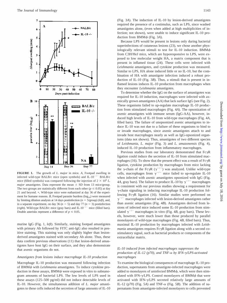
To determine the effect of IL-10 on disease progression in leish-maniasis, we infected mice deficient in IL-10 on a BALB/c back-ground and compared them with wild-type mice. BALB/c mice aregenetically susceptible toL. major infection (3), and thereforewild-type mice produce progressive nonhealing lesions (Fig. 1A)that increased in size until day 36, when the lesions began to ul-cerate and metastasize. On day 36, there were in excess of 13 109
organisms per infected footpad (Fig. 1B). For humane reasons,these mice were euthanized at this time. In contrast to wild-typeBALB/c mice, congenic mice lacking IL-10 were relatively resis-tant to infection, showing only modest increases in lesion sizethrough the 11-wk observation period (Fig. 1A). At 2 wk postin-fection, a time when footpad swelling in the two groups had not yetbegun to exhibit differences, mice lacking IL-10 already had;100-fold fewer parasites in their lesions than wild-type mice(Fig. 1B). By the fifth week, IL-102/2 mice had 1000-fold fewerorganisms in their lesions, and by the 11 wk only;100 organismscould be detected per infected foot in IL-102/2 mice (1036 128).Thus, mice lacking IL-10 are relatively resistant toLeishmaniainfection.
Lesion amastigotes are coated with surface Ig
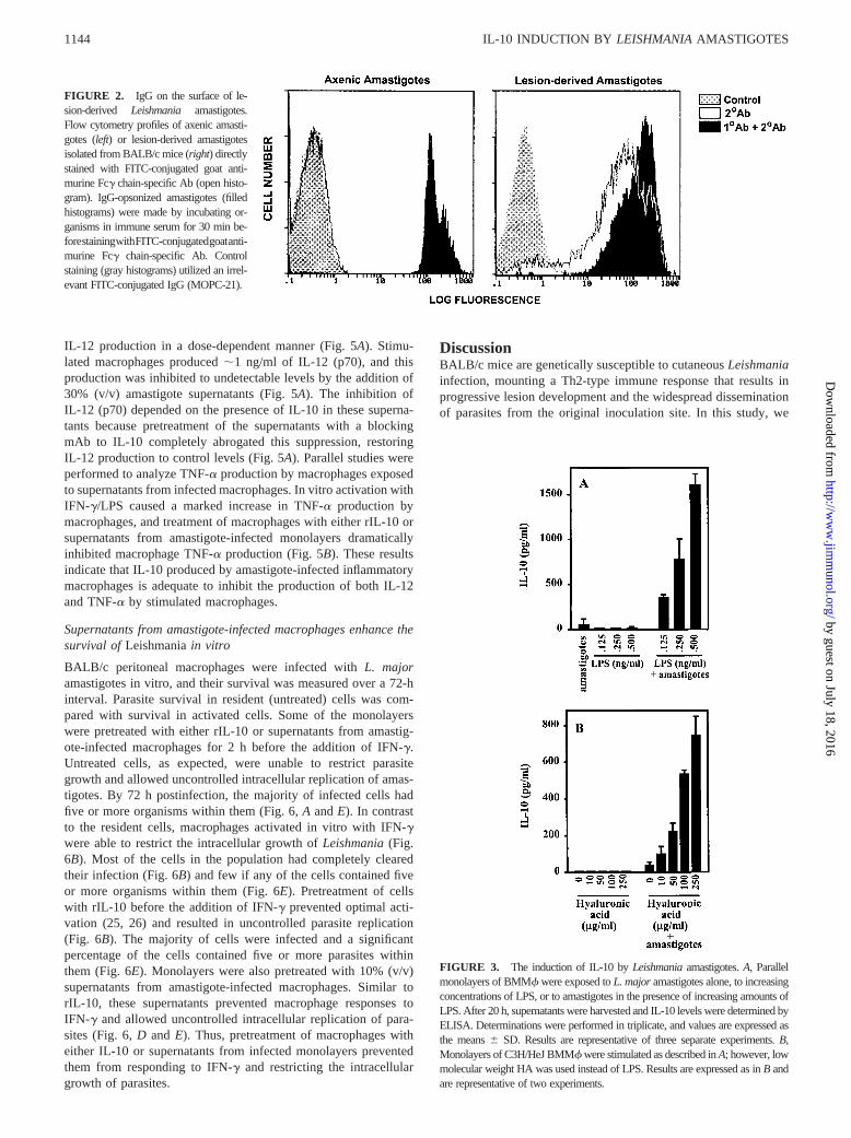
Previous studies have demonstrated that lesion-derived amasti-gotes have host IgG on their surface (11–13). To confirm theseobservations, flow cytometry was performed to identify host IgGon the surface of lesion-derived amastigotes. Amastigotes wereisolated from the footpads of infected BALB/c mice and directlystained with FITC-conjugated Ab to the Fc fragment of murineIgG (Fig. 2 , open histograms). By flow cytometry, lesion-derivedamastigotes stained positively for murine IgG (Fig. 2,right). Vir-tually all of the organisms in the population were positive for sur-face Ig, and the majority expressed relatively high levels of IgGwith a mean fluorescence intensity of over 10. In contrast, axenicamastigotes grown in vitro in the absence of IgG were devoid ofsurface IgG (Fig. 2,left). Their mean fluorescence intensity wasnot substantially different from unstained organisms (gray histo-grams). Preincubation of these organisms with antiserum to amas-tigotes as the primary Ab, followed by the FITC anti-IgG (filledhistograms), resulted in axenic amastigotes staining positively for
3 Abbreviations used in this paper: BMMf, bone marrow-derived macrophages; AA,axenically grown amastigotes; HA, hyaluronic acid.
1142 IL-10 INDUCTION BY LEISHMANIAAMASTIGOTES
by guest on July 18, 2016http://w
ww
.jimm
unol.org/D
ownloaded from
murine IgG (Fig. 1,left). Similarly, staining footpad amastigoteswith primary Ab followed by FITC anti-IgG also resulted in pos-itive staining. This staining was only slightly higher than lesion-derived amastigotes stained with secondary Ab alone. Thus, thesedata confirm previous observations (11) that lesion-derived amas-tigotes have host IgG on their surface, and they also demonstratethat axenic organisms do not.
Amastigotes from lesions induce macrophage IL-10 production
Macrophage IL-10 production was measured following infectionof BMMf with Leishmaniaamastigotes. To induce cytokine pro-duction in these assays, BMMf were exposed in vitro to subnano-gram amounts of bacterial LPS. The low levels of LPS used inthese assays (125–500 pg/ml) did not induce detectable levels ofIL-10. However, the simultaneous addition ofL. major amasti-gotes to these cells induced the secretion of large amounts of IL-10
(Fig. 3A). The induction of IL-10 by lesion-derived amastigotesrequired the presence of a costimulus, such as LPS, since washedamastigotes alone, (even when added at high multiplicities of in-fection; not shown), were unable to induce significant IL-10 pro-duction from BMMf (Fig. 3A).
Because LPS would be present in lesions only during bacterialsuperinfections of cutaneous lesions (23), we chose another phys-iologically relevant stimuli to test for IL-10 induction. BMMffrom C3H/HeJ mice, which are hyporesponsive to LPS, were ex-posed to low molecular weight HA, a matrix component that ispresent in inflamed tissue (24). These cells were infected withLeishmaniaamastigotes, and cytokine production was measured.Similar to LPS, HA alone induced little or no IL-10, but the com-bination of HA with amastigote infection induced a robust pro-duction of IL-10 (Fig. 3B). Thus, a stimuli that is present in in-flamed lesions induces IL-10 production from macrophages whenthey encounterLeishmaniaamastigotes.
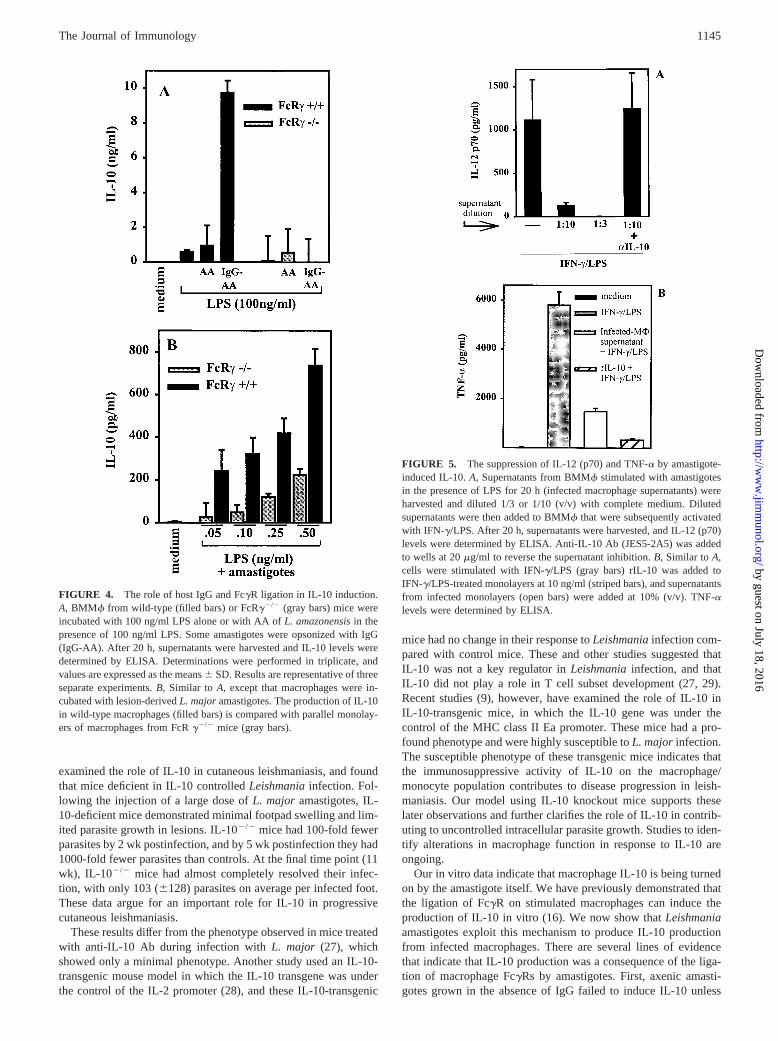
To determine whether the IgG on the surface of amastigotes wasrequired for IL-10 induction, macrophages were infected with ax-enically grown amastigotes (AA) that lack surface IgG (see Fig. 2).These organisms failed to up-regulate macrophage IL-10 produc-tion from stimulated macrophages (Fig. 4A). The opsonization ofaxenic amastigotes with immune serum (IgG-AA), however, in-duced high levels of IL-10 from wild-type macrophages (Fig. 4A,filled bars). The failure of unopsonized axenic amastigotes to in-duce IL-10 was not due to a failure of these organisms to bind toor invade macrophages, since axenic amastigotes attach to andinvade host macrophages nearly as well as IgG-opsonized organ-isms (data not shown). Thus, amastigotes of two different speciesof Leishmania,L. major (Fig. 3) andL. amazonensis(Fig. 4),induced IL-10 production from inflammatory macrophages.
Previous studies from our laboratory demonstrated that FcgRligation could induce the secretion of IL-10 from stimulated mac-rophages (16). To show that the present effect was a result of FcgRligation, cytokine production by macrophages from mice lackingthe g-chain of the FcgR (g2/2) was analyzed. Unlike wild-typecells, macrophages fromg2/2 mice failed to up-regulate IL-10when infected with axenic amastigotes opsonized with IgG (Fig.4A, gray bars). The failure to produce IL-10 byg2/2 macrophagesis consistent with our previous studies showing a requirement forg-chain signaling in inducing macrophage IL-10 production fol-lowing FcgR ligation (16). Similar studies were performed ong2/2 macrophages infected with lesion-derived amastigotes ratherthan axenic amastigotes (Fig. 4B). Amastigotes derived from le-sions of infected mice induced some IL-10 production from stim-ulatedg2/2 macrophages in vitro (Fig. 4B, gray bars). These lev-els, however, were much lower than those produced by parallelmonolayers of wild-type macrophages (Fig. 4B, filled bars). Thus,maximal IL-10 production by macrophages infected withLeish-maniaamastigotes requires FcgR ligation along with a second co-stimulatory signal, such as bacterial products or components of theextracellular matrix.
IL-10 induced from infected macrophages suppresses theproduction of IL-12 (p70), and TNF-a by IFN-g/LPS-activatedmacrophages
To examine the biological consequences of macrophage IL-10 pro-duction, supernatants from amastigote-infected macrophages wereadded to monolayers of uninfected BMMf, which were then stim-ulated with IFN-g/LPS. Control monolayers of BMMf that wereactivated with IFN-g/LPS secreted relatively large amounts ofIL-12 (p70) (Fig. 5A) and TNF-a (Fig. 5B). The addition of su-pernatants from amastigote-infected monolayers to cells prevented
FIGURE 1. The growth ofL. major in mice. A, Footpad swelling ininfected wild-type BALB/c mice (open symbols) and IL-102/2 BALB/cmice (filled symbols) was compared following the injection of 23 106 L.major amastigotes. Data represent the mean6 SD from 13 mice/group.The two groups are statistically different from each other (p# 0.05) at day21 and beyond.p, Wild-type mice were euthanized at day 36 of the experi-ment for humane reasons.B, Footpad parasite burdens (log10) were measuredby limiting dilution analysis at 14 days postinfection (n 5 5/group) (left), and,in a separate experiment, on day 36 (n 5 5) and day 77 (n5 3) postinfection(right). Wild-type BALB/c mice (gray bars) and IL-102/2 mice (filled bars).Double asterisks represent a difference ofp # 0.05.
1143The Journal of Immunology
by guest on July 18, 2016http://w
ww
.jimm
unol.org/D
ownloaded from
IL-12 production in a dose-dependent manner (Fig. 5A). Stimu-lated macrophages produced;1 ng/ml of IL-12 (p70), and thisproduction was inhibited to undetectable levels by the addition of30% (v/v) amastigote supernatants (Fig. 5A). The inhibition ofIL-12 (p70) depended on the presence of IL-10 in these superna-tants because pretreatment of the supernatants with a blockingmAb to IL-10 completely abrogated this suppression, restoringIL-12 production to control levels (Fig. 5A). Parallel studies wereperformed to analyze TNF-a production by macrophages exposedto supernatants from infected macrophages. In vitro activation withIFN-g/LPS caused a marked increase in TNF-a production bymacrophages, and treatment of macrophages with either rIL-10 orsupernatants from amastigote-infected monolayers dramaticallyinhibited macrophage TNF-a production (Fig. 5B). These resultsindicate that IL-10 produced by amastigote-infected inflammatorymacrophages is adequate to inhibit the production of both IL-12and TNF-a by stimulated macrophages.
Supernatants from amastigote-infected macrophages enhance thesurvival ofLeishmaniain vitro
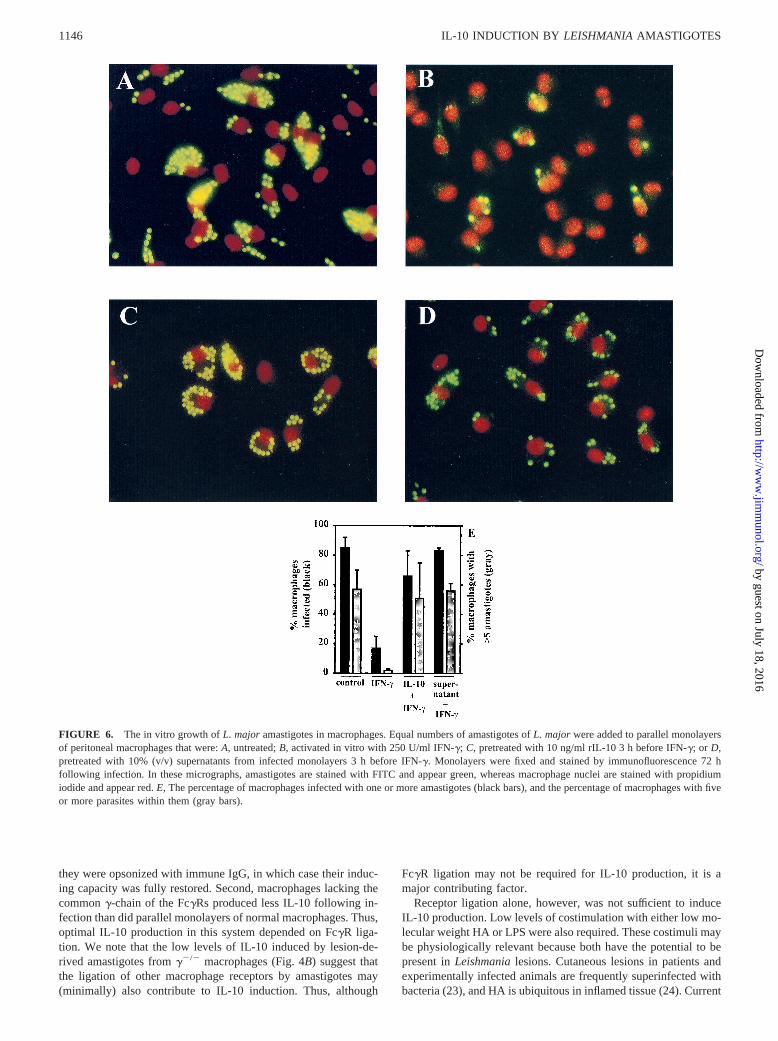
BALB/c peritoneal macrophages were infected withL. majoramastigotes in vitro, and their survival was measured over a 72-hinterval. Parasite survival in resident (untreated) cells was com-pared with survival in activated cells. Some of the monolayerswere pretreated with either rIL-10 or supernatants from amastig-ote-infected macrophages for 2 h before the addition of IFN-g.Untreated cells, as expected, were unable to restrict parasitegrowth and allowed uncontrolled intracellular replication of amas-tigotes. By 72 h postinfection, the majority of infected cells hadfive or more organisms within them (Fig. 6,A andE). In contrastto the resident cells, macrophages activated in vitro with IFN-gwere able to restrict the intracellular growth ofLeishmania(Fig.6B). Most of the cells in the population had completely clearedtheir infection (Fig. 6B) and few if any of the cells contained fiveor more organisms within them (Fig. 6E). Pretreatment of cellswith rIL-10 before the addition of IFN-g prevented optimal acti-vation (25, 26) and resulted in uncontrolled parasite replication(Fig. 6B). The majority of cells were infected and a significantpercentage of the cells contained five or more parasites withinthem (Fig. 6E). Monolayers were also pretreated with 10% (v/v)supernatants from amastigote-infected macrophages. Similar torIL-10, these supernatants prevented macrophage responses toIFN-g and allowed uncontrolled intracellular replication of para-sites (Fig. 6,D and E). Thus, pretreatment of macrophages witheither IL-10 or supernatants from infected monolayers preventedthem from responding to IFN-g and restricting the intracellulargrowth of parasites.
DiscussionBALB/c mice are genetically susceptible to cutaneousLeishmaniainfection, mounting a Th2-type immune response that results inprogressive lesion development and the widespread disseminationof parasites from the original inoculation site. In this study, we
FIGURE 3. The induction of IL-10 byLeishmaniaamastigotes.A, Parallelmonolayers of BMMf were exposed toL. majoramastigotes alone, to increasingconcentrations of LPS, or to amastigotes in the presence of increasing amounts ofLPS. After 20 h, supernatants were harvested and IL-10 levels were determined byELISA. Determinations were performed in triplicate, and values are expressed asthe means6 SD. Results are representative of three separate experiments.B,Monolayers of C3H/HeJ BMMf were stimulated as described inA; however, lowmolecular weight HA was used instead of LPS. Results are expressed as inB andare representative of two experiments.
FIGURE 2. IgG on the surface of le-sion-derived Leishmania amastigotes.Flow cytometry profiles of axenic amasti-gotes (left) or lesion-derived amastigotesisolated from BALB/c mice (right) directlystained with FITC-conjugated goat anti-murine Fcg chain-specific Ab (open histo-gram). IgG-opsonized amastigotes (filledhistograms) were made by incubating or-ganisms in immune serum for 30 min be-forestainingwithFITC-conjugatedgoatanti-murine Fcg chain-specific Ab. Controlstaining (gray histograms) utilized an irrel-evant FITC-conjugated IgG (MOPC-21).
1144 IL-10 INDUCTION BY LEISHMANIAAMASTIGOTES
by guest on July 18, 2016http://w
ww
.jimm
unol.org/D
ownloaded from
examined the role of IL-10 in cutaneous leishmaniasis, and foundthat mice deficient in IL-10 controlledLeishmaniainfection. Fol-lowing the injection of a large dose ofL. major amastigotes, IL-10-deficient mice demonstrated minimal footpad swelling and lim-ited parasite growth in lesions. IL-102/2 mice had 100-fold fewerparasites by 2 wk postinfection, and by 5 wk postinfection they had1000-fold fewer parasites than controls. At the final time point (11wk), IL-102/2 mice had almost completely resolved their infec-tion, with only 103 (6128) parasites on average per infected foot.These data argue for an important role for IL-10 in progressivecutaneous leishmaniasis.
These results differ from the phenotype observed in mice treatedwith anti-IL-10 Ab during infection withL. major (27), whichshowed only a minimal phenotype. Another study used an IL-10-transgenic mouse model in which the IL-10 transgene was underthe control of the IL-2 promoter (28), and these IL-10-transgenic
mice had no change in their response toLeishmaniainfection com-pared with control mice. These and other studies suggested thatIL-10 was not a key regulator inLeishmaniainfection, and thatIL-10 did not play a role in T cell subset development (27, 29).Recent studies (9), however, have examined the role of IL-10 inIL-10-transgenic mice, in which the IL-10 gene was under thecontrol of the MHC class II Ea promoter. These mice had a pro-found phenotype and were highly susceptible toL. major infection.The susceptible phenotype of these transgenic mice indicates thatthe immunosuppressive activity of IL-10 on the macrophage/monocyte population contributes to disease progression in leish-maniasis. Our model using IL-10 knockout mice supports theselater observations and further clarifies the role of IL-10 in contrib-uting to uncontrolled intracellular parasite growth. Studies to iden-tify alterations in macrophage function in response to IL-10 areongoing.
Our in vitro data indicate that macrophage IL-10 is being turnedon by the amastigote itself. We have previously demonstrated thatthe ligation of FcgR on stimulated macrophages can induce theproduction of IL-10 in vitro (16). We now show thatLeishmaniaamastigotes exploit this mechanism to produce IL-10 productionfrom infected macrophages. There are several lines of evidencethat indicate that IL-10 production was a consequence of the liga-tion of macrophage FcgRs by amastigotes. First, axenic amasti-gotes grown in the absence of IgG failed to induce IL-10 unless
FIGURE 4. The role of host IgG and FcgR ligation in IL-10 induction.A, BMMf from wild-type (filled bars) or FcRg2/2 (gray bars) mice wereincubated with 100 ng/ml LPS alone or with AA ofL. amazonensisin thepresence of 100 ng/ml LPS. Some amastigotes were opsonized with IgG(IgG-AA). After 20 h, supernatants were harvested and IL-10 levels weredetermined by ELISA. Determinations were performed in triplicate, andvalues are expressed as the means6 SD. Results are representative of threeseparate experiments.B, Similar toA, except that macrophages were in-cubated with lesion-derivedL. majoramastigotes. The production of IL-10in wild-type macrophages (filled bars) is compared with parallel monolay-ers of macrophages from FcRg2/2 mice (gray bars).
FIGURE 5. The suppression of IL-12 (p70) and TNF-a by amastigote-induced IL-10.A, Supernatants from BMMf stimulated with amastigotesin the presence of LPS for 20 h (infected macrophage supernatants) wereharvested and diluted 1/3 or 1/10 (v/v) with complete medium. Dilutedsupernatants were then added to BMMf that were subsequently activatedwith IFN-g/LPS. After 20 h, supernatants were harvested, and IL-12 (p70)levels were determined by ELISA. Anti-IL-10 Ab (JES5-2A5) was addedto wells at 20mg/ml to reverse the supernatant inhibition.B, Similar toA,cells were stimulated with IFN-g/LPS (gray bars) rIL-10 was added toIFN-g/LPS-treated monolayers at 10 ng/ml (striped bars), and supernatantsfrom infected monolayers (open bars) were added at 10% (v/v). TNF-alevels were determined by ELISA.
1145The Journal of Immunology
by guest on July 18, 2016http://w
ww
.jimm
unol.org/D
ownloaded from
they were opsonized with immune IgG, in which case their induc-ing capacity was fully restored. Second, macrophages lacking thecommong-chain of the FcgRs produced less IL-10 following in-fection than did parallel monolayers of normal macrophages. Thus,optimal IL-10 production in this system depended on FcgR liga-tion. We note that the low levels of IL-10 induced by lesion-de-rived amastigotes fromg2/2 macrophages (Fig. 4B) suggest thatthe ligation of other macrophage receptors by amastigotes may(minimally) also contribute to IL-10 induction. Thus, although
FcgR ligation may not be required for IL-10 production, it is amajor contributing factor.
Receptor ligation alone, however, was not sufficient to induceIL-10 production. Low levels of costimulation with either low mo-lecular weight HA or LPS were also required. These costimuli maybe physiologically relevant because both have the potential to bepresent inLeishmanialesions. Cutaneous lesions in patients andexperimentally infected animals are frequently superinfected withbacteria (23), and HA is ubiquitous in inflamed tissue (24). Current
FIGURE 6. The in vitro growth ofL. major amastigotes in macrophages. Equal numbers of amastigotes ofL. major were added to parallel monolayersof peritoneal macrophages that were:A, untreated;B, activated in vitro with 250 U/ml IFN-g; C, pretreated with 10 ng/ml rIL-10 3 h before IFN-g; or D,pretreated with 10% (v/v) supernatants from infected monolayers 3 h before IFN-g. Monolayers were fixed and stained by immunofluorescence 72 hfollowing infection. In these micrographs, amastigotes are stained with FITC and appear green, whereas macrophage nuclei are stained with propidiumiodide and appear red.E, The percentage of macrophages infected with one or more amastigotes (black bars), and the percentage of macrophages with fiveor more parasites within them (gray bars).
1146 IL-10 INDUCTION BY LEISHMANIAAMASTIGOTES
by guest on July 18, 2016http://w
ww
.jimm
unol.org/D
ownloaded from
studies are underway to define other costimulatory stimuli, such aschemokine stimulation, that may cooperate with receptor ligationto induce IL-10 production.
The present studies may provide a partial explanation for tworecent observations showing that mice lacking IgG or FcgRs areactually more resistant toLeishmaniainfection. Working in a cu-taneous model ofL. amazonensisinfection, Kima and colleagues(30) showed that the commong-chain of the FcgR was requiredfor optimal lesion progression in mice. These results support ourhypothesis that IgG-opsonized amastigotes use Fc receptors duringinfection to enhance macrophage IL-10 production. Smelt and col-leagues (31) have shown that visceral infection withL. donovaniwas diminished in mice lacking IgG. This observation would alsobe consistent with a role for IgG-induced IL-10 in contributing tolesion progression during leishmaniasis.
We examined the consequences of macrophage IL-10 produc-tion by adding supernatants from amastigote-infected macrophages tonaive monolayers, which were then exposed to IFN-g/LPS. Superna-tants from infected monolayers inhibited the activation of macro-phages exposed to IFN-g/LPS. These treated macrophages pro-duced significantly less TNF-a, and they were virtually unable toproduce IL-12. Importantly, these pretreated monolayers failed tocontrol Leishmaniainfection. The majority of the cells in themonolayer were infected, and most of the cells had multiple or-ganisms growing within them (Fig. 6,D–E). Thus, a prior encoun-ter with IL-10 renders macrophages refractory to the activatingeffects of IFN-g and prevents them from eliminating intracellularparasites, as previously reported (26).
In summary, we have examined the interaction ofLeishmaniaamastigotes with tissue macrophages and have identified an unex-pected role for host IgG. Rather than simply acting as a classicalopsonin to accelerate parasite phagocytosis, an additional role ofsurface IgG is to induce the production of IL-10 by macrophages.This induction prevents these cells from responding to IFN-g andeliminating intracellular parasites. This work suggests that an im-portant way thatLeishmaniaparasites modify the host immuneresponse is by exploiting the antiinflammatory effects of FcgRligation to induce the production of IL-10.
AcknowledgmentsWe thank Dr. Donna Rennick, DNAX Research Institute, for generouslysupplying breeding pairs of the IL-102/2 mice.
References1. Green, S. J., M. S. Meltzer, J. B. Hibbs, Jr., and C. A. Nacy. 1990. Activated
macrophages destroy intracellularLeishmania majoramastigotes by an L-argi-nine-dependent killing mechanism.J. Immunol. 144:278.
2. Liew, F. Y., S. Millott, C. Parkinson, R. M. Palmer, and S. Moncada. 1990.Macrophage killing ofLeishmaniaparasite in vivo is mediated by nitric oxidefrom L-arginine.J. Immunol. 144:4794.
3. Locksley, R. M., and P. Scott. 1991. Helper T-cell subsets in mouse leishmani-asis: induction, expansion and effector function.Immunol. Today 12:A58.
4. Gasim, S., A. M. Elhassan, E. A. Khalil, A. Ismail, A. M. Kadaru, A. Kharazmi,and T. G. Theander. 1998. High levels of plasma IL-10 and expression of IL-10by keratinocytes during visceral leishmaniasis predict subsequent development ofpost-kala-azar dermal leishmaniasis.Clin. Exp. Immunol. 111:64.
5. Karp, C. L., S. H. el-Safi, T. A. Wynn, M. M. Satti, A. M. Kordofani,F. A. Hashim, M. Hag-Ali, F. A. Neva, T. B. Nutman, and D. L. Sacks. 1992. Invivo cytokine profiles in patients with kala-azar: marked elevation of both inter-leukin-10 and interferon-g. J. Clin. Invest. 91:1644.
6. Kaye, P. M., A. J. Curry, and J. M. Blackwell. 1991. Differential production ofTh1- and Th2-derived cytokines does not determine the genetically controlled or
vaccine-induced rate of cure in murine visceral leishmaniasis.J. Immunol. 146:2763.
7. Ghalib, H. W., M. R. Piuvezam, Y. A. Skeiky, M. Siddig, F. A. Hashim,A. M. el-Hassan, D. M. Russo, and S. G. Reed. 1993. Interleukin 10 productioncorrelates with pathology in humanLeishmania donovaniinfections. J. Clin.Invest. 92:324.
8. Louzir, H., P. C. Melby, A. Ben Salah, H. Marrakchi, K. Aoun, R. Ben Ismail,and K. Dellagi. 1998. Immunologic determinants of disease evolution in localizedcutaneous leishmaniasis due toLeishmania major. J. Infect. Dis. 177:1687.
9. Groux, H., F. Cottrez, M. Rouleau, S. Mauze, S. Antonenko, S. Hurst, T. McNeil,M. Bigler, M. G. Roncarolo, and R. L. Coffman. 1999. A transgenic model toanalyze the immunoregulatory role of IL-10 secreted by antigen-presenting cells.J. Immunol. 162:1723.
10. Kane, M. M., and D. M. Mosser. 2000.Leishmaniaparasites and their ploys todisrupt macrophage activation.Curr. Opin. Hematol. 7:26.
11. Guy, R. A., and M. Belosevic. 1993. Comparison of receptors required for entryof Leishmania majoramastigotes into macrophages.Infect. Immun. 61:1553.
12. Pearson, R. D., and D. Roberts. 1990. Host immunoglobulin on spleen-derivedLeishmania donovaniamastigotes.Am. J. Trop. Med. Hyg. 43:263.
13. Peters, C., T. Aebischer, Y. D. Stierhof, M. Fuchs, and P. Overath. 1995. The roleof macrophage receptors in adhesion and uptake ofLeishmania mexicanaamas-tigotes.J. Cell Sci. 108:3715.
14. Love, D. C., J. D. Esko, and D. M. Mosser. 1993. A heparin binding activity onLeishmaniaamastigotes which mediates attachment to cellular proteoglycans.J. Cell Biol. 123:759.
15. Sutterwala, F. S., G. J. Noel, R. Clynes, and D. M. Mosser. 1997. Selectivesuppression of interleukin-12 induction after macrophage receptor ligation.J. Exp. Med. 185:1977.
16. Sutterwala, F. S., G. J. Noel, P. Salgame, and D. M. Mosser. 1998. Reversal ofproinflammatory responses by ligating the macrophage Fcg receptor type I.J. Exp. Med. 188:217.
17. Berger, S., R. Chandra, H. Ballo, R. Hildenbrand, and H. J. Stutte. 1997. Immunecomplexes are potent inhibitors of interleukin-12 secretion by human monocytes.Eur. J. Immunol. 27:2994.
18. Takai, T., M. Li, D. Sylvestre, R. Clynes, and J. V. Ravetch. 1994. FcRg chaindeletion results in pleiotrophic effector cell defects.Cell 76:519.
19. Hodgkinson, V. H., L. Soong, S. M. Duboise, and D. McMahon-Pratt. 1996.Leishmania amazonensis: cultivation and characterization of axenic amastigote-like organisms.Exp. Parasitol. 83:94.
20. Love, D. C., M. M. Kane, and D. M. Mosser. 1998.Leishmania amazonensis: thephagocytosis of amastigotes by macrophages.Exp. Parasitol. 88:161.
21. Mosser, D. M., and P. J. Edelson. 1985. The mouse macrophage receptor for iC3b(CR3) is a major mechanism in the phagocytosis ofLeishmaniapromastigotes.J. Immunol. 135:2785.
22. Afonso, L. C., and P. Scott. 1993. Immune responses associated with suscepti-bility of C57BL/10 mice toLeishmania amazonensis.Infect. Immun. 61:2952.
23. el-On, J., R. Sneier, and E. Elias. 1992.Leishmania major: bacterial contamina-tion of cutaneous lesions in experimental animals.Isr. J. Med. Sci. 28:847.
24. Hodge-Dufour, J., P. W. Noble, M. R. Horton, C. Bao, M. Wysoka,M. D. Burdick, R. M. Strieter, G. Trinchieri, and E. Pure. 1997. Induction ofIL-12 and chemokines by hyaluronan requires adhesion-dependent priming ofresident but not elicited macrophages.J. Immunol. 159:2492.
25. Vouldoukis, I., P.-A. Becherel, V. Riveros-Moreno, M. Arock, O. da Silva, P.Debre, D. Mazier, and M. D. Mossalayi. 1997. Interleukin-10 and interleukin-4inhibit intracellular killing of Leishmania infantumand Leishmania majorbyhuman macrophages by decreasing nitric oxide generation.Eur. J. Immunol. 27:860.
26. Vieth, M., A. Will, K. Schroppel, M. Rollinghoff, and A. Gessner. 1994. Inter-leukin-10 inhibits antimicrobial activity againstLeishmania majorin murinemacrophages.Scand. J. Immunol. 40:403.
27. Chatelain, R., S. Mauze, and R. L. Coffman. 1999. ExperimentalLeishmaniamajor infection in mice: role of IL-10.Parasite Immunol. 21:211.
28. Hagenbaugh, A., S. Sharma, S. M. Dubinett, S. H.-Y. Wei, R. Aranda,H. Cheroutre, D. J. Fowell, S. Binder, B. Tsao, R. M. Locksley, et al. 1997.Altered immune responses in interleukin 10 transgenic mice.J. Exp. Med. 12:2101.
29. Soares, M. B. P., J. R. David, and R. G. Titus. 1997. An in vitro model forinfection with Leishmania majorthat mimics the immune response in mice.In-fect. Immun. 65:2837.
30. Kima, P. E., S. L. Constant, L. Hannum, M. Colmenares, K. S. Lee,A. M. Haberman, M. J. Schlomchik, and D. McMahon-Pratt. 2000. Internaliza-tion of Leishmania mexicanacomplex amastigotes via the Fc receptor is requiredto sustain infection in murine cutaneous leishmaniasis.J. Exp. Med. 191:1063.
31. Smelt, S. C., S. E. J. Cotterell, C. R. Engwerda, and P. M. Kaye. 2000. B cell-deficient mice are highly resistant toLeishmania donovaniinfection, but developneutrophil-mediated tissue pathology.J. Immunol. 164:3681.
1147The Journal of Immunology
by guest on July 18, 2016http://w
ww
.jimm
unol.org/D
ownloaded from


