Embed Size (px)
Citation preview

The role of the proteins Kar9 and Myo2 in orienting the mitoticspindle of budding yeastDale L. Beach, Julie Thibodeaux, Paul Maddox, Elaine Yeh and Kerry Bloom
Background: Two genetic ‘pathways’ contribute to the fidelity of nuclearsegregation during the process of budding in the yeast Saccharomycescerevisiae. An early pathway, involving Kar9p and other proteins, orients themitotic spindle along the mother–bud axis. Upon the onset of anaphase,cytoplasmic dynein provides the motive force for nuclear movement into thebud. Loss of either pathway results in nuclear-migration defects; loss of both islethal. Here, to visualize the functional steps leading to correct spindleorientation along the mother–bud axis, we imaged live yeast cells expressingKar9p and dynein as green fluorescent protein fusions.
Results: Transport of Kar9p into the bud was found to require the myosinMyo2p. Kar9p interacted with microtubules through the microtubule-bindingprotein Bim1p and facilitated microtubule penetration into the bud. Oncemicrotubules entered the bud, Kar9p provided a platform for microtubulecapture at the bud cortex. Kar9p was also observed at sites of microtubuleshortening in the bud, suggesting that Kar9p couples microtubule shorteningto nuclear migration.
Conclusions: Thus, Kar9p provides a key link between the actin cytoskeletonand microtubules early in the cell cycle. A cooperative mechanism betweenKar9p and Myo2p facilitates the pre-anaphase orientation of the spindle. Later,Kar9p couples microtubule disassembly with nuclear migration.
BackgroundDuring the process of budding in the yeast Saccharomycescerevisiae, the mitotic spindle aligns with the mother–budaxis to correctly distribute the duplicated genomes. Thedeterminants of spindle polarity (reviewed in [1]) includedynein (Dhc1p) and dynactin components, kinesins(Kip3p), microtubule-binding proteins (Bim1p/Yeb1p)and cell-polarity markers (Kar9p, Bni1p/She5p andBud6p/Aip3p). Dynein is synthetically lethal withbni1/she5, bud6/aip3 and kar9 [2–4], defining two primarypathways that establish spindle orientation. The ‘early’pathway orients the spindle with respect to themother–bud axis and proximity to the bud neck, while the‘late’ pathway coincides with anaphase onset to translocatethe daughter nucleus into the bud. Mutations in onepathway are compensated by the other; mutations in bothare lethal. For instance, both kar9 and dynein mutationsgenerate binucleate cells [5–7], but the double mutant istemperature sensitive or lethal [2,4,8].
Originally identified as a karyogamy mutant, kar9 waslater found to affect nuclear position during mitosis [5,9].A small population of binucleate cells occurs in theabsence of Kar9p. Kar9p is localized specifically at the budtip in vegetative cells, and to the tip of the mating projec-tion in cells induced to enter the mating pathway [5].
Although Bni1p and Bud6p are required to maintainKar9p at the bud cortex [3], both Bni1p and Bud6p alsoaffect bipolar bud-site selection and actin polarity [10–12].A role for actin in spindle orientation has been described[13–17], but the interaction between the actin and micro-tubule cytoskeletons remains unknown.
Specific actin alleles and mutations of actin-associated pro-teins produce off-axis spindles as well as binucleate cells[15–17]. Actin cables and not actin patches [18] arerequired at the inception of the cell cycle to correctlyorient the spindle [14]. Transport of polarity determinantsalong actin cables may be the key requirement for actinfunction early in the cell cycle. Type V myosins and actincables are required for transport of a variety of componentsinto the bud [19–21]. Ash1p is a cell-fate determinantwhose localization is specified in part by the polar distribu-tion of the mRNA transcript [22,23]. ASH1 mRNA localiza-tion to the bud tip is dependent on Bni1p and Bud6p[22–25]. Of the five genes originally described for the local-ization of the Ash1p (SHE1–SHE5), SHE1 is a class Vmyosin (MYO4), and SHE5 is BNI1 [26]. Subsequentinvestigation identified an ASH1 mRNA anchorage defectfor bud6 similar to bni1/she5 [24]. In the absence of eitherprotein, ASH1 mRNA is released from a tight associationwith the bud tip and moves throughout the bud. Thus,
Address: Department of Biology, University of NorthCarolina, 212 Coker Hall CB3280, Chapel Hill,North Carolina 27599-3280, USA.
Correspondence: Kerry BloomE-mail: [email protected]
Received: 12 May 2000Revised: 25 October 2000Accepted: 25 October 2000
Published: 15 November 2000
Current Biology 2000, 10:1497–1506
0960-9822/00/$ – see front matter © 2000 Elsevier Science Ltd. All rights reserved.
Research Paper 1497
Bni1p and Bud6p provide positional cues for spindle orien-tation and contribute to asymmetric mRNA localization.
KAR9 is one of the few spindle-polarity determinants thatdoes not exhibit such pleiotropic phenotypes. In theabsence of Kar9p, the nucleus fails to congress to the budneck or the mating projection [3,5]. The kar9 cells exhibitnuclear migration phenotypes similar to kip3, bim1/yeb1,bni1/she5 and bud6/aip3 [3,8,27–30]. However, mutations inkip3, bim1/yeb1, bni1/she5 and bud6/aip3 have additional phe-notypes that exacerbate nuclear migration, such as alteredmicrotubule dynamics (bim1/yeb1 and kip3 [27,28,31]) ordiminished actin integrity (bni1/she5 and bud6/aip3). Kar9pinteracts biochemically with the microtubule plus-endbinding protein Bim1p [31–33], and requires actin, Bni1pand Bud6p to maintain a cortical attachment within thebud [3,5]. Here, by imaging live yeast cells expressingKar9p as a green fluorescent protein fusion (Kar9–GFP),we observed the dynamic cortical and microtubule local-ization of Kar9p and the facilitated orientation of themicrotubule towards the incipient bud site. Kar9p trans-port into the bud was dependent on MYO2 [34], whichencodes a class V myosin, and a polarized actin cytoskele-ton. We also observed Kar9p at sites of microtubule short-ening in the bud, indicating Kar9p to be a key componentof the mechanism that couples microtubule shortening tonuclear migration.
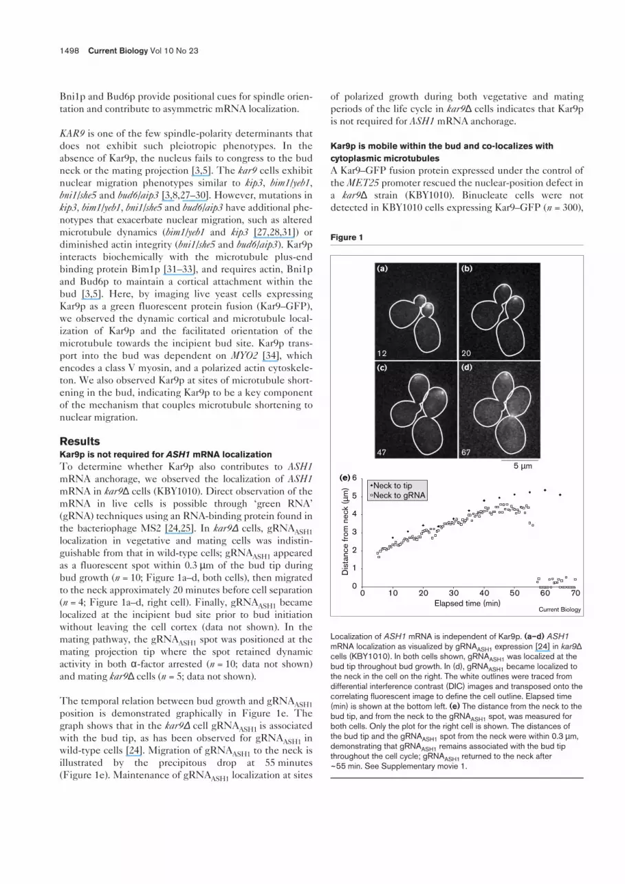
ResultsKar9p is not required for ASH1 mRNA localizationTo determine whether Kar9p also contributes to ASH1mRNA anchorage, we observed the localization of ASH1mRNA in kar9∆ cells (KBY1010). Direct observation of themRNA in live cells is possible through ‘green RNA’(gRNA) techniques using an RNA-binding protein found inthe bacteriophage MS2 [24,25]. In kar9∆ cells, gRNAASH1localization in vegetative and mating cells was indistin-guishable from that in wild-type cells; gRNAASH1 appearedas a fluorescent spot within 0.3 µm of the bud tip duringbud growth (n = 10; Figure 1a–d, both cells), then migratedto the neck approximately 20 minutes before cell separation(n = 4; Figure 1a–d, right cell). Finally, gRNAASH1 becamelocalized at the incipient bud site prior to bud initiationwithout leaving the cell cortex (data not shown). In themating pathway, the gRNAASH1 spot was positioned at themating projection tip where the spot retained dynamicactivity in both α-factor arrested (n = 10; data not shown)and mating kar9∆ cells (n = 5; data not shown).
The temporal relation between bud growth and gRNAASH1position is demonstrated graphically in Figure 1e. Thegraph shows that in the kar9∆ cell gRNAASH1 is associatedwith the bud tip, as has been observed for gRNAASH1 inwild-type cells [24]. Migration of gRNAASH1 to the neck isillustrated by the precipitous drop at 55 minutes(Figure 1e). Maintenance of gRNAASH1 localization at sites
of polarized growth during both vegetative and matingperiods of the life cycle in kar9∆ cells indicates that Kar9pis not required for ASH1 mRNA anchorage.
Kar9p is mobile within the bud and co-localizes withcytoplasmic microtubulesA Kar9–GFP fusion protein expressed under the control ofthe MET25 promoter rescued the nuclear-position defect ina kar9∆ strain (KBY1010). Binucleate cells were notdetected in KBY1010 cells expressing Kar9–GFP (n = 300),
1498 Current Biology Vol 10 No 23
Figure 1
Localization of ASH1 mRNA is independent of Kar9p. (a–d) ASH1mRNA localization as visualized by gRNAASH1 expression [24] in kar9∆cells (KBY1010). In both cells shown, gRNAASH1 was localized at thebud tip throughout bud growth. In (d), gRNAASH1 became localized tothe neck in the cell on the right. The white outlines were traced fromdifferential interference contrast (DIC) images and transposed onto thecorrelating fluorescent image to define the cell outline. Elapsed time(min) is shown at the bottom left. (e) The distance from the neck to thebud tip, and from the neck to the gRNAASH1 spot, was measured forboth cells. Only the plot for the right cell is shown. The distances ofthe bud tip and the gRNAASH1 spot from the neck were within 0.3 µm,demonstrating that gRNAASH1 remains associated with the bud tipthroughout the cell cycle; gRNAASH1 returned to the neck after~55 min. See Supplementary movie 1.
(a)
(e)
(b)
(c) (d)
12 20
47 67
5 µm
0
1
2
3
4
5
6
Elapsed time (min)Current Biology
Dis
tanc
e fro
m n
eck
(µm
) Neck to tipNeck to gRNA
0 10 20 30 40 50 60 70

compared with 11% (n = 309) in untransformed kar9∆ cells.Additionally, Kar9–GFP expression from the MET25 pro-moter did not produce any binucleate cells after a 2 hourinduction (n = 300), and the nucleus was properly posi-tioned within the mother cell prior to anaphase onset(n = 300). An integrated Kar9–GFP fusion proteinexpressed from the endogenous promoter maintainedsimilar localization (Table 1).
Kar9–GFP, expressed in wild-type cells (YEF473A) fromthe MET25 promoter, was observed as cortical spots(Figure 2a,c,d) and either punctate or continuous lineararrays (Figure 2b,e; see also Table 1g–j). Typically, 1–2 cor-tical spots were observed within each cell, and eachremained mobile within the bud during growth. Spotsmoved with an average velocity of 1.96 ± 1.24 µm/minute(n = 10) within the bud. This velocity is rapid when com-pared with the average velocities of Bud6–GFP (0.116 µm/minute) and gRNAASH1 (0.31 µm/minute) [24], yet veryslow in comparison with actin patch movements (16.8 µm/minute) [35]. Cortical Kar9–GFP movements were diffu-sional, as cortical spots remained motile in the presence ofsodium azide and deoxyglucose, which inhibit ATP produc-tion (see below and Table 1) [36]. The Kar9–GFP corticalspots migrated to the neck 30 ± 10 minutes (n = 6) prior tocell separation (Figure 2c, lower cell; Figure 2e, upper cell;Table 1d). Approximately 20 minutes (n = 4) after the Kar9–GFP became localized to the neck, it was released from thecortex and observed as spots and/or filaments within thecytoplasm (Figure 2e, lower cell; Figure 2f, upper cell).Unlike ASH1 mRNA, Kar9p–GFP became cytoplasmic, asspots and linear arrays, before being localized to the budsite. Prior to the next budding cycle, Kar9–GFP linear arraysoriented towards the incipient bud site 13 ± 4 minutes(n = 7) before bud emergence (see below).
Kar9–GFP linear arrays depended on microtubules, asevidenced by their absence upon nocodazole treatment(Table 1). Microtubules can be imaged by either decorat-ing the filament with a dynein–GFP fusion protein [37]or by labeling the tubulin directly (GFP–Tub1) [38].Line scans of microtubules (see Materials and methods)differentiated periodic Kar9–GFP spots from continuousdynein–GFP decoration along microtubules. WhileKar9–GFP appeared as discrete peaks of fluorescentintensity along the filament, microtubules labeled bydynein–GFP displayed linear fluorescence as didGFP–Tub1 [38] or Tub3–GFP (K.B., unpublished data).Dynamic growth (0.808 ± 0.426 µm/minute; n = 35) andshortening (0.574 ± 0.253 µm/minute; n = 39) of Kar9–GFPlinear arrays coincided with microtubule dynamics mea-sured in an isogenic strain containing dynein–GFP(0.798 ± 0.29 µm/minute growth, n = 12; 0.791 ± 0.347 µm/minute shortening, n = 19) and in kar9∆ cells containingdynein-GFP (0.703 ± 0.416 µm/minute growth, n = 17;0.630 ± 0.325 µm/minute shortening; n = 16). Therefore,microtubule dynamics in kar9∆ cells and cells expressingKar9–GFP are not significantly different from the wild type.
Kar9p preferentially localizes along microtubules in theabsence of cortical anchorsThe dynamic movement of Kar9–GFP between corticalspots and microtubules is indicative of the bipartite distri-bution of Kar9p. Cells treated with latrunculin-A (todepolymerize actin filaments) or deleted for BNI1/SHE5or BUD6/AIP3 showed a loss of Kar9–GFP spots from thebud cortex and an increase in microtubule-associatedKar9–GFP (Figure 2g–i; Table 1); bni1∆/she5∆ cells had an83% decrease in Kar9p spots at the bud cortex;bud6∆/aip3∆ cells decreased cortical Kar9–GFP by 75%.Latrunculin-A-treated cells decreased cortical Kar9–GFP
Research Paper Kar9p and Myo2p in spindle orientation Beach et al. 1499
Table 1
Kar-9–GPF localization.
a b c d e f g h i j n
Wild type 21.0 36.0 4.0 20.0 10.5 0.0 1.5 3.0 1.5 1.5 132Integrant 22.0 32.0 0.5 38.0 1.6 0.5 0.5 4.1 0.0 1.1 187Wild type + Lat-A 28.6 5.4 4.3 5.4 5.7 11.6 0.6 13.4 21.3 3.6 164bni1∆ 17.7 6.1 6.1 4.7 1.9 0.0 0.0 17.8 11.2 34.6 214bud6∆ 17.9 9.0 12.1 8.5 7.6 0.0 0.0 17.0 0.9 28.3 223bim1∆ 11.3 62.4 3.4 16.0 4.0 2.6 0.0 0.4 0.0 0.0 532Azide + dGlu 13.5 11.5 0.0 10.6 7.7 2.4 0.0 10.2 22.1 19.2 208Wild type + Nz 31.7 42.7 5.7 8.4 7.5 0.9 0.0 2.2 0.0 0.9 227bni1∆ + Nz 18.0 0.0 12.0 43.0 15.0 12.0 0.0 0.0 0.0 0.0 194
The table shows the percentage of each cell population with theindicated pattern of Kar9–GFP localization (columns a–j). All cell typeslisted expressed a Kar9–GFP fusion protein from a CEN plasmid viathe MET25 promoter, except ‘Integrant’, where the GFP coding region
was fused to the endogenous KAR9 chromosomal locus (seeMaterials and methods). Lat-A, latrunculin-A; azide + dGlu, sodiumazide and deoxyglucose; Nz, nocodazole.
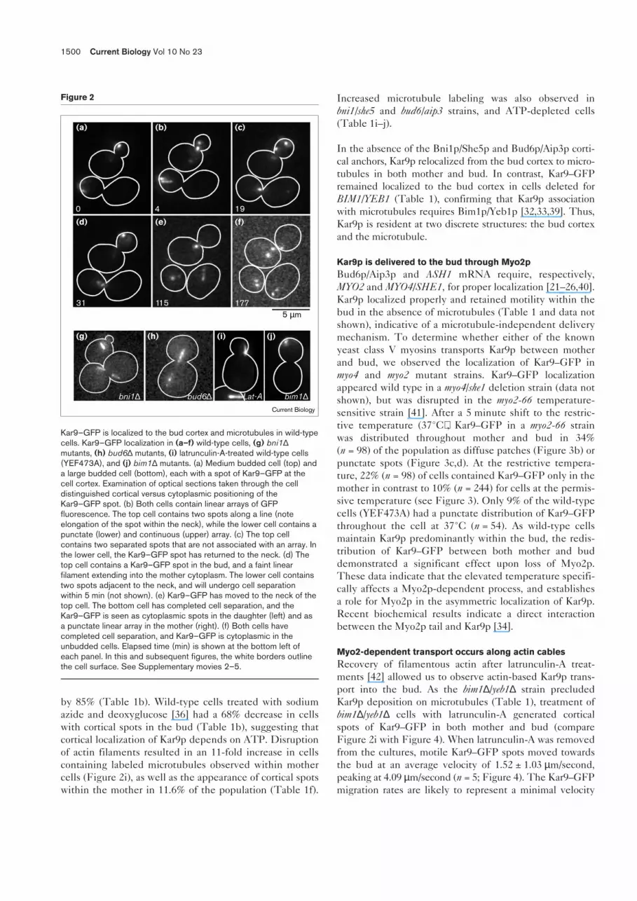
by 85% (Table 1b). Wild-type cells treated with sodiumazide and deoxyglucose [36] had a 68% decrease in cellswith cortical spots in the bud (Table 1b), suggesting thatcortical localization of Kar9p depends on ATP. Disruptionof actin filaments resulted in an 11-fold increase in cellscontaining labeled microtubules observed within mothercells (Figure 2i), as well as the appearance of cortical spotswithin the mother in 11.6% of the population (Table 1f).
Increased microtubule labeling was also observed inbni1/she5 and bud6/aip3 strains, and ATP-depleted cells(Table 1i–j).
In the absence of the Bni1p/She5p and Bud6p/Aip3p corti-cal anchors, Kar9p relocalized from the bud cortex to micro-tubules in both mother and bud. In contrast, Kar9–GFPremained localized to the bud cortex in cells deleted forBIM1/YEB1 (Table 1), confirming that Kar9p associationwith microtubules requires Bim1p/Yeb1p [32,33,39]. Thus,Kar9p is resident at two discrete structures: the bud cortexand the microtubule.
Kar9p is delivered to the bud through Myo2pBud6p/Aip3p and ASH1 mRNA require, respectively,MYO2 and MYO4/SHE1, for proper localization [21–26,40].Kar9p localized properly and retained motility within thebud in the absence of microtubules (Table 1 and data notshown), indicative of a microtubule-independent deliverymechanism. To determine whether either of the knownyeast class V myosins transports Kar9p between motherand bud, we observed the localization of Kar9–GFP inmyo4 and myo2 mutant strains. Kar9–GFP localizationappeared wild type in a myo4/she1 deletion strain (data notshown), but was disrupted in the myo2-66 temperature-sensitive strain [41]. After a 5 minute shift to the restric-tive temperature (37°C), Kar9–GFP in a myo2-66 strainwas distributed throughout mother and bud in 34%(n = 98) of the population as diffuse patches (Figure 3b) orpunctate spots (Figure 3c,d). At the restrictive tempera-ture, 22% (n = 98) of cells contained Kar9–GFP only in themother in contrast to 10% (n = 244) for cells at the permis-sive temperature (see Figure 3). Only 9% of the wild-typecells (YEF473A) had a punctate distribution of Kar9–GFPthroughout the cell at 37°C (n = 54). As wild-type cellsmaintain Kar9p predominantly within the bud, the redis-tribution of Kar9–GFP between both mother and buddemonstrated a significant effect upon loss of Myo2p.These data indicate that the elevated temperature specifi-cally affects a Myo2p-dependent process, and establishesa role for Myo2p in the asymmetric localization of Kar9p.Recent biochemical results indicate a direct interactionbetween the Myo2p tail and Kar9p [34].
Myo2-dependent transport occurs along actin cablesRecovery of filamentous actin after latrunculin-A treat-ments [42] allowed us to observe actin-based Kar9p trans-port into the bud. As the bim1∆/yeb1∆ strain precludedKar9p deposition on microtubules (Table 1), treatment ofbim1∆/yeb1∆ cells with latrunculin-A generated corticalspots of Kar9–GFP in both mother and bud (compareFigure 2i with Figure 4). When latrunculin-A was removedfrom the cultures, motile Kar9–GFP spots moved towardsthe bud at an average velocity of 1.52 ± 1.03 µm/second,peaking at 4.09 µm/second (n = 5; Figure 4). The Kar9–GFPmigration rates are likely to represent a minimal velocity
1500 Current Biology Vol 10 No 23
Figure 2
Kar9–GFP is localized to the bud cortex and microtubules in wild-typecells. Kar9–GFP localization in (a–f) wild-type cells, (g) bni1∆mutants, (h) bud6∆ mutants, (i) latrunculin-A-treated wild-type cells(YEF473A), and (j) bim1∆ mutants. (a) Medium budded cell (top) anda large budded cell (bottom), each with a spot of Kar9–GFP at thecell cortex. Examination of optical sections taken through the celldistinguished cortical versus cytoplasmic positioning of theKar9–GFP spot. (b) Both cells contain linear arrays of GFPfluorescence. The top cell contains two spots along a line (noteelongation of the spot within the neck), while the lower cell contains apunctate (lower) and continuous (upper) array. (c) The top cellcontains two separated spots that are not associated with an array. Inthe lower cell, the Kar9–GFP spot has returned to the neck. (d) Thetop cell contains a Kar9–GFP spot in the bud, and a faint linearfilament extending into the mother cytoplasm. The lower cell containstwo spots adjacent to the neck, and will undergo cell separationwithin 5 min (not shown). (e) Kar9–GFP has moved to the neck of thetop cell. The bottom cell has completed cell separation, and theKar9–GFP is seen as cytoplasmic spots in the daughter (left) and asa punctate linear array in the mother (right). (f) Both cells havecompleted cell separation, and Kar9–GFP is cytoplasmic in theunbudded cells. Elapsed time (min) is shown at the bottom left ofeach panel. In this and subsequent figures, the white borders outlinethe cell surface. See Supplementary movies 2–5.
Current Biology
(a) (b) (c)
(d) (e) (f)
(g) (h) (i) (j)
0 4 19
31 115 1775 µm
bni1∆ bud6∆ Lat-A bim1∆

for Kar9p transport, as cells must re-establish a polarizedactin cytoskeleton following exposure to latrunculin-Aduring image acquisition. Kar9p particles halted at theneck for approximately 1–10 seconds before continuinginto the bud (n = 5; note the spot at the neck in Figure 4d).By generating a Kymograph (see Materials and methods)for a linear region corresponding to the path of a Kar9–GFPspot (Figure 4a–d), a second particle was detected follow-ing the same route in approaching the bud neck(Figure 4e,f). Therefore, Kar9p particles are transported tothe bud neck via Myo2p along ‘tracks’ within the mother.These tracks may be actin cables within the mother, polar-ized towards the bud [25,42–45].
Temperature-sensitive alleles of tropomyosin specificallylose filamentous actin at the restrictive temperature [19].To determine the role of actin cables in localizing Kar9p,we imaged Kar9–GFP in tmp1-2, tpm2∆ cells (ABY971) atboth the restrictive (35°C) and permissive (22°C) temper-ature. Transport of Kar9–GFP into the bud was greatlydiminished in the absence of actin cables (22.4% in themother cell at 35°C, n = 85; 3.2% in the mother cell at22°C, n = 94). The large fraction of cells with Kar9p in the
body of the mother cell in tmp1-2, tpm2∆ cells illustratesthe requirement for filamentous actin in Kar9p transportto the bud.
Kar9p facilitates microtubule search and capture at thebud siteMicrotubules probe the cell cortex in a ‘search andcapture’ mechanism prior to finding the bud, and, report-edly, bind cortical-attachment sites associated with theactin cytoskeleton [37,46–50]. Kar9p is present prior tobud emergence as evidenced by the persistence ofKar9–GFP produced from the endogenous promoter(Table 1a). As Kar9p is delivered early to the neck viaactin and myosin, and associates with microtubules viaBim1p/Yeb1p, Kar9p may provide the critical linkbetween the microtubule and actin cytoskeletons.
Cells were observed between cell separation and initialmicrotubule penetration of the bud in the presence andabsence of Kar9p. Where the direction of the bud isdefined as 0°, and perpendicular to the mother–bud axis is90°, microtubules in kar9∆ cells emanated from thespindle pole body (SPB) at 72.3° ± 54.4 (n = 9) from themother–bud axis at the time of bud emergence. In con-trast, wild-type microtubules were within 43.1° ± 29.8(n = 7) [27]. Microtubules in the wild-type strain(YEF473A) intersected, and remained associated with thepre-bud site 8.7 ± 11.7 minutes (n = 10) prior to bud emer-gence, while in the kar9∆ strain (KBY1010) microtubulesentered the bud 36.8 ± 18.3 minutes (n = 7) after budemergence. The timing of microtubule entry into the budin the kar9∆ strain was remarkably similar to the timing ofnuclear migration to the neck in a bim1∆/yeb1∆ strain(36.7 minutes) [51]. Thus, microtubules in kar9∆ cellswere delayed in finding the bud site by 45.5 minutes(8.7 minutes prior + 36.8 minutes post). This delay maycontribute to the frequency of binucleate cells found inkar9∆ strains. Six of 12 kar9∆ cells constructed the mitoticspindle before microtubules detected the bud, whilemicrotubules entered the bud prior to spindle formation in10 of 10 wild-type cells.
To determine whether Kar9p facilitates microtubule pen-etration of the bud, we observed Kar9–GFP early in thecell cycle. The microtubule decorated by Kar9–GFP inFigure 5a extended into the cytoplasm of the cell approx-imately 30° off the mother–bud axis (cell separation fol-lowed in 34 minutes; Figure 5d). The Kar9–GFP-labeledmicrotubule contacted the cell cortex (Figure 5b,g), priorto localization at the incipient bud site (Figure 5c), andcontinued to migrate towards the incipient bud site(n = 5). Once the microtubule reached the bud site,Kar9–GFP was concentrated at that site (Figure 5c;n = 3). The microtubule remained associated with thebud site, then immediately entered the emergent bud(Figure 5d; n = 7).
Research Paper Kar9p and Myo2p in spindle orientation Beach et al. 1501
Figure 3
MYO2, which encodes a class V myosin, is required for Kar9plocalization in the bud. (a,b) Selected images of individual myo2-66cells expressing Kar9–GFP at 23°C (a) when held at 23°C or(b) transferred to 37°C for 5 min before imaging. (c) A myo2-66 cellwith punctate Kar9–GFP distribution at the permissive temperature(23°C), then shifted to the restrictive temperature (37°C) for 5 min.(d) Stereo images of a three-dimensional reconstruction (see Materialsand methods) of the cell shown in (c). Large spots were visible alongthe cortex, and smaller spots could be seen in the cytoplasm.
Current Biology
(a)
(b)
(c) (d)
23ºC
37ºC
5 µm

Kar9p couples microtubule dynamics with nuclear migrationTo examine the relation between Kar9p within the budand microtubules, we coexpressed both Kar9–GFP anddynein–GFP. Microtubules, visualized by dynein–GFPlabeling, were observed extending into the bud and con-tacting Kar9–GFP spots. The Kar9p–microtubule inter-sections were unstable, lasting an average of 2.36 minutes(n = 5), similar to previous reports of the persistence ofmicrotubule–cortex interactions [37]. Microtubule–Kar9p
cortical spot interactions were visible as the intersection oflinear, filamentous microtubules with fluorescent spots atthe cortex (see Figure 6c,e,f), and occurred 54% ± 9.5(n = 6) of the time, on average, throughout the cell cycle.
Kar9p–microtubule contacts facilitated the orientation andpositioning of the nucleus at the bud neck (Figure 6).Kar9–GFP was localized as a spot in the bud, and a micro-tubule growing from the SPB (bright spot distant from theneck) extended towards the bud (Figure 6a). Once themicrotubule bundle entered the bud and connected withthe Kar9p spot (at the tip of the bud in Figure 6a–c), thenucleus (marked by the SPB) moved towards and, ulti-mately, adjacent to the neck (Figure 6c–f). Microtubulesshortened as the nucleus traveled towards the neck at anaverage velocity of 0.404 ± 0.153 µm/minute (n = 5).Migration of the nucleus towards the neck arrested when
1502 Current Biology Vol 10 No 23
Figure 4
Kar9p is transported along actin filamentswithin the mother towards the bud.(a–d) Following a 15 min treatment with50 µM latrunculin-A, bim1∆/yeb1∆(KBY1017) cells expressing Kar9–GFP wereallowed to recover, and the movements ofKar9–GFP spots were imaged at 3 framesper second. (a) Two spots were labeled withinthe mother domain of the cell (arrowhead andarrow). (b–d) While the central spot (arrow)remained stationary, the left spot (arrowhead)migrated towards the neck. (d) The motilespot remained adjacent to the neck (10 secfor this spot). Elapsed time (in sec) is showncorresponding to the time scale in (f). (e) Acomposite of a bright-field image (red), anepifluorescent image of GFP signal (green),and traces generated by the ‘Track Points’function of Metamorph (see Materials andmethods) of Kar9–GFP particles through thecell (blue). Kar9–GFP within the bud isapparent, while the traces show the path
followed by Kar9–GFP towards the bud. Therectangular region was used to form theKymograph (see Materials and methods).(f) The Kymograph function extracts a user-defined region (rectangle in panel e) of animage to be displayed with sequential timepoints colinear within a single image. Elapsedtime is represented along the vertical axis(sec), and distance (µm) along the horizontalaxis (oriented to the image in panel e by thetriangle and circle). The vertical, off-centertrace is the non-motile spot in the middle of
the cell. Single- and double-headed arrowsmark the beginning and end of two sigmoid-shaped traces of motile Kar9–GFP spotsbeing translocated from the mother towardsthe bud neck. The top trace is the same spotindicated by the arrowhead in (a–d) andwithin the rectangle in (e), and the lower traceis a second spot following the same path(indicated by the arrow in panels a,c,d).Vertical regions of each sigmoidal traceindicate stationary periods. SeeSupplementary movie 6.
Current Biology
(a) (b) (c) (d) (e) (f)
5 µm 5 µm
Tim
e (s
ec)
0
14
7
3.5 µmDistance
Figure 5
Kar9p facilitates microtubule delivery to the bud site. (a–c) Sequentialimages of a Kar9–GFP-labeled microtubule and (f–h) the respectivestereo images of three-dimensional renderings (see Materials andmethods). The incipient bud site is labeled by an arrow in (a–c).Bipolar budding in this haploid strain (YEF473A) is occasionallyobserved. (a) A large budded, post mitotic cell with Kar9–GFP-labeledmicrotubules in both mother and daughter cells. The asterisk marks theSPB. The plus-end of the microtubule is oriented towards the incipientbud site. (b) Microtubule movement to contact the cell cortex.Inspection of the optical sections (not shown) and (g) three-dimensional stereo view demonstrated cortical attachment of themicrotubule. (c) The microtubule plus-end was positioned at theincipient bud site 32 min prior to cell separation. (d) The new bud isvisible, and a microtubule decorated by Kar9–GFP has penetrated thesmall bud. (e) DIC image of the three cells shown in the stereoimages. The large budded cell is represented in (a–c). Elapsed time(min) is shown. Current Biology
5 µm
(a)
(b)
(c)
(f)
(g)
(h)
(d)
(e)
0
1
2
34

microtubules lost contact with the Kar9p spot (Figure 6d),then resumed once contact was re-established (Figure 6e).
The distance between the SPB and the Kar9–GFP spotwithin the bud is plotted in Figure 6g. Microtubule–Kar9pcontacts were scored in frames where the labeled micro-tubule terminated in a region occupied by a Kar9–GFPspot. Periods with the most dramatic nuclear movementstowards the bud (~20 minutes and ~30 minutes) coincidedwith intersection of the Kar9–GFP spot and microtubule.In the cell shown, the apparent microtubule–Kar9p attach-ment remained stable after the nucleus had reached theneck. The SPB did not proceed into the bud, although co-localization of Kar9–GFP and microtubules was evident(Figure 6g, ~40–60 minutes).
Positioning the nucleus at the neck in vegetative cells isanalogous to nuclear migration to the tip of the matingprojection [49]. Kar9–GFP localizes to the mating projec-tion tip, and is required for nuclear orientation and migra-tion during the mating process [5,9]. We observed nuclearmigration towards the mating projection tip of α-factor-treated cells expressing both dynein–GFP and Kar9–GFP.In cells entering the mating pathway, nuclei rotate oppo-site to the direction of microtubule growth until themating projection tip is located (similar to nuclear move-ment in unbudded vegetative cells). Microtubules formedpersistent connections with the Kar9–GFP spot, and thenucleus migrated toward the mating projection(Figure 7a–f) at 0.32 ± 0.13 µm/minute (n = 11), which wassimilar to the velocity of nuclear migration to corticalKar9p in vegetative cells.
The distance from the SPB to the mating projection tip isplotted in Figure 7g, which demonstrates the movementof the SPB towards the mating projection base. After themicrotubule had oriented towards the mating projection
(27–30 minutes), nuclear movement towards the matingprojection coincided with microtubule depolymerization[49]. After the nucleus was positioned at the base of themating projection, the distance between the SPB and themating projection lengthened and shortened over theensuing 45 minutes. SPB movement was facilitated bymicrotubule growth and shrinkage while maintaining aninteraction with the Kar9–GFP spot. Thus, Kar9p–micro-tubule interactions facilitate nuclear migration in bothvegetative and mating cycles.
Directed movement to the neck is differentiated fromsteady state, oscillatory nuclear movements in vegetative(Figure 6g) and mating (Figure 7g) cells. Directional trans-port of the nucleus towards the neck is observed inFigure 6 (~20–40 minutes) and Figure 7 (~25–55 minutes).During directed nuclear movement to the neck, 62% ± 20(n = 6) of time points showed an intersection betweenKar9–GFP and microtubules, 69% ± 9.4 (n = 5) of whichresulted in directed movement of the nucleus towards theneck. Thus, not every microtubule intersection with aKar9–GFP spot results in directed nuclear movement.Following nuclear congression, oscillatory movementsmaintained a steady state positioning of the nucleus adja-cent to the neck or mating projection until either anaphaseor cell fusion occurred (Figure 6, ~40–65 minutes; andFigure 7, ~55–85 minutes). The persistence of the associa-tion of microtubules to Kar9–GFP spots increased to75% ± 28 (n = 4) of these time points, but did not result indirected movements past the neck.
DiscussionKar9p, Bud6p, Bni1p and Bim1p together constitute anearly pathway to position the nucleus at the bud neckbefore the onset of anaphase. We observed that Kar9p waspresent throughout the cell cycle in dynamic equilibriumbetween microtubules and sites of polarized growth. Its
Research Paper Kar9p and Myo2p in spindle orientation Beach et al. 1503
Figure 6
Kar9p couples dynamic microtubules withnuclear migration. (a–f) Selected time-lapseimages of a cell expressing both Kar9–GFP(spot in bud) and dynein–GFP (decorates themicrotubule and SPB in the mother). The SPBis the bright spot at the right end of themicrotubule. (a,b) The Kar9–GFP spot ismobile within the bud. The microtubule in themother domain is oriented with the plus-endtowards the bud, scanning the bud cortex tolocate the bud site. (c) The microtubule hasmade contact with the cortical Kar9–GFPspot in the bud, and the SPB has movedtowards the neck. (d) The microtubule hasseparated from the Kar9–GFP spot. Duringthis time, SPB movement towards the budceases. (e) The microtubule is recaptured byKar9–GFP, and SPB migration continues
towards the bud. (f) The nucleus is positionedat the neck with the microtubule extendinginto the bud. Elapsed time (min) is shown.These images were generated using a Delta-Vision imaging workstation. (g) The distancebetween the Kar9–GFP spot and the SPB
was measured for each frame in the timelapse and plotted against time. Each pointdisplaying an intersection or separation of theKar9–GFP spot and the microtubule wasscored by observing individual opticalsections. See Supplementary movie 7.
Current Biology
(a) (b) (c)
(d)
(g)
(e) (f)19 21
41 53
5 µm
Distance from SPB to Kar9p
01234567
0 10 20 30 40 50 60 70Elapsed time ( min)
Dis
tanc
e (µ
m)
Apparent separationApparent intersection
0
28

association with microtubules was susceptible to nocoda-zole treatment and required BIM1 (Figure 2g–j, Table 1)[5,32,33,39]. Localization of Kar9p at the bud cortex wassensitive to treatment with latrunculin-A and was depen-dent on actin, BNI1 and BUD6 (Figure 2g–j, Table 1) [3].Kar9p has not been shown, either biochemically or geneti-cally, to associate simultaneously with microtubules andactin filaments. Time-lapse images of live cells coexpress-ing Kar9–GFP and dynein–GFP demonstrated that bothproteins co-localized at the bud cortex, and correlatednuclear movements towards the bud with Kar9p–micro-tubule interactions in both vegetative and mating cells(see below).
Kar9–GFP was observed primarily as a cortical spot thatwas motile within the bud and not confined to the bud tip(Table 1, columns b–d). The asymmetric distribution of
Kar9p was established through an actomyosin transportsystem. Directional transport towards the bud was pro-vided by two type V myosins, encoded by MYO2 [52] andMYO4 [53]. Myo2p supports polarized secretion [20],whereas the only known cargo for Myo4p is ASH1 mRNA.In the absence of Myo2p, but not Myo4p, Kar9–GFP spotswere present within the mother cell domain (Figure 3),demonstrating MYO2-dependent transport of Kar9p [34].Single particle tracking of Kar9–GFP allowed us to obtainin vivo velocities for Myo2p-based motility. Kar9p fol-lowed a direct path towards the neck at approximately1.52 µm/second (Figure 4), which is faster than in vivoMyo4p transport of ASH1 mRNA (0.2–0.4 µm/second)[25] and in vitro motility assays using chick brain myosin V(0.2–0.4 µm/second) [54].
Directional transport by Myo2p requires actin cablespolarized with their growing ends towards the bud [20].Tropomyosin, an actin filament binding protein, isrequired to maintain actin cables, but does not effect theintegrity of the actin patches [19]. Aberrant Kar9–GFPlocalization in the absence of tropomyosin implicates actincables in Kar9p delivery to the bud, and further supports aMyo2p-dependent, actomyosin delivery system for Kar9p.Two independent spots of Kar9–GFP traveled approxi-mately 3.5 µm within the same 0.3 µm-wide path throughthe cell between mother and bud, separated by about2.6 seconds (Figure 4f). The similar trajectories followedby both Kar9p particles indicate that actin filamentspersist long enough to allow transport of multiple particleson a single filament. Whether individual cables providethe substrate for different class V myosins (ASH1 mRNAvia Myo4p; Kar9p via Myo2p), or dedicated cables existfor one type of cargo (Kar9p versus vesicular traffic)remains to be determined.
In the absence of Kar9p, microtubules are delayed indetecting the bud site by approximately 45 minutes, poten-tially leading to off-axis microtubule spindles and theobserved production of binucleated cells [5]. While Kar9p,ASH1mRNA, and Bud6p are localized similarly in buddedcells, only Kar9p does not localize to the incipient bud site.Instead, Kar9–GFP was observed on microtubules and atthe spindle pole body prior to bud emergence (Figure 2e,f).Kar9–GFP-decorated microtubules became orientedtowards the bud site prior to bud emergence (Figure 5), andtowards the mating projection tip (Figure 7a–f). Thus, puta-tive transport of Kar9p-decorated microtubules along polar-ized actin fibers towards the bud would provide a‘facilitated’ microtubule search and capture mechanismcontributing to the fidelity of nuclear migration.
Once microtubules penetrate the bud, Kar9p at the budcortex can provide a microtubule anchorage site [3,5].Dynein–GFP-labeled microtubules reaching the bud cortexor the mating projection terminated at spots of Kar9–GFP
1504 Current Biology Vol 10 No 23
Figure 7
Kar9p directs microtubule orientation towards the mating projection.(a–f) Selected time-lapse images of a cell expressing both Kar9–GFPand dynein–GFP in the presence of α-factor. Dynein–GFP decoratedthe SPB (open arrowhead) and the microtubule, while Kar9–GFPlabeled the plus-end of the microtubule (solid arrowhead). (a–d) Themicrotubule rotated approximately 180° counter-clockwise to orient theplus-end of the microtubule at the tip of the mating projection. Rotationwas complete at the 26:53 time point. (d–f) Following microtubuleorientation, microtubule shortening moved the SPB to the base of themating projection. Elapsed time (in min) is shown. (g) The distancebetween the SPB and the mating projection tip was measured for eachframe in the time-lapse series and plotted against time. Microtubuleinteractions with the Kar9–GFP spot were retained throughout thetime-lapse series.
Current Biology
5 µm54:2342:2331:53
19:519:294:29
(a) (b) (c)
(d)
(g)
(e) (f)
Kar9pDirect sMT Orientation
0
0.25
0.50
0.75
1.00
1.25
1.05
1.75
2.00 Distance from SPB to mating projection
Time (min)0 10 20 30 40 50 60 70 80 90
Dis
tanc
e (µ
m)

(Figure 6c,e,f, Figure 7d,f). Microtubule interactions withKar9p in the bud lasted between 2–3 minutes, suggestingthat microtubules transiently attach at Kar9p cortical sites.Following microtubule capture within the bud, thenucleus migrates subjacent to the neck and remains thereuntil anaphase onset (Figure 6) [4,51]. Similarly, nuclearcongression to the mating projection tip was accompaniedby a persistent association of microtubules with aKar9–GFP spot (Figure 7).
Nuclear movements towards and away from the bud ormating projection were coupled with shortening andlengthening of the microtubules connecting the spindlepole body to the Kar9p spot at the cortex (Figures 6a–fand 7a–f). The motor protein Kip3p is thought to stimu-late microtubule disassembly, and is presumed to functionin association with Kar9p, as kip3p dhc1 and kar9 dhc1double mutants are lethal [8,27,30]. Kip3p at the micro-tubule–Kar9p junction could stimulate microtubule short-ening, providing the underlying mechanism for nuclearmigration. Kip3p has recently been shown to facilitatenuclear movement to the bud neck [4,8,27,30], but pullingforces exerted by Kip3p and associated proteins in the‘early’ nuclear orientation pathway appear to be too weakto move the nucleus through the narrow neck [4]. Thus, acooperative mechanism between Kar9p and Myo2p aswell as Kar9p and Kip3p facilitates pre-anaphase nuclearpositioning at the neck but not beyond. Subsequently, thelate nuclear-orientation pathway, including the micro-tubule-based motor, dynein and associated proteins, sup-ports translocation of the daughter nucleus into the bud inconjunction with spindle elongation for the successfulcompletion of mitosis.
Materials and methodsVelocity measurements, microscopy and image processingKar9p velocity measurements were obtained measuring the point-to-point movements of Kar9–GFP spots. Kar9–GFP spots were trackedusing the ‘Track Points’ function of the Metamorph (Universal ImagingCorporation) software package. The distance of spot movements at30 sec (Figures 1,6) or 0.3 sec (Figure 4) intervals provided instanta-neous velocities representing a minimal speed at each time point, andaveraged over time. Only movements between sequential images wereconsidered for velocity measurements. Because the spots frequentlychanged direction between long time points, continuous velocitiescould not be measured.
Microscopy and digital imaging, including optical sectioning, was per-formed as described in [37]. Five optical sections were taken at 0.75 µmincrements through the cell for a total of 3.0 µm per time point. Rapidimaging rates of cells at 3 frames per second included only a singleoptical section. The central optical section including both a transmittedlight and an epifluorescent (GFP) image was focused at the cell neck.Images were captured using a Hamamatsu Orca II (model C4742-98)CCD camera mounted on a Nikon Eclipse E600FN using 100 × 1.4 NAPlan Apochromat objective with 1 × magnification to the camera. Imagetaken at elevated temperature for analysis of temperature sensitive strainsused a Nikon Eclipse TE300 inverted microscope with a phase contrast100 × 1.4 NA Plan Apochromat objective with 1 × magnification to aHamamatsu Orca (model C4742-95) camera. The inverted microscopestage was heated to 35°C over 25–30 min using a Nevtek air steam stage
incubator (model ASI400). The Metamorph software package forWindows was used for microscope automation, image acquisition andimage analysis. Images for publication were manipulated for scaling, size,resolution (300 dpi) and arrangement with Windows versions of AdobePhotoshop and Corel Draw.
The ‘Kymograph’ function of Metamorph software was used to create across-sectional display of pixel intensity values along a linear region (linewidth of 5 pixels = 0.3 µm) for each image of a time-lapse series(Figure 4e). The ‘Linescan’ function of Metamorph provided numericalvalues and a graph representing the grayscale pixel values along a linearregion for a single image plane from a time-lapse series. Three-dimen-sional reconstructions of cells were generated using the ‘3D Reconstruc-tion’ function of Metamorph set for either full circle or shallow tilehorizontal reconstructions with a Z distance of 1 µm per image for the fiveimages of each optical section series. The ‘Stereographic Views’ functionof Metamorph generated side-by-side images to view the three-dimen-sional reconstruction in printed format, with a 10° rotation betweenimages. Images acquired using a DeltaVision (Applied Precision) decon-volution microscope were used in Figure 6. Briefly, using a 100X 1.4 NAPlan Apo lens on a Nikon TE200 inverted microscope, z-series consistingof 9 200 ms fluorescent (using a HQ-FITC filter set, Chroma Filters) expo-sures (0.3 µm step size) were acquired at 30 sec intervals. The z-seriestime lapse was then deconvolved using DeltaVision software and is pre-sented as a maximum projection.
Supplementary materialSupplementary material including additional methodological detail isavailable at http://current-biology.com/supmat/supmatin.htm.
AcknowledgementsWe thank John Pringle, Katja Schwartz and Rita Miller for strains and plas-mids; Ted Salmon for imaging advice and critical reading of the manuscript;Chad Pearson for critical reading of the manuscript; and Susan Whitfield,Jennifer Stemple and Molly Hays for technical assistance. This work is sup-ported by National Institutes of Health grant GM32238 to K.B.
References1. Heil-Chapdelaine RA, Adames NR, Cooper JA: Formin’ the
connection between microtubules and the cell cortex. J Cell Biol1999, 144:809-811.
2. Lee L, Klee SK, Evangelista M, Bonne C, Pellman D: Control ofmitotic spindle position by the Saccharomyces cerevisiae forminBni1p. J Cell Biol 1999, 144:947-961.
3. Miller R, Matheos D, Rose M: The cortical localization of themicrotubule orientation protein, Kar9p, Is dependent upon actinand proteins required for polarization. J Cell Biol 1999,144:963-975.
4. Yeh E, Yang C, Chin E, Maddox P, Salmon ED, Lew DJ, et al.:Dynamic positioning of mitotic spindles in yeast: role ofmicrotubule motors and cortical determinants. Mol Biol Cell 2000,11:1-13.
5. Miller RK, Rose MD: Kar9p is a novel cortical protein required forcytoplasmic microtubule orientation in yeast. J Cell Biol 1998,140:377-390.
6. Eshel D, Urrestarazu LA, Vissers S, Jauniaux JC, van Vliet-Reedijk JC,Planta RJ, et al.: Cytoplasmic dynein is required for normal nuclearsegregation in yeast. Proc Natl Acad Sci USA 1993,90:11172-11176.
7. Li YY, Yeh E, Hays T, Bloom K: Disruption of mitotic spindleorientation in a yeast dynein mutant. Proc Natl Acad Sci USA1993, 90:10096-10100.
8. Miller RK, Heller KK, Rose MD: The kinesin-related proteins, Kip2pand Kip3p, function differently in nuclear migration in yeast.Mol Biol Cell 1998, 9:2051-2068.
9. Kurihara LJ, Beh CT, Latterich M, Schekman R, Rose MD: Nuclearcongression and membrane fusion: two distinct events in theyeast karyogamy pathway. J Cell Biol 1994, 126:911-923.
10. Evangelista M, Blundell K, Longtine MS, Chow CJ, Adames N,Pringle JR, et al.: Bni1p, a yeast formin linking Cdc42p and theactin cytoskeleton during polarized morphogenesis. Science1997, 276:118-122.
Research Paper Kar9p and Myo2p in spindle orientation Beach et al. 1505

11. Amberg DC, Zahner JE, Mulholland JW, Pringle JR, Botstein D:Aip3p/Bud6p, a yeast actin-interacting protein that is involved inmorphogenesis and the selection of bipolar budding sites. MolBiol Cell 1997, 8:729-753.
12. Fujiwara T, Tanaka K, Mino A, Kikyo M, Takashi K, Shimizu K, et al.:Rho1p-Bni1p-Spa2p interactions: implication in localization ofBni1p at the bud site and regulation of the actin cytoskeleton inSaccharomyces cerevisiae. Mol Biol Cell 1998, 9:1221-1233.
13. Palmer RE, Sullivan DS, Huffaker T, Koshland D: Role of astralmicrotubules and actin in spindle orientation and migration in thebudding yeast, Saccharomyces cerevisiae. J Cell Biol 1992,119:583-593.
14. Theesfeld CL, Irazoqui JE, Bloom K, Lew DJ: The role of actin inspindle orientation changes during the Saccharomyces cerevisiaecell cycle. J Cell Biol 1999, 146:1019-1032.
15. Wang T, Bretscher A: Mutations synthetically lethal with tpm1∆∆ liein genes involved in morphogenesis. Genetics 1997,147:1595-1607.
16. Yang CH, Snyder M: The nuclear-mitotic apparatus protein isimportant in the establishment and maintenance of the bipolarmitotic spindle apparatus. Mol Biol Cell 1992, 3:1259-1267.
17. Adams AEM, Botstein D, Drubin DG: Requirement of yeast fimbrinfor actin organization and morphogenesis in vivo. Nature 1991,354:404-408.
18. Kopecká M, Gabriel M: The aberrant positioning of nuclei and themicrotubular cytoskeleton in Saccharomyces cerevisiae due toimproper actin function. Microbiology 1998, 144:1783-1797.
19. Pruyne DW, Schott DH, Bretscher A: Tropomyosin-containing actincables direct the Myo2p-dependent polarized delivery ofsecretory vesicles in budding yeast. J Cell Biol 1998,143:1931-1945.
20. Karpova TS, Reck-Peterson SL, Elkind NB, Mooseker MS, Novick PJ,Cooper JA: Role of actin and Myo2p in polarized secretion andgrowth of Saccharomyces cerevisiae. Mol Biol Cell 2000,11:1727-1737.
21. Jin H, Amberg DC: The secretory pathway mediates localization ofthe cell polarity regulator Aip3p/Bud6p. Mol Biol Cell 2000,11:647-661.
22. Long RM, Singer RH, Meng X, Gonzalez I, Nasmyth K, Jansen R-P:Mating type switching in yeast controlled by asymmetriclocalization of ASH1 mRNA. Science 1997, 277:383-387.
23. Takizawa PA, Sil A, Swedlow JR, Herskowitz I, Vale RD: Actin-dependent localization of an RNA encoding a cell-fatedeterminant in yeast. Nature 1997, 389:90-93.
24. Beach DL, Salmon ED, Bloom K: Localization and anchoring ofmRNA in budding yeast. Curr Biol 1999, 9:569-578.
25. Bertrand E, Chartrand P, Schaefer M, Shenoy SM, Singer RH,Long RM: Localization of ASH1 mRNA particles in living yeast.Mol Cell 1998, 2:437-445.
26. Jansen RP, Dowzer C, Michaelis C, Galova M, Nasmyth K: Mothercell-specific HO expression in budding yeast depends on theunconventional myosin Myo4p and other cytoplasmic proteins.Cell 1996, 84:687-697.
27. DeZwaan TM, Ellingson E, Pellman D, Roof DM: Kinesin-relatedKIP3 of Saccharomyces cerevisiae is required for a distinct step innuclear migration. J Cell Biol 1997, 138:1023-1040.
28. Straight AF, Sedat JW, Murray AW: Time-lapse microscopy revealsunique roles for kinesins during anaphase in budding yeast. J CellBiol 1998, 143:687-694.
29. Schwartz K, Richards K, Botstein D: BIM1 encodes a microtubule-binding protein in yeast. Mol Biol Cell 1997, 8:2677-2691.
30. Cottingham FR, Hoyt MA: Mitotic spindle positioning inSaccharomyces cerevisiae is accomplished by antagonisticallyacting microtubule motor proteins. J Cell Biol 1997, 138:1041-1053.
31. Tirnauer JS, O’Toole EO, Berrueta L, Bierer BE, Pellman D: YeastBim1p promotes the G1-specific dynamics of microtubules. J CellBiol 1999, 145:993-1007.
32. Korinek WS, Copeland MJ, Chaudhuri A, Chant J: Molecular linkageunderlying microtubule orientation toward cortical sites in yeast.Science 2000, 287:2257-2259.
33. Lee L, Tirnauer JS, Li J, Schuyler SC, Liu JY, Pellman D: Positioningof the mitotic spindle by a cortical-microtubule capturemechanism. Science 2000, 287:2260-2262.
34. Yin H, Pruyne D, Huffaker TC, Bretscher A: Myosin V orients themitotic spindle in yeast. Nature 2000, 406:1013-1015.
35. Waddle JA, Karpova TS, Waterston RH, Cooper JA: Movement ofcortical actin patches in yeast. J Cell Biol 1996, 132:861-870.
36. Marshall WF, Straight A, Marko JF, Swedlow J, Dernberg A,Belmont L, et al.: Interphase chromosomes undergo constraineddiffusional motion in living cells. Curr Biol 1997, 7:930-939.
37. Shaw SL, Yeh E, Maddox P, Salmon ED, Bloom K: Astralmicrotubule dynamics in yeast: a microtubule-based searchingmechanism for spindle orientation and nuclear migration into thebud. J Cell Biol 1997, 139:985-994.
38. Straight AF, Marshall WF, Murray AW: Mitosis in living buddingyeast: anaphase A but no metaphase plate. Science 1997,277:574-578.
39. Miller RK, Cheng S-C, Rose MD: Bim1p/Yeb1p mediates theKar9p-dependent cortical attachment of cytoplasmicmicrotubules. Mol Biol Cell 2000, 11:2949-2959.
40. Munchow S, Sauter C, Jansen RP: Association of the class Vmyosin Myo4p with a localized messenger RNA in budding yeastdepends on She proteins. J Cell Sci 1999, 112:1511-1518.
41. Lillie SH, Brown SS: Suppression of a myosin defect by a kinesin-related gene. Nature 1992, 356:358-361.
42. Ayscough KR, Stryker J, Pokala N, Sanders M, Crews P, Drubin DG:High rates of actin filament turnover in budding yeast and rolesfor actin in establishment and maintenance of cell polarityrevealed using the actin inhibitor latrunculin-A. J Cell Biol 1997,137:399-416.
43. Adams AEM, Pringle JR: Relationship of actin and tubulindistribution to bud growth in wild-type and morphogenetic-mutant Saccharomyces cerevisiae. J Cell Biol 1984, 98:934-945.
44. Kilmartin JV, Adams EM: Structural rearrangements of tubulin andactin during the cell cycle of the yeast Saccharomyces. J Cell Biol1984, 98:922-933.
45. Li R, Zheng Y, Drubin DG: Regulation of cortical actin cytoskeletonassembly during polarized cell growth in budding yeast. J CellBiol 1995, 128:599-615.
46. Carminati JL, Stearns T: Microtubules orient the mitotic spindle inyeast through dynein-dependent interactions with the cell cortex.J Cell Biol 1997, 138:629-641.
47. Goode BL, Wong JJ, Butty A-C, Peter M, McCormack AL, Yates JR,et al.: Coronin promotes the rapid assembly and cross-linking ofactin filaments and may link the actin and microtubulecytoskeletons in yeast. J Cell Biol 1999, 144:83-98.
48. Goode BL, Drubin DG, Barnes G: Functional cooperation betweenthe microtubule and actin cytoskeletons. Curr Opin Cell Biol 2000,12:63-71.
49. Maddox P, Chin E, Mallavarapu A, Yeh E, Salmon ED, Bloom K:Microtubule dynamics from mating through the first zygoticdivision in the budding yeast Saccharomyces cerevisiae. J CellBiol 1999, 144:977-987.
50. Heil-Chapdelaine RA, Tran NK, Cooper JA: The role ofSaccharomyces cerevisiae coronin in the actin and microtubulecytoskeletons. Curr Biol 1998, 8:1281-1284.
51. Adames NR, Cooper JA: Microtubule interactions with the cellcortex causing nuclear movements in Saccharomyces cerevisiae.J Cell Biol 2000, 149:1-13.
52. Johnston GC, Prendergast JA, Singer RA: The Saccharomycescerevisiae MYO2 gene encodes an essential myosin for vectorialtransport of vesicles. J Cell Biol 1991, 113:539-551.
53. Haarer BK, Petzold A, Lillie SH, Brown SS: Identification of MYO4,a second class V myosin gene in yeast. J Cell Sci 1994,107:1055-1064.
54. Cheney RE, O’Shea, Heuser MK, Coelho MV, Wolenski JS,Espreafico EM, et al.: Brain myosin-V is a two-headedunconventional myosin with motor activity. Cell 1993, 75:13-23.
1506 Current Biology Vol 10 No 23
Because Current Biology operates a ‘Continuous PublicationSystem’ for Research Papers, this paper has been publishedon the internet before being printed. The paper can beaccessed from http://biomednet.com/cbiology/cub


