Embed Size (px)
Citation preview

10.1101/gad.559610Access the most recent version at doi: 2010 24: 766-782Genes Dev.
Peter Meister, Benjamin D. Towbin, Brietta L. Pike, et al.
developmentelegansC.The spatial dynamics of tissue-specific promoters during
MaterialSupplemental http://genesdev.cshlp.org/content/suppl/2010/04/12/24.8.766.DC1.html
References
http://genesdev.cshlp.org/content/24/8/766.full.html#related-urlsArticle cited in:
http://genesdev.cshlp.org/content/24/8/766.full.html#ref-list-1This article cites 66 articles, 31 of which can be accessed free at:
serviceEmail alerting
click heretop right corner of the article orReceive free email alerts when new articles cite this article - sign up in the box at the
http://genesdev.cshlp.org/subscriptions go to: Genes & DevelopmentTo subscribe to
Copyright © 2010 by Cold Spring Harbor Laboratory Press
Cold Spring Harbor Laboratory Press on October 17, 2012 - Published by genesdev.cshlp.orgDownloaded from

The spatial dynamics of tissue-specificpromoters during C. elegans development
Peter Meister, Benjamin D. Towbin, Brietta L. Pike, Aaron Ponti, and Susan M. Gasser1
Friedrich Miescher Institute for Biomedical Research, CH-4058 Basel, Switzerland
To understand whether the spatial organization of the genome reflects the cell’s differentiated state, we examinedwhether genes assume specific subnuclear positions during Caenorhabditis elegans development. Monitoring theradial position of developmentally controlled promoters in embryos and larval tissues, we found that smallintegrated arrays bearing three different tissue-specific promoters have no preferential position in nuclei ofundifferentiated embryos. However, in differentiated cells, they shifted stably toward the nuclear lumen whenactivated, or to the nuclear envelope when silent. In contrast, large integrated arrays bearing the same promotersbecame heterochromatic and nuclear envelope-bound in embryos. Tissue-specific activation of promoters in theselarge arrays in larvae overrode the perinuclear anchorage. For transgenes that carry both active and inactivepromoters, the inward shift of the active promoter was dominant. Finally, induction of master regulator HLH-1prematurely induced internalization of a muscle-specific promoter array in embryos. Fluorescence in situhybridization confirmed analogous results for the endogenous endoderm-determining gene pha-4. We proposethat, in differentiated cells, subnuclear organization arises from the selective positioning of active and inactivedevelopmentally regulated promoters. We characterize two forces that lead to tissue-specific subnuclearorganization of the worm genome: large repeat-induced heterochromatin, which associates with the nuclearenvelope like repressed genes in differentiated cells, and tissue-specific promoters that shift inward in a dominantfashion over silent promoters, when they are activated.
[Keywords: C. elegans; development; gene regulation; nuclear organization; chromatin]
Supplemental material is available at http://www.genesdev.org.
Received September 28, 2009; revised version accepted February 25, 2010.
During metazoan development, the generation of differ-entiated cell types requires the orchestrated expression ofthousands of genes. As cells differentiate, they becomeprogressively committed to specific lineages and, concom-itantly, their developmental potential becomes restricted(Yamanaka 2009). The reduction of cell fate potentialis accompanied by an extinction of lineage-inappropriateexpression programs, mediated largely by local and higher-order chromatin modifications (Boyer et al. 2006; Mohnand Schubeler 2009). As the number of repressed genesincreases, the expression of genes appropriate for a givendifferentiation program ensues.
Based on studies in yeast, flies, and mammalian cells,it has been argued that nuclear subcompartments influ-ence both gene repression and activation (for review, seeSpector 2003; Taddei et al. 2004; Akhtar and Gasser 2007;Schneider and Grosschedl 2007). Hence, it may be ex-pected that the organization of chromatin in the nucleus iscell type-specific. To date, studies addressing this questionfocused primarily on differentiation-specific juxtaposition
of silent genes to heterochromatin and the clustering ofactive genes in transcription foci (Kosak and Groudine2004b; Fraser and Bickmore 2007). Both events are well-characterized in hematopoietic lineages, where the radialorganization of tissue-specific genes tends to reflect ex-pression competence (Schneider and Grosschedl 2007). Forexample, developmental stage-specific repositioning awayfrom the nuclear periphery has been shown for the mouseIgH and IgK loci upon activation and rearrangement of thelocus during lymphocyte development (Brown et al. 2001;Kosak et al. 2002). In T cells, large-scale repositioningfurther correlated with chromosomal contraction (Skoket al. 2007).
Gene relocalization has also been observed duringmouse embryonic stem (ES) cell differentiation in vitro.For example, the monoallelically expressed GFAP geneshows differentiation- and activity-dependent reposition-ing during astrocyte differentiation (Takizawa et al. 2008),as does the MASH1 locus during neural induction inES cells (Williams et al. 2006). Thus, there are severaldocumented cases in which the repositioning of a geneoccurs at a specific stage of hematopoietic differentiationor during ES cell differentiation in vitro. These studiesfocused on the relationship of activated genes with their
1Corresponding author.E-MAIL [email protected]; FAX 41-61-697-3976.Article is online at http://www.genesdev.org/cgi/doi/10.1101/gad.559610.
766 GENES & DEVELOPMENT 24:766–782 � 2010 by Cold Spring Harbor Laboratory Press ISSN 0890-9369/10; www.genesdev.org
Cold Spring Harbor Laboratory Press on October 17, 2012 - Published by genesdev.cshlp.orgDownloaded from

chromosomal territory, with another coordinately regu-lated gene, or relative to centromeric heterochromatin (forreview, see Fraser and Bickmore 2007). However, re-positioning was not always observed upon gene activation(Hewitt et al. 2004), and it remained unclear why somegenes shift position upon activation while others do not.
The question of how genes are positioned within thenucleus is particularly relevant in the context of organ-ismal development. Is nuclear reorganization essentialfor differentiated gene expression? Is transcription a pre-requisite for relocalization (Ragoczy et al. 2006)? Is itsufficient? Does relocalization depend on the type of pro-moter, or only on transcriptional activity? And finally,are there different degrees of organization in pluripotentversus terminally differentiated cells? To examine thesequestions, we established a system for the live imaging ofgenes and promoters in the nematode Caenorhabditiselegans. Worms provide a simple but powerful modelfor the study of differentiation, because each of the 959somatic cells can be tracked from embryonic stagesthrough larval and adult development due to invariantpatterns of cell commitment (Sulston and Horvitz 1977).
Here we use live imaging of stably integrated reporterconstructs and fluorescence in situ hybridization (FISH)of endogenous loci to analyze the position of chromatinelements during C. elegans development. To study thepromoter dependence of locus positioning, lacO siteswere integrated adjacent to promoter-containing trans-genes to enable their visualization through a GFP-LacIfusion (for review, see Belmont 2001). In mammaliancultured cells, the integration of lacO sites in large trans-genic arrays allowed one to monitor the events coupledwith trans-activator binding, chromatin remodeling,and later steps in gene activation, such as chromatin de-condensation, mRNA processing, and mRNA export(Tumbar et al. 1999; Muller et al. 2001; Janicki et al.2004; Voss et al. 2006). Directed movement of an activepromoter was tracked previously for a VP16-inducedarray, although, in most instances studied, chromatindynamics conform to a model of ‘‘constrained diffusion’’(Gasser 2002; Chuang et al. 2006). Here, a similar taggingmethod allows us to track the position of both smallintegrated transgenes and larger heterochromatic arraysin developing worms. We monitored the radial position-ing of developmentally regulated promoters as theybecome induced or repressed during normal differentia-tion in tissues of three germ layers.
Due to the polymer nature of the chromosomal fiber,the spatial position and dynamics of a genomic locusare inevitably influenced by neighboring sequences(Tajbakhsh et al. 2000; Mahy et al. 2002; Gartenberget al. 2004). To avoid this problem, small transgenesbearing lacO-binding sites and developmentally regu-lated promoters allowing coincident analysis of expres-sion and promoter position were integrated randomlyinto the worm genome. We identified the parametersresponsible for changes in radial positioning duringdifferentiation-driven gene activation, and confirmedthe relevance of our live imaging results by whole-mountFISH for genomic loci. To see if tissue-specific induction
of promoters can overcome the perinuclear sequestrationof repeat-induced heterochromatin, we also created largerarrays of the same promoters. In all three systems—largearrays, smaller transgenes, and FISH—we find that tissue-specific developmentally regulated promoters are locatedat the nuclear periphery when the promoters are silent.When activated, however, they shifted to an internalnuclear position that was maintained in a tissue-specificmanner into adulthood. The shift did not require mitoticdivision, and was not a consequence of transcriptionalactivity alone, since ubiquitously expressed promotersdid not shift arrays from the nuclear periphery.
We conclude that developmentally controlled pro-moters can drive cell type-specific nuclear organizationin worms: They bind the nuclear periphery in tissues inwhich they are silent, and are selectively shifted tonuclear lumen in differentiated cells. We find that earlyembryonic nuclei have less spatial organization, sincesilent tissue-specific promoters have no preferential dis-tribution. Large heterochromatic arrays can nonethelessbe sequestered at the nuclear periphery in embryos. Weconclude that nuclear organization in worms is tissue-specific and developmentally regulated. In differentiatedcells, developmentally regulated promoters determineposition in a dominant manner, overriding other pro-moters and repeat-induced heterochromatin.
Results
Creation of lacO-tagged transgenic strainsby bombardment
We exploited the well-characterized lacO/GFP-LacI sys-tem to score for perinuclear transgene position in livingworms (Robinett et al. 1996; Carmi et al. 1998; Kaltenbachet al. 2000; Gonzalez-Serricchio and Sternberg 2006). Tocreate transgenic strains with tagged chromatin in vivo,we used ballistic transformation, in which worms arebombarded with DNA-coated gold beads (Praitis et al.2001). Rare integration events result in the stable propa-gation of the exogenous DNA, with anywhere from oneto 50 copies of the plasmid at one integration site (seeSupplemental Fig. 1A). Transformants were backcrossed toeliminate second site events, and we selected for small,integrated transgenes that were stable through meiotic andmitotic division.
We generated transgenes bearing tissue-specific, de-velopmentally regulated promoters driving fluorescentreporters (myo-3TmCherry for muscle; pha-4TmCherryThis-24 for gut) (Fig. 1A, Murray et al. 2008). Promoter sizeswere small (2.5–4.1 kb, respectively), but were shownpreviously to support tissue-specific expression in livingworms (Murray et al. 2008). Cointegrated are lacO sitesand the transformation marker unc-119+, which isexpressed in most neurons (Maduro and Pilgrim 1995).We also created a line bearing a lacO-tagged unc-119+
marker alone, to score the effect of this neuronal-expressed promoter. By quantitative real-time PCR ofthe plasmid-borne bla gene, we estimate the total numberof integrated plasmid backbones in the strains used here to
Nuclear reorganization and worm development
GENES & DEVELOPMENT 767
Cold Spring Harbor Laboratory Press on October 17, 2012 - Published by genesdev.cshlp.orgDownloaded from

range from 10 to 54 copies per haploid complement(Supplemental Figs. 1A, 2B, GW304). Genetic cosegrega-tion argues that each strain analyzed has a single locus ofintegration; consistently, FISH with differentially labeledprobes for the bla gene and the tissue-specific promoter tagone overlapping locus (data not shown). Appropriate tis-sue-specific expression of these transgenes was confirmedby complementation of the unc phenotype by unc-119, andby the muscle-specific and gut-specific expression of
myo-3TmCherry and pha-4TmCherryThis-24, respec-tively (see Supplemental Fig. 1B,C; Murray et al. 2008).
Visualization of a lacO-containing promoter-bearingtransgene required the ubiquitous expression of a GFP-LacI fusion at low levels, which was achieved by placingit downstream from the baf-1 promoter. In the absence oflacO target sites, GFP-LacI gives a homogeneous nuclearfluorescence, first visible around the 20-cell embryo stage(Fig. 1B, �lacO). In strains carrying both the gfp-lacI and
Figure 1. Developmentally regulated promoters arepositioned randomly in undifferentiated embryonicnuclei and relocate upon differentiation depending onexpression status. (A) Outline of the plasmids used tocreate small bombarded transgenes. mCherry is drivenby developmentally regulated promoters. An array of256 lacO sites was cobombarded with the unc-119+
marker. (B) GFP signal in embryonic cells from a strainexpressing GFP-LacI without (�lacO; strain GW395) orwith bombarded transgenes containing lacO arrays(+lacO; strain GW397); see A. Bar, 2 mm. (C) Partial3D reconstitution of a 120-cell-stage embryo (strainGW397) carrying a lacO-tagged transgene gwIs28[myo-
3TmCherry; 256xlacO; unc-119+] and expressing GFP-LacI. The embryo is stained for GFP (anti-GFP, green),the nuclear lamina (anti-LMN-1, red), and DNA(Hoechst, blue). Bar, 1 mm. (D) Quantification of radialpositioning of GFP-LacI-tagged transgenes. Through-focus stacks of images are acquired at 200-nm intervals.In the plane where the GFP-LacI focus is brightest, thenuclear cross-section is divided in three concentriczones of equal surface area. The ratio of the distancefrom the spot to the periphery (black line) and thenuclear radius (red line/2) is determined for each spot.Random localization would lead to 33% in each zone.(E) Quantification of small transgene position in early-stage embryos before mCherry is detectable using themethod described in D. The strains used are myo-
3TmCherry (strain GW397), pha-4TmCherry (strainGW429), and unc-119+ only (strain GW401). n = numberof foci counted. x2 versus random: P = 0.1 (GW397), P =
0.9 (GW429), and P = 0.66 (GW401). (F) As E, except fornuclei in L1 larvae of the indicated cell types (intestinalcells: green bars; hypodermal and seam cells: blue bars),all of which have spherical nuclei. The strains usedare myo-3TmCherry (strain GW455), pha-4TmCherry(strain GW431), and unc-119+ only (strain GW447).These three strains carry the same small transgenes asGW397, GW429, and GW401 scored in E, respectively,but also express a GFP-LMN-1 fusion from anothertransgene to identify the nuclear periphery. Cells wereidentified by their position and/or mCherry expression;hypoderm and seam cell results were combined. x2
versus random: P < 10�4 in all tissues and all strains.x2 between intestinal and hypodermal distributions: P <
2 3 10�16). (G) To quantify position in ellipsoid nuclei,the shortest radial distance between the GFP-LacI focusand the NE identified by GFP-LMN-1 is measured inthe plane of focus. Bar, 2 mm. (H) Quantification of thesmall transgene array (myo-3TmCherry) in L1 larvae
muscle (black bars), hypoderm and seam cells (combined, blue bars), and intestinal cells (green bars) in strain GW455, using the methoddescribed in G. Muscle cells are identified by mCherry expression. Random distribution of distances obtained from a simulation usingsimilar nuclear shapes is shown as a red dotted line (Kolgomorov-Smirnov vs. random: P < 0.002; between muscle and hypoderm: P <
10�11; between muscle and intestine: P < 10�9).
Meister et al.
768 GENES & DEVELOPMENT
Cold Spring Harbor Laboratory Press on October 17, 2012 - Published by genesdev.cshlp.orgDownloaded from

a lacO-tagged transgene, we detected two small GFP fociin every cell, reflecting a single site of lacO insertion oneach chromosomal homolog (Fig. 1B, +lacO).
Transgene position reflects the transcriptional statusof developmentally regulated genes
In order to assess whether bombardment-derived trans-genes have a characteristic subnuclear localization, wequantified lacO-tagged array position relative to thenuclear envelope (NE). Embryos were stained by immu-nofluorescence for nuclear lamin (LMN-1) and GFP, andstacks of images were acquired (Fig. 1C). For each nu-cleus, the optical section with strongest GFP signal wasdivided into three concentric zones of equal surface, andlacO focus position was determined relative to thesezones (Fig. 1D; Hediger et al. 2004). A randomly distrib-uted locus yields 33% in each zone (Fig. 1D). This isa robust method for determining position within spher-ical nuclei, such as those in worm embryos and yeast(Meister et al. 2010).
We scored localization of the lacO-tagged transgenesbearing different tissue-specific promoters (unc-119, myo-3TmCherry, and pha-4TmCherry) in early-stage embryos,where all three promoters are either transcriptionally silent(no mCherry detectable) or known to be expressed in a verylow number of cells (unc-119) (Maduro and Pilgrim 1995).In all cases, the integrated transgenes showed a randomdistribution with respect to the NE of the undifferentiatedembryonic nuclei (Fig. 1E; Supplemental Fig. 2).
We next investigated transgene position in four distinctdifferentiated cell types of the first (L1) larval stage. Sub-nuclear position was determined relative to the nuclearlamina of muscle, gut, hypodermal, and seam cells. In-testinal, hypodermal, and seam cell nuclei are roughlyspherical, and therefore amenable to the three-zonemethod described above. In contrast to the random distri-bution scored in embryos, the myo-3TmCherry transgenewas significantly enriched at the NE in all three cell types(hypodermal and seam cell results are pooled, as theyderive from a common ectodermal lineage) (Fig. 1F).
Since muscle cell nuclei become elongated and flattenedin larvae, an alternative mode of measurement was re-quired. Instead of three-zone measurements, we scored theshortest distance from the nuclear periphery (GFP-lamin)to the center of the GFP-LacI signal in 95 nuclei of variousL1 larvae (Fig. 1G). We compared this mode of measure-ment with a three-dimensional (3D) interpolation method,in which measurements were also made in a plane aboveand below the plane of focus. Because resolution is pooralong the Z-axis, we eliminated spots that fell within 0.4mm of the nuclear top or bottom. Simulations using theseparameters on ellipsoid structures resembling the musclenucleus yielded a variation of <8% between two-dimen-sional (2D) and 3D measurements (Supplemental Fig. 3).
In the muscle cells of L1 larvae, where the myo-3 pro-moter is active, we found the lacO-tagged myo-3TmCherrytransgene strongly enriched in the nuclear center, witha peak at 500–600 nm from the NE (Fig. 1H, muscle nuclei).We rescored intestinal, hypodermal, and seam cells with
this distance-from-periphery method, and found that, re-spectively, 60% and 80% of the myo-3 transgenes were <200nm from the nuclear lamina, a position correlated withrepression of the myo-3TmCherry construct (Fig. 1H,hypodermal/seam cell and intestinal nuclei). We concludethat, in larval differentiated tissues, a transgene bearing2.5 kb of the myo-3 promoter assumes a position thatreflects its transcriptional activity: The silent promoter wasclosely associated with the NE, while in muscle cells wheremyo-3 was active, the transgene shifted to the nuclear core.
We similarly monitored the position in L1 larval-stageworms of the tagged transgene bearing a truncated pha-4promoter driving mCherryThis-24 fusion, which is selec-tively active in intestinal cells (Murray et al. 2008). Again,in gut cells, the tagged construct shifted to an internallocation (Fig. 1F, pha-4TmCherry, intestinal nuclei),while in both hypodermal and seam cells, in which thepha-4 promoter was repressed, the transgene accumu-lated at the NE (Fig. 1F, pha-4TmCherry, hypodermal andseam nuclei).
Since all of our constructs also carry the unc-119+
bombardment marker, it was necessary to rule out thatthis neuron-specific gene somehow determines transgeneposition. We found that transgenes bearing the lacO-tagged unc-119+ alone were systematically found in theoutermost zone of intestine, hypodermal, or seam cellnuclei, in which the gene is silent (Fig. 1F, unc-119+ only;Maduro and Pilgrim 1995). Thus, we conclude that theinternal shift of the myo-3 and pha-4 promoters in muscleand intestinal cells, respectively, must be due to the acti-vation of tissue-specific promoters. Since the neuronal-specific unc-119+ gene is integrated alongside the myo-3or pha-4 promoters, it appears that, when induced, thesetissue-specific promoters override the perinuclear posi-tion of the silent tissue-specific gene unc-119+. Thissuggests that internal positioning does not occur pas-sively; e.g., due to loss of a perinuclear anchor.
It is unlikely that the results obtained with this set ofpha-4 or myo-3 promoter-containing transgenes could bedue to flanking sequences at their sites of insertion, sinceit would mean that each integration landed in a zone thatbehaves in a tissue-specific manner, typical for the pro-moter integrated at that site. Nonetheless, to examinewhether flanking sequences can override the behaviordescribed above, we carried out the same analysis withtwo independently derived strains that bear the samemyo-3 and pha-4 promoter-containing transgene con-structs. The results were very similar to those in Figure1 (Supplemental Fig. 2), again suggesting that the con-served shift that we document for active tissue-specificpromoters in differentiated tissues does not reflect thetransgene context, but the promoter itself.
Together, these results allow us to generalize based onthree different tissue-specific constructs in three tissues ofdistinct lineage: muscle, gut, and ectodermal hypoderm.Tissue-specific promoter transgenes were distributed ran-domly throughout the nucleoplasm in early embryos,while, by the time the relevant tissues had been formedin L1 larvae, gene position correlated with the transcrip-tional status of the promoter. Tissue-specific expression
Nuclear reorganization and worm development
GENES & DEVELOPMENT 769
Cold Spring Harbor Laboratory Press on October 17, 2012 - Published by genesdev.cshlp.orgDownloaded from

was dominant over silent promoters, causing those to shiftinward, while inactive transgenes accumulated at thenuclear periphery. Importantly, neither shift in positionrequired the appropriate tissue-specific coding sequence or39 untranslated region (UTR), but was triggered by thepromoters, which ranged in size from 2.5 to 4.1 kb.
Creation of large gene arrays tagged by GFP-LacI
In many species, heterochromatin is found at the NE,although it is excluded from nuclear pores (for review, seeAkhtar and Gasser 2007). Thus, the perinuclear position-ing of inactive tissue-specific transgenes in worms couldreflect either the heterochromatic state of the silencedpromoter or their active recruitment to pores. To monitorthe behavior of worm heterochromatin, we sought togenerate arrays that form heterochromatin in early em-bryos. It has been described previously that larger genearrays created by gonad injection become transcription-ally repressed by various mechanisms, including HP1-mediated heterochromatization (Hsieh and Fire 2000;Bessler et al. 2010). Thus, we injected a myo-3:rfp reporteron a plasmid backbone, which forms a megabase-sizedconcatemer that can be integrated by X-ray irradiation.Transformants were screened for a stable, single-siteintegration. They were backcrossed extensively to allowus to monitor a megabase stretch of chromosome-borneheterochromatin (Fig. 2A).
To visualize the large integrated array, a baf-1Tgfp-lacIplasmid was coinjected with the myo-3:rfp plasmid. Eachcointegrated plasmid of the concatemer had a single lacOsite, allowing the array-expressed GFP-LacI protein tofluorescently tag its own locus. Indeed, we observed twobright foci of GFP fluorescence in each nucleus of thetransformed organism, from the 20-cell-stage embryo tothe adult worm (Fig. 2C, GW76). Quantitative PCR(qPCR) for the ampicillin resistance gene (bla) and themyo-3 promoter showed the presence of ;280 copies ofthe myo-3 marker and, as expected, of ;515 plasmidbackbones, since both baf-1Tgfp-lacI and myo-3Trfpplasmids carry bla (values were normalized to the single-copy worm locus lmn-1) (Fig. 2B). In agreement with theirhigher plasmid copy number, these large arrays occupya bigger volume in the nucleus than the above-describedlow-copy integrants (Fig. 2F).
To prove that the focus truly represents the GFP-LacIbinding to the integrated array, we generated a strain thatwas heterozygous for the large inserted array by crossingto a nontransgenic strain. All cells in the resulting off-spring now had one focus per nucleus (Fig. 2D), while, aftercrossing to another transgenic line that carries a secondintegrated array, all offspring had four spots per nucleus(Fig. 2E). Thus, our system is genetically robust and allowsus to monitor the subnuclear position of large integratedgene arrays bearing tissue-specific promoters.
Large arrays serve as a model of heterochromatin
To examine the chromatin status of these large integratedgene arrays, we immunostained embryos for characteristic
heterochromatin modifications; namely, histone H3K9and H3K27 trimethylation (H3K9/27me3). In embryoscarrying large arrays, H3K9me3 colocalized precisely withthe GFP-LacI signal (Fig. 3A). In the absence of the array,H3K9me3 was present at low levels in the embryo in anuneven, punctate distribution, with no particular enrich-ment at the NE (data not shown). The H3K27me3 mark isbound and deposited by Polycomb Repressor Complexes 1and 2 in flies and mammals (for review, see Schuetten-gruber et al. 2007), and generally coincides with repressedpromoters in differentiated tissues. However, H3K27me3 isalso present at uncommitted promoters in pluripotentmouse ES cells (for review, see Boyer et al. 2006). Consis-tently, in the transgenic worm embryos, large integratedarrays showed a strong enrichment of H3K27 tri- anddimethylation marks (Fig. 3B; data not shown), althoughits staining was less restricted than that of H3K9me3 (Fig.3A). Finally, we note that the chromatin modificationtypical for active promoters (H3K4me3) was excluded from
Figure 2. Integrated plasmids in the worm genome can bedetected by GFP-LacI. (A) Outline of the plasmids used to createthe integrated [baf-1Tgfp-lacI; myo-3Trfp] array. The baf-1
promoter drives GFP-LacI expression in all cells. The cytoplas-mic RFP marker under the control of the myo-3 promoter isspecifically active in muscle cells. (B) Quantification by qPCR ofcopy number for plasmids present in the arrays shown in Figures1C and 2C. Numbers are normalized to the endogenous single-copy gene (lmn-1). AmpR = bla. (C) GFP signal in an embryohomozygous for an integrated [baf-1Tgfp-lacI; myo-3Trfp] array(strain GW76). In each nucleus, two spots can be observed. Bar, 2mm. (D) GFP signal in two nuclei from an embryo heterozygousfor the [baf-1Tgfp-lacI; myo-3Trfp] array (F1 from strain GW76crossed to wild-type N2). Bar, 2 mm. (E) GFP signal in onenucleus from an embryo homozygous for two arrays: the [baf-1Tgfp-lacI; myo-3Trfp] array and an unrelated array, pxIs6[pha-
4TgfpTh2b] (strain GW81). Bar, 2 mm. (F) GFP signal in a nucleusfrom a strain GW318 carrying both a large array (gwIs4[baf-
1Tgfp-lacI; myo-3Trfp]) and a small transgene (gwIs28[myo-3TmCherry; 256xlacO; unc-119+]). Arrowheads indicate smalltransgenic arrays. Bar, 2 mm.
Meister et al.
770 GENES & DEVELOPMENT
Cold Spring Harbor Laboratory Press on October 17, 2012 - Published by genesdev.cshlp.orgDownloaded from

the large arrays (Fig. 3C). Large arrays are therefore re-miniscent of repetitive heterochromatin, being enrichedfor repressive histone methylation marks. Nonetheless,we know that some of the array-borne baf-1 promoters areactive, as we detect baf-1-driven GFP-LacI in every cell. Itappears that large ‘‘heterochromatin-like’’ arrays at the NEare permissive for at least low-level transcription fromubiquitously expressed promoters.
We also monitored histone modifications on thesmaller bombarded transgenes whose positions werescored in Figure 1. In this case, neither the H3K9me3
nor the H3K27me3 mark coincided with the GFP sig-nal that represents the small integrated transgene(Fig. 3D,E). Similar results were obtained with smalltransgenes bearing the pha-4 promoter (see below). Thus,in embryonic nuclei, large arrays, but not small trans-genes, bear the characteristic histone marks of repressedheterochromatin.
Large heterochromatic arrays are associatedwith the NE in embryos
Careful observation of the large [myo-3Trfp baf-1Tgfp-
lacI] arrays in embryonic nuclei suggested a perinuclearlocalization (Fig. 2C). Quantitative scoring of the positionof the large [myo-3Trfp baf-1Tgfp-lacI] array in embry-onic nuclei confirmed that the integrated array is in-deed highly enriched at the NE in embryos (Fig. 3F). Wealso investigated a similar large array containing [pha-
4TgfpTh2b; rol-6] but without the second plasmid ex-pressing GFP-LacI. This integrated array showed a similarperinuclear localization in embryos when scored by FISH(Fig. 3G), allowing us to conclude that it is not the pre-sence of the LacI–lacO interaction that causes peripheralassociation. To confirm that high-level housekeepinggene expression was compatible with peripheral position-ing, we monitored a third array that expresses GFP from
Figure 3. Large arrays carrying silentchromatin modifications are at the nu-clear periphery. (A) Immunostaining ofH3K9me3 and GFP, and their colocalizationin embryos of the strain GW76 carryingthe [baf-1Tgfp-lacI; myo-3Trfp] array. Aprojection of multiple planes of a decon-volved Deltavision wide-field image isshown. Bar, 3 mm. (B) As A, for H3K27me3
and GFP, with colocalization in GW76embryos. Bar, 3 mm. (C) As A, for H3K4me3
and GFP in GW76 embryos. Bar, 3 mm. (D)As A, for H3K9me3 and GFP, in embryos ofGW318, which bears both a large array thatexpresses GFP-LacI ([baf-1Tgfp-lacI; myo-
3Trfp]) and a small bombarded transgeneexpressing mCherry under transcriptionalcontrol of the myo-3 promoter. The largearrays, but not the brighter small trans-genes, colocalize with H3K9me3 staining.Bar, 3 mm. (E) As D, for H3K27me3 and GFP,in embryos of GW318. Bar, 3 mm. (F)Quantification of the subnuclear positionin embryonic nuclei of the [baf-1Tgfp-lacI;myo-3Trfp] array from strain GW76 usingthe three-zone method (Fig. 1D). (G) As F,for an unrelated pxIs6[pha-4TgfpTh2b] ar-ray in early embryos of strain SM469. (H)As F, for localization of highly active con-stitutively expressed promoter [sur-5Tgfp]
in a large integrated array (strain GW427) inembryos probed by FISH.
Nuclear reorganization and worm development
GENES & DEVELOPMENT 771
Cold Spring Harbor Laboratory Press on October 17, 2012 - Published by genesdev.cshlp.orgDownloaded from

the strong constitutively expressed promoter of acetyl-CoAsynthetase [sur-5Tgfp] (Kim et al. 2005). This con-stitutively expressed large array was also sequesteredefficiently at the NE (zone 1), like the arrays bearing silenttissue-specific promoters (Fig. 3F–H). This is in contrastto the random localization scored in embryos forthe bombardment-derived transgene carrying the samemyo-3 or pha-4 promoter (Fig. 1E). We conclude that thelarge integrated arrays assume a heterochromatic state thatcoincides with perinuclear attachment. A strong constitu-tively expressed promoter like sur-5 was unable to over-come NE attachment; thus, transcription, per se, does notpromote or require internal localization in embryos.
Differentiation-induced promoters overcomearray anchoring
Given our observation that tissue-specific promoter acti-vation correlated with accumulation of small transgene
arrays in the nuclear lumen (Fig. 1F,H), it was logical toask whether tissue-specific induction of the myo-3 pro-moter in the context of this large heterochromatic arraycould override its sequestration at the NE. To examinethis, we quantified the radial position of the array in RFP-positive muscle cells and in a variety of nonmuscletissues (pharynx, seam cells, hypoderm, and nerve) fromthe same animals (Fig. 4A).
Using the distance measurement techniques describedabove, we first measured the radial distance of the [myo-3Trfp baf-1Tgfp-lacI] array from the NE in differentiated,nonmuscle nuclei; namely, those of hypodermal andintestinal cells. We found that the silent large arrayremains positioned <200 nm from the NE in >90% ofnonmuscle cells (Fig. 4B,C). In contrast, in muscle wherethe myo-3 promoter is active, we detected a systematicshift to the nuclear core (Fig. 4D,E). The distances betweenthe myo-3 array and NE in muscle were distributed around
Figure 4. Relocation of differentiation-induced arrays tothe nuclear interior. (A) An L1-stage larva of strainGW111 carrying the [baf-1Tgfp-lacI; myo-3Trfp] arrayand expressing GFP-LMN-1 to highlight the nuclearperiphery. The four lines of muscle nuclei can be ob-served due to RFP expression (labeled M), while internalintestine nuclei are labeled I. Bar, 10 mm. (B) GFP signalin two examples of hypodermal nuclei (top) and intestinalnuclei (bottom) from the L1 larva of strain GW111 shownin A. Bar, 2 mm. (C) Quantification of the radial distance(in nanometers) of the [baf-1Tgfp-lacI; myo-3Trfp] arrayto the periphery in nonmuscle cells in strain GW111, asin Figure 1G. (D) GFP signal in two examples of musclenuclei from the L1 larva of strain GW111 shown in A.Bar, 2 mm. (E) Quantification of the radial distance (innanometers) of the [baf-1Tgfp-lacI; myo-3Trfp] array tothe periphery in muscle cells in strain GW111, calculatedon the focal plane as described in Figure 1G. Randomdistribution in similarly shaped nuclei is shown as a reddotted line. Kolgomorov-Smirnov versus random distri-butions: P < 10�15. (F) GFP and RFP signal in a musclenucleus from an L1 larva of strain GW171 carrying bothan active array ([baf-1Tgfp-lacI; myo-3Trfp]) and an in-active array {caIs[pha-4TlacZ rol-6(su1006)]}. Bar, 2 mm.(G) Quantification of the radial distance (in nanometers)of active array [baf-1Tgfp-lacI; myo-3Trfp] and inactivearray caIs[pha-4TlacZ rol-6(su1006)] in muscle cells ofthe strain GW171, as described in Figure 1G. Randomdistribution in similarly shaped nuclei is shown as a reddotted line. Kolgomorov-Smirnov versus random distri-butions: P < 0.001.
Meister et al.
772 GENES & DEVELOPMENT
Cold Spring Harbor Laboratory Press on October 17, 2012 - Published by genesdev.cshlp.orgDownloaded from

600 nm, roughly the length of the semiminor axis of theellipsoid nucleus (Fig. 4E). This distribution is clearlydistinct from a random distribution, which was generatedin silico Fig. 4E, red dotted line, Kolgomorov-Smirnov test:P < 0.01).
Intriguingly, the internal-shifted myo-3 arrays werebrighter in muscle cells, apparently due to high levels ofexpression from the baf-1Tgfp-lacI construct, yet thearray appeared quite compact. To ensure that this in-ternal, compact array was being expressed, we scored fornascent mRNA by RT–PCR across the exon–intronjunction of the RFP transgene. Nascent RNA was readilydetected in synchronized 1-d-old L1 larvae (SupplementalFig. 4A), arguing that the internally positioned, com-pacted array was indeed transcriptionally active in differ-entiated L1-stage muscle. We conclude that large myo-3arrays (spanning >3.5 Mb) relocate from the NE inembryos to the nuclear lumen in muscle cells. The shiftcoincides with activation of the muscle-specific pro-moter, without need for a muscle-specific gene, sincethe myo-3 promoter drives RFP.
To monitor the position of a repressed tissue-specificpromoter in muscle, we coupled the myo-3 array witha second integrated array carrying the pha-4 promoter,which is silent in muscle and selectively active in gutlineage cells of embryos, as well as larval and adult in-testine ([pha-4TlacZ rol-6(su1006)]) (Azzaria et al. 1996). Inmuscle cells carrying both myo-3 and pha-4 arrays, wescored two foci in the nuclear lumen and two at the nuclearperiphery, each pair having a slightly different intensity(Fig. 4F). Quantification of distances to the periphery of allfoci showed a bimodal distribution: Half were located <100nm from the periphery, and half were between 0.5 and 1 mmfrom the NE (Fig. 4G). This distribution is statisticallydifferent from a random simulated distribution in nucleiwith the same shape (Fig. 4G, red dotted line, P-values forKolgomorov-Smirnov test are in legend), and is unlike myo-3Trfp alone, which is exclusively internal. Thus, in a dif-ferentiated tissue, the inward shift of the myo-3 array doesnot occur for an intestine-specific promoter, and thuscorrelates with activity.
Transcriptional activation precedes arraydecondensation and repositioning
We then examined the [pha-4TlacZ rol-6(su1006)] arrayin developing gut, to examine the kinetics of array de-compaction. The decondensation of gene arrays due totargeted transcription factors has been monitored inmammalian cells carrying artificial constructs, whichcan be induced either by binding the VP16 transactivatoror by the CMV promoter (Tumbar et al. 1999; Nye et al.2002; Dietzel et al. 2004). Here, the size of our largeheterochromatic arrays allowed us to examine whetherendogenous levels of transcription factors induce a detect-able decondensation of tandemly amplified tissue-specificpromoters.
We first scored the position of the [pha-4TlacZ rol-6(su1006)] array in early and late embryonic develop-ment. The pha-4 promoter on this array became active at
the so-called 8E cell stage when intestinal precursor cells(E cells) (see Fig. 5A, arrowheads) are clearly visible(intestinal precursor cells) (see Fig. 5A, arrowheads;Azzaria et al. 1996). In early embryos and in non-E cellsof the 8E embryos, we observed two foci, both of whichwere at the NE (Fig. 5B,C). In later embryos, no clearexpansion of the arrays could be observed in the E cellsuntil the 1.5-fold stage (Fig. 5A, bottom panel). At thispoint, embryos start moving, so the next stage that couldbe scored was the early L1 larvae, shortly after hatching.At this stage, the pha-4 promoter is strongly active, asconfirmed by b-gal staining (Supplemental Fig. 5), and wecould identify in these nuclei two large, internally locatedfluorescent structures (Fig. 5D). These internal, decon-densed ‘‘clouds’’ of chromatin detected by GFP-LacIextended from the nuclear periphery and protruded intothe nuclear lumen (Fig. 5D, arrows). Within a givenworm, all gut cells presented similar fluorescence pat-terns, and in no worm could we observe peripheral foci.Intriguingly, in older L1 or L2 larvae, which we score bybody size, the pha-4 arrays recompacted yet remainedinternal (Fig. 5E).
The truncated pha-4 promoter used here to transcribelacZ is activated at the 8E cell stage (eight intestinal cellsamong ;100 cells in total) (see Azzaria et al. 1996). Thetiming of transcription, decompaction, and relocationscored by live imaging of developmental stages arguedfor a clear order of events: Transcription appears to ini-tiate first (in the 8E cells of 100-cell embryos), and chro-matin then decondenses and persists partially unfoldedat the earliest L1 larval stage. The relocalization of theactivated array to the nuclear lumen occurs in this win-dow. Finally, the chromatin of the array recompacts as thelarval intestine matures, retaining LacZ expression (Sup-plemental Fig. 5).
rol-6 arrays shift inward in hypodermal cells
The pha-4TlacZ gene array also carries the rol-6(su1006)gene under control of its endogenous promoter. Althoughthe mechanism of its activation is poorly characterized,rol-6 is known to be expressed specifically in the epithe-lial hypoderm cells, and not in the neighboring seamcells (Sassi et al. 2005). Hypodermal cells are of ectodermorigin and can be identified by their position close to thecuticle of the worm (Sulston and Horvitz 1977). In strainscarrying the [pha-4TlacZ rol-6(su1006)] insertion, thearray is seen as a compact pair of spots in hypodermalcells, but at least one and sometimes both foci wereshifted away from the periphery of hypodermal nuclei(Fig. 5F). This may indicate that either only one copy ofthe array is activated for rol-6, or the repressed pha-4promoter dominates and retains one copy of the array atthe NE. Nonetheless, since the array is not peripheral asin muscle (Fig. 4F,G), we conclude that activation of thehypodermal-specific promoter also leads to relocalizationaway from the NE, as observed in muscle and gut.
To determine whether the rol-6 copies on the compactbut internal arrays are actively expressed, we exploitedthe fact that the array-borne rol-6 allele contains the
Nuclear reorganization and worm development
GENES & DEVELOPMENT 773
Cold Spring Harbor Laboratory Press on October 17, 2012 - Published by genesdev.cshlp.orgDownloaded from

su1006 mutation, which differs by 1 nucleotide from theendogenous copy. This creates a restriction fragmentlength polymorphism (RFLP) that can be monitored byappropriate primer-based PCR. Using selective amplifi-cation of the wild-type and mutated rol-6 RNAs, we showthat the gene on the array is at least 100 times morehighly transcribed in hypodermal cells than the genomicallele, suggesting that a large fraction of the gene copieson the array are expressed (Supplemental Fig. 4B). How-ever, unlike the unfolding observed in E cells, we wereunable to capture a transient decompaction of the arrayduring early hypodermal differentiation.
Ectopic muscle differentiation can induce myo-3array relocalization
Does the relocation of an activated tissue-specific pro-
moter depend on stage-specific nuclear ‘‘maturation,’’ or
can it simply be driven by turning on the master regulator
of the tissue-specific promoter? It has been shown pre-
viously that a heat-shock (HS)-induced expression of the C.
elegans MyoD homolog HLH-1 in early embryos results in
the induction of a muscle-specific transcriptional program
and premature differentiation of muscle-like cells (Fukushige
and Krause 2005). This pathway includes expression of the
Figure 5. Differentiation-induced reloca-tion of large arrays is accompanied bydecondensation and is observed in multipletissues. (A) GFP signal in embryos duringearly development from strain GW583carrying the caIs[pha-4TlacZ rol-6(su1006)]
array. (Arrowheads) Intestinal precursor cells(E lineage). Bar, 2 mm. (B) GFP signal in earlyembryonic cells from strain GW583. Bar,2 mm. (C) Quantification of the subnuclearposition of thecaIs[pha-4TlacZ rol-6(su1006)]
array using the three-zone method (Fig. 1D)in embryonic nuclei from strain GW583. (D)GFP signal in intestine cell nuclei fromGW583 early L1 larvae. The arrays, activein intestinal cells, are seen detached from thenuclear lamina (arrows). Bar, 2 mm. Auto-fluorescence from the gut is marked as stars.(E) GFP signal in intestine cell nuclei fromlate L1 larvae of strain GW583. Bar, 2 mm.Autofluorescence from the gut is marked asstars. (F) GFP signal in hypodermal cellnuclei from L1 larvae of strain GW583. Bar,2 mm.
Meister et al.
774 GENES & DEVELOPMENT
Cold Spring Harbor Laboratory Press on October 17, 2012 - Published by genesdev.cshlp.orgDownloaded from
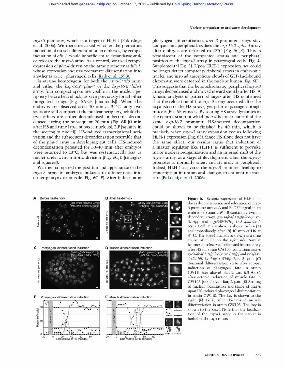
myo-3 promoter, which is a target of HLH-1 (Fukushigeet al. 2006). We therefore asked whether the prematureinduction of muscle differentiation in embryos, by ectopicinduction of hlh-1, would be sufficient to decondense and/or relocate the myo-3 array. As a control, we used ectopicexpression of pha-4 driven by the same promoter as hlh-1,whose expression induces premature differentiation intoanother fate; i.e., pharyngeal cells (Kalb et al. 1998).
In strains homozygous for both the myo-3Trfp arrayand either the hsp-16.2Tpha-4 or the hsp-16.2Thlh-1array, four compact spots are visible at the nuclear pe-riphery before heat shock, as seen previously for all otherintegrated arrays (Fig. 6AE,F [diamonds]). When theembryos are observed after 10 min at 34°C, only twospots are still compact at the nuclear periphery, while thetwo others are either decondensed or become decon-densed during the subsequent 20 min (Fig. 6B [0 minafter HS and time lapse of boxed nucleus], E,F [squares inthe scoring of nuclei]). HS-induced transcriptional acti-vation and the subsequent decondensation resemble thatof the pha-4 array in developing gut cells. HS-induceddecondensation persisted for 30–40 min after embryoswere returned to 23°C, but was systematically lost asnuclei underwent mitotic division (Fig. 6C,E [trianglesand squares]).
We then compared the position and appearance of themyo-3 array in embryos induced to differentiate intoeither pharynx or muscle (Fig. 6C–F). After induction of
pharyngeal differentiation, myo-3 promoter arrays staycompact and peripheral, as does the hsp-16.2Tpha-4 arrayafter embryos are returned to 23°C (Fig. 6C,E). This isreminiscent of the compacted status and peripheralposition of the myo-3 array in pharyngeal cells (Fig. 4;Supplemental Fig. 5). Upon HLH-1 expression, we couldno longer detect compact peripheral arrays in embryonicnuclei, and instead amorphous clouds of GFP-LacI-boundchromatin were detected in the nuclear lumen (Fig. 6D).This suggests that the heterochromatic, peripheral myo-3arrays decondensed and moved inward shortly after HS. Akinetic analysis of pattern changes after HS confirmedthat the relocation of the myo-3 array occurred after theexpansion of the HS arrays, yet prior to passage throughmitosis (Fig. 6F, crosses). By scoring HS array dynamics inthe control strain in which pha-4 is under control of thesame hsp-16.2 promoter, HS-induced decompactioncould be shown to be finished by 40 min, which isprecisely when myo-3 array expansion occurs followingHLH-1 expression (Fig. 6F). Since HS alone does not havethe same effect, our results argue that induction ofa master regulator like HLH-1 is sufficient to provokemajor nuclear reorganization and an internal shift of themyo-3 array, at a stage of development when the myo-3promoter is normally silent and its array is peripheral.Indeed, HLH-1 activates the myo-3 promoter leading totranscription initiation and changes in chromatin struc-ture (Fukushige et al. 2006).
Figure 6. Ectopic expression of HLH-1 in-duces decondensation and relocation of myo-
3 promoter arrays A and B. GFP signal in anembryo of strain GW110 containing two in-dependent arrays: gwIs4[baf-1Tgfp-lacI;myo-
3Trfp] and cgc3595Is[hsp-16.2Tpha-4;rol-
6(su1006)]. The embryo is shown before (A)and immediately after (B) 10 min of HS at34°C. The boxed nucleus is shown as a timecourse after HS on the right side. Similarfeatures are observed before and immediatelyafter HS for strain GW105, containing arraysgwIs4[baf-1Tgfp-lacI;myo-3Trfp] and gvIs[hsp-
16.2Thlh-1;rol-6(su1006)]. Bar, 2 mm. (C)Terminal differentiation state after ectopicinduction of pharyngeal fate in strainGW110 (see above). Bar, 2 mm. (D) As C,after ectopic induction of muscle fate inGW105 (see above). Bar, 2 mm. (E) Scoringof nuclear localization and shape of arraysupon HS-induced pharyngeal differentiationin strain GW110. The key is shown to theright. (F) As E, after HS-induced muscledifferentiation in strain GW105. The key isshown to the right. Note that the localiza-tion of the myo-3 array in the center isheritable through mitosis.
Nuclear reorganization and worm development
GENES & DEVELOPMENT 775
Cold Spring Harbor Laboratory Press on October 17, 2012 - Published by genesdev.cshlp.orgDownloaded from

Small transgenes and large arrays mimic endogenouslocus positioning
To evaluate whether our observations of arrays and trans-
genes are valid for endogenous loci, we performed FISH in
whole C. elegans embryos. It should be noted that FISH
probes encompass 15–30 kb, and that 30 kb of an en-
dogenous 30-nm chromatin spans ;250 nm. Therefore,
‘‘gene-specific’’ FISH also labels the flanking chromatin,
making it difficult to discriminate what determines the
spatial position of a locus. Taking this into account, we
chose three regions in the genome with distinct average
expression levels over a 30-kb segment (based on pub-
lished whole-embryo expression data) (Baugh et al. 2005).
The first two regions encompass baf-1 and tbb-1, both
ubiquitously active housekeeping genes that are tran-
scribed throughout early embryonic development, andboth found in active regions of the genome (Fig. 7A, baf-1and tbb-1 regions). The third region contains the develop-mentally regulated gene pha-4, which is silent in earlyembryos (20- to 50-cell stage) and activated in pharyngealprecursor cells (Fig. 7A, pha-4 region). Importantly, wenote that the genes around the endogenous pha-4 locus arealso largely silent in embryos (Baugh et al. 2005).
We performed FISH for baf-1 and tbb-1 regions inembryos, and quantified their positions relative to theNE (Fig. 7B). Both were distributed randomly, whichagrees with the random distribution of small transgenesin early embryos (cf. Figs. 1E and 7C). When we scored forthe endogenous pha-4 region in very early embryoniccells where it is inactive, the locus was enriched at thenuclear periphery (zone 1 in >80% of cells) (Fig. 7D). The
Figure 7. Localization of endogenous genes by FISH relative to the NE. (A) Heat maps of the transcription of the genomic regionsencompassing 60 kb around the baf-1, tbb-1, and pha-4 genes, respectively. (Green) High RNA levels; (red) no or very little RNA, basedon Baugh et al. (2005). (B) Partial 3D projection of an immunofluorescence/FISH experiment in strain SM469 expressing gfp-h2b undertranscriptional control of the pha-4 promoter from an array (pxIs6[pha-4TgfpTh2b]) with a probe recognizing the genomic pha-4 locus.(Green) Anti-GFP; (red) pha-4 FISH; (blue) DAPI. Bar, 1 mm. (C) Quantification of FISH signal position for the baf-1 and tbb-1 activehousekeeping gene regions in 20- to 50-cell-stage wild-type embryos. Nuclear localization was scored as described in Figure 1D. (D)Quantification of FISH signal position for the pha-4 locus for early embryos (left panel, x2 test vs. random: P < 10�16) and in laterembryos (right panel) with active pha-4 promoter (pha-4, active), as judged by the presence of the GFP-H2B signal, or with silent pha-4
(pha-4, inactive, no GFP signal). GFP-H2B is under control of the full-length pha-4 promoter in an array in strain SM469. x2 versusrandom: P = 0.6 (active pha-4), P < 10�16 (inactive pha-4). (E) Partial 3D projection of a FISH experiment in intestinal cells and headnuclei (to scale) in wild-type N2 adult worms with a probe recognizing the genomic pha-4 locus. Several spots are observed in intestinalcells, as this tissue is polyploid. (Red) pha-4 FISH; (blue) Hoechst. Bar, 1 mm. (F) Quantification of FISH signal in intestinal nuclei (whitebars) and head nuclei excluding those inside the pharynx (gray bars) of wild-type adult worms, using the method described in Figure 1G.Kolgomorov-Smirnov between head and intestinal distributions: P < 10�16. (G) Same data as in F, quantified using the zoning methoddescribed in Figure 1D. x2 versus random: P < 10�16 (head nuclei), P < 0.07 (intestine nuclei).
Meister et al.
776 GENES & DEVELOPMENT
Cold Spring Harbor Laboratory Press on October 17, 2012 - Published by genesdev.cshlp.orgDownloaded from

pha-4 promoter-bearing transgene was not excluded fromthe NE in early embryos, but rather showed a randomdistribution (Fig. 1E). Thus, it is likely that the moreperipheral position of the genomic region containing pha-4reflects a chromosomal context rich in repressed genes.
Relocalization of the pha-4 locus in pharyngealprecursor cells
We could nonetheless examine by FISH whether theendogenous pha-4 domain changes position in responseto developmentally induced expression. Genomic FISH isfairly efficient in embryos, allowing us to examine theposition of the endogenous pha-4 gene in two states ofactivity in late embryos (Mango et al. 1994). To identifythe cells that express pha-4, we made use of a strainexpressing GFP-histone from the complete pha-4 pro-moter (Fig. 7B, green nuclei; Horner et al. 1998). In thisstrain, we performed FISH using a probe specific for theendogenous pha-4 locus that does not detect the trans-genic pha-4 promoter driving gfp-h2b. Quantification ofthe radial distribution of the FISH signal showed that thelocus was distributed randomly with respect to the NE incells that expressed pha-4 (Fig. 7D, pha-4, later embryos,white bars). In embryonic cells in which pha-4 wasinactive, the locus was significantly peripheral (64% inzone 1) (Fig. 7D, later embryos, gray bars).
To explore whether the activity-correlated localiza-tion of endogenous pha-4 was maintained throughoutworm development, we performed FISH in dissected adultintestine and head. Figure 7E shows representative pic-tures of nuclei from these two tissues. We note that adultintestinal cells become polyploid, resulting in more thantwo signals per nucleus (Hedgecock and White 1985). Asobserved for differentiating embryos, in adult nonpha-ryngeal head cells in which pha-4 is inactive, this genomesegment is highly enriched at the NE (Fig. 7E–G, headnuclei, nonpharyngeal). In contrast, in gut cell nuclei ,thepha-4 genomic region shifted to an internal site (>40% are>1200 nm from the NE) (Fig. 7F). These results are fullyconsistent with our conclusions based on developmen-tally regulated transgenes: The transgene behavior allowsus to propose that developmentally regulated promotersthemselves are sufficient to promote a shift to the nuclear
interior upon gene activation. Tissue-specific inactivationof such a locus may also account for its peripheralsequestration, while the NE-association of a heterochro-matic large array is a default state that can be overcome byactivation of a tissue-specific promoter. We summarizeour conclusions in Figure 8.
Discussion
We developed a system to analyze the subnuclear posi-tion of genes and genome segments during the develop-ment of C. elegans. We find that tissue-specific promotersare able to determine gene localization by shifting therelevant DNA to either an internal position when activeor the nuclear periphery when silenced (Figs 1F,H, 4E).This phenomenon is confirmed for three different pro-moters in differentiated tissues derived from three differ-ent germ layers of the same organism. The subnuclearpositions scored cannot simply reflect the site of trans-gene integration, since each construct behaves in a tissue-specific manner. In contrast, there is little detectablenuclear compartmentalization for small transgenic arraysin early embryos, although the embryonic NE has thepotential to sequester an endogenous silent domain(pha-4) and artificial heterochromatic domains (large arrays).This latter phenomenon is array size-dependent and canbe correlated with the deposition of histone marks typicalfor both constitutive and facultative heterochromatin(H3K9me3 and H3K27me3) (Figs. 2F, 3). Importantly, ourstudy allows us to determine a hierarchy of subnuclearlocalization signals: Heterochromatic anchorage can beovercome by the activation of a developmentally regu-lated promoter, even though transcription, per se (e.g., ofhousekeeping promoters sur-5 and baf-1) (Fig. 3F,H), is notsufficient to trigger the release of a heterochromatic locusfrom the NE. This latter phenomenon is reminiscent ofsituations in both yeast and human cells, in which theartificial tethering of integrated sequences to the NE wasfound to reduce expression of only some promoters(Towbin et al. 2009) .
That subnuclear positioning can enhance both herita-ble repression and transcriptional induction of specificgenes has been demonstrated most clearly in buddingyeast (for review, see Spector 2003; Taddei et al. 2004;
Figure 8. Model summarizing gene position-ing during differentiation Two major forcesdrive tissue-specific subnuclear organization ofthe worm genome: repeat-induced heterochro-matin, which associates with the NE, andtissue-specific promoters that shift inward in adominant fashion when they are activated.Tissue-specific promoters shift in a nondomi-nant manner to the NE in cells in which theyare inactive.
Nuclear reorganization and worm development
GENES & DEVELOPMENT 777
Cold Spring Harbor Laboratory Press on October 17, 2012 - Published by genesdev.cshlp.orgDownloaded from

Akhtar and Gasser 2007). Yet to date it is unclear whatrenders a promoter sensitive or insensitive to modulationby subnuclear positioning. Here, by using small transgenearrays carrying developmentally regulated promoters andlarge heterochromatic arrays, we were able to identifysome of the features that influence gene positioningduring development. It is important to examine nuclearorganization in the context of the whole organism, sincegenetic programming is not established by transcriptionfactors alone, but in concert with cytoskeletal signalsthat stem from the tissue environment (Zhang et al. 2001;Mislow et al. 2002).
High gene copy number arrays as a modelfor heterochromatin–NE interaction
We show that integrated transgenic arrays accumulaterepressive histone marks (H3K9me3 and H3K27me3) andbecome peripherally sequestered in a size-dependent man-ner. Up to 50 promoter copies do not become ‘‘heterochro-matic,’’ while an integrated array of 500 copies does. Itsassociation with the NE is consistent with data showinga peripheral enrichment of gene-poor and underacetylatedgenomic domains in cultured mammalian cells, flies, andyeast (O’Keefe et al. 1992; Akhtar and Gasser 2007), yet itdoes not answer the question of whether heterochromatin-linked modifications cause the anchorage. The disperseddistribution of H3K9me3 and H3K27me3 in early embryoniccells makes it unlikely that either modification alone issufficient to mediate binding to the NE in embryonic cells.For the same reason, it is unlikely that the H3K9me3 ligandHPL-2 (one of two worm HP1 homologs) can mediate theinteraction (F Palladino, P Meister, and SM Gasser, pers.comm.). Nonetheless, histone methylation events and thepackaging of the sequence within large repetitive arraysdistinguish large from small arrays.
Preliminary RNAi assays against single genes of theSuv3–9 SET domain family (SET-6/11/13/15/20/21/23)(Andersen and Horvitz 2007) did not abolish the periph-eral sequestration of large arrays in C. elegans (BDTowbin, unpubl.), although H3K9me3 levels were reducedto 10% of wild-type levels in embryos lacking met-2, theSetDB1 methyltransferase homolog (Andersen andHorvitz 2007). The mutation of mes-2, an Ezh2 methyl-transferase homolog that deposits H3K27me3, also had noeffect on either array positioning in embryos or the de-gree of compaction of large arrays (Capowski et al. 1991;Supplemental Fig. 7). This result is in contrast to a pre-vious study (Yuzyuk et al. 2009) that reported an effect onextrachromosomal DNA array structure. This discrep-ancy may be attributed to the nature of the arrays used inthe two studies. Our integrated arrays are of low sequencecomplexity, containing only C. elegans promoters, genesencoding fluorescent proteins, and a small amount ofplasmid, while the previous study diluted plasmids ;1/10with herring sperm DNA during injection. Moreover, wemonitor mitotically stable integrated arrays, whileYuzyuk et al. (2009) examined extrachromosomal arrays.
We note that the failure to see release from the NE afterdown-regulation of a single methyltransferase does not
argue that histone marks have no role in either thepositioning or compaction of chromatin. Rather, it mayindicate that redundant pathways are involved in arraylocalization, consistent with the presence of multipletypes of histone methylation on the transgenes we scored.If this were true, then double or even triple mutationsin histone methyltransferases may be required to seechanges in heterochromatin anchoring. Similarly, wetested single mutants defective for key enzymes of theRNAi machinery (rde-1, rde-3, and mut-7), and found thatthey had no obvious effect on large array localization orcompaction (BD Towbin, unpubl.). This is somewhatsurprising, since part of the RNAi machinery has beenimplicated in large transgene array silencing in the germ-line (Grishok et al. 2005; Kim et al. 2005). Again, this maysimply reflect the redundancy of mechanisms that lead toa peripheral gene position.
Tissue-specific promoter relocation correlateswith cell differentiation
A major insight arising from our study is that tissue-specific promoters are a major force in defining develop-mental stage-specific nuclear organization. They not onlydrive the internal positioning of developmentally regu-lated promoters, but override the peripheral tethering oflarge arrays (Figs. 3F, 4E). Housekeeping gene promotersare not able to do this. Taken at face value, this impliesthat factors bound to developmentally controlled pro-moters are able to mediate gene repositioning. Similarconclusions were based on a FISH study that followed theb-globin locus during fetal liver differentiation (Ragoczyet al. 2006). In this study, the initially peripheral silentlocus moved toward the nuclear lumen upon activationin a manner dependent on its locus control region. Theassociation of active RNA polymerase II (PolII) andevidence of active transcription were both observed priorto relocation, suggesting that transcription precedes andperhaps promotes the subnuclear repositioning. In oursystem as well, array expansion and the complete in-ternal shift of pha-4 promoter arrays occurred signifi-cantly after the initiation of transcription (Fig. 5D;Azzaria et al. 1996).
Once cell type differentiation was completed in L1larvae, large arrays bearing the activated developmentallyregulated promoter remained internal and compact,whether in hypodermal, intestine, or muscle cells (Figs.4D, 5D,F). We show that these compact but internalizedarrays are still actively transcribed (Supplemental Figs. 4,5). Moreover, the endogenous pha-4 locus was also shownto remain internal in adult gut cells (Fig. 7E–G). A com-pact yet active gene configuration was also reported fortranscriptional induction by a targeted estrogen receptor(ER) (Nye et al. 2002). In this study, the tethering of ER toan array of genes could promote chromatin decondensa-tion, while estradiol-mediated activation of the ER pro-moted a partial refolding of the array (Nye et al. 2002;Carpenter et al. 2004). Nye et al. (2002) interpret theseresults as showing a reduction of transcriptional activa-tion. In view of our results, which document continued
Meister et al.
778 GENES & DEVELOPMENT
Cold Spring Harbor Laboratory Press on October 17, 2012 - Published by genesdev.cshlp.orgDownloaded from

transcription of the compacted array, the refolding of aninternalized array could reflect establishment of a stabi-lized active state. Whereas decondensation may reflectchromatin opening to allow increased transcription factorbinding, a stabilized compact state of the locus mayreflect a state of equilibration in which the promoter as-sumes a constitutively active conformation. Intriguingly,the mouse IgK locus also appeared to contract duringprogrammed differentiation of T cells (Skok et al. 2007).Stable localization to the nuclear lumen may be equiva-lent to the organization of activated genes in ‘‘transcrip-tion factories’’ (Fraser and Bickmore 2007).
Unfulfilled potential for gene positioningin early embryonic nuclei
Intriguingly, there is little or no positioning of silenttransgenes in early embryonic nuclei, while in differen-tiated tissues, the same constructs shift to the nuclearperiphery when inactive. Nonetheless, large heterochro-matic arrays show that the machinery necessary forperinuclear tethering is present in early embryos. Anearlier report showed that there are distinct lamin-associated domains on chromosomes in early fly embryos(Pickersgill et al. 2006), and that lamin interaction atsome of these sites is lost during differentiation. Thisstudy used DNA methylation by a lamin–DNA methyl-ase fusion (Dam-ID), which scores both transient andstable DNA–lamin interactions, whereas quantitativeimaging approaches monitor steady-state situations. In-deed, imaging data from the same tissue in flies did notcompletely agree with the Dam-ID results (Pickersgillet al. 2006). Thus, based on data published to date, it isunclear whether early embryos in other organisms alsohave reduced levels of nuclear organization.
Nonetheless, in worms, we clearly document a pro-gressive establishment of gene positioning during cell-type commitment and differentiation. Analysis of smalltransgenes carrying the pha-4 promoter shows that, asearly as the 100-cell stage (8E), there is specific enrich-ment of the transgene in the nuclear interior in cellswithin which the promoter is active, or at the peripheryin cells where it is off, while, up to this point, the pha-4transgene was distributed randomly. This transition hasnot been addressed in studies of fly or mammaliandifferentiation. For example, the FACS-sorted fetal livercells used in the Ragoczy study (Ragoczy et al. 2006) hadalready gone through differentiation (being 90% ery-throid) (Trimborn et al. 1999), and studies using ES cellsshowed perinuclear enrichment of the silent b-globin andIgH loci prior to induced differentiation.
Active gene positioning dominates over repressedgene sequestration
A key question on gene positioning is how flankingsequences influence the position of a given locus. Thisaspect renders the interpretation of FISH data, whichgenerally probe large domains, rather complicated. Usinga 0.55-Mb region, Zink et al. (2004) studied localization of
three adjacent genes with different transcriptional status,and found that each gene behaved independently of theothers with respect to positioning in a transcription-dependent manner. However, it was not asked whetheractive or inactive loci were dominant for positioning.Here we examined the effect of having two differentiallyexpressed promoters adjacent within a transgene array. Indifferentiated tissues, we find that position is determinedin a dominant fashion by the active promoter: The activetissue-specific promoter shifts the silent one away fromthe nuclear periphery (e.g., unc-119). Given that a smalltransgene containing only the unc-119 promoter is NE-associated in the differentiated cells where it is not ex-pressed, we propose that peripheral sequestration is a de-fault position for chromatin lacking activated promoters,but only once cell fate decisions have been made. Thiswas also proposed for inactive erythroid-specific genesin precursor cells (for review, see Kosak and Groudine2004a; Schneider and Grosschedl 2007).
Based on our results, we propose a model (Fig. 8) inwhich developmentally controlled promoters are firstrendered accessible to the transcriptional machinery bya master regulator that opens the promoter for transcrip-tion. An ensuing high-level transcription leads to theunfolding of the chromatin domain, and, finally, depend-ing on the constellation of factors bound to the promoter,an active tissue-specific gene may shift away from theperiphery in the appropriate cell. The gene is thenretained in the nuclear lumen, possibly by interactionwith the transcriptional machinery or other intranuclearcomponents. Importantly, this can be induced by ectopicinduction of a master regulator like HLH-1, althoughtranscription driven from a housekeeping promoter doesnot produce the same effect. Given that we see a similarshift of genes in all tissues, even though the master reg-ulators for each differentiation event are unique, we pro-pose that promoter-bound remodeling factors recruited toestablish tissue-specific expression may drive cell type-specific nuclear organization.
Our system provides a powerful genetic model inwhich to study differentiation-regulated aspects of nu-clear organization. It allows RNAi screens to be coupledwith nonlethal mutations to dissect the machinery re-sponsible for the establishment of nuclear organizationdriven by developmentally regulated genes.
Materials and methods
Molecular biology and transgenic strains
The GFP-LacI expression plasmid was created by fusing the gfp
coding sequence from plasmid pPD117 (Fire vector library) to lacI
under transcriptional control of the baf-1 promoter, followed bythe let-858 terminator. qPCR to determine copy number wasperformed as described in the Supplemental Material.
Two types of large arrays were created for expression of theGFP-LacI construct. The first one has a vit-5Tgfp marker anddoes not contain any lacO-binding site. The second one hasa myo-3Trfp marker (2.5-kb promoter region) and a single lacO
site on both plasmids used to create the array, which wasintegrated by X-ray irradiation. Following integration, the strains
Nuclear reorganization and worm development
GENES & DEVELOPMENT 779
Cold Spring Harbor Laboratory Press on October 17, 2012 - Published by genesdev.cshlp.orgDownloaded from

were backcrossed four to eight times to wild-type N2. Othertransgenic strains contain gfpTlmn-1 (Liu et al. 2000) as a low-copy integrant obtained by microparticle bombardment, and twoclassical integrated arrays; namely, a [pha-4TlacZ; rol-6] array[a 9-kb fragment of the pha-4 promoter and a 4-kb fragment ofrol-6(su1006), comprising 2 kb of promoter and 2 kb of codingsequence] (Azzaria et al. 1996) and a pha-4TGFPTh2b array(Horner et al. 1998).
For small bombarded transgenes, the unc-119(ed3) III strain(DP38) or its derivate expressing GFP-LacI was cobombardedwith a lacO repeat construct (pSR1) (Rohner et al. 2008), the unc-
119 rescuing construct, and plasmids driving expression ofmCherry from either a myo-3 (2.5-kb promoter region) or a smallpha-4 promoter (Murray et al. 2008). Strains were backcrossed tounc-119(ed3)III parents following integration.
Microscopy
Live microscopy was carried out on 2% agarose pads supple-mented with 0.1% azide when needed. For microscopy ofembryos and worms, either a spinning disk confocal microscope(Visitron, Puchheim) (Figs. 2C–E, 4–6), a wide-field monochro-mator deconvolution microscope (TillVision,; Grafelfing) (Figs.1B, 2F), or a Deltavision wide-field deconvolution microscope(Deltavision, Applied Precision) (Figs. 1C–H, 3, 7) was used. Foreach picture, a stack with a z-spacing of 0.2 mm was taken. Stackswere aligned and merged using the software Qu (written inMatlab; available on request). 3D reconstructions used Imarissoftware (Bitplane). For quantitative analysis of arrays, trans-genes, and locus position, measurements were made with ImageJusing PointPicker (http://bigwww.epfl.ch/thevenaz/pointpicker).
FISH
For large array FISH, an 800-base-pair (bp) fragment of bacterialbla was obtained by PCR. Probes were labeled using nicktranslation with Alexa-546-modified dUTP (Roche Nick trans-lation kit using Invitrogen Alexa-546 dUTP). FISH was per-formed as follows: Embryos from bleached worms were fixedfor 2 min in 2% paraformaldehyde (PAF) and spread on poly-L-lysine-coated slides. They were freeze-cracked on dry ice beforea 2-min fixation in�20°C methanol. Following fixation, sampleswere rehydrated progressively in 2-min baths of 90%, 70%, 50%,and 25% ethanol. FISH was carried out in a Ventana slideprocessor using the standard protocol from the manufacturer.Briefly, DNA was denatured with HCl and heat before probeaddition. Probe and samples were incubated for 1 h to overnightat 37°C before stringent washes in SSC buffers. Samples wereDAPI-stained quickly prior to mounting in ProLong Gold anti-fade (Invitrogen).
For single-gene FISH, cosmids F38A6 (cut with NotI/KpnI),B0464, and W09D10 (Sanger Center) covering pha-4, baf-1, andtbb-1 genes, respectively, were labeled with Alexa-546 using theFISHTag kit (Invitrogen). Staining procedure was carried out as inCsankovszki et al. (2004) except that slides were post-fixeddirectly after freeze-cracking to preserve nuclear integrity. Imageacquisition was carried out on a Deltavision RT wide-fieldmicroscope, and position scoring relative to nuclear peripherywas done with ImageJ software.
Immunofluorescence staining
For FISH/immunofluorescence analysis of GFP expression, em-bryos were first immunostained with monoclonal anti-GFP(Roche) and Alexa-488 goat anti-mouse. Stained embryos werepost-fixed for 10 min in 4% PAF before the FISH. For histone
modification mark staining, embryos from bleached worms werefreeze-cracked on dry ice, fixed in methanol for 30 sec, and post-fixed in 1% PAF for 2 min. For lamin/GFP staining, embryoswere fixed for 5 min in 2% PAF before freeze-cracking, followedby post-fixation/dehydration in 70%/80%/95%/100% ethanol.For both, after three washes in PBS 0.25% Triton X-100 (PBS-T),slides were blocked in PBS 0.5% BSA before overnight incuba-tion with primary antibody (anti-H3K9me3, H3K27me3 [gifts of T.Jenuwein]; anti-LMN-1 [gift of Y. Gruenbaum]; H3K4me3 [Up-state Biotechnologies 07-473]; anti-GFP [Roche 11814460001]) at4°C. After three washes with PBS-T, samples were incubated for30 min with secondary antibodies (Alexa-488 anti-mouse,Alexa546 anti-rabbit; Invitrogen) at room temperature beforefinal washes and DNA staining with Hoechst 33258.
Acknowledgments
We thank Y. Gruenbaum for the baf-1 promoter, worm strains,and helpful discussions; T. Jenuwein for antibodies; and J.D.McGhee and the CGC for strains. We are grateful to M. Thomasfor excellent technical assistance; the FMI C. elegans laborato-ries for guidance; and J. Alcedo, R. Ciosk, D. Schubeler, A. Peters,and R. Terranova for helpful comments on the manuscript. Thiswork was supported by the EU NOE ‘‘Epigenome,’’ the SNSFNCCR ‘‘Frontiers in Genetics,’’ the Novartis Research Founda-tion, and a fellowship from the Human Frontiers Science Pro-gram to B.L.P.
References
Akhtar A, Gasser SM. 2007. The nuclear envelope and tran-
scriptional control. Nat Rev Genet 8: 507–517.Andersen EC, Horvitz HR. 2007. Two C. elegans histone
methyltransferases repress lin-3 EGF transcription to inhibit
vulval development. Development 134: 2991–2999.Azzaria M, Goszczynski B, Chung MA, Kalb JM, McGhee JD.
1996. A fork head/HNF-3 homolog expressed in the pharynx
and intestine of the Caenorhabditis elegans embryo. Dev
Biol 178: 289–303.Baugh LR, Hill AA, Claggett JM, Hill-Harfe K, Wen JC, Slonim
DK, Brown EL, Hunter CP. 2005. The homeodomain protein
PAL-1 specifies a lineage-specific regulatory network in the
C. elegans embryo. Development 132: 1843–1854.Belmont AS. 2001. Visualizing chromosome dynamics with
GFP. Trends Cell Biol 11: 250–257.Bessler JB, Andersen EC, Villeneuve AM. 2010. Differential
localization and independent acquisition of the H3K9me2
and H3K9me3 chromatin modifications in the Caenorhab-
ditis elegans adult germ line. PLoS Genet 6: e1000830. doi:
10.1371/journal.pgen.1000830.Boyer LA, Mathur D, Jaenisch R. 2006. Molecular control of
pluripotency. Curr Opin Genet Dev 16: 455–462.Brown KE, Amoils S, Horn JM, Buckle VJ, Higgs DR,
Merkenschlager M, Fisher AG. 2001. Expression of a- and
b-globin genes occurs within different nuclear domains in
haemopoietic cells. Nat Cell Biol 3: 602–606.Capowski EE, Martin P, Garvin C, Strome S. 1991. Identification
of grandchildless loci whose products are required for normal
germ-line development in the nematode Caenorhabditis
elegans. Genetics 129: 1061–1072.Carmi I, Kopczynski JB, Meyer BJ. 1998. The nuclear hormone
receptor SEX-1 is an X-chromosome signal that determines
nematode sex. Nature 396: 168–173.Carpenter AE, Ashouri A, Belmont AS. 2004. Automated
microscopy identifies estrogen receptor subdomains with
Meister et al.
780 GENES & DEVELOPMENT
Cold Spring Harbor Laboratory Press on October 17, 2012 - Published by genesdev.cshlp.orgDownloaded from

large-scale chromatin structure unfolding activity. Cytome-
try A 58: 157–166.Chuang CH, Carpenter AE, Fuchsova B, Johnson T, de Lanerolle
P, Belmont AS. 2006. Long-range directional movement of aninterphase chromosome site. Curr Biol 16: 825–831.
Csankovszki G, McDonel P, Meyer BJ. 2004. Recruitment andspreading of the C. elegans dosage compensation complexalong X chromosomes. Science 303: 1182–1185.
Dietzel S, Zolghadr K, Hepperger C, Belmont AS. 2004. Differ-ential large-scale chromatin compaction and intranuclearpositioning of transcribed versus non-transcribed transgenearrays containing b-globin regulatory sequences. J Cell Sci
117: 4603–4614.Fraser P, Bickmore W. 2007. Nuclear organization of the genome
and the potential for gene regulation. Nature 447: 413–417.Fukushige T, Krause M. 2005. The myogenic potency of HLH-1
reveals wide-spread developmental plasticity in early C.elegans embryos. Development 132: 1795–1805.
Fukushige T, Brodigan TM, Schriefer LA, Waterston RH, KrauseM. 2006. Defining the transcriptional redundancy of earlybodywall muscle development in C. elegans: Evidence fora unified theory of animal muscle development. Genes &
Dev 20: 3395–3406.Gartenberg MR, Neumann FR, Laroche T, Blaszczyk M, Gasser
SM. 2004. Sir-mediated repression can occur independentlyof chromosomal and subnuclear contexts. Cell 119: 955–967.
Gasser SM. 2002. Visualizing chromatin dynamics in interphasenuclei. Science 296: 1412–1416.
Gonzalez-Serricchio AS, Sternberg PW. 2006. Visualization of C.
elegans transgenic arrays by GFP. BMC Genet 7: 36. doi:10.1186/1471-2156-7-36.
Grishok A, Sinskey JL, Sharp PA. 2005. Transcriptional silencingof a transgene by RNAi in the soma of C. elegans. Genes &
Dev 19: 683–696.Hedgecock EM, White JG. 1985. Polyploid tissues in the
nematode Caenorhabditis elegans. Dev Biol 107: 128–133.Hediger F, Taddei A, Neumann FR, Gasser SM. 2004. Methods
for visualizing chromatin dynamics in living yeast. Methods
Enzymol 375: 345–365.Hewitt SL, High FA, Reiner SL, Fisher AG, Merkenschlager M.
2004. Nuclear repositioning marks the selective exclusion oflineage-inappropriate transcription factor loci during Thelper cell differentiation. Eur J Immunol 34: 3604–3613.
Horner MA, Quintin S, Domeier ME, Kimble J, Labouesse M,Mango SE. 1998. pha-4, an HNF-3 homolog, specifies pha-ryngeal organ identity in Caenorhabditis elegans. Genes &
Dev 12: 1947–1952.Hsieh J, Fire A. 2000. Recognition and silencing of repeated
DNA. Annu Rev Genet 34: 187–204.Janicki SM, Tsukamoto T, Salghetti SE, Tansey WP, Sachidanandam
R, Prasanth KV, Ried T, Shav-Tal Y, Bertrand E, Singer RH, et al.2004. From silencing to gene expression: Real-time analysis insingle cells. Cell 116: 683–698.
Kalb JM, Lau KK, Goszczynski B, Fukushige T, Moons D,Okkema PG, McGhee JD. 1998. pha-4 is Ce-fkh-1, a forkhead/HNF-3a,b,g homolog that functions in organogenesisof the C. elegans pharynx. Development 125: 2171–2180.
Kaltenbach L, Horner MA, Rothman JH, Mango SE. 2000. TheTBP-like factor CeTLF is required to activate RNA poly-merase II transcription during C. elegans embryogenesis.Mol Cell 6: 705–713.
Kim JK, Gabel HW, Kamath RS, Tewari M, Pasquinelli A, RualJF, Kennedy S, Dybbs M, Bertin N, Kaplan JM, et al. 2005.Functional genomic analysis of RNA interference in C.
elegans. Science 308: 1164–1167.
Kosak ST, Groudine M. 2004a. Form follows function: The
genomic organization of cellular differentiation. Genes &
Dev 18: 1371–1384.Kosak ST, Groudine M. 2004b. Gene order and dynamic do-
mains. Science 306: 644–647.Kosak ST, Skok JA, Medina KL, Riblet R, Le Beau MM, Fisher
AG, Singh H. 2002. Subnuclear compartmentalization of
immunoglobulin loci during lymphocyte development. Sci-
ence 296: 158–162.Liu J, Rolef Ben-Shahar T, Riemer D, Treinin M, Spann P, Weber
K, Fire A, Gruenbaum Y. 2000. Essential roles for Caeno-
rhabditis elegans lamin gene in nuclear organization, cell
cycle progression, and spatial organization of nuclear pore
complexes. Mol Biol Cell 11: 3937–3947.Maduro M, Pilgrim D. 1995. Identification and cloning of unc-
119, a gene expressed in the Caenorhabditis elegans nervous
system. Genetics 141: 977–988.Mahy NL, Perry PE, Bickmore WA. 2002. Gene density and
transcription influence the localization of chromatin outside
of chromosome territories detectable by FISH. J Cell Biol
159: 753–763.Mango SE, Lambie EJ, Kimble J. 1994. The pha-4 gene is required
to generate the pharyngeal primordium of Caenorhabditis
elegans. Development 120: 3019–3031.Meister P, Gehlen L, Varela E, Kalck V, Gasser SM. 2010.
Visualizing yeast chromosomes and nuclear architecture. In
Methods in enzymology (ed. J Weissman et al.), pp. 537–569.
Elsevier, New York.Mislow JM, Kim MS, Davis DB, McNally EM. 2002. Myne-1,
a spectrin repeat transmembrane protein of the myocyte
inner nuclear membrane, interacts with lamin A/C. J Cell
Sci 115: 61–70.Mohn F, Schubeler D. 2009. Genetics and epigenetics: Stability
and plasticity during cellular differentiation. Trends Genet
25: 129–136.Muller WG, Walker D, Hager GL, McNally JG. 2001. Large-scale
chromatin decondensation and recondensation regulated by
transcription from a natural promoter. J Cell Biol 154: 33–48.Murray JI, Bao Z, Boyle TJ, Boeck ME, Mericle BL, Nicholas TJ,
Zhao Z, Sandel MJ, Waterston RH. 2008. Automated analysis
of embryonic gene expression with cellular resolution in C.
elegans. Nat Methods 5: 703–709.NyeAC,RajendranRR,StenoienDL,ManciniMA,Katzenellenbogen
BS, Belmont AS. 2002. Alteration of large-scale chromatin
structure by estrogen receptor. Mol Cell Biol 22: 3437–3449.O’Keefe RT, Henderson SC, Spector DL. 1992. Dynamic organi-
zation of DNA replication in mammalian cell nuclei: Spatially
and temporally defined replication of chromosome-specific
a-satellite DNA sequences. J Cell Biol 116: 1095–1110.Pickersgill H, Kalverda B, de Wit E, Talhout W, Fornerod M, van
Steensel B. 2006. Characterization of the Drosophila mela-
nogaster genome at the nuclear lamina. Nat Genet 38: 1005–
1014.Praitis V, Casey E, Collar D, Austin J. 2001. Creation of low-
copy integrated transgenic lines in Caenorhabditis elegans.
Genetics 157: 1217–1226.Ragoczy T, Bender MA, Telling A, Byron R, Groudine M. 2006.
The locus control region is required for association of the
murine b-globin locus with engaged transcription factories
during erythroid maturation. Genes & Dev 20: 1447–1457.Robinett CC, Straight A, Li G, Willhelm C, Sudlow G, Murray A,
Belmont AS. 1996. In vivo localization of DNA sequences and
visualization of large-scale chromatin organization using lac
operator/repressor recognition. J Cell Biol 135: 1685–1700.
Nuclear reorganization and worm development
GENES & DEVELOPMENT 781
Cold Spring Harbor Laboratory Press on October 17, 2012 - Published by genesdev.cshlp.orgDownloaded from

Rohner S, Gasser SM, Meister P. 2008. Modules for cloning-freechromatin tagging in Saccharomyces cerevisiae. Yeast 25:235–239.
Sassi HE, Renihan S, Spence AM, Cooperstock RL. 2005. GeneCATCHR—gene cloning and tagging for Caenorhabditiselegans using yeast homologous recombination: A novelapproach for the analysis of gene expression. Nucleic Acids
Res 33: e163. doi: 10.1093/nar/gni164.Schneider R, Grosschedl R. 2007. Dynamics and interplay of
nuclear architecture, genome organization, and gene expres-sion. Genes & Dev 21: 3027–3043.
Schuettengruber B, Chourrout D, Vervoort M, Leblanc B,Cavalli G. 2007. Genome regulation by polycomb andtrithorax proteins. Cell 128: 735–745.
Skok JA, Gisler R, Novatchkova M, Farmer D, de Laat W,Busslinger M. 2007. Reversible contraction by looping of theTcra and Tcrb loci in rearranging thymocytes. Nat Immunol
8: 378–387.Spector DL. 2003. The dynamics of chromosome organization
and gene regulation. Annu Rev Biochem 72: 573–608.Sulston JE, Horvitz HR. 1977. Post-embryonic cell lineages of the
nematode, Caenorhabditis elegans. Dev Biol 56: 110–156.Taddei A, Hediger F, Neumann FR, Gasser SM. 2004. The
function of nuclear architecture: A genetic approach. AnnuRev Genet 38: 305–345.
Tajbakhsh J, Luz H, Bornfleth H, Lampel S, Cremer C, Lichter P.2000. Spatial distribution of GC- and AT-rich DNA se-quences within human chromosome territories. Exp CellRes 255: 229–237.
Takizawa T, Gudla PR, Guo L, Lockett S, Misteli T. 2008. Allele-specific nuclear positioning of the monoallelically expressedastrocyte marker GFAP. Genes & Dev 22: 489–498.
Towbin BD, Meister P, Gasser SM. 2009. The nuclear enve-lope—A scaffold for silencing? Curr Opin Genet Dev 19:180–186.
Trimborn T, Gribnau J, Grosveld F, Fraser P. 1999. Mechanismsof developmental control of transcription in the murinea- and b-globin loci. Genes & Dev 13: 112–124.
Tumbar T, Sudlow G, Belmont AS. 1999. Large-scale chromatinunfolding and remodeling induced by VP16 acidic activationdomain. J Cell Biol 145: 1341–1354.
Voss TC, John S, Hager GL. 2006. Single-cell analysis ofglucocorticoid receptor action reveals that stochastic post-chromatin association mechanisms regulate ligand-specifictranscription. Mol Endocrinol 20: 2641–2655.
Williams RR, Azuara V, Perry P, Sauer S, Dvorkina M, JorgensenH, Roix J, McQueen P, Misteli T, Merkenschlager M, et al.2006. Neural induction promotes large-scale chromatinreorganisation of the Mash1 locus. J Cell Sci 119: 132–140.
Yamanaka S. 2009. Elite and stochastic models for inducedpluripotent stem cell generation. Nature 460: 49–52.
Yuzyuk T, Fakhouri TH, Kiefer J, Mango SE. 2009. The poly-comb complex protein mes-2/E(z) promotes the transitionfrom developmental plasticity to differentiation in C. ele-
gans embryos. Dev Cell 16: 699–710.Zhang Q, Skepper JN, Yang F, Davies JD, Hegyi L, Roberts RG,
Weissberg PL, Ellis JA, Shanahan CM. 2001. Nesprins: Anovel family of spectrin-repeat-containing proteins thatlocalize to the nuclear membrane in multiple tissues. J Cell
Sci 114: 4485–4498.Zink D, Amaral MD, Englmann A, Lang S, Clarke LA, Rudolph C,
Alt F, Luther K, Braz C, Sadoni N, et al. 2004. Transcription-dependent spatial arrangements of CFTR and adjacent genesin human cell nuclei. J Cell Biol 166: 815–825.
Meister et al.
782 GENES & DEVELOPMENT
Cold Spring Harbor Laboratory Press on October 17, 2012 - Published by genesdev.cshlp.orgDownloaded from


