Embed Size (px)
Citation preview
NOCTURNAL PRODUCTION OF CONIDIA BY SCLEROSPORA GRAMINICOLA1
By WnxiAM H. WKSTON, Jr.
Formerly Pathologist in Charge of Downy Mildew Investigations, Office of Cereal Inves- tigations, Bureau of Plant Industry y United States Department of Agriculture
INTRODUCTION
While studying the two conidial Sclerosporas (S. philippinensis Weston and 5. spontanea Weston) so destructive to maize in the Philippines, the writer found that in these species conidia are produced only at night when the host surface is covered with dew or other moisture. This naturally raised the question whether S. graminicola (Sacc.) Schroet., the type species of the genus, exhibits the same peculiarity. The fact that it does and the bearing .of this fact on our knowledge of the im- portance and relationship of the conidial stage of the species are dis- cussed in the present paper.
The genus Sclerospora was established by Schroeter 0<?)2 in 1879 on 5. graminicola, a species which since then has been found quite widely distributed throughout temperate and tropical parts of the world, principally on species of Setaria, and rarely on other Gramineae. In the course of its life history 5. graminicola, like most other Perono- sporales, passes through two phases of development: The one, charac- terized by production of immediately germinating conidia, achieving rapid spread ; the other, characterized by formation of resistant oospores, serving to insure survival through such unfavorable conditions as winter and drought. In 5. graminicola the conidial stage, which usually devel- ops first, appearing as a whitish downy growth on the surface of chlo- rotic areas of the host, generally seems to be of short duration, rela- tively inconspicuous, rather rare, and involves but little apparent in- jury to the host. As a result, this stage has not been commonly or abundantly collected and is represented by scanty and unsatisfactory herbarium material. Moreover, it has not been studied in detail. In species of such related genera as Plasmopara (10, 28) and Peronospora (6, 7y S), the conidial condition has been investigated intensively, quan- titative measurements have been made, restrictions of parasitism have been tested, and morphological aspects of all stages of development have been worked out minutely and illustrated fully. In 5. graminicola, however, the conidial stage, save in such publications as those of Butler (2), Kulkarni {12), Shirai (24), et al., has been dismissed summarily with brief diagnostic or morphological descriptions, few measurements, and inadequate^lllustrations.
On the contrary, the oosporic phase of Sclerospora graminicola which follows the conidial with a marked distortion and shredding of the leaves and floral parts of the host, is persistent, conspicuous, abundant, and obviously severely destructive to the host. As a result, it has been collected frequently and in abundance, and is well represented in most
1 Accepted for publication Dec. 14, 1923. a Reference is made by number (italic) to "literature cited," 783-784.
Journal of Agricultural Research, Vol. XXVII, No. 10 'Washington, D. C. Mar. 8, 1924
% Key No. G-353 (771)
772 Journal of Agricultural Research vol. xxvii, No. i©
herbaria; while detailed study has been made on the morphology (2, 12) and cytology (26) of its development, on comparative spore measure- ments (27), and on the relation to the host attacked (2, 22, 23).
Since the genus was founded, nine other species have been described. Of these, Sclerospora macrospora Sacc. (J^), which includes 5. krie- geriana Magn., according to Traverso (27), 5. miscanthi T. Miyake (Jó), S. farlowii Griffiths (9), and 5. magnusiana Sorokin (25), have been reported as yet only in the oosporic condition. The remaining five species are alike in that: First, their conidial phase is predominant and destructive, the oosporic being absent or rare; second, their conidia germinate invariably by hyphae; third, they occur in the oriental Tropics and on members of the tribes Maydeae and Andropogoneae. They are thus in contrast to the type species, 5. graminicola, which is destructive and predominant in its oosporic condition, which typically produces zoospores in conidium germination, and which is of world-wide distribution, mostly on members of the Paniceae.
While investigating two of the conidial Sclerosporas of the Orient, namely, 5. philippinensis and 5. spontanea, species exceedingly de- structive to maize in the Philippine Islands, the writer (29, 30) found that in both, conidia were produced on the leaves of the infected plants only at night when they were covered with a layer of dew. On dewy nights, from about midnight to dawn, the innumerable successively emerging conidiophores formed a conspicuous and luxuriant growth of grayish down, and furnished abundant living material in all stages of development, quite different from the scanty remains, killed and de- formed by drying, that persisted for collection or study during the following day. This nocturnal conidiophore production proved to be a very fixed and characteristic process in 5. philippinensis Weston and 5. spontanea Weston. Examination of the publications of other investigators suggested strongly that this condition holds also in the other conidial Sclerosporas of maize and related crops in the Orient, namely, 5. javanica Palm (17) of Java, 5. mayáis (Rac.) Butl. (j) of India, and ¿>. sacchari T. Miyake (16) of Formosa. Indeed, in the case of the last species, which has been introduced recently into the Philippines (JJ, JJ), there is now no doubt that conidium production is nocturnal, as material collected at intervals during the night and sent to the writer through the cooperation of H. Atherton Lee, of the Bureau of Science in Manila, shows this to be true.
PRODUCTION OF CONIDIA IN SCLEROSPORA GRAMINICOLA
Whether the type species, Sclerospora graminicola,* also produces its conidia only at night is a question that naturally presents itself, in view of the fact that this species differs in the several respects already noted from the conidial Sclerosporas of the Orient which show this peculiarity. Opportunity to investigate this point occurred while the writer was attending the summer conference of cereal pathologists at the University of Minnesota in 1920. On the grounds of the College of Agriculture, not too far from the plant pathology laboratory which Dean Freeman and Doctor Stakman very generously made available for night work, were found several plants of Setaria viridis (L.) Beauv. obviously infected with the conidial stage of Sclerospora graminicola.
The fact that the tissue of these plants was pervaded extensively by vigorous mycelium of the parasite, and the leaf surface was marked by
Mar. s, 1924 Nocturnal Production of Sclerospora 773
whitish patches of shriveled conidiophores persisting on the leaves from previous productive nights, indicated that the plants were still support- ing conidiophore formation under favorable conditions. Frequent care- ful inspection of these plants throughout the day of July 23, however, showed conclusively that, during this daytime at least, no conidiophore production took place. Accordingly, in the late afternoon, the plants were prepared for night study by removing from the leaves all remnants of previous conidiophore production by carefully scrubbing the surface with moist cotton swabs. That night, beginning at about 8 p. m., when dew deposition commenced, the plants were examined at hourly inter- vals and were found, indeed, to produce conidiophores in abundance.
By cutting free-hand sections of living, infected leaves, stripping off bits of epidermis, macerating pieces of tissue, and carefully scraping off the down of conidiophores as it developed, the process of conidiophore emergence and conidium production through the night was followed step by step in detail. After production had ceased, as the plants dried off in the early morning of the next day, periodic examination of the plants was continued until afternoon; and this again brought out the fact that no conidiophores were produced during the day. The fact that in Sclerospora graminicola conidium formation does indeed occur at night was established by these observations ; but in order to supplement them, as they extended over two days and one night only, the most vigorous of the infected Setaria plants were transplanted to Washington, D. C, where they were studied further.
The process of conidiophore production involves, as the writer found in the conidial Philippine Sclerosporas, the following phases: The pre- liminary paling of the leaf areas which are to bear conidia, the quantity of conidia produced on such areas, the length of time this production may go on night after night, the importance of moisture in inducing conidiophore production, the development of conidiophores and conidia thus induced, and the nightly schedule followed by this development. The writer did not have opportunity to work out in detail these several points for 5. graminicola as he did in the Philippine Sclerosporas, but studied chiefly the development of the conidiophores and conidia, the nightly schedule followed by this development, and its dependence on dew or similar moisture. The results of this study follow.
DEVELOPMENT OF THE CONIDIOPHORES
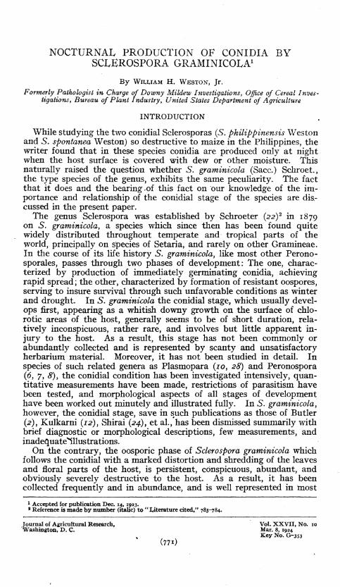
The conidiophores of Sclerospora graminicola develop from the infected Setaria leaves only through the stomata, a large proportion of which may be productive. Consequently, conidiophore production is usually more abundant from the under surface of the leaf where stomata are more numerous, although this abundance is dependent also on other factors such as the amount and distribution of dew on the leaf and the length of time successive nocturnal production from the plant has gone on. Beneath stomata from which conidiophores are to emerge, the air cham- ber is filled with stout, irregularly lobed, densely granular, mycelial branches closely crowded together (PI. 1, A, B), arising from the less conspicuous mycelial strands running between the mesophyll cells. From these substomatal knots, prolongations push through the stomatal slit over which they form a compact group of several minute bulbous out- growths. In some cases there is evidence that the stomatal slit, normally •closed at night, is forcibly pushed open by these emerging branches
774 Journal of Agricultural Research vol. xxvii, NO. »
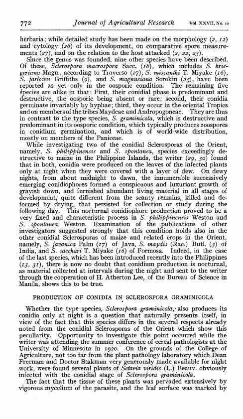
(PI. i, C-F). The bulbous outgrowths next elongate severally to club-shaped stalks which project perpendicularly from the leaf surface and grow rapidly larger (PI. i, G, H). At its swollen apex, each.of these produces successively the two to four stout primary branches (PI. i, I-K) ; while from these latter similarly arise the secondary branches (PL i, L-O). In like manner, successive series of branches develop until eventually the more or less extensive, usually dichotomous, branch system is complete, and the ultimate tips terminate in tapering sterigmata (PL 2, A). The very apex of each sterigma, beginning as a small globular swelling (PL 2, B-E), enlarges gradually, until finally it attains the shape and size of the mature conidium (PL 2, F), which then is separated from the neck of the sterigma by a wall.
Obviously, the development of the conidiophores and conidia in Sclerospora graminicola as just outlined agrees very closely in its several stages with that already established for S. philippinensis and S. spon- tanea. Moreover, S. graminicola apparently agrees with these Philippine species in the way in which its conidia are shed. While studying the Philippine species, the writer became convinced that the conidia do not fall passively from the sterigmata as has been assumed, but rather are forcibly snapped off when the outbulging of the opposed walls of the basal apiculus of the conidium and of the sterigma tip suddenly overcomes the adhesion of their contiguous surfaces. This point has not been settled for S. gramini- cola; but such indications as the outbulging of the formerly flatly apposed walls of the sterigma tip and conidium base when released, lead to the conclusion that the species resembles those of the Philippines in this respect also.
DEPENDENCE ON NOCTURNAI, MOISTURE
The process of conidiophore development in Sclerospora graminicola adheres to a regular schedule. When the infected leaves were wet with dew, at about 8 p. m., the outgrowths had protruded from the stomatal slit at about 11 p. m., and the first conidiophores and conidia were mature at approximately 2 a. m., while others which had begun to develop mean- while, matured successively, so that production continued, reaching its greatest abundance at perhaps 3 a. m., and only ceasing when the dew dried from the leaves in the morning sun. This nightly schedule agrees very closely with that found in 5. philippinensis and S. spontanea (32) under conditions of dew deposition normally obtaining in the Philippines.
Conidiophore production in S. graminicola, as in the Philippine species, is vitally dependent on the presence and persistence of dew or other moisture on the leaves. Infected Setaria plants that were kept dry dur- ing the night never showed conidiophore formation even though their leaves were obviously thoroughly invaded by vigorous mycelium, while similar plants exposed to dew supported abundant conidiophore pro- duction. After the leaf surface of infected plants had been wet with dew for about five hours, production began; if the moisture dried off prematurely, production coincidentally ceased ; if moisture persisted un- duly into the morning, production was thus much prolonged. As in the Philippine Sclerosporas also, conidium production in S. graminicola is very sensitive in its response to moisture changes, so much so that,when studied at the University of Minnesota, the Setaria plants in such differ- ent localities as in a glade among trees, at the edge of a wood on a small hill, and in an open lot, showed slight variations in their schedule of
Mar s, 1924 Nocturnal Production of Sclerospora 775
conidiophore production as a result of local differences in amount or time of dew deposition.
How long Sclerospora graminicola may continue noctumally producing conidia on infected Setaria plants, how tolerant of this continued pro- duction the host may be, and how much of its life span may be taken up
t by the conidium-forming period of the fungus, are points which, in this case, were not worked out in detail as they had been for the Philippine Sclerosporas '{32). In one case, however, evei* though the transplanted Setarias grew very poorly, one plant showed intermittent conidiophore formation for more than two weeks. Also, in the field, there is abundant evidence that the period of conidium production continues much longer than this, even extending over as great a proportion of the total life of the host as did the Sclerosporas studied by the writer (32) on maize in the Philippines.
A study of large numbers of S.-gramm¿c0/a-infected Setaria plants of various ages in the fields near the College of Agriculture at St. Paul, Minn., showed the progress of the disease to be as follows: Production of conidia begins on newty unfolding leaves in symptomatically chlorotic streaks which, by their extent and position even in very young plants, indicate a fundamental systemic infection starting in the young seedling and giving rise to thorough and extensive invasion of the host tissue. The production of conidia continues during each favorable night on these earlier leaves, and also continually starts up afresh on leaves successively appearing. Gradually, as the host matures, conidiophore formation from the host surface is superseded by the development of oogonia on the intramatrical mycelium, beginning in the lower, first unfolded, older leaves, and working slowly upward. The uppermost, latest, and youngest leaves are the last to be affected, and may continue abundant conidi- ophore production until the head, usually deformed, sterile, and vires- cent, is full grown. Finally, however, even these ultimate leaves also are given over to oogonium formation, and show the shredding of tissue which marks the maturity of the oogonial phase of the fungus.
The conidial stage of Sclerospora gramimcola} because its relation to nocturnal moisture has not been understood, has been considered from the time of Schroeter to the present as fleeting, transitory, fugacious, and evanescent. Now, however, that we know the period of conidium pro- duction may be of relatively long duration, we must alter our conception of the evanescence of the conidial stage. It is fugacious in the sense that conidiophore production takes place for only a few hours during the night; but it is decidedly persistent in the sense that this production may go on night after night throughout a-large proportion of the total life of the host.
CONIDIAL SCLEROSPORA GRAMINICOLA COMPARED TOOTHER SPECIES
Material of the conidiophores and conidia of Sclerospora graminicola gathered thus at night during the period of optimum conidiophore production is infinitely more satisfactory for study than the dried and shriveled remnants which persist from previous nights and may be collected during the day. • Consequently, such material gives a somewhat broader conception of the character of the species than that which we gain from the usual descriptions and figures based on dried material, and also permits a more adequate comparison with the conidiophores and conidia of other species.
776 Journal of Agricultural Research vol. xxvii. NO. i
CONIDIOPHORES
In the case of the conidiophores, for example, a study of optimum nocturnal material shows clearly that the conidiophores are larger, more extensively branched, and structurally more complex and elaborate, than most of the illustrations and descriptions indicate. The first description of the conidial stage of this species was published by Schroeter (22) and * the first illustrations were shown by Fischer (5, fig. 71). Together these present a vivid characterization of the conidiophores as decidedly short (about IOOJU) and thick (about i2/¿) with few stubby branches bearing a relatively small number of conidia (about 15). No appreciable departure from this characterization is found in the publications by such subse- quent investigators in Europe as Berlese (J), Malbranche and Letendre ( 14), Massée (15, pi. i, fig. 16), Saccardo (iç), et al. ; or such in America as Saunders {21, pi. 161 fig. 4), Parlow (4), Wilson (jj), etal. ; or in India as Butler (2, fig. 6, 7), and Kulkarni (j2, fig. 1-3). In Japan, how- ever, Shirai (24, fig. 16, 17) described the conidiophores as much larger (100 to 240/x by 12 to 19/x) and with a somewhat more elaborate branching system ; but the fact that he found conidia of two strangely different size-classes (24 to 28.8M by 16.8 to 19.2/11, and 38.4 to 57.6M by 19.2 to 24M) arouses the suspicion that he was not dealing with S. graminicola alone, and to some extent invalidates his characterization of the species.
The writer, after studying the conidiophores of Sclerospora graminicola in the progressive stages of their development under optimum conditions at night, is convinced that the descriptions and drawings of the investi- gators just mentioned are based on nontypical, poorly developed speci- mens, the last belated stragglers of the nocturnal production, caught by the morning sun before they had developed conidia, and dried to a con- dition still less typical and favorable for study. In contrast to such dried specimens, material scraped from Setaria leaves at the time of optimum nocturnal production shows conidiophores that are much larger and better developed than we had been led to believe characteristic of 5. graminicola, and which approximate, in this respect, those of such luxuriant species as the conidial Sclerosporas of the Orient. If, for example, one compares the accompanying figures of 5. graminicola (PI. 2, I, K, O) with those of 5. spontanea (30, pi. 79, fig. A), and S. philip- pinensis (2c, pi. 24, fig. O, pi. 25, fig. B) all of which are of very nearly the same magnification, it is obvious that they show a general resemblance which is not even suggested by previous illustrations.
After comparing them carefully, however, the writer finds that even the largest and most elaborate conidiophores of S. graminicola differ markedly in certain essential features from the conidiophores of such oriental species as 5. philippinensis and 5. spontanea.
First, the total length in these Philippine species is as a rule much greater (260 to 400^) than in the case of 5. graminicola, even the most luxuriant nocturnal material of which has a length of about 150^ with occasional extremes as low as loo/x or as high as 200/x.
Second, the branching system also shows differences which are much more qualitative and absolute than are mere distinctions in size. In the Philippine species, for example, usually three,- sometimes two or four, primary branches of approximately equal size and extent of development all spread out at angles of about 45 o or less from the main axis in very close succession; and all are of equal rank—no one of them being either in direction, position, or extent of growth more to be considered a continua-
Mar. s, 1924 Nocturnal Production of Sclerospora 777
tion of the main axis than any other. As a result of this also, the conidia arising from the branch tips are arranged approximately in a hollow hemisphere. In 5. graminicola, on the contrary, one of the primary branches stands out more or less obviously as a continuation of the main axis (PI. 2, I, K, L, O, Q) both in direction and in extent of growth. From this continuation of the main axis other main, and secondary branches grow out at irregular intervals, usually at angles of 45 o to 90o. As a result, the conidia at the ends of the branches lie more frequently in irregularly disposed groups (PI. 2, I, K, L, P) than in an approxi- mate hemisphere (PI. 2, O, Q).
Third, the sterigmata of 5. graminicola, as a rule, are shorter and more broadly bottle or tenpin shaped (PL 2, N, T, V, W) than are those of the Philippine Sclerosporas. The shape, however, is somewhat de- pendent on position, single sterigmata at the tips of the branches (PL 2, U) being more elongate than those borne in groups of two, three, or four (PL 2, T, J, W). Moreover, the sterigmata, which in the Philippine Sclerosporas almost invariably continue the direction of the branch tip that bears them, may stand out from the branch tip even at right angles in S. graminicola (PL 2, I, J, L). Finally, the lower part of the conidiophore of S. graminicola is not usually marked off by a cross wall into a basal cell or foot cell that is distinct from the superior portion of the main axis. Occasionally such basal cells are encountered (PL 2, K, X, Y); but this condition is the exception; and hence is in distinct contrast to the Philippine species in which it is a character- istic feature. Occasionally, also, the base is distinguished by an incom- plete transverse septum (PL 2, K) or by a decided thickening of its longitudinal wall (PL 2, A, P, Z).
All these distinctions—size of the conidiophores, character and extent of the branch system, form and direction of the sterigmata, and extent of septation or thickening at the base of the main axis—are matters of degree which should be expressed quantitatively to facilitate compari- sons. Yet, even when considered as qualitative differences, they show clearly that the conidiophores of S. graminicola, though they may approxi- mate those of such typically conidial oriental forms as S. philippinensis and 5. spontanea in luxuriance and general appearance, are indeed dis- tinct from them.
CONIDIA
It is the conidia themselves, however, that are the most distinguishing feature of Sclerospora graminicola. These bodies differ markedly from those of other Sclerosporas in size, shape, structure, and germination. The size of the conidia varies, and for adequate presentation requires quantitative expression based on measurements of large numbers of conidia. Moreover, to be ideally satisfactory the conidia should be caught on the slide in dew when snapped off from the conidiophores at maturity and measured at once. Unfortunately, the writer was not able in this case to make all the measurements under these ideal conditions as he did for the Philippine Sclerosporas. Rather, most of the measurements were made from material scraped from abundantly productive leaves at 3 a. m., then killed by Flemming's weaker solution, and mounted in dilute glyc- erin and eosin. A comparison of measurements made thus with the rela- tively few which the writer had opportunity to make from fresh material indicates that if all the measurements had been made under ideal con- ditions the modes of length and of diameter probably would have been
83798—24 5
77« Journal of Agricultural Research vol. xxvn, No. 10
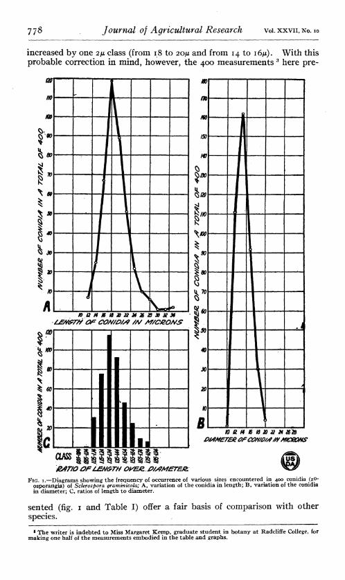
increased by one 2/* class (from 18 to 20/* and from 14 to I6M). With this probable correction in mind, however, the 400 measurements 3 here pre-
no
m
h
!*
i
\
/
i v i s íw « BBHKISaaMXttXXM
imer/v OP COMDM /V M/CSONS
h 40
fB
^
L i_
IE E OASS llik; rS
/SO
m
HO
I»
r r U 70
49
X
20
B NßHtitößäM 2628
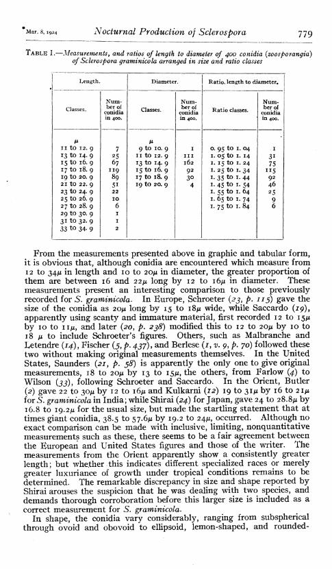
&fr/0 OFIMA/GTH OkWZJ>/S?MJETEJZ FIG. I.—Diagrams showing the frequency of occurrence of various sizes encountered in 400 conidia (zp-
osporangia) of Soleros pora graminicola; A, variation of the conidia in length; B, variation of the conidia in diameter; C, ratios of length to diameter.
sented (fig. i and Table I) offer a fair basis of comparison with other species.
8 The writer is indebted to Miss Margaret Kemp, graduate student in botany at Radcliffe College, for making one half of the measurements embodied in the table and graphs.
Mar. 8, 1924 Nocturnal Production of Sclerospora 779
TABLE I.—Measurements, and ratios of length to diameter of 400 conidia (zoosporangia) of Sclerospora graminicola arranged in size and ratio classes
Length. Diameter. Ratio, length to diameter.
Classes. Num- ber of
conidia in 400.
Classes. Num- ber of
conidia in 400.
Ratio classes. Num- ber of
conidia in 400.
M II to 12. 9 13 to 14. 9 15 to 16. 9 17 to 18. 9 19 to 20. 9 21 to 22. 9 23 to 24. 9 25 to 26. 9 27 to 28. 9 29 to 30. 9 31 to 32. 9 33 to 34. 9
7 25 67
119 89 Si 22 IO 6 I I 2
M 9 to 10. 9
II tO 12. 9 13 to 14. 9 15 to 16. 9 17 to 18. 9 19 to 20. 9
I III 162 92 30
4
0. 95 to 1. 04 1. 05 to 1. 14 i. 15 to I. 24 i. 25 to I. 34 i. 35 to I. 44 I. 45 to I. 54 1. 55 to I. 64 i. 65 to i. 74 1. 75 to I. 84
I 31 75
"S
46 25
9 6
From the measurements presented above in graphic and tabular form, it is obvious that, although conidia are encountered which measure from 12 to 34JU in length and 10 to 20/¿ in diameter, the greater proportion of them are between 16 and 22/1 long by 12 to IôJJL in diameter. These measurements present an interesting comparison to those previously recorded for S. graminicola. In Europe, Schroeter (23, p. 113) gave the size of the conidia as 20/* long by 15 to i8ju wide, while Saccardo (79), apparently using scanty and immature material, first recorded 12 to 15/1 by 10 to iiju, and later (20, p. 238) modified this to 12 to 20ju by 10 to 18 ¡x to include Schroeter's figures. Others, such as Malbranche and Letendre (14), Fischer (5, p. 437), and Berlese (1, v. 9, p. 70) followed these two without making original measurements themselves. In the United States, Saunders {21, p. 58) is apparently the only one to give original measurements, 18 to 20/* by 13 to 15/x, the others, from Farlow (4) to Wilson (JJ), following Schroeter and Saccardo. In the Orient, Butler {2) gave 22 to 30JU by 12 to I6JU and Kulkarni (12) 19 to 31/x by 16 to 21/x for S. graminicola in India; while Shirai {24) for Japan, gave 24 to 28.8/1 by 16.8 to 19.2/¿ for the usual size, but made the startling statement that at times giant conidia, 38.5 to 57.6/x by 19.2 to 24/x, occurred. Although no exact comparison can be made with inclusive, limiting, nonquantitative measurements such as these, there seems to be a fair agreement between the European and United States figures and those of the writer. The measurements from the Orient apparently show a consistently greater length; but whether this indicates different specialized races or merely greater luxuriance of growth under tropical conditions remains to be determined. The remarkable discrepancy in size and shape reported by Shirai arouses the suspicion that he was dealing with two species, and demands thorough corroboration before this larger size is included as a correct measurement for S. graminicola.
In shape, the conidia vary considerably, ranging from subspherical through ovoid and obovoid to ellipsoid, lemon-shaped, and rounded-
780 Journal of Agricultural Research vol. xxvn, NO. IO
cyliudric. Fully mature spores are perhaps most commonly broadly ellipsoid or broadly cylindric. A qualitative idea of the usual variations in form may be gained from the representative conidia grouped in Plate 2,M. A quantitative idea of the relative predominance of the short, broad shape is given by the ratios of length to diameter which are grouped in Table 1 (p. 779) and in the diagram (fig. 1).
Obviously, both in their small size and rotund shape the conidia, as revealed in this nocturnaliy collected material, mark Sclerospora gram- inicola as distinct from the other conidial Sclerosporas—namely, the typically ellipsoid, large-spored species of the Orient. Sclerospora javanica of Java, however, if we may judge from the 20 measurements given by Palm (77), has rotund conidia ranging from 22 to 26^ in length by 16 to 20¡JL in diameter, but most commonly 24 by 18/x, a size so closely approaching that of the conidia of 5. graminicola reported from Japan by Shirai {24) and from India by Butler (2) and Kulkami (12) that distinc- tion based on conidia alone might be difficult were it not for the con- siderably different structure and germination of these spores.
The wall of the conidium is thin (0.5-1/*) and of cellulose save at the apex where, as the conidium matures, the single so-called papilla of dehiscence develops (PI. 2, G, M, O, T). This is a specialized tip, approximately plano-convex in shape, although of variable thickness and area, and of modified cellulose (pectin or hemicellulose) composition. At the opposite (basal) end of the conidium there persists in some cases an apiculus of attachment (PI. 2, G), which is merely the point at which the conidium was affixed to the sterigma and is not at all compar- able to the apical papilla.
At germination the apical papilla of dehiscence softens and gelatinizes, leaving a terminal pore (PI. 2, H) through which escape the several zoospores into which the granular conidium content has by then become differentiated. The conidium is thus in effect a zoosporangium, but like many other Phycomycete zoosporangia it has the potentialities of a conidium and may, under circumstances unfavorable to zoospore emergence, germinate by sending out hyphae. Under ordinary circum- stances, however, hyphal germination is rare, the writer having seen only a few instances among many observations. Also, even when germinat- ing by hyphae, the conidia are distinguished by the presence of the apical papilla of dehiscence from conidia of other species of Sclerospora in which hyphal germination is the rule.
The process of germination, involving as it does the maturing of the zoosporangium, the development of the zoospores, the formation of the apical papilla of dehiscence, the deliquescence of this structure, and the escape and subsequent behavior of the zoospores, shows many points of interest which the writer hopes to work out in detail and present at a later date.
It is the conidia, then, as they appear in favorable night-collected material which particularly distinguish the conidial stage of Sclerospora graminicola. Their short, thick form and small size, although relative characters, are of some diagnostic value in distinguishing the species. The possession of an apical papilla of dehiscence, however, is the one absolute distinction. By this character, and secondarily by the zoosporic germination which usually follows, 5. graminicola, as yet, must stand as a unique representative of the genus. In this connection, Kulkarni's {12) study on this species in India is of especial interest. This investigator found that, although the characteristic oogonial stage of S. graminicola
Mar. s, 1924 Nocturnal Production of Sclerospora 781
on Pennisetum typhoideum Rich, and Andropogon sorghum (L.) Brot, is apparently the same on both hosts, the conidial stage found on the latter host differed markedly from the typical conidial S. graminicola which developed on Pennisetum. Conidia produced on A. sorghum, by their distinctly subspherical shape, absolute lack of an apical papilla of de- hiscence, and invariable germination by tubes, consistently present, even though agreeing in size, a very decided contrast to the broadly elliptic, apically papillate, zoospore-forming conidia of the type. More- over, the branch system of the Andropogon fungus was more extensive than that of the type, and the sterigmata reached a length of 16.3^ while those of the type were but 8.3/x. Also, the fungi showed differences in their effect on the two hosts in the field and in their failure to produce cross infection. As a result, Kulkami establishes the fungus on A, sorghum as S. graminicola var. Andropogonis sorghi.
In the opinion of the writer, this is certainly a distinct species, one which, aside from other differences, needs only the absolute criterion that its conidia lack an apical papilla of dehiscence to distinguish it without question from 5. graminicola. It is clearly a species closely allied to the destructive, predominantly conidial Sclerosporas of the Orient even though it is apparently connected with an oogonial stage, presumably that of S. graminicola. Also, it promises most interesting results if studied intensively through numerous cross inoculations, comparative measurements of large numbers of conidia, and persistent efforts to determine whether it is or is not actually genetically connected with the typical S. graminicola oogonia on various hosts. Even before such an investigation is made, however, we are, in the writer's opinion, justified in regarding the apical papilla of dehiscence of the conidia of S. gramini- cola as a diagnostic feature of absolute value—a feature as yet confined to this species alone.
GENERAL DISCUSSION
Because it differs, in the respects which have been discussed, from all other known conidial members of the genus, there is the more signifi- cance and interest in the fact that Sclerospora graminicola shows the same main features of nocturnal conidiophore production which char- acterize at least three of these other conidial species. This fundamental agreement in behavior, together with the general similarity in structure and development, is sufficient, in the opinion of the writer, to outweigh the difference in germination. It seems undesirable, therefore, to estab- lish a new genus on the species whose conidia germinate by hyphae, at least until much more extensive comparative study of the several species has given us further basis for such a rearrangement. Within the genus, however, there might be advantages in following Ito's (11) suggestion of establishing one subgenus, Eusclerospora, to include S. graminicola, and another, Peronosclerospora, to comprise the species the conidia of which germinate directly.
Of the Sclerosporas with known conidial stages, four species, 5. philip- pinensis, S. spontanea, S. sacchari, and S. graminicola, alike have been found by the writer to be characterized by nocturnal conidiophore pro- duction. It will rest with future investigation to justify the natural assumption that this feature is common to all conidial Sclerosporas. It will rest with future investigation also to decide with finality what factor cr combination of factors is operative at night to inducfe conidiophore
782 Journal of Agricultural Research vol. xxvn, No. 10
production. These observations on 5. graminicola corroborate the writ- er's earlier decision (j2), that persistent dew or other moisture on the infected leaves is of primary importance. More precise physiologic study probably will show, however, that the relationship is more subtle and complex than this would imply.
In any case, now that we know that Sclerospora graminicola pro- duces its conidia only at night when the infected leaves are covered with a layer of moisture, we are in a position to study the dispersal of the conidia, their relation to the dissemination of the species, and the part the conidial stage as a whole plays in the life history of the fungus ; to investigate intensively the physiologic and morphologic aspects of its parasitism, and the immunity or susceptibility shown by more or less related hosts; in short, to determine facts of immediate application to- ward the control of this destructive parasite.
SUMMARY
In the peronosporaceous genus Sclerospora, oogonial and conidial stages are known. Both of these develop regularly in the type species, S. graminicola (Sacc.) Schroet. Of the nine remaining species four are known as yet only in the oosporic condition. All the other five alike are predominant and destructive in their conidial phase—the oogonial being absent or very rare; all show germination of the conidia by hyphae; and all occur in the oriental Tropics on grasses of the tribes Maydeae and Andropogoneae.
To these five conidial species, 5. graminicola is apparently in distinct contrast; for, although it does, indeed, develop a conidial stage, the oogonial condition is the predominant and obviously destructive one; the conidia (zoosporangia) germinate by zoospores; and the distribution is world wide—the host plant usually being a grass of the tribe Paniceae (Setaria, etc.).
Despite these differences, however, the writer finds that the conidial phase of Sclerospora graminicola shows certain of the same fundamental features of development which he has found recently to be always present in three of the five typically oriental species. The first fundamental feature is that 5. graminicola produces its conidiophores only at night and when the surface of the infected leaves is covered with a layer of dew or similar moisture.
The second fundamental feature is that this production of conidio- phores runs a well-defined course as follows : The knots of stout hyphae crowded in the substom'atal air chambers push out prolongations through the stomatal slit, and form bulbous outgrowths which elongate succes- sively to clávate stalks; and these in turn develop at their tips a more or less extensive branch system, and ultimately sterigmata and conidia.
The third fundamental feature is that this process of conidiophore pro- duction follows a regular nocturnal schedule which is vitally dependent on the presence and persistence of dew or other moisture on the leaf surface.
The writer describes and illustrates conidiophore development in 5. graminicola and compares the species with S". philippinensis and 5. spon- tanea in the regularity and dependence on moisture of its nocturnal schedule. The fact that conidiophore production is nocturnal only, ta t during the day there remain only remnants of the previous night's
Mar. s, 1924 Nocturnal Production of Scier os pora 783
crop, and that the spores and conidiophores can not survive desiccation explains why so little concerning the conidial stage has been known hitherto.
When studied in a fresh condition as they form at night, the conidio- phores are found to be larger and much more elaborate and complex than one would have assumed from previous descriptions and illustrations— even approximating in luxuriance the conidiophores of the typically conidial species of the Orient. Nevertheless, S. graminicola stands as distinct from all other species now known. Its conidiophores have cer- tain essentially characteristic features ; while its conidia each develop an apical papilla of dehiscence, and hence germinate by emitting zoospores.
The fugacity of the conidial stage has been overemphasized hereto- fore; for, although it is fugacious in the sense that conidiophores develop only for a few hours each night, yet it is distinctly persistent in that this development may be repeated night after night for a considerable part of the life of the host.
LITERATURE CITED (i) BERUîSIî, E. J.
1898-1904. SAGGIO DI UNA MONOGRAFÍA DELLE PERONOSPORACEE (1898-1903). In Riv. Patol. Veg., s. 2, v. 6, p. 79-110, pi. 7-10; v. 7, p. 19-37, 1898; v.9,p. 1-126, illus., 1901; v. 10, p. 185-298, illus., 1904. (Reprintea.)
(2) BUTLER, E. J. 1907. SOME DISEASES OP CEREALS CAUSED BY SCLEROSPORA GRAMINICOLA.
In Mem. Dept. Agr. India Bot. Ser., v. 2, no. 1, 24 p., 5 pi.
(3) 1913. THE DOWNY MILDEW OP MAIZE (SCLEROSPORA MAYDIS (RAC.) BUTL.). In
Mem. Dept. Agr. India Bot. Ser., v. 5, no. 5, p. 275-280, pi. 8-9. (4) FARLOW, W. G.
1884. ADDITIONS TO THE PERONOSPOREAE OP THE UNITED STATES. In Bot. Gaz., v. 9, p. 37-40.
(5) FISCHER, Alfred. 1892. PHYCOMYCETES. 505 p., illus. Leipzig. (Rabenhorst, Ludwig, KRYP-
TOGAMEN-FLORA VON DEUTSCHLAND, OESTERREICH UND DER SCHWEIZ. Aufl. 2, Bd. I, Abt. 4.)
(6) GäUMANN, Ernst. 1918. ÜBER DIE FORMEN DER PERONOSPORA PARASÍTICA (PERS.) FRIES. In Beul.
Bot. Centralbl., Bd. 35, Abt. 1, Heft 3, p. 395-533, 47 fig. Zitierte Literatur, p. 531-533-
(7) 1918. ÜBER DIE SPEZI ALIS ATION DER PERONOSPORA AUF EINIGEN SCRO-
PHULARiACEEN. In Ann. Mycol., v. 16, p. 189-199, 6 fig. Zitierte Literatur, p. 199.
(8) 1918. ÜBER DIE SPEZIALISATION DER PERONOSPORA CALOTHECA DE BARY. In
Svensk Bot. Tidskr., Bd. 12, p. 433-445, 2 fig. Literaturverzeichnis, p. 445.
(9) GRIFFITHS, D. 1907. CONCERNING SOME WEST AMERICAN FUNGI. In Bui. Torrey Bot. Club,
v. 34, p. 207-211. (Continued article.) (10) IsTvÁNFFi, G. DE, and PALINKAS, G.
1913. éTUDES SUR LE MILDIOU DE LA VIGNE. In Ann. Inst. Cent. Ampélologique Roy. Hongrois, t. 4, p. 1-122, pi. 1-9.
(11) ITO, Seiya. 1913. KLEINE NOTIZEN UEBER PARASITISCHE PILZE JAPANS. In Bot. Mag.
Tokyo, v. 27, p. 217-223. (12) KULKARNI, G. S.
1913. OBSERVATIONS ON THE DOWNY MILDEW (SCLEROSPORA GRAMINICOLA (SACC.) SCHROET.) OF BAjRi AND jo WAR. In Mem. Dept. Agr. India Bot. Ser., v. 5, p. 268-273, pi- 6-7.
(13) LEE, H. A., and MEDALLA, M. G. 1921. LEAF STRIPE DISEASE OF SUGAR CANE IN THE PHILIPPINES. In Science,
n. s., v. 54, p. 274-275.
784 Journal of Agricultural Research vol. xxvii, No. 10
14) MAUBRANCHE and LETENDRE.
1883. CHAMPIGNONS NOUVEAUX OU PEU COMMUNS RÉCOLTÉS EN NORMANDIE. (Deuxième liste). In Bul. Soc. Amis Sei. Nat. Rouen, sér. 2, t. 19, p.119-148.
15) MASSéE, George E., and MASSéE, Ivy. 1913. MILDEWS, RUSTS, AND SMUTS. A SYNOPSIS OF THE FAMILIES PERONO-
SPORACEAE, ERYSIPHACEAE, UREDINACEAE, AND USITL AGIN ACE AE. 229 p., 5 pi. London.
16) MIYAKE, Tsutome. 1911. ON A FUNGUS DISEASE OF SUGAR CANE CAUSED BY NEW PARASITIC FUNGUS,
SCLEROSPORA SACCHARi T. MiY. Rpt. Sugar Exp. Sta. Govt. For- mosa, Div. Path. Bui. 1, 61 p., 9 pi. (In Japanese.)
17) PALM, Björn. 1918. ONDERZOEKINGEN OVER DE OMO LYER VAN DE MAIS. In Meded. Lab.
Plantenziekten [Batavia] no. 32, 78 p., 1 fig., 7 pi. English summary, P- 55-57- Literatuurlijst, p. 78.
18) SACCARDO, P. A. 1890. FUNGI ALIQUOT AUSTRALIENSES. In Hedwigia, Bd. 29, p. 154-156.
19) 1882. FUNGHI VENETI NOV V. CRITICI V. MYCOLOGIAE VENETAE ADDENDI. Series
13. In Michelia, v. 2, p. 528-648. 20 )
1888. SYLLOGE FUNGORUM. V. 7. PataVÜ. 21) SAUNDERS, De Alton.
1894. PROTOPHYTA-PHYCOPHYTA. In Nebr. Univ. Bot. Sem. Flora of Nebraska, v. 1, pt. 1, p. 13-68, pi. 1-22.
22) SCHROETER, Josef. 1879. PROTOMYCES GRAMINICOLA SACCARDO. In Hedwigia, Bd. 18, p. 83-87.
>3) 1893. PERONOSPORINEAE. In Engler, A., and Prantl, L- DIE NATüRLICHEN
PFLANZENFAMILIEN, Teil i, Abt. i, p. 108-119, fig. 92-102. 24) SHIRAI, Mitsutaro.
1897. NOTES ON THE FUNGOUS DISEASES OF SETARIA ITáLICA. In Bot. Mag. Tokyo, v. 11, no. 122, p. 25-29; pi. 2 in v. 11, no. 123.
25) SOROKIN, N. 1889. MATERIAUX POUR LA FLORE CRYPTOGAMIQUE DE L'ASIE CENTRALE. In
Rev. Mycol., ann. 11, p. 136-152, pi. 90. 26) STEVENS, Frank L.
1902. STUDIES IN THE FERTILIZATION OF PHYCOMYCETES. In Bot. Gaz., V. 34, p. 420-425» pi- I7-
27) TRAVERSO, G. B. 1902. NOTE CRITICHE SOPRA LE " SCLEROSPORA " PARASSlTE DI GRAMINACEE.
In Malpighia, ann. 16, p. 280-290. Bibliographical footnotes. 28) WARTEN WEILER, Alfred.
1918. BEITRÄGE ZUR SYSTEMATIK UND BIOLOGIE EINIGER PLASMOPARA-ARTEN. In Ann. Mycol., Bd. 16, p. 249-299, pl. 1-3. Literaturverzeichnis, p. 296-298.
29) WESTON, William H., jr. 1920. PHILIPPINE DOWNY MILDEW OF MAIZE. In Jour. Agr. Research, v. 19,
p. 97-122, 3 fig., pl. A-B (col.), 16-25. Literature cited, p. 121-122.
1921. ANOTHER CO NIDI AL SCLEROSPORA OF PHILIPPINE MAIZE. In JoUT. Agr. Research, v. 20, p. 669-684, 1 fig., pl. 76-79. Literature cited, p. 684.
30)
31)
32) 1923. PRODUCTION AND DISPERSAL OF CONIDIA IN THE PHILIPPINE SCLEROSPORAS
OF MAIZE. In Jour. Agr. Research, v. 23, p. 239-278, 2 fig., 10 pl. Literature cited, p. 277-278.
33) WILSON, Guy West. 1907. STUDIES IN NORTH AMERICAN PERONOSPORALES. II. PHYTOPHTHOREAfl
AND RHYSOTHECEAE. In Bui. Torrey Bot. Club, v. 34, p. 387-416.
1921. A NOTE RELATIVE TO THE RECENT APPEARANCE OF THE SUGAR CANE DOWNY MILDEW IN THE PHILIPPINES. In Phytopathology, v. n, p. 37I-375- I/iterature cited, p. 375.
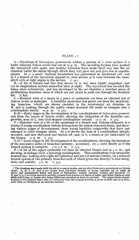
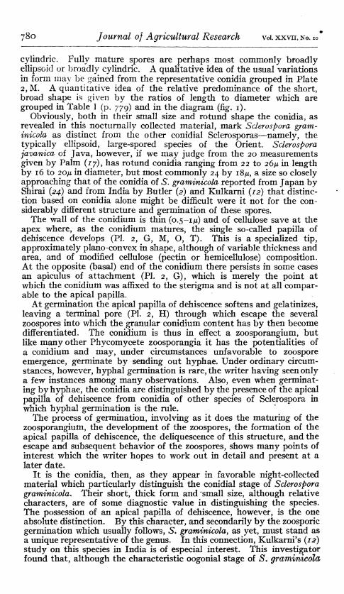
PLATE i «
A.—Mycelium of Sclerospora graminicola within a portion of a cross section of a badly infected Setaria viridis leaf cut at 10 p. m. The invading hyphae have pushed the mesophyll cells apart, and knobby branches have made their way into the air chamber under the stoma through which they will grow out and give rise to conidio- phores. At a, a small, bulbous haustorium has penetrated an epidermal cell, and at 6 a branch of the mycelium appears in cross section as it runs between the meso- phyll cells at right angles to the section. X 425.
B.—A bit of Setaria leaf like that shown in A, but more highly magnified, and cut in longitudinal section somewhat later at night. The mycelium has invaded the tissue more extensively, and has developed in the air chamber a crowded group of proliferating branches, some of which are just about to push out through the stomatal slit. X 850.
C.—Exterior view of a stoma in a piece of epidermis cut from an infected leaf of Setaria viridis at midnight. A knoblike protrusion has grown out from the proliferat- ing branches, which are shown crowded in the substomatal air chamber in B, and is pushing through the partly closed stomatal slit ready to elongate into a conidiophore initial. 12 p. m. X 375.
D-E.—Early stages in the development of the conidiophores of Sclerospora gramini- cola from the leaves of Setaria viridis, showing the elongation of the knoblike out- growths, seen in C, into club-shaped conidiophore initials. 12 p. m. X 375.
F.—Exterior view of a bit of the epidermis of a Setaria leaf, looking obliquely at a clump of young conidiophore initials arising from the almost obscured stoma, and show- ing various stages of development, from young knoblike outgrowths that have just emerged to older elongate stalks. At a is shown the base of a conidiophore already matured, and now shriveled and broken off; and, at 6, a stoma as yet uninvaded by the fungus. 12 p. m. X 375.
G-O.—Later stages in the development of the conidiophores, showing the formation of the successive series of branches (primary, secondary, etc.), until finally at O the branch system is complete. 2 to 3a.m. X375.
P.—A bit of the upper epidermis cut from an infected Setaria leaf at 3 a. m., and showing, in oblique view, a maturing conidiophore. This conidiophore is an unusually stunted one, bearing only eight still spherical and not yet mature conidia, on a reduced branch system of two primary branches each of which gives rise directly to four sterig- mata and conidia. 3 a. m. X 375.
a The drawings were made with the aid of a camera lucida, and are all of Sclerospora graminicola on Setaria viridis. A, B, C, F, and P are drawn from free-hand sections of living material cut at night in water, and immediately killed and fixed in Flemming's weaker solution, stained, and mounted. D, E, and G to O are from material carefully scraped from productive leaves at night, and either drawn at once while still living, or drawn after being killed and mounted as were the sections. The approximate magni- fication of the printed figure after its reduction from the original drawing is given in each case, as is also* scale with lou divisions as an absolute measure.
Nocturnal Production of Conidia by Sclerospora PLATE I
Journai of Agricuitural Research Washington, D. C.
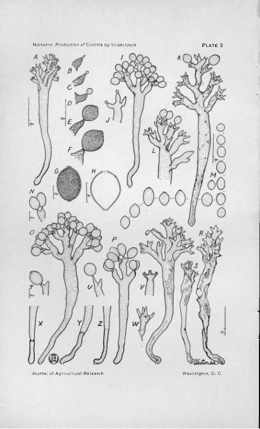
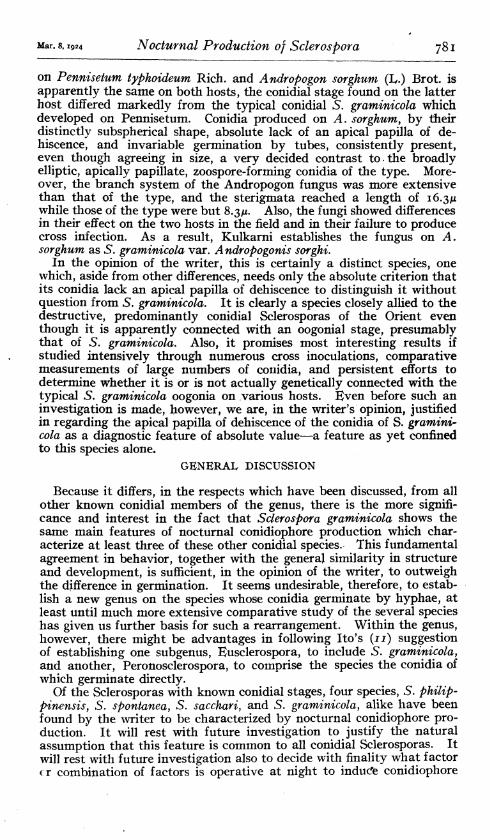
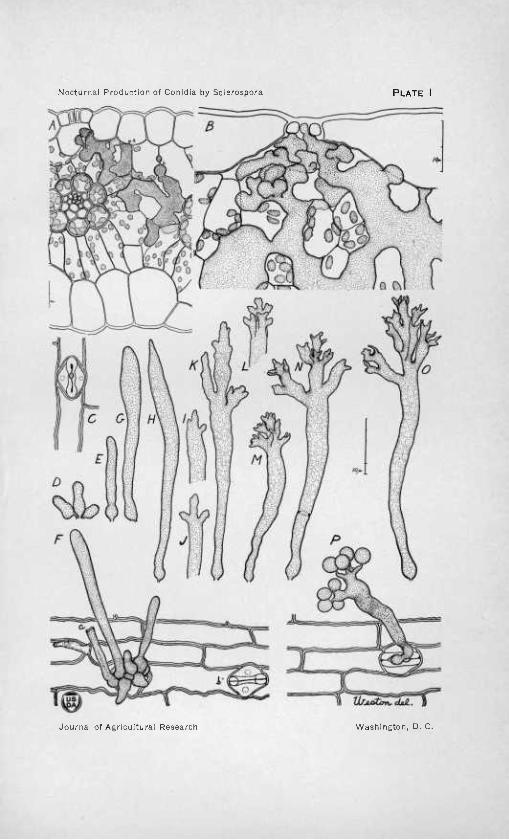
Nocturnal Production of Conidia by Scierospora PLATE 2
Journai of Agricuiturai Research Washington, D. C.
PLATE 2 a
A.—A conidiophore of Sclerospora graminicola with the branch system completed and its ultimate tips elongating into sterigmata which will give rise to the conidia. The wall at the base of the main axis is thickened into a differentiated, footlike portion. X375.
B-F.—Successive stages in the development of the conidia from the tips of the sterigmata. Note that at a relatively early stage, the wall at the tip begins to thicken and becomes modified into the characteristic apical papilla of dehiscence. X 850.
G.—Mature, recently shed, conidium with well-developed papilla of dehiscence at its apex, and the small apiculus by which it was attached to the sterigma still per- sistent at the base. The content has not yet begun to divide into zoospores, but is still un differentiated and granular. X 850.
H.—The empty wall of a conidium (i. e., zoosporangium) which has germinated by emitting zoospores through the pore left by the softening and dissolution of the terminal papilla of dehiscence. X 1,200.
I.—Conidiophore at a later stage.kthan that shown in A, with a vigorous, well- developed branch system which bears 18 only partly developed conidia. These conidia are still spherical, and have not as yet developed the terminal papillae of dehiscence. X 375.
J.—Detail of a branch tip of the three-pronged type frequently encountered (cf. fig. 1 ). Note that the young conidia which are just beginning to develop from the three sterigmata are not of exactly the same stage of development. X 600.
K.—A large, well-developed conidiophore which has shed all but two of its conidia. The branch system shows a somewhat extreme case of the continuing of the main axis, a characteristic of Sclerospora graminicola. An attempt has been made to show the detail of the content as it appears when the conidiophore is at this stage. An incomplete septation at the base delimits the basal cell which is occasionally en- countered in this species. X 375.
L,.—A still more extreme case of continuation of the main axis in the branch system of the conidiophore. X 375.
M.—Representative conidia showing the various shapes and sizes most frequently encountered. Note that each has a terminal papilla of dehiscence. The content, which is conventionally stippled here, is shown in detail in G. X 375.
N.—Detail view of two conidia still in place but already mature and about to be shed from the sterigmata. X 600.
O.—Typical, vigorous, well-developed conidiophore, like that in figure 1, but in a later stage of development. This is shown by the maturity of its 24 conidia, their larger size, more rotund ellipsoidal shape, and prominent papillae of dehiscence. Note the absence of any foot cell or basal thickening of the main axis wall. 2 a. m. X 375.
P.—A small, stunted conidiophore. The branch system, in comparison to that of a typical well-developed individual such as the one shown in O, is much reduced. Its three primary branches give rise directly to eight sterigmata and conidia, which, however, are larger than the more numerous ones of O. Note that the wall of the main axis is thickened at its base. 3 a. m. X 375.
Q,—A typical, well-developed conidiophore which only recently has shed its 34 conidia, as is shown by the bottle-shaped and rounded tips of its sterigmata, and by the beginning of the disintegration of its content. 3 a. m. X 375.
R.—The shriveled and shrunken remains of such a conidiophore, which had de- veloped during some previous night, and had remained, dried, on the leaf until scraped off and examined during the day. X 375.
S.—A similar, mummied conidiophore, but one more completely shrunken and collapsed after longer drying. X 375.
T.—Detail of a branch tip of the dichotomous type so frequently encountered. One sterigma still bears a mature conidium; while the other by its bulging, rounded apex shows that it has only recently discharged.
U.—Detail of a similar branch tip with one typical, short sterigma which has already shed its conidium, and one rather unusually elongate sterigma bearing a conidium only partly developed. X 600.
V.—Detail of a branch tip with six sterigmata which have a somewhat uncommon arrangement resembling slightly that of the sterigmata of Bremia. X 600.
W.—A branch tip of a type frequently encountered, showing four approximately equal pronglike sterigmata standing out at equal distances from the club-shaped branch tip. X 600.
X-Y.—Typical examples of the kind of basal cells occasionally found in coni- diophores of Sclerospora graminicola. X 375-
Z.—Base of a conidiophore showing the thickened wTall that at times marks off a differentiated foot portion instead of a basal cell. X 375-
o The drawings were made with the aid of a camera lucida, and are all of Sclerospora graminicola on Setaria viridis. Figures H and I were drawn from living material; the others from material scraped from leaves bearing abundant conidiophores at the time of maximum nocturnal production, and immediately killed with Flemming's weaker solution, stained (chiefly with eosin), and mounted in dilute glycerin. The approximate magnification of the printed figure after its reduction from the original drawing is given in each case, and also a IOM scale is shown as an absolute measure.


![Nocturnal stomatal conductance responses to rising [CO2], temperature and drought](https://img.pdfslide.net/doc/110x75/63368450b5f91cb18a0bdc2d/nocturnal-stomatal-conductance-responses-to-rising-co2-temperature-and-drought.jpg)


