Embed Size (px)
Citation preview

Pharmaceutics 2021, 13, 558. https://doi.org/10.3390/pharmaceutics13040558 www.mdpi.com/journal/pharmaceutics
Review
Topical Antiseptic Formulations for Skin and Soft
Tissue Infections
Thi Phuong Nga Hoang 1, Muhammad Usman Ghori 1 and Barbara R. Conway 1,2,*
1 Department of Pharmacy, School of Applied Sciences, University of Huddersfield,
Huddersfield HD1 3DH, UK; [email protected] (T.P.N.H.); [email protected] (M.U.G.) 2 Institute of Skin Integrity and Infections Prevention, University of Huddersfield, Huddersfield HD1 3DH, UK
* Correspondence: [email protected]
Abstract: Skin and soft tissue infections (SSTIs) are usually acute conditions of inflammatory micro-
bial occupation of the skin layers and underlying soft tissues. SSTIs are one of the most frequent
types of infection, typically requiring medical intervention and contribute to morbidity and mortal-
ity in both primary care and hospitalised patients. Due to the dramatic rise of antibiotic resistance,
antiseptic agents can be potential alternatives for the prevention and treatment of SSTIs. Notably,
they are commonly recommended in many global practical guidelines for use in per- and post- op-
erative procedures. A range of antiseptics, including chlorhexidine, triclosan, alcohol, and pov-
idone-iodine, are used and are mainly formulated as traditional, simple dosage forms such as solu-
tions and semi-solids. However, in recent years, there have been studies reporting the potential for
nanotechnology in the delivery of antiseptics. In this review, we have collated the scientific literature
that focuses on topical antiseptic formulations for prevention and treatment of SSTIs, and have di-
vided findings into traditional and advanced formulations. We conclude that although nanotech-
nological formulations have demonstrated potential advantages for delivering drugs; nevertheless,
there is still scope for traditional formulations and further development of optimised topical formu-
lations to address the rise of antimicrobial resistance.
Keywords: skin; soft tissue infections; antiseptic; nanocarriers; formulations; nanoparticles
1. Skin and Soft Tissue Infections
Skin and soft tissue infections (SSTIs) refer to acute conditions of inflammatory mi-
crobial occupation of the skin layers and underlying soft tissues [1,2]. The consequences
have implications on healthcare, not only in low and middle income countries, but also
globally [3]. SSTIs, is considered as one of the most frequent types of infection, typically
require medical intervention and contribute to morbidity and mortality in both primary
care and hospitalised patients [2]. It is estimated that 7–10% of hospital administrations
in North America in 2005 were as a consequence of skin and soft tissue infections [4]. In
the United States there was an increase of 65% in patients admitted with SSTIs in different
hospital departments, from 32.1 visits per 1000 population in 1997 to 48.1 visits per 1000
population in 2005 [5]. Likewise, Lee and co-workers surveyed SSTIs occurrence in the US
from 2000 to 2012 and reported that the total prevalence of SSTIs rose from 2.4 million to
3.3 million (an increase of nearly 40%) during this period [6]. In 2013, almost a third of the
US population asked for medical advice related to skin conditions [7]. The incidence of
SSTIs has increased, possibly as a result of an ageing population, the escalation of multi-
drug-resistant strains and the increasing numbers of immunocompromised patients as a
consequence of immunosuppressive therapy, cancer, transplant interventions, or
HIV⁄AIDS [2,8]. Global Health Metrics reported in 2017 regarding the prevalence, inci-
dence, and years lived with disability (YLDs) covering 354 diseases in 195 countries; ac-
cordingly, there were nearly 4.2 billion new cases of skin and subcutaneous diseases
Citation: Hoang, T.P.N.; Ghori, M.U.;
Conway, B.R. Topical Antiseptic
Formulations for Skin and Soft Tissue
Infections. Pharmaceutics 2021, 13, 558.
https://doi.org/10.3390/
pharmaceutics13040558
Academic Editor: Teresa Cerchiara
Received: 19 March 2021
Accepted: 10 April 2021
Published: 15 April 2021
Publisher’s Note: MDPI stays neu-
tral with regard to jurisdictional
claims in published maps and institu-
tional affiliations.
Copyright: © 2021 by the authors. Li-
censee MDPI, Basel, Switzerland.
This article is an open access article
distributed under the terms and con-
ditions of the Creative Commons At-
tribution (CC BY) license (http://crea-
tivecommons.org/licenses/by/4.0/).

Pharmaceutics 2021, 13, 558 2 of 35
worldwide. Around 50% of these were fungal skin diseases (accounting for more than 2.1
billion), whereas the incident cases linked with bacterial and viral pathogens were 0.27
and 0.12 billion, respectively [9].
Pathophysiology of SSTIs is related to an interruption in the balance between the
immune barrier of the host and the pathogenicity of microbial population colonizing hu-
man skin [2]. Cellulitis, as an example, is caused by pathogens disrupting skin integrity,
and is more prevalent in patients with comorbidities [10]. Disruption of the protective
cutaneous layers can be caused by chemical and physical impacts such as ulceration,
trauma, bites or surgical wounds, thermal injury, or previous inflammation [2,10]. Both
the patient and the environment are key factors contributing to the risk of developing an
SSTI. Older people or those with long-term conditions such as critical illness, obesity, car-
diovascular diseases, and chronic kidney disease failure will be at higher risk of skin
breakdown. Patients with spinal cord injury and paralysis that result in the alteration of
skin perfusion and temperature control are also considered to be at higher risk. External
factors which are likely to impair the skin barrier function can be scratching, pressure,
shear and friction, UV exposure, or radiation contact in cancer patients [2,11]. Addition-
ally, biofilm formation, the development of which enables microbes to survive and adapt
to unfavourable conditions, has become a severe problem in the healthcare fields, respon-
sible for 65% of nosocomial infections [12]. Biofilm is produced following a cell attaching
to a surface, multiplying, maturating, and then creating an extracellular polymeric matrix
which resists environmental impacts such as mechanical forces and antibiotics. This struc-
ture is detachable, affording opportunities for microorganisms to transmit into new sites
and spread infections. Biofilms have been observed in medical devices such as intrave-
nous and urinary catheters, stents, implants, ventilator tubes, or heart valves, contributing
to the growing challenge of antimicrobial resistance [13].
In children, bacterial skin infections are more prevalent than fungal, parasitic and
viral infections [14]. The major causative pathogens associated with skin and soft tissue
infections are Gram-positive microorganisms, typically Staphylococcus aureus (including
methicillin-resistant S. aureus/MRSA strains) and beta-hemolytic streptococci [1]. The
most frequent Gram-negative strain isolated was Klebsiella sp. [15]. S. aureus was respon-
sible for more than 40% of total SSTIs cases in 2003, and was a frequent cause of cellulitis,
abscesses and wound infections [2]. The incidence of S. aureus-related skin and soft tissue
infections increased two-fold from 2001 to 2009 in the US [16]. However, it was reported
that the proportion of hospital administrations caused by MRSA-related skin and soft tis-
sue infections (SSTIs) declined by 29% over the next five years [17].
Patients with dermatologic conditions often encounter physiological, psychological,
as well as financial issues; not only that, many cutaneous concerns can lead to systemic
diseases [18]. Moreover, comorbidity factors, such as diabetes, immuno-compromisation,
obesity, liver and kidney failure, and cardiovascular diseases, have repercussions on treat-
ment costs and prolong the length of stay in hospital [19]. Suaya et al. determined that the
cost of SSTI hospitalizations due to S. aureus in 2009 was $4.50 billion, which was 34%
higher than in 2001 [16]. According to the Global Burden of Disease Study, 15 different
dermatologic concerns accounted for 1.79% of the total global burden of disease in 2013.
This was calculated using disability-adjusted life years (DALYs) index, of which cellulitis,
viral skin diseases and fungal skin diseases accounted for 0.04%, 0.16%, and 0.15%, re-
spectively. Skin and subcutaneous conditions, next to iron deficiency anaemia, tuberculo-
sis, and sensory organ diseases were the leading reasons inducing disability in the world
[9,18].
The management of SSTIs often depends on the relative severity. Uncomplicated
SSTIs, located in superficial layers, typically can be controlled with a topical antimicrobial
agent, heat packs or minor incision and wound exudate draining, while more complicated
cases with involvement of deeper layers with high-risk factors often require systemic an-
tibiotic therapy and hospital administration [2]. With regards to the emergence of resistant

Pharmaceutics 2021, 13, 558 3 of 35
bacteria and antimicrobial stewardship, there is an overall drive to reduce any unneces-
sary and inappropriate use of antibiotics. Owing to the broad-spectrum of antimicrobial
activity alongside with the varying inhibitory mechanisms, topical antiseptics are advo-
cated as a potential alternative to topical antibiotics in the treatment of minor skin infec-
tions [20–22]. Although the safety and clinical effectiveness of many antiseptic agents have
not been widely demonstrated so far [23], they bring potential benefits in the prevention
of infections in wounds [20] and are still commonly recommended during pre- and per-
operative processes which are documented in many global practical guidelines [24]. Fur-
ther, a wide range of antiseptics are used mainly as simple dosage forms like solutions
and semi-solids, but there have been numerous studies to implement formulation strate-
gies in order to potentially influence therapeutic efficacy in recent years [25]. Hence, the
purpose of this review paper is to collate the studies involving formulation of antiseptics
for application via the topical route in the prevention and treatment of skin and soft tissue
infections.
2. Antiseptics
Antiseptics are biocidal products that can kill or impact the growth of disease-caus-
ing bacteria in, or on, living tissue, e.g., on the skin. Ideal properties for antiseptics include
widespread and rapid bioactivity against bacteria, fungi and viruses, no toxicity or dam-
age to the healthy tissue, and insignificant absorption into the systemic circulation follow-
ing external application [26]. Antiseptic products may contain one or more active ingredi-
ents and are presented in various formulations and preparations, for example, antimicro-
bial hand washes, surgical scrubs, preoperative preparations, tinctures, ointments,
creams, mouth-rinses, and toothpaste. They are commonly used as pre-operative skin
preparations for prevention of surgical site infections [27], as routine skin hygiene such as
hand-washes and hand rub products or for treating skin and wound diseases [26]. For
skin and wound infections in deeper skin layers, antibiotics are more normally prescribed;
in contrast, topical antiseptics are preferred for infections at the outermost surface. In such
cases, the aim is to minimize any microbial colonization in a wound or on the skin surface
without causing any deleterious effects on the living tissue or impeding the healing pro-
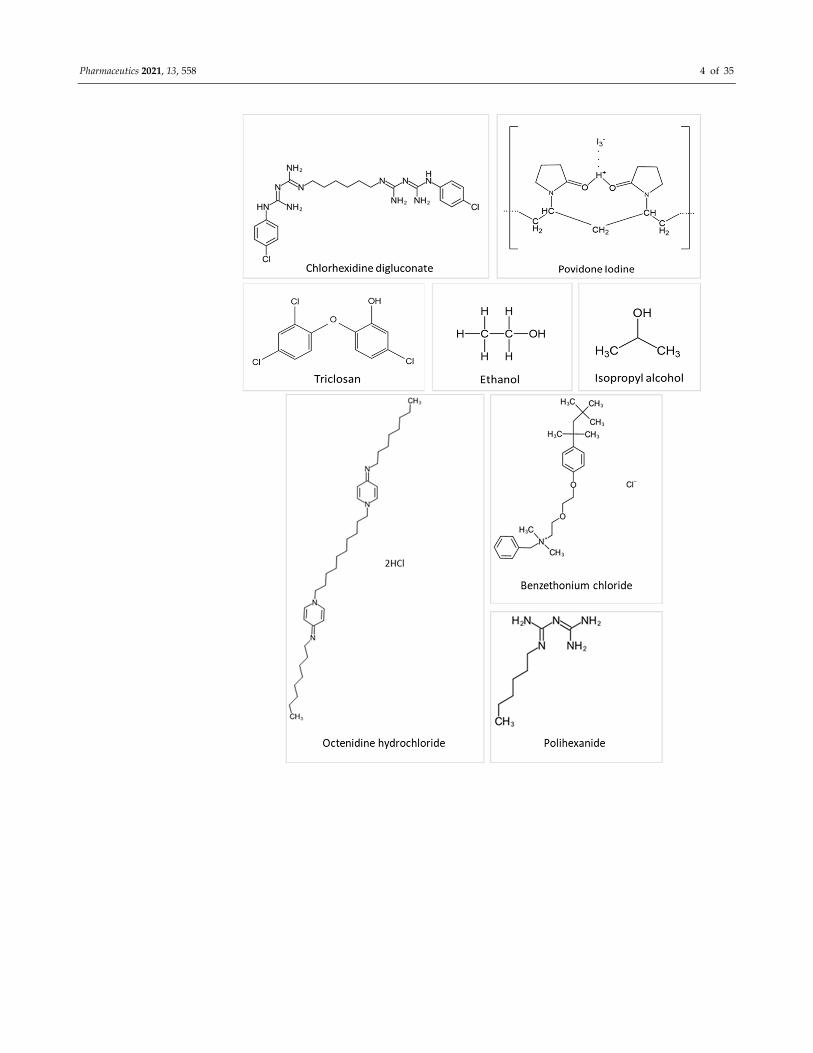
cess [26,28]. Chemical structures of commonly used antiseptics are depicted in Figure 1.
Pharmaceutics 2021, 13, 558 4 of 35

Pharmaceutics 2021, 13, 558 5 of 35
Figure 1. Chemical structures of several antiseptic agents.
2.1. Chlorhexidine
Chlorhexidine is a cationic polybiguanide (bisbiguanide) [29]. It primarily used as
salt forms because of its insolubility in water. Chlorhexidine gluconate (CHG) and other
salts like chlorhexidine diacetate, dihydrochloride, and dihydrobromide are used as surf-
icial disinfectants (disinfection of the skin and hands), in cosmetics (in creams, toothpaste,
hair care products, deodorants, and antiperspirants), and pharmaceutical products (e.g.,
preservative in eye drops, wound dressings, and antiseptic mouth-rinse) [26]. Chlorhexi-
dine is supplied typically in solution from 0.5 to 4% w/v. Chlorhexidine gluconate 2% w/v
(CHG) in 70% v/v isopropyl alcohol (IPA) is particularly recommended for pre-operative
skin cleansing by several organizations, such as Health Protection Scotland (2013), the

Pharmaceutics 2021, 13, 558 6 of 35
Centre for Disease Prevention and Control (2017), National Institute for Health and Clin-
ical Excellence (2013) and the World Health Organization (2017) [30,31]. Chlorhexidine
solutions at concentrations of 0.5% w/v and above, with alcohol, are employed to prepare
skin prior to peripheral venous catheter insertion to prevent catheter-related bloodstream
infections [32]. Chlorhexidine is a broad-spectrum antibacterial, active against both Gram-
positive and Gram-negative bacteria, while exhibiting some activity on yeasts, dermato-
phytes, and some lipid-enveloped viruses [26]. Furthermore, Macias et al. concluded that
CHG in IPA is preferred to 1% w/v triclosan in 70% IPA when a prolonged antisepsis is
required due to its longer-lasting residual effect, [33]. Alcoholic CHG solutions at both
0.5% and 1.0% w/v concentrations were better than 10% w/v aqueous povidone-iodine
(PVP-I) in minimizing microbial colony formation related to intravascular catheters [34].
Gels containing 2% w/v CHG also demonstrated a higher fungicidal activity than a com-
parative nanosilver gel against C. albicans [35].
The mechanism of antimicrobial activity of chlorhexidine is that the positively
charged molecule binds to the negatively charged lipid bacterial cell surface, thus weak-
ening the cell membrane integrity, followed by leakage of cytoplasm and precipitation of
proteins and nucleic acids at lower concentrations and membrane disruption at higher
concentrations [26,36]. Due to this non-specific mechanism of action, chlorhexidine use is
widespread. However, there are some issues with its use, such as potential toxicity in the
eyes, ears and brain, it can become inactivated in the presence of non-ionic surfactants,
and it may cause dry skin [26,37]. Recently, the Food and Drug Administration (FDA)
released a warning regarding the increasing occurrence of rare but severe allergic reac-
tions to CHG. According to the FDA, healthcare specialists should take into account the
patient’s allergy history prior to prescribing CHG-based products [38]. Furthermore, some
recent studies have indicated that the increased use of CHG may be responsible for cross-
resistance to colistin and daptomycin and the reduced susceptibility (manifested by
higher CHG minimum inhibitory concentrations) against several skin pathogens such as
Klebsiella pneumoniae, multidrug-resistant Acinetobacter baumannii, S. epidermidis, S. aureus,
and vancomycin-resistant enterococci (VRE) [39–42].
2.2. Triclosan
Triclosan (2,4,4′-trichloro-2′-hydroxydiphenyl ether) is a phenoxyphenol compound
that has been principally considered as an antibacterial and antifungal agent [26]. Triclo-
san has a very low aqueous solubility of 0.012 g/L at 20 °C [43]. It is a common ingredient
in various antiseptic products, especially in antimicrobial soap, body and hand washes
and toothpaste. It is typically used at concentrations of 0.1 to 2% w/v, with or without other
active antimicrobials such as alcohols, to bring about long-lasting activity on the skin.
Triclosan is active against Gram-positive bacteria, including Staphylococcus species. More-
over, it may also have an effect on Gram-negative bacteria and yeast, with some weaker
activity against enveloped viruses, pseudomonads, and fungi [26]. Originally, triclosan
was thought to target the cell membrane in a non-specific mechanism, however, recent
studies have found a specific bacteriostatic action for triclosan on bacteria through inhibi-
tion of the bacterial fatty acid biosynthetic pathway. At the higher concentrations found
in antiseptic preparations (2–20 mg/mL), there is a hypothesis that triclosan acts as a bio-
cide with multiple actions on lipid, RNA and protein synthesis, leading to cell lysis [42,44].
The antimicrobial activity of triclosan-containing antiseptics can be influenced by formu-
lation effects, for example, there is a synergistic activity with chelating agents (e.g., EDTA)
in destroying the Gram-negative bacterial cell wall thereby improving uptake into cells.
Triclosan shows negligible irritation and allergic skin reactions and it can retain persistent
on the skin surface [26]. However, because of the lack of the scientific literature regarding
the safety and effectiveness of triclosan for human health, in December 2017, the FDA
issued a final rule prohibiting the use of triclosan in certain over-the-counter antiseptic
preparations [23].

Pharmaceutics 2021, 13, 558 7 of 35
2.3. Povidone-Iodine (PVP-I)
Povidone-iodine (PVP-I), which is a complex of elemental iodine loosely bound to
the carrier polyvinylpyrrolidone, is used as a broad-spectrum antimicrobial agent against
bacteria, viruses, fungi, and protozoa at relatively low concentrations [26,45]. Typically,
PVP-I is widely used as a topical antiseptic and disinfectant for skin and wound infections,
mostly in solution, dry powder and lotion formulations. Application as an iodophor im-
proves both solubility and stability while releasing the active iodine gradually from the
polymer network over time. Therefore, its residual antimicrobial activity is maintained
stably while side effects associated with iodine such as irritation and brown staining on
the skin and mucous membranes are reduced. Its precise mechanism of action is still un-
known, but it is believed that the active iodine species acts as an oxidizing agent which
reacts with cell walls, membranes, and cytoplasm by exchanging and inactivating func-
tional groups of amino acids (e.g., lysine, histidine, cysteine, and arginine). The conse-
quence is the loss of cell structure and function [26].
2.4. Alcohol
Alcohols offer rapid and broad-ranging activity against bacteria, fungi, and viruses
although less is known about their activity against protozoa and bacterial spores, but they
are sporistatic. Isopropanol (isopropyl alcohol), ethanol and n-propanol are the most pop-
ular alcohols used as antiseptics and disinfectants. Their exact mechanism of action is not
clear but they are able to cause denaturation and precipitation of proteins thus destroying
cell membranes and leading to cell lysis. Concentrations ranging from 60% to 80% v/v are
recommended for maximum antimicrobial activity because, in more concentrated solu-
tions, alcohol quickly coagulates protein-based molecules present externally on the cell
wall and interferes with penetration into the cell, therefore limiting further effects on pro-
tein-based inner cell compositions. Other potential attributes are relative stability, and low
toxicity, odour and cost. Alcohols are also used as preservatives and common solvents for
other biocides such as chlorhexidine [26].
2.5. Essential Oils
Essential oils are secondary metabolic products found in various parts of plants (such
as flowers, seeds, leaves, peels, buds, barks, wood, or roots), and can be extracted by hy-
dro-distillation and steam distillation, mechanical processes, or by “dry” distillation from
some woods [26,46]. They are complex mixtures containing hundreds of compounds and
their exact chemical composition depends on extraction processes and specific conditions.
For example, dry vapour steam distillation is used when there is a requirement to mini-
mize ester hydrolysis (e.g., linalyl acetate), or cohobating is proposed to improve the
quantity of particular compounds such as sulfur compounds [46]. Essential oils and their
components have been used in a wide range of products, from fragrances, toothpastes,
cosmetics, to aromatherapy and phytomedicine, with tea tree oil and eugenol, being com-
bined in many commercial antiseptic preparations, such as Ord River Tea Tree Antiseptic
Cream®, Australian Tea Tree Antiseptic Cream® or Manuka Doctor ApiRevive Manuka
and Tea Tree Antiseptic Gel® [26]. In dermatology, essential oils are primarily used for
treating skin infections (62% of total cases), followed by skin inflammation and general
skin maintenance at 20% and 18%, respectively [47]. Relative bioactivity varies between
the different oils. In particular, tea tree oil demonstrates bactericidal activity (at 0.25 to
0.5% v/v), fungicidal activity (at 0.06–1% v/v), fungistatic activity (within 0.03–0.5% v/v) as
well as activity against yeasts and dermatophytes (including Candida and Trichophyton).
Tea tree oil, amongst others, presents persistent and long-lasting activity on the skin after
application. Despite most essential oils presenting antimicrobial effectiveness at low con-
centrations, they have been reported to generate irritancy and allergenicity following ap-
plication to skin and mucous membranes [26,47]. Almost 1.8% of patients tested with 5%
and 10% tea tree oil patches experienced allergic contact dermatitis [48].

Pharmaceutics 2021, 13, 558 8 of 35
2.6. Silver Compounds
The active element is the silver ion (Ag2+) in silver nitrate (AgNO3) and silver sulfadi-
azine (AgSD). Generally, topical silver antiseptics are applied for prevention of skin and
wound infections mostly caused by S. aureus and Pseudomonas in cream or solution forms
and used in eye drop preparations for bacterial infections in neonates [26]. There are a
number of studies indicating the valuable role of silver in wound care [49]. Additionally,
silver compounds are also commonly used to cover surfaces prone to bacterial coloniza-
tion such as catheters or dental instruments. Many commercial silver-based products are
now available in many forms such as Atrauman Ag® Wound Dressing, Urgotul® SSD An-
tibacterial Contact Layer, Flamazine® Antibacterial Cream, Colloidal Silver Spray®, Silver
Solution® Antimicrobial Wound Gel, MSM+Silver® Water Drops, or Natural Sense Colloi-
dal Silver ® Eye Drops.
Silver compounds exhibit bacteriostatic and bactericidal activity at fairly low concen-
trations, especially on Gram-positive bacteria. Regarding the mechanism of action, active
silver ions bind to sulfhydryl, amino, and carboxyl groups of amino acids on microorgan-
ism surfaces, thus denaturing proteins, and disrupting the cell wall and membrane func-
tions. Silver also specifically inhibits cell wall metabolism and electron transport as well
as the respiration chain [26,50]. Following application of topical antiseptic, respiratory
sprays, implanted medical devices or wound dressings, silver has been shown to be ab-
sorbed into the systemic circulation, mostly in conjugation with protein and then depos-
ited in human tissues, with higher levels in skin, kidneys, eyes, brain, liver, and bone mar-
row [51]. Argyria is a rare cutaneous condition resulted by excessive or chronic use of
preparations containing silver, with the most characteristic symptom being the discolour-
ation of skin into blue or blue-grey, especially in sunlight-exposed areas [52].
2.7. Other Antiseptic Agents
(i) Quaternary ammonium compounds (QACs) are cationic surfactants which have
both hydrophobic and hydrophilic groups [26]. QACs target cell walls and membranes.
They are quickly absorbed, interacting with membrane lipids, thus disrupting cell struc-
ture and function or cause denaturation of essential cell proteins, and leaking of cytoplas-
mic material [26]. The antimicrobial activities of QACs are governed by their chemical
structure and the type of formulation with activity being impacted by fatty substances or
anionic surfactants. For example, benzethonium chloride (BZT) is used as a topical anti-
infective and an antiseptic effective against bacteria, fungi, moulds, and viruses [53]. Fur-
ther, benzalkonium chloride (BZK) is used widely as antimicrobial preservative or biocide
surfactant, and it is especially commonly found in ophthalmic solutions [54]. BZK displays
broad-spectrum activities against bacteria, fungi, virus, algal, but not endospores [55]. The
widespread use of BZK has been reported to contribute to the increase in antibiotic re-
sistance concerns [56]. Cetylpyridinium chloride (CPC) demonstrates antiseptic behav-
iour against Gram-positive pathogens and yeasts but has no effect on Gram-negative mi-
croorganisms and mycobacteria. CPC is commonly found as an active ingredient in
mouthwashes, toothpastes, lozenges, or mouth sprays for treating minor mouth and
throat infections [57,58].
(ii) Octenidine dihydrochloride (OCT) is also a cationic surfactant, belongs to the bi-
pyridine group [59] and has been reported for a wide range of applications such as pre-
operative skin preparations, prevention, and treatment skin and wound infections [60].
Its spectrum of activity covers both Gram-positive and Gram-negative pathogens includ-
ing MRSA [60]. Octenidine reduced high-level mupirocin-resistant Staphylococcus aureus
isolates in vitro by more than 7log cycles at concentrations as low as 0.001% w/w within
only 30 s [61]. Similar findings were reported for multidrug-resistant Gram-negative bac-
teria [62]. Octenidine dihydrochloride (0.1%) with 30% v/v 1-propanol and 45% v/v 2-pro-
panol was more effective than 74% v/v ethanol with 10% v/v 2-propanol for eradication of
skin colonization in central venous catheter sites over 24h [63]. Moreover, octenidine was
Pharmaceutics 2021, 13, 558 9 of 35
highly effective in reduction of infections associated with biofilm formation on orthopae-
dic implants infections, compared to gentamicin [64].
(iii) Polihexanide (PHMB) is a biguanide antiseptic whose chemical structure is sim-
ilar to chlorhexidine [65]. The positively charged molecular species interacts electrostati-
cally with the negative-charged lipopolysaccharide compounds of bacterial cell mem-
brane, leading to the leakage of intracellular components; therefore, PHMB can be effec-
tive on both Gram-negative and Gram-positive pathogens [66]. Octenidol® and
ProntOral® mouthwashes, which contain octenidine and polyhexamethylene respectively,
displayed similar antimicrobial potency as 0.2% chlorhexidine digluconate in eliminating
Streptococcus sanguinis, Streptococcus mutans, Candida albicans, and Fusobacterium nucleatum
[67]. Additionally, both 0.02% PHMB and 0.05% OCT were superior than NaCl 0.09% w/v
solution in removal of biofilms of Pseudomonas aeruginosa [68]. PHMB is well tolerated
when applied to both skin, and wounds [69].
3. Topical Antiseptic Formulations
Scopus, Google Scholar, Science Direct, and PubMed databases were used to search
the literature with keywords; “Topical formulation” OR “Transdermal formulations” OR
“Topical antiseptic formulations”. The results were further filtered with only original re-
search articles written in English language selected. These were then divided into tradi-
tional and advanced antiseptic formulations groups, and their details have been discussed
in the following sections.
3.1. Traditional Antiseptic Formulations
In this section traditional antiseptic formulations intended for topical delivery will
be discussed and a summary of these studies is presented in Table 1.
Table 1. Summarised characteristics of traditional skin antiseptic formulations.
Drug Concentration Formulation type Combination Carrier polymer Manufacturing
Technique Study Characteristics Reference
Chlorhexidine
gluconate
(CHG)
Dermal polymeric
patch Eudragit RL100
To characterize
properties of
developed patches
regarding their drug
release and
antimicrobial activity.
[36]
Chlorhexidine
gluconate
2% CHG in
70% isopropyl
alcohol (IPA)
Solution Acrylate
copolymer
To test the
effectiveness of
adding a film-forming
acrylate copolymer to
a topical CHG-based
preparation on
minimizing CHG loss,
compared to a
marketed CHG
solution
[70]
Chlorhexidine
gluconate
2% CHG in
70% IPA Solution
To contrast the
residual effects of 2%
CHG in 70% IPA v/v
and 1% triclosan in
70% IPA v/v on skin
bacterial communities
[33]
Chlorhexidine
gluconate
2% CHG in
70% IPA Solution
To compare the
antiseptic activity of
10% sodium
[71]

Pharmaceutics 2021, 13, 558 10 of 35
hypochlorite and 2%
CHG in 70% IPA
Chlorhexidine
gluconate
2% CHG in
70% ethanol Solution
To appraise the
desiccation and
ethanol resistance
of multidrug-resistant
Acinetobacter
baumannii with
biofilms (MDRAB-Bs).
To compare the
antiseptic activities of
a combination of
CHG and 70% ethanol
with 70% ethanol
disinfectants used for
MDRAB-Bs
[72]
Chlorhexidine
base
Mucoadhesive
polymer patches
Psyllium and
three types of
semi-synthetic
hydroxypropyl
methyl celluose
A casting-
solvent
evaporation
technique
To test the
effectiveness of
polysaccharide
psyllium in the
mucoadhesive
patches for
controlling release
[73]
Triclosan
Methoxy
amidated pectin-
based
mucoadhesive
buccal patch.
β-cyclodextrin
To develop buccal
patches
and determine drug
release, antimicrobial
and in vitro
absorption from
patches
[74]
Triclosan 0.3% Soap
To study the in vitro
and in vivo
antibacterial activity
in soap.
[75]
Triclosan 0.3% Shampoo
To assess the
antimicrobial efficacy
of the shampoo
against bacteria and
fungi
[76]
Povidone-
iodine
(PVP-I)
10% Ointment
To compare the in
vitro antibiofilm effect
of diluted PVP-I
ointment with other
six tested products
against P. aeruginosa
and multi-species
biofilms of C. albicans
and MRSA
[77]
Povidone-
iodine
4% PVP-I skin
cleanser, 7.5%
PVP-I surgical
scrub, 10%
PVP-I solution
and 3.2% PVP-
I/alcohol
solution
Hand wash and
hand rub
To study the in vitro
potency of four hand
hygiene formulations
of povidone iodine
against Ebola virus
[78]

Pharmaceutics 2021, 13, 558 11 of 35
Povidone-
Iodine
Alginate
hydrogels Vancomycin
Vancomycin
loaded chitosan
nanoparticles
(CNPs) by ionic
gelation method
Modified ionic
gelation
method
To assess in vitro
release of vancomycin
and PVP-I from the
hydrogel.
To assess the
bactericidal and
antibiofilm efficacy of
hydrogels
[79]
Povidone-
Iodine 1% and 2% Solution
To analyse the
effectiveness and
safety of 1% or 2%
PVP-I topical solution
in patients with
cancer therapy-
associated paronychia
during 6–8 weeks.
[80]
Thiolated PVP
and
Thiolated
PVP-iodine
complex
Solution
2-(2-acryloyl–
Ethyl
disulfanyl)-
nicotinic acid
(ACENA)
To test in vitro
mucoadhesive
properties and the
release of iodine from
thiolated PVP-Iodine
complexes
[81]
Isopropanol 75% (w/w) Hand rub Glycerol
0.725% (w/w)
To investigate the role
of glycerol in pre-
surgical hand rub
products, based on
EN 12791, especially
after 3 h of
application
[82]
Isopropyl
alcohol 70% (v/v) Solution
To study the potency
of isopropyl alcohol
and chlorhexidine in
the prevention of
blood cultures
impurities
[83]
Ethanol 96% Solution
Isopropanol-
30g and
ortophenilphe
nol-0.1g
To determine the
effect of the
combination of 96%
ethanol, 30g
isopropanol, 0.1g
ortophenilphenol and
PVP-I in minimizing
surgical-site
infections, compared
to that of single use
PVP-I
[84]
Silver
Chloride Colloidal solution
To study the
suspension potency
on the microbial
autotrophic and
heterotrophic growth
[85]
Benzethonium
chloride (BZT) 0.2% Lotion
To test the
antimicrobial efficacy
of an ethanol- based
antiseptic and water-
[86]

Pharmaceutics 2021, 13, 558 12 of 35
based antiseptic
products containing
0.2% BZT
Tea tree oil 3% Soap
To assess the potency
of 0.3% Melaleuca
alternifolia essential oil
versus 0.5% triclosan
hand soap
formulations
[87]
Tea tree oil
Tea tree 10%
cream, tea tree 5%
body wash
To compare the
efficacy of the
combination of tea
tree 10% cream and
tea tree 5% body wash
with the standard
theory in eliminating
MRSA
[88]
Triclosan 0.1–0.45% w/v Soap
To evaluate the
efficacy of soaps with
and without triclosan
and investigate
potential hazards in
the emergence of
antibiotic resistance
[89]
Tea tree oil
4% tea tree oil
nasal ointment
and 5% tea tree oil
body wash
To compare the
ability to eradicate
MRSA between the
combination of a 4%
tea tree oil nasal
ointment and 5% tea
tree oil body wash
with a standard
theory of 2%
mupirocin nasal
ointment and
triclosan body wash
[90]
3.1.1. Solutions
Comparisons of antiseptic performance of solution formulations are relatively well
reported [91]. Particularly, in a two-step study, 2% w/v chlorhexidine gluconate in 70% v/v
isopropyl alcohol was proven to have more substantive efficacy against organisms from
the skin of human volunteers compared to 10% w/v sodium hypochlorite and 10% w/v
povidone-iodine [71] (Figure 2). Similarly, it demonstrated a longer-lasting residual effect
than triclosan (1% w/v) in 70% v/v IPA, making it more suitable than other antiseptics for
procedures such as catheter insertion or surgery [33].

Pharmaceutics 2021, 13, 558 13 of 35
Figure 2. Agar plate in which the substantive effect can be seen. The plate was divided into 3
zones; in each one an antiseptic was tested. Only the zone in contact with skin washed with chlor-
hexidine showed an inhibition zone. Reproduced with permission from [71], American Jornal of
Infection Control, 2013.
Chlorhexidine gluconate 2% w/v in 70% v/v ethanol was effective in eradicating mul-
tidrug-resistant Acinetobacter baumannii with biofilms (MDRAB-Bs) with no MDRAB-Bs
detected after only 1 min of contact (Figure 3) [72].
Figure 3. Antiseptic efficacies ethanol and CHG in ethanol solutions. The 2% CHG in 70% ethanol
eliminated the MDRAB-Bs completely at the 1 min time point. The 0.5% CHG in 70% isopropyl
alcohol eliminated the MDRAB-Bs completely at 3 min time point. However, the70% ethanol elim-
inated the MDRAB-B completely at 10 min time point. * Indicates significantly lower MDRAB
CFUs treated with 2% CHG in 70% ethanol agent than 0.5 CHG in 70% isopropyl alcohol. (Three-
way Analysis of Variance (ANOVA) with Scheffe’s post hoc test, p < 0.005). #. Indicates significant
lower multidrug-resistant Acinetobacter baumannii colony forming units (MDRAB CFUs) treated
with 2% CHG in 70% ethanol agent than 70% ethanol agent. (Three-way ANOVA with Scheffe’s
post hoc test, p < 0.005). Reproduced with permission from [72], Journal of Microbiology, Imunol-
ogy and Infection, 2018.

Pharmaceutics 2021, 13, 558 14 of 35
On the other hand, according to Koburger et al. (2010), with reference to minimum
inhibitory concentration (MICs) and minimum bactericidal concentration (MBCs) values,
the antimicrobial effect of polyhexanide and octenidine were deemed to be greater than
chlorhexidine digluconate, PVP-iodine and triclosan against the tested microorganisms.
In a quantitative suspension test (to determine the minimal concentrations to achieve at
least a reduction of 3.8 log cycles for C. albicans and 4.8 logs for S. aureus and P. aeruginosa),
octenidine was more effective than triclosan at all-time points [92]. Another recent clinical
trial found that 70% isopropyl alcohol solution was equivalent to 2% chlorhexidine glu-
conate in 70% IPA for skin antisepsis [83], supporting the use of cheaper antiseptics like
alcohol [83]. Furthermore, it was found that the simultaneous application of 10% w/v PVP-
I and a topical antiseptic, Alkosol® (96% ethanol, 30g isopropanol, and 0.1g ortophenil-
phenol), in a two-step pre-operative procedure, reduced the extent of surgical site infec-
tions as only 6% of included patients had at least one symptom of inflammation after 24
h of surgery, compared to 40% for PVP-I alone [84].
Bashir et al. reported that addition of a film-forming polymer such as an acrylate to
a pre-operative solution preparation of 2% chlorhexidine in 70% isopropyl alcohol effec-
tively reduced bacterial colonization in an ex vivo model. This was due to the sustained
presence of CHG on the skin surface, thus potentially leading to more sustained antimi-
crobial activity in prevention of surgical site infections [70].
A topical povidone-iodine solution was employed in a phase 2 trial for the treatment
of cancer therapy-related paronychia—an acute nail infection caused by targeted and cy-
totoxic remedies. Twice daily application of 2% PVP-I solution had a positive effect on
clinical outcomes and quality of life [80].
3.1.2. Patch Formulations
A novel mucoadhesive buccal patch which comprised matrix-forming polymers low
methoxy amidated pectin (AMP) and 20% w/w Carbopol (CAR) was loaded with 4 mg of
triclosan. The patch also included β-cyclodextrin-epichlorohydrin polymer (EPIβCD) and
anionic carboxymethylated β-cyclodextrin-epichlorohydrin polymer (CMEPIβCD) to im-
prove triclosan (TCS) solubility, as well as its release from the patch. The TCS-EPIβCD
complex did improve solubility, compared to a TCS-parent β-cyclodextrin complex alt-
hough the presence of 1% (w/v) AMP compromised the complexation and solubilizing
properties of both polymeric β-cyclodextrin derivatives (CMEPIβCD and EPIβCD). In ad-
dition, the buccal patches formulated with TCS- EPIβCD in combination with AMP-CAR
80:20 (w/w) provided immediate and stable drug release and efficacy against Streptococcus
mutans isolated from the oral cavity [74].
In 2015, a similar study assessed the capability of the polysaccharide psyllium to con-
trol the release rate of chlorhexidine from a buccal muco-adhesive patch for local perio-
dontal application. Combining semi-synthetic polymers including sodium carboxymethyl
cellulose and hydroxypropyl methyl cellulose (HPMC) with psyllium had the advantages
of providing zero-order kinetics for drug release and effective antimicrobial activity
against Gram-positive and Gram-negative bacteria [73].
Eudragit® RL 100 was used as the gel-forming agent in chlorhexidine-based medi-
cated dermal patches. Eudragit® RL 100 is a complex made up of “ethyl acrylate, methyl
methacrylate and low content of methacrylic acid ester with quaternary ammonium
groups” [93]. The amount of quaternary ammonium groups in the RL type is greater than
other Eudragit polymers, rendering it more permeable [94,95]. It is widely used as a drug
vehicle, controlled release agent, film former, bioadhesive material or suspending agent
[96]. Typically, the dermal patches containing Eudragit® RL 100 exhibited efficacious ac-
tivity against the tested microorganisms [36].

Pharmaceutics 2021, 13, 558 15 of 35
3.1.3. Gels
Gels, along with creams and ointments, are common semisolid formulations used for
dermal applications [97]. Gels may be spread easily and offer a cooling effect as a result
of solvent volatilization after application [98]. They can be categorized into hydrogels and
organogels; hydrogels mainly include water in the liquid phases, while organogels com-
prise organic solvents [99]. Furthermore, the term “emugels” (as emulsified gels) is used
to refer to biphasic systems which encompass a dispersed aqueous gel and a lipid base.
Emugels were developed in order to enhance the occlusive characteristics of gels [97].
A thiolated povidine–iodine complex was developed with the intention of enhancing
mucoadhesive properties. The gel-forming ability of thiolated PVP and thiolated PVP-I
on contacting the mucosal surface and the mucoadhesive features were assessed. Both the
thiolated PVP and thiolated PVP-I complex demonstrated merits, such as increasing vis-
cosity and improving the mucoadhesion, as well as controlling iodine release from the
gels, compared to unprocessed PVP and PVP-I complex [81].
3.1.4. Lotions
Lotions are utilized particularly (but not popularly in clinical applications) as topical
formulations of active substances (i.e., antibiotics, antiseptics, or corticosteroids), intended
for treatment of localized cutaneous disorders [98,99]. Moreoever, lotions are more easily
applied to sizeable skin areas than more viscous creams or ointments [99].
An aqueous antiseptic lotion containing benzethonium chloride (BZT) at 0.2% was
reported to have a rapid and wide-spectrum antimicrobial efficacy equivalent to 76% v/v
ethanol [86] when was tested according to standard Time-Kill protocols [100]. Combined
with its known persistence and low propensity for skin irritation, a BZT-aqueous based
antiseptic product has advantages over alcohol-based formulations [86,101].
3.1.5. Ointments
Ointments are often selected for their tenacity on the skin to extend a drug’s thera-
peutic activity over a long time as well as producing a protective layer covering the sites
of application. However, they can be associated with irritation due to their occlusive na-
ture arising from their tallowy characteristics [98].
The combination of ointment and body wash containing tea tree oil at 4% and 5%,
respectively, was reported to be better than a conventional regime consisting of 2% mupi-
rocin nasal ointment and triclosan body wash for prevention of MRSA-induced infections
[90].
An in vitro study tested the PVP-I ointment at numerous concentrations (both stand-
ard and diluted concentrations) versus six others antiseptic preparations and a silver-
based wound dressing, in terms of eliminating biofilms of Pseudomonas aeruginosa, Candida
albicans, and MRSA. Following treatment with PVP-I ointment at all concentrations, there
were no viable biofilms of P. aeruginosa detected after 4 and 24 h. Additionally, PVP-I
ointment containing 10% w/v active PVP-I was deemed effective at eradiating biofilm ma-
terials of C. albicans and MRSA at both 4 and 24h following application and performed
better than the other tested antimicrobial agents [77].
3.1.6. Creams
There are two main types of cream, oil in water and water in oil creams, of which,
o/w cream is more popularly utilized to produce a local effect in case of external disorders,
for instance, skin and wound infections [98].
A therapeutic regime of tea tree oil comprising tea tree oil 10% cream and tea tree oil
5% body wash was proposed for eradicating MRSA colonization. There was no significant
difference with the standard therapy of 2% mupirocin nasal ointment, 4% chlorhexidine
gluconate soap, and 1% silver sulfadiazine cream [88].

Pharmaceutics 2021, 13, 558 16 of 35
3.1.7. Washes/Rubs
The FDA defined antiseptic washes, also known as antibacterial soaps, as products
used with water and are rinsed off after use, including hand washes, hand soaps and body
washes [102]. Antiseptic rubs (also called hand “sanitizers,” or antiseptic wipes) are sub-
stituted when soap and water are inconvenient; they are left on and there is no need to
rinse with water [102].
Four different hand wash and hand rub formulations of PVP-I, including 4% PVP-I
skin cleanser, 10% PVP-I solution, 3.2% PVP-I in 78% alcohol, and 7.5% PVP-I surgical
scrub were compared in a suspension test against Ebola virus (EBOV) and modified vac-
cinia virus Ankara (MVA) in vitro. Viral titres of MVA and EBOV were reduced by more
than 99.99% under both clean environments (0.3 g/L bovine serum albumin; BSA) and
contaminated environments (3.0 g/L BSA with 3.0 mL/L erythrocytes) within 15 s of expo-
sure. Among those products, PVP-I solution in an alcohol mixture of 2-propanol and eth-
anol was the most efficacious at early timepoints. PVP-I could have an important role in
limiting diseases related with Ebola, especially in combination with alcohol [78].
Glycerol, which is often used as a humectant, can restrict the clinical effect of pre-
operative hand rubs of isopropanol. A hand rub preparation based on isopropanol with-
out glycerol, comprising a combination of ethylhexylglycerin, dexpanthenol, and a fatty
alcohol, was more effective in eradiating skin pathogens than the product containing glyc-
erol [103].
Triclosan is one of the most popular antimicrobial agents used in soaps. However, a
systematic literature review indicated that triclosan based soaps, used at the concentra-
tions commonly found commercially (0.1–0.45% w/v), were not more efficacious in pre-
venting infections than non-antimicrobial soaps [89]. The effectiveness of triclosan in an-
tibacterial soaps was tested against twenty isolated strains proposed by FDA [104] either
in vitro or in vivo. It was found that antibacterial soaps containing 0.3% w/w triclosan did
not show a superior effect compared to plain soaps under experimental conditions. This
could be a consequence of a short exposure time, or the impact of surfactants in soaps like
sodium laureth sulphate on diminishing the bactericidal activity of triclosan [75]. This re-
sult led to an US FDA ruling issued in 2013 that all consumer antiseptic wash products
need to have demonstrable clinical benefit prior to commercialization, in comparison to
plain soap and water [104]. Moreover, the latest FDA ruling released at the end of 2019
announced that three active antiseptic ingredients, benzalkonium chloride, alcohol (etha-
nol or ethyl alcohol), and isopropyl alcohol are not suitable for use as consumer antiseptic
rubs [105].
In contrast, the antifungal and antibacterial effects of a medical triclosan-based sham-
poo was tested against five isolated microorganisms. Based on the inhibition zones, at all
concentrations diluted from original concentration of 0.3% w/w (from 10% to 90%), the
shampoo had efficacious antimicrobial activity against all three fungal species and one
bacterial species (E. coli), but no effect on Staphylococcus aureus. Generally, antimicrobial
shampoos, (e.g., triclosan), have shown efficacy in preventing and treating skin and scalp
disorders, such as dandruff whose major cause is Malassezia globose [76]. An antiseptic
soap with tea tree oil at 0.3% exhibited a similar efficacy in eliminating E. coli load on
hands as a soap containing triclosan at 0.5% [87].
3.2. Advanced Pharmaceutical Formulations
Nanocarriers are colloidal drug delivery systems comprising dispersed particles with
diameters less than 500 nm [106]. Nanocarriers have potential applications for parental,
oral, dermal and transdermal administration routes [106]. They have been reported to
present some merits over conventional preparations such as ameliorated bio-distribution
and pharmacokinetics, enhanced therapeutic potency, minimized toxicity, controlled re-
lease, increased bioavailability, or drug delivery to target destinations [107,108].

Pharmaceutics 2021, 13, 558 17 of 35
The following sections review the published studies using nanotechnology for deliv-
ery of antiseptic agents (key findings are also summarised in Table 2).
Table 2. Summarised characteristics of advanced skin antiseptic formulations.
Drug Concentration Formulation
Type Combination
Carrier
Polymer
Manufacturing
Technique
Study
Characteristics Reference
Chlorhexidine
gluconate 0.2%
Nanogel
containing
magnetic Cobalt
iron oxide
nanoparticles
Chitosan and
gelatin
Solution casting
method
To investigate
the release and
pH-dependent
response of
chlorhexidine
gluconate from
a magnetic
nanogel
[109]
Chlorhexidine
base
Poly(epsilon-
caprolactone)
nanocapsules
Poly(epsilon-
caprolactone)
Solvent
displacement
method
To evaluate the
antibacterial
ability of
poly(epsilon-
caprolactone)
nanocapsules
containing
chlorhexidine
base and the
absorption of
active into the
stratum
corneum
[110]
Chlorhexidine
base
α-, β-, and γ-
cyclodextrin
methacrylate
(CD-MA)
containing poly
(methyl
methacrylate)
(PMMA) based
nanogels
CD-MA
containing
nanogels were
synthesized by
the radical
precipitation
polymerization
technique.
To study the
capacity of
chlorhexidine
base in PMMA
nanogels.
To assess the
bactericidal
against
Staphylococcus
aureus of CD-
MA nanogels
[111]
Chlorhexidine
digluconate Nanoemulsions
Eucalyptus oil
(EO) or Olive
oil (OO)
HSH followed
by probe
ultrasonication
To investigate
the drug
release, skin
permeation and
retention of
CHG from
nanoemulsions.
To evaluate
impact of
methacrylate
powder
dressing in
controlling the
CHG release
[112]
Triclosan
(TCS)
Chitosan-coated
nanocapsule
Poly(epsilon-
caprolactone)
(PCL)
Interfacial
deposition of
preformed
polymers
To characterize
properties of
nanocapsule
comprised of α-
[113]

Pharmaceutics 2021, 13, 558 18 of 35
bisabolol and
TCS.
To study the
antimicrobial
activity against
tested
pathogens.
To testify the
compatibility as
incorporating
nanocapsule
into wound
dressings
Triclosan 10%, 30%, and
50%
Poly-L-lactide
(PLLA)/triclosa
n nanoparticles
Poly-L-lactide
(PLLA)
Emulsification–
diffusion
technique
To evaluate the
release of
triclosan from
PLLA
nanoparticles
and its
antimicrobial
activities
[114]
Triclosan 0.5% w/w
Nanoparticles
stabilized by
branched
diblock
copolymers
Branched
diblock
copolymers:
PEG-b-PNIPAM
(BDP 1); PEG-b-
PBMA (BDP 2);
PEG-b-PSty
(BDP 3)
Emulsion-
freeze-drying
technique
To assess
fungicidal
ability against
C. albicans of
triclosan
nanoparticles
[115]
Triclosan Nanoparticles Eudragit E 100
Emulsification–
diffusion by
solvent
displacement
method
To compare in
vitro
percutaneous
permeation of
nanoparticles
containing
triclosan, with
two commercial
formulations
used for
treating acne,
including a
solution and an
o/w emulsion
[116]
Triclosan
Solid lipid
nanoparticles
(SLNs)
Glyceryl
behenate (GB)
and Glyceryl
palmitostearate
(GP)
Hot high shear
homogenisation
followed by
probe
ultrasonication
To investigate
the impact of
SLNs in
delivery of TCS
to deeper skin
layers and hair
follicles and
compare the
permeation
ability of GB-
SLNs and GP-
SLNs
[112]

Pharmaceutics 2021, 13, 558 19 of 35
Triclosan Nanoemulsions
Eucalyptus oil
(EO) or Olive
oil (OO)
HSH followed
by probe
ultrasonication
method
To develop and
characterise
stable
nanoemulsion
formulations.
To evaluate the
ability of NEs in
improving skin
retention of
TCN
[112]
Tea tree
essential oil
(TTO)
10.0 mg mL−1
Nanoemulsions
(TTO-NE) and
polymeric
nanocapsules
(TTO-NC)
Poly(e-
caprolactone)
TTO-NE by
spontaneous
emulsification
and TTO-NC by
interfacial
deposition of
the preformed
polymer
methods
To investigate
the in vitro
fungicidal
potency against
Trichophyton
rubrum of TTO-
NE and TTO-
NC systems
[117]
Tea tree
essential oil
(TTO)
Hydrogels
containing
Nanoemulsions
(TTO-NE) and
nanocapsules
(TTO-NC)
Poly(e-
caprolactone)
Nanoemulsion:
spontaneous
emulsification
Nanocapsules:
interfacial
deposition of
preformed
polymer
To evaluate
physicochemica
l properties of
hydrogels and
their efficacy in
wound healing
and protecting
skin from UV-B
rays
[118]
Tea tree oil
(TTO)
Emulgel (EG)
containing
TTO-loaded
nanoemulsion
(NE)
Nanoemulsion:
High energy
emulsification
To evaluate the
physicochemica
l properties, the
ex vivo
penetration,
antimicrobial
potency and
safety of topical
emulgel
[119]
Tea tree oil
(TTO)
Nanoemulsions
(NE)
Silver
nanoparticles
(Ag-NPs)
To investigate
cytotoxicity as
well as
antimicrobial
ability of the
prepared
nanoemulsions
against
clindamycin-
resistant
Escherichia coli
and S. aureus.
To appraise the
synergistic
effect of TTO
NE and Ag NPs
against tested
microorganisms
[120]

Pharmaceutics 2021, 13, 558 20 of 35
Silver
Silver
nanoparticle
(Ag NPs)
Polyvinyl
alcohol (PVA)
To estimate the
suspension
efficacy on the
autotrophic and
heterotrophic
growth.
To investigate
silver species
properties
[85]
Benzalkonium
chloride (BZK)
0.6% BZK for in
vitro studies
and 0.2% BZK
for in vivo
studies.
Nanoemulsion EDTA
High-energy
homogenization
using high
shear
conditions
To evaluate the
in vitro and in
vivo
antimicrobial
effect against
isolated
bacterial species
[121]
Cetylpyridiniu
m chloride
(CPC)
Oil in water
nanoemulsions
To assess the
fungicidal
potency
[122]
Polyhexanide
(PHMB) 0.05%
nanoparticle-
emulsion
Lipofundin®
MCT 20%
To compare the
efficacy of a
particle- and
non-particle
antiseptic
formulations
[123]
Poly-
hexamethylene
biguanide
hydrochloride
(PHMB)
and
cetylpyridinium
chloride (CPC)
0.2 and 2.0%
(w/w) of PHMB
0.05 and 2.5%
(w/w) of CPC
Liquid
crystalline
systems (LCS)
glyceryl
monooleate
(GMO)
To investigate
the release of
PHMB from
liquid
crystalline
systems, and its
antimicrobial
activity as
incorporated
into these
systems
[103]
Octenidine
dihydrochloride 0.1%
Phosphatidylch
oline
formulation
Soybean
phosphatidylch
oline
(Phospholipon
90G)
To assess the
antimicrobial
potency of
octenidine
formulations
[124]
Thyme oil 1,2 and 3% v/v Nanoemulsion Chitosan-
Alginate Ultrasonication
To investigate
the potential
application of
alginate–
chitosan
polyelectrolyte
complexes films
containing
thyme oil
nanoemulsion
in wound
dressings
[125]

Pharmaceutics 2021, 13, 558 21 of 35
3.2.1. Nanoemulsions
Nanoemulsions are transparent or translucent emulsion systems with droplet sizes
below 500 nm [107]. These colloidal systems can carry effectively both hydrophilic and
hydrophobic drugs into the skin [107]. Compared to traditional topical preparations like
gels, creams and ointments, nanoemulsions have been reported to enhance permeation
through the skin [126].
A topical o/w nanoemulsion containing cetylpyridinium chloride demonstrated ac-
tivity against a range of pathogenic fungi, including T. mentagrophytes, T. rubrum, E. floc-
cosum, Trichophyton tonsurans, and Microsporum spp. as well as 12 species of hyphaes. Fur-
thermore, it was more active against azole-resistant C. albicans, and azole-susceptible
yeast, compared to other antifungal agents [122]. A benzalkonium chloride loaded
nanoemulsion formulation prepared using a high shear homogenization method demon-
strated efficacious activity against methicillin-resistant Staphylococcus aureus in vitro in
mouse and porcine infected wound models. It promoted wound healing as a consequence
of reducing inflammation within deep dermal layers and proinflammatory cytokine levels
[121]. The formulation had previously been shown to reduce both bacterial colonisation
and symptoms of inflammation in burn wounds [127].
Triclosan based nanoemulsions (NEs) were prepared by high shear homogenization
followed by probe ultrasonication and using a range of different concentrations of olive
oil (OO) and eucalyptus oil (EO) to dissolve TCS. TCS-loaded NEs containing EO had
benefits over OO and solutions, in terms of both physicochemical properties and skin per-
meation ability. Similar results were found with nanoemulsions of CHG, as the inclusion
of EO increased penetration into the skin, consequently improving drug retention for lo-
calised action. Thus, there are opportunities for nanoemulsions for both dermal hydro-
philic and hydrophobic drug delivery [112]. A nanoemulsion of tea tree oil (TTO), pre-
pared using a highspeed homogenizer, produced wider zones of growth inhibition
against all isolated microbes than that available gel products with no observed skin irrita-
tion [119].
It was reported that there was no serious toxicity caused by a tea tree oil nanoemul-
sion incorporating silver nanoparticles. TTO NE was prepared by a low energy method
using Tween 80 and Span 80 while Ag NPs were prepared using sodium borohydride as
a reducing agent and sodium citrate as a stabilizer. This combination demonstrated anti-
bacterial activity against selected microorganisms (from 90 to 95%) at the highest concen-
tration tested (14 μg/mL). Further, blending Ag NPs into a nanoemulsion (the operating
process is shown in Figure 4) led to synergistic activity against clindamycin-resistant E.
coli and an additive influence on S. aureus [120]. Thyme oil nanoemulsion, prepared by an
ultrasonication method, was loaded into chitosan–alginate polyelectrolyte complex (PEC)
via a casting/solvent evaporation method. These PEC films could limit the growth of both
Gram-negative E.coli and Gram-positive S. aureus bacteria 135].

Pharmaceutics 2021, 13, 558 22 of 35
Figure 4. Schematic illustration of preparation procedure of TTO NE + Ag NPs (a), Transmission electron micrograph and
size distribution of TTO NE and Ag NPs as inset (b,c). UV–Vis spectroscopy of TTO NE + Ag NPs and Ag NPs, as well as
TTO (d). Optical images of Ag NPs and TTO NE + Ag NPs (e). Reproduced with permission from [120], AAPS
PharmSciTech, 2018.
3.2.2. Nanogels
Nanogels are nanoscale three-dimensional hydrogel globules made up of physically
or chemically cross-linked hydrophilic polymer networks [128]. When nanogels are ap-
plied as dermatological preparations, the hypothesis is that the entrapment of nanoparti-
cles in the gel matrix will extend exposure times on the skin and as a result, extend the
duration of therapeutic potency [126].
Chlorhexidine was incorporated into poly(methyl methacrylate) (PMMA) nanogels
with α-, β-, or γ-cyclodextrin methacrylate (CD-MA). Field-emission-scanning electron
microscope (FESEM) images are shown in Figure 5. This technique enabled chlorhexidine
base (CHX) to be entrapped within the nanogel network and, owing to the presence of
CD-MA, CHX was released slowly from the material surface into aqueous solution and
PBS buffer systems due to decomplexation and redispersion of particles. The inhibitory
activity of chlorhexidine base on the growth of S. aureus emanated from not only the nano-
gel surface, but also the aqueous environment [111].

Pharmaceutics 2021, 13, 558 23 of 35
Figure 5. Field‐emission‐scanning electron microscope (FESEM) images of 0.24 mmol β-CD-MA
(DS2) nanogels before (a) and after complexation with 70 μg mL−1 chlorhexidine (CHX) on alumi-
num surface. (b). Cryo-FESEM image of 0.47 mmol β -CD-MA (DS4) nanogels before (c) and after
complexation with 70 μg mL−1 CHX (d). The inset in (a–d) shows a dispersion of the β-CD-MA
nanogels in a cuvette. Photography of 0.47 mmol β-CD-MA (DS4) nanogels with different CHX
content coated on glass plates (e) and FESEM images of the nanogel film consisting of the 0.47
mmol CD-MA (DS4) nanogels with 70 μg mL−1 CHX (f). The second insets in (a,b,f) show enlarged
images of the nanogels. Reproduced with permission from [111], Macromolecular Bioscience,
2017.
Magnetic nanogels containing cobalt iron oxide nanoparticles were developed for the
purpose of controlling pH-related release of CHG. It was found that that the magnetic
nanogel was pH-responsive and its electroactivity increased at alkaline pH values. In ad-
dition, chlorhexidine was most active and was optimally released at pHs from 6 to 7, i.e.,
when it is ionized. Therefore, it was proposed that these nanogels would be useful for
burns treatment as the pH of the environment is higher than normal [109].
3.2.3. Nanoparticles
The inhibitory effects on autotrophic and heterotrophic microbial growth by silver
nanoparticles (Ag NPs), silver ions and silver chloride colloids were assessed by Choi et
al. (2008). According to the results of a short-term existent respirometry appraisal, at

Pharmaceutics 2021, 13, 558 24 of 35
1mg/L silver, silver nanoparticles had a much greater influence on prohibiting nitrifying
microbe growth than other forms. Based on an automatic microtiter appraisal, at silver
content of 4.2 μM, Ag ions inhibited completely the growth of E. coli. None of three silver
forms caused cell membrane lysis at 1mg/L Ag [85]. Colloidal silver formulations encom-
passing silver nanoparticles were effective against both Gram-positive and Gram-nega-
tive pathogens and excellent fungistatic properties were also reported after 7–14 days con-
tact with the silver colloids, especially in case of systems using poly (N-vinylpyrrolidone)
and Na-lauryl sulfate as stabilizers [129].
The antiseptic efficacy of an oil-in-water emulsion containing nanoparticles of poly-
hexamethylene biguanide hydrochloride (PHMB) was found to be more immediate and
long-lasting on human skin colonies in comparison with PHMB solutions, with the dura-
tion of effect extending up to 150 min [123].
A topical alginate gel (Alg gel) (Figure 6) containing PVP-I and vancomycin-loaded
chitosan nanoparticles (CNPs) was developed in order to impede and treat orthopedic
implant associated infections (OIAIs) [79]. This formulation displayed sustained release
of active compounds at the specific sites as well as good biocompatibility and hemocom-
patibility. Furthermore, this study indicated beneficial antibiofilm and antibacterial activ-
ity against Staphylococcus aureus, which is the key cause of OIAIs [79].
Figure 6. (A) Photograph of CNPs-PI-Alg (left) and Blank-Alg gel (right), (B) Injectability of CNPs-
PI-Alg gel, FEG-SEM images of (C1, C2) Blank-Alg gel and (D1, D2) CNPs-PI-Alg gel; red arrow
indicates CNPs. Reproduced with permission from [79], International Journal of Biological Macro-
molecules, 2018.
Nanoparticles containing TCS for the treatment of acne were found to penetrate rap-
idly into hair follicles and provided a controlled and targeted transport of the antiseptic.
Permeation studies found that nanoparticles and emulsions had similar permeation abil-
ity albeit lower than a control solution, but retention of TCS in the skin was similar for
solution and nanoparticles and highest for emulsion formulations [116].

Pharmaceutics 2021, 13, 558 25 of 35
Solid lipid nanoparticles of triclosan were prepared for topical skin application using
glyceryl behenate (GB) and glyceryl palmitostearate (GP) lipids [112]. Solid lipid nano-
particles provide a hydrophobic lipid network for drugs with low aqueous solubility
[108]. Overall, solid lipid nanoparticles prepared with GP presented more advantages
than with GB, such as smaller size, higher TCS loading, better permeation ability through
skin (at 5% concentration of GP), and more TCS retained within the skin [112].
Another formulation approach to ameliorate issues with the relative hydrophobicity
of triclosan was to incorporate branched deblock copolymers as stabilizers in the nanoen-
capsulation process. Three different amphiphilic branched di-block copolymers were syn-
thesized via the copolymerization of a vinyl monomer (butyl methacrylate, styrene, or N-
isopropylacrylamide) and a covalently cross-linked core. The obtained triclosan nanopar-
ticles presented a sixfold higher antimicrobial efficacy against Candida albicans than triclo-
san solution [115].
Polymeric nanoparticles (PNPs) are solid, nanostructures colloidal particles with
sizes of 10–100nm produced using biodegradable polymers such as polylactide-polygly-
colide copolymers, and polycaprolactones, or natural polymers, such as gelatine, albumin,
and collagen [130]. PNPs are generally classified into two types: nanospheres and
nanocapsules. Nanocapsules are composed of an outer solid polymeric membrane encap-
sulating an inner liquid core of oil or water in which the drug is dispersed whereas in
nanospheres, actives are enmeshed within the polymer matrix structure [108].
Triclosan was encapsulated into poly L-Lactide (PLLA) nanoparticles (at loadings of
10%, 30%, and 50% w/w) by an emulsification–diffusion method and were shown to inhibit
bacterial growth thus potential applications in the personal care and surgical implant
products, drug delivery systems and wound dressing were proposed [114].
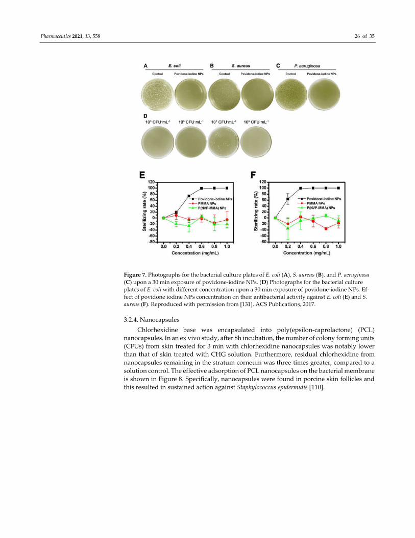
Polymeric nanoparticles (NP) containing PVP-I were fabricated using a surfactant-
free emulsion copolymerization followed by an iodination procedure. The nanoparticles
eliminated 100% of the isolated organisms, including E. coli and S. aureus, and P. aeruginosa
(Figure 7) and the decreased hydrophobicity enabled the PVP-I to be amalgamated into
conventional products like glue, ink, or dye [131].
Pharmaceutics 2021, 13, 558 26 of 35
Figure 7. Photographs for the bacterial culture plates of E. coli (A), S. aureus (B), and P. aeruginosa
(C) upon a 30 min exposure of povidone-iodine NPs. (D) Photographs for the bacterial culture
plates of E. coli with different concentration upon a 30 min exposure of povidone-iodine NPs. Ef-
fect of povidone iodine NPs concentration on their antibacterial activity against E. coli (E) and S.
aureus (F). Reproduced with permission from [131], ACS Publications, 2017.
3.2.4. Nanocapsules
Chlorhexidine base was encapsulated into poly(epsilon-caprolactone) (PCL)
nanocapsules. In an ex vivo study, after 8h incubation, the number of colony forming units
(CFUs) from skin treated for 3 min with chlorhexidine nanocapsules was notably lower
than that of skin treated with CHG solution. Furthermore, residual chlorhexidine from
nanocapsules remaining in the stratum corneum was three-times greater, compared to a
solution control. The effective adsorption of PCL nanocapsules on the bacterial membrane
is shown in Figure 8. Specifically, nanocapsules were found in porcine skin follicles and
this resulted in sustained action against Staphylococcus epidermidis [110].

Pharmaceutics 2021, 13, 558 27 of 35
Figure 8. Scanning electron micrographs of 0.6% chlorhexidine base loaded PCL nanocapsules
localization on stratum corneum-associated bacteria. Drug loaded nanocapsules adsorbed on bac-
teria membrane (BC). Reproduced with permission from [110], Journal of Controlled Release,
2002.
Nanoemulsions and nanocapsules containing 10 mg/mL TTO were evaluated in two
different infectious nail models. Generally, the nanosystems were effective at reducing the
growth of T. rubrum which was evidenced through the significant diminution of microor-
ganism count as well as the smallest zones of T. rubrum growth after exposure. Particu-
larly, compared to the nanoemulsion, the tea tree oil nanocapsules were more efficacious
against fungi [117]. Further studies incorporated these TTO loaded nanosystems into hy-
drogel preparations. Based on the results of in vivo studies, hydrogels comprised of TTO
nanocarriers reduced inflammation caused by UV-B radiation and in the wound healing
process, with the most effective being TTO nanocapsule hydrogels [118].
Nanocapsule formulations have been proposed to address increasing antimicrobial
resistance. Triclosan nanocapsules were formulated by interfacial deposition and used
chitosan as a coating layer and α-bisabolol as an oily core. Positively charged chitosan was
included to optimize interaction with negative charged microorganism membranes and
α-bisabolol was selected for its ability to disperse lipophilic drugs such as triclosan. Re-
sultant MICs of nanocapsules coated with chitosan were lower than other formulations
and the chitosan-coated nanocapsules were incorporated into wound dressings where
they were shown to extend the duration and extent of antimicrobial activity [113].
3.2.5. Other Novel Pharmaceutical Formulations
A novel formulation comprising phospholipid (Phospholipon90G) and octenidine
dihydrochloride was developed as an alternative for phenoxyethanol, which is often
added as solubility enhancer for octenidine but may cause irritation, especially on the
Pharmaceutics 2021, 13, 558 28 of 35
mucosae and open wounds). According to an antiseptic efficacy test, the lipid-based for-
mulation had a similar inhibitory potency as a marketed product Octanisept®, but had
potentially wider application due to the elimination of phenoxyethanol from the formu-
lation [124].
Liquid crystalline systems (LCS) of glyceryl monooleate (GMO) and water were de-
veloped as delivery systems for PHMB and cetylpyridinium chloride (CPC). The authors
found that the inclusion of the active drugs into LCS affected the drug release, but not the
creation of the liquid crystalline phases. Because of the interaction between CPC and
GMO, the drug was trapped in the matrix and not likely to release into the medium, lead-
ing to a deleterious impact on bactericidal activity. In contrast, PHMB was released at a
constant rate, thus having prolonged antibacterial activity against tested pathogens. In
general, the evidence from this study suggests that the liquid crystalline systems can used
as a carrier for PHMB [103].
Advanced drug delivery systems have been increasingly investigated for topical ad-
ministration, primarily applying numerous forms of nano-technology. These formula-
tions demonstrated superior therapeutic activities in prevention and treatment of skin and
wound infections, compared to conventional dosage forms. However, they show promis-
ing potential in vitro but there is a lack of data on products moving into clinical trials and
onto the market.
The safety profile and potential toxic effects of nanomaterials is not fully understood
and risk/benefit ratio has to be considered [107]. Following topical application, the parti-
cles need to remain at the site of action and not enhance uptake into the systemic circula-
tion. Skin permeation studies of formulations must confirm that there is limited absorp-
tion through the skin, and this may be further complicated by any infection that compro-
mises the natural barrier function of the skin
Potential toxicity of nanocarriers can also be caused by chemical mechanisms due to
the production of reactive oxygen species, dissolution and release of toxic ions, disturb-
ance of electron/ion cell membrane transport activity, oxidative damage through catalysis,
lipid peroxidation, and surfactant properties. Meanwhile, the nanoparticle size and sur-
face properties of nanoformulations are considered as physical factors result in toxicolog-
ical effects. They relate to membrane damage and disruption of membrane activity, and
can affect transport processes, protein conformation/folding, and protein aggregation/fi-
brillation [132]. Specifically, several studies revealed that silver nanoparticles may cause
genotoxic and cytotoxic on human cells [133–135]. However, the benefits have been
demonstrated in vitro and with the growing issues of antimicrobial resistance, there is
increasing pressure to use what we already have more effectively.
Finally, Figure 9 summarises the annual distribution of publications focused on anti-
septic formulations included in this review article. As is evident in Figure 9, the there is a
large increase in the number of publications after 2013.

Pharmaceutics 2021, 13, 558 29 of 35
Figure 9. The number of publications on antiseptic formulations for skin and soft tissue infections each year.
4. Conclusions
The current review has successfully gathered comprehensive information on various
antiseptic formulations employed to prevent and treat skin and soft tissue infections. It is
evident from the current review that research in recent years has established topical,
mostly dermal, delivery as a promising route. Its ability to bypass the hepatic first-pass
metabolism and easy accessibility yet relatively impermeability holds great promise, es-
pecially in the treatment of skin infections. This distinctive advantage allows the applica-
tion of a wide range of external dosage forms that can be easily removed if necessary.
These formulations have evolved from simple ointments, creams, and solutions to ad-
vanced nanotechnological assisted formulations. However, it is of equal importance that
these sophisticated formulations should address clinical and market needs. It is expected
that this review will be a helpful resource for formulation scientists to understand and
further to develop the antiseptic skin formulations to achieve specific therapeutic objec-
tives.
Author Contributions: Conceptualization, M.U.G. and B.R.C.; methodology, M.U.G and T.P.N.H.;
writing—T.P.N.H; writing—review and editing, M.U.G and B.R.C.; visualization, M.U.G. and
B.R.C.; supervision, M.U.G. and B.R.C.; project administration, B.R.C. All authors have read and
agreed to the published version of the manuscript.
Funding: This research received no external funding.
Institutional Review Board Statement: Not applicable.
Informed Consent Statement: Not applicable.
Conflicts of Interest: The authors declare no conflict of interest.
0
1
2
3
4
5
6
7
8
9
10
Publications number by year
Pharmaceutics 2021, 13, 558 30 of 35
References
1. Dryden, M.S. Skin and soft tissue infection: Microbiology and epidemiology. Int. J. Antimicrob. Agents 2009, 34, 2–7.
2. Tognetti, L.; Martinelli, C.; Berti, S.; Hercogova, J.; Lotti, T.; Leoncini, F.; Moretti, S. Bacterial skin and soft tissue infections:
Review of the epidemiology, microbiology, aetiopathogenesis and treatment. J. Eur. Acad. Dermatol. 2012, 26, 931–941.
3. Selcuk, M.; Aysegul, U.; Demet, K.; Kalih, L.C.. Bacterial skin infections: Epidemiology and latest research. Turk. J. Fam. Med.
Prim. Care 2015, 9, 65–74.
4. Vinh, D.C.; Embil, J.M. Rapidly progressive soft tissue infections. Lancet Infect. Dis. 2005, 5, 501–513.
5. Hersh, A.L.; Chambers, H.F.; Maselli, J.H.; Gonzales, R. National trends in ambulatory visits and antibiotic prescribing for skin
and soft-tissue infections. Arch. Intern. Med. 2008, 168, 1585–1591.
6. Lee, G.C.; Boyd, N.K.; Lawson, K.A.; Frei, C.R. Incidence and cost of skin and soft tissue infections in the United States. Value
Health 2015, 18, A245.
7. Lim, H.W.; Collins, S.A.B.; Resneck, J.S.; Jr.; Bolognia, J.L.; Hodge, J.A.; Rohrer, T.A.; Van Beek, M.J.; Margolis, D.J.; Sober, A.J.;
Weinstock, M.A.; et al. The burden of skin disease in the United States. J. Am. Acad. Dermatol. 2017, 76, 958–972.
8. Esposito, S.; Noviello, S.; Leone, S. Epidemiology and microbiology of skin and soft tissue infections. Curr. Opin. Infect. Dis.
2016, 29, 109–115.
9. James, S.L.; Abate, D.; Abate, K.H.; Abay, S.M.; Abbafati, C.; Abbasi, N.; Abbastabar, H.; Abd-Allah, F.; Abdela, J.; Abdelalim,
A.; et al. Global, regional, and national incidence, prevalence, and years lived with disability for 354 diseases and injuries for
195 countries and territories, 1990–2017: A systematic analysis for the Global Burden of Disease Study 2017. The Lancet 2018,
392, 1789–1858.
10. Moffarah, A.S.; Al Mohajer, M.; Hurwitz, B.L.; Armstrong, D.G. Skin and soft tissue infections. Microbiol. Immunocompromised
Host 2016, 4, 691–708.
11. Wounds UK. Best Practice Statement: Maintaining Skin Integrity. Available online: https://www.wounds-
uk.com/resources/details/maintaining-skin-integrity (accessed on 19 December 2019).
12. Malheiro, J.; Simões, M. 4-Antimicrobial resistance of biofilms in medical devices. In Biofilms and Implantable Medical Devices,
Deng, Y., Lv, W., Eds.; Woodhead Publishing: Cambridge, UK, 2017; pp. 97–113.
13. English, J.S.C. General Dermatology: An Atlas of Diagnosis and Management; Clinical Publishing: Oxford, UK, 2007.
14. World Health Organization. Epidemiology and Management of Common Skin Diseases in Children in Developing Countries.
Availabe online: https://apps.who.int/iris/handle/10665/69229 (accessed on 15 December 2019).
15. Poulakou, G.; Lagou, S.; Tsiodras, S. What’s new in the epidemiology of skin and soft tissue infections in 2018? Curr. Opin. Infect.
Dis. 2019, 32, 77–86.
16. Suaya, J.A.; Mera, R.M.; Cassidy, A.; O’Hara, P.; Amrine-Madsen, H.; Burstin, S.; Miller, L.G. Incidence and cost of
hospitalizations associated with Staphylococcus aureus skin and soft tissue infections in the United States from 2001 through
2009. BMC Infect. Dis. 2014, 14, 296–296.
17. Klein, E.Y.; Mojica, N.; Jiang, W.; Cosgrove, S.E.; Septimus, E.; Morgan, D.J.; Laxminarayan, R. Trends in methicillin-resistant
Staphylococcus aureus hospitalizations in the United States, 2010–2014. Clin. Infect. Dis. 2017, 65, 1921–1923.
18. Karimkhani, C.; Dellavalle, R.P.; Coffeng, L.E.; Flohr, C.; Hay, R.J.; Langan, S.M.; Nsoesie, E.O.; Ferrari, A.J.; Erskine, H.E.;
Silverberg, J.I.; et al. Global skin disease morbidity and mortality: An update from the Global Burden of Disease Study 2013.
JAMA Dermatol. 2017, 153, 406–412.
19. Kaye, K.S.; Petty, L.A.; Shorr, A.F.; Zilberberg, M.D. Current epidemiology, etiology, and burden of acute skin infections in the
United States. Clin. Infect. Dis. 2019, 68, S193–S199.
20. Drosou, A.; Falabella, A.; Kirsner, R.S. Antiseptics on Wounds: An Area of Controversy. Availabe online:
https://www.woundsresearch.com/article/1585 (accessed on 27 November 2019).
21. Leaper, D. Topical antiseptics in wound care: Time for reflection. Int. Wound J. 2011, 8, 547–549.
22. Leaper, D.J.; Schultz, G.; Carville, K.; Fletcher, J.; Swanson, T.; Drake, R. Extending the TIME concept: What have we learned in
the past 10 years?. Int. Wound J. 2012, 9, 1–19.
23. The, U.S. Food and Drug Administration (FDA). Q&A for Consumers: Health Care Antiseptics. Availabe online:
https://www.fda.gov/drugs/information-drug-class/qa-consumers-health-care-antiseptics (accessed on 21 Decmber 2019).
24. World Health Organization. Summary of a systematic literature review on surgical site preparation. Availabe online:
https://www.who.int/gpsc/appendix8.pdf (accessed on 12 November 2019).
25. Chang, R.K.; Raw, A.; Lionberger, R.; Yu, L. Generic development of topical dermatologic products: Formulation development,
process development, and testing of topical dermatologic products. AAPS J. 2013, 15, 41–52.
26. McDonnell, G.E. Antisepsis, Disinfection, and Sterilization: Types, Action, and Resistance; ASM Press: Washington, DC, USA, 2007.
27. Berríos-Torres, S.I.; Umscheid, C.A.; Bratzler, D.W.; Leas, B.; Stone, E.C.; Kelz, R.R.; Reinke, C.E.; Morgan, S.; Solomkin, J.S.;
Mazuski, J.E.; et al. Centers for Disease Control and Prevention Guideline for the Prevention of Surgical Site Infection, 2017.
JAMA Surgery 2017, 152, 784–791.
Pharmaceutics 2021, 13, 558 31 of 35
28. Claesen, J. Topical antiseptics and the skin microbiota. J. Investig. Dermatol. 2018, 138, 2106–2107.
29. Tanzer, J.M.; Slee, A.M.; Kamay, B.A. Structural requirements of guanide, biguanide, and bisbiguanide agents for antiplaque
activity. Antimicrob. Agents Chemother. 1977, 12, 721–729.
30. World Health Organization. Global Guidelines on the Prevention of Surgical Site Infection. Availabe online:
https://apps.who.int/iris/bitstream/handle/10665/250680/9789241549882-eng.pdf?sequence=8 (accessed on 11 December 2019).
31. Privitera, G.P.; Costa, A.L.; Brusaferro, S.; Chirletti, P.; Crosasso, P.; Massimetti, G.; Nespoli, A.; Petrosillo, N.; Pittiruti, M.;
Scoppettuolo, G.; et al. Skin antisepsis with chlorhexidine versus iodine for the prevention of surgical site infection: A systematic
review and meta-analysis. Am. J. Infect. Control. 2017, 45, 180–189.
32. O’Grady, N.P.; Alexander, M.; Burns, L.A.; Dellinger, E.P.; Garland, J.; Heard, S.O.; Lipsett, P.A.; Masur, H.; Mermel, L.A.;
Pearson, M.L.; et al. Guidelines for the prevention of intravascular catheter-related infections. Clin. Infect. Dis. 2011, 52, e162–
e193.
33. Macias, J.H.; Alvarez, M.F.; Arreguin, V.; Muñoz, J.M.; Macias, A.E.; Alvarez, J.A. Chlorhexidine avoids skin bacteria
recolonization more than triclosan. Am. J. Infect. Control. 2016, 44, 1530–1534.
34. Yasuda, H.; Sanui, M.; Abe, T.; Shime, N.; Komuro, T.; Hatakeyama, J.; Matsukubo, S.; Kawano, S.; Yamamoto, H.; Andoh, K.;
et al. Comparison of the efficacy of three topical antiseptic solutions for the prevention of catheter colonization: A multicenter
randomized controlled study. Crit. Care 2017, 21, 320–320.
35. Mozayeni, M.A.; Hadian, A.; Bakhshaei, P.; Dianat, O. Comparison of antifungal activity of 2% chlorhexidine, calcium
hydroxide, and nanosilver gels against Candida albicans. J. Dent. 2015, 12, 109–117.
36. Auda, S.H.; Mahrous, G.M.; Ibrahim, M.A.; Shazly, G.A.; Salem-Bekhit, M.M. Novel chlorhexidine dermal patches, preparation
characterization and antimicrobial evaluation. Polym. Bull. 2017, 74, 3995–4007.
37. Zinn, J.; Jenkins, J.B.; Swofford, V.; Harrelson, B.; McCarter, S. Intraoperative patient skin prep agents: Is there a difference?
AORN J. 2010, 92, 662–674.
38. Boyce, J.M. Best products for skin antisepsis. Am. J. Infect. Control. 2019, 47, 17–22.
39. Hayashi, M.; Kawamura, K.; Matsui, M.; Suzuki, M.; Suzuki, S.; Shibayama, K.; Arakawa, Y. Reduction in chlorhexidine efficacy
against multi-drug-resistant Acinetobacter baumannii international clone II. Hosp. Infect. 2017, 95, 318–323.
40. Bhardwaj, P.; Ziegler, E.; Palmer, K.L. Chlorhexidine Induces VanA-type vancomycin resistance genes in Enterococci.
Antimicrob. Agents Chemother. 2016, 60, 2209.
41. Wand, M.E.; Bock, L.J.; Bonney, L.C.; Sutton, J.M. Mechanisms of increased resistance to chlorhexidine and cross-resistance to
colistin following exposure of Klebsiella pneumoniae clinical isolates to chlorhexidine. Antimicrob. Agents Chemother. 2016, 61,
e01162–01116.
42. Williamson, D.A.; Carter, G.P.; Howden, B.P. Current and emerging topical antibacterials and antiseptics: Agents, action, and
resistance patterns. Clin. Microbiol. Rev. 2017, 30, 827–860.
43. Lee, J.; Kwack, S.; Shin, C.; Jang, H.-J.; Kim, H.; Kim, M.; Seo, D.-W.; Lee, B.; Kim, K.-B. Risk assessment of triclosan, a cosmetic
preservative. Toxicol. Res. 2019, 35, 137–154.
44. Schweizer, H.P. Triclosan: A widely used biocide and its link to antibiotics. FEMS Microbiol. Lett. 2001, 202, 1–7.
45. Günther Hierholzer, E.R.W.; Karen Reimer. Topische Infektions therapie und Prophylaxe. Aktueller Stellenwert von PVP-Jod.
Stuttgart Thieme 1996.
46. Do, T.K.T.; Hadji-Minaglou, F.; Antoniotti, S.; Fernandez, X. Authenticity of essential oils. Trends Anal. Chem. 2015, 66, 146–157.
47. Orchard, A.; van Vuuren, S. commercial essential oils as potential antimicrobials to treat skin diseases. Evid. Based Complement.
Alternat. Med. 2017, 2017, 4517971–4517971.
48. Rutherford, T.; Nixon, R.; Tam, M.; Tate, B. Allergy to tea tree oil: Retrospective review of 41 cases with positive patch tests over
4.5 years. Aust. J. Dermatol 2007, 48, 83–87.
49. Graham, C. The role of silver in wound healing. Br. J. Nurs. 2005, 14, S22–S28.
50. Jung, W.K.; Koo, H.C.; Kim, K.W.; Shin, S.; Kim, S.H.; Park, Y.H. Antibacterial activity and mechanism of action of the silver
ion in Staphylococcus aureus and Escherichia coli. Appl. Environ. Microbiol. 2008, 74, 2171–2178.
51. Lansdown, A. Silver in health care: Antimicrobial effects and safety in use. Curr. Probl. Dermatol. 2006, 33, 17–34.
52. Weedon, D. 14-Cutaneous deposits. In Weedon’s Skin Pathology (Third Edition); Weedon, D., Ed.; Churchill Livingstone:
Edinburgh, UK, 2010; pp. 369–396.e320.
53. National Center for Biotechnology Information. Benzethonium Chloride, CID=8478. Availabe online:
https://pubchem.ncbi.nlm.nih.gov/compound/Benzethonium-chloride (accessed on 20 December 2019).
54. Magee, P. 24-Antiseptic drugs and disinfectants. In Side Effects of Drugs Annual; Aronson, J.K., Ed.; Elsevier: Amsterdam, The
Netherlands, 2010; 32, pp. 437–443.
55. McKeen, L. 1-Introduction to Food Irradiation and Medical Sterilization. In The Effect of Sterilization on Plastics and Elastomers
(Third Edition); McKeen, L., Ed.; William Andrew Publishing: Boston, MA, USA, 2012; pp. 1–40.

Pharmaceutics 2021, 13, 558 32 of 35
56. Kim, M.; Weigand, M.R.; Oh, S.; Hatt, J.K.; Krishnan, R.; Tezel, U.; Pavlostathis, S.G.; Konstantinidis, K.T. Widely used
benzalkonium chloride disinfectants can promote antibiotic resistance. Appl. Environ. Microbiol. 2018, 84, 17.
57. Pitten, F.-A.; Kramer, A. Efficacy of cetylpyridinium chloride used as oropharyngeal antiseptic. Arzneimittelforschung 2001, 51,
588–595.
58. National Center for Biotechnology Information. Cetylpyridinium chloride, CID=31239. Availabe online:
https://pubchem.ncbi.nlm.nih.gov/compound/Cetylpyridinium-chloride (accessed on 15 December 2019).
59. Al-Doori, Z.; Goroncy-Bermes, P.; Gemmell, C.G.; Morrison, D. Low-level exposure of MRSA to octenidine dihydrochloride
does not select for resistance. J. Antimicrob. Chemother. 2007, 59, 1280–1281.
60. Hübner, N.O.; Siebert, J.; Kramer, A. Octenidine dihydrochloride, a modern antiseptic for skin, mucous membranes and
wounds. Skin Pharmacol. Physiol. 2010, 23, 244–258.
61. Conceição, T.; de Lencastre, H.; Aires-de-Sousa, M. Efficacy of octenidine against antibiotic-resistant Staphylococcus aureus
epidemic clones. J. Antimicrob. Chemother. 2016, 71, 2991–2994.
62. Alvarez-Marin, R.; Aires-de-Sousa, M.; Nordmann, P.; Kieffer, N.; Poirel, L.J.E.J.o.C.M.; Diseases, I. Antimicrobial activity of
octenidine against multidrug-resistant Gram-negative pathogens. Eur. J. Clin. Microbiol. Infect. Dis. 2017, 36, 2379–2383.
63. Dettenkofer, M.; Jonas, D.; Wiechmann, C.; Rossner, R.; Frank, U.; Zentner, J.; Daschner, F.D. Effect of skin disinfection with
octenidine dihydrochloride on insertion site colonization of intravascular catheters. Infection 2002, 30, 282–285.
64. Marzenna, B.; Anna, R.; Marek, K.; Anna, P. Penetration of a selected antibiotic and antiseptic into a biofilm formed on
orthopedic steel implants. Orthop. Traumatol. Rehabil. 2007, 9, 310–318.
65. Fjeld, H.; Lingaas, E. Polyhexanide-safety and efficacy as an antiseptic. Tidsskr Nor Legeforen 2016, 136, 707–711.
66. Yanai, R.; Ueda, K.; Nishida, T.; Toyohara, M.; Mori, O. Effects of ionic and surfactant agents on the antimicrobial activity of
polyhexamethylene biguanide. Eye Contact Lens 2011, 37, 85–89.
67. Rohrer, N.; Widmer, A.F.; Waltimo, T.; Kulik, E.M.; Weiger, R.; Filipuzzi-Jenny, E.; Walter, C. Antimicrobial efficacy of 3 oral
antiseptics containing octenidine, polyhexamethylene biguanide, or citroxx: Can chlorhexidine be replaced? Infect. Control.
Hosp. Epidemiol. 2010, 31, 733–739.
68. Harbs, N.; Siebert, J. In vitro efficacy of octenidine and polihexanide against biofilms composed of Pseudomonas aeruginosa.
GMS Krankenh. Interdiszip. 2007, 2, 45.
69. Hübner, N.O.; Kramer, A. Review on the Efficacy, Safety and Clinical Applications of Polihexanide, a Modern Wound
Antiseptic. Skin Pharmacol. Physiol. 2010, 23, 17–27.
70. Bashir, M.H.; Hollingsworth, A.; Schwab, D.; Prinsen, K.S.; Paulson, J.E.; Morse, D.J.; Bernatchez, S.F. Ex vivo and in vivo
evaluation of residual chlorhexidine gluconate on skin following repetitive exposure to saline and wiping with 2%
chlorhexidine gluconate/70% isopropyl alcohol pre-operative skin preparations. J. Hosp. Infect. 2019, 102, 256–261.
71. Macias, J.H.M.D.; Arreguin, V.M.D.; Munoz, J.M.M.D.; Alvarez, J.A.M.D.; Mosqueda, J.L.M.D.; Macias, A.E.M.D. Chlorhexidine
is a better antiseptic than povidone iodine and sodium hypochlorite because of its substantive effect. Am. J. Infect. Control. 2013,
41, 634–637.
72. Chiang, S.-R.; Jung, F.; Tang, H.-J.; Chen, C.-C.; Chen, C.-H.; Chou, H.-Y.; Chuang, Y.-C. Desiccation and ethanol resistances of
multidrug resistant Acinetobacter baumannii embedded in biofilm: The favorable antiseptic efficacy of combination
chlorhexidine gluconate and ethanol. J. Microbiol. Immunol. Infect. 2018, 51, 770–777.
73. Cavallari, C.; Brigidi, P.; Fini, A. Ex-vivo and in-vitro assessment of mucoadhesive patches containing the gel-forming
polysaccharide psyllium for buccal delivery of chlorhexidine base. Int. J. Pharm. 2015, 496, 593–600.
74. Jug, M.; Kosalec, I.; Maestrelli, F.; Mura, P. Development of low methoxy amidated pectin-based mucoadhesive patches for
buccal delivery of triclosan: Effect of cyclodextrin complexation. Carbohydr. Polym. 2012, 90, 1794–1803.
75. Kim, S.A.; Moon, H.; Lee, K.; Rhee, M.S. Bactericidal effects of triclosan in soap both in vitro and in vivo. J. Antimicrob. Chemother.
2015, 70, 3345–3352.
76. Igwebike-ossi, C.D.; Francis, O.O.; Oke, B. Formulation and antimicrobial activity of triclosan based conditioning medicated
shampoo. Int. J. Appl. Chem. 2017, 13, 787–798.
77. Hoekstra, M.J.; Westgate, S.J.; Mueller, S. Povidone-iodine ointment demonstrates in vitro efficacy against biofilm formation.
Int. Wound J. 2017, 14, 172–179.
78. Eggers, M.; Eickmann, M.; Kowalski, K.; Zorn, J.; Reimer, K. Povidone-iodine hand wash and hand rub products demonstrated
excellent in vitro virucidal efficacy against Ebola virus and modified vaccinia virus Ankara, the new European test virus for
enveloped viruses. BMC Infect. Dis. 2015, 15, 375–375.
79. Pawar, V.; Topkar, H.; Srivastava, R. Chitosan nanoparticles and povidone iodine containing alginate gel for prevention and
treatment of orthopedic implant associated infections. Int. J. Biol. Macromol. 2018, 115, 1131–1141.
80. Capriotti, K.D.; Anadkat, M.; Choi, J.; Kaffenberger, B.; McLellan, B.; Barone, S.; Kukoyi, O.; Goldfarb, S.; Lacouture, M. A
randomized phase 2 trial of the efficacy and safety of a novel topical povidone-iodine formulation for cancer therapy-associated
paronychia. Investig. New Drugs 2019, 37, 1247–1256.

Pharmaceutics 2021, 13, 558 33 of 35
81. Jalil, A.; Matuszczak, B.; Nguyen Le, N.-M.; Mahmood, A.; Laffleur, F.; Bernkop-Schnürch, A. synthesis and characterization of
thiolated pvp-iodine complexes: Key to highly mucoadhesive antimicrobial gels. Mol. Pharm. 2018, 15, 3527–3534.
82. Suchomel, M.; Weinlich, M.; Kundi, M. Influence of glycerol and an alternative humectant on the immediate and 3-hours
bactericidal efficacies of two isopropanolbased antiseptics in laboratory experiments in vivo according to EN 12791 Antimicrob.
Resist. Infect. Control. 2017, 6, 72.
83. Martínez, J.M.D.; Macías, J.H.M.D.; Arreguín, V.M.D.; Álvarez, J.A.M.D.; Macías, A.E.M.D.; Mosqueda-Gómez, J.L.M.D.
Isopropyl alcohol is as efficient as chlorhexidine to prevent contamination of blood cultures. Am. J. Infect. Control. 2017, 45, 350–
353.
84. Djozic, H.; Pandza, H.; Hasukic, S.; Custovic, S.; Pandza, B.; Krupalija, A.; Beciragic, E. Efficiency of local antiseptic alkosol
(ethanol, isopropanol-30g and ortophenilphenol) and povidone iodide on the incidence of surgical site infection after inguinal
hernioplasty. Med. Arch. 2016, 70, 108–111.
85. Choi, O.; Deng, K.K.; Kim, N.-J.; Ross, L.; Surampalli, R.Y.; Hu, Z. The inhibitory effects of silver nanoparticles, silver ions, and
silver chloride colloids on microbial growth. Water Res. 2008, 42, 3066–3074.
86. Czerwinski, S.E.; Cozean, J.; Cozean, C. Novel water-based antiseptic lotion demonstrates rapid, broad-spectrum kill compared
with alcohol antiseptic. J. Infect. Public Health 2014, 7, 199–204.
87. Gnatta, J.R.; Pinto, F.M.; Bruna, C.Q.; Souza, R.Q.; Graziano, K.U.; Silva, M.J. Comparison of hand hygiene antimicrobial
efficacy: Melaleuca alternifolia essential oil versus triclosan. Rev. Lat. Am. Enfermagem 2013, 21, 1212–1219.
88. Dryden, M.S.; Dailly, S.; Crouch, M. A randomized, controlled trial of tea tree topical preparations versus a standard topical
regimen for the clearance of MRSA colonization. J. Hosp. Infect. 2004, 56, 283–286.
89. Aiello, A.E.; Larson, E.L.; Levy, S.B. Consumer antibacterial soaps: Effective or just risky? Clin. Infect. Dis. 2007, 45, S137–S147.
90. Caelli, M.; Porteous, J.; Carson, C.F.; Heller, R.; Riley, T.V. Tea tree oil as an alternative topical decolonization agent for
methicillin-resistant Staphylococcus aureus. J. Hosp. Infect. 2000, 46, 236–237.
91. Picheansathian, W. A systematic review on the effectiveness of alcohol-based solutions for hand hygiene. Int. J. Nurs. Pract.
2004, 10, 3–9.
92. Koburger, T.; Hübner, N.-O.; Braun, M.; Siebert, J.; Kramer, A. Standardized comparison of antiseptic efficacy of triclosan, PVP–
iodine, octenidine dihydrochloride, polyhexanide and chlorhexidine digluconate. J. Antimicrob. Chemother. 2010, 65, 1712–1719.
93. Sonje, A.; Chandra, A. Comprehencive review on eudragit polymers. Int. Res. J. Pharm. 2013, 4, 71–74.
94. Kaur, G.; Grewal, J.; Jyoti, K.; Jain, U.K.; Chandra, R.; Madan, J. Chapter 15-Oral controlled and sustained drug delivery systems:
Concepts, advances, preclinical, and clinical status. In Drug Targeting and Stimuli Sensitive Drug Delivery Systems; Grumezescu,
A.M., Ed.; William Andrew Publishing: Boston, MA, USA, 2018; pp. 567–626.
95. Brady, J.; Dürig, T.; Lee, P.I.; Li, J.X. Chapter 7-Polymer Properties and Characterization. In Developing Solid Oral Dosage Forms;
Qiu, Y., Chen, Y., Zhang, G.G.Z., Yu, L., Mantri, R.V., Eds.; Academic Press: Boston, MA, USA, 2017; pp. 181–223.
96. Patra, C.N.; Priya, R.; Swain, S.; Kumar Jena, G.; Panigrahi, K.C.; Ghose, D. Pharmaceutical significance of Eudragit: A review.
Future, J. Pharm. Sci. 2017, 3, 33–45.
97. Benson, H.A.E.; Watkinson, A.C. Topical and Transdermal Drug Delivery: Principles and Practice, 1st ed.; Wiley: Hoboken, NJ, USA,
2011.
98. Jones, D.S. Pharmaceutics: Dosage Form and Design; Pharmaceutical Press: London,UK, 2016.
99. Mahato, R.I.; Narang, A.S. Pharmaceutical Dosage Forms and Drug Delivery, 2nd ed.; CRC Press: Boca Raton, FL, USA, 2012.
100. FDA-Tentative Final Monograph. Topical antimicrobial products for over-the-counter-use, 21 CFR 333 and 369. Fred. Regist.
1994, 59.
101. Erasmus, V.; Daha, T.J.; Brug, H.; Richardus, J.H.; Behrendt, M.D.; Vos, M.C.; van Beeck, E.F. Systematic review of studies on
compliance with hand hygiene guidelines in hospital care. Infect. Control. Hosp. Epidemiol. 2010, 31, 283–294.
102. US FDA. Topical Antiseptic Products: Hand Sanitizers and Antibacterial Soaps. Availabe online:
https://www.fda.gov/drugs/information-drug-class/topical-antiseptic-products-hand-sanitizers-and-antibacterial-soaps
(accessed on 12 December 2019).
103. Souza, C.; Watanabe, E.; Aires, C.P.; Lara, M.G. Effect of liquid crystalline systems containing antimicrobial compounds on
infectious skin bacteria. AAPS PharmSciTech. 2017, 18, 2110–2119.
104. US FDA. Safety and Effectiveness of Consumer Antiseptics; Topical Antimicrobial Drug Products for Over-the-Counter Human
Use; Proposed Amendment of the Tentative Final Monograph; Reopening of Administrative Record. Availabe online:
https://www.federalregister.gov/documents/2013/12/17/2013–29814/safety-and-effectiveness-of-consumer-antiseptics-topical-
antimicrobial-drug-products-for (accessed on 25 December 2019).
105. US FDA. Safety and Effectiveness of Consumer Antiseptic Rubs; Topical Antimicrobial Drug Products for Over-the-Counter
Human Use. Availabe online: https://www.federalregister.gov/documents/2019/04/12/2019–06791/safety-and-effectiveness-of-
consumer-antiseptic-rubs-topical-antimicrobial-drug-products-for (accessed on 25 December 2019).
106. Neubert, R.H. Potentials of new nanocarriers for dermal and transdermal drug delivery. Eur. J. Pharm. Biopharm. 2011, 77, 1–2.

Pharmaceutics 2021, 13, 558 34 of 35
107. Liang, X.-J. Nanopharmaceutics: The Potential Application of Nanomaterials; World Scientific: Hackensack, NJ, USA, 2013.
108. Din, F.U.; Aman, W.; Ullah, I.; Qureshi, O.S.; Mustapha, O.; Shafique, S.; Zeb, A. Effective use of nanocarriers as drug delivery
systems for the treatment of selected tumors. Int. J. Nanomed. 2017, 12, 7291–7309.
109. Khan, M.A.; Mujahid, M.; Gul, I.H.; Hussain, A. Electrochemical study of magnetic nanogel designed for controlled release of
chlorhexidine gluconate. Electrochim. Acta 2019, 295, 113–123.
110. Lboutounne, H.; Chaulet, J.-F.; Ploton, C.; Falson, F.; Pirot, F. Sustained ex vivo skin antiseptic activity of chlorhexidine in poly(ϵ-
caprolactone) nanocapsule encapsulated form and as a digluconate. J. Control. Release 2002, 82, 319–334.
111. Kettel, M.J.; Heine, E.; Schaefer, K.; Moeller, M. Chlorhexidine loaded cyclodextrin containing PMMA nanogels as antimicrobial
coating and delivery systems. Macromol. Biosci. 2017, 17, 1600230.
112. Kakadia, P.G. Formulation and Evaluation of Nanoencapsulated Antimicrobial Agents for Dermal Delivery. Ph.D. Thesis,
University of Huddersfield, UK, 2016.
113. De Marchi, J.G.B.; Jornada, D.S.; Silva, F.K.; Freitas, A.L.; Fuentefria, A.M.; Pohlmann, A.R.; Guterres, S.S. Triclosan resistance
reversion by encapsulation in chitosan-coated-nanocapsule containing α-bisabolol as core: Development of wound dressing.
Int. J. Nanomed. 2017, 12, 7855—7868.
114. Davachi, S.M.; Kaffashi, B. Preparation and Characterization of Poly L-Lactide/Triclosan Nanoparticles for Specific Antibacterial
and Medical Applications. Int. J. Polym. Mater. 2015, 64, 497–508.
115. Wais, U.; Nawrath, M.M.; Jackson, A.W.; Zhang, H. Triclosan nanoparticles via emulsion-freeze-drying for enhanced
antimicrobial activity. Colloid. Polym. Sci. 2018, 296, 951–960.
116. Domínguez-Delgado, C.L.; Rodríguez-Cruz, I.M.; Escobar-Chávez, J.J.; Calderón-Lojero, I.O.; Quintanar-Guerrero, D.; Ganem,
A. Preparation and characterization of triclosan nanoparticles intended to be used for the treatment of acne. Eur. J. Pharm.
Biopharm. 2011, 79, 102–107.
117. Flores, F.C.; de Lima, J.A.; Ribeiro, R.F.; Alves, S.H.; Rolim, C.M.B.; Beck, R.C.R.; da Silva, C.B. Antifungal activity of
nanocapsule suspensions containing tea tree oil on the growth of trichophyton rubrum. Mycopathologia 2013, 175, 281–286.
118. Flores, F.C.; De Lima, J.A.; Da Silva, C.R.; Benvegnu, D.; Ferreira, J.; Burger, M.E.; Beck, R.C.; Rolim, C.M.; Rocha, M.I.; Da Veiga,
M.L.; et al. Hydrogels containing nanocapsules and nanoemulsions of tea tree oil provide antiedematogenic effect and improved
skin wound healing. J. Nanosci. Nanotechnol. 2015, 15, 800–809.
119. Sinha, P.; Srivastava, S.; Mishra, N.; Singh, D.K.; Luqman, S.; Chanda, D.; Yadav, N.P. Development, optimization, and
characterization of a novel tea tree oil nanogel using response surface methodology. Drug Dev. Ind. Pharm. 2016, 42, 1434–1445.
120. Najafi-taher, R.; Ghaemi, B.; Kharazi, S.; Rasoulikoohi, S.; Amani, A. Promising antibacterial effects of silver nanoparticle-loaded
tea tree oil nanoemulsion: A synergistic combination against resistance threat. AAPS PharmSciTech. 2018, 19, 1133–1140.
121. Cao, Z.; Spilker, T.; Fan, Y.; Kalikin, L.M.; Ciotti, S.; LiPuma, J.J.; Makidon, P.E.; Wilkinson, J.E.; Baker, J.R.; Jr.; Wang, S.H.
Nanoemulsion is an effective antimicrobial for methicillin-resistant Staphylococcus aureus in infected wounds. Nanomedicine
2017, 12, 1177–1185.
122. Pannu, J.; McCarthy, A.; Martin, A.; Hamouda, T.; Ciotti, S.; Fothergill, A.; Sutcliffe, J. NB-002, a Novel Nanoemulsion with
Broad Antifungal Activity against Dermatophytes, Other Filamentous Fungi, and Candida albicans. Antimicrob. Agents
Chemother. 2009, 53, 3273–3279.
123. Ulmer, M.; Patzelt, A.; Vergou, T.; Richter, H.; Müller, G.; Kramer, A.; Sterry, W.; Czaika, V.; Lademann, J. In vivo investigation
of the efficiency of a nanoparticle-emulsion containing polihexanide on the human skin. Eur. J. Pharm. Biopharm. 2013, 84, 325–
329.
124. Szostak, K.; Czogalla, A.; Przybyło, M.; Langner, M. New lipid formulation of octenidine dihydrochloride. J. Liposome Res. 2018,
28, 106–111.
125. Hamedi, H.; Moradi, S.; Tonelli, A.E.; Hudson, S.M. Preparation and characterization of chitosan–alginate polyelectrolyte
complexes loaded with antibacterial thyme oil nanoemulsions. Appl. Sciences 2019, 9, 3933.
126. Javadzadeh, Y.; Azharshekoufeh Bahari, L. Chapter 8-Therapeutic Nanostructures for Dermal and Transdermal Drug Delivery.
In Nano- and Microscale Drug Delivery Systems, Grumezescu, A.M., Ed.; Elsevier: Amsterdam, The Netherlands, 2017; pp. 131–
146.
127. Hemmila, M.R.; Mattar, A.; Taddonio, M.A.; Arbabi, S.; Hamouda, T.; Ward, P.A.; Wang, S.C.; Baker, J.R.; Jr. Topical
nanoemulsion therapy reduces bacterial wound infection and inflammation after burn injury. Surgery 2010, 148, 499–509.
128. Zhang, H.; Zhai, Y.; Wang, J.; Zhai, G. New progress and prospects: The application of nanogel in drug delivery. Mater. Sci. Eng.
C. 2015, 60.
129. Petica, A.; Gavriliu, S.; Lungu, M.; Buruntea, N.; Panzaru, C. Colloidal silver solutions with antimicrobial properties. Mater. Sci.
Eng. B 2008, 152, 22–27.
130. Bamrungsap, S.; Zhao, Z.; Chen, T.; Wang, L.; Li, C.; Fu, T.; Tan, W. Nanotechnology in therapeutics: A focus on nanoparticles
as a drug delivery system. Nanomedicine 2012, 7, 1253–1271.

Pharmaceutics 2021, 13, 558 35 of 35
131. Gao, T.; Fan, H.; Wang, X.; Gao, Y.; Liu, W.; Chen, W.; Dong, A.; Wang, Y.-J. Povidone-Iodine-Based Polymeric Nanoparticles
for Antibacterial Applications. ACS Appl. Mater. Interfaces 2017, 9, 25738–25746.
132. Jamuna, B.A.; Ravishankar, R.V. Environmental Risk, Human Health, and Toxic Effects of Nanoparticles. In Nanomaterials for
Environmental Protection; Wiley: Hoboken, NJ, USA, 2014; pp. 523–535.
133. Hackenberg, S.; Scherzed, A.; Kessler, M.; Hummel, S.; Technau, A.; Froelich, K.; Ginzkey, C.; Koehler, C.; Hagen, R.;
Kleinsasser, N. Silver nanoparticles: Evaluation of DNA damage, toxicity and functional impairment in human mesenchymal
stem cells. Toxicol. Lett. 2011, 201, 27–33.
134. Beer, C.; Foldbjerg, R.; Hayashi, Y.; Sutherland, D.S.; Autrup, H. Toxicity of silver nanoparticles-nanoparticle or silver ion?
Toxicol. Lett. 2012, 208, 286–292.
135. Chairuangkitti, P.; Lawanprasert, S.; Roytrakul, S.; Aueviriyavit, S.; Phummiratch, D.; Kulthong, K.; Chanvorachote, P.;
Maniratanachote, R. Silver nanoparticles induce toxicity in A549 cells via ROS-dependent and ROS-independent pathways.
Toxicol. In Vitro 2013, 27, 330–338.


