Embed Size (px)
Citation preview
�����������������
Citation: Chakraborty, P.; Ghosh, A.
Topical Spray of dsRNA Induces
Mortality and Inhibits Chilli Leaf
Curl Virus Transmission by Bemisia
tabaci Asia II 1. Cells 2022, 11, 833.
https://doi.org/10.3390/
cells11050833
Academic Editors: Ahmed Hadidi,
Henryk Hanokh Czosnek and
Suleyman Allakhverdiev
Received: 28 December 2021
Accepted: 18 February 2022
Published: 28 February 2022
Publisher’s Note: MDPI stays neutral
with regard to jurisdictional claims in
published maps and institutional affil-
iations.
Copyright: © 2022 by the authors.
Licensee MDPI, Basel, Switzerland.
This article is an open access article
distributed under the terms and
conditions of the Creative Commons
Attribution (CC BY) license (https://
creativecommons.org/licenses/by/
4.0/).
cells
Article
Topical Spray of dsRNA Induces Mortality and Inhibits ChilliLeaf Curl Virus Transmission by Bemisia tabaci Asia II 1Prosenjit Chakraborty and Amalendu Ghosh *
Insect Vector Laboratory, Advanced Centre for Plant Virology, Indian Agricultural Research Institute,New Delhi 110012, India; [email protected]* Correspondence: [email protected] or [email protected]
Abstract: Chilli leaf curl virus (ChiLCV; genus: Begomovirus), transmitted by Bemisia tabaci (Gennadius)(Hemiptera: Aleyrodidae) in a persistent-circulative manner, is a major constraint in chilli production.The present study demonstrates for the first time that a topical spray of naked double-strandedRNA (dsRNA) on chilli plants causes mortality and inability to acquire and transmit ChiLCV in B.tabaci. dsRNA targeting heat shock protein 70 (hsp70) and fasciclin 2 (fas2) of B. tabaci Asia II 1 was firstassessed under controlled conditions through oral delivery. Hsp70 and fas2 dsRNA resulted in upto 82.22% and 72% mortality of B. tabaci and around 12.4- and 8.5-fold decreases in mRNA levels,respectively, 24 h post-ingestion. ChiLCV copies in hsp70 dsRNA-fed B. tabaci steadily decreasedwith an increase in dsRNA concentration and were undetectable at a higher concentration of dsRNA.However, ChiLCV copies significantly increased in fas2 dsRNA-fed B. tabaci. Transmission of ChiLCVby B. tabaci was completely inhibited post-24 h feeding on hsp70 dsRNA at 3 µg/mL. Naked hsp70dsRNA was topically sprayed on ChiLCV-infected chilli plants like an insecticide. 67.77% mortalityof B. tabaci, 4.6-fold downregulation of hsp70 mRNA, and 1.34 × 10¹⁵-fold decreased ChiLCV copiesin B. tabaci were recorded when adults were exposed to the dsRNA-treated plants under semi-fieldconditions. Foliar application of naked dsRNA reduced the ChiLCV transmission by 75% withoutany visible symptoms in the inoculated plants. A total of 2 consecutive sprays of dsRNA providedsignificant protection to B. tabaci for up to 20 days under semi-field conditions.
Keywords: silverleaf whitefly; begomovirus; RNAi; topical application; heat shock protein 70; fasciclin2; virus transmission; virus-vector relationship
1. Introduction
Silverleaf whitefly [Bemisia tabaci (Gennadius), Hemiptera: Aleyrodidae] is a phloem-feeding hemipteran insect that has been reported to infest over 600 plant species world-wide [1–3]. B. tabaci has been considered a complex of at least 46 morphologically in-distinguishable cryptic species [4–6]. B. tabaci adults are about 1 mm long with smalltriangular-shaped bodies. The body is yellowish with distinct hyaline wings dusted withwhite powdery wax. Adults B. tabaci can be distinguished from other whiteflies by theposition in which the wings are held over the body. The wings are held close to the bodyand tent-like in B. tabaci. It sucks phloem sap, leaving the affected plants extremely weak,and secretes a honeydew onto the surface of the leaves that promotes the growth of sootymold fungus [7]. Besides direct damage caused by feeding, B. tabaci transmits more thana hundred begomoviruses, carlaviruses, criniviruses, cytorhabdoviruses, ipomoviruses,poleroviruses, and torradoviruses ([8–16]. Among them, begomoviruses represent 90%of the viruses transmitted by B. tabaci. Begomovirus species have become widespread inCentral America, the Dominican Republic, Israel, Mexico, Trinidad, and across South EastAsia, including Cambodia, Indonesia, India, and Thailand [17]. These viruses can cause anestimated yield loss of 50–90% in tomatoes and other crops, including beans, cassava, chilli,cotton, cucurbits, eggplant, papaya, and potatoes [18,19].
Cells 2022, 11, 833. https://doi.org/10.3390/cells11050833 https://www.mdpi.com/journal/cells
Cells 2022, 11, 833 2 of 20
Chilli (Capsicum annum L., family Solanaceae) is one of the economically importantcrops produced in tropical and sub-tropical countries [20]. Chilli leaf curl virus (ChiLCV,genus Begomovirus, family Geminiviridae) is a major constraint of chilli production, causingannual losses of about USD 15 billion [21]. ChiLCV is a monopartite begomovirus thatcontains one circular, single-stranded DNA-A component of 2.7 kb in size. ChiLCV is trans-mitted by B. tabaci in a persistent-circulative manner. Capsicum spp. are the primary hostsfor ChiLCV, but it also infects tomato and amaranth [22,23]. ChiLCV has been responsiblefor several epidemics in India and Sri Lanka [24,25]. The disease is typically manifested inthe infected plants as upward curling, puckering, bunching of leaves, blistering of inter-veinal areas, thickening, and swelling of the veins, shortening of internodes and petioles,and stunting of the whole plant. The leaves become smaller and severely affected plantsproduce fewer and deformed fruits. Yield losses of 20–50% have been recorded in chilli [26],which may rise to 100% in the case of simultaneous infestation with thrips and mites [27].Control options for ChiLCV and B. tabaci are very limited as insecticides continue to losetheir efficacy due to the emergence of insecticide-resistant B. tabaci populations. Besides,insecticides adversely affect the environment and human health. Considering the continuedlack of consumer acceptance, transgenic plant technology is not a feasible strategy. In recentyears, RNA interference (RNAi) has shown promise in the management of insect pests andplant viruses. Double-stranded RNA (dsRNA), upon entering the host cell, is processedby the Dicer enzyme and, in association with RNA-induced silencing complex (RISC),cleaves the targeted mRNA [28]. Interrupting the interrelationship between B. tabaci andbegomovirus using RNAi is a promising approach.. For successful persistent-circulativetransmission, the begomovirus particles need to cross the midgut barrier of B. tabaci tocirculate in hemolymph and reach the primary salivary glands [29,30]. Several proteinsat the midguts of B. tabaci, such as heat shock proteins (Hsp), cyclophilins, peptidoglycanrecognition protein, and a midgut protein, are known to interact with begomovirus coatprotein (CP) for successful internalization [31–34]. RNA-Seq of B. tabaci in response to be-gomoviruses revealed significant upregulations of hsp70, fasciclin 2 (fas2), and several othertranscripts [31,35]. In gene regulatory network analyses, these genes were enriched withhigher degrees of interactions. The same trend was recorded in the mRNA expression inreverse transcriptase quantitative real-time PCR (RT-qPCR) post-exposure to ChiLCV [35].Although host cellular chaperones like Hsp70, along with other cochaperones play animportant role in several cellular processes, host immunity, and stress responses [36], hsp70has been reported to be associated with begomovirus transmission by B. tabaci [31]. Neu-ral cell adhesion molecule (NCAM) orthologues in B. tabaci like fas2 function in synapticdevelopment and growth [37,38]. Besides, NCAM molecules are known as receptors invirus replication [39]. The purpose of the present study is to validate the functions ofB. tabaci hsp70 and fas2 in ChiLCV transmission using RNAi and explore the potentiality touse them as novel genetic tools for pest management.
Although several potential RNAi targets were identified for insects, including B.tabaci [40–43], most of those studies were conducted under controlled experimental con-ditions, and their efficacy under field conditions is limited. Rapid degradation of dsRNAin the extracellular environment and lack of reliable dsRNA delivery technique limits itsapplicability under field conditions. The objective of the present study was to induceresistance against B. tabaci and ChiLCV by spray-on application of dsRNA. In the presentstudy, B. tabaci hsp70 and fas2 were silenced and resultant effects in mortality and virustransmission were reported. For the first time, the efficacy of spray-on application of nakeddsRNA against a hemipteran insect was demonstrated under semi-field conditions. Theoutcomes of the study provide a novel, eco-friendly option for protection against B. tabacias well as ChiLCV, which can reduce economic losses caused by the virus-vector complex.
Cells 2022, 11, 833 3 of 20
2. Materials and Methods2.1. Whitefly Population
A homogeneous isofemale line of B. tabaci was raised from a single adult female andhas been maintained on eggplants (var. Navkiran, Mahyco) at the Advanced Centre forPlant Virology, Indian Agricultural Research Institute (IARI), New Delhi, since 2015. Thepopulation was characterized by sequencing the mitochondrial cytochrome oxidase subunitI (mtCOI) gene (Table 1). The genotype or cryptic species of the B. tabaci population wasconfirmed based on Bayesian Inference phylogeny, considering a genetic divergence cutoffof 4% as described by Rehman et al. [6]. The population was maintained under controlledenvironmental conditions at 28 ± 2 ◦C, 60 ± 10% relative humidity, and a 16 h light-8 hdark photoperiod.
2.2. Virus Culture
The inoculum of ChiLCV was collected from a pure culture maintained at the Ad-vanced Centre for Plant Virology, IARI, New Delhi, India. ChiLCV was maintained in chilli(var. Preeti, Nunhems) by B. tabaci inoculation under insect-proof conditions. The identityof the virus was further confirmed by amplifying the DNA-A component in PCR withprimer pairs Begomo F and Begomo R [44] (Table 1) and sequencing.
2.3. Designing and Synthesis of dsRNA
For the selection of dsRNA fragments, complete gene sequences of B. tabaci hsp70(Accession Nos. HM367079, HM013712, EU934240, HM013709) and fas2 (Accession No.XM_019049173) were downloaded from NCBI. The conserved sequences of the hsp70 andfas2 genes were analyzed in siRNA Wizard 3.1, Invivogen. The segments showing po-tential siRNA-forming regions were tested for cross-reactivity with other organisms likeHomo sapiens (chromosomes, unplaced and unlocalized scaffolds), Mus musculus (chromo-somes, unplaced and unlocalized scaffolds), Aves (taxid:8782), Lepidoptera (taxid:7088),Hymenoptera (taxid:7399), Formicidae (taxid:36668), and plants (taxid:3193) in NCBI us-ing BLAST analysis. The region specific to B. tabaci with no cross-reactivity with otherorganisms was finally selected for synthesis of dsRNA.
Primers targeting the dsRNA segment were designed in NCBI primer blast (https://www.ncbi.nlm.nih.gov/tools/primer-blast/ (Accessed on 4 June 2021) (Table 1). Theprimers AG137F-AG138R (for hsp70) and AG283F-AG284R (for fas2) were validated ina gradient PCR. The dsRNA stretches were amplified from isofemale B. tabaci Asia II 1DNA in PCR with initial denaturation at 94 ◦C for 5 min, 30 cycles of 94 ◦C for 30 s,56 ◦C for 30 s, and 72 ◦C for 30 s and a final extension step at 72 ◦C for 10 min. ThePCR products were cloned into an L4440 plasmid vector between two T7 promoters. Therecombinant plasmids were transformed into RNase III mutant Escherichia coli HT115 cells.Total RNA from the recombinant E. coli HT115 cells was extracted using Trizol reagent(Invitrogen, CA, USA) following the manufacturer’s protocol, and dsRNA was purifiedfollowing Ahn et al. [45] with modifications. In brief, the recombinant E. coli HT115 cellswere incubated for 12 h with continuous shaking at 37 ◦C followed by induction of T7promoter with 1M isopropyl-β-D-1-thiogalactopyranoside (IPTG). The bacterial cells wereharvested in a 1.5 mL microcentrifuge tube by centrifugation at 10,000× g for 5 min andresuspended in 1 mL Trizol. The cell suspension was mixed by vortexing and kept atroom temperature for 5 min. The cell suspension was mixed with 200 µL chloroform,vortexed for <10 s, and incubated at room temperature for 10 min. The mixture was thencentrifuged at 16,000× g for 10 min at 4 ◦C. The upper clear aqueous phase was transferredto a fresh tube and 0.8 volume of ice-chilled isopropanol was added. The solution wasmixed properly and incubated at 4 ◦C for 10 min. The mixture was again centrifuged at16,000× g for 10 min at 4 ◦C, and the supernatant was discarded. The pellet was washedwith 70% ethanol, air-dried, and resuspended in 30 µL nuclease-free water. Total RNAisolated from recombinant E. coli HT115 cells was mixed with 1X RNA loading dye (ThermoFisher Scientific, MA, USA), heated at 70 ◦C for 5 min, and visualized on 2% agarose gel
Cells 2022, 11, 833 4 of 20
stained with GoodView (BR Biochem, New Delhi, India). The total RNA was then treatedwith 1 unit of DNase I, RNase-free (Thermo Fisher Scientific) and 1 unit of RNase A,DNase-, and protease-free (Thermo Fisher Scientific) in the presence of 500 mM sodiumchloride and incubated for 1 hr at 37 ◦C to eliminate the DNA and single-stranded RNAcontaminants. The enzymes were inactivated by chloroform extraction, and the remainingdsRNA was resuspended in nuclease-free water. The purified dsRNA was quantifiedin a spectrophotometer (NanoDrop 2000, Thermo Fisher Scientific), and the integrity ofthe specific dsRNA was confirmed on 2% native agarose gel stained with GoodView andvisualized in a gel documentation system (Maestrogen, Xiangshan District, Taiwan).
Table 1. Primers used in the study.
Primer Name Primer Sequence (5’-3’) Amplicon Size
AnnealingTemperature in
PCR/Real-TimePCR
Amplified Region Purpose Reference
C1-J-2195 TTGATTTTTTGGTCATCCAGAAGT860 bp 53 ◦C B. tabaci mtCOI
Detection of B.tabaci crypticspecies
[46]L2-N-3014 TCCAATGCACTAATCTGCCATATTA
Begomo F ACGCGTGCCGTGCTGCTGCCCCCATTGTCC2.7 kb 57 ◦C Begomovirus DNA-A Detection of
begomovirus [44]Begomo R ACGCGTATGGGCTGYCGAAGTTSAGAC
AG137F TCAAAGAACATTTTTGTGCTACT128 bp 56 ◦C
B. tabaci hsp70dsRNA
dsRNAsynthesis
This studyAG138R GACCATTGTCTAGGTCTTCATTT
AG283F CTGGTGTTTTGACAATCGAC150 bp 56 ◦C B. tabaci fas2 dsRNA
dsRNAsynthesis andRT-qPCR
This studyAG284R TGATTATGCCTTCTTCCGTC
AG177F ACATGGAAAAGATCTGGCAT121 bp 56 ◦C B. tabaci β-actin RT-qPCR This study
AG178R TGAGTCATCTTTTCACGGTT
AG204F GTCAATGATTGCAGTAAGCC105 bp 56 ◦C B. tabaci hsp70 RT-qPCR This study
AG205R TTCCCTCATTTTCGTAAGCA
AG149F TGAACAGGCCCATGAACAG290 bp 53 ◦C ChiLCV coat
protein
qPCR andChiLCVdetection
[47]AG150R ACGGACAAGGAAAAACATCAC
2.4. Bioassay of dsRNA through Artificial Feeding under Controlled Conditions
The purified hsp70 and fas2 dsRNA were individually and orally delivered to B. tabaciadults following the method of Upadhyay et al. [40] with modifications. In brief, an artificialdiet was prepared by mixing 20% sucrose and 5% yeast extract in sterile distilled water andautoclaved. The artificial diet was supplemented with 1.0, 2.0, and 3.0 µg/mL of dsRNA andsandwiched between 2 layers of UV-sterilized stretched Parafilm M membranes on the openmouth of a 15 mL cylindrical pet bottle (3.5 cm diameter and 16 cm height). A hole was madeon the wall of the bottle and covered with muslin cloth for ventilation. A batch of 30 adults ofB. tabaci was released in each bottle to feed on the dsRNA mixed with the artificial diet for24 h. Sterile distilled water instead of dsRNA was served as control. After 24 h of feeding,percent mortality was calculated and compared with untreated control. Three replicatesfor each concentration were maintained and repeated twice. The mean mortalities in eachtreatment were corrected by normalizing mortality in the control set. Mean differences amongthe categories were separated by Tukey’s test at a confidence interval of 95% using XLSTAT2014.5.03. The surviving B. tabaci from several such replicates were used for determiningthe relative expression of hsp70 and fas2 mRNA and assessing the ChiLCV acquisition andtransmission efficiency post-dsRNA feeding as described below.
2.5. Estimating Relative Expression of hsp70 and fas2 mRNA
Relative expression of B. tabaci hsp70 mRNA post-dsRNA exposure was estimated byRT-qPCR assay following the 2−∆∆C
T method [48]. The β-actin gene served as endogenouscontrol. The primer pairs (AG204F-AG205R for hsp70; AG283F-AG284R for fas2; andAG177F-AG178R for β-actin) used in RT-qPCR are listed in Table 1. After 24 h of has70
Cells 2022, 11, 833 5 of 20
and fas2 dsRNA feeding at 1.0, 2.0, and 3.0 µg/mL as described above, the surviving B.tabaci were collected from each of the treatments separately. About 30 surviving B. tabaciin 3 replicates were used for each of the treatments and doses for estimating the relativeexpression of the target genes. The B. tabaci adults were crushed in 1 mL Trizol reagentwithin microcentrifuge tubes using a hand automizer. Total RNA was isolated as describedearlier. Total RNA was quantified in a spectrophotometer (NanoDrop 2000, Thermo FisherScientific), and complementary DNA was synthesized using the FIREScript RT cDNAsynthesis kit (Solis BioDyne, Tartu, Estonia) with 1.0 µg template RNA for each set. Thereaction mixture contained 1X RT reaction buffer, 1.0 µg template RNA, 5.0 µM oligodT primer, 500 µM dNTP mix, 10 units of FIREScript RT, and 1 unit of RiboGrip RNaseinhibitor. The reverse transcription was carried out in a thermocycler (T100, Bio-Rad, CA,USA) at 42 ◦C for 60 min, followed by enzyme inactivation at 85 ◦C for 5 min. The relativeRT-qPCR assay was carried out in an Insta Q48M real-time PCR (Himedia, Mumbai, India)with 20 µL reaction mixture containing 10 µL of 1X Maxima SYBR green master mix, 10 µMROX passive reference dye, 10 pmole each forward and reverse primer (AG204F-AG205Rfor hsp70; AG283F-AG284R for fas2; and AG177F-AG178R for β-actin), and 2 µL templatecDNA. Thermal cycling was performed as initial denaturation at 94 ◦C for 5 min, 30 cyclesof 94 ◦C for 30 sec, 56 ◦C for 30 s, and 72 ◦C for 30 s. Since SYBR Green I dye bindsnon-specifically to any double-stranded DNA, a dissociation or melting stage was carriedout after every reaction to determine the specificity of the amplicons based on the meltingcurve. The RT-qPCR was performed with three biological and two technical replicates. Thefold change in expression was normalized by excluding the changes in cycle threshold(CT) value of the endogenous control, β-actin. Log2 fold change value was calculated, andrelative expression of mRNA was determined by normalizing the log2 2−∆∆C
T values ofthe dsRNA-treated samples with untreated control [48]. Statistical analysis and preparationof graphs were carried out in Microsoft Excel 2016.
2.6. Quantification of Virus Copies in B. tabaci and Transmission of ChiLCV
A portion of B. tabaci that survived post-feeding of dsRNA at 1.0, 2.0, and 3.0 µg/mL asdescribed above was allowed to feed on ChiLCV-infected chilli plants (var. Preeti) for 24 h.About 50 surviving B. tabaci in 3 replicates were used for each of the treatments and doses.ChiLCV copies acquired after 24 h feeding by dsRNA-exposed B. tabaci were estimated byabsolute quantification in qPCR. A standard curve of ChiLCV was prepared using a cloneof partial ChiLCV CP gene in a pJET1.0 vector (Thermo Fisher Scientific). Then, 10-foldserial dilutions (5 × 10² to 5 × 10⁻⁵ ng) of linearized plasmid were amplified in qPCR asdescribed below. The standard curve was prepared by plotting a linear regression curvewith log10 DNA dilutions on the X-axis and CT values on the Y-axis. Three replicatesof each dilution were used in preparing the standard curve. The statistical analysis andpreparation of graphs were carried out in Microsoft Excel 2016.
After 24 h of acquisition, DNA was isolated using CTAB extraction buffer [49] fromB. tabaci (30 per replicate) exposed to different doses (1.0, 2.0, and 3.0 µg/mL) of hsp70and fas2 dsRNA separately. Briefly, B. tabaci adults were crushed in 1 mL CTAB extractionbuffer (2% CTAB, 100 mM Tris-HCl pH 8.0, 20 mM EDTA pH 8.0, 1.4 M NaCl, and 2 µLβ-mercaptoethanol). The homogenate was heated in a dry bath at 65 ◦C for 30 min, andan equal volume of chloroform-isoamyl alcohol (24:1) was added to it. The mixture wasvortexed for <10 s, incubated at room temperature, and centrifuged at 16,000× g for15 min. The upper aqueous phase was transferred to a fresh microcentrifuge tube, and anequal volume of isopropanol was added to precipitate the DNA. The solution was thencentrifuged at 16,000× g for 15 min to pellet down the DNA and washed with 70% ethanol.The pellet was air-dried and dissolved in 30 µL sterile distilled water followed by qPCRwith the ChiLCV-specific primers AG149F and AG150R [47] (Table 1). The qPCR wascarried out in an Insta Q48M real-time PCR with a 20 µL reaction mixture as describedabove. Thermal cycling was performed as initial denaturation at 94 ◦C for 5 min, 30 cyclesof 94 ◦C for 30 s, 53 ◦C for 30 s, and 72 ◦C for 30 s. Melting curve analysis was carried out
Cells 2022, 11, 833 6 of 20
after each reaction to check the specificity of the amplicons. Each treatment was comprisedof three biological and two technical replicates.
The mean CT values obtained in qPCR were fitted into the standard curve, and theresulting concentration was used for the calculation of virus copy number in MicrosoftExcel 2016 using the following formula. Mean differences among the mean virus copieswere separated by Tukey’s test at a confidence interval of 95% using XLSTAT 2014.5.03.
Virus copy number (N) =
(x × 6.022 × 10²³)(n × 660 × 109)
where N = number of viral copies, x = amount of amplicon in ng, and n = length oflinearized plasmid DNA.
For the ChiLCV transmission experiment, B. tabaci surviving different doses (1.0, 2.0,and 3.0 µg/mL) of hsp70 and fas2 dsRNA exposure were allowed to feed on ChiLCV-infected chilli plants for 24 h. After acquisition feeding, they were released onto healthychilli plants (var. Preeti) at the 4–6 leaf stage for 24 h of inoculation feeding and eliminatedthereafter. Two adult females per plant were released. All the plants were maintainedunder insect-proof conditions and monitored for symptom development. ChiLCV infectionin inoculated plants was confirmed by PCR 35 days post-inoculation. The transmissionefficiency was calculated as the percent of plants infected by ChiLCV post-24 h inoculationfeeding by dsRNA-treated B. tabaci. B. tabaci without any exposure to dsRNA was usedas a control. For each treatment, three biological replicates were used, and each replicatecontained five plants. Tukey’s test was used to test significant differences in transmissionefficiency. p-values less than 0.05 were considered statistically significant.
2.7. Topical Spray of hsp70 dsRNA under Contained Semi-Field Conditions
A 10-fold higher dose of dsRNA (30 µg/mL) than the controlled condition assay wasused to test its efficacy in a contained experiment under semi-field conditions where plantswere covered by insect-proof nets and kept in the open air. The dsRNA was not artificiallyfed or applied directly on B. tabaci. Instead, naked hsp70 dsRNA was topically sprayedon the ChiLCV-infected chilli plants (var. Preeti) like an insecticide in an insect-proof nethouse. Sterile distilled water was sprayed as a control. The plants were allowed to air-dry,and 100 virus-free B. tabaci adults were released on each dsRNA-treated ChiLCV-infectedplant after 24 h of dsRNA spray. A total of 3 biological replicates, each containing 10 plants,were maintained. Percent mortality of B. tabaci 24 h after release on dsRNA-treated plantswas calculated by normalizing the mean mortality in the control set. About 30 survivingB. tabaci in 3 replicates were used for determining the hsp70 mRNA levels in RT-qPCRas described above. The other portion of surviving B. tabaci was used for transmissionof ChiLCV and was released onto 4–6 leaf stage healthy chilli plants (5 adults/plant) for24 h of inoculation feeding. All the plants were maintained in insect-proof conditionsand tested in PCR for ChiLCV-infection 35 days post-inoculation. A total of 5 biologicalreplicates containing 12 plants each were used, and transmission efficiency was calculatedas the percent of plants infected by ChiLCV. Tukey’s test was performed to determine thesignificant differences in mean mortality and transmission efficiency among the categoriesat a confidence interval of 95%.
2.8. Stability of hsp70 dsRNA in Leaf Tissue
To assess the stability of the dsRNA on the topically sprayed plants, apical leaf tissuewas collected at 1, 3, 6, 24, and 48 h post-dsRNA spray in 3 replicates. Plants sprayedwith sterile distilled water served as control. Total RNA was isolated from 100 mg ofplant tissues using Trizol reagent as described above. cDNA was synthesized using hsp70dsRNA-specific primer (AG137F and AG138R), and the presence of hsp70 dsRNA waschecked in PCR as described above. The experiment was repeated twice.
Cells 2022, 11, 833 7 of 20
2.9. Persistent Efficacy of hsp70 dsRNA to Eradicate B. tabaci
The persistency in eradicating B. tabaci population by topical spray of hsp70 dsRNAwas evaluated in a contained experiment under semi-field conditions. Chilli plants weretopically sprayed with hsp70 dsRNA at 30 µg/mL. B. tabaci were released on the dsRNA-treated plants after 24 h of dsRNA spray. At every 24 h, old B. tabaci adults were manuallyeliminated, and fresh adults were released to check the persistency in the efficacy of dsRNA.A total of 30 adults per plant were released each and every time, and percent mortalitywas recorded at 24 h post each release. This was continued until the mortality percentagedecreased significantly. Upon significant decrease in mortality after the first spray, thesecond application of dsRNA was undertaken. The mortality percentage was recorded at24 h intervals after the second spray, as described above. Sterile distilled water was sprayedin place of dsRNA as untreated control. Three biological replicates, each containing fiveplants, were used to calculate the percent mortality.
3. Results3.1. Characterization of B. tabaci and Begomovirus
A homogeneous population of B. tabaci developed from a single adult female wascharacterized by sequencing the mtCOI gene. PCR amplification of B. tabaci mtCOI withC1-J-2195 and L2-N-3014 primers (Table 1) showed an expected amplicon of ~860 bp on anagarose gel. The nucleotide (nt) sequence showed 99.99% homology in BLASTn analysiswith other B. tabaci Asia II 1 sequences in NCBI. The sequence can be retrieved using theGenBank Accession No. MT920041. Bayesian inference phylogeny considering geneticdivergence cutoff of 4% revealed that the population belonged to the cryptic species B.tabaci Asia II 1 (data not presented).
The identity of the virus was confirmed by sequencing the DNA-A component frominfected chilli plants. PCR amplification of the DNA-A using Begomo F and Begomo Rprimer produced a 2.7 kb product visualized on 1% agarose gel. Bidirectional sequencingof the cloned products produced a 2763 nt sequence comprising complete DNA-A thatshowed 100% nucleotide identity to ChiLCV isolates upon BLASTn analysis. The sequencecan be retrieved by GenBank Accession No. OM513903.
3.2. Synthesis of dsRNA Targeting B. tabaci hsp70 and fas2
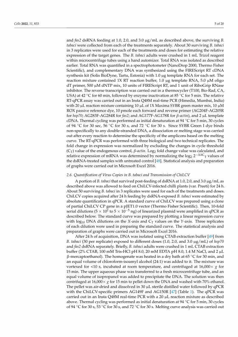
A conserved 128 nt-long (2416 to 2543 nt) fragment of the hsp70 gene (~2.5 kb) and a150 nt-long (1085 to 1234 nt) fragment of the fas2 gene (~1.4 kb) of B. tabaci were selected fordsRNA designing (Figure 1a,b). In siRNA Wizard 3.1, the 128 nt-long B. tabaci hsp70 fragmentproduced a putative siRNA of 21 nt (5’-GAUCCAUCCAUGCCGUUAAUC-3’). The 150 nt-longfas2 fragment also yielded one putative siRNA of 21 nt (5’-GGACGGAAGAAGGCAUAAUCA-3’). The dsRNA sequence was specific to B. tabaci, and no cross-reactivity was recorded withother organisms such as Homo sapiens, Mus musculus, Aves (taxid:8782), Lepidoptera (taxid:7088),Hymenoptera (taxid:7399), Formicidae (taxid:36668), and plants (taxid:3193) in BLAST anal-ysis. PCR amplification of targeted B. tabaci hsp70 and fas2 fragments with AG137F-AG138Rand AG283F-AG284R primer pairs gave single expected amplicons of ~130 bp and ~150 bp,respectively. The fragments were cloned between two T7 promoters of L4440 RNAi vector(Addgene 1654 provided by Andrew Fire, Carnegie Institution for Science, Washington, DC,USA). The recombinant plasmids were transformed into RNase III mutant Escherichia coli HT115cells (provided by Caenorhabditis Genetics Center, Minneapolis, MN, USA). Total RNA isolatedfrom recombinant E. coli HT115 cells and the dsRNA purified from total RNA using DNase Iand RNase A were visualized in agarose gel electrophoresis (Figure 1c). The purified dsRNA ofhsp70 and fas2 produced single specific bands of ~130 bp and ~150 bp on 2% native agarose gel(Figure 1c). Upon bidirectional sequencing of the recombinant plasmids, both the sequences ofhsp70 (Accession No. MZ158306) and fas2 (Accession No. MZ766125) showed 100% homologywith B. tabaci hsp70 and fas2, respectively.
Cells 2022, 11, 833 8 of 20
Figure 1. Designing and synthesis of dsRNA targeting Bemisia tabaci hsp70 and fas2 mRNA. (a) Aconserved region of 128 (2416 to 2543) nt of B. tabaci hsp70 was selected for designing dsRNA. Theputative siRNA is marked within the red box. (b) A conserved region of 150 (1085 to 1234) nt ofB. tabaci fas2 was selected for designing dsRNA. The putative siRNA is marked within the red box.(c) Total RNA isolated from recombinant E. coli HT115 cells (L1–2); hsp70 dsRNA purified from totalRNA using DNase I and RNase A (L3–6); and fas2 dsRNA purified from total RNA using DNase Iand RNase A (L7–9) on 2% agarose gel stained with GoodView, M = 100 bp plus ladder.
3.3. Effect of dsRNA on Mortality and mRNA Expression of B. tabaci under Controlled Conditions
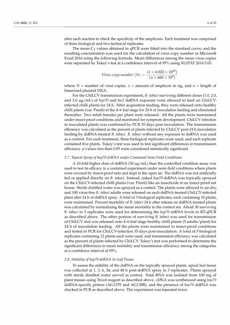
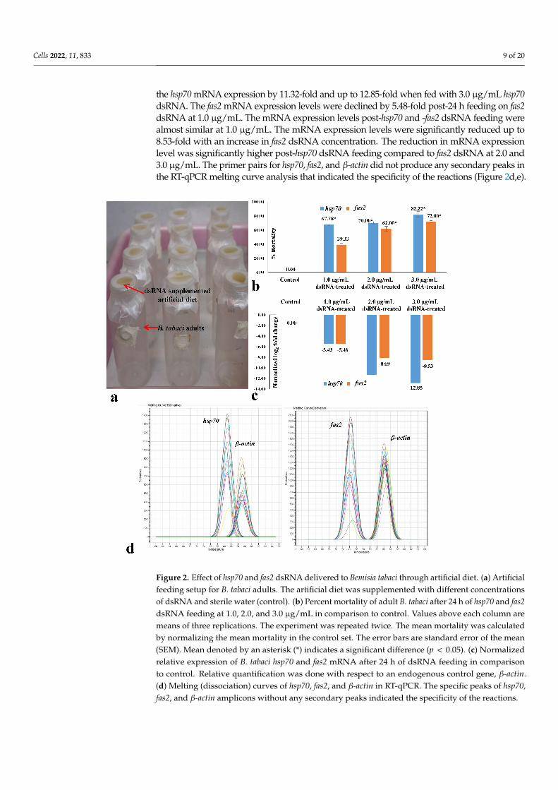
The efficacy of hsp70 and fas2 dsRNA was first evaluated under controlled laboratoryconditions. Significantly higher mortality of B. tabaci adults was recorded in hsp70 dsRNA thanfas2 dsRNA when orally delivered under controlled laboratory conditions (Figure 2a). Mortalityof 67.77% of B. tabaci was observed at 24 h upon feeding on 1.0 µg/mL hsp70 dsRNA. Themortality of B. tabaci significantly increased with an increase in dsRNA concentration. Themean mortality was 70.0% when hsp70 dsRNA was fed to B. tabaci adults at 2.0 µg/mL, and itincreased significantly up to 82.22% at 3.0 µg/mL hsp70 dsRNA (Figure 2b). Oral delivery offas2 dsRNA at 1.0 µg/mL exhibited 39.33% mean mortality after 24 h (Figure 2b). Significantlyhigher mortality (62 and 72%) of B. tabaci adults was recorded when 2.0 and 3.0 µg/mL fas2dsRNA was orally delivered to B. tabaci under controlled laboratory conditions. At all doses, themortality of B. tabaci post-fas2 dsRNA exposure was significantly lower than the hsp70 dsRNAat p < 0.05. However, no morpho-deformities were observed in killed B. tabaci adults eitherwith hsp70 or fas2 dsRNA.
RT-qPCR analysis showed that hsp70 dsRNA significantly reduced the hsp70 mRNAexpression levels in treated B. tabaci. Hsp70 mRNA levels in adult B. tabaci were decreased by5.43-fold with respect to an endogenous control gene, β-actin, post-24 h feeding on hsp70 dsRNA(Figure 2c). The mRNA expression levels were significantly reduced with an increase in hsp70dsRNA concentration. An exposure to hsp70 dsRNA at 2.0 µg/mL for 24 h significantly reduced
Cells 2022, 11, 833 9 of 20
the hsp70 mRNA expression by 11.32-fold and up to 12.85-fold when fed with 3.0 µg/mL hsp70dsRNA. The fas2 mRNA expression levels were declined by 5.48-fold post-24 h feeding on fas2dsRNA at 1.0 µg/mL. The mRNA expression levels post-hsp70 and -fas2 dsRNA feeding werealmost similar at 1.0 µg/mL. The mRNA expression levels were significantly reduced up to8.53-fold with an increase in fas2 dsRNA concentration. The reduction in mRNA expressionlevel was significantly higher post-hsp70 dsRNA feeding compared to fas2 dsRNA at 2.0 and3.0 µg/mL. The primer pairs for hsp70, fas2, and β-actin did not produce any secondary peaks inthe RT-qPCR melting curve analysis that indicated the specificity of the reactions (Figure 2d,e).
Figure 2. Effect of hsp70 and fas2 dsRNA delivered to Bemisia tabaci through artificial diet. (a) Artificialfeeding setup for B. tabaci adults. The artificial diet was supplemented with different concentrationsof dsRNA and sterile water (control). (b) Percent mortality of adult B. tabaci after 24 h of hsp70 and fas2dsRNA feeding at 1.0, 2.0, and 3.0 µg/mL in comparison to control. Values above each column aremeans of three replications. The experiment was repeated twice. The mean mortality was calculatedby normalizing the mean mortality in the control set. The error bars are standard error of the mean(SEM). Mean denoted by an asterisk (*) indicates a significant difference (p < 0.05). (c) Normalizedrelative expression of B. tabaci hsp70 and fas2 mRNA after 24 h of dsRNA feeding in comparisonto control. Relative quantification was done with respect to an endogenous control gene, β-actin.(d) Melting (dissociation) curves of hsp70, fas2, and β-actin in RT-qPCR. The specific peaks of hsp70,fas2, and β-actin amplicons without any secondary peaks indicated the specificity of the reactions.
Cells 2022, 11, 833 10 of 20
3.4. Effect of dsRNA on Virus Acquisition and Transmission by B. tabaci under Controlled Conditions
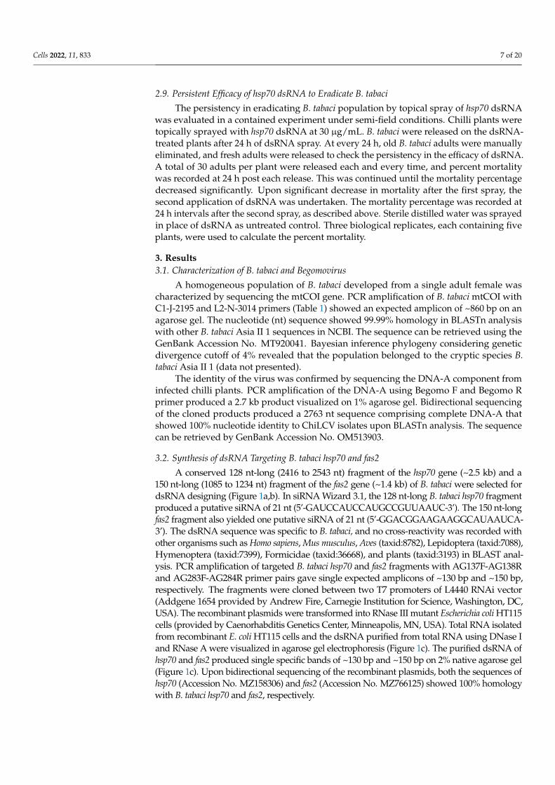
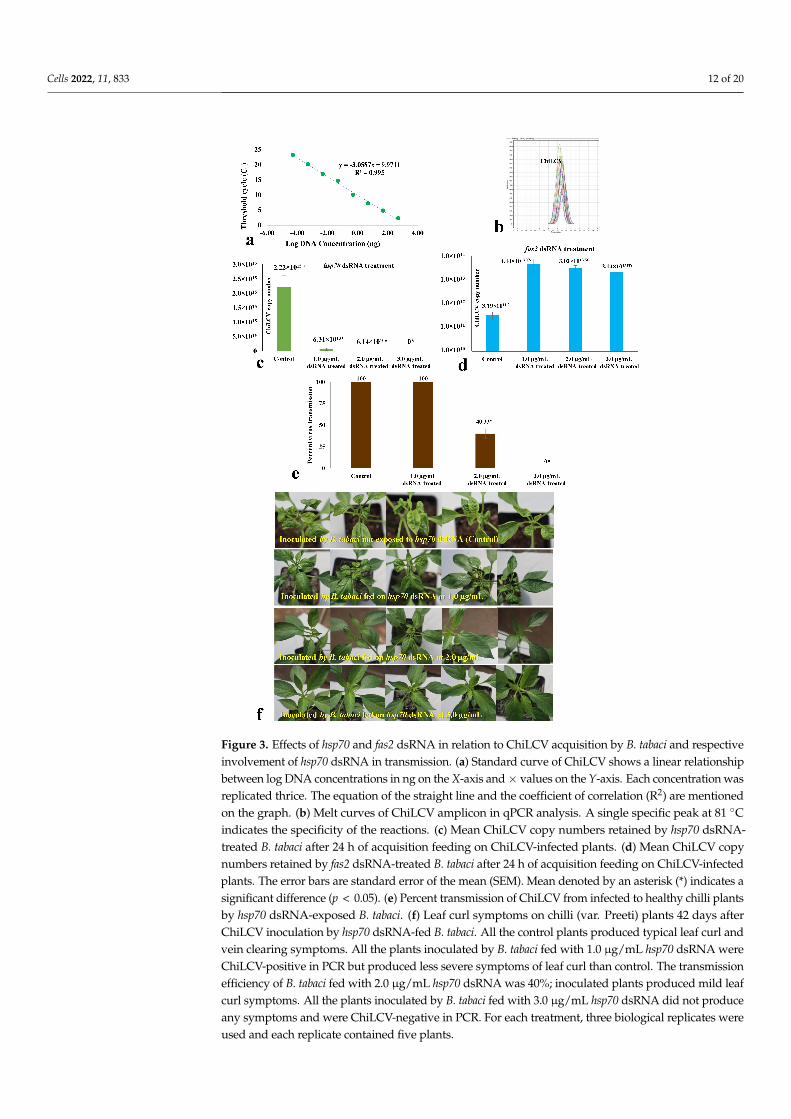
After 24 h of dsRNA feeding, the adult B. tabaci were allowed to feed on ChiLCV-infectedplants for 24 h. A standard curve of ChiLCV was prepared in qPCR using ten-fold dilutionsof a clone of the partial ChiLCV CP gene. The standard curve of ChiLCV showed a coefficientof correlation (R2) of 0.995 and high amplification efficiency near to 100%, indicating optimalconditions for absolute quantification (Figure 3a). The virus copies acquired by the B. tabaciwere quantified by fitting the mean CT values obtained in qPCR into the standard curve.The specificity of the reactions was confirmed by a single peak at 81 ◦C in the melting curveanalysis of qPCR (Figure 3b). The virus copy number acquired by hsp70 dsRNA-treated B.tabaci was significantly less than the untreated control. When B. tabaci was fed with steriledistilled water instead of hsp70 dsRNA, the mean ChiLCV copy number was 2.22 × 10¹⁵,whereas it decreased by 35.18-fold (6.31 × 10¹³ copies) post-exposure to hsp70 dsRNA at1.0 µg/mL (Figure 3c). The virus copy number in treated B. tabaci further decreased witha gradual increase in hsp70 dsRNA concentration. At 2.0 µg/mL hsp70 dsRNA, ChiLCVcopy numbers were 6.14 × 10¹⁰ in B. tabaci i.e., 3.62 × 10⁴-fold lower than the untreatedcontrol. The virus copies were undetectable in B. tabaci with a further increase in hsp70 dsRNAconcentration to 3.0 µg/mL. The retention of ChiLCV might be completely ceased at a higherconcentration of hsp70 dsRNA or beyond the detection limit using qPCR. In contrast, theChiLCV copy number acquired by fas2 dsRNA-treated B. tabaci was significantly higherthan the untreated control (Figure 3d). When B. tabaci was fed with sterile distilled waterinstead of fas2 dsRNA, the mean ChiLCV copy number was 3.19 × 10¹¹, whereas the viruscopies increased significantly by 139.18-fold (4.44 × 10¹³ copies) at 1.0 µg/mL of fas2 dsRNAfeeding. However, the mean virus copies were estimated to be 3.02 × 10¹³ and 2.14 × 10¹³
after exposure to 2.0 and 3.0 µg/mL fas2 dsRNA, respectively. Although significantly highernumbers of virus copies were recorded in all fas2 dsRNA doses compared to the control,higher doses of fas2 dsRNA recorded a decline in virus copies compared to the lower doses.As the virus copies in B. tabaci increased post-fas2 dsRNA feeding, it was not assessed furtherfor inhibiting transmission of ChiLCV and topical application.
A similar pattern was recorded in the transmission of ChiLCV from infected to healthychilli plants by hsp70 dsRNA-fed B. tabaci. A 100% transmission of ChiLCV was recordedwhen B. tabaci was not exposed to hsp70 dsRNA in the control set. All the inoculatedplants produced strong leaf curl and vein-clearing symptoms and tested ChiLCV positivein PCR. Although there was a significant decrease in ChiLCV copies in B. tabaci exposedto 1.0 µg/mL hsp70 dsRNA, all the test plants were found to be infected with ChiLCV inPCR testing at 35 days post-inoculation. Probably even a lower concentration of ChiLCV inB. tabaci was sufficient to make 100% transmission. However, the severity of the leaf curlsymptoms in inoculated plants was less than the untreated control. The transmission ofChiLCV was decreased to 40% when B. tabaci were fed with 2.0 µg/mL hsp70 dsRNA for24 h (Figure 3e). The inoculated plants that tested positive in PCR produced mild leaf curlsymptoms. Interestingly, there was no transmission of ChiLCV when B. tabaci was exposedto 3.0 µg/mL hsp70 dsRNA. All the inoculated plants were without any leaf curl symptoms(Figure 3f) and tested ChiLCV-free in PCR at 35 days post-inoculation. Probably virustransmission by B. tabaci completely inhibited at a higher concentration of hsp70 dsRNA.
3.5. Effect of Topical Spray of hsp70 dsRNA on Mortality and Virus Transmission by B. tabaciunder Semi-Field Conditions
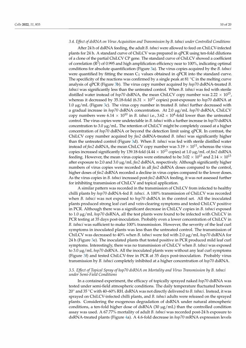
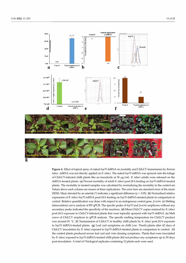
In a contained experiment, the efficacy of topically sprayed naked hsp70 dsRNA wastested under semi-field atmospheric conditions. The daily temperature fluctuated between20◦ and 35 ◦C with 40–60% RH. dsRNA was not directly delivered to B. tabaci. Instead, it wassprayed on ChiLCV-infected chilli plants, and B. tabaci adults were released on the sprayedplants. Considering the exogenous degradation of dsRNA under natural atmosphericconditions, a ten-fold higher dose of dsRNA (30 µg/mL) than the controlled conditionassay was used. A 67.77% mortality of adult B. tabaci was recorded post-24 h exposure todsRNA-treated plants (Figure 4a). A 4.6-fold decrease in hsp70 mRNA expression levels
Cells 2022, 11, 833 11 of 20
of B. tabaci exposed to dsRNA-treated plants was also noted in RT-qPCR with respect tothe endogenous control gene, β-actin (Figure 4b). Specific peaks of hsp70 and β-actin inthe melting curve analysis without any secondary peaks confirmed the specificity of thereactions (Figure 4c).
The surviving B. tabaci adults post-exposure to dsRNA-treated plants were furtherassessed for ChiLCV acquisition and transmission efficiency. A 1.84 × 10⁸-fold decreasein ChiLCV copies was observed in B. tabaci post-24 h exposure to ChiLCV-infected plantsthat were sprayed with hsp70 dsRNA at 30 µg/mL (Figure 4d). The melt curve of ChiLCVamplicon in qPCR produced specific peaks without any secondary peaks that confirmedthe specificity of the reactions (Figure 4e). When a portion of the same B. tabaci populationwas used for inoculation of ChiLCV in healthy chilli plants, a 75% decrease in transmissionefficiency was confirmed in PCR test (Figure 4f) with no visible symptoms in the inoculatedplants up to 50 days post-inoculation (Figure 4g). Probably, the virus titer was too lowin inoculated plants to produce strong visible symptoms, whereas 100% transmissionand severe leaf curl symptoms were noted in plants inoculated by unexposed B. tabaci.No transmission of ChiLCV was recorded in the plants mock-inoculated with untreatedvirus-free B. tabaci.
3.6. Stability and Persistent Efficacy of hsp70 dsRNA in Eradicating B. tabaci Population
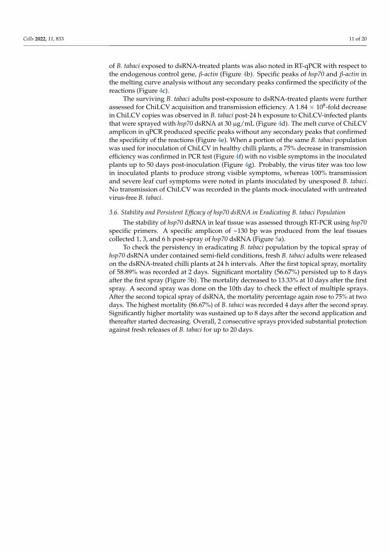
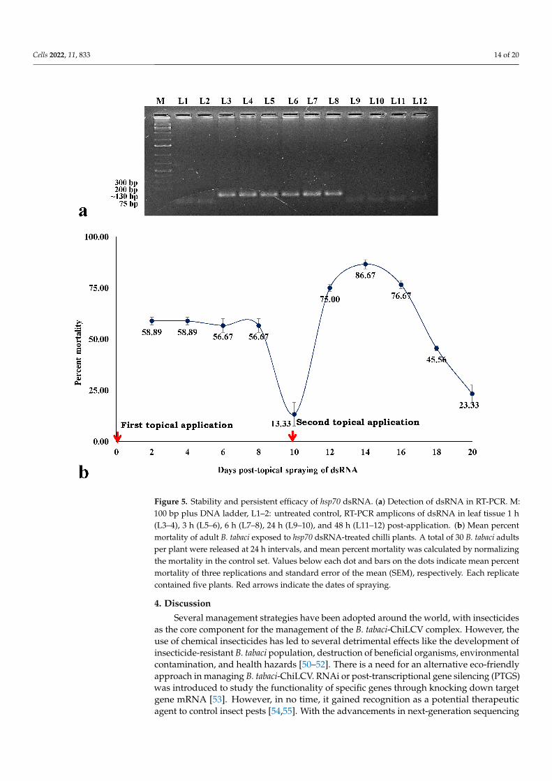
The stability of hsp70 dsRNA in leaf tissue was assessed through RT-PCR using hsp70specific primers. A specific amplicon of ~130 bp was produced from the leaf tissuescollected 1, 3, and 6 h post-spray of hsp70 dsRNA (Figure 5a).
To check the persistency in eradicating B. tabaci population by the topical spray ofhsp70 dsRNA under contained semi-field conditions, fresh B. tabaci adults were releasedon the dsRNA-treated chilli plants at 24 h intervals. After the first topical spray, mortalityof 58.89% was recorded at 2 days. Significant mortality (56.67%) persisted up to 8 daysafter the first spray (Figure 5b). The mortality decreased to 13.33% at 10 days after the firstspray. A second spray was done on the 10th day to check the effect of multiple sprays.After the second topical spray of dsRNA, the mortality percentage again rose to 75% at twodays. The highest mortality (86.67%) of B. tabaci was recorded 4 days after the second spray.Significantly higher mortality was sustained up to 8 days after the second application andthereafter started decreasing. Overall, 2 consecutive sprays provided substantial protectionagainst fresh releases of B. tabaci for up to 20 days.
Cells 2022, 11, 833 12 of 20
Figure 3. Effects of hsp70 and fas2 dsRNA in relation to ChiLCV acquisition by B. tabaci and respectiveinvolvement of hsp70 dsRNA in transmission. (a) Standard curve of ChiLCV shows a linear relationshipbetween log DNA concentrations in ng on the X-axis and × values on the Y-axis. Each concentration wasreplicated thrice. The equation of the straight line and the coefficient of correlation (R2) are mentionedon the graph. (b) Melt curves of ChiLCV amplicon in qPCR analysis. A single specific peak at 81 ◦Cindicates the specificity of the reactions. (c) Mean ChiLCV copy numbers retained by hsp70 dsRNA-treated B. tabaci after 24 h of acquisition feeding on ChiLCV-infected plants. (d) Mean ChiLCV copynumbers retained by fas2 dsRNA-treated B. tabaci after 24 h of acquisition feeding on ChiLCV-infectedplants. The error bars are standard error of the mean (SEM). Mean denoted by an asterisk (*) indicates asignificant difference (p < 0.05). (e) Percent transmission of ChiLCV from infected to healthy chilli plantsby hsp70 dsRNA-exposed B. tabaci. (f) Leaf curl symptoms on chilli (var. Preeti) plants 42 days afterChiLCV inoculation by hsp70 dsRNA-fed B. tabaci. All the control plants produced typical leaf curl andvein clearing symptoms. All the plants inoculated by B. tabaci fed with 1.0 µg/mL hsp70 dsRNA wereChiLCV-positive in PCR but produced less severe symptoms of leaf curl than control. The transmissionefficiency of B. tabaci fed with 2.0 µg/mL hsp70 dsRNA was 40%; inoculated plants produced mild leafcurl symptoms. All the plants inoculated by B. tabaci fed with 3.0 µg/mL hsp70 dsRNA did not produceany symptoms and were ChiLCV-negative in PCR. For each treatment, three biological replicates wereused and each replicate contained five plants.
Cells 2022, 11, 833 13 of 20
Figure 4. Effect of topical spray of naked hsp70 dsRNA on mortality and ChiLCV transmission by Bemisiatabaci. dsRNA was not directly applied on B. tabaci. The naked hsp70 dsRNA was sprayed onto the foliageof ChiLCV-infected chilli plants like an insecticide at 30 µg/mL. B. tabaci adults were released on thedsRNA-treated plants. (a) Percent mortality of adult B. tabaci post-24 h feeding on hsp70 dsRNA-treatedplants. The mortality in treated samples was calculated by normalizing the mortality in the control set.Values above each column are means of three replications. The error bars are standard error of the mean(SEM). Mean denoted by an asterisk (*) indicates a significant difference (p < 0.05). (b) Normalized relativeexpression of B. tabaci hsp70 mRNA post-24 h feeding on hsp70 dsRNA-treated plants in comparison tocontrol. Relative quantification was done with respect to an endogenous control gene, β-actin. (c) Melting(dissociation) curve analysis of RT-qPCR. The specific peaks of hsp70 and β-actin amplicons without anysecondary peaks indicated the specificity of the reactions. (d) Mean ChiLCV copies retained by B. tabacipost-24 h exposure to ChiLCV-infected plants that were topically sprayed with hsp70 dsRNA. (e) Meltcurve of ChiLCV amplicon in qPCR analysis. The specific melting temperature for ChiLCV productwas around 81 ◦C. (f) Transmission of ChiLCV in healthy chilli plants by B. tabaci post-24 h exposureto hsp70 dsRNA-treated plants. (g) Leaf curl symptoms on chilli (var. Preeti) plants after 42 days ofChiLCV inoculation by B. tabaci exposed to hsp70 dsRNA-treated plants in comparison to control. Allthe control plants produced severe leaf curl and vein-clearing symptoms. Plants that were inoculatedby B. tabaci exposed to hsp70 dsRNA-treated chilli plants did not produce any symptoms up to 50 dayspost-inoculation. A total of 5 biological replicates containing 12 plants each were used.
Cells 2022, 11, 833 14 of 20
Figure 5. Stability and persistent efficacy of hsp70 dsRNA. (a) Detection of dsRNA in RT-PCR. M:100 bp plus DNA ladder, L1–2: untreated control, RT-PCR amplicons of dsRNA in leaf tissue 1 h(L3–4), 3 h (L5–6), 6 h (L7–8), 24 h (L9–10), and 48 h (L11–12) post-application. (b) Mean percentmortality of adult B. tabaci exposed to hsp70 dsRNA-treated chilli plants. A total of 30 B. tabaci adultsper plant were released at 24 h intervals, and mean percent mortality was calculated by normalizingthe mortality in the control set. Values below each dot and bars on the dots indicate mean percentmortality of three replications and standard error of the mean (SEM), respectively. Each replicatecontained five plants. Red arrows indicate the dates of spraying.
4. Discussion
Several management strategies have been adopted around the world, with insecticidesas the core component for the management of the B. tabaci-ChiLCV complex. However, theuse of chemical insecticides has led to several detrimental effects like the development ofinsecticide-resistant B. tabaci population, destruction of beneficial organisms, environmentalcontamination, and health hazards [50–52]. There is a need for an alternative eco-friendlyapproach in managing B. tabaci-ChiLCV. RNAi or post-transcriptional gene silencing (PTGS)was introduced to study the functionality of specific genes through knocking down targetgene mRNA [53]. However, in no time, it gained recognition as a potential therapeuticagent to control insect pests [54,55]. With the advancements in next-generation sequencing
Cells 2022, 11, 833 15 of 20
and transcriptomics technology, several genes have been identified as potential targets tocontrol B. tabaci and its viruses [33–40,56–58]. However, most studies are limited to theoral feeding of the dsRNA to B. tabaci through artificial diet or genetic modification of thehost plants [40,43,59,60]. None of these RNAi molecules have been tested for their efficacyor found effective under field conditions, limiting their large-scale adoption. RNAi usingdsRNA constructs is not suitable for field conditions due to rapid degradation and a lackof reliable delivery agents. In the present study, for the first time, we demonstrated theefficacy of a naked dsRNA against an insect pest by spray-on application under semi-fieldconditions. The dsRNA construct targeting the hsp70 gene of B. tabaci not only inducedmortality in B. tabaci but also inhibited the transmission of ChiLCV from infected to healthychilli plants by B. tabaci.
Hsp70 and fas2 were found to be significantly upregulated in viruliferous B. tabaci alongwith several other genes [30,35,57]. Gene regulatory networking showed these genes wereenriched with higher degrees of interactions [35]. Hsp70 transcripts increased upon ingestionof tomato yellow leaf curl virus (TYLCV) and squash leaf curl virus (SLCV) in DNA microar-ray [31]. The role of hsp70 in begomovirus transmission is well studied [30,31,57,60,61]. TYLCVCP and Hsp70 interacted in vitro and co-localized within midgut epithelial cells [30,31]. Thefas2 mRNA expression level was upregulated in viruliferous B. tabaci by 2.6 and 4.518-fold inthe RNA-Seq and RT-qPCR assay, respectively. The Fas2 orthologue in mammals (NCAM) isknown to serve as a receptor for the rabies virus [39]. Hence, we targeted to knock down thesetwo genes, hsp70 and fas2, to induce immunity to ChiLCV infection in B. tabaci. Conserved128 and 150 nt stretches in the hsp70 and fas2 genes, respectively, were found to be specific toB. tabaci without any cross-reactivity to humans, mice, birds, butterflies, bees, ants, and plants.Each produced a putative siRNA of 21 nt. dsRNA was synthesized by in vivo transcriptionto harvest large quantities of dsRNA at a low cost. Bacterially expressed dsRNA is economi-cal and convenient, as reported in several studies [62–64]. Before evaluating the efficacy oftopically sprayed dsRNA, we tested it under controlled laboratory conditions. Both hsp70and fas2 dsRNA were orally delivered to B. tabaci adults by mixing with an artificial diet in afeeding setup. Similar types of artificial feeding setup were reported to be efficient in the oraldelivery of dsRNA [40–42]. A significant knockdown of the B. tabaci population was recordedat 24 h, which gradually increased with an increase in dsRNA concentration. Up to 82.22%mortality of B. tabaci was recorded after 24 h feeding on 3.0 µg/mL hsp70 dsRNA. Similarly,fas2 dsRNA at 3.0 µg/mL caused 72% mortality of B. tabaci 24 h post-feeding. The dsRNAexposure downregulated the hsp70 and fas2 mRNA expressions by 12.85- and 8.53-fold, respec-tively. The expression of an endogenous control gene was unregulated post-dsRNA exposure.This indicated the specificity of the dsRNA constructs to target genes. Hsp belongs to themultifunctional molecular chaperone family involved in the aggregation of damaged proteins,transportation, assembly, and disassembly of multi-structured units under stressed condi-tions [36,65]. The mortality of B. tabaci post-dsRNA feeding may be due to the loss of functionvia depletion of hsp70 mRNA, which might interrupt the normal biological processes in B.tabaci. In Drosophila melanogaster, Fas2 is involved in synaptic development and growth [37,38].It is also known to function in intracellular signaling pathways that involve mitogen-activatedprotein kinase (MAPK) and regulate intracellular calcium levels [38]. However, the functionof fas2 in B. tabaci is uncharacterized. Kanakala et al. [60] reported twisting of wings in B. tabaciupon silencing hsp70. However, we did not record any morphological deformities in treatedB. tabaci. Hsp70 and fas2 might not be involved in the morphogenesis of B. tabaci, or longerexposure to dsRNA is required to produce such deformities.
The persistent circulative transmission of ChiLCV by B. tabaci indicates that the virusparticles reach the midgut after ingestion. The virions cross the midgut barrier to becomecirculative in the hemolymph and accumulate in the primary salivary glands [30]. Hsp70,along with other molecular chaperones, plays an important role in the translocation of viralproteins besides its function in viral replication, assembly, and disassembly of the viralproteins inside the hosts [66–70]. Hsp70 has been found to interact with TYLCV CP in vitroand co-localize within midgut epithelial cells [31]. In the present study, oral delivery of
Cells 2022, 11, 833 16 of 20
hsp70 dsRNA significantly reduced ChiLCV copies in B. tabaci Asia II 1 and its transmissionto healthy chilli plants as well. The resistance to ChiLCV in B. tabaci was enhanced at higherconcentrations of hsp70 dsRNA. ChiLCV copies in B. tabaci were imperceptible when B.tabaci fed on 3 µg/mL hsp70 dsRNA for 24 h. The transmission of ChiLCV from infectedto healthy plants by B. tabaci Asia II 1 was completely inhibited at 3 µg/mL hsp70 dsRNAtreatment under controlled conditions. Our findings are consistent with Kanakala et al. [60],who reported the deleterious effect of B. tabaci hsp70 dsRNA on TYLCV transmission. TheTYLCV titer decreased in B. tabaci Middle East Asia Minor 1 (MEAM 1) after feeding onhsp70 dsRNA-expressing tomato plants. Transmission of TYLCV also dropped by 12%post-silencing of hsp70 [60]. In the present study, the inability of B. tabaci Asia II 1 toretain and transmit ChiLCV post-silencing of hsp70 indicates that it might also play animportant function in ChiLCV infection. However, Gotz et al. [31] reported an increase inTYLCV transmission upon blocking Hsp70 by anti-Hsp70 antibody in B. tabaci MEAM1. Itwas hypothesized that the Hsp70-TYLCV interaction mediates the degradation of virions.Downregulation of hsp70, hsp40, and hsp20 resulted in 3.1-, 1.5-, and 1.2-fold increases incotton leaf curl virus (CLCuV) titer within B. tabaci. Significantly increased transmissionefficiency of CLCuV was noted in B. tabaci Asia II 1 when hsp70 was silenced [61]. Thedifference may be due to the variation in virus species and B. tabaci cryptic species. Theinteractions of B. tabaci with begomoviruses are not conserved and alter with cryptic speciesof B. tabaci and begomovirus species [71,72]. B. tabaci MEAM1 has two genes (Bta03000 andBta02903) annotated as hsp70, and their amino acid sequences differ by 97.55% [56]. Theymight also exhibit differential responses to begomovirus infection. Unlike hsp70 dsRNAtreatment, an increase in the ChiLCV copies in B. tabaci was recorded post-fas2 dsRNAfeeding. The Fas2 orthologue in mammals (NCAM) is known to serve as a receptor for therabies virus. Although NCAM promotes the penetration of the virus in cells, it suppressesvirus replication via induction of Interferon-ß [39], which is mainly involved in innateimmunity against viral infection. Upregulation of fas2 transcripts in B. tabaci post-ChiLCVinfection might be due to the innate immune response against the virus infection [35].We hypothesized that fas2 has a negative regulatory role in ChiLCV infection, and thus,knocking down fas2 increased the virus titer in B. tabaci. Hence, silencing fas2 was notconsidered to induce resistance to ChiLCV despite its efficacy in eradicating B. tabaci.
Although RNAi has emerged as a promising alternative to suppress crop pests [73,74],there are limitations in the application of dsRNA as a ‘spray-on’ technology for fielduse. Reduced stability in the extracellular environments of dsRNA contributes to poorRNAi response. The naked dsRNA quickly degrades in higher temperatures and UVradiation under natural environmental conditions. Besides, several insect species, mainlylepidopteran insects, have been observed to be refractory to RNAi due to degradation andpoor intracellular transport of exogenous dsRNA [75,76]. Nucleases are the major factorslimiting the efficacy of dsRNA [75–77]. In the first phase of the study, hsp70 dsRNA showedhigh potential in eradicating B. tabaci and inhibiting ChiLCV transmission. In the next phase,the dsRNA construct was assessed under natural environmental conditions by sprayinglike an insecticide. Adult B. tabaci are highly mobile, so spraying on adult flies was notpreferred. Hence, naked hsp70 dsRNA without any delivery agent was topically sprayedonto the foliage of the ChiLCV-infected chilli plant under contained semi-field conditions.B. tabaci adults were released on the dsRNA-sprayed plants to evaluate the dsRNA’sefficacy in eradicating B. tabaci and inhibiting the transmission of ChiLCV. Consideringthe extracellular degradation of dsRNA and delivery through the host plant, a 10-foldhigher dose than the controlled assay was used for topical application. A 67.77% mortalitywith a 4.6-fold decrease in hsp70 mRNA levels in B. tabaci exposed to the dsRNA-sprayedplants was noted after 24 h of release. Further, 1.84 × 10⁸-fold decreased ChiLCV copiesand 75% reduced transmission of ChiLCV indicated the efficiency of topically sprayedhsp70 dsRNA in managing both the virus and its vector. Although the efficacy of topicallyapplied naked dsRNA against plant viruses is known [78–80], this is the first evidence ofits efficacy against a hemipteran insect like B. tabaci.
Cells 2022, 11, 833 17 of 20
Topical application of dsRNA could provide resistance for 12 to 21 days against plantviruses [78,80–82]. In the present study, dsRNA was detected in the leaf tissues up to6 h post-application. Significant mortality of B. tabaci on the treated plants persisted upto 8 days and was further sustained up to 20 days with a second spray on the 10th day.Protection of the plants during the early growth stage is crucial in the management ofbegomoviruses. Begomovirus infection at a 5-leaf stage in tomatoes reduced yield by95% [83]. Infection of ChiLCV at the seedling stage leads to a considerable reduction inplant height, internodal length, and fruiting. Hence, a 20-day shield by 2 consecutivesprays of naked hsp70 dsRNA would protect the crucial phase of crop growth from theinvasion of B. tabaci and ChiLCV and reduce yield losses. The foliar spray of hsp70 dsRNAwas effective under semi-field conditions where the daily temperature fluctuated between20–35 ◦C with 40–60% RH. The efficacy and persistency of topically sprayed naked dsRNAat a wide temperature range endorse its potentiality for field uses as a spray-on technology.
5. Conclusions
In conclusion, the spray-on application of naked dsRNA targeting hsp70 of B. tabaciprovides resistance to both ChiLCV and its vector, B. tabaci. This is a novel alternative tohazardous insecticides and may be further assessed on a large scale for its efficacy underreal-field conditions.
Author Contributions: A.G. conceived and designed the research. P.C. conducted the experiments,recorded experimental data, and performed analysis. A.G. reviewed the data. P.C. wrote the draftmanuscript. A.G. wrote and edited the final manuscript. All authors have read and agreed to thepublished version of the manuscript.
Funding: This study was supported by the IARI and Department of Biotechnology, Government ofIndia (BT/PR25296/NER/95/1116/2017).
Institutional Review Board Statement: Not applicable.
Informed Consent Statement: Not applicable.
Data Availability Statement: Sequences have been deposited to GenBank under accession numbersMT920041, MW399222, MZ158306, MZ766125, and OM513903.
Acknowledgments: The support received from IARI, New Delhi, and the Department of Biotechnol-ogy, Government of India is thankfully acknowledged.
Conflicts of Interest: The authors declare that they have no conflict of interest.
Ethical Approval: The research meets ethical guidelines and adheres to the legal requirements of thestudy country. This research does not involve human subjects.
References1. Oliveira, M.R.V.; Henneberry, T.J.; Anderson, P. History, Current Status, and Collaborative Research Projects for Bemisia tabaci.
Crop Prot. 2001, 20, 709–723. [CrossRef]2. Chowda-Reddy, R.; Kirankumar, M.; Seal, S.E.; Muniyappa, V.; Valand, G.B.; Govindappa, M.; Colvin, J. Bemisia tabaci
Phylogenetic Groups in India and the Relative Transmission Efficacy of Tomato Leaf Curl Bangalore Virus by an Indigenous andan Exotic Population. J. Integr. Agric. 2012, 11, 235–248. [CrossRef]
3. Legarrea, S.; Barman, A.; Marchant, W.; Diffie, S.; Srinivasan, R. Temporal Effects of a Begomovirus Infection and Host PlantResistance on the Preference and Development of an Insect Vector, Bemisia tabaci, and Implications for Epidemics. PLoS ONE 2015,10, e0142114. [CrossRef]
4. Mugerwa, H.; Seal, S.; Wang, H.L.; Patel, M.V.; Kabaalu, R.; Omongo, C.A.; Alicai, T.; Tairo, F.; Ndunguru, J.; Sseruwagi, P.; et al.African Ancestry of New World, Bemisia tabaci-Whitefly Species. Sci. Rep. 2018, 8, 2734. [CrossRef]
5. Jiu, M.; Hu, J.; Wang, L.J.; Dong, J.F.; Song, Y.Q.; Sun, H.Z. Cryptic Species Identification and Composition of Bemisia tabaci(Hemiptera: Aleyrodidae) Complex in Henan Province, China. J. Insect Sci. 2017, 17, 78–84. [CrossRef]
6. Rehman, M.; Chakraborty, P.; Tanti, B.; Mandal, B.; Ghosh, A. Occurrence of a New Cryptic Species of Bemisia tabaci (Hemiptera:Aleyrodidae): An Updated Record of Cryptic Diversity in India. Phytoparasitica 2021, 49, 869–882. [CrossRef]
7. Byrne, D.N.; Bellows, T.S. Whitefly Biology. Annu. Rev. Entomol. 1991, 36, 431–457. [CrossRef]8. Jones, D.R. Plant Viruses Transmitted by Whiteflies. Eur. J. Plant Pathol. 2003, 109, 195–219. [CrossRef]
Cells 2022, 11, 833 18 of 20
9. Navas-Castillo, J.; Fiallo-Olivé, E.; Sánchez-Campos, S. Emerging Virus Diseases Transmitted by Whiteflies. Annu. Rev. Phytopathol.2011, 49, 219–248. [CrossRef]
10. Brown, J.K.; Zerbini, F.M.; Navas-Castillo, J.; Moriones, E.; Ramos-Sobrinho, R.; Silva, J.C.; Fiallo-Olivé, E.; Briddon, R.W.;Hernández-Zepeda, C.; Idris, A.; et al. Revision of Begomovirus Taxonomy Based on Pairwise Sequence Comparisons. Arch.Virol. 2015, 160, 1593–1619. [CrossRef]
11. Orfanidou, C.G.; Pappi, P.G.; Efthimiou, K.E.; Katis, N.I.; Maliogka, V.I. Transmission of Tomato Chlorosis Virus (ToCV) by Bemisiatabaci Biotype Q and Evaluation of Four Weed Species as Viral Sources. Plant Dis. 2016, 100, 2043–2049. [CrossRef] [PubMed]
12. Zanardo, L.G.; Carvalho, C.M. Cowpea mild mottle virus (Carlavirus, Betaflexiviridae): A review. Trop. Plant Pathol. 2017, 42,417–430. [CrossRef]
13. Ghosh, S.; Kanakala, S.; Lebedev, G.; Kontsedalov, S.; Silverman, D.; Alon, T.; Mor, N.; Sela, N.; Luria, N.; Dombrovsky, A.;et al. Transmission of a New Polerovirus Infecting Pepper by the Whitefly Bemisia tabaci. J. Virol. 2019, 93, e00488-19. [CrossRef][PubMed]
14. Costa, T.M.; Inoue-Nagata, A.K.; Vida, A.H.; Ribeiro, S.G.; Nagata, T. The recombinant isolate of cucurbit aphid-borne yellowsvirus from Brazil is a polerovirus transmitted by whiteflies. Plant Pathol. 2020, 69, 1042–1050. [CrossRef]
15. Pinheiro-Lima, B.; Pereira-Carvalho, R.C.; Alves-Freitas, D.M.T.; Kitajima, E.W.; Vidal, A.H.; Lacorte, C.; Godinho, M.T.; Fontenele,R.S.; Faria, J.C.; Abreu, E.F.M.; et al. Transmission of the Bean-Associated Cytorhabdovirus by the Whitefly Bemisia tabaciMEAM1. Viruses 2020, 12, 1028. [CrossRef]
16. Cornejo-Franco, J.F.; Reyes-Proaño, E.G.; Mollov, D.; Mowery, J.; Quito-Avila, D.F. Transmission and Pathogenicity of PapayaVirus E: Insights from an Experimental Papaya Orchard. Plant Dis. 2022, 2017, 2467940. [CrossRef]
17. Mansoor, S.; Briddon, R.W.; Zafar, Y.; Stanley, J. Geminivirus Disease Complexes: An Emerging Threat. Trends Plant Sci. 2003, 8,128–134. [CrossRef]
18. Briddon, R.W.; Patil, B.L.; Bagewadi, B.; Nawaz-ul-Rehman, M.S.; Fauquet, C.M. Distinct Evolutionary Histories of the DNA-Aand DNA-B Components of Bipartite Begomoviruses. BMC. Evol. Biol. 2010, 10, 97. [CrossRef]
19. Saeed, S.T.; Samad, A. Emerging Threats of Begomoviruses to the Cultivation of Medicinal and Aromatic Crops and TheirManagement Strategies. Virus Dis. 2017, 28, 1–17. [CrossRef]
20. CABI. Capsicum Annuum (Bell Pepper). Invasive Species Compendium. Available online: https://www.cabi.org/isc/datasheet/15784 (accessed on 13 April 2021).
21. Oraon, U.B.; Tarafdar, J. Occurrence and distribution of chilli leaf curl complex disease in West Bengal. Biomed. J. Sci. Tech. Res.2018, 3, 3515–3519. [CrossRef]
22. Kumar, Y.; Hallan, V.; Zaidi, A.A. Chilli Leaf Curl Palampur Virus Is a Distinct Begomovirus species Associated with a Betasatellite.Plant Pathol. 2011, 60, 1040–1047. [CrossRef]
23. Khan, A.J.; Akhtar, S.; Al-Zaidi, A.M.; Singh, A.K.; Briddon, R.W. Genetic Diversity and Distribution of a Distinct Strain of ChiliLeaf Curl Virus and Associated Betasatellite Infecting Tomato and Pepper in Oman. Virus Res. 2013, 177, 87–97. [CrossRef][PubMed]
24. Senanayake, D.M.J.B.; Mandal, B.; Lodha, S.; Varma, A. First Report of Chilli Leaf Curl Virus Affecting Chilli in India. Plant Pathol.2007, 56, 343. [CrossRef]
25. Senanayake, D.M.J.B.; Jayasinghe, J.E.; Shilpi, S.; Wasala, S.K.; Mandal, B. A New Begomovirus–Betasatellite Complex IsAssociated with Chilli Leaf Curl Disease in Sri Lanka. Virus Genes 2013, 46, 128–139. [CrossRef]
26. Thakur, H.; Jindal, S.K.; Sharma, A.; Dhaliwal, M.S. Chilli Leaf Curl Virus Disease: A Serious Threat for Chilli Cultivation. J. PlantDis. Prot. 2018, 125, 239–249. [CrossRef]
27. Menike, G.D.N.; De Costa, D.M. Variation of Field Symptoms and Molecular Diversity of the Virus Isolates Associated with ChilliLeaf Curl Complex in Different Agroecological Regions of Sri Lanka. Trop. Agric. Res. 2017, 28, 144. [CrossRef]
28. Geley, S.; Müller, C. RNAi: Ancient Mechanism with a Promising Future. Exp. Gerontol. 2004, 39, 985–998. [CrossRef]29. Rosen, R.; Kanakala, S.; Kliot, A.; Cathrin Pakkianathan, B.; Farich, B.A.; Santana-Magal, N.; Elimelech, M.; Kontsedalov, S.;
Lebedev, G.; Cilia, M.; et al. Persistent, Circulative Transmission of Begomoviruses by Whitefly Vectors. Curr. Opin. Virol. 2015,15, 1–8. [CrossRef]
30. Czosnek, H.; Hariton-Shalev, A.; Sobol, I.; Gorovits, R.; Ghanim, M. The Incredible Journey of Begomoviruses in Their WhiteflyVector. Viruses 2017, 9, 273. [CrossRef]
31. Götz, M.; Popovski, S.; Kollenberg, M.; Gorovits, R.; Brown, J.K.; Cicero, J.M.; Czosnek, H.; Winter, S.; Ghanim, M. Implication ofBemisia tabaci Heat Shock protein 70 in Begomovirus-Whitefly Interactions. J. Virol. 2012, 86, 13241–13252. [CrossRef]
32. Kanakala, S.; Ghanim, M. Implication of the Whitefly Bemisia tabaci Cyclophilin B Protein in the Transmission of Tomato YellowLeaf Curl Virus. Front. Plant Sci. 2016, 7, 1702. [CrossRef] [PubMed]
33. Rana, V.S.; Popli, S.; Saurav, G.K.; Raina, H.S.; Chaubey, R.; Ramamurthy, V.V.; Rajagopal, R. A Bemisia tabaci Midgut ProteinInteracts with Begomoviruses and Plays a Role in Virus Transmission. Cell. Microbiol. 2016, 18, 663–678. [CrossRef] [PubMed]
34. Wang, Z.Z.; Shi, M.; Huang, Y.C.; Wang, X.W.; Stanley, D.; Chen, X.X. A Peptidoglycan Recognition Protein Acts in Whitefly(Bemisia tabaci) Immunity and Involves in Begomovirus Acquisition. Sci. Rep. 2016, 6, 37806. [CrossRef] [PubMed]
35. Nekkanti, A. Changes in Transcriptome of Whitefly (Bemisia Tabaci Gennadius) Associated with Begomovirus Infection. Master’sThesis, Post Graduate School, ICAR-Indian Agricultural Research Insitute, New Delhi, India, 2021; p. 66.
Cells 2022, 11, 833 19 of 20
36. Hartl, F.U.; Hayer-Hartl, M. Molecular Chaperones in the Cytosol: From Nascent Chain to Folded Protein. Science 2002, 295,1852–1858. [CrossRef]
37. Packard, M.; Mathew, D.; Budnik, V. FASt Remodeling of Synapses in Drosophila. Curr. Opin. Neurobiol. 2003, 13, 527–534.[CrossRef]
38. Kristiansen, L.V.; Hortsch, M. Fasciclin II: The NCAM Ortholog in Drosophila melanogaster. Adv. Exp. Med. Biol. 2010, 663,387–401. [CrossRef]
39. Hotta, K.; Motoi, Y.; Okutani, A.; Kaku, Y.; Noguchi, A.; Inoue, S.; Yamada, A. Role of GPI-Anchored NCAM-120 in Rabies VirusInfection. Microbes Infect. 2007, 9, 167–174. [CrossRef]
40. Upadhyay, S.K.; Chandrashekar, K.; Thakur, N.; Verma, P.C.; Borgio, J.F.; Singh, P.K.; Tuli, R. RNA interference for the Control ofWhiteflies (Bemisia tabaci) by Oral Route. J. Biosci. 2011, 36, 153–161. [CrossRef]
41. Li, H.; Guan, R.; Guo, H.; Miao, X. New Insights into an RNAi Approach for Plant Defense Against Piercing-Sucking andStem-Borer Insect Pests. Plant Cell Environ. 2015, 38, 2277–2285. [CrossRef]
42. Vyas, M.; Raza, A.; Ali, M.Y.; Ashraf, M.A.; Mansoor, S.; Shahid, A.A.; Brown, J.K. Knock Down of Whitefly Gut Gene Expressionand Mortality by Orally Delivered Gut Gene-Specific dsRNAs. PLoS ONE 2017, 12, e0168921. [CrossRef]
43. Bar, L.; Czosnek, H.; Sobol, I.; Ghanim, M.; Hariton Shalev, A. Down Regulation of Dystrophin Expression in Pupae of theWhitefly Bemisia tabaci Inhibits the Emergence of Adults. Insect Mol. Biol. 2019, 28, 662–675. [CrossRef]
44. Akhter, A.; Qazi, J.; Saeed, M.; Mansoor, S. A Severe Leaf Curl Disease on Chilies in Pakistan Is Associated with MultipleBegomovirus Components. Plant Dis. 2009, 93, 962. [CrossRef] [PubMed]
45. Ahn, S.J.; Donahue, K.; Koh, Y.; Martin, R.R.; Choi, M.Y. Microbial-Based Double-Stranded RNA Production to Develop Cost-Effective RNA interference Application for Insect Pest Management. Int. J. Insect Sci. 2019, 11, 1179543319840323. [CrossRef][PubMed]
46. Simon, C.; Frati, F.; Beckenbach, A.; Crespi, B.; Liu, H.; Flook, P. Evolution, Weighting, and Phylogenetic Utility of MitochondrialGene Sequences and a Compilation of Conserved Polymerase Chain Reaction Primers. Ann. Entomol. Soc. Am. 1994, 87, 651–701.[CrossRef]
47. Roy, B.; Chakraborty, P.; Ghosh, A. How many begomovirus copies are acquired and inoculated by its vector, whitefly (Bemisiatabaci) during feeding? PLoS ONE 2021, 16, e0258933. [CrossRef] [PubMed]
48. Livak, K.J.; Schmittgen, T.D. Analysis of Relative Gene Expression Data Using Real-Time Quantitative PCR and the 2−∆∆CT
Method. Methods 2001, 25, 402–408. [CrossRef]49. Doyle, J.J.; Doyle, J.L. Isolation of Plant DNA from Fresh Tissue. Focus 1990, 12, 13–15.50. Abdullah, N.M.M.; Singh, J.; Sohal, B.S. Behavioral Hormoligosis in Oviposition Preference of Bemisia tabaci on Cotton. Pestic.
Biochem. Physiol. 2006, 84, 10–16. [CrossRef]51. Bacci, L.; Crespo, A.L.; Galvan, T.L.; Pereira, E.J.; Picanço, M.C.; Silva, G.A.; Chediak, M. Toxicity of Insecticides to the Sweetpotato
Whitefly (Hemiptera: Aleyrodidae) and Its Natural Enemies. Pest Manag. Sci. 2007, 63, 699–706. [CrossRef]52. Houndété, T.A.; Kétoh, G.K.; Hema, O.S.; Brévault, T.; Glitho, I.A.; Martin, T. Insecticide Resistance in Field Populations of Bemisia
tabaci (Hemiptera: Aleyrodidae) in West Africa. Pest Manag. Sci. 2010, 66, 1181–1185. [CrossRef]53. Sen, G.L.; Blau, H.M. A Brief History of RNAi: The Silence of the Genes. FASEB J. 2006, 20, 1293–1299. [CrossRef] [PubMed]54. Baum, J.A.; Bogaert, T.; Clinton, W.; Heck, G.R.; Feldmann, P.; Ilagan, O.; Johnson, S.; Plaetinck, G.; Munyikwa, T.; Pleau, M.; et al.
Control of Coleopteran Insect Pests Through RNA interference. Nat. Biotechnol. 2007, 25, 1322–1326. [CrossRef] [PubMed]55. Huvenne, H.; Smagghe, G. Mechanisms of dsRNA Uptake in Insects and Potential of RNAi for Pest Control: A Review. J. Insect
Physiol. 2010, 56, 227–235. [CrossRef] [PubMed]56. Chen, W.; Hasegawa, D.K.; Kaur, N.; Kliot, A.; Pinheiro, P.V.; Luan, J.; Stensmyr, M.C.; Zheng, Y.; Liu, W.; Sun, H.; et al. The
Draft Genome of Whitefly Bemisia tabaci MEAM1, a Global Crop Pest, Provides Novel Insights into Virus Transmission, HostAdaptation, and Insecticide Resistance. BMC Biol. 2016, 14, 110. [CrossRef]
57. Ding, T.B.; Li, J.; Chen, E.H.; Niu, J.Z.; Chu, D. Transcriptome Profiling of the Whitefly Bemisia tabaci MED in Response toSingle Infection of Tomato Yellow Leaf Curl Virus, Tomato Chlorosis Virus, and Their Co-Infection. Front. Physiol. 2019, 10, 302.[CrossRef]
58. Hussain, S.; Farooq, M.; Malik, H.J.; Amin, I.; Scheffler, B.E.; Scheffler, J.A.; Liu, S.S.; Mansoor, S. Whole Genome Sequencingof Asia II 1 Species of Whitefly Reveals That Genes Involved in Virus Transmission and Insecticide Resistance Have GeneticVariances Between Asia II 1 and MEAM1 Species. BMC Genom. 2019, 20, 507. [CrossRef]
59. Grover, S.; Jindal, V.; Banta, G.; Taning, C.N.T.; Smagghe, G.; Christiaens, O. Potential of RNA interference in the Study andManagement of the Whitefly, Bemisia tabaci. Arch. Insect Biochem. Physiol. 2019, 100, e21522. [CrossRef]
60. Kanakala, S.; Kontsedalov, S.; Lebedev, G.; Ghanim, M. Plant-Mediated Silencing of the Whitefly Bemisia tabaci Cyclophilin B andHeat Shock protein 70 Impairs Insect Development and Virus Transmission. Front. Physiol. 2019, 10, 557. [CrossRef]
61. Kaur, R.; Gupta, M.; Singh, S.; Joshi, N.; Sharma, A. Enhancing RNAi Efficiency to Decipher the Functional Response of PotentialGenes in Bemisia tabaci Asia II-1 (Gennadius) Through dsRNA Feeding Assays. Front. Physiol. 2020, 11, 123. [CrossRef]
62. Tenllado, F.; Martínez-García, B.; Vargas, M.; Díaz-Ruíz, J.R. Crude Extracts of Bacterially Expressed dsRNA Can Be Used toProtect Plants Against Virus Infections. BMC Biotechnol. 2003, 3, 3. [CrossRef]
63. Leelesh, R.S.; Rieske, L.K. Oral Ingestion of Bacterially Expressed dsRNA Can Silence Genes and Cause Mortality in a HighlyInvasive, Tree-Killing Pest, the Emerald Ash Borer. Insects 2020, 11, 440. [CrossRef] [PubMed]
Cells 2022, 11, 833 20 of 20
64. Cherenko, V.A.; Filipenko, E.A.; Golubeva, T.S. Silencing of Nicotiana Benthamiana Phytoendesaturase Using dsRNA SynthesizedIn Vivo. In IOP Conference Series: Earth and Environmental Science; IOP Publishing: Bristol, UK, 2021; p. 723. [CrossRef]
65. Gregersen, N.; Bross, P.; Andrese, B.S.; Pedersen, C.B.; Corydon, T.J.; Bolund, L. The Role of Chaperone-Assisted Folding andQuality Control in Inborn Errors of Metabolism: Protein Folding Disorders. J. Inherit. Metab. Dis. 2001, 24, 189–212. [CrossRef][PubMed]
66. Mayer, M.P. Recruitment of Hsp70 Chaperones: A Crucial Part of Viral Survival Strategies. Rev. Physiol. Biochem. Pharmacol. 2005,153, 1–46. [CrossRef]
67. Serva, S.; Nagy, P.D. Proteomics Analysis of the Tombusvirus Replicase: Hsp70 Molecular Chaperone Is Associated with theReplicase and Enhances Viral RNA Replication. J. Virol. 2006, 80, 2162–2169. [CrossRef]
68. Ivanovic, T.; Agosto, M.A.; Chandran, K.; Nibert, M.L. A Role for Molecular Chaperone Hsc70 in Reovirus Outer CapsidDisassembly. J. Biol. Chem. 2007, 282, 12210–12219. [CrossRef] [PubMed]
69. Czosnek, H. Tomato Yellow Leaf Curl Virus Disease. In Management, Molecular Biology, Breeding for Resistance; Czosnek, H., Ed.;Springer: Berlin/Heidelberg, Germany, 2007; Volume 440. [CrossRef]
70. Gorovits, R.; Moshe, A.; Ghanim, M.; Czosnek, H. Recruitment of the Host Plant Heat Shock protein 70 by Tomato Yellow LeafCurl Virus Coat Protein Is Required for Virus Infection. PLoS ONE 2013, 8, e70280. [CrossRef]
71. Jiu, M.; Zhou, X.P.; Liu, S.S. Acquisition and Transmission of Tobacco Curly Shoot Virus by the B Biotype of the Whitefly, Bemisiatabaci. Acta Phytophylacica Sin. 2006, 33, 168–172. [CrossRef]
72. Guo, X.J.; Feng, J.N. Comparisons of Expression Levels of Heat Shock Proteins (hsp70 and hsp90) from Anaphothrips obscurus(Thysanoptera: Thripidae) in Polymorphic Adults Exposed to Different Heat Shock Treatments. J. Insect Sci. 2018, 18, 15.[CrossRef]
73. Palli, S.R. RNA interference in Colorado Potato Beetle: Steps Toward Development of dsRNA as a Commercial Insecticide. Curr.Opin. Insect Sci. 2014, 6, 1–8. [CrossRef]
74. Zotti, M.J.; Smagghe, G. RNAi Technology for Insect Management and Protection of Beneficial Insects from Diseases: Lessons,Challenges and Risk Assessments. Neotrop. Entomol. 2015, 44, 197–213. [CrossRef]
75. Garbutt, J.S.; Reynolds, S.E. Induction of RNA interference Genes by Double-Stranded RNA. Implications for Susceptibility toRNA interference. Insect Biochem. Mol. Biol. 2012, 42, 621–628. [CrossRef] [PubMed]
76. Shukla, J.N.; Kalsi, M.; Sethi, A.; Narva, K.E.; Fishilevich, E.; Singh, S.; Mogilicherla, K.; Palli, S.R. Reduced Stability andIntracellular Transport of dsRNA Contribute to Poor RNAi Response in Lepidopteran Insects. RNA Biol. 2016, 13, 656–669.[CrossRef] [PubMed]
77. Christiaens, O.; Whyard, S.; Vélez, A.M.; Smagghe, G. Double-Stranded RNA Technology to Control Insect Pests: Current Statusand Challenges. Front. Plant Sci. 2020, 11, 451. [CrossRef] [PubMed]
78. Namgial, T.; Kaldis, A.; Chakraborty, S.; Voloudakis, A. Topical Application of Double-Stranded RNA Molecules ContainingSequences of Tomato Leaf Curl Virus and Cucumber Mosaic Virus Confers Protection Against the Cognate Viruses. Physiol. Mol.Plant Pathol. 2019, 108, 101432. [CrossRef]
79. Worrall, E.A.; Bravo-Cazar, A.; Nilon, A.T.; Fletcher, S.J.; Robinson, K.E.; Carr, J.P.; Mitter, N. Exogenous Application of RNAi-Inducing Double-Stranded RNA Inhibits Aphid-Mediated Transmission of a Plant Virus. Front. Plant Sci. 2019, 10, 265. [CrossRef][PubMed]
80. Konakalla, N.C.; Bag, S.; Deraniyagala, A.S.; Culbreath, A.K.; Pappu, H.R. Induction of Plant Resistance in Tobacco (Nicotianatabacum) Against Tomato Spotted Wilt Orthotospovirus Through Foliar Application of dsRNA. Viruses 2021, 13, 662. [CrossRef]
81. Konakalla, N.C.; Kaldis, A.; Berbati, M.; Masarapu, H.; Voloudakis, A.E. Exogenous Application of Double-Stranded RNAMolecules from TMV p126 and CP Genes Confers Resistance Against TMV in Tobacco. Planta 2016, 244, 961–969. [CrossRef]
82. Kaldis, A.; Berbati, M.; Melita, O.; Reppa, C.; Holeva, M.; Otten, P.; Voloudakis, A. Exogenously Applied dsRNA MoleculesDeriving from the Zucchini Yellow Mosaic Virus (ZYMV) Genome Move Systemically and Protect Cucurbits Against ZYMV. Mol.Plant Pathol. 2018, 19, 883–895. [CrossRef]
83. Kumar, P.; Kumar, M. Leaf Curl Disease: A Significant Constraint in the Production of Tomato in India. In Advances in PlantPathology; Kumatu, J.N., Ed.; IntechOpen: London, UK, 2018; pp. 93–101. [CrossRef]


