Embed Size (px)
Citation preview
BioMed Central
Reproductive Biology and Endocrinology
ss
Open AcceResearchUbp43 gene expression is required for normal Isg15 expression and fetal developmentLea A Rempel1,2, Kathleen J Austin1, Kenneth J Ritchie3, Ming Yan3, Meifeng Shen3, Dong-Er Zhang3, Luiz E Henkes4 and Thomas R Hansen*1,4Address: 1Department of Animal Science, University of Wyoming, Laramie, Wyoming, 82071, USA, 2Currently Institute of Maternal-Fetal Biology and the Division of Cancer & Developmental Biology, Department of Pathology and Laboratory Medicine, University of Kansas Medical Center, Kansas City, Kansas 66160, USA, 3Department of Molecular and Experimental Medicine, The Scripps Research Institute, La Jolla, California 92037, USA and 4Department of Biomedical Sciences, Animal Reproduction and Biotechnology Laboratory, Colorado State University, Fort Collins, Colorado 80523, USA
Email: Lea A Rempel - [email protected]; Kathleen J Austin - [email protected]; Kenneth J Ritchie - [email protected]; Ming Yan - [email protected]; Meifeng Shen - [email protected]; Dong-Er Zhang - [email protected]; Luiz E Henkes - [email protected]; Thomas R Hansen* - [email protected]
* Corresponding author
AbstractBackground: Isg15 covalently modifies murine endometrial proteins in response to earlypregnancy. Isg15 can also be severed from targeted proteins by a specific protease called Ubp43(Usp18). Mice lacking Ubp43 (null) form increased conjugated Isg15 in response to interferon. TheIsg15 system has not been examined in chorioallantoic placenta (CP) or mesometrial (MM)components of implantation sites beyond 9.5 days post coitum (dpc). It was hypothesized thatdeletion of Ubp43 would cause disregulation of Isg15 in implantation sites, and that this would affectpregnancy rates.
Methods: Heterozygous (het) Ubp43 mice were mated and MM and CP implantation sites werecollected on 12.5 and 17.5 days post-coitum (dpc).
Results: Free and conjugated Isg15 were greater on 12.5 versus 17.5 dpc in MM. Free andconjugated Isg15 were also present in CP, but did not differ due to genotype on 12.5 dpc. However,null CP had greater free and conjugated Isg15 when compared to het/wt on 17.5 dpc. Null progenydied in utero with fetal genotype ratios (wt:het:null) of 2:5:1 on 12.5 and 2:2:1 on 17.5 dpc.Implantation sites were disrupted within the junctional zone and spongiotrophoblast, containedless vasculature based on lectin B4 staining and contained greater Isg15 mRNA and VEGF proteinin Ubp43 null when compared to wt placenta.
Conclusion: It is concluded that Isg15 and its conjugates are present in implantation sites duringmid to late gestation and that deletion of Ubp43 causes an increase in free and conjugated Isg15 atthe feto-maternal interface. Also, under mixed genetic background, deletion of Ubp43 results infetal death.
Published: 26 March 2007
Reproductive Biology and Endocrinology 2007, 5:13 doi:10.1186/1477-7827-5-13
Received: 19 January 2007Accepted: 26 March 2007
This article is available from: http://www.rbej.com/content/5/1/13
© 2007 Rempel et al; licensee BioMed Central Ltd. This is an Open Access article distributed under the terms of the Creative Commons Attribution License (http://creativecommons.org/licenses/by/2.0), which permits unrestricted use, distribution, and reproduction in any medium, provided the original work is properly cited.
Page 1 of 15(page number not for citation purposes)
Reproductive Biology and Endocrinology 2007, 5:13 http://www.rbej.com/content/5/1/13
BackgroundInterferon stimulated gene product 15 (Isg15) is a ubiqui-tin-like protein that is transiently produced in the uterusduring early pregnancy in several species including pri-mates [1], ruminants [2-4], pigs [5] and mice [6,7]. Isg15post-translationally modifies other proteins through cov-alent attachment using a mechanism that is similar to, yetdistinct from ubiquitinylation. This conjugation (Isgyla-tion) to target proteins involves three classes of enzymes,an E1 and several E2s and E3s. Isg15 functions as a ubiq-uitin homolog to regulate general cellular processes suchas RNA splicing, chromatin remodeling/polymerase IItranscription, cytoskeleton organization and regulation,stress responses, and translation by conjugating to andregulating intracellular proteins [8-14]. It also exists in anon-conjugated form and has been shown to be releasedfrom cells and to have a cytokine-like role [15,16].
Within ruminant species, conceptus (embryo proper andsurrounding membranes)-secreted interferon (IFN)-tau(τ) interacts with type I IFN receptors on the endometrialsurface to activate the Janus kinase-signal transducers andactivators of transcription (JAK-STAT) signal cascade[17,18]. Activation of the JAK-STAT pathway leads to theincreased production of Isg15. In mice and humans, aconceptus-secreted IFN has not been identified, howeverIsg15 is up-regulated in the uterus in these species as well[1,6,7,19,20]. For example, Isg15 is up-regulated 3.9-foldin human decidualized stromal cells by embryo-secretoryproducts [20]. Furthermore, induction of Isg15 in mousedecidua might be due to an interferon-like cytokine that isreleased by trophoblast giant cells [7].
Cleavage of ubiquitin from target proteins plays animportant role in numerous biological events such as;proteasome-mediated protein degradation [21], preim-plantation embryo development [22], cell growth and dif-ferentiation [23-25], transcriptional activation [23], andsignal transduction [26]. There are at least five distinctfamilies of deubiquitinating enzymes with four sub-families that are commonly referred to as cysteine pro-teases [27]. Ubiquitin processing proteases (Ubps/Usps)are the largest and most diverse group within the cysteineprotease subfamilies. Sequence homology is limitingamong Ubps, however short consensus sequences sur-round conserved cysteine and histidine residue boxes [26-28]. Diversity in these amino acids may provide specificityfor ubiquitin and ubiquitin-like proteases.
Ubp43 (Usp18) is an Isg15-specific protease [29,30] thatis up-regulated in response to IFN or lipopolysaccharide[31,32]. Overexpression of Ubp43 in monocyte cellsinhibits cytokine-induced terminal differentiation [29].Chemistry-based proteomics were used to identify poten-
tial ubiquitin and ubiquitin-like proteases, whichrevealed that Ubp43 was specific to Isg15 [33,34].
Mice that survive to birth with a disrupted Ubp43 genehad increased Isg15 conjugation and a decreased lifeexpectancy due to hydrocephalus and associated neurode-generative disease [30]. These data support the require-ment of proper regulation of Isgylation by Ubp43cleavage. However, more recent experiments suggest thatUbp43 regulates IFN signaling independent of its iso-peptidase activity towards Isg15 [35]. The data indicatethat Ubp43 binds directly to IFN receptor (AR2 subunit)and inhibits receptor-associated JAK activity. Also, Isg15 -/- and Isg15 E1 (Ube1L) -/- mice that survived to termwere reported to be viable and fertile and had no obviousabnormalities [36,37]. Furthermore, the major pheno-types of Ubp43 deficient mice were not rescued in Isg15/Ubp43 or Ube1L/Ubp43 double knockout mice [37,38].
Isg15 is localized to antimesometrial decidua duringimplantation in mice [6,7]. Implantation, which involvesadhesion of trophoblast with the endometrium that is fol-lowed by invasion, vascularization and placentationbegins 4.5–7.5 days post-coitum (dpc) on the antimes-ometrial pole when the decidualized stroma essentialserves as a maternal placenta. The primary adhesion, orattachment occurs on the avascular antimesometrial pole.Later, decidualization extends to the vascular mesometrialpole to facilitate development of the placenta (Figure 1).We first localized Isg15 to the antimesometrial pole of theimplantation site [6]. Currently, it is unknown if Isg15 isexpressed later than 9.5 dpc in the antimesometrial pole,or if it also appears in the mesometrial decidua and pla-centa.
A description of embryo development and implantationsites in Ubp43 -/- mice is lacking. Because Isg15 and itsconjugates are transiently produced in the uterus duringpregnancy in several mammalian species it was theorizedthat placental development and function might beimpaired by Ubp43 deletion. Therefore, the objective wasto determine if altered fetal Ubp43 gene expression in aheterozygous uterine environment affected the Isg15 sys-tem of maternal and fetal tissues and whether this resultedin lowered fetal viability.
MethodsBreeding and tissue collectionExperimental procedures using mice were reviewed andapproved by the University of Wyoming Institutional Ani-mal Care and Use Committee. Heterozygous (Ubp43+/-)C57 BL/6 × 129 × Swiss Webster males and females [30]were mated to produce wild-type (wt), heterozygous (+/-)and null (-/-) fetuses within a heterozygous uterine envi-ronment. Presence of a vaginal plug on the morning after
Page 2 of 15(page number not for citation purposes)
Reproductive Biology and Endocrinology 2007, 5:13 http://www.rbej.com/content/5/1/13
pairings was designated as 0.5 dpc. Tissues were collectedon 12.5 (4 litters; 30 fetuses) or 17.5 (6 litters; 35 fetuses)dpc. Maternal and fetal tissues consisted of uterine tissueat the placental interface or mesometrial (MM) tissue,uterine tissue surrounding the fetus or anti-mesometrial(AM) tissue, and the fetal-derived placental tissue referredto as fetal chorioallantoic placenta (CP) (Figure 1).
At the time of tissue collection a portion of fetal tissue wasalso collected to determine genotype by PCR techniques.Briefly, tissue was lysed per DNeasy (Qiagen Inc., Valen-cia, CA) protocol to acquire genomic DNA. Genotypingby PCR technique was conducted using the followingprimers; Ubp43 exon 2 anti-sense strand, 5'-GCCT-GGAAGTGAAGTTGTGGACTCCCG-3'; Ubp43 exon 2sense strand, 5'-CCAGCGTGAGTACTGCTGCGGCTCAG-3'; and β-galactosidase sense strand, 5'-CGTAACCGT-GCATCTGCCAGTTTGAGG-3'. Amplification of a 150 bpfragment resulted from a wt allele and a 370 bp fragmentrepresented a null allele (data not shown). PCR condi-tions were; 95°C – 2 minutes; (95°C – 15 seconds, 60°C– 15 seconds and 72°C – 30 seconds) × 30 cycles; 72°C –2 minute extension.
Protein analysisTissue lysates from maternal (MM and AM tissue 12.5 dpc:n = 4 wt, 2 het, and 2 null; 17.5 dpc: n = 4 wt, 4 het, and2 null) and fetal compartments (CP tissue 12.5 dpc: n = 4wt, 2 het, and 2 null; 17.5 dpc: n = 4 wt, 4 het, and 2 null)were prepared by homogenizing tissue (100 mg/ml) in 1×Laemmli buffer [39]. Equal concentrations of lysates(12.5 μg per lane) were loaded onto 1D-SDS PAGE gels,electrophoretically transferred to 0.2 μ nitrocellulose andwestern blot detection was performed using rabbit poly-clonal antibodies [anti-mouse Isg15 antibody (1:30,000;[6], anti-human VEGF antibody (1:500; sc-152, SantaCruz Biotechnology, Inc., Santa Cruz, CA), anti-mouseAngiopoietin-1 antibody (1:1,000; ab8451, Abcam Inc.,Cambridge, MA), anti-mouse VEGF Flt-1 Receptor anti-body (1:500; sc-316, Santa Cruz Biotechnology, Inc.,Santa Cruz, CA), and anti-mouse AMP kinase antibody(1:1,000; A1475-01B, US Biological, Swampscott, MA)].Secondary antibodies against rabbit were conjugated toeither alkaline phosphatase or horseradish peroxidase(Promega, Madison, WI) for visualization of bands. A no-first antibody was used as a control to identify non-spe-cific bands. Detection of a secondary antibody reactive
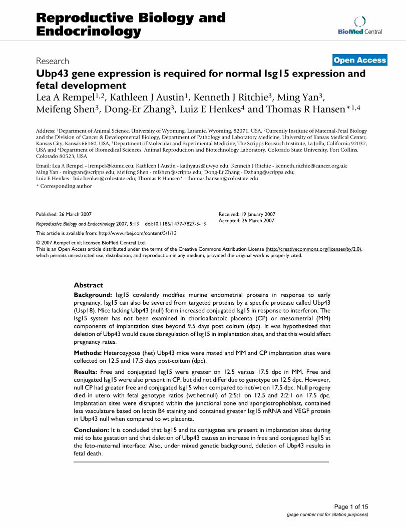
Illustration of fetal development and Ubp43 genotype on day 17.5 of pregnancy (A) and description of how implantation sites were collected (B)Figure 1Illustration of fetal development and Ubp43 genotype on day 17.5 of pregnancy (A) and description of how implantation sites were collected (B). A litter from a day 17.5 pregnant mouse shows that all Ubp43 null offspring were dead. Genotype ratio was 2:2:1 (wt:het:null) on day 17.5. On day 12.5, 75% of Ubp43 null mice were dead with a genotype ratio of 2:5:1. There also was a significant loss of +/- fetuses between 12.5 and 17.5 suggesting that Ubp43 gene dosage may also be involved. Panel B shows a cross-sectional representation of a mouse uterus illustrating the various tissues collected for protein and RNA analysis. Anti-mesometrial (AM) represents uterine tissue surrounding the fetal compartment. Fetal chorioallantoic placenta (CP) represents fetal-derived placental tissue. Mesometrial decidua (MM) represents uterine tissue in direct contact with fetal-derived placenta.
Chorioallantoic
Placenta
Anti-Mesometrial (AM)
Mesometrial (MM)6 -/-
1 -/-
2 +/-3 +/+
4 +/+ 5 -/-
A B
17. 5 dpc
Page 3 of 15(page number not for citation purposes)
Reproductive Biology and Endocrinology 2007, 5:13 http://www.rbej.com/content/5/1/13
protein was used to verify loading. No differences wereseen among samples (P > 0.10; data not shown). Immu-noreactive bands were scanned using UNSCANIT (SilkScientific, Orem, UT).
RNA analysisTissues from 12.5 dpc (n = 3 wt, 3 het, and 2 null) or 17.5dpc (n = 3 wt, 2 het, and 2 null) chroioallantoic placentawere homogenized in Tri Reagent (Molecular ResearchCenter, Inc., Cincinnati, OH). Seven μg of total RNA fromeach sample was loaded per well and separated on a 1.5%denaturing agarose gel. Separated RNA was transferred tonitrocellulose by capillary transfer and baked at 80°C fortwo hours. Membranes were prehybridized for 3 hours at42°C in buffer (50% formamide, 5× SSC, 50 mM Na2PO4,5× Denhardts, 0.1% SDS, 0.1 mg/ml salmon spermDNA). Blots were then hybridized in the same buffer withradiolabeled probe.
A partial murine Isg15 or Ubp43 cDNA was synthesizedusing total RNA from pregnant murine uterine tissue andRT-PCR with the following primers; Isg15 anti-sensestrand, 5'-ATGGCCTGGGACCTAAAGGTG; Isg15 sensestrand, 5'-AAGCTCAGCCAGAACTGGTCT; Ubp43 anti-sense strand, 5'-ATGGGCAAGGGGTTTG-3'; and Ubp43sense strand, 5'-TCAGGATCCAGTCTTC-3'. Ampliconswere subcloned into ZeroBlunt (Invitrogen, Carlsbad, CA)vector and sequenced to confirm identity. Complemen-tary DNA was radiolabeled using 50 μCi [α-32P]dCTP andKlenow in a standard random primer labeling reaction.Northern blots were washed 3×, 5 minutes each at 42°C(2× SSC/0.1% SDS or 1× SSC/0.1% SDS). Membraneswere exposed to film for 6 days at -80°C. Northern blotresults were normalized to an 18S rRNA band. Autoradio-grams were scanned and quantitated using UNSCANIT.
Bacterial and viral screeningTo verify that the immune system of these mice was notcompromised by bacteria or pathogens we submitted tis-sue samples to the following accredited facilities for anal-ysis. Tissue samples from a breeding pair that was housedin our facility for six months were submitted to the Wyo-ming State Veterinary Laboratory (Laramie, WY) and sero-logical samples were submitted to Charles RiverLaboratories (Wilmington, MA).
Morphology of day 12.5 implantation sitesMurine implantation sites were fixed in 4 % paraformal-dehyde, paraffin-embedded, serially sectioned at 6 μmand then stained with hematoxylin/eosin (H&E) forstandard histological examination. Immunohistochemis-try was performed with 20 μg/ml of Isolectin B4 antibodyin 0.1% BSA-PBS (Vector Lab) and 20 μg/ml HorseradishPeroxidase Streptavidin (Vector Lab) as the enzyme conju-
gate. The staining was developed with AEC (Vector Lab)and counterstained with hematoxylin.
Statistical analysisTo determine differences among genotypes and dpc, datawere analyzed using the General Linearized Model of Sta-tistical Analysis Systems (SAS, 1998). The effects of geno-type on concentrations of and expression of free Isg15 andconcentrations of conjugated Isg15 and VEGF were ana-lyzed using one-way ANOVA, followed by protected pre-planned t-test (P < 0.05) comparisons.
ResultsGenotyping and genetic profileFetal development for Ubp43 -/-, -/+ and +/+ offspring isrepresented in Figure 1. Genotype ratio was 2:5:1 on day12.5 and 2:2:1 on day 17.5. All null offspring were deadon day 17.5, whereas 75% of null offspring were dead onday 12.5. Several +/- offspring also died between 12.5 and17.5 dpc. Chi square analysis revealed that genotypicratios for live offspring on 12.5 (P < 0.05) and 17.5 dpc (P< 0.01) did not follow the normal expected mendelianratio of 1:2:1.
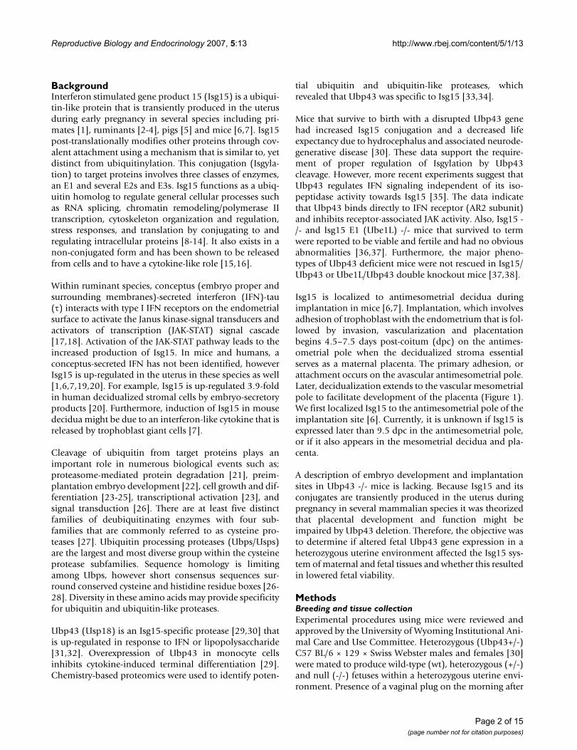
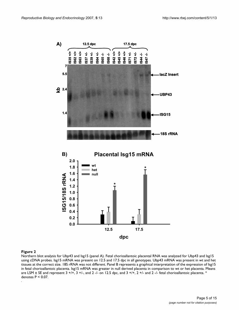
Expression of Isg15 and Ubp43 in fetal-derived placentaNorthern blot analysis of CP tissue for Isg15 and Ubp43verified expression of Isg15 in all genotypes (Figure 2).Ubp43 mRNA was only present in northern blots at thecorrect size within wt- and het-derived tissues. Null fetal-derived tissues containing the beta-galactosidase (lacZ)reporter had an expected shift in the size of the mRNAband of approximately 3 kilobases. Null CP tissue hadgreater (P < 0.07) expression of Isg15 mRNA regardless ofdpc in contrast to wt or het (Figure 2). All samples werenormalized to an 18S rRNA as a loading control.
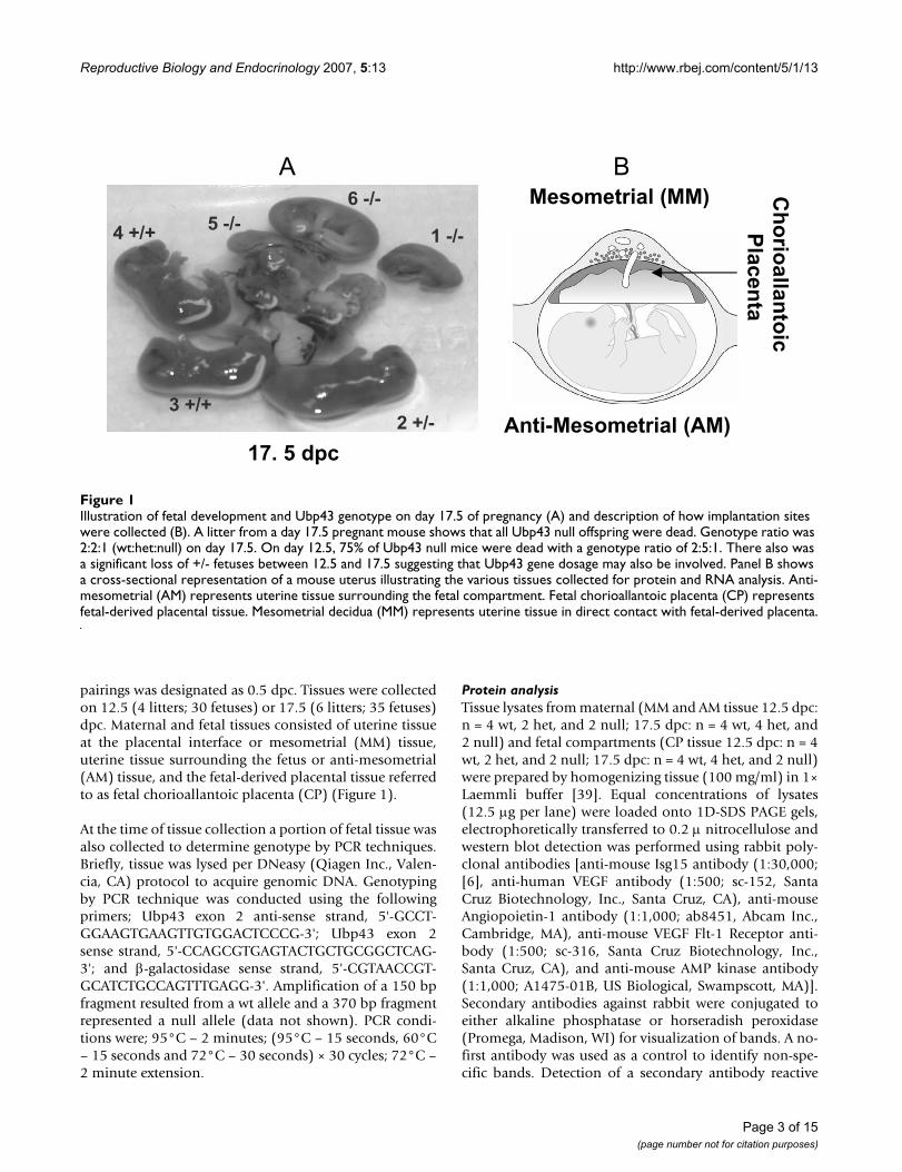
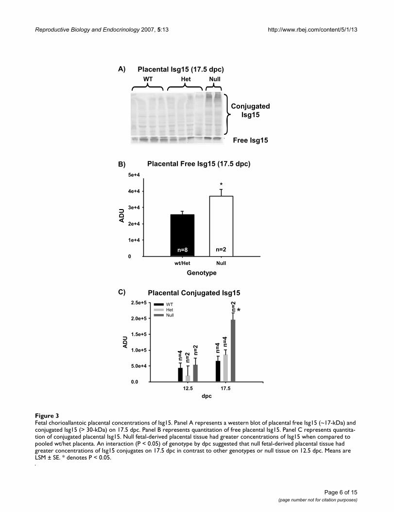
Protein expression of Isg15 in fetal and maternal tissuesSince Ubp43 regulates the conjugation state of Isg15 (free-or conjugated) we evaluated the influence of increasedUbp43 mRNA and subsequent Isg15 mRNA in Ubp43 -/-fetuses on the protein levels of Isg15 in fetal and maternaltissues. Fetal chorioallantoic placenta were analyzed bywestern blot for abundance of Isg15 and conjugated Isg15proteins. Fetal placental concentrations of Isg15 weregreater (P < 0.05) in null compared to pooled wt and hetfetal-derived tissues on 17.5 dpc (Figure 3). Interestingly,an interaction occurred where concentrations of conju-gated Isg15 proteins were greater (P < 0.05) in null CP on17.5 dpc in contrast to all other genotypes on 12.5 or 17.5dpc.
To determine if fetal Ubp43 deletion altered levels of freeand conjugated Isg15 in maternal tissues, western blotanalysis was also conducted using MM and AM derivedtissue. Mesometrial tissue had greater (P < 0.05) Isg15 and
Page 4 of 15(page number not for citation purposes)
Reproductive Biology and Endocrinology 2007, 5:13 http://www.rbej.com/content/5/1/13
Page 5 of 15(page number not for citation purposes)
Northern blot analysis for Ubp43 and Isg15 (panel A)Figure 2Northern blot analysis for Ubp43 and Isg15 (panel A). Fetal chorioallantoic placental RNA was analyzed for Ubp43 and Isg15 using cDNA probes. Isg15 mRNA was present on 12.5 and 17.5 dpc in all genotypes. Ubp43 mRNA was present in wt and het tissues at the correct size. 18S rRNA was not different. Panel B represents a graphical interpretation of the expression of Isg15 in fetal chorioallantoic placenta. Isg15 mRNA was greater in null derived placenta in comparison to wt or het placenta. Means are LSM ± SE and represent 3 +/+, 3 +/-, and 2 -/- on 12.5 dpc, and 3 +/+, 2 +/- and 2 -/- fetal chorioallantoic placenta. * denotes P < 0.07.
7
5.5
2.4
1.4
IS35
+/+
IS62
+/+
IS63
+/+
IS37
+/-
IS39
+/-
IS64
+/-
IS65
-/-
IS68
-/-
IS42
+/+
IS45
+/+
IS46
+/+
IS71
+/-
IS72
+/-
IS44
-/-
IS47
-/-
12.5 dpc 17.5 dpc
ISG15
UBP43
lacZ Insert
kb
18S rRNA
A)
7
5.5
2.4
1.4
IS35
+/+
IS62
+/+
IS63
+/+
IS37
+/-
IS39
+/-
IS64
+/-
IS65
-/-
IS68
-/-
IS42
+/+
IS45
+/+
IS46
+/+
IS71
+/-
IS72
+/-
IS44
-/-
IS47
-/-
12.5 dpc 17.5 dpc
ISG15
UBP43
lacZ Insert
kb
18S rRNA
A)
Placental Isg15 mRNA
dpc12.5 17.5
ISG
15/1
8S rR
NA
0.00.20.40.60.81.01.21.41.61.82.0
wt het null
*
*
B)
Reproductive Biology and Endocrinology 2007, 5:13 http://www.rbej.com/content/5/1/13
Page 6 of 15(page number not for citation purposes)
Fetal chorioallantoic placental concentrations of Isg15Figure 3Fetal chorioallantoic placental concentrations of Isg15. Panel A represents a western blot of placental free Isg15 (~17-kDa) and conjugated Isg15 (> 30-kDa) on 17.5 dpc. Panel B represents quantitation of free placental Isg15. Panel C represents quantita-tion of conjugated placental Isg15. Null fetal-derived placental tissue had greater concentrations of Isg15 when compared to pooled wt/het placenta. An interaction (P < 0.05) of genotype by dpc suggested that null fetal-derived placental tissue had greater concentrations of Isg15 conjugates on 17.5 dpc in contrast to other genotypes or null tissue on 12.5 dpc. Means are LSM ± SE. * denotes P < 0.05.
Placental Conjugated Isg15
dpc12.5 17.5
AD
U
0.0
5.0e+4
1.0e+5
1.5e+5
2.0e+5
2.5e+5 WT HetNull
FI Site Concentrations of ISG15 on 17.5 dpc
Genotypewt/Het Null
AD
U
0
1e+4
2e+4
3e+4
4e+4
5e+4
*
A)
WT Het NullC) Day 17.5 of Pregnancy
Placental Free Isg15 (17.5 dpc)
Placental Isg15 (17.5 dpc)A)
B)
C)
ConjugatedIsg15
Free Isg15
n=8 n=2
n=4 n=
4n=
2
n=2
n=2
n=4
*
Placental Conjugated Isg15
Reproductive Biology and Endocrinology 2007, 5:13 http://www.rbej.com/content/5/1/13
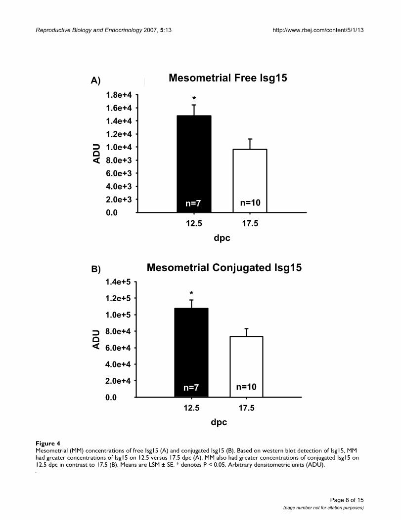
conjugated Isg15 on 12.5 than on 17.5 dpc (Figure 4).Mesometrial concentrations of Isg15 or conjugated Isg15did not differ among genotypes (data not shown).
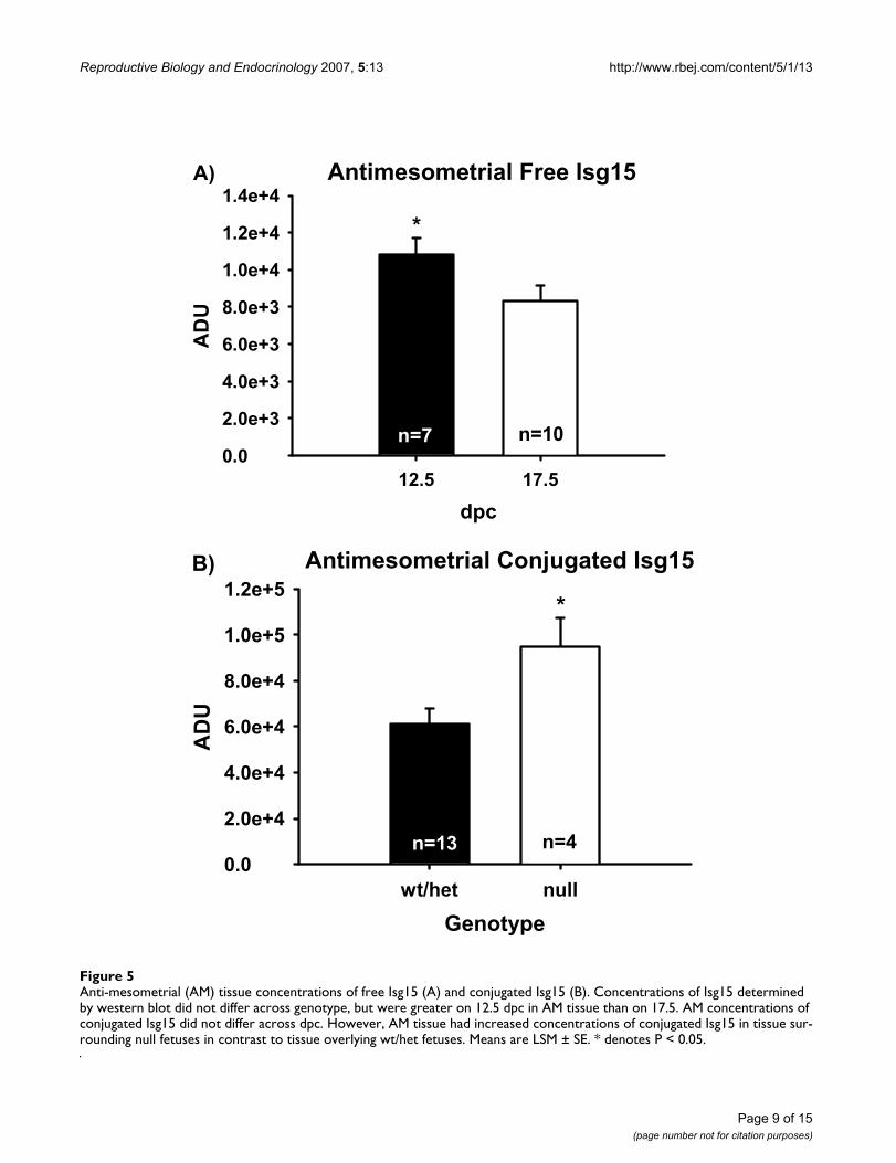
Anti-mesometrial tissue was also analyzed for free Isg15and its conjugates by western blot. No interactions (dpc ×genotoype) were observed in AM tissue for free or conju-gated Isg15, therefore only main effects (dpc or genotype)are reported. Concentrations of AM Isg15 were greater inthose tissues surrounding null fetuses regardless of dpc(data not shown). Overall concentrations of Isg15 in AMtissue were greater (P < 0.05) on 12.5 versus 17.5 dpc (Fig-ure 5). No differences in conjugated Isg15 were seenbetween AM tissues surrounding wt or het fetuses, there-fore data were pooled. Concentrations of conjugatedIsg15 were greater (P < 0.05) in AM surrounding nullfetuses in contrast to AM encompassing wt and hetfetuses.
Comparison of MM versus AM tissue identified that freeIsg15 was not different (P > 0.05) between the two tissues.However MM decidua in contact with fetal-derived pla-centa had greater (P < 0.05) levels of conjugated Isg15 incontrast to AM tissue (data not shown).
Virological and bacterial analysis of mouse colonyTo verify that fetal losses were not due to a compromisedsanitary environment of the animal facility, maternal andfetal tissues from a Ubp43 heterozygous interbreedingwere tested for a broad spectrum of bacteria and viruses(Wyoming State Veterinary Diagnostic Laboratory, Lara-mie, WY) and serum samples collected from a breedingpair were screened for viruses (Charles River Laboratories,Wilmington, MA). Both virological and bacteriologicalanalyses were negative.
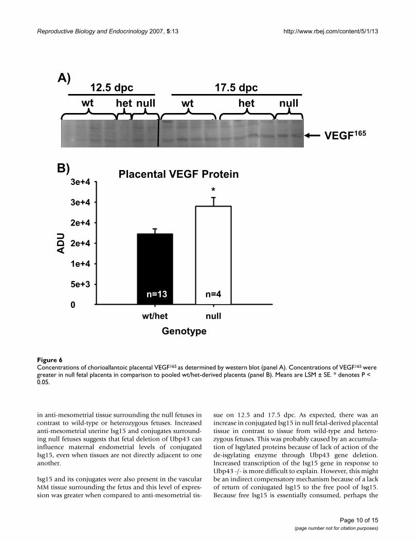
Environmental influence of elevation on angiogenic and hypoxic markers in the placental compartmentTo test if fetal losses were due to the increased elevation ofthe facility at UW (approximately 2,183 m; when com-pared to sea level for the facility at The Scripps ResearchInstitute), which may alter oxygen tension, fetal andmaternal derived tissues were analyzed by western blot forvarious angiogenic markers such as VEGF, angiopoietin-1and Flt-1 receptor or hypoxia markers such as GLUT-1 andAMP kinase-1alpha. No differences were seen within tis-sues based on dpc or genotype for the angiogenic markers– angiopoietin-1 or Flt-1 receptor or the hypoxic markers.However in CP tissue, concentrations of VEGF weregreater in null fetal-derived tissue in comparison topooled wt/het fetal-derived placental tissues (Figure 6Aand 6B).
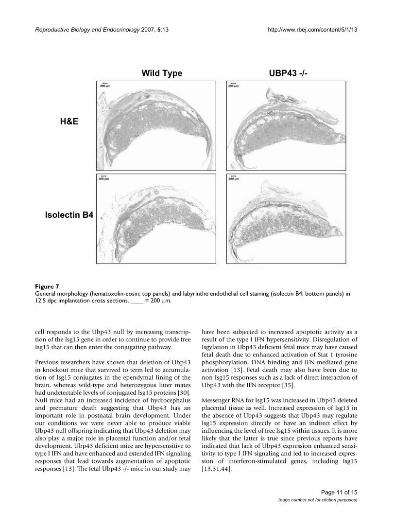
Morphological evaluation of placental tissueHematoxylin and eosin staining revealed that decidualand junctional or spongiotrophoblast zones appearednormal in 12.5 dpc implantation sites from Ubp43 wtfetuses. Gross evaluation of Ubp43 null mice consistentlyshowed more densely packed cells within the labyrinthlayer. Also the junctional layer was disrupted, which is nottypical of a mid-gestation placenta. The decidual tissue ofthe null-derived placenta had less cellular compaction incontrast to the Ubp43 wt placenta. Ubp43 heterzygousfetuses had variable morphology in implantation sitesthat was not consistently similar to the wt or the nullfetuses. Complementary characterization of the morphol-ogy of the junctional zone was performed using immuno-histochemistry techniques for isolectin B4 (Figure 7,lower panels). Isolectin B4 has greater specificity for laby-rinth-derived cells and leaves the junctional zone prima-rily devoid of stain [40]. Placental tissues derived fromUbp43 null fetuses had large infarcted areas in contrast towt Ubp43 fetal-derived placentas.
DiscussionEndometrial expression of Isg15 occurs during early preg-nancy in several mammals; including the cow [3], ewe [4],sow [5] mouse [6,7,20]) and primates [1,20,41]. Inter-feron-stimulated gene product 15 is hypothesized to playan intrinsic role during implantation and placentation byforming an isopeptide bond with intracellular proteins,potentially altering their activity. An Isg15-specific pro-tease, Ubp43, is also up-regulated by type I IFNs in a man-ner that is similar to up-regulation of Isg15 [42].Therefore, it was postulated that regulation of Ubp43within the placenta could play a significant role duringpregnancy and fetal development.
Previous reports by our group [6] and others [7] describedincreased Isg15 mRNA within AM decidua when com-pared to deciduoma of psuedopregnant mice on 7.5 dpc.We also reported that AM decidual Isg15 mRNA increasedfrom 4.5 to 7.5 and from 7.5 to 9.5 [6]. Expression ofIsg15 mRNA was restricted to maternal tissue and was notevident in conceptus-derived tissue on 7.5 dpc. [43]. Dur-ing later stages of pregnancy, MM decidua also expressedIsg15 and its conjugates on 12.5 and 17.5 dpc regardlessof fetal Ubp43 genotype. The fact that free and conjugatedIsg15 were present through 17.5 dpc in mice may suggestthat not only is Isgylation necessary in the uterus duringearly pregnancy, it may also influence placental function,implantation and fetal development throughout preg-nancy.
The level of Isg15 within the anti-mesometrial uterine tis-sue was greater on 12.5 dpc versus 17.5 dpc, which wassimilar to the temporal pattern of expression inMMdecidua. However, Isg15 and its conjugates were increased
Page 7 of 15(page number not for citation purposes)
Reproductive Biology and Endocrinology 2007, 5:13 http://www.rbej.com/content/5/1/13
Page 8 of 15(page number not for citation purposes)
Mesometrial (MM) concentrations of free Isg15 (A) and conjugated Isg15 (B)Figure 4Mesometrial (MM) concentrations of free Isg15 (A) and conjugated Isg15 (B). Based on western blot detection of Isg15, MM had greater concentrations of Isg15 on 12.5 versus 17.5 dpc (A). MM also had greater concentrations of conjugated Isg15 on 12.5 dpc in contrast to 17.5 (B). Means are LSM ± SE. * denotes P < 0.05. Arbitrary densitometric units (ADU).
MI Site Concentrations of ISG15
dpc12.5 17.5
AD
U
0.02.0e+34.0e+36.0e+38.0e+31.0e+41.2e+41.4e+41.6e+41.8e+4 *
A)
MI Site Concentrations of Conjugated ISG1
dpc12.5 17.5
AD
U
0.0
2.0e+4
4.0e+4
6.0e+4
8.0e+4
1.0e+5
1.2e+5
1.4e+5*
B)
Mesometrial Free Isg15
Mesometrial Conjugated Isg15
n=7 n=10
n=7 n=10
Reproductive Biology and Endocrinology 2007, 5:13 http://www.rbej.com/content/5/1/13
Page 9 of 15(page number not for citation purposes)
Anti-mesometrial (AM) tissue concentrations of free Isg15 (A) and conjugated Isg15 (B)Figure 5Anti-mesometrial (AM) tissue concentrations of free Isg15 (A) and conjugated Isg15 (B). Concentrations of Isg15 determined by western blot did not differ across genotype, but were greater on 12.5 dpc in AM tissue than on 17.5. AM concentrations of conjugated Isg15 did not differ across dpc. However, AM tissue had increased concentrations of conjugated Isg15 in tissue sur-rounding null fetuses in contrast to tissue overlying wt/het fetuses. Means are LSM ± SE. * denotes P < 0.05.
NI Concentrations of ISG15
dpc12.5 17.5
AD
U
0.0
2.0e+3
4.0e+3
6.0e+3
8.0e+3
1.0e+4
1.2e+4
1.4e+4*
A)
NI Concentrations of Conjugated ISG15
Genotypewt/het null
AD
U
0.0
2.0e+4
4.0e+4
6.0e+4
8.0e+4
1.0e+5
1.2e+5*
B)
Antimesometrial Free Isg15
Antimesometrial Conjugated Isg15
n=7 n=10
n=13 n=4
Reproductive Biology and Endocrinology 2007, 5:13 http://www.rbej.com/content/5/1/13
in anti-mesometrial tissue surrounding the null fetuses incontrast to wild-type or heterozygous fetuses. Increasedanti-mesometrial uterine Isg15 and conjugates surround-ing null fetuses suggests that fetal deletion of Ubp43 caninfluence maternal endometrial levels of conjugatedIsg15, even when tissues are not directly adjacent to oneanother.
Isg15 and its conjugates were also present in the vascularMM tissue surrounding the fetus and this level of expres-sion was greater when compared to anti-mesometrial tis-
sue on 12.5 and 17.5 dpc. As expected, there was anincrease in conjugated Isg15 in null fetal-derived placentaltissue in contrast to tissue from wild-type and hetero-zygous fetuses. This was probably caused by an accumula-tion of Isgylated proteins because of lack of action of thede-isgylating enzyme through Ubp43 gene deletion.Increased transcription of the Isg15 gene in response toUbp43 -/- is more difficult to explain. However, this mightbe an indirect compensatory mechanism because of a lackof return of conjugated Isg15 to the free pool of Isg15.Because free Isg15 is essentially consumed, perhaps the
Concentrations of chorioallantoic placental VEGF165 as determined by western blot (panel A)Figure 6Concentrations of chorioallantoic placental VEGF165 as determined by western blot (panel A). Concentrations of VEGF165 were greater in null fetal placenta in comparison to pooled wt/het-derived placenta (panel B). Means are LSM ± SE. * denotes P < 0.05.
B) FI Concentrations of VEGF
Genotype
wt/het null
AD
U
0
5e+3
1e+4
2e+4
2e+4
3e+4
3e+4*
Placental VEGF Protein
n=13 n=4
A)
wt wt hethet null null
12.5 dpc 17.5 dpc
VEGF165
Page 10 of 15(page number not for citation purposes)
Reproductive Biology and Endocrinology 2007, 5:13 http://www.rbej.com/content/5/1/13
cell responds to the Ubp43 null by increasing transcrip-tion of the Isg15 gene in order to continue to provide freeIsg15 that can then enter the conjugating pathway.
Previous researchers have shown that deletion of Ubp43in knockout mice that survived to term led to accumula-tion of Isg15 conjugates in the ependymal lining of thebrain, whereas wild-type and heterozygous litter mateshad undetectable levels of conjugated Isg15 proteins [30].Null mice had an increased incidence of hydrocephalusand premature death suggesting that Ubp43 has animportant role in postnatal brain development. Underour conditions we were never able to produce viableUbp43 null offspring indicating that Ubp43 deletion mayalso play a major role in placental function and/or fetaldevelopment. Ubp43 deficient mice are hypersensitive totype I IFN and have enhanced and extended IFN signalingresponses that lead towards augmentation of apoptoticresponses [13]. The fetal Ubp43 -/- mice in our study may
have been subjected to increased apoptotic activity as aresult of the type I IFN hypersensitivity. Disregulation ofIsgylation in Ubp43 deficient fetal mice may have causedfetal death due to enhanced activation of Stat 1 tyrosinephosphorylation, DNA binding and IFN-mediated geneactivation [13]. Fetal death may also have been due tonon-Isg15 responses such as a lack of direct interaction ofUbp43 with the IFN receptor [35].
Messenger RNA for Isg15 was increased in Ubp43 deletedplacental tissue as well. Increased expression of Isg15 inthe absence of Ubp43 suggests that Ubp43 may regulateIsg15 expression directly or have an indirect effect byinfluencing the level of free Isg15 within tissues. It is morelikely that the latter is true since previous reports haveindicated that lack of Ubp43 expression enhanced sensi-tivity to type I IFN signaling and led to increased expres-sion of interferon-stimulated genes, including Isg15[13,31,44].
General morphology (hematoxolin-eosin; top panels) and labyrinthe endothelial cell staining (isolectin B4; bottom panels) in 12.5 dpc implantation cross sectionsFigure 7General morphology (hematoxolin-eosin; top panels) and labyrinthe endothelial cell staining (isolectin B4; bottom panels) in 12.5 dpc implantation cross sections. ____ = 200 μm.
H&E
Isolectin B4
Wild Type UBP43 -/-
Page 11 of 15(page number not for citation purposes)
Reproductive Biology and Endocrinology 2007, 5:13 http://www.rbej.com/content/5/1/13
Implantation sites appeared disrupted in Ubp43 nullwhen compared to wt mice. The junctional zone and thedecidua were less densely compact in Ubp43 null whencompared to wt fetuses. This disregulation of Isg15through deletion of Ubp43 is hypothesized to contributeto 75% fetal mortality on day 12.5 of pregnancy.
Vascular endothelial growth factor is upregulated inresponse to hypoxia during physiological conditions,including such events as wound healing [45,46]. Vascularendothelial growth factor is expressed and localizedwithin trophoblast cells of various species [47-49] and isconsidered to be a very powerful mitogenic and ang-iogenic factor [50,51]. In vitro studies on VEGF165 incuba-tion with human trophoblast cells inhibited cellmigration through an extracellular matrix chamber [52].The increased levels of VEGF165 in the placenta mayinhibit appropriate implantation resulting in fetal mortal-ity. Albeit, since the mortality rate was 75 and 100% on12.5 and 17.5 dpc, respectively, the increased VEGF maybe a result of fetal resorption activity or it may haveincreased in response to hypoxia. Der and co-workers [42]reported that VEGF-C mRNA is up-regulated in responseto IFN-α and -γ. Therefore another possible cause forincreased VEGF in null fetal-derived tissue may be an indi-rect result of increased sensitivity to IFNs due to Ubp43deletion.
Furthermore Ubp43 -/- mice are hypersensitive to Type IIFN, implicating a role for Ubp43 to downregulate IFNresponses (Ritchie et al., 2002). These investigators lateridentified that Ubp43 attenuated IFN signaling by directinteraction with the region of the IFNAR2 receptor subu-nit that interacts with JAK1 [35]. Subsequently interactionof Ubp43 to the IFNAR2 receptor suppressed JAK1 inter-actions and concomitantly decreased downstream phos-phorylation cascades and other IFN-responsive signalingevents. These actions of Ubp43 are independent of itseffects on Isg15.
An expected Mendelian ratio of 1:2:1 wt:het:null liveprogeny from het interbreedings of Ubp43 knockout micewas achieved previously using C57 BL/6 × 129 back-ground with the pGK-Neo fragment [30]. However, dur-ing colony expansion we were never able to produce anyviable null offspring at the University of Wyoming (UW)facility by breeding het (C57 BL/6 × 129 crossed to SwissWebster to remove the pGK Neo fragment) pairs. In addi-tion to loss of null fetuses by 12.5 dpc, we also observedloss of +/- fetuses by 17.5 dpc. We estimate that the hetanimals at the UW facility had less than 50% Swiss Web-ster genetic contribution.
Ubp43 +/- interbreedings (C57 BL/6 × 129 × Swiss Web-ster; n = 10 litters) were followed to term, and from these
offspring 16 were +/+ and 31 were +/-, implicating that theMendelian ratio for offspring was 1:2 for wt:het, as wouldbe expected when considering the lack of null offspring.Therefore, we recorded fetal genotype ratios on 12.5 and17.5 dpc in utero to investigate the loss of null offspring.On 12.5 dpc we had a ratio of 2:5:1 from a total of 30 liveand dead fetuses (4 litters). And on 17.5 dpc the ratio was2:2:1 calculated from 35 live and dead fetuses (6 litters).On 12.5 dpc only 25% of the null fetuses were viable andby 17.5 dpc all null fetuses were non-viable. There alsowas a noticeable loss of +/- fetuses by 17.5 dpc. The loss ofheterozygous fetuses by 17.5 dpc was unexpected andmight be caused by a Ubp43 gene dosage due to loss ofone allele. When considering live fetuses only, genotypesdid not follow normal expected Mendelian ratios on 12.5and 17.5 dpc.
The same mice at the Scripps Research Institute producedUbp43 -/- mice from heterozygous breedings. At the ageof genotyping (3–4 wks old), a ratio of 1:4:1 from 58 pupswas observed. However, work done at Scripps found thatbackcrossing Ubp43 het mice (C57 BL/6 × 129) with C57BL/6 mice to F10 generation did not produce any viableUbp43 -/- pups. Similar to reports by the UW facility,homozygous Ubp43 null mice died in utero by 12.5 dpc.It is believed that genetic drift, influenced by the contribu-tion from C57/BL 6 lineage, altered the outcome at theUW facility. The founder animals at the UW facility couldhave influenced the expressionicity of the +/- interbreed-ings by either increased genetic contribution from C57BL/6 or potential deletion of the 129 pGK-Neo fragmentby crossing into the Swiss Webster strain.
Differences in viable offspring due to location may be areflection of facility environmental cues. However, bacte-rial and viral analyses of Ubp43 mice from our facilitywere negative suggesting that other causes may be respon-sible for fetal loss. The slight changes in angiogenic andhypoxia markers provided preliminary evidence thathypoxia played a major role in fetal loss, however futureexperiments are planned to further study this possibility.Mouse genetic background and associated genetic modifi-ers plays a significant role in sensitivity to interferon [53]and may also explain why fetuses died in the presentexperiment, but apparently were born and then died post-natally in other Ubp43 -/- experiments [30,38]. Thegenetic drift due to a mixed background may actually havecaused the unexpected fetal loss.
ConclusionIn summary, we have found that the Isg15 system waspresent in maternal and, for the first time reported, infetal-placental tissues. As expected, wild-type and hetero-zygous fetal-derived tissue did not appear to differ or alterthe maternal or fetal Isg15 system. Furthermore, Isg15
Page 12 of 15(page number not for citation purposes)
Reproductive Biology and Endocrinology 2007, 5:13 http://www.rbej.com/content/5/1/13
mRNA was present through late stages of pregnancy infetal tissue. Ubp43 deletion was verified in null fetal tissueby genotyping and northern blot analysis. And, VEGF wasthe only angiogenic marker that was altered by Ubp43deletion.
Deletion of the Ubp43 gene causes disregulation of theIsg15 system, a disrupted implantation site and fetaldeath in transgenic mice in the present study. This resultis different from reports in the original Ubp43 -/- paper[30] and from a more recent UBP43/Isg15 double -/-paper [38] where no impact on reproduction wasreported. However, work done at Scripps revealed thatbackcrossing Ubp43 het mice (C57 BL/6 × 129) with C57BL/6 mice to the F10 generation did not produce any via-ble Ubp43 -/- pups and these fetuses also died by 12.5dpc. For this reason, and because the Ubp43 -/- mice arefrom different genetic backgrounds, we suspect that thereare strain specific genetic modifiers (not unlike humanpopulations), that may impact the lethality of deletion ofUbp43.
Also, the original Isg15 -/- was not described to have anyreproductive problems and also was not described to beinvolved with antiviral responses [36]. Since the originalIsg15 -/- report, it has been demonstrated that deletion ofIsg15 does indeed affect antiviral responses and surviva-bility in response to influenza A/WSN/33 and influenzaB/Lee/40 virus, herpes simplex virus type 1, gammaher-pesvirus 68 and Sindbis virus infection [54]. Thus, thereare differences in the Isg15 responses due to differentviruses. And even though these investigators report no dif-ference in reproductive phenotype, there may be straindifferences as just described for the Ubp43 mice in addi-tion to subtle environmental differences such as mildhypoxia due to locatioin of the mice. For example, we alsohave recently obtained the Isg15 -/- mice from the Knobe-loch laboratory [38] and have observed a 50% fetal deathrate by 12.5 dpc (our unpublished results), which pro-vides support for the concept that this reproductive phe-notype is influenced by genetic background and/orlocation.
It is concluded that proper regulation of the Ubp43 andIsg15 systems are required for fetal viability and develop-ment. Up-regulation of Isg15 conjugates in Ubp43 nullfetuses may influence cellular responses and signal trans-duction within maternal and fetal components of the pla-centa, thereby altering proper implantation or function ofthe placental unit. Deletion of Ubp43 causes up-regula-tion of conjugated Isg15 in null fetuses, which may influ-ence targeted proteins within maternal and fetalcomponents of the placenta. Disruption of these targetproteins may influence proper implantation or functionof the placental unit causing deleterious effects on placen-
tation and fetal development in Ubp43 null mice.Whether the deletion of Ubp43 induces an abnormalaccumulation of conjugated Isg15 which then contributesto fetal lethality, or this is confounded or pre-empted bynon-Isg15 actions such as Isg15-independent impact ofUbp43 on the IFN receptor [35] awaits further study.
Authors' contributionsLR conducted the animal breeding, genotyping, and tissueharvesting. LR also carried out the molecular techniquesincluding western blotting and northern analysis. LR pre-pared and statistically analyzed data, drafted the manu-script and assisted with editing. KA assisted withmolecular techniques and editing. KR, MY, MS, and D-EZcreated and provided the transgenic mice. D-EZ also pro-vided guidance and insight during manuscript editing. LHconducted the immunohistochemistry. TH assisted withthe design and statistical analyses of the experiments aswell as writing and editing of the manuscript.
AcknowledgementsThis work was supported by NIH HD 032475-20 and NIH INBRE P20 RR 016474 to T.R.H. and NIH CA079849 to D.E.Z. The authors thank Dr. Michael J. Soares and Dr. Toshihiro Konno at the Institute of Maternal-Fetal Biology and the Division of Cancer & Developmental Biology, Department of Pathology and Laboratory Medicine, University of Kansas Medical Center for helpful discussions.
References1. Bebington C, Bell SC, Doherty FJ, Fazleabas AT, Fleming SD: Locali-
zation of ubiquitin and ubiquitin cross-reactive protein inhuman and baboon endometrium and decidua during themenstrual cycle and early pregnancy. Biol Reprod 1999,60(4):920-928.
2. Austin KJ, Pru JK, Hansen TR: Complementary deoxyribonucleicacid sequence encoding bovine ubiquitin-cross reactive pro-tein: a comparison with ubiquitn and a 15-kDa ubiquitinhomolog. Endocrine 1996, 5:191-197.
3. Hansen TR, Austin KJ, Johnson GA: Transient ubiquitin cross-reactive protein gene expression in the bovineendometrium. Endocrinology 1997, 138(11):5079-5082.
4. Johnson GA, Austin KJ, Collins AM, Murdoch WJ, Hansen TR:Endometrial ISG17 mRNA and a related mRNA are inducedby interferon-tau and localized to glandular epithelial andstromal cells from pregnant cows. Endocrine 1999,10(3):243-252.
5. Joyce MM, Hansen TR, Johnson GA: Interferon-stimulated gene17 is expressed in the porcine uterus and may be critical toplacental development across species. Biol Reprod 2002,66(Suppl 1):185.
6. Austin KJ, Bany BM, Belden EL, Rempel LA, Cross JC, Hansen TR:Interferon-stimulated gene-15 (Isg15) expression is up-regu-lated in the mouse uterus in response to the implanting con-ceptus. Endocrinology 2003, 144(7):3107-3113.
7. Bany BM, Cross JC: Post-implantation mouse conceptuses pro-duce paracrine signals that regulate the uterineendometrium undergoing decidualization. Dev Biol 2006,294(2):445-456.
8. Zhao C, Denison C, Huibregtse JM, Gygi S, Krug RM: Human ISG15conjugation targets both IFN-induced and constitutivelyexpressed proteins functioning in diverse cellular pathways.Proc Natl Acad Sci U S A 2005, 102(29):10200-10205.
9. Yuan W, Aramini JM, Montelione GT, Krug RM: Structural basisfor ubiquitin-like ISG 15 protein binding to the NS1 proteinof influenza B virus: a protein-protein interaction functionthat is not shared by the corresponding N-terminal domain
Page 13 of 15(page number not for citation purposes)
Reproductive Biology and Endocrinology 2007, 5:13 http://www.rbej.com/content/5/1/13
of the NS1 protein of influenza A virus. Virology 2002,304(2):291-301.
10. Yuan W, Krug RM: Influenza B virus NS1 protein inhibits con-jugation of the interferon (IFN)-induced ubiquitin-like ISG15protein. Embo J 2001, 20(3):362-371.
11. Padovan E, Terracciano L, Certa U, Jacobs B, Reschner A, Bolli M,Spagnoli GC, Borden EC, Heberer M: Interferon stimulated gene15 constitutively produced by melanoma cells induces e-cad-herin expression on human dendritic cells. Cancer Res 2002,62(12):3453-3458.
12. Giannakopoulos NV, Luo JK, Papov V, Zou W, Lenschow DJ, JacobsBS, Borden EC, Li J, Virgin HW, Zhang DE: Proteomic identifica-tion of proteins conjugated to ISG15 in mouse and humancells. Biochem Biophys Res Commun 2005, 336(2):496-506.
13. Malakhova OA, Yan M, Malakhov MP, Yuan Y, Ritchie KJ, Kim KI,Peterson LF, Shuai K, Zhang DE: Protein ISGylation modulatesthe JAK-STAT signaling pathway. Genes Dev 2003,17(4):455-460.
14. Malakhov MP, Kim KI, Malakhova OA, Jacobs BS, Borden EC, ZhangDE: High-throughput immunoblotting. Ubiquitiin-like pro-tein ISG15 modifies key regulators of signal transduction. JBiol Chem 2003, 278(19):16608-16613.
15. D'Cunha J, Ramanujam S, Wagner RJ, Witt PL, Knight E Jr., BordenEC: In vitro and in vivo secretion of human ISG15, an IFN-induced immunomodulatory cytokine. J Immunol 1996,157(9):4100-4108.
16. D'Cunha J, Knight E Jr., Haas AL, Truitt RL, Borden EC: Immu-noregulatory properties of ISG15, an interferon-inducedcytokine. Proc Natl Acad Sci U S A 1996, 93(1):211-215.
17. Hansen TR, Austin KJ, Perry DJ, Pru JK, Teixeira MG, Johnson GA:Mechanism of action of interferon-tau in the uterus duringearly pregnancy. J Reprod Fertil Suppl 1999, 54:329-339.
18. Perry DJ, Austin KJ, Hansen TR: Cloning of interferon-stimulatedgene 17: the promoter and nuclear proteins that regulatetranscription. Mol Endocrinol 1999, 13(7):1197-1206.
19. Austin KJ, Bany BM, Belden EL, Rempel LA, Cross JC, Hansen TR:Interferon-stimulated gene-15 (Isg15) expression is up-regu-lated in the mouse uterus in response to the implanting con-ceptus. Endocrinology 2003, 144:3107-3113.
20. Hess AP, Hamilton AE, Talbi S, Dosiou C, Nyegaard M, Nayak N,Genbecev-Krtolica O, Mavrogianis P, Ferrer K, Kruessel J, FazleabasAT, Fisher SJ, Giudice LC: Decidual Stromal Cell Response toParacrine Signals from the Trophoblast: Amplification ofImmune and Angiogenic Modulators. Biol Reprod 2006.
21. Finley D, Chau V: Ubiquitination. Annu Rev Cell Biol 1991, 7:25-69.22. Pantaleon M, Kanai-Azuma M, Mattick JS, Kaibuchi K, Kaye PL, Wood
SA: FAM deubiquitylating enzyme is essential for preimplan-tation mouse embryo development. Mech Dev 2001,109(2):151-160.
23. Moazed D, Johnson D: A deubiquitinating enzyme interactswith SIR4 and regulates silencing in S. cerevisiae. Cell 1996,86(4):667-677.
24. Papa FR, Hochstrasser M: The yeast DOA4 gene encodes a deu-biquitinating enzyme related to a product of the human tre-2 oncogene. Nature 1993, 366(6453):313-319.
25. Park KC, Kim JH, Choi EJ, Min SW, Rhee S, Baek SH, Chung SS, BangO, Park D, Chiba T, Tanaka K, Chung CH: Antagonistic regulationof myogenesis by two deubiquitinating enzymes, UBP45 andUBP69. Proc Natl Acad Sci U S A 2002, 99(15):9733-9738.
26. D'Andrea A, Pellman D: Deubiquitinating enzymes: a new classof biological regulators. Crit Rev Biochem Mol Biol 1998,33(5):337-352.
27. Amerik AY, Hochstrasser M: Mechanism and function of deubiq-uitinating enzymes. Biochim Biophys Acta 2004, 1695(1-3):189-207.
28. Wilkinson KD: Ubiquitination and deubiquitination: targetingof proteins for degradation by the proteasome. Semin Cell DevBiol 2000, 11(3):141-148.
29. Liu LQ, Ilaria R Jr., Kingsley PD, Iwama A, van Etten RA, Palis J, ZhangDE: A novel ubiquitin-specific protease, UBP43, cloned fromleukemia fusion protein AML1-ETO-expressing mice, func-tions in hematopoietic cell differentiation. Mol Cell Biol 1999,19(4):3029-3038.
30. Ritchie KJ, Malakhov MP, Hetherington CJ, Zhou L, Little MT, Mala-khova OA, Sipe JC, Orkin SH, Zhang DE: Dysregulation of protein
modification by ISG15 results in brain cell injury. Genes Dev2002, 16(17):2207-2212.
31. Li XL, Blackford JA, Judge CS, Liu M, Xiao W, Kalvakolanu DV, HasselBA: RNase-L-dependent destabilization of interferon-induced mRNAs. A role for the 2-5A system in attenuationof the interferon response. J Biol Chem 2000, 275(12):8880-8888.
32. Malakhova O, Malakhov M, Hetherington C, Zhang DE: Lipopoly-saccharide activates the expression of ISG15-specific pro-tease UBP43 via interferon regulatory factor 3. J Biol Chem2002, 277(17):14703-14711.
33. Hemelaar J, Galardy PJ, Borodovsky A, Kessler BM, Ploegh HL, OvaaH: Chemistry-based functional proteomics: mechanism-based activity-profiling tools for ubiquitin and ubiquitin-likespecific proteases. J Proteome Res 2004, 3(2):268-276.
34. Hemelaar J, Borodovsky A, Kessler BM, Reverter D, Cook J, Kolli N,Gan-Erdene T, Wilkinson KD, Gill G, Lima CD, Ploegh HL, Ovaa H:Specific and covalent targeting of conjugating and deconju-gating enzymes of ubiquitin-like proteins. Mol Cell Biol 2004,24(1):84-95.
35. Malakhova OA, Kim KI, Luo JK, Zou W, Kumar KG, Fuchs SY, ShuaiK, Zhang DE: UBP43 is a novel regulator of interferon signal-ing independent of its ISG15 isopeptidase activity. Embo J2006, 25(11):2358-2367.
36. Osiak A, Utermohlen O, Niendorf S, Horak I, Knobeloch KP: ISG15,an interferon-stimulated ubiquitin-like protein, is not essen-tial for STAT1 signaling and responses against vesicular sto-matitis and lymphocytic choriomeningitis virus. Mol Cell Biol2005, 25(15):6338-6345.
37. Kim KI, Yan M, Malakhova O, Luo JK, Shen MF, Zou W, de la TorreJC, Zhang DE: Ube1L and protein ISGylation are not essentialfor alpha/beta interferon signaling. Mol Cell Biol 2006,26(2):472-479.
38. Knobeloch KP, Utermohlen O, Kisser A, Prinz M, Horak I: Reexam-ination of the role of ubiquitin-like modifier ISG15 in thephenotype of UBP43-deficient mice. Mol Cell Biol 2005,25(24):11030-11034.
39. Laemmli UK: Cleavage of structural proteins during theassembly of the head of bacteriophage T4. Nature 1970,227(259):680-685.
40. Stewart J, Bebington CR, Mukhtar DD: Lectin binding character-istics of mouse placental cells. J Anat 2000, 196 ( Pt 3):371-378.
41. Bebington C, Doherty FJ, Fleming SD: Ubiquitin cross-reactiveprotein gene expression is increased in decidualizedendometrial stromal cells at the initiation of pregnancy. MolHum Reprod 1999, 5(10):966-972.
42. Der SD, Zhou A, Williams BR, Silverman RH: Identification ofgenes differentially regulated by interferon alpha, beta, orgamma using oligonucleotide arrays. Proc Natl Acad Sci U S A1998, 95(26):15623-15628.
43. Austin KJ, Carr AL, Pru JK, Hearne CE, George EL, Belden EL, HansenTR: Localization of ISG15 and conjugated proteins in bovineendometrium using immunohistochemistry and electronmicroscopy. Endocrinology 2004, 145(2):967-975.
44. Kang D, Jiang H, Wu Q, Pestka S, Fisher PB: Cloning and charac-terization of human ubiquitin-processing protease-43 fromterminally differentiated human melanoma cells using arapid subtraction hybridization protocol RaSH. Gene 2001,267(2):233-242.
45. Lokmic Z, Darby IA, Thompson EW, Mitchell GM: Time courseanalysis of hypoxia, granulation tissue and blood vesselgrowth, and remodeling in healing rat cutaneous incisionalprimary intention wounds. Wound Repair Regen 2006,14(3):277-288.
46. Neeman M, Abramovitch R, Schiffenbauer YS, Tempel C: Regulationof angiogenesis by hypoxic stress: from solid tumours to theovarian follicle. Int J Exp Pathol 1997, 78(2):57-70.
47. Wei P, Yu FQ, Chen XL, Tao SX, Han CS, Liu YX: VEGF, bFGF andtheir receptors at the fetal-maternal interface of the rhesusmonkey. Placenta 2004, 25(2-3):184-196.
48. Chakraborty I, Das SK, Dey SK: Differential expression of vascu-lar endothelial growth factor and its receptor mRNAs in themouse uterus around the time of implantation. J Endocrinol1995, 147(2):339-352.
49. Dunk C, Ahmed A: Expression of VEGF-C and activation of itsreceptors VEGFR-2 and VEGFR-3 in trophoblast. Histol His-topathol 2001, 16(2):359-375.
Page 14 of 15(page number not for citation purposes)
Reproductive Biology and Endocrinology 2007, 5:13 http://www.rbej.com/content/5/1/13
Publish with BioMed Central and every scientist can read your work free of charge
"BioMed Central will be the most significant development for disseminating the results of biomedical research in our lifetime."
Sir Paul Nurse, Cancer Research UK
Your research papers will be:
available free of charge to the entire biomedical community
peer reviewed and published immediately upon acceptance
cited in PubMed and archived on PubMed Central
yours — you keep the copyright
Submit your manuscript here:http://www.biomedcentral.com/info/publishing_adv.asp
BioMedcentral
50. Ferrara N, Davis-Smyth T: The biology of vascular endothelialgrowth factor. Endocr Rev 1997, 18(1):4-25.
51. Leung DW, Cachianes G, Kuang WJ, Goeddel DV, Ferrara N: Vascu-lar endothelial growth factor is a secreted angiogenicmitogen. Science 1989, 246(4935):1306-1309.
52. Fitzpatrick TE, Lash GE, Yanaihara A, Charnock-Jones DS, Macdon-ald-Goodfellow SK, Graham CH: Inhibition of breast carcinomaand trophoblast cell invasiveness by vascular endothelialgrowth factor. Exp Cell Res 2003, 283(2):247-255.
53. De Maeyer-Guignard J, Dandoy F, Bailey DW, De Maeyer E: Inter-feron structural genes do not participate in quantitative reg-ulation of interferon production by If loci as shown in C57BL/6 mice that are congenic with BALB/c mice at the alphainterferon gene cluster. J Virol 1986, 58(3):743-747.
54. Lenschow DJ, Lai C, Frias-Staheli N, Giannakopoulos NV, Lutz A,Wolff T, Osiak A, Levine B, Schmidt RE, Garcia-Sastre A, Leib DA,Pekosz A, Knobeloch KP, Horak I, Virgin HW: IFN-stimulatedgene 15 functions as a critical antiviral molecule against influ-enza, herpes, and Sindbis viruses. Proc Natl Acad Sci U S A 2007,104(4):1371-1376.
Page 15 of 15(page number not for citation purposes)


