Embed Size (px)
Citation preview

Published: April 22, 2011
r 2011 American Chemical Society 9726 dx.doi.org/10.1021/jp1119348 | J. Phys. Chem. C 2011, 115, 9726–9739
ARTICLE
pubs.acs.org/JPCC
Ultrafast Relaxation of the Poly(3-hexylthiophene) Emission SpectrumNatalie Banerji,† Sarah Cowan,† Eric Vauthey,‡ and Alan J. Heeger*,†
†Center for Polymers and Organic Solids, University of California at Santa Barbara, Santa Barbara, California 93106-5090, United States‡Department of Physical Chemistry, University of Geneva, 30 Quai Ernest-Ansermet, CH-1211 Geneva 4, Switzerland
1. INTRODUCTION
The optoelectronic properties of conjugated polymers com-bined with “plastic” mechanical behavior and solution processa-bility make them highly interesting materials with applications inorganic electronics.1�5 In bulk heterojunction (BHJ) solar cells,an electron-donating conjugated polymer is blended with afullerene electron acceptor, such as [6,6]-phenyl C60 butyric acidmethyl ester (PCBM).6�12 This yields a nanoscale morphologyin the thin film active layer with a high donor�acceptor interfacialarea for efficient photoinduced charge separation and with phase-separated fullerene and polymer networks for charge transport tothe electrodes. Regioregular poly(3-hexylthiophene), abbreviated asP3HT throughout the text, has been thoroughly investigated andis considered a state of the art material. High power conversionefficiencies, around 5%, can be obtained in thermally annealedP3HT:PCBM devices.8,13�15
A key to optoelectronic device functioning is the interaction ofthe conjugated polymer with light. Numerous steady-state andtime-resolved spectroscopic studies have been conducted inpristine P3HT and P3HT:PCBM blends in order to understandthe nature and evolution of neutral and charged excitations,16�56
but many aspects of the photophysics remain unclear. This isexplained by the general complexity of light-induced processes inconjugated polymers,57�60 as well as by additional complications
that arise because of the strong interchain interactions in thepartially microcrystalline film morphology of P3HT. Indeed, theP3HT chains in thin films π-stack into two-dimensional lamellaesheets perpendicular to the substrate with interchain distances ofonly 3.8 Å.61 Amorphous conjugated polymers can be treated asone-dimensional semiconductors where excitations are localizedon single polymer chains.62 On the other hand, it has been shownin P3HT films that both neutral excitations17,26,29,36,40,42,43,45,48,54
and charged polarons20,46,49�51 are at least partially delocalizedover neighboring polymer chains and become quasi-two-dimen-sional interchain species. This infers unique properties to thepolymer, such as a relatively high charge carrier mobility[approximately 0.1 cm2/(V s)]61 and a high yield, up to 30%, ofphotogenerated charge carriers in pristine P3HT films.38
We report here an ultrafast emission study of pristine P3HT inspin-cast thin films and dilute chlorobenzene (CB) solution, asmeasured by femtosecond-resolved fluorescence up-conversion(FU). This technique selectively probes neutral singlet excita-tions and is not concerned with charged polarons or triplet states,in contrast to transient absorption spectroscopy. Data recorded
Received: December 15, 2010Revised: March 17, 2011
ABSTRACT: The femtosecond-resolved evolution of the emis-sion spectrum of the important conjugated polymer poly(3-hexylthiophene) (P3HT) is presented. Detailed fluorescenceup-conversion spectroscopy was performed on P3HT solid-state films and on P3HT in chlorobenzene solution. Twoexcitation wavelengths and several emission wavelengths, cover-ing the entire fluorescence spectrum, were used. The data werecomplemented by polarization-sensitive measurements. Ourglobal analysis allowed a reconstruction of the time-resolvedemission spectra with 200 fs temporal resolution, so that spectralchanges due to the early relaxation processes following π�π* interband absorption in the pristine polymer could becomprehensively characterized. Absorption occurs in isolated polymer chains in solution and in the solid state (including interchaininteractions) for the film. In both cases, we find evidence of delocalization of the electrons and holes formed in the energy bandsdirectly after photoexcitation with excess energy. This is followed by ultrafast (∼100 fs) self-localization of the primaryphotoexcitation and by relatively slow exciton formation (∼1 ps). Further relaxation occurs with time constants ranging fromhundreds of femtoseconds to tens of picoseconds, due to exciton hopping to sites with lower energy and to a slow conformationalplanarization of the polymer backbone. Depolarization, a spectral red shift, and important changes in the vibronic structure areobserved as a consequence of this relaxation. Finally, relaxed intrachain and interchain singlet excitons are formed in solution andfilm, respectively, on a 100�200 ps time scale. They decay with a∼500 ps time constant, by intersystem crossing in solution and bynonradiative recombination in the film. Our results are consistent with and strongly support the conclusions we obtained from asimilar time-resolved fluorescence study of the polymer PCDTBT (J. Am. Chem. Soc. 2010, 132, 17459): ultrafast charge separationin polymer:fullerene blends seems to occur before localization of the primary excitation to form a bound exciton.

9727 dx.doi.org/10.1021/jp1119348 |J. Phys. Chem. C 2011, 115, 9726–9739
The Journal of Physical Chemistry C ARTICLE
over several emission wavelengths are compared for two excita-tion wavelengths and are supplemented by fluorescence polar-ization measurements. Such a comprehensive and systematicapproach has not been used in previously reported time-resolvedemission investigations of P3HT.16,17,21,23,24,37,44,54 Further-more, those studies obtained either single-wavelength FU dy-namics with a∼200 fs time resolution or entire emission spectrawith a lower time resolution of several picoseconds typical forstreak cameras. Here, we use the FU dynamics obtained at manyemission wavelengths to reconstruct the emission spectra with a∼200 fs time resolution. This reveals important spectral changesdirectly following light absorption in P3HT, in particular wherethe relative intensity of the vibronic transitions is concerned.Detailed information about the early relaxation dynamics of theprimary photoexcitations in the pristine polymer can be re-trieved. Those relaxation processes are important to understand,since they occur on a similar time scale as charge separation inP3HT:PCBMblends.18,32 Therefore, they are directly relevant tothe functioning of BHJ solar cells.
2. EXPERIMENTAL METHODS
2.1. Samples. Regioregular poly(3-hexylthiophene) wassynthesized by RiekeMetals, Inc., according to the Rieke method(Mn = 33.4 kDa, Mw = 79.9 kDa, PDI = 2.4, regioregularity =92.1%). For thin film fabrication, a 10 mg/mL solution of P3HTin chlorobenzene (Sigma-Aldrich, anhydrous, 99.8%) was spin-cast on a circular quartz substrate (Saint-Gobain Spectrosil 2000)at 3500 rpm in a nitrogen glovebox. After annealing the thin filmon a hot plate at 60 �C for 1 h and letting it rest at roomtemperature overnight, it was encapsulated in inert atmospherebetween two quartz disks using epoxy resin (DELO-KATIO-BOND LP655), as described in detail elsewhere.63 The opticaldensity of the solid sample was 0.26 at the visible maximum(∼540 nm), which corresponds to a film thickness of about45 nm, as inferred from the absorption coefficient published inthe literature.36 The steady-state absorption and fluorescencespectra of the encapsulated film revealed no sign of polymerdegradation over a period of 1 month. For solution measure-ments, P3HT was dissolved overnight in chlorobenzene (Acros,Extra Dry, AcroSeal, 99.8%), which had previously been degassedby argon bubbling for 15 min. The solutions were placed in a1 mm cell which consisted of two Spectrosil quartz disks separatedby a Teflon spacer. The optical density at the visible maximum(455 nm) over 1 mm was 0.33 for the FU measurements with500 nm excitation and 0.55 for 400 nm excitation, whichcorresponds to concentrations of about 0.08 mg/mL and 0.14mg/mL, respectively. The shape of the absorption spectrum wasthe same at the two concentrations.2.2. Steady-State Measurements. Steady-state absorption
spectra were measured with a Cary 50 (Varian) spectrophoto-meter, while fluorescence emission and excitation spectrawere recorded with a Cary Eclipse (Varian) fluorimeter (slitwidth 3 nm) and repeated with a Photon Technology Inter-national fluorimeter to verify reproducibility (slit width 2 nm,data not shown). All fluorescence spectra were corrected forthe wavelength-dependent sensitivity of the detection. Whenrepresenting the emission spectra on a wavenumber scalein cm�1, the fluorescence intensity was multiplied by thesquare of the wavelength (λ2) in order to account for theband-pass constant in wavelength imposed by the monochro-mator of the fluorimeter. All spectra shown here were recorded
with the same solutions or thin film as used in the FUmeasurements. Similar spectral shapes were obtained withhighly diluted solutions, so that concentration effects can beruled out.2.3. Time-Resolved Emission Measurements. Emission
dynamics on the femtosecond time scale were obtained usingthe fluorescence up-conversion setup previously described.63,64
In brief, the 800 or 1000 nm output of a tunable Mai Tai HP(Spectra-Physics) mode-locked Ti:sapphire laser system (100 fspulse duration, 80 MHz repetition rate) was frequency doubledfor sample excitation at 400 or 500 nm, respectively. The pumpintensity per pulse was adjusted in the range of 6�40 μJ/cm2
(with a spot diameter of 20 μm) in order to keep the excitationdensity constant at the two excitation wavelengths. The latter wasapproximately 5� 1013 or 4� 1014 photons/cm3 in solution (fortwo separate measurement sessions) and 1.5 � 1018 photons/cm3 in the film. The measured sample fluorescence was detectedby sum-frequency generation with a delayed gate pulse, and thenthe up-converted signal was dispersed in a monochromator andits intensity measured with a photomultiplier tube. The polariza-tion of the pump beam was at the magic angle (54.7�) relative tothat of the gate pulses, except for polarization-sensitive measure-ments, where it was set to 0� (parallel) and 90� (perpendicular).Measurements were done at room temperature in ambientconditions. To minimize degradation, the sample cell (containingthe solution or thin film) was constantly rotated during themeasurement. At least two scans of the dynamics in the �5 to1000 ps (or 300 ps) range were averaged at each emissionwavelength. No significant degradation of the samples wasobserved between two scans or when comparing the steady-stateabsorption and fluorescence spectra before and after the FUmeasurements.2.4. Analysis of the FU Data. Again, details are described
elsewhere,63,65 so only a summary is given here. For each sample,the normalized time profiles obtained by FU at various emissionwavelengths were analyzed globally using the convolution of aGaussian-shaped instrument response function (IRF) with thesum of four exponential terms. The width of the IRFwas found tobe around 130 fs with the thin film and around 150�200 fs withsolution samples. The wavelength-dependent amplitudes (orpre-exponential factors) were scaled to the steady-state emissionspectrum, assuming that the steady-state emission intensity at agiven wavelength is the time integral of the correspondingemission time profile. The time-resolved emission spectra werethen reconstructed using the analytical expression for the timeprofiles at different wavelengths with the parameters (IRF, timeconstants, and scaled amplitudes) obtained from the fittingprocedure. Note that the spectra obtained in this way are aconvolution of the sample response and the IRF. This is why weonly show spectra after 200 fs. A similar spectral shape wouldhave been obtained if the spectra were directly reconstructedfrom the properly scaled raw experimental data; the recon-structed spectra are therefore not compromised by the numberof fitting parameters, as long as they reproduce the data well. Forfemtosecond-resolved fluorescence anisotropy measurements,the anisotropy decay, r(t), was calculated from the FU timeprofiles with the polarization of the pump beam parallel andperpendicular with respect to the gate beam, using the standardequation. This r(t) was analyzed using the sum of exponentialterms. The measured magic angle traces could always be well-reproduced by the data calculated from the parallel and perpen-dicular curves.

9728 dx.doi.org/10.1021/jp1119348 |J. Phys. Chem. C 2011, 115, 9726–9739
The Journal of Physical Chemistry C ARTICLE
3. RESULTS
3.1. Steady-State Spectra. As shown in Figure 1A, the visibleabsorption spectrum of P3HT dissolved in CB solution consistsof a broad unstructured band with a maximum at 21 980 cm�1,which we assign to the lowest energy π�π* interband transition.The band gap is estimated from the absorption edge as equal to18 360 cm�1 (2.28 eV). The corresponding emission spectrumdiffers in shape from the absorption band, since it is muchnarrower and displays a vibronic progression. It peaks in the 0�0band at 17 000 cm�1, which implies a Stokes shift, i.e., energydifference between the emission maximum and the absorptionedge, of 1360 cm�1 (0.17 eV). The emission spectra obtainedwith 400 and 500 nm excitation are so similar that they cannot bedistinguished in Figure 1A, suggesting fast relaxation to acommon emitting state, the singlet exciton. The fluorescenceexcitation spectrum we measured in solution is slightly broaderthan the absorption spectrum and displays structure (Figure 1A).This result could be reproduced at very low polymer concentra-tion (absorbance ∼0.05) and using different fluorimeters. It isthus unlikely to be an artifact or a consequence of high sampleabsorption.The absorption and emission spectra of P3HT thin film spin-
cast from CB are considerably different from those of P3HT insolution, as shown in Figure 1B. The absorption spectrum isstrongly red-shifted with a maximum at 18 280 cm�1. It displaysstructure such as a pronounced shoulder at 16 780 cm�1. Theband gap is about 15 250 cm�1 (1.89 eV). The low-energy tailbelow the band gap is most probably due to midgap localizedstates, although there could be a contribution of increasedreflection losses caused by changes in the refractive index nearthe band edge. The emission spectrum is also red-shifted in thefilm compared to solution; it is again independent of excitationwavelength, displays vibronic structure, and is much narrowerthan the absorption spectrum. The 0�0 transition is, however,less pronounced than in solution, whereas the 0�1 and 0�2
sidebands are enhanced. The 0�1 transition at 13 700 cm�1 isnow clearly the emission maximum, giving a Stokes shift of1550 cm�1 (0.19 eV). Within the experimental uncertainty, thefluorescence excitation spectrum in the P3HT film closelymatches the absorption spectrum. It should be noted from theexcitation spectrum that the absorption below the band gap(probably midgap states) does not significantly contribute to theemission.3.2. Time-Resolved Emission for P3HT in Chlorobenzene
Solution. 3.2.1. Excitation at 500 nm. For P3HT in CB solution,excitation at 500 nm (2.48 eV) occurs on the low-energy side ofthe absorption spectrum (Figure 1A), with an excess energyrelative to the band edge of 0.20 eV. The time profiles obtainedby FU with 500 nm excitation at various emission wavelengthsare depicted in Figure 2. The intensity decays almost to zerowithin the time window of 1 ns (see the inset), while the earlyemission dynamics are nonexponential and depend strongly onthe emission wavelength. The fast decay at high energy andconcomitant rise at lower energy suggest an initial red shift ofthe emission spectrum. This is in qualitative agreement withprevious FU investigations of P3HT in chloroform solution(using lower molecular weight polymer and other excitationwavelengths).16,37 Unlike those studies, many more emissionwavelengths covering the entire spectrum were recorded here,and we chose to analyze the dynamics globally using the sumof four exponential functions. Time constants of τ1 = 0.7 ps, τ2 =6.0 ps, τ3 = 41 ps, and τ4 = 530 ps were found, and the spectra ofthe pre-exponential amplitude factors are shown in Figure 3A.The amplitude spectrum of τ4 is all-positive and identical tothe steady-state emission spectrum. We therefore assign the530 ps time constant to the decay of the relaxed singletexciton, in excellent agreement with the lifetime reported inthe literature.36,37,54,55
The three amplitude spectra related to the shorter timeconstants, τ1�τ3, are much weaker than the one of τ4 and theyare not related to any excited population decay (Figure 3A).They are positive (spectral decay) at low wavelengths andnegative (spectral rise) at increasingly high wavelengths. Thisis characteristic of a progressive relaxation of the photoexcitation,leading to spectral changes. Our data and analysis provide us with
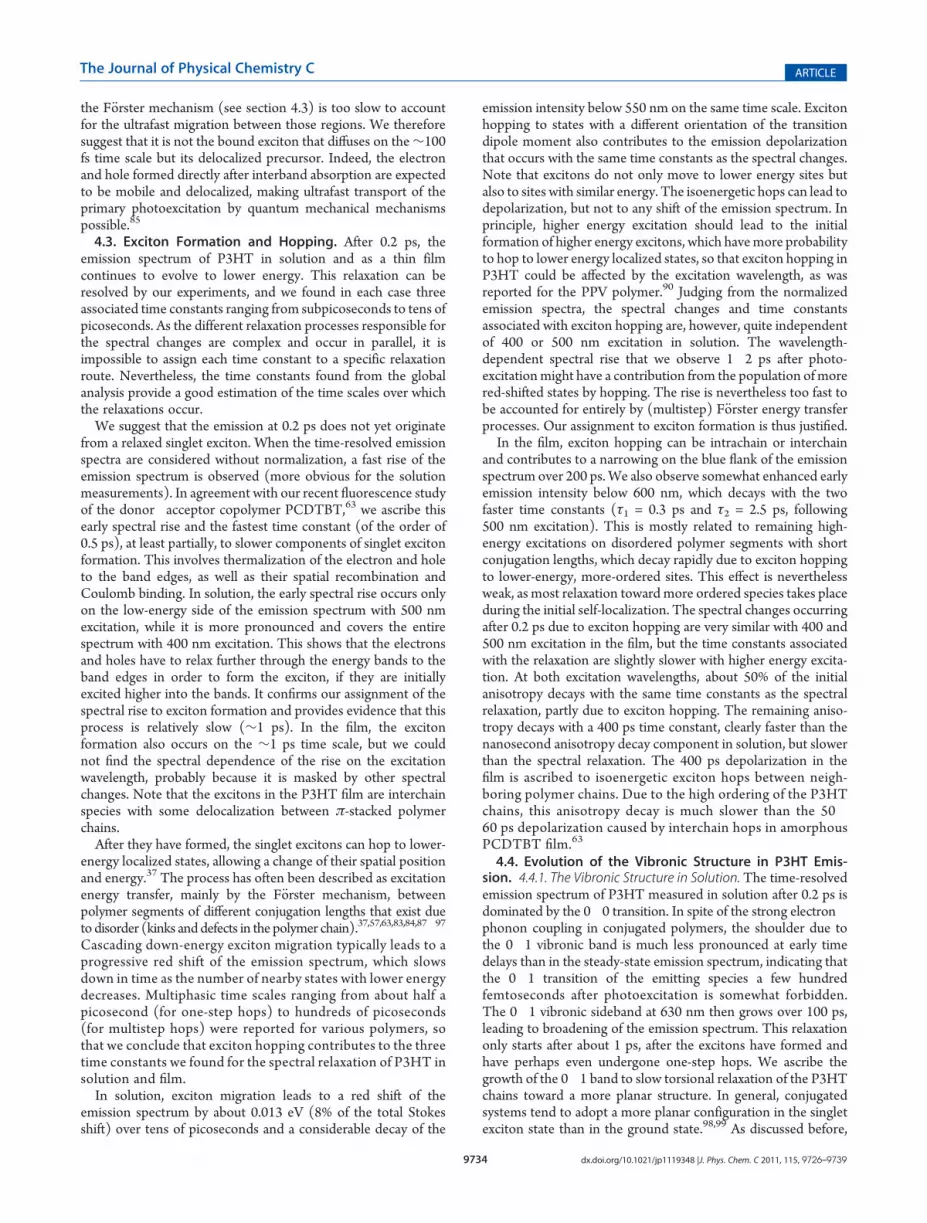
Figure 1. Steady-state spectra of P3HT in chlorobenzene solution (A)and as a thin film (B). The absorption spectra (thick black lines), thefluorescence excitation spectra probed near the emission maximum(gray lines with small markers), as well as the fluorescence emissionspectra with 400 nm excitation (blue lines) and with 500 nm excitation(green lines) are shown. The excitation wavelengths (400 nm or500 nm) used in the fluorescence up-conversion measurements areindicated as vertical dashed lines. The thin dotted lines identify theinterband absorption edge from the absorption spectrum.
Figure 2. Ultrafast emission time profiles over 10 ps of P3HT inchlorobenzene solution after excitation at 500 nm. The probed emissionwavelengths are shown in the legend. The solid lines correspond to thebest multiexponential global fit. The inset shows the time profile of the590 nm emission over 1000 ps.

9729 dx.doi.org/10.1021/jp1119348 |J. Phys. Chem. C 2011, 115, 9726–9739
The Journal of Physical Chemistry C ARTICLE
unique femtosecond-resolved emission spectra of P3HT, recon-structed from the global fitting parameters (Figure 3B). They arenormalized in Figure 3C, for better visualization of the spectralshape evolution. The first emission spectrum that can be resolvedwith the FU instrumentation (at 0.2 ps) displays already >90% ofthe total Stokes shift, pointing to considerable relaxation on a∼100 fs time scale. It is also much narrower than the mirror imageof the absorption spectrum and has relatively weak oscillatorstrength in the 0�1 vibronic band, compared to steady-stateemission. After 200 fs, there is an important rise of the low-energypart of the emission spectrum over a few picoseconds (seen in thenon-normalized spectra of Figure 3B). The spectrum also shiftsto lower energy by about 0.013 eV (8% of the total Stokes shift)over tens of picoseconds (Figure 3C). Decay of the intensitybelow 550 nm occurs on the same time scale. The slowestrelaxation in the emission spectrum, which only starts after about1 ps, is the growth of the 0�1 vibronic sideband at 630 nm. Thisleads to an overall broadening of the spectrum and takes 100 psto complete (Figure 3C).3.2.2. Excitation at 400 nm. We also measured the emission
dynamics of P3HT in CB solution following excitation at400 nm. This occurs on the high-energy side of the absorptionspectrum (Figure 1A), with 0.82 eV excess energy compared tothe band edge. Figure 4A shows that the emission time profile at580 nm is strongly affected by the excitation wavelength. Thereis a pronounced early rise only following 400 nm absorption.
The outcome of the global analysis of the time profiles recordedthroughout the emission spectrum, following excitation at bothwavelengths, is compared in Figure 5. The data were obtainedunder very similar conditions for 400 and 500 nm excitation, on aseparate occasion and with a slightly higher excitation power thanthe data presented before for 500 nm (Figure 2,3), which explainsthe minor differences.The early spectral rise, which occurs over a few picoseconds, is
strongly affected by the excitation wavelength as shown on thetop of Figure 5. It occurs only on the low-energy side of the
Figure 5. Outcome of the global analysis of the emission time profiles ofP3HT in chlorobenzene solution after excitation at 500 nm (A) and400 nm (B). The top panels show the reconstructed time-resolvedemission spectra, while the normalized reconstructed time-resolvedemission spectra are in the bottom panels.
Figure 3. Outcome of the global analysis of the emission time profiles ofP3HT in chlorobenzene solution after excitation at 500 nm: (A) decayassociated amplitude spectra, (B) reconstructed time-resolved emissionspectra, and (C) normalized reconstructed time-resolved emissionspectra.
Figure 4. Ultrafast emission time profiles (A) and anisotropy decay (B)of P3HT in chlorobenzene solution recorded at an emission wavelengthof 580 nm after excitation at 500 nm (green) and 400 nm (blue). Theinset shows the anisotropy decay over a longer time scale. The solid linescorrespond in each case to the best multiexponential fit. The fittingparameters obtained for the anisotropy decay are given in the inset table.

9730 dx.doi.org/10.1021/jp1119348 |J. Phys. Chem. C 2011, 115, 9726–9739
The Journal of Physical Chemistry C ARTICLE
emission spectrum with 500 nm excitation, while it is morepronounced and covers the entire spectrum with 400 nm excita-tion. The normalized emission spectra shown at the bottom ofFigure 5 reveal, however, that the effective spectral shape and itstime-resolved changes are much less dependent on excitationwavelength than the initial rise. The shape of the earliest resolved0.2 ps spectrum, the spectral red shift, and the growth of the 0�1vibronic sideband are all very similar with 400 and 500 nmexcitation, and the time scales for the spectral relaxation are thesame (0.7, 6.0, and 41 ps). This points to a weak dependence ofthe processes responsible for this spectral relaxation on theexcitation wavelength. Note that τ4, the exciton lifetime, couldnot be determined precisely for the data in Figure 5, as thedynamics were only recorded up to 300 ps. It can be said,however, that exciton decay occurs on a similar time scale(hundreds of picoseconds) at both excitation wavelengths.3.2.3. Anisotropy Measurements. Finally, polarization-sensitive
FU measurements were obtained and the calculated anisotropyat an emission wavelength of 580 nm is compared for 400 and500 nm excitation in Figure 4B. The initial anisotropy r0, mea-sured with the 0.2 ps time resolution, is in both cases lower thanthe theoretical maximum of 0.4 (when the transition dipolemoments of the absorption and emission are parallel). What ismost interesting is that r0 is considerably lower with 400 nm
excitation (0.15) than with 500 nm excitation (0.35). After 0.2 ps,the anisotropy at 580 nm evolves in a quite similar manner for400 and 500 nm excitation (Figure 4B). About 35% of r0 is inboth cases lost with time constants of 0.7, 6.0, and 41 ps, the sameas for the spectral relaxation (see the inset table in Figure 4B).The remaining ∼65% of the initial anisotropy has a very longnanosecond lifetime, because the anisotropy in the isolatedchains is now only lost by very slow orientational diffusion ofthe polymer molecule. We measured the anisotropy decay atseveral emission wavelengths throughout the spectrum, butfound no significant difference within the experimental error(data not shown). This indicates that the emitting dipoles for the0�0 and the 0�1 vibronic transitions are parallel.3.3. Time-Resolved Emission for P3HT Thin Film.
3.3.1. Excitation at 500 nm. For the P3HT spin-cast thin film,excitation at 500 nm occurs on the high-energy side of the absorp-tion band with 0.59 eV excess energy compared to the band edge(Figure 1B). This is much more than the 0.20 eV excess energyinjected in P3HT solution at the same excitation wavelength. Thetime profiles recorded in the film with 500 nm excitation alsodepend strongly on the emission wavelength, implying relaxationthat leads to spectral changes (Figure 6A). We found timeconstants for the spectral relaxation in P3HT film of τ1 = 0.3 ps, τ2= 2.5 ps, and τ3 = 40 ps, which are comparable but slightly fasterthan in solution. The outcome of the global analysis for the filmdata recorded with 500 nm excitation is shown in Figure 7. Theamplitude spectra in panel A confirm that τ1�τ3 are due to
Figure 6. (A) Ultrafast emission time profiles of P3HT thin film afterexcitation at 500 nm. The probed emission wavelengths are shown in thelegend. The dashed line shows the time profile of the 590 nm emission ofP3HT in chlorobenzene solution after excitation at 500 nm. (B)Ultrafast emission time profiles of P3HT thin film recorded at anemission wavelength of 620 nm after excitation at 500 nm with differentexcitation intensities (the fluorescence intensity is divided by theexcitation power). (C) Anisotropy decay of P3HT thin film recordedat 650 nm after excitation at 500 nm (green) and 400 nm (blue). Thesolid lines correspond in each case to the best multiexponential fit. Thefitting parameters obtained for the anisotropy decay are given in theinset table.
Figure 7. Outcome of the global analysis of the emission time profiles ofP3HT thin film after excitation at 500 nm: (A) decay associatedamplitude spectra, (B) reconstructed time-resolved emission spectra,and (C) normalized reconstructed time-resolved emission spectra.

9731 dx.doi.org/10.1021/jp1119348 |J. Phys. Chem. C 2011, 115, 9726–9739
The Journal of Physical Chemistry C ARTICLE
spectral changes, while τ4 = 470 ps is the exciton lifetime. Thelatter is on the same order of magnitude as the exciton decayobserved in solution (530 ps). The similarity of the time scales isconfirmed by the comparison of the fluorescence time profiles inthe film (at 730 nm) and in solution (at 590 nm) shown inFigure 6A. Unlike in solution, the amplitudes of the significantspectral changes are comparable to the one of the exciton decay inthe film.Although we cannot obtain the entire emission spectrum due
to experimental constraints above 750 nm, it appears that theshape of the earliest resolved spectrum at 0.2 ps (panels B and Cof Figure 7) is already considerably narrower than the absorptionspectrum. The overall spectral position is also close to the one forsteady-state emission, showing that the majority of the Stokesshift has taken place faster than our time resolution. We note thatthe 0.2 ps spectrum in the film is very different from thecorresponding spectrum in solution (strong red-shift), indicatinga negligible contribution from nonaggregated (isolated) P3HTchains to the emission at this time delay. The earliest resolvedspectrum in the film shows a well-resolved vibronic structurewhere both the 0�0 transition and the 0�1 sideband are visible,centered at 665 and 730 nm, respectively. A striking observationis the much higher relative intensity of the 0�0 band comparedto the 0�1 band, the opposite of what occurs in the steady-statespectrum.The normalized time-resolved emission spectra depicted in
Figure 7C show that the relative intensity of the 0�0 banddecreases in time. It has about the same intensity as the 0�1 bandafter 10 ps, and this inversion of the 0�0 and 0�1 relativeintensity continues up to 200 ps, when the steady-state shapewith a much more intense 0�1 band is reached. The amplitudespectra in Figure 7A show that the three first time constants, τ1 =0.3 ps, τ2 = 2.5 ps, and τ3 = 40 ps, are all associated with the decayof the 0�0 emission band. Apart from the inversion of the 0�0and 0�1 relative band intensities, other spectral changes occurfor P3HT film upon 500 nm excitation. The reconstructed time-resolved emission spectra in Figure 7B reveal a∼1 ps spectral riseabove 680 nm. The effect is quite weak compared to the solutionmeasurements, but might be more pronounced on the red flankof the emission spectrum, which is experimentally not accessible.From the amplitude spectra in Figure 7A, it is clear that only τ1 =0.3 ps contributes to the initial spectral rise. Finally, Figure 7C
shows enhanced early emission intensity below 600 nm, whichdecays with the τ1 = 0.3 ps and τ2 = 2.5 ps time constants. Thereis also a narrowing over 200 ps of the blue flank of the emissionspectrum.To ensure the absence of singlet exciton annihilation in our
measurements, we varied the excitation intensity. The fluores-cence time profiles scaled almost linearly with excitation intensityat the used pump power of 1.50 mW (see comparison with thedynamics obtained at a 0.50 mW excitation power in Figure 6B).Small effects of annihilation are seen at 2.80 mW. They becamevery important with a pump power of 8 mW, where τ3 and τ4were reduced to 21 and 220 ps, respectively. The overall shape ofthe spectral changes was however not affected (data not shown).3.3.2. Excitation at 400 nm. We also measured the FU time
profiles of P3HT film following excitation in the high-energy tailof the absorption spectrum at 400 nm, with 1.12 eV excess energycompared to the band edge. The time-resolved emission spectra,reconstructed from the global analysis of the data, are depicted inFigure 8. There is hardly any difference compared to the datarecorded with 500 nm excitation. Within the experimentaluncertainty, the shape of the 0.2 ps spectrum is very close atthe two excitation wavelengths. The spectral changes occurringafter 0.2 ps, i.e., the narrowing on the blue side of the spectrumand the inversion of the 0�0 and 0�1 band intensities, are alsovery similar with 400 and 500 nm excitation. It is interesting tonote that the initial spectral rise starts at 680 nm independently ofthe excitation wavelength, in contrast to the higher-energy startof the rise observed with 400 nm excitation compared to 500 nmexcitation in solution (Figure 5A). The strong decay of the 0�0band in the P3HT film probably masks any spectral rise in thehigh-energy part of the spectrum. The only evidence that morerelaxation is needed in the film when more excess energy isinitially provided is that the time constants for the spectralrelaxation are slower with 400 nm excitation (τ1 = 0.6, ps, τ2 =5.3 ps, and τ3 = 60 ps).3.3.3. Anisotropy Measurements. Finally, the anisotropy de-
cay of the 620 nm emission, with both excitation wavelengths, isshown in Figure 6C. The initial anisotropy measured within the200 fs time resolution is much lower than 0.4. Again, r0 is smallerwith 400 nm excitation (0.15) than with 500 nm excitation(0.23). The difference is less pronounced than in solution, wherer0 with 500 nm excitation was higher (Figure 4B). At bothexcitation wavelengths, about 50% of the initial anisotropydecays with the same time constants as the spectral relaxation(inset table in Figure 6C). The remaining anisotropy decays witha 400 ps time constant, clearly faster than the nanosecondanisotropy decay component in solution (Figure 4B), but slowerthan the spectral relaxation.
4. DISCUSSION
On the basis of experimental results obtained from photo-conductivity studies and ultrafast investigations of the infraredactive vibrational (IRAV) modes in PPV and MEH-PPV66�71
and on evidence obtained from highly oriented MEH-PPVchains in a polyethylene matrix,72�75 we view conjugated poly-mers as quasi-one-dimensional semiconductors and describetheir electronic structure within an energy band picture.1,62 Wenote here that the band versus molecular interpretation is notdirectly relevant to the scope of the current paper. The importantpoint is that absorption leads to a delocalized primary photo-excitation, which then relaxes to amore localized exciton state. As
Figure 8. Reconstructed time-resolved emission spectra obtained bythe global analysis of the emission time profiles of P3HT thin film afterexcitation at 400 nm.

9732 dx.doi.org/10.1021/jp1119348 |J. Phys. Chem. C 2011, 115, 9726–9739
The Journal of Physical Chemistry C ARTICLE
discussed in the following, the initial delocalization has beenshown by several independent groups and was not necessarilydescribed within the semiconductor approach. Several processescontribute to the relaxation of the primary photoexcitation ofP3HT to the relaxed singlet exciton: (1) self-localization, (2)exciton formation, (3) exciton hopping, and (4) slow torsionalrearrangements. They will be discussed on the basis of our time-resolved emission results.4.1. Interpretation of the Steady-State Spectra. We inter-
pret the visible absorption spectrum of P3HT as an allowedπ�π* interband transition yielding electrons and holes in theconduction and valence band, respectively. Emission on theother hand occurs from a relaxed singlet exciton state. In oursolution measurements, both absorption and emission are essen-tially intrachain processes within isolated polymer chains. In-deed, polymer aggregation (between chains and due to chainfolding) is considered negligible at the concentrations used andgiven the fact that CB is a “good” solvent. Our absorption andemission spectra are also consistent with the spectral shapesreported previously for nonaggregated P3HT in dilutesolution.40 Strong conformational (mostly torsional) disorderof the dissolved P3HT chains in the ground state leads to theinhomogeneously broadened and structureless absorption spec-trum. The difference between the absorption spectrum and thebroader/more structured fluorescence excitation spectrum in-dicates that the fluorescence quantum yield differs within thedistribution of absorbing conformations. The more fluorescentconformers are on both sides of the absorption band, while theless fluorescent ones absorb in its center.The shape of the emission spectrum in solution is independent
of excitation wavelength. Even if a large distribution of confor-mations is initially excited, relaxation eventually occurs to acommon emitting intrachain exciton state with less torsionaldisorder. Judging from the excitation spectrum, the low-fluo-rescent conformations relax to this state in lower yield. Therelaxation toward a more localized singlet exciton after the initialinterband absorption and toward higher order explains thenarrower and more structured emission spectrum. The intra-chain emission spectrum has previously been well-reproducedwith a Franck�Condon model, using a Huang�Rhys factor of 1and assuming that the 0.18 eV CdC stretching vibrationpredominantly couples to the electronic transition.40 A struc-tured emission spectrum contrasting with a broad absorptionspectrum was also observed for phenyleneethynylene oligomersin solution, where the effect was as well ascribed to a more planarand conformationally constrained excited state compared to atorsionally disordered ground state.76
The considerable differences between the solution and thinfilm steady-state spectra can be accounted for by the self-organization of P3HT into microcrystalline π-stacked lamellarsheets in the film. This brings interchain character to theabsorption and emission transitions. Although several modelshave been proposed for the film spectra,44,45 the most successfulone is given by Spano.42,43,77 Here, the P3HT chains in the filmare treated as weak H-aggregates. The excitonic coupling be-tween chains is lower than the exciton�phonon coupling, as theexcitonic interactions are decreased due to the important con-jugation length within the polymer chains. The low-energy sideof the π�π* absorption band of thin film P3HT can be well-modeled using H-aggregates as the absorbing species.29,40,42,43
All the vibrational structure occurs in this part of the spectrum,and the relative absorbance of the 0�0 and 0�1 vibronic peaks
gives access to the excitonic coupling energy, which is related tothe conjugation length. The presence of vibronic structure (notcompletely masked by inhomogeneous broadening) indicatesless ground state conformational disorder in the H-aggregatescompared to the P3HT chains in solution, i.e., there is a smallerdistribution of torsional conformers and conjugation lengths inthe more extended and planar polymer chains of the film.The high-energy side of the broad absorption band of P3HT
film cannot be reproduced with the H-aggregate model. Clarket al. suggested that this part of the spectrum has a strongcontribution from nonaggregated (amorphous, disordered)P3HT chains, with intrachain absorption and about 40% lessoscillator strength than the H-aggregates.29,40 They noticed thatthe shape of the high-energy film absorption strongly resemblesthe structureless solution spectrum, once the H-aggregate partof the spectrum is subtracted. After photoexcitation of the dis-ordered P3HT chains, there is, however, relaxation towardH-aggregates. The shape of the steady-state emission spectrum isindependent of the excitation wavelength and thus always arisesfrom the same species. The similarity of the fluorescence excita-tion and absorption spectrum also confirms that the relaxation toa common emitting state (the interchain singlet exciton) is nearquantitative, independently of where P3HT is initially excited.Note that, unlike in solution, there is little effect of torsionaldisorder on the excitation spectrum in the film. The emissionspectrum of P3HT film has been previously modeled usingH-aggregates as the sole emitting species; it can be reproducedusing a modified Franck�Condon expression with a Huang�Rhys factor of 1 and a vibrational progression dominated by the0.18 eV CdC stretching vibration.40 The 0�0 transition isforbidden in the H-aggregates (although it can be enhanced bydisorder and thermal activation) while the sidebands are allowed.Hence the 0�1 transition is the band maximum in the emissionspectrum of P3HT film.4.2. Ultrafast (<200 fs) Relaxation Processes. The time-
resolution of our FU instrumentation does not allow us todirectly measure processes occurring on the ∼100 fs time scale.A lot can nevertheless be learned about this ultrafast relaxation bylooking at its consequences on the shape of the earliest resolved0.2 ps emission spectrum and the initial anisotropy. For P3HT inboth solution and film, >90% of the total Stokes shift takes placefaster than 0.2 ps, and there is important loss of anisotropy whichis higher with 400 nm excitation than 500 nm excitation. The 0.2ps spectrum is also narrower than the mirror image of theabsorption spectrum, and its shape does not significantly dependon excitation wavelength. All these observations point to con-siderable relaxation during the 200 fs that followπ�π* interbandabsorption in P3HT, which we ascribe to self-localization of theinitially delocalized photoexcitation.Light absorption in conjugated polymers yields mobile elec-
trons and holes within the energy bands, which are considerablydelocalized along the polymer chain. Self-localization of thecharge carriers into a smaller number of repeat units within100 fs, associated with local structural lattice distortions that arestrongly coupled to the electronic excitation, has been predicted30 years ago.78 The self-localization of the primary photoexcita-tion has recently been experimentally observed for many poly-mers, including polythiophene derivatives.19,30,34,79�84 Dynamiclocalization in MEH-PPV was, for example, observed by a fasterdecay of the polarization anisotropy in the fluorescence and TAdynamics, as compared to derivatives with shorter conjugationlength,79 as well as by three-pulse photon echo experiments in

9733 dx.doi.org/10.1021/jp1119348 |J. Phys. Chem. C 2011, 115, 9726–9739
The Journal of Physical Chemistry C ARTICLE
films and solution.80,81 In the latter study, absorption intodelocalized states that arise from electronically coupled polymersegments was suggested to model the data. The localization ofthe primary photoexcitation, which was found to be delocalizedover at least 11 nm in MEH-PPV,79 is largely driven by localgeometrical relaxation from the more twisted geometry of theground state to a planar quinoidal structure in the excitedsite.19,30,34,37,79�81 This process in P3HT is dominated by onlytwo phonon modes: the 0.18 eV CdC stretching vibration and alower frequency (∼0.016 eV) torsional motion.30 There isevidence that it is not a stochastic but a correlated process.30
The correlation of transition frequencies within the ensemble ofexcitations was also observed by Collini and Scholes for MEH-PPV chains in solution.85 Those authors conclude that the highdelocalization of the primary excitation allows its quantummechanical transport in space by coherent excitation energytransfer on the ∼100 fs time scale. Ultrafast migration of theprimary photoexcitation is therefore possible, although thisoccurs by a very different mechanism than the much slowerincoherent hopping of a bound exciton (F€orster mechanism,discussed in section 4.3).83
4.2.1. Self-Localization in Isolated P3HT Chains. As men-tioned before, the important Stokes shift (>90%) occurring forthe emission of P3HT in CB solution within 200 fs is evidence ofthe initial photoexcitation localization. Moreover, we found thatthe 0.2 ps emission spectrum has already considerably narrowedcompared to the absorption spectrum, which is a consequence ofthe localization and of the ultrafast geometrical relaxation drivingthis localization. Ultrafast torsional relaxation obviously reducesinhomogeneous broadening, implying that the conformationaldisorder in the ensemble of emitting species has been reducedcompared to the ground state distribution. Selective excitation ofcertain torsional conformers with 500 nm excitation is unlikely asthe origin of such a pronounced narrowing of the emissionspectrum, since the laser pulse is relatively broad and carriesexcess energy, so that a distribution of conformers is excited. Thevery similar shape of the 0.2 ps spectrum obtained following 400and 500 nm excitation is unexpected, since different torsionalconformers within the inhomogeneous distribution are excited.Apparently, any differences in the excited conformers are evenedout during the ultrafast ∼100 fs local structural rearrangements.The very strong emission depolarization during the initial
<100 fs self-localization is caused by several processes: The localstructural (torsional) rearrangements, the localization of theinitially delocalized excitation around kinks and bends in thepolymer chain (which reorients the dipole), and the quantum-assisted transport of the delocalized excitation.37,79,83�85 Weobserve a considerably lower initial anisotropy with 400 nmexcitation (0.15) than with 500 nm excitation (0.35), clearly incontrast with our previous result in PCDTBT, where r0 wasabout 0.3 at both wavelengths.63 The wavelength-dependence ofr0 is, however, consistent with a transient absorption study byGuo et al., where the initial anisotropy in the excited-stateabsorption of regiorandom P3HT film was found to by muchhigher upon 500 nm compared to 400 nm excitation.26 Ourresults evidence more relaxation and more initial delocalizationof the primary photoexcitation in isolated P3HT chains ifabsorption at higher energy occurs higher into the energy bands.With more initial delocalization, the self-localization leads tomore loss of the initial polarization memory and gives a lowermeasured r0. Our data agrees with a previously reported inter-mediate value of r0 (∼0.25) for P3HT in chloroform following
excitation at 470 nm.37 The authors also observed that r0decreases with increasing polymer molecular weight, whichmight also be an effect of increased delocalization.4.2.2. Self-Localization in P3HT Film. As in solution, ultrafast
<200 fs localization of the primary excitation in P3HT filmmanifests as an important Stokes shift, emission depolarization,and narrowing of the 0.2 ps spectrum. Due to the interchaininteractions of the P3HT aggregates formed in the film, the initialdelocalization spans neighboring polymer chains. This favors theformation of long-lived delocalized polarons by the self-localiza-tion of the electrons and holes on separate polymer chains.46,49�51
The yield of macroscopic charge carriers in P3HT film is as high as30%,38 but since they are formed during the <100 fs self-localization,26 they have no effect on the emission dynamicsresolved in our experiments. Again, r0 is lower with 400 nmexcitation (0.15) than with 500 nm excitation (0.23) in the film,because of the increased initial delocalization and increasedrelaxation if excitation occurs higher into the energy bands. Thedifference is less pronounced than in solution, becausemore excessenergy is brought to the film than to P3HT solution at 500 nm, andbecause the interchain character of the absorption allows a higherinitial delocalization in the film at any excitation wavelength.We note that the 0.2 ps emission spectra recorded in the film
following 400 or 500 nm excitation are quite similar and relativelyclose in energy to the steady-state emission, which stems solelyfrom H-aggregates. This is somewhat surprising, since it haspreviously been determined that about 55% of the P3HT chainsin films cast from high boiling point solvents are amorphous andhave an absorption spectrum similar to dissolved P3HT.29 A highproportion of amorphous P3HT is excited at 500 nm, whileamorphous chains are almost exclusively excited at 400 nm.Similar to absorption, the emission spectrum of amorphousP3HT should be close to that of the isolated polymer in solution,i.e., have a 0�0 emission maximum around 585 nm and highfluorescence quantum yield (isolated P3HT in solution has aquantum yield of 33%, versus 2% in thin film36). The emissionmaximum for P3HT film at 0.2 ps is, however, at 665 nmindependent of the excitation wavelength, and the intensity inthe 585 nm region is only very slightly enhanced compared to thesteady-state spectrum. This suggests that relaxation to aggregatesoccurs in <200 fs, so that there is no significant contribution ofamorphous P3HT emission already after 0.2 ps.There are two possible explanations for this phenomenon.
First, the polymer chains absorbing at high energy might not becompletely isolated/amorphous but rather in poorly crystallineaggregates, where the P3HT chains are still parallel but interactless due to high disorder. In solution, we observed a decrease oftorsional inhomogeneity during the <200 fs geometrical relaxa-tion. This is likely to occur also in the film and might be enoughto convert the more disordered aggregates to more ordered andcrystalline species on an ultrafast time scale. The second possi-bility is that the photoexcitation migrates from amorphous tocrystalline polymer regions within <200 fs. This raises the samequestion that we recently brought up concerning the transport ofthe photoexcitation to an interface in polymer:fullerene bulkheterojunction blends, prior to charge separation:63 How can theexcited species move so fast? An exciton diffusion coefficient of1.8� 10�3 cm2 s�1 was reported for P3HT,86 which means thatan exciton can diffuse on the 100 fs time scale by about 0.1 nm (inone dimension) or 0.2 nm (in three dimensions). The separationbetween amorphous and crystalline polymer regions should bemuch longer, on the order of 10 nm, so that exciton hopping by
9734 dx.doi.org/10.1021/jp1119348 |J. Phys. Chem. C 2011, 115, 9726–9739
The Journal of Physical Chemistry C ARTICLE
the F€orster mechanism (see section 4.3) is too slow to accountfor the ultrafast migration between those regions. We thereforesuggest that it is not the bound exciton that diffuses on the∼100fs time scale but its delocalized precursor. Indeed, the electronand hole formed directly after interband absorption are expectedto be mobile and delocalized, making ultrafast transport of theprimary photoexcitation by quantum mechanical mechanismspossible.85
4.3. Exciton Formation and Hopping. After 0.2 ps, theemission spectrum of P3HT in solution and as a thin filmcontinues to evolve to lower energy. This relaxation can beresolved by our experiments, and we found in each case threeassociated time constants ranging from subpicoseconds to tens ofpicoseconds. As the different relaxation processes responsible forthe spectral changes are complex and occur in parallel, it isimpossible to assign each time constant to a specific relaxationroute. Nevertheless, the time constants found from the globalanalysis provide a good estimation of the time scales over whichthe relaxations occur.We suggest that the emission at 0.2 ps does not yet originate
from a relaxed singlet exciton. When the time-resolved emissionspectra are considered without normalization, a fast rise of theemission spectrum is observed (more obvious for the solutionmeasurements). In agreement with our recent fluorescence studyof the donor�acceptor copolymer PCDTBT,63 we ascribe thisearly spectral rise and the fastest time constant (of the order of0.5 ps), at least partially, to slower components of singlet excitonformation. This involves thermalization of the electron and holeto the band edges, as well as their spatial recombination andCoulomb binding. In solution, the early spectral rise occurs onlyon the low-energy side of the emission spectrum with 500 nmexcitation, while it is more pronounced and covers the entirespectrum with 400 nm excitation. This shows that the electronsand holes have to relax further through the energy bands to theband edges in order to form the exciton, if they are initiallyexcited higher into the bands. It confirms our assignment of thespectral rise to exciton formation and provides evidence that thisprocess is relatively slow (∼1 ps). In the film, the excitonformation also occurs on the ∼1 ps time scale, but we couldnot find the spectral dependence of the rise on the excitationwavelength, probably because it is masked by other spectralchanges. Note that the excitons in the P3HT film are interchainspecies with some delocalization between π-stacked polymerchains.After they have formed, the singlet excitons can hop to lower-
energy localized states, allowing a change of their spatial positionand energy.37 The process has often been described as excitationenergy transfer, mainly by the F€orster mechanism, betweenpolymer segments of different conjugation lengths that exist dueto disorder (kinks anddefects in the polymer chain).37,57,63,83,84,87�97
Cascading down-energy exciton migration typically leads to aprogressive red shift of the emission spectrum, which slowsdown in time as the number of nearby states with lower energydecreases. Multiphasic time scales ranging from about half apicosecond (for one-step hops) to hundreds of picoseconds(for multistep hops) were reported for various polymers, sothat we conclude that exciton hopping contributes to the threetime constants we found for the spectral relaxation of P3HT insolution and film.In solution, exciton migration leads to a red shift of the
emission spectrum by about 0.013 eV (8% of the total Stokesshift) over tens of picoseconds and a considerable decay of the
emission intensity below 550 nm on the same time scale. Excitonhopping to states with a different orientation of the transitiondipole moment also contributes to the emission depolarizationthat occurs with the same time constants as the spectral changes.Note that excitons do not only move to lower energy sites butalso to sites with similar energy. The isoenergetic hops can lead todepolarization, but not to any shift of the emission spectrum. Inprinciple, higher energy excitation should lead to the initialformation of higher energy excitons, which havemore probabilityto hop to lower energy localized states, so that exciton hopping inP3HT could be affected by the excitation wavelength, as wasreported for the PPV polymer.90 Judging from the normalizedemission spectra, the spectral changes and time constantsassociated with exciton hopping are, however, quite independentof 400 or 500 nm excitation in solution. The wavelength-dependent spectral rise that we observe 1�2 ps after photo-excitationmight have a contribution from the population of morered-shifted states by hopping. The rise is nevertheless too fast tobe accounted for entirely by (multistep) F€orster energy transferprocesses. Our assignment to exciton formation is thus justified.In the film, exciton hopping can be intrachain or interchain
and contributes to a narrowing on the blue flank of the emissionspectrum over 200 ps. We also observe somewhat enhanced earlyemission intensity below 600 nm, which decays with the twofaster time constants (τ1 = 0.3 ps and τ2 = 2.5 ps, following500 nm excitation). This is mostly related to remaining high-energy excitations on disordered polymer segments with shortconjugation lengths, which decay rapidly due to exciton hoppingto lower-energy, more-ordered sites. This effect is neverthelessweak, as most relaxation toward more ordered species takes placeduring the initial self-localization. The spectral changes occurringafter 0.2 ps due to exciton hopping are very similar with 400 and500 nm excitation in the film, but the time constants associatedwith the relaxation are slightly slower with higher energy excita-tion. At both excitation wavelengths, about 50% of the initialanisotropy decays with the same time constants as the spectralrelaxation, partly due to exciton hopping. The remaining aniso-tropy decays with a 400 ps time constant, clearly faster than thenanosecond anisotropy decay component in solution, but slowerthan the spectral relaxation. The 400 ps depolarization in thefilm is ascribed to isoenergetic exciton hops between neigh-boring polymer chains. Due to the high ordering of the P3HTchains, this anisotropy decay is much slower than the 50�60 ps depolarization caused by interchain hops in amorphousPCDTBT film.63
4.4. Evolution of the Vibronic Structure in P3HT Emis-sion. 4.4.1. The Vibronic Structure in Solution. The time-resolvedemission spectrum of P3HT measured in solution after 0.2 ps isdominated by the 0�0 transition. In spite of the strong electron�phonon coupling in conjugated polymers, the shoulder due tothe 0�1 vibronic band is much less pronounced at early timedelays than in the steady-state emission spectrum, indicating thatthe 0�1 transition of the emitting species a few hundredfemtoseconds after photoexcitation is somewhat forbidden.The 0�1 vibronic sideband at 630 nm then grows over 100 ps,leading to broadening of the emission spectrum. This relaxationonly starts after about 1 ps, after the excitons have formed andhave perhaps even undergone one-step hops. We ascribe thegrowth of the 0�1 band to slow torsional relaxation of the P3HTchains toward a more planar structure. In general, conjugatedsystems tend to adopt a more planar configuration in the singletexciton state than in the ground state.98,99 As discussed before,

9735 dx.doi.org/10.1021/jp1119348 |J. Phys. Chem. C 2011, 115, 9726–9739
The Journal of Physical Chemistry C ARTICLE
some of the more local geometrical relaxation occurs on theultrafast time scale and is responsible for self-localization of thephotoexcitation. The slower torsional rearrangements are asso-ciated with low-frequency vibrations and involve a conforma-tional change over a larger portion of the polymer backbone(several repeat units). It has also been suggested that they arethermally activated.37
Slow excited-state planarization of a polythiophene derivativein toluene solution has been reported to occur in 15 ps and tolead to an increase of conjugation length by about two repeatunits.88 For P3HT in solution, the torsional relaxation respon-sible for the change in vibronic structure contributes to the 6 and41 ps time constants of the spectral relaxation and possibly alsocauses some spectral red shift. Within the experimental uncer-tainty, the process is independent of the excitation wavelength.As the rearrangements can reorient the emitting dipole, theycontribute, together with exciton hopping, to the emissiondepolarization on the picosecond/tens of picoseconds time scale.Due to its similar time scale and effect on the conjugationlength, torsional relaxation is strongly entangled with excitonhopping. It has been suggested that torsional relaxation be-comes the predominant mechanism for shorter polymer chainsor with low excitation energy, when exciton hopping becomesless favored.37,88
Spectral relaxation due to torsional relaxation has beeninvestigated in detail for conjugated oligomeric model systemsin solution.76,100,101 In those studies, the planarization typicallyleads to a red shift of the spontaneous or stimulated emissionspectrum, together with a decrease of the inhomogeneousbroadening of the vibronic bands, so that the spectrum becomesmore structured. This clearly differs from the spectral broadeningbrought about by the growth of the 0�1 sideband seen here forP3HT in solution. A decrease of the inhomogeneous broadeningis not at the origin of the slow appearance of this vibronicsideband, since the spectrum has already considerably narrowedduring the <200 fs relaxation processes. This leads to theconclusion that the slow torsional relaxation in P3HT reallyenhances the oscillator strength of the vibronic 0�1 transition,possibly by increasing the Franck�Condon coupling betweenthe electronic excitation and the predominantly Franck�Condonactive CdC stretching vibration. Further theoretical work isnecessary to understand why the 0�1 emission is so small in thespecies formed after the <200 fs ultrafast geometrical changescausing self-localization and how the slower structural changesthen enhance this transition.4.4.2. The Vibronic Structure in the Thin Film. In the emission
spectrum of P3HT film 0.2 ps after light absorption, the relativeintensity of the 0�0 band is strikingly higher compared to the0�1 band. As discussed before (section 4.2.2), we are confidentthat the early emission stems already from P3HT aggregates,even if amorphous or disordered P3HT regions are initiallyexcited. The important 0�0 intensity indicates, however, thatthey are not yet relaxed H-aggregates, for which the 0�0transition is symmetry forbidden.43,102 We observe a decreaseof the 0�0 emission intensity occurring with the three timeconstants of the spectral relaxation (0.3, 2.5, and 40 ps with500 nm excitation), until the steady-state spectrum with a 0�1bandmaximum is reached. The significant decay of the 0�0 bandwith the 0.3 ps time constant (which is largely associated withthermalization to the band edges and exciton formation) isconsistent with the theoretical result of Spano et al. that the0�0 oscillator strength is higher when the electron and hole are
toward the top of the energy bands.77 The 0�0 emissiontherefore decreases rapidly during thermalization to the bandedges. It continues to decrease more slowly, which we mainlyassign to torsional relaxation leading to a planarization of theentire polymer backbone, as described earlier in the context ofthe solution data. The torsional relaxation also contributes,together with exciton hopping, to the narrowing observed over200 ps on the blue flank of the emission spectrum and toemission depolarization. It is very similar with 400 and 500 nmexcitation.The steady-state emission spectrum of P3HT in solution
(isolated chains) and in the film (H-aggregates) has beenmodeled with a Franck�Condon expression and a Huang�Rhysfactor of 1 (for the film data, the 0�0 amplitude is variable anduncoupled from the rest of the progression).40 The same modelwas used to describe the emission spectra of P3HT diluted in aninert ultrahigh molecular weight (UHMW) polyethylene matrixat different concentrations; again a Huang�Rhys factor of 1independent of the extent of aggregation was found.17 We didnot perform a quantitative Franck�Condon analysis of our time-resolved emission spectra, given the rather limited number ofspectral points. Qualitatively, it appears, however, that theHuang�Rhys factor changes with time, leading to the decreaseof the 0�0 band intensity. The Huang�Rhys factor representsthe coupling of the electronic excitation to the Franck�Condonactive vibrational modes (in this case predominantly the 0.18 eVCdC stretching vibration), which depends on the displacementof the nuclear potential wells in the ground and excited states.Wesuggest that slow torsional relaxation, involving low-frequencymodes that are not necessarily Franck�Condon active, shifts theequilibrium position of the CdC vibration in the excited state. Asa consequence, the potential well as a function of the CdCstretching coordinate shifts and hence the Hung�Rhys factorchanges.Torsional relaxation to a more planar polymer chain config-
uration also increases order in the polymer chain, which affectsthe vibronic structure of the emission spectrum. Indeed, remain-ing disorder after the initial self-localization, but before theslower planarization, can lift the symmetry constraints andexplain the presence of the symmetry-forbidden 0�0 band.Simulations of the steady-state spectra have shown that disorderenhances the 0�0 emission, while it hardly affects the vibronicsidebands.77 It is interesting to mention here that the 0�0transition is also thermally activated, i.e., more pronounced atroom temperature compared to 10 K.77,102 It has recently beenobserved for P3HT in an UHMW polyethylene matrix that theemission time profiles recorded in the 0�0 band and 0�1 bandonly depend on the emission wavelength for high P3HT con-centration, proving the important role of aggregates in thespectral changes.17 Vibrational cooling mediated by torsionalrelaxation was used to explain the result. In this case, theenhanced early emission in the 665 nm region (near the 0�0band) originates from vibrationally hot excited aggregate stateswith more torsional disorder and symmetry-allowed transitionsto the electronic ground state (same parity).102 After vibrationalrelaxation to the lowest vibrational level in the electronicallyexcited state, the 0�0 transition at 665 nm becomes forbidden.The inversion of the vibronic bands that we report for pristine
P3HT film is unlikely to be a relaxation from nonaggregated toaggregated sites by exciton hopping, as was suggested on thebasis of a similar observation for P3HT film at 10 K recorded onthe nanosecond time scale.40 This would imply a stronger

9736 dx.doi.org/10.1021/jp1119348 |J. Phys. Chem. C 2011, 115, 9726–9739
The Journal of Physical Chemistry C ARTICLE
spectral red-shift, which we do not observe at room temperature(the position of the 0�0 band shifts by only about 0.015 eV,while the 0�1 band does not shift at all between 0.2 and 200 ps).Exciton migration to interchain aggregates has been observed forfilms of PTOPT, a substituted polythiophene derivative withclose interchain packing but less crystallinity than P3HT, andthere was indeed a much more pronounced red shift.48 Kobaya-shi et al. explained the inversion of the 0�0 and 0�1 emissionintensities observed for a regioregular polythiophene derivativeat low temperature (measured with a streak camera and 7 psresolution) by emission from two distinct intrachain states ofopposite parity and with different lifetimes.44 As it has today beenwell-established that absorption and emission in P3HT films hasinterchain character, this interpretation is less likely.4.5. ExcitonDecay. In solution, we found a 530ps time constant
for the decay of the relaxed singlet exciton, in excellent agreementwith the lifetime reported in the literature.36,37,54,55 A majordeactivation channel of the intrachain exciton in isolated P3HTchains is intersystem crossing to the triplet state; emission occurswith a quantum yield of about 30% in CB.36,55 Singlet annihila-tion as a decay mechanism can be excluded here, since the timeconstants showed no dependence on the excitation power at thelow intensities used. In the film, we measured an exciton lifetimeof 470 ps, again in accordance with previously publishedvalues.17,36 This is on the same order of magnitude as the excitondecay observed in solution. The strong reduction of the fluores-cence quantum yield from 33% to 2%,36 when going from solutionto film, can therefore not be explained by a reduction of the excitonlifetime. It is rather due to the high yield of nonemitting polarons inthe P3HT films directly from the primary photoexcitation36,38 anddue to the lower oscillator strength for emission in H-aggregatescompared to free polymer chains.40,46 Intersystem crossing ofsinglet intrachain excitons to the triplet state was observed intransient absorption studies of P3HT solution and in regioran-dom P3HT films, but the interchain character of the singletexcitons in regioregular P3HT films was found to preventformation of triplet excitons.26,36,46 We therefore assign the 470ps interchain exciton decay in the P3HT film to nonradiativeexciton recombination to the ground state. Although the∼500 psexciton decay ofH-aggregates in the film andof free polymer chainsin solution is in both cases predominantly nonradiative,17 weconclude that it occurs by different mechanisms.
5. CONCLUSIONS
Detailed fluorescence up-conversion measurements, followedby global analysis of the emission time profiles and reconstructionof the time-resolved emission spectra with a 200 fs resolution,allowed a comprehensive understanding of the photophysics ofpristine regioregular P3HT in dilute chlorobenzene solution andsolid thin film. The isolated P3HT chains in solution are torsion-ally highly disordered in the ground state, leading to an inhomo-geneously broadened, structureless absorption spectrum. Theabsorption is assigned to an interband π�π* transition that isentirely intrachain. On the other hand, the π�π* absorption inspin-cast P3HT film has interchain character, since the polymerorganizes into microcrystalline π-stacked lamellar sheets. Thisyields a strongly red-shifted spectrum where a vibronic structurecan by discerned, as the ordering reduces the inhomogeneousbroadening. There is nevertheless remaining torsional disorder inthe film, explaining why only the low-energy part of the spectrumcan be modeled as absorption by H-aggregates. The high-energy
absorption arises from amorphous polymer chains or highly dis-ordered P3HT aggregates.
In both film and solution, the initially highly delocalized andmobile electrons and holes, formed in the energy bands by theπ�π* interband absorption, self-localize within ∼100 fs. Thisrelaxation is driven by ultrafast local geometrical relaxationinvolving the CdC stretching and a torsional vibrational mode.The ultrafast localization explains why the earliest emissionspectra that we could measure 0.2 ps after excitation are alreadystrongly Stokes-shifted (>90%). In film and solution, the shape ofthe earliest 0.2 ps emission spectrum displays much less inho-mogeneous broadening than the absorption spectrum, showingthat the <200 fs processes effectively decrease the torsionaldisorder. The early 0.2 ps emission spectrum is also virtuallyindependent of the excitation wavelength, indicating that anydifferences due to selective excitation of certain conformers insolution or amorphous/crystalline regions in the film are evenedout on the ultrafast time scale. We made the important observa-tion that emission in the film only 0.2 ps after photoexcitationoccurs mainly from aggregates, even if a high proportion ofamorphous/disordered P3HT chains is initially excited. Eitherthe ultrafast geometrical relaxation is sufficient to convert thedisordered polymer to more crystallinity or the photoexcitationcan migrate from amorphous to ordered regions in <200 fs. Thismigration is too fast to be accounted for by F€orster hopping of abound exciton, so we suggest instead quantum mechanicaltransport of the primary delocalized photoexcitation.
Moreover, for the dilute and solid sample, we observed astrong loss of the emission anisotropy during the ultrafastrelaxation, caused by the local structural rearrangements, bythe localization of the initially delocalized excitation aroundkinks and bends in the polymer chain and by the quantum-assisted transport of the delocalized excitation. Very importantly,there was in both cases a stronger ultrafast depolarization withexcitation at 400 nm compared to 500 nm. This providesevidence that absorption with more excess energy, higher intothe energy bands, gives rise to a higher initial delocalization andto more loss of anisotropy during the self-localization.
We have recently discussed the implications of the ultrafastrelaxation processes in pristine PCDTBT on the photoinducedcharge separation in PCDTBT:PC70BM bulk heterojunctionblends.63 Judging from their similar time scales, the chargeseparation occurs during the initial self-localization, before thephotoexcitation becomes bound in a singlet exciton. We sug-gested that the high initial mobility of the electrons and holesdirectly after the π�π* interband transition and their highdelocalization, allowing quantum mechanical spatial displace-ment,85 could lead to their ultrafast transport to a polymer:fullerene interface and account for the observed ultrafast chargeseparation rate in BHJ blends. The charge separation in annealedP3HT:PCBM blends also occurs in less than 120 fs,32 thus inparallel with the self-localization of the photoexcitation in P3HT.A significant distance (∼10 nm) has to be covered by thisexcitation in <120 fs to reach an interface, since there isexperimental evidence that phase separation between the poly-mer and fullerene is necessary for a well-functioning solar cell.8
The present experiments provide strong confirmation that theprimary photoexcitation, before it self-localizes and becomes anexciton, is highly delocalized in P3HT. This result supports themechanism for charge separation that we proposed in PCDTBTand allows generalization to P3HT. Note that the ultrafasttransport of the delocalized photoexcitation from amorphous

9737 dx.doi.org/10.1021/jp1119348 |J. Phys. Chem. C 2011, 115, 9726–9739
The Journal of Physical Chemistry C ARTICLE
to crystalline regions in pristine P3HT probably occurs by thesame mechanism as the transport to a fullerene interface inP3HT:PCBM blends.
Following the ultrafast self-localization, further spectral relaxa-tion occurs with 0.7, 6.0, and 41 ps time constants in solution, ateither excitation wavelength. In the film, the relaxation is slightlyslower with 400 nm excitation (0.6, ps, 5.3 and 60 ps) comparedto 500 nm excitation (0.3, 2.5, and 40 ps), because more excessenergy is initially brought to the system.We ascribe the relaxationthat occurs in∼1 ps to singlet exciton formation, which involvesthermalization of the photoexcited electron and hole to the bandedges, as well as their spatial recombination and Coulombbinding. Exciton formation is observed as an early spectral riseof the emission spectrum in solution and film. For P3HT insolution, this rise starts at higher energy when the polymer isexcited higher into the energy bands at 400 nm, confirming ourassignment. The relatively slow exciton formation representsfurther evidence that <120 fs charge separation in P3HT:PCBMblends occurs before the primary photoexcitation becomes abound exciton. Once the exciton is formed, it can hop to localizedstates with lower energy that exist as a consequence of disorder.All three time constants of the observed spectral relaxation andconcomitant depolarization have a contribution from excitonhopping. This F€orster-type transport of the exciton to shorterpolymer segments leads to a fast decrease of the emissionintensity on the high energy side of the spectrum by 0.5�1 psone-step hops. Then cascading and decelerating energy migra-tion leads to the observed slightly progressive red-shift of theemission spectrum. The exciton hopping is clearly slower thanthe <120 fs charge separation in annealed P3HT:PCBM filmsand therefore cannot account for the ultrafast transport of theexcitation to an interface nor for the <200 fs transport of thephotoexcitation from amorphous to crystalline regions in pristineP3HT film.
The main spectral changes that we observed are, however,related to the vibronic structure of the emission. In solution, the0�1 sideband is more forbidden at early time delays. It growswith the 6.0 and 41 ps time constants due to slow planarization ofthe entire polymer backbone which increases the 0�1 oscillatorstrength and also turns the emission dipole (depolarization).This slow torsional relaxation is related to low frequency vibra-tions and possibly thermally activated. In the P3HT film, both the0�0 and 0�1 bands are present at 0.2 ps, but the 0�0 band has amuch higher relative intensity, although this transition is sym-metry forbidden in perfect H-aggregates. This band decays to amuch smaller relative intensity with the three time constants ofthe spectral relaxation and depolarization, which we mainlyascribe to slow torsional relaxation of the polymer backbone.The planarization affects theHuang�Rhys factor by changing theequilibrium position of the Franck�Condon active CdC stretch-ing vibration, reduces disorder-induced 0�0 emission, and med-iates vibrational relaxation of hot aggregates with allowedemission in the 0�0 spectral region. The fastest subpicoseconddecay of the 0�0 band could also be caused by intrabandthermalization to the band edges.
Finally, a relaxed intrachain exciton is formed in solution, and arelaxed interchain exciton is formed in the film on the 100�200ps time scale. These species gives rise to the steady-state emissionspectrum, but they clearly appear too slowly to be involved in thecharge separation occurring in P3HT:PCBM blends. Significantpolarization anisotropy is conserved in the intrachain exciton insolution, which can only decay on the nanosecond time scale by
large-scale polymer reorientation. The remaining anisotropy inthe film decays faster (with 400 ps), as the interchain excitonshave the possibility to hop to neighboring polymer chains. Bothintra- and interchain excitons decay on the 500 ps time scale, byintersystem crossing to the triplet state in solution and bynonradiative recombination in the film. The fluorescence quan-tum yield is nevertheless much lower for the film due to theinterchain character of the emission from H-aggregates and tothe loss of the initial excitation to polaron species.
’AUTHOR INFORMATION
Corresponding Author*E-mail: [email protected].
’ACKNOWLEDGMENT
N.B. thanks the Swiss National Science Foundation forFellowship support (fellowship for prospective researchersPBGEP2-125859). S.C. thanks the UCSB Center for EnergyEfficient Materials, an Energy Frontier Research Center, fundedby the U.S. Department of Energy under award number DE-SC0001009. We thank the Department of Energy (BES-DOE-ER46535; A. Kini, Program Officer) for research support. A.J.H.thanks Dr. Daniel Moses for stimulating discussions regardingphotoinduced charge transfer in BHJ blends.
’REFERENCES
(1) Heeger, A. J. Chem. Soc. Rev. 2010, 39, 2354–2371.(2) Skotheim, T. A.; Reynolds, J. R.; Eds. Conjugated polymers,
theory, synthesis, properties, and characterization. Handbook of Con-ducting Polymers, 3rd ed.; CRC Press, Taylor & Francis Group: BocaRaton, FL, 2007.
(3) Dennler, G.; Sariciftci, N. S.; Brabec, C. J. SemiconductingPolymers, 2nd ed.; Wiley: New York, 2007; Vol. 2, pp 455�530.
(4) Malliaras, G.; Friend, R. Phys. Today 2005, 58, 53–58.(5) Forrest, S. R. Nature 2004, 428, 911–918.(6) Thompson, B. C.; Frechet, J. M. J. Angew. Chem., Int. Ed. 2008,
47, 58–77.(7) Kim, J. Y.; Lee, K.; Coates, N. E.; Moses, D.; Nguyen, T.-Q.;
Dante, M.; Heeger, A. J. Science (Washington, D.C.) 2007, 317, 222–225.(8) Ma, W.; Yang, C.; Gong, X.; Lee, K.; Heeger, A. J. Adv. Funct.
Mater. 2005, 15, 1617–1622.(9) Brabec, C. J. Sol. Energy Mater. Sol. Cells 2004, 83, 273–292.(10) Brabec, C. J.; Sariciftci, N. S.; Hummelen, J. C. Adv. Funct.
Mater. 2001, 11, 15–26.(11) Shaheen, S. E.; Brabec, C. J.; Sariciftci, N. S.; Padinger, F.;
Fromherz, T.; Hummelen, J. C. Appl. Phys. Lett. 2001, 78, 841–843.(12) Yu, G.; Gao, J.; Hummelen, J. C.; Wudl, F.; Heeger, A. J. Science
(Washington, D.C.) 1995, 270, 1789–1791.(13) Kim, J. Y.; Kim, S. H.; Lee, H. H.; Lee, K.; Ma, W. L.; Gong, X.;
Heeger, A. J. Adv. Mater. (Weinheim, Ger.) 2006, 18, 572–576.(14) Reyes-Reyes, M.; Kim, K.; Carroll, D. L. Appl. Phys. Lett. 2005,
87, 083506.(15) Li, G.; Shrotriya, V.; Huang, J. S.; Yao, Y.; Moriarty, T.; Emery,
K.; Yang, Y. Nat. Mater. 2005, 4, 864–868.(16) Xie, Y.; Li, Y.; Xiao, L.; Qiao, Q.; Dhakal, R.; Zhang, Z.; Gong,
Q.; Galipeau, D.; Yan, X. J. Phys. Chem. C 2010, 114, 14590–14600.(17) Parkinson, P.; Muller, C.; Stingelin, N.; Johnston, M. B.; Herz,
L. M. J. Phys. Chem. Lett. 2010, 1, 2788–2792.(18) Marsh, R. A.; Hodgkiss, J. M.; Albert-Seifried, S.; Friend, R. H.
Nano Lett. 2010, 10, 923–930.(19) Lee, Y. H.; Yabushita, A.; Hsu, C. S.; Yang, S. H.; Iwakura, I.;
Luo, C.W.;Wu, K. H.; Kobayashi, T.Chem. Phys. Lett. 2010, 498, 71–76.

9738 dx.doi.org/10.1021/jp1119348 |J. Phys. Chem. C 2011, 115, 9726–9739
The Journal of Physical Chemistry C ARTICLE
(20) Guo, J. M.; Ohkita, H.; Benten, H.; Ito, S. J. Am. Chem. Soc.2010, 132, 6154–6164.(21) Cook, S.; Liyuan, H.; Furube, A.; Katoh, R. J. Phys. Chem. C
2010, 114, 10962–10968.(22) Cook, S.; Katoh, R.; Furube, A. J. Nanoelectron. Optoelectron.
2010, 5, 115–119.(23) Trotzky, S.; Hoyer, T.; Tuszynski, W.; Lienau, C.; Parisi, J.
J. Phys. D. Appl. Phys. 2009, 42, 055105.(24) Ruseckas, A.; Shaw, P. E.; Samuel, I. D. W. Dalton Trans.
2009, 10040–10043.(25) Piris, J.; Dykstra, T. E.; Bakulin, A. A.; van Loosdrecht, P. H.M.;
Knulst, W.; Trinh, M. T.; Schins, J. M.; Siebbeles, L. D. A. J. Phys. Chem.C 2009, 113, 14500–14506.(26) Guo, J. M.; Ohkita, H.; Benten, H.; Ito, S. J. Am. Chem. Soc.
2009, 131, 16869–16880.(27) Cook, S.; Katoh, R.; Furube, A. J. Phys. Chem. C 2009,
113, 2547–2552.(28) Clarke, T. M.; Jamieson, F. C.; Durrant, J. R. J. Phys. Chem. C
2009, 113, 20934–20941.(29) Clark, J.; Chang, J.-F.; Spano, F. C.; Friend, R. H.; Silva, C.Appl.
Phys. Lett. 2009, 94, 163306.(30) Wells, N. P.; Blank, D. A. Phys. Rev. Lett. 2008, 100, 086403.(31) Parkinson, P.; Lloyd-Hughes, J.; Johnston, M. B.; Herz, L. M.
Phys. Rev. B 2008, 78, 115321.(32) Hwang, I. W.; Moses, D.; Heeger, A. J. J. Phys. Chem. C 2008,
112, 4350–4354.(33) Ferguson, A. J.; Kopidakis, N.; Shaheen, S. E.; Rumbles, G.
J. Phys. Chem. C 2008, 112, 9865–9871.(34) Du, J.; Wang, Z.; Feng, W.; Yoshino, K.; Kobayashi, T. Phys.
Rev. B: Condens. Matter Mater. Phys. 2008, 77, 195205.(35) Cunningham, P. D.; Hayden, L. M. J. Phys. Chem. C 2008,
112, 7928–7935.(36) Cook, S.; Furube, A.; Katoh, R. Energy Environ. Sci. 2008,
1, 294–299.(37) Wells, N. P.; Boudouris, B. W.; Hillmyer, M. A.; Blank, D. A.
J. Phys. Chem. C 2007, 111, 15404–15414.(38) Sheng, C. X.; Tong, M.; Singh, S.; Vardeny, Z. V. Phys. Rev. B
2007, 75, 085206.(39) Janssen, G.; Aguirre, A.; Goovaerts, E.; Vanlaeke, P.; Poortmans,
J.; Manca, J. Eur. Phys. J.: Appl. Phys. 2007, 37, 287–290.(40) Clark, J.; Silva, C.; Friend, R. H.; Spano, F. C. Phys. Rev. Lett.
2007, 98, 206406.(41) Saeki, A.; Seki, S.; Sunagawa, T.; Ushida, K.; Tagawa, S. Philos.
Mag. 2006, 86, 1261–1276.(42) Spano, F. C. Chem. Phys. 2006, 325, 22–35.(43) Spano, F. C. J. Chem. Phys. 2005, 122, 234701.(44) Kobayashi, T.; Hamazaki, J.; Kunugita, H.; Ema, K.; Endo, T.;
Rikukawa, M.; Sanui, K. Phys. Rev. B 2003, 67, 205214.(45) Brown, P. J.; Thomas, D. S.; Kohler, A.; Wilson, J. S.; Kim, J. S.;
Ramsdale, C. M.; Sirringhaus, H.; Friend, R. H. Phys. Rev. B 2003,67, 064203.(46) Jiang, X. M.; Osterbacka, R.; Korovyanko, O.; An, C. P.;
Horovitz, B.; Janssen, R. A. J.; Vardeny, Z. V. Adv. Funct. Mater. 2002,12, 587–597.(47) Westerling, M.; Osterbacka, R.; Stubb, H. Synth. Met. 2001,
119, 623–624.(48) Ruseckas, A.; Namdas, E. B.; Theander, M.; Svensson, M.;
Yartsev, A.; Zigmantas, D.; Andersson,M. R.; Inganas, O.; Sundstrom, V.J. Photochem. Photobiol. A 2001, 144, 3–12.(49) Osterbacka, R.; An, C. P.; Jiang, X. M.; Vardeny, Z. V. Synth.
Met. 2001, 116, 317–320.(50) Korovyanko, O. J.; Osterbacka, R.; Jiang, X. M.; Vardeny, Z. V.;
Janssen, R. A. J. Phys. Rev. B: Condens. Matter Mater. Phys. 2001,64, 235122.(51) Osterbacka, R.; An, C. P.; Jiang, X. M.; Vardeny, Z. V. Science
(Washington, D.C.) 2000, 287, 839–842.(52) Shiga, T.; Ikawa, T.; Okada, A. J. Appl. Polym. Sci. 1998,
67, 259–266.
(53) Samuel, I. D. W.; Magnani, L.; Rumbles, G.; Murray, K.; Stone,B. M.; Moratti, S. C.; Holmes, A. B. Proc. SPIE-Int. Soc. Opt. Eng. 1997,3145, 163–170.
(54) Magnani, L.; Rumbles, G.; Samuel, I. D.W.; Murray, K.; Moratti,S. C.; Holmes, A. B.; Friend, R. H. Synth. Met. 1997, 84, 899–900.
(55) Kraabel, B.; Moses, D.; Heeger, A. J. J. Chem. Phys. 1995,103, 5102–5108.
(56) Hotta, S.; Rughooputh, S. D. D. V.; Heeger, A. J.; Wudl, F.Macromolecules 1987, 20, 212–215.
(57) Scheblykin, I. G.; Yartsev, A.; Pullerits, T.; Gulbinas, V.;Sundstrom, V. J. Phys. Chem. B 2007, 111, 6303–6321.
(58) Barford, W. Electronic and Optical Properties of ConjugatedPolymers; Oxford University Press: Oxford, UK, 2005.
(59) Lanzani, G.; Cerullo, G.; Polli, D.; Gambetta, A.; Zavelani-Rossi, M.; Gadermaier, C. Phys. Status Solidi A 2004, 201, 1116–1131.
(60) Sariciftci, N. S. Primary Photoexcitations in Conjugated Polymers:Molecular Exciton versus Semiconductor Band Model; World Scientific:Singapore, 1997.
(61) Sirringhaus, H.; Brown, P. J.; Friend, R. H.; Nielsen, M. M.;Bechgaard, K.; Langeveld-Voss, B. M. W.; Spiering, A. J. H.; Janssen,R. A. J.; Meijer, E. W.; Herwig, P.; de Leeuw, D. M. Nature 1999,401, 685–688.
(62) Heeger, A. J.; Sariciftci, N. S.; Namdas, E. B. Semiconducting andMetallic Polymers; Oxford University Press: Oxford, UK, 2010.
(63) Banerji, N.; Cowan, S.; Leclerc, M.; Vauthey, E.; Heeger, A. J.J. Am. Chem. Soc. 2010, 132, 17459.
(64) Morandeira, A.; Engeli, L.; Vauthey, E. J. Phys. Chem. A 2002,106, 4833–4837.
(65) Furstenberg, A.; Vauthey, E. J. Phys. Chem. B 2007,111, 12610–12620.
(66) Moses, D.; Schmechel, R.; Heeger, A. J. Synth. Met. 2003,139, 807–810.
(67) Moses, D.;Wang, J.; Heeger, A. J.; Kirova, N.; Brazovski, S. Proc.Natl. Acad. Sci. U. S. A. 2001, 98, 13496–13500.
(68) Miranda, P. B.; Moses, D.; Heeger, A. J. Synth. Met. 2001,119, 619–620.
(69) Miranda, P. B.; Moses, D.; Heeger, A. J. Phys. Rev. B 2001,6408, 081201.
(70) Moses, D.; Dogariu, A.; Heeger, A. J. Phys. Rev. B 2000,61, 9373–9379.
(71) Moses, D.; Soci, C.; Miranda, P.; Heeger, A. J. Chem. Phys. Lett.2001, 350, 531–536.
(72) Hagler, T. W.; Pakbaz, K.; Heeger, A. J. Phys. Rev. B 1995,51, 14199–14206.
(73) Hagler, T. W.; Pakbaz, K.; Heeger, A. J. Phys. Rev. B 1994,49, 10968–10975.
(74) Hagler, T. W.; Pakbaz, K.; Voss, K. F.; Heeger, A. J. Phys. Rev. B1991, 44, 8652–8666.
(75) Hagler, T. W.; Pakbaz, K.; Moulton, J.; Wudl, F.; Smith, P.;Heeger, A. J. Polym. Commun. 1991, 32, 339–342.
(76) Duvanel, G.; Grilj, J.; Schuwey, A.; Gossauer, A.; Vauthey, E.Photochem. Photobiol. Sci. 2007, 6, 956–963.
(77) Spano, F. C.; Clark, J.; Silva, C.; Friend, R. H. J. Chem. Phys.2009, 130, 074904.
(78) Su, W. P.; Schrieffer, J. R. Proc. Natl. Acad. Sci. U. S. A. 1980,77, 5626–5629.
(79) Ruseckas, A.; Wood, P.; Samuel, I. D. W.; Webster, G. R.;Mitchell, W. J.; Burn, P. L.; Sundstrom, V. Phys. Rev. B 2005, 72, 115214.
(80) Yang, X. J.; Dykstra, T. E.; Scholes, G. D. Phys. Rev. B 2005,71, 045203.
(81) Dykstra, T. E.; Kovalevskij, V.; Yang, X. J.; Scholes, G. D. Chem.Phys. 2005, 318, 21–32.
(82) Milota, F.; Sperling, J.; Tortschanoff, A.; Szocs, V.; Kuna, L.;Kauffmann, H. F. J. Lumin. 2004, 108, 205–209.
(83) Grage, M. M. L.; Zaushitsyn, Y.; Yartsev, A.; Chachisvilis, M.;Sundstrom, V.; Pullerits, T. Phys. Rev. B 2003, 67, 205207.
(84) Grage, M. M. L.; Pullerits, T.; Ruseckas, A.; Theander, M.;Inganas, O.; Sundstrom, V. Chem. Phys. Lett. 2001, 339, 96–102.

9739 dx.doi.org/10.1021/jp1119348 |J. Phys. Chem. C 2011, 115, 9726–9739
The Journal of Physical Chemistry C ARTICLE
(85) Collini, E.; Scholes, G. D. Science (Washington, D.C.) 2009,323, 369–373.(86) Shaw, P. E.; Ruseckas, A.; Samuel, I. D. W. Adv. Mater. 2008,
20, 3516–3520.(87) Pina, J.; deMelo, J. S.; Burrows, H. D.; Bunnagel, T.W.; Dolfen,
D.; Kudla, C. J.; Scherf, U. J. Phys. Chem. B 2009, 113, 15928–15936.(88) Westenhoff, S.; Beenken, W. J. D.; Friend, R. H.; Greenham,
N. C.; Yartsev, A.; Sundstrom, V. Phys. Rev. Lett. 2006, 97, 166804.(89) Sperling, J.; Milota, F.; Kauffmann, H. F. Opt. Spectrosc. 2005,
98, 729–739.(90) Sperling, J.; Milota, F.; Tortschanoff, A.; Warmuth, C.; Mollay,
B.; Bassler, H.; Kauffmann, H. F. J. Chem. Phys. 2002, 117, 10877–10887.(91) Kennedy, S. P.; Garro, N.; Phillips, R. T. Phys. Rev. B 2001,
6411, 115206.(92) Meskers, S. C. J.; Hubner, J.; Oestreich, M.; Bassler, H. J. Phys.
Chem. B 2001, 105, 9139–9149.(93) Warmuth, C.; Tortschanoff, A.; Brunner, K.; Mollay, B.;
Kauffmann, H. F. J. Lumin. 1998, 76�7, 498–501.(94) Hayes, G. R.; Samuel, I. D. W.; Phillips, R. T. Synth. Met. 1997,
84, 889–890.(95) Hayes, G. R.; Samuel, I. D. W.; Phillips, R. T. Phys. Rev. B 1995,
52, 11569–11572.(96) Kersting, R.; Mollay, B.; Rusch, M.; Wenisch, J.; Leising, G.;
Kauffmann, H. F. J. Chem. Phys. 1997, 106, 2850–2864.(97) Kersting, R.; Lemmer, U.; Mahrt, R. F.; Leo, K.; Kurz, H.;
Bassler, H.; Gobel, E. O. Phys. Rev. Lett. 1993, 70, 3820–3823.(98) Karabunarliev, S.; Baumgarten, M.; Bittner, E. R.; Mullen, K.
J. Chem. Phys. 2000, 113, 11372–11381.(99) Sluch, M. I.; Godt, A.; Bunz, U. H. F.; Berg, M. A. J. Am. Chem.
Soc. 2001, 123, 6447–6448.(100) Di Paolo, R. E.; de Melo, J. S.; Pina, J.; Burrows, H. D.;
Morgado, J.; Macanita, A. L. ChemPhysChem 2007, 8, 2657–2664.(101) Hintschich, S. I.; Dias, F. B.; Monkman, A. P. Phys. Rev. B
2006, 74, 045210.(102) Bittner, E. R.; Karabunarliev, S.; Herz, L. M. J. Chem. Phys.
2007, 126, 191102.