Embed Size (px)
Citation preview

ii
Promotor: Prof. Dr. Ir. Monica Höfte
Laboratory of Phytopathology
Department of Crop Protection
Faculty of Bioscience Engineering
Ghent University
Co-Promoter: Dr. Ir. Obedi I. Nyamangyoku
Department of Crop Science
School of agriculture, Rural Development and Agricultural Economics
College of Agriculture, Animal Science and Veterinary Medicine
University of Rwanda, RWANDA
Dean : Prof. Dr. Ir. Marc Van Meirvenne
Rector : Prof. Dr. Anne De Paepe
iii
Ir. Vincent de Paul Bigirimana
Characterization of sheath rot pathogens from major rice-
growing areas in Rwanda
Thesis submitted in fulfilment of the requirements for the degree of Doctor (PhD) in Applied
Biological Sciences
iv
Dutch translation of the title: Karakterisatie van pathogenen die “sheath rot”
veroorzaken in de belangrijkste rijstgebieden in Rwanda
Cover illustration: Some sheath rot disease features:
- Left upper side: microscopic picture of the reverse side of Fusarium andiyazi isolate
RFNG10 on PDA medium;
- Left lower side: microscopic picture of the front side of Fusarium andiyazi isolate
RFNG10 isolate on PDA medium;
- Center: illustration of rice sheath rot symptoms on a rice plant;
- Right side: illustration of a phylogenetic tree of Pseudomonas isolates associated with
rice sheath rot symptoms in Rwanda and the Philippines.
This work was financially supported by a PhD grant from the Belgian Technical Cooperation
(BTC) (reference number: 10RWA/0018). Additional funding was provided by the Ghent
University.
Cite as:
BIGIRIMANA V.P. 2016. Characterisation of sheath rot pathogens from major rice-growing
areas in Rwanda. PhD thesis, Ghent University, Belgium.
ISBN Number: 978-90-5989-904-9
The author and the Promoters give the authorization to consult and to copy parts of this work
for personal use only. Every other use is subject to the copyright laws. Permission to
reproduce any material contained in this work should be obtained from the author.
v
Members of the jury
Prof. Dr. Ir. Kris VERHEYEN (Chairman)
Forest & Nature Laboratory
Department of Forest and Water Management
Faculty of Bioscience Engineering, Ghent University, BELGIUM
Prof. Dr. Ir. Tina KYNDT (Secretary)
Epigenetics & Defense Research Group
Department of Molecular Biotechnology
Faculty of Bioscience Engineering, Ghent University, BELGIUM
Prof. Dr. Ir. Monica HOFTE (Promoter)
Laboratory of Phytopathology
Department of Crop Protection
Faculty of Bioscience engineering, Ghent University, BELGIUM
Dr. Ir. Obedi I. NYAMANGYOKU (Co-Promoter)
Department of Crop Science
School of agriculture, Rural Development and Agricultural Economics
College of Agriculture, Animal Science and Veterinary Medicine
University of Rwanda, RWANDA
Prof. Dr. Ir. Kris AUDENAERT
Department of Applied Biosciences
Faculty of Bioscience Engineering, Ghent University, BELGIUM
Dr. Ir. Joseph BIGIRIMANA
Eastern and Southern Africa Regional Office
International Rice Research Institute (IRRI)
and Faculty of Agricultural Sciences
University of Burundi, BURUNDI
Dr. Bart COTTYN
Crop Protection
Plant Sciences Research Unit
Institute for Agricultural and Fisheries Research (ILVO), BELGIUM
vi
ACKNOWLEDGMENTS
At the end of these doctoral studies, I would like to express my gratitude to many people who
contributed in their success.
My recognition goes first to my Promoter, Prof. Dr. Ir. Monica HOFTE, professor and Head
of the Laboratory of Phytopathology at Ghent University, who guided me in the application
for admission, writing the research project, conducting the research work, meeting
administrative formalities, and in finalizing these studies. You managed to maintain the
scientific rigor in an uncommon scientific research context. Dr. Ir. Obedi Ishibwela
NYAMANGYOKU, Senior Lecturer in Plant breeding and Crop physiology at the University
of Rwanda, you served as my Co-Promoter. Your knowledge and experience on agriculture
and science in general in developing countries and your support in administrative formalities
provided me with an invaluable help.
My doctoral dissertation is evaluated by a team composed of professors Kris VERHEYEN
(Chairman), Tina KYNDT (Secretary), Kris AUDENAERT and Monica HOFTE, from Ghent
University and Dr. Ir. Joseph BIGIRIMANA from IRRI and the University of Burundi, Dr.
Bart COTTYN from ILVO (You also provided us with Pseudomonas strains from the
Philippines) and Dr. Ir. Obedi I. NYAMANGYOKU, from the University of Rwanda. I thank
you all for having accepted to take part in the evaluation of my doctoral studies and for the
way you guided me in improving the PhD dissertation. It is an honor for me to be a graduate
of Ghent University.
In my doctoral studies, I interacted with many people from the Laboratory of Phytopathology.
I think particularly to Lien B. and Jonas, who helped me in getting familiar with Ghent
surroundings and initiated me to the working environment in the Laboratory of
Phytopathology. Pauline, in addition to your academic talents, you are also gifted for group
animation and designing thought-provoking messages. I commend your initiative of creating a
Phytopathology Running Team; having participated twice in the Ghent Ekiden is satisfying
for me. Nathalie, I appreciated frank talks that we had. You kept encouraging me in the
writing phase. Osvaldo and Zhong, I respect the willingness to help that you were expressing
to me. Khuong, thank you for facilitating the continuation of my work in Ghent in 2014-2015,
when my scholarship was ending. David, you were guiding the “rice team” when I started my
doctoral studies. Your scientific mind is admirable. Kaat, I hope that we will keep in touch
about the latest developments on rice sheath rot; thank you for translating the summary in
Dutch. Soraya, I like that you know about living conditions in various parts of the world. Ilse,
your practical assistance in laboratory manipulations and preparing the manuscript for
printing was of capital importance. Bjorn and Nadia, your cooperation was invaluable in
smoothly organizing my studies and stays in Ghent. In each of my stays in Ghent, I was
sitting in a new office and I realize that worked in all the offices of the laboratory; Cristiana,
Ilse and Lisa, I shared with you the office in my last stay and I value your companionship. I
also think about all the other staff that I met: Alaa, Andrea, Ellen, Eric, Evelien de W.,
Evelien van B., Feyisara, Henok, Huang, Jasper, Jing, Jolien C., Jolien D’A., Katrien,
Lien T., Lieselotte, Lieve, Maria, Nam, Nga, Njira, Olervis, Olumide, Saman, Sarah, Seifi
(Soren), Sewwandi, Silke, Suzane, Zabi. I appreciated the company of all of you when we
were taking the lunch or participating in events together. To the whole group, for those who
vii
graduated and that are now working, I wish you the best in your career and for those who are
preparing their degrees, good luck.
The Belgian Technical Cooperation (BTC) facilitated my studies. Additional funding was
provided by the University of Ghent. From the selection for scholarship, the start of studies
and their progression, BTC’s assistance proved capital. I think particularly to their teams in
Kigali (John, Salama) and Brussels (Célestin, Nicolas, Françoise, Princia) that made this PhD
happen.
My studies were conducted in partnership with the University of Rwanda, College of
Agriculture Animal Science and Veterinary Medicine (Ex-Higher Institute of Agriculture and
Animal Husbandry, ISAE). I appreciate the way I was supported by the various authorities at
UR and ex-ISAE (Rectors, Vice-Rectors, Deans, Heads of Departments, Directors,
Professionals, Officers) and the personnel in the laboratory work, the regular teaching
activities and in meeting administrative obligations. I also think about the colleagues with
whom I was working in the Crop Production Department when I started my doctoral studies
and those with whom I am working now in the Crop Science Department. I also have in mind
colleagues with whom I share lunch and talks in the restaurant at Busogo.
I can not forget all the people who contributed in my education at Nemba (now Nemba and
Mbuga P. S., Gakenke District), Saint Jean Nkumba and Saint Léon Kabgayi Seminaries,
National University of Rwanda (now part of the University of Rwanda), Catholic University
of Louvain (UCL), Ex-FUSA Gembloux (now Gemboux ABT, part of the University of
Liège). You gave me a strong intellectual base and instilled in me human values without
which I would not have furthered studies.
In my research, I interacted with many farmers, field technicians, researchers and people
working in various institutions. I thank all of them and think particularly to many farmers who
allowed me to take sheath rot diseased rice plant samples from their fields.
My family accepted my long stays while in Belgium and a very busy schedule in Rwanda. I
am very grateful to their support. I think particularly to Isabelle, Audrey and Carmina and also
to Antoine, Immaculée, Jeanne d’Arc, Bassilien, Assumpta, François-Xavier, Father Angelo,
Alice, Liliane, Epimaque and Benjamin. Théoneste, Béatrice, their children and Agnès who
were regularly supporting me in my stays in Belgium. I am cognizant to your efforts.
To you all, who contributed in the success of these studies, in one way or another, feel that I
am grateful to your support.
Vincent de Paul BIGIRIMANA
viii
Table of contents
ACKNOWLEDGMENTS ................................................................................................................ VI
LIST OF ABBREVIATIONS ............................................................................................................. XI
PROBLEM STATEMENT AND OBJECTIVES..................................................................................... 1
Problem statement .......................................................................................................................................... 1
Research objectives .......................................................................................................................................... 3
Thesis outline ................................................................................................................................................... 4
1 LITERATURE REVIEW ON RICE PRODUCTION IN RWANDA .................................................... 5
2 REVIEW ON RICE SHEATH ROT: AN EMERGING UBIQUITOUS DESTRUCTIVE DISEASE
COMPLEX .................................................................................................................................. 13
2.1 Introduction....................................................................................................................................... 13
2.2 Sarocladium oryzae: the major fungal rice sheath rot pathogen ......................................................... 18
2.2.1 Pathogen description and symptoms .................................................................................................. 18
2.2.2 Epidemiology ....................................................................................................................................... 22
2.2.3 Pathogenicity determinants ................................................................................................................ 24
2.2.4 Interactions with other diseases and pests ......................................................................................... 25
2.2.5 Control methods .................................................................................................................................. 27
2.3 Fusarium fujikuroi, a species complex associated with rice sheath rot .............................................. 28
2.3.1 Pathogen description and symptoms .................................................................................................. 28
2.3.2 Epidemiology ....................................................................................................................................... 29
2.3.3 Pathogenicity determinants ................................................................................................................ 29
2.3.4 Interactions with other diseases and pests ......................................................................................... 31
2.3.5 Control methods .................................................................................................................................. 31
2.3.6 Other Fusarium spp. associated with rice sheath rot .......................................................................... 31
2.3.7 Other reported rice sheath rot associated fungi ................................................................................. 32
2.4 Pseudomonas fuscovaginae: the most important bacterial pathogen associated with rice sheath rot 33
2.4.1 Pathogen description and symptoms .................................................................................................. 33
2.4.2 Epidemiology ....................................................................................................................................... 34
2.4.3 Pathogenicity determinants ................................................................................................................ 35
2.4.4 Interactions with other diseases and pests ......................................................................................... 36
2.4.5 Control methods .................................................................................................................................. 36
2.4.6 Other Pseudomonas spp. associated with rice sheath rot .................................................................. 36
2.4.7 Other reported sheath rot associated bacteria ................................................................................... 38
2.5 Conclusions and perspectives ............................................................................................................ 39
ix
3 ESTIMATION OF THE IMPORTANCE OF RICE SHEATH ROT IN RWANDA BASED ON
DIAGNOSIS AND DISEASE INTENSITY EVALUATION .................................................................... 43
3.1 Introduction....................................................................................................................................... 44
3.2 Materials and methods ...................................................................................................................... 44
3.3 Results ............................................................................................................................................... 45
3.3.1 Disease incidence ................................................................................................................................ 45
3.3.2 Disease severity ................................................................................................................................... 46
3.3.3 Yield loss .............................................................................................................................................. 47
3.4 Discussion .......................................................................................................................................... 47
4 SAROCLADIUM ORYZAE AND FUSARIUM SPP. ARE ASSOCIATED WITH RICE SHEATH ROT IN
RWANDA ................................................................................................................................... 51
4.1 Introduction....................................................................................................................................... 52
4.2 Materials and methods ...................................................................................................................... 53
4.2.1 Field sampling ...................................................................................................................................... 53
4.2.2 Pathogen isolation and purification .................................................................................................... 53
4.2.3 Morphological characterization........................................................................................................... 54
4.2.4 DNA extraction, amplification and ITS-sequencing ............................................................................. 54
4.2.5 Identification using BLAST and phylogenetic analysis ......................................................................... 54
4.2.6 Pathogenicity assays ............................................................................................................................ 57
4.2.7 Fumonisin analysis ............................................................................................................................... 58
4.3 Results ............................................................................................................................................... 58
4.3.1 Morphological characteristics ............................................................................................................. 58
4.3.2 Molecular identification and phylogenetic analysis ............................................................................ 60
4.3.3 Pathogenicity results ........................................................................................................................... 66
4.3.4 Fumonisin production.......................................................................................................................... 68
4.4 Discussion .......................................................................................................................................... 69
5 PSEUDOMONAS ISOLATES ASSOCIATED WITH RICE SHEATH ROT DISEASE COMPLEX IN
RWANDA AND THE PHILIPPINES ................................................................................................ 73
5.1 Introduction....................................................................................................................................... 74
5.2 Materials and methods ...................................................................................................................... 75
5.2.1 Research area ...................................................................................................................................... 75
5.2.2 Sampling methods ............................................................................................................................... 75
5.2.3 Samples preparation for isolation ....................................................................................................... 75
5.2.4 Morphological and biochemical characterization of samples ............................................................. 76
5.2.5 Molecular identification of samples .................................................................................................... 77
5.2.6 Pathogenicity testing ........................................................................................................................... 78
x
5.3 Results ............................................................................................................................................... 79
5.3.1 Sampling and quick initial screening for identification ........................................................................ 79
5.3.2 Molecular identification of Pseudomonas-like isolates ....................................................................... 82
5.3.3 Pathogenicity tests .............................................................................................................................. 89
5.4 Discussion .......................................................................................................................................... 92
6 GENERAL DISCUSSION AND PERSPECTIVES ........................................................................ 97
6.1 Introduction....................................................................................................................................... 97
6.2 Research findings in relation with the objectives ............................................................................... 98
6.3 Connecting findings to hypotheses .................................................................................................. 101
6.4 Perspectives .................................................................................................................................... 104
ANNEX 1: IDENTIFICATION OF VIRULENCE ASSOCIATED LOCI IN THE EMERGING BROAD HOST
RANGE PLANT PATHOGEN PSEUDOMONAS FUSCOVAGINAE .................................................. 107
ANNEX 2: CONTRIBUTION TO THE ESTABLISHMENT OF THELISTOF PESTS OF PHYTOSANITARY
IMPORTANCE ON RICE IN RWANDA BY PRA METHODOLOGY AND FIELD SURVEY ................... 121 Introduction ................................................................................................................................................. 122
Methodology ............................................................................................................................................... 123
Results: Establishing the rice pest list for Rwanda ........................................................................................ 124
Discussion and perspectives ......................................................................................................................... 127
SUMMARY ............................................................................................................................... 129
SAMENVATTING ...................................................................................................................... 133
REFERENCES ............................................................................................................................ 137
CURRICULUM VITAE ................................................................................................................ 159
xi
LIST OF ABBREVIATIONS
ACP Acyl carrier protein
ANOVA Analysis of variance
API Analytical profile index
APS Adenosine-5’-phosphosulfate
ATP Adenosine triphosphate
BLAST Basic Local Alignment Search Tool
BTC Belgian Technical Cooperation
CABI Centre for Agriculture and Biosciences International
CAVM College of Agriculture, Animal Science and Veterinary Medicine
CBS Centraalbureau voor Schimmelcultures/ KNAW Fungal Biodiversity Centre
CMC Carboxymethyl cellulose
CoA Coenzyme A
DNH Dihydroxynaphtalene
EDGAR Efficient Database framework for comparative Genome Analyses using
BLAST score Ratios
efe ethylene forming enzyme
ELISA Enzyme-Linked ImmunoSorbent Assay
EPS Extracellular polymeric substances
FAO Food and Agriculture Organisation
FB Fumonisin B
FP-A Fuscopeptin-A
FP-B Fuscopeptin-B
FSNWG/MAS Food Security &Nutrition Working Group/Market Analysis Subgroup
GDP Gross Domestic Product
GRiSP Global Rice Science Partnership
ICGEB International Centre for Genetic Engineering and Biotechnology
IPM Integrated Pest Management
IPPC International Plant Protection Convention
IRRI International Rice Research Institute
ISPMs International Standards for Phytosanitary Measures
ITS Internal Transcribed Spacer
KB Medium King’s B Medium
LB Medium Luria Bertani Medium
MEGA Molecular Evolutionary Genetics Analysis
MINAGRI Rwanda Ministry of Agriculture and Animal Resources
MLSA Multilocus Sequence Analysis
MLST Multilocus Sequence Typing
MUSCLE MUltiple Sequence Comparison by Log-Expectation
NBY Nutrient broth yeast extract agar
NISR National Institute of Statistics of Rwanda
ORF Open reading frame
PAPS 3’-phosphoadenosine 5’-phosphosulfate
PCR Polymerase Chain Reaction
PDA Potato Dextrose Agar
PDI Percent Disease Index
Pfv Pseudomonas fuscovaginae
PRA Pest Risk Analysis
xii
PRB Périmètre Rizicole de Butare (Butare Rice-growing Area)
PTE Phosphotriesterase
QS Quorum Sensing
RAB Rwanda Agriculture Board
RAPD Random Amplified Polymorphic DNA
RAST Radioallergosorbent Test
RDB Rwanda Development Board
ROS Reactive Oxygen Species
rpm Revolutions per minute
SAP Shrimp Alkaline Phosphatase
SES Standard Evaluation System
SPS Measures Sanitary and Phytosanitary Measures
T3SS Type Three Secretion System
T6SS Type 6 Secretion System
TAE Tris-Acetate-EDTA
TEF Translational Elongation Factor
USAID United States Agency for International Development
VFDB Virulence Factor Database
WARDA West Africa Rice Development Association
WTO World Trade Organisation
YDC Yeast extract dextrose-CaCO3
rpm Revolutions per minute
RSSP Rural Sector Support Project
SAP Shrimp Alkaline Phosphatase
SES Standard Evaluation System
SPS Measures Sanitary and Phytosanitary Measures
T3SS Type Three Secretion System
T6SS Type 6 Secretion System
TAE Tris-Acetate-EDTA
TEF Translational Elongation Factor
UCORIRWA Union des Coopératives Rizicoles du Rwanda
UN-WFP United Nations World Food Program
USAID United States Agency for International Development
VFDB Virulence Factor Database
WARDA West Africa Rice Development Association
WTO World Trade Organisation
YDC Yeast extract dextrose-CaCO3
Problem statement and objectives
1
PROBLEM STATEMENT AND OBJECTIVES
Problem statement Rwanda is a high-altitude tropical country, characterized by a mountainous landscape and is
therefore called “land of the thousand hills”. Rwanda has a surface of only 26 338 km2
and
has one of the highest population densities (415 people/km2) and growth rates (2.9%) in
Africa. Its economy is agriculture-based and in 2009, the sector contributed 34% to the Gross
Domestic Product (GDP), employed 80% of the active population and generated 70% of
foreign earnings (NISR, 2010). Agriculture is perceived as the leading sector for economic
transformation (USAID, 2010; Bizimana et al., 2012) and the country considers that its
agriculture must be highly productive, value-adding, market-oriented and integrated into the
economy. In the part of Rwanda’s GDP due to agriculture, 84% comes from food crops
(World Bank, 2011; Gapusi et al., 2013) and rice is one of the most commercialized crops, as
47% of its produce is sold on markets (NISR, 2012).
Rice constitutes the primary staple food for more than half of the world population and Asia
represents the largest producing and consuming region. In 2013, 740,902.53 metric tons of
rice (paddy) were harvested worldwide (IRRI, 2015a). The largest rice-producing countries
are China and India plus Indonesia, Bangladesh, Vietnam, Thailand, and Myanmar. The Asian
top seven make up 80% of the world’s production (GRiSP, 2013). In Latin America, rice is
principally produced in Brazil, Peru, Colombia and Ecuador. In the United States rice
production is dominant in California and the southern states near the Mississippi river. The
leading European producers are Italy, Spain and Russia. Rice is also grown in Australia
although its production is currently being threatened by recurring drought (GRiSP, 2013). In
Africa, rice is a sought-after staple in many countries, the largest producers being Egypt,
Madagascar and Nigeria.
In Rwanda, rice is one of the crops promoted by the agricultural policy. Other important food
crops are maize, sorghum, banana, cassava, common bean, soyabean, and wheat. Major cash
crops are coffee, tea, pyrethrum (Kathiresan, 2011; Bizimana et al., 2012) and sugar cane, an
important industrial crop, grown and processed in the country. Since Rwanda aims to
diversify its high-value and export products, efforts are being invested in horticultural
products: pineapples, mangoes, avocadoes, passion fruit, macadamia nuts, French beans,
dessert banana, courgettes, etc. (MINAGRI, 2009a). In 2012, rice was grown on
approximately 12,000 ha and the production estimates were of 80,000 tonnes, with an
average yield of 5.5 t/ha (RAB, 2013), which is far higher than the average in the Eastern and
Problem statement and objectives
2
Southern African countries which is below 2t/ha (Singh et al., 2010). The interest in rice
production in Rwanda stems from many factors: (1) rice can be grown in marshland areas and
as such relieve the increasing pressure on hillside land for food production, currently rice is
produced on 12000 ha out of 66094 ha that can be used; (2) Rwanda has many rivers for
irrigation and the annual rainfall is about 1500 mm/year (MINAGRI, 2013a); (3) rice is easy
to handle and store, has a shorter growing season compared to other tropical crops like
cassava and its by-products can be used to feed animals, as a substrate in mushroom
production and for energy supply; (4) rice is a profitable crop in Rwanda thanks to relatively
low production costs and two rice harvests per year are possible. For all these reasons, rice
production has a high potential in Rwanda. It is currently grown in the Western, Southern and
Eastern provinces of the country.
However, the national rice production lags far behind the demand and the country imports
continuously increasing quantities of rice. Those imports are estimated at 41.13% of the
national demand in 2012, from 28.57% in 2008 (RDB, 2014a). Studies have shown that the
imports are of long grain rice indica type, which is preferred by the urban population, while
the majority of the national production is of short grain japonica types, which are more
adapted to the Rwanda rice-growing agroecology with a tropical climate tempered by the
altitude (Promar, 2012). The locally produced rice is considered of lower quality and thus sold
at a low price compared to the imported rice (Manneh and El Namaky, 2010; Stryker, 2013).
Many attempts have been made to introduce long grain indica varieties in Rwanda, but they
suffer from abiotic and biotic stress and usually fail in 2-3 growing seasons (RAB, 2014).
Initiatives for developing rice varieties specifically adapted to the Rwandan environment,
started in the early 1990s and were spearheaded by Japan and China. Unfortunately, these
activities were interrupted by war and the genocide in 1994 (Promar, 2012).
Despite a high promise, there are various constraints to rice production in Rwanda, including
poor quality of the available seed and grain and lack of knowledge about rice pests and
diseases. One of the diseases typically associated with grain discolouration and the reduction
of rice grain quality is rice sheath (brown) rot (Zeigler and Alvarez, 1987; Gopalakrishnan et
al., 2010). This disease used to be prevalent on rice grown at high altitudes in countries such
as Nepal, Japan, Madagascar and Burundi, although its intensity increased with the
intensification of the agricultural systems, triggered by the necessity of feeding a rapidly
growing human population (Mew et al., 2004b). Based on disease symptoms, there are
indications that rice sheath rot may be present in Rwanda, causing poor grain quality. In other
Problem statement and objectives
3
parts of the world, rice sheath rot is caused by fungi like Sarocladium oryzae (Lakshmanan,
1993b; Gopalakrishnan et al., 2010), while the name rice sheath brown rot is used when the
causal agent is Pseudomonas fuscovaginae (Zeigler and Alvarez, 1987), although both
organisms cause very similar symptoms. Nothing is known about the causal agents in
Rwanda, but since especially Pseudomonas fuscovaginae has been associated with sheath rot
symptoms at high altitudes, it is to be expected that this pathogen is also present in Rwanda.
Since rice sheath rot disease can be a major cause of rice grain quality reduction, this study
aims to understand more about this disease in Rwanda. Knowledge about the causal agents
and their pathogenicity mechanisms is essential to develop strategies and practices for control
and prevention, and also paves the way for breeding for resistance against sheath rot-causing
pathogens.
Research objectives This study has the following scientific questions:
1. What is the importance and impact of rice sheath rot in Rwanda?
2. Which are the pathogens causing rice sheath rot in Rwanda? Are Pseudomonas
fuscovaginae and Sarocladium oryzae, the most important sheath rot pathogens,
present in Rwanda?
3. What is the origin of the sheath rot pathogens found in Rwanda? Are they native or
introduced with the plant material?
4. Is there an interaction between these organisms and the prevailing environmental
conditions in Rwanda?
5. Which control options can be taken to face rice sheath rot?
At the start of the study we formulated the following hypotheses:
- Lack of adaptation to prevailing abiotic conditions in Rwanda makes introduced rice
varieties more susceptible to pests and diseases such as sheath rot;
- Quick rice development in Rwanda, with limited capacity of control and research on
pests and diseases, has resulted in the build-up of large populations of pests and
diseases, including sheath rot causing pathogens, which also negatively affect the rice
grain quality.
Problem statement and objectives
4
Thesis outline In Chapter 1 we give information on rice production in Rwanda, looking at its potential and
constraints.
In Chapter 2 we review the current knowledge about the rice sheath rot disease, focusing on
the three major pathogens that have been associated with this disease: Sarocladium oryzae,
Fusarium spp., and Pseudomonas fuscovaginae.
In Chapter 3 we have tried to estimate the importance of rice sheath rot in Rwanda and
presents data about disease incidence, disease severity and yield loss.
In Chapter 4, we isolated and characterized fungi associated with rice sheath rot symptoms
in Rwanda. The main genera that were found are Sarocladium and Fusarium and isolates
were identified by morphological and molecular techniques, tested for pathogenicity and toxin
production (Fusarium spp.).
In Chapter 5 we isolated and characterized bacteria associated with rice sheath rot in Rwanda
and compared them with those found in comparable studies conducted in other parts of the
world, especially in the Philippines. Unexpectedly, Pseudomonas fuscovaginae was not
found, but many other Pseudomonas spp. appear to be associated with rice sheath rot.
In Chapter 6 we provide a general discussion and develop perspectives for future research. .
The thesis also contains two annexes. Annex 1 presents a study about virulence associated
loci in Pseudomonas fuscovaginae that was carried out by researchers in Italy and Australia
and to which we contributed by carrying out pathogenicity tests on rice. Annex A2 contains a
list of organisms that qualify as pests of phytosanitary importance in Rwanda useful for
routine field inspections and to conduct Pest Risk Analysis (PRA) when doing trade and
when receiving or importing seeds.
Chapter 1
5
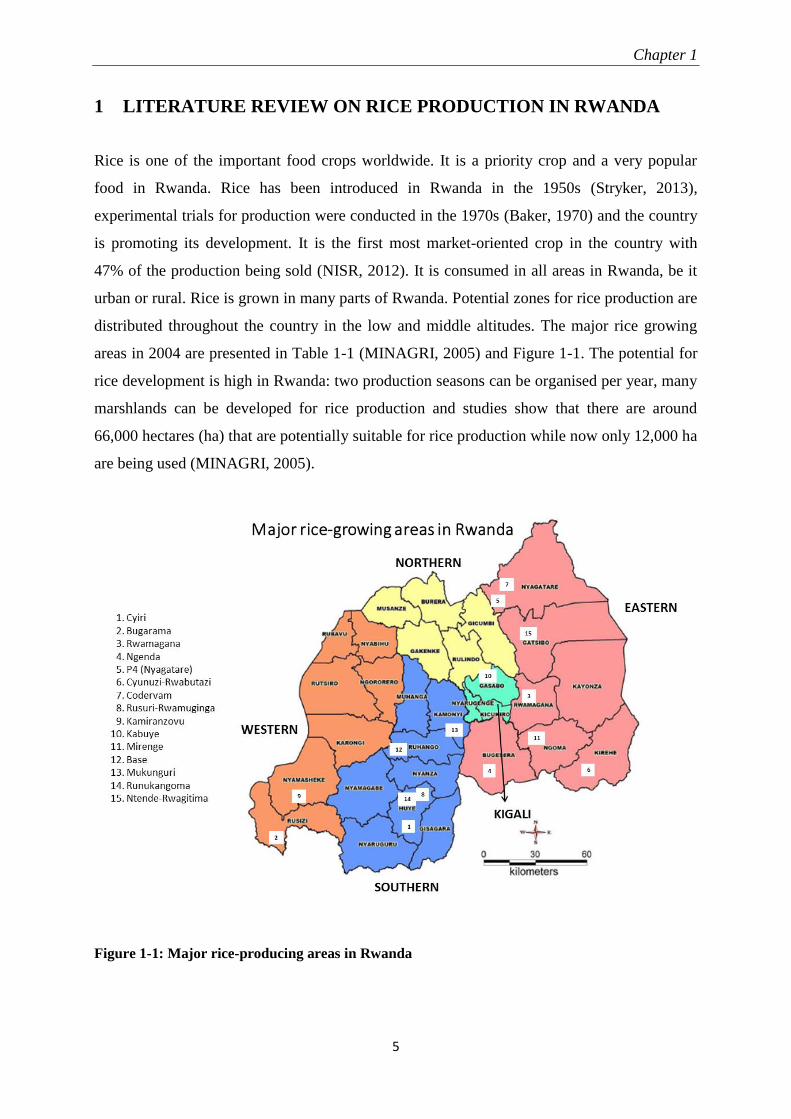
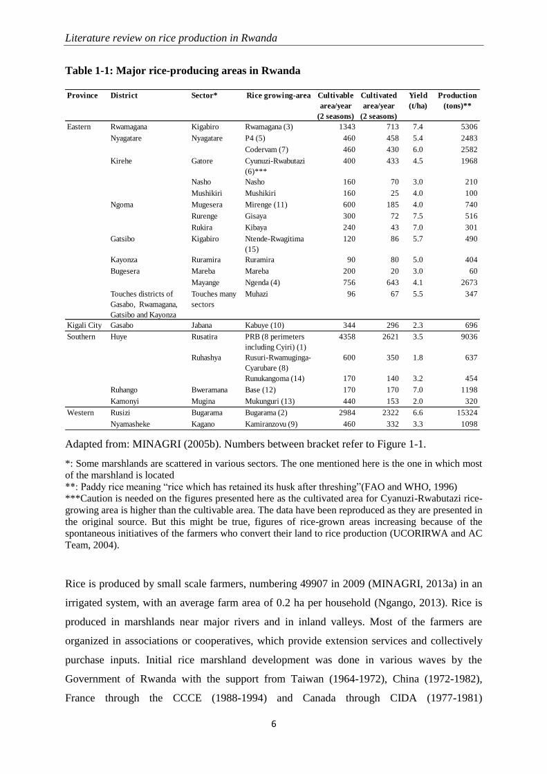
1 LITERATURE REVIEW ON RICE PRODUCTION IN RWANDA
Rice is one of the important food crops worldwide. It is a priority crop and a very popular
food in Rwanda. Rice has been introduced in Rwanda in the 1950s (Stryker, 2013),
experimental trials for production were conducted in the 1970s (Baker, 1970) and the country
is promoting its development. It is the first most market-oriented crop in the country with
47% of the production being sold (NISR, 2012). It is consumed in all areas in Rwanda, be it
urban or rural. Rice is grown in many parts of Rwanda. Potential zones for rice production are
distributed throughout the country in the low and middle altitudes. The major rice growing
areas in 2004 are presented in Table 1-1 (MINAGRI, 2005) and Figure 1-1. The potential for
rice development is high in Rwanda: two production seasons can be organised per year, many
marshlands can be developed for rice production and studies show that there are around
66,000 hectares (ha) that are potentially suitable for rice production while now only 12,000 ha
are being used (MINAGRI, 2005).
Figure 1-1: Major rice-producing areas in Rwanda
Literature review on rice production in Rwanda
6
Table 1-1: Major rice-producing areas in Rwanda
Adapted from: MINAGRI (2005b). Numbers between bracket refer to Figure 1-1.
*: Some marshlands are scattered in various sectors. The one mentioned here is the one in which most
of the marshland is located
**: Paddy rice meaning “rice which has retained its husk after threshing”(FAO and WHO, 1996)
***Caution is needed on the figures presented here as the cultivated area for Cyanuzi-Rwabutazi rice-
growing area is higher than the cultivable area. The data have been reproduced as they are presented in
the original source. But this might be true, figures of rice-grown areas increasing because of the
spontaneous initiatives of the farmers who convert their land to rice production (UCORIRWA and AC
Team, 2004).
Rice is produced by small scale farmers, numbering 49907 in 2009 (MINAGRI, 2013a) in an
irrigated system, with an average farm area of 0.2 ha per household (Ngango, 2013). Rice is
produced in marshlands near major rivers and in inland valleys. Most of the farmers are
organized in associations or cooperatives, which provide extension services and collectively
purchase inputs. Initial rice marshland development was done in various waves by the
Government of Rwanda with the support from Taiwan (1964-1972), China (1972-1982),
France through the CCCE (1988-1994) and Canada through CIDA (1977-1981)
Province District Sector* Rice growing-area Cultivable
area/year
(2 seasons)
Cultivated
area/year
(2 seasons)
Yield
(t/ha)
Production
(tons)**
Rwamagana Kigabiro Rwamagana (3) 1343 713 7.4 5306
Nyagatare Nyagatare P4 (5) 460 458 5.4 2483
Codervam (7) 460 430 6.0 2582
Kirehe Gatore Cyunuzi-Rwabutazi
(6)***
400 433 4.5 1968
Nasho Nasho 160 70 3.0 210
Mushikiri Mushikiri 160 25 4.0 100
Ngoma Mugesera Mirenge (11) 600 185 4.0 740
Rurenge Gisaya 300 72 7.5 516
Rukira Kibaya 240 43 7.0 301
Gatsibo Kigabiro Ntende-Rwagitima
(15)
120 86 5.7 490
Kayonza Ruramira Ruramira 90 80 5.0 404
Bugesera Mareba Mareba 200 20 3.0 60
Mayange Ngenda (4) 756 643 4.1 2673
Touches districts of
Gasabo, Rwamagana,
Gatsibo and Kayonza
Touches many
sectors
Muhazi 96 67 5.5 347
Kigali City Gasabo Jabana Kabuye (10) 344 296 2.3 696
Huye Rusatira PRB (8 perimeters
including Cyiri) (1)
4358 2621 3.5 9036
Ruhashya Rusuri-Rwamuginga-
Cyarubare (8)
600 350 1.8 637
Runukangoma (14) 170 140 3.2 454
Ruhango Bweramana Base (12) 170 170 7.0 1198
Kamonyi Mugina Mukunguri (13) 440 153 2.0 320
Rusizi Bugarama Bugarama (2) 2984 2322 6.6 15324
Nyamasheke Kagano Kamiranzovu (9) 460 332 3.3 1098
Eastern
Southern
Western
Chapter 1
7
(UCORIRWA and AC Team, 2004). In rice development, there are also spontaneous rice
development initiatives, in which farmers commit to convert their land to rice production and
carry out the initial works, especially the land levelling, installation of water irrigation and
drainage systems and find the initial inputs (seeds, fertilizers and pesticides). In the start, rice
development initiators taught farmers about rice production techniques. Nowadays, farmers
learn about rice farming from their neighbours and the acquisition of new techniques is
facilitated by the government and farmers groupings. There are currently many stakeholders
in the rice value chain including the following: key ministries and public institutions
(MINAGRI, MINICOM, MINEAC, RAB, RBS, PSF), rural development projects and
programs (RSSP, PADAB, PAIRB, KWAMP, GAA, CIP, PHHS), international organizations
(FAO, IFDC, UN-WFP), bilateral and multilateral agencies (BTC, USAID, DFID, JICA), rice
federation (FUCORIRWA), unions, rice farmers’ cooperatives, and individual farmers, rice
millers/processors, rice traders/importers, input suppliers, etc. (MINAGRI, 2013a).
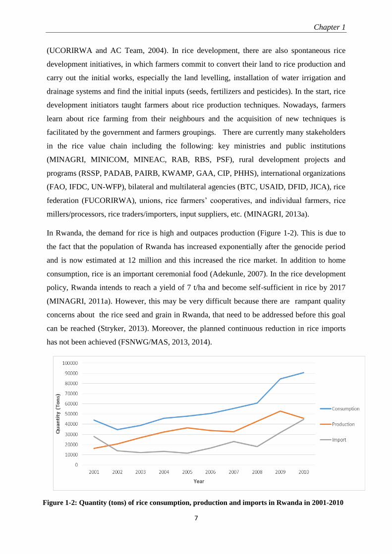
In Rwanda, the demand for rice is high and outpaces production (Figure 1-2). This is due to
the fact that the population of Rwanda has increased exponentially after the genocide period
and is now estimated at 12 million and this increased the rice market. In addition to home
consumption, rice is an important ceremonial food (Adekunle, 2007). In the rice development
policy, Rwanda intends to reach a yield of 7 t/ha and become self-sufficient in rice by 2017
(MINAGRI, 2011a). However, this may be very difficult because there are rampant quality
concerns about the rice seed and grain in Rwanda, that need to be addressed before this goal
can be reached (Stryker, 2013). Moreover, the planned continuous reduction in rice imports
has not been achieved (FSNWG/MAS, 2013, 2014).
Figure 1-2: Quantity (tons) of rice consumption, production and imports in Rwanda in 2001-2010
Literature review on rice production in Rwanda
8
Rwanda has a particular climate and topography, as it is mountainous with low temperatures
to which most selected rice varieties are not adapted. The average temperature in Rwanda is
19.84°C (NISR, 2013) which is below the best temperature interval for growing rice
estimated in the range of 20-38°C (CABI, 2007). This is a problem in most rice production
areas in Rwanda except in Bugarama (Western Province) where the average temperature is
around 24°C. The effects of the poor adaptation to the climate are long growing cycles and
grain sterility (Manneh and El Namaky, 2010). All these conditions warrant a breeding
program that would screen available cultivars, create varieties adapted to the Rwandan agro-
ecology and contribute in maintaining them. The most successful varieties grown in Rwanda
were introduced by Chinese and Japanese breeders before the genocide and have a Japonica
profile, though precise information is not available (Promar, 2012). A list of varieties that
were grown in Rwanda in 2003 is given in Table 1-2 (Jagwe et al., 2003).
Table 1-2: Varieties of rice in Rwanda
Variety Rice growing area where the variety is grown
Zhong geng (Local name Kigori) Rwamagana, Cyili, Kabuye, Nyagatare, Bugesera, Mukunguri
Yun Keng 136 Rwamagana, Cyili, Kabuye, Nyagatare, Bugesera, Mukunguri
Yun yine 4 Rwamagana
Yunertian 01 Rwamagana, Cyili, Kabuye, Nyagatare, Bugesera, Mukunguri
Xinum 175 Nyagatare, Rwamagana
Fac V046 Cyili
Basmati 370 Bugarama
IRON 280 Bugarama
BG 400-1 Bugarama
IRAT Bugarama
The short grain, Kigori rice, produced in Rwanda, is mostly milled in artisanal structures and
is consumed mainly in production and rural areas (Stryker, 2013; Ntirenganya, 2014). In
urban areas people prefer long grain imported rice of the Indica type, which is considered to
be of higher quality (World Bank, 2011; Ntirenganya, 2014). When the locally produced
Kigori rice is processed in modern mills, it is not competitive vis-à-vis imported Indica rice,
as it is sold with a discount of 10-20% (Stryker, 2013).
In 2010, many new rice varieties, mainly of the Indica type, were released. Those are: Kigega
(Pedigree-P-: VANDANA/IR 64), Nzahaha (P: IR 81431-B-B-162), Terimbere (P: WAB
Chapter 1
9
01291/4*IR64), Rumbuka (P: WAB 56-50/CG 14), Nemeyubutaka (P: WAB 56-50/CG 14),
Kimaranzara (P: WAB 56-104/CG 14), Ndamirabahinzi (P: Unknown, Breeding line: WAB
569-35-1-1-1-HB), Muhinzi (P: WAB 56-104/CG 14), Kanyabukungu (P: IRAT
104/PALAWAN), Imberabyombi (P: Unknown, Breeding line: WAB 788-19-1-1-2-HB),
Ndengera (P: IR 833-6-2-1-1//IR 1561-149-1/IR 1737), Nsizebashonje (P: PETA/TANGKAI
ROTAN), Garukuhinge (P: IR 19660-73-4/IR 2415-90-4-3-2//IR 54), Mbakungahaze (P:
SIAM 29/DEE-GEO-WOO-GEN), Ndamirabana (P: SIAM 29/KAOHSIUNG 68), Kungahara
(P: WAB450-24-2-3-P3-HB/IG10), Jyambere (P: 11975/IR 13146-45-2-3), Mpembuke (P:
Unknown, Breeding line: WAB923-B-6-AL1), Mbangukira (P: Unknown, Breeding line:
IR05N499), Cyicaro (P: WAB 56-104/CG 14) (Ndikumana and Gasore, 2010).
There are also the varieties of: Intsinzi, Gakire, Muturage, Tebuka (WAT 1276-22-2) and
Intsindagirabigega (B24-2) (MINAGRI, 2009b). Mutware et al. (2014) cite varieties of
Nerica, Muturage (WAT 54-TGR-1-5) and Facagro 56.
Some New Rice for Africa (NERICA) varieties have been tested in Rwanda but not many of
them are grown. Rwanda practices lowland irrigated rice while most of the NERICA varieties
are rainfed upland or lowland varieties, the agroecology of Rwanda, with low temperature
may not be suitable to most NERICA varieties (WARDA, 2005; Somado et al., 2008).
The seed is the first input in agriculture, on which depends the performance of other inputs.
Currently, the rice varieties in use are degenerated and some of them are a mix of different
varieties (Jagwe et al., 2003) as for many years there has not been a rice breeding programme
for Rwanda (Promar, 2012). In general, considering all the crops, only 2% of the farmers in
Rwanda use improved seeds and Rwanda is a large seed importer (RDB, 2014b).
The existing seed production and distribution system in the country could not keep up with
the rapid increase in marshland development and many farmers do not have access to
sufficient quantities of quality seed. It is important to be cautious about the quality, as no
organized seed evaluation system exists (MINAGRI, 2011a). Progressive farmers feel the
need for broadening varietal options as the easily available varieties are short-grain ones,
which have a low market value. In recent years, Rwanda has been using seeds imported from
international and regional networks. At their arrival in the country, they undergo quick
adaptability studies, after which the best performing ones are released to farmers for
cultivation. There is not much information about the efficacy after release in Rwanda
(Kathiresan, 2010; Mutware and Burger, 2014), but it has been reported that most long grain
Literature review on rice production in Rwanda
10
Indica varieties fail after 2-3 years of cultivation (Promar, 2012) while short-grain varieties
perform better.
Next to the poor quality of the available seeds, Jagwe et al. (2003) defined the following
major constraints to rice production in Rwanda: pests and diseases, deterioration and
destruction of the drainage and irrigation infrastructure, insufficient research, insufficient use
of farm inputs, and lack of adequate drying and processing facilities. Since 2003, there have
been improvements on most of these constraints, especially because of a supportive rice
development policy but some constraints persist. For infrastructures for drainage and
irrigation, the surface of marshlands developed or maintained for rice production increased
from 5.500 ha in 2003 to 12.000 ha in 2010 (Promar, 2012). Many farmers unions and
cooperatives have their own rice drying facilities (Promar, 2012) and also there are
improvements in the processing facilities (Stryker, 2013). The proportion of farmers who use
fertilizers increased from 11 to 29% in 2005-2006 to 2010-2011 and the pesticide use rate
increased from 24 to 31% in the same period (NISR, 2012).
The problems in rice production in Rwanda that have not been solved up to date deal with
poor quality of the available seed and grain, and lack of research about pest and diseases and
their management.
Kathiresan (2010) attests that there is poor progress in crop protection practices. Because of
this, the pressure of pests and diseases is high as the intensive monocropping of rice has
gradually built up pest and disease populations. There is also a lack of knowledge on
appropriate control measures and limited access to pest control advice (RAB, 2014).
The locally produced rice grain is of average quality (Adekunle, 2007).The poor physical
appearance of rice produced in Rwanda may have different causes: rudimentary milling
equipment, varietal appearance, lack of homogeneity in the planting material, physiological
stress in development, but can also be due to diseases and pests. Most of the factors cited here
have been studied, but less is known about the impact of pest and diseases in reducing the
quality of the grain, a problem faced by Rwanda.
The major pests and diseases reported on rice in Rwanda are: Diopsis thoracica (stalk-eyed
fly) and Magnaporthe oryzae (Rice blast) (MINAGRI, 2009b). Given that no research is
conducted on pests and diseases, most studies recognize that a large population of pests and
diseases has built up throughout the time (MINAGRI, 2011a). In fact, most of these reports
are based on surveys where the important informants are farmers, with less connection to
Chapter 1
11
diagnosis systems. Rice yellow mottle virus was recently reported (Ndikumana et al., 2011).
Rice can potentially be attacked in Rwanda by many more pests and diseases (see Annex 2).
Rice sheath rot disease is associated with grain discolouration and the reduction of grain
quality. Until the current study was conducted, the presence of rice sheath rot was reported
but no further studies about this disease had been conducted and the causal agents of this
disease in Rwanda are unknown. Rice sheath rot disease will be further discussed in the next
chapter.
In conclusion it can be stated that the potential of rice as both a food and cash crop in Rwanda
is high. However, there are factors that limit the full expression of the potential of rice. It is
important to address seed and grain quality issues in rice production in Rwanda as the supply
of national production lags behind the demand in quantity and in quality. These two aspects
have much in common: the quantity of the produce depends on the used planting material,
which is still of low quality, the quality of the produce depends on the production conditions
on field, which depend on crop husbandry practices including plant protection against pests
and diseases. This last point is difficult to certify as there is not sufficient information on pests
and diseases in the country.
Though a lasting solution to these problems can be found in the long run, in the short term it
is possible to reinforce the pests and disease control aspects through the evaluation of seeds
by organising on-field inspections and laboratory diagnosis and analyses, and collecting
information about the planting materials in use in the country and their performance. Research
data on the rice performance in the Rwandan environment are also needed. From the
information gathered in these initiatives, and by maintaining the good rice policy development
that prevailed until now, breeding initiatives can be initiated, having in mind the current
market segmentation in which highly demanded rice is the long grain in urban centres while
the short grain varieties are more adapted to the Rwandan agroecology and play a capital role
in food security in rural areas.

Chapter 2
13
2 REVIEW ON RICE SHEATH ROT: AN EMERGING UBIQUITOUS
DESTRUCTIVE DISEASE COMPLEX
Vincent de Paul Bigirimana, Gia Khuong Hoang Hua, Obedi Ishibwela Nyamangyoku,
Monica Höfte
The major part of this section has been published as:
Bigirimana, V.D.P., G.K.H. Hua, O.I. Nyamangyoku, and M. Höfte. 2015. Rice sheath rot :
An emerging ubiquitous destructive disease complex. Frontiers in Plant Sience 6 (December):
1066:16 pages.
Abstract
Around one century ago, a rice disease characterized mainly by rotting of sheaths was
reported in Taiwan. The causal agent was identified as Acrocylindrium oryzae, later known as
Sarocladium oryzae. Since then it has become clear that various other organisms can cause
similar disease symptoms, including Fusarium spp. and fluorescent pseudomonads. These
organisms have in common that they produce a range of phytotoxins that induce necrosis in
plants. The same agents also cause grain discoloration, chaffiness and sterility and are all
seed-transmitted. Rice sheath rot disease symptoms are found in all rice-growing areas of the
world. The disease is now getting momentum and is considered as an important emerging rice
production threat. The disease can lead to variable yield losses, which can be as high as 85%.
This review aims at improving our understanding of the disease etiology of rice sheath rot and
mainly deals with the three most reported rice sheath rot pathogens: Sarocladium oryzae, the
Fusarium fujikuroi complex and Pseudomonas fuscovaginae. Causal agents, pathogenicity
determinants, interactions among the various pathogens, epidemiology, geographical
distribution and control options will be discussed.
2.1 Introduction Rice is an important crop worldwide, serving as the staple food for half of the world
population and additionally being used in industry and for animal feed. Rice is grown in
Review on rice sheath rot
14
various agro-ecological zones in tropical and subtropical areas, especially in Asia, the
continent accounting for 90% of the world production (IRRI, 2015b). It faces many
production constraints, including pests and diseases.
The major feature of rice sheath rot disease is rotting and discoloration of the sheath, leading
to chaffiness and sterility of resulting grains. For many years, rice sheath rot was considered
as a minor and geographically limited disease. It is only recently that it gained momentum and
became widespread. Since the green revolution in Asia in the 1960s, there have been
substantial changes in rice farming systems: use of high yielding varieties, increased use of
fertilizers, efficient systems of water use, seeding methods, etc. Crop intensification practices
such as increased plant density, a high rate of nitrogen fertilizers and the use of semi-dwarf
and photoperiod-insensitive cultivars, favor the susceptibility of rice to some diseases and the
sheath rot complex is one of them. It is hypothesized that the new photoperiod-insensitive
cultivars have lost the capacity of avoiding flowering under conditions of high humidity and
high temperature, conditions that are conducive to effective disease attacks (Mew et al.,
2004b). Additionally, the last decades saw the boosting of international exchange of planting
materials which may have contributed to the spread of the disease.
Rice sheath rot is a disease complex that can be caused by various fungal and bacterial
pathogens. Major pathogens associated with rice sheath rot are fungi such as Sarocladium
oryzae and Fusarium spp. belonging to the Fusarium fujikuroi complex and the bacterial
pathogen Pseudomonas fuscovaginae. A variety of other pathogens have been associated with
rice sheath rot. An overview is given in Table 2-1.
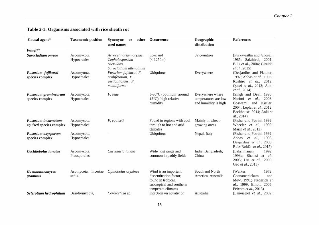
Chapter 2
15
Table 2-1: Organisms associated with rice sheath rot
Causal agent* Taxonomic position Synonyms or other
used names
Occurrence Geographic
distribution
References
Fungi**
Sarocladium oryzae Ascomycota,
Hypocreales
Acrocylindrium oryzae,
Cephalosporium
caerulans,
Sarocladium attenuatum
Lowland
(< 1250m)
32 countries (Purkayastha and Ghosal,
1985; Sakthivel, 2001;
Bills et al., 2004; Giraldo
et al., 2015)
Fusarium fujikuroi
species complex
Ascomytota,
Hypocreales
Fusarium fujikuroi, F.
proliferatum, F.
verticillioides, F.
moniliforme
Ubiquitous Everywhere (Desjardins and Plattner,
1997; Abbas et al., 1998;
Kushiro et al., 2012;
Quazi et al., 2013; Aoki
et al., 2014)
Fusarium graminearum
species complex
Ascomycota,
Hypocreales
F. zeae 5-30°C (optimum around
15°C), high relative
humidity
Everywhere where
temperatures are low
and humidity is high
(Singh and Devi, 1990;
Naeimi et al., 2003;
Goswami and Kistler,
2004; Leplat et al., 2012;
Backhouse, 2014; Aoki et
al., 2014)
Fusarium incarnatum-
equiseti species complex
Ascomycota,
Hypocreales
F. equiseti Found in regions with cool
through to hot and arid
climates
Mainly in wheat-
growing areas
(Fisher and Petrini, 1992;
Wheeler et al., 1999;
Marín et al., 2012)
Fusarium oxysporum
species complex
Ascomycota,
Hypocreales
- Ubiquitous Nepal, Italy (Fisher and Petrini, 1992;
Abbas et al., 1995;
Desjardins et al., 2000;
Ruiz-Roldán et al., 2015)
Cochliobolus lunatus Ascomycota,
Pleosporales
Curvularia lunata Wide host range and
common in paddy fields
India, Bangladesh,
China
(Lakshmanan, 1992,
1993a; Shamsi et al.,
2003; Liu et al., 2009;
Gao et al., 2015)
Gaeumannomyces
graminis
Asomycota, Incertae
sedis
Ophiobolus oryzinus Wind is an important
dissemination factor;
found in tropical,
subtropical and southern
temperate climates
South and North
America, Australia
(Walker, 1972;
Gnanamanickam and
Mew, 1991; Frederick et
al., 1999; Elliott, 2005;
Peixoto et al., 2013)
Sclerotium hydrophilum Basidiomycota, Ceratorhiza sp. Infection on aquatic or Australia (Lanoiselet et al., 2002;
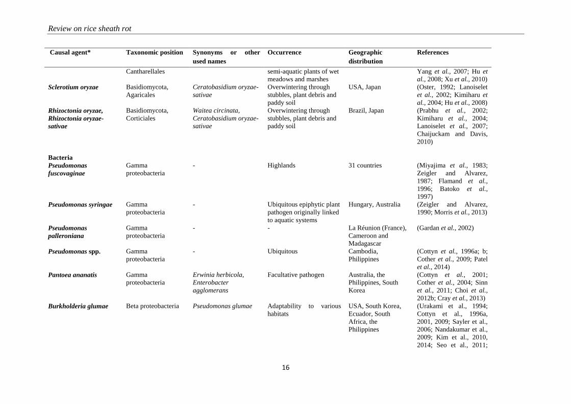
Review on rice sheath rot
16
Causal agent* Taxonomic position Synonyms or other
used names
Occurrence Geographic
distribution
References
Cantharellales semi-aquatic plants of wet
meadows and marshes
Yang et al., 2007; Hu et
al., 2008; Xu et al., 2010)
Sclerotium oryzae Basidiomycota,
Agaricales
Ceratobasidium oryzae-
sativae
Overwintering through
stubbles, plant debris and
paddy soil
USA, Japan (Oster, 1992; Lanoiselet
et al., 2002; Kimiharu et
al., 2004; Hu et al., 2008)
Rhizoctonia oryzae,
Rhizoctonia oryzae-
sativae
Basidiomycota,
Corticiales
Waitea circinata,
Ceratobasidium oryzae-
sativae
Overwintering through
stubbles, plant debris and
paddy soil
Brazil, Japan (Prabhu et al., 2002;
Kimiharu et al., 2004;
Lanoiselet et al., 2007;
Chaijuckam and Davis,
2010)
Bacteria
Pseudomonas
fuscovaginae
Gamma
proteobacteria
- Highlands 31 countries (Miyajima et al., 1983;
Zeigler and Alvarez,
1987; Flamand et al.,
1996; Batoko et al.,
1997)
Pseudomonas syringae Gamma
proteobacteria
- Ubiquitous epiphytic plant
pathogen originally linked
to aquatic systems
Hungary, Australia (Zeigler and Alvarez,
1990; Morris et al., 2013)
Pseudomonas
palleroniana
Gamma
proteobacteria
- - La Réunion (France),
Cameroon and
Madagascar
(Gardan et al., 2002)
Pseudomonas spp. Gamma
proteobacteria
- Ubiquitous Cambodia,
Philippines
(Cottyn et al., 1996a; b;
Cother et al., 2009; Patel
et al., 2014)
Pantoea ananatis Gamma
proteobacteria
Erwinia herbicola,
Enterobacter
agglomerans
Facultative pathogen Australia, the
Philippines, South
Korea
(Cottyn et al., 2001;
Cother et al., 2004; Sinn
et al., 2011; Choi et al.,
2012b; Cray et al., 2013)
Burkholderia glumae Beta proteobacteria Pseudomonas glumae Adaptability to various
habitats
USA, South Korea,
Ecuador, South
Africa, the
Philippines
(Urakami et al., 1994;
Cottyn et al., 1996a,
2001, 2009; Sayler et al.,
2006; Nandakumar et al.,
2009; Kim et al., 2010,
2014; Seo et al., 2011;
Chapter 2
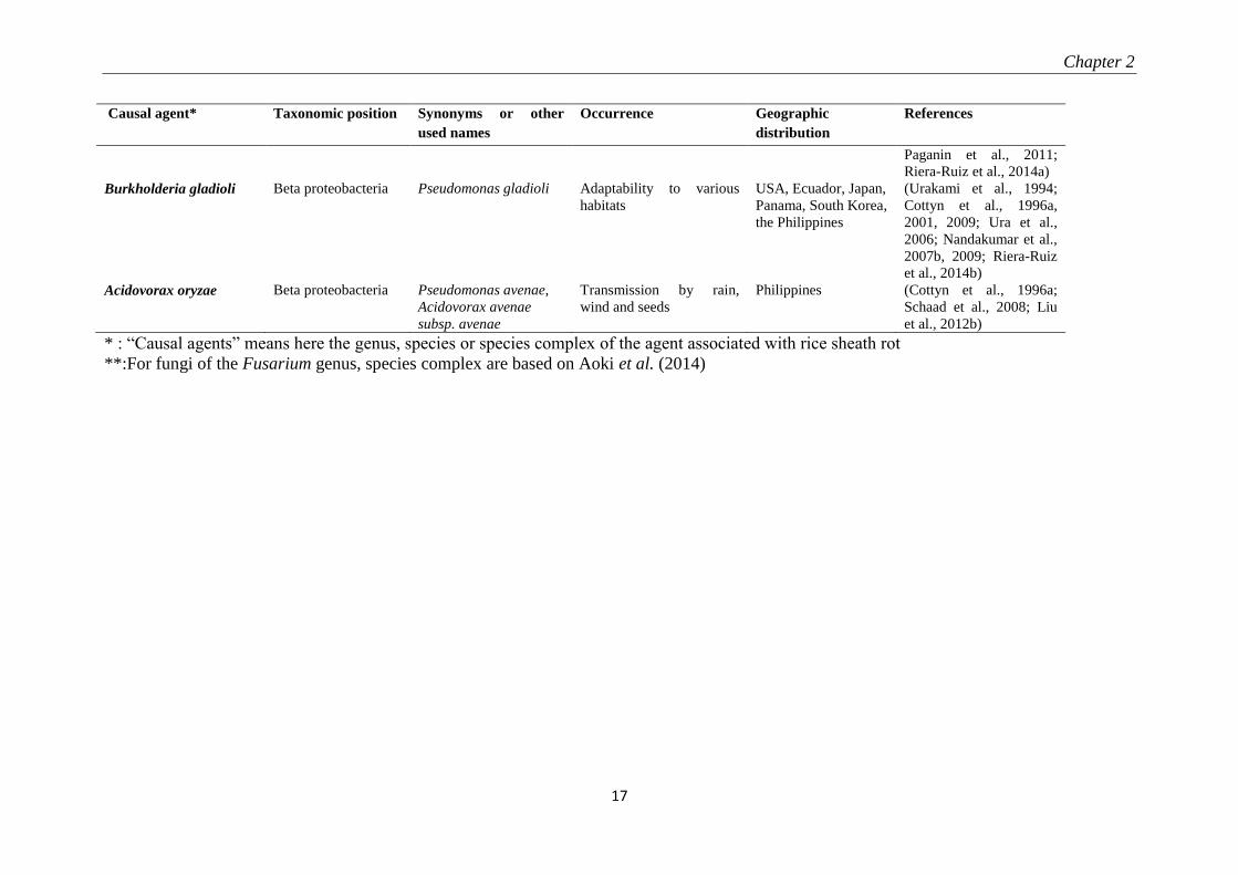
17
Causal agent* Taxonomic position Synonyms or other
used names
Occurrence Geographic
distribution
References
Paganin et al., 2011;
Riera-Ruiz et al., 2014a)
Burkholderia gladioli Beta proteobacteria Pseudomonas gladioli Adaptability to various
habitats
USA, Ecuador, Japan,
Panama, South Korea,
the Philippines
(Urakami et al., 1994;
Cottyn et al., 1996a,
2001, 2009; Ura et al.,
2006; Nandakumar et al.,
2007b, 2009; Riera-Ruiz
et al., 2014b)
Acidovorax oryzae Beta proteobacteria Pseudomonas avenae,
Acidovorax avenae
subsp. avenae
Transmission by rain,
wind and seeds
Philippines (Cottyn et al., 1996a;
Schaad et al., 2008; Liu
et al., 2012b)
* : “Causal agents” means here the genus, species or species complex of the agent associated with rice sheath rot
**:For fungi of the Fusarium genus, species complex are based on Aoki et al. (2014)
Review on rice sheath rot
18
The various described sheath rot agents all cause very similar disease symptoms (Cottyn et
al., 1996b). This explains why there are practically no comprehensive studies mentioning the
link between the presence and quantity of disease inoculum and yield loss (Mew and
Gonzales, 2002). The unpredictable nature of the factors acting in the pathosystem explains
why losses attributed to Sarocladium oryzae can be as variable as in the range of 20-85%
(Sakthivel, 2001).
The context of an increasing world population with shrinking natural resources imposes to
adopt sustainable production methods, responding to the food demand but also using
efficiently and sustainably key resources (Savary et al., 2000; Mew et al., 2004b). The
development of sound control practices against rice sheath rot is hampered by the fact that this
disease is poorly understood. This review would like to contribute in filling the rice sheath rot
missing information gap. It explores the available information on the following aspects:
causal agents and symptoms, host range, physiological and biochemical impact, virulence
factors, synergism and interactions among causal factors, ecology of causal agents,
epidemiology and impact, geographical distribution and relationships with farming systems
and control methods. In this review, more emphasis will be put on rice sheath rot symptoms
caused by S. oryzae, Fusarium spp., and P. fuscovaginae, since they are considered to be the
most important rice sheath rot pathogens (Table 2-1).
2.2 Sarocladium oryzae: the major fungal rice sheath rot pathogen
2.2.1 Pathogen description and symptoms
S. oryzae was originally described as Acrocylindrium oryzae, the first organism to be
associated with rice sheath rot symptoms isolated in Taiwan in 1922 (Mew and Gonzales,
2002). The genus Sarocladium was established in 1975 (Gams and Hawksworth, 1975) and
currently encompasses 16 species including plant pathogens, saprobes, mycoparasites,
endophytes and potential human pathogens (Giraldo et al., 2015). The genus belongs to the
order of the Hypocreales in the Phylum Ascomycota. S. attenuatum was originally described
as a distinct species causing rice sheath rot, but is nowadays considered as a synonym of S.
Chapter 2
19
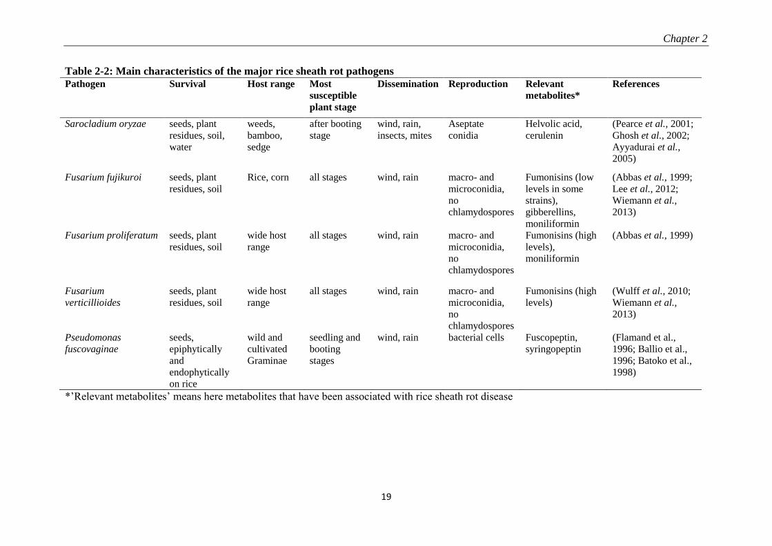
Table 2-2: Main characteristics of the major rice sheath rot pathogens
*’Relevant metabolites’ means here metabolites that have been associated with rice sheath rot disease
Pathogen Survival Host range Most
susceptible
plant stage
Dissemination Reproduction Relevant
metabolites*
References
Sarocladium oryzae seeds, plant
residues, soil,
water
weeds,
bamboo,
sedge
after booting
stage
wind, rain,
insects, mites
Aseptate
conidia
Helvolic acid,
cerulenin
(Pearce et al., 2001;
Ghosh et al., 2002;
Ayyadurai et al.,
2005)
Fusarium fujikuroi seeds, plant
residues, soil
Rice, corn all stages wind, rain macro- and
microconidia,
no
chlamydospores
Fumonisins (low
levels in some
strains),
gibberellins,
moniliformin
(Abbas et al., 1999;
Lee et al., 2012;
Wiemann et al.,
2013)
Fusarium proliferatum seeds, plant
residues, soil
wide host
range
all stages wind, rain macro- and
microconidia,
no
chlamydospores
Fumonisins (high
levels),
moniliformin
(Abbas et al., 1999)
Fusarium
verticillioides
seeds, plant
residues, soil
wide host
range
all stages wind, rain macro- and
microconidia,
no
chlamydospores
Fumonisins (high
levels)
(Wulff et al., 2010;
Wiemann et al.,
2013)
Pseudomonas
fuscovaginae
seeds,
epiphytically
and
endophytically
on rice
wild and
cultivated
Graminae
seedling and
booting
stages
wind, rain bacterial cells Fuscopeptin,
syringopeptin
(Flamand et al.,
1996; Ballio et al.,
1996; Batoko et al.,
1998)
Review on rice sheath rot
20
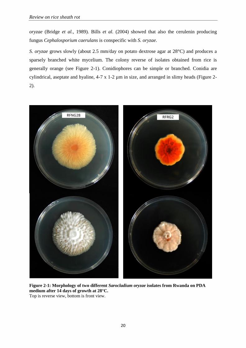
oryzae (Bridge et al., 1989). Bills et al. (2004) showed that also the cerulenin producing
fungus Cephalosporium caerulans is conspecific with S. oryzae.
S. oryzae grows slowly (about 2.5 mm/day on potato dextrose agar at 28°C) and produces a
sparsely branched white mycelium. The colony reverse of isolates obtained from rice is
generally orange (see Figure 2-1). Conidiophores can be simple or branched. Conidia are
cylindrical, aseptate and hyaline, 4-7 x 1-2 µm in size, and arranged in slimy heads (Figure 2-
2).
Figure 2-1: Morphology of two different Sarocladium oryzae isolates from Rwanda on PDA
medium after 14 days of growth at 28°C.
Top is reverse view, bottom is front view.
Chapter 2
21
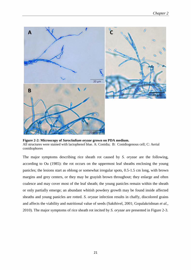
The major symptoms describing rice sheath rot caused by S. oryzae are the following,
according to Ou (1985): the rot occurs on the uppermost leaf sheaths enclosing the young
panicles; the lesions start as oblong or somewhat irregular spots, 0.5-1.5 cm long, with brown
margins and grey centers, or they may be grayish brown throughout; they enlarge and often
coalesce and may cover most of the leaf sheath; the young panicles remain within the sheath
or only partially emerge; an abundant whitish powdery growth may be found inside affected
sheaths and young panicles are rotted. S. oryzae infection results in chaffy, discolored grains
and affects the viability and nutritional value of seeds (Sakthivel, 2001; Gopalakrishnan et al.,
2010). The major symptoms of rice sheath rot incited by S. oryzae are presented in Figure 2-3.
Figure 2-2: Microscopy of Sarocladium oryzae grown on PDA medium.
All structures were stained with lactophenol blue. A: Conidia; B: Conidiogenous cell; C: Aerial
conidiophores

Review on rice sheath rot
22
2.2.2 Epidemiology
In general, S. oryzae is present in all rice-growing countries worldwide, being very common
in rainy seasons (Mew and Gonzales, 2002). It has so far been reported in the following
countries (CABI, 2007): Bangladesh, Brunei Darussalam, China, India, Indonesia, Japan,
Malaysia, Nepal, Pakistan, Philippines, Saudi Arabia, Sri Lanka, Tajikistan, Thailand,
Uzbekistan, Vietnam, Burundi, Cameroon, Côte d'Ivoire, Gambia, Kenya, Madagascar, Niger,
Nigeria, Senegal, Tanzania, Mexico, USA, Argentina, Brazil, Venezuela, and Australia. S.
oryzae is mostly found in lowland environments (Pearce et al., 2001), and hot and humid
weather favors the disease (Sakthivel, 2001). Sharma et al. (1997) stated that S. oryzae
infections in Nepal were found below 1250 m. Temperatures of 20-30°C and relative
humidity in the range of 65 to 85% favor sheath rot development (Sakthivel, 2001).
The pathogen survives in infected seeds, plant residues (straw, stubble), but also in soil, water
or weeds when environmental conditions are favorable. Plants at various growth stages can be
affected; the fungus enters through stomata or wounds, and is most destructive after booting
stage but also infects other growth stages (Pearce et al., 2001). The entry of S. oryzae in the
plant is facilitated mostly by insect and mite damage or the weakening of the plant by other
pathogens (Pearce et al., 2001). Secondary infections may be wind-borne through injured
tissues. Less is known about the seed-borne disease transmission.
A B C D E
Figure 2-3: Rice sheath rot symptoms caused by Sarocladium oryzae. A-B: Diseased sheaths on which
white fungal structures can be seen; C-D: Discoloured grains in a diseased panicle; E: Non-filled grains from
a diseased plant.
Chapter 2
23
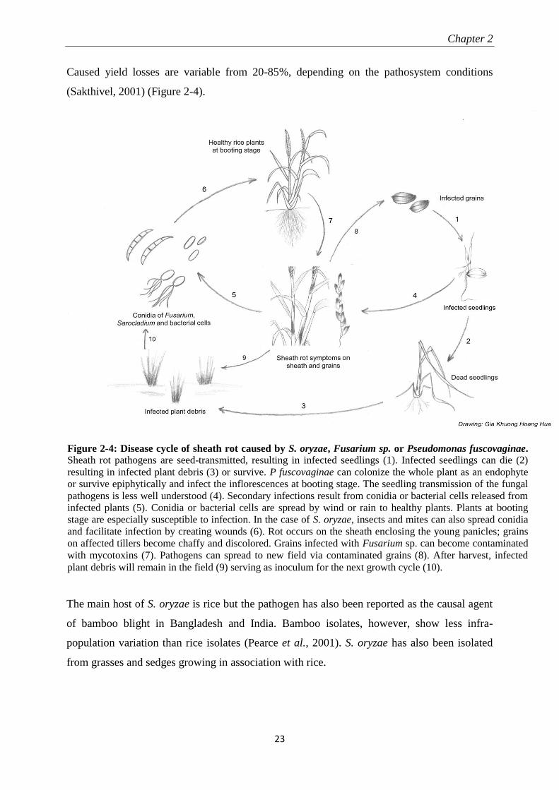
Caused yield losses are variable from 20-85%, depending on the pathosystem conditions
(Sakthivel, 2001) (Figure 2-4).
The main host of S. oryzae is rice but the pathogen has also been reported as the causal agent
of bamboo blight in Bangladesh and India. Bamboo isolates, however, show less infra-
population variation than rice isolates (Pearce et al., 2001). S. oryzae has also been isolated
from grasses and sedges growing in association with rice.
Figure 2-4: Disease cycle of sheath rot caused by S. oryzae, Fusarium sp. or Pseudomonas fuscovaginae.
Sheath rot pathogens are seed-transmitted, resulting in infected seedlings (1). Infected seedlings can die (2)
resulting in infected plant debris (3) or survive. P fuscovaginae can colonize the whole plant as an endophyte
or survive epiphytically and infect the inflorescences at booting stage. The seedling transmission of the fungal
pathogens is less well understood (4). Secondary infections result from conidia or bacterial cells released from
infected plants (5). Conidia or bacterial cells are spread by wind or rain to healthy plants. Plants at booting
stage are especially susceptible to infection. In the case of S. oryzae, insects and mites can also spread conidia
and facilitate infection by creating wounds (6). Rot occurs on the sheath enclosing the young panicles; grains
on affected tillers become chaffy and discolored. Grains infected with Fusarium sp. can become contaminated
with mycotoxins (7). Pathogens can spread to new field via contaminated grains (8). After harvest, infected
plant debris will remain in the field (9) serving as inoculum for the next growth cycle (10).
Review on rice sheath rot
24
2.2.3 Pathogenicity determinants
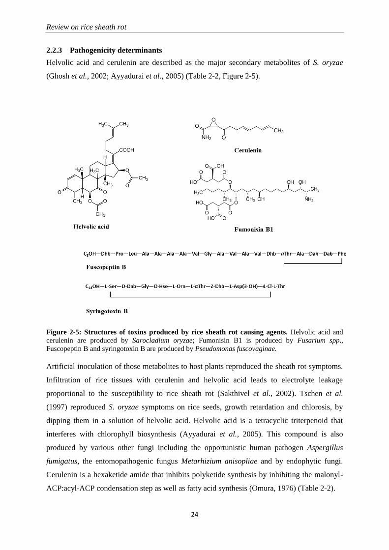
Helvolic acid and cerulenin are described as the major secondary metabolites of S. oryzae
(Ghosh et al., 2002; Ayyadurai et al., 2005) (Table 2-2, Figure 2-5).
Figure 2-5: Structures of toxins produced by rice sheath rot causing agents. Helvolic acid and
cerulenin are produced by Sarocladium oryzae; Fumonisin B1 is produced by Fusarium spp.,
Fuscopeptin B and syringotoxin B are produced by Pseudomonas fuscovaginae.
Artificial inoculation of those metabolites to host plants reproduced the sheath rot symptoms.
Infiltration of rice tissues with cerulenin and helvolic acid leads to electrolyte leakage
proportional to the susceptibility to rice sheath rot (Sakthivel et al., 2002). Tschen et al.
(1997) reproduced S. oryzae symptoms on rice seeds, growth retardation and chlorosis, by
dipping them in a solution of helvolic acid. Helvolic acid is a tetracyclic triterpenoid that
interferes with chlorophyll biosynthesis (Ayyadurai et al., 2005). This compound is also
produced by various other fungi including the opportunistic human pathogen Aspergillus
fumigatus, the entomopathogenic fungus Metarhizium anisopliae and by endophytic fungi.
Cerulenin is a hexaketide amide that inhibits polyketide synthesis by inhibiting the malonyl-
ACP:acyl-ACP condensation step as well as fatty acid synthesis (Omura, 1976) (Table 2-2).
Chapter 2
25
Though the most described virulence factors of S. oryzae are helvolic acid and cerulenin, the
fungus also produces cellulolytic, proteolytic, pectinolytic and oxidative enzymes that play a
role in pathogenicity (Joe and Manibhushanrao, 1995; Pearce et al., 2001). Gopalakrishnan et
al. (2010) observed a pronounced decrease in sugar, starch and protein and an increase in
phenol content in rice seeds infected with S. oryzae. This probably explains why infected
grains are chaffy and germinate poorly.
2.2.4 Interactions with other diseases and pests
Experimental tests have shown that S. oryzae, by the production of toxins, like cerulenin,
limits the development of other fungi and emerges as the major pathogen (Gnanamanickam
and Mew, 1991; Silva-Lobo et al., 2011). Gnanamanickam and Mew (1991) observed that the
antibiotic properties of cerulenin extracted from S. oryzae block the development of many rice
stem-attacking fungi, like Sclerotium oryzae, Gaeumannomyces graminis var. graminis,
Magnaporthe oryzae and Rhizoctonia solani. In this context it is interesting to notice that
cerulenin has been reported to inhibit melanin biosynthesis in Colletotrichum lagenarium
(Kubo et al., 1986). DHN (=1,8 dihydroxynapthalene)-melanin in fungi is synthesized by a
polyketide pathway which starts from malonyl-CoA which is converted to the first detectable
intermediate of the melanin pathway 1,3,6,8-tetrahydroxynapthalene via a polyketide
synthase. DHN-melanin is an important virulence factor in several pathogenic fungi including
M. oryzae and G. graminis var. graminis (Henson et al., 1999). In addition, helvolic acid has
strong antibacterial activities mainly against Gram-positive bacteria (Tschen et al., 1997).
This could explain why in many situations S. oryzae emerges as the major pathogen.
Review on rice sheath rot
26
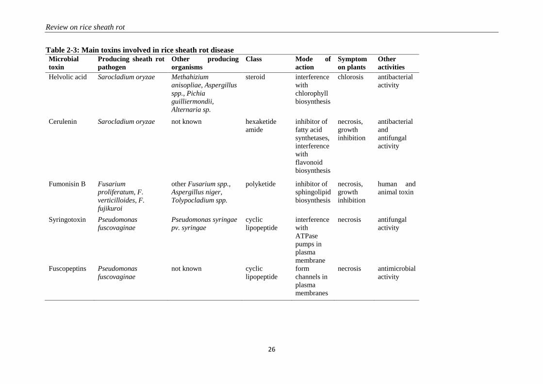
Table 2-3: Main toxins involved in rice sheath rot disease
Microbial
toxin
Producing sheath rot
pathogen
Other producing
organisms
Class Mode of
action
Symptom
on plants
Other
activities
Helvolic acid Sarocladium oryzae Methahizium
anisopliae, Aspergillus
spp., Pichia
guilliermondii,
Alternaria sp.
steroid interference
with
chlorophyll
biosynthesis
chlorosis antibacterial
activity
Cerulenin Sarocladium oryzae not known hexaketide
amide
inhibitor of
fatty acid
synthetases,
interference
with
flavonoid
biosynthesis
necrosis,
growth
inhibition
antibacterial
and
antifungal
activity
Fumonisin B Fusarium
proliferatum, F.
verticilloides, F.
fujikuroi
other Fusarium spp.,
Aspergillus niger,
Tolypocladium spp.
polyketide inhibitor of
sphingolipid
biosynthesis
necrosis,
growth
inhibition
human and
animal toxin
Syringotoxin Pseudomonas
fuscovaginae
Pseudomonas syringae
pv. syringae
cyclic
lipopeptide
interference
with
ATPase
pumps in
plasma
membrane
necrosis antifungal
activity
Fuscopeptins Pseudomonas
fuscovaginae
not known cyclic
lipopeptide
form
channels in
plasma
membranes
necrosis antimicrobial
activity
Chapter 2
27
Initial work on sheath rot was done in India, and Amin et al. (1974) already realized the
disease complexity, as the causal agent was already thought to be associated with stem borers.
A study on four groups of insects: green leaf hopper, brown plant hopper, mealy bugs and
earhead bugs showed that brown plant hoppers and mealy bugs fed on rice infected with S.
oryzae carry the fungus on their body and can transmit it to healthy plants (Gopalakrishnan et
al., 2009). Some of the S. oryzae effects like sterility result from its synergism with a mite
Steneotarsonemus spinki (Ou, 1985; Karmakar, 2008; Hummel et al., 2009). It was observed
that wounding of plants facilitated their infection by S. oryzae and most of the infected plants
proved also to be attacked by stem borers and from time to time by yellow dwarf virus (Ou,
1985). The fact that spraying a spore suspension of S. oryzae on earhead bug (Leptocorisa
acuta)-infected rice tillers results in the development of rice sheath rot disease symptoms in
12 days (Lakshmanan et al., 1992) shows that the entry of S. oryzae in rice plants might be
facilitated. Sakthivel (2001) realized that the infection occurs after the plant has been
weakened by other diseases and stem borer infestation.
Bacterial sheath brown rot, caused by P. fuscovaginae, may occur together with sheath rot
caused by S. oryzae. Other factors that have been associated with S. oryzae include rice tungro
virus (Venkataraman et al., 1987) and Fusarium spp. (Sakthivel, 2001).
2.2.5 Control methods
S. oryzae is controlled by sanitary, chemical and biological measures.
Sanitary control methods involve the following practices (Sakthivel, 2001): using healthy
seeds since the disease is referred to as being seed-borne; limiting insect population in rice
fields as they are involved in disease transmission; avoiding densely planting as this
predisposes plants to fungal attacks; avoiding heavy doses of nitrogen fertilizers; increasing
potassium content in the fertilizer formula for reducing the disease impact, as more potassium
causes more phenol production; adopting different cultural practices for limiting the disease
attack impact: field sanitation, crop residue management, control of weeds, etc.
Various fungicides have been used to control sheath rot but as they cannot kill the fungus
inside the glumes, their efficacy is limited (Sakthivel, 2001). Other control tests combined
fungicides with insecticides and gave better results (Lakshmanan, 1992). Foliar spray of
micronutrients is also said to reduce disease incidence and increase grain yield (Sakthivel,
2001). Some plant extracts are reported to be effective against the disease: neem, pungam oil
and rubber cakes (Narasimhan et al., 1998; Sakthivel, 2001).
Review on rice sheath rot
28
The use of biological control agents may have potential (Sakthivel and Gnanamanickam,
1987; Mew et al., 2004a). Many pseudomonads can act efficiently for controlling S. oryzae,
by favoring antagonism, for example through the inhibition of fungal development as do some
P. fluorescens strains, or by inducing systemic resistance (Saravanakumar et al., 2009).
Breeding for resistance to sheath rot seems the best option, but it is limited by its multiple
causal agents. High-yielding nitrogen-responsive rice cultivars are highly susceptible to
sheath rot. Resistance to S. oryzae has been identified in tall rice varieties (Amin, 1976).
Hemalatha et al. (1999) developed a method of screening for resistance against S. oryzae
based on a crude toxin preparation and Lakshmanan (1993) went further by screening for
resistance against S. oryzae and one of its vectors, the rice mealy bug. The screening of
resistance against S. oryzae that was developed by Amin (1976) does not seem to have been
continued. Ayyadurai et al. (2005) analyzed S. oryzae isolates from North East and South
India and found a high variability in pathogenicity, phytotoxic metabolite production and
RAPD band patterns. This variability should be taken into account in breeding efforts.
2.3 Fusarium fujikuroi, a species complex associated with rice sheath rot
2.3.1 Pathogen description and symptoms
Sheath rot in rice has also been associated with Fusarium spp. belonging to the Fusarium
fujikuroi complex. The F. fujikuroi complex largely corresponds to the Section Liseola,
established by Wollenweber and Reinking (1935), in which Nelson et al. (1983) recognized
four species (including F. moniliforme and F. proliferatum), but also accommodates certain
species originally classified in other Fusarium sections. Progress in molecular taxonomy has
shown that there are around 50 species in the F. fujikuroi complex and the number keeps
increasing (Reviewed in Kvas et al., 2009). The complex is currently divided in three large
clades, the African clade, the Asian clade and the American clade. The main organisms
associated with rice are F. verticillioides from the African clade and the closely related
species F. proliferatum and F. fujikuroi from the Asian clade.
Abbas et al. (1998) described rice sheath rot symptoms caused by F. proliferatum as follows:
blanked or partially blanked panicle with reddish-brown to off-white florets or kernels are
often covered with a white to pinkish white powder consisting of microconidia and
conidiophores of F. proliferatum; the flag leaf sheath develops a rapidly enlarging lesion, first
dull to dark brown and later off-white to tan with a reddish brown border, that eventually
encompasses the entire sheath and may result in the death of the leaf blade; lower leaf sheaths
Chapter 2
29
may eventually develop lesions as well, but rarely more than two leaf sheaths show
symptoms; and a dense white to pinkish white powder consisting of microconidia and
conidiophores of F. proliferatum covers the sheath lesions, especially evident during humid
periods.
2.3.2 Epidemiology
Rice-pathogenic Fusarium species, because of their high diversity, are ubiquitous in nature
(Park et al., 2005). Symptoms of rice sheath rot caused by any of the members of the
Fusarium fujikuroi species complex are widespread due to their large variability and at least
one of their members is found in any part of the rice-growing world. The different species of
Fusarium forming the F. fujikuroi complex (mainly Fusarium fujikuroi, F. verticillioides and
F. proliferatum) cause various symptoms on different plant parts and are responsible of yield
losses of 40% in Nepal (Desjardins et al., 2000) and even up to 60% in Korea (Park et al.,
2005).
Fusarium proliferatum, which is pathogenic to rice, also attacks some other plants of the
Poaceae family. F. proliferatum is widespread and its hosts vary from maize to mango (Leslie
et al., 2007), including chestnut (Kushiro et al., 2012), and banana (Li et al., 2012). As the
organisms causing rice sheath rot have many hosts, they can easily find alternate hosts in the
environment, especially weeds.
Fusarium spp. are seed-transmitted and at maturity, infected grains contain mycotoxins
(Wulff et al., 2010). F. fujikuroi was one of a number of microbes isolated from the surface of
rice seeds; highest levels of microbes were recorded at harvesting. F. fujikuroi survived for up
to 26 months in infected grains and 28 months in dried stubble of certain rice cultivars. The
fungus was detected for up to 10 and 13 months, respectively, in unsterilized and sterilized
soils that were infected with fungal propagules (Sunder and Satyavir, 1998). F. proliferatum
can survive in infected grains even when they are preserved in stressing conditions. In fact,
Kushiro et al. (2012) could recover F. proliferatum in grains preserved at 4-5°C for 6 months.
In normal conditions, the survival is longer.
2.3.3 Pathogenicity determinants
Two categories of metabolites are involved in pathogenicity and interaction with plants,
gibberellins and mycotoxins. According to Wulff et al. (2010), only strains of F. fujikuroi
were able to produce gibberellin A and these strains cause abnormal elongation of rice plants,
the so-called bakanae disease. Main species producing mycotoxins, like fumonisin B (FB)
Review on rice sheath rot
30
(Table 2-2, Figure 2-5), have been reported to cause rice sheath rot (Wulff et al., 2010).
Fumonisins are linear, polyketide-derived molecules that are also known as major mycotoxins
that pose health risks to humans and animals. F. proliferatum is among the largest producers
of fumonisins and is often associated with rice sheath rot (Abbas et al., 1999; Kushiro et al.,
2012; Quazi et al., 2013). In addition, F. verticillioides strains are notorious fumonisin
producers (Wulff et al., 2010). Isolates belonging to various other related Fusarium species
have been shown to produce fumonisins (Table 2-2). Fumonisin biosynthetic genes have also
been found in more distantly related fungi such as Aspergillus niger and Tolypocladium spp.
The evolution of the fumonisin gene cluster in Fusarium is complex and not adequately
represented by the species phylogeny. It is hypothesized that a combination of multiple
horizontal gene transfer, cluster duplication and loss has shaped the current distribution of the
fumonisin gene cluster (Proctor et al., 2013). The role of fumonisins in the ecology and
pathology of Fusarium is poorly understood. Abbas et al. (1998) observed that the
concentration of fumonisins coincides with the intensity of sheath and panicle symptoms in
rice plants showing sheath rot under Fusarium attacks. Toxins are apparently concentrated in
the external grain part since their concentration in the grain reduced 75-80% after hulling.
One of the major fumonisins, FB1, is conceived as a virulence factor in Fusarium-induced
diseases in plants (Glenn et al., 2008). FB1 inhibits ceramide synthase (Williams et al., 2007),
an enzyme involved in sphingolipid biosynthesis in both animals and plants. This has
numerous consequences on the cell physiology and results in increased cell death and
inhibition of plasma membrane ATPases (Gutiérrez-Nájera et al., 2005). Members of the F.
fujikuroi complex also produce a variety of other mycotoxins, including moniliformin. It has
been shown that F. proliferatum isolates from field samples of rice with Fusarium sheath rot
disease are capable of producing both fumonisins and moniliformin in culture. Both
mycotoxins were also detected in naturally contaminated rice samples (Abbas et al., 1999).
The phytotoxicity of moniliformin is well documented (Abbas et al., 1995). Moniliformin
was shown to arrest mitosis of maize root meristematic cells at the metaphase stage (Styer and
Cutler, 1984). The factors triggering the infection of F. proliferatum to rice plants still need to
be further investigated (Kushiro et al., 2012). Genome sequencing revealed the presence of a
wide variety of secondary metabolite gene clusters in F. fujikuroi and F. verticillioides,
including clusters for bikaverin, fusarubins, fusarins, fumonisins, and fusaric acid.
Beauvericin and gibberellin gene clusters, however, were only present in F. fujikuroi
(Wiemann et al., 2013).
Chapter 2
31
2.3.4 Interactions with other diseases and pests
There are reports of association of Fusarium spp. with Sarocladium oryzae in the rice sheath
rot disease (Sakthivel, 2001). Islam et al. (2000) realized that in many seeds, numerous
organisms are detected at the same time as Fusarium, including Alternaria padwickii,
Curvularia spp., S. oryzae, Magnaporthe oryzae, Bipolaris oryzae and Michrodochium
oryzae.
2.3.5 Control methods
Cultural and sanitary methods to control rice sheath rot caused by Fusarium spp. include the
use of clean seeds and water to separate light weight seeds (IRRI, 2015a). In chemical control,
some fungicides are very effective against the fungus: thiophanate-methyl, benomyl,
difenoconazole, penconazole (Ilyas and Iftikhar, 1997) and seed treatment is also advised.
Seed dressing with antagonistic yeasts in combination with thermotherapy appears to be a
promising strategy to control F. fujikuroi on rice seeds (Matić et al., 2014). Soil inoculation
with the fungus Talaromyces sp. isolate KNB422 controlled seed-borne diseases on rice
seedlings including F. fujikuroi as effectively as chemical pesticides (Miyake et al., 2012).
2.3.6 Other Fusarium spp. associated with rice sheath rot
Fusarium graminearum is grouped in the Fusarium graminearum sambucinum complex
(Aoki et al., 2014) and is pathogenic to many plants, mainly causing wheat head blight
(Goswami and Kistler, 2004; Leplat et al., 2012). It has also been reported to cause sheath rot
on rice (Singh and Devi, 1990; Naeimi et al., 2003).
Fusarium equiseti belongs to the Fusarium incarnatum-equiseti species complex (Aoki et al.,
2014) and has been mainly reported as a pathogen for barley (Marín et al., 2012) and wheat
(Castellá and Cabañes, 2014). It was also isolated from rice stem tissues (Fisher and Petrini,
1992).
Fusarium oxysporum forms its own group according to the phylogenetic relationships of key
Fusarium species (Aoki et al., 2014). Though most of the time it has been associated only
with vascular diseases and not with Poaceae plants (Agrios, 2005), it has been isolated from
rice plant tissues (Fisher and Petrini, 1992; Abbas et al., 1995; Desjardins et al., 2000) and is
pathogenic on young rice plants (Prabhu and Bedendo, 1983; Fisher and Petrini, 1992). Some
F. oxysporum isolates are known to produce fumonisins (Proctor et al., 2008), but whether
isolates associated with rice sheath rot symptoms produce these mycotoxins has not been
tested.
Review on rice sheath rot
32
2.3.7 Other reported rice sheath rot associated fungi
Cochliobolus lunatus causes black kernel disease on rice and has been identified as the causal
agent of rice sheath rot in India and Bangladesh (Lakshmanan, 1992, 1993a; Shamsi et al.,
2003). There are no extensive studies on its pathogenesis on rice, but its virulence is attributed
to the methyl 5-(hydroxymethyl) furan-2-carboxylate (M5HF2C) toxin (Liu et al., 2009; Gao
et al., 2015).
Gaeumannomyces graminis var. graminis (Syn.: Ophiobolus oryzinus) causes crown sheath
rot or black sheath rot on rice (Walker, 1972; Frederick et al., 1999; Peixoto et al., 2013) and
its virulence is linked to the production of DHN-melanin (Frederick et al., 1999).
Sclerotium hydrophilum was recognized as an agent of sheath leaf necrosis by Lanoiselet et
al. (2002). The fungus was isolated from infected rice sheaths and was shown to cause rice
leaf sheath disease. But S. hydrophilum is not the only sclerotial disease of rice. Rhizoctonia
fumigata, R. oryzae-sativae, R. oryzae and R. solani are reported to induce the same
symptoms as S. hydrophilum leaf sheath disease (Kimiharu et al., 2004). The damage caused
by all these diseases is high when they reach the top leaf sheath of the plant. The symptoms of
all these diseases are pronounced at the heading stage and increase as the plant matures. Most
of the time, the rice sclerotial diseases cause overlapping symptoms in places where sheath
blight caused by R. oryzae frequently occurs, although their pathogenesis is different (Prabhu
et al., 2002). These diseases have in common with Sarocladium oryzae, the most reported rice
sheath rot pathogen, and other sheath rot agents that their symptoms are more pronounced in
the reproductive stage and around physiological maturity (Oster, 1992). Also, in the
description of the symptoms of R. oryzae-sativae (Syn: Ceratobasidium oryzae-sativae),
(Lanoiselet et al., 2007) mentioned classical sheath rot disease associated symptoms like the
rotting of the culm and grain sterility.
The diseases caused by Cochliobolus lunatus, Gaeumannomyces graminis, Sclerotium
hydrophilum, Rhizoctonia fumigata, R. oryzae-sativae, R. oryzae, R. solani, though they are
closer to rice sheath rot agents in terms of symptomatology, will not be extensively covered in
this review, considering that they have been primarily described based on plant parts different
from the rice sheath.
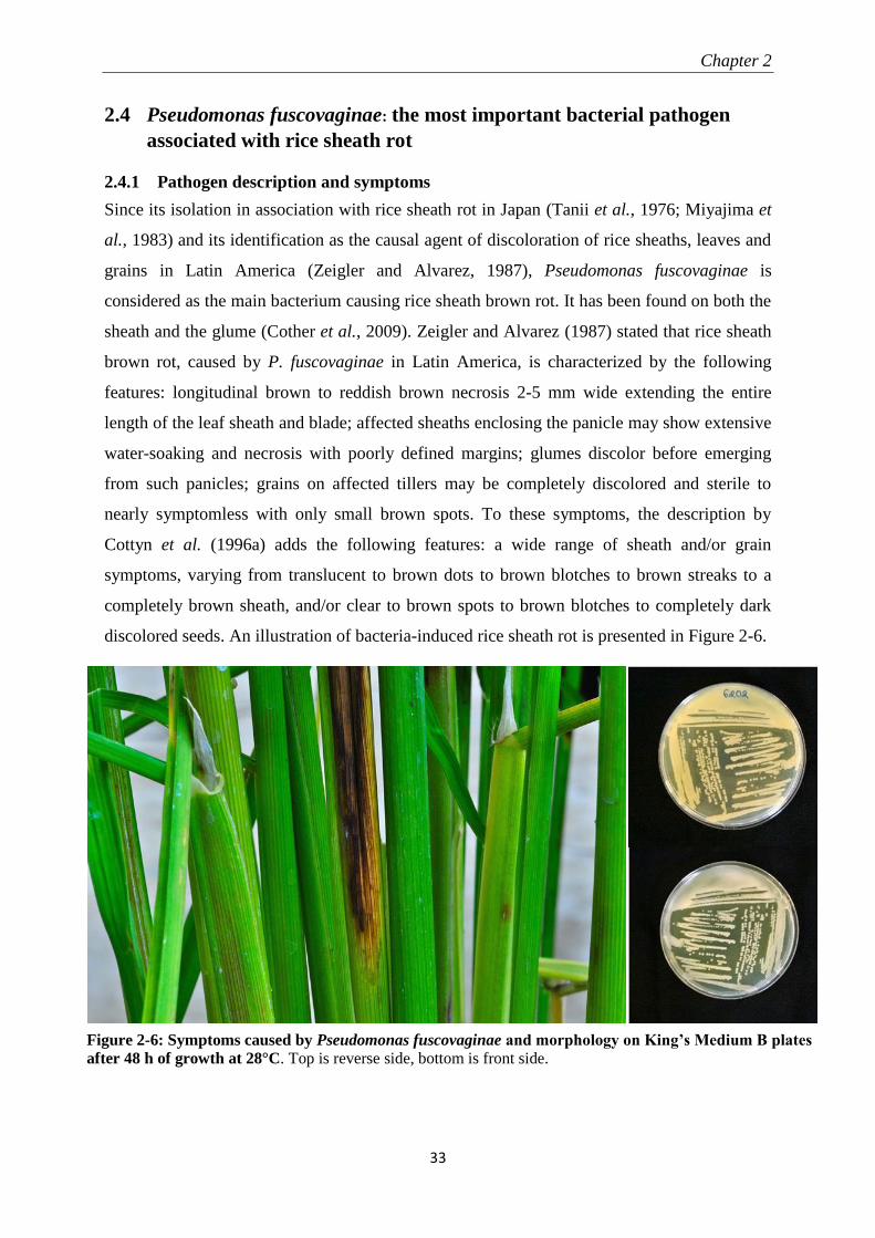
Chapter 2
33
2.4 Pseudomonas fuscovaginae: the most important bacterial pathogen
associated with rice sheath rot
2.4.1 Pathogen description and symptoms
Since its isolation in association with rice sheath rot in Japan (Tanii et al., 1976; Miyajima et
al., 1983) and its identification as the causal agent of discoloration of rice sheaths, leaves and
grains in Latin America (Zeigler and Alvarez, 1987), Pseudomonas fuscovaginae is
considered as the main bacterium causing rice sheath brown rot. It has been found on both the
sheath and the glume (Cother et al., 2009). Zeigler and Alvarez (1987) stated that rice sheath
brown rot, caused by P. fuscovaginae in Latin America, is characterized by the following
features: longitudinal brown to reddish brown necrosis 2-5 mm wide extending the entire
length of the leaf sheath and blade; affected sheaths enclosing the panicle may show extensive
water-soaking and necrosis with poorly defined margins; glumes discolor before emerging
from such panicles; grains on affected tillers may be completely discolored and sterile to
nearly symptomless with only small brown spots. To these symptoms, the description by
Cottyn et al. (1996a) adds the following features: a wide range of sheath and/or grain
symptoms, varying from translucent to brown dots to brown blotches to brown streaks to a
completely brown sheath, and/or clear to brown spots to brown blotches to completely dark
discolored seeds. An illustration of bacteria-induced rice sheath rot is presented in Figure 2-6.
Figure 2-6: Symptoms caused by Pseudomonas fuscovaginae and morphology on King’s Medium B plates
after 48 h of growth at 28°C. Top is reverse side, bottom is front side.
Review on rice sheath rot
34
The genus Pseudomonas belongs to the subclass Gammaproteobacteria of the Gram-negative
bacteria and currently comprises 144 species. Based on multilocus sequence analysis, P.
fuscovaginae belongs together with P. asplenii to the P. asplenii subgroup as defined by
Gomila et al. (2015). These two species are closely related and some authors consider them to
be synonymous (Vancanneyt et al., 1996). The original description of P. fuscovaginae in
Miyajima et al. (1983) is the following: the cells are aerobic, Gram negative, non-spore-
forming, rod-shaped with round ends, 0.5 -0.8 x 2.0-3.5 µm. Cells occur singly or in pairs and
are motile by means of one to four polar flagella. They oxidize glucose in oxidation-
fermentation medium, and they produce a green fluorescent pigment, oxidase and arginine
dihydrolase. Denitrification, β-glucosidase, pit formation on polypectate gel and growth at
37°C are negative. Characteristics that distinguish this species from other fluorescent
pseudomonads which are positive for arginine dihydrolase and oxidase are its inability to
utilize 2-ketogluconate or inositol.
Whole genome sequence analysis of various P. fuscovaginae strains has revealed that these
pathogens do not form a single monophyletic group. At least two subgroups have been
identified and strains from Madagascar, Japan, China and Australia clustered separately from
P. fuscovaginae-like strains from the Philippines (Quibod et al., 2015).
2.4.2 Epidemiology
P. fuscovaginae was first reported in literature as the pathogen responsible for rice sheath rot
disease in cold and humid tropical highlands in Japan (Miyajima et al., 1983), Burundi
(Duveiller et al., 1988), Madagascar (Rott et al., 1989) and Nepal (Sharma et al., 1997), but it
was later found even in lowlands (Cottyn et al., 1996b). P. fuscovaginae is also associated
with high rainfall (Sharma et al., 1997). The bacterium causes quantitative and qualitative
losses (Zeigler and Alvarez, 1987). For losses in quality, symptomatic grains cannot be
accepted in seed certification chains, mills accept them with a discount and they have a poor
marketing value.
CABI (2007) reports the presence of P. fuscovaginae in 31 countries: Former Yugoslavia,
Russian Federation, China, Indonesia, Japan, Nepal, Philippines, Burundi, Democratic
Republic of Congo, Madagascar, Rwanda, Tanzania, Costa Rica, Cuba, Dominican Republic,
El Salvador, Guatemala, Jamaica, Nicaragua, Panama, Trinidad and Tobago, Mexico,
Argentina, Bolivia, Brazil, Chile, Colombia, Ecuador, Peru, Suriname, and Uruguay.
Recently, the disease has been diagnosed in Australia (Cother et al., 2009).
Chapter 2
35
The host range of P. fuscovaginae seems to be restricted to wild and cultivated Graminae
(Tanii et al., 1976; Miyajima et al., 1983).
P. fuscovaginae is seed-transmitted and infected seedlings often die. When infection occurs at
a later stage, the lower part of the sheath becomes brown and later on, the whole sheath
becomes necrotic. The pathogenicity of P. fuscovaginae is expressed at grain, seedling and
booting stage levels. P. fuscovaginae is able to colonize the whole plant as an endophyte
(Adorada et al., 2015). If the seedling survives, P. fuscovaginae has an epiphytic life until the
booting stage when it infects inflorescences, resulting in the formation of infected grains or
the panicle abortion. The population of the bacterium is maintained at a low level from early
growth stages up to the booting stage. The bacterium can survive epiphytically on the host
plant with low pathogen population in the tissue and this explains how the disease can be
seed-borne, but only express symptoms at the booting stage (Batoko et al., 1997). This can
also be linked to the fact that the booting stage and the reproductive phase in general, is the
most stress-sensitive stage in the rice plant development (Fageria, 2007).
2.4.3 Pathogenicity determinants
Different phytotoxins are involved in the disease development. Batoko et al. (1997) could
reproduce sheath brown rot symptoms by inoculating seedlings with toxins from bacteria.
They concluded that toxins are an integral part of the plant-pathogen interactions in rice
bacterial sheath rot. Flamand et al. (1996) found that a cell-free extract from P. fuscovaginae
that could induce the same symptoms as P. fuscovaginae contained five peptidic compounds
(A, B, C, D and E) and two others (fuscopeptins A and B). Peptidic compound D is identical
to syringotoxin, a lipodepsinonapeptide containing nine amino acids acylated by 3-
hydroxytetradecanoic acid (Table 2-2, Figure 2-5) that is also produced by P. syringae pv.
syringae pathogenic on citrus (Ballio et al., 1990). The structure of fuscopeptins was
elucidated by Ballio et al. (1996). Fuscopeptins are lipodepsipeptides containing 19 amino
acids. Fuscopeptin A is acylated by 3-hydroxyoctanoate while fuscopeptin B is acylated by 3-
hydroxydecanoate (Table 2-2, Figure 2-5). Both compounds target the plasma membrane and
inhibit H+-ATPase and act synergistically with syringotoxin (Batoko et al., 1998).
Toxins from P. fuscovaginae are non-host specific, the pathogen inducing disease symptoms
on many plants of the Poaceae family in addition to rice (Miyajima et al., 1983), and have
more detrimental effect on culm elongation (Batoko et al., 1997). The non-host specificity
may also be justified by the symptoms induction by P. fuscovaginae on Chenopodium quinoa
(Mattiuzzo et al., 2011), a plant belonging to the Amaranthaceae family. Toxins are
Review on rice sheath rot
36
immediately dissolved in the plant and thus become difficult to recover (Batoko et al., 1997).
Phytotoxin concentration increases at the booting stage of rice, which stimulate their large
production by the bacterium. The capacity of the plant to detoxify the toxins plays a pivotal
role and could constitute a starting point in breeding for resistance against P. fuscovaginae.
Patel et al. (2014) (see Annex 1) were able to isolate nine mutants of P. fuscovaginae via
random Tn5 mutagenesis which showed altered virulence on rice. Besides mutants affected in
phytotoxin production, also mutants in type IV pili biosynthesis, type VI secretion, arginine
biosynthesis and sulfur metabolism were obtained indicating that these processes are also
involved in pathogenicity on rice.
2.4.4 Interactions with other diseases and pests
Most of the time P. fuscovaginae was found together with Sarocladium oryzae in sheath rot
diseased plants (Zeigler and Alvarez, 1987; Cottyn et al., 1996b).
2.4.5 Control methods
Some cultural and sanitation practices against P. fuscovaginae are indicated like burning farm
remains: stubbles, ratoons; treatment of seeds by dipping them in water at 65°C before sowing
(Zeigler and Alvarez, 1987); introducing rotation; checking the quality of seeds and as it is a
seed-borne disease, using healthy seeds. Host plant resistance is also considered as an option.
There are limited sources of resistance to rice sheath rot (Adorada et al., 2013), while this is a
must in developing a control strategy against the disease. There are various methods that can
be used for screening resistance and Adorada et al. (2013) suggested using the pin-prick
method. About the chemical control, streptomycin, alone or in combination with
oxytetracycline, can effectively control rice sheath rot (CABI, 2007).
2.4.6 Other Pseudomonas spp. associated with rice sheath rot
Besides P. fuscovaginae, a variety of other poorly characterized fluorescent pseudomonads
have been associated with rice sheath rot since the 1950s. The first characterized sheath rot
associated Pseudomonas was P. oryzicola (Klement, 1955). Later on it was decided that this
pathogen is equivalent to P. syringae pv. syringae (Young et al., 1978). Besides P. syringae
and P. fuscovaginae, various other pseudomonads have been consistently found in rice sheath
rot related studies (Zeigler and Alvarez, 1987; Cottyn et al., 1996a; b; Cother et al., 2010;
Saberi et al., 2013). Only a few of those other pseudomonads have been fully identified
except by biochemical tests.
Chapter 2
37
Zeigler and Alvarez (1987) attempted to put rice sheath rot-associated pseudomonads into
groups, which were continued and named, based on BIOLOG features, by Cottyn et al.
(1996a). In their work, they defined, based on the guidelines for the taxonomy of
Proteobacteria, originally called purple bacteria (Woese, 1987), four main groups of
Gammaproteobacteria associated with rice sheath rot and grain discoloration named after the
representative species: P. putida, P. aeruginosa, P. fuscovaginae, and a composite group
related to P. marginalis, P. corrugata, P. fluorescens, P. aureofaciens and P. syringae. Also
Saberi et al. (2013) concluded, based on biochemical tests, that sheath rot and grain
discoloration caused by Pseudomonas spp. in Iran are related to P. marginalis, P. putida, and
P. syringae.
The question whether these associated Pseudomonas spp. are really pathogenic on rice
remains posed for many years. From the start, few species emerged as the most pathogenic
compared to others which were causing some low levels of the disease. Zeigler and Alvarez
(Zeigler and Alvarez, 1987) already mentioning minor sheath and grain disorders caused by
fluorescent pseudomonads, P. fuscovaginae being the principal causal agent. Gardan et al.
(2002) isolated P. palleroniana from La Réunion (France), Cameroon and Madagascar from
healthy or necrotic rice seeds and from diseased tissue of leaf sheaths. The P. palleroniana
isolates inoculated to rice seedlings were either non-pathogenic or weakly pathogenic. On the
contrary, typical symptoms of bacterial sheath brown rot were induced by P. fuscovaginae
strain CFBP3078, introduced in the experiment for comparison. This shows that among the
pseudomonads found with rice sheath rot, there are differences in virulence and P.
palleroniana is among the weakly pathogenic organisms.
However, caution is needed in the interpretation of the pathogenicity level for the different
species of the pseudomonads associated with rice sheath rot. Cother et al.(2010) isolated a
pseudomonad causing a widespread disease similar to sheath brown rot in Cambodia. This
bacterium was related to P. parafulva and P. fulva, which belong to the P. putida group as
defined by Gomila et al. (2015), and was clearly pathogenic on rice.
In the meantime, the taxonomy of pseudomonads has made important progress especially
thanks to molecular identification method development. In a recently published classification
of Pseudomonas genus, based on the Multilocus Sequence Analysis Technique (MLSA),
Gomila et al. (2015) defined 19 groups and subgroups. Most of the sheath rot associated
pseudomonads probably belong to the P. chlororaphis, P. fluorescens, P. asplenii (= P.
Review on rice sheath rot
38
fusovaginae) subgroup or the P. putida group, though the groupings are difficult to define
currently as many isolates have not yet been fully analyzed.
2.4.7 Other reported sheath rot associated bacteria
Pantoea ananatis, considered globally as a facultative pathogen (Cray et al., 2013), was
demonstrated as a sheath rot pathogen with typical symptoms of necrotic spots and
discoloration on glumes and stems, indistinct chlorosis but with no water-soaking and its
pathogenicity testing satisfied Koch’s postulates (Choi et al., 2012a). The disease had
previously been reported in the Philippines (Cottyn et al., 2001) and in Australia (Cother et
al., 2004), but its importance, though it is reported to reduce the grain quality when it infects
the glumes, was never assessed. It was only presumed to be low. Furthermore, in
pathogenicity tests, Cother et al. (2004) recovered the pathogen from the plants that had not
been inoculated, which prompted the hypothesis that the organism lives as an epiphyte and
triggers disease symptoms when the plant is under physiological stress. Also Choi et al.
(2012) linked the disease appearance to favorable environmental conditions.
Burkholderia glumae and B. gladioli are becoming important rice pathogens (Nandakumar et
al., 2007a). B. glumae (formerly Pseudomonas glumae) was reported as the agent of rice grain
discoloration in Latin America (Zeigler and Alvarez, 1989) after it had been reported as a
grain rotter in Asia. It was later detected in North America, in association with B. gladioli,
causing bacterial panicle blight (Nandakumar et al., 2009). The two pathogens, in addition to
being seed-borne, can also be soil-borne (Nandakumar et al., 2008). Disease symptoms are
observed at the sheath and grain levels. Though the disease is seed-borne, the presence of the
bacteria in the sheath plays a capital role in the infection of the emerging panicle. Toxoflavin,
a toxin produced by both species, is considered to be the main pathogenicity determining
factor (Suzuki et al., 2004; Ura et al., 2006), while a lipase produced by Burkholderia
glumae (Pauwels et al., 2012) and tropolone produced by B. gladioli (Wang et al., 2013) have
also been implicated in pathogenicity.
Acidovorax oryzae (Schaad et al., 2008), formerly called Pseudomonas avenae and
Acidovorax avenae subsp. avenae (Willems et al., 1992), causes bacterial brown stripe on rice
(Shakya et al., 1985; Kadota et al., 1991; Song et al., 2004). Symptoms start as brown stripes
at the bottom of the stems and frequently extend into the sheaths (Liu et al., 2012a). This
bacterium has consistently been detected in rice sheath rot related studies (Cottyn et al.,
1996a; b; Cortesi et al., 2005; Cother et al., 2010). Recently the type IV pili assembly protein
PilP has been implicated in the pathogenicity of A. oryzae on rice (Liu et al., 2012a).
Chapter 2
39
2.5 Conclusions and perspectives Since rice sheath rot symptoms were first described in Taiwan in 1922 and attributed to
Sarocladium oryzae, various reports of similar or related disease symptoms have been
produced in different parts of the world. Rice sheath rot is now getting momentum as an
emerging destructive rice disease but on which the scientific understanding is still limited.
There are three main species or complexes of organisms that can cause rice sheath rot:
Sarocladium oryzae, the Fusarium fujikuroi complex and Pseudomonas fuscovaginae, but
there are many others that are reported to induce symptoms close to those of rice sheath rot.
Interestingly, all three groups of major sheath rot causing pathogens produce phytotoxins that
cause necrosis and can mimic the disease symptoms, which is probably the reason why they
all cause similar looking disease symptoms. The principle that “everything is everywhere, but,
the environment selects” (de Wit and Bouvier, 2006) applies to rice sheath rot; organisms that
can potentially cause rice sheath rot are many and can be found everywhere nowadays, but the
environment probably selects the ones that can adapt to the prevailing environmental
conditions in a given area. This situation results in the overlapping of symptoms in the rice
sheath rot disease complex (Johanson et al., 1998; Hu et al., 2008) especially at the rice
reproductive stage, the most stress-sensitive phase in rice development (Fageria, 2007). There
can be even synergism among the rice sheath rot-associated organisms or with arthropods or
other groups of organisms. Due to changes in agriculture and in the society in general, like the
developments in the farming systems and increased mobility in general, there are also changes
in plant health problems, some diseases becoming more important than before, like rice
sheath rot, which is now becoming a serious threat to rice production in many parts of the
world.
It is proven that most sheath rot associated pathogens have an endophytic (latent) phase in
their lifecycle, waiting for the plant to become stressed so that they can attack it (Fisher and
Petrini, 1992). This phenomenon is not recent, it was observed since many years. Hsieh et al.
( 1977) attested the presence of F. moniliforme (now known as the F. fujikuroi complex) on
plants without causing visible disease symptoms. New empirical data are needed about most
of the organisms thought to be endophytic as some of them have pathogenic potential and are
waiting for conducive conditions to attack the plant. Factors governing the expression of the
virulence are not yet clearly understood (Andrews and Harris, 2000). There is an urgent need
of associating molecular, genetic and pathogenicity data for elucidating the role and
Review on rice sheath rot
40
interactions with endophytes given that at the plant level, the answer to pathogens and
endophytes is the same (Andrews and Harris, 2000).
The large variability observed in rice sheath rot associated Pseudomonas and Fusarium
genera is intriguing. It would be interesting to investigate whether the isolates in these two
groups that can cause sheath rot have obtained phytotoxin-encoding gene clusters by
horizontal gene transfer. At least in the case of fumonisins, it has been shown that the
fumonisin gene cluster has spread among Fusarium spp. and related genera by a combination
of horizontal gene transfer, cluster duplication and loss (Proctor et al., 2013). It should be
tested whether the sheath rot causing Fusarium isolates all contain the fumonisin gene cluster
or other phytotoxin encoding gene clusters. Horizontal gene transfer is also a widespread
phenomenon in fluorescent pseudomonads (Silby et al., 2011) and it is known that many gene
clusters for secondary metabolites, including cyclic lipopeptides, are located on genomic
islands. Again, this could be systematically tested for Pseudomonas isolates associated with
rice sheath rot.
Rice sheath rot has become a highly destructive rice disease with a high variability in yield
loss levels varying from 20 to 85%. It is caused by many pathogenic agents varying
depending on the area, grown varieties, prevailing environmental conditions, the farming
system, other pests, etc. Not much progress has been achieved in the control of the disease,
partly because the etiology of the disease is difficult to establish. For facing the disease, a
better understanding about it is needed and this review is contributing in that aim. As rice
sheath rot disease is complex by nature, its control strategy must be inspired by the Integrated
Pest Management (IPM) approach. The solution remains site-specific. Limiting the number
of potential pathogens harbored by the plant, making the plant environment less conducive to
pathogen development, etc. should be the central elements in the control approach, which can
be complemented by other methods, indicated according to the context. The IPM approach is
particularly relevant now that there is a need for feeding and responding to the other needs of
a constantly increasing population while the production must be conducted in a sustainable
way, meaning that the overreliance on pesticide must leave the room to scientifically-proven
environmentally-friendly crop protection practices.
Chapter 2
41
Acknowledgments
V.P. Bigirimana received a doctoral grant from the Belgian Technical Cooperation (BTC)
(project reference: 10RWA/0018). This work was supported by a grant from the Special
Research Fund of Ghent University (GOA 01GB3013)

Chapter 3
43
3 ESTIMATION OF THE IMPORTANCE OF RICE SHEATH ROT IN
RWANDA BASED ON DIAGNOSIS AND DISEASE INTENSITY
EVALUATION
Vincent de Paul Bigirimana, Obedi Ishibwela Nyamangyoku, Monica Höfte
Abstract
Rice is a staple food for half of the world population, and is produced worldwide in tropical
and subtropical areas. Rice is one of the priority crops in Rwanda. The demand for rice is high
and the country imports, as of 2011, approximately 30% of the rice consumed. Pests and
diseases are recognized major limiting factors preventing rice from expressing its full
production potential. Rice sheath rot is one of the emerging and destructive rice diseases, yet
poorly studied. Its major symptoms are found on the terminal leaf sheath which turns brown,
grayish and ends up dying. Grains from diseased plants are discolored, chaffy and germinate
poorly. The disease symptoms are more pronounced at the booting stage. A study was
undertaken to evaluate the importance of rice sheath rot in Rwanda. Data for rice sheath rot
intensity, id est incidence and severity, were taken in the agricultural season of September
2013 to January 2014, from the major rice-growing areas of the country: Bugarama, Cyili,
Rwamagana, Nyagatare and Rwagitima. Data for yield loss were taken in Rwagitima and
Nyagatare. The average disease incidence was 13.5% and the average measure for disease
severity (lesion length on a diseased tiller) was 157 mm. In the considered period, the disease
was more pronounced in Rwamagana than in other areas. The yield loss is estimated at 57%
yield loss based on the weight of diseased grains in comparison to healthy ones. Rice sheath
rot symptoms are found in all rice-growing areas of Rwanda and may be caused by many
different pathogens like Sarocladium oryzae, Fusarium spp., Pseudomonas fuscovaginae,
Burkholderia glumae, Acidovorax avenae, etc. and it is impossible to tell the difference
between these agents based on induced symptoms; all the sheath rot causing pathogens are
seed-transmitted and as the seed system is centrally organized, it is possible for a disease to be
transmitted from one place to another through seed movement. Sarocladium oryzae and
Fusarium spp. have recently been diagnosed in Rwanda. Considering the destructive nature of
the disease and its widespread distribution, efforts are needed for limiting its impact. As
curative options are limited, preventive measures, ensuring that the seed grain is healthy,
should be encouraged as most of these pathogens are seed-transmitted and also given that
many currently grown rice cultivars are susceptible to sheath rot pathogens.
Estimation of the importance of rice sheath rot in Rwanda
44
3.1 Introduction Rice is the first staple food worldwide, produced all over the world in tropical and subtropical
areas. Rice is popular for many reasons among which we can mention the following: it
responds well to farm inputs, can be processed and preserved, has an increasing market, gives
a good yield compared to many other tropical crops and is adapted to many agroecological
zones. Rice production is promoted in many countries including Rwanda. The situation and
importance of rice in Rwanda are presented in the section 2.1.
Rice production faces a series of constraints among which pests and diseases are important.
Sheath rot disease (see Chapter 2) is one of frequently found rice diseases, but which is not
scientifically clearly understood yet. It occurs in many parts of the world.
Many organisms have been associated with rice sheath rot symptoms including fungal
pathogens (see Chapter 2 and Bigirimana et al., 2015). It is difficult to tell the causal agent
based on the induced symptoms, as the symptoms are highly similar. Losses caused by rice
sheath rot vary from 3 to 85% (Ou, 1985).
In the aim of contributing to the generation of information on rice sheath rot, a study was
planned to evaluate the importance of sheath rot in rice fields in Rwanda. For this purpose,
data about disease incidence, disease severity and yield loss were collected.
3.2 Materials and methods Data were collected during the agricultural season of September 2013 to January 2014 in the
major rice-growing areas of Rwanda (see Figure 1-1 in Chapter 1), which are Bugarama
(Bugarama sector, Western Province), Cyili (Rusatira Sector, Southern Province),
Rwamagana (Kigabiro Sector, Eastern Province), and Nyagatare and Rwagitima (Nyagatare
and Rugarama Sectors, Eastern Province). Altitudes, rainfall data and average temperatures
for these areas are given in Table 3-1.
Table 3-1: Altitude, rainfall and average temperature for the major rice-growing areas
in Rwanda*
Location Closest meteorological
station
Altitude (m) Rainfall (mm) Average
temperature
(°C)
Rwamagana Kibungo 1680 979 18.9
Bugarama Bugarama 900 1098 24.0
Nyagatare and
Rwagitima
Gabiro 1470 783 20.4
Cyili Rubona 1706 1154 19.2
* Source: MINAGRI (2002)
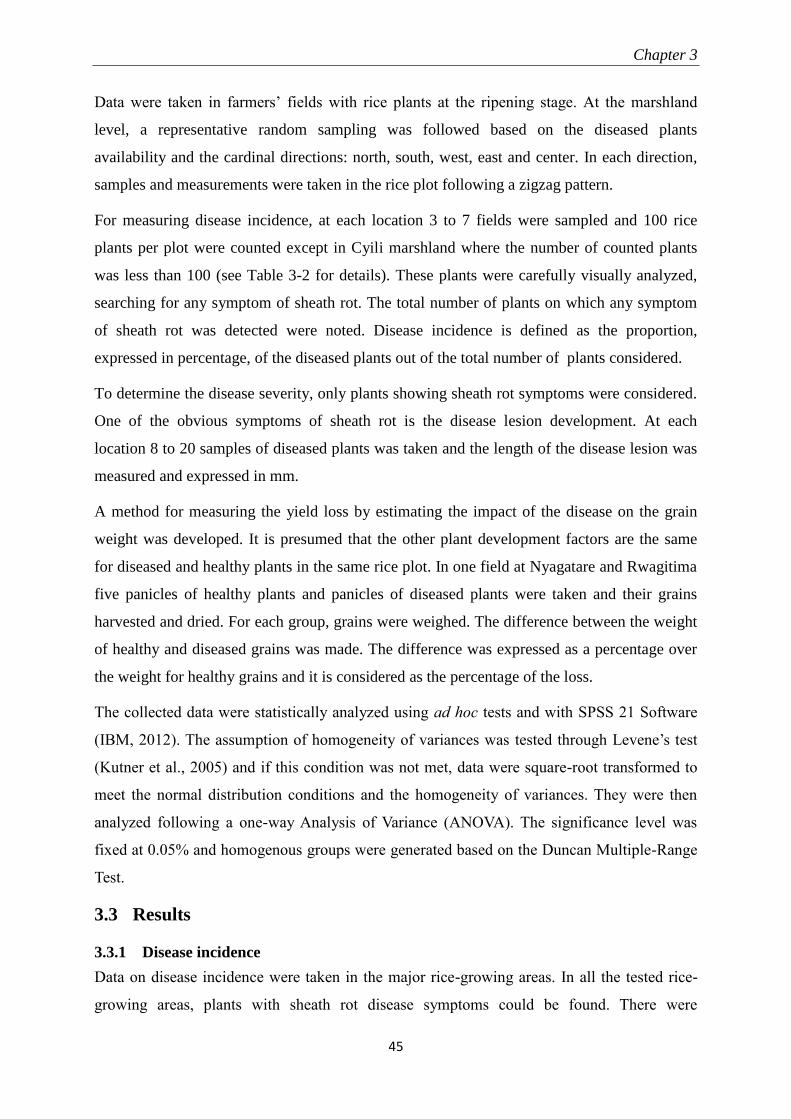
Chapter 3
45
Data were taken in farmers’ fields with rice plants at the ripening stage. At the marshland
level, a representative random sampling was followed based on the diseased plants
availability and the cardinal directions: north, south, west, east and center. In each direction,
samples and measurements were taken in the rice plot following a zigzag pattern.
For measuring disease incidence, at each location 3 to 7 fields were sampled and 100 rice
plants per plot were counted except in Cyili marshland where the number of counted plants
was less than 100 (see Table 3-2 for details). These plants were carefully visually analyzed,
searching for any symptom of sheath rot. The total number of plants on which any symptom
of sheath rot was detected were noted. Disease incidence is defined as the proportion,
expressed in percentage, of the diseased plants out of the total number of plants considered.
To determine the disease severity, only plants showing sheath rot symptoms were considered.
One of the obvious symptoms of sheath rot is the disease lesion development. At each
location 8 to 20 samples of diseased plants was taken and the length of the disease lesion was
measured and expressed in mm.
A method for measuring the yield loss by estimating the impact of the disease on the grain
weight was developed. It is presumed that the other plant development factors are the same
for diseased and healthy plants in the same rice plot. In one field at Nyagatare and Rwagitima
five panicles of healthy plants and panicles of diseased plants were taken and their grains
harvested and dried. For each group, grains were weighed. The difference between the weight
of healthy and diseased grains was made. The difference was expressed as a percentage over
the weight for healthy grains and it is considered as the percentage of the loss.
The collected data were statistically analyzed using ad hoc tests and with SPSS 21 Software
(IBM, 2012). The assumption of homogeneity of variances was tested through Levene’s test
(Kutner et al., 2005) and if this condition was not met, data were square-root transformed to
meet the normal distribution conditions and the homogeneity of variances. They were then
analyzed following a one-way Analysis of Variance (ANOVA). The significance level was
fixed at 0.05% and homogenous groups were generated based on the Duncan Multiple-Range
Test.
3.3 Results
3.3.1 Disease incidence
Data on disease incidence were taken in the major rice-growing areas. In all the tested rice-
growing areas, plants with sheath rot disease symptoms could be found. There were
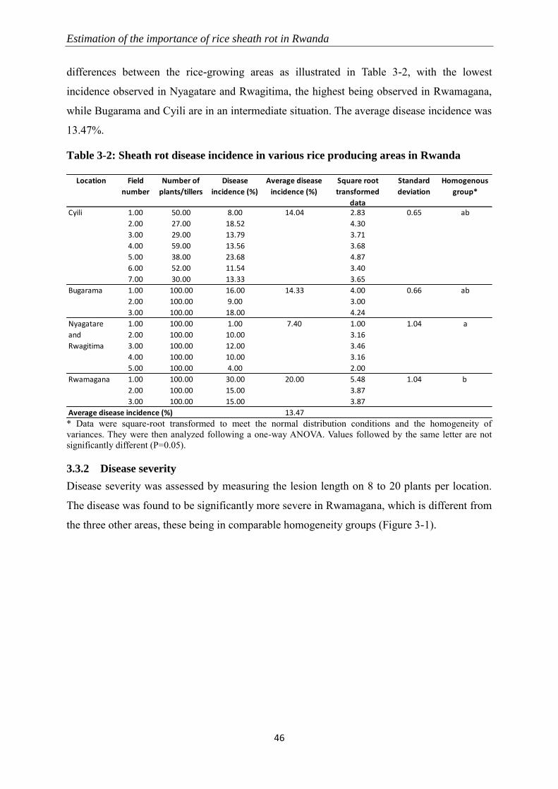
Estimation of the importance of rice sheath rot in Rwanda
46
differences between the rice-growing areas as illustrated in Table 3-2, with the lowest
incidence observed in Nyagatare and Rwagitima, the highest being observed in Rwamagana,
while Bugarama and Cyili are in an intermediate situation. The average disease incidence was
13.47%.
Table 3-2: Sheath rot disease incidence in various rice producing areas in Rwanda
* Data were square-root transformed to meet the normal distribution conditions and the homogeneity of
variances. They were then analyzed following a one-way ANOVA. Values followed by the same letter are not
significantly different (P=0.05).
3.3.2 Disease severity
Disease severity was assessed by measuring the lesion length on 8 to 20 plants per location.
The disease was found to be significantly more severe in Rwamagana, which is different from
the three other areas, these being in comparable homogeneity groups (Figure 3-1).
Location Field
number
Number of
plants/tillers
Disease
incidence (%)
Average disease
incidence (%)
Square root
transformed
data
Standard
deviation
Homogenous
group*
1.00 50.00 8.00 2.83 0.65
2.00 27.00 18.52 4.30
3.00 29.00 13.79 3.71
4.00 59.00 13.56 3.68
5.00 38.00 23.68 4.87
6.00 52.00 11.54 3.40
7.00 30.00 13.33 3.65
1.00 100.00 16.00 4.00 0.66
2.00 100.00 9.00 3.00
3.00 100.00 18.00 4.24
1.00 100.00 1.00 1.00 1.04
2.00 100.00 10.00 3.16
3.00 100.00 12.00 3.46
4.00 100.00 10.00 3.16
5.00 100.00 4.00 2.00
1.00 100.00 30.00 5.48 1.04
2.00 100.00 15.00 3.87
3.00 100.00 15.00 3.87
Average disease incidence (%) 13.47
a
Rwamagana 20.00 b
Nyagatare
and
Rwagitima
7.40
ab
Bugarama 14.33 ab
Cyili 14.04
Chapter 3
47
Figure 3-1: Rice sheath rot severity per rice-growing area
Data were analyzed by a one-way ANOVA test. Bars represent mean lesion lengths and standard
deviations based on 8 to 20 plants in each location. Bars with the same letters are not significantly
different (P=0.05).
3.3.3 Yield loss
Values used for estimating the yield loss were generated on dried grain samples taken in
Nyagatare and Rwagitima. The weight of grains from five healthy plants and diseased plants
were compared. The average weight of one grain harvested from a healthy plant was 21.22
mg (± 4.51 mg), the average weight of one grain harvested from a diseased plant was 9.14 mg
(± 3.19 mg). The calculated yield loss is estimated at 56.93%. A Student test (t-test) revealed
that these means are significantly different from each other at P =0.05.
3.4 Discussion Rice sheath rot symptoms were found in all rice-growing areas of Rwanda considered in this
study: Bugarama, Cyili, Rwamagana and Nyagatare and Rwagitima. It is likely that the
inoculum is present in nearly all the rice planting materials available in the country. As the
seed production system is centrally organized at the national level, the disease development
potential is minimized through seed inspection and production but still the planting material
released has some level of rice sheath rot pathogens inoculum. In another study, a diagnosis
has been conducted on sheath rot associated organisms in Rwanda and it was observed that
the major fungal agents of rice sheath rot, Sarocladium oryzae and Fusarium spp. are present
in the country (See Chapter 4). The fact that nearly all the available rice varieties are attacked
can be partly explained by the realization that many current rice cultivars are susceptible to
attacks of Sarocladium oryzae (Mew et al., 2004b) and Fusarium disease complexes (Savary
Estimation of the importance of rice sheath rot in Rwanda
48
et al., 2012). And it has been proven that some species included in those Fusarium complexes
cause rice sheath rot (Abbas et al., 1998). It is already established since many years that
Sarocladium oryzae causes rice sheath rot. Rwamagana showed the highest disease incidence
and disease severity. Disease incidence was the lowest at Nyagatare and Rwagitima although
data were not significantly different from Cyili and Bugarama due to high variability among
fields in Nyagatare and Rwagitima. Disease is influenced by many factors that enter into the
disease tetrahedron (Savary et al., 2012): the plant, the pathogen, the environment and the
human factors. There are obviously variations in the combination of these factors in the
different rice-growing areas, resulting in different disease expressions in those areas. One of
the sources of variability may be the rainfall and/or altitude. Rainfall is the lowest in
Nyagatare and Rwagitima which may partly explain the lower disease incidence in this
region.
The yield loss in terms of grain weight, comparing grains from healthy and diseased plants, is
more than the half; it is estimated at 56.93%. This loss may justify the chaffiness that is
observed on grains from sheath rot diseased plants (Gopalakrishnan et al., 2010). But it is
important to realize that the losses due to the disease are higher than those counted on grains
especially in terms of quality. It has been reported that the sheath rot associated pathogens are
seed-transmitted, meaning that when the seed is infected, in a system where the seed is most
of the time taken from the harvest, the following generation is also exposed to the disease,
unless strong preventive measures are taken. This pathogen seed-transmission is reported for
both Sarocladium oryzae (Ou, 1985) and Fusarium spp. (Wulff et al., 2010) that cause sheath
rot symptoms. In addition, the yield loss also occurs by the reduction of the marketing quality
for the produce when coming from diseased plants as it receives a reduced price. This is
particularly true for Rwanda where local rice production is not competitive on the market,
being considered of low quality (Stryker, 2013).
The data were taken and analyzed per rice-growing areas and symptoms were found in all the
surveyed areas, but the study shows also a high level of variability within the same rice-
growing area. The grown rice variety is one of the important factors that can be responsible
for the variability in the disease severity due to various disease susceptibility levels for
different varieties. Though farmers in the same area in Rwanda use a limited number of
varieties per season, there is still a variability in their variety choice, motivated, among others
by the disease susceptibility of the planting material. This aspect should be further explored in
subsequent studies.
Chapter 3
49
When rice sheath rot is caused by Fusarium spp. agents, there is a risk of infection by toxins
when consuming the harvested grains as most species of the Fusarium spp. complex can
produce them (Wulff et al., 2010). This is important for the community in general as food
intoxications must be prevented. There is currently no information about toxins infection in
Rwanda, but we can presume that it is not negligible, as cereals, id est maize, sorghum and
wheat, in addition to rice, are among the major crops in the country and that it is not
uncommon to see symptoms of attacks by Fusarium spp.
Rice is becoming an important food crop in Rwanda. Pests and diseases are among the factors
that are still hampering its development. Rice sheath rot disease, one of the important and yet
not well known scientifically rice disease is present in Rwanda, in all major rice-growing
areas. The disease causes important yield losses, in addition to reducing the grain quality for
consumption or use as seed. An action plan for knowing more about the disease and tackling
it is highly needed.
Acknowledgments
This study, supervised by the Ghent University and the University of Rwanda/CAVM, was
financially supported by grants from the Belgian Technical Cooperation given to
BIGIRIMANA V.P
.

Chapter 4
51
4 SAROCLADIUM ORYZAE AND FUSARIUM SPP. ARE ASSOCIATED
WITH RICE SHEATH ROT IN RWANDA
Vincent de Paul Bigirimana, Gia Khuong Hoang Hua, Lien Bertier, Obedi Ishibwela
Nyamangyoku and Monica Höfte
The major part of this section has been submitted to Plant Pathology and is under
revision
Abstract
Sheath rot disease occurs in most rice-growing regions of the world and varying yield losses
have been reported. In this study, rice plants showing sheath rot symptoms were sampled
from various marshlands in Rwanda and fungal organisms associated with the disease were
isolated, purified, and identified by both morphological and molecular methods. Sarocladium
oryzae and Fusarium spp. were recovered from the diseased fields in Rwanda. Sarocladium
oryzae isolates were genetically very similar, while the majority of the Fusarium isolates were
closely related to F. andiyazi, a species belonging to the F. fujikuroi complex. Other
Fusarium isolates retrieved from sheath rot lesions belonged to the F. oxysporum, F.
incarnatum/equiseti and the F. graminearum complex. Phylogenetic analysis reveals a strong
relationship between the S. oryzae and F. andiyazi isolates found in Rwanda and those
reported in other rice growing areas. Sarocladium oryzae and the Fusarium isolates caused
very similar symptoms on rice plants although the indica variety CO39 appeared to be more
resistant to both fungal genera than the japonica variety Nipponbare. Fumonisin production
could be detected in rice plants inoculated with Fusarium isolates from the fujikuroi complex
and the oxysporum complex, but was not correlated with disease severity.
S. oryzae and Fusarium spp. associated with rice sheath rot in Rwanda
52
4.1 Introduction Rwanda is a densely populated high altitude country in Central/East-Africa. There is a lot of
interest in rice (Oryza sativa) production in Rwanda because the crop can be grown in
marshland areas and as such relieve the increasing pressure on hillside land for food
production. Rice has been introduced in Rwanda in the 1950s (Stryker, 2013) and is
cultivated in irrigated systems at low and middle altitude (900 to 2000 m). The presence of
rice sheath rot symptoms has been reported in Rwanda but has not further been investigated
(Bigirimana et al., 2013). Rice sheath rot is an emerging disease worldwide and can cause
varying yield losses. Rice sheath rot can be caused by toxin-producing fungal pathogens (see
Chapter 2 and Bigirimana et al., 2015 for a review). The main fungal species reported is
Sarocladium oryzae (Sakthivel, 2001; Bills et al., 2004; Ayyadurai et al., 2005), but also
Fusarium spp. belonging to the F. fujikuroi complex such as F. proliferatum, F.
verticillioides, and F. fujikuroi (Abbas et al., 1998; Wulff et al., 2010) have been associated
with rice sheath rot. Sarocladium oryzae is a seed-borne pathogen and its mycelium can
survive in plant residues, weed hosts and soil (Sakthivel, 2001). Pathogenicity of S. oryzae is
related to its capacity to produce the phytotoxins helvolic acid and cerulenin (Sakthivel et al.,
2002). Artificial inoculation of these toxins on rice plants can reproduce sheath rot symptoms
(Ghosh et al., 2002; Ayyadurai et al., 2005). The main toxins produced by species belonging
to the F. fujikuroi complex are the fumonisins (Ma et al., 2013). The role of fumonisins in
Fusarium-induced pathogenicity is controversial: Abbas et al. (1998) attested that in planta
fumonisin concentrations were positively correlated with the levels of the disease caused by
Fusarium spp. but Dastjerdi and Karlovsky (2015) showed that fumonisin-producing and non-
producing Fusarium verticillioides isolates could both infect seedlings at the same level and
thus concluding that fumonisins are not involved in the infection.
Despite the fact that the occurrence of rice sheath rot can cause considerable yield losses, the
disease etiology is poorly understood. Thus, the current study aimed at identifying the fungal
organisms associated with rice sheath rot in Rwanda. By building the baseline on sheath rot
disease in Rwanda, this study paves the way for breeding for resistance and development of
management strategies against rice sheath rot.
Chapter 4
53
4.2 Materials and methods
4.2.1 Field sampling
In 2011 and 2013 samples were taken from the traditional rice-growing areas of Bugarama
(Bugarama Sector, Western Province), Rwamagana (Kigabiro Sector, Eastern Province),
Nyagatare and Rwagitima (respectively Nyagatare and Rugarama Sectors, Eastern Province)
in Rwanda. To reflect the change in rice production in Rwanda, in 2013 additional diseased
rice plant samples were taken in the new rice growing areas Rugeramigozi (Nyamabuye
Sector, Southern Province) and Kabuye (Jabana Sector, Kigali City). All locations where the
sampling was conducted are illustrated in Figure 1-1, whereas the major agro-bioclimatic
features of the sampling areas are listed in Table 4-1.
Table 4-1: Agro-bioclimatic characteristics of the sampling regions in Rwanda (Gasana,
2002; MINAGRI, 2005)
Province Sampling region Altitude
(m)
Annual Rice
cultivation area (ha)
Average temperature
(°C)
Annual rainfall
(mm)
Southern Province Rugeramigozi 1706 160 19.2 1154
Western Province Bugarama 900 2321 24.0 1098
Eastern Province Nyagatare and
Rwagitima
1470 973 20.4 783
Rwamagana 1680 713 18.9 979
Kigali City Kabuye 1270 296 21.7 951
Samples of diseased plants were taken in Agricultural Season B (from March to August) of
2011, and in Agricultural Season A (from September to February) of 2013. In each rice-
growing area, sheath rot diseased plant samples were taken in a representative way, following
the four cardinal directions (north, south, east and west) and the centre of the marshland. In
each direction, there was a careful search for plants exhibiting sheath rot symptoms.
Whenever these were found, samples of diseased (rotted) sheaths were collected, following a
zigzag pattern.
4.2.2 Pathogen isolation and purification
Plant tissues and un-emerged panicles showing typical sheath rot symptoms were cut in small
pieces and surface-disinfected in 70% ethanol for 2 min, followed by rinsing four times with
sterile water. Surface-sterilized samples were placed on potato dextrose agar (PDA; Difco)
plates and incubated at 28°C for seven to ten days. Fungal hyphae initially developing from
diseased plant tissues were transferred to fresh PDA plates and pure cultures were stored on
PDA slants at room temperature.
S. oryzae and Fusarium spp. associated with rice sheath rot in Rwanda
54
4.2.3 Morphological characterization
Cultures of purified isolates were grown on PDA plates at 28°C for at least two weeks and
their morphological characteristics were identified microscopically and macroscopically. For
microscopic analysis, thirty randomly selected macroconidia were measured. The
presence/absence of chlamydospores was also recorded. For macroscopic analysis,
pigmentation on PDA and the growth rate of fungal colony were observed.
4.2.4 DNA extraction, amplification and ITS-sequencing
Fungal isolates were grown on potato dextrose broth (PDB; Difco) at 28°C for one week. The
fungal biomass was harvested and ground in liquid nitrogen to produce a fine powder. Total
genomic DNA was extracted using the Wizard Genomic DNA Purification Kit (Promega) or
the DNeasy Plant Mini Kit (Qiagen). The Internal Transcribed Spacer (ITS) region was
amplified using primers ITS1 (5’-TCCGTAGGTGAACCTGCGG-3’) and ITS4 (5’-
TCCGTAGGTGAACCTGCGG -3’) (White et al., 1990). Fusarium isolates were further
identified by amplifying the Translation Elongation Factor (TEF) using primer pair TEF-1-F
(5’-ATGGGTAAGGAAGACAAGAC-3’) and TEF-2-R (5’-
GGAAGTACCAGTGATCATGTT-3’) (O’Donnell et al., 1998). PCR reactions were done in
25 µL of a solution consisting of 2 µL genomic DNA (100 ng µL-1
), 5 µL PCR buffer (5x;
Promega), 5 µL Q-solution (Qiagen), 0.5 µL dNTPs (10 mM; Fermentas GmbH), 1.75 µL of
each primer (10 µM), 0.15 µL Taq DNA polymerase (5 units µL−1
; Fermentas GmbH) and
8.85 µL ultrapure sterile water. Amplification was performed using a Flexcycler PCR
Thermal Cycler (Analytik Jena) programmed for an initial denaturation step at 94°C for 10
min, followed by 35 cycles of denaturation at 94°C for 1 min, primer annealing at 55°C for 1
min and extension at 72°C for 1 min. Cycling ended with a final extension step at 72°C for 10
min. The amplicons were separated by horizontal electrophoresis using 2% agarose gels in
Tris-Acetate-EDTA (TAE)-buffer at 100 V for 25 min and visualized by ethidium bromide
staining on a UV transilluminator. PCR products were purified using QIAquick PCR
Purification Kit (Qiagen) and the sequences of both strands were determined by LGC
Genomics GmbH (Berlin, Germany) using Sanger sequencing. Before use in blasting or
building phylogenetic trees, sequences were manually edited.
4.2.5 Identification using BLAST and phylogenetic analysis
Consensus sequences of all isolates generated with BioEdit version 7.1.11 were blasted
against GenBank using the BLASTn tool. Isolates belonging to fungal species most related to
those present in our collection were selected as reference for phylogenetic tree analysis. Most
Chapter 4
55
Sarocladium sequences were taken from Giraldo et al. (2015). Fusarium TEF sequences were
retrieved from the Fusarium MLST database or GenBank. Reference isolates belonging to the
F. fujikuroi group were taken from Wulff et al. (Wulff et al., 2010). Sequences of
Acremonium variecolor and Cylindrocarpon sp. were taken as outgroups.
The accession numbers of all sequences are shown in Table 4-2.
Table 4-2: Sequences of sheath rot-associated isolates and standard strains obtained
from GenBank and Fusarium MLST Database used for building phylogenetic trees
Genus Species Isolate Origin Accession
number
Reference
Fusarium spp. F. andiyazi 30ALH Rice, China FN252387 (Wulff et al., 2010)
1ALH Rice, Burkina Faso FN252388 (Wulff et al., 2010)
38ALH Rice, India FN252389 (Wulff et al., 2010)
Fusarium sp. NRRL 46612 Tomato, Sardinia - Fusarium MLST
Fusarium sp. NRRL 31654 Hadrodemas warscewiczianum,
Guatemala
- Fusarium MLST
F. fujikuroi 21ALH Rice, Vietnam FN252404 (Wulff et al., 2010)
7ALH Rice, Nepal FN252407 (Wulff et al., 2010)
20ALH Rice, Vietnam FN252398 (Wulff et al., 2010)
F. verticillioides 52 ALH Rice, Vietnam FN179342 (Wulff et al., 2010)
32 ALH Rice, China FN179344 (Wulff et al., 2010)
56ALH Rice, Bangladesh FN252385 (Wulff et al., 2010)
F. proliferatum 24ALH Rice, Cote d'Ivoire FN252391 (Wulff et al., 2010)
34ALH Rice, China FN252396 (Wulff et al., 2010)
51ALH Rice, Vietnam FN252397 (Wulff et al., 2010)
F. boothii NRRL 29011 South Africa AF212445 (O’Donnell et al., 2000)
F. graminearum NRRL 31084 Wheat, USA HM744693 (Yli-Mattila et al., 2011)
F. vorosii NRRL 38208 Wheat, Japan DQ459746 (Starkey et al., 2007)
F. asiaticum NRRL 13818 Barley, Japan AF212451 (O’Donnell et al., 2000)
F. incarnatum-equiseti
species complex
NRRL 25080 Brown planthopper, China JF740711 (O’Donnell et al., 2012)
F714 Wheat, Spain KF962952 (Castellá and Cabañes,
2014)
F. oxysporum species
complex
NRRL 38271 Lentil, Colombia FJ985361 (Donnell et al., 2009)
NRRL 22543 Elaeidis, Suriname FJ985270 (Donnell et al., 2009)
NRRL 22554 Chrysanthemum sp., Nigeria FJ985274 (Donnell et al., 2009)
Sarocladium
spp.
S. zeae CBS 800.69 Zea mays stalk, USA FN691451 (Giraldo et al., 2015)
S. bacillisporum CBS 119.79 Smoked slided meat, Sweden HG965001 (Giraldo et al., 2015)
CBS 212.79 Insect, Romania HG965002 (Giraldo et al., 2015)
CBS 388.67 Soil, The Netherlands HG965003 (Giraldo et al., 2015)
CBS 425.67 Soil, Canada HE608639 (Giraldo et al., 2012)
CBS 485.67 Unknown HG965004 (Giraldo et al., 2015)
CBS 787.69 Teleutosorus of Puccinia graminis on
Lolium temulentum, Italy
HG965005 (Giraldo et al., 2015)
S. bactrocephalum CBS 749.69 Ustilago sp., Canada HG965006 (Giraldo et al., 2015)
UTHSC 09-384 Eye, USA HG965007 (Giraldo et al., 2015)
S. oryzae and Fusarium spp. associated with rice sheath rot in Rwanda
56
Genus Species Isolate Origin Accession
number
Reference
S. bifurcatum CBS 383.73 Bamboo, India HG965008 (Giraldo et al., 2015)
UTHSC 05-3311 Bronchoalveolar lavage fluid, USA HG965009 (Giraldo et al., 2015)
UTHSC 07-3446 Bronchial wash fluid, USA HG965010 (Giraldo et al., 2015)
S. gamsii CBS 425.73 Pandanus lerum, Sri Lanka HG965014 (Giraldo et al., 2015)
CBS 707.73 Pandanus lerum, Sri Lanka HG965015 (Giraldo et al., 2015)
S. glaucum CBS 191.80 Bamboo, Cambodia HG965016 (Giraldo et al., 2015)
CBS 309.74 Air, above sugarcane field, India HG965017 (Giraldo et al., 2015)
CBS 382.73 Bamboo, India HG965018 (Giraldo et al., 2015)
CBS 456.74 Sugar, Mauritius HG965019 (Giraldo et al., 2015)
CBS 100350 Bamboo, Japan HG965020 (Giraldo et al., 2015)
S. hominis UTHSC 02-2564 Leg, USA HG965011 (Giraldo et al., 2015)
UTHSC 04-1034 Calf, USA HG965012 (Giraldo et al., 2015)
UTHSC 04-3464 Sputum, USA HG965013 (Giraldo et al., 2015)
S. implicatum CBS 397.70A Saccharum officinarum, Jamaica HG965021 (Giraldo et al., 2015)
CBS 825.73 Saccharum officinarum, India HG965022 (Giraldo et al., 2015)
CBS 959.72 Desert soil, Egypt HG965023 (Giraldo et al., 2015)
S. kiliense CBS 122.29 Skin, Germany FN691446 (Perdomo et al., 2011)
S. ochraceum CBS 428.67 Zea mays, Kenya HG965025 (Giraldo et al., 2015)
S. oryzae CBS 180.74 Rice, India HG965026 (Giraldo et al., 2015)
CBS 399.73 Rice, India HG965027 (Giraldo et al., 2015)
LS449-1 Rice, India KP999971 Genbank
CBS 414.81 Rice, Nigeria HG965028 (Giraldo et al., 2015)
CBS 361.75 Rice, Kenya AY567005 (Bills et al., 2004)
CBS 120.817 Rice, Panama This study
WA13481 Rice, Australia JQ965668 (Lanoiselet et al., 2012)
S. pseudostrictum UTHSC 02-1892 Sputum, USA HG965029 (Giraldo et al., 2015)
S. strictum CBS 640.75 Decaying wood, The Netherlands HG965030 (Giraldo et al., 2015)
S. subulatum MUCL 9939 Soil, Egypt HG965031 (Giraldo et al., 2015)
UTHCS 07-110 Bone, USA HG965032 (Giraldo et al., 2015)
S. summerbellii CBS 430.70 Soil, The Netherlands HG965034 (Giraldo et al., 2015)
CBS 797.69 Canna indica, The Netherlands HG965035 (Giraldo et al., 2015)
CBS891.73 Dead leaf, Sri Lanka HG965036 (Giraldo et al., 2015)
CBS951.72 Soil, The Netherlands HG965037 (Giraldo et al., 2015)
S. terricola MUCL 42865 Palm grove, Morocco HG965040 (Giraldo et al., 2015)
UTHSC 03-2933 Bronchial wash fluid, USA HG965041 (Giraldo et al., 2015)
UTHSC 04-956 Sinus, USA HG965042 (Giraldo et al., 2015)
UTHSC 07-3260 Bone, USA HG965043 (Giraldo et al., 2015)
UTHSC 08-844 Bronchoalveolar lavage fluid, USA HG965045 (Giraldo et al., 2015)
For multiple alignments, the MUSCLE algorithm was applied. Phylogenetic trees and
distance matrix were constructed using the maximum-likelihood algorithm with 1000
replicates using MEGA6 (Tamura et al., 2013).
Chapter 4
57
(Disease class x number of plants falling under that class)PDI (%) x 100
Total number of plants observed x Maximum class
4.2.6 Pathogenicity assays
Twelve isolates representing the different fungal species isolated from rice with sheath rot
symptoms in Rwanda were selected for pathogenicity tests on rice plants. Inoculum was
prepared according to the standard grain inoculum technique (Sakthivel and Gnanamanickam,
1987). Briefly, rice grains were soaked in water for 1 h, excess water was removed and the
grains were autoclaved twice on two different days. Ten fungal discs (diameter = 5 mm) taken
from the edge of 7-day-old fungal colonies grown on PDA were used to inoculate sterile
grains and 0.5 mL of sterile distilled water was added before inoculated grains were incubated
at 28°C for 14 days. Every two days, grains were shaken to prevent the formation of clumps.
Rice seeds of indica cultivar CO39 and japonica cultivar Nipponbare were surface sterilized
in 2% sodium hypochlorite solution for 20 min, rinsed five times in sterile distilled water, and
placed in Petri dishes containing sterile moistened filter papers. After one week of incubation
at 28°C, sets of 12 germinated seeds were sown in a perforated plastic tray (22 x 15 x 6 cm)
filled with 900 g potting soil (Structural; Snebbout, Kaprijke, Belgium). Rice seedlings were
maintained in a growth chamber (28°C, 60% relative humidity) and 10-week-old plants were
used for inoculation. One grain fully colonized by fungal hyphae was introduced in the
junction point between the sheath of the second youngest plant leaf and the stem. Inoculation
points were covered with moist cotton pad and wrapped with Parafilm to maintain humidity.
Plants were evaluated 10 days post inoculation based on the Standard Evaluation System
(SES) scale developed by IRRI (IRRI, 2002): 0 = no incidence on flag leaf sheath; 1 = less
than 1% of flag leaf sheath area showing symptoms; 3 = 1-5% of flag leaf sheath area
showing symptoms; 5 = 6-25% of flag leaf sheath area showing symptoms; 7 = 26-50% of
flag leaf sheath area showing symptoms; and 9 = 51-100% of flag leaf sheath area showing
symptoms. Each treatment consisted of 12 plants, which were cultivated in the same plastic
tray, and the entire experiment was repeated once.
Disease severity was presented as Percent disease index (PDI) according to the following
formula:
Disease severity data were analyzed using non-parametric Kruskal-Wallis and Mann-Whitney
tests since the conditions of normality and homogeneity of variances were not met. All
statistic tests were performed with SPSS 22.0 (SPSS Inc., Illinois, USA) and the significance
level was fixed at 0.05.
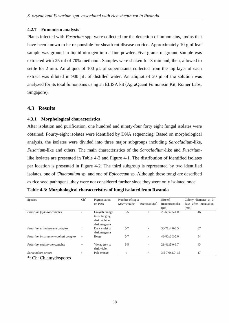
S. oryzae and Fusarium spp. associated with rice sheath rot in Rwanda
58
4.2.7 Fumonisin analysis
Plants infected with Fusarium spp. were collected for the detection of fumonisins, toxins that
have been known to be responsible for sheath rot disease on rice. Approximately 10 g of leaf
sample was ground in liquid nitrogen into a fine powder. Five grams of ground sample was
extracted with 25 ml of 70% methanol. Samples were shaken for 3 min and, then, allowed to
settle for 2 min. An aliquot of 100 µL of supernatants collected from the top layer of each
extract was diluted in 900 µL of distilled water. An aliquot of 50 μl of the solution was
analyzed for its total fumonisins using an ELISA kit (AgraQuant Fumonisin Kit; Romer Labs,
Singapore).
4.3 Results
4.3.1 Morphological characteristics
After isolation and purification, one hundred and ninety-four forty eight fungal isolates were
obtained. Fourty-eight isolates were identified by DNA sequencing. Based on morphological
analysis, the isolates were divided into three major subgroups including Sarocladium-like,
Fusarium-like and others. The main characteristics of the Sarocladium-like and Fusarium-
like isolates are presented in Table 4-3 and Figure 4-1. The distribution of identified isolates
per location is presented in Figure 4-2. The third subgroup is represented by two identified
isolates, one of Chaetomium sp. and one of Epicoccum sp. Although these fungi are described
as rice seed pathogens, they were not considered further since they were only isolated once.
Table 4-3: Morphological characteristics of fungi isolated from Rwanda
Species Ch* Pigmentation
on PDA
Number of septa Size of
(macro)conidia
(µm)
Colony diameter at 3
days after inoculation
(mm)
Macroconidia Microconidia*
Fusarium fujikuroi complex - Grayish orange
to violet grey,
dark violet or
dark magenta
3-5 + 25-60x2.5-4.0 46
Fusarium graminearum complex + Dark violet or
dark magenta
5-7 - 38-71x4.0-6.5 67
Fusarium incarnatum-equiseti complex + Beige
5-7 - 42-80x3.2-5.6 54
Fusarium oxysporum complex + Violet grey to
dark violet
3-5 - 21-41x5.0-6.7 43
Sarocladium oryzae / Pale orange / / 3.5-7.0x1.0-1.5 17
*: Ch: Chlamydospores
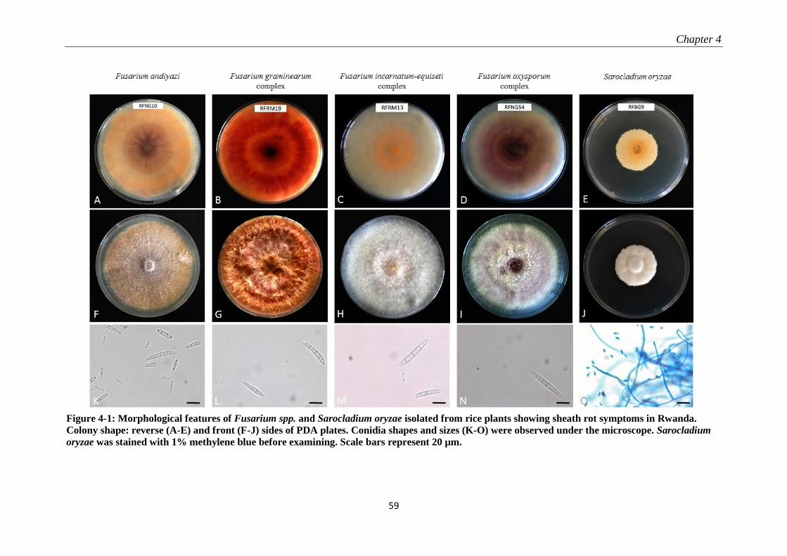
Chapter 4
59
Figure 4-1: Morphological features of Fusarium spp. and Sarocladium oryzae isolated from rice plants showing sheath rot symptoms in Rwanda.
Colony shape: reverse (A-E) and front (F-J) sides of PDA plates. Conidia shapes and sizes (K-O) were observed under the microscope. Sarocladium
oryzae was stained with 1% methylene blue before examining. Scale bars represent 20 µm.
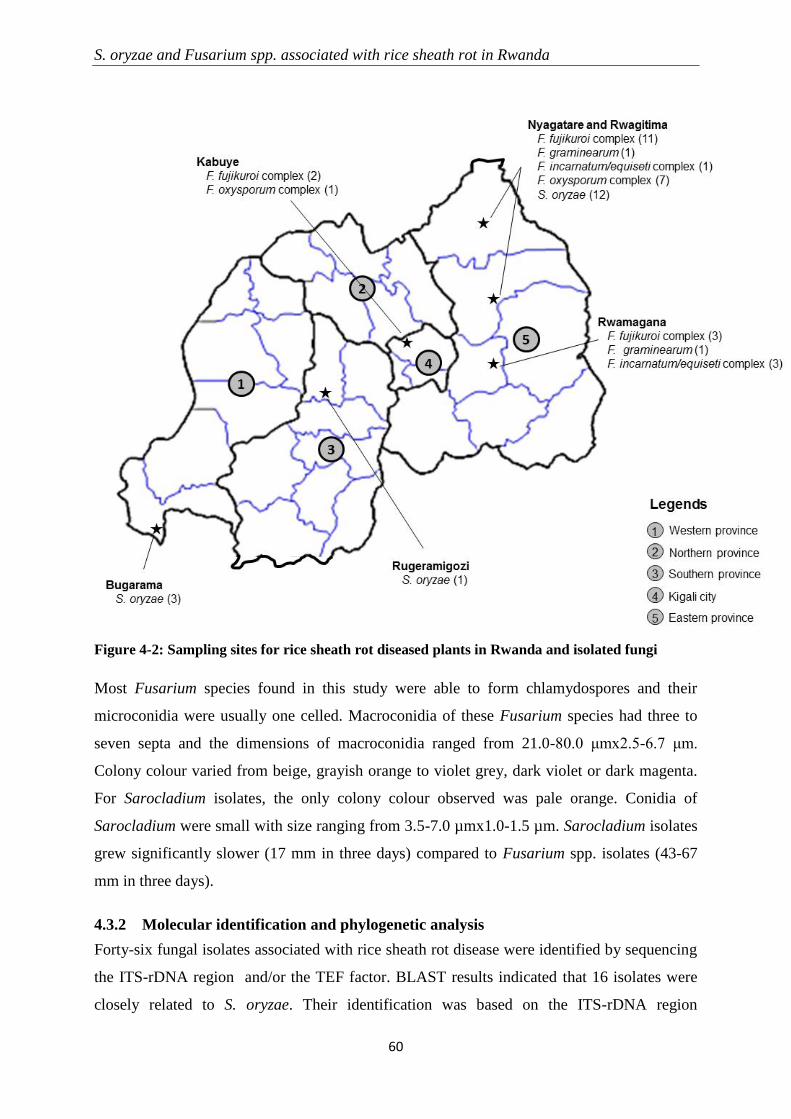
S. oryzae and Fusarium spp. associated with rice sheath rot in Rwanda
60
Figure 4-2: Sampling sites for rice sheath rot diseased plants in Rwanda and isolated fungi
Most Fusarium species found in this study were able to form chlamydospores and their
microconidia were usually one celled. Macroconidia of these Fusarium species had three to
seven septa and the dimensions of macroconidia ranged from 21.0-80.0 μmx2.5-6.7 μm.
Colony colour varied from beige, grayish orange to violet grey, dark violet or dark magenta.
For Sarocladium isolates, the only colony colour observed was pale orange. Conidia of
Sarocladium were small with size ranging from 3.5-7.0 µmx1.0-1.5 µm. Sarocladium isolates
grew significantly slower (17 mm in three days) compared to Fusarium spp. isolates (43-67
mm in three days).
4.3.2 Molecular identification and phylogenetic analysis
Forty-six fungal isolates associated with rice sheath rot disease were identified by sequencing
the ITS-rDNA region and/or the TEF factor. BLAST results indicated that 16 isolates were
closely related to S. oryzae. Their identification was based on the ITS-rDNA region
Chapter 4
61
sequencing. These isolates were obtained from Bugarama, Nyagatare and Rwagitima, and
Rugeramigozi. The remaining 30 isolates were assigned to different species of the Fusarium
genus. Most of them were identified by sequencing the TEF factor though both TEF factor
and ITS-rDNA region were sequenced for some of them. The majority of the Fusarium
isolates belonged to the F. fujikuroi complex (50% of Fusarium isolates) and originated from
Kabuye, Nyagatare and Rwagitima, and Rwamagana. The second most dominant group was
the F. oxysporum complex (30%) found in Kabuye and Nyagatare and Rwagitima, followed
by the F. incarnatum/equiseti complex (13.3%), and the F. graminearum complex (6.7%)
(Figure 4-2 and Table 4-4). These last two complexes were found in Nyagatare and
Rwagitima, and Rwamagana. For more precise identification, the TEF gene of several
Fusarium isolates was also sequenced.
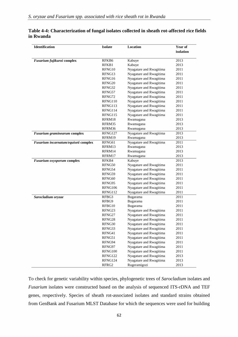
S. oryzae and Fusarium spp. associated with rice sheath rot in Rwanda
62
Table 4-4: Characterization of fungal isolates collected in sheath rot-affected rice fields
in Rwanda
Identification Isolate Location Year of
isolation
Fusarium fujikuroi complex RFKB6 Kabuye 2013
RFKB1 Kabuye 2013
RFNG10 Nyagatare and Rwagitima 2011
RFNG13 Nyagatare and Rwagitima 2011
RFNG16 Nyagatare and Rwagitima 2011
RFNG20 Nyagatare and Rwagitima 2011
RFNG32 Nyagatare and Rwagitima 2011
RFNG57 Nyagatare and Rwagitima 2011
RFNG72 Nyagatare and Rwagitima 2011
RFNG110 Nyagatare and Rwagitima 2011
RFNG113 Nyagatare and Rwagitima 2011
RFNG114 Nyagatare and Rwagitima 2011
RFNG115 Nyagatare and Rwagitima 2011
RFRM18 Rwamagana 2013
RFRM35 Rwamagana 2013
RFRM36 Rwamagana 2013
Fusarium graminearum complex RFNG127 Nyagatare and Rwagitima 2013
RFRM19 Rwamagana 2013
Fusarium incarnatum/equiseti complex RFNG61 Nyagatare and Rwagitima 2011
RFRM13 Rwamagana 2013
RFRM14 Rwamagana 2013
RFRM17 Rwamagana 2013
Fusarium oxysporum complex RFKB4 Kabuye 2013
RFNG50 Nyagatare and Rwagitima 2011
RFNG54 Nyagatare and Rwagitima 2011
RFNG59 Nyagatare and Rwagitima 2011
RFNG60 Nyagatare and Rwagitima 2011
RFNG95 Nyagatare and Rwagitima 2011
RFNG106 Nyagatare and Rwagitima 2011
RFNG112 Nyagatare and Rwagitima 2011
Sarocladium oryzae RFBG3 Bugarama 2011
RFBG9 Bugarama 2011
RFBG10 Bugarama 2011
RFNG23 Nyagatare and Rwagitima 2011
RFNG27 Nyagatare and Rwagitima 2011
RFNG28 Nyagatare and Rwagitima 2011
RFNG30 Nyagatare and Rwagitima 2011
RFNG33 Nyagatare and Rwagitima 2011
RFNG41 Nyagatare and Rwagitima 2011
RFNG51 Nyagatare and Rwagitima 2011
RFNG94 Nyagatare and Rwagitima 2011
RFNG97 Nyagatare and Rwagitima 2011
RFNG100 Nyagatare and Rwagitima 2011
RFNG122 Nyagatare and Rwagitima 2013
RFNG124 Nyagatare and Rwagitima 2013
RFRG2 Rugeramigozi 2013
To check for genetic variability within species, phylogenetic trees of Sarocladium isolates and
Fusarium isolates were constructed based on the analysis of sequenced ITS-rDNA and TEF
genes, respectively. Species of sheath rot-associated isolates and standard strains obtained
from GenBank and Fusarium MLST Database for which the sequences were used for building
Chapter 4
63
phylogenetic trees are presented in Table 4-2. Only one S. oryzae isolate, RFRG2, clustered
with CBS strain 180.74 from India, which is the epitype of S. oryzae, and strains from Kenya
(CBS 361.75), Panama (CBS 120.817) and Australia (WA13481). All other S. oryzae isolates
clustered with CBS strain 399.73 obtained from India (Figure 4-3).
Sarocladium oryzae strain CBS 414.81 from Nigeria seems more distantly related to the other
S. oryzae isolates. It should be noted that strains CBS 399.73 and CBS 414.81 were originally
classified as S. attenuatum, a species that is considered to be conspecific with S. oryzae (Bills
et al., 2004; Giraldo et al., 2015). The ITS sequence of strain CBS 414.81 is clearly different
from strain CBS 399.73 (Result of the comparative blasting in NCBI are: Identity: 98%;
Query coverage: 100%).

S. oryzae and Fusarium spp. associated with rice sheath rot in Rwanda
64
Figure 4-3: Phylogenetic tree for Sarocladium-related sequences identified by the ITS region.
Sequences were aligned using MUSCLE and the tree was generated by the maximum-likelihood
algorithm. The numbers above branches represent bootstrap percentiles from 1,000 replicates.
See Table 4-2 for a full list of Sarocladium reference isolates used to build the tree.
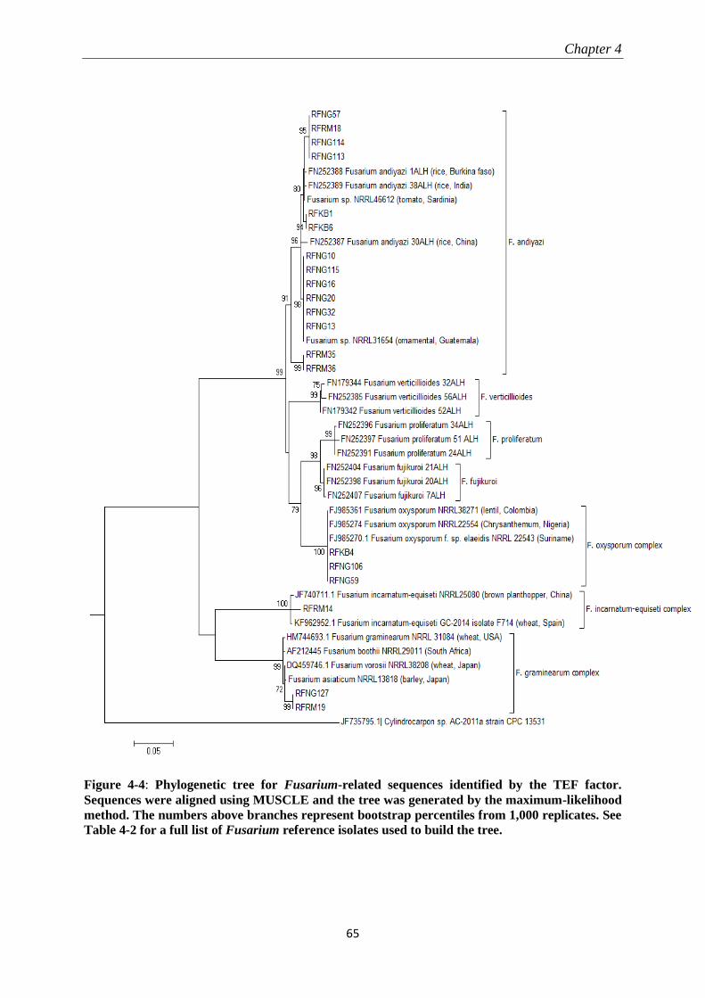
Chapter 4
65
Figure 4-4: Phylogenetic tree for Fusarium-related sequences identified by the TEF factor.
Sequences were aligned using MUSCLE and the tree was generated by the maximum-likelihood
method. The numbers above branches represent bootstrap percentiles from 1,000 replicates. See
Table 4-2 for a full list of Fusarium reference isolates used to build the tree.

S. oryzae and Fusarium spp. associated with rice sheath rot in Rwanda
66
In contrast to the homogeneity observed among Sarocladium isolates from Rwanda, isolates
of the Fusarium genus showed more variability (Figure 4-4).
The 14 sampled isolates belonging to the F. fujikuroi complex clustered with F. andiyazi
isolates from rice from India, Burkina Faso and China. Isolates RFNG127 and RFRM19
grouped with isolates belonging to the F. graminearum complex and were most closely
related to a F. asiaticum isolate from barley in Japan (NRRL 13818). The F. oxysporum
isolates sampled in Rwanda were highly similar and grouped together with F. oxysporum
isolates from Nigeria and Colombia. The TEF sequence of isolate RFRM14 showed the
highest sequence similarity to F. incarnatum/equiseti isolates obtained from wheat in Spain
(isolate F714) and from a brown planthopper in China (NRRL 25080).
4.3.3 Pathogenicity results
In planta pathogenicity tests showed that S. oryzae and all Fusarium isolates tested are able to
induce sheath rot symptoms on rice (Table 4-5). Interestingly, inoculation of Fusarium
isolates and S. oryzae isolates resulted in a similar pattern of infection and symptomatology
on two different rice cultivars: Nipponbare (Japonica) (Figure 4-5a) and CO39 (Indica)
(Figure 4-5b).
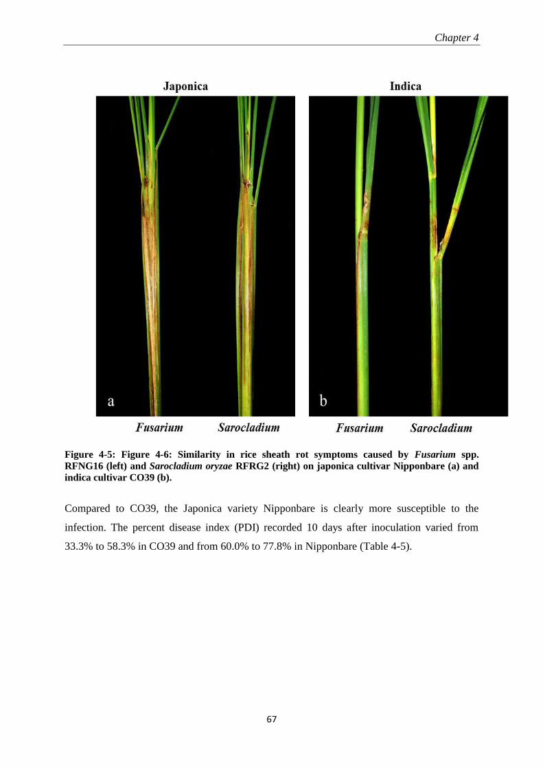
Chapter 4
67
Figure 4-5: Figure 4-6: Similarity in rice sheath rot symptoms caused by Fusarium spp.
RFNG16 (left) and Sarocladium oryzae RFRG2 (right) on japonica cultivar Nipponbare (a) and
indica cultivar CO39 (b).
Compared to CO39, the Japonica variety Nipponbare is clearly more susceptible to the
infection. The percent disease index (PDI) recorded 10 days after inoculation varied from
33.3% to 58.3% in CO39 and from 60.0% to 77.8% in Nipponbare (Table 4-5).

S. oryzae and Fusarium spp. associated with rice sheath rot in Rwanda
68
Table 4-5: Determination of pathogenicity and in planta fumonisin production of fungal
isolates collected in Rwanda
Identification Isolate Rice cultivar and characteristics
Nipponbare CO39
PDI* Fumonisin
concentration** (mg/kg)
PDI Fumonisin
concentration (mg/kg)
Control 0.0 a 0.000 0.0 a 0.000
Fusarium fujikuroi species
complex
RFNG13 66.7 b 0.000 42.6 c 0.000
RFNG16 69.7 bc 0.206 37.0 b 0.117
RFNG113 63.0 b 0.384 37.0 b 0.282
RFNG114 60.5 b 0.576 40.7 c 0.509
RFNG115 77.8 c 0.536 35.2 b 0.084
Fusarium graminearum species
complex
RFRM19 77.8 c 0.000 40.7 c 0.000
Fusarium incarnatum/equiseti
species complex
RFNG61 60.0 b 0.000 35.2 b 0.000
Fusarium oxysporum species
complex
RFKB4 66.7 b 0.941 46.3 c 0.547
RFNG59 63.6 b 0.576 41.4 c 0.293
Sarocladium oryzae RFBG3 72.2 c - 44.4 c -
RFNG41 72.2 c - 58.3 c -
RFRG2 68.5 bc - 33.3 b -
*: Treatments followed by the same letter are not significantly different (P: 0.05)
**: Conversion from ppm to SI (mg/kg) following R-Biopharm (2015)
No association was found between Fusarium species and aggressiveness since little variation
in pathogenic potential was observed in isolates among and within the species tested. The PDI
observed on Nipponbare plants ranged from 60.5% to 77.8% for isolates of F. fujikuroi
complex, from 63.6% to 66.7% for isolates of F. oxysporum complex, was 77.8% for the
isolate of the F. graminearum complex, and was 60.0% for the isolate from the F.
incarnatum/equiseti complex.
The levels of disease severity induced by S. oryzae on Nipponbare plants were comparable to
those of Fusarium spp. with a PDI ranging from 68.5 to 72.2%.
4.3.4 Fumonisin production
Fumonisins could be detected in Nipponbare and CO39 rice plants inoculated with Fusarium
isolates from the fujikuroi complex and the oxysporum complex. No fumonisins could be
detected in rice plants inoculated with isolates belonging to the graminearum or
incarnatum/equiseti complex. Fumonisin production was not correlated with disease severity
since non-fumonisin producers were equally aggressive on rice plants as fumonisin producers.
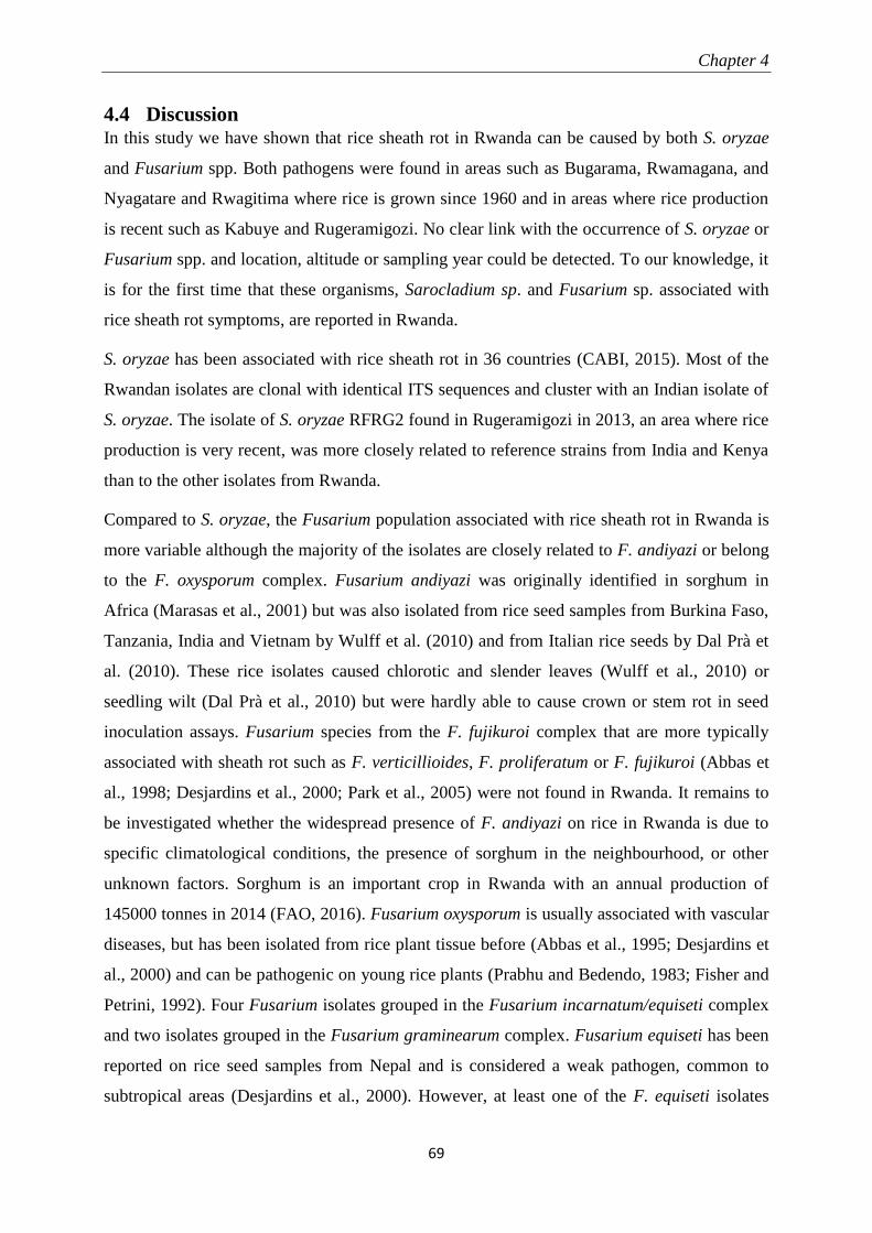
Chapter 4
69
4.4 Discussion In this study we have shown that rice sheath rot in Rwanda can be caused by both S. oryzae
and Fusarium spp. Both pathogens were found in areas such as Bugarama, Rwamagana, and
Nyagatare and Rwagitima where rice is grown since 1960 and in areas where rice production
is recent such as Kabuye and Rugeramigozi. No clear link with the occurrence of S. oryzae or
Fusarium spp. and location, altitude or sampling year could be detected. To our knowledge, it
is for the first time that these organisms, Sarocladium sp. and Fusarium sp. associated with
rice sheath rot symptoms, are reported in Rwanda.
S. oryzae has been associated with rice sheath rot in 36 countries (CABI, 2015). Most of the
Rwandan isolates are clonal with identical ITS sequences and cluster with an Indian isolate of
S. oryzae. The isolate of S. oryzae RFRG2 found in Rugeramigozi in 2013, an area where rice
production is very recent, was more closely related to reference strains from India and Kenya
than to the other isolates from Rwanda.
Compared to S. oryzae, the Fusarium population associated with rice sheath rot in Rwanda is
more variable although the majority of the isolates are closely related to F. andiyazi or belong
to the F. oxysporum complex. Fusarium andiyazi was originally identified in sorghum in
Africa (Marasas et al., 2001) but was also isolated from rice seed samples from Burkina Faso,
Tanzania, India and Vietnam by Wulff et al. (2010) and from Italian rice seeds by Dal Prà et
al. (2010). These rice isolates caused chlorotic and slender leaves (Wulff et al., 2010) or
seedling wilt (Dal Prà et al., 2010) but were hardly able to cause crown or stem rot in seed
inoculation assays. Fusarium species from the F. fujikuroi complex that are more typically
associated with sheath rot such as F. verticillioides, F. proliferatum or F. fujikuroi (Abbas et
al., 1998; Desjardins et al., 2000; Park et al., 2005) were not found in Rwanda. It remains to
be investigated whether the widespread presence of F. andiyazi on rice in Rwanda is due to
specific climatological conditions, the presence of sorghum in the neighbourhood, or other
unknown factors. Sorghum is an important crop in Rwanda with an annual production of
145000 tonnes in 2014 (FAO, 2016). Fusarium oxysporum is usually associated with vascular
diseases, but has been isolated from rice plant tissue before (Abbas et al., 1995; Desjardins et
al., 2000) and can be pathogenic on young rice plants (Prabhu and Bedendo, 1983; Fisher and
Petrini, 1992). Four Fusarium isolates grouped in the Fusarium incarnatum/equiseti complex
and two isolates grouped in the Fusarium graminearum complex. Fusarium equiseti has been
reported on rice seed samples from Nepal and is considered a weak pathogen, common to
subtropical areas (Desjardins et al., 2000). However, at least one of the F. equiseti isolates
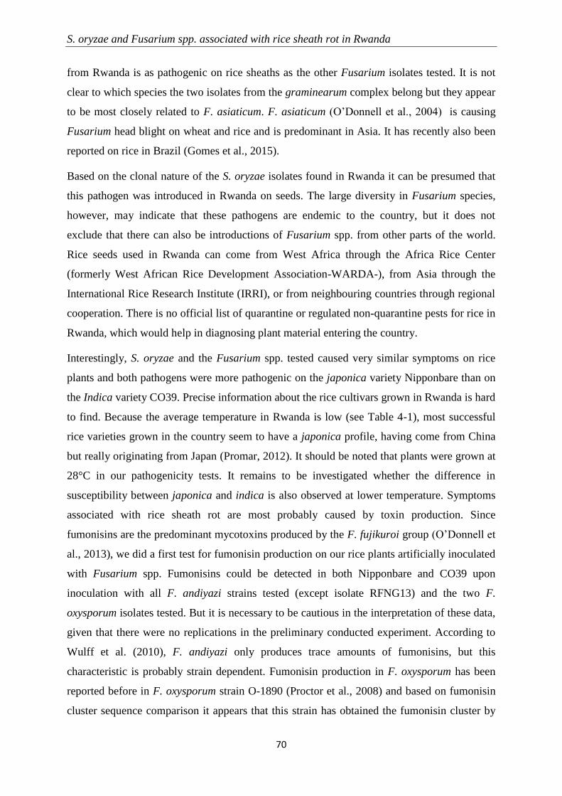
S. oryzae and Fusarium spp. associated with rice sheath rot in Rwanda
70
from Rwanda is as pathogenic on rice sheaths as the other Fusarium isolates tested. It is not
clear to which species the two isolates from the graminearum complex belong but they appear
to be most closely related to F. asiaticum. F. asiaticum (O’Donnell et al., 2004) is causing
Fusarium head blight on wheat and rice and is predominant in Asia. It has recently also been
reported on rice in Brazil (Gomes et al., 2015).
Based on the clonal nature of the S. oryzae isolates found in Rwanda it can be presumed that
this pathogen was introduced in Rwanda on seeds. The large diversity in Fusarium species,
however, may indicate that these pathogens are endemic to the country, but it does not
exclude that there can also be introductions of Fusarium spp. from other parts of the world.
Rice seeds used in Rwanda can come from West Africa through the Africa Rice Center
(formerly West African Rice Development Association-WARDA-), from Asia through the
International Rice Research Institute (IRRI), or from neighbouring countries through regional
cooperation. There is no official list of quarantine or regulated non-quarantine pests for rice in
Rwanda, which would help in diagnosing plant material entering the country.
Interestingly, S. oryzae and the Fusarium spp. tested caused very similar symptoms on rice
plants and both pathogens were more pathogenic on the japonica variety Nipponbare than on
the Indica variety CO39. Precise information about the rice cultivars grown in Rwanda is hard
to find. Because the average temperature in Rwanda is low (see Table 4-1), most successful
rice varieties grown in the country seem to have a japonica profile, having come from China
but really originating from Japan (Promar, 2012). It should be noted that plants were grown at
28°C in our pathogenicity tests. It remains to be investigated whether the difference in
susceptibility between japonica and indica is also observed at lower temperature. Symptoms
associated with rice sheath rot are most probably caused by toxin production. Since
fumonisins are the predominant mycotoxins produced by the F. fujikuroi group (O’Donnell et
al., 2013), we did a first test for fumonisin production on our rice plants artificially inoculated
with Fusarium spp. Fumonisins could be detected in both Nipponbare and CO39 upon
inoculation with all F. andiyazi strains tested (except isolate RFNG13) and the two F.
oxysporum isolates tested. But it is necessary to be cautious in the interpretation of these data,
given that there were no replications in the preliminary conducted experiment. According to
Wulff et al. (2010), F. andiyazi only produces trace amounts of fumonisins, but this
characteristic is probably strain dependent. Fumonisin production in F. oxysporum has been
reported before in F. oxysporum strain O-1890 (Proctor et al., 2008) and based on fumonisin
cluster sequence comparison it appears that this strain has obtained the fumonisin cluster by
Chapter 4
71
horizontal gene transfer from Fusarium bulbicola or a closely related strain. It would be
interesting to have a closer look at the fumonisin gene cluster in the F. oxysporum strains
from Rwanda to see whether they are similar to the one described for F. oxysporum strain O-
1980. Fumonisin production, however, does not explain the symptoms observed upon
inoculation with Fusarium spp. since non-fumonisin producers such as F. andiyazi isolate
RFNG13 and the isolates belonging to the graminearum and incarnatum/equiseti complex
were as pathogenic as fumonisin producers. A more extensive analysis of mycotoxins
produced in vitro and in planta by the various Fusarium strains from Rwanda is needed and
will be the subject of future studies. It should be noted that members of the F. fujikuroi
complex are also known to produce moniliformin. Moniliformin is phytotoxic and it has been
shown that F. proliferatum isolates from field samples of rice with sheath rot symptoms are
capable of producing both fumonisins and moniliformin in culture. Both mycotoxins were
also detected in naturally contaminated rice samples (Abbas et al., 1999). Recently
moniliformin was detected on rice samples from Nigeria (Rofiat et al., 2015). Fusarium
oxysporum and F. equiseti are also known to produce moniliformin (Desjardins, 2006). Since
moniliformin has not been directly implicated in any outbreak of toxicosis in animals and
humans and detection in biological matrices is not easy (Desjardins, 2006), this toxin is not as
widely studied as other Fusarium mycotoxins.
More information is needed to understand the epidemiology of S. oryzae and the Fusarium
spp. causing sheath rot on in Rwanda. We do not know whether these organisms can co-occur
in the same lesion or on the same plant and how they interact with each other or with the
plant. Moreover, the influence of cold stress on the occurrence of sheath rot should be studied.
Any strategy to control sheath rot in Rwanda should take into account that this disease can be
caused by various pathogens.
Acknowledgements
V.P. Bigirimana received a doctoral grant from the Belgian Technical Cooperation (BTC)
(project reference: 10RWA/0018). This work was supported by a grant from the Special
Research Fund of Ghent University (GOA 01GB3013). We thank prof. Kris Audenaert for his
help with the fumonisin analysis.

Chapter 5
73
5 PSEUDOMONAS ISOLATES ASSOCIATED WITH RICE SHEATH
ROT DISEASE COMPLEX IN RWANDA AND THE PHILIPPINES
Vincent de Paul Bigirimana, Gia Khuong Hoang Hua, Lien Bertier, Bart Cottyn, Obedi
Ishibwela Nyamangyoku and Monica Höfte
Abstract
Rice is an important food crop worldwide in tropical and subtropical areas. Rice plants are
attacked by many pests and diseases and some of them cause sheath rot symptoms. Less is
known about the causal agents of rice sheath rot. In this study, aiming to know more about
sheath rot associated bacterial organisms, diseased plants were collected and bacteria were
isolated from plant tissues exhibiting sheath rot symptoms. Samples were collected for
analysis from the major rice-growing areas of Rwanda. Previously collected isolates from the
Philippines were also studied. Bacterial isolates were characterized by morphological,
biochemical and molecular features. The molecular diagnosis was based on the sequencing of
16S rDNA, rpoB and genes. An initial quick screening based on 16S rDNA revealed the
presence of three dominant genera: Stenotrophomonas, Delftia and Pseudomonas. The
distribution of the major isolates was relatively the same in all the sampled areas. More efforts
were put in the characterization of samples of Pseudomonas genus, and they were put in 4
different subgroups defined after molecular identification, depending on the closely related
Pseudomonas species: P. chlororaphis, P. fluorescens, P. asplenii/fuscovaginae and P.
putida. Within subgroups, subsets were created according to the similarities among the
isolates. There is not a big difference between isolates from Rwanda and those from the
Philippines, except that isolates related to P. saponiphila are the most present in Rwanda
while they are absent in the Philippines. Also, isolates closely related to two pathogenic
isolates P. palleroniana (P. fluorescens subgroup) and P. fuscovaginae (P.
asplenii/fuscovaginae subgroup) were found in the Philippines but not in Rwanda. A subset
was tested for pathogenicity on rice plants. Isolates appeared mildly pathogenic, compared to
reference related pathogenic strains. The role of identified pseudomonad isolates, as most of
them qualify as plant-inhabiting endophytes and/or potential biological control agents, while
some others qualify as isolates with low pathogenic potential, is tentatively explained, but still
has to be investigated. Considering the complexity of the rice sheath rot disease causing
factors, developing an integrated pest management strategy is likely to be the most effective
Pseudomonas isolates associated with rice sheath rot disease
74
control option especially as the lifestyle and pathogenic status of identified organisms may
have links with the rice plant, its growing environment and other organisms harbored by the
plant.
5.1 Introduction Rice production is threatened by some constraints of which the susceptibility to attacks by
pests and diseases is important. Rice sheath rot is one of the diseases attacking rice. Rice
sheath rot is characterized by the following symptoms (Zeigler and Alvarez, 1987): a brown
stripe on the sheath that can extend along the midrib of the leaf lamina for nearly its entire
length, failure of the panicles to emerge properly from the boot when the flag leaf sheath is
severely affected, panicles from diseased plants produce discolored and poorly filled grains.
The disease can cause significant yield losses, with the possibility of also causing seed
discoloration (thus reducing its value), degeneration and even sterility. Some bacteria are
known as rice sheath rot causal agents, like Pseudomonas fuscovagine (Zeigler and Alvarez,
1987, 1990; Jaunet et al., 1996; CABI, 2007), Burkholderia glumae (Fory et al., 2014) and
Acidovorax oryzae (Cottyn et al., 1996b; Schaad et al., 2008). A review on rice sheath rot
associated organisms is presented in Chapter 2 and Bigirimana et al. (2015).
In addition to Pseudomonas fuscovaginae, all the studies on rice sheath rot find many other
Pseudomonas isolates associated with rice sheath rot and grain discoloration symptoms
(Zeigler and Alvarez, 1987, 1990; Cottyn et al., 1996a; b; Gardan et al., 2002; Cother et al.,
2010), which have not been, until now, completely identified by molecular methods, except
for P. palleroniana (Gardan et al., 2002). Those pseudomonads have been identified by
biochemical/physiological tests.
Cottyn et al. (1996a; b) worked on rice bacterial diseases in the Philippines and conducted a
survey to determine which bacterial pathogens were associated with sheath rot and grain
discoloration symptoms. Samples were collected from 16 provinces and the disease symptoms
were found in samples from 12 provinces. Suspected pathogenic bacteria were tested on
seedling sheaths and produced symptoms characteristic of infections caused by Burkholderia
glumae and Pseudomonas fuscovaginae, which are not distinctive according to the causal
bacteria. A connection with the isolates from these works has been established by introducing
in the current study isolates from the Philippines, taking advantage of the current advances in
the molecular identification tools, methods that were not available when the study was first
conducted.
Chapter 5
75
Rice is one of the priority crops in Rwanda, a country for which agriculture constitutes the
backbone of the economy, given that around 80% of the population depend on it for their
livelihoods (MINAGRI, 2011b). In this study, we want to see whether P. fuscovaginae is
present in Rwanda and isolate any other bacteria associated with rice sheath rot symptoms.
The isolated organisms are identified and characterized for pathogenicity. Samples were
collected from the major rice-growing areas of Rwanda.
The goal of this study is to generate information that can improve the understanding of the
rice sheath rot disease, an emerging rice disease and its causal bacterial agents, especially the
less studies pseudomonads, so that a sound management strategy against it can be developed.
5.2 Materials and methods
5.2.1 Research area
Isolation of bacteria associated with sheath rot symptoms on rice has been realized on samples
collected from valleys in the major rice-growing areas of Rwanda. The valleys where samples
were taken were the following: Bugarama (Bugarama Sector, Western Province), Cyili
(Rusatira Sector, Southern Province), Rwamagana (Kigabiro Sector, Eastern Province),
Nyagatare and Rwagitima (Nyagatare and Rugarama Sectors, Eastern Province) and
Mukunguri (Mugina Sector, Southern Province) (Figure 1-1). Samples were collected in July-
August 2011 and 2012, while rice plants were at fruit development according to the growth-
stages description (Lancashire et al., 1991).
5.2.2 Sampling methods
A representative sampling of sheath rot diseased rice plants in Rwanda was followed so that
the samples taken represent the situation on the whole rice-growing marshland. Samples were
taken from plants showing sheath rot symptoms following all the cardinal directions. In each
plot or direction, at least three diseased plant samples were taken and labeled. Field samples
were preserved at 4 °C before analysis in the laboratory.
5.2.3 Samples preparation for isolation
Sample preparation consisted of 8 steps: (1) Localizing the tissue area where the disease
symptoms are well expressed and cutting it from the remaining parts of the plant; (2)
Separating the tissues where the disease started and which are at an advanced degradation
stage, which are discarded, from the freshly infected tissues, which are retained for the further
isolation work, proceeding by dividing that chosen part into small parts of approximately 5
mm of length; (3) Doing surface sterilization by dipping samples of diseased plants tissues in
Pseudomonas isolates associated with rice sheath rot disease
76
ethanol 70% for 2 minutes; (4) Cleaning the sterilized diseased plant parts 4 times in sterile
distilled water; (5) Measuring 1 ml of the physiological solution 0.85% and pouring it in a
sterile mortar where also a small quantity of autoclaved small grains of sand are poured;
grinding diseased plant samples with a sterile pestle in the physiological solution in the
mortar; (6) Gradual dilution (10-1
, 10-2
, 10-3
) of the liquid from the diseased plants by adding
20 µl of it to 180 µl of the physiological solution; (7) Taking 50 µl of the diluted sample,
spreading them on King’s B agar medium (KB), tightly closing the Petri dish using parafilm
paper and incubating at 28°C; (8) The growth of colonies was evaluated after 48 hours and
individual, single and pure colonies constituting isolates were obtained after continuous
subcultures. For long term preservation, isolates were maintained in cryotubes in a mixture of
50% of Luria-Bertani (LB) liquid medium and 50% of glycerol 40% at -80°C.
5.2.4 Morphological and biochemical characterization of samples
The bacterial colonies that developed on culture media were first characterized using
morphological and biochemical features. The tests were performed on the isolates following
the principle that a small number of tests can help predicting the genera of major plant
pathogenic bacteria (Schaad et al., 2001) (Table 5-1). The tests conducted are the following:
colour observation, Gram coloration, development speed based on the size acquired after 48
hours, observation of the colour of the colonies on YDC (Yeast extract-dextrose-CaCO3) or
NBY (Nutrient-Broth yeast extractagar) media, appearance of mucoid colonies at 30°C on
YDC medium, fluorescence of pigments on KB medium, diffusible non fluorescent pigments
on KB, and growth at 40°C. These biochemical tests were performed searching for three
bacterial genera, Pseudomonas, Burkholderia and Acidovorax recognized as sheath rot
pathogens (Cottyn et al., 1996a; b; Cother et al., 2010).
Though there are limited exceptions, four tests are the most important for predicting the
genera (Table 5-1): Pseudomonas genus can be predicted by the emission of fluorescent
pigments on KB medium, Acidovorax genus can be predicted by mucoid colonies on YDC at
30°C and growth at 40°C, and Burkholderia genus can be predicted by the growth at 40°C
and the emission of diffusible non-fluorescent pigments on KB.
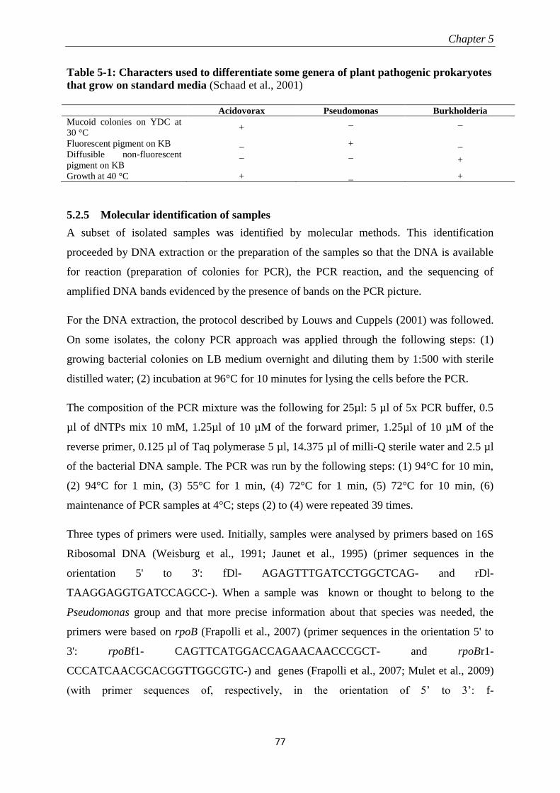
Chapter 5
77
Table 5-1: Characters used to differentiate some genera of plant pathogenic prokaryotes
that grow on standard media (Schaad et al., 2001)
Acidovorax Pseudomonas Burkholderia
Mucoid colonies on YDC at
30 °C +
_ _
Fluorescent pigment on KB _ + _
Diffusible non-fluorescent
pigment on KB
_ _ +
Growth at 40 °C + _ +
5.2.5 Molecular identification of samples
A subset of isolated samples was identified by molecular methods. This identification
proceeded by DNA extraction or the preparation of the samples so that the DNA is available
for reaction (preparation of colonies for PCR), the PCR reaction, and the sequencing of
amplified DNA bands evidenced by the presence of bands on the PCR picture.
For the DNA extraction, the protocol described by Louws and Cuppels (2001) was followed.
On some isolates, the colony PCR approach was applied through the following steps: (1)
growing bacterial colonies on LB medium overnight and diluting them by 1:500 with sterile
distilled water; (2) incubation at 96°C for 10 minutes for lysing the cells before the PCR.
The composition of the PCR mixture was the following for 25µl: 5 µl of 5x PCR buffer, 0.5
µl of dNTPs mix 10 mM, 1.25µl of 10 µM of the forward primer, 1.25µl of 10 µM of the
reverse primer, 0.125 µl of Taq polymerase 5 µl, 14.375 µl of milli-Q sterile water and 2.5 µl
of the bacterial DNA sample. The PCR was run by the following steps: (1) 94°C for 10 min,
(2) 94°C for 1 min, (3) 55°C for 1 min, (4) 72°C for 1 min, (5) 72°C for 10 min, (6)
maintenance of PCR samples at 4°C; steps (2) to (4) were repeated 39 times.
Three types of primers were used. Initially, samples were analysed by primers based on 16S
Ribosomal DNA (Weisburg et al., 1991; Jaunet et al., 1995) (primer sequences in the
orientation 5' to 3': fDl- AGAGTTTGATCCTGGCTCAG- and rDl-
TAAGGAGGTGATCCAGCC-). When a sample was known or thought to belong to the
Pseudomonas group and that more precise information about that species was needed, the
primers were based on rpoB (Frapolli et al., 2007) (primer sequences in the orientation 5' to
3': rpoBf1- CAGTTCATGGACCAGAACAACCCGCT- and rpoBr1-
CCCATCAACGCACGGTTGGCGTC-) and genes (Frapolli et al., 2007; Mulet et al., 2009)
(with primer sequences of, respectively, in the orientation of 5’ to 3’: f-
Pseudomonas isolates associated with rice sheath rot disease
78
ACTTCCCTGGCACGGTTGACCA- and r-TCGACATGCGACGGTTGATGTC-, PsEG30F-
ATYGAAATCGCCAARCG- and PsEG790R- CGGTTGATKTCCTTGA-).
The amplicons were separated by electrophoresis on a 1% agarose gel stained with ethidium
bromide. PCR products were purified as instructed in the QIAquick PCR Purification Kit
(Qiagen, Helden, Germany) or by the Exo-SAP-IT (Affymetrix, Santa Clara, California,
USA). The DNA concentration was measured with the Thermo Nanodrop spectrophotometer
(NanoDrop Products, Wilmington, DE, USA) and when it was higher than 20 ng/µl, the
sample was prepared for sequencing, using the same primers as those used in the sequencing.
Sequencing was done by LGC Genomics (Berlin, Germany). Sequences were cleaned with
BioEdit software. They were then aligned by Multiple Sequence Comparison by Log-
Expectation (MUSCLE). Phylogenetic trees were constructed using the maximum likelihood
algorithm with 1000 replicates using MEGA 6 Software (Tamura et al., 2013).
5.2.6 Pathogenicity testing
5.2.6.1 Plant material
The rice Indica cultivar Co39 was used for pathogenicity testing. Plantlets used were 5 weeks
old. Rice plants were grown in potting soil. They were regularly watered with a nutrient
solution composed, per 1 liter, of 2 g of FeSO4.7H2O and 0.9 g of NH4SO4. The plants were
grown in a growth chamber at 28°C, 70% relative humidity and at the day photoperiod of
12/12 hours.
5.2.6.2 The isolates and inoculum preparation
From the collection, bacteria were grown on KB solid medium for 48 hours. Individual
colonies were then grown on LB liquid medium for 24 hours. The medium and the bacteria in
suspension were centrifuged at 13000 rpm for 2 minutes, after which the liquid supernatant
was discarded. The centrifugate was 10 times diluted with 0.85% saline buffer, as indicated
(Schaad et al., 2001), taking reference from the used volume of bacteria in suspension in the
medium. The bacterial suspension was thoroughly mixed with a vortex. Before inoculation,
the bacterial suspension was preserved at 4°C. Inoculation of the bacteria in the plants was
performed by injection (Rott et al., 1989) with a 1 ml syringe (Becton Dickinson, Spain) and a
25GX1” needle (Becton Dickinson, Ireland) pumping 100 µl into the plant. After injection,
inoculated plantlets were kept in a saturated humid chamber at 28°C for 24 hours.
Chapter 5
79
Thirty-four isolates from Rwanda were tested for pathogenicity. The Pseudomonas
fuscovaginae isolate LMG2158 was used as the positive control while sterile distilled water
served as the negative control. Two other isolates, Pseudomonas protegens CHA0 and P.
protegens Pf-5, taxonomically closely related to the isolates from Rwanda, but known as
biological control agents, were also added to the experiment.
5.2.6.3 Experimental design and statistical analysis.
Data for disease development were taken 10 days after inoculation. They were collected
based on a scoring scale with 6 levels, adapted after previous works (Mattiuzzo et al., 2011),
and following a gradual comparison of symptoms, where 0 represents a healthy plant and 5 a
highly diseased plant (Table 5-2). Recorded data were analysed as qualitative data through
non-parametric tests. Disease indexes were calculated for the different isolates. The
attribution of ranks and equality of means for the different treatments were tested in SPSS 22
software (IBM Corporation, 2013) and the values for pairwise comparison were generated in
PAST software Version 2.17C (Hammer et al., 2001). Based on pairwise comparison results,
isolates were assigned to homogenous subsets.
Table 5-2: Severity rating scale for P. fuscovaginae on rice (Mattiuzzo et al., 2011)
Severity value Description
0 No symptoms only the sign of the injection puncture
1 Necrosis around the puncture till 1 cm
2 Necrosis around the puncture and chlorosis from 1 to 3 cm on the stem
3 Necrosis around the puncture till 5 cm on the stem
4 Necrosis around the puncture for the two-thirds of the new leaf
5 Necrosis around the puncture throughout the new leaf
5.3 Results
5.3.1 Sampling and quick initial screening for identification
Diseased plants, showing typical sheath rot symptoms were collected from the chosen
sampling areas. Efforts were put in capturing, as much as possible, samples coming from the
different parts of each rice growing-area, so as to have a representative picture of bacteria
associated with sheath rot diseased rice plants. In total, 368 isolates were obtained and the
number of isolates recovered in 2011 and 2012 per rice growing area is given in Table 5-3.
Pseudomonas isolates associated with rice sheath rot disease
80
Table 5-3: Origin and number of bacterial isolates obtained
Rice- growing area/Year of
collection
2011 2012 Total % per
location
Bugarama (BG) 24 19 43 11.7
Cyili (CY) 57 31 88 23.9
Mukunguri (MK) 0 43 43 11.7
Nyagatare and Rwagitima (NG) 65 48 113 30.7
Rwamagana (RM) 45 33 78 21.2
Unspecified location in Rwanda (XX) 0 3 3 0.8
Total 191 177 368
The isolates were screened initially using the morphological tests described in Table 5-1
searching for Pseudomonas, Burkholderia and Acidovorax genera. Since these tests were not
conclusive it was decided to identify a subset of 53 isolates by sequencing based on 16S
rDNA. Isolate choice was based on morphological and biochemical features. The molecular
identification revealed that no Burkholderia or Acidovorax isolates could be found. The
sequenced isolates belonged to three major genera: Stenotrophomonas, Delftia and
Pseudomonas (Table 5-4) and they were found in all rice-growing areas. In addition, 1 isolate
of Enterobacter and 1 isolate of Bacillus were found. A phylogenetic tree based on the partial
16S rDNA sequences of some of these isolates is presented in Figure 5-1.
Table 5-4: Bacterial genera detected in the first screening for the identification of sheath
rot associated isolates from Rwanda (based on 16SrDNA)
Rice- growing area/Genus Stenotrophomonas Pseudomonas Delftia Total
Bugarama 4 0 1 5
Cyili 6 3 0 9
Mukunguri 8 2 4 14
Nyagatare and Rwagitima 6 6 4 16
Rwamagana 2 3 1 6
Unspecified location in Rwanda 0 1 0 1
Total 26 15 10 51
Chapter 5
81
Figure 5-1: Phylogenetic tree for bacterial sequences identified by 16S rDNA. Sequences were
aligned using MUSCLE and the tree was generated using the maximum likelihood algorithm.
The numbers above branches represent bootstrap percentiles from 1,000 replicates
RBBG28-0
RBRM17-0
RBNG59-0
RBRM43-0
RBNG69-0
RBNG6-0
RBCY24-0
RBMK2-0
RBRM11-0
RBMK4-0
RBRM59-0
RBBG26-0
RBCY54-0
RBMK5-0
RBCY34-0
RBMK12-0
gi|1657407|emb|X95923.1|_Stenotrophomonas_maltophilia_LMG_958-T
RBMK29-0
RBCY70-0
RBMK15-0
RBCY65-0
RBMK37-0
RBCY64-0
RBNG62-0
RBNG14-0
Stenotrophomonas group
gi|584459739|gb|KF857481.1|_Pseudomonas_chlororaphis_strain_HT66
gi|218938052|gb|FJ483519.1|_Pseudomonas_fuscovaginae_strain_ICMP_5940
gi|639127967|gb|KJ806393.1|_Pseudomonas_entomophila_strain_M-X7A
RBXX1-0
gi|636560845|ref|NR_116905.1|_Pseudomonas_saponiphila_DSM_9751
RBNG3-0
RBCY30-0
RBCY35-0
RBNG2-0
ENA|AJ417071|AJ417071.1_Pseudomonas_protegens_strain_PGNR1
gi|526132719|gb|KF278708.1|_Pseudomonas_putida_strain_Pp
Pseudomonas group
Outgroup gi|2584744|emb|Z96091.1|_Erwinia_carotovora_LMG_2466_16S_ribosomal_RNA
RBMK32-0
RBMK10-0
RBMK21-0
RBBG32-0
RBNG74-0
RBNG49-0
RBNG51-0
RBRM33-0
RBMK16-0
gi|636558410|ref|NR_114466.1|_Delftia_acidovorans_strain_ACM_489
gi|313482925|gb|HQ436355.1|_Delftia_tsuruhatensis_strain_IPPBC_R15
gi|359805184|dbj|AB681119.1|_Delftia_tsuruhatensis__strain_NBRC_16741
Delftia group
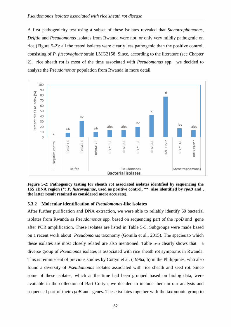
Pseudomonas isolates associated with rice sheath rot disease
82
A first pathogenicity test using a subset of these isolates revealed that Stenotrophomonas,
Delftia and Pseudomonas isolates from Rwanda were not, or only very mildly pathogenic on
rice (Figure 5-2): all the tested isolates were clearly less pathogenic than the positive control,
consisting of P. fuscovaginae strain LMG2158. Since, according to the literature (see Chapter
2), rice sheath rot is most of the time associated with Pseudomonas spp. we decided to
analyze the Pseudomonas population from Rwanda in more detail.
Figure 5-2: Pathogenicy testing for sheath rot associated isolates identified by sequencing the
16S rDNA region (*: P. fuscovaginae, used as positive control, **: also identified by rpoB and ,
the latter result retained as considered more accurate).
5.3.2 Molecular identification of Pseudomonas-like isolates
After further purification and DNA extraction, we were able to reliably identify 69 bacterial
isolates from Rwanda as Pseudomonas spp. based on sequencing part of the rpoB and gene
after PCR amplification. These isolates are listed in Table 5-5. Subgroups were made based
on a recent work about Pseudomonas taxonomy (Gomila et al., 2015). The species to which
these isolates are most closely related are also mentioned. Table 5-5 clearly shows that a
diverse group of Pseumonas isolates is associated with rice sheath rot symptoms in Rwanda.
This is reminiscent of previous studies by Cottyn et al. (1996a; b) in the Philippines, who also
found a diversity of Pseudomonas isolates associated with rice sheath and seed rot. Since
some of these isolates, which at the time had been grouped based on biolog data, were
available in the collection of Bart Cottyn, we decided to include them in our analysis and
sequenced part of their rpoB and genes. These isolates together with the taxomonic group to
a
ab
bc
ab abc abcbc
c
d
bcabc
0
10
20
30
40
50
60
70
80
90
100
Neg
ativ
e co
ntr
ol
RB
NG
51
-0
RB
NG
49
-0
RB
RM
17
-0
RB
CY
35
-0
RB
NG
3-0
RB
CY
30
-0
RB
NG
2-0
LMG
21
58
*
RB
CY
34
-0
RB
CY3
9-0
**
- Delftia Pseudomonas Stenotrophomonas
Per
cen
t d
isea
se in
dex
(%)
Bacterial isolates
Chapter 5
83
which they belong are listed in Table 5-6. We studied the phylogenetic relatedness between
the isolates from Rwanda and the Philippines by generating a phylogenetic tree based on the
concatenated rpoB and sequences. In this tree, also sequences of reference strains (type
strains or published species strains closely related to those found in this study) were
introduced. The reference strains and some of their features are presented in Table 5-7. The
phylogenetic tree is given in Figure 5-3.
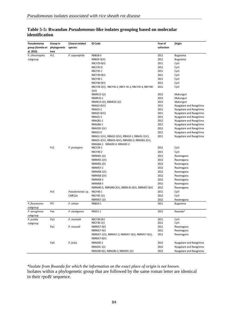
Pseudomonas isolates associated with rice sheath rot disease
84
Table 5-5: Rwandan Pseudomonas-like isolates grouping based on molecular
identification
*Isolate from Rwanda for which the information on the exact place of origin is not known.
Isolates within a phylogenetic group that are followed by the same roman letter are identical
in their rpoB/ sequence.
Pseudomonas
group (Gomila et
al. 2015)
Group in
phylogenetic
tree
Closest related
species
ID Code Year of
collection
Origin
Pc1 P. saponiphila RBBG8-0 2011 Bugarama
RBBG9-3(III) 2011 Bugarama
RBCY29-0(II) 2011 Cyili
RBCY35-0 2011 Cyili
RBCY35-1 2011 Cyili
RBCY39-0(II) 2011 Cyili
RBCY39-1 2011 Cyili
RBCY40-0(II) 2011 Cyili
2011 Cyili
RBMK13-1(I) 2012 Mukunguri
RBMK25-1 2012 Mukunguri
RBMK13-2(I), RBMK25-2(I) 2012 Mukunguri
RBNG2-0(IV) 2011 Nyagatare and Rwagitima
RBNG3-2 2011 Nyagatare and Rwagitima
RBNG5-0(IV) 2011 Nyagatare and Rwagitima
RBNG5-3 2011 Nyagatare and Rwagitima
RBNG81-1 2012 Nyagatare and Rwagitima
RBNG84-1 2012 Nyagatare and Rwagitima
RBNG92-1(V) 2012 Nyagatare and Rwagitima
RBNG3-0 2011 Nyagatare and Rwagitima
RBNG2-1(IV), RBNG2-2(IV), RBNG3-1, RBNG5-1(IV),
RBNG5-2(IV), RBNG5-4(IV), RBNG81-0, RBNG81-2(V),
RBNG84-2, RBNG92-0, RBNG92-2
2011 Nyagatare and Rwagitima
Pc2 P. protegens RBCY29-1 2011 Cyili
RBCY39-2 2011 Cyili
RBRM41-1(I) 2012 Rwamagana
RBRM55-1(II) 2012 Rwamagana
RBRM55-2(I) 2012 Rwamagana
RBRM57-2 2012 Rwamagana
RBRM58-1(II) 2012 Rwamagana
RBRM58-2(II) 2012 Rwamagana
RBRM58-3 2012 Rwamagana
RBRM58-5 2012 Rwamagana
RBRM40-0, RBRM40-2(II), RBRM 41-0(II), RBRM57-0(II) 2012 Rwamagana
Pc3 RBCY40-1 2011 Cyili
RBCY45-1(I) 2012 Cyili
RBRM57-1(I) 2012 Rwamagana
P. fluorescens
subgroup
Pf1 P. simiae RBBG9-1 2011 Bugarama
P. aeruginosa
subgroup
Pae P. alcaligenes RBXX1-1 2012 Rwanda*
Pp3 P. monteilii RBCY30-0(I) 2011 CyiliRBCY30-1(I) 2011 Cyili
Pp1 P. mosselii RBRM17-0(I) 2011 Rwamagana
RBRM17-4(I) 2011 Rwamagana
RBRM17-1(II), RBRM17-2, RBRM17-3(II), RBRM17-5(II),
RBRM17-6(II)
2011 Rwamagana
Pp4 P. fulva RBNG90-1 2012 Nyagatare and Rwagitima
RBNG91-1(I) 2012 Nyagatare and Rwagitima
RBNG90-0(I), RBNG90-2, RBNG91-2(I) 2012 Nyagatare and Rwagitima
P. chlororaphis
subgroup
P. putida
subgroup
RBCY29-2(II), RBCY35-2, RBCY-35-3, RBCY35-4, RBCY40-
2(III)
Pseudomonas sp.
CMR12a
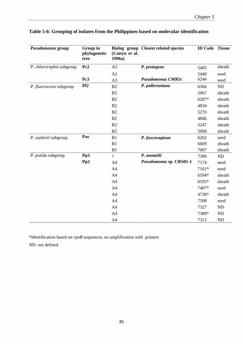
Chapter 5
85
Table 5-6: Grouping of isolates from the Philippines based on molecular identification
Pseudomonas group Group in
phylogenetic
tree
Biolog group
(Cottyn et al.
1996a)
Closest related species ID Code Tissue
P. chlororaphis subgroup Pc2 A2 P. protegens 5405 sheath
A2 5440 seed
Pc3 A3 Pseudomonas CMR5c 6244 seed
P. fluorescens subgroup Pf2 B2 P. palleroniana 6366 ND
B2 5067 sheath
B2 6287* sheath
B2 4834 sheath
B2 5270 sheath
B2 4846 sheath
B2 5247 sheath
B2 5068 sheath
P. asplenii subgroup Pas B1 P. fuscovaginae 6202 seed
B1
6609 sheath
B1 7007 sheath
P. putida subgroup Pp3 ? P. montelii 7390 ND
Pp2 A4 Pseudomonas sp. CRS01-1 7174 seed
A4 7161* seed
A4 6594* sheath
A4 6593* sheath
A4 7407* seed
A4 4736* sheath
A4 7308 seed
A4 7327 ND
A4 7389* ND
A4 7313 ND
*Identification based on rpoB sequences, no amplification with primers
ND: not defined
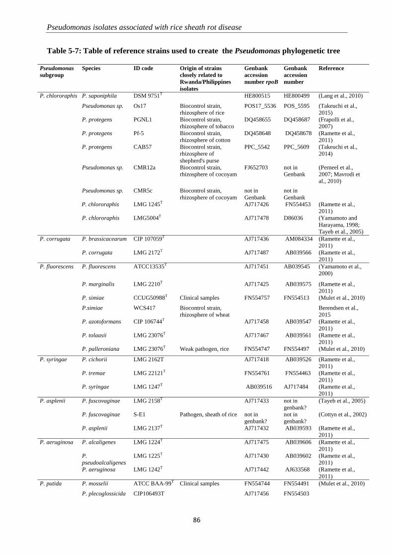
Pseudomonas isolates associated with rice sheath rot disease
86
Table 5-7: Table of reference strains used to create the Pseudomonas phylogenetic tree
Pseudomonas
subgroup
Species ID code Origin of strains
closely related to
Rwanda/Philippines
isolates
Genbank
accession
number rpoB
Genbank
accession
number
Reference
P. chlororaphis P. saponiphila DSM 9751T HE800515 HE800499 (Lang et al., 2010)
Pseudomonas sp. Os17 Biocontrol strain,
rhizosphere of rice
POS17_5536 POS_5595 (Takeuchi et al.,
2015)
P. protegens PGNL1 Biocontrol strain,
rhizosphere of tobacco
DQ458655 DQ458687 (Frapolli et al.,
2007)
P. protegens Pf-5 Biocontrol strain,
rhizosphere of cotton
DQ458648 DQ458678 (Ramette et al.,
2011)
P. protegens CAB57 Biocontrol strain,
rhizosphere of
shepherd's purse
PPC_5542 PPC_5609 (Takeuchi et al.,
2014)
Pseudomonas sp. CMR12a Biocontrol strain,
rhizosphere of cocoyam
FJ652703 not in
Genbank
(Perneel et al.,
2007; Mavrodi et
al., 2010)
Pseudomonas sp. CMR5c Biocontrol strain,
rhizosphere of cocoyam
not in
Genbank
not in
Genbank
P. P. chlororaphis LMG 1245T AJ717426 FN554453 (Ramette et al.,
2011)
P. chlororaphis LMG5004T AJ717478 D86036 (Yamamoto and
Harayama, 1998;
Tayeb et al., 2005)
P. corrugata P. brassicacearum CIP 107059T AJ717436 AM084334 (Ramette et al.,
2011)
P. corrugata LMG 2172T AJ717487 AB039566 (Ramette et al.,
2011)
P. fluorescens P. fluorescens ATCC13535T AJ717451 AB039545 (Yamamoto et al.,
2000)
P. marginalis LMG 2210T AJ717425 AB039575 (Ramette et al.,
2011)
P. simiae CCUG50988T Clinical samples FN554757 FN554513 (Mulet et al., 2010)
P.simiae WCS417 Biocontrol strain,
rhizosphere of wheat
Berendsen et al.,
2015
P. azotoformans CIP 106744T AJ717458 AB039547 (Ramette et al.,
2011)
P. tolaasii LMG 23076T AJ717467 AB039561 (Ramette et al.,
2011)
P. palleroniana LMG 23076T Weak pathogen, rice FN554747 FN554497 (Mulet et al., 2010)
P. syringae P. cichorii LMG 2162T AJ717418 AB039526 (Ramette et al.,
2011)
P. tremae LMG 22121T FN554761 FN554463 (Ramette et al.,
2011)
P. syringae LMG 1247T AB039516 AJ717484 (Ramette et al.,
2011)
P. asplenii P. fuscovaginae LMG 2158T AJ717433 not in
genbank?
(Tayeb et al., 2005)
P. fuscovaginae S-E1 Pathogen, sheath of rice not in
genbank?
not in
genbank?
(Cottyn et al., 2002)
P. asplenii LMG 2137T AJ717432 AB039593 (Ramette et al.,
2011)
P. aeruginosa P. alcaligenes LMG 1224T AJ717475 AB039606 (Ramette et al.,
2011)
P.
pseudoalcaligenes
LMG 1225T AJ717430 AB039602 (Ramette et al.,
2011)
P. aeruginosa LMG 1242T AJ717442 AJ633568 (Ramette et al.,
2011)
P. putida P. mosselii ATCC BAA-99T Clinical samples FN554744 FN554491 (Mulet et al., 2010)
P. plecoglossicida CIP106493T AJ717456 FN554503
Chapter 5
87
Pseudomonas
subgroup
Species ID code Origin of strains
closely related to
Rwanda/Philippines
isolates
Genbank
accession
number rpoB
Genbank
accession
number
Reference
Pseudomonas sp.
(parafulva)
CRS01-1 Biocontrol strain,
rhizosphere of rice
CP009747 CP009747 (Liu et al., 2015)
P. monteilii CIP104883T Clinical samples AJ717455 FN554488 (Tayeb et al., 2005;
Mulet et al., 2010)
P. putida LMG 2257T AJ717474 AB039581 (Ramette et al.,
2011)
P. parafulva DSM17004T AJ717471 FN554500 (Mulet et al., 2010)
P. fulva CIP 106765T AJ717419 AB039586 (Tayeb et al., 2005)
P. fulva YMC09/4/B4619 Clinical samples FN599524 FN599525 (Seok et al., 2010)
Outgroup E. coli K-12 substr.
MG1655
U00096.3 U00096.3 (Blattner et al.,
1997)
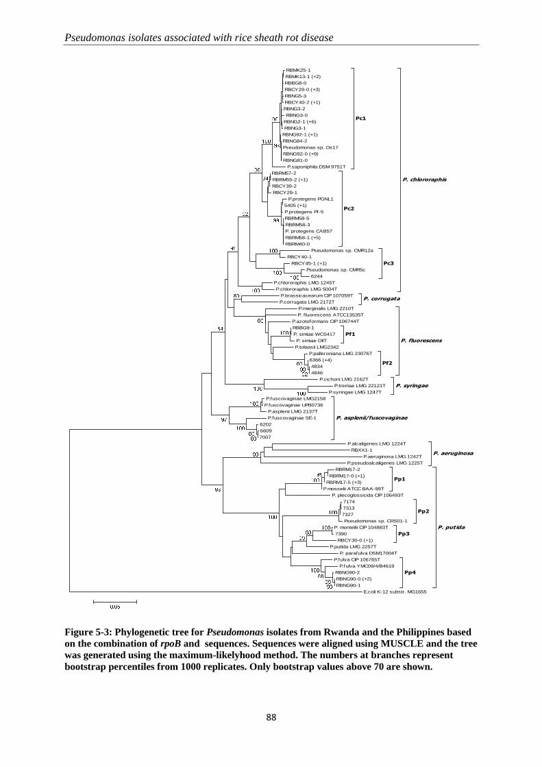
Pseudomonas isolates associated with rice sheath rot disease
88
Figure 5-3: Phylogenetic tree for Pseudomonas isolates from Rwanda and the Philippines based
on the combination of rpoB and sequences. Sequences were aligned using MUSCLE and the tree
was generated using the maximum-likelyhood method. The numbers at branches represent
bootstrap percentiles from 1000 replicates. Only bootstrap values above 70 are shown.
RBMK25-1
RBMK13-1 (+2)
RBBG8-0
RBCY29-0 (+3)
RBNG5-3
RBCY40-2 (+1)
RBNG3-2
RBNG3-0
RBNG2-1 (+6)
RBNG3-1
RBNG92-1 (+1)
RBNG84-2
Pseudomonas sp. Os17
RBNG92-0 (+9)
RBNG81-0
P.saponiphila DSM 9751T
Pc1
RBRM57-2
RBRM55-2 (+1)
RBCY39-2
RBCY29-1
P.protegens PGNL1
5405 (+1)
P.protegens Pf-5
RBRM58-5
RBRM58-3
P. protegens CAB57
RBRM58-1 (+5)
RBRM40-0
Pc2
Pseudomonas sp. CMR12a
RBCY40-1
RBCY45-1 (+1)
Pseudomonas sp. CMR5c
6244
Pc3
P.chlororaphis LMG 1245T
P.chlororaphis LMG 5004T
P. chlororaphis
P.brassicacearum CIP 107059T
P.corrugata LMG 2172TP. corrugata
P.marginalis LMG 2210T
P. f luorescens ATCC13535T
P.azotoformans CIP 106744T
RBBG9-1
P. simiae WCS417
P. simiae OliT
Pf1
P.tolaasii LMG2342
P.palleroniana LMG 23076T
6366 (+4)
4834
4846
Pf2
P. fluorescens
P.cichorii LMG 2162T
P.tremae LMG 22121T
P.syringae LMG 1247T
P. syringae
P.fuscovaginae LMG2158
P.fuscovaginae UPB0736
P.asplenii LMG 2137T
P.fuscovaginae SE-1
6202
6609
7007
P. asplenii/fuscovaginae
P.alcaligenes LMG 1224T
RBXX1-1
P.aeruginosa LMG 1242T
P.pseudoalcaligenes LMG 1225T
P. aeruginosa
RBRM17-2
RBRM17-0 (+1)
RBRM17-5 (+3)
P.mosselii ATCC BAA-99T
Pp1
P. plecoglossicida CIP 106493T
7174
7313
7327
Pseudomonas sp. CRS01-1
Pp2
P. monteilii CIP 104883T
7390
RBCY30-0 (+1)
Pp3
P.putida LMG 2257T
P. parafulva DSM17004T
P.fulva CIP 106765T
P.fulva YMC09/4/B4619
RBNG90-2
RBNG90-0 (+2)
RBNG90-1
Pp4
P. putida
E.coli K-12 substr. MG1655
Chapter 5
89
The identified pseudomonad isolates from Rwanda, based on the Pseudomonas species to
which they are closely related, could be grouped into 4 subgroups (P. chlororaphis, P.
fluorescens, P. putida, P. aeruginosa) and 8 clusters within these subgroups (written
according to the Pseudomonas subgroups: Pc1, Pc2, Pc3, Pf1, Pp1, Pp3, Pp4 and P.
aeruginosa) (Table 5-5). Isolates from the Philippines could be grouped in 4 subgroups (P.
chlororaphis, P. fluorescens, P. asplenii/fuscovaginae, P. putida) and 6 clusters (Pc2, Pc3,
Pf1, Pf2, P. asplenii/fuscovaginae, Pp2, Pp3). Cluster Pc2 corresponds with Biolog group A2,
cluster Pc3 corresponds with Biolog group A3, cluster Pf2 with Biolog group B2 and cluster
Pp2 with Biolog group A4. Biolog groups are illustrated in Cottyn et al. (1996b). Globally,
the isolates from Rwanda are found in the same subgroups as those from the Philippines, but
the clusters are different. In Rwanda, the majority of the isolates are similar to Pseudomonas
sp. Os17, a strain with biological control potential obtained from the rhizosphere of rice in
Japan and related to P. saponiphila (Pc1 cluster) while these were not found among the
isolates from the Philippines. The Pf2 cluster, containing isolates related to P. palleroniana,
and the subgroup of P. asplenii/fuscovaginae, the one containing described pathogens, are not
represented in Rwanda. The P. putida subgroup in the Philippines is dominated by isolates
related to Pseudomonas sp. CRS01-1, a biocontrol strain obtained from the rhizosphere of rice
in China, while in Rwanda this subgroup is dominated by isolates related to P. mosselii and P.
fulva.
5.3.3 Pathogenicity tests
Based on the results of the identification, isolates representing the different Pseudomonas
subgroups were chosen for undergoing pathogenicity testing. Some isolates induced disease
lesion development after inoculation in the plant. The typical lesions induced by
Pseudomonas fuscovaginae, which served as a reference for scoring on the scale, are
presented in Figure 5-4.
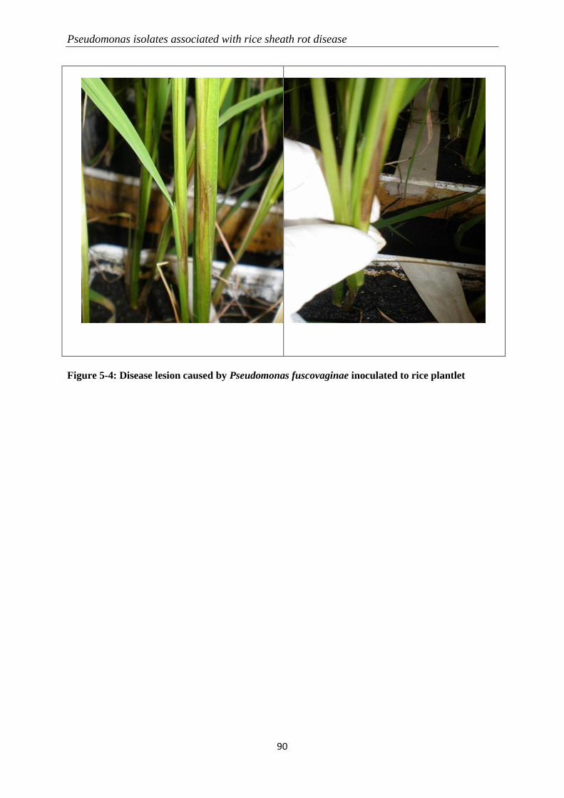
Pseudomonas isolates associated with rice sheath rot disease
90
Figure 5-4: Disease lesion caused by Pseudomonas fuscovaginae inoculated to rice plantlet
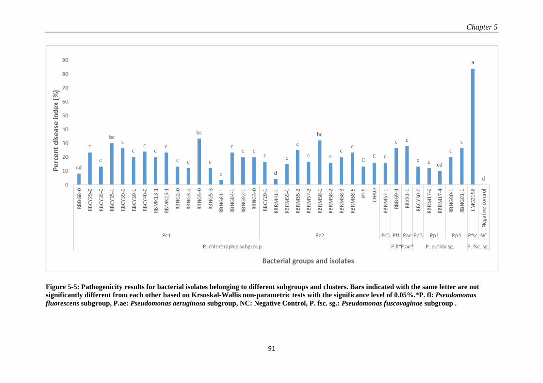
Chapter 5
91
Figure 5-5: Pathogenicity results for bacterial isolates belonging to different subgroups and clusters. Bars indicated with the same letter are not
significantly different from each other based on Krsuskal-Wallis non-parametric tests with the significance level of 0.05%.*P. fl: Pseudomonas
fluorescens subgroup, P.ae: Pseudomonas aeruginosa subgroup, NC: Negative Control, P. fsc. sg.: Pseudomonas fuscovaginae subgroup .
Pseudomonas isolates associated with rice sheath rot disease
92
Pathogenicity data presented in Figure 5-5 show that all these isolates are mildly pathogenic at
best and the disease severity they cause is low compared to the disease severity induced by P.
fuscovaginae, strain LMG2158.
5.4 Discussion We observed rice with sheath rot symptoms in all the rice-growing areas of Rwanda sampled:
Bugarama, Cyili, Mukunguri, Nyagatare and Rwagitima, and Rwamagana. This is in agreement
with the general observation that many recent studies point on the emerging importance of rice
sheath rot disease in various rice production systems (Mew et al., 2004b; Razak et al., 2009). In
this chapter, we specifically focused on bacteria and found three major bacterial genera
associated with rice sheath rot symptoms in Rwanda: Stenotrophomons, Delftia, and
Pseudomonas. We emphasized on the further characterization of the Pseudomonas spp. since
Pseudomonas fuscovaginae and other pseudomonads have been associated with rice sheath rot
symptoms in other parts of the world. Other reported sheath rot associated bacteria such as
Burkholderia glumae (Fory et al., 2014), Burkholderia gladioli (Nandakumar et al., 2009) and
Acidovorax oryzae (Cottyn et al., 1996b; Schaad et al., 2008) were not found in Rwanda. The
genera Stenotrophomonas and Delftia were also found to be associated to rice in previous studies
but are not considered to be pathogenic on plants (Cottyn et al., 1996b; Mano et al., 2007;
Ghasemi et al., 2012).
Stenotrophomonas maltophilia was first described as Pseudomonas maltophilia by Hugh et al.
(1961). It later became known as Xanthomonas maltophilia (Swings et al., 1983) and is now
included in the genus Stenotrophomonas (Palleroni and Bradbury, 1993). Stenotrophomonas
maltophilia is an endophyte which is commonly isolated from rice leaves (Mano and Morisaki,
2008) and seeds (Hardoim et al., 2012). Though S. maltophilia is known to grow on a limited
number of media in the laboratory, it is found in various environments. There is probably a lot of
variability within Stenotrophomonas: the work of Alavi et al. (2014) has shown that there is a
difference in human pathogenic and drug-resistant S. maltophilia and the plant-associated S.
maltophilia. Those differences are seen in virulence factors and heat shock proteins and should be
studied in more detail.
Delftia, described as a new genus by Wen et al. (Wen et al., 1999), is considered as a Plant
Growth Promoting Bacterium (PGPB), a diazotroph organism and a potential biocontrol agent
(Han et al., 2005) and was found in various substrates in the environment. The type species is
Chapter 5
93
Delftia acidovorans (Wen et al., 1999). Another important described species is Delftia
tsuruhatensis, which was first isolated from activated sludge in Japan (Shigematsu et al., 2003).
Pseudomonas isolates are regularly associated with rice sheath rot and grain discoloration
symptoms (Zeigler and Alvarez, 1987, 1990; Cottyn et al., 1996a; b; Gardan et al., 2002; Cother
et al., 2010). In the past, those pseudomonads were usually only identified by
biochemical/physiological tests, and except for P. fuscovaginae and P. palleroniana, their
taxonomic status had not been unraveled (Gardan et al., 2002). We further identified the
Pseudomonas isolates by sequencing part of the rpoB and genes since taxonomic studies have
shown that intragenic diversity among Pseudomonas spp. can be specifically evidenced by the
rpoB and the rpoD based sequences (Frapolli et al., 2007; Mulet et al., 2009). Molecular
identification techniques have replaced those based on serology, enzymology and metabolism
(Höfte and De Vos, 2006) and it is clear that molecular methods are needed to avoid taxonomic
inconsistencies (Gomila et al., 2015). Most isolates characterized in this work are related to
Pseudomonas species that were only recently described, for example P. saponiphila (Lang et al.,
2010), P. protegens (Ramette et al., 2011), P. fulva (Uchino et al., 2001), P. mosselii (Naik et al.,
2008; Mehri et al., 2011) and P. simiae (Berendsen et al., 2015), but their similarity with
described species is rarely total (100%). Intriguingly, various reference strains closely related to
isolates from Rwanda and the Philippines appear to be associated with the rhizosphere and are
known as biocontrol agents, such as Pseudomonas sp. Os17 (Pc1 in the P. chlororaphis group)
and Pseudomonas sp. CRS01-1 (Pp2 in the P. putida group) isolated from the rice rhizosphere
(Takeuchi et al., 2015), P. protegens strains Pf-5 from the cotton rhizosphere (Ramette et al.,
2011), P. protegens Cab57 from shepherd’s purse rhizosphere, Pseudomonas sp. CMR12a and
CMR5c from the cocoyam rhizosphere (Perneel et al., 2007), and P. simiae WCS417 from the
rhizosphere or wheat (Berendsen et al., 2015). No isolate from Rwanda proved to be a major
pathogen and the major rice sheath rot pathogen P. fuscovaginae, was not found. The conducted
pathogenicity tests have shown that the Pseudomonas isolates from Rwanda are weakly
pathogenic at best. Isolates from the Philippines included P. fuscovaginae and P. palleroniana. P.
palleroniana was isolated from rice sheaths of diseased plants before (Gardan et al., 2002)
though it proved to be weakly pathogenic in our pathogenicity tests.
The role played by the identified Pseudomonas isolates still has to be further investigated because
currently it is unclear whether they are pathogens or may have potential for being used biological
Pseudomonas isolates associated with rice sheath rot disease
94
control agents. The possibility of synergism between the organisms and/or favorable
agroecological factors in disease induction should not be ruled out. When interpreting
pathogenicity data, it is also important to take into account the difficulty of artificially
reproducing natural field conditions, and also the short duration of experimentations. As the rice
sheath rot is complex by nature, research for improving the understanding on it should continue
and check if there are interactions between the detected agents and how they work, if it is by
additive effects or by synergism.
The differentiation between pathogenic and beneficial organism is never strict. In fact, studies
have shown that some mechanisms found in pathogenic bacteria like the Type Three Secretion
System (T3SS) (Höfte and De Vos, 2006) or other mechanisms for host colonization are also
found in bacteria thought to be endophytes (Schmidt et al., 2012). This situation is well
documented for Herbaspirillum rubrisubalbicans (Monteiro et al., 2012) and it is important to
check if it is the same situation with the numerous pseudomonads observed with rice sheath rot.
Moreover, it has recently been shown that P. protegens Pf-5, a well studied biological control
bacterium, produces toxoflavin (Philmus et al., 2015), a yellow pigmented toxin. This product is
a known virulence factor of Burkholderia glumae, causing bacterial wilt on many field crops
including rice (Jeong et al., 2003; Koh et al., 2011). This discovery offers new insights about the
necessity of further research on the role played by many pseudomonads found in sheath rot
diseased plants, potentially producing toxoflavin, which can be partly responsible for sheath rot
disease symptoms. On the other hand, Pseudomonas bacteria from the P. chlororaphis and P.
fluorescens group are known to produce many secondary metabolites that play a capital role in
biocontrol such as phenazines (Mavrodi et al., 2010), 2,4-diacetylphloroglucinol, pyoluteorin
(Ramette et al., 2011) and cyclic lipopeptides (Olorunleke et al., 2015), which may have activity
against sheath rot fungi such as Sarocladium oryzae and Fusarium spp., but this remains to be
investigated.
The question remains how bacteria that are usually associated with the rhizosphere can end up in
the sheath of rice plants. Various hypotheses can be formulated: (i) attraction by plant exudates
in the reproduction phase, as it was observed with P. fuscovaginae on rice (Batoko et al., 1997);
(ii) rice in Rwanda and the Philippines is produced in an irrigated system, in which the root
system is most of the time covered with water, limiting oxygen availability which may incite
bacteria to move to tissues with more access to oxygen; (iii) they are pathogenic but their
Chapter 5
95
pathogenesis mechanism is not known yet, for example by the production of unknown
toxins/polysaccharides; (iv) they have coevolved with other sheath rot pathogens like the fungus
Sarocladium oryzae and they became resistant to the antibiotics produced by this fungus. Few
studies have been conducted on the diversity of organisms in the phyllosphere, compared to the
rhizosphere, but it has been documented that the bacterial population in the rhizosphere is not
much different from that in the phyllosphere (Sørensen and Sessitsch, 2007; Schlaeppi and
Bulgarelli, 2015). Bacteria that have a plant-associated lifestyle without causing any symptoms
are generally called endophytes and can be found in the rhizosphere, but also in the phyllosphere,
in seeds or in vegetative plant materials.
In the meantime, as it is now established that rice sheath rot is caused by many agents, using
healthy planting material can be of utmost importance, considering the possibility of basically
breaking the disease cycle. Rice remains an important crop in Rwanda, the Philippines and
elsewhere in the world, in tropical and subtropical areas. The planting material exchange keeps
increasing, as high quality cultivars are developed in international research centers and sent to
countries for production, multiplication and distribution. It is of utmost importance to guaranty
the planting material health quality as it has been proven with all the sheath rot associated
pathogens that the disease is seed-transmitted. The report that we are presenting is based on a
representative sample of the bacteria isolated from sheath rot diseased plants in Rwanda and the
Philippines. Conducting more isolates identification is a good option but the fact that most plants
in production are infected by various isolates is established. It is urgent to start simple seed
treatment practices like the heat therapy mentioned by Zeigler and Alvarez (1987). Putting this in
an integrated pest management approach could be more promising, as many factors, probably
varying in each agroecosystem, are associated with rice sheath rot disease, to which Rwanda and
many other rice-growing countries are exposed.

Chapter 6
97
6 GENERAL DISCUSSION AND PERSPECTIVES
6.1 Introduction This research was undertaken with the main goal of understanding the situation of rice sheath rot
disease in Rwanda, its causal agents, compared to those found elsewhere in the world, and
contributing in developing strategies for facing it. This study was motivated by many factors
among which we can cite:
importance gained by rice in Rwanda, where it is one of the priority crops (Bizimana et
al., 2012);
threat posed by rice sheath rot disease in the rice-producing countries;
improvements in scientific information and tools for studying pathogens,
suspicion that bacteria or fungi are associated with rice sheath rot in Rwanda,
necessity of comparing the situation of rice sheath rot disease in Rwanda, as for the
importance and the causal agents with the situation in other parts of the world.
Rice is an important crop worldwide. Though Asia contributes in the production at around 90%
(IRRI, 2015b), rice is becoming important in other continents also, for example rice production
has been increasing on average by 3.5% in Africa since 1961 (Timmer, 2010). Rice provides 21%
of energy and 15% of protein to humans (Zibaee, 2013).
Rice has emerged as a promising crop in Rwanda. It is a young crop in the country, having been
introduced in the 1950s (Kathiresan, 2010) but the experiments exploring the potential for
production at large scale having been conducted only in the 1970s (Baker, 1970). Having been
growing rice recently, Rwanda is cooperating with other countries and international research
centers, most of which have a long rice growing tradition (UCORIRWA and AC Team, 2004;
MINAGRI, 2013a). Rice is produced in an irrigated system in marshlands, performs with a yield
of 5.34 tons/ha (IRRI, 2015b), which is high considering the situation in the region and in Africa.
The pro-rice promotion policy, followed in the last decades, has also undeniably helped rice
development in Rwanda. Still, the rice sector in the country is experiencing some problems in
cultivar development and crop protection aspects. In fact, though the production has been
increasing, the demand has been much more increasing and the country has to import a large part
of the rice consumed. There are problems of presence of less adapted varieties, poor seed quality,
low national production quantity, low market value, as the locally produced rice is less
General discussion and perspectives
98
competitive on the market (Stryker, 2013), and poor knowledge on rice pests and diseases
(Promar, 2012; MINAGRI, 2013a). Improvements in rice production in Rwanda seem a must for
harnessing the potential and respond to the demand and this requires efforts in addressing the
limitations including the curbing of pests and diseases, of which many are not yet known, such
as rice sheath rot.
There are not many publications about rice production in Rwanda. Like in other areas, there are
not many scholarly works about Rwanda. In fact, the country was one of last African countries to
be known in the Western World, the first European, Oscar Baumann having entered there only in
1892 (Meyer, 1902). He would be followed by Gustav Adolf von Götzen. When the Berlin
conference was held in 1884-1885, the country was vaguely known as part of the African Great
Lakes region. Its status would be really defined later with the Brussels conference declaration in
1890.
Rice sheath rot disease has been detected in other parts of the world where it is considered to be
caused by fungi like S. oryzae and Fusarium spp. and bacteria, especially those of the
Pseudomonas genus, particularly P. fuscovaginae (Bigirimana et al., 2015), this study wanted to
compare the findings from previous studies and those from Rwanda so as to effectively
contribute in rice sheath rot control.
6.2 Research findings in relation with the objectives Considering the objectives of the study, the following answers can be formulated.
What is the importance and impact of rice sheath rot in Rwanda?
Rice sheath rot is an important disease in Rwanda. It is found in all the rice-growing areas in the
country which were surveyed in the study and its intensity (incidence and severity) is high. The
comparison between grains from a diseased and a healthy plant has shown that there is a yield
loss estimated at 56.93% in diseased plants. To this quantitative loss, must be added a qualitative
loss, as most of the pathogens isolated on sheath rot diseased rice plants have the capacity to
produce mycotoxins, which are harmful to humans and animals consuming diseased rice (Jestoi,
2008; Wulff et al., 2010; Nesic et al., 2014). Our results gave the indication that toxins are
produced by Fusarium isolates from Rwanda, but the connection to the disease prevalence needs
more extended studies. There are no reference studies on the epidemiology from which a
threshold can be established, and this may justify why there is large variability in reported yield
Chapter 6
99
losses (Ou, 1985; CABI, 2007) and also most studies were conducted on a particular
development stage, like seedling (Prabhu and Bedendo, 1983; Dastjerdi and Karlovsky, 2015) or
harvested kernels (Wulff et al., 2010; Chowdhury et al., 2015).
Which are the pathogens causing rice sheath rot in Rwanda? Are Pseudomonas
fuscovaginae and Sarocladium oryzae the most important sheath rot pathogens present in
Rwanda?
Based on the findings from this study, rice sheath rot in Rwanda is probably caused by fungi.
Sarocladium oryzae, fungi belonging to the Fusarium fujikuroi, F. oxysporum and F.
incarnatum/equiseti species complex have been isolated in this study. Not much is known about
pathogenesis inducing factors, but given that the isolated species produce toxins, cerulenin and
helvolic acid for Sarocladium oryzae (Sakthivel et al., 2002; Bills et al., 2004; Hittalmani et al.,
2016), and especially fumonisin for Fusarium spp.(Abbas et al., 1998; Wulff et al., 2010), it can
be hypothesized that sheath rot symptoms are partially due to the effects of toxins produced by
fungi.
Unexpectedly, this study did not detect Pseudomonas fuscovaginae in Rwanda. P. fuscovaginae
is the most reported sheath rot causing bacterium (Miyajima et al., 1983; Zeigler and Alvarez,
1987; Cottyn et al., 2002; Cother et al., 2009). But the study has detected many pseudomonads,
found in other related studies (Zeigler and Alvarez, 1990; Cottyn et al., 1996a; b; Cother et al.,
2010), for which the role, pathogenic or having a beneficial role for the plant, still has to be
investigated. One of the merits of this research consists in having given insights in identifying
those poorly identified bacteria as these isolates proved, through pathogenicity tests poorly
pathogenic.
Sustainable crop protection practices are needed for facing pests and diseases that hamper rice
production. In that aim, the starting point is the correct identification of the pests and diseases. In
the past, the classical crop protection diagnosis practices were based on morphological or
biochemical methods. In this study, Sarocladium oryzae identification was based on the
sequencing of the ITS region. There are not many new developments in Sarocladium
identification. Fusarium identification took into account the sequencing of the ITS region and the
TEF factor and the grouping of isolates was made after Aoki et al. (2014). The general
information on the bacteria was obtained through the sequencing based on 16S rDNA and the
primers based on rpoB and genes were used for Pseudomonas isolates, to further put into
General discussion and perspectives
100
evidence the intragenic diversity. The molecular methods of identification, with their high level
of precision and the relatively short time that they require, have revolutionised the identification
approaches and are incessantly being improved (Atkins and Clark, 2004; López et al., 2009).
Concerning the bacteria, none of the reported major bacterial pathogens causing rice sheath rot
elsewhere, such as Pseudomonas fuscovaginae, P. syringae, Acidovorax oryzae ( formerly A.
avenae), Burkholderia glumae and Burkholderia gladioli has been detected in Rwanda.
One of the striking findings about bacteria is that species traditionally found in the plant
rhizosphere, are this time found in the phyllosphere. Although this has not been largely reported,
it is probably because there have not been many researches on the organisms living in the
phyllosphere; another study confirms that there is not a big difference between rhizosphere and
phyllosphere organisms (Sørensen and Sessitsch, 2007). An illustration comes from studies on
Herbaspirillum rubrisubalbicans, which show that it can move from the rhizosphere to the
phyllosphere (Monteiro et al., 2012).
What is the origin of the sheath rot pathogens found in Rwanda? Are they native or
introduced with the plant material?
Though Rwanda is a landlocked country, the pathogens that are found there are not far too
different from those found in other parts of the world. In the Sarocladium oryzae strains, most of
them are different from those found elsewhere in the world but few are clustering with reference
strains from other parts of the world. Sarocaldium oryzae may have been introduced in Rwanda
through seed movement. Various types of Fusarium spp. isolates were found. The major groups
associated with rice sheath rot belonging to the F. fujikuroi species complex, F.
incarnatum/equiseti species complex, and F. graminearum species complex are found in
Rwanda. These organisms have regularly been found on rice seeds (Mew and Gonzales, 2002).
This can be understood in the general observation that Fusarium organisms are ubiquitous. There
are probably both native and introduced Fusarium strains. The pathogenic pseudomonads (P.
fuscovaginae, P. syringae) were not found, it is the poorly studied pseudomonads that were
found. Comparing isolates from Rwanda and those from the Philippines, the major groups were
the same, except that P. fuscovaginae and the mildly pathogenic P. palleroniana were found in
the Philippines while missing in Rwanda, where the P. saponiphila-like isolates are dominating.

Chapter 6
101
Is there an interaction between these organisms and the prevailing environmental
conditions in Rwanda?
We can hypothesize that all rice cultivars suffer from the low temperatures prevailing in Rwanda
(Promar, 2012) and are susceptible to rice sheath rot. The tolerance found in Japonica varieties,
while Indica varieties fail after 2 to 3 seasons of cultivation can be explained by the capacity of
the Japonicas to tolerate low temperatures while the Indicas do not have it. On sheath rot disease,
however, both types are susceptible, as even our pathogenicity experimentations have shown.
Which control options can be taken to face rice sheath rot?
This study gives the base for studying rice sheath rot in Rwanda. However, the information
generated does not lead yet to precise control options. As some of the sheath rot pathogens are
known, and taking reference from the review of published information, it is estimated that an
action plan adapted to the Rwandan context can be developed. Considering the numerous factors
involved in sheath rot disease development, among which there are many causal agents, a large
host range and the possibility of disease development in almost all the rice growing areas and in
most production systems, while not much research is available, an integrated disease control
approach is advisable, the time that more information become available and indicate to give
preference to one method or another. Integrated disease management practices, centered on the
potential rice pests and diseases presented in Annex 2 and the actions improving and preserving
the quality of the seed (Zeigler and Alvarez, 1987, 1989) can be followed, waiting for more
durable solutions like the creation of rice lines adapted to the Rwandan environment.
6.3 Connecting findings to hypotheses This study is based on two hypothesis: (1) lack of adaptation to prevailing abiotic conditions in
Rwanda makes introduced rice varieties more susceptible to pests and diseases such as sheath rot;
(2) quick rice development in Rwanda, with limited capacity of control and research on pests and
diseases, has resulted in the build-up of large populations of pests and diseases, including sheath
rot causing pathogens, which also negatively affect the rice grain quality. The data already
available tend to support them.
On the first hypothesis, the environment of Rwanda favours rice cultivars of Japonica type, as
they are the ones most adapted to low temperatures, but both of the variety types Japonica and
Indica are susceptible to rice sheath rot. In the preliminary data on our study, fungal isolates of
General discussion and perspectives
102
Sarocladium oryzae and Fusarium spp. induced sheath rot disease symptoms on both Nipponbare
cultivar of the Japonica type and CO39 cultivar of the Indica type, though symptoms were more
pronounced on the former than on the latter. Though this experiment was not replicated many
times, it gives preliminary facts but needs to be extended for giving more accurate information
about the susceptibility levels. This experiment tends to confirm the susceptibility of the two rice
subspecies, confirming that most rice materials currently in use are susceptible to sheath rot
attacks (Mew et al., 2004b). Obtaining more information on the genetic features of rice varieties
in use in Rwanda can give more insights in understanding the susceptibility. It is not known how
comparable Nipponbare is in relation to Rwandan Japonica cultivars. Seeds grown in the 1980s
came from Yunnan Province, China and when they became severely attacked by rice blast, Xinan
175 variety, of Japonica type, developed by Japanese in the 1950 in Taiwan, was introduced
(Promar, 2012). It is a parent material for many varieties currently used in Rwanda. Besides, the
pathogenicity experiment was conducted at 28°C, a temperature far higher than 19.84°C, the
average for Rwanda (NISR, 2013), while Japonica varieties are highly affected by a temperature
increase (Yoshida, 1981; Shah et al., 2014). It can be presumed that at a high temperature,
Japonica type become more stressed and thus more susceptible to the diseases. Putting our
findings in relation with the rice situation in Rwanda, as the adapted Japonica cultivars do not
have a good market value, most of the last introductions consist of cultivars of Indica type, the
ones appreciated on the market. Bugarama, a warm low altitude plain, is the only place where
long grain varieties of Indica type are grown satisfactorily. In other places, those Indica varieties
become stressed by low temperatures prevailing in Rwanda, and when to this pressure there is
also the stress caused by rice sheath rot diseases and other diseases, fail to adapt, and the farmers
came back to their “Japonica” varieties, which constitute their “safe haven” varieties. Moreover,
the Japonica varieties respond well to the use of farm inputs like fertilizers and the improved
production conditions might be predisposing plants to conditions conducive to attack and
proliferation of rice sheath rot.
For the second hypothesis, rice is developing very fast in Rwanda, the crop protection practices
have not been developing at the same pace. Also, the planting material introduced in the country
is not fully checked for its health status. Most sheath rot pathogens are seed-transmitted
(Bigirimana et al., 2015; Chowdhury et al., 2015). This situation resulted in the buildup of large
stocks of pests and diseases including rice sheath rot.
Chapter 6
103
There are many pathogenic fungal isolates that are found in Rwanda: Sarocladium oryzae,
Fusarium spp. Sarocladium oryzae strains present less variability, prompting us to think that it is
a recent introduction through seeds and grains. Fusarium spp. have more variability, indicating
that some of its isolates may be native to the country while others may have been introduced
through the seed movement, Fusarium spp. being regularly found in rice seeds (Mew and
Gonzales, 2002). The disease is associated with many bacterial strains, continuously reported in
diagnostic studies (Zeigler and Alvarez, 1990; Cottyn et al., 1996a; b) but their role in the
pathogenicity is unknown. The marginal research and practices on rice crop protection that exist
in Rwanda, while the country regularly imports seeds and rice for consumption, are unable to
realise that a large population of pathogens has built up on rice throughout the time. These
pathogens could also be partly responsible of the discolouration and poor quality of the locally-
produced rice in Rwanda as this has been observed elsewhere (Zeigler and Alvarez, 1989).
The aim of this research is to contribute in addressing the rice sheath rot problem. If in the long
run many actions can be envisaged, there are few options in the short run. Rice can be attacked
by many pests and diseases in Rwanda (see Annex A2). In fact, as the country meets tropical and
subtropical conditions, most pathogens attacking rice can get established, once they are
introduced in Rwanda. It is important to reinforce the disease diagnosis system and develop
regulatory tools for preventing the introduction of new pests and diseases. It is likely that the
international exchange of planting materials will keep intensifying. A list of pathogens that can
attack rice in Rwanda, based on the available information and the PRA methodology is presented
in Annex A2. Besides this, there is also an urgency in starting to adopt simple practices,
protecting the seed so that it is cleaned before planting, and this must be done on field during
production and in post harvest. Some of such practices are displayed by Zeigler and Alvarez
(1987, 1989). In the crop husbandry, efforts to promote the good balance in nitrogen fertilization
should be reinforced at the same time as maintaining health quality in seed production,
multiplication and distribution systems. In the long run, when there is enough information about
the pathogens attacking rice and the genetic information on the planting material in use in the
country, a breeding program, adapted to the special tropical semi-temperate climate of Rwanda,
but also taking lessons from other rice-growing countries like the Philippines which have
experienced the same situation, should be mounted. These actions could significantly contribute
in addressing the question of the lack of adapted high yielding varieties , resistant to rice sheath
General discussion and perspectives
104
rot and other important pests and diseases and also avail rice with a high marketing value, as the
imported Indica long grain varieties are preferred to the adapted short grain varieties.
6.4 Perspectives There are many perspectives for further studies and field action on rice sheath rot in Rwanda. An
action plan can be developed with the available information. This action plan must take into
account the following elements: type of pests and diseases present, crop husbandry practices,
production objectives, agroecological factors and must also explore the potential for breeding for
resistance. In that aim, some of the actions that can be taken fall in the following areas:
It is important to develop an efficient link between the field practices and the research so
that field people can have access to advanced tools like scientific information and
diagnosis guidance. Illustrated and annotated manuals of sheath rot pathogens, regularly
updated, can help in addressing this issue on the practical side. It is clear that there are
pathogen introductions in Rwanda, for example for Sarocladium oryzae, and practices
must be adapted to prevent such introductions.
Efforts must be put in understanding the pathogenicity factors of the species identified as
associated with rice sheath rot disease. The knowledge on mycotoxins and their role in
pathogenicity is of particular importance. This can help in overcoming the controversy
existing now in which some studies confirm that mycotoxins constitute a pathogenicity
factor (Abbas et al., 1995) while others attest that there is no correlation between
mycotoxin production and pathogenicity (Dastjerdi and Karlovsky, 2015). Conducting a
study on the infection on the whole growth cycle can provide conclusive information on
this issue as the results of these studies come from one plant development stage. The work
on the pathogenesis induced by Sarocladium oryzae and Pseudomonas fuscovaginae will
be in the future facilitated by the availability of the information on their genome which
has been sequenced (Patel et al., 2012; Hittalmani et al., 2016). Some Fusarium species
have also been sequenced (Waalwijk et al., 2004).
The role played by the pseudomonads needs further studies to be clearly elucidated. Their
large variability warrants more studies on their importance and their ecology in the
phyllosphere, given that they were mainly reported in the rhizosphere. Moreover, their
relationships with fungi of the Sarocladium oryzae and Fusarium genera, both associated
with rice sheath rot symptoms, justifies the necessity for conducting those studies. In this
Chapter 6
105
regard, investigations on biofilm formation and the study of the type of secondary
metabolism products that are released associated with the coinoculation of Pseudomonas
and Sarocladium/Fusarium on rice plants can lead to more insights about the
understanding of the interactions and guide for the biological control exploration.
However, it can not be ruled out that those bacteria, or some of them, may be pathogens
with an unknown pathogenicity mechanism. Their ecology throughout different
development stages deserves also to be studied. It is for example confirmed that P.
fuscovaginae passes by epiphytic phases before attacking rice and inducing sheath brown
rot, taking advantage of the increased susceptibility of the plant at the booting stage
(Batoko et al., 1997). This situation should be explored for the other pseudomonads also.
Research is needed on practices that can reduce the pathogen inoculum in rice seeds, as
most sheath rot pathogens are seed-transmitted, building on what is already known.
The information on the rice genetic material in use in Rwanda is needed. It can give more
insights in understanding why rice sheath rot is an important disease in Rwanda.
Efforts must be put in breeding rice varieties adapted to the Rwanda context: low
temperature, good response to input use, good culinary properties. On this last point, there
is a need of breeding rice varieties adapted to the unique conditions of a tropical climate
tempered by the altitude and this is possible as there are elsewhere many breeding
initiatives for taking advantage of the different rice genotypes (Yang et al., 2012). The
results presented were obtained on Nipponbare and CO39 varieties. The information on
the relationship between those varieties and the ones used in Rwanda can improve in
understanding the failure observed with Indica varieties while the Japonica varieties
become tolerant. Efforts must be put in enquiring about the situation of the Japan-China
initiative in 1990-1994 to develop rice varieties adapted to Rwandan environment
(Promar, 2012) that was interrupted so as build on its findings and design anew such kind
of breeding activity.
A comprehensive study on the epidemiology of disease induced by the major rice sheath
rot pathogens (S. oryzae, Fusarium spp., P. fuscovaginae) is needed. This study should
take into account among others: pathogen variability, seasonal variability, temperature,
relative humidity, location, yield losses, rice cultivars and the transmission rate. As the
aim of this study would be to advise on sheath rot disease control, trials must be

General discussion and perspectives
106
conducted in both controlled and semi-controlled environments, so as to nearly reproduce
the real rice-growing conditions.
Annex 1
107
ANNEX 1: IDENTIFICATION OF VIRULENCE ASSOCIATED LOCI
IN THE EMERGING BROAD HOST RANGE PLANT PATHOGEN
PSEUDOMONAS FUSCOVAGINAE
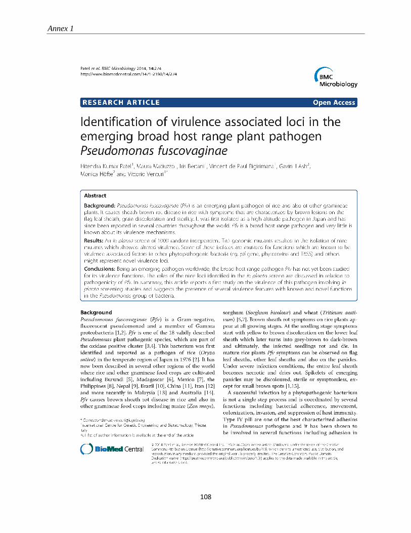
Annex 1
108
Annex 1
109
Annex 1
110
Annex 1
111
Annex 1
112
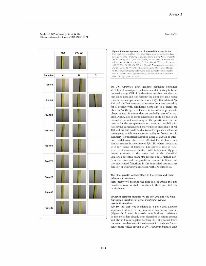
Annex 1
113
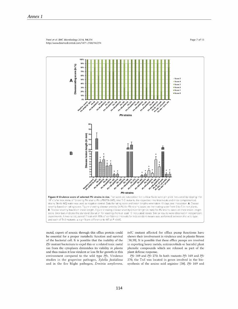
Annex 1
114
Annex 1
115
Annex 1
116
Annex 1
117
Annex 1
118
Annex 1
119
Annex 1
120
Annex 2
121
ANNEX 2: CONTRIBUTION TO THE ESTABLISHMENT OF THE
LIST OF PESTS OF PHYTOSANITARY IMPORTANCE ON RICE IN
RWANDA BY PRA METHODOLOGY AND FIELD SURVEY
Vincent de Paul BIGIRIMANA, Béatrice UWUMUKIZA, Monica HÖFTE, Obedi I.
NYAMANGYOKU
Abstract
Rice (Oryza sativa) is one of the priority crops in Rwanda. Efforts are being put in improving rice
production and productivity. Also the Rwandan agriculture sector is in the transition from
subsistence farming to market-oriented agriculture. Its mission is to transform and modernise
agriculture and livestock to ensure food security and contribute to the national economy. One of
the requirements to achieve this objective is efficiently organising the plant protection aspects in
production and after harvest. Continuous inspection and surveillance by rice professionals are of
utmost importance for meeting this requirement. Rice is attacked by many pests and diseases in
the many agroecological conditions where it is grown in the world. Rwanda has in general a
tropical climate tempered by average high altitude. There is not sufficient information on pests
and diseases that can attack rice in conditions of high altitude and low temperature, like those
found in Rwanda. This initiative aims at developing a list of pests of phytosanitary importance in
Rwanda which can guide rice professionals dealing with plant protection and phytosanitary issues
especially in developing the phytosanitary regulation in compliance with the requirements of
international trade in accordance with the World Trade Organisation in the agreement on the
application of Sanitary and Phytosanitary Measures (WTO-SPS Agreement).
The list of rice pests and diseases is based on published information available in professional and
scientific databases, particularly from the Crop Protection Compendium of CABI and also from
field surveys. The status of each individual pest or disease, considering the agro ecology of
Rwanda, is established based on the Pest Risk Analysis (PRA) methodology. For each pest, the
following aspects are covered: taxonomic group, presence or absence in Rwanda, caused losses,
likelihood of entry, proliferation and establishment in Rwanda, appreciation of the risk
represented expressed in whether it must be included in the list of pests of phytosanitary
importance or not.
A final list of pests of phytosanitary importance for rice in Rwanda is suggested. It is composed
of 188 organisms from all the groups of pests (fungi, bacteria, viruses, nematodes, arthropods,
birds, weeds and rodents) that have been found to meet the phytosanitary importance criteria as
specified in the International Standards for Phytosanitary Measures (ISPMs) developed for
implementing the International Plant Protection Convention (IPPC) to which Rwanda is
signatory.
Annex 2
122
Introduction The economy of Rwanda is based on agriculture. As of 2011 this sector employs 80% of the total
active population and contributes to the GDP at 31%. The agricultural policy aims to increase the
productivity so that the sector can ensure food security and release excess for the market.
Agriculture is in transition from subsistence farming to market-oriented agriculture. It is
estimated that the agricultural sector will have the greatest impact on the economy in terms of
poverty reduction and wealth creation, as it is the largest employer (Bizimana et al., 2012).
Rice is one of the important crops in Rwanda. Although its introduction is recent (Baker, 1970),
in 2012, the crop is grown on 12000 ha, the total national production is estimated at 80000 tons
and the yield per hectare is 5.5 tons (RAB, 2013), which is high in a developing country context
and compared to some countries with a long tradition of rice growing (Singh et al., 2010). Rice is
the most marketed crop in Rwanda, with 47% of its production share being sold on markets
(NISR, 2012). In the same time, the popularity of rice has continuously been increasing, and the
national production cannot meet the demand. The country is now importing more than 40% of the
consumed rice (RAB, 2013).
There are many initiatives aiming at increasing rice production in the country by finding
solutions to the major constraints. Rice production faces some constraints among which the
pressure of pests and diseases is one of the most important (RAB, 2014). The climate of Rwanda,
characterized by low temperatures, compared to other tropical and equatorial countries (Sikkens
and Steenhuis, 1988) and an elevated topography, is conducive to the development of many pests,
as those of tropical and of subtropical areas can be established. Given that the breeding and seed
sector in Rwanda is in its development, considering large quantities of rice are imported in the
country for consumption or as seed, but also as Rwanda is intending to become a rice exporter in
the coming years (MINAGRI, 2011a), legal plant protection measures are perceived as an
efficient way that can help crop protection and production professionals in dealing with rice
health aspects. The International Plant Protection Convention (IPPC) is the international legal
instrument for plant health related issues (Baker et al., 2005). In fact, the IPPC and its related
International Standards for Phytosanitary Measures (ISPMs) aim to protect plant health at
international level through trade but also at regional and national levels in guiding in the
development of diagnostic and surveillance methods meeting international standards (Petter et al.,
2008). Thus it is capital to define the phytosanitary importance that must be given to each pest
Annex 2
123
known to attack rice. A precautionary approach, technically justified based on scientific
principles and evidence as explained by Griffin (2000), helps to decide whether the considered
pest qualify as a pest of phytosanitary importance or not.
A list of pests of phytosanitary importance on rice in Rwanda has been prepared, which can help
Rwanda to conduct PRA and manage rice protection issues according to the principles of the
IPPC and the WTO-SPS agreement, Rwanda being signatory of both of these conventions.
Methodology In the proposed list of pests of phytosanitary importance for Rwanda, a pest is defined as,
according to the International Plant Protection Convention (FAO, 1997), “any species, strain or
biotype of plant, animal or pathogenic agent injurious to plants or plant products”. The pest risk
analysis (PRA) is followed for determining the importance of each individual pest and is defined
as ”the process of evaluating biological and other scientific and economic evidence to determine
whether a pest should be regulated and the strength of any phytosanitary measures to be taken
against it” (FAO, 1997).
A list of the pests reported on rice has been produced, based on the rice pest in the
Commonwealth Agricultural Bureaux database (CABI, 2007) and also from the list of pests in the
surveillance report of the Rwanda Ministry of Agriculture and Animal Resources (MINAGRI,
2013b). The MINAGRI conducts routine pest surveillance activities. For rice, the survey covered
the areas of Rwamagana, Nyagatare, Cyili, Bugarama and Runda. It consisted in looking at
diseased plants and conducting a diagnosis based on pest attack symptom pictures and
information in technical literature and manuals.
The Pest Risk Analysis (PRA) was conducted on the pests present on the list. Pests were analysed
on the criteria: taxonomic group, presence or absence in Rwanda, level caused losses, likelihood
of entry, proliferation and establishment in Rwanda, appreciation of the risk represented
expressed in whether it must be included in the list of pests of phytosanitary importance or not.
Are included in the list the pests considered as important and which are absent in Rwanda and
exceptionally those that are present in Rwanda but which would qualify as regulated non-
quarantine pests, the latter expression meaning “a non-quarantine pest whose presence in plants
for planting affects the intended use of those plants with an economically unacceptable impact
and which is therefore regulated within the territory of the importing contracting party” (FAO,
1997).
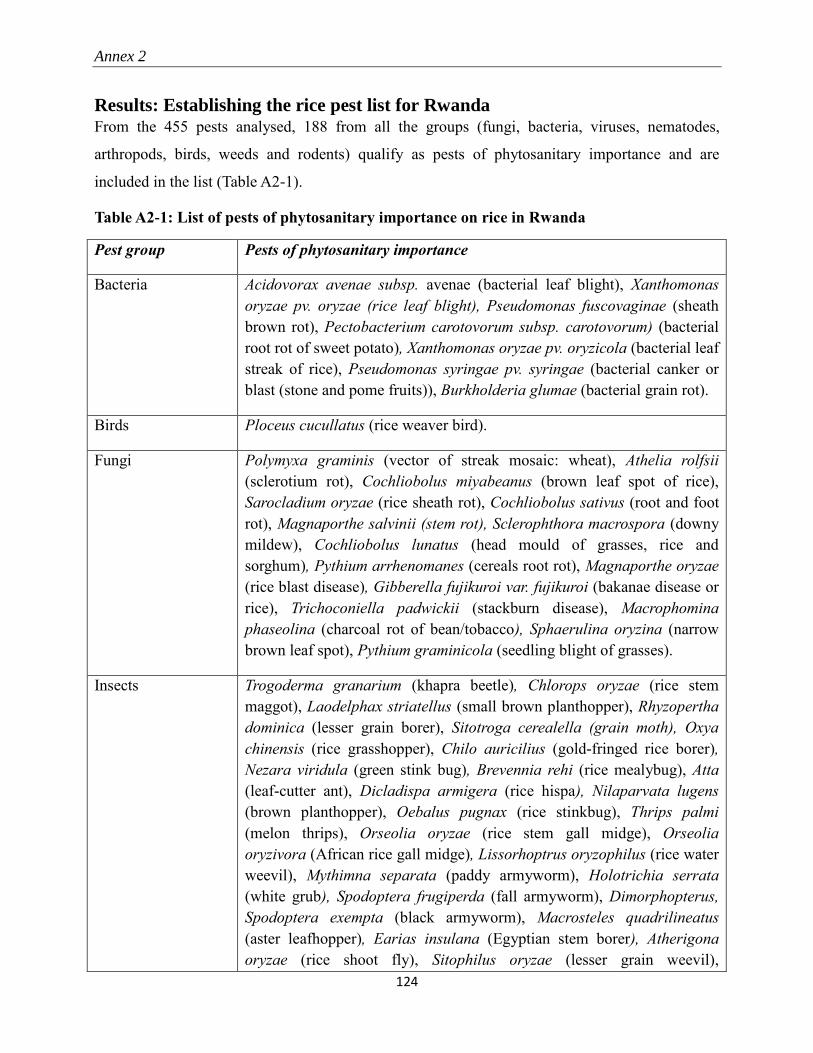
Annex 2
124
Results: Establishing the rice pest list for Rwanda From the 455 pests analysed, 188 from all the groups (fungi, bacteria, viruses, nematodes,
arthropods, birds, weeds and rodents) qualify as pests of phytosanitary importance and are
included in the list (Table A2-1).
Table A2-1: List of pests of phytosanitary importance on rice in Rwanda
Pest group Pests of phytosanitary importance
Bacteria Acidovorax avenae subsp. avenae (bacterial leaf blight), Xanthomonas
oryzae pv. oryzae (rice leaf blight), Pseudomonas fuscovaginae (sheath
brown rot), Pectobacterium carotovorum subsp. carotovorum) (bacterial
root rot of sweet potato), Xanthomonas oryzae pv. oryzicola (bacterial leaf
streak of rice), Pseudomonas syringae pv. syringae (bacterial canker or
blast (stone and pome fruits)), Burkholderia glumae (bacterial grain rot).
Birds Ploceus cucullatus (rice weaver bird).
Fungi Polymyxa graminis (vector of streak mosaic: wheat), Athelia rolfsii
(sclerotium rot), Cochliobolus miyabeanus (brown leaf spot of rice),
Sarocladium oryzae (rice sheath rot), Cochliobolus sativus (root and foot
rot), Magnaporthe salvinii (stem rot), Sclerophthora macrospora (downy
mildew), Cochliobolus lunatus (head mould of grasses, rice and
sorghum), Pythium arrhenomanes (cereals root rot), Magnaporthe oryzae
(rice blast disease), Gibberella fujikuroi var. fujikuroi (bakanae disease or
rice), Trichoconiella padwickii (stackburn disease), Macrophomina
phaseolina (charcoal rot of bean/tobacco), Sphaerulina oryzina (narrow
brown leaf spot), Pythium graminicola (seedling blight of grasses).
Insects Trogoderma granarium (khapra beetle), Chlorops oryzae (rice stem
maggot), Laodelphax striatellus (small brown planthopper), Rhyzopertha
dominica (lesser grain borer), Sitotroga cerealella (grain moth), Oxya
chinensis (rice grasshopper), Chilo auricilius (gold-fringed rice borer),
Nezara viridula (green stink bug), Brevennia rehi (rice mealybug), Atta
(leaf-cutter ant), Dicladispa armigera (rice hispa), Nilaparvata lugens
(brown planthopper), Oebalus pugnax (rice stinkbug), Thrips palmi
(melon thrips), Orseolia oryzae (rice stem gall midge), Orseolia
oryzivora (African rice gall midge), Lissorhoptrus oryzophilus (rice water
weevil), Mythimna separata (paddy armyworm), Holotrichia serrata
(white grub), Spodoptera frugiperda (fall armyworm), Dimorphopterus,
Spodoptera exempta (black armyworm), Macrosteles quadrilineatus
(aster leafhopper), Earias insulana (Egyptian stem borer), Atherigona
oryzae (rice shoot fly), Sitophilus oryzae (lesser grain weevil),
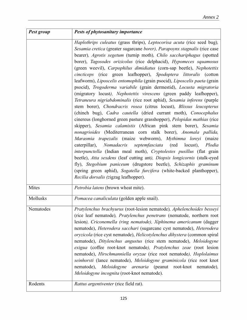
Annex 2
125
Pest group Pests of phytosanitary importance
Haplothrips culeatus (grass thrips), Leptocorisa acuta (rice seed bug),
Sesamia cretica (greater sugarcane borer), Parapoynx stagnalis (rice case
bearer), Agrotis segetum (turnip moth), Chilo sacchariphagus (spotted
borer), Tagosodes orizicolus (rice delphacid), Hypomeces squamosus
(green weevil), Carpophilus dimidiatus (corn-sap beetle), Nephotettix
cincticeps (rice green leafhopper), Spodoptera littoralis (cotton
leafworm), Liposcelis entomophila (grain psocid), Liposcelis paeta (grain
psocid), Trogoderma variabile (grain dermestid), Locusta migratoria
(migratory locust), Nephotettix virescens (green paddy leafhopper),
Tetraneura nigriabdominalis (rice root aphid), Sesamia inferens (purple
stem borer), Chondracris rosea (citrus locust), Blissus leucopterus
(chinch bug), Cadra cautella (dried currant moth), Conocephalus
cinereus (longhorned green pasture grasshopper), Pelopidas mathias (rice
skipper), Sesamia calamistis (African pink stem borer), Sesamia
nonagrioides (Mediterranean corn stalk borer), Anomala pallida,
Marasmia trapezalis (maize webworm), Mythimna loreyi (maize
caterpillar), Nomadacris septemfasciata (red locust), Plodia
interpunctella (Indian meal moth), Cryptolestes pusillus (flat grain
beetle), Atta sexdens (leaf cutting ant), Diopsis longicornis (stalk-eyed
fly), Stegobium paniceum (drugstore beetle), Schizaphis graminum
(spring green aphid), Sogatella furcifera (white-backed planthopper),
Recilia dorsalis (zigzag leafhopper).
Mites Petrobia latens (brown wheat mite).
Mollusks Pomacea canaliculata (golden apple snail).
Nematodes Pratylenchus brachyurus (root-lesion nematode), Aphelenchoides besseyi
(rice leaf nematode), Pratylenchus penetrans (nematode, northern root
lesion), Criconemella (ring nematode), Xiphinema americanum (dagger
nematode), Heterodera sacchari (sugarcane cyst nematode), Heterodera
oryzicola (rice cyst nematode), Helicotylenchus dihystera (common spiral
nematode), Ditylenchus angustus (rice stem nematode), Meloidogyne
exigua (coffee root-knot nematode), Pratylenchus zeae (root lesion
nematode), Hirschmanniella oryzae (rice root nematode), Hoplolaimus
seinhorsti (lance nematode), Meloidogyne graminicola (rice root knot
nematode), Meloidogyne arenaria (peanut root-knot nematode),
Meloidogyne incognita (root-knot nematode).
Rodents Rattus argentiventer (rice field rat).
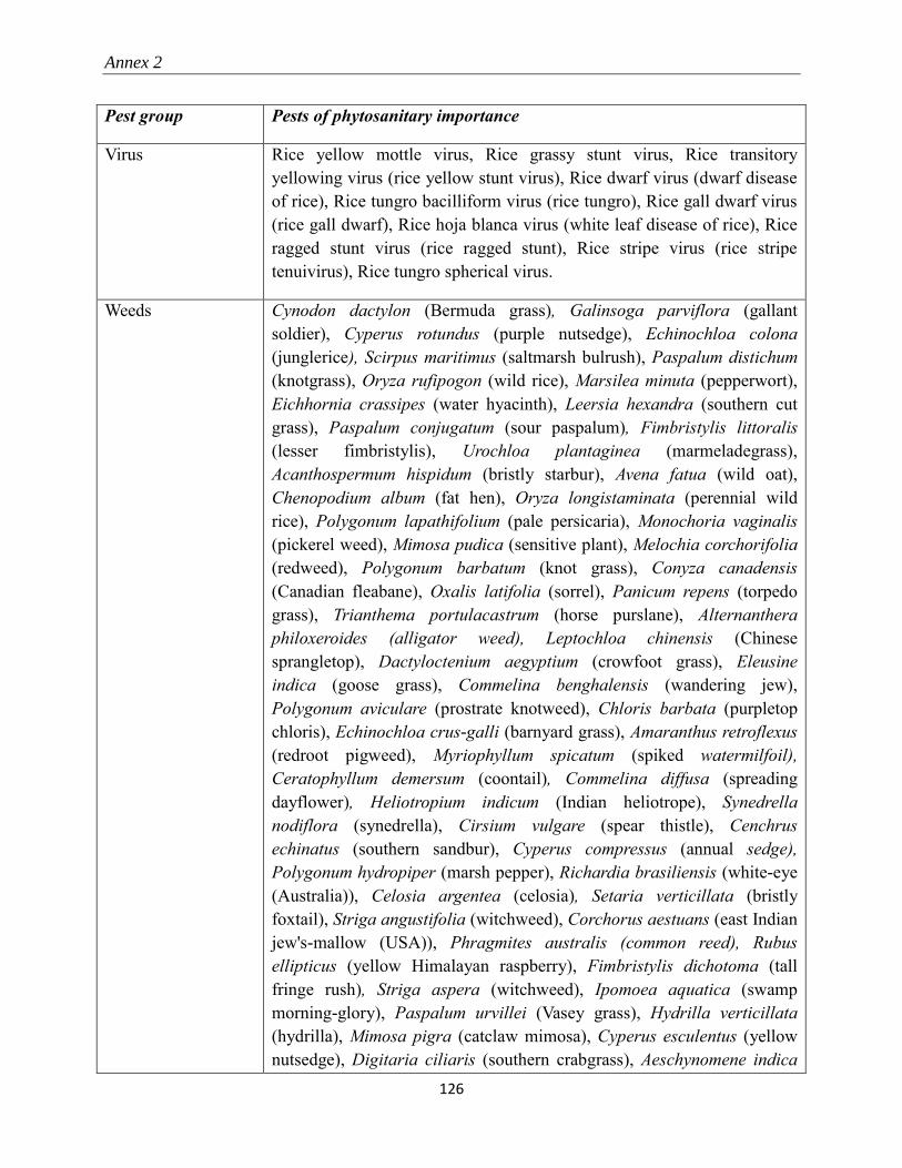
Annex 2
126
Pest group Pests of phytosanitary importance
Virus Rice yellow mottle virus, Rice grassy stunt virus, Rice transitory
yellowing virus (rice yellow stunt virus), Rice dwarf virus (dwarf disease
of rice), Rice tungro bacilliform virus (rice tungro), Rice gall dwarf virus
(rice gall dwarf), Rice hoja blanca virus (white leaf disease of rice), Rice
ragged stunt virus (rice ragged stunt), Rice stripe virus (rice stripe
tenuivirus), Rice tungro spherical virus.
Weeds Cynodon dactylon (Bermuda grass), Galinsoga parviflora (gallant
soldier), Cyperus rotundus (purple nutsedge), Echinochloa colona
(junglerice), Scirpus maritimus (saltmarsh bulrush), Paspalum distichum
(knotgrass), Oryza rufipogon (wild rice), Marsilea minuta (pepperwort),
Eichhornia crassipes (water hyacinth), Leersia hexandra (southern cut
grass), Paspalum conjugatum (sour paspalum), Fimbristylis littoralis
(lesser fimbristylis), Urochloa plantaginea (marmeladegrass),
Acanthospermum hispidum (bristly starbur), Avena fatua (wild oat),
Chenopodium album (fat hen), Oryza longistaminata (perennial wild
rice), Polygonum lapathifolium (pale persicaria), Monochoria vaginalis
(pickerel weed), Mimosa pudica (sensitive plant), Melochia corchorifolia
(redweed), Polygonum barbatum (knot grass), Conyza canadensis
(Canadian fleabane), Oxalis latifolia (sorrel), Panicum repens (torpedo
grass), Trianthema portulacastrum (horse purslane), Alternanthera
philoxeroides (alligator weed), Leptochloa chinensis (Chinese
sprangletop), Dactyloctenium aegyptium (crowfoot grass), Eleusine
indica (goose grass), Commelina benghalensis (wandering jew),
Polygonum aviculare (prostrate knotweed), Chloris barbata (purpletop
chloris), Echinochloa crus-galli (barnyard grass), Amaranthus retroflexus
(redroot pigweed), Myriophyllum spicatum (spiked watermilfoil),
Ceratophyllum demersum (coontail), Commelina diffusa (spreading
dayflower), Heliotropium indicum (Indian heliotrope), Synedrella
nodiflora (synedrella), Cirsium vulgare (spear thistle), Cenchrus
echinatus (southern sandbur), Cyperus compressus (annual sedge),
Polygonum hydropiper (marsh pepper), Richardia brasiliensis (white-eye
(Australia)), Celosia argentea (celosia), Setaria verticillata (bristly
foxtail), Striga angustifolia (witchweed), Corchorus aestuans (east Indian
jew's-mallow (USA)), Phragmites australis (common reed), Rubus
ellipticus (yellow Himalayan raspberry), Fimbristylis dichotoma (tall
fringe rush), Striga aspera (witchweed), Ipomoea aquatica (swamp
morning-glory), Paspalum urvillei (Vasey grass), Hydrilla verticillata
(hydrilla), Mimosa pigra (catclaw mimosa), Cyperus esculentus (yellow
nutsedge), Digitaria ciliaris (southern crabgrass), Aeschynomene indica
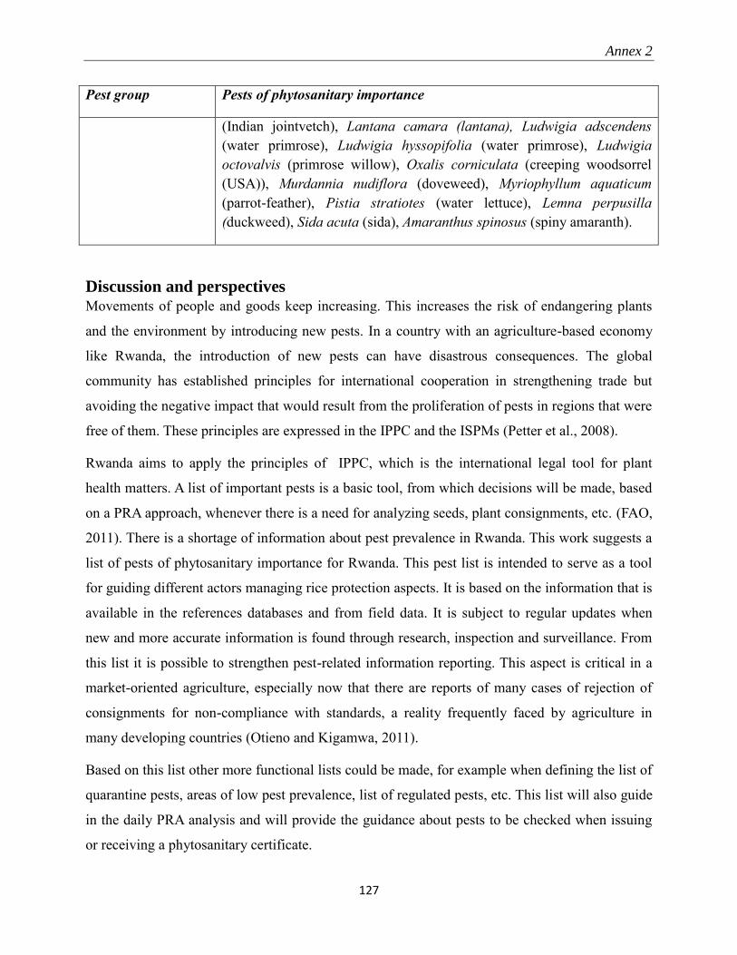
Annex 2
127
Pest group Pests of phytosanitary importance
(Indian jointvetch), Lantana camara (lantana), Ludwigia adscendens
(water primrose), Ludwigia hyssopifolia (water primrose), Ludwigia
octovalvis (primrose willow), Oxalis corniculata (creeping woodsorrel
(USA)), Murdannia nudiflora (doveweed), Myriophyllum aquaticum
(parrot-feather), Pistia stratiotes (water lettuce), Lemna perpusilla
(duckweed), Sida acuta (sida), Amaranthus spinosus (spiny amaranth).
Discussion and perspectives Movements of people and goods keep increasing. This increases the risk of endangering plants
and the environment by introducing new pests. In a country with an agriculture-based economy
like Rwanda, the introduction of new pests can have disastrous consequences. The global
community has established principles for international cooperation in strengthening trade but
avoiding the negative impact that would result from the proliferation of pests in regions that were
free of them. These principles are expressed in the IPPC and the ISPMs (Petter et al., 2008).
Rwanda aims to apply the principles of IPPC, which is the international legal tool for plant
health matters. A list of important pests is a basic tool, from which decisions will be made, based
on a PRA approach, whenever there is a need for analyzing seeds, plant consignments, etc. (FAO,
2011). There is a shortage of information about pest prevalence in Rwanda. This work suggests a
list of pests of phytosanitary importance for Rwanda. This pest list is intended to serve as a tool
for guiding different actors managing rice protection aspects. It is based on the information that is
available in the references databases and from field data. It is subject to regular updates when
new and more accurate information is found through research, inspection and surveillance. From
this list it is possible to strengthen pest-related information reporting. This aspect is critical in a
market-oriented agriculture, especially now that there are reports of many cases of rejection of
consignments for non-compliance with standards, a reality frequently faced by agriculture in
many developing countries (Otieno and Kigamwa, 2011).
Based on this list other more functional lists could be made, for example when defining the list of
quarantine pests, areas of low pest prevalence, list of regulated pests, etc. This list will also guide
in the daily PRA analysis and will provide the guidance about pests to be checked when issuing
or receiving a phytosanitary certificate.
Annex 2
128
Acknowledgments
This study, supervised by the Ghent University and the University of Rwanda/CAVM, was
financially supported by grants from the Belgian Technical Cooperation given to BIGIRIMANA
V.P.
Summary
129
SUMMARY
Rwanda is a developing country located in Eastern/Central Africa whose economy is based on
agriculture. Rice is one of the priority crops in Rwanda. Rice is becoming a popular crop, is
supported by the agricultural policy and it has good performance. It was tested in Rwanda in the
1950s but its production at large scale is recent. The current rice demand is significantly
exceeding the national production and so a large part of the rice consumed in the country,
estimated at around 40%, is imported. One of the factors contributing in limiting rice production
is the impact of pests and diseases. There are not many studies on pests and diseases attacking
rice in Rwanda. Some of the symptoms of rice sheath rot, an emerging rice disease, like the
appearance of extending gray lesions and the non-emergence of infected sheaths, were found in
Rwanda and there is a problem of quality in the locally produced rice, expressed for instance by
the browning of the grains. The aim of this study was to study the situation of rice sheath rot in
Rwanda on the aspects of importance and the causal agents. Bacteria and fungi, were isolated
from the major rice-growing areas, identified, tested for pathogenicity and some management
options are discussed.
Rice sheath rot as a disease attacking rice is known for more than a century, is ubiquitous and
highly destructive. Rice sheath rot was first described in Taiwan. Its symptoms consist of the
rotting of the sheath, associated with browning of the grains, which become largely sterile when
harvested from a diseased plant. In addition, the germinated seeds can transmit the disease.
Second, the disease was reported in most other rice-producing countries all over the world. Third,
the disease is destructive, with yield losses varying from 20-85%, depending on the causal agent,
used rice cultivars, production system, agro-climatic conditions, etc. Most studies cited
Sarocladium oryzae, Fusarium spp. and Pseudomonas fuscovaginae as causal agents, but many
more organisms induce sheath rot disease symptoms in various parts of the world where rice is
grown. A review on the sheath rot reported agents is presented in this work in the literature
chapter. The relationships between the various sheath rot causal agents are not yet known.
The symptoms of rice sheath rot were detected in Rwanda, a country where rice is promoted as a
food security and cash crop. The importance of rice sheath rot as a disease was assessed for
incidence and severity. Data were taken in 2013-2014, from the major rice-growing areas:
Bugarama, Cyili, Rwamagana, Nyagatare and Rwagitima. The disease incidence was 13.5% and
Summary
130
the average measure for disease severity (lesion length on a diseased tiller) was 157 mm. There
are variations on the importance of the disease per area, Rwamagana being more affected by the
disease in the considered period. As for the yield loss, calculated by comparing weight loss in
diseased and healthy grains, it was estimated at 57%. There is an additional loss caused by the
poor quality of grains harvested on diseased plants. Rice sheath rot is causing sizeable losses to
rice production in Rwanda.
Fungi were isolated from sheath rot diseased rice plants from Rwanda, identified and
characterised for pathogenicity on rice plants. The isolated fungi were purified and identified
through morphological and molecular methods, by sequencing the ITS and the TEF genomic
regions. The species identified are Sarocladium oryzae and Fusarium spp. Sarocladium isolates
were similar while there was a large variability in the Fusarium isolates, most of them being
comparable to Fusarium andiyazi and some others being related to species complexes like F.
oxysporum, F. incarnatum/equiseti and F. graminearum. The identified isolates were tested for
pathogenicity, induced sheath rot symptoms and the caused symptoms were comparable for the
two groups. There were differences in susceptibility to the disease between two tested rice
cultivars, Nipponbare cultivar of the Japonica type being more attacked than CO39 cultivar of
the Indica type. In Rwanda, it is mainly the cultivars of the japonica types that are grown and
which perform well, probably because Rwanda, although the country is located in the tropics, its
climate is tempered by the altitude, making it difficult for varieties of the Indica type to adapt to
low temperatures. The pathogenicity of Sarocladium is thought to be linked to toxins, cerulenin
and helvolic acid while the pathogenicity of Fusarium is attributed to mycotoxins, among which
there are fumonisins. Fumonisins were found and measured in most Fusarium isolates from
Rwanda. Their concentration was not correlated with the disease intensity, though this has to be
confirmed by further extensive studies.
Three major genera of bacterial strains were isolated from sheath rot diseased rice plants,
Pseudomonas, Stenotrophomonas and Delftia. Stenotrophomonas and Delftia are commonly
associated with the rice plants as endophytes. More emphasis was put on further identification of
the Pseudomonas isolates by sequencing the rpoB and genes. P. fuscovaginae, the most reported
rice sheath brown rot agent, was not found in the identified Rwandan isolates. Intriguingly, many
of the isolates were related to well-known rhizosphere potential biocontrol agents that are not
commonly associated with the phyllosphere. These isolates proved to be mildly pathogenic in
Summary
131
artificial inoculations. Isolates from Rwanda were found to be related to the sheath rot associated
isolates from the Philippines. But the lack of a 100 % similarity leads to the thinking that most of
the isolates from Rwanda may be new species.
An integrated control seems the option that can work for preventing rice sheath rot. In fact the
disease is caused by many agents, some of them being unknown. Moreover, even for the known
pathogens, pathogenicity mechanisms are not clearly understood. In addition, the interactions
between the various pathogenic agents are not understood. It proves then difficult to apply
specific control measures; thus it is necessary to develop an integrated control strategy. In the
case of Rwanda, a list of potential rice pests, considering the Rwandan agroecology is suggested
in Annex A2. It can be updated as more scientific information become available, and can guide in
designing integrated control options, adapted to the farming system in use in Rwanda.
This work, based on the knowledge on rice sheath rot in the world and specific data from
Rwanda, has paved the way for further studies on rice sheath rot, one of the poorly studied yet
important rice pathogen worldwide. Now that rice has become the first staple food worldwide,
producing one-fifth of calorie consumption and that its production is expanding, rice sheath rot
must be an important element to consider in rice development initiatives, for avoiding important
and unnecessary yield losses, which can reach up to 85% of the production, in the world where it
is required to increase the production for feeding an ever-increasing world population with
limited and degrading natural resources, particularly in tropical and developing countries where
most of rice production is conducted. The efforts on rice sheath rot should concentrate on
furthering the knowledge of causal agents and the interactions among themselves and with the
plant, and understanding the pathogenicity mechanisms, so us to guide the breeding for resistance
and the development of sound integrated control options.

Samenvatting
133
SAMENVATTING
Rwanda is een ontwikkelingsland in Oost/Centraal Afrika. De economie is er hoofdzakelijk
gebaseerd op landbouw met rijst als een van de belangrijkste gewassen. De rijstproductie kent
een stijgende trend, en hoewel het gewas reeds vijftig jaar geleden voor het eerst geteeld werd in
Rwanda, is de grootschalige productie ervan slechts een recent verschijnsel. Het landbouwbeleid
van Rwanda promoot rijst als een markt gewas ter garantie van de voedselveiligheid. Dit heeft als
gevolg dat rijst een sterke stijging in populariteit kent. De vraag naar rijst neemt echter sterker toe
dan de productie zelf, met als gevolg dat tot 40% van de geconsumeerde rijst geïmporteerd wordt.
De belangrijkste limiterende factoren van de rijstproductie zijn ziekten en plagen, waaronder
Sheath rot. Een opkomende, maar weinig bestudeerde ziekte in rijst is het sheath rot complex.
Sheath rot, oftewel bladschederot, wordt onder meer gekenmerkt door grijsbruine laesies op de
bladschede en bruine rijstkorrels. Deze typische symptomen werden geobserveerd in
verscheidene rijstvelden in Rwanda. Bovendien is de kwaliteit van lokaal geproduceerde rijst laag
ten gevolge van onder meer verkleuring van de granen. Deze studie beoogde bijgevolg het belang
van sheath rot in Rwanda te onderzoeken en na te gaan welke pathogenen aan de basis van deze
ziekte liggen. Hiertoe werden stalen verzameld uit de belangrijkste rijst producerende regio’s in
Rwanda waarna de schimmels en bacteriën werden geïsoleerd uit de laesies. Daarnaast werden
ook mogelijke controlemethoden behandeld.
Reeds een eeuw geleden werd sheath rot voor het eerst beschreven in Taiwan. Sindsdien heeft de
ziekte zich wereldwijd verspreid en zorgt voor productieverliezen van 20-85%. Deze laatste
variëren afhankelijk van de ziekteverwekker, de rijstcultivar, het productiesysteem, de agro-
klimatologische condities, etc. In de literatuur wordt deze ziekte geassocieerd met voornamelijk
Sarocladium oryzae, Fusarium spp. en Pseudomonas fuscovaginae. Al deze pathogenen
veroorzaken gelijkaardige symptomen namelijk necrotische laesies op de bladschede waarbij de
ontluikende bloeiwijze verkleurt of helemaal wegrot. De geproduceerde zaden zijn steriel,
verhard en bruin en bovendien is de ziekte zaad overdraagbaar. Naast deze drie voornaamste
pathogenen, kunnen echter nog verscheidene andere pathogenen sheath rot veroorzaken in rijst
producerende regio’s over heel de wereld. In het literatuur hoofdstuk wordt een overzicht
gegeven van de verscheidene sheath rot pathogenen. Het is echter niet duidelijk in welke mate
deze pathogenen samen kunnen voorkomen en onderling interageren.
Samenvatting
134
Om het belang van sheath rot in Rwanda te onderzoeken, werden de incidentie en de ziektegraad
bepaald. De data werden verzameld in 2013-2014 in de belangrijkste rijst producerende regio’s
van Rwanda, namelijk Bugarama, Cyili, Rwamagana, Nyagatare en Rwagitima. De incidentie
was 13.5% en de gemiddelde ziektegraad (laesielengte) was 157mm. Het belang van de ziekte
varieerde naargelang de regio waarbij Rwamagana de sterkst getroffen regio bleek te zijn. De
productieverliezen werden bepaald door het gewichtsverschil tussen zieke en gezonde granen.
Deze werden geschat op 57%. Daarnaast zijn er echter nog additionele productieverliezen door
de daling in de kwaliteit van de rijst met als gevolg dat sheath rot voor aanzienlijke
productieverliezen zorgt in Rwanda.
De schimmels die geïsoleerd werden uit planten met sheath rot, werden geïdentificeerd en getest
op pathogeniciteit op rijst planten. Met zowel morfologische als moleculaire methoden, namelijk
door het sequeneren van de genetische regio’s ITS en TEF, werden de schimmels geïdentificeerd
als Sarocladium oryzae en Fusarium spp. De Sarocladium isolaten waren genetisch sterk
verwant, terwijl de Fusarium isolaten een grotere variatie vertoonden. Het merendeel van de
Fusarium isolaten was nauw verwant met Fusarium andiyazi. Daarnaast werden isolaten verwant
met Fusarium complexen zoals F. oxysporum, F. incarnatum/equiseti en F. graminearum
geïsoleerd. De isolaten werden vervolgens getest op pathogeniciteit bij twee rijstcultivars,
namelijk de Nipponbare cultivar van het Japonica type en de CO39 cultivar van het indica type.
Hieruit bleek dat beide groepen isolaten gelijkaardige symptomen induceerden en dat Nipponbare
een grotere gevoeligheid vertoonde dan CO39. Het zijn echter vooral cultivars van het Japonica
type die in Rwanda geteeld worden daar deze beter aangepast zijn aan het het gematigde klimaat
dat in Rwanda heerst. Hoewel het land zich in de tropen bevindt, heerst een gematigd klimaat ten
gevolge van de hoogteligging. De lage temperaturen maken Rwanda ongeschikt voor het kweken
van cultivars van het Indica type. De pathogeniteit van Sarocladium is te wijten aan de toxines
cerulenin en helvolic acid. Fusarium spp. daarentegen produceren verscheidenen mycotoxines
waaronder fumonisines. Voor merendeel van de Fusarium isolaten konden fumonisines
gedetecteerd worden. Uit kwantificatie van de geproduceerde fumonisine gehaltes in planta bleek
dat de concentratie fumonisines niet gecorreleerd was met de ziektegraad. Om dit te bevestigen
dienen verdere studies plaats te vinden.
De bacterie isolaten die geïsoleerd werden uit sheath rot laesies behoorden tot de genera
Pseudomonas, Stenotrophomonas en Delftia. Stenotrophomonas en Delftia zijn typische
endofyten van rijstplanten. Daarom werd in deze studie de nadruk gelegd op de identificatie van
Samenvatting
135
de Pseudomonas isolaten door middel van sequenering van de en rpoB genen. Hieruit bleek dat
P. fuscovaginae, de meest beschreven sheath brown rot pathogeen, niet geïsoleerd werd uit de
sheath rot planten. Het merendeel van de Pseudomonas isolaten bleek daarentegen nauw verwant
te zijn met bekende biocontrole stammen uit de rhizosfeer. Opmerkelijk is dat, hoewel deze
isolaten werden opgezuiverd uit de bladschede, deze zelden geassocieerd zijn met de fyllosfeer.
Ook deze isolaten werden getest voor pathogeniciteit door middel van artificiële inoculaties en
bleken slechts mild pathogeen te zijn. De isolaten uit Rwanda bleken verwant te zijn met
Pseudomonas isolaten uit sheath rot planten in de Filippijnen. Omdat er echter geen 100%
similariteit is met gekende isolaten, zijn de isolaten uit Rwanda vermoedelijk nieuwe soorten.
Omdat de sheath rot veroorzaakt wordt door verscheidene pathogenen waarvan het mechanisme
van pathogeniteit niet gekend is en bovendien niet alle sheath rot pathogenen al gekend zijn, is
het zeer moeilijk om specifieke controlemethoden toe te passen. Daarom lijkt een geïntegreerde
controlemethode de enige optie om sheath rot te voorkomen. Voor Rwanda werd een lijst
opgesteld met de potentiële ziekten en plagen waarbij de agro-ecologie van Rwanda in rekening
wordt gebracht. Deze lijst kan dan aangevuld worden wanneer nieuwe informatie beschikbaar
wordt. Tevens kan deze lijst als leidraad dienen bij de ontwikkeling van nieuwe geïntegreerde
controlemethoden, aangepast aan de gebruikte landbouwmethoden in Rwanda.
Dit doctoraatsproefschrift, dat de reeds gekende informatie over sheath rot wereldwijd
combineert met specifieke verworven data uit Rwanda, maakt een synthese van de destructieve
doch weinig onderzochte ziekte sheath rot. Deze synthese vormt de basis voor verdere studies
over deze ziekte. Het belang van sheath rot wordt voorspeld nog toe te nemen daar, hoewel rijst
reeds de belangrijkste voedselbron ter wereld is en 20% van de calorieconsumptie voorziet, de
rijstconsumptie nog steeds een stijgende trend kent. Om productieverliezen tot 85% te
voorkomen, is het daarom belangrijk om deze destructieve spreidende ziekte in rekening te
brengen bij ontwikkelingsinitiatieven in de rijstteelt. Het merendeel van de rijstproductie vindt
immers plaats in ontwikkelingslanden, waar een stijging in de rijstproductie vereist is. Opdat
gescreend kan worden naar resistente cultivars en doeltreffende geïntegreerde controlemethoden
kunnen ontwikkeld worden, is het van groot belang dat verdere studies zich concentreren op de
identificatie en karakterisatie van de sheath rot pathogenen. Daarnaast dienen hun
pathogeniteitsmechanismen, hun onderlinge interactie en deze met de gastheerplant onderzocht te
worden

References
137
REFERENCES
Abbas, H.K., D.C. Boyette, and E.R. Hoagland. 1995. Phytotoxicity of Fusarium, other fungal
isolates, and of the phytotoxins fumonisin, fusaric acid, and moniliformin to jimsonweed.
Phytoprotection 76(1): 17–25.
Abbas, H.K., R.D. Cartwright, W.T. Shier, M.M. Abouzied, C.B. Bird, L.G. Rice, P.F. Ross,
G.L. Sciumbato, and F.I. Meredith. 1998. Natural occurrence of fumonisins in rice with
Fusarium sheath rot disease. Plant Disease 82(1): 22–25.
Abbas, H.K., R.D. Cartwright, W. Xie, C.J. Mirocha, J.L. Richard, T.J. Dvorak, G.L. Sciumbato,
and W.T. Shier. 1999. Mycotoxin production by Fusarium proliferatum isolates from rice
with Fusarium sheath rot disease. Mycopathologia 147(2): 97–104.
Adekunle, J. 2007. Culture and Customs of Rwanda. Greenwood Publishing Group, Westport,
Connecticut, USA.
Adorada, D.L., B.J. Stodart, C.V. Cruz, G. Gregorio, I. Pangga, and G.J. Ash. 2013.
Standardizing resistance screening to Pseudomonas fuscovaginae and evaluation of rice
germplasm at seedling and adult plant growth stages. Euphytica 192(1): 1–16.
Adorada, D.L., B.J. Stodart, I.B. Pangga, and G.J. Ash. 2015. Implications of bacterial
contaminated seed lots and endophytic colonization by Pseudomonas fuscovaginae on rice
establishment. Plant Pathology 64(1): 43–50.
Agrios, G.N. 2005. Plant Pathology. 5th Ed. Elsevier Academic Press, New York, NY, USA.
Alavi, P., M.R. Starcher, G.G. Thallinger, C. Zachow, H. Müller, and G. Berg. 2014.
Stenotrophomonas comparative genomics reveals genes and functions that differentiate
beneficial and pathogenic bacteria. BMC Genomics 15: 482: 15 pages.
Amin, K. 1976. Sources of resistance to Acrocylindrium sheath-rot of rice. Plant Disease
Reporter 60(1): 72–73.
Amin, K., B. Sharma, and C. Das. 1974. Occurence in India of sheath rot of rice caused by
Acrocylindrium. Plant Disease Reporter 58(4): 358–360.
Andrews, J.H., and R.F. Harris. 2000. The ecology and biogeography of microorganisms on plant
surfaces. Annual Review of Phytopathology 38: 145–180.
Aoki, T., K. O’Donnell, and D.M. Geiser. 2014. Systematics of key phytopathogenic Fusarium
species: current status and future challenges. Journal of General Plant Pathology 80(3): 189–
201.
Atkins, S.D., and I.M. Clark. 2004. Fungal molecular diagnostics: a mini review. Journal of
Applied Genetics 45(1): 3–15.
Ayyadurai, N., S.I. Kirubakaran, S. Srisha, and N. Sakthivel. 2005. Biological and molecular
variability of Sarocladium oryzae, the sheath rot pathogen of rice (Oryza sativa L.). Current
Microbiology 50(6): 319–23.
Backhouse, D. 2014. Global distribution of Fusarium graminearum, F. asiaticum and F. boothii
References
138
from wheat in relation to climate. European Journal of Plant Pathology 139(1): 161–173.
Baker, P.R. 1970. The introduction of rice in Rwanda. Journal of Tropical Geography 31: 27–32.
Baker, R., R. Cannon, P. Bartlett, and I. Barker. 2005. Novel strategies for assessing and
managing the risks posed by invasive alien species to global crop production and
biodiversity. Annals of Applied Biology 146(2): 177–191.
Ballio, A., F. Bossa, L. Camoni, D. Di Giorgio, M.-C. Flamand, H. Maraite, G. Nitti, P. Pucci,
and A. Scaloni. 1996. Structure of fuscopeptins, phytotoxic metabolites of Pseudomonas
fuscovaginae. FEBS Letters 381(3): 213–216.
Ballio, A., F. Bossa, A. Collina, M. Gallo, N.S. Iacobellis, M. Paci, P. Pucci, A. Scaloni, A.
Segre, and M. Simmaco. 1990. Structure of syringotoxin, a bioactive metabolite of
Pseudomonas syringae pv. syringae. FEBS Letters 269(2): 377–380.
Batoko, H., J. Bouharmont, J.-M. Kinet, and H. Maraite. 1997. Involvement of Toxins Produced
by Pseudomonas fuscovaginae in Aetiology of Rice Bacterial Sheath Brown Rot. Journal of
Phytopathology-Phytopathologische Zeitschrift 145(11-12): 525–531.
Batoko, H., A. de Kerchove d’Exaerde, J.-M. Kinet, J. Bouharmont, R.A. Gage, H. Maraite, and
M. Boutry. 1998. Modulation of plant plasma membrane H+-ATPase by phytotoxic
lipodepsipeptides produced by the plant pathogen Pseudomonas fuscovaginae. Biochimica
et Biophysica Acta 1372(2): 216–226.
Berendsen, R.L., M.C. van Verk, I.A. Stringlis, C. Zamioudis, J. Tommassen, C.M.J. Pieterse,
and P.A.H.M. Bakker. 2015. Unearthing the genomes of plant-beneficial Pseudomonas
model strains WCS358, WCS374 and WCS417. BMC Genomics 16(1): 539.
Bigirimana, V. de P., M. Höfte, and O.I. Nyamangyoku. 2013. Isolation and characterisation of
fungi associated with rice sheath rot in Rwanda. p. 224. In 65th International Symposium
on Crop Protection: Abstracts. Ghent, Belgium.
Bigirimana, V. de P., G.K.H. Hua, O.I. Nyamangyoku, and M. Höfte. 2015. Rice sheath rot : An
emerging ubiquitous destructive disease complex. Frontiers in Plant Sience 6(December):
1066:16 pages.
Bills, G.F., G. Platas, and W. Gams. 2004. Conspecificity of the cerulenin and helvolic acid
producing “Cephalosporium caerulens”, and the hypocrealean fungus Sarocladium oryzae.
Mycological Research 108(11): 1291–1300.
Bizimana, C., F. Usengumukiza, J. Kalisa, and J. Rwirahira. 2012. Trends in key agricultural and
rural development indicators in Rwanda. Kigali, Rwanda.
Blattner, F.R., G.I. Plunkett, A.C. Bloch, T.N. Perna, V. Burland, M. Riley, J. Collado-Vides,
D.J. Glasner, K.C. Rode, F.G. Mayhew, J. Gregor, W.N. Davis, A.H. Kirkpatrick, A.M.
Goeden, J.D. Rose, B. Mau, and Y. Shao. 1997. The complete genome sequence of
Escherichia coli K-12. Science 277(5331): 1453–1462.
Bridge, P.D., D.L. Hawskworth, D.F. Kavishe, and P.A. Farnell. 1989. A revision of the species
concept in Sarocladium, the causal agent of sheath-rot in rice and bamboo blight, based on
biochemical and morphometric analyses. Plant Pathology 38: 239–245.
References
139
CABI. 2007. Crop Protection Compendium. CAB International, Wallingford, UK.
CABI. 2015. Invasive Species Compendium. Available at
http://www.cabi.org/isc/datasheet/48393.
Castellá, G., and F.J. Cabañes. 2014. Phylogenetic diversity of Fusarium incarnatum-equiseti
species complex isolated from Spanish wheat. Antonie van Leeuwenhoek 106(2): 309–17.
Chaijuckam, P., and R.M. Davis. 2010. Efficacy of natural plant products on the control of
aggregate sheath spot of rice. Plant Disease 94(8): 986–992.
Choi, O.-H., H.-Y. Kim, Y.-S. Lee, J.-W. Kim, J.-S. Moon, and I.-G. Hwang. 2012a. First report
of sheath rot of rice caused by Pantoea ananatis in Korea. The Plant Pathology Journal
28(3): 331–331.
Choi, O., J.Y. Lim, Y.-S. Seo, I. Hwang, and J. Kim. 2012b. Complete genome sequence of the
rice pathogen Pantoea ananatis strain PA13. Journal of bacteriology 194(2): 531.
Chowdhury, T.I.M., S. Mian, M.A.T. Mia, M.Y. Rafii, and M.A. Latif. 2015. Agro-ecological
variations of sheath rot disease of rice caused by Sarocladium oryzae and DNA
fingerprinting of the pathogen’s population structure. Genetics and Molecular Research
14(4): 18140–18152.
Cortesi, P., E. Bartoli, C. Pizzatti, W. Song, and N. Schaad. 2005. First report of Acidovorax
avenae ssp. avenae on rice in Italy. Journal of Plant Pathology 87(1): 76–76.
Cother, E.J., D.H. Noble, R.J. Van De Ven, V. Lanoiselet, G. Ash, N. Vuthy, P. Visarto, and B.
Stodart. 2010. Bacterial pathogens of rice in the Kingdom of Cambodia and description of a
new pathogen causing a serious sheath rot disease. Plant Pathology 59(5): 944–953.
Cother, E.J.A., R.B. Reinke, C.A. Mckenzie, V.M.C. Lanoiselet, and D.H.A. Noble. 2004. An
unusual stem necrosis of rice caused by Pantoea ananas and the first record of this pathogen
on rice in Australia. Australasian Plant Pathology 33: 495–503.
Cother, E.J., B. Stodart, D.H. Noble, R. Reinke, and R.J. van de Ven. 2009. Polyphasic
identification of Pseudomonas fuscovaginae causing sheath and glume lesions on rice in
Australia. Australasian Plant Pathology 38(3): 247–261.
Cottyn, B., H. Barrios, T. George, and V. Cruz. 2002. Characterization of rice sheath rot from
Siniloan, Philippines. International Rice Research Notes 27(1): 39–40.
Cottyn, B., J. Debode, E. Regalado, T.W. Mew, and J. Swings. 2009. Phenotypic and genetic
diversity of rice seed-associated bacteria and their role in pathogenicity and biological
control. Journal of Applied Microbiology 107(3): 885–97.
Cottyn, B., E. Regalado, B. Lanoot, M. De Cleene, T.W. Mew, and J. Swings. 2001. Bacterial
populations associated with rice seed in the tropical environment. Phytopathology 91(3):
282–292.
Cottyn, B., M. VanOutryve, M. Cerez, M. DeCleene, J. Swings, and T. Mew. 1996a. Bacterial
diseases of rice.2. Characterization of pathogenic bacteria associated with sheath rot
complex and grain discoloration of rice in the Philippines. Plant Disease 80(4): 438–445.
Cottyn, B., M. VanOutryve, M. Cerez, M. DeCleene, J. Swings, T. Mew, M. VanOutryve, J.
References
140
Barroga, J. Swings, and T. Mew. 1996b. Bacterial diseases of rice.1. Pathogenic bacteria
associated with sheath rot complex and grain discoloration of rice in the Philippines. Plant
Disease 80(4): 438–445.
Cray, J.A., A.N.W. Bell, P. Bhaganna, A.Y. Mswaka, D.J. Timson, and J.E. Hallsworth. 2013.
The biology of habitat dominance; can microbes behave as weeds? Microbial Biotechnology
6(5): 453–92.
Dal Prà, M., S. Tonti, D. Pancaldi, P. Nipoti, and I. Alberti. 2010. First report of Fusarium
andiyazi associated with rice bakanae in Italy. Plant Disease 94(8): 1070.
Dastjerdi, R., and P. Karlovsky. 2015. Systemic infection of maize, sorghum, rice, and beet
seedlings with fumonisin-producing and nonproducing Fusarium verticillioides strains. The
Plant Pathology Journal 31(4): 334–42.
Desjardins, A.E. 2006. Fusarium mycotoxins: chemistry, genetics and biology. American
Phytopathological Society, St. Paul, MN, USA.
Desjardins, E.A., K.H. Manandhar, R.D. Plattner, G.G. Manandhar, S.M. Poling, and C.M.
Maragos. 2000. Fusarium species from Nepalese rice and production of mycotoxins and
gibberellic acid by selected species. Applied and Environmental Microbiology 66(3): 1020–
1025.
Desjardins, A.E., and R.D. Plattner. 1997. Production of Fumonisin B1 and Moniliformin by
Gibberella fujikuroi from rice from various geographic areas. Applied and Environmental
Microbiology 63(5): 1838–1842.
Donnell, K.O., C. Gueidan, S. Sink, P.R. Johnston, P.W. Crous, A. Glenn, R. Riley, N.C.
Zitomer, P. Colyer, C. Waalwijk, T. Van Der Lee, A. Moretti, S. Kang, H. Kim, D.M.
Geiser, J.H. Juba, R.P. Baayen, M.G. Cromey, S. Bithell, D.A. Sutton, K. Skovgaard, R.
Ploetz, H.C. Kistler, M. Elliott, M. Davis, B.A.J. Sarver, K. O’Donnell, C. Gueidan, S. Sink,
P.R. Johnston, P.W. Crous, A. Glenn, R. Riley, N.C. Zitomer, P. Colyer, C. Waalwijk, T.
Van Der Lee, A. Moretti, S. Kang, H. Kim, D.M. Geiser, J.H. Juba, R.P. Baayen, M.G.
Cromey, S. Bithell, D.A. Sutton, K. Skovgaard, R. Ploetz, H. Corby Kistler, M. Elliott, M.
Davis, and B.A.J. Sarver. 2009. A two-locus DNA sequence database for typing plant and
human pathogens within the Fusarium oxysporum species complex. Fungal Genetics and
Biology 46(12): 936–948.
Duveiller, E., K. Miyajima, F. Snacken, A. Autrique, and H. Maraite. 1988. Characterization of
Pseudomonas fuscovaginae and differentiation from other fluorescent pseudomonads
occurring on rice in Burundi. Journal of Phytopathology 122(2): 97–107.
Elliott, M.L. 2005. Survival, growth and pathogenicity of Gaeumannomyces graminis var.
graminis with different methods of long-term storage. Mycologia 97(4): 901–907.
Fageria, N.K. 2007. Yield Physiology of Rice. Journal of Plant Nutrition 30(6): 843–879.
FAO. 1997. International Plant Protection Convention (IPPC): New revised text. Rome, Italy.
FAO. 2011. ISPM 2: Framework for Pest Risk Analysis. FAO/IPPC, Rome, Italy.
FAO. 2016. Sorghum production quantity for Rwanda in 2014: Output preview.
FAOSTATAvailable at http://faostat3.fao.org/download/Q/QC/E (verified 3 June 2016).
References
141
FAO, and WHO. 1996. CODEX standard for rice: CODEX stan 198-1995. Codex alimentarius:
53–60.
Fisher, P.J., and O. Petrini. 1992. Fungal saprobes and pathogens as endophytes of rice (Oryza
sativa L.). New Phytologist 120(1): 137–143.
Flamand, M.-C., S. Pelsser, E. Ewbank, and H. Maraite. 1996. Production of syringotoxin and
other bioactive peptides by Pseudomonas fuscovaginae. Physiological and Molecular Plant
Pathology 48(4): 217–231.
Fory, P.A., L. Triplett, C. Ballen, J.F. Abello, J. Duitama, M.G. Aricapa, G.A. Prado, F. Correa,
J. Hamilton, J.E. Leach, J. Tohme, and G.M. Mosquera. 2014. Comparative analysis of two
emerging rice seed bacterial pathogens. Phytopathology 104(5): 436–44.
Frapolli, M., G. Défago, and Y. Moënne-Loccoz. 2007. Multilocus sequence analysis of
biocontrol fluorescent Pseudomonas spp. producing the antifungal compound 2,4-
diacetylphloroglucinol. Environmental Microbiology 9(8): 1939–55.
Frederick, B.A., T.-C. Caesar-Tonthat, M.H. Wheeler, K.B. Sheehan, W.A. Edens, and J.M.
Henson. 1999. Isolation and characterisation of Gaeumannomyces graminis var. graminis
melanin mutants. Mycological Research 103(1): 99–110.
FSNWG/MAS. 2013. East Africa Cross-border Trade Bulletin April 2013. East Africa Cross-
Border Trade Bulletin (April): 1–5.
FSNWG/MAS. 2014. East Africa Crossborder Trade Bulletin January 2014. East Africa Cross-
Border Trade Bulletin (January): 1–8.
Gams, W., and D.L. Hawksworth. 1975. Identity of Acrocylindrium oryzae Sawada and a similar
fungus causing sheath-rot of rice. Kavaka 3: 57–61.
Gao, J.-X., J. Jing, T. Liu, and J. Chen. 2015. Identification of Clt-1- regulated proteins
associated with the production of non-host-specific toxin and pathogenicity in Cochliobolus
lunatus. Journal of Phytopathology 163(1): 33–41.
Gapusi, R.J., I. Tona, R.A. Nyirigira, R. Freddy, S. Sirikari, J. d’Arc Uwimana, C. Byamushana,
B. Bizeye, P. Hakizimana, G.R. Jean, T. Isibo, N.R. Antoine, R. Freddy, S. Sylvere, and U.
Jeanne. 2013. The state of plant genetic resources for food and agriculture in Rwanda:
Country report. Kigali, Rwanda.
Gardan, L., P. Bella, J.-M. Meyer, R. Christen, P. Rott, W. Achouak, and R. Samson. 2002.
Pseudomonas salomonii sp. nov., pathogenic on garlic, and Pseudomonas palleroniana sp.
nov., isolated from rice. International Journal of Systematic and Evolutionary Microbiology
52(6): 2065–2074.
Gasana, K.J. 2002. Classification of Rwandan agro-bioclimatic regions. p. 6. In MINAGRI (ed.),
Agenda agricole-Ajenda y’ubuhinzi. Rwanda Ministry of Agriculture and Animal Resources
and PASAR-SISA, Kigali, Rwanda.
Ghasemi, A., M. Rostami, and T. Yazdaninia. 2012. In Vitro antagonistic and biodegradation
activity of a newly isolated Delftia tsuruhatensis from rice plant in Iran. International
Journal of Agriculture and Crop Sciences 4(2): 40–44.
References
142
Ghosh, M.K., R. Amudha, S. Jayachandran, and N. Sakthivel. 2002. Detection and quantification
of phytotoxic metabolites of Sarocladium oryzae in sheath rot-infected grains of rice. Letters
in Applied Microbiology 34(6): 398–401.
Giraldo, A., J. Gené, J. Cano, S. de Hoog, and J. Guarro. 2012. Two new species of Acremonium
from Spanish soils. Mycologia 104(6): 1456–65.
Giraldo, A., J. Gené, D.A. Sutton, H. Madrid, G.S. de Hoog, J. Cano, C. Decock, P.W. Crous,
and J. Guarro. 2015. Phylogeny of Sarocladium ( Hypocreales). Persoonia 34: 10–24.
Glenn, A.E., N.C. Zitomer, A.M. Zimeri, L.D. Williams, R.T. Riley, and R.H. Proctor. 2008.
Transformation-mediated complementation of a FUM gene cluster deletion in Fusarium
verticillioides restores both fumonisin production and pathogenicity on maize seedlings.
Molecular Plant-microbe Interactions 21(1): 87–97.
Gnanamanickam, S.S., and T.W. Mew. 1991. Interactions between Sarocladium oryzae and stem
attacking fungal pathogens of rice. Plant and Soil 138: 213–219.
Gomes, L.B., T.J. Ward, E. Badiale-Furlong, and E.M. Del Ponte. 2015. Species composition,
toxigenic potential and pathogenicity of Fusarium graminearum species complex isolates
from southern Brazilian rice. Plant Pathology 64(4): 980–987.
Gomila, M., A. Peña, M. Mulet, and J. Lalucat. 2015. Phylogenomics and systematics in
Pseudomonas. Frontiers in Microbiology 6(March): 1–13.
Gopalakrishnan, C., A. Kamalakannan, and V. Valluvaparidasan. 2010. Effect of seed-borne
Sarocladium oryzae, the incitant of rice sheath rot on rice seed quality. Journal of Plant
Protection Research 50(1): 98–102.
Gopalakrishnan, C., V. Valluvaparidasan, and T. Anand. 2009. Role of rice insect-pests in
transmission of rice sheath rot pathogen, Sarocladium oryzae. Research On Crops 10(1):
159–161.
Goswami, R.S., and H.C. Kistler. 2004. Heading for disaster: Fusarium graminearum on cereal
crops. Molecular Plant Pathology 5(6): 515–25.
Griffin, R.L. 2000. The precautionary approach and phytosanitary measures. p. 1153–1168. In
The BCPC conference: Pests and diseases, Volume 3. British Crop Protection Council,
Brighton, UK.
GRiSP. 2013. Rice almanac. 4th ed. International Rice Research Institute, Los Baños,
Philippines.
Gutiérrez-Nájera, N., R. a Muñoz-Clares, S. Palacios-Bahena, J. Ramírez, S. Sánchez-Nieto, J.
Plasencia, and M. Gavilanes-Ruíz. 2005. Fumonisin B1, a sphingoid toxin, is a potent
inhibitor of the plasma membrane H+-ATPase. Planta 221(4): 589–96.
Hammer, Ø., D.A.T. Harper, and P.D. Ryan. 2001. PAST : Paleontological statistics software
package for education and data analysis. Paleontologia Electronica 4(1): 1–9.
Han, J., L. Sun, X. Dong, Z. Cai, X. Sun, H. Yang, Y. Wang, and W. Song. 2005.
Characterization of a novel plant growth-promoting bacteria strain Delftia tsuruhatensis
HR4 both as a diazotroph and a potential biocontrol agent against various plant pathogens.
References
143
Systematic and Applied Microbiology 28(1): 66–76.
Hardoim, P.R., C.C.P. Hardoim, L.S. van Overbeek, and J.D. van Elsas. 2012. Dynamics of seed-
borne rice endophytes on early plant growth stages. PloS ONE 7(2): e30438.
Hemalatha, R., S. Jebaraj, J. Raja, T. Raguchander, A. Ramanathan, R. Samiyappan, and P.
Balasubramanian. 1999. Employing a crude toxin preparation from Sarocladium oryzae as a
molecular sieve to select sheath rot-resistant somaclones of rice. Journal of Plant
Biochemistry and Biotechnology 8(2): 75–80.
Henson, J.M., M.J. Butler, and A.W. Day. 1999. The dark side of the mycelium: Melanins of
phytopathogenic fungi. Annual Review of Phytopathology 37: 447–471.
Hittalmani, S., H.B. Mahesh, C. Mahadevaiah, and M.K. Prasannakumar. 2016. De novo genome
assembly and annotation of rice sheath rot fungus Sarocladium oryzae reveals genes
involved in helvolic acid and cerulenin biosynthesis pathways. BMC Genomics 17(1): 271.
Höfte, M., and P. De Vos. 2006. Plant pathogenic Pseudomonas species. p. 507–533. In
Gnanamanickam, S.S. (ed.), Plant-Associated Bacteria.
Hsieh, W., S. Smith, and W. Snyder. 1977. Mating groups in Fusarium moniliforme.
Phytopathology 67(8): 1041–1043.
Hu, C.J., Y.R. Li, Y.W. Wei, and S.L. Huang. 2008. A PCR-based method to detect Sclerotium
hydrophilum in infected rice leaf sheaths. Australasian Plant Pathology 37(1): 40.
Hugh, R. 1961. Pseudomonas maltophilia, an Alcaligenes -like Species. Journal of General
Microbiology 26(3): 123–132.
Hummel, N.A., B.A. Castro, E.M. McDonald, M.A. Pellerano, and R. Ochoa. 2009. The panicle
rice mite, Steneotarsonemus spinki Smiley, a re-discovered pest of rice in the United States.
Crop Protection 28(7): 547–560.
IBM. 2012. IBM SPSS Statistics Base 21.
IBM Corporation. 2013. IBM SPSS Statistics for Windows, Version 22.0. Armonk, NY, USA.
Ilyas, M.B., and K. Iftikhar. 1997. Screening of rice germplasm and fungitoxicants against
bakanae disease of rice. Pakistan Journal of Phytopathology 9(1): 67–73.
IRRI. 2002. Standard evaluation system for rice (SES). International Rice Research Institute,
Manilla, the Philippines.
IRRI. 2015a. Rice knowledge bank: Fact sheet for bakanae disease. Available at
http://www.knowledgebank.irri.org/training/fact-sheets/pest-
management/diseases/item/bakanae (verified 16 April 2015).
IRRI. 2015b. World rice statistics in 2013. Available at htttp://ricestat.irri.org (verified 8 April
2015).
Islam, M., Q. Jahan, K. Bunnarith, S. Viangkum, and S. Merca. 2000. Evaluation of seed health
of some rice varieties under different conditions. Botanical Bulletin of Academia Sinica
41(4): 293–297.
References
144
Jagwe, J.N., G. Okoboi, P. Hakizimana, J. Tuyisinge, and M. Rucibigango. 2003. Marketing
survey of the rice sub-sector in Rwanda. Kigali, Rwanda.
Jaunet, T., G. Laguerre, P. Lemanceau, R. Frutos, and J.L. Notteghem. 1995. Diversity of
Pseudomonas fuscovaginae and other fluorescent pseudomonads isolated from diseased rice.
Phytopathology 85: 1534–1541.
Jaunet, T., J.L. Notteghem, and F. Rapilly. 1996. Pathogenicity process of Pseudomonas
fuscovaginae, the causal agent of sheath brown rot of rice. Journal of Phytopathology 144(9-
10): 425–430.
Jeong, Y., J. Kim, S. Kim, Y. Kang, T. Nagamatsu, and I. Hwang. 2003. Toxoflavin produced by
Burkholderia glumae causing rice grain rot is responsible for inducing bacterial wilt in many
field crops. Plant Disease 87(8): 890–895.
Jestoi, M. 2008. Emerging fusarium-mycotoxins fusaproliferin, beauvericin, enniatins, and
moniliformin: a review. Critical Reviews in Food Science and Nutrition 48(1): 21–49.
Joe, Y., and K. Manibhushanrao. 1995. Biochemical constituents of differentially virulent
isolates of Sarocladium spp. causing sheath rot disease of rice. Zeitschrift fur
Pflanzenkrankheiten und Pflanzenschutz-Journal of Plant Diseases and Protection 102(3):
291–298.
Johanson, A., H.C. Turner, G.J. McKay, and A.E. Brown. 1998. A PCR-based method to
distinguish fungi of the rice sheath-blight complex, Rhizoctonia solani, R. oryzae and R.
oryzae-sativae. FEMS Microbiology Letters 162(2): 289–294.
Kadota, I., A. Ohuchi, and K. Nishiyama. 1991. Serological properties and specificity of
Pseudomonas avenae Manns 1909, the causal agent of bacterial brown stripe of rice.
Abstract. Japanese Journal of Phytopathology 57(2): 268–273.
Karmakar, K. 2008. Steneotarsonemus spinki Smiley (Acari : Tarsonemidae) - A yield reducing
mite of rice crops in West Bengal, India. International Journal of Acarology 34(1): 95–99.
Kathiresan, A. 2010. Enabling self-sufficiency and competitiveness of Rwanda rice: Issues and
policy options. Kigali, Rwanda.
Kathiresan, A. 2011. Strategies for sustainable crop intensification in Rwanda. Kigali, Rwanda.
Kim, J.-W., Y.-S. Kang, J.-G. Kim, O.-H. Choi, and I.-G. Hwang. 2010. Occurrence of
Burkholderia glumae on rice and field crops in Korea. The Plant Pathology Journal 26(3):
271–272.
Kim, S., J. Park, J. Lee, D. Shin, D.-S. Park, J.-S. Lim, I.-Y. Choi, and Y.-S. Seo. 2014.
Understanding pathogenic Burkholderia glumae metabolic and signaling pathways within
rice tissues through in vivo transcriptome analyses. Gene 547(1): 77–85.
Kimiharu, I., G. Qingyuan, and A. Masao. 2004. Overwintering of rice sclerotial disease fungi,
Rhizoctonia and Sclerotium spp. in paddy fields in Japan. Plant Pathology Journal 3(2): 81–
87.
Klement, Z. 1955. A new bacterial disease of rice caused by Pseudomonas oryzicola n. sp. Acta
Microbiologica Academiae Scientiarum Hungaricae 2: 265–74.
References
145
Koh, S., H. Kim, J. Kim, E. Goo, Y.-J. Kim, O. Choi, N.-S. Jwa, J. Ma, T. Nagamatsu, J.S.
Moon, and I. Hwang. 2011. A novel light-dependent selection marker system in plants. Plant
Biotechnology Journal 9(3): 348–58.
Kubo, Y., M. Katoh, I. Furusawa, and J. Shishiyama. 1986. Inhibition of melanin biosynthesis by
cerulenin in appressoria of Colletotrichum lagenarium. Experimental Mycology 10(4): 301–
306.
Kushiro, M., H. Saitoh, Y. Sugiura, T. Aoki, S. Kawamoto, and T. Sato. 2012. Experimental
infection of Fusarium proliferatum in Oryza sativa plants; fumonisin B1 production and
survival rate in grains. International Journal of Food Microbiology 156(3): 204–8.
Kutner, C.J., J. Neter, and W. Li. 2005. Applied linear statistical models. 5th Ed. McGraw-Hill
Irwin, New York, NY.
Kvas, M., W.F.O. Marasas, B.D. Wingfield, M.J. Wingfield, and E.T. Steenkamp. 2009.
Diversity and evolution of Fusarium species in the Gibberella fujikuroi complex. Fungal
Diversity 34: 1–21.
Lakshmanan, P. 1992. Sheath rot of rice incited by Curvularia lunata in Tamil Nadu, India.
Tropical Pest Management 38(1): 107–107.
Lakshmanan, P. 1993a. Evaluation of genetic resistance from Oryza sativa and Oryza officinalis
against Cochliobolus lunatus in Tamil Nadu, India. Phytoparasitica 21(1): 63–68.
Lakshmanan, P. 1993b. Efficient method of assessing resistance to sheath rot in rice. Crop
Protection 12(3): 189–192.
Lakshmanan, P., S.M. Kumar, and R. Velusamy. 1992. Role of earhead bug (Leptocorisa acuta)
feeding on sheath rot disease caused by Sarocladium oryzae in Oryza sativa in India.
Phytoparasitica 20(2): 107–112.
Lancashire, P.D., H. Bleiholder, T. Van Den Boom, P. Langelüddeke, R. Stauss, E. Weber, and
A. Witzenberger. 1991. A uniform decimal code for growth stages of crops and weeds.
Annals of Applied Biology 119(3): 561–601.
Lang, E., M. Burghartz, S. Spring, J. Swiderski, and C. Spröer. 2010. Pseudomonas
benzenivorans sp. nov. and Pseudomonas saponiphila sp. nov., represented by xenobiotics
degrading type strains. Current Microbiology 60(2): 85–91.
Lanoiselet, V.M., E.J. Cother, and G.J. Ash. 2007. Aggregate sheath spot and sheath spot of rice.
Crop Protection 26(6): 799–808.
Lanoiselet, V.M., E.J. Cother, G.J. Ash, and J.D.I. Harper. 2002. First report of Sclerotium
hydrophilum on leaf sheath of rice (Oryza sativa) in South-Eastern Australia. Plant
Pathology 51(6): 813–813.
Lanoiselet, V., M.P. You, Y.P. Li, C.P. Wang, R.G. Shivas, and M.J. Barbetti. 2012. First report
of Sarocladium oryzae causing sheath rot on rice (Oryza sativa ) in Western Australia. Plant
Disease 96(9): 1382–1382.
Lee, S.-H., J.-H. Kim, S.-W. Son, T. Lee, and S.-H. Yun. 2012. Fumonisin production by field
isolates of the Gibberella fujikuroi species complex and Fusarium commune obtained from
References
146
rice and corn in Korea. Research in Plant Disease 18(4): 310–316.
Leplat, J., H. Friberg, M. Abid, and C. Steinberg. 2012. Survival of Fusarium graminearum, the
causal agent of Fusarium head blight. A review. Agronomy for Sustainable Development
33(1): 97–111.
Leslie, J.F., L.L. Anderson, R.L. Bowden, and Y.-W. Lee. 2007. Inter- and intra-specific genetic
variation in Fusarium. International Journal of Food Microbiology 119(1-2): 25–32.
Li, J., G. Jiang, B. Yang, X. Dong, L. Feng, S. Lin, F. Chen, M. Ashraf, and Y. Jiang. 2012. A
luminescent bacterium assay of fusaric acid produced by Fusarium proliferatum from
banana. Analytical and Bioanalytical Chemistry 402(3): 1347–54.
Liu, T., L. Liu, X. Jiang, X. Huang, and J. Chen. 2009. A new furanoid toxin produced by
Curvularia lunata, the causal agent of maize Curvularia leaf spot. Canadian Journal of Plant
Pathology 31: 22–27.
Liu, H., W.-X. Tian, M. Ibrahim, B. Li, G.-Q. Zhang, B. Zhu, and G.-L. Xie. 2012a.
Characterization of pilP, a gene required for twitching motility, pathogenicity, and biofilm
formation of Acidovorax avenae subsp. avenae RS-1. European Journal of Plant Pathology
134(3): 551–560.
Liu, Q., Y. Zhang, N. Yu, Z. Bi, A. Zhu, X. Zhan, W. Wu, P. Yu, D. Chen, S. Cheng, and L. Cao.
2015. Genome sequence of Pseudomonas parafulva CRS01-1, an antagonistic bacterium
isolated from rice field. Journal of Biotechnology 206: 89–90.
Liu, Y., S. Zuo, L. Xu, Y. Zou, and W. Song. 2012b. Study on diversity of endophytic bacterial
communities in seeds of hybrid maize and their parental lines. Archives of Microbiology
194(12): 1001–12.
López, M.M., P. Llop, A. Olmos, E. Marco-Noales, M. Cambra, and E. Bertolini. 2009. Are
molecular tools solving the challenges posed by detection of plant pathogenic bacteria and
viruses? Current Issues in Molecular Biology 11: 13–46.
Louws, J.F., and A.D. Cuppels. 2001. Molecular techniques. p. 321–337. In Schaad, N.W., Jones,
J.B., Chun, W. (eds.), Laboratory guide for the identification of plant pathogenic bacteria.
3rd Ed. APS, St. Paul, Minnesota, USA.
Ma, L.-J., D.M. Geiser, R.H. Proctor, A.P. Rooney, K. O’Donnell, F. Trail, D.M. Gardiner, J.M.
Manners, and K. Kazan. 2013. Fusarium pathogenomics. Annual Review of Microbiology
67: 399–416.
Manneh, B., and R. El Namaky. 2010. Report on rice trip in Ethiopia, Rwanda, Tanzania and
Uganda.
Mano, H., and H. Morisaki. 2008. Endophytic Bacteria in the Rice Plant. Microbes and
Environments 23(2): 109–117.
Mano, H., F. Tanaka, C. Nakamura, H. Kaga, and H. Morisaki. 2007. Culturable endophytic
bacterial flora of the maturing leaves and roots of rice plants (Oryza sativa) cultivated in a
paddy field. Microbes and Environments 22(2): 175–185.
Marasas, W.F.O., J.P. Rheeder, S.C. Lamprecht, K.A. Zeller, and J.F. Leslie. 2001. Fusarium
References
147
andiyazi sp. nov., a new species from sorghum. Mycologia 93(6): 1203.
Marín, P., A. Moretti, A. Ritieni, M. Jurado, C. Vázquez, and M.T. González-Jaén. 2012.
Phylogenetic analyses and toxigenic profiles of Fusarium equiseti and Fusarium
acuminatum isolated from cereals from Southern Europe. Food Microbiology 31(2): 229–
37.
Matić, S., D. Spadaro, A. Garibaldi, and M.L. Gullino. 2014. Antagonistic yeasts and
thermotherapy as seed treatments to control Fusarium fujikuroi on rice. Biological Control
73: 59–67.
Mattiuzzo, M., I. Bertani, S. Ferluga, L. Cabrio, J. Bigirimana, C. Guarnaccia, S. Pongor, H.
Maraite, and V. Venturi. 2011. The plant pathogen Pseudomonas fuscovaginae contains two
conserved quorum sensing systems involved in virulence and negatively regulated by RsaL
and the novel regulator RsaM. Environmental Microbiology 13(1): 145–62.
Mavrodi, D. V, T.L. Peever, O. V Mavrodi, J.A. Parejko, J.M. Raaijmakers, P. Lemanceau, S.
Mazurier, L. Heide, W. Blankenfeldt, D.M. Weller, and L.S. Thomashow. 2010. Diversity
and evolution of the phenazine biosynthesis pathway. Applied and Environmental
Microbiology 76(3): 866–79.
Mehri, I., Y. Turki, M. Chair, H. Cherif, A. Hassen, J.-M. Meyer, and M. Gtari. 2011. Genetic
and functional heterogeneities among fluorescent Pseudomonas isolated from environmental
samples. Journal of General and Applied Microbiology 57(2): 101–114.
Mew, T.W., B. Cottyn, R. Pamplona, H. Barrios, L. Xiangmin, C. Zhiyi, L. Fan, N. Nil-panit, P.
Arunyanart, P. Van Kim, and P. Van Du. 2004a. Applying rice seed-associated antagonistic
bacteria to manage rice sheath blight in developing countries. Plant Disease 88(5): 557–564.
Mew, T.W., and P. Gonzales. 2002. A handbook of rice seedborne fungi. IRRI, Science
Publishers, Los Baños, Philippines; Enfield, NH, USA.
Mew, T.W., H. Leung, S. Savary, C.M. Vera Cruz, and J.E. Leach. 2004b. Looking ahead in rice
disease research and management. Critical Reviews in Plant Sciences 23(2): 103–127.
Meyer, H. 1902. Oskar Baumann (originally in German). p. 255–256. In General German
Biography. 46th ed. Historical Commission of the Bavarian Academy of Sciences.
MINAGRI. 2002. Farmer’s diary. Ministry of Agriculture and Animal Resources, Kigali,
Rwanda.
MINAGRI. 2005. Programme national pour la production du riz, période: 2006-2016. Kigali,
Rwanda.
MINAGRI. 2009a. Plan for the Transformation of Agriculture in Rwanda – Phase II ( PSTA II ).
Kigali, Rwanda.
MINAGRI. 2009b. Farmer’s diary. Rwanda Ministry of Agriculture and Animal Resources,
Kigali, Rwanda.
MINAGRI. 2011a. National rice development strategy (Period 2011-2018). Ministry of
Agriculture and Animal Resources, Kigali, Rwanda.
MINAGRI. 2011b. Annual Report FY 2010/2011. Kigali, Rwanda.
References
148
MINAGRI. 2013a. National rice development strategy (2011-2018): Revised in August 2013.
Ministry of Agriculture and Animal Resources, Kigali, Rwanda.
MINAGRI. 2013b. Plant pest surveillance report. Ministry of Agriculture and Animal Resources,
Kigali, Rwanda.
Miyajima, K., A. Tanii, and T. Akita. 1983. Pseudomonas fuscovaginae sp. nov., nom. rev.
International Journal of Systematic Bacteriology 33(3): 656–657.
Miyake, T., H. Tateishi, Y. Sakuma, and T. Saishoji. 2012. A novel soil-type biopesticide
KNB422-soil against rice seedling diseases. Journal of Pesticide Science 37(2): 129–134.
Monteiro, R.A., E. Balsanelli, R. Wassem, A.M. Marin, L.C.C. Brusamarello-Santos, M.A.
Schmidt, M.Z. Tadra-Sfeir, V.C.S. Pankievicz, L.M. Cruz, L.S. Chubatsu, F.O. Pedrosa, and
E.M. Souza. 2012. Herbaspirillum-plant interactions: microscopical, histological and
molecular aspects. Plant and Soil 356(1-2): 175–196.
Morris, C.E., C.L. Monteil, and O. Berge. 2013. The life history of Pseudomonas syringae:
linking agriculture to earth system processes. Annual Review of Phytopathology 51: 85–
104.
Mulet, M., A. Bennasar, J. Lalucat, and E. García-Valdés. 2009. An -based PCR procedure for
the identification of Pseudomonas species and for their detection in environmental samples.
Molecular and Cellular Probes 23(3-4): 140–7.
Mulet, M., J. Lalucat, and E. García-Valdés. 2010. DNA sequence-based analysis of the
Pseudomonas species. Environmental Microbiology 12(6): 1513–30.
Mutware, J.S., and K. Burger. 2014. Factors influencing the adoption of improved rice varieties
in Rwanda: An application of the conditional logit model (CLM). p. 267–279. In Vanlauwe,
B., van Asten, P., Blomme, G. (eds.), Challenges and Opportunities for Agricultural
Intensification of the Humid Highland Systems of Sub-Saharan Africa. Springer.
Naeimi, S., S.M. Okhovvat, G.A. Hedjaroude, and V. Khosravi. 2003. Sheath rot of rice in Iran.
Abstract. Communications in Agricultural and Applied Biological Sciences 68(4 Pt B): 681–
4.
Naik, P.R., N. Sahoo, D. Goswami, N. Ayyadurai, and N. Sakthivel. 2008. Genetic and
functional diversity among fluorescent pseudomonads isolated from the rhizosphere of
banana. Microbial Ecology 56(3): 492–504.
Nandakumar, R., S. Babu, G. Amutha, T. Raguchander, and R. Samiyappan. 2007a. Variation in
toxin production among isolates of Sarocladium oryzae, the rice sheath rot pathogen. Plant
Pathology Journal 6(2): 120–126.
Nandakumar, R., P.A. Bollich, A.K.M. Shahjahan, D.E. Groth, and M.C. Rush. 2008. Evidence
for the soilborne nature of the rice sheath rot and panicle blight pathogen, Burkholderia
gladioli. Canadian Journal of Plant Pathology-Revue Canadienne de Phytopathologie 30(1):
148–154.
Nandakumar, R., M.C. Rush, and F. Correa. 2007b. Association of Burkholderia glumae and B.
gladioli with panicle blight symptoms on rice in Panama. Plant Disease 91(6): 767–767.
References
149
Nandakumar, R., A.K.M. Shahjahan, X.L. Yuan, E.R. Dickstein, D.E. Groth, C.A. Clark, R.D.
Cartwright, and M.C. Rush. 2009. Burkholderia glumae and B. gladioli cause bacterial
panicle blight in rice in the Southern United States. Plant Disease 93(9): 896–905.
Narasimhan, V., K. Rajappan, C. Ushamalini, and A.A. Kareem. 1998. Efficacy of new EC
formulations of neem oil and pungam oil for the management of sheath rot disease of rice.
Phytoparasitica 26(4): 301–306.
Ndikumana, I., and E.R. Gasore. 2010. Description of new rice varieties introduced in Rwanda.
Kigali, Rwanda.
Ndikumana, I., R. Gasore, I. Issaka, A. Pinel-Galzi, A. Onasanya, A. Hassani-Mehraban, D.
Fargete, D. Peters, and Y. Séré. 2011. Rice yellow mottle virus in Rwanda: First report and
evidence of strain circulation. New Disease Reports 23: 18.
Nelson, P.E., T.A. Toussoun, and W.F.O. Marasas. 1983. Fusarium Species: an illustrated
manual for identification. Pennsylvania State University Press, University Park,
Pennsylvania, USA.
Nesic, K., S. Ivanovic, and V. Nesic. 2014. Fusarial toxins : Secondary metabolites of Fusarium
fungi. p. 101–120. In Whitacre, D.M. (ed.), Reviews of Environmental Contamination and
Toxicology. Springer International Publishing, Switzerland.
Ngango, J. 2013. The promotion of rice production as a solution to the lower levels of saving and
income in the rural areas of Rwanda. Case study of KOAIRWA Cooperative, Study period
of 2007-2011.
NISR. 2010. Statistical Yearbook 2009. Kigali, Rwanda.
NISR. 2012. EICV3 Thematic report: Agriculture. Kigali, Rwanda.
NISR. 2013. Rwanda statistical yearbook 2013. Kigali, Rwanda.
Ntirenganya, E. 2014. Local products struggle to make it into supermarket shelves. The New
Times (September 30, 2014).
O’Donnell, K., E. Cigelnik, and H.I. Nirenberg. 1998. Molecular systematics and
phylogeography of the Gibberella fujikuroi Species Complex. Mycologia 90(3): 465–493.
O’Donnell, K., R.A. Humber, D.M. Geiser, S. Kang, B. Park, V.A.R.G. Robert, P.W. Crous, P.R.
Johnston, T. Aoki, A.P. Rooney, S.A. Rehner, S. Mycologia, N.M. April, and D.M. Geiser.
2012. Phylogenetic diversity of insecticolous fusaria inferred from multilocus DNA
sequence data and their molecular identification via FUSARIUM-ID and Fusarium MLST.
Mycologia 104(2): 427–445.
O’Donnell, K., H.C. Kistler, B.K. Tacke, and H.H. Casper. 2000. Gene genealogies reveal global
phylogeographic structure and reproductive isolation among lineages of Fusarium
graminearum, the fungus causing wheat scab. Proceedings of the National Academy of
Sciences of the United States of America 97(14): 7905–10.
O’Donnell, K., A.P. Rooney, R.H. Proctor, D.W. Brown, S.P. McCormick, T.J. Ward, R.J.N.
Frandsen, E. Lysøe, S.A. Rehner, T. Aoki, V.A.R.G. Robert, P.W. Crous, J.Z. Groenewald,
S. Kang, and D.M. Geiser. 2013. Phylogenic analyses of RPB1 and RPB2 support a middle
References
150
Cretaceous origin for a clade comprising all agriculturally and medically important Fusaria.
Fungal Genetics and Biology 52: 20–31.
O’Donnell, K., T.J. Ward, D.M. Geiser, H. Corby Kistler, and T. Aoki. 2004. Genealogical
concordance between the mating type locus and seven other nuclear genes supports formal
recognition of nine phylogenetically distinct species within the Fusarium graminearum
clade. Fungal Genetics and Biology 41(6): 600–23.
Olorunleke, F.E., N.P. Kieu, and M. Hofte. 2015. Recent advances in Pseudomonas biocontrol. p.
167–198. In Murillo, J., Vinatzer, B.A., Jackson, R.W., Dawn, L.A. (eds.), Bacteria-plant
interactions : advanced research and future trends. Caister Academic Press.
Omura, S. 1976. The antibiotic cerulenin, a novel tool for biochemistry as an inhibitor of fatty-
acid synthesis. Bacteriological Reviews 40(3): 681–697.
Oster, J. 1992. Reaction of a resistant breeding line and susceptible California rice cultivars to
Sclerotium oryzae. Plant Disease 76(7): 740–744.
Otieno, W., and J.N. Kigamwa. 2011. Challenges in compliance with official standards and
implications in market access: Case of Kenya’s horticultural produce. Acta horticulturae:
263–268.
Ou, S.H. 1985. Rice diseases. CMI, Kew, UK.
Paganin, P., S. Tabacchioni, and L. Chiarini. 2011. Pathogenicity and biotechnological
applications of the genus Burkholderia. Central European Journal of Biology 6(6): 997–
1005.
Palleroni, N.J., and J.F. Bradbury. 1993. Stenotrophomonas, a new bacterial genus for
Xanthomonas maltophilia (Hugh 1980) Swings et al. 1983. International Journal of
Systematic Bacteriology 43(3): 606–609.
Park, J.W., S.-Y. Choi, H.-J. Hwang, and Y.-B. Kim. 2005. Fungal mycoflora and mycotoxins in
Korean polished rice destined for humans. International Journal of Food Microbiology
103(3): 305–14.
Patel, H., M. Matiuzzo, I. Bertani, V. de P. Bigirimana, G.J. Ash, M. Höfte, and V. Venturi.
2014. Identification of virulence associated loci in the emerging broad host range plant
pathogen Pseudomonas fuscovaginae. BMC Microbiology 14(November): 274.
Patel, H.K., D.P. da Silva, G. Devescovi, H. Maraite, K. Paszkiewicz, D.J. Studholme, and V.
Venturi. 2012. Draft genome sequence of Pseudomonas fuscovaginae, a broad-host-range
pathogen of plants. Journal of Bacteriology 194(10): 2765–6.
Pauwels, K., M.M. Sanchez del Pino, G. Feller, and P. Van Gelder. 2012. Decoding the folding
of Burkholderia glumae lipase: folding intermediates en route to kinetic stability. PloS ONE
7(5): e36999.
Pearce, D.A., P.D. Bridge, and D.L. Hawksworth. 2001. Species concept in Sarocladium, the
causal agent in sheath rot in rice and bamboo blight. p. 285–292. In Sreenivasaprasad, S.,
Johnson, R. (eds.), Major fungal diseases of rice: Recent advances. Springer, The
Netherlands.
References
151
Peixoto, C.N., G. Ottoni, M.C.C. Filippi, V.L. Silva-Lobo, and A.S. Prabhu. 2013. Biology of
Gaeumannomyces graminis var. graminis isolates from rice and grasses and epidemiological
aspects of crown sheath rot of rice. Tropical Plant Pathology 38(6): 495–504.
Perdomo, H., D. a Sutton, D. García, a W. Fothergill, J. Cano, J. Gené, R.C. Summerbell, M.G.
Rinaldi, and J. Guarro. 2011. Spectrum of clinically relevant Acremonium species in the
United States. Journal of Clinical Microbiology 49(1): 243–56.
Perneel, M., J. Heyrman, A. Adiobo, K. De Maeyer, J.M. Raaijmakers, P. De Vos, and M. Höfte.
2007. Characterization of CMR5c and CMR12a, novel fluorescent Pseudomonas strains
from the cocoyam rhizosphere with biocontrol activity. Journal of Applied Microbiology
103(4): 1007–20.
Petter, F., A.S. Roy, and I. Smith. 2008. International standards for the diagnosis of regulated
pests. European Journal of Plant Pathology 121: 331–337.
Philmus, B., B.T. Shaffer, T.A. Kidarsa, Q. Yan, J.M. Raaijmakers, T.P. Begley, and J.E. Loper.
2015. Investigations into the biosynthesis, regulation, and self-resistance of toxoflavin in
Pseudomonas protegens Pf-5. ChemBioChem 16(12): 1782–1790.
Prabhu, A.S., and I.P. Bedendo. 1983. Basal node rot of rice caused by Fusarium oxysporum in
Brazil. Plant Disease 67: 228–229.
Prabhu, A., M. Filippi, G. da Silva, and G. de Santos. 2002. Resistance of rice cultivars to
Rhizoctonia solani and Rhizoctonia oryzae. Pesquisa Agropecuaria Brasileira 37(5): 589–
595.
Proctor, R.H., M. Busman, J.-A. Seo, Y.W. Lee, and R.D. Plattner. 2008. A fumonisin
biosynthetic gene cluster in Fusarium oxysporum strain O-1890 and the genetic basis for B
versus C fumonisin production. Fungal Genetics and Biology 45(6): 1016–26.
Proctor, R.H., F. Van Hove, A. Susca, G. Stea, M. Busman, T. van der Lee, C. Waalwijk, A.
Moretti, and T.J. Ward. 2013. Birth, death and horizontal transfer of the fumonisin
biosynthetic gene cluster during the evolutionary diversification of Fusarium. Molecular
Microbiology 90(2): 290–306.
Promar. 2012. Agriculture, Forestry and Fisheries of Rwanda: Fact-finding survey for the support
of aid to developing countries. Tokyo, Japan.
Purkayastha, R.P., and A. Ghosal. 1985. Analysis of cross-reactive antigens of Acrocylindrium
oryzae and rice in relation to sheath rot disease. Physiological Plant Pathology 27(2): 245–
252.
Quazi, S.A.J., S. Meon, H. Jaafar, and Z.A.B.M. Ahmad. 2013. Characterization of Fusarium
proliferatum through species specific primers and its virulence on rice seeds. International
Journal of Agriculture and Biology 15: 649–656.
Quibod, I.L., G. Grande, E.G. Oreiro, F.N. Borja, G.S. Dossa, R. Mauleon, C.V. Cruz, and R.
Oliva. 2015. Rice-infecting Pseudomonas genomes are highly accessorized and harbor
multiple putative virulence mechanisms to cause sheath brown rot. Plos ONE 10(9):
e0139256.
RAB. 2013. Overview of rice development in Rwanda. Rwanda Agriculture Board, Kigali,
References
152
Rwanda.
RAB. 2014. Rice programme. Available at http://www.rab.gov.rw/spip.php?article29 (verified 15
October 2014).
Ramette, A., M. Frapolli, M. Fischer-Le Saux, C. Gruffaz, J.-M. Meyer, G. Défago, L. Sutra, and
Y. Moënne-Loccoz. 2011. Pseudomonas protegens sp. nov., widespread plant-protecting
bacteria producing the biocontrol compounds 2,4-diacetylphloroglucinol and pyoluteorin.
Systematic and Applied Microbiology 34(3): 180–188.
Razak, A.A., N.A.I.M. Zainudin, S.N.M. Sidiqe, N.A. Ismail, N.M.I.N. Mohamad, and B. Salleh.
2009. Sheath brown rot disease of rice caused by Pseudomonas fuscovaginae in the
Peninsular Malaysia. Journal of Plant Protection Research 49(3): 244–249.
R-Biopharm. 2015. Good ELISA practice manual. Innovative. Darmstadt, Germany.
RDB. 2014a. Investment opportunity: Rice production & processing. Available at
http://www.rdb.rw/fileadmin/user_upload/Documents/Agriculture/Rice production and
processing.pdf (verified 15 October 2016).
RDB. 2014b. Opportunities for seed industry in Rwanda. Available at
http://www.rdb.rw/fileadmin/user_upload/Documents/Agriculture/Seed production.pdf
(verified 21 October 2014).
Riera-Ruiz, C., J. Vargas, C. Cedeño, P. Quirola, M. Escobar, J.M. Cevallos-Cevallos, M. Ratti,
and E.L. Peralta. 2014a. First Report of Burkholderia glumae Causing Bacterial Panicle
Blight on Rice in Ecuador. Plant Disease 98(7): 988–988.
Riera-Ruiz, C., J. Vargas, J.M. Cevallos-Cevallos, M. Ratti, and E.L. Peralta. 2014b. First report
of bacterial panicle blight of rice caused by Burkholderia gladioli in Ecuador. Plant Disease
98(11): 1577–1577.
Rofiat, A.-S., F. Fanelli, O. Atanda, M. Sulyok, G. Cozzi, S. Bavaro, R. Krska, A.F. Logrieco,
and C.N. Ezekiel. 2015. Fungal and bacterial metabolites associated with natural
contamination of locally processed rice (Oryza sativa L.) in Nigeria. Food additives &
contaminants. Part A, Chemistry, analysis, control, exposure & risk assessment 32(6): 950–
9.
Rott, P., J.L. Notteghem, and P. Frossard. 1989. Identification and characterization of
Pseudomonas fuscovaginae, the causal agent of bacterial sheath brown rot of rice, from
Madagascar and other countries. Plant Disease 73(2): 133–137.
Ruiz-Roldán, C., Y. Pareja-Jaime, J.A. González-Reyes, and M.I. G-Roncero. 2015. The
transcription factor con7-1 is a master regulator of morphogenesis and virulence in
Fusarium oxysporum. Molecular Plant-microbe Interactions 28(1): 55–68.
Saberi, E., N. Safaie, and H. Rahimian. 2013. Characterization of bacterial strains associated with
sheath rot complex and grain discoloration of rice in North of Iran (Mazandaran province).
Journal of Bacteriology Research 5(5): 51–61.
Sakthivel, N. 2001. Sheath rot disease of rice: Current status and control strategies. p. 271–283.
In Sreenivasaprasad, S., Johnson, R. (eds.), Major fungal diseases of rice: Recent advances.
Springer, The Netherlands.
References
153
Sakthivel, N., R. Amudha, and S. Muthukrishnan. 2002. Production of phytotoxic metabolites by
Sarocladium oryzae. Mycological Research 106(1150): 609–614.
Sakthivel, N., and S.S. Gnanamanickam. 1987. Evaluation of Pseudomonas fluorescens for
suppression of sheath rot disease and for enhancement of grain yields in rice (Oryza sativa
L.). Applied and Environmental Microbiology 53(9): 2056–2059.
Saravanakumar, D., N. Lavanya, K. Muthumeena, T. Raguchander, and R. Samiyappan. 2009.
Fluorescent pseudomonad mixtures mediate disease resistance in rice plants against sheath
rot (Sarocladium oryzae) disease. BioControl 54(2): 273–286.
Savary, S., F. Horgan, L. Willocquet, and K.L. Heong. 2012. A review of principles for
sustainable pest management in rice. Crop Protection 32: 54–63.
Savary, S., L. Willocquet, F.A. Elazegui, N.P. Castilla, and P.S. Teng. 2000. Rice pest constraints
in tropical Asia : Quantification of yield losses due to rice pests in a range of production
situations. Plant Disease 84(3): 357–369.
Sayler, R.J., R.D. Cartwright, and Y. Yang. 2006. Genetic characterization and real-time PCR
detection of Burkholderia glumae, a newly emerging bacterial pathogen of rice in the United
States. Plant Disease 90(5): 603–610.
Schaad, N.W., J.B. Jones, and W. Chun. 2001. Laboratory guide for identification of plant
pathogenic bacteria. 3rd Ed. APS, St. Paul, Minnesota.
Schaad, N.W., E. Postnikova, A. Sechler, L.E. Claflin, A.K. Vidaver, J.B. Jones, I. Agarkova, A.
Ignatov, E. Dickstein, and B.A. Ramundo. 2008. Reclassification of subspecies of
Acidovorax avenae as A. Avenae (Manns 1905) emend., A. cattleyae (Pavarino, 1911) comb.
nov., A. citrulli Schaad et al., 1978) comb. nov., and proposal of A. oryzae sp. nov.
Systematic and Applied Microbiology 31(6-8): 434–446.
Schlaeppi, K., and D. Bulgarelli. 2015. The plant microbiome at work. Molecular Plant-Microbe
Interactions 28(3): 212–217.
Schmidt, M.A., E. Balsanelli, H. Faoro, L.M. Cruz, R. Wassem, V.A. de Baura, V. Weiss, M.G.
Yates, H.M.F. Madeira, L. Pereira-Ferrari, M.H.P. Fungaro, F.M. de Paula, L.F.P. Pereira,
L.G.E. Vieira, F.L. Olivares, F.O. Pedrosa, E.M. de Souza, and R.A. Monteiro. 2012. The
type III secretion system is necessary for the development of a pathogenic and endophytic
interaction between Herbaspirillum rubrisubalbicans and Poaceae. BMC Microbiology 12:
98: 17 pages.
Seo, Y.S., J. Kim, H. Kim, E. Goo, B. Lee, J.S. Lee, J.S. Moon, and I. Hwang. 2011. Rice sheath
and grain rot caused by Burkholderia gladioli in Korea. Journal of Plant Pathology 93(S4):
87–87.
Seok, Y., H. Shin, Y. Lee, I. Cho, S. Na, D. Yong, S.H. Jeong, and K. Lee. 2010. First report of
bloodstream infection caused by Pseudomonas fulva. Journal of Clinical Microbiology
48(7): 2656–7.
Shah, F., L. Nie, K. Cui, T. Shah, W. Wu, C. Chen, L. Zhu, F. Ali, S. Fahad, and J. Huang. 2014.
Rice grain yield and component responses to near 2°C of warming. Field Crops Research
157: 98–110.
References
154
Shakya, D.D., F. Vinther, and S.B. Mathur. 1985. World wide distribution of a bacterial stripe
pathogen of rice identified as Pseudomonas avenae. Phytopathologische Zeitschrift 114(3):
256–259.
Shamsi, S., A. Khan, A. Shahzahan, and S. Miah. 2003. Fungal species associated with sheaths
and grains of sheath rot affected rice varieties from Bangladesh. Bangladesh Journal of
Botany 32(1): 17–22.
Sharma, S., B. Sthapit, P. Pradhanang, and K. Joshi. 1997. Bacterial sheath brown rot of rice
caused by Pseudomonas fuscovaginae in Nepal. p. 107–112. In Conference on Rice for
Highlands. CIRAD.
Shigematsu, T., K. Yumihara, Y. Ueda, M. Numaguchi, S. Morimura, and K. Kida. 2003. Delftia
tsuruhatensis sp. nov., a terephthalate-assimilating bacterium isolated from activated sludge.
International Journal of Systematic and Evolutionary Microbiology 53(Pt 5): 1479–1483.
Sikkens, R., and T. Steenhuis. 1988. Rwanda: Development and management of the small marais.
Kigali, Rwanda.
Silby, M.W., C. Winstanley, S.A.C. Godfrey, S.B. Levy, and R.W. Jackson. 2011. Pseudomonas
genomes: diverse and adaptable. FEMS Microbiology Reviews 35(4): 652–80.
Silva-Lobo, V.L., M. V. Cortes, L.P. Silva, M.C. Filippi, and A.S. Prabhu. 2011. Inhibition of
Magnaporthe oryzae and Rhizoctonia solani by Sarocladium oryzae, the causal agent of
sheath rot in rice. Phytopathology 101(6): S167–S167.
Singh, I.N., and R.K.T. Devi. 1990. Fusarium graminearum a causal agent of rice sheath rot
disease. New report/Records. Indian Phytopathology 43(4): 593.
Singh, R.K., Z.L. Kanyeka, A. Luzi-Kihupi, and J. Rickman. 2010. The rice varietal release
system — a big bottleneck for increased productivity in Eastern and Southern Africa. p. 12–
14. In 28th International Rice Research Conference. Hanoi, Vietnam.
Sinn, J.P., B.K. Gugino, M.A. Mansfield, L.M. Wittmer, M.R. Ebner, and T.W. Mcnellis. 2011.
Molecular genetic identification of virulence determinants of Pantoea ananatis, a causal
agent of onion center rot. Phytopathology 101(6): S274–S274.
Somado, E., R. Guei, and S. Keya. 2008. NERICA: the new rice for Africa-a compendium.
Cotonou, Benin.
Song, W.Y., H.M. Kim, C.Y. Hwang, and N.W. Schaad. 2004. Detection of Acidovorax avenae
ssp. avenae in rice seeds using BIO-PCR. Journal of Phytopathology 152(11-12): 667–676.
Sørensen, J., and A. Sessitsch. 2007. Plant-associated bacteria—lifestyle and molecular
interactions. p. 211–236. In van Elsas, J.D., Jansson, J.K., Trevors, J.T. (eds.), Modern Soil
Microbiology. CRC Press, Taylor & Francis Group, Boca Raton, FL.
Starkey, D.E., T.J. Ward, T. Aoki, L.R. Gale, H.C. Kistler, D.M. Geiser, H. Suga, B. Tóth, J.
Varga, and K. O’Donnell. 2007. Global molecular surveillance reveals novel Fusarium head
blight species and trichothecene toxin diversity. Fungal Genetics and Biology 44(11): 1191–
1204.
Stryker, J.D. 2013. Developing competitive rice value chains: Panel discussion. p. 324–331. In
References
155
Wopereis, M.C.S., Johnson, D.E., Nourollah, A., Tollens, E., Jalloh, A. (eds.), Realizing
Africa’s rice promise. CABI, Surrey, UK.
Styer, C.H., and H.G. Cutler. 1984. Effects of moniliformin on mitosis in maize (Zea mays L).
Plant and Cell Physiology 25(6): 1077–1082.
Sunder, S., and Satyavir. 1998. Survival of Fusarium moniliforme in soil, grains and stubbles of
paddy. Indian Phytopathology 51(1): 47–50.
Suzuki, F., H. Sawada, K. Azegami, and K. Tsuchiya. 2004. Molecular characterization of the tox
operon involved in toxoflavin biosynthesis of Burkholderia glumae. Journal of General
Plant Pathology 70: 97–107.
Swings, J., P. Devos, M. Vandenmooter, and J. Deley. 1983. Transfer of Pseudomonas
maltophilia Hugh 1981 to the genus Xanthomonas as Xanthomonas maltophilia (Hugh
1981) comb. nov. International Journal of Systematic Bacteriology 33(2): 409–413.
Takeuchi, K., N. Noda, Y. Katayose, Y. Mukai, H. Numa, K. Yamada, and N. Someya. 2015.
Rhizoxin analogs contribute to the biocontrol activity of a newly isolated Pseudomonas
strain. Molecular Plant-Microbe Interactions 28(3): 333–342.
Takeuchi, K., N. Noda, and N. Someya. 2014. Complete genome sequence of the biocontrol
strain Pseudomonas protegens Cab57 discovered in Japan reveals strain specific diversity of
this species (PJ Janssen, Ed.). PLoS ONE 9(4): e93683.
Tamura, K., G. Stecher, D. Peterson, A. Filipski, and S. Kumar. 2013. MEGA6: Molecular
Evolutionary Genetics Analysis version 6.0. Molecular Biology and Evolution 30(12):
2725–9.
Tanii, A., K. Miyajima, and T. Akita. 1976. The sheath brown rot disease of rice plant and its
causal bacterium, Pseudomonas fuscovaginae. Annales of the Phytopathological Society of
Japan 42: 540–548.
Tayeb, L.A., E. Ageron, F. Grimont, and P. a D. Grimont. 2005. Molecular phylogeny of the
genus Pseudomonas based on rpoB sequences and application for the identification of
isolates. Research in Microbiology 156(5-6): 763–73.
Timmer, C. 2010. The changing role of rice in Asia’s food security. ADB Sustainable
Development.
Tschen, J., L. Chen, S. Hsieh, and T. Wu. 1997. Isolation and phytotoxic effects of helvolic acid
from plant pathogenic fungus Sarocladium oryzae. Botanical Bulletin of Academia Sinica
38(4): 251–256.
Uchino, M., O. Shida, T. Uchimura, and K. Komagata. 2001. Recharacterization of Pseudomonas
fulva Iizuka and Komagata 1963, and proposals of Pseudomonas parafulva sp. nov. and
Pseudomonas cremoricolorata sp. nov. The Journal of General and Applied Microbiology
47(5): 247–261.
UCORIRWA, and AC Team. 2004. Etude diagnostique des coopératives et associations rizicoles:
Rapport final. Union des Coopératives Rizicoles au Rwanda.
Ura, H., N. Furuya, K. Iiyama, M. Hidaka, K. Tsuchiya, and N. Matsuyama. 2006. Burkholderia
References
156
gladioli associated with symptoms of bacterial grain rot and leaf-sheath browning of rice
plants. Journal of General Plant Pathology 72(2): 98–103.
Urakami, T., C. Itoyoshida, H. Araki, T. Kijima, K. Suzuki, and K. Komagata. 1994. Transfer of
Pseudomonas plantarii and Pseudomonas glumae to Burkholderia as Burkholderia spp. and
description of Burkholderia vandii sp. nov. International Journal of Systematic Bacteriology
44(2): 235–245.
USAID. 2010. Staple foods value chain analysis: Rwanda Country report.
Vancanneyt, M., U. Torck, D. Dewettinck, M. Vaerewijck, and K. Kersters. 1996. Grouping of
pseudomonads by SDS-PAGE of whole-cell proteins. Systematic and Applied Microbiology
19(4): 556–568.
Venkataraman, S., A. Ghosh, and R. Mahajan. 1987. Synergistic effect of rice tungro virus and
Sarocladium oryzae on sheath rot disease of rice. International Journal of Tropical Plant
Diseases 5(2): 141–145.
Waalwijk, C., T. van der Lee, I. de Vries, T. Hesselink, J. Arts, and G.H.J. Kema. 2004. Synteny
in Toxigenic Fusarium Species: The Fumonisin Gene Cluster and the Mating Type Region
as Examples. European Journal of Plant Pathology 110(5/6): 533–544.
Walker, J. 1972. Type studies on Gaeumannomyces graminis and related fungi. Transactions of
the British Mycological Society 58(3): 427–457.
Wang, M., M. Hashimoto, and Y. Hashidoko. 2013. Repression of tropolone production and
induction of a Burkholderia plantarii pseudo-biofilm by carot-4-en-9,10-diol, a cell-to-cell
signaling disrupter produced by Trichoderma virens. PloS ONE 8(11): e78024.
WARDA. 2005. Rwanda bounces back with rice as a staple food priority. p. 63. In Annual
Report 2004–2005: Forward in Partnership. Cotonou, Benin.
Weisburg, W.G., S.M. Barns, D.A. Pelletier, and D.J. Lane. 1991. 16S ribosomal DNA
amplification for phylogenetic study. Journal of Bacteriology 173(2): 697–703.
Wen, A., M. Fegan, C. Hayward, S. Chakraborty, and L. Sly. 1999. Phylogenetic relationships
among members of the Comamonadaceae, and description of Delftia acidovorans (den
Dooren de Jong 1926 and Tamaoka et al. 1987) gen. nov., comb. nov. International Journal
of Systematic Bacteriology 49: 567–576.
Wheeler, M.H., R.D. Stipanovic, and L.S. Puckhaber. 1999. Phytotoxicity of equisetin and epi-
equisetin isolated from Fusarium equiseti and F. pallidoroseum. Mycological Research
103(8): 967–973.
White, T.J., T. Bruns, S. Lee, and J. Taylor. 1990. Amplification and direct sequencing of fungal
ribosomal RNA genes for phylogenetics. p. 315–322. In Innis, M.A., Gefland, D.H.,
Sninsky, J.J., White, T.J. (eds.), PCR Protocols: A guide to methods and applications.
Academic Press Inc., San Diego, CA, USA.
Wiemann, P., C.M.K. Sieber, K.W. Von Bargen, L. Studt, M. Niehaus, J.J. Espino, K. Huß, C.B.
Michielse, S. Albermann, D. Wagner, S. V Bergner, L.R. Connolly, A. Fischer, G. Reuter,
K. Kleigrewe, T. Bald, B.D. Wingfield, R. Ophir, S. Freeman, M. Hippler, and B.
Tudzynski. 2013. Deciphering the cryptic genome : Genome-wide analyses of the rice
References
157
pathogen Fusarium fujikuroi reveal complex regulation of secondary metabolism and novel
metabolites. PLOS Pathogens 9(6): e1003475.
Willems, A., M. Goor, S. Thielemans, M. Gillis, K. Kersters, and J. Deley. 1992. Transfer of
several phytopathogenic Pseudomonas species to Acidovorax as Acidovorax avenae subsp
avenae subsp. nov., com. nov., Acidovorax avenae subsp. citrulli, Acidovorax avenae subsp
cattleyae, a. International Journal of Systematic Bacteriology 42(1): 107–119.
Williams, L.D., A.E. Glenn, A.M. Zimeri, C.W. Bacon, M.A. Smith, and R.T. Riley. 2007.
Fumonisin disruption of ceramide biosynthesis in maize roots and the effects on plant
development and Fusarium verticillioides-induced seedling disease. Journal of Agricultural
and Food Chemistry 55(8): 2937–2946.
de Wit, R., and T. Bouvier. 2006. “Everything is everywhere, but, the environment selects”; what
did Baas Becking and Beijerinck really say? Environmental Microbiology 8(4): 755–8.
Woese, C. 1987. Bacterial evolution. Microbiological Reviews 51(2): 221–271.
Wollenweber, H.W., and O.A. Reinking. 1935. Die Fusarien, ihre Beschreibung, Schadwirkung
und Bekämpfung. P. Parey, Berlin.
World Bank. 2011. Rwanda economic update: Seeds for higher growth. Spring edition 2011.
Kigali, Rwanda.
Wulff, E.G., J.L. Sørensen, M. Lübeck, K.F. Nielsen, U. Thrane, and J. Torp. 2010. Fusarium
spp. associated with rice Bakanae: ecology, genetic diversity, pathogenicity and
toxigenicity. Environmental Microbiology 12(3): 649–57.
Xu, Z., T.C. Harrington, M.L. Gleason, and J.C. Batzer. 2010. Phylogenetic placement of plant
pathogenic Sclerotium species among teleomorph genera. Mycologia 102(2): 337–346.
Yamamoto, S., and S. Harayama. 1998. Phylogenetic relationships of Pseudomonas putida
strains deduced from the nucleotide sequences of gyrB, ropD and 16S rRNA genes.
International Journal of Systematic Bacteriology 48: 813–819.
Yamamoto, S., H. Kasai, D.L. Arnold, R.W. Jackson, A. Vivian, and S. Harayama. 2000.
Phylogeny of the genus Pseudomonas: intrageneric structure reconstructed from the
nucleotide sequences of gyrB and genes. Microbiology 146(10): 2385–2394.
Yang, C., Y. Kawahara, H. Mizuno, J. Wu, T. Matsumoto, and T. Itoh. 2012. Independent
domestication of Asian rice followed by gene flow from japonica to indica. Molecular
Biology and Evolution 29(5): 1471–9.
Yang, J.-H., H.-X. Liu, G.-M. Zhu, Y.-L. Pan, L.-P. Xu, and J.-H. Guo. 2007. Diversity analysis
of antagonists from rice-associated bacteria and their application in biocontrol of rice
diseases. Journal of Applied Microbiology 104: 91–104.
Yli-Mattila, T., T.J. Ward, K. O’Donnell, R.H. Proctor, A.A. Burkin, G.P. Kononenko, O.P.
Gavrilova, T. Aoki, S.P. McCormick, and T.Y. Gagkaeva. 2011. Fusarium sibiricum sp.
nov, a novel type A trichothecene-producing Fusarium from northern Asia closely related to
F. sporotrichioides and F. langsethiae. International Journal of Food Microbiology 147(1):
58–68.
References
158
Yoshida, S. 1981. Fundamentals of rice crop science. IRRI.
Young, J.M., D.W. Dye, J.F. Bradbury, C.G. Panagopoulos, and C.F. Robbs. 1978. A proposed
nomenclature and classification for plant pathogenic bacteria. New Zealand Journal of
Agricultural Research 21: 153–177.
Zeigler, R.S., and E. Alvarez. 1987. Bacterial sheath brown rot of rice caused by Pseudomonas
fuscovaginae in Latin America. Plant Disease 71: 592–597.
Zeigler, R.S., and E. Alvarez. 1989. Grain discoloration of rice caused by Pseudomonas glumae
in Latin America. Plant Disease 73(4): 368.
Zeigler, S.R., and E. Alvarez. 1990. Characteristics of Pseudomonas spp. causing grain
discolouration and sheath rot of rice, and associated Pseudomonas epiphytes. Plant Disease
74: 917–922.
Zibaee, A. 2013. Rice: Importance and future. Journal of Rice Research 1(2): e102.
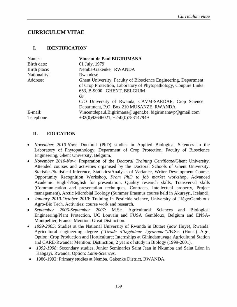
Curriculum vitae
159
CURRICULUM VITAE
I. IDENTIFICATION
Names: Vincent de Paul BIGIRIMANA Birth date: 01 July, 1979
Birth place: Nemba-Gakenke, RWANDA
Nationality: Rwandese
Address: Ghent University, Faculty of Bioscience Engineering, Department
of Crop Protection, Laboratory of Phytopathology, Coupure Links
653, B-9000 GHENT, BELGIUM
Or C/O University of Rwanda, CAVM-SARDAE, Crop Science
Department, P.O. Box 210 MUSANZE, RWANDA
E-mail: [email protected], [email protected]
Telephone +32(0)92646021; +250(0)783147949
II. EDUCATION
November 2010-Now: Doctoral (PhD) studies in Applied Biological Sciences in the
Laboratory of Phytopathology, Department of Crop Protection, Faculty of Bioscience
Engineering, Ghent University, Belgium.
November 2010-Now: Preparation of the Doctoral Training Certificate/Ghent University.
Attended courses and activities organised by the Doctoral Schools of Ghent University:
Statistics/Statistical Inference, Statistics/Analysis of Variance, Writer Development Course,
Opportunity Recognition Workshop, From PhD to job market workshop, Advanced
Academic English/English for presentation, Quality research skills, Transversal skills
(Communication and presentation techniques, Contracts, Intellectual property, Project
management), Arctic Microbial Ecology (Summer Erasmus course held in Akureyri, Iceland).
January 2010-October 2010: Training in Pesticide science, University of Liège/Gembloux
Agro-Bio Tech. Activities: course work and research.
September 2006-September 2007: M.Sc. Agricultural Sciences and Biological
Engineering/Plant Protection, UC Louvain and FUSA Gembloux, Belgium and ENSA-
Montpellier, France. Mention: Great Distinction.
1999-2005: Studies at the National University of Rwanda in Butare (now Huye), Rwanda:
Agricultural engineering degree (“Grade d’Ingénieur Agronome”)/B.Sc. (Hons.) Agr.,
Option: Crop Production and Horticulture; Internships at Gihindamuyaga Agricultural Station
and CARE-Rwanda; Mention: Distinction; 2 years of study in Biology (1999-2001).
1992-1998: Secondary studies, Junior Seminaries Saint Jean in Nkumba and Saint Léon in
Kabgayi. Rwanda. Option: Latin-Sciences.
1986-1992: Primary studies at Nemba, Gakenke District, RWANDA.
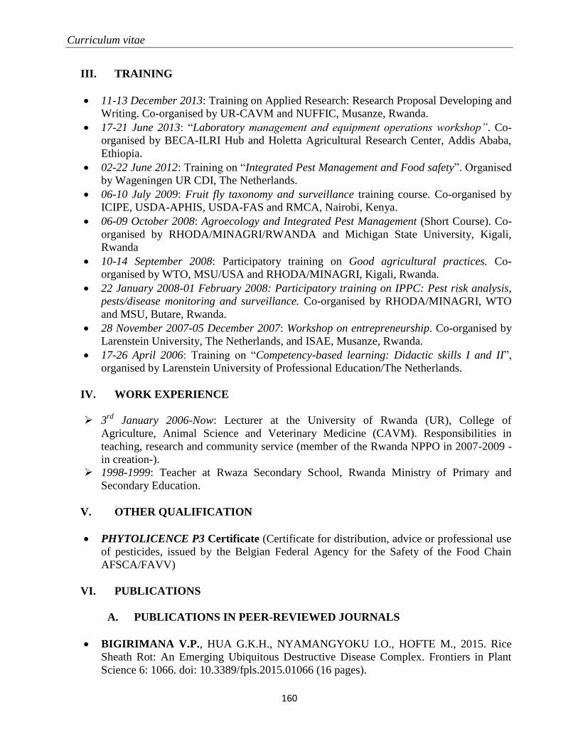
Curriculum vitae
160
III. TRAINING
11-13 December 2013: Training on Applied Research: Research Proposal Developing and
Writing. Co-organised by UR-CAVM and NUFFIC, Musanze, Rwanda.
17-21 June 2013: “Laboratory management and equipment operations workshop”. Co-
organised by BECA-ILRI Hub and Holetta Agricultural Research Center, Addis Ababa,
Ethiopia.
02-22 June 2012: Training on “Integrated Pest Management and Food safety”. Organised
by Wageningen UR CDI, The Netherlands.
06-10 July 2009: Fruit fly taxonomy and surveillance training course. Co-organised by
ICIPE, USDA-APHIS, USDA-FAS and RMCA, Nairobi, Kenya.
06-09 October 2008: Agroecology and Integrated Pest Management (Short Course). Co-
organised by RHODA/MINAGRI/RWANDA and Michigan State University, Kigali,
Rwanda
10-14 September 2008: Participatory training on Good agricultural practices. Co-
organised by WTO, MSU/USA and RHODA/MINAGRI, Kigali, Rwanda.
22 January 2008-01 February 2008: Participatory training on IPPC: Pest risk analysis,
pests/disease monitoring and surveillance. Co-organised by RHODA/MINAGRI, WTO
and MSU, Butare, Rwanda.
28 November 2007-05 December 2007: Workshop on entrepreneurship. Co-organised by
Larenstein University, The Netherlands, and ISAE, Musanze, Rwanda.
17-26 April 2006: Training on “Competency-based learning: Didactic skills I and II”,
organised by Larenstein University of Professional Education/The Netherlands.
IV. WORK EXPERIENCE
3rd
January 2006-Now: Lecturer at the University of Rwanda (UR), College of
Agriculture, Animal Science and Veterinary Medicine (CAVM). Responsibilities in
teaching, research and community service (member of the Rwanda NPPO in 2007-2009 -
in creation-).
1998-1999: Teacher at Rwaza Secondary School, Rwanda Ministry of Primary and
Secondary Education.
V. OTHER QUALIFICATION
PHYTOLICENCE P3 Certificate (Certificate for distribution, advice or professional use
of pesticides, issued by the Belgian Federal Agency for the Safety of the Food Chain
AFSCA/FAVV)
VI. PUBLICATIONS
A. PUBLICATIONS IN PEER-REVIEWED JOURNALS
BIGIRIMANA V.P., HUA G.K.H., NYAMANGYOKU I.O., HOFTE M., 2015. Rice
Sheath Rot: An Emerging Ubiquitous Destructive Disease Complex. Frontiers in Plant
Science 6: 1066. doi: 10.3389/fpls.2015.01066 (16 pages).
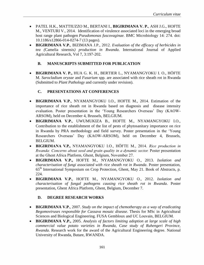
Curriculum vitae
161
PATEL H.K., MATTIUZZO M., BERTANI I., BIGIRIMANA V. P., ASH J.G., HOFTE
M., VENTURI V., 2014. Identification of virulence associated loci in the emerging broad
host range plant pathogen Pseudomonas fuscovaginae. BMC Microbiology 14: 274. doi:
10.1186/s12866-014-0274-7 (13 pages).
BIGIRIMANA V.P., BIZIMANA J.P., 2012. Evaluation of the efficacy of herbicides in
tea (Camelia sinensis) production in Rwanda. International Journal of Applied
Agricultural Research, Vol 7, 3:197-202.
B. MANUSCRIPTS SUBMITTED FOR PUBLICATION
BIGIRIMANA V. P., HUA G. K. H., BERTIER L., NYAMANGYOKU I. O., HÖFTE
M. Sarocladium oryzae and Fusarium spp. are associated with rice sheath rot in Rwanda
(Submitted to Plant Pathology and currently under revision).
C. PRESENTATIONS AT CONFERENCES
BIGIRIMANA V.P., NYAMANGYOKU I.O., HOFTE M., 2014. Estimation of the
importance of rice sheath rot in Rwanda based on diagnosis and disease intensity
evaluation. Poster presentation in the ‘Young Researchers Overseas’ Day (KAOW-
ARSOM), held on December 4, Brussels, BELGIUM.
BIGIRIMANA V.P., UWUMUKIZA B., HOFTE M., NYAMANGYOKU I.O.,
Contribution to the establishment of the list of pests of phytosanitary importance on rice
in Rwanda by PRA methodology and field survey. Poster presentation in the ‘Young
Researchers Overseas’ Day (KAOW-ARSOM), held on December 4, Brussels,
BELGIUM.
BIGIRIMANA V.P., NYAMANGYOKU I.O., HÖFTE M., 2014. Rice production in
Rwanda: Concerns about seed and grain quality in a dynamic sector. Poster presentation
at the Ghent Africa Platform, Ghent, Belgium, November 27.
BIGIRIMANA V.P., HOFTE M., NYAMANGYOKU O., 2013. Isolation and
characterisation of fungi associated with rice sheath rot in Rwanda. Poster presentation,
65th
International Symposium on Crop Protection, Ghent, May 21. Book of Abstracts, p.
224.
BIGIRIMANA V.P., HOFTE M., NYAMANGYOKU O., 2012. Isolation and
characterisation of fungal pathogens causing rice sheath rot in Rwanda. Poster
presentation, Ghent Africa Platform, Ghent, Belgium, December 7.
D. DEGREE RESEARCH WORKS
BIGIRIMANA V.P., 2007. Study on the impact of chemotherapy as a way of eradicating
Begomoviruses responsible for Cassava mosaic disease. Thesis for MSc in Agricultural
Sciences and Biological Engineering. FUSA Gembloux and UC Louvain, BELGIUM.
BIGIRIMANA V.P., 2005. Analysis of factors limiting adoption at large scale of high
commercial value potato varieties in Rwanda, Case study of Ruhengeri Province,
Rwanda. Research work for the award of the Agricultural Engineering degree. National
University of Rwanda, Butare, RWANDA.
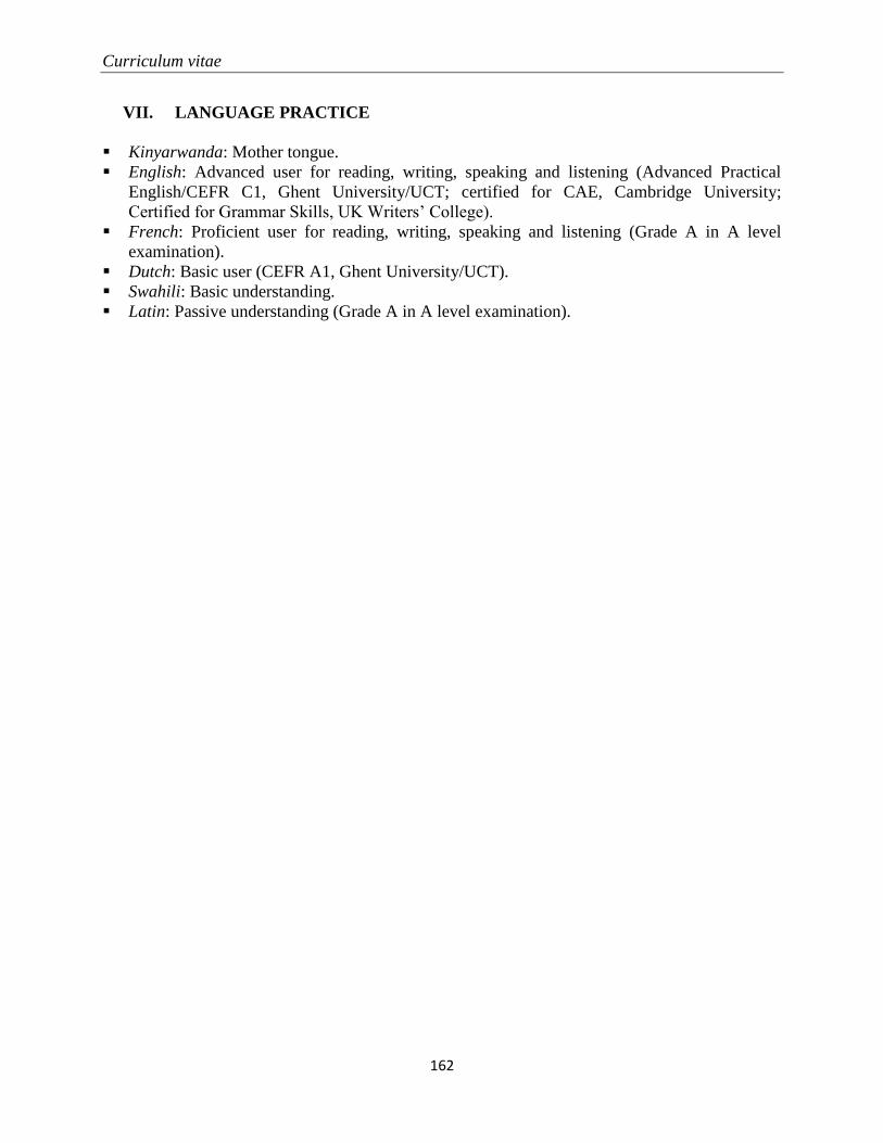
Curriculum vitae
162
VII. LANGUAGE PRACTICE
Kinyarwanda: Mother tongue.
English: Advanced user for reading, writing, speaking and listening (Advanced Practical
English/CEFR C1, Ghent University/UCT; certified for CAE, Cambridge University;
Certified for Grammar Skills, UK Writers’ College).
French: Proficient user for reading, writing, speaking and listening (Grade A in A level
examination).
Dutch: Basic user (CEFR A1, Ghent University/UCT).
Swahili: Basic understanding.
Latin: Passive understanding (Grade A in A level examination).