Embed Size (px)
Citation preview
life
Article
Using a Syrian (Golden) Hamster Biological Model for theEvaluation of Recombinant Anthrax Vaccines
Tatiana Kravchenko, Galina Titareva, Irina Bakhteeva , Tatiana Kombarova, Alexander Borzilov ,Raisa Mironova, Kseniya Khlopova and Vitalii Timofeev *
�����������������
Citation: Kravchenko, T.; Titareva,
G.; Bakhteeva, I.; Kombarova, T.;
Borzilov, A.; Mironova, R.; Khlopova,
K.; Timofeev, V. Using a Syrian
(Golden) Hamster Biological Model
for the Evaluation of Recombinant
Anthrax Vaccines. Life 2021, 11, 1388.
https://doi.org/10.3390/life11121388
Academic Editor: Daniele Focosi,
Antonio Fasanella and Domenico
Galante
Received: 2 November 2021
Accepted: 9 December 2021
Published: 11 December 2021
Publisher’s Note: MDPI stays neutral
with regard to jurisdictional claims in
published maps and institutional affil-
iations.
Copyright: © 2021 by the authors.
Licensee MDPI, Basel, Switzerland.
This article is an open access article
distributed under the terms and
conditions of the Creative Commons
Attribution (CC BY) license (https://
creativecommons.org/licenses/by/
4.0/).
State Research Center for Applied Microbiology and Biotechnology (SRCAMB), 142279 Obolensk, Russia;[email protected] (T.K.); [email protected] (G.T.); [email protected] (I.B.); [email protected] (T.K.);[email protected] (A.B.); [email protected] (R.M.); [email protected] (K.K.)* Correspondence: [email protected] or [email protected]
Abstract: In this paper, we demonstrate that a Syrian hamster biological model can be applied to thestudy of recombinant anthrax vaccines. We show that double vaccination with recombinant proteins,such as protective antigen (PA) and fusion protein LF1PA4, consisting of lethal factor I domain (LF)and PA domain IV, leads to the production of high titers of specific antibodies and to protection frominfection with the toxicogenic encapsulated attenuated strain B. anthracis 71/12. In terms of antibodyproduction and protection, Syrian hamsters were much more comparable to guinea pigs than mice.We believe that Syrian hamsters are still underestimated as a biological model for anthrax research,and, in some cases, they can be used as a replacement or at least as a complement to the traditionallyused mouse model.
Keywords: anthrax; Bacillus anthracis; Syrian hamsters; biomodels; vaccines
1. Introduction
Anthrax is a disease caused by the Gram-positive, spore-forming bacterium Bacillusanthracis. It is well established that B. anthracis exhibits its pathogenic properties due tothe presence of two plasmids encoding two main factors of pathogenicity. Plasmid pXO2carries the genes of a poly-γ-D-glutamic acid capsule synthesis (the capBCAD operon). Thiscapsule protects B. anthracis from the host’s immune response. Plasmid pXO1 carries genesencoding a three-component anthrax toxin, including a lethal factor (LF, metalloprotease),edema factor (EF, adenylate cyclase), and a protective antigen (PA, a non-toxic protein thatbinds to the host cell and ensures the transfer of LF and EF into the host cell).
Anthrax has some selectivity, and it affects different animals with different efficacy.The most sensitive to anthrax are ungulate herbivores, and it is among them that the vastmajority of cases of anthrax are recorded [1–3]. At the same time, it is the ungulates thatform the basis of agriculture; therefore, any epidemics among them lead to significanteconomic losses and the risk of transmission of infection to humans. Although humansare less susceptible to anthrax than ungulates, the disease can be severe and fatal forhumans [1].
Therefore, the study of the anthrax pathogenesis mechanisms, the determinationof the B. anthracis strains’ biological properties, and the development of methods forpreventative vaccines and treatment of livestock and humans anthrax remain urgent.However, almost all laboratories studying anthrax are forced to use in their work notungulates and or primates imitating humans, but more accessible biological models, suchas small rodents and rabbits. The use of ungulates and non-human primates is limited notonly by bioethical considerations but also by economic reasons: the cost of the animalsand their housing in BSL3-BSL4 vivariums make the use of such animals unreasonablyexpensive for routine work. However, rodents differ from ungulates and primates in thecharacteristics of the infectious process. Although the epidemiology of anthrax in wild
Life 2021, 11, 1388. https://doi.org/10.3390/life11121388 https://www.mdpi.com/journal/life
Life 2021, 11, 1388 2 of 14
rodents is practically unknown, they are more likely to be resistant to infection in naturalconditions. The main argument in favor of this statement is the absence of death in wildand synanthropic rodents during anthrax outbreaks in herbivores, although these rodentsfeed on the same food as diseased herbivores and even eat the corpses of animals killedby anthrax [4]. Additionally, there are not many published works on the experimentalinfection of wild rodents. For example, as reported by Walker et al., 1967 [5] it is mentionedthat an injection of B. anthracis spores to black rats did not lead to the development ofanthrax in them. A larger-scale experiment described in [6] gave somewhat ambiguousresults: 8 out of 12 studied species were susceptible to infection, 2 species were resistant,and 2 more showed too wide a dispersion of results to draw a conclusion. As for rabbits,we were unable to find any mention of natural (or even experimental) anthrax in wildlagomorphs, which also indicates that these animals do not suffer from anthrax in nature.The features of anthrax in wild animals are very interesting, but from a practical pointof view, the same features are more important in laboratory animals, such as mice, rats,guinea pigs, and rabbits. Each of these species (and sometimes individual inbred lineswithin the same species) differs in sensitivity to anthrax and in the features of the infectiousprocess. Therefore, researchers have to select a biological model for each specific task andsometimes use several species of animals in one experiment. A comprehensive descriptionof the use of different laboratory animals for anthrax research can be found, for example, ina very detailed review by Welkos et al. [7]. The mouse model is most often used in routinework. These advantages of mice reduce the cost of experiments and make it possible toincrease the number of animals used, which means an increase in the statistical reliabilityof the results obtained. In addition, mice are susceptible to infection with single-plasmidB. anthracis strains. Such strains are non-virulent to humans, and experiments with themcan be carried out not only in the BSL3–4 laboratories. On the one hand, this greatlyfacilitates the design of the experiment, but, on the other hand, the results obtained canhardly be extrapolated to the anthrax of the main hosts and require confirmation usinganother biological model infected with two-plasmid strains. Rats are relatively resistant toinfection, but at the same time, are very sensitive to the anthrax toxin; therefore, rats areused primarily to assess the effectiveness of toxin-neutralizing agents. Models of guineapigs and rabbits are more adequate, but the disadvantage is the high cost of the animalsand their housing in the vivarium.
In summary, we can say that the most common biomodels are either cheap and avail-able, but not quite adequate, such as mice and to some extent rats, or are more adequate,but significantly more expensive, such as guinea pigs and rabbits. Such a situation led usto the desire to find another biomodel, which would be closer in cost to mice, but in termsof adequacy closer to that of guinea pigs.
We wondered if Syrian hamsters could be such a biomodel. Hamsters appear to bea promising laboratory animal for anthrax research. They are cheap, small, and do notrequire special conditions in the vivarium. For these reasons, hamsters can be used inlarge numbers in experiments. This is important for increasing the degree of the statisticalreliability of the results obtained. At the same time, hamsters are larger than mice, andthis increases the convenience of carrying out all manipulations with them. Nevertheless,despite these advantages, hamsters are rarely used for anthrax studies. Neither in the above-mentioned review [7] nor in the other literary sources we were able to find a sufficientlydetailed description of the possibilities of anthrax research using a hamster biomodel.
Previously, some researchers have attempted to use hamsters to evaluate the efficacyof anthrax vaccines, but the results were not entirely consistent. Thus, Pomerantsev [8]reported on the possibility of effective vaccination of hamsters with a B. anthracis livevaccine strain. However, Fellows [9] vaccinated hamsters with the human anthrax vaccineAVA and found that although all vaccinated animals had high antibody titers, vaccinationdid not prevent death from infection with virulent B. anthracis strains. As a result, thequestion of whether the hamster model is suitable for studying anthrax vaccines remainedcontroversial. We have tried to bring some clarity to this issue. In this work, we immunized
Life 2021, 11, 1388 3 of 14
three species of laboratory animals: C57BL/6 mice, outbred guinea pigs, and outbred Syrian(golden) hamsters with two recombinant proteins: rPA63 (protective antigen of B. anthracis)and the fusion protein rLF1PA4 containing the I domain of the protective antigen andthe IV domain of the lethal factor. The effectiveness of vaccination was assessed by thechallenge of the immunized animals with the encapsulated toxicogenic attenuated strainB. anthracis 71/12.
2. Materials and Methods2.1. Bacterial Strains
To infect the vaccinated animals, we used strain B. anthracis 71/12, deposited in theSRCAMB collection. This strain is a subclone of the Tsenkowski II vaccine, and it hasa plasmid profile of pXO1+ pXO2+.
The E. coli DH5α strain was used to construct the expression vectors; the E. coliBL21 strain was used to express the recombinant proteins.
2.2. Cloning
PA without native signal sequence (rPA63) was cloned into the commercial expressionvector pET-SUMO (Champion™ pET SUMO Expression System, Thermo Fisher Scientific,Waltham, MA, USA).
The fusion protein rLF1PA4, consisting of LF domain I and PA domain IV, was clonedinto the commercial expression vector pLATE51 (aLICator LIC Cloning and ExpressionSystem, Thermo Fisher Scientific). The cloned fragments were generated by PCR usinga template of B. anthracis 71/12 genomic DNA. All primers used for PCR are listed inTable 1. The PA63 locus was amplified using primers no. 10 and 8. The LF1PA4 locuswas obtained by Gibson assembly of two fragments. One of them (LF 1 domain DNA)was obtained by three consecutive PCR reactions, in which the product of the first wasused as a template for the second, and the product of the second was used as a templatefor the third. For these reactions, we used primers no. 1 and 4, 2 and 5, and 3 and 5,respectively. The second fragment (PA 4 domain DNA) was obtained in two sequentialreactions using primers no. 6 and 8, and 7 and 9, respectively. The loci of the LF and PAgenes obtained as a result of these PCR had regions complementary to each other and tothe pLATE51 plasmid. This made it possible to use Gibson’s assembly of these fragments(with Gibson Assembly® Cloning Kit, New England Biolabs, Ipswich, MA, USA) and toclone the product of this fusion into plasmid pLATE51 using LIC cloning (according to themanufacturer’s instructions).
Table 1. Primers used in the work.
No. Primer Name Primer Sequence
1 G-LF1-F1 ATGGCGGGCGGTCATGGTG
2 G-LF1-F2Bam ACAAGGGATCCATGGCGGGCGGTC
3 G-LF1-F3 GGTGATGATGATGACAAGGGATCCATGGC
4 G-LF1-R CCGTTGATCTTTAAGTTCTTCCAAGGATAG
5 G-LF1-RAn TGCGTTTAATTCCGCCCGTTGATCTTTAAGTTC
6 G-PA4-F1 GCGGAATTAAACGCAACTAACATATA
7 G-PA4F2An GAACTTAAAGATCAACGGGCGGAATTAAACGC
8 G-PA4-R1Bam GGATCCTTATCCTATCTCATAGCCTTTTTTAG
9 G-PA4-R2 GGAGATGGGAAGTCATTAGGATCCTTATCCTATCTCAT
10 PA63F1 TACTGGACCGATTCTCAAAATAAAAAAGAA
Genomic DNA isolation was performed using the Genomic DNA Purification Kit(Thermo Fisher Scientific). Plasmid isolation was performed using a GeneJET Plasmid
Life 2021, 11, 1388 4 of 14
Miniprep Kit DNA (Thermo Fisher Scientific). Isolation of DNA fragments from agarosegel was carried out using the GeneJET Gel Extraction Kit (Thermo Fisher Scientific).
2.3. Biochemical Research Methods
The solubilization of recombinant proteins from inclusion bodies was carried outunder denaturing conditions: the rLF1PAIV protein in the presence of 6 M urea and therPA63 protein in the presence of 8 M urea.
Isolation and purification of proteins were carried out under denaturing conditionsby chelating affinity chromatography using a Chelating Sepharose Fast Flow sorbent (GEHealthcare). The obtained protein fractions were analyzed by SDS-PAGE electrophoresis(10–12.5%) [10]. A set of standard proteins Spectra Multicolor Broad Range 260–10 kDa(Thermo Fisher Scientific) was used as molecular weight markers.
Fractions of the purified proteins were renatured by dialysis against 20 mM Tris (pH7.9), and the denatured protein was removed by centrifugation at 10,000 rpm for 30 min.The protein concentration in the supernatant was assessed spectrophotometrically usingan Ultrospec 3100 pro scanning spectrophotometer (Amersham), taking into account themolar extinction coefficients calculated for each protein.
For immunoblotting [11], Hybond P PVDF membranes (GE Healthcare, Chicago, IL,USA) and rabbit monospecific polyclonal antisera to native PA and LF at a dilution of 1:100 were used. To detect bound rabbit antibodies, we used a conjugate of secondary goatantibodies to rabbit IgG (Sigma, Burlington, MA, USA) in a working dilution of 1:10,000and a DAB (AppliChem, Council Bluffs, IA, USA).
Antibody levels in the sera of immunized animals were assessed by ELISA accordingto [12]. Protein antigens at a concentration of 5 µg/mL were adsorbed onto 96-well mediumsorption plates (Greiner Bio-One, Frickenhausen, Germany), adsorption was performedovernight at 4 ◦C, then free binding sites were inactivated with a 1% BSA solution at 37 ◦Cfor 30 min.
Sera from immune and native animals were used in initial dilutions of 1:200, thentitrated in 1:2 steps and incubated for 1 h at 37 ◦C. The serum of intact animals of thecorresponding species was used as a control. Bound antibodies were detected with solu-tions of secondary goat antibodies conjugated with horseradish peroxidase in a workingdilution of 1:10,000 and incubated for 1 h at 37 ◦C. Staining was carried out with a solutionof orthophenylenediamine (OPD) with the addition of hydrogen peroxide to 0.03% for20 min at room temperature in the dark. Then the reaction was stopped with a 0.1 M HCl.The optical density in the wells was measured at a wavelength of 492 nm on a MultiscanFC plate spectrophotometer (Thermo Fisher Scientific).
2.4. Animal Experiments2.4.1. Ethics Statement
All protocols for animal experiments were approved by the State Research Center forApplied Microbiology and Biotechnology Bioethics Committee (Permit No: VP-2021/1).They were performed in compliance with NIH Animal Welfare Insurance #A5476-01 issuedon 02/07/2007 and the European Union guidelines and regulations on the handling,care, and protection of laboratory animals (https://eur-lex.europa.eu/eli/dir/2010/63/oj(accessed on 1 November 2021)).
A minimum number of animals was used for the experiments. The approved protocolsprovided scientifically validated humane endpoints, including pre-set criteria for theeuthanasia of moribund mice via CO2 inhalation. In our study, the animals were euthanizedwhen they became lethargic, dehydrated, moribund, unable to rise, or non-responsive totouch. The health conditions of the animals were monitored at least twice a day.
2.4.2. Animals
Animals were purchased from the breading center “Stolbovaya” of the «ScientificCenter of Biomedical Technology» of the Federal Medical-Biological Agency of Russia
Life 2021, 11, 1388 5 of 14
(https://www.pitomniki-stolbovaya.com (accessed on 1 November 2021)) and breadingcenter “Andreevka” of the «Scientific Center of Biomedical Technology» of the FederalMedical-Biological Agency of Russia (http://andreevka.msk.ru/index.htm (accessed on1 November 2021)).
C57BL/6 mice (6–8 weeks old, 18–20 g), outbred Syrian (golden) hamsters (6–8 weeks-old, 80–100 g), and guinea pigs (6–8 weeks-old, 275 ± 25 g) of both genders were used inthe experiments. Animals were housed in polycarbonate cages with space for comfortablemovement (5 mice or 4 hamsters in a 484 cm2-cage and 3 guinea pigs in an 864 cm2-cage)with ad libitum access to food (Mouse Mixed Fodder PK-120, and Guinea Pig Mixed FodderKK–122, Laboratorkorm, Russia) and tap water, under constant temperature and humidityconditions (22 ± 2 ◦C and 50 ± 10%, respectively) and a 12 h light/12 h dark cycle.
The general structure of the animal experiment is shown in Table S1.
2.4.3. Immunization
Mice (n = 100), guinea pigs (n = 80), and golden hamsters (n = 80) were immunizedsubcutaneously twice with an interval of 28 days. Animals were vaccinated subcutaneouslyin the inner part of the upper thigh. Purified recombinant proteins rLF1PA4 and rPA63were injected with PBS at doses of 50 µg protein for mice and 100 µg protein for guineapigs and hamsters. The dose volume was 100 µL for mice and hamsters and 250 µL forguinea pigs. Guinea pigs were vaccinated with twice the dose of recombinant proteinsthan mice due to their greater body weight and body size and the characteristics of theirimmune response. It was less clear which dose of recombinant anthrax proteins should beused to immunize hamsters. This information is not available at all since hamsters werealmost never used in such studies. However, when immunizing hamsters with the humanvaccine, AVA Fellows [9] used this vaccine at the same dosage that is commonly used forguinea pigs (this dose was higher than that used to immunize mice). Therefore, we alsoused the same dose of vaccine for hamsters as for guinea pigs. Proteins were administeredboth without additives and with complete (first immunization) and incomplete (secondimmunization) Freund’s adjuvant (FA) in a 1:1 volumetric ratio. One group of animalsof each species was immunized only with complete (first immunization) and incomplete(second immunization) FA without recombinant proteins.
2.4.4. Blood Sampling
At 14 days after the second immunization, blood samples were taken to determine the levelof specific antibodies to the recombinant proteins rLF1PA4 and rPA63. In guinea pigs, blood wasobtained from the marginal ear vein, in hamsters and mice, from the retroorbital sinus.
2.4.5. Challenge
At 28 days after the second immunization, the animals were subcutaneously chal-lenged with B. anthracis 71/12 strain. In total, five groups of immune animals of each specieswere challenged: two groups immunized with proteins rLF1PA4 and rPA63, two groupsimmunized with proteins with FA (rLF1PA4 + FA and rPA63 + FA), and one group thatwas administered only FA. Additionally, the sixth group of naive animals was used asa negative control.
Each group had four subgroups challenged with different doses. We used doses of50 to 50,000 spores/animal for guinea pigs and Syrian hamsters (4 animals per subgroup),and 5 to 5000 spores/animal for C57BL/6 mice (5 animals per subgroup). Thus, the numberof infected animals was: 96 guinea pigs (80 immune and 16 intact), 96 Syrian hamsters(80 immune and 16 intact), and 120 mice (100 immune and 20 intact).
We observed the animals for 21 days after infection; the surviving animals werehumanely euthanized by CO2 inhalation.
Life 2021, 11, 1388 6 of 14
2.5. Statistics
The obtained data were statistically processed using the GraphPad Prism 7 program(https://www.graphpad.com/ (accessed on 1 November 2021)). Survival curves wereplotted and analyzed using log-rank (Mantel–Cox) tests. In some cases, the levels ofsignificance of differences (P) were determined using one-way ANOVA (Mann–Whitney).P values less than 0.05 were considered significant.
LD50 values were determined according to the method of Kärber, as modified byAshmarin and Vorob’ov [13].
3. Results3.1. Recombinant Protein-Producer Strains
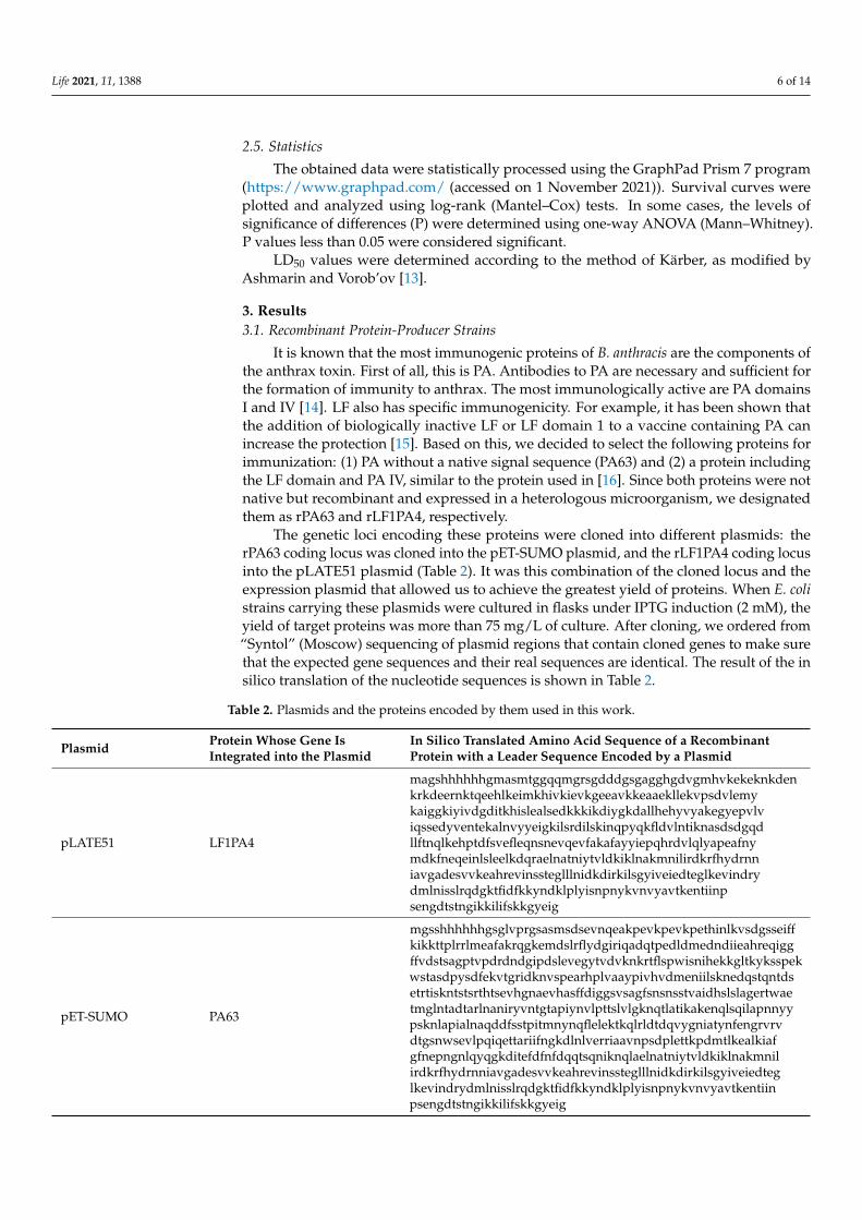
It is known that the most immunogenic proteins of B. anthracis are the components ofthe anthrax toxin. First of all, this is PA. Antibodies to PA are necessary and sufficient forthe formation of immunity to anthrax. The most immunologically active are PA domainsI and IV [14]. LF also has specific immunogenicity. For example, it has been shown thatthe addition of biologically inactive LF or LF domain 1 to a vaccine containing PA canincrease the protection [15]. Based on this, we decided to select the following proteins forimmunization: (1) PA without a native signal sequence (PA63) and (2) a protein includingthe LF domain and PA IV, similar to the protein used in [16]. Since both proteins were notnative but recombinant and expressed in a heterologous microorganism, we designatedthem as rPA63 and rLF1PA4, respectively.
The genetic loci encoding these proteins were cloned into different plasmids: therPA63 coding locus was cloned into the pET-SUMO plasmid, and the rLF1PA4 coding locusinto the pLATE51 plasmid (Table 2). It was this combination of the cloned locus and theexpression plasmid that allowed us to achieve the greatest yield of proteins. When E. colistrains carrying these plasmids were cultured in flasks under IPTG induction (2 mM), theyield of target proteins was more than 75 mg/L of culture. After cloning, we ordered from“Syntol” (Moscow) sequencing of plasmid regions that contain cloned genes to make surethat the expected gene sequences and their real sequences are identical. The result of the insilico translation of the nucleotide sequences is shown in Table 2.
Table 2. Plasmids and the proteins encoded by them used in this work.
Plasmid Protein Whose Gene IsIntegrated into the Plasmid
In Silico Translated Amino Acid Sequence of a RecombinantProtein with a Leader Sequence Encoded by a Plasmid
pLATE51 LF1PA4
magshhhhhhgmasmtggqqmgrsgdddgsgagghgdvgmhvkekeknkdenkrkdeernktqeehlkeimkhivkievkgeeavkkeaaekllekvpsdvlemykaiggkiyivdgditkhislealsedkkkikdiygkdallhehyvyakegyepvlviqssedyventekalnvyyeigkilsrdilskinqpyqkfldvlntiknasdsdgqdllftnqlkehptdfsvefleqnsnevqevfakafayyiepqhrdvlqlyapeafnymdkfneqeinlsleelkdqraelnatniytvldkiklnakmnilirdkrfhydrnniavgadesvvkeahrevinssteglllnidkdirkilsgyiveiedteglkevindrydmlnisslrqdgktfidfkkyndklplyisnpnykvnvyavtkentiinpsengdtstngikkilifskkgyeig
pET-SUMO PA63
mgsshhhhhhgsglvprgsasmsdsevnqeakpevkpevkpethinlkvsdgsseiffkikkttplrrlmeafakrqgkemdslrflydgiriqadqtpedldmedndiieahreqiggffvdstsagptvpdrdndgipdslevegytvdvknkrtflspwisnihekkgltkyksspekwstasdpysdfekvtgridknvspearhplvaaypivhvdmeniilsknedqstqntdsetrtiskntstsrthtsevhgnaevhasffdiggsvsagfsnsnsstvaidhslslagertwaetmglntadtarlnaniryvntgtapiynvlpttslvlgknqtlatikakenqlsqilapnnyypsknlapialnaqddfsstpitmnynqflelektkqlrldtdqvygniatynfengrvrvdtgsnwsevlpqiqettariifngkdlnlverriaavnpsdplettkpdmtlkealkiafgfnepngnlqyqgkditefdfnfdqqtsqniknqlaelnatniytvldkiklnakmnilirdkrfhydrnniavgadesvvkeahrevinssteglllnidkdirkilsgyiveiedteglkevindrydmlnisslrqdgktfidfkkyndklplyisnpnykvnvyavtkentiinpsengdtstngikkilifskkgyeig
Life 2021, 11, 1388 7 of 14
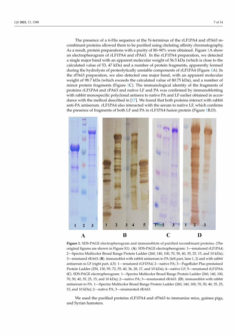
The presence of a 6-His sequence at the N-terminus of the rLF1PA4 and rPA63 re-combinant proteins allowed them to be purified using chelating affinity chromatography.As a result, protein preparations with a purity of 80–90% were obtained. Figure 1A showan electropherogram of rLF1PA4 and rPA63. In the rLF1PA4 preparation, we detecteda single major band with an apparent molecular weight of 56.5 kDa (which is close to thecalculated value of 53, 47 kDa) and a number of protein fragments, apparently formedduring the hydrolysis of proteolytically unstable components of rLF1PA4 (Figure 1A). Inthe rPA63 preparation, we also detected one major band, with an apparent molecularweight of 98.7 kDa (which exceeds the calculated value of 80.75 kDa), and a number ofminor protein fragments (Figure 1C). The immunological identity of the fragments ofproteins rLF1PA4 and rPA63 and native LF and PA was confirmed by immunoblottingwith rabbit monospecific polyclonal antisera to native PA and LF earlier obtained in accor-dance with the method described in [17]. We found that both proteins interact with rabbitanti-PA antiserum. rLF1PA4 also interacted with the serum to native LF, which confirmsthe presence of fragments of both LF and PA in rLF1PA4 fusion protein (Figure 1B,D).
1
Figure 1. SDS-PAGE electropherogram and immunoblots of purified recombinant proteins. (Theoriginal figures are shown in Figure S1). (A). SDS-PAGE electropherogram: 1—renatured rLF1PA4;2—Spectra Multicolor Broad Range Protein Ladder (260, 140, 100, 70, 50, 40, 35, 25, 15, and 10 kDa);3—renatured rRA63; (B). immunoblot with rabbit antiserum to PA (left part, lane 1, 2) and with rabbitantiserum to LF (right part, 4,5): 1—renatured rLF1PA4; 2—native PA; 3—PageRuler Plus prestainedProtein Ladder (250, 130, 95, 72, 55, 40, 36, 28, 17, and 10 kDa); 4—native LF; 5—renatured rLF1PA4.(C). SDS-PAGE electropherogram: 1—Spectra Multicolor Broad Range Protein Ladder (260, 140, 100,70, 50, 40, 35, 25, 15, and 10 kDa); 2—native PA; 3—renaturated rRA63. (D). immunoblot with rabbitantiserum to PA. 1—Spectra Multicolor Broad Range Protein Ladder (260, 140, 100, 70, 50, 40, 35, 25,15, and 10 kDa); 2—native PA; 3—renaturated rRA63.
We used the purified proteins rLF1PA4 and rPA63 to immunize mice, guinea pigs,and Syrian hamsters.
Life 2021, 11, 1388 8 of 14
3.2. Study of Immunogenic Properties of rLF1PA4 and rPA63
At 14 days after the second immunization, we took blood samples from animals ineach group and measured the titers of antibodies to the rLF1PA4 and rPA63 in sera obtainedfrom this blood by ELISA. The mean values of reciprocal antibody titers are presented inTable 3 and Figure 2. The significance of differences between groups was determined usingone-way ANOVA.
Table 3. Values of reciprocal titers of antibodies to rLF1PA4 and rPA63 in the sera of experimental animals.
Antigens (Recombinant Proteins withor without Freund’s Adjuvant (FA))
Values of Reciprocal Titers (Min ÷ Max)
Guinea Pigs Syrian Hamsters C57BL/6 Mice
rLF1PA4 3440(400 ÷ 12,800)
5760(3200 ÷ 6400)
7040(3200 ÷ 12,800)
rLF1PA4 + FA 389,120(102,400 ÷ 819,200)
450,560(204,800 ÷ 819,200)
12,800(400 ÷ 76,800)
Increase in titers when adding FA torLF1PA4 (times) 113 78 1.8
rPA63 34,560(6400 ÷ 102,400)
11,520(6400 ÷ 25,600)
1760(800 ÷ 3200)
rPA63 + FA 409,600(409,600 ÷ 409,600)
655,360(204,800 ÷ 819,200)
51,200(800 ÷ 102,400)
Increase in titers when adding FA torPA63 (times) 11.8 56.8 29
FA without immunogenic proteins <200 <200 <800
Unvaccinated control <200 <200 <800
2
Figure 2. Reciprocal titers of antibodies to rPA63 and rLF1PA4 14 days after the second immunization.Graph was generated by GraphPad Prism 7 software. Mean values with upper and lower limitsare indicated.
Life 2021, 11, 1388 9 of 14
As can be seen from the data presented (Figure 2), immunization of the animals ledto the formation of a pool of specific antibodies in them. In vaccinated guinea pigs andSyrian hamsters, we found high antibody titers to the target proteins. The administrationof proteins with added FA caused the expected statistically significant increase in titers by1–2 orders of magnitude. Mice showed the greatest variation in individual antibody titervalues, and the difference in antibody titer between groups, including the unvaccinatedgroup, was insignificant in most cases.
3.3. Comparison of Immunization Protection for Different Animal Species
At 28 days after the second immunization, the animals were challenged with the B. anthracis71/12 strain. The data on the death of animals and LD50 values are presented in Table 4.
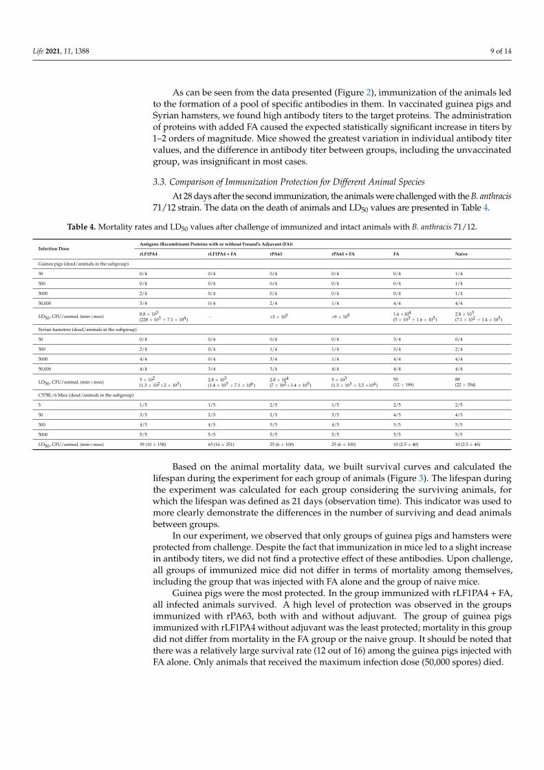
Table 4. Mortality rates and LD50 values after challenge of immunized and intact animals with B. anthracis 71/12.
Infection DoseAntigens (Recombinant Proteins with or without Freund’s Adjuvant (FA))
rLF1PA4 rLF1PA4 + FA rPA63 rPA63 + FA FA Naive
Guinea pigs (dead/animals in the subgroup)
50 0/4 0/4 0/4 0/4 0/4 1/4
500 0/4 0/4 0/4 0/4 0/4 1/4
5000 2/4 0/4 0/4 0/4 0/4 1/4
50,000 3/4 0/4 2/4 1/4 4/4 4/4
LD50, CFU/animal, (min÷max) 8.8 × 10³
(228 × 103 ÷ 7.1 × 10⁴)− >5 × 10⁵ >9 × 10⁵ 1.6 ×10⁴
(5 × 10³ ÷ 1.6 × 10⁵)2.8 × 10³
(7.1 × 10² ÷ 1.4 × 10⁵)
Syrian hamsters (dead/animals in the subgroup)
50 0/4 0/4 0/4 0/4 3/4 0/4
500 2/4 0/4 1/4 1/4 3/4 2/4
5000 4/4 0/4 3/4 1/4 4/4 4/4
50,000 4/4 3/4 3/4 4/4 4/4 4/4
LD50, CFU/animal, (min÷max) 5 × 10²
(1.3 × 10²÷2 × 10³)2.8 × 10⁵
(1.4 × 10⁵ ÷ 7.1 × 10⁶)2.8 × 10⁴
(7 × 10²÷1.4 × 10⁵)5 × 10³
(1.3 × 10³ ÷ 3.2 ×10⁴)50(12 ÷ 199)
88(22 ÷ 354)
C57BL/6 Mice (dead/animals in the subgroup)
5 1/5 1/5 2/5 1/5 2/5 2/5
50 3/5 2/5 2/5 3/5 4/5 4/5
500 4/5 4/5 5/5 4/5 5/5 5/5
5000 5/5 5/5 5/5 5/5 5/5 5/5
LD50, CFU/animal, (min÷max) 39 (10 ÷ 158) 63 (16 ÷ 251) 25 (6 ÷ 100) 25 (6 ÷ 100) 10 (2.5 ÷ 40) 10 (2.5 ÷ 40)
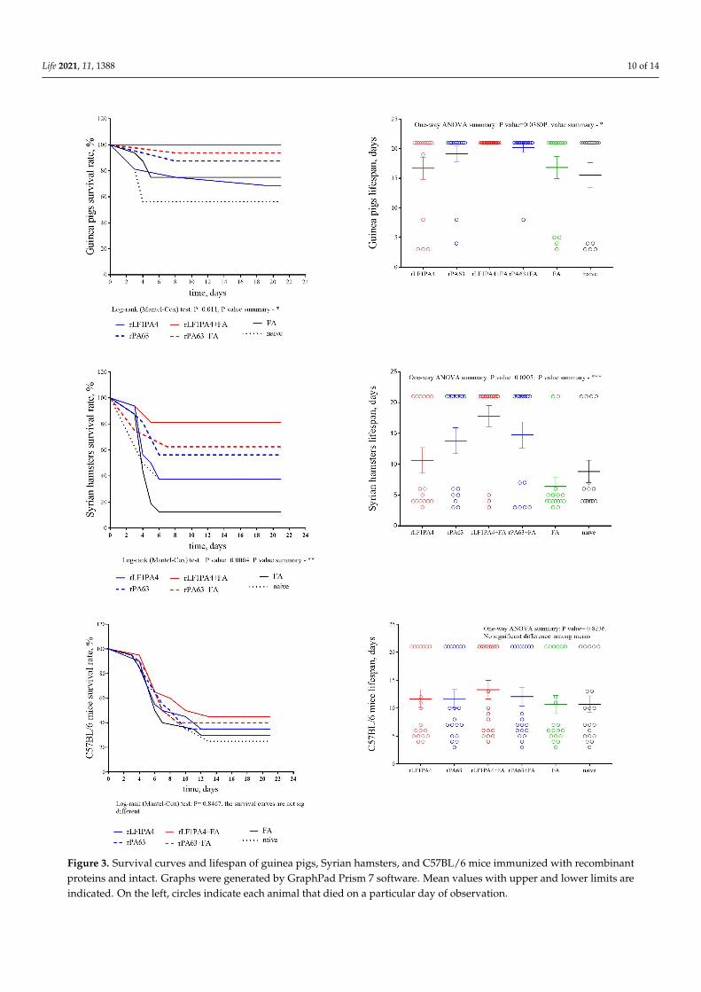
Based on the animal mortality data, we built survival curves and calculated thelifespan during the experiment for each group of animals (Figure 3). The lifespan duringthe experiment was calculated for each group considering the surviving animals, forwhich the lifespan was defined as 21 days (observation time). This indicator was used tomore clearly demonstrate the differences in the number of surviving and dead animalsbetween groups.
In our experiment, we observed that only groups of guinea pigs and hamsters wereprotected from challenge. Despite the fact that immunization in mice led to a slight increasein antibody titers, we did not find a protective effect of these antibodies. Upon challenge,all groups of immunized mice did not differ in terms of mortality among themselves,including the group that was injected with FA alone and the group of naive mice.
Guinea pigs were the most protected. In the group immunized with rLF1PA4 + FA,all infected animals survived. A high level of protection was observed in the groupsimmunized with rPA63, both with and without adjuvant. The group of guinea pigsimmunized with rLF1PA4 without adjuvant was the least protected; mortality in this groupdid not differ from mortality in the FA group or the naive group. It should be noted thatthere was a relatively large survival rate (12 out of 16) among the guinea pigs injected withFA alone. Only animals that received the maximum infection dose (50,000 spores) died.
Life 2021, 11, 1388 10 of 14
3
Figure 3. Survival curves and lifespan of guinea pigs, Syrian hamsters, and C57BL/6 mice immunized with recombinantproteins and intact. Graphs were generated by GraphPad Prism 7 software. Mean values with upper and lower limits areindicated. On the left, circles indicate each animal that died on a particular day of observation.
Life 2021, 11, 1388 11 of 14
Syrian hamsters, although they were slightly less protected, showed the same trendsas guinea pigs. Thus, a protective effect was recorded in Syrian hamsters in the threegroups immunized with rLF1PA4 + FA, rPA63, and rPA63 + FA. The mortality rate ofanimals immunized with rLF1PA4 without FA did not differ significantly from the mortalityrates in the FA group and in the unvaccinated control group. However, unlike guineapigs, hamsters were not protected from infection by administering FA alone withoutimmunogenic proteins. In the FA group, almost all hamsters died (14 out of 16); only oneanimal out of four infected with doses of 50 and 500 spores/animal survived. In the FAgroup, almost all hamsters died (14 out of 16); only two animals infected with minimaldoses (50 and 500 spores) survived. Thus, we can say that the hamster model apparentlyallows minimizing the influence of the adjuvant on the final vaccine protection. This canbe considered an advantage of the hamster model.
In general, we can see that the immunization of Syrian hamsters with the recombinantcomponents of the anthrax toxin leads both to the production of specific antibodies (anti-body titers are comparable to those in guinea pigs) and to protection from challenge withan encapsulated toxicogenic attenuated B. anthracis strain. In addition, the hamster modeleven showed some advantages over the guinea pig model. The influence of the adjuvanton the final result is less pronounced in hamsters than in guinea pigs (perhaps due to lessstimulation of nonspecific immunity). Additionally, due to the relatively high virulence ofthe used strain for hamsters, they have a more pronounced difference in the survival rateof vaccinated and naive animals.
Thus, a model of Syrian hamsters infected with an encapsulated toxicogenic attenuatedstrain of B. anthracis can be successfully used to assess the effectiveness of recombinantanthrax vaccines. This model, even if inferior to the guinea pig model, has significantadvantages over the mouse model. Taking into account the low cost of hamsters, thismodel can be used instead of the mouse model as a first-line model in vaccine studies.
4. Discussion
As is widely known, it is the toxin that primarily affects the host organism duringanthrax, and antibodies to the toxin (primarily to PA) are able to protect the host organismfrom lethal anthrax [16,18]. Therefore, the main task of vaccination is to lead to theformation of such antibodies, but at the same time not to cause serious damage to themacro-organism. Different approaches have been proposed for this at different times andin different countries. Historically, live attenuated strains were the first to be proposed asvaccines. They can be divided into two groups:
(1) Strains with a reduced copy number of the plasmid pXO1 and a low level oftoxin expression, such as Pasteur II, Tsenkovsky II (variant spellings of the surnameTsenkovsky in the name of the vaccine in the English-language literature: Zenkowsky [19],Qiankefusiji [14] Russian: Цeнкoвcкий), and Rentian II [14];
(2) Single-plasmid strains lacking the pXO2 plasmid but retaining the ability to syn-thesize toxins, such as Sterne (a veterinary vaccine widely used all over the world) andSTI-1 and A16R, which are human vaccines used in Russia and China, respectively [19].
Another approach seems to be safer: the use of the culture liquid of pXO1+ pXO2−
strains, adsorbed on aluminum compounds. These are vaccines such as AVA (anthraxvaccine adsorbed, USA) and AVP (anthrax vaccine precipitated, Great Britain). Thistechnology also has its drawbacks; the main factor is composition instability, as the ratio ofthe components can vary from batch to batch. This problem could be solved, for example,by using recombinant anthrax toxin proteins for vaccination. This approach appears to bea rather promising idea, the implementation of which has been attempted for quite sometime [15,16,19,20]. However, the main problem in the development of any anthrax vaccinesis testing their efficacy. The fact is that, as we discussed in the introduction, the mostcommon laboratory animal, mice, is not a completely adequate model for experimentalanthrax. In nature, they are insensitive to alimentary infection with B. anthracis spores,but in the laboratory, infection with natural virulent strains is fatal for them even if they
Life 2021, 11, 1388 12 of 14
were vaccinated. Moreover, mice are highly susceptible to laboratory infection with pXO2+
strains, even if these strains are unable to synthesize the toxin. Despite this, mice arecheap, and some inbred lines are susceptible to infection with vaccine pXO1+ pXO2−
strains, which makes it possible to carry out the corresponding experiments not only inBSL3-4 laboratories; therefore, the mouse model remains quite common. Nevertheless, themouse model’s peculiarities mean that the results obtained need to be confirmed in otheranimal species [7]. Thus, it may not be easy to prove, using only mice, that a vaccine underdevelopment is really worth the investment of resources and testing on more expensiveanimals. Therefore, the search for a cheap and affordable animal model that could replaceor at least supplement the mouse model in anthrax vaccines research seems to be a ratherurgent problem.
As we have shown, the Syrian hamsters would be such a “first-line” model in somecases. They are five times cheaper than guinea pigs and 15 times cheaper than rabbits. Atthe same time, their response to immunization with recombinant anthrax proteins is com-parable to that of guinea pigs. Syrian hamsters, similar to guinea pigs, produce antibodiesin comparable amounts, and these antibodies protect them from experimental infection.
It can be noted that we did not use a highly virulent natural strain for infection, butan attenuated two-plasmid strain 71/12, which is a subclone of the Tsenkovsky II vaccine.This strain has a slightly reduced virulence for laboratory animals (including hamsters).On the one hand, capsular toxicogenic vaccine strains such as Pasteur II and Tsenkovsky IIdiffer little from natural strains in their biological properties; they act on the host organismwith a full set of virulence factors and retain sufficient virulence for animals. On the otherhand, the decrease in this virulence is sufficient for these strains to be considered a vaccine;therefore, working with them is possible with less biological safety measures than workingwith natural B. anthracis strains.
Therefore, strain 71/12 seemed to us convenient for assessing the effectiveness ofanthrax vaccines on small animals; it does not require BSL3–4 conditions and, due toattenuation, does not lead to rapid and total death of animals, which allows us to registerdifferences between groups.
Previously, Fellows et al. [9] reported that vaccination of hamsters with the AVAvaccine did not result in any protection. In light of our findings, we believe that ourresults and the results described in [9] do not contradict each other. Fellows used highlyvirulent natural strains that overcame the immunization of animals. We used an attenuatedtwo-plasmid strain, which could not overcome the protection in immune hamsters andguinea pigs but led to the death of naive animals. Thus, it may be that recombinant anthraxvaccines cannot protect hamsters from infection with completely virulent natural strains ofB. anthracis. This means that it is only possible to evaluate the effectiveness of recombinantvaccines using strains such as Pasteur II and Tsenkovsky II.
We can also put forward an alternative hypothesis to explain the discrepancy betweenour results and the results obtained by Fellows. It could be noted that both our study andFellows used different vaccines to immunize hamsters. Fellows used the AVA vaccine, andwe used the single-determinant protein vaccines. AVA is more complex and contains othertoxin components (EF) and other secreted proteins. In theory, the presence of other proteinscould reduce the AVA protectiveness against anthrax infection concretely for hamsters.This assumption is speculative and requires experimental testing using a wide variety ofdifferent anthrax vaccines in order to assess whether all of them are able to protect hamstersfrom infection. However, even if it turns out that hamsters in anthrax vaccine studies canonly be used together with strains such as Tsenkovsky II, we believe that this model is stillquite interesting.
To summarize, we can say that Syrian hamsters have thus far been an underestimatedbiological model for anthrax research. In this work, we have shown that hamsters canbe used to assess the effectiveness of recombinant anthrax vaccines. A model of Syrianhamsters and their infection with two-plasmid vaccine strains may prove to be more useful
Life 2021, 11, 1388 13 of 14
than the traditional model of infecting immune mice with single-plasmid strains and couldbe much cheaper than guinea pig models.
Supplementary Materials: The following are available online at https://www.mdpi.com/article/10.3390/life11121388/s1, Table S1: Animal experiment scheme; Figure S1: Original western blot figuresof Figure 1.
Author Contributions: Conceptualization, G.T. and V.T.; methodology, G.T., T.K. (Tatiana Kravchenko)and V.T.; validation, T.K. (Tatiana Kravchenko), I.B. and G.T.; formal analysis, T.K. (Tatiana Kravchenko),and I.B.; investigation, T.K. (Tatiana Kravchenko), G.T., T.K. (Tatiana Kombarova), A.B., R.M. and K.K.;resources, A.B.; data curation, I.B.; writing—original draft preparation, T.K. (Tatiana Kravchenko),I.B. and V.T.; visualization, I.B.; supervision, A.B. and V.T.; project administration, V.T.; fundingacquisition, V.T. All authors have read and agreed to the published version of the manuscript.
Funding: This work was supported by the Ministry of Science and Higher Education of the RussianFederation, agreement №075-15.2019-1671 dated 31 October 2019. The funder had no role in studydesign, data collection and analysis, decision to publish, or preparation of the manuscript.
Institutional Review Board Statement: All protocols for animal experiments were approved by theState Research Center for Applied Microbiology and Biotechnology Bioethics Committee PermissionNo: VP-2021/1, dated 28 May 2021.
Informed Consent Statement: Not applicable.
Data Availability Statement: All data used for this study are available in the text of the article andin the Supplementary Materials.
Conflicts of Interest: The authors declare no conflict of interest.
References1. Anthrax in Humans and Animals, 4th ed.; World Health Organization: Geneva, Switzerland, 2008.2. Hugh-Jones, M.E.; de Vos, V. Anthrax and wildlife. Rev. Sci. Tech. 2002, 21, 359–383. [CrossRef] [PubMed]3. Smith, I.M. A brief review of anthrax in domestic animals. Postgrad. Med. J. 1973, 49, 571–572. [CrossRef] [PubMed]4. Young, A.; Stillman, R.; Smith, M.J.; Korstjens, A.H. An experimental study of vertebrate scavenging behavior in a Northwest
European woodland context. J. Forensic Sci. 2014, 59, 1333–1342. [CrossRef] [PubMed]5. Walker, J.S.; Klein, F.; Lincoln, R.E.; Fernelius, A.L. A unique defense mechanism against anthrax demonstrated in dwarf swine. J.
Bacteriol. 1967, 93, 2031–2032. [CrossRef] [PubMed]6. Marchette, N.J.; Lundgren, D.L.; Smart, K.L. Intracutaneous anthrax infection in wild rodents. J. Infect. Dis. 1957, 101, 148–153.
[CrossRef] [PubMed]7. Welkos, S.; Bozue, J.; Twenhafel, N.; Cote, C. Animal Models for the Pathogenesis, Treatment, and Prevention of Infection by
Bacillus anthracis. Microbiol. Spectr. 2015, 3, 269–311. [CrossRef] [PubMed]8. Pomerantsev, A.P.; Staritsin, N.A.; Mockov, Y.V.; Marinin, L.I. Expression of cereolysine AB genes in Bacillus anthracis vaccine
strain ensures protection against experimental hemolytic anthrax infection. Vaccine 1997, 15, 1846–1850. [CrossRef]9. Fellows, P.F.; Linscott, M.K.; Little, S.F.; Gibbs, P.; Ivins, B.E. Anthrax vaccine efficacy in golden Syrian hamsters. Vaccine 2002, 20,
1421–1424. [CrossRef] [PubMed]10. Laemmli, U.K. Cleavage of structural proteins during the assembly of the head of bacteriophage T4. Nature 1970, 227, 680–685.
[CrossRef] [PubMed]11. Towbin, H.; Staenelin, T.; Gordon, J. Electroforetic transfer of proteins from polyacrilamide gels to nitrocellulose sheets: Procedure
and some applications. Proc. Natl. Acad. Sci. USA 1979, 76, 4350–4354. [CrossRef] [PubMed]12. Ngo, T.T.; Lenhoff, H.M. Enzyme-Mediated Immunoassay; Softcover Reprint of the Original 1st ed. 1985; Plenum Press: New York,
NY, USA, 2012; 444.13. Ashmarin, I.P.; Vorob’ev, A.A. Statistical Methods in the Microbiological Research; State Press of Medical Literature: Leningrad,
Russia, 1962; pp. 85–104.14. Liang, X.; Zhang, H.; Zhang, E.; Wei, J.; Li, W.; Wang, B.; Dong, S.; Zhu, J. Identification of the pXO1 plasmid in attenuated Bacillus
anthracis vaccine strains. Virulence 2016, 7, 578–586. [CrossRef] [PubMed]15. Clark, A.; Wolfe, D.N. Current State of Anthrax Vaccines and Key R&D Gaps Moving Forward. Microorganisms 2020, 8, 651.
[CrossRef] [PubMed]16. Baillie, L.W.; Huwar, T.B.; Moore, S.; Mellado-Sanchez, G.; Rodriguez, L.; Neeson, B.N.; Flick-Smith, H.C.; Jenner, D.C.; Atkins,
H.S.; Pasetti, M.F.; et al. An anthrax subunit vaccine candidate based on protective regions of Bacillus anthracis protective antigenand lethal factor. Vaccine 2010, 28, 6740–6748. [CrossRef] [PubMed]
Life 2021, 11, 1388 14 of 14
17. Klimpel, K.R.; Molloy, S.S.; Thomas, G.; Leppla, S.H. Anthrax toxin protective antigen is activated by a cell surface protease withthe sequence specificity and catalytic properties of furin. Proc. Natl. Acad. Sci. USA 1992, 89, 10277–10281. [CrossRef] [PubMed]
18. Abboud, N.; Casadevall, A. Immunogenicity of Bacillus anthracis Protective Antigen Domains and Efficacy of Elicited AntibodyResponses Depend on Host Genetic Background. Clin. Vaccine Immunol. 2008, 15, 1115–1123. [CrossRef] [PubMed]
19. Kaur, M.; Singh, S.; Bhatnagar, R. Anthrax vaccines: Present status and future prospects. Expert Rev. Vaccines 2013, 12, 955–970.[CrossRef] [PubMed]
20. Leffel, E.K.; Bourdage, J.S.; Williamson, E.D.; Duchars, M.; Fuerst, T.R.; Fusco, P.C. Recombinant protective antigen anthraxvaccine improves survival when administered as a postexposure prophylaxis countermeasure with antibiotic in the New Zealandwhite rabbit model of inhalation anthrax. Clin. Vaccine Immunol. 2012, 19, 1158–1164. [CrossRef] [PubMed]


