Embed Size (px)
Citation preview

RESEARCH ARTICLE
b-Amyloid peptides display protective activity againstthe human Alzheimer’s disease-associated herpes simplexvirus-1
Karine Bourgade • Hugo Garneau • Genevieve Giroux •
Aurelie Y. Le Page • Christian Bocti • Gilles Dupuis •
Eric H. Frost • Tamas Fulop Jr.
Received: 15 August 2014 / Accepted: 28 October 2014
� Springer Science+Business Media Dordrecht 2014
Abstract Amyloid plaques, the hallmark of Alzhei-
mer’s disease (AD), contain fibrillar b-amyloid (Ab)
1-40 and 1-42 peptides. Herpes simplex virus 1 (HSV-1)
has been implicated as a risk factor for AD and found to
co-localize within amyloid plaques. Ab 1-40 and Ab1-42 display anti-bacterial, anti-yeast and anti-viral
activities. Here, fibroblast, epithelial and neuronal cell
lines were exposed to Ab 1-40 or Ab 1-42 and
challenged with HSV-1. Quantitative analysis revealed
that Ab 1-40 and Ab 1-42 inhibited HSV-1 replication
when added 2 h prior to or concomitantly with virus
challenge, but not when added 2 or 6 h after virus
addition. In contrast, Ab 1-40 and Ab 1-42 did not
prevent replication of the non-enveloped human ade-
novirus. In comparison, antimicrobial peptide LL-37
prevented HSV-1 infection independently of its
sequence of addition. Our findings showed also that
Ab 1-40 and Ab 1-42 acted directly on HSV-1 in a cell-
free system and prevented viral entry into cells. The
sequence homology between Ab and a proximal
transmembrane region of HSV-1 glycoprotein B sug-
gested that Ab interference with HSV-1 replication
could involve its insertion into the HSV-1 envelope. Our
data suggest that Ab peptides represent a novel class of
antimicrobial peptides that protect against neurotropic
enveloped virus infections such as HSV-1. Overpro-
duction of Ab peptide to protect against latent herpes
viruses and eventually against other infections, may
contribute to amyloid plaque formation, and partially
explain why brain infections play a pathogenic role in
the progression of the sporadic form of AD.
Keywords Beta-amyloid peptides � Herpes simplex
virus-1 � Human adenovirus type 5 � Viral replication
inhibition � Antimicrobial peptides � Alzheimer’s
diseaseElectronic supplementary material The online version ofthis article (doi:10.1007/s10522-014-9538-8) contains supple-mentary material, which is available to authorized users.
K. Bourgade � H. Garneau � A. Y. Le Page �T. Fulop Jr. (&)
Research Center on Aging, Graduate Program in
Immunology, Faculty of Medicine and Health Sciences,
University of Sherbrooke, 3001, 12th Avenue North,
Sherbrooke, QC J1H 5N4, Canada
e-mail: [email protected]
G. Giroux � E. H. Frost
Department of Microbiology and Infectious Diseases,
Faculty of Medicine and Health Sciences, University of
Sherbrooke, Sherbrooke, QC, Canada
C. Bocti
Department of Medicine, Faculty of Medicine and Health
Sciences, University of Sherbrooke, Sherbrooke, QC,
Canada
G. Dupuis
Department of Biochemistry, Graduate Program in
Immunology, Faculty of Medicine and Health Sciences,
University of Sherbrooke, Sherbrooke, QC, Canada
123
Biogerontology
DOI 10.1007/s10522-014-9538-8

Introduction
Alzheimer’s disease (AD) is the most common form of
dementia in the world (Tam and Pasternak 2012). It is
estimated that nearly 35.6 million people suffer from
this form of dementia and, with aging of the global
population, this number is expected to triple in the next
40 years or so (WHO Report 2013) as aging is the
most important risk factor for its development. AD is
characterized by progressive pathological changes
associated with loss of cognitive functions and mem-
ory (Castellani et al. 2010). The hallmark of AD, in
addition to neuronal loss, is the presence of cortical
senile plaques and neurofibrillary tangles in the brain
of affected patients (Karran et al. 2011; Seeman and
Seeman 2011). The major components of these senile
plaques are b-amyloid (Ab) peptides 1-40 and 1-42,
derived from successive cleavage of amyloid precur-
sor protein (APP) by b- (BACE) and c-secretases
(Pearson and Peers 2006). Accumulation of Abpeptides in a filamentous (insoluble) form (Griciuc
et al. 2013) leads to neuronal degeneration and cortical
atrophy (Castellani et al. 2010). However, a soluble
form of Ab peptides is present at nanomolar concen-
trations in the blood of healthy individuals, suggesting
that Ab peptides play a physiological role that has not
been clearly defined yet (Perneczky et al. 2013).
Accumulation of Ab peptides in plaques of the
familial form of AD is due to gene mutations of plasma
membrane-bound proteases (APP, PSEN-1) involved
in Ab production (Bekris et al. 2010). In contrast,
causes of Ab peptide accumulation in the sporadic
form of AD are less clearly understood. In this case, Abpeptide accumulation appears to be caused by over-
production or by a defect in their clearance and
degradation by microglial cells (Bekris et al. 2010)
which is further altered by age related immune changes
(immunosenescence and inflamm-aging) (Larbi et al.
2009; Franceschi and Campisi 2014; Streit and Xue
2014). Although the b-amyloid hypothesis is viewed as
one of the main pathophysiological cascades associ-
ated with AD (Benilova et al. 2012), parasitic, bacterial
and viral infections have also been correlated with AD
(Querfurth and LaFerla 2010, Miklossy 2011). With
respect to the role of viral infections in AD, it has been
reported that herpes simplex virus type 1 (HSV-1) is
more frequently present in AD patients’ brain than in
non-AD individuals (Miklossy 2011), that HSV-1 co-
localizes within amyloid plaques (Wozniak et al. 2009)
and that HSV-1 sero-positive individuals are at
increased risk of AD (Letenneur et al. 2008). Other
members of the herpes virus family, namely HSV-2,
CMV, and HHV-6, have also been detected in the brain
of AD patients (Lin et al. 2002) or have been associated
with its pathogenesis (Piacentini et al. 2014).
It is thought that over 90 % of the world’s popula-
tion is latently infected with viruses of the herpes
family as a result of generally asymptomatic initial
infections. The host’s immune system then holds the
virus in a latent state, but it can be reactivated under
conditions of immune deficiency or stress (Nicoll et al.
2012). Reactivation can lead to asymptomatic, minor
(e.g., herpes labialis infections) or major effects such
as herpes encephalitis. It is important to emphasize that
herpes encephalitis affects the same regions of the
brain as those linked to AD (hippocampus and frontal
and temporal cortical lobes) (Denaro et al. 2003).
The structure of HSV-1 comprises an external enve-
lope derived from the nuclear membrane of the host cell
that gave rise to the virus initially. It is composed of a lipid
bilayer membrane and several types of membrane-
embedded glycoproteins that protect the encapsidated
DNA and its tegument proteins (Grunewald et al. 2003).
The envelope plays a central role in virus recognition and
infection of target cells (Reske et al. 2007). The first step
in HSV-1 infection is attachment and fusion of the viral
envelope with the cell plasma membrane. Three viral
glycoproteins compose the minimal assembly of the
fusion machinery (Eisenberg et al. 2012). These viral
glycoproteins are glycoprotein B (gB) and heterodimeric
glycoprotein H/glycoprotein L (gH/gL). A number of
additional viral envelope proteins participate in cognate
cellular receptor recognition to ensure viral tropism
(Akhtar and Shukla 2009). This process induces forma-
tion of pores which allow entry of viral component into
the cytoplasm of the cells (Akhtar and Shukla 2009). gB
primary structure is made of 904 amino acid residues that
are organized into five domains, including one membrane
proximal region, one transmembrane domain and one
cytoplasmic domain (Heldwein et al. 2006). gB is
retained in the HSV-1 envelope through a single
transmembrane sequence that comprises positions
775–795 of the protein. It has been reported that a
hydrophobic membrane proximal region (positions
713–763) bears significant homology to the C-terminal
region of Ab 1-42 (Cribbs et al. 2000). Viruses that do not
possess an envelope, infect cells as a result of cell surface
attachment to the glycocalyx followed by receptor
Biogerontology
123

recognition of protein constituents of the capsid, uncoat-
ing and delivery of viral genome or through endocytosis,
intracellular trafficking and processing (Sun et al. 2013).
Members of the family of non-enveloped adenoviruses
bind to a variety of cell receptors depending on their
targets (Cupelli and Stehle 2011; Nemerow et al. 2012;
Russell 2009; Smith et al. 2010).
Wozniak et al. (2007) have reported that HSV-1
and CMV infections are associated with accumulation
of Ab peptides in cells. Furthermore, these authors
have shown that antiviral treatments could be used to
reduce formation of Ab peptides in HSV-1 infected
cells (Wozniak et al. 2011), possibly opening an anti-
viral approach for clinical therapy of AD (Itzhaki and
Wozniak 2012). With respect to a physiological role of
Ab peptides, it has recently been reported that they
possess antimicrobial properties that are reminiscent
of the archetypal human antimicrobial peptide (AMP)
LL-37 (Soscia et al. 2010). The report showed that Abpeptides displayed AMP activity equivalent to or, in
some cases, superior to LL-37 against a number of
clinically relevant bacteria and yeasts. In addition,
homogenates of AD brains had a similar antimicrobial
activity, suggesting that Ab peptides may perform
some essential protective function in the brain.
Interestingly, while preparing our manuscript, a recent
report showed also that Ab peptides displayed antivi-
ral activities against the enveloped influenza A virus
(White et al. 2014).
Taken together, these findings support the notion
that Ab peptides may be natural antimicrobial agents,
particularly in the human brain, secreted by neuronal
cells in response to pathogen infections (Piacentini
et al. 2014). Our present observations extend the
findings of Ab peptides as AMP which include
antiviral activity against enveloped HSV-1 but not
against non-enveloped human adenovirus. We propose
a mechanism of antiviral action for Ab peptides and
suggest possible links between HSV-1 infection and
latency, Ab peptides and the sporadic form of AD.
Materials and methods
MRC-5, A549 and H4 cell cultures
MRC-5 and A549 cells were obtained from Diagnostic
Hybrids (Athens, OH). H4 cells came from the
American Type Culture Collection (Manassas, VA).
Cells were cultured in Dulbecco’s modified Eagle
medium (DMEM) supplemented with 10 % foetal
bovine serum (FBS), penicillin G (2.5 IU/ml) and
streptomycin (50 lg/ml). Serum and antibiotics were
from Wisent Bioproducts (St-Bruno, QC). Cultures
were allowed to reach confluence before use.
Viruses
HSV-1 was an isolate obtained from the clinical
microbiology and virology laboratory of the Centre
Hospitalier Universitaire de Sherbrooke (CHUS). An
inoculum of HAd5 was obtained from the American
Type Culture Collection.
Peptides
Synthetic Ab 1-40, Ab 1-42, scrambled Ab 1-40,
scrambled Ab 1-42 and antimicrobial LL-37 peptides
were obtained from Anaspec (Fremont, CA). All
peptides were used at a concentration of 20 lg/ml.
Amino acid sequences of peptides are illustrated in
Table S1 (Supporting information).
Cell viability
Cell viability was determined using the Trypan blue
cell exclusion method. A solution (0.04 % w/v) of
Trypan blue was purchased from Life Technologies
Inc. (Burlington, ON).
Cell treatments with b-amyloid peptides, viruses
and DNA isolation
MRC-5, A549 and H4 cells cultured in DMEM (100 ll)
in 96-well plates were treated under various conditions.
HSV-1 and HAd5 were titrated by end point dilution with
MRC-5 and A549 cells using serial dilutions of each virus
in 96-well plates. Based on this titration, cells were
infected in all experiments at a ratio of 0.01 ID50 per cell.
Cells were treated with Ab 1-40, Ab 1-42, scrambled Ab1-40, scrambled Ab 1-42, or LL-37 peptides and exposed
to HSV-1 or HAd5, as described in the legends of the
Figures. Viral DNA replication was stopped by freezing
the cell suspension 24 h (HSV-1) or 48 h (HAd5) post
infection. DNA was extracted with an in-house protocol
using alkaline lysis and ethanol precipitation. Aliquots
(1 ll) were amplified by real-time PCR (qPCR) without
purification to detect viral and b-actin DNA.
Biogerontology
123

DNA amplification by real-time PCR
Viral and human DNA were analyzed in total DNA
samples. To detect HSV-1 using the LightCycler
instrument, a mastermix of the following reaction
components was prepared at the indicated final concen-
trations, 4.5 ll H2O, 0.15 ll forward primer (0.5 lM),
0.15 ll reverse primer (0.5 lM), 0.03 ll probe
(0.2 lM) and 7.5 ll Roche Probe Master Mix (Roche
Diagnostic Canada, Laval, QC). HSV-1 mastermix
(14 ll/well) and DNA (1 ll/well) were added to a
96-well plate. The plate was sealed, centrifuged and
placed in the Roche LightCycler 480 II instrument.
Fluorescent probe for HSV-1 and, forward and reverse
primers are listed in Table S2. Human adenovirus type 5
(HAd5) and b-actin DNA were analyzed in total DNA
samples using the Roche LightCycler 480 II instrument
with SybrGreen Master Mix (Roche Diagnostic Can-
ada). For LightCycler reaction of HAd5 and b-actin
detection, a mastermix of the following reaction com-
ponents was prepared to the indicated final concentra-
tion, 5.5 ll H2O, 0.15 ll forward primer (0.5 lM),
0.15 ll reverse primer (0.5 lM) and 7.5 ll Roche
SybrGreen Master Mix (Roche Diagnostic Canada).
HAd5 and b-actin mastermix (14 ll/well) and DNA
(1 ll/well) were added to a 96-well plate. The plate was
sealed, centrifuged and placed in the Roche LightCycler
480 II instrument. Forward and reverse primers are
listed in Table S2. Probes and primers were from
Integrated DNA Technologies (Coralville, IA). Ther-
mocycling conditions were as follows, denaturation
program (95 �C for 5 min), amplification and quantifi-
cation program repeated 45 times (95 �C for 10 s, 55 �C
for 30 s and, 72 �C for 15 s with a single fluorescence
measurement) followed by melting curve program
(40–95 �C with a heating rate of 0.11 �C per second
and a continuous fluorescence measurement) and,
finally, a cooling step to 40 �C for 30 s. For use of the
mathematical model required for DNA quantification,
the threshold cycle Ct (Cq (quantification cycle) (Bustin
et al. 2009) was determined for each transcript. Cq is
defined as the point at which fluorescence rises above
background fluorescence. The second derivative max
method was performed using the LightCycler software
version 1.5.0 (Roche Diagnostics) which served to
determine Cq in each experiment. The linear range of
the exponential phase of DNA amplification was
observed after 22–40 cycles of amplification. Each
experiment used a positive and negative control to
ensure accuracy. Amplified viral DNA was normalized
to amplified b-actin DNA (2exp (b-actinCq -
viralCq) 9 1,000) to correlate viral DNA variations to
cell response rather cell number.
Immunofluorescent detection of intracellular
distribution of FITC-labelled Ab 1-42 peptide
in MRC-5 cells
MRC-5 cells were seeded in 6-chamber culture slides
(BD Falcon, Franklin Lakes, NJ). Confluent cells were
treated with Ab1-42-FITC peptide (rPeptide, Bogart,
GA) (20 lg/ml) for 24 h. Cells were then washed with
PBS and fixed with 4 % (w/w) paraformaldehyde for
10 min at room temperature. Cells were mounted using
the ProLong Antifade kit (Life Technologies Inc.
Burlington, ON) that contained DAPI DNA-stain. Fluo-
rescence was recorded using a Nikon Eclipse TE2000-S
(Nikon, Mississauga, ON) fluorescent microscope.
Effect of Ab peptides on HSV-1 interaction
with MRC-5 cells
MRC-5 cells were infected with HSV-1 in the absence
or the presence of Ab 1-40 or Ab 1-42 peptides, as
described under section ‘‘Cell treatments with b-amy-
loid peptides, viruses and DNA isolation’’. After 15 min
or 2 h of incubation, supernatants were removed and
frozen. Adherent cells were scraped off, transferred to
fresh culture medium which was frozen. DNA present in
cell homogenates and supernatants was respectively
extracted and viral DNA quantitated by qPCR.
Interaction of b-amyloid peptides with HSV-1
in a cell-free system
Protein A/G-coated Sepharose beads (BioVision,
Milpitas, CA) were suspended in ten volumes of
DMEM. Four different experimental conditions were
used. In one set, the beads (20 ll suspension) were
mixed with 200 ll of a solution containing FITC-
labelled Ab 1-42 (20 lg/ml) for 30 min at 4 �C under
gentle end-over-end mixing. In a second set, the beads
(20 ll) were coated with an anti-gB mAb (clone 10B7,
Abcam, Cambridge, MA, 2 lg in each experiment)
dissolved in DMEM (200 ll), for 1 h at 4 �C under
gentle end-over-end mixing. In a third set, the beads
(20 ll suspension) were incubated with a mixture of
HSV-1 (4000 ID50) and FITC-labelled Ab 1-42
Biogerontology
123

(20 lg/ml) for 30 min at 4 �C under gentle end-over-
end mixing. For this three sets, the antibody-coated or
not coated beads were washed (PBS) three times and
mixed with FITC-labelled Ab 1-42 (20 lg/ml) over-
night at 4 �C under gentle end-over-end mixing. In a
fourth set, anti-gB-coated beads (20 ll) were incu-
bated with a mixture of FITC-labelled Ab 1-42
peptides (20 lg/ml) and HSV-1 (4000 ID50), over-
night at 4 �C under gentle end-over-end mixing. In all
cases, the beads were washed (PBS) and fluorescence
intensity was measured using a micro plate reader
(Victor X5, Perkin Elmer, Waltham, MA).
Statistical analysis
Data were analyzed using the Graph-Pad Prism 5 (La
Jolla, CA) or Excel software (Microsoft Canada Inc.,
Mississauga, ON). Statistical analyses were performed
by two-tailed Student’s t test.
Results
Time-course of infection of MRC-5 and A549 cells
with HSV-1 or HAd5
MRC-5 and A549 cells were exposed to HSV-1 or
HAd5 (0.01 ID50 per cell in each case) at 37 �C, for
various periods of time. Virus replication was mea-
sured by real-time PCR. Optimal levels of virus
replication in MRC-5 cells were detected at 24 h in the
case of HSV-1 (Fig. S1A) and at 48 h in the case of
A549 cells infected with HAd5 (Fig. S1B).
Effects of Ab 1-40 and Ab 1-42 on HSV-1
replication in MRC-5, A549 and H4 cells
The effect of Ab 1-40 and Ab 1-42 on HSV-1 replication
was investigated in three human cell lines. A series of
experiments were set using MRC-5 cells. These cells
derived from normal human foetal lung fibroblasts and
are known to possess HSV-1 susceptibility. Virus
replication was quantitated by real-time PCR and results
normalized to b-actin DNA. Data were represented
relative to viral replication in MRC-5 cells cultured in
the absence of Ab peptides which was arbitrarily set to
100 %. Addition of Ab 1-40 or Ab 1-42 2 h prior to
exposure to HSV-1 or, concomitantly with HSV-1,
resulted respectively in 82 and 91 % decreases in viral
DNA replication for Ab 1-40 (Fig. 1a) and 85 and 83 %
for Ab 1-42 (Fig. 1b). The interfering effect of these
peptides was not observed when they had been added 2
or 6 h after exposure of the cells to the virus (Fig. 1a, b).
Cell viability was 79 ± 11 % in the case of cells
incubated with HSV-1, 79 ± 7 % for cells incubated
with Ab peptides and 86 ± 19 % for cells incubated
with HSV-1 and Ab peptides. Control experiments
performed under similar experimental conditions using
scrambled sequences of Ab 1-40 (Fig. 1c) or Ab 1-42
(Fig. 1d) revealed a lack of effect on HSV-1 replication.
We next tested whether the protective effects of Abpeptides against HSV-1 infection extended to cells of a
different lineage. The A549 adenocarcinoma lung
epithelial cell line was chosen for this purpose. Using
experimental conditions and data analysis similar to
those described in the case of MRC-5 cells, it was found
that interference with HSV-1 replication was observed
when Ab 1-40 or Ab 1-42 were added 2 h before or
simultaneously with HSV-1. In these experiments, 83
and 62 % reduction of viral replication was observed for
Ab 1-40 (Fig. 2a) and 60 and 59 % reduction of viral
replication was observed for Ab 1-42 (Fig. 2b). Addi-
tion of the Ab peptides 2 or 6 h after the cells had been
exposed to HSV-1 had no significant effect on viral
replication (Fig. 2a, b). Control experiments performed
under similar experimental conditions using scrambled
sequences of Ab 1-40 (Fig. 2c) or Ab 1-42 (Fig. 2d)
showed a lack of effect on HSV-1 replication.
H4 cells were used to obtain further evidence of the
general protective effect of Ab peptides against HSV-1
replication in cells of human origin. Of interest, these
cells are derived from a human neuroglioma and were
used here as a model to test Ab-dependent protection of
brain cells. Experiments were performed as described
in the case of MRC-5 and A549 cells and HSV-1
replication quantitated by real-time PCR. Data analysis
and display were as above. Results showed that
interference with HSV-1 replication was observed
when Ab 1-40 or Ab 1-42 were added 2 h before or
simultaneously with HSV-1. In these experiments, 88
and 75 % reduction of viral replication was observed in
the case of Ab 1-40 (Fig. 3a) and 83 and 70 % in the
case of Ab 1-42 (Fig. 3b). As observed in the case of
MRC-5 and A549 cells, addition of the Ab peptides 2
or 6 h after the cells had been exposed to HSV-1 had no
significant effect on viral replication (Fig. 3a, b).
Control experiments performed under similar experi-
mental conditions using scrambled sequences of Ab
Biogerontology
123

1-40 (Fig. 3c) or Ab 1-42 (Fig. 3d) showed a lack of
effect on HSV-1 replication.
Lack of effect of Ab 1-40 and Ab 1-42 on HAd5
replication in MRC-5 and A549 cells
The effect of Ab peptides on non-enveloped HAd5 was
investigated under similar experimental conditions to
those used with HSV-1. Ab 1-40 and Ab 1-42 did not
display anti-viral activity whether they were added 2 h
before, concomitantly, or after (2 and 6 h) MRC-5 cells
were exposed to the virus (Fig. 4a, b). As expected,
control (scrambled) Ab 1-40 (Fig. 4c) and Ab 1-42
(Fig. 4d) did not display any anti-viral activity. In another
series of experiments, we performed similar assays using
A549 cells as a target. Results showed that Ab 1-40
(Fig. 5a) and Ab 1-42 (Fig. 5b) did not display anti-viral
activity when added 2 h before, simultaneously or 2 and
6 h after exposing the cells to HAd5. Corresponding
scrambled (control) Ab 1-40 (Fig. 5c) and Ab 1-42
(Fig. 5d) did not inhibit viral replication.
Effect of the human cathelicidin LL-37 on HSV-1
replication in MRC-5 cells
The anti-viral activity of Ab 1-40 and Ab 1-42 against
HSV-1 replication was reminiscent of the properties of
LL-37, the only human member of the cathelicidin family
of AMP (Frasca and Lande 2012). The activity of LL-37
was tested on HSV-1 replication in MRC-5 cells. Results
showed that LL-37 drastically decreased HSV-1 replica-
tion in these cells whether it was added before, simul-
taneously or after cell challenge with HSV-1 (Fig. 6).
These observations suggested that LL-37 was cytotoxic
Fig. 1 Ab 1-40 and Ab 1-42 inhibition of HSV-1 replication in
MRC-5 cells. MRC-5 cells were exposed to HSV-1 (0.01 ID50
per cell) and, Ab 1-40 (20 lg/ml) (a) or Ab 1-42 (20 lg/ml)
(b) were added 2 h before, simultaneously or 2 and 6 h after
exposing the cells to the virus. Control experiments were
performed using scrambled Ab 1-40 (20 lg/ml) (c) or scram-
bled Ab 1-42 (20 lg/ml) (d). Viral replication was stopped after
24 h by freezing the cell suspension. DNA was isolated and
aliquots were analyzed by real-time PCR. Results were
computed as the ratio of viral DNA relative to b-actin DNA.
Data are shown relative to HSV-1 replication in the absence of
Ab peptides arbitrarily set at 100 %. They are a combination of
3–4 independent experiments performed in triplicate and are
displayed as the mean ± SEM. The asterisks (*) correspond to
p \ 0.05 (*), p \ 0.01 (**), p \ 0.001 (***) and p \ 0.0001
(****), respectively
Biogerontology
123

to MRC-5 cells. However, cell viability (68 ± 9 % in the
case of LL-37 and 88 ± 9 % in the case of cells
incubated with LL-37 and HSV-1) was similar to
experiments performed with Ab 1-42, suggesting that
the effect of LL-37 was not due to cytotoxicity.
Immunofluorescent detection of Ab 1-42 in MRC-
5 cells
An immunofluorescence study was performed to
determine the location of Ab peptides in MRC-5 cells.
When MRC-5 cells were incubated with FITC-labelled
Ab 1-42 for 24 h, fluorescence was primarily associ-
ated with the cell plasma membrane, weakly present in
the cytoplasm and absent in the nucleus (Fig. 7).
Effect of washing MRC-5 cells incubated with Abpeptides prior to exposure to HSV-1
In order to investigate the possibility that Ab peptides
interacted with HSV-1 outside the cell and prevented
attachment or entry of HSV-1, cells were exposed
(2 h) to Ab 1-40 or Ab 1-42, followed by non-
stringent washing with PBS. Cells were then exposed
to HSV-1 and viral replication determined by real-
time PCR amplification. Data showed that control
experiments (treatment with Ab peptides and then
infection without washing) resulted in a significant
decrease (57 %, for both Ab 1-40 and Ab 1-42) in viral
replication (Fig. 8). In marked contrast, when cells
were treated with Ab peptides followed by washing
and then infected, viral replication proceeded to the
Fig. 2 Ab 1-40 and Ab 1-42 inhibition of HSV-1 replication in
A549 cells. A549 cells were exposed to HSV-1 (0.01 ID50 per
cell) and, Ab 1-40 (20 lg/ml) (a) or Ab 1-42 (20 lg/ml)
(b) were added 2 h before, simultaneously or 2 and 6 h after
exposing the cells to the virus. Control experiments were
performed using scrambled Ab 1-40 (20 lg/ml) (c) or scram-
bled Ab 1-42 (20 lg/ml) (d). Virus replication was analyzed by
real-time PCR 24 h later. Viral replication was stopped after
24 h by freezing the cell suspension. DNA was isolated and
aliquots were analyzed by real-time PCR. Results were
computed as the ratio of viral DNA relative to b-actin DNA.
Data are shown relative to HSV-1 replication in the absence of
Ab peptides arbitrarily set at 100 %. They are a combination of
2–5 independent experiments performed in triplicate and are
displayed as the mean ± SEM. The asterisks (*) correspond to
p \ 0.05 (*), p \ 0.01 (**), p \ 0.001 (***) and p \ 0.0001
(****), respectively
Biogerontology
123

same extent as that observed in control conditions
without exposure to Ab peptides.
Ab peptide inhibition of HSV-1 interaction
with MRC-5 cells
In order to evaluate the effect of Ab peptides on HSV-
1 interaction with MRC-5 cells, cells were infected
with HSV-1 in the absence of Ab peptides or in the
presence of Ab 1-40 or 1-42. Viral DNA associated
with the cells or remaining in the supernatant was
separately quantified 15 min or 2 h post-infection
(Table 1). Results showed that 15 min post-infection
in the absence of Ab peptides, 68 % of viral DNA was
associated with the cells. This value decreased to 1 and
4 %, respectively, when Ab 1-40 or Ab 1-42 were
present, indicating nearly complete inhibition of HSV-
1 interaction with the cells. Viral DNA quantification
2 h post-infection revealed that 80 % of viral DNA
was associated with the cells in the absence of Abpeptides. However, the percentage of cell-associated
viral DNA decreased by 50–60 % when Ab 1-40 or
Ab 1-42 were present (Table 1).
Interaction of Ab 1-42 with HSV-1 in a cell-free
system
A cell-free system was used to obtain further evidence
that Ab peptides interacted with HSV-1 outside the
cells used in this study. Protein A/G Sepharose beads
were left uncoated or were coated with an anti-gB
mAb. The uncoated or anti-gB-coated beads were then
mixed with FITC-labeled Ab 1-42 or a mixture of
FITC-Ab 1-42 and HSV-1. Results showed that FITC-
Ab 1-42 interacted with the uncoated beads, suggest-
ing non-specific interactions with the support (Fig. 9).
Fig. 3 Ab 1-40 and Ab 1-42 inhibition of HSV-1 replication in
H4 cells. H4 cells were exposed to HSV-1 (0.01 ID50 per cell)
and, Ab 1-40 (20 lg/ml) (a) or Ab 1-42 (20 lg/ml) (b) were
added 2 h before, simultaneously or 2 and 6 h after exposing the
cells to the virus. Control experiments were performed using
scrambled Ab 1-40 (20 lg/ml) (c) or scrambled Ab 1-42 (20 lg/
ml) (d). Viral replication was stopped after 24 h by freezing the
cell suspension. DNA was isolated and aliquots were analyzed
by real-time PCR. Results were computed as the ratio of viral
DNA relative to b-actin DNA. Data are shown relative to HSV-1
replication in the absence of Ab peptides arbitrarily set at
100 %. They are a combination of six independent experiments
performed in duplicate and are shown as the mean ± SEM. The
asterisks (*) correspond to p \ 0.05 (*), p \ 0.01 (**),
p \ 0.001 (***) and p \ 0.0001 (****) respectively
Biogerontology
123

When anti-gB-coated beads were used, higher levels
of fluorescent Ab 1-42 were detected, suggesting
additive non-specific interactions. Of importance, the
addition of a mixture of HSV-1 and FITC-Ab 1-42 to
anti-gB-coated beads showed levels of fluorescence
that were significantly (p \ 0.003) higher than in
experiments using beads not coated with the anti gB
mAb (Fig. 9). These observations were taken as
further evidence that Ab 1-42 interacted directly with
HSV-1.
Discussion
The key findings in this study were that Ab 1-40 and
Ab 1-42 displayed antiviral activity in a model of
HSV-1 infection of human fibroblasts, epithelial and
neuronal cells. HSV-1 can cause infections which may
remain latent throughout the life of the host (Egan
et al. 2013) with the latent virus residing in the regions
of the brain affected by AD and subject to reactivation,
even in the brain. For example, herpes simplex
encephalitis has recently been reported to occur even
8 years following initial clinical manifestations (Rig-
amonti et al. 2013). Here, the protective effect of Abpeptides depended on prior or simultaneous addition
of the peptides to the cell cultures (Figs. 1, 2 and 3).
Sequence-scrambled amyloid peptides were ineffi-
cient, consistent with the interpretation that their
antiviral properties resided with their intact primary
structure. The observations that the favorable protec-
tion of Ab peptides was related to their sequence of
addition to the cells suggested that their effect
occurred before HSV-1 entered the cells or that they
interfered with the machinery of viral membrane
fusion, or both. In contrast, the amyloid peptides were
not efficient in preventing infection of cells with
HAd5, independently of the sequence of addition to
the cells (Figs. 4 and 5). Taken together, the data
suggested that Ab 1-40 and Ab 1-42 peptides could
possess antiviral properties which seem to be depen-
dent on the presence of a viral envelope.
Fig. 4 Lack of effect of Ab 1-40 and Ab 1-42 on HAd5
replication in MRC-5 cells. MRC-5 cells were exposed to HAd5
(0.01 ID50 per cell) and, Ab 1-40 (20 lg/ml) (a) or Ab 1-42
(20 lg/ml) (b) were added 2 h before, simultaneously or 2 and
6 h after exposing the cells to the virus. Control experiments
were performed using scrambled Ab 1-40 (20 lg/ml) (c) or
scrambled Ab 1-42 (20 lg/ml) (d). Viral replication was
stopped after 48 h by freezing the cell suspension. DNA was
isolated and aliquots were analyzed by real-time PCR. Results
were computed as the ratio of viral DNA relative to b-actin
DNA. Data are shown relative to HSV-1 replication in the
absence of Ab peptides arbitrarily set at 100 %. They are a
combination of two independent experiments performed in
triplicate and are shown as the mean ± SEM
Biogerontology
123

It has recently been shown that HSV-1 infection of
human or rat neuronal cells induces APP amyloido-
genic processing (De Chiara et al. 2010; Piacentini
et al. 2011). Our present observations extend these
previous reports by showing the reason why the cell
turns on APP amyloidogenic processing in the pre-
sence of HSV (and probably other pathogenic micro-
organisms as well). We propose that it is part of an
intrinsic immune mechanism to inhibit the pathogen’s
replication. Although initially efficient, with time this
mechanism of protection can be overcome in the brain
where HSV-1 remains latent and may periodically
reactivate. This situation triggers continued APP
amyloidogenic processing during life which can ulti-
mately lead to Ab deposition in the brain, in particular
when deficiency in Ab clearance by microglia is
further altered by immunosenescence (Larbi et al.
2009; Franceschi and Campisi 2014; Streit and Xue
2014). Other innate defense mechanisms have been
shown to have possible deleterious effects, therefore
Ab could be added to this list (Carty et al. 2014).
The interpretation of Ab peptides behaving as AMP
was reminiscent of the reported properties of the
human cathelicidin LL-37. This AMP is secreted by
immune cells such as neutrophils in response to
microbial attacks (Turner et al. 1998). LL-37 is able
to insert itself into and lyse membranes (Burton and
Steel 2009), although microbial membranes are pre-
ferred targets, depending on the nature of the chal-
lenge. For example, in the absence of pathogens, LL-37
can lyse eukaryotic plasma membranes (Bowdish et al.
2005). In the present study, LL-37 strongly inhibited
HSV-1 replication in MRC-5 cells in a manner that was
independent of its sequence of addition to the cell
cultures (Fig. 6). These observations were in marked
contrast with the effects of Ab 1-40 and Ab 1-42.
Nevertheless, the outcome of LL-37 and Ab peptides
with respect to preventing HSV-1 replication in MRC-
5 cells was similar, suggesting that Ab could represent
a natural defence against herpes viruses. However, the
AMP activity of LL-37 was markedly higher than Abpeptides and time-independent, suggesting differences
Fig. 5 Lack of effect of Ab 1-40 and Ab 1-42 on HAd5
replication in A549 cells. A549 cells were exposed to HAd5
(0.01 ID50 per cell) and, Ab 1-40 (20 lg/ml) (a) or Ab 1-42
(20 lg/ml) (b) were added 2 h before, simultaneously or 2 and
6 h after exposing the cells to the virus. Control experiments
were performed using scrambled Ab 1-40 (20 lg/ml) (c) or
scrambled Ab 1-42 (20 lg/ml) (d). Viral replication was
stopped after 48 h by freezing the cell suspension. DNA was
isolated and aliquots were analyzed by real-time PCR. Results
were computed as the ratio of viral DNA relative to b-actin
DNA. Data are shown relative to HSV-1 replication in the
absence of Ab peptides arbitrarily set at 100 %. They are a
combination of 2–3 independent experiments performed in
triplicate and are shown as the mean ± SEM
Biogerontology
123
in the mechanism of action of these AMP. LL-37 is able
to enter eukaryote cells and attack intracellular patho-
gens (Turner et al. 1998). The possibility that Ab 1-40
and Ab 1-42 possessed a similar property was inves-
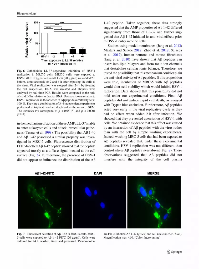
tigated in MRC-5 cells. Fluorescence distribution of
FITC-labelled Ab 1-42 peptide showed that the peptide
appeared mostly as a diffuse signal located at the cell
surface (Fig. 6). Furthermore, the presence of HSV-1
did not appear to influence the distribution of the Ab
1-42 peptide. Taken together, these data strongly
suggested that the AMP properties of Ab 1-42 differed
significantly from those of LL-37 and further sug-
gested that Ab 1-42 initiated its anti-viral effects prior
to HSV-1 entry into the cells.
Studies using model membranes (Jang et al. 2013;
Masters and Selkoe 2012; Zhao et al. 2012; Sciacca
et al. 2012), human neurons and mouse fibroblasts
(Jang et al. 2010) have shown that Ab peptides can
insert into lipid bilayers and form toxic ion channels
that destabilize cellular ionic balance. Therefore, we
tested the possibility that this mechanism could explain
the anti-viral activity of Ab peptides. If this proposition
were true, incubation of MRC-5 with Ab peptides
would alter cell viability which would inhibit HSV-1
replication. Data showed that this possibility did not
hold under our experimental conditions. First, Abpeptides did not induce rapid cell death, as assayed
with Trypan blue exclusion. Furthermore, Ab peptides
acted very early in the viral replicative cycle as they
had no effect when added 2 h after infection. We
showed that they prevented association of HSV-1 with
cells. We obtained evidence that this effect was caused
by an interaction of Ab peptides with the virus rather
than with the cell by simple washing experiments.
Indeed, washing MRC-5 cells that had been exposed to
Ab peptides revealed that, under these experimental
conditions, HSV-1 replication was not different than
control where Ab peptides were absent (Fig. 8). These
observations suggested that Ab peptides did not
interfere with the integrity of the cell plasma
Fig. 6 Cathelicidin LL-37-dependent inhibition of HSV-1
replication in MRC-5 cells. MRC-5 cells were exposed to
HSV-1 (0.01 ID50 per cell) and LL-37 (20 lg/ml) was added 2 h
before, simultaneously or 2 and 6 h after exposing the cells to
the virus. Viral replication was stopped after 24 h by freezing
the cell suspension. DNA was isolated and aliquots were
analyzed by real-time PCR. Results were computed as the ratio
of viral DNA relative to b-actin DNA. Data are shown relative to
HSV-1 replication in the absence of Ab peptides arbitrarily set at
100 %. They are a combination of 3–4 independent experiments
performed in triplicate and are displayed as the mean ± SEM.
The asterisks (*) correspond to p \ 0.05 (*) and p \ 0.0001
(****)
A 1-42-FITC DAPI MERGE
Fig. 7 Fluorescent detection of Ab 1-42 in MRC-5 cells. MRC-
5 cells were exposed to Ab 1-42-FITC (20 lg/ml). Cells were
cultured for 24 h, washed, fixed and processed. Pseudo-colors
are FITC-labelled Ab 1-42 (green) and cell nuclei (DAPI, blue).
Magnification was 960. (Color figure online)
Biogerontology
123

membrane and that their AMP properties were likely
due to interactions with HSV-1. This possibility was
further tested in a cell-free system, based on fluores-
cence detection assays. In one series of experiments,
Protein A/G Sepharose beads were coated with an anti-
HSV-1 antibody that served to retain HSV-1. Exposing
antibody-bound beads to a solution of Ab 1-42 peptides
and HSV-1 showed that the immune complex retained
a significant greater quantity of the fluorescent Abpeptide than in the absence of anti-gB (Fig. 9). This
finding was taken as further evidence that Ab peptide
interacted directly with HSV-1. Thus we propose that
the anti-viral activity of Ab peptides involved initial
interactions with the HSV-1 envelope that occurred
outside the cells, resulting in decreased attachment to
or fusion with the host cell membrane (Table 1). This
interpretation is further supported by the inability of
Fig. 8 Effect of washing of Ab 1-40 or Ab 1-42 on HSV-1
replication. MRC-5 cells were exposed to HSV-1 (0.01 ID50 per
cell) (a) and viral and b-actin DNA were isolated 24 h later and
amplified. Cells were exposed to Ab 1-40 (b) or Ab 1-42 peptide
(d) (20 lg/ml) added 2 h prior to addition of HSV-1. Cells were
also exposed to Ab 1-40 (c) or Ab 1-42 (e) (20 lg/ml) for 2 h,
washed (PBS) and HSV-1 (0.01 ID50 per cell) was added. Viral
replication was stopped after 24 h by freezing the cell
suspension. DNA was isolated and aliquots were analyzed by
real-time PCR. Results were computed as the ratio of viral DNA
relative to b-actin DNA. Data are shown relative to HSV-1
replication in the absence of Ab peptides arbitrarily set to
100 %. They are representative of three independent experi-
ments performed in duplicate and are displayed as the
mean ± SEM. The asterisks (*) correspond to p \ 0.05 (*)
and p \ 0.01 (**)
Table 1 Ab peptide inhibition of HSV-1 interaction with MRC-5 cells
HSV-1 HSV-1 ? Ab 1-40 HSV-1 ? Ab 1-42
Time of incubation
15 min 2 h 15 min 2 h 15 min 2 h
Cell homogenate 274 (68 %) 477 (80 %) 2 (1 %) 181 (50 %) 8 (4 %) 147 (40 %)
Supernatant 128 120 256 181 194 223
MRC-5 cells were incubated with HSV-1 in the absence or presence of Ab peptides (20 lg/ml in each case) for the indicated periods
of time, at 37 �C. Supernatants were removed by aspiration and viral DNA present in supernatant and cell homogenate was amplified
by real-time PCR
Data are expressed as ID50 equivalents
The numbers in parentheses correspond to the percentages of viral DNA present in cell homogenates relative to total DNA
Fig. 9 Protein A/G Sepharose beads (20 ll) were incubated
with FITC-labelled Ab 1-42 (20 lg/ml) or a mixture of HSV-1
(0.1 ID50) and FITC-labelled Ab 1-42 (20 lg/ml), under
conditions described under the ‘‘Materials and methods’’
section. After washing (PBS), emitted fluorescence was
measured using a micro plate reader. Alternatively, the beads
were coated with an anti-gB (agB) mAb, washed and incubated
overnight with with FITC-labelled Ab 1-42 (20 lg/ml) or a
mixture of HSV-1 (0.1 ID50) and FITC-labelled Ab 1-42 (20 lg/
ml). Data are representative of two independent experiments
performed in hexaplicate and are shown as the mean fluores-
cence intensity (MFI) ± SEM. The asterisks (*) correspond to
p \ 0.01 (**), p \ 0.001 (***) and p \ 0.0001 (****)
respectively
Biogerontology
123

Ab peptides to interfere with cell infection by a non-
enveloped virus (HAd5).
An intriguing possibility to explain the selective
effect of Ab peptides is that they initially insert into
the membrane of the envelope of HSV-1, at least
transiently and alter the viral membrane’s physical
properties in a way that affects the process of viral
replication. Evidence supporting this possibility
comes from the work of Cribbs et al. (2000) who
showed that a transmembrane sequence of HSV-1
fusion protein gB shared homology with Ab 1-42. A
high degree of similarity (67 %) was shown to exist
between the membrane proximal region (positions
713–763) of the gB sequence and the C-terminal
sequence of Ab 1-42 peptides (Fig. S2). This partial
sequence of gB has been associated with neurotoxicity
(Yankner et al. 1989) and fibril formation (Halverson
et al. 1990). Furthermore, Cribbs et al. (2000) showed
that the two homologous sequences corresponded to
peptides with similar physical properties in a number
of assays, including aggregation and cytotoxicity.
The bulk of results presented here lead to the
conclusion that Ab peptides possess AMP activity
directed against Alzheimer’s disease-associated HSV-
1 pathogen. Our observations are in agreement with
those of White et al. (2014) with respect to AMP
activity against viruses such as HSV-1 and influenza
virus and a direct interaction of Ab with HSV-1. In
addition, our observations further suggest selectivity
of enveloped viruses as a target of Ab peptides as
opposed to non-enveloped viruses. Our findings
suggest that the production of Ab peptides, at least
initially in the course of the sporadic form of AD, may
be for protection against microbial aggression. Infec-
tion by HSV-1 has been shown to lead to a release of
Ab peptides by human and rat neurons (De Chiara
et al. 2010; Piacentini et al. 2011) which could restrict
virus spread. However, a sustained induction of Abpeptide production by brain cells may become dele-
terious with aging. Indeed, as life progresses, microg-
lia becomes less efficient at eliminating both viruses
and Ab peptides. As a consequence, extracellular
deposition of Ab peptides increases and contributes to
the progression toward AD in the aging brain.
Acknowledgments This work was supported by Grants-in-aid
from the Canadian Institute of Health Research (CIHR) (No.
106634), the Universite de Sherbrooke, and the Research Center
on Aging.
Conflict of interests None.
References
Akhtar J, Shukla D (2009) Viral entry mechanisms: cellular and
viral mediators of herpes simplex virus entry. FEBS J
276:7228–7236
Bekris LM, Yu CE, Bird TD, Tsuang DW (2010) Genetics of
Alzheimer disease. J Geriatr Psychiatry Neurol 23:213–227
Benilova I, Karran E, De Strooper B (2012) The toxic Ab oli-
gomer and Alzheimer’s disease: an emperor in need of
clothes. Nat Neurosci 15:349–357
Bowdish DM, Davidson DJ, Lau YE, Lee K, Scott MG, Han-
cock RE (2005) Impact of LL-37 on anti-infective immu-
nity. J Leukoc Biol 77:451–459
Burton MF, Steel PG (2009) The chemistry and biology of LL-
37. Nat Prod Rep 26:1572–1584
Bustin SA, Benes V, Garson JA, Hellemans J, Huggett J, Ku-
bista M, Mueller R, Nolan T, Pfaffl MW, Shipley GL,
Vandesompele J, Wittwer CT (2009) The MIQE guide-
lines: minimum information for publication of quantitative
real-time PCR experiments. Clin Chem 55:611–622
Carty M, Reinert L, Paludan SR, Bowie AG (2014) Innate
antiviral signalling in the central nervous system. Trends
Immunol 35:79–87
Castellani RJ, Rolston RK, Smith MA (2010) Alzheimer dis-
ease. Disease-a-Month 56:484–546
Cribbs DH, Azizeh BY, Cotman CW, LaFerla FM (2000) Fibril
formation and neurotoxicity by a herpes simplex virus
glycoprotein B fragment with homology to the Alzhei-
mer’s A beta peptide. Biochemistry 39:5988–5994
Cupelli K, Stehle T (2011) Viral attachment strategies: the many
faces of adenoviruses. Curr Opin Virol 1:84–91
De Chiara G, Marcocci ME, Civitelli L, Argnani R, Piacentini
R, Ripoli C, Manservigi R, Grassi C, Garaci E, Palamara
AT (2010) APP processing induced by herpes simplex
virus type 1 (HSV-1) yields several APP fragments in
human and rat neuronal cells. PLoS ONE 5:e13989
Denaro FJ, Staub P, Colmer J, Freed DM (2003) Coexistence of
Alzheimer disease neuropathology with herpes simplex
encephalitis. Cell Mol Biol 49:1233–1240
Egan KP, Wu S, Wigdahl B, Jennings SR (2013) Immunological
control of herpes simplex virus infections. J Neurovirol
19:328–345
Eisenberg RJ, Atanasiu D, Cairns TM, Gallagher JR, Krum-
menacher C, Cohen GH (2012) Herpes virus fusion and
entry: a story with many characters. Viruses 4:800–832
Franceschi C, Campisi J (2014) Chronic inflammation (inflam-
maging) and its potential contribution to age-associated
diseases. J Gerontol A Biol Sci Med Sci 69(Suppl 1):S4–S9
Frasca L, Lande R (2012) Role of defensins and cathelicidin
LL37 in auto-immune and auto-inflammatory diseases.
Curr Pharm Biotechnol 13:1882–1897
Griciuc A, Serrano-Pozo A, Parrado AR, Lesinski AN, Asselin
CN, Mullin K, Hooli B, Choi SH, Hyman BT, Tanzi RE
(2013) Alzheimer’s disease risk gene CD33 inhibits mi-
croglial uptake of amyloid beta. Neuron 78:631–643
Grunewald K, Desai P, Winkler DC, Heymann JB, Belnap DM,
Baumeister W, Steven AC (2003) Three-dimensional
Biogerontology
123

structure of herpes simplex virus from cryo-electron
tomography. Science 302:1396–1398
Halverson K, Fraser PE, Kirschner DA, Lansbury PT (1990)
Molecular determinants of amyloid deposition in Alzhei-
mer’s disease: conformational studies of synthetic beta-
protein fragments. Biochemistry 29:2639–2644
Heldwein KE, Lou H, Bender FC, Cohen GH, Eisenberg RJ,
Harrison SC (2006) Crystal structure of glycoprotein B
from herpes simplex virus 1. Science 313:217–220
Itzhaki RF, Wozniak MA (2012) Could antivirals be used to
treat Alzheimer’s disease? Future Microbiol 7:307–309
Jang H, Arce FT, Ramachandran S, Capone R, Azimova R,
Kagan BL, Nussinov R, Lal R (2010) Truncated beta-
amyloid peptide channels provide an alternative mecha-
nism for Alzheimer’s disease and Down syndrome. Proc
Natl Acad Sci USA 107:6538–6543
Jang H, Connelly L, Arce FT, Ramachandran S, Kagan BL, Lal
R, Nussinov R (2013) Mechanisms for the insertion of
toxic, fibril-like b-amyloid oligomers into the membrane.
J Chem Theory Comput 9:822–833
Karran E, Mercken M, De Strooper B (2011) The amyloid
cascade hypothesis for Alzheimer’s disease: an appraisal
for the development of therapeutics. Nat Rev Drug Discov
10:698–712
Larbi A, Pawelec G, Witkowski JM, Schipper HM, Derhov-
anessian E, Goldeck D, Fulop T (2009) Dramatic shifts in
circulating CD4 but not CD8 T cell subsets in mild Alz-
heimer’s disease. J Alzheimers Dis 17(1):91–103
Letenneur L, Peres K, Fleury H, Garrigue I, Barberger-Gateau P,
Helmer C, Orgogozo JM, Gauthier S, Dartigues JF (2008)
Seropositivity to herpes simplex virus antibodies and risk
of Alzheimer’s disease: a population-based cohort study.
PLoS ONE 3:e3637
Lin WR, Wozniak MA, Cooper RJ, Wilcock GK, Itzhaki RF
(2002) Herpesviruses in brain and Alzheimer’s disease.
J Pathol 197:395–402
Masters CL, Selkoe DJ (2012) Biochemistry of amyloid b-
protein and amyloid deposits in Alzheimer disease. Cold
Spring Harb Perspect Med 2:a006262
Miklossy J (2011) Emerging roles of pathogens in Alzheimer
disease. Expert Rev Mol Med 13:e30
Nemerow GR, Stewart PL, Reddy VS (2012) Structure of
human adenovirus. Curr Opin Virol 2:115–121
Nicoll MP, Proenca JT, Efstathiou S (2012) The molecular basis
of herpes simplex virus latency. FEMS Microbiol Rev
36:684–705
Pearson HA, Peers C (2006) Physiological roles for amyloid
beta peptides. J Physiol 575:5–10
Perneczky R, Guo LH, Kagerbauer SM, Werle L, Kurz A,
Martin J, Alexopoulos P (2013) Soluble amyloid precursor
protein beta as blood-based biomarker of Alzheimer’s
disease. Transl Psychiatry 3:e227
Piacentini R, Civitelli L, Ripoli C, Marcocci ME, De Chiara G,
Garaci E, Azzena GB, Palamara AT, Grassi C (2011) HSV-
1 promotes Ca2?-mediated phosphorylation and Ab accu-
mulation in rat cortical neurons. Neurobiol Aging
32(2323):e13–e26
Piacentini R, De Chiara G, Li Puma DD, Ripoli C, Marcocci
ME, Garaci E, Palamara AT, Grassi C (2014) HSV-1 and
Alzheimer’s disease: more than a hypothesis. Front Phar-
macol 5:97
Querfurth HW, LaFerla FM (2010) Alzheimer’s disease. N Engl
J Med 362:329–344
Reske A, Pollara G, Krummenacher C, Chain BM, Katz DR
(2007) Understanding HSV-1 entry glycoproteins. Rev
Med Virol 17:205–215
Rigamonti A, Lauria G, Mantero V, Salmaggi A (2013) A case
of late herpes simplex encephalitis relapse. J Clin Virol
58:269–270
Russell WC (2009) Adenoviruses: update on structure and
function. J Gen Virol 90:1–20
Sciacca MF, Kotler SA, Brender JR, Chen J, Lee DK, Rama-
moorthy A (2012) Two-step mechanism of membrane
disruption by Ab through membrane fragmentation and
pore formation. Biophys J 103:702–710
Seeman P, Seeman N (2011) Alzheimer’s disease b-amyloid
plaque formation in human brain. Synapse 65:1289–1297
Smith JG, Wiethoff CM, Stewart PL, Nemerow GR (2010)
Adenovirus. Curr Top Microbiol Immunol 343:195–224
Soscia SJ, Kirby JE, Washicosky KJ, Tucker SM, Ingelsson M,
Hyman B, Burton MA, Goldstein LE, Duong S, Tanzi RE,
Moir RD (2010) The Alzheimer’s disease-associated
amyloid beta-protein is an antimicrobial peptide. PLoS
ONE 5:e9505
Streit WJ, Xue QS (2014) Human CNS immune senescence and
neurodegeneration. Curr Opin Immunol 29C:93–96
Sun E, He J, Zhuang X (2013) Live cell imaging of viral entry.
Curr Opin Virol 3:34–43
Tam JH, Pasternak SH (2012) Amyloid and Alzheimer’s dis-
ease: inside and out. Can J Neurol Sci 39:286–298
Turner J, Cho Y, Dinh NN, Waring AJ, Lehrer RI (1998)
Activities of LL-37, a cathelin-associated antimicrobial
peptide of human neutrophils. Antimicrob Agents Che-
mother 42:2206–2214
White MR, Kandel R, Tripathi S, Condon D, Taubenberger J,
Hartshorn KL (2014) Alzheimer’s associated b-amyloid
protein inhibits influenza A virus and modulates viral
interactions with phagocytes. PLoS ONE 9:e101364
WHO report (2013) Dementia: a public health priority. 11 Apr
2013
Wozniak MA, Itzhaki RF, Shipley SJ, Dobson CB (2007)
Herpes simplex virus infection causes cellular beta-amy-
loid accumulation and secretase upregulation. Neurosci
Lett 429:95–100
Wozniak MA, Mee AP, Itzhaki RF (2009) Herpes simplex virus
type 1 DNA is located within Alzheimer’s disease amyloid
plaques. J Pathol 217:131–138
Wozniak MA, Frost AL, Preston CM, Itzhaki RF (2011) An-
tivirals reduce the formation of key Alzheimer’s disease
molecules in cell cultures acutely infected with herpes
simplex virus type 1. PLoS ONE 6:e25152
Yankner BA, Dawes LR, Fisher S, Villa-Komaroff L, Oster-
Granite ML, Neve RL (1989) Neurotoxicity of a fragment
of the amyloid precursor associated with Alzheimer’s
disease. Science 245:417–420
Zhao LN, Long H, Mu Y, Chew LY (2012) The toxicity of
amyloid b oligomers. Int J Mol Sci 13:7303–7307
Biogerontology
123


