Embed Size (px)
Citation preview

Variations in relative humidity modulate Leptosphaeria spp.pathogenicity and interfere with canola mechanismsof defence
Abdelbasset El Hadrami &W. G. Dilantha Fernando & Fouad Daayf
Received: 13 November 2008 /Accepted: 14 September 2009# KNPV 2009
Abstract Blackleg (phoma stem canker), caused byLeptosphaeria spp., is an important disease of canola(oilseed rape, Brassica napus). Control strategies relyon the use of resistant cultivars, chemical and disease-reducing cropping practices. In Canada, the pathogenpopulation is represented by L. maculans and L.biglobosa, which are considered to be highly andweakly aggressive, respectively. It is largely admittedthat L. biglobosa isolates are not able to cause asignificant amount of stem canker and develop on theplant only when it becomes senescent, late in theseason. The prevalence of L. maculans over L.biglobosa has been considered to be linked to thelow aggressiveness of the latter. However, in thisstudy, we show that L. biglobosa isolates couldbecome highly aggressive in terms of lesion appear-ance on cotyledons, if the right conditions oftemperature and relative humidity (RH) are provided.Percent germination of inoculated pycnidiospores wasnot affected by the RH regimes tested. This is the firststudy to show the importance of RH as a factorconditioning the pathogenicity of L. biglobosa iso-lates on canola cotyledons. Concurrent changes in thehost defence mechanisms against L. biglobosa iso-lates in response to variations in the RH were also
investigated. Under high RH, the increase in diseasecaused by the weakly aggressive isolates coincidedwith a reduced accumulation of lignin at the earlystages of infection.
Keywords Canola (oilseed rape) .
Leptosphaeria maculans . L. biglobosa . Blackleg .
Phoma canker . Relative humidity . Temperature .
Phenolics . Lignin . Hypersensitive reaction
AbbreviationsCf/Avr Cladosporium fulvum/Avirulentd.a.i. Day after inoculationHR Hypersensitive reactionPG Pathogenicity groupRH Relative humidity
Introduction
Canola (oilseed rape; Brassica napus) is the secondlargest oilseed crop worldwide (Raymer 2002). It islargely grown in Canada, China, USA, Australia,and several European countries. This crop is knownfor the quality and health benefits of its oil, butinterest has also been growing towards its use as analternative source for biodiesel and other by-products. The most important fungal disease ofcanola is blackleg (phoma stem canker; West et al.2001; Fitt et al. 2006), caused by a complex of
Eur J Plant PatholDOI 10.1007/s10658-009-9532-1
A. El Hadrami :W. G. D. Fernando : F. Daayf (*)Department of Plant Science, University of Manitoba,222 Agriculture Building,Winnipeg, Manitoba R3T 2N2, Canadae-mail: [email protected]

phylogenetically-related ascomycete species, includ-ing Leptosphaeria maculans and L. biglobosa (ana-morph: Phoma lingam) (Williams and Fitt 1999;West et al. 2001; Shoemaker and Brun 2001;Mendès-Pereira et al. 2003).
The control of this disease complex relies on theuse of resistant cultivars along with chemical andcultural practices (West et al. 2001; Sivasithamparamet al. 2005; Gladders et al. 2006). Previously, thepathogen populations consisted of five pathogenicitygroups (PG1, PG2, PG3, PG4 and PGT) as definedby Mengistu et al. (1991). These five PGs werefound in western Canada (West et al. 2001) and cansurvive on naturally-infected rapeseed and canolastubble for a long period (Petrie 1995). PG1 has beenreclassified since as L. biglobosa (Shoemaker andBrun 2001), and is represented by weakly aggressiveisolates developing into a canker only late in theseason when plants are senescent. Leptosphaeriabiglobosa is still prevailing over L. maculans incertain canola and rapeseed growing areas i.e., eastEurope (Jedryczka et al. 1994, 2002) and has beenproven to cause major damage to winter-type cultivarsof rapeseed and decrease the yield under fieldconditions, especially in Poland (Jedryczka 2007).PG2 on the other hand, has been the most prevalentgroup of L. maculans.
Isolates from these two species can co-exist oncanola plants (West et al. 2001, 2002; Mahuku et al.1996a, b). However, the way they interact with eachother on plant tissues is poorly understood. Pre-inoculation of cv. Westar seedlings with pycnidio-spores of an L. biglobosa isolate was shown to induceboth local and systemic resistance to an L. maculansisolate under controlled conditions (Mahuku et al.1996a, b). However, conflicting results from co-inoculation of the two types of isolates on cv. Surpass400, in Australia, have shown that the effect of L.biglobosa on L. maculans is only local (Li et al.2006). More recent results have shown both local andsystemic decrease of the spot lesion area (Liu et al.2006). Such an effect was confirmed in the field andwas comparable to the effect of certain chemicaldefence activators (Liu et al. 2006). Based on theseand earlier observations, it seems interesting to useisolates of L. biglobosa to control subsequent infec-tions by L. maculans. However, isolates to be used forsuch cross-protection should be weakly aggressiveunder various conditions. Therefore, before selecting
an L. biglobosa isolate that can be used as a biologicalcontrol agent (Daayf et al. 2003b), it is important tostudy its pathogenic variability under different envi-ronmental conditions.
The objectives of the present study were toinvestigate (i) the effects of temperature and relativehumidity (RH) on the pathogenic variability ofselected isolates; and (ii) how the variation of suchconditions impacts on the canola defence in responseto these isolates.
Material and methods
Plant materials
Three canola cultivars, Westar, Quinta and Glacier,exhibiting differential responses to Leptosphaeriaspp. isolates were selected for this study. They weregrown in a Metromix soil mixture amended withNPK (20:20:20) under cool fluorescent light with a16 h photoperiod at 20/16°C day/night and wateredregularly.
Leptosphaeria spp. isolates, inoculation and diseaseassessment
Fourteen Leptosphaeria spp. isolates were used in thisstudy. They were all collected from infected canolaplants grown in Manitoba, Canada. After single-sporeisolation, each isolate was propagated on V8-agarmedium containing 200 ml of V8, 15 g of agar and0.75 g of CaCO3 l
−1. Based on an initial screening ondifferential cultivars, five of these isolates were foundto be L. biglobosa (former PG1) and nine were L.maculans (3 PG2, 2 PG3, 2 PG4, and 2 PGT). Theidentity and classification of the isolates to either L.maculans or biglobosa were verified based on thecolony morphology as well as using GPI-isozymesalong with species-specific primers described byKuusk et al. (2002) and Liu et al. (2006) (data notshown). Isolates of L. biglobosa were found to be L.biglobosa ‘canadensis’ as defined by Mendès-Pereiraet al. (2003).
For inoculum production, the isolates were incu-bated for two weeks under continuous cool-whitefluorescent light at 20±2°C. Pycnidiospores werethen harvested in water by gently scraping the surfaceof the cultures. Inoculations were performed either on
Eur J Plant Pathol

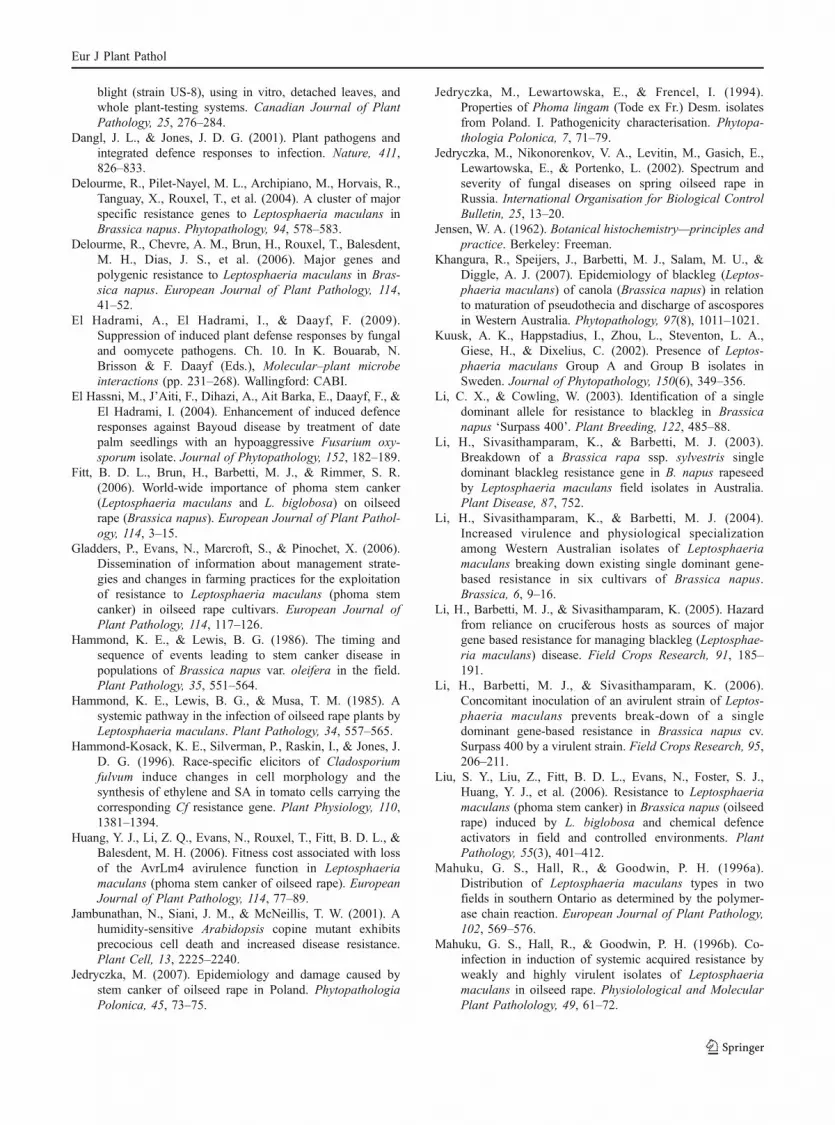
cotyledons from 8 day-old plants or on 4–6 leaf-adultplants by placing 10 µl droplets of a spore suspensioncalibrated at 2×10⁷ pycnidiospores ml−1 on a freshlymade wound. Disease rating was conducted 12 daysafter inoculation (d.a.i.) following a scale rangingfrom 0 (no disease) to 9 (large necrotic symptomsproducing large number of pycnidia) (Fig. 1).
Relative humidity (RH) and temperature conditions
The pathogenicity of selected isolates was tested, undera range of temperature and RH conditions, on differen-tial cvs Westar, Quinta and Glacier at the cotyledonstage. In a preliminary study, five isolates from L.biglobosa (former PG1) were screened against areference L. maculans isolate (former PG2). Conditionstested consisted of three temperature regimes (20/16;25/23, and 30/27°C day/night) that cover the range oftemperatures typically recorded in western Canadaunder an ambient RH. The same set of six isolateswas tested at 20/16°C day/night under two RHconditions: (i) a constant 45–50% RH (low RH), (ii)incubation in saturated atmosphere for three days, thenunder 70% RH (high RH) until rating at 12 d.a.i.
The 15 isolates were further tested for theirpathogenicity in highly-controlled atmosphere cab-inets where three different temperature regimes 20/16, 25/23, and 30/25°C day/night were combinedwith three RH conditions (low RH 45–50%;intermediate RH 70% and high RH≥85%). Thegrowth cabinets were limited to a top RH≈85%,which did not allow for the testing of combina-tions involving saturated atmosphere (experimentdesign reported below).
At all times, inoculum from each tested isolatewas placed on open water-agar plates and incubat-ed at the same environmental conditions tested,then checked for germination. In all includedresults, inoculum germination reached 85–92.5%.Additional experiments including reference isolatesexhibiting various degrees of pathogenicity wereconducted under regular conditions (20/16°C day/night; ambient RH). Both of these measures werecarried out to exclude any effect of a poorgermination of the inoculum on further developmentof blackleg symptoms and to assign the observedpathogenicity of the isolates to the environmentalconditions tested.
0 1
5 7
3
9
Wound
Grey-green tissue collapse
Dark necrotic tissue
Chlorosis Sharp non-darkened margin
Sharp darkened margin
Pycnidia
Fig. 1 Rating scale used toassess blackleg disease oncanola cotyledons 12 d.a.i.
Eur J Plant Pathol

Histology and lignin staining
Canola cotyledons from all tested cultivars, eitherhealthy or inoculated with various isolates, werefreshly harvested and discoloured in Carnoy’s solu-tion overnight (ethanol:chloroform:acetic acid, 6:3:1,v/v/v). The following day, cotyledon tissues wererinsed using distilled water, stained in phloroglucinol-HCl and observed under light microscopy (Jensen1962).
High pressure liquid chromatography (HPLC)analysis of secondary metabolites
Soluble phenolics of canola leaves (500 mg–1 g) wereextracted using 80% methanol (Daayf et al. 2003a; ElHassni et al. 2004). After centrifugation, the pelletwas re-suspended twice in 1.5 ml of 80% methanol.Pooled methanolic fractions were incubated under anitrogen stream until the organic phase had evaporat-ed. The remaining aqueous phase was then mixedwith petroleum ether to remove chlorophyll, waxesand lipids from the leaf extracts. The cleared aqueousphase was then extracted three times with ethylacetate. After a final evaporation of the ethyl acetatefraction, the residue was suspended in 200 μl of puremethanol and immediately subjected to analysis orstored at −20°C until used.
For HPLC analysis a Waters 2690 separationmodule was used. This module is equipped with anautosampler and a Waters 996 photodiode arraydetector, and fitted with a 5 µm LiChrospher® 100RP-18 guard column (LiChroCART® 4-4, Germany)and a reverse-phase 5 µm 250-4 LiChrospher® 100RP-18 column (LiChroCART® 4-4, Germany). Thecolumn was eluted at a flow of 1 ml min−1 with agradient using a solvent system composed of A: 0.1%H3PO4-acidified water and B: HPLC grade acetoni-trile. The gradient used to carry the analysis was asfollows: (time [min]/A [%]/B [%]) = 0/100/0, 5/95/5,10/95/5, 14/90/10, 20/80/20, 23/80/20, 30/65/35, 35/65/35, 43/50/50, 48/25/75, 55/0/100, 60/0/100.Injected volumes were 15 µl per sample and eachinjection was repeated twice. Data were analysedusing the EmpowerTM 2.0 (Waters, Ville-Saint-Laurent PQ, Canada). Compounds were identifiedbased on their retention time, their typical UVspectra in reference to our database and through co-elution with commercial standards. Quantification of
hydroxycinnamates was conducted by reference to astandard curve using a serial dilution of commercialchrologenic acid (Sigma-Aldrich Co.).
Experiment design and data analysis
All experiments were conducted in a randomisedcomplete block design. For every trial, 20–24 seed-lings were used per replicate isolate × cultivar ×treatment and each trial was repeated independentlythree times. Collected data were submitted to varianceanalysis using the GLM model of the SAS 9.1programme software (SAS Institute Inc., Cary, N.C.,U.S.A. 2004). The homogeneity of the variances andthe symmetry of the distribution were checked foreach analysed variable included in the ANOVAanalysis. When the F-test was significant at P<0.05,mean values were compared according to Newman-Keuls test. Non-wounded and wounded and non-inoculated controls were used in different trials andincluded in the analysis when necessary. Statisticasoftware (StatSoft 1999) was also used to generategraphics from the data. For the soluble phenolicsanalysis, three bulk sub-samples were formed out ofthe 20–24 seedlings used per replicate isolate ×cultivar × treatment and extracted. Each one of thesesamples was subjected to HPLC analysis at leasttwice.
Results
Initial pathogenicity screening
No significant effect of the three tested temperatureregimes was detected on the pathogenicity of theselected isolates (Fig. 2). Leptosphaeria biglobosaisolates exhibited low pathogenicity on all three testedcultivars regardless of the temperature conditiontested. Leptosphaeria maculans isolates were highlyaggressive on cv. Westar, especially at 20/16 and 25/23°C day/night (Fig. 2). Reactions of the isolates oncvs Quinta and Glacier depended on their pathoge-nicity grouping (former PG2, 3, 4 or T). However, nosignificant effect of the temperature was recorded.Isolates formerly known as PG2 produced blacklegsymptoms only on cv. Westar, whereas isolates fromPG3 and PG4 were highly aggressive on Westar andGlacier and on the three tested cultivars, respectively.
Eur J Plant Pathol

The same set of six isolates has been tested at 20/16°C day/night under high (3 d.a.i. under saturatedatmosphere followed by constant 70% RH) and lowRH conditions (constant 45–50% RH). Interestingly,
under high RH levels, L. biglobosa isolates (formerPG1) caused severe disease on all three testedcultivars (Fig. 3). Their level of aggressiveness wasas high as L. maculans isolates (Figs. 3 & 4). In
±1,96*Std.-Error ±1,00*Std.-Error Mean
Cultivar x Isolate
Dis
ease
sco
re
20/
16
-2
1
4
7
10
25/
23
-2
1
4
7
10
Westar
30/
25
-2
1
4
7
10
Con
trol
PG
2-86
PG
1-A
L19
PG
1-A
L26
PG
1-56
PG
1-84
PG
1-18
Glacier
Con
trol
PG
2-86
PG
1-A
L19
PG
1-A
L26
PG
1-56
PG
1-84
PG
1-18
Quinta
Con
trol
PG
2-86
PG
1-A
L19
PG
1-A
L26
PG
1-56
PG
1-84
PG
1-18
SS df MS F P-value 1: Cultivar 86.4 2
2124
1224
567
643.2 752.5 0.000
2: Isolate 295.4 49.2 857.7 0.0003: Temp. 1.8 0.9 15.9 1.82E-071*2 584.3 48.7 848.1 0.0001*3 3.6 0.9 15.9 2.47E-122*3 12.3 1.0 17.9 0.0001*2*3 20.2 0.8 14.7 0.000Error 32.6 0.06
Fig. 2 Disease scores recorded on three canola cultivars(Westar, Glacier and Quinta) after inoculation with either oneL. maculans (86-14) or one of the five L. biglobosa isolatestested (AL19; AL26; 03-56-02; 84-12; 04-18-01) and incuba-tion under three conditions of temperature (20/16°C, 25/23°C
and 30/27°C day/night). The ANOVA table summarises thesignificance of each tested effect and their interactions at P<0.05. SS: sum of squares; df: degree of freedom; MS: meansquare
Eur J Plant Pathol

addition, cotyledons infected with L. biglobosa iso-lates displayed blackleg symptoms as early as 8 d.a.i.and turned senescent at 10 d.a.i. compared tocotyledons kept either as controls or inoculated with
Leptosphaeria maculans isolate. Pycnidia were pres-ent on the cotyledons in both cases. Leptosphaeriabiglobosa isolate apparent aggressiveness was 2 to 12times higher under high RH conditions as compared
±1,96*Std.-Error ±1,00*Std.-Error Mean
Cultivar x Isolate
Dis
ease
sco
re
Hig
h R
H
-2
1
4
7
10
Westar
Low
RH
-2
1
4
7
10
Con
trol
PG
2-86
PG
1-A
L19
PG
1-A
L26
PG
1-56
PG
1-84
PG
1-18
Glacier
Con
trol
PG
2-86
PG
1-A
L19
PG
1-A
L26
PG
1-56
PG
1-84
PG
1-18
Quinta
Con
trol
PG
2-86
PG
1-A
L19
PG
1-A
L26
PG
1-56
PG
1-84
PG
1-18
SS df MS F 1: Cultivar 109.2 54.6 93.6 0.0002: Isolate 156.6 26.1 44.8 0.0003: RH 308.4 308.4 528.9 0.0001*2 111.5 9.3 15.9 2.32E-281*3 23.9 11.9 20.5 3E-092*3 235.2 39.2 67.2 0.0001*2*3 83.4 6.9 11.9 3.96E-21Error 269.4
261
1226
12462 0.6
P-value
Fig. 3 Disease scores recorded on three canola cultivars(Glacier, Quinta and Westar) after inoculation with either oneL. maculans (86-14) or one of the five L. biglobosa isolatestested (AL19; AL26; 03-56-02; 84-12; 04-18-01) and incuba-tion under two relative humidity (RH) conditions: low RH
consisting of continuous 45–50% RH or high RH consisting of3 d.a.i. at saturated atmosphere followed by an incubation at70% RH. The ANOVA table summarises the significance ofeach tested effect and their interactions at P<0.05. SS: sum ofsquares; df: degree of freedom; MS: mean square
Eur J Plant Pathol

to when they were incubated under low RH. Thereference L. maculans isolate (former PG2), on theother hand, showed a decrease in aggressivenessunder high RH conditions even though the percentageof germinating inoculum was high and almost sameas the other isolates (89–91%; Fig. 3). Similarobservations were also made on other isolates fromthe same pathogenicity group and in the field duringwet summers in western Canada, where former PG2isolates have been predominant (data not shown).Complementary experiments conducted with the sameisolates on detached leaves from adult seedlings (with4–6 fully-developed leaves) and incubated undersaturated atmosphere in the laboratory confirmed theabove-mentioned results (data not shown).
For further investigations, the selected set ofisolates was extended to 15 and was subjected topathogenicity screening in highly controlled atmo-sphere cabinets, where three different temperatureregimes 20/16, 25/23, 30/25°C day/night were com-bined with three RH conditions (low RH 45–50%;intermediate level of RH 70% and high RH≈85%(maximum limit)). Under these conditions, the per-cent germination of inoculum was estimated at 85–92.1% for all tested isolates. Results are shown inFig. 5 and confirmed the previous observations with
the effect of RH on the variation of the pathogenicityof the tested isolates of L. maculans. No significanteffect of the temperature was apparent. The observedeffect in the combination temperature × RH wasmainly due to RH. Interestingly, higher levels of RHcombined with either low (20/16°C) or high temper-atures (30/25°C) negatively impacted on the perfor-mance of L. maculans isolates more than it did on L.biglobosa. The growth cabinets used were limited to amaximum 85% RH, which did not allow for testingthe combination of various temperatures with thesaturated atmosphere.
Soluble phenolics analysis
Soluble phenolics were extracted and analysed byHPLC from plants kept as controls or inoculated witheither L. biglobosa or L. maculans isolates andincubated under various temperature or RH condi-tions. Several differences were observed amongcultivars in the type and the quantity of solublephenolics accumulated in response to various isolates(Fig. 6). The major induced compounds have beenidentified as hydroxycinnamates (i.e. derivatives ofchlorogenic, ferulic and coumaric acids; Fig. 7).These derivatives accumulated in all treatments as
0
1
2
3
4
5
6
7
8
9
10
Con
trol
PG
1 A
L-19
PG
1 A
L-26
PG
1 03
-56-
02
PG
1 04
-18-
01
PG
1 84
-12
PG
2 86
-14
PG
2 04
-09-
17
PG
2 04
-09-
29
PG
3 04
-09-
34
PG
3 04
-09-
74
PG
4 04
-09-
05
PG
4 04
-09-
27
PG
T 0
4-09
-11
PG
T 0
4-09
-75
Isolates
***
***
****
***
*
0
1
2
3
4
5
6
7
8
9
10
Con
trol
PG
1 A
L-19
PG
1 A
L-26
PG
1 03
-56-
02
PG
1 04
-18-
01
PG
1 84
-12
PG
2 86
-14
PG
2 04
-09-
17
PG
2 04
-09-
29
PG
3 04
-09-
34
PG
3 04
-09-
74
PG
4 04
-09-
05
PG
4 04
-09-
27
PG
T 0
4-09
-11
PG
T 0
4-09
-75
Isolates
Dis
ease
sco
re***
***
****
***
*
Fig. 4 Disease scores onthree canola cultivars (□Glacier, Quinta and ■Westar) after inoculationwith Leptosphaeria spp.isolates (reported by theirformer pathogenicity group-ing PG1, PG2, PG3, PG4and PGT) and incubationunder high RH conditions.Asterisks show significantdecrease in disease score(P<0.05) among all of thetreatments
Eur J Plant Pathol

±1,96*Std.-Error ±1,00*Std.-Error Mean
Cultivar x Isolate
Dis
ease
sco
reT
empe
ratu
re a
nd R
H c
ondi
tion
20 &
50
0
5
10
20 &
70
0
5
1020
& 8
5
0
5
10
25 &
50
0
5
10
25 &
70
0
5
10
25 &
85
0
5
10
30 &
50
0
5
10
30 &
70
0
5
10
Glacier
30 &
85
0
5
10
Con
trol
PG
1-19
PG
1-26
PG
1-56
PG
1-18
PG
1-84
PG
2-86
PG
2-17
PG
2-29
PG
3-34
PG
3-74
PG
4-05
PG
4-27
PG
T-1
1P
GT
-75
Quinta
Con
trol
PG
1-19
PG
1-26
PG
1-56
PG
1-18
PG
1-84
PG
2-86
PG
2-17
PG
2-29
PG
3-34
PG
3-74
PG
4-05
PG
4-27
PG
T-1
1P
GT
-75
WestarC
ontr
olP
G1-
19P
G1-
26P
G1-
56P
G1-
18P
G1-
84P
G2-
86P
G2-
17P
G2-
29P
G3-
34P
G3-
74P
G4-
05P
G4-
27P
GT
-11
PG
T-7
5
SS df MS F P-value 1: Tm&RH 537.3 67.2 531.0 0.0002: Cultivar 3421.0 1710.5 13523.9 0.0003: Isolate 5568.2 397.7 3144.6 0.0001*2 191.7 12 94.7 0.0001*3 1126.7 10.1 79.5 0.0002*3 1345.7 48.1 379.9 0.0001*2*3 470.4 2.1 16.6 0.000Error 153.7
82
1416
11228
2241215 0.1
Fig. 5 Disease scores on three canola cultivars (Glacier, Quintaand Westar) after inoculation with Leptosphaeria spp. isolates(reported by their former pathogenicity grouping PG1; PG2;PG3; PG4 and PGT) and incubation under various combina-tions of RH and temperature conditions (20/16; 25/23; and 30/27°C day/night) × (continuous 45–50% RH; 70% RH; ≈ 85%
RH) i.e. 20 & 50 reported on the graph represents incubation at20/16°C day/night under continuous 45–50% RH. The ANOVAtable summarises the significance of each tested effect and theirinteractions at P<0.05. SS: sum of squares; df: degree offreedom; MS: mean square
Eur J Plant Pathol

early as 2 d.a.i. However, the rate by which theiraccumulation dropped overtime was differential. Afaster decrease in their accumulation was observed intreatments where plants were inoculated with L.biglobosa isolates compared to the non-inoculated
controls and to plants inoculated with L. maculansisolates.
Soluble phenolics accumulation also varied, bothqualitatively and quantitatively, with the temperatureand RH conditions tested. Their concentration de-
Cultivar x Isolate
µg e
q. c
hlor
ogen
ic a
cid
g-1
FW RH
Hig
h
-2
6
14
22
NW&NICtrl
RH
Lo
w
-2
6
14
22
W Q G
NI Ctrl
W Q G
PG1-19
W Q G
PG1-26
W Q G
PG1-56
W Q G
PG1-18
W Q G
PG1-84
W Q G
PG2-86
W Q G
PG2-17
W Q G
PG2-29
W Q G
PG3-34
W Q G
PG3-74
W Q G
PG4-05
W Q G
PG4-27
W Q G
PGT-11
W Q G
PGT-75
W Q G
±1,96*Std.-Error ±1,00*Std.-Error Mean
SS df MS F P-value 1: Cultivar 43.3 21.7 543.5 0.0002: Isolate 215.3 14.4 360.0 0.0003: RH 628.9 628.9 15777.2 0.0001*2 412.9 13.8 345.3 0.0001*3 33.1 16.6 415.4 0.0002*3 153.7 10.2 257.1 0.0001*2*3 349.4 11.6 292.2 0.000Error 3.8
215
130
2153096 0.04
Fig. 6 Variation in amount of soluble phenolics (µg eq.chlorogenic acid g–1 FW) in the cotyledon tissues of threecanola cultivars (Westar (W), Quinta (Q) and Glacier (G)) inresponse to wounding and/or to inoculation with Leptosphaeriaspp. isolates after incubation under low (constant 45–50%) orhigh RH regimes (3 days at saturated atmosphere followed by a
constant 70% RH until the appearance of the lesions). Eachdata point represents the mean of two individual trials ± 1.96standard-errors. The ANOVA table summarises the significanceof each tested effect and their interactions at P<0.05. SS: sumof squares; df: degree of freedom; MS: mean square
Eur J Plant Pathol

creased in all tested cultivars when RH conditionsincreased (Figs. 8 & 9).
Histological observations
Cotyledons maintained as controls or subjected toinoculation with L. biglobosa or other L. maculansisolates were stained with phloroglucinol-HCl whichrecognises the cinnamic aldehyde groups in the ligninmatrix. Cotyledons inoculated with L. biglobosaisolates showed a thick multi-layer of lignifiedmaterial around the infection site, as compared tothe wounded non-inoculated controls (Fig. 10). Coty-ledons inoculated with L. maculans isolates displayeddiffused lesions, where less lignified material accu-mulated (Fig. 10). Similar diffused lesions with lesslignified material around were also observed inresponse to L. biglobosa isolates inoculated onto thethree tested cultivars and incubated under specifichigh RH conditions (saturated atmosphere followedby constant 70% RH) (Fig. 10).
Discussion
Blackleg is the most important disease of canola,oilseed rape and other Brassicae. Resistance incultivated germplasm comprises partial resistance(quantitative resistance) and in many cases this is
supplemented with one or more major resistancegenes (AvrLm1-9 and LepR1-3; Delourme et al.2004, 2006) such as those controlling callose deposit(Li and Cowling 2003; Li et al. 2003, 2004, 2005).Although deploying cultivars with major resistancegene seems to be efficient in reducing the diseaseseverity in certain countries (i.e., Canada), suchresistance is frequently overcome within a fewseasons (i.e., cv. Surpass 400 carrying LepR3 fromB. rapa ssp. sylvestris in Australia) (Li and Cowling2003; Li et al. 2003, 2004, 2005; Huang et al. 2006).Such a breakdown of resistance may be in part relatedto the pathogen’s diversity and variability, which isoften not tested in early screens of breeding materialthat uses only one or a few isolates, and which mayincrease as a result of selection pressure on thepathogen due to intensive cultivation of resistantcultivars (Rouxel et al. 2003).
Fig. 7 HPLC profile ofsoluble phenolics from ca-nola cv. Westar inoculatedwith L. biglobosa. Thespectra represent the majorhydroxycinnamates detectedin the extract. Their UV-absorbance corresponds totheir relative abundance inthe chromatogram
Fig. 8 Variation of the soluble phenolics profile in cvs Westar,Quinta and Glacier, either healthy or inoculated with differentLeptosphaeria spp. isolates, under three temperature regimes ofincubation (20/16; 25/23; 30/25°C day/night). For spaceconvenience only results from one L. biglobosa and one L.maculans isolate are shown here. Similar patterns wereobserved with other isolates. The major accumulated com-pounds are deemed to be hydroxycinnamates as shown inFig. 6. The numbers reported on each chromatogram representamount of soluble phenolics (μg eq. chlorogenic acid/g FW ±standard-error)
b
Eur J Plant Pathol

Con
trol
PG
1P
G2
Con
trol
PG
1P
G2
Wes
tar
Qui
nta
Gla
cier
Wes
tar
Qui
nta
Gla
cier
Con
trol
PG
1P
G2
AU
5 10 15 20 25 30 35 40 45 50 55 60 655 10 15 20 25 30 35 40 45 50 55 60 650.0
0.1
0.2
0.3
0.0
0.1
0.2
0.30.0
0.1
0.2
0.3
0.0
0.1
0.2
0.30.0
0.1
0.2
0.3
0.0
0.1
0.2
0.30.0
0.1
0.2
0.3
0.0
0.1
0.2
0.30.0
0.1
0.2
0.3
0.0
0.1
0.2
0.30.0
0.1
0.2
0.3
0.0
0.1
0.2
0.30.0
0.1
0.2
0.3
0.0
0.1
0.2
0.30.0
0.1
0.2
0.3
0.0
0.1
0.2
0.30.0
0.1
0.2
0.3
0.0
0.1
0.2
0.3 20/16oC 25/23oC 30/25oC
Minutes
5 10 15 20 25 30 35 40 45 50 55 60 65
Minutes
5 10 15 20 25 30 35 40 45 50 55 60 65 5 10 15 20 25 30 35 40 45 50 55 60 655 10 15 20 25 30 35 40 45 50 55 60 65
0.58 ± 0.12 0.99 ± 0.08 1.4 ± 0.14
3.13 ± 0.31 1.93 ± 0.25 1.94 ± 0.3
1.21 ± 0.08 0.49 ± 0.05
0.04 ± 0.00
0.87 ± 0.14 2.81 ± 0.62
0.04 ± 0.01
1.36 ± 0.15 1.01 ± 0.15 0.81 ± 0.08
0.56 ± 0.08 0.04 ± 0.00 0.03 ± 0.00
0.82 ± 0.04
2.18 ± 0.31 1.66 ± 0.23
1.59 ± 0.33 0.99 ± 0.16 0.83 ± 0.21
0.05 ± 0.00 0.05 ± 0.01 0.05 ± 0.01
Eur J Plant Pathol

Low RHHigh RH
Con
trol
PG
1P
G2
Wes
tar
Con
trol
Qui
nta
PG
1P
G2
Gla
cier
Con
trol
PG
1P
G2
AU
Minutes5 10 15 20 25 30 35 40 45 50 55 60 65
Minutes5 10 15 20 25 30 35 40 45 50 55 60 65
Minutes5 10 15 20 25 30 35 40 45 50 55 60 65
Minutes5 10 15 20 25 30 35 40 45 50 55 60 65
0.0
0.1
0.2
0.3
0.0
0.1
0.2
0.30.0
0.1
0.2
0.3
0.0
0.1
0.2
0.30.0
0.1
0.2
0.3
0.0
0.1
0.2
0.30.0
0.1
0.2
0.3
0.0
0.1
0.2
0.30.0
0.1
0.2
0.3
0.0
0.1
0.2
0.30.0
0.1
0.2
0.3
0.0
0.1
0.2
0.30.0
0.1
0.2
0.3
0.0
0.1
0.2
0.30.0
0.1
0.2
0.3
0.0
0.1
0.2
0.30.0
0.1
0.2
0.3
0.0
0.1
0.2
0.3Fig. 9 Variation in the pro-file of soluble phenolics ofcvs Westar, Quinta andGlacier, either healthy orinoculated with differentLeptosphaeria spp. isolates,under three RH regimes ofincubation (low RH: con-stant 45–50% RH; high RH:3 days at saturated atmo-sphere followed by andincubation at 70% RH). Thetemperatures of theseexperiments were set at 20/16°C day/night. For spaceconvenience, only resultsfor one L. biglobosa (formerPG1) and one L. maculans(former PG2) isolate areshown here. Similar patternswere observed with the oth-er tested isolates. The majoraccumulated compounds aredeemed to be hydroxycin-namates as shown in Fig. 6
Eur J Plant Pathol

In the present study, we investigated the impact ofRH and temperature on the pathogenicity of severalLeptosphaeria spp. isolates on the canola cvs Westar,Glacier and Quinta that previously served as differ-entials for the former PGs of L. maculans senso lato.Pathogenicity tests corroborated the expected diseaseratings on the susceptible cv. Westar, with higherdisease scores in response to L. maculans isolates(formerly known as PG2, PG3, PG4 and PGT) andlower scores in response to L. biglobosa isolates.Cultivars Quinta and Glacier displayed differentialresponses to L. maculans isolates and exhibited, in thecase of resistance, restrained infections recalling ahypersensitive reaction (HR). These responses werenot affected by the temperature conditions tested.High RH, on the other hand, enhanced the aggres-siveness of isolates, on all tested cultivars, as well as
the virulence of the L. biglobosa isolates (i.e., on cvsQuinta and Glacier, supposedly resistant to theseisolates). This effect of RH is not thought to be due toenhanced pycnidiospore germination and penetration,since germination in droplets of inoculum remainedhigh under all conditions tested. The effect can beexplained by a gain in pathogenicity, or by suppres-sion of plant HR-like reactions and other defencemechanisms (i.e., lignin accumulation; Ramos et al.1997; El Hadrami et al. 2009).
Another possible explanation of these findingswould be that the pathogen growth on the surface ofthe leaves was enhanced under high RH allowing it toeither keep ahead of the host defence responses or tobypass them, as reported for L. maculans (Hammondet al. 1985; Hammond and Lewis 1986) and otherpathogens (Wang et al. 2004, 2008) that are able to
Fig. 10 Clarified canola cotyledons before (1) and after (2, 3)phloroglucinol-HCl staining. (2) The cotyledons were incubat-ed at regular RH conditions (45–50%). (3) The cotyledons wereincubated at high RH conditions (saturation for 3 dai and 70%
until rating). A: non-wounded and non-inoculated control. B:wounded and non-inoculated control. C: inoculated with L.maculans (former PG2). D: inoculated with L. biglobosa(former PG1)
Eur J Plant Pathol

overcome plant defence mechanisms. Others havealso shown the impact of temperature, RH and rainfallon the progression of blackleg epidemics in the field(West et al. 2001). However, few have investigatedthe effect of RH on the expression of the HR in B.napus to Leptosphaeria spp. For instance Huang et al.(2006) and Khangura et al. (2007) found Rlm6- andRlm4-dependent resistance were overcome under highRH and temperature conditions. However, the mech-anisms by which this had occurred and the ways RHinterferes with the HR, are not well understood. Inpathosystems where RH interfered with resistanceexpression, no specific RH-regulated factor that couldaffect the expression of the HR has been identified(Hammond-Kosack et al. 1996; Weymann et al. 1995;Jambunathan et al. 2001; Yoshioka et al. 2001; Wanget al. 2005). Yet, the suppression of the HR, expectedfrom the interaction of Cf/Avr gene products, wasascribed to a direct or an indirect functional blockingof key steps in the Cf downstream pathway.
Wang et al. (2005) indicated that transcript accu-mulation of over 60 HR-, signalling- and defence-related genes was reversed in Cf/Avr seedlingsincubated under HR-inhibiting high RH conditions,compared to those incubated under low RH condi-tions. May et al. (1996) and Hammond-Kosack et al.(1996) have also reported on the dramatic repressionof induction of the oxidative burst, including lipidperoxidation and glutathione accumulation, as well asethylene and salicylic acid synthesis and accumula-tion under similar RH conditions. Altogether, alongwith the study of Zhou et al. (2004) on ssi-mediatedHR-like cell death, these results demonstrated that thehigh RH-sensing factors, as well as the HR-inhibitingfactors should be located upstream in the resistancegene signalling pathways. Alternatively, suppressionof the HR-like reaction under high RH conditions, asobserved in the present study, could be the result of amisrecognition between the R genes from cvs Quintaor Glacier and their cognate L. maculans effectorproteins, due to a shortage in functional R proteinsand/or their guardees (van der Biezen and Jones 1998;Dangl and Jones 2001). The activation of an inhibitorof the recognition complex under high RH conditionscould not be ruled out as this has been shown using theA. thaliana RPW8 gene, which is required for thedevelopment of an HR-like cell death (Xiao et al.2003). In this case, high levels of RH lowered theexpression HR-like response and suppressed cell death.
Besides the effect on the expression/suppression ofthe HR-like reaction, RH seems to be affecting thepathogenicity of Leptosphaeria spp. isolates. In thepresent study, we showed that under high levels ofRH, isolates of L. biglobosa became virulent andhighly aggressive, in terms of symptom developmenton cotyledons on the three tested cultivars. Concur-rently, under high RH conditions, canola plantssynthesised less hydroxycinnamates and ultimatelyaccumulated less lignin in their cell walls, comparedto plants incubated under ambient RH conditions.Obviously, a plant incubated under high RH con-ditions grows faster and taller than under normalconditions. Elongating cells require less lignin depo-sition in their cell walls. Although this is an advantagefor growth, it constitutes a disadvantage when theplant is infected by a pathogen. The scarcity of ligninin the cell walls may lead to larger lesions and extensivedevelopment of pathogens. This way, RH could beaffecting canola defences, such as the amount andquality of accumulated lignin in response to infection.
The findings of the present study shed light on theimportance of RH on the interaction of canolacultivars with various isolates of Leptosphaeria spp.They also suggest a difference in sensitivity to RHbetween L. maculans and L. biglobosa, allowing L.maculans to infect at relatively low RH. This clearlydemonstrates that RH has a significant effect on thepathogenicity of L. biglobosa and on subsequentplant defence responses. This should be taken intoconsideration if isolates of L. biglobosa were to betested as a resistance inducer against blackleg incanola, and in pathogenicity testing that is part ofbreeding programmes.
Acknowledgements This research was supported by theCanola Council of Canada (to WGDF and FD), Agri-FoodResearch and Development Initiative (ARDI-MAAS), andNatural Sciences and Engineering Research Council (NSERC)(to FD).
References
Daayf, F., El Bellaj, M., El Hassni, M., J’Aiti, F., & ElHadrami, I. (2003a). Elicitation of soluble phenolics indate palm (Phoenix dactylifera L.) callus by Fusariumoxysporum f. sp. albedinis culture medium. Environmentaland Experimental Botany, 49, 41–47.
Daayf, F., Adam, L., & Fernando, D. (2003b). Comparativescreening of bacteria for biological control of potato late
Eur J Plant Pathol
blight (strain US-8), using in vitro, detached leaves, andwhole plant-testing systems. Canadian Journal of PlantPathology, 25, 276–284.
Dangl, J. L., & Jones, J. D. G. (2001). Plant pathogens andintegrated defence responses to infection. Nature, 411,826–833.
Delourme, R., Pilet-Nayel, M. L., Archipiano, M., Horvais, R.,Tanguay, X., Rouxel, T., et al. (2004). A cluster of majorspecific resistance genes to Leptosphaeria maculans inBrassica napus. Phytopathology, 94, 578–583.
Delourme, R., Chevre, A. M., Brun, H., Rouxel, T., Balesdent,M. H., Dias, J. S., et al. (2006). Major genes andpolygenic resistance to Leptosphaeria maculans in Bras-sica napus. European Journal of Plant Pathology, 114,41–52.
El Hadrami, A., El Hadrami, I., & Daayf, F. (2009).Suppression of induced plant defense responses by fungaland oomycete pathogens. Ch. 10. In K. Bouarab, N.Brisson & F. Daayf (Eds.), Molecular–plant microbeinteractions (pp. 231–268). Wallingford: CABI.
El Hassni, M., J’Aiti, F., Dihazi, A., Ait Barka, E., Daayf, F., &El Hadrami, I. (2004). Enhancement of induced defenceresponses against Bayoud disease by treatment of datepalm seedlings with an hypoaggressive Fusarium oxy-sporum isolate. Journal of Phytopathology, 152, 182–189.
Fitt, B. D. L., Brun, H., Barbetti, M. J., & Rimmer, S. R.(2006). World-wide importance of phoma stem canker(Leptosphaeria maculans and L. biglobosa) on oilseedrape (Brassica napus). European Journal of Plant Pathol-ogy, 114, 3–15.
Gladders, P., Evans, N., Marcroft, S., & Pinochet, X. (2006).Dissemination of information about management strate-gies and changes in farming practices for the exploitationof resistance to Leptosphaeria maculans (phoma stemcanker) in oilseed rape cultivars. European Journal ofPlant Pathology, 114, 117–126.
Hammond, K. E., & Lewis, B. G. (1986). The timing andsequence of events leading to stem canker disease inpopulations of Brassica napus var. oleifera in the field.Plant Pathology, 35, 551–564.
Hammond, K. E., Lewis, B. G., & Musa, T. M. (1985). Asystemic pathway in the infection of oilseed rape plants byLeptosphaeria maculans. Plant Pathology, 34, 557–565.
Hammond-Kosack, K. E., Silverman, P., Raskin, I., & Jones, J.D. G. (1996). Race-specific elicitors of Cladosporiumfulvum induce changes in cell morphology and thesynthesis of ethylene and SA in tomato cells carrying thecorresponding Cf resistance gene. Plant Physiology, 110,1381–1394.
Huang, Y. J., Li, Z. Q., Evans, N., Rouxel, T., Fitt, B. D. L., &Balesdent, M. H. (2006). Fitness cost associated with lossof the AvrLm4 avirulence function in Leptosphaeriamaculans (phoma stem canker of oilseed rape). EuropeanJournal of Plant Pathology, 114, 77–89.
Jambunathan, N., Siani, J. M., & McNeillis, T. W. (2001). Ahumidity-sensitive Arabidopsis copine mutant exhibitsprecocious cell death and increased disease resistance.Plant Cell, 13, 2225–2240.
Jedryczka, M. (2007). Epidemiology and damage caused bystem canker of oilseed rape in Poland. PhytopathologiaPolonica, 45, 73–75.
Jedryczka, M., Lewartowska, E., & Frencel, I. (1994).Properties of Phoma lingam (Tode ex Fr.) Desm. isolatesfrom Poland. I. Pathogenicity characterisation. Phytopa-thologia Polonica, 7, 71–79.
Jedryczka, M., Nikonorenkov, V. A., Levitin, M., Gasich, E.,Lewartowska, E., & Portenko, L. (2002). Spectrum andseverity of fungal diseases on spring oilseed rape inRussia. International Organisation for Biological ControlBulletin, 25, 13–20.
Jensen, W. A. (1962). Botanical histochemistry—principles andpractice. Berkeley: Freeman.
Khangura, R., Speijers, J., Barbetti, M. J., Salam, M. U., &Diggle, A. J. (2007). Epidemiology of blackleg (Leptos-phaeria maculans) of canola (Brassica napus) in relationto maturation of pseudothecia and discharge of ascosporesin Western Australia. Phytopathology, 97(8), 1011–1021.
Kuusk, A. K., Happstadius, I., Zhou, L., Steventon, L. A.,Giese, H., & Dixelius, C. (2002). Presence of Leptos-phaeria maculans Group A and Group B isolates inSweden. Journal of Phytopathology, 150(6), 349–356.
Li, C. X., & Cowling, W. (2003). Identification of a singledominant allele for resistance to blackleg in Brassicanapus ‘Surpass 400’. Plant Breeding, 122, 485–88.
Li, H., Sivasithamparam, K., & Barbetti, M. J. (2003).Breakdown of a Brassica rapa ssp. sylvestris singledominant blackleg resistance gene in B. napus rapeseedby Leptosphaeria maculans field isolates in Australia.Plant Disease, 87, 752.
Li, H., Sivasithamparam, K., & Barbetti, M. J. (2004).Increased virulence and physiological specializationamong Western Australian isolates of Leptosphaeriamaculans breaking down existing single dominant gene-based resistance in six cultivars of Brassica napus.Brassica, 6, 9–16.
Li, H., Barbetti, M. J., & Sivasithamparam, K. (2005). Hazardfrom reliance on cruciferous hosts as sources of majorgene based resistance for managing blackleg (Leptosphae-ria maculans) disease. Field Crops Research, 91, 185–191.
Li, H., Barbetti, M. J., & Sivasithamparam, K. (2006).Concomitant inoculation of an avirulent strain of Leptos-phaeria maculans prevents break-down of a singledominant gene-based resistance in Brassica napus cv.Surpass 400 by a virulent strain. Field Crops Research, 95,206–211.
Liu, S. Y., Liu, Z., Fitt, B. D. L., Evans, N., Foster, S. J.,Huang, Y. J., et al. (2006). Resistance to Leptosphaeriamaculans (phoma stem canker) in Brassica napus (oilseedrape) induced by L. biglobosa and chemical defenceactivators in field and controlled environments. PlantPathology, 55(3), 401–412.
Mahuku, G. S., Hall, R., & Goodwin, P. H. (1996a).Distribution of Leptosphaeria maculans types in twofields in southern Ontario as determined by the polymer-ase chain reaction. European Journal of Plant Pathology,102, 569–576.
Mahuku, G. S., Hall, R., & Goodwin, P. H. (1996b). Co-infection in induction of systemic acquired resistance byweakly and highly virulent isolates of Leptosphaeriamaculans in oilseed rape. Physiolological and MolecularPlant Patholology, 49, 61–72.
Eur J Plant Pathol

May, M., Hammond-Kosack, K. E., & Jones, J. D. G. (1996).Involvement of reactive oxygen species, glutathionemetabolism, and lipid peroxidation in the Cf-gene-dependent defense responses of tomato cotyledons in-duced by race-specific elicitors of Cladosporium fulvum.Plant Physiology, 110, 1367–1379.
Mendès-Pereira, E., Balesdent, M. H., Brun, H., & Rouxel, T.(2003). Molecular phylogeny of the Leptosphaeria mac-ulans-L. biglobosa species complex. Mycological Re-search, 107, 287–304.
Mengistu, A., Rimmer, S. R., Koch, E., & Williams, P. H.(1991). Pathogenicity of isolates of Leptosphaeria mac-ulans on Brassica napus cultivars and their diseasereaction profiles on rapid-cycling Brassicas. Plant Dis-ease, 75, 1279–1282.
Petrie, G. A. (1995). Long-term survival and sporulation ofLeptosphaeria maculans (blackleg) on naturally-infectedrapeseed/canola stubble in Saskatchewan. Canadian PlantDisease Survey, 75, 23–34.
Ramos, T., El Bellaj, M., El Idrissi-Tourane, A., Daayf, F., & ElHadrami, I. (1997). Phenolamides of palm rachis, compo-nents of defense reaction of date palm against Fusariumoxysporum f. sp. albedinis, the causal agent of Bayoud.Journal of Phytopathology, 145, 487–493.
Raymer, P. L. (2002). Canola: An emerging oilseed crop. In J.Janick & A. Whipkey (Eds.), Trends in new crops and newuses (pp. 122–126). Alexandria: ASHS.
Rouxel, T., Penaud, A., Pinochet, X., Brun, H., Gout, L.,Delourme, R., et al. (2003). A 10-year survey ofpopulations of Leptosphaeria maculans in France indi-cates a rapid adaptation towards the Rlm1 resistance geneof oilseed rape. European Journal of Plant Pathology,109, 871–881.
SAS Institute Inc., Cary, N. C., USA (2004). SAS 9.1 programsoftware.
Shoemaker, R. A., & Brun, H. (2001). The teleomorph of theweakly aggressive isolates segregate of Leptosphaeriamaculans. Canadian Journal of Botany, 79, 412–419.
Sivasithamparam, K., Barbetti, M. J., & Li, H. (2005).Recurring challenges from a necrotrophic fungal plantpathogen: a case study with Leptosphaeria maculans(Causal Agent of Blackleg Disease in Brassicas) inWestern Australia. Annals of Botany, 96, 363–377.
StatSoft (1999) Statistica’99 for Windows. StatSoft eds.van der Biezen, E. A., & Jones, J. D. G. (1998). Plant disease
resistance proteins and the “gene-for-gene” concept.Trends in Biochemical Sciences, 23, 454–456.
Wang, X., El Hadrami, A., Adam, L., & Daayf, F. (2004). US-1and US-8 genotypes of Phytophthora infestans differen-tially affect local, proximal and distal gene expressionof phenylalanine ammonia-lyase and 3-hydroxy, 3-methylglutaryl CoA reductase in potato leaves. Physio-logical and Molecular Plant Pathology, 65, 157–167.
Wang, C., Cai, X., & Zheng, Z. (2005). High humidityrepresses Cf-4/Avr4- and Cf-9/Avr9-dependent hypersensi-tive cell death and defense gene expression. Planta, 222(6), 947–956.
Wang, X., El Hadrami, A., Adam, L., & Daayf, F. (2008).Differential activation and suppression of potato defenceresponses by Phytophthora infestans isolates representingUS-1 and US-8 genotypes. Plant Pathology, 57, 1026–1037.
Weymann, K., Hunt, M., Uknes, S., Neuenschwander, U.,Lawton, K., Steiner, H. Y., et al. (1995). Suppression andrestoration of lesion formation in Arabidopsis lsd mutants.Plant Cell, 7, 2013–2022.
West, J. S., Kharbanda, P., Barbetti, M. J., & Fitt, B. D. L.(2001). Epidemiology and management of Leptosphaeriamaculans (phoma stem canker) in Australia, Canada andEurope. Plant Pathology, 50, 10–27.
West, J. S., Fitt, B. D. L., Leech, P. K., Biddulph, J. E., Huang,Y. J., & Balesdent, M. H. (2002). Effects of timing ofLeptosphaeria maculans ascospore release and fungicideregime on phoma leaf spot and phoma stem cankerdevelopment on winter oilseed rape (Brassica napus) insouthern England. Plant Pathology, 51, 454–463.
Williams, R. H., & Fitt, B. D. L. (1999). Differentiating A andB groups of Leptosphaeria maculans, causal agent of stemcanker (blackleg) of oilseed rape. Plant Pathology, 48,161–175.
Xiao, S., Brown, S., Patrick, E., Brearley, C., & Turner, J. G.(2003). Enhanced transcription of the Arabidopsis diseaseresistance genes RPW8.1 and RPW8.2 via a salicylic acid-dependent amplification circuit is required for hypersensi-tive cell death. Plant Cell, 15, 33–45.
Yoshioka, K., Kachroo, P., Tsui, F., Sharma, S. B., Shah, J., &Klessig, D. F. (2001). Environmentally sensitive, SA-dependent defence responses in the cpr22 mutant ofArabidopsis. The Plant Journal, 26, 447–459.
Zhou, F., Menke, F. L. H., Yoshioka, K., Moder, W., Shirano,Y., & Klessig, D. F. (2004). High humidity suppressesssi4-mediated cell death and disease resistance upstream ofMAP kinase activation, H2O2 production and defense geneexpression. The Plant Journal, 39, 920–932.
Eur J Plant Pathol


