Embed Size (px)
Citation preview

Vermont StatewideEmergency Medical ServicesProtocols
2020
EmergencyMedicalServices

2013
Vermont Department of Health
Office of Public Health Preparedness and Emergency Medical Services
Vermont Statewide EMS Protocols – 2020 (replaces 2018 version)
Legend Definition
Emergency Medical Technician (EMT)
Advanced Emergency Medical Technician (AEMT)
Paramedic
Extended Care Protocol
CAUTION – Red Flag topic
Telephone Medical Direction
Pediatric
Blue underline – text formatted as a hyperlink
These protocols are a “living document” developed and drafted by the Protocol Workgroup of Vermont Emergency Medical Services.
These 2020 Vermont Statewide EMS Protocols were reviewed, edited, and approved of by all of Vermont's District Medical Advisors and other stakeholders. Any deviation from these protocols must be approved in writing by the Vermont EMS Office.
Please Note: For visual clarity, trademark and registered symbols have not been included with drug, product, or equipment names.
Questions and comments should be directed to:
Vermont Department of HealthDivision of Emergency Preparedness, Response & Injury PreventionPost Office Box 70Burlington, VT 05402(802) 863-7310(800) 244-0911 (in VT)
This document may not be amended or altered; however, it may be reproduced and distributed.
DISCLAIMER: Although the authors of this document have made great efforts to ensure that all the information is accurate, there may be errors. The authors cannot be held responsible for any such errors. For the latest corrections to these protocols, see the Vermont EMS website at: http://www.vermontems.org
20132013
E
A
P
X

Vermont Statewide EMS Protocols 2020 – Table of Contents
(Alphabetical order by section) Page
Preface……………………………………………………………………………………. vi
SECTION 1 – General Patient CareRoutine Patient Care…………………………………………………………………….. 1.0Routine Patient Care (EMR)...………………………………………………………….. 1.1Extended Care Guidelines……………………………………………………………….1.2
SECTION 2 – Medical ProtocolsAbdominal Pain (Non Traumatic) – Adult………..……………………………….…… 2.0AAbdominal Pain (Non Traumatic) – Pediatric……....…………………………….……2.0PAdrenal Insufficiency – Adult/Pediatric…………………………………………….…...2.1Allergic Reaction/Anaphylaxis – Adult…………………………………………………. 2.2AAllergic Reaction/Anaphylaxis – Pediatric……………………………………….……. 2.2PAltered Mental Status (Unknown Etiology) – Adult…..…………………………….… 2.3AAltered Mental Status (Unknown Etiology) – Pediatric…………………………….… 2.3PAsthma/COPD/RAD – Adult……………………………………………………………..2.4AAsthma/Bronchiolitis/RAD/Croup – Pediatric…………….…………………………… 2.4PBehavioral Emergencies Including Suicide Attempts & Threats – Adult/Pediatric...2.5Brief Resolved Unexplained Event (BRUE) – Pediatric.…………..………………… 2.6Diabetic Emergencies (Hyperglycemia) – Adult……….…………………...………… 2.7ADiabetic Emergencies (Hyperglycemia) – Pediatric……………………..…………… 2.7PDiabetic Emergencies (Hypoglycemia) – Adult……….…………………...……….… 2.8ADiabetic Emergencies (Hypoglycemia) – Pediatric……………………..……….…… 2.8PExertional Heat Stroke……………………………………………………………...….…2.9Hyperthermia (Environmental) – Adult & Pediatric…………………………………… 2.10Hypothermia (Environmental) – Adult & Pediatric……………………….…………… 2.11Nausea/Vomiting – Adult & Pediatric……………………………………….…………. 2.12Nerve Agent/Organophosphate Poisoning – Adult………………………………...… 2.13ANerve Agent/Organophosphate Poisoning – Pediatric………………………………. 2.13PNewborn Care…………………………………………………………………….……… 2.14Newborn Resuscitation………………………………………………………………….. 2.15Normal Labor and Delivery………………………………………………………………2.16Obstetrical Emergencies…………………………………………………………………2.17Pain Management – Adult………………………………………………………………. 2.18APain Management – Pediatric………………………………………………………….. 2.18PPoisoning/Substance Abuse/Overdose – Adult………………………………………. 2.19APoisoning/Substance Abuse/Overdose – Pediatric………………………………….. 2.19PSeizures – Adult………………………………………………………………………….. 2.20ASeizures – Pediatric………………………………………………………………………2.20PSeptic Shock – Adult…………………………………………………………………….. 2.21ASeptic Shock – Pediatric………………………………………………………………... 2.21PShock – Adult…………………………………………………………………………….. 2.22AShock – Pediatric………………………………………………………………………… 2.22PSmoke Inhalation/Carbon Monoxide Poisoning – Adult……………….…………….. 2.23ASmoke Inhalation/Carbon Monoxide Poisoning – Pediatric………….……………… 2.23PStroke – Adult…………….………………………………………………………………. 2.24Syncope – Adult & Pediatric.…………………………………………………………….2.25

Vermont Statewide EMS Protocols 2020 – Table of Contents
(Alphabetical order by section) Page
Section 3 – Cardiac ProtocolsAcute Coronary Syndrome – Adult………………………………………………….…. 3.0Bradycardia – Adult ……………………………………………………………………... 3.1ABradycardia – Pediatric……………………………………………………………….….3.1PCardiac Arrest – Adult…………………………………………………………………… 3.2ACardiac Arrest – Pediatric…………………………………………………………….….3.2PCongestive Heart Failure (Pulmonary Edema) – Adult…..………………………….. 3.3Post Resuscitative Care – Adult……………………………………………………...…3.4APost Resuscitative Care – Pediatric………………………………………………….…3.4PTachycardia – Adult ……………………………………………………………………... 3.5ATachycardia – Pediatric……………………………………………………………….… 3.5PTeam-Focused CPR – Adult & Pediatric……………………………….……………... 3.6
Section 4 – Trauma ProtocolsBurns/Electrocution/Lightning – Adult & Pediatric…………………………….……… 4.0Crush/Suspension Injury – Adult & Pediatric……..……………………………….….. 4.1Drowning/Submersion Injuries – Adult & Pediatric…………………………………... 4.2Eye & Dental Injuries – Adult & Pediatric……………………………………………… 4.3Musculoskeletal Injuries – Adult & Pediatric.……….…………………………...….… 4.4Spinal Motion Restriction……………………………………………………………….. 4.5Thoracic and Abdominal Injuries – Adult & Pediatric………….…………………….. 4.6Tranexamic Acid (TXA) – Adult ………………………….…………………………….. 4.7Traumatic Brain Injury – Adult & Pediatric…………………………………………….. 4.8Traumatic Cardiac Arrest………………………………………………………………...4.9Traumatic Emergencies………………………………………………………………….4.10
Section 5 – Airway Protocols & ProceduresAirway Management Procedure………………………………………………………... 5.0Airway Management Protocol – Adult…………………………………………………. 5.1AAirway Management Protocol – Pediatric……………………………………….…..... 5.1PAutomated Transport Ventilator.....………………………………………………..…… 5.2Continuous Positive Airway Pressure (CPAP) – Adult & Pediatric..……………….. 5.3Foreign-Body Obstruction………………………………………………………………. 5.4Gum Elastic Bougie/Flexguide…………………………………………………………. 5.5Nasotracheal Intubation…………………………………………………………………. 5.6Orotracheal Intubation…………………………………………………………………... 5.7Percutaneous Cricothyrotomy………………………………………………………….. 5.8Suctioning of Inserted Airway…………..…………………………………………….… 5.9Supraglottic Airway – Adult & Pediatric..…………………………………………….…5.10Surgical Cricothyrotomy………………………………………………………………….5.11Tracheostomy Care – Adult & Pediatric..……………………………………………… 5.12
Section 6 – Medical ProceduresAdvanced Spinal Assessment..………………………………………………………… 6.0COVID-19 Assessment and Transport…..…………….……………………………… 6.1COVID-19 Field Triage Guidance……………………………………………………… 6.2Double Sequential Defibrillation………………………………………………………... 6.3Ebola Virus Disease…………………..…………………………………………...……. 6.4ECG Acquisition, Transmission and Interpretation.………………………………….. 6.5Intraosseous Access…………………………………………………………………….. 6.6Measles………………..………………………………………………………………….. 6.7Needle Decompression Thoracostomy (NDT).……………………………………….. 6.8 (Section 6 continued on next page)

Vermont Statewide EMS Protocols 2020 – Table of Contents
(Alphabetical order by section) Page
Section 6 – Medical Procedures (continued from previous page)Restraints…………………………………………………………………………………. 6.9Taser (Conducted Electrical Weapon) Probe Removal and Assessment……….... 6.10Tourniquet & Hemostatic Agent – Adult & Pediatric…………………………...…….. 6.11Vascular Access Via Pre-Existing Central Catheter…………...…………………….. 6.12Waveform Capnography…………………………...…………………………………….6.13
Section 7 – Prerequisite ProtocolsInterfacility Transfer……………………………………………………………………… 7.0Interfacility Transport of Patients with IV Heparin by Paramedics..………………… 7.1Rapid Sequence Intubation (RSI).….………….………………………………………. 7.2
Section 8 – Medical PoliciesAir Medical Transport……………………………………………………………………. 8.0Baby Safe Haven………………………………………………………………………… 8.1Bariatric Triage, Care & Transport………...…………………………………………… 8.2Bloodborne/Airborne Pathogens……………………………………………………….. 8.3Communications…………………………………………………………………………. 8.4Communications Failure………………………………………………………………… 8.5Consent for Treatment of a Minor……………………………………………………… 8.6Crime Scene/Preservation of Evidence……………………………………………….. 8.7Do Not Resuscitate (DNR) & Clinician Orders (COLST) and DNR/COLST Form... 8.8Hospice…………………………………………………………………………………… 8.9Implantable Ventricular Assist Device (VAD)……..………………………………….. 8.10Naloxone Leave Behind Opioid Overdose Rescue Program.…….………………… 8.11Non-EMS Personnel at the Emergency Scene……..………………………………... 8.12Pediatric Transportation………………………………………..……………………….. 8.13Police Custody…………………………………………………………………….……... 8.14Refusal of Care (and Patient Non-Transport Form)...………………………………...8.15Rehabilitation – Scene and Training Guidelines for EMS……..…………………….. 8.16Resuscitation Initiation and Termination……..………………………………………...8.17Safe Response and Transportation Guidelines……………………………...………..8.18Strangulation……………………………………………………………………………... 8.19Trauma Triage and Transport Decision……………………………………………….. 8.20Victims of Violence………………………………………………………..……………... 8.21
Section 9 – Hazmat & MCIHazardous Materials Exposure…..…………………………………………………….. 9.0Mass/Multiple Casualty Triage…………………………………………………………. 9.1Radiation Injuries – Adult & Pediatric..………………………………………………… 9.2
AppendicesVermont Adult Medication Reference..…………………………………………….….. A1Pediatric Color Coded Appendix………………………..……………………………… A2Adult Drip Rate Charts……………...…………………………………..………...…….. A3Scope of Practice………………………………………………………………………… A4VT Critical Care Paramedic (CCP) Scope of Practice…..…………………………… A5Cardiac & CPAP Algorithms…………………………………………………………..... A6EMS in the Warm Zone…………………………………………………………...…….. A7Cardiac Arrest – Pediatric (2018 Protocol)……………………………………...…….. A8

2013
Preface
Vermont EMS has taken extreme caution to ensure all information is accurate and in accordance with professional standards in effect at the time of publication. These protocols, policies, or procedures MAY NOT BE altered or modified. 2020
Preface
Preface Continues
We welcome you to the 2020 Statewide Vermont EMS protocols. These protocols represent the work of many people across the state and the continued evolution of prehospital medicine in Vermont. In this process, these protocols have been reviewed by and specific feedback has been received and incorporated from:
Members of the Protocol WorkgroupAll 13 physicians who serve as District Medical AdvisorsDepartment of Mental Health Department of Children and FamiliesVermont State PoliceOffice of the Chief Medical ExaminerHoward Center Disability Rights Vermont Vermont Ethics Network Vermont Stroke Task ForceVermont American Heart Association Northern New England Poison Control Over 80 other EMS providers, agencies, and districts
The Vermont Department of Health Division of Emergency Preparedness, Response and Injury Prevention has attempted to ensure that all information in these protocols is accurate and in accordance with the best medical evidence available and relevant professional guidelines as commonly practiced at the time of publication. Use of these protocols is intended for Vermont licensed EMS organizations and their affiliated licensed personnel functioning under Medical Direction. EMS District Medical Advisors may restrict but not expand the scope of practice at each level as outlined in these protocols. On occasion, drug shortages may require substitutions as communicated by the State EMS Medical Director. Vermont EMS personnel, instructors, and organizations are free to reproduce this document in whole or in part for educational, QA/QI, field guidance, or similar purposes.
We continually scan for errors of all types (medication dosing, spelling, grammar, or punctuation), clarify wording that may be confusing, incorporate feedback from EMS providers, and monitor medical literature to keep abreast of current EMS practice. Please contact the EMS office with any suggestions for future revisions or corrections at [email protected]
All licensed providers functioning within the Vermont EMS system are required to be familiar with the contents of this document pertinent to their level of training and licensure. Updates to these protocols prior to the next full revision will be posted on the Vermont EMS website and sent via email to all agency heads, district chairs, and district medical advisors. Agency heads are responsible for assuring that any updates are provided to their affiliated personnel and any required training and credentialing occurs. Any updates will also be sent to all licensed EMS providers that have provided Vermont EMS with a valid email address and are on the Vermont EMS listserv. Contact the office to add your email address to this listserv if you do not already receive periodic updates.
When using an electronic version of this document, you will find hyperlinks to each referenced protocol.

Preface
Vermont EMS has taken extreme caution to ensure all information is accurate and in accordance with professional standards in effect at the time of publication. These protocols, policies, or procedures MAY NOT BE altered or modified.
Preface
Preface Continues
IMPORTANT CLARIFICATIONS AND EXPLANATIONS
Protocol ImplementationThese protocols are written for the National EMS Scope of Practice Model levels (EMR, EMT, AEMT and Paramedic). When an entire agency has completed training on these protocols, they may begin to use these new protocols. Appendix 4 contains the scope of practice matrix.
EMR Scope of PracticeThe skills and interventions of the EMR scope of practice are described in the EMR Routine Patient Care section of this document.
Protocol LabelingProtocols that are labeled #.A or #.P indicate the adult and pediatric versions of that protocol when appropriate. If no designation is listed and it is not obvious (such as newborn resuscitation), the protocol applies to both adult and pediatric patients.
Standing Orders Are CumulativeStanding orders are those orders that may be carried out by an EMS provider – at their discretion – without the need for on-line Medical Direction. However, EMS providers at any level of training are encouraged to contact on-line Medical Direction in cases where they believe treatment beyond standing orders is warranted, cases where there is uncertainty regarding treatment or in cases involving medicolegal or jurisdictional issues.
The standing orders for AEMTs and Paramedics inherently include the standing orders of the lower levels. The sequence of standing orders as they appear in these protocols is not necessarily the order in which they might be executed by a provider.
Calling for Advanced Life SupportThroughout the protocols, in any case where an AEMT or Paramedic can provide interventions beyond those of an EMT, the protocol indicates, “Call for paramedic intercept, if available. If paramedic intercept is not available, call for AEMT intercept, if available.” When the protocol says call for Paramedic or AEMT intercept, it means consider obtaining an intercept based upon the clinical situation and availability. The intent of this statement is to indicate those clinical situations where a paramedic can provide assessment and interventions beyond those of an AEMT and those situations where an AEMT can provide assessment and interventions beyond those of an EMT. Nothing in these protocols should be interpreted as requiring paramedic level care on certain calls or statewide. When paramedic care is available in the system that has been established locally whether through that agency’s own personnel or through mutual aid or intercept agreement, the protocols indicate which clinical situations should receive that level of care. The same principle applies to the statement of when to call for an AEMT.
Transfer of CareWhen transferring care of a patient, an on-duty EMS provider must ensure the receiving caregiver is licensed at an equal or higher level unless the patient’s condition and reasonably anticipated complications can be effectively managed by a lower level provider’s scope of
2020

Preface
Vermont EMS has taken extreme caution to ensure all information is accurate and in accordance with professional standards in effect at the time of publication. These protocols, policies, or procedures MAY NOT BE altered or modified. 2020
Preface
Preface Continues
practice. For example, a paramedic who is a member of a first responder agency may transfer care of a patient with an uncomplicated ankle injury to an EMT for transport. On the other hand, a patient who receives interventions at a higher level on the scene shall only have care transferred to the same or higher level provider.
Example 1: A Paramedic with a first responder agency treats a patient with an uncomplicated broken toe, but does not administer any narcotic analgesia or provide other paramedic-level interventions beyond assessment. There is no reasonable expectation that the patient may need advanced interventions during transport. Care may be transferred to an AEMT- or EMT-level crew on the ambulance service which responds.
Example 2: An AEMT that is off-duty and outside of their normal response area assists as a bystander on a patient that has a seizure. Since this provider is off-duty and does not carry medications or other devices which require a physician order on their person, they have provided no AEMT-level care to the patient beyond assessment. The care of this patient may be transferred to the EMT-level crew that is responsible for the jurisdiction.
Example 3: An AEMT that responds with a first-responder agency arrives on the scene of a cardiac arrest and begins treatment including starting an IV or IO. The ambulance service that responds has EMT-level providers. Care may not be transferred to the EMT crew. The AEMT or higher provider must transport with the patient to the hospital.
Requests for Out-of-Scope ProceduresPlease note that while Medical Direction may have some variation from facility to facility, on-line Medical Direction may not direct providers to practice outside their scope of practice. Likewise, providers should not ask to perform procedures outside their scope of practice as defined within these protocols. Providers that perform a procedure outside their scope of practice risk the loss of their EMS licensure.
Medication and Equipment OptionsMultiple medications are sometimes listed within a protocol and multiple options for some medical equipment are provided (eg. LMA, i-gel, King-LT, different types of Intraosseous devices, etc.). This is intended to provide Medical Direction and agencies with options for treatment and help deal with inevitable medication shortages. This should not be interpreted as requiring agencies to stock all of the medications or devices listed in a given classification. As an example, agencies may choose to stock only one benzodiazepine or may choose to stock multiple options. When a medication becomes unavailable to an agency and there is no alternative listed in these protocols, the agency head or designee should contact the Vermont EMS office in a timely fashion. The state Medical Director will work with the agency, hospital, and other parties to identify and approve appropriate alternatives and any training that may be required for a medication not usually listed or approved.

Preface
Vermont EMS has taken extreme caution to ensure all information is accurate and in accordance with professional standards in effect at the time of publication. These protocols, policies, or procedures MAY NOT BE altered or modified.
Preface
Preface Continues
Extended Care ProtocolsThroughout the document you will find sections in relevant protocols identified with an “X” in blue. These are intended to be used in remote settings where transport will be significantly delayed or impossible due to wilderness or disaster settings. These are not intended for transports of normal travel distance and time.
Incident CommandIncident command will be structured in accordance with the Incident Command System (ICS) of the National Incident Management System (NIMS).
Off-Duty EMS PersonnelThese protocols apply statewide. EMS providers that are bystanders when off duty outside the normal response area of their affiliated agency should provide BLS care and notify 911. Once the agency with jurisdiction arrives, care should be transferred.
On-Duty EMS Crews Outside of Normal Response AreaThese protocols apply statewide and therefore cover mutual aid responses as well as incidental patient contact regardless of where in Vermont it occurs.
Example 1: ABC Rescue squad comes across a car crash while returning to their station after transporting to a hospital that is in a different EMS district. ABC Rescue follows these statewide protocols.
Example 2: XYZ Fire/Rescue is called to provide mutual aid into a different EMS district on a mass-casualty call. XYZ Fire/Rescue follows these statewide protocols.
Protocol Determination Regarding State BordersAmbulance services that are licensed in Vermont and a bordering state shall follow the protocols of the state where patient contact is made, regardless of the destination.
Ambulance services that are licensed in Vermont only shall follow these Vermont protocols at all times.
Continuous Quality ImprovementQuality improvement permeates every aspect of our lives… we strive for a better outcome with each decision. The Vermont Statewide EMS Protocols are no different. With each edition, we endeavor to make them better than they were before, knowing that we will improve and refine them in the future as evidence, experience and technology dictate.
The Vermont Department of Health wishes to thank the entire Vermont EMS community for its involvement in updating the Vermont Statewide EMS Protocols. The continued quality of this
2020

Preface
Vermont EMS has taken extreme caution to ensure all information is accurate and in accordance with professional standards in effect at the time of publication. These protocols, policies, or procedures MAY NOT BE altered or modified.
Preface
document comes from your thoughtful suggestions and feedback.
We would like to thank the members of the protocol workgroup, who made an outstanding contribution to the development of these protocols. These are truly your protocols.
We would also like to thank New Hampshire Bureau of EMS for providing an excellent model for these protocols.
Be safe,
Dan BatsieEMS Chief
Daniel Wolfson, MDMedical Director
Jamie BensonEMS Intern
Division of Emergency Preparedness, Response and Injury PreventionVermont Department of Health
2020

General P
atient Care 1.0
Vermont EMS has taken extreme caution to ensure all information is accurate and in accordance with professional standards in effect at the time of publication. These protocols, policies, or procedures MAY NOT BE altered or modified. 2020
Routine Patient Care 1.0
Appearance Work of Breathing Circulation to Skin
Adult
Pediatric
Muscle tone, interactiveness,
consolability, gaze/look, speech/cry
Awake, speaking, eye opening, agitated, limp,
unresponsive
Labored, noisy, fast, slow, equal chest rise
Pallor, mottling, cyanosis
Pink, flushed, pale, ashen, cyanosis
Airway sounds, body position, head bobbing, chest wall retractions, nasal flaring, grunting
Comparison of Adult and Pediatric Assessment Triangle
RESPOND TO SCENE IN A SAFE MANNER Review dispatch information. Use lights and sirens when responding, as appropriate per emergency medical dispatch information and
local guidelines. (See Safe Response and Transportation Guidelines 8.18.) Use Incident Command System (ICS) for all responses and scene management.
SCENE ARRIVAL AND SIZE-UP Standard precautions, scene safety, environmental hazards assessment, number of patients, need for
additional resources, and bystander safety. Initiate Mass Casualty Incident procedures as necessary. Call for Paramedic intercept, if available, for patients with unstable vital signs, respiratory distress or
other life-threatening conditions. If Paramedic intercept is not available, call for AEMT intercept, if available.
PATIENT APPROACH Determine mechanism of injury / nature of illness. If patient is in cardiac arrest, refer to the Cardiac Arrest Protocol - Adult 3.2A or Cardiac Arrest Protocol -
Pediatric 3.2P. Determine if pediatric protocols apply. “Pediatric Patient” is defined as a child who fits on a length-
based resuscitation tape up to 36 kg (79 lbs) or 145 cm (57 in). Vermont EMS strongly encourages the use of a pediatric reference system when treating pediatric patients. Agencies should adopt and train with a system that uses weight, length, or age to identify normal ranges of vital signs and appropriate equipment sizes. The system should also identify pediatric medication doses by volume and minimize the need for medication math. Contact Medical Direction in case any uncertainty exists regarding drug dosing.
Establish responsiveness. General impression.
Determine if DNR/COLST protocol applies (DNR/COLST Policy 8.8).
AIRWAY AND BREATHING Airway
o Assess the patient for a patent airway. If the airway is not patent, take immediate action to correct it. Assess breathing: rate, effort, tidal volume, and breath sounds.
o If breathing is inadequate, ventilate with 100% oxygen using bag-valve-mask.o Administer oxygen as appropriate with a target of achieving 94 – 98% saturation (88 – 92% in
COPD).o Consider waveform capnography (i.e., EtCO2) and/or CO-oximetry, if available.o Assess lung sounds and chest.
CIRCULATION ASSESSMENT Assess patient’s circulation including pulse, skin signs and capillary refill time. Control serious bleeding using direct pressure, pressure bandages, tourniquets, and/or hemostatic
bandages. See Tourniquet & Hemostatic Agent Procedure – Adult & Pediatric 6.11. Establish IV access and fluid resuscitation as appropriate for the patient’s condition.
o For adult patients, administer fluids to maintain systolic blood pressure per the Shock Protocol – Adult 2.22A.
o For pediatric patients, administer fluids based on physiological signs and therapeutic end-points per the Shock Protocol -- Pediatric 2.22P. Administer IV fluid using a volume-controlled device/method such as an inline 3-way stopcock or similar device.
o Consider obtaining a blood sample, per receiving hospital’s preference. Note: An AEMT may draw a blood sample during an IV initiation, but must first be trained and credentialed by their agency and hospital.
NOTE: An IV for the purposes of these protocols is a saline lock or intravenous line with 0.9% NaCl (normal saline), unless otherwise specified in an individual protocol. Routes of medication administration when written as “IV” can also include “IO”. Lactated Ringers may be used as a direct substitute for Normal Saline, except when administering certain medications. See Vermont Adult Medication Reference Appendix A1.
Protocol Continues

2013
DISABILITY ASSESSMENT Assess level of consciousness appropriate for age; use Glasgow Coma Scale for trauma. If altered level of consciousness, check finger stick blood glucose via glucometer. Utilize spinal motion restriction if indicated by assessment, see Advanced Spinal Assessment Procedure
6.0. For pediatric patients requiring spinal motion restriction, see Pediatric Transportation Policy 8.13.
TRANSPORT DECISION In general, patients should be transported to the closest appropriate hospital. Operational needs and/or
patient preference should be considered. The destination hospital and mode of transport are determined by the EMS provider with the highest
medical level providing patient care. Regionalized systems of care for STEMI, stroke and trauma patients may necessitate transport to a
hospital beyond the nearest facility. Notify receiving facility as early as possible. Lights and sirens should be justified by the need for immediate medical intervention that is beyond the
capabilities of the ambulance crew using available supplies and equipment. Use of lights and sirens should be documented on the patient care report. Exceptions can be made under extraordinary circumstances.
Consider aeromedical transportation when indicated by patient acuity and ground transport time. See Air Medical Transport Policy 8.0 and Trauma Triage and Transport Decision Policy 8.20.
SECONDARY/FOCUSED ASSESSMENT AND TREATMENT Obtain chief complaint, history of present illness, and prior medical history. Complete a physical assessment as appropriate for the patient’s presentation. Refer to appropriate protocol(s) for further treatment options. Determine level of pain. Consider field diagnostic tests including: cardiac monitoring, obtain and transmit 12-lead ECG, blood
glucose, temperature, stroke assessment, pulse oximetry, waveform capnography, point-of-care ultrasound, etc.
Dress and bandage lacerations and abrasions. Cover evisceration with a sterile dressing to prevent heat loss. Maintain normal body temperature. Stabilize impaled objects. Do not remove an impaled object unless it interferes with CPR or your ability to
maintain the patient’s airway. Monitor vital signs at least every 15 minutes (at least every 5 minutes if the patient is unstable).
MAJOR MULTIPLE SYSTEM TRAUMA See Traumatic Emergencies Protocol 4.10.
CIRCUMSTANCES NOT COVERED UNDER STATEWIDE EMS PROTOCOLS It is impossible to write a protocol for every potential situation. In rare instances where the patient’s best
interests may not be specifically addressed in a protocol, contact on-line Medical Direction. Please note that while Medical Direction can have some variation from facility to facility, on-line Medical
Direction may not direct providers to practice outside their scope of practice, and likewise, providers should not ask to perform procedures outside their scope of practice as defined within these protocols.
Protocol Continues
General P
atient Care 1.0
Routine Patient Care 1.0
Vermont EMS has taken extreme caution to ensure all information is accurate and in accordance with professional standards in effect at the time of publication. These protocols, policies, or procedures MAY NOT BE altered or modified. 2020
Glasgow Coma Scale
Best Motor Response Score
Obeys commands/spontaneousLocalizes painWithdraws from painDecorticate posturing/flexionDecerebrate posturing/extensionNo response
654321
Best Verbal Response
Score
OrientedDisorientedInappropriate wordsMoans, unintelligibleNo response
54321
Eye Opening Score
OpenTo voiceTo Pain
No response
4321
BabblesIrritableCries to painMoansNo response
Verbal - Infants
Protocol Continues

General P
atient Care 1.0
Routine Patient Care 1.0
When a child tires and is unable to maintain adequate oxygenation, respiratory failure occurs and may lead to rapid cardiac arrest.
RESPIRATORY REFERENCE TABLES
* Ventilation rates should be titrated to goal EtCO2, if available, or patient conditions (e.g. severe asthma, aspirin overdose, traumatic brain injury). Note: In children, pulse oximetry may identify clinically significant hypoxia that may be missed through evaluation of skin signs alone.
Abnormal Pediatric Vital Signs
Vermont EMS has taken extreme caution to ensure all information is accurate and in accordance with professional standards in effect at the time of publication. These protocols, policies, or procedures MAY NOT BE altered or modified. 2020
Normal
EtCO2 Reading
35 mmHg – 45 mmHg
Ranges General Patient Care
Usually indicate adequate ventilation; validate with clinical assessment (see below)
Greater than 45 mmHg HypercarbiaConsider increasing ventilatory rate, assess adjuncts for occlusions
Less than 35 mmHg Hypocarbia Consider slowing ventilatory rate.
ETCO2 Readings and Ventilatory Rates
Pediatric Respiratory FailurePediatric Respiratory Distress
Able to maintain adequate oxygenation by using extra effort to move air.
Symptoms include increased respiratory rate, sniffing position, nasal flaring, abnormal breath sounds, head bobbing, intercostal retractions, mild tachycardia.
Respiratory distress in children and infants must be promptly recognized and aggressively treated as patient may decompensate quickly.
Hallmarks of respiratory failure are respiratory rate less than 20 breaths per minute for children <6 years old; less than 12 breaths per minute for children <16 years old; and >60 breaths per minutes for any child; cyanosis, marked tachy- cardia or bradycardia, poor peripheral perfusion, decreased muscle tone, mottling, and depressed mental status.
Signs and Symptoms of Pediatric Respiratory Distress or Failure
Protocol Continues
Patient
Adult
Child
Infant
Basic Airway
10 – 12 breaths per minute
12 – 20 breaths per minute
20 – 30 breaths per minute
Supraglottic/ETT*
6 – 10 breaths per minute
8 – 10 breaths per minute
8 – 10 breaths per minute
Bag-Valve-Mask Ventilation (BVM) Rates
Percent O2 Saturation
≥ 94%
Ranges
Normal
General Patient CareUsually indicate adequate oxygenation; validate with clinical assessment (see below)
90% – 93% Mild hypoxiaConsider O2 to maintain 94 - 98% saturation (88 – 92% in COPD patients).
Less than 90% Moderate to severe hypoxia Give oxygen to maintain saturation 94 - 98%, as needed.
Pulse Oximetry Readings and Oxygen Administration
Notes: If pulse oximeter’s heart rate is not the same as ECG monitor’s heart rate, oxygen saturation reading may not be reliable.
If patient is profoundly anemic or dehydrated, oxygen saturation may be 100%, but patient may be hypoxemic.
False pulse oximetry readings may occur in the following: hypothermia, hyperthermia, acidosis, alkalosis, hypoperfusion, carbon monoxide poisoning, hemoglobin abnormality (sickle cell anemia), vasoconstriction, and in the presence of nail polish.
Age Heart Rate Resp Rate Systolic BP Temp (°C)0 d - 1 m > 205 > 60 < 60 < 36 or > 381 m - 3 m > 205 > 60 < 70 < 36 or > 383 m - 1 yr > 190 > 60 < 70 < 36 or > 38.5
1 yr - 2 yrs > 190 > 40 < 70 + (age in yrs x 2) < 36 or > 38.52 yrs - 4 yrs > 140 > 40 < 70 + (age in yrs x 2) < 36 or > 38.54 yrs - 6 yrs > 140 > 34 < 70 + (age in yrs x 2) < 36 or > 38.5
6 yrs - 10 yrs > 140 > 30 < 70 + (age in yrs x 2) < 36 or > 38.510 yrs - 13 yrs > 100 > 30 < 90 < 36 or > 38.5
≥ 13 yrs > 100 > 25 < 90 < 36 or > 38.5

General P
atient Care 1.1
Vermont EMS has taken extreme caution to ensure all information is accurate and in accordance with professional standards in effect at the time of publication. These protocols, policies, or procedures MAY NOT BE altered or modified. 2020
EMR Routine Patient Care1.1
Appearance Work of Breathing Circulation to Skin
Adult
Pediatric
Muscle tone, interactiveness,
consolability, gaze/look, speech/cry
Awake, speaking, eye opening, agitated, limp,
unresponsive
Labored, noisy, fast, slow, equal chest rise
Pallor, mottling, cyanosis
Pink, flushed, pale, ashen, cyanosis
Airway sounds, body position, head bobbing, chest wall retractions, nasal flaring, grunting
Comparison of Adult and Pediatric Assessment Triangle
RESPOND TO SCENE IN A SAFE MANNER Review dispatch information. Use lights and sirens when responding, as appropriate per emergency medical dispatch information
and local guidelines. (See Safe Response and Transportation Guidelines 8.18.) Use Incident Command System (ICS) for all responses and scene management.
SCENE ARRIVAL AND SIZE-UP Standard precautions, scene safety, environmental hazards assessment, number of patients, need
for additional resources, and bystander safety. Initiate Mass Casualty Incident procedures as necessary. Call for Paramedic intercept, if available, for patients with unstable vital signs, respiratory distress
or other life-threatening conditions. If Paramedic intercept is not available, call for AEMT intercept, if available.
PATIENT APPROACH Determine mechanism of injury / nature of illness. If patient is in cardiac arrest refer to the Cardiac Arrest Protocol -- Adult 3.2A or Cardiac Arrest
Protocol - Pediatric 3.2P. Determine if pediatric protocols apply. “Pediatric Patient” is defined as a child who fits on a
length-based resuscitation tape up to 36 kg (79 lbs) or 145 cm (57 in). Vermont EMS strongly encourages the use of a pediatric reference system when treating pediatric patients. Agencies should adopt and train with a system that uses weight, length, or age to identify normal ranges of vital signs and appropriate equipment sizes. The system should also identify pediatric medication doses by volume and minimize the need for medication math.
Establish responsiveness. General Impression.
Determine if DNR/COLST protocol applies (Do Not Resuscitate (DNR) & Clinical Orders (COLST) 8.8).
AIRWAY AND BREATHING Airway Assess breathing: rate, effort, tidal volume, and breath sounds.
o If breathing is inadequate, ventilate with 100% oxygen using bag-valve-mask.o Administer oxygen to address signs of hypoxia.o Assess lung sounds and chest.
CIRCULATION ASSESSMENT Assess patient’s circulation including pulse, skin signs and capillary refill time. Control serious bleeding using direct pressure, pressure bandages, tourniquets, or hemostatic
bandages. See Tourniquet & Hemostatic Agent Procedure – Adult & Pediatric 6.11.
DISABILITY ASSESSMENT Assess level of consciousness appropriate for age. Utilize spinal motion restriction if patient has a mechanism of injury that could cause a spinal injury.
See Spinal Motion Restriction Procedure 4.5.
SECONDARY/FOCUSED ASSESSMENT AND TREATMENT Obtain chief complaint, history of present illness, and prior medical history. Complete a physical assessment as appropriate for the patient’s presentation. Refer to appropriate protocol(s) for further treatment options. Determine level of pain.
Protocol Continues

2013
SECONDARY/FOCUSED ASSESSMENT AND TREATMENT (CONTINUED) Dress and bandage lacerations and abrasions. Cover evisceration with a sterile dressing to prevent heat loss. Maintain normal body temperature. Stabilize impaled objects. Do not remove an impaled object unless it interferes with CPR or your
ability to maintain the patient’s airway. Monitor vital signs at least every 15 minutes (at least every 5 minutes if the patient is unstable). Perform basic splinting as indicated.
MAJOR MULTIPLE SYSTEM TRAUMA See Traumatic Emergencies Protocol 4.10.
CIRCUMSTANCES NOT COVERED UNDER STATEWIDE EMS PROTOCOLS It is impossible to write a protocol for every potential situation. In rare instances where the patient’s
best interests may not be specifically addressed in a protocol, contact on-line Medical Direction. Please note that while Medical Direction can have some variation from facility to facility, on-line
Medical Direction may not direct providers to practice outside their scope of practice, and likewise, providers should not ask to perform procedures outside their scope of practice as defined within these protocols.
EMR SCOPE OF PRACTICE It is understood that emergency medical responders will function up to their scope of practice outlined by the National EMS Scope of Practice Model using the Vermont EMT-level protocols and American Heart Association guidelines for Healthcare Provider CPR. Airway Management – Adult & Pediatric (See Airway Management Protocol -- Adult 5.1A or Airway
Management Protocol – Pediatric 5.1P.)o BVM o Cleared, Openedo Oral Suctioning o Oropharyngeal Airwayo Oxygen Administrationo Naloxone Intranasal
Cardiac Management – Adult & Pediatric (See Cardiac Arrest Protocol – Adult 3.2A or Cardiac Arrest Protocol – Pediatric 3.2P.)o CPR – Cardiopulmonary Resuscitationo Defibrillation – AED
Other Skillso Anaphylaxis: May assist patient with use of patient’s own epinephrine auto injector.o Burn Care (See Burns/Electrocution/Lightning Protocol – Adult & Pediatric 4.0.)o Childbirth (See Obstetrical Emergencies Protocol 2.17)o Cold / Hot Pack (See Musculoskeletal Injuries Protocol – Adult & Pediatric 4.4.)o Cervical Spine Stabilization – Manual Stabilization Onlyo Cervical and Spinal Motion Restriction (if trained) – (See Advanced Spinal Assessment
Procedure 6.0.)o Extremity Hemorrhage (See Tourniquet & Hemostatic Agent Procedure – Adult & Pediatric
6.11.)o Nerve Agent Autoinjectors (See Nerve Agent/Organophosphate Poisoning Protocol – Adult
2.13A or Nerve Agent/Organophosphate Poisoning Protocol – Pediatric 2.13P.)o Splinting (if trained) (See Musculoskeletal Injuries Protocol – Adult & Pediatric 4.4.)o Wound Care (See Musculoskeletal Injuries Protocol – Adult & Pediatric 4.4.)
Protocol Continues
General P
atient Care 1.1
Vermont EMS has taken extreme caution to ensure all information is accurate and in accordance with professional standards in effect at the time of publication. These protocols, policies, or procedures MAY NOT BE altered or modified.
EMR Routine Patient Care 1.1
2020
Protocol Continues

General P
atient Care 1.1
EMR Routine Patient Care
Vermont EMS has taken extreme caution to ensure all information is accurate and in accordance with professional standards in effect at the time of publication. These protocols, policies, or procedures MAY NOT BE altered or modified. 2020
1.1
When a child tires and is unable to maintain adequate oxygenation, respiratory failure occurs and may lead to rapid cardiac arrest.
RESPIRATORY REFERENCE TABLES
* Ventilation rates should be titrated to goal EtCO2, if available, or patient conditions (e.g. severe asthma, aspirin overdose, traumatic brain injury).
Abnormal Pediatric Vital Signs
Pediatric Respiratory FailurePediatric Respiratory Distress
Able to maintain adequate oxygenation by using extra effort to move air.
Symptoms include increased respiratory rate, sniffing position, nasal flaring, abnormal breath sounds, head bobbing, intercostal retractions, mild tachycardia.
Hallmarks of respiratory failure are respiratory rate less than 20 breaths per minute for children <6 years old; less than 12 breaths per minute for children <16 years old; and >60 breaths per minutes for any child; cyanosis, marked tachy- cardia or bradycardia, poor peripheral perfusion, decreased muscle tone, mottling, and depressed mental status.
Signs and Symptoms of Pediatric Respiratory Distress or Failure
Age Heart Rate Resp Rate Systolic BP Temp (°C)0 d - 1 m > 205 > 60 < 60 < 36 or > 381 m - 3 m > 205 > 60 < 70 < 36 or > 383 m - 1 yr > 190 > 60 < 70 < 36 or > 38.5
1 yr - 2 yrs > 190 > 40 < 70 + (age in yrs x 2) < 36 or > 38.52 yrs - 4 yrs > 140 > 40 < 70 + (age in yrs x 2) < 36 or > 38.54 yrs - 6 yrs > 140 > 34 < 70 + (age in yrs x 2) < 36 or > 38.5
6 yrs - 10 yrs > 140 > 30 < 70 + (age in yrs x 2) < 36 or > 38.510 yrs - 13 yrs > 100 > 30 < 90 < 36 or > 38.5
≥ 13 yrs > 100 > 25 < 90 < 36 or > 38.5
Patient
Adult
Child
Infant
Basic Airway
10 – 12 breaths per minute
12 – 20 breaths per minute
20 – 30 breaths per minute
Supraglottic/ETT*
6 – 10 breaths per minute
8 – 10 breaths per minute
8 – 10 breaths per minute
Bag-Valve-Mask Ventilation (BVM) Rates
Protocol Continues

General P
atient Care 1.2
Extended Care Guidelines
Vermont EMS has taken extreme caution to ensure all information is accurate and in accordance with professional standards in effect at the time of publication. These protocols, policies, or procedures MAY NOT BE altered or modified. 2020
When Vermont’s EMS providers treat patients in remote or difficult environments and ambulance transport to hospital care is significantly delayed, it may be necessary to provide extended patient care. Extended care applies to any low resource setting where access to definitive care is delayed or impossible. This may be due to a remote location or infrastructure destruction.
Extended care patients may require repeat administration of medications beyond what is specified in regular protocols or assistance with administration of the patient’s prescribed medication. In an extended care environment, EMS providers will follow the following guidelines:
1. Every effort should be made to contact Medical Direction for guidance.
2. If Medical Direction is unavailable, it is reasonable to administer repeat medication dosing at the same intervals as prescribed in protocol or as prescribed for patient’s own medications. Caution must be used due to cumulative effects that may result in over-sedation, hypotension, respiratory depression, etc.
3. If changes to regular protocol are necessary for medication use in extended care situations, these changes appear in the specific protocol under a separate Extended Care Section denoted by an .
4. Interventions performed during extended care circumstances must remain within the provider’s scope of practice.
Special circumstances to consider in an extended care environment: Protecting patient from the environment while awaiting extrication and/or transport. This may
require an improvised shelter and insulation to protect the patient and providers from rain, snow and wind.
Requesting additional resources/personnel early if an extended care call is suspected. Oral fluids to maintain a patient’s hydration and high energy foods to maintain caloric
requirements, if the patient is conscious and able to swallow. Limited resources due to difficulty accessing patient and/or transporting equipment to the
patient’s location. These resources may include:o Oxygeno Suctiono Cardiac Monitor/AEDo Pulse Oximetryo Capnographyo Glucose Metero BP Cuff and Stethoscopeo Intravenous accesso Medicationso Communication with online Medical Direction
X
1.2

This page has been left intentionally blank

Routine Patient Care.. Maintain the patient NPO (nothing by mouth). Allow patient to assume a position of comfort. Acquire and transmit 12-lead ECG, if available, for patients age ≥40. Minimize scene time. If patient has uncontrolled pain, unstable vital signs, or signs and symptoms of
an acute abdomen, call for Paramedic intercept, if available. If not available, call for AEMT intercept.
PEARLS: Obtain complete abdominal history
o History of pain (OPQRST)o History of recent traumao History of nausea/vomiting (color, bloody, coffee grounds)o History of bowel movement (last BM, diarrhea, bloody, tarry)o History of urine output (painful, dark, bloody)o History of prior abdominal surgeryo History of acute onset of back paino History last menses in female/vaginal bleeding/pelvic paino History of anticoagulant medicationo SAMPLE history
Abdominal physical assessment:o Ask the patient to point to the area of pain (palpate this area last).o Gently palpate for tenderness, distention, rigidity, guarding, and pulsatile masses. Also
palpate the flank for CVA (costovertebral angle) tenderness.o An acute abdomen is rigid with guarding, distension, and diffuse tenderness and may
indicate a surgical emergency. An acute abdomen can be caused by many things including the following: appendicitis, cholecystitis, duodenal ulcer perforation, diverticulitis, abdominal aortic aneurysm, kidney infection, urinary tract infection, kidney stone, ectopic pregnancy, pelvic inflammatory disease or pancreatitis.
P
A
E
EMT STANDING ORDERS
ADVANCED EMT STANDING ORDERS
PARAMEDIC STANDING ORDERS
2013
Medical P
rotocol 2.0AAbdominal Pain (Non Traumatic) –
Adult
Vermont EMS has taken extreme caution to ensure all information is accurate and in accordance with professional standards in effect at the time of publication. These protocols, policies, or procedures MAY NOT BE altered or modified. 2020
2.0A
Establish IV access See Nausea/Vomiting Protocol – Adult & Pediatric 2.12. If patient is hypotensive, see Shock Protocol – Adult 2.22A. Contact Medical Direction for additional fluid orders.
See Pain Management Protocol – Adult 2.18A. Assess and monitor the cardiac rhythm, treat as indicated.

PEARLS: Obtain complete abdominal history
o History of pain (OPQRST)o History of recent traumao History of nausea/vomiting (color, bloody, coffee grounds)o History of bowel movement (last BM, diarrhea, bloody, tarry)o History of urine output (painful, dark, bloody)o History of prior abdominal surgeryo History of acute onset of back paino History last menses in female, if applicable/vaginal bleeding/pelvic paino SAMPLE history
Abdominal physical assessment:o Ask the patient to point to the area of pain (palpate this area last).o Gently palpate for tenderness, distention, rigidity, guarding, and pulsatile masses. Also
palpate the flank for CVA (costovertebral angle) tenderness.o An acute abdomen is rigid with guarding, distension, and diffuse tenderness and may
indicate a surgical emergency. An acute abdomen can be caused by many things including the following: appendicitis, cholecystitis, bowel obstruction, kidney infection, urinary tract infection, kidney stone, ectopic pregnancy, pelvic inflammatory disease, pancreatitis or constipation.
2013
Routine Patient Care. Maintain the patient NPO (nothing by mouth). Allow patient to assume a position of comfort. Minimize scene time. If patient has uncontrolled pain, unstable vital signs, or signs and symptoms of
an acute abdomen, call for Paramedic intercept, if available. If not available, call for AEMT intercept.
See Pain Management Protocol – Pediatric 2.18P. See Nausea/Vomiting Protocol – Adult & Pediatric 2.12.P
EMT STANDING ORDERS
PARAMEDIC STANDING ORDERS
2013
E
A
Medical P
rotocol 2.0P
Abdominal Pain (Non-Traumatic) –Pediatric
Vermont EMS has taken extreme caution to ensure all information is accurate and in accordance with professional standards in effect at the time of publication. These protocols, policies, or procedures MAY NOT BE altered or modified. 2020
2.0P
ADVANCED EMT STANDING ORDERS Establish IV access. If patient is hypotensive, see Shock Protocol – Pediatric 2.22P. Contact Medical Direction for additional fluid orders.
Medical P
rotocol 2.1
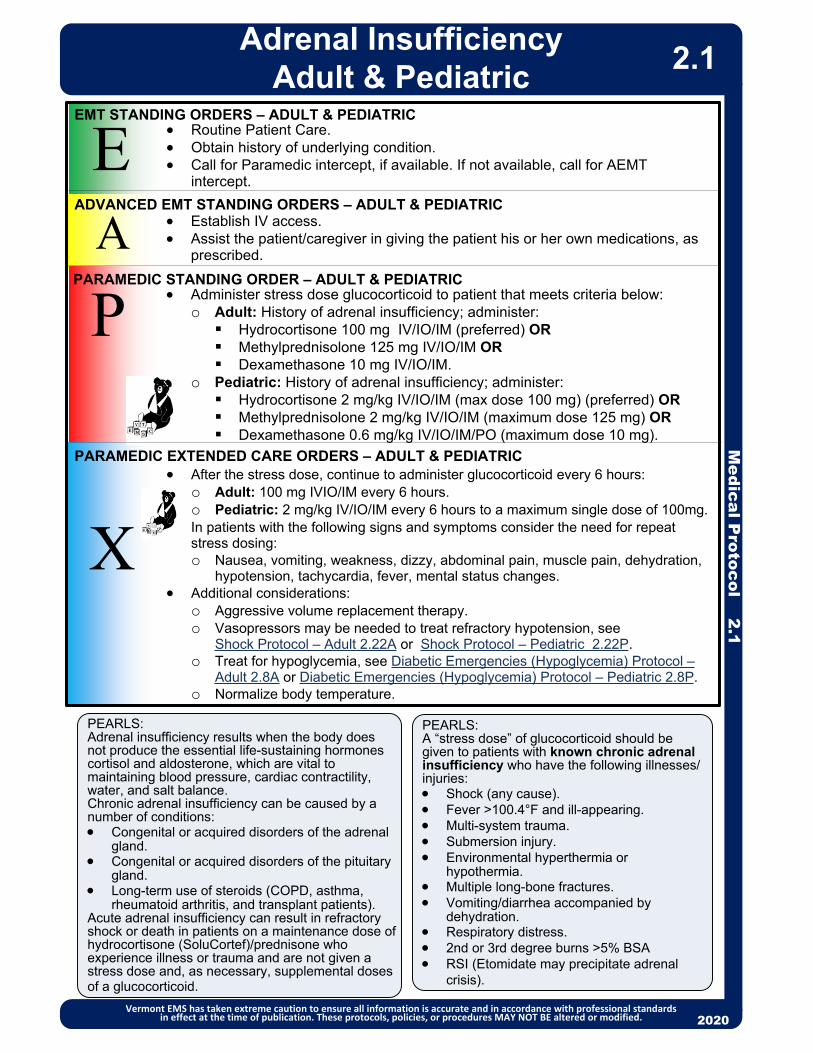
PEARLS:Adrenal insufficiency results when the body does not produce the essential life-sustaining hormones cortisol and aldosterone, which are vital to maintaining blood pressure, cardiac contractility, water, and salt balance.Chronic adrenal insufficiency can be caused by a number of conditions: Congenital or acquired disorders of the adrenal
gland. Congenital or acquired disorders of the pituitary
gland. Long-term use of steroids (COPD, asthma,
rheumatoid arthritis, and transplant patients). Acute adrenal insufficiency can result in refractory shock or death in patients on a maintenance dose of hydrocortisone (SoluCortef)/prednisone who experience illness or trauma and are not given a stress dose and, as necessary, supplemental doses of a glucocorticoid.
EA
Routine Patient Care. Obtain history of underlying condition. Call for Paramedic intercept, if available. If not available, call for AEMT
intercept.
Establish IV access. Assist the patient/caregiver in giving the patient his or her own medications, as
prescribed.
P
PEARLS:A “stress dose” of glucocorticoid should be given to patients with known chronic adrenal insufficiency who have the following illnesses/injuries: Shock (any cause). Fever >100.4°F and ill-appearing. Multi-system trauma. Submersion injury. Environmental hyperthermia or
hypothermia. Multiple long-bone fractures. Vomiting/diarrhea accompanied by
dehydration. Respiratory distress. 2nd or 3rd degree burns >5% BSA RSI (Etomidate may precipitate adrenal
crisis).
X
After the stress dose, continue to administer glucocorticoid every 6 hours:o Adult: 100 mg IVIO/IM every 6 hours.o Pediatric: 2 mg/kg IV/IO/IM every 6 hours to a maximum single dose of 100mg.
In patients with the following signs and symptoms consider the need for repeat stress dosing:o Nausea, vomiting, weakness, dizzy, abdominal pain, muscle pain, dehydration,
hypotension, tachycardia, fever, mental status changes. Additional considerations:
o Aggressive volume replacement therapy.o Vasopressors may be needed to treat refractory hypotension, see
Shock Protocol – Adult 2.22A or Shock Protocol – Pediatric 2.22P.o Treat for hypoglycemia, see Diabetic Emergencies (Hypoglycemia) Protocol –
Adult 2.8A or Diabetic Emergencies (Hypoglycemia) Protocol – Pediatric 2.8P.o Normalize body temperature.
EMT STANDING ORDERS – ADULT & PEDIATRIC
ADVANCED EMT STANDING ORDERS – ADULT & PEDIATRIC
PARAMEDIC STANDING ORDER – ADULT & PEDIATRIC
PARAMEDIC EXTENDED CARE ORDERS – ADULT & PEDIATRIC
2013
Adrenal InsufficiencyAdult & Pediatric
Vermont EMS has taken extreme caution to ensure all information is accurate and in accordance with professional standards in effect at the time of publication. These protocols, policies, or procedures MAY NOT BE altered or modified. 2020
2.1
Administer stress dose glucocorticoid to patient that meets criteria below:o Adult: History of adrenal insufficiency; administer: Hydrocortisone 100 mg IV/IO/IM (preferred) ORMethylprednisolone 125 mg IV/IO/IM ORDexamethasone 10 mg IV/IO/IM.
o Pediatric: History of adrenal insufficiency; administer: Hydrocortisone 2 mg/kg IV/IO/IM (max dose 100 mg) (preferred) OR Methylprednisolone 2 mg/kg IV/IO/IM (maximum dose 125 mg) ORDexamethasone 0.6 mg/kg IV/IO/IM/PO (maximum dose 10 mg).

Routine Patient Care. For anaphylaxis administer: (anterolateral thigh preferred administration site)
o Adult epinephrine autoinjector 0.3 mg IM OR o Epinephrine (1:1,000) (1 mg/mL): Administer 0.3 mg (0.3 mL) IM.Contact Medical Direction for additional dosing.
Do not delay transport. (Patients receiving epinephrine must be transported.) Call for Paramedic intercept, if available. If not available, call for AEMT intercept.
PEARLS: Known/likely allergen exposure AND hypotension or respiratory compromise, OR Systemic allergic reaction (multi-system), including two or more of the following:
o Respiratory distresso Airway compromise/impending airway compromise
Wheezing/stridor, swelling of lips/tongue, any airway structures, throat tightness or difficulty/inability swallowing
o Widespread hives, itching, swelling, flushingo Gastrointestinal symptoms: vomiting, abdominal paino Altered mental status, syncope, cyanosis, delayed capillary refill or decreased level of
consciousness associated with known or suspected allergic reactiono Signs of shock Shock Protocol – Adult 2.22A.
Do not delay transport for other than epinephrine administration. Patients can present with anaphylaxis without a prior history of allergy. Wheezing may be caused by anaphylaxis but it is not the only sign. Consider patients with history of asthma as having a high risk of anaphylaxis.
P
A
E
X
EMT STANDING ORDERS
ADVANCED EMT STANDING ORDERS
PARAMEDIC STANDING ORDERS
PARAMEDIC EXTENDED CARE ORDERS
2013
Medical P
rotocol 2.2A
Allergic Reaction/Anaphylaxis – Adult
Vermont EMS has taken extreme caution to ensure all information is accurate and in accordance with professional standards in effect at the time of publication. These protocols, policies, or procedures MAY NOT BE altered or modified. 2020
2.2A
May repeat epinephrine 0.3 mg IM every 5 – 15 min as needed for continued symptoms (maximum 3 doses.) For additional dosing, contact Medical Direction.
Establish IV access. Administer 500 – 1000 mL bolus 0.9% NaCl for SBP < 90 mmHg.
For bronchospasm, consider the administration of albuterol 2.5 mg via nebulizer. May repeat every 5 minutes for continued symptoms OR
Ipratropium 0.5 mg and albuterol 2.5 mg via nebulizer (DuoNeb). May repeat every 5 minutes (maximum 3 doses). Contact Medical Direction for additional dosing.
For anaphylaxis refractory to IM epinephrine, consider epinephrine infusion 2 – 10 mcg/min, titrated to effect (infusion pump required).
Diphenhydramine 25 – 50 mg IM/IV/IO to treat pruritus.
If symptomatic, consider: Methylprednisolone 1 mg/kg IV (max 125 mg) every 6 hours OR Dexamethasone 0.6 mg/kg IV/IO/IM/PO (maximum dose 10 mg) Diphenhydramine 25 – 50 mg PO. May repeat every 4-6 hours as needed
(maximum dose of 300 mg/24 hours).
CAUTION: Epinephrine is available in different routes and concentrations. Providers are advised to re-check the dosing and concentration prior to administration.
In anaphylaxis, epinephrine should not be delayed by taking the time to administer second-line medications such as diphenhydramine.
To administer Epinephrine via syringe, EMTs must be credentialed through the Ready-Check-Inject Program.

PEARLS: Known/likely allergen exposure AND hypotension or respiratory compromise, OR Systemic allergic reaction (multi-system), including two or more of the following:
o Respiratory distresso Airway compromise/impending airway compromise
Wheezing/stridor, swelling of lips/tongue, any airway structures, throat tightness or difficulty/inability swallowing
o Widespread hives, itching, swelling, flushingo Gastrointestinal symptoms: vomiting, abdominal paino Altered mental status, syncope, cyanosis, delayed capillary refill or decreased level of consciousness
associated with known or suspected allergic reactiono Signs of shock Shock Protocol – Pediatric 2.22P.
Do not delay transport for other than epinephrine administration. Patients can present with anaphylaxis without a prior history of allergy. Wheezing may be caused by anaphylaxis but it is not the only sign. Consider patients with history of asthma as having a high risk of anaphylaxis.
2013
Routine Patient Care. For anaphylaxis, administer: (anterolateral thigh preferred administration site)
o Pediatric epinephrine autoinjector 0.15 mg IM for patients less than 25 kg, 0.3 mg IM for patients greater than 25 kg OR
o Epinephrine (1:1,000) (1 mg/mL): Administer 0.15 mg (0.15 mL) IM for patients less than 25 kg, 0.3 mg (0.3 mL) IM for patients greater than 25 kg. Contact Medical Direction for additional dosing.
Do not delay transport. (Patients who receive epinephrine must be transported.) Call for Paramedic intercept, if available. If not available, call for AEMT intercept.
For anaphylaxis refractory to IM epinephrine, consider epinephrine infusion. Infuse 0.1 – 1 micrograms/kg/minute via pump until symptoms resolve.
Diphenhydramine 1 mg/kg PO/IV/IM/IO to treat pruritis (maximum dose 50 mg).PX
EMT STANDING ORDERS
PARAMEDIC STANDING ORDERS
If symptomatic, consider: Methylprednisolone 1 mg/kg IV (max 125 mg) every 6 hours if symptomatic OR Dexamethasone 0.6 mg/kg IV/IO/IM/PO (maximum dose 10 mg). Diphenhydramine 1 mg/kg PO. May repeat every 4 – 6 hours as needed (maximum
dose of 50 mg).
PARAMEDIC EXTENDED CARE ORDERS
2013
E
A
Medical P
rotocol 2.2P
Allergic Reaction/Anaphylaxis –Pediatric
Vermont EMS has taken extreme caution to ensure all information is accurate and in accordance with professional standards in effect at the time of publication. These protocols, policies, or procedures MAY NOT BE altered or modified. 2020
2.2P
ADVANCED EMT STANDING ORDERS Epinephrine (1:1,000) (1 mg/mL): Administer 0.01 mg/kg (0.01 mL/kg) IM (max
single dose 0.3 mg).o May repeat epinephrine every 5 – 15 min as needed for continued symptoms.
(Maximum 3 doses.) Contact Medical Direction for additional dosing. For bronchospasm, consider administration of albuterol 2.5 mg via nebulizer x 1
dose OR ipratropium 0.5 mg and albuterol 2.5 mg via nebulizer (DuoNeb). May repeat every 5 – 15 minutes (maximum 3 doses). Contact Medical Direction for additional dosing.
Establish IV access. Administer 20 mL/kg bolus 0.9% NaCl if hypotension. May repeat x 2 as needed.
CAUTION: Epinephrine is available in different routes and concentrations. Providers are advised to re-check the dosing and concentration prior to administration.
In anaphylaxis, epinephrine should not be delayed by taking the time to administer second-line medications such as diphenhydramine.
To administer Epinephrine via syringe, EMTs must be credentialed through the Ready-Check-Inject Program.

2013
Medical P
rotocol 2.3A
Altered Mental Status (Unknown Etiology) – Adult
Vermont EMS has taken extreme caution to ensure all information is accurate and in accordance with professional standards in effect at the time of publication. These protocols, policies, or procedures MAY NOT BE altered or modified. 2020
2.3A
PEARLS: Altered mental status may be caused by many factors including the following: stroke, drug overdose,
infection, hypoglycemia, hyperglycemia or trauma. AEMT or Paramedic may titrate use of naloxone in patients with respiratory depression to avoid
transition to combative behavior by patient. Use appropriate discretion regarding immediate intubation of patients who may quickly regain
consciousness, such as hypoglycemic patients after administration of dextrose, or opiate overdose cases after administration of naloxone.
Routine Patient Care. Administer oxygen as appropriate with a target of achieving 94 – 98% saturation. Assist inadequate ventilations with BVM (bag-valve-mask ventilation). If respiratory
arrest, manage airway with OPA/NPA and maintain oxygenation and ventilations with BVM (bag-valve-mask ventilation).
Anticipate and avoid aspiration. Obtain glucose reading via glucometer. If blood glucose < 60 with associated altered mental status, refer to Diabetic
Emergencies (Hypoglycemia) Protocol – Adult 2.8A. If the patient’s mental status and respiratory effort are severely depressed, consider
restraint and administer:o A single spray of NARCAN® Nasal Spray (4mg) into one nostril. May repeat every
3 – 5 minutes if no response or if patient relapses to a maximum of 12 mg OR o Naloxone 1 mg (1 mL) per nostril via atomizer for a maximum of 2 mg. May repeat
every 3 – 5 minutes if no response to a maximum of 12 mg.o Patients given naloxone should be transported to emergency department for further
evaluation. Consider acquiring and transmitting 12-lead ECG if available. If trauma can be excluded, transport patient in the coma/recovery position. If trauma
suspected, see Advanced Spinal Assessment Procedure 6.0. Perform stroke assessment. Refer to Stroke Protocol – Adult 2.24 as indicated. See Poisoning/Substance Abuse/Overdose – Adult 2.19A. Call for Paramedic intercept, if available. If not available, call for AEMT intercept. Minimize scene time.
Advanced airway management. Assess and monitor cardiac rhythm. Treat as indicated per appropriate protocol. If suspect toxicology, refer to Poisoning/Substance Abuse/Overdose Protocol - Adult
2.19A. If hypotension persists after 2 liter fluid bolus, consider vasopressors. See Shock
Protocol – Adult 2.22A. If patient is violent or agitated, consider restraint. See Behavioral Emergencies
Including Suicide Attempts & Threats Protocol 2.5.
P
EMT STANDING ORDERS
PARAMEDIC STANDING ORDERS
A
ADVANCED EMT STANDING ORDERS Establish IV/IO access. For severe respiratory depression, administer naloxone 0.4 – 2 mg IV/IM/IO/SQ/
intranasalo Consider restraint. See Restraints Procedure 6.7.o Titrate to response.o If no response, may repeat initial dose every 3 – 5 minutes to a total of 12 mg.
If hypoglycemia, administer dextrose. See Diabetic Emergencies (Hypoglycemia) Protocol – Adult 2.8A.
If hyperglycemic, give 500 mL bolus 0.9% NaCl IV/IO. See Diabetic Emergencies (Hyperglycemia) Protocol – Adult 2.7A.
Advanced airway as indicated. If hypotensive (SBP <90), administer fluid bolus 500 mL 0.9% NaCl IV/IO. Contact
Medical Direction for additional fluid or medication orders.
E

PEARLS: Altered mental status may be caused by many factors including the following: stroke, drug overdose,
infection, hypoglycemia, hyperglycemia, or trauma. AEMT or Paramedic may titrate use of naloxone in patients with respiratory depression to avoid
transition to combative behavior by patient. Use appropriate discretion regarding immediate intubation of patients who may quickly regain
consciousness, such as hypoglycemic patients after administration of dextrose, or opiate overdose cases after administration of naloxone.
2013
Routine Patient Care. Administer oxygen as appropriate with a target of achieving 94 – 98% saturation. Assist inadequate ventilations with BVM (bag-valve-mask ventilation). If respiratory
arrest, manage airway with OPA/NPA and maintain oxygenation and ventilations with BVM (bag-valve-mask ventilation).
Anticipate and avoid aspiration. Obtain glucose reading via glucometer. If blood glucose < 60 with associated altered mental status, refer to Diabetic
Emergencies (Hypoglycemia) Protocol – Pediatric 2.8P. If the patient’s mental status and respiratory effort are severely depressed:
o Administer a single spray of NARCAN® Nasal Spray (4mg) into one nostril ORo Administer via atomizer: Infant & Toddler: Naloxone 0.5 mg (0.5 mL) per nostril for a total of 1 mg.Small Child and Larger: Naloxone 1 mg (1 mL) per nostril for a maximum
dose of 2 mg.o For both, may repeat every 3 – 5 minutes if no response to a maximum of 12 mg. o Patients given naloxone should be transported to emergency department for
further evaluation. If trauma can be excluded, transport patient in the coma/recovery position. If trauma
suspected, see Advanced Spinal Assessment Procedure 6.0. See Poisoning/Substance Abuse/Overdose – Pediatric 2.19P. Call for Paramedic intercept, if available. If unavailable, call for AEMT intercept. Minimize scene time.
Advanced airway management. Assess and monitor cardiac rhythm. Treat as indicated per appropriate protocol. If suspect toxicology, refer to Poisoning/Substance Abuse/Overdose Protocol –
Pediatric 2.19P. If hypotension persists after 60 mL/kg fluid bolus, consider vasopressors, see Shock
Protocol – Pediatric 2.22P. If patient is violent or agitated, consider sedation. See Behavior Emergencies Including
Suicide Attempts & Threats Protocol 2.5.
P
EMT STANDING ORDERS
PARAMEDIC STANDING ORDERS
2013
E
A
Medical P
rotocol 2.3P
Altered Mental Status (Unknown Etiology) – Pediatric
Vermont EMS has taken extreme caution to ensure all information is accurate and in accordance with professional standards in effect at the time of publication. These protocols, policies, or procedures MAY NOT BE altered or modified. 2020
2.3P
ADVANCED EMT STANDING ORDERS Establish IV/IO access. If hypoglycemia, administer dextrose. See Diabetic Emergencies (Hypoglycemia)
Protocol – Pediatric 2.8P. If hyperglycemia, administer 10 mL/kg bolus of 0.9% NaCl IV/IO. See Diabetic
Emergencies (Hyperglycemia) Protocol – Pediatric 2.7P. For severe respiratory depression, administer naloxone 0.1 mg/kg IV/IO/IM/SQ/
intranasal, maximum dose 2 mg. o Consider restraint. See Restraints Procedure 6.9.o If no response, may repeat initial dose every 3 – 5 minutes to a total of 12 mg.
Advanced airway as indicated. If hypotensive per age-based tables, administer fluid bolus 20 mL/kg 0.9% NaCl IV/IO.
May repeat x 2. o Contact Medical Direction for additional fluid or medication orders.

P
A
E
EMT STANDING ORDERS
ADVANCED EMT STANDING ORDERS
PARAMEDIC STANDING ORDERS
2013
Me
dic
al P
roto
col 2
.4A
Asthma/COPD/RAD – Adult
Vermont EMS has taken extreme caution to ensure all information is accurate and in accordance with professional standards in effect at the time of publication. These protocols, policies, or procedures MAY NOT BE altered or modified. 2020
PEARLS:• IVs should only be placed when there are clinical concerns of dehydration, in order to
administer fluids, or when administering IV medications.• Beware of patients with a “silent chest” (absence of breath sounds) as this may indicate
severe reactive airway disease (RAD) with bronchospasm and impending respiratory failure.• Remember that not all wheezing is caused by asthma and that not all asthmatics wheeze.• Patients with congestive heart failure may present with lung sounds that mimic asthma
(“cardiac wheeze”).
2.4A
• Consider steroid:
o Methylprednisolone 125 mg IV/IO/IM OR
o Dexamethasone 10 mg IV/IO/IM/PO
• For patients who do not respond to treatments, or for impending respiratory failure, consider:
o Magnesium sulfate 2 g in 50 mL D5W or 0.9% NaCl IV/IO over 10 minutes.
• Routine Patient Care.
• Place patient in position of comfort. May prefer sitting up.
• Administer oxygen as appropriate with a target of achieving 94 – 98% saturation (88 - 92% in COPD); increase the oxygen rate with caution and observe for fatigue, decreased mentation, and respiratory failure.
• Consider:
o Albuterol metered-dose inhaler (MDI) 2-4 puffs (with spacer, if available). May repeat every 5 minutes for continued symptoms; OR
o Ipratropium bromide 0.5 mg and albuterol 2.5 mg (DuoNeb) via nebulizer. May repeat every 5 minutes for continued symptoms (maximum 3 doses); AND/OR
o Albuterol 2.5 mg via nebulizer. May repeat every 5 minutes for continued symptoms.
• Call for Paramedic intercept, if available. If not available, call for AEMT intercept.
• In a respiratory pandemic situation: Consider use of MDIs preferentially to nebulized medications if patient tolerates. To conserve MDIs, use patient’s own MDI, if available, and transport for use in hospital.
• For patients who do not respond to treatments, or for impending respiratory failure, continue nebulizers and consider CPAP up to a maximum of 10 – 15 cm H2O pressure support. See Continuous Positive Airway Pressure (CPAP) Procedure 5.3.
• For patients who do not respond to treatments, or for impending respiratory
failure, consider epinephrine autoinjector 0.3 mg IM (preferred) OR epinephrine (1:1,000) (1 mg/mL) 0.3 mg (0.3 mL) IM. Contact Medical Direction for additional doses.
• Consider IV access.
Caution is advised in the administration of beta-agonists to patients with coronary artery disease. Obtain and transmit ECG if possible. Contact Medical Direction for medication orders if in doubt.
• Albuterol metered-dose inhaler (MDI) 2 – 4 puffs. May repeat every 5 minutes for continued symptoms.X
EXTENDED CARE ORDERS

Medical P
rotocol 2.4P
2013
Asthma/Bronchiolitis/RAD/Croup – Pediatric
Vermont EMS has taken extreme caution to ensure all information is accurate and in accordance with professional standards in effect at the time of publication. These protocols, policies, or procedures MAY NOT BE altered or modified. 2020
2.4P
Procedure Continues
P
ASTHMA, BRONCHIOLITIS, CROUP – EMT STANDING ORDERS
ASTHMA – PARAMEDIC STANDING ORDERS
E
A
Consider: o Dexamethasone 0.6 mg/kg PO/IV/IO/IM (PO preferred)
(maximum dose 10 mg) OR o Methylprednisolone 2 mg/kg IV/IO/IM (maximum dose 125 mg).
For patients who do not respond to treatment or for impending respiratory failure, consider:o Magnesium sulfate 40 mg/kg IV/IO in 100 mL D5W over 20
minutes. (maximum single dose 2 grams).
ASTHMA – ADVANCED EMT STANDING ORDERS
Routine Patient Care. Administer oxygen as appropriate with a target of achieving 94 – 98% saturation. For patients ≤ 2 years old who present with increased work of breathing and
rhinorrhea, provide nasal suctioning with saline drops and bulb syringe. Call for Paramedic intercept, if available. If not available, call for AEMT intercept.
For impending respiratory failure, continue nebulizers and consider CPAP (see Continuous Positive Airway Pressure Procedure 5.3).
For patients who do not respond to treatments, or for impending respiratory failure, consider epinephrine auto injector 0.15 mg IM for patient less than 25 kg or 0.3 mg IM for patient greater than 25 kg OR epinephrine (1:1,000) (1 mg/mL) 0.01 mg/kg (0.01 mL/kg) IM, anterolateral thigh preferred (maximum single dose 0.3 mg.) Contact Medical Direction for additional dosing.
P
PCROUP – PARAMEDIC STANDING ORDERS
BRONCHIOLITIS – PARAMEDIC STANDING ORDERS Consider suctioning. For patients who do not respond to suctioning or for impending
respiratory failure, consider epinephrine (1:1,000) (1 mg/mL) 3 mg (3 mL) diluted to 3 mL 0.9% NaCl via nebulizer.o Contact Medical Direction for additional dosing.
Consider high-flow nasal cannula, if available.
Wheezing in patients ≥ 2 years old or
history of asthma
Yes
Wheezing in patients < 2 years
old
No
Yes
History of stridor or
barky cough
Yes
No
Consider dexamethasone 0.6 mg/kg IV/IO/IM/PO. PO preferred. Maximum dose 10 mg.
Croup with stridor at rest: o Epinephrine (1:1,000) (1 mg/mL) 3 mg (3 mL) diluted to 3 mL 0.9%
NaCl via nebulizer. Repeat every 5 – 15 minutes for continued symptoms. (Maximum 3 doses.) Contact Medical Direction for additional dosing.
Yes
ASTHMA – EMT STANDING ORDERS Consider:
o Albuterol metered-dose inhaler (MDI) 2-4 puffs (with spacer, if available). May repeat every 5 minutes for continued symptoms; OR
o Ipratropium 0.5 mg and albuterol 2.5 mg (DuoNeb) via nebulizer. May repeat dose every 5 minutes for continued symptoms. (Maximum 3 doses.); AND/OR
o Albuterol 2.5 mg via nebulizer every 5 minutes. In a respiratory pandemic situation: Consider use of MDIs
preferentially to nebulized medications if patient tolerates. To conserve MDIs, use patient’s own MDI, if available, and transport for use in hospital.
E
Yes

Medical P
rotocol 2.4P
Asthma/Bronchiolitis/RAD/Croup – Pediatric
Vermont EMS has taken extreme caution to ensure all information is accurate and in accordance with professional standards in effect at the time of publication. These protocols, policies, or procedures MAY NOT BE altered or modified. 2020
2.4P
PEARLS The IV formulation of dexamethasone may be given by mouth.
Epiglottitis A potentially life-threatening swelling of the supraglottic structures, which may result in sudden,
complete upper airway obstruction. Signs and symptoms include severe sore throat, difficulty breathing, which may improve when
leaning forward, stridor, and a high temperature (fever). For suspected epiglottitis, transport the patient in an upright position and limit your assessment
and interventions.
Bronchiolitis Incidence peaks in 2-6 month old infants. Frequent history of low-grade fever, runny nose, and sneezing. Signs and symptoms include: tachypnea, rhinorrhea, wheezes and / or crackles.
Croup Incidence peaks in children over age 6 months. Signs and symptoms include: hoarseness, barking cough, inspiratory stridor, signs of
respiratory distress. Avoid procedures that will distress child with severe croup and stridor at rest.
Pneumonia Signs and symptoms include: tachypnea, fever, intercostal retractions, cough, hypoxia and
chest pain.Tachypnea in children is defined as:
< 1 year: > 60 bpm 1 – 4 years: > 40 bpm 5 – 13: > 30 bpm > 13 years: > 16 bpm
CONSIDER DIFFERENTIAL DIAGNOSIS: Asthma Pneumonia (See CPAP Protocol 5.3 for respiratory failure) Bronchiolitis Anaphylaxis (See Allergic Reaction/Anaphylaxis Protocol 2.2P.) Croup Sepsis (See Septic Shock Protocol – Pediatric 2.20P.) Foreign body airway obstruction (See Foreign-Body Obstruction Procedure 5.4.)
Respiratory distress in children must be promptly recognized and aggressively treated. Respiratory arrest is the most common cause of cardiac arrest in children.
Child with a “silent chest” may have severe bronchospasm with impending respiratory failure.

EMT/ADVANCED EMT/PARAMEDIC STANDING ORDERS – ADULT & PEDIATRIC
• Routine Patient Care.• Approach patient using the SAFER Model. • Observe and record the patient’s behavior.• Consider associated domestic violence or child abuse, see Victims of Violence Policy
8.21.• Determine if patient is under the care of mental health professionals and record
contact information. • Assess for risk to self and others. Ask patient directly if he/she is thinking about
hurting self or others.• A patient who is a danger to self or others may not refuse care. If patient refuses care
and requires medical care or is danger to self or others, contact police and, if available, the local mental health crisis agency. (Refer to Police Custody Policy 8.14 and/or Refusal of Care Policy 8.15.)
• If the patient does not appear to be an immediate threat to self or others and refuses transport:o Encourage patient to seek mental health evaluation.o Provide the mental health center emergency services number 1-800-273-TALK. o Avoid leaving the patient alone, if possible. Assist in contacting responsible family/
friend.• For patient with suspected Excited/Agitated Delirium:
o See Restraint Procedure 6.9.o Treat hyperthermia. See Hyperthermia (Environmental) Protocol – Adult &
Pediatric 2.10.o Monitor cardiac activity (Paramedic only) and oxygen levels.
• If physical restraint is required, make sure adequate personnel are present. This generally means four people, one for each of the patient ’s extremities.
• If physical restraints are used and patient continues to be violent/agitated, call for Paramedic intercept if available. Refer to Restraint Procedure 6.7.
• Establish an airway, maintain as indicated, suction as needed. • Treat other injuries and illnesses. • If the patient is at risk for suicide or violence towards others:
o Transport to a hospital for evaluation. o If patient refuses transport, contact law enforcement and, if available, the local
mental health crisis agency for assistance. • Should it appear that the patient will not be transported, contact Medical Direction.
2013
Me
dic
al P
roto
col 2
.5
Behavioral Emergencies
Including Suicide Attempts & Threats
Vermont EMS has taken extreme caution to ensure all information is accurate and in accordance with professional standards in effect at the time of publication. These protocols, policies, or procedures MAY NOT BE altered or modified. 2020
2.5
SAFER ModelS Stabilize the situation by lowering stimuli, including voice. A Assess and acknowledge crisis by validating patient’s feelings and not minimizing them. F Facilitate identification and activation of resources (clergy, family, friends, or police).E Encourage patient to use resources and take actions in his/her best interest.R Recovery/referral - leave patient in the care of a responsible person, professional or transport to appropriate medical facility. Do not leave the patient alone when EMS clears the scene.
PEARLS: Consider all possible medical / traumatic causes for behavior and treat appropriately:
• Hypoglycemia
• Hypoxia
• Head Injury, stroke, seizure postictal
• Poisoning, substance abuse, drug, alcohol
• Infection
• Excited/Agitated Delirium is characterized by extreme restlessness, irritability, and/or high fever. Patients exhibiting these signs are at high risk for sudden death.
• Medications should be administered cautiously in frail or debilitated patients; lower doses should be considered.
• Monitor airway and vital signs closely.
EA
P
//
EA
P
//
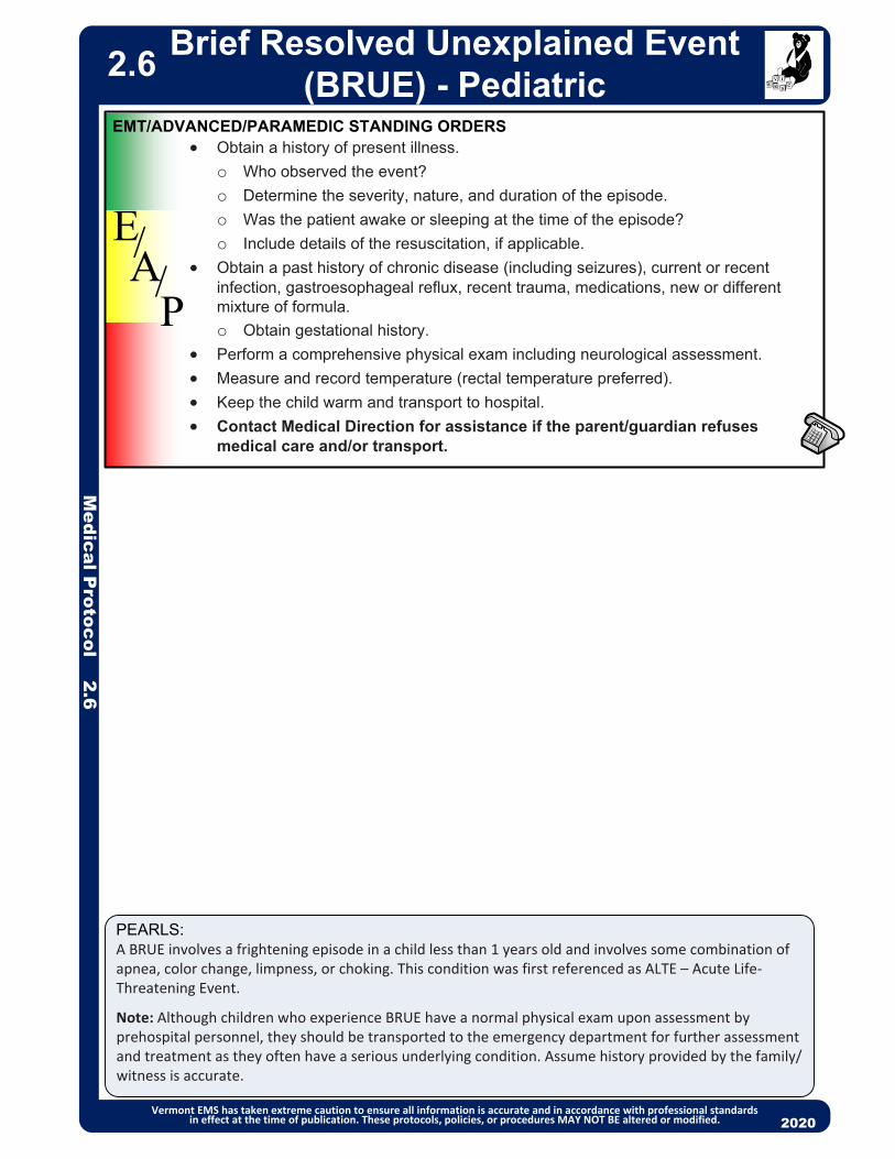
Medical P
rotocol 2.6
PEARLS:A BRUE involves a frightening episode in a child less than 1 years old and involves some combination of apnea, color change, limpness, or choking. This condition was first referenced as ALTE – Acute Life‐Threatening Event.
Note: Although children who experience BRUE have a normal physical exam upon assessment by prehospital personnel, they should be transported to the emergency department for further assessment and treatment as they often have a serious underlying condition. Assume history provided by the family/witness is accurate.
2013
Obtain a history of present illness.
o Who observed the event?
o Determine the severity, nature, and duration of the episode.
o Was the patient awake or sleeping at the time of the episode?
o Include details of the resuscitation, if applicable.
Obtain a past history of chronic disease (including seizures), current or recent infection, gastroesophageal reflux, recent trauma, medications, new or different mixture of formula.
o Obtain gestational history.
Perform a comprehensive physical exam including neurological assessment.
Measure and record temperature (rectal temperature preferred).
Keep the child warm and transport to hospital.
Contact Medical Direction for assistance if the parent/guardian refuses medical care and/or transport.
EMT/ADVANCED/PARAMEDIC STANDING ORDERS
EA
P
//
Brief Resolved Unexplained Event (BRUE) - Pediatric
Vermont EMS has taken extreme caution to ensure all information is accurate and in accordance with professional standards in effect at the time of publication. These protocols, policies, or procedures MAY NOT BE altered or modified. 2020
2.6

2013
Establish IV access. Administer 1,000 mL bolus of 0.9% NaCl IV/IO . Reassess and administer 1,000
mL of 0.9% NaCl IV/IO, if indicated. For nausea/vomiting, see Nausea/Vomiting Protocol – Adult & Pediatric 2.12.
EEMT STANDING ORDERS
ADVANCED EMT/PARAMEDIC STANDING ORDERS
2013
A/P
Medical P
rotocol 2.7A
Diabetic Emergencies (Hyperglycemia) – Adult
Vermont EMS has taken extreme caution to ensure all information is accurate and in accordance with professional standards in effect at the time of publication. These protocols, policies, or procedures MAY NOT BE altered or modified. 2020
2.7A
Routine Patient Care. Obtain glucose reading via glucometer. Call for Paramedic intercept, if available. If not available, call for AEMT
intercept.
X Oral fluids: if the patient is not vomiting, provide oral hydration with water.
o Patient must be alert enough to swallow and protect airway.
EMT/ADVANCED EMT/PARAMEDIC EXTENDED CARE ORDERS
Hyperglycemic emergency is defined as blood glucose greater than or equal to 250 mg/dL with associated signs and symptoms, such as altered mental status, increased respiratory rate, or dehydration.
PEARLS: Diabetic ketoacidosis is a life-threatening emergency defined as uncontrolled hyperglycemia with the
signs and symptoms of ketoacidosis. Signs and symptoms of diabetic ketoacidosis include uncontrolled blood glucose greater than 250 mg/
dL, weakness, altered mental status, abdominal pain, nausea, and vomiting, polyuria (excessive urination) polydipsia (excessive thirst), a fruity odor on the breath (from ketones) and tachypnea.
Common causes of diabetic ketoacidosis include infection, acute coronary syndrome and medication non-compliance.
Hyperglycemic Hyperosmolar Nonketotic Syndrome (HHNS) is characterized by blood glucose levels greater than 600 mg/dL and profound dehydration with significant neurologic deficits (e.g., coma, altered mental status). Treatment is the same in the field as it is for DKA.
Hyperglycemia may be detrimental to patients at risk for cerebral ischemia such as victims of stroke, cardiac arrest and head trauma.

E Establish IV access. Administer 10 mL/kg bolus of 0.9% NaCl IV/IO.
o Contact on-line Medical Direction for additional fluid bolus orders.
EMT STANDING ORDERS
ADVANCED EMT/PARAMEDIC STANDING ORDERS
Medical P
rotocol 2.7P
Diabetic Emergencies (Hyperglycemia) – Pediatric
Vermont EMS has taken extreme caution to ensure all information is accurate and in accordance with professional standards in effect at the time of publication. These protocols, policies, or procedures MAY NOT BE altered or modified.
PEARLS: Use 10 mL/kg fluid bolus to avoid potential risk of cerebral edema.
Diabetic ketoacidosis is a life-threatening emergency defined as uncontrolled hyperglycemia with the signs and symptoms of ketoacidosis.
Signs and symptoms of diabetic ketoacidosis include uncontrolled blood glucose greater than 250 mg/dL, weakness, altered mental status, abdominal pain, nausea, and vomiting, polyuria (excessive urination) polydipsia (excessive thirst), a fruity odor on the breath (from ketones) and tachypnea.
Common causes of diabetic ketoacidosis include infection, acute coronary syndrome and medication non-compliance.
Hyperglycemic Hyperosmolar Nonketotic Syndrome (HHNS) is characterized by blood glucose levels greater than 600 mg/dL and profound dehydration with significant neurologic deficits (e.g., coma, altered mental status). Treatment is the same in the field as it is for DKA.
Hyperglycemia may be detrimental to patients at risk for cerebral ischemia such as victims of stroke, cardiac arrest and head trauma.
2.7P
Routine Patient Care. Obtain glucose reading via glucometer. Call for Paramedic intercept, if available. If not available, call for AEMT
intercept.
A/P
Hyperglycemic emergency is defined as blood glucose greater than or equal to 250 mg/dL with associated signs and symptoms, such as altered mental status, increased respiratory rate, or dehydration.
X Oral fluids: if the patient is not vomiting, provide oral hydration with water.o Patient must be alert enough to swallow and protect airway.
EMT/ADVANCED EMT/PARAMEDIC EXTENDED CARE ORDERS
2020

Establish IV access. Administer up to 25 grams dextrose 10% (preferred) or dextrose 50% IV. Recheck
glucose 5 minutes after administration. May repeat up to 25 grams dextrose 10% or 50% IV if glucose level is < 60
mg/dl with continued altered mental status.o If unable to establish IV access, administer glucagon 1 mg IM. Recheck
glucose 15 minutes after administration of glucagon.May repeat glucagon 1 mg IM if glucose level is < 60 mg/dl with continued
altered mental status.
E
PEARLS: There are no statistically significant differences in the median recovery time to a GCS score of 15
following administration of D10% versus D50%. D10% could benefit patients in controlling their post-treatment high blood sugar levels.
Causes of hypoglycemia include medication misuse or overdose, missed meal, infection, cardiovascular insults (e.g., myocardial infarction, arrhythmia), or changes in activity (e.g., exercise).
Diabetics are not the only persons who become hypoglycemic. Alcoholics, some poisoned patients, and others may develop problems of glucose metabolism.
Sulfonylurea (e.g., glyburide, glipizide) toxicity can last up to 72 hours. Patients with corrected hypoglycemia who are taking these agents are at particular risk for recurrent hypoglycemia and frequently require hospital admission. These patients should be evaluated in the Emergency Department.
When administering dextrose, monitor IV site for signs of extravasation.
AEMT/Paramedic: If patient has a severe hypoglycemic emergency with altered mental status or seizures and the provider is unable to establish IV access, the provider may administer dextrose via intraosseous (IO). See Intraosseous Access Procedure 6.6.
EMT STANDING ORDERS
ADVANCED EMT/PARAMEDIC STANDING ORDERS
A/P
Medical P
rotocol 2.8A
Diabetic Emergencies (Hypoglycemia) – Adult
Vermont EMS has taken extreme caution to ensure all information is accurate and in accordance with professional standards in effect at the time of publication. These protocols, policies, or procedures MAY NOT BE altered or modified.
2.8A
Routine Patient Care. Obtain glucose reading via glucometer. Oral glucose: administer 1 – 2 tubes of commercially prepared glucose gel, or
15 – 30 mL (1 – 2 tablespoons) of Pure Vermont Maple Syrup, or equivalent, for standard dose of 15 – 30 grams sugar. Patient must be alert enough to swallow and protect airway.
Call for Paramedic intercept, if available. If not available, call for AEMT intercept.
For patients with an insulin pump who are hypoglycemic with associated altered mental status (GCS < 15):o Stop the pump or remove catheter at insertion site if patient cannot ingest
oral glucose or ALS is not available.o Leave the pump connected and running if able to ingest oral glucose or
receive ALS interventions. Do not treat and release hypoglycemic patients without contacting Medical
Direction to discuss cause of hypoglycemic episode, interventions taken and plan for follow-up.
Dextrose 10% is the preferred formulation for administration. A sterile IV bag containing 250 mL of D10W will deliver the standard dose of 25 grams of glucose IV. Bolus up to the entire 250 mL bag as quickly as possible, stopping when patient’s mental status returns to baseline and glucose level is ≥ 60 mg/dl. Often only 100 – 200 mL of dextrose 10% is necessary.
Hypoglycemic emergency is defined as blood glucose less than 60 mg/dL and associated signs with symptoms, such as altered mental status.
2020

2013
E
Establish IV access. Administer 10% dextrose IV, dosing per length-based resuscitation tape or 5 mL/
kg. Recheck glucose 5 minutes after administration of dextrose.May repeat dextrose dose if glucose level is < 60 mg/dl with continued
altered mental status.o If unable to obtain IV access, administer glucagon IM. Recheck glucose 15
minutes after administration of glucagon.Patients < 20 kg (44 lb), give glucagon 0.5 mg IM. Patients > 20 kg (44 lb), give glucagon 1 mg IM.
EMT STANDING ORDERS
ADVANCED EMT/PARAMEDIC STANDING ORDERS
2013
Medical P
rotocol 2.8P
Diabetic Emergencies (Hypoglycemia) – Pediatric
Vermont EMS has taken extreme caution to ensure all information is accurate and in accordance with professional standards in effect at the time of publication. These protocols, policies, or procedures MAY NOT BE altered or modified. 2020
PEARLS: Causes of hypoglycemia include medication misuse or overdose, missed meal, infection, cardiovascular
insults, trauma, traumatic brain injury, hypothermia, adrenal insufficiency, or changes in activity (e.g., exercise).
When administering dextrose, monitor IV site for signs of extravasation.
2.8P
Routine Patient Care. Obtain glucose reading via glucometer. Oral glucose: administer 1 tube of commercially prepared glucose gel, or 15 mL
(1 tablespoon) of Pure Vermont Maple Syrup, or equivalent. Patient must be alert enough to swallow and protect airway.
Call for Paramedic intercept, if available. If not available, call for AEMT intercept. For patients with an insulin pump who are hypoglycemic with associated altered
mental status (GCS < 15):o Stop the pump or remove catheter at insertion site if patient cannot ingest oral
glucose or ALS is not available.o Leave the pump connected and running if able to ingest oral glucose or receive
ALS interventions. Do not treat and release hypoglycemic patients without contacting Medical
Direction to discuss cause of hypoglycemic episode, interventions taken and plan for follow-up.
A/P
AEMT/Paramedic: If patient has a severe hypoglycemic emergency with altered mental status or seizures and the provider is unable to establish IV access, the provider may administer dextrose 10% via intraosseous (IO). See Intraosseous Access Procedure 6.6.
Pediatric Dextrose Dosing ChartLength (cm) Weight (kg) Color (Age) Volume of Dextrose 10% (mL)< 59.5 3 to 5 Gray (0 – 3 months) 2059.5 – 66.5 6 to 7 Pink (3 – 6 months) 3566.5 – 74 8 to 9 Red (7 – 10 months) 4374 – 84.5 10 to 11 Purple (11 – 18 months) 5084.5 – 97.5 12 to 14 Yellow (19 – 35 months) 7097.5 – 110 15 to 18 White (3 – 4 years) 80110 – 122 19 to 22 Blue (5 – 6 years) 100122 – 137 23 to 29 Orange (7 – 9 years) 135137 – 150 30 to 36 Green (10 – 12 years) 180
Hypoglycemic emergency is defined as blood glucose less than 60 mg/dl with associated altered mental status.

2013
Exertional Heat Stroke (EHS) is a unique and emergent hyperthermic condition that
occurs in individuals performing intense physical activity, typically not limited to warm environments.
INDICATION
Consider EHS in any intensely-exercising athlete, laborer, fire, police or EMS
personnel with altered mental status.
PROTOCOL
1. Perform rapid routine assessment (<5 minutes). Assess for other causes of
AMS, including but not limited to hypoxia, hypoglycemia, inadequate perfusion,
head injury or seizure.
2. Consider EHS in any intensely-exercising athlete, laborer, fire, police or EMS
personnel with altered mental status.
3. If EHS has been confirmed and appropriate cooling has been initiated by an
appropriate onsite medical team, athletic trainer, coach, or instructor, DO NOT
interrupt cooling for assessment or transport.
4. If care has not been initiated and EHS is suspected, immediately perform a
rectal temperature (TREC).
5. If TREC is at or above 40ºC (104ºF), initiate immediate rapid cooling to a
temperature less than 40ºC within 30 minutes of collapse.
a. Best practice for cooling an EHS patient is whole-body cold water
immersion from the neck down.
b. Immersion in ice water-filled body bag or tarp may also yield acceptable
cooling rates.
c. Ice packs, fans, cold water dousing or shower do not achieve acceptable cooling rates. Rotating ice water towels covering as much of the body
surface area as possible should be considered a minimum cooling modality
enroute.
6. Discontinue cooling at 39ºC (102ºF). If a TREC is not available, cooling should
not be interrupted or delayed in cases of suspected EHS. Cool for a minimum
of 20 minutes / clinical improvement if resources available on scene, or
transport with best available active cooling method (body bag with ice water or
rotating ice water-soaked towels).7. Do not interrupt cooling for diarrhea, emesis, combativeness, or seizures. IV/IM
medications are rarely needed.
8. Transport to closest receiving facility and notify that EHS is suspected.
EMT/ADVANCED EMT/PARAMEDIC STANDING ORDERS
2013
Me
dic
al P
roto
col 2
.9Exertional Heat Stroke (EHS)
Vermont EMS has taken extreme caution to ensure all information is accurate and in accordance with professional standards in effect at the time of publication. These protocols, policies, or procedures MAY NOT BE altered or modified. 2020
2.9
EA
P
//
EA
P
//
• For events with medical personnel and cooling means on-site, the only appropriate
standard is to cool the EHS patient in place. Transportation of an EHS patient should
only be done if it is impossible to adequately cool the patient, or after adequate
cooling has been verified by a rectal temperature.
• The only accurate or acceptable body temperature measurement in exercising
individuals is a rectal temperature (TREC).
• EMS must ensure early pre-notification to hospitals if they will be receiving an
inadequately-cooled EHS patient, or suspect EHS in a scenario where treatment has
not been initiated.

2013
EE
• Routine Patient Care.
• Consider exertional heat stroke in any intensely-exercising athlete, laborer, fire,
police or EMS personnel, etc. with altered mental status. See Exertional Heat
Stroke Protocol 2.9.
• Move victim to a cool area and shield from the sun or any external heat source.
• Remove as much clothing as is practical and loosen any restrictive garments.
• If alert and oriented, give small sips of cool liquids.
• Monitor and record vital signs and level of consciousness. If altered mental
status, check blood glucose level.
• Determine patient’s core temperature, if possible (rectal temperature preferred).
If thermometer not available, assume heat stroke if altered mental status present. Diagnosis of hyperthermia is based on clinical signs.
• If temperature is ≥ 40ºC (104ºF) or if altered mental status is present, begin
active cooling by:
o Whole-body cold water immersion from the neck down. Note: Elderly
patients are less able to withstand sudden shocks, so begin with less
aggressive cooling measures like misting or cool wet cloths. When you
reassess the patient, if there is little or no improvement, use more aggressive measures.
o Continually misting the exposed skin with tepid water while fanning patient.
o Rotating ice water towels covering as much of the body surface area as
possible.
• Discontinue active cooling if shivering occurs that cannot be managed by
Paramedics (see below) or temperature is < 38.9ºC (102º F).
• Call for Paramedic intercept, if available. If not available, call for AEMT
intercept.
• Establish IV access.
• Administer 500 mL 0.9% NaCl room temperature or chilled IV fluid bolus for
dehydration even if vital signs are normal.
P• If uncontrolled shivering occurs during cooling:
o Midazolam 2.5 mg IV/intranasal, may repeat once in 5 minutes OR 5 mg IM
may repeat once in 10 minutes OR
o Lorazepam 1 mg IV, may repeat once in 5 minutes OR 2 mg IM, may
repeat once in 10 minutes OR
o Diazepam 2 mg IV, may repeat once in 5 minutes.
• Administer 20 mL/kg 0.9% NaCl room temperature or chilled IV fluid bolus for
dehydration even if vital signs are normal.
• If uncontrolled shivering occurs during cooling:
o Midazolam 0.1 mg/kg IV (single maximum dose 1 mg) OR 0.2 mg/kg
intranasal/IM. (Note: a 5 mg/mL concentration is recommended for
intranasal administration.) OR
o Lorazepam 0.1 mg/kg IV/IM (single maximum dose 1 mg) OR
o Diazepam 0.2 mg/kg IV OR 0.5 mg/kg PR (single maximum dose 2 mg IV
OR 4 mg PR).
A
EMT STANDING ORDERS – ADULT & PEDIATRIC
ADVANCED EMT STANDING ORDERS – ADULT
PARAMEDIC STANDING ORDERS – ADULT
PARAMEDIC STANDING ORDERS – PEDIATRIC
2013
Me
dic
al P
roto
col 2
.10
Hyperthermia (Environmental)
– Adult & Pediatric
Vermont EMS has taken extreme caution to ensure all information is accurate and in accordance with professional standards in effect at the time of publication. These protocols, policies, or procedures MAY NOT BE altered or modified. 2020
2.10
Protocol Continues

Hyperthermia:
PO (oral) and other non-core thermometers may be inaccurate when the temperature is outside the normal range.
Elevated temperature may be due to environmental exposure, pharmacologic agents, or excited (agitated) delirium, see also Behavioral Emergencies Including Suicide Attempts & Threats Protocol – Adult & Pediatric 2.5, Septic Shock Protocol - Adult 2.21A, Septic Shock Protocol - Pediatric 2.21P, Poisoning/Substance Abuse/Overdose Protocol – Adult 2.19A or Poisoning/Substance Abuse/Overdose Protocol – Pediatric 2.19P. Mortality and morbidity are directly related to the length of time the victim is subject to the heat stress.
Sweating (or lack of sweating) is an unreliable indicator of the severity of heat illness.
Of primary concern are the patient's vital signs and mental status.
The patient's baseline health status and medications greatly determine the likelihood of developing and recovering from heat illness.
The very young and very old are at greatest risk of heat illness.
For events with high risk of hyperthermia, consider having an ice bath available on scene. See Rehabilitation – Scene and Training Guidelines for EMS 8.16, Exertional Heat Stroke Protocol 2.9.
Note for Paramedics: Benzodiazepine doses for stopping shivering are lower than those for stopping seizures by approximately half.
2013
Medical P
rotocol 2.10
Hyperthermia (Environmental) – Adult & Pediatric
Vermont EMS has taken extreme caution to ensure all information is accurate and in accordance with professional standards in effect at the time of publication. These protocols, policies, or procedures MAY NOT BE altered or modified. 2020
2.10
Protocol Continues

2013
E
Routine Patient Care. Ensure scene safety. Classify hypothermia clinically on the basis of vital signs, level of consciousness and
intensity of shivering. Core temperature, if available, provides additional information (see chart).
Handle gently. Maintain patient in horizontal position. Continue rewarming during transport. Warm ambulance to 24°C (75.2°F) if possible.
Prevent further heat loss using insulation and vapor barrier. Move to a warm, sheltered environment. Gently remove (cut off) wet clothing and dry patient.
Obtain blood glucose. Support shivering with calorie replacement if alert and able to swallow.
Mildly hypothermic patients should not be allowed to stand or walk for 30 minutes, while being kept as warm as possible with calorie replacement and shelter.
Patients with moderate to severe hypothermia require active external rewarming with chemical, electrical, or forced-air heating packs/blankets. Active rewarming is also beneficial for mild hypothermia.
Assess patient for signs of life and pulse carefully for a minimum of 60 seconds to confirm respiratory arrest or cardiac arrest.
If pulse and breathing are present, continue rewarming techniques. If pulse and breathing are absent, start CPR unless contraindications to CPR
exist. o Contraindications to CPR in the hypothermic patient include: Obvious signs of
irreversible death, chest wall not compressible as whole body is frozen solid, a valid DNR order, avalanche burial > 35 minutes and airway packed with snow, or rescuers exhausted or in danger.
o Rigor mortis or fixed and dilated pupils are NOT a contraindication to CPR in hypothermia.
Hypothermic patients without contraindications to CPR should have continued CPR and should not be considered for Termination of Resuscitation (TOR) until the core temperature has been rewarmed to 32°C (90°F) with no ROSC.o Contraindications to prolonged CPR include patients who are thought to have
cardiac arrest before cooling (temperature is thought to have been above 32°C (90°F) at the time of cardiac arrest). Causes of cardiac arrest before cooling include major trauma, witnessed normothermic arrest and avalanche burial < 35 mins.
Transport patient to closest appropriate hospital. Patients with prehospital cardiac instability (MAP < 65 (systolic blood pressure < 90 mm Hg) or ventricular arrhythmias, core temperature < 28°C (82°F) and those in cardiac arrest should be transported directly to a center capable of providing cardiopulmonary bypass (CPB) or extracorporeal membrane oxygenation (ECMO), if feasible.
CPR may be delayed or given intermittently if necessary to accomplish evacuation.
EMT STANDING ORDERS - ADULT & PEDIATRIC
2013
Medical P
rotocol 2.11
Hypothermia (Environmental) – Adult & Pediatric
Vermont EMS has taken extreme caution to ensure all information is accurate and in accordance with professional standards in effect at the time of publication. These protocols, policies, or procedures MAY NOT BE altered or modified. 2020
2.11
Consider hypothermia if patient has a history of cold exposure or a disease that predisposes them to hypothermia, patient’s trunk feels cold on examination, or core temperature is < 35°C (95°F).
Establish IV/IO access. Administer warm IV 0.9% NaCl 40°C - 42°C (104°F – 107.6°F) in 500 mL boluses. Provide airway management as required. With advanced airway, ventilate at half
standard rate. Administer dextrose IV/IO if hypoglycemic. Continue CPR if indicated:
o If < 30°C VT or VF or AED advises shock: one shock at maximum power.o Warm 1 – 2°C or > 30°C prior to additional shocks.o No vasoactive drugs until 30°C or above. From 30 – 35°C, increase dosing interval
to twice as long as normal. Consider epinephrine (1:10,000) (0.1 mg/mL) 1 mg IV/IO, up to 3 doses.
o Contact Medical Direction for guidance.
ADVANCED EMT/PARAMEDIC STANDING ORDERS
A/P Protocol Continues

Medical P
rotocol 2.11Hypothermia (Environmental) –
Adult & Pediatric
Vermont EMS has taken extreme caution to ensure all information is accurate and in accordance with professional standards in effect at the time of publication. These protocols, policies, or procedures MAY NOT BE altered or modified. 2020
2.11
Protocol Continues
Pearls A rapid assessment of the patient’s core temperature may be performed by placing a warm ungloved hand
against the skin of a patient’s back, or chest. If the skin feels warm, hypothermia is unlikely. Hypothermic patients are often significantly dehydrated and may require repeat fluid boluses.
Absence of vital signs, rigor mortis, dependent lividity or fixed and dilated pupils may be present in patients with reversible hypothermia and are not a contraindication to CPR. Assume that a hypothermic patient can be resuscitated even if there is an absence of vital signs, any sign of life, rigor mortis, or fixed and dilated pupils.
Detecting a pulse in a patient with hypothermia may be difficult. Signs of life and pulse should be checked carefully for 60 seconds. Persistent breathing or movement by the patient should prompt a strategy of watchful waiting, but if no signs of life are detected, then cardiopulmonary resuscitation (CPR) should be started. Since metabolic needs are so low in severely hypothermic patients, a rate of only a few beats per minute is enough to provide adequate perfusion to vital organs. In such cases, it is better to attempt to maintain effective cardiac activity than to start CPR and cause VF.
Owing to the decrease in cerebral oxygen requirements with cooling, survival without neurologic impairment may be possible even when it is necessary to perform CPR for hours. Transportation to CPB/ECMO capable facilities with continuing CPR may be justified if hypothermia is present or suspected. Contact destination hospital in advance to ensure availability of CPB/ECMO.
Immediate continuous CPR is recommended for cardiac arrest due to primary severe hypothermia. Mechanical chest-compression devices should be used when available and CPR-interruptions avoided. If this is not possible, CPR can be delayed or performed intermittently. Based on available data in a patient with a core temperature <28◦C or unknown with unequivocal hypothermic cardiac arrest, evidence supports alternating 5 min CPR and ≤5 min without CPR. With core temperature < 20◦C, current evidence supports alternating 5 min CPR and ≤ 10 min without CPR. If field conditions are not amenable to the safe application of delayed or intermittent CPR, contact Medical Direction to consider Termination of Resuscitation.
If a patient with cardiac arrest due to hypothermia is rewarmed to a core body temperature that is higher than 32°C and asystole persists, irreversible cardiac arrest is very likely, and termination of CPR should be considered.
A severely elevated serum potassium level is associated with non-survival and is considered a marker of hypoxia before cooling. Termination of CPR should be considered when the potassium level is higher than 12mmol/L.
A hypothermic patient should be assessed for coexisting injuries or illnesses that may mimic or conceal the signs and symptoms of hypothermia. The use of vital signs, mental status and presence or absence of shivering may be unreliable if the patient has another condition that coexists with hypothermia. Many conditions such as hypoglycemia, alcohol intoxication and exhaustion can cause altered mental status and can decrease or abolish shivering. A heart rate higher than expected for a given level of hypothermia may be due to another cause such as traumatic blood loss.

PEARLS: To reduce incidence of dystonic reactions, administer prochlorperazine and
metoclopramide slowly, over 1 – 2 minutes. Consider other causes of nausea such as the following: cardiac, GI bleeding, pregnancy,
toxicologic, diabetes. Nausea/vomiting is a common finding associated with acute coronary syndrome.
Consider obtaining 12-lead ECG when appropriate. Do not administer Ondansetron if patient has known, prolonged QT on ECG.
2013
P
E
X
EMT STANDING ORDERS – ADULT & PEDIATRIC
PARAMEDIC STANDING ORDERS – ADULT
ADVANCED EMT STANDING ORDERS – ADULT
A Consider Ondansetron 4 mg PO/ODT/IV/IM. May give IV solution by
oral route. Establish IV access if patient appears dehydrated. Consider 500 mL 0.9% NaCl IV fluid bolus for dehydration.
o May repeat 250 mL IV bolus if transport exceeds 15 minutes and patient’s condition has not improved.
Ondansetron 4 mg IV/IM OR Prochlorperazine 5 – 10 mg IV, or 5 mg IM OR Metoclopramide 5 mg IV.
o May repeat any of the above medications once after 10 minutes if nausea/vomiting persists.
Apply ECG; monitor for arrythmia.
PARAMEDIC STANDING ORDERS – PEDIATRIC
Ondansetron 2 mg ODT for patient 8 – 15 kg, 4 mg ODT patient ≥ 16 kg OR Ondansetron 0.1 mg/kg IV (maximum single dose 4mg). Consider 20 mL/kg IV fluid bolus for dehydration.
For motion sickness: consider diphenhydramine:o Adult: 25 mg PO/chewed o Ages 2 – 5 years: 6.25 mg PO o Ages 6 – 11 years: 12.5 – 25 mg PO
Paramedic only: May repeat IM prochlorperazine or metoclopramide every 4 – 6 hours as needed.
2013
PARAMEDIC EXTENDED CARE ORDERS – ADULT & PEDIATRIC
Medical P
rotocol 2.12
Nausea/Vomiting –Adult & Pediatric
Vermont EMS has taken extreme caution to ensure all information is accurate and in accordance with professional standards in effect at the time of publication. These protocols, policies, or procedures MAY NOT BE altered or modified. 2020
2.12
Routine Patient Care.
Nausea, vomiting and epigastric abdominal pain may be signs of acute coronary syndrome in adults. Consider obtaining and transmitting ECG, if available.
Antidote: For dystonic reactions caused by EMS administration of prochlorperazine or metoclopramide, administer diphenhydramine 25 – 50 mg IV/IM.

2013
P
EMT/ADVANCED EMT STANDING ORDERS
PARAMEDIC STANDING ORDERS
2013
EA/
2013
PARAMEDIC MEDICAL DIRECTION – MAY CONSIDER:
Medical P
rotocol 2.13ANerve Agents/Organophosphate
Poisoning – Adult
Vermont EMS has taken extreme caution to ensure all information is accurate and in accordance with professional standards in effect at the time of publication. These protocols, policies, or procedures MAY NOT BE altered or modified. 2020
2.13A
If symptoms persist after the administration of 3 DuoDote kits (or MARK I kits) and field conditions permit:o Initiate cardiac monitoring.o Establish IV access.o Atropine 2 mg IV/IO; repeat every 5 minutes until excess secretions cease (stop).o Pralidoxime 1 – 2 gram IV. Reconstitute pralidoxime 1 gram vial with 20 mL sterile water for injection. Dilute reconstituted pralidoxime 1 gram to 100 mL of 0.9% NaCl (may dilute 1-2
grams in this manner). Infuse over 5 minutes (1 gram dose) to 10 minutes (2 gram dose).
o Diazepam 5 mg IV every 5 minutes OR 10 mg IM OR diazepam auto-injector (10 mg) every 10 minutes, as needed. Instead of diazepam, may use either: Lorazepam 1 mg IV may repeat once in 5 minutes OR 2 mg IM, may repeat once
in 10 minutes ORMidazolam 2.5 mg IV/intranasal every 5 minutes OR 5 mg IM every 10 minutes
as needed.
Pralidoxime maintenance infusion: o Reconstitute pralidoxime 1 gram vial with 20 mL of sterile water for injection (SWFI)
or 0.9% NaCl. o Dilute reconstituted pralidoxime 1 gram to 100 mL of 0.9% NaCl. o Infuse 1 gram over 15-30 minutes, followed by a continuous infusion at 500 mg/hr, to
a maximum of 12 grams/day.
Routine Patient Care. Consider contacting Poison Control at (800) 222-1222 as soon as practical for
consultation. Assess for SLUDGEM (Salivation, Lacrimation, Urination, Defecation, Gastric upset,
Emesis, Muscle twitching/miosis (constricted pupils) and KILLER Bs (Bradycardia, Bronchorrhea, Bronchospasm).
Remove to cold zone after decontamination and monitor for symptoms. Antidotal therapy should be started as soon as symptoms appear. All antidote auto-injections must be administered IM Auto-injectors consist of atropine and pralidoxime and are often referred to as DuoDote
or Mark I kits.
Determine dosing according to the following symptom assessment and guidelines.
Category Description/Examples Autoinjector Dose
Mild
Worsening
Severe
If at any time after the first dose the patient develops any additional symptoms, or if symptoms worsen
1 DuoDote (or MARK I kit);
Monitor patient every 10 minutes
2 DuoDotes (or 2 Mark I kits); Monitor
patient every 10 minutes.
3 DuoDotes (or 3 Mark I kits) AND 1 diazepam 10 mg
auto-injector.
If ANY of the following are present: Strange/confused behavior, severe difficulty breathing/copious airway secre-tions, severe muscle twitching, involuntary urination/defeca-tion, convulsions, loss of consciousness, respiratory arrest
If TWO (2) or more of the following are present: Blurred vision/miosis (pupil constriction), excessive teary eyes, excessive runny nose, increased salivation, chest tightness/dyspnea, tremors/twitching, nausea/vomiting, wheezing/coughing/secretions, acute stomach cramps, tachycardia, bradycardia

2013
P
EMT/ADVANCED EMT STANDING ORDERS
PARAMEDIC STANDING ORDERS
2013
EA/
2013
In the unlikely event that field conditions permit, follow weight-based dosing and treatment guidelines: o Initiate cardiac monitoring.o Establish IV access.o Atropine 0.05 – 0.1 mg/kg IV or IM (minimum dose of 0.1 mg, maximum single dose
5mg); repeat every 2 – 5 minutes as needed.o Pralidoxime 25 – 50 mg/kg IV (maximum dose 1 gram) or IM (maximum dose of 2
grams), may repeat within 30 – 60 minutes as needed, then again every hour for 1 – 2 doses as needed.
o Diazepam 0.3 mg/kg IV (0.5 mg/kg per rectum) (maximum dose 10 mg), repeat every 5 – 10 minutes as needed.Instead of diazepam, may use either:Lorazepam 0.1 mg/kg IV/IM (maximum dose 4 mg), repeat every 5 – 10 minutes
as needed ORMidazolam 0.2 mg/kg IM/intranasal/IV, repeat every 5 – 10 minutes as needed.
Pralidoxime maintenance infusion: o Reconstitute pralidoxime 1 gram vial with 20 mL of sterile water for injection (SWFI) or
0.9% NaCl. o Dilute reconstituted pralidoxime 50 mg/kg (maximum 1 gram) to 100 mL of 0.9%
NaCl. o Infuse entire volume over 15 – 30 minutes.
PARAMEDIC MEDICAL DIRECTION – MAY CONSIDER:
*DuoDote (or MARK I kit) may be used for pediatric patients < 1 year old in a life-threatening situation with exposure symptoms when no pediatric doses of atropine or pralidoxime chloride are available.
Medical P
rotocol 2.13P
Nerve Agents/Organophosphate Poisoning – Pediatric
Vermont EMS has taken extreme caution to ensure all information is accurate and in accordance with professional standards in effect at the time of publication. These protocols, policies, or procedures MAY NOT BE altered or modified. 2020
2.13P
Routine Patient Care.
Consider contacting Poison Control at (800) 222-1222 as soon as practical for consultation.
Assess for SLUDGEM (Salivation, Lacrimation, Urination, Defecation, Gastric upset, Emesis, Muscle twitching/miosis (constricted pupils) and KILLER Bs (Bradycardia, Bronchorrhea, Bronchospasm).
Remove to cold zone after decontamination and monitor for symptoms.
Antidotal therapy should be started as soon as symptoms appear.
All antidote auto-injections must be administered IM.Determine dosing according to the following symptom assessment and guidelines.
Length (cm)Weight (kg) Color (Age)
Atropine Dose -- MILD and SEVERE
Pralidoxime Dose MILD
Pralidoxime Dose SEVERE
< 59.5 3 to 5 Gray (0 - 3 months) 60 mg IM59.5 to 66.5 6 to 7 Pink (3 - 6 months) 100 mg IM66.5 to 74 8 to 9 Red (7 - 10 months) 125 mg IM74 to 84.5 10 to 11 Purple (11 - 18 mos) 150 mg IM84.5 to 97.5 12 to 14 Yellow (19 - 35 mos) 200 mg IM97.5 to 110 15 to 18 White (3 - 4 years) 250 mg IM110 to 122 19 to 22 Blue (5 - 6 years) 300 mg IM122 to 137 24 to 30 Orange (7 - 9 years) 400 mg IM137 to 150 30 to 40 Green (10 - 12 years) 500 mg IM
0.25 mg IM
0.5 mg IM
1 mg IM
Give 3 MILD doses in rapid succession IM.
If symptoms persist, may
repeat series 1 hour after last IM
injection.
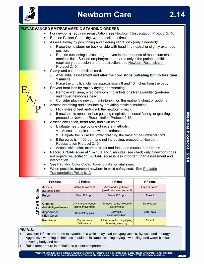
2013
EMT/ADVANCED EMT/PARAMEDIC STANDING ORDERS For newborns requiring resuscitation, see Newborn Resuscitation Protocol 2.15. Routine Patient Care—dry, warm, position, stimulate. Assess airway by positioning and clearing secretions (only if needed):
o Place the newborn on back or side with head in a neutral or slightly extended position.
o Routine suctioning is discouraged even in the presence of meconium-stained amniotic fluid. Suction oropharynx then nares only if the patient exhibits respiratory depression and/or obstruction, see Newborn Resuscitation Protocol 2.15.
Clamp and cut the umbilical cord:o After initial assessment and after the cord stops pulsating but no less than
1 minute.o Place the umbilical clamps approximately 8 and 10 inches from the baby.
Prevent heat loss by rapidly drying and warming:o Remove wet linen, wrap newborn in blankets or silver swaddler (preferred)
and cover newborn’s head.o Consider placing newborn skin-to-skin on the mother’s chest or abdomen.
Assess breathing and stimulate by providing tactile stimulation:o Flick soles of feet and/or rub the newborn’s back.o If newborn is apneic or has gasping respirations, nasal flaring, or grunting,
proceed to Newborn Resuscitation Protocol 2.15. Assess circulation, heart rate, and skin color:
o Evaluate heart rate by one of several methods:Auscultate apical beat with a stethoscope.Palpate the pulse by lightly grasping the base of the umbilical cord.
o If the pulse is < 100 bpm and not increasing, proceed to Newborn Resuscitation Protocol 2.15.
o Assess skin color; examine trunk and face; and mucus membranes. Record APGAR score at 1 minute and 5 minutes (see chart) only if newborn does
not require resuscitation. APGAR score is less important than assessment and intervention.
See Pediatric Color Coded Appendix A2 for vital signs. When possible, transport newborn in child safety seat. See Pediatric
Transportation Policy 8.13.
2013
EA
P
//
Medical P
rotocol 2.14Newborn Care
Vermont EMS has taken extreme caution to ensure all information is accurate and in accordance with professional standards in effect at the time of publication. These protocols, policies, or procedures MAY NOT BE altered or modified. 2020
PEARLS: Newborn infants are prone to hypothermia which may lead to hypoglycemia, hypoxia and lethargy.
Aggressive warming techniques should be initiated including drying, swaddling, and warm blankets covering body and head.
Raise temperature in ambulance patient compartment.
2.14
Below 100 bpm
AP
GA
R S
cale
Active Movement
Over 100 bpm
Cry, sneeze, cough, active movement
Completely pink
Vigorous cryFull breaths
Feature EvaluatedActivity
(Muscle Tone)
Pulse
Grimace(Irritability/reflexes)
Appearance(Skin Color)
Respiration
2 Points 1 Point
Arms and legs flexed(Weak, some movement)
Grimace (some flexion of extremities)
Body pink, Extremities blue
Slow, irregular, or gasping breaths, weak cry
0 Points
Limp or flaccid
Absent
No reflexes
Blue, pale
Absent

2013
P
EMT/ADVANCED EMT STANDING ORDERS
PARAMEDIC STANDING ORDERS
If bag valve mask ventilation is inadequate or chest compressions are indicated, intubate the infant using a 3.0 mm to 4.0 mm endotracheal tube. (For an infant born before 28 weeks gestation, a 2.5 mm endotracheal tube should be used.)o Meconium aspiration may be indicated if airway is obstructed.o After direct visualization, improvement in heart rate and EtCO2 are the best
indicators of whether the tube is properly placed in the trachea. Establish IV/IO access. Obtain blood sample if possible.
o If hypovolemia is suspected, administer 10 mL/kg bolus 0.9% NaCl over 5 – 10 minutes.
o If the heart rate fails to improve with chest compressions, administer epinephrine (1:10,000) (0.1 mg/mL) 0.01 – 0.03 mg/kg IV (0.1 – 0.3 mL/kg).
o IV/IO is preferred route for epinephrine—if there is a delay in establishing access, may administer via ETT 0.05 – 0.1 mg/kg (1:10,000) (0.1 mg/mL).
o If glucose level is < 60 mg/dl:Administer dextrose per Pediatric Color Coded Appendix A2.
Routine Patient Care—initial steps identified in Newborn Care Protocol 2.14. Call for Paramedic intercept, if available. If not available, call for AEMT
intercept. For premature infants, consider additional warming techniques, including
newborn heating pad or wrapping the torso and extremities of the baby in food-grade or medical-grade plastic wrap.
If the mouth or nose is obstructed or heavy secretions are present, suction oropharynx then nares using a bulb syringe or mechanical suction using the lowest pressure that effectively removes the secretions, not to exceed 120 mmHg.
If ventilations are inadequate, or if the chest fails to rise, or the heart rate is less than 100, initiate positive pressure (bag-valve-mask) ventilations at 40 – 60 breaths per minute. o Note: Resuscitation should be initiated with room air. Use oxygen if newborn
is premature or low birth weight. If no response to resuscitation after 90 seconds, supplement with 100% oxygen.
o Inflation pressures should be individualized to achieve an increase in heart rate or movement of the chest with each breath. Be aware that bag-valve-mask pop-off valves may deliver inconsistent results.
After 30 seconds of ventilations, assess heart rate:o Auscultate apical beat with a stethoscope or palpate the pulse by lightly
grasping the base of the umbilical cord. For heart rate < 100, reassess ventilatory technique and continue ventilations. For heart rate <60 after attempts to correct ventilations:
o Initiate CPR at a 3:1 ratio (for a rate of 90 compressions/minute and 30 ventilations/minute). Minimize interruptions. Reassess every 60 seconds; if not improving, continue CPR with 100% oxygen until recovery of a normal heart rate, then resume room air.
PEARLS: ALS NOTES: Flush all meds with 0.5 to 1.0 mL 0.9% NaCl or follow all ETT meds with
positive-pressure ventilation.
2013
EA/
Medical P
rotocol 2.15
Newborn Resuscitation
Vermont EMS has taken extreme caution to ensure all information is accurate and in accordance with professional standards in effect at the time of publication. These protocols, policies, or procedures MAY NOT BE altered or modified. 2020
2.15

2013
EMT/ADVANCED EMT/PARAMEDIC STANDING ORDERSThis protocol should be used for an imminent delivery prior to arrival at the hospital. Imminent delivery is evidenced by crowning at the vaginal opening. Routine Patient Care. Obtain OB history. Check for crowning. If there are no signs of crowning proceed with transport.
If crowning is present prepare for delivery. Notify receiving facility. Uncomplicated labor and delivery does not require emergent transport. Place the mother in a comfortable, supine position. Place sterile drapes about
the perineum. Prepare the OB kit and pediatric resuscitation equipment. Gently and carefully assist expulsion of the newborn from the birth canal in its
natural descent. Do not pull or push the newborn. Prevent explosive delivery of the baby’s head by placing your gloved hand on the baby’s head.
Upon complete presentation of newborn’s head:o Instruct the mother to stop pushing.o Support the head. Bulb-suction the mouth then nostrils if obstructed.o Check to be certain the umbilical cord is not wrapped about the neck.
Unwrap if necessary or if unable to remove apply two umbilical clamps and cut between the clamps to release the cord.
o Once the newborn’s airway is clear and the cord is free from around its neck, instruct the mother to push on her next contraction to complete delivery.
For care of newborn see Newborn Care Protocol 2.14. For newborns requiring resuscitation, see Newborn Resuscitation Protocol 2.15.
Following delivery of the newborn, the mother’s vagina may continue to ooze blood. Do not pull on the umbilical cord.
Apply firm continuous massage manually to the mother’s lower abdomen (uterine fundus) to help reduce postpartum hemorrhage. Encourage breastfeeding if the mother prefers, as this will aid in the contraction of the uterus which will help stop the bleeding and facilitate delivery of the placenta.
Do not attempt to examine the patient internally. Never pack the vagina to stop bleeding. Apply a sanitary napkin to the vaginal opening.
If the placenta does deliver, preserve it in a plastic bag and transport it with the mother. Do not delay transport to wait for the placenta to deliver.
After delivery of all placenta(s):o Oxytocin 10 Units IM In multiple pregnancy, do not give until all placentas are delivered.
2013
EA
P
//
Medical P
rotocol 2.16Normal Labor and Delivery
Vermont EMS has taken extreme caution to ensure all information is accurate and in accordance with professional standards in effect at the time of publication. These protocols, policies, or procedures MAY NOT BE altered or modified. 2020
2.16
PEARLS: Signs of imminent delivery:
o Membrane rupture or bloody show.o Contractions.o Urge to move bowels.o Urge to push.
PEARLS: OB assessment:
o Length of pregnancy.o Number of pregnancies. o Number of viable births. o Number of non-viable births. o Last menstrual period.o Due date.o Prenatal care. o Number of expected babies.o Stimulant or depressant drug use.
PARAMEDIC STANDING ORDERS

20132013
Medical P
rotocol 2.17
Obstetrical Emergencies
Vermont EMS has taken extreme caution to ensure all information is accurate and in accordance with professional standards in effect at the time of publication. These protocols, policies, or procedures MAY NOT BE altered or modified. 2020
2.17Recognition: 3rd trimester bleeding: vaginal bleeding occurring ≥ 28 weeks of gestation. Preterm labor: onset of labor/contractions prior to the 37th week of gestation Malpresentation: presentation of the fetal buttocks or limbs. Prolapsed umbilical cord: umbilical cord precedes the fetus. Shoulder dystocia: failure of the fetal shoulder to deliver shortly after delivery of the head. Postpartum hemorrhage: active bleeding after uterine massage and oxytocin administration. Pre-eclampsia/Eclampsia: BP > 160/100, severe headache, visual disturbances, edema, RUQ
pain, seizures
Discourage pushing by the mother Place mother in knee-chest or Trendelenberg position. Place a gloved hand into the mother’s vagina and decompress the umbilical
cord by elevating the presenting fetal part off of the cord. Wrap cord in warm, sterile saline soaked dressing.
Place mother in knee-chest or Trendelenberg position. Do not attempt delivery; transport emergently as surgery is likely.
For breech birth (presentation of buttock):
Do not pull on newborn. Support newborn and allow delivery to proceed normally.
If the legs have delivered, gently elevate the trunk and legs to aid delivery of the head.
If the head is not delivered within 30 seconds of the legs, place two fingers into the vagina to locate the infant’s mouth. Press the vaginal wall away from the infant’s mouth to maintain the fetal airway.
For third trimester bleeding Suspect placenta previa (placenta is implanted in the lower uterine segment) Suspect placental abruption (placenta is separated from the uterine wall before
delivery); because hemorrhage may occur into the pelvic cavity, shock can develop despite relatively little vaginal bleeding.
Do not perform digital examination Place patient in the left lateral position Monitor hemodynamic stability (see Shock Protocol – Adult 2.22A)
Routine Patient Care Do not delay transport for patients with obstetrical emergencies, provide early
notification to the receiving facility. If gestational age is known to be < 20 weeks, transport to closest hospital. If gestational age is known to be > 20 weeks or fundus is palpable at or above
the umbilicus, contact Medical Direction regarding destination determination.
EMR & EMT STANDING ORDERS
E
For shoulder dystocia:
Suspect if newborn’s head delivers normally and then retracts back into perineum because shoulders are trapped.
Discourage pushing by the mother Support the baby’s head, do not pull on it. Suction the nasopharyx and oropharynx, as needed Position mother with buttocks dropped off end of stretcher and thighs flexed
upward (McRobert’s position). Apply firm pressure with an open hand immediately above pubic symphysis (McRobert’s maneuver).
If the above method is unsuccessful, consider rolling the patient to the all-fours position. Policy Continues
For limb presentation:
For prolapsed cord:

PRE-ECLAMPSIA / ECLAMPSIA
Pre-eclampsia/Eclampsia is most commonly seen in the last 10 weeks of gestation, during labor, or up to 48 hours post-partum. It also may occur up to several weeks post-partum.
PEARL:The amount of bleeding is difficult to estimate. Menstrual pad holds between 5 – 15 mL depending on type of pad. Maternity pad holds 100 mL when completely saturated. Chux pad holds 500 mL. Estimate the amount of bleeding by number of saturated pads in last 6 hours.Consider transporting the soiled linen to the hospital to help estimate blood loss.
Medical P
rotocol 2.17Obstetrical Emergencies
Vermont EMS has taken extreme caution to ensure all information is accurate and in accordance with professional standards in effect at the time of publication. These protocols, policies, or procedures MAY NOT BE altered or modified. 2020
EMR & EMT STANDING ORDERS
For cardiac arrest in the pregnant patient (regardless of etiology)
See Cardiac Arrest Protocol – Adult 3.2A For patient ≥ 20 week gestation or if the fundus is palpable at or above the level
of the umbilicus, apply left lateral uterine displacement (LUD) with the patient in the supine position to decrease aortocaval compression. LUD should be maintained during CPR. If ROSC is achieved, the patient should be placed in the left lateral position.
Transport to nearest emergency department.
AADVANCED EMT STANDING ORDERS
Establish IV access above the diaphragm. For preterm labor:
o 20 mL/kg 0.9% NaCl, may repeat once
EMT/ADVANCED EMT STANDING ORDERS Routine Patient Care. Ensure quiet environment / dim lights / limited use of siren. If pregnant, place patient in left lateral recumbent position.
PARAMEDIC STANDING ORDERS
EAP
ADVANCED EMT STANDING ORDERS
Establish vascular access.
For patients in the third trimester of pregnancy or postpartum who are seizing or who are post-ictal, consider: Magnesium sulfate, 4 grams in 10 mL D5W or 0.9% NaCl slow IV push over 5
minutes. See Seizures Protocol – Adult 2.20A.
For postpartum hemorrhage:
Vigorously massage fundus until uterus is firm. If possible, initiate breast feedingE
PARAMEDIC STANDING ORDERS
P After delivery of placenta(s):
o Oxytocin 10 Units IM OR oxytocin 20 Units in 1,000 mL 0.9% NaCl, bolus first 500 mL then infuse remainder at 200 mL/hour.
o Note: In multiple pregnancy, do not give until all placentas are delivered. Continued hemorrhage after uterine massage and oxytocin administration, see
Tranexamic Acid (TXA) Protocol – Adult 4.7 and consider Tranexamic Acid (TXA):o Mix 1 gram of TXA in 100 ml of 0.9% NaCl or LR; infuse via wide open IV/IO
bolus over approximately 10 minutes.o Notify receiving facility of TXA administration prior to arriving.
2.17

2013
P
E
EMT STANDING ORDERS
PARAMEDIC STANDING ORDERS
ADVANCED EMT STANDING ORDERS
A Establish IV access. Consider the following for pain control:
o Acetaminophen 1000 mg IVo Nitrous oxide: Appropriate for patients with pain from isolated extremity injuries (suspected
fractures) or global soft tissue injuries (e.g., burns or road rash). Nitrous oxide is contraindicated for the following patients and conditions:
o Any altered mental status/ o Pneumothorax inability to follow instructions o Head injury, including concussiono Pregnancy o Head or facial trauma preventing proper sealo Abdominal pain o Chest/thoracic traumao Chest pain o Abdominal traumao Headache/migraine o Diving injuryNote: Nitrous oxide may only be used if the patient has not received an opiate. Use of nitrous oxide requires approval of local Medical Direction, additional
training, and use of scavenger/ventilation fan. The patient must be able to self-administer this medication.
For mild or moderate pain, consider ketorolac 15 mg IV or 30 mg IM (no repeat).o See contraindications, and contact Medical Direction for use of Ketorolac.o Consider as first-line agent for renal colic.
For severe pain or pain refractory to above, consider one of the following: (Maintain SBP ≥ 100)o Fentanyl (preferred first-line narcotic agent):25 – 100 mcg slow IV, every 2 – 5 minutes to a total of 300 mcg titrated to
pain relief OR 50 – 100 mcg IM/IN, every 5 minutes to a total of 300 mcg titrated to pain
relief ORo Morphine: 2 –5 mg IV/IM every 10 minutes to a total of 20 mg titrated to pain relief
AND/ORo Ketamine: (next page)
Routine Patient Care. Have the patient rate his/her pain from 0 to 10, or use another appropriate pain
scale. Document value each time pain is assessed. Non-pharmacological pain control: Use ample padding when splinting
musculoskeletal injuries; consider the application of a cold pack for 30 minutes. If not contraindicated, consider acetaminophen 325 – 1000 mg PO, no repeat. For moderate to severe pain, call for Paramedic intercept, if available. If not, call
for AEMT intercept.
2013
Medical P
rotocol 2.18A
Pain Management – Adult
Vermont EMS has taken extreme caution to ensure all information is accurate and in accordance with professional standards in effect at the time of publication. These protocols, policies, or procedures MAY NOT BE altered or modified. 2020
2.18A
Policy Continues
Contraindications to narcotics: GCS < 15 or mentation not appropriate for age, hypotension SBP < 100, SpO2 < 90% on 15L O2, hypoventilation, allergic to narcotics, or condition preventing administration (blocked nose or no IV). If no contraindications and pain scale ≥ 4, may consider narcotics.
Contraindication of acetaminophen: Hypersensitive to acetaminophen or any component of the formulation; severe hepatic impairment or severe active liver disease. Avoid acetaminophen in patients with a history of alcohol abuse or who have taken medications containing acetaminophen within the past 4 hours.

Medical P
rotocol 2.18APain Management – Adult
Vermont EMS has taken extreme caution to ensure all information is accurate and in accordance with professional standardsin effect at the time of publication. These protocols, policies, or procedures MAY NOT BE altered or modified. 2020
PEARLS: Place the patient in a position of comfort, if possible. Give reassurance, psychological support, and distraction. Avoid coaching the patient; simply ask them to rate his/her pain on a scale from 0 – 10, where 0 is no
pain at all and 10 is the worst pain they have ever experienced.
Reassess and document the patient’s pain level and vital signs every 5 minutes. Narcotics are not recommended for first-line treatment of headache. Consult Medical Direction. EMS professionals should not attempt to differentiate between pain and drug-seeking behavior,
which could lead to undertreatment of pain.
Medications should be administered cautiously in frail, debilitated, or patients over 65 years of age; lower doses should be considered.
Use caution for altered mental status, hypoventilation, hypotension, or allergy.
Avoid ketorolac in patients with NSAID allergy, aspirin-sensitive asthma, renal insufficiency, pregnancy, or known peptic ulcer disease.
-------------------------------------------------------------------------------------------------------------------- Ketamine should be considered in patients with severe pain, hemodynamic compromise, pain
refractory to opiates, patients on chronic opiate treatment, patients unresponsive to other medications and patients with history of substance use disorder and receiving medication assisted treatment (e.g. methadone, buprenorphine).
Ketamine contraindicated in patients unable to tolerate hyperdynamic states such as those with known or suspected aortic dissection, myocardial infarction, and aortic aneurysm, and those that cannot tolerate hypertension.
Avoid ketamine in patients with known schizophrenia. Ketamine may cause appearance of intoxication at higher doses. Dysphoria (emergence
reaction) may occur as the medication effects wear off.
2.18A
PPARAMEDIC STANDING ORDERS
o Ketamine:0.25 mg/kg IV infusion (in 50 – 100 mL bag 0.9% NaCl over 10 minutes.) May
be administered via bolus. (Consider lower 0.15 mg/kg IV dose for frail or elderly patients.) OR
0.5 mg/kg IM/IN.May repeat ketamine dose every 15 – 20 minutes to a total of 1 mg/kg.
Contact Medical Direction for additional dosing.
For nausea: see Nausea/Vomiting Protocol 2.12. Contact Medical Direction for guidance in patients with:
o Altered mental status or o Additional doses of a medication, oro Benzodiazepines administration alone or in conjunction with narcotic administration
for patients with musculoskeletal spasms.
Antidote: For dysphoria (emergence reaction) caused by ketamine administer midazolam 1 - 2 mg IV/IM every 5 minutes as needed. Antidote: For hypoventilation from opiate administration by EMS personnel, assist ventilations and administer naloxone 0.4 to 2.0 mg SQ/IV/IO/IM or 2.0 – 4.0 intranasal as needed.

2013
EMT STANDING ORDERS Routine Patient Care. Assess pain severity. Consider all patients as candidates for pain management
regardless of transport time. Have the patient rate his/her pain from 0 to 10, or Wong-Baker “faces” scale
(appropriate for chidren ages 4 – 12):
Document pain scale value each time assessed. Consider Paramedic intercept for moderate to severe pain (scale ≥ 4), if available.
If not available, call for AEMT intercept.
2013
E
A
Medical P
rotocol 2.18P
Pain Management – Pediatric
Vermont EMS has taken extreme caution to ensure all information is accurate and in accordance with professional standards in effect at the time of publication. These protocols, policies, or procedures MAY NOT BE altered or modified. 2020
2.18P
ADVANCED EMT STANDING ORDERS Establish IV access. Acetaminophen 15 mg/kg IV/PO (maximum 1000 mg). Consider nitrous oxide:
o Appropriate for patients age ≥ 9 years with pain from isolated extremity injuries (suspected fractures) or global soft tissue injuries (e.g., burns or road rash).
o The patient must be able to self-administer this medication. Nitrous oxide is contraindicated for the following patients and conditions:
o Any altered mental status/ o Pneumothorax inability to follow instructions o Head injury, including concussiono Pregnancy o Head or facial trauma preventing proper sealo Abdominal pain o Chest/thoracic traumao Chest pain o Abdominal traumao Headache/migraine o Diving injury
Note: Nitrous oxide may only be used if the patient has not received an opiate. Use of nitrous oxide requires approval of local Medical Direction, additional training,
and use of scavenger/ventilation fan. Contact Medical Direction for guidance for patients under the age of 9 who do
not meet any other contraindications
P
PARAMEDIC STANDING ORDERS Assess patient for contraindications: GCS < 15 or mentation not appropriate for
age, hypotension, SpO2 < 90% on 15L O2, hypoventilation, allergic to narcotics, condition preventing administration (blocked nose or no IV). If no contraindications may consider:o Fentanyl 1 mcg/kg IV/IM/IO/Intranasal (maximum initial dose 100 mcg).
Administer slow over 2-3 minutes. (Fentanyl is preferred narcotic agent.) OR o Morphine 0.1 mg/kg IV/IO (maximum initial dose 10 mg). Administer slow over
2 – 3 minutes AND/ORo Ketamine 0.25 mg/kg IV infusion (in 50-100 mL bag 0.9% NaCl over 10
minutes) or 0.5 mg/kg IM/IN. May be administered via bolus.May repeat ketamine dose every 15 – 20 minutes to a total of 1 mg/kg.
Contact Medical Direction for additional dosing.o Reassess patient every 5 minutes. If no contraindications and patient still in
moderate to severe pain may redose fentanyl or morphine at 5 – 10 minute intervals at half the original dose to a total of 3 doses.
Contact Medical Direction for guidance with all patients with multi-systems trauma or for requests to provide additional doses of a medication.

20132013
Medical P
rotocol 2.18PPain Management – Pediatric
Vermont EMS has taken extreme caution to ensure all information is accurate and in accordance with professional standards in effect at the time of publication. These protocols, policies, or procedures MAY NOT BE altered or modified. 2020
2.18P
Antidote: For hypoventilation from opiate administration by EMS personnel, administer naloxone 0.1 mg/kg IV/IO/IM/SQ up to 2 mg total dose OR 2.0 – 4.0 mg intranasal.
Appropriate for children age 4 and below
0 1 2
Face
No particular expression
or smile
Occasional grimace or frown,
withdrawn, disinterested
Frequent to constant frown,
clenched jaw, quivering chin
Legs
Normal position or
relaxed Uneasy, restless, tense Kicking or legs drawn up
Activity
Lying quietly, normal
position, moves easily
Squirming, shifting back and forth,
tense Arched, rigid or jerking
Cry
No cry (awake or
asleep)
Moans or whimpers, occasional
complaint
Crying steadily, screams or
sobs, frequent complaints
Consolability Content, relaxed
Reassured by occasional touching,
hugging, or being talked to, distractable Difficult to console or comfort
Categories
Scoring
Each of the five categories is scored from 0 to 2, which results in a total score between 0 and 10.
Face, Legs, Activity, Cry, Consolability (FLACC) Behavioral Scale

Routine Patient Care. Assist inadequate ventilations with BVM (bag-valve-mask) ventilation. If patient is in cardiac arrest, begin immediate chest compressions (See Cardiac
Arrest Protocol – Adult 3.2A.) For suspected opioid overdose with severe respiratory depression, administer:
o A single spray of NARCAN® Nasal Spray (4mg) into one nostril. May repeat every 3 – 5 minutes if no response or if patient relapses to a maximum of 12 mg; OR
o Naloxone 1 mg (1 mL) per nostril via atomizer for a maximum of 2 mg. May repeat every 3 – 5 minutes if no response to a maximum of 12 mg.
o Patients given naloxone should be transported to emergency department for further evaluation.
For suspected CO toxicity or isolated cyanide poisoning, apply 100% non-rebreather oxygen. See Smoke Inhalation Protocol - Adult 2.23A.
For decontamination/hazardous materials exposure, see Hazardous Materials Exposure 9.0.
For hypoglycemia, see Diabetic Emergencies (Hypoglycemia) Protocol - Adult 2.8A.
For seizures, see Seizure Protocol - Adult 2.20A. Acquire and transmit 12-lead ECG if available. Call for Paramedic intercept, if available. If not available, call for AEMT intercept. Consider contacting Poison Control at (800) 222-1222 as soon as practical for
consultation.
2013
P
E
EMT STANDING ORDERS
PARAMEDIC STANDING ORDERS
AADVANCED EMT STANDING ORDERS
For severe respiratory depression, administer naloxone 0.4 – 2 mg IV/IM/IO/SQ/intranasal. o Establish IV access.o Consider restraint. See Restraint Procedure 6.7.o Titrate to response.o If no response, may repeat initial dose every 3 – 5 minutes to a total of 12
mg. Ingested Poison:
o Contact Medical Direction to consider activated charcoal 25 – 50 grams PO if ingestion is non-caustic substance, occurred within last 60 minutes, if patient is awake/alert, and protecting airway.
Suggested Treatments Beta Blocker and Ca Channel Blocker, see Bradycardia Protocol – Adult 3.1A. Dystonic Reaction:
o Diphenhydramine 25 – 50 mg IV/IM Organophosphates, see Nerve Agent/Organophosphate Protocol – Adult 2.13A. For severe agitation, seizures or hyperthermia:
o Midazolam 2.5 mg IV/intranasal, may repeat once in 5 minutes OR 5 mg IM, may repeat once in 10 minutes; OR
o Lorazepam 1 mg IV, may repeat once in 5 minutes OR 2 mg IM may repeat once in 10 minutes; OR
o Diazepam 2 mg IV, may repeat once in 5 minutes, Tricyclic with symptomatic dysrhythmias, (e.g., tachycardia and wide QRS > 110
ms):o Sodium bicarbonate 1 to 2 mEq/kg IV/IO.
Medical P
rotocol 2.19A
Poisoning/SubstanceAbuse/Overdose – Adult
Vermont EMS has taken extreme caution to ensure all information is accurate and in accordance with professional standards in effect at the time of publication. These protocols, policies, or procedures MAY NOT BE altered or modified. 2020
2.19A
Protocol Continues

Signs & Symptoms Acetaminophen: Initially normal or nausea/vomiting. If not detected and treated, may
cause irreversible liver failure. Anticholinergic: Tachycardia, fever, dilated pupils, mental status changes. Aspirin: Abdominal pain, vomiting, pulmonary edema, tachypnea, fever, tinnitus and/
or altered mental status. Renal dysfunction, liver failure, and/or cerebral edema among other things can take place later. Consider in elderly with altered mental status.
Cardiac Medications: Dysrhythmias, altered mental status, hypotension, hypoglycemia.
Carbon Monoxide (CO) Poisoning: A colorless, oderless gas causing headache, dizziness, weakness, nausea, mental changes, seizure, coma, death.
Sedatives/Depressants: Bradycardia, hypotension, decreased temperature, decreased respirations, pinpoint or non-specific pupils (miosis).
Dystonic Reaction: Neurological movement disorder, in which sustained muscle contractions cause twisting and repetitive movements or abnormal postures. This may be induced by antipsychotics, such as haloperidol, or anti-emetics such as prochlorperazine or metoclopramide.
Akathisia: May consist of feelings of anxiety, agitation, and jitteriness, as well as inability to sit still / pacing. This may be induced by antipsychotics, such as haloperidol, or anti-emetics such as prochlorperazine or metoclopramide.
Opioids: Respiratory arrest or hypoventilation, evidence of opiate use (bystander report, drug paraphernalia, opioid prescription bottles, “track marks”), depressed mental status, miosis (constricted or “pinpoint” pupils).
Organophosphates: Bradycardia, increased secretions, nausea, vomiting, diarrhea, pinpoint pupils, SLUDGEM, BBB. See Nerve Agent/Organophosphate Poisoning Protocol – Adult 2.13A
Solvents: Nausea, coughing, vomiting, and mental status change. Sympathomimetic/Stimulants: Tachycardia, hypertension, increased temperature,
dilated pupils, anxiety, paranoia, diaphoresis. Examples are bath salts, cocaine, caffeine, methamphetamine, ecstasy, ADHD drugs, thyroid meds (rarely), albuterol.
Tricyclic (Cyclic): Seizures, dysrhythmias, hypotension, decreased mental status or coma.
Medical P
rotocol 2.19APoisoning/Substance
Abuse/Overdose – Adult
Vermont EMS has taken extreme caution to ensure all information is accurate and in accordance with professional standards in effect at the time of publication. These protocols, policies, or procedures MAY NOT BE altered or modified. 2020
2.19A
History Route, time, quantity and substance(s) Reason if known: intentional or
accidental.
This protocol is designed to provide general guidelines for treatment. Specific treatments or antidotes may be appropriate as directed by on-line Medical Direction. Consultation with Poison Control is encouraged.
PEARLS: If possible, bring container/bottles, and/or
contents Pulse oximetry may NOT be accurate for
toxic inhalational patients.

Routine Patient Care. Assist inadequate ventilations with BVM (bag-valve-mask) ventilation. If patient is in cardiac arrest, begin immediate chest compressions (See Cardiac
Arrest Protocol – Pediatric 3.2P.) For suspected opioid overdose with severe respiratory depression:
o Administer a single spray of NARCAN® Nasal Spray (4mg) into one nostril; OR
o Administer via atomizer: Infant & Toddler: Naloxone 0.5 mg (0.5 mL) per nostril for a total of 1
mg.Small Child and Larger: Naloxone 1 mg (1 mL) per nostril for a
maximum of 2 mg.o For both, may repeat every 3 – 5 minutes if no response to a maximum of 12
mg. o Patients given naloxone should be transported to emergency department for
further evaluation. For suspected CO toxicity or isolated cyanide poisoning, apply 100% non-
rebreather oxygen. See Smoke Inhalation Protocol - Pediatric 2.23P. For decontamination/hazardous materials exposure: refer to Hazardous Materials
Exposure 9.0. For hypoglycemia, see Diabetic Emergencies (Hypoglycemia) Protocol –
Pediatric 2.8P. For seizures, see Seizures Protocol – Pediatric 2.20P. Call for Paramedic intercept if available. If not available, call for AEMT intercept. Consider contacting Poison Control at (800) 222-1222 as soon as
practical for consultation.
2013
P
EMT STANDING ORDERS
PARAMEDIC STANDING ORDERSSuggested Treatments Beta Blocker and Ca Channel Blocker, see Bradycardia Protocol – Pediatric
3.1P. Dystonic Reaction:
o Diphenhydramine 0.5 mg/kg IV/IM up to maximum dose 50 mg. Narcotic:
o Naloxone 0.1 mg/kg IV/IO/IM/SQ/intranasal. Maximum initial dose 2 mg. Repeat every 2 minutes as needed (maximum dose 12 mg).
Organophosphates, see Nerve Agent/Organophosphate Protocol – Pediatric 2.13P.
Tricyclic with symptomatic dysrhythmias, (e.g., tachycardia and wide QRS > 110 ms):o Sodium bicarbonate 1 to 2 mEq/kg IV. Protocol Continues
Medical P
rotocol 2.19P
Poisoning/SubstanceAbuse/Overdose – Pediatric
Vermont EMS has taken extreme caution to ensure all information is accurate and in accordance with professional standards in effect at the time of publication. These protocols, policies, or procedures MAY NOT BE altered or modified. 2020
2.19P
ADVANCED EMT STANDING ORDERS
E
A Establish IV access. For severe respiratory depression, administer naloxone 0.1 mg/kg IV/IO/IM/SQ/
intranasal. Maximum initial dose 2 mg. Minimum initial dose 0.4 mg. o Consider restraint. See Restraint Procedure 6.7.o If no response, may repeat initial dose every 3 - 5 minutes to a total of 12
mg. Ingested Poison:
o Contact Medical Direction to consider activated charcoal per length-based resuscitation tape if ingestion is non-caustic substance and occurred within last 60 minutes and if patient is awake/alert and protecting airway.

Signs & Symptoms Acetaminophen: initially normal or nausea/vomiting. If not detected and treated, may
cause irreversible liver failure. Anticholinergic: tachycardia, fever, dilated pupils, mental status changes. Aspirin: abdominal pain, vomiting, pulmonary edema, tachypnea, fever, tinnitus and/
or altered mental status. Renal dysfunction, liver failure, and/or cerebral edema among other things can take place later.
Cardiac Medications: dysrhythmias, altered mental status, hypotension, hypoglycemia.
Carbon Monoxide (CO) Poisoning: A colorless, oderless gas causing headache, dizziness, weakness, nausea, mental changes, seizure, coma, death.
Sedatives/Depressants: bradycardia, hypotension, decreased temperature, decreased respirations, pinpoint or non-specific pupils (miosis).
Dystonic Reaction: Neurological movement disorder, in which sustained muscle contractions cause twisting and repetitive movements or abnormal postures. This may be induced by antipsychotics, such as haloperidol, or anti-emetics such as prochlorperazine or metoclopramide.
Akathisia: May consist of feelings of anxiety, agitation, and jitteriness, as well as inability to sit still / pacing. This may be induced by antipsychotics, such as haloperidol, or anti-emetics such as prochlorperazine or metoclopramide.
Opioids: respiratory arrest or hypoventilation, evidence of opiate use (bystander report, drug paraphernalia, opioid prescription bottles, “track marks”), miosis (constricted or pinpoint pupils).
Organophosphates: bradycardia, increased secretions, nausea, vomiting, diarrhea, pinpoint pupils), SLUDGEM, BBB. See Nerve Agent/Organophosphate Poisoning Protocol – Pedatric 2.13P.
Solvents: nausea, coughing, vomiting, and mental status change. Sympathomimetic/Stimulants: tachycardia, hypertension, increased temperature,
dilated pupils, anxiety, paranoia, diaphoresis. Examples are bath salts, cocaine, caffeine, methamphetamine, ecstasy, ADHD drugs, thyroid meds (rarely), albuterol.
Tricyclic (Cyclic): seizures, dysrhythmias, hypotension, decreased mental status or coma.
Medical P
rotocol 2.19PPoisoning/Substance
Abuse/Overdose – Pediatric
Vermont EMS has taken extreme caution to ensure all information is accurate and in accordance with professional standards in effect at the time of publication. These protocols, policies, or procedures MAY NOT BE altered or modified. 2020
2.19P
HISTORY: Route, time, quantity and substance(s) Reason if known: intentional, accidental or
criminal.
This protocol is designed to provide general guidelines for treatment. Specific treatments or antidotes may be appropriate as directed by on-line Medical Direction. Consultation with Poison Control is encouraged.
PEARLS: Possible, bring container/bottles, and/or
contents Pulse oximetry may NOT be accurate
for toxic inhalation patients.

Routine Patient Care. Check finger stick glucose by glucometer. If the blood glucose reading is < 60
mg/dl, treat patient for seizures and see Diabetic Emergencies (Hypoglycemia) Protocol – Adult 2.8A.
If diazepam rectal gel (Diastat) or intranasal midazolam (Versed) has been prescribed by the patient’s physician, assist the patient or caregiver with administration in accordance with physician’s instructions.
If the patient has an implanted vagus nerve stimulator (VNS), suggest that family use the VNS magnet to activate the VNS and assist if required. o To activate the VNS, move the magnet across the skin directly on the
VNS device 2 – 3 times, then remove the magnet. If unsuccessful, repeat every 3 – 5 minutes for a total of 3 times.
Note: Do not delay medication administration. Call for Paramedic intercept, if available. If not available, call for AEMT
intercept.
2013
P
EMT STANDING ORDERS
PARAMEDIC STANDING ORDERS Do not delay administration of medications to start IV. For seizure lasting > 5 minutes (status epilepticus), administer:
o Midazolam 10 mg IM (preferred) or intranasal, may repeat 10 mg IM or intranasal every 10 minutes (maximum dose 20 mg) (Note: a 5 mg/mL concentration is recommended for IM/intranasal administration.); OR
o Midazolam 5 mg IV repeated every 5 minutes until seizure activity is resolved (maximum dose 20 mg) OR
o Lorazepam 1 – 2 mg IV, every 5 minutes (maximum dose 8 mg) ORo Diazepam 5 – 10 mg IV, then 2.5 mg every 5 minutes (maximum dose
20 mg). Consider magnesium sulfate, 4 grams in 10 mL D5W or 0.9% NaCl slow IV
push over 5 minutes, in the presence of seizures in the third trimester of pregnancy or post partum.
Contact Medical Direction for additional dosing.
PEARLS: Do not attempt to restrain the patient; protect the patient from injury. History preceding a seizure is very important. Find out what precipitated the seizure (e.g.,
medication non-compliance, infection, seizure history, trauma, hypoglycemia, alcohol/substance abuse, third-trimester pregnancy or post partum).
Post partum patients may experience eclamptic seizures up to several weeks after giving birth. Status epilepticus is defined as any generalized seizure lasting more than 5 minutes. This is a true
emergency requiring rapid airway control, treatment (including benzodiazepines), and transport. Any seizure still present upon EMS arrival and/or lasting more than 5 minutes should be treated
with benzodiazepines. Diazepam and lorazepam are not effective when administered IM and should be given IV. The preferred initial dose of benzodiazepine is midazolam IM/intranasal. After initial dose,
establish an IV in case additional medication doses are needed. If an IV is already established, administer benzodiazepine IV.
E
AMedical P
rotocol 2.20A
Seizures – Adult
Vermont EMS has taken extreme caution to ensure all information is accurate and in accordance with professional standardsin effect at the time of publication. These protocols, policies, or procedures MAY NOT BE altered or modified. 2020
2.20A
ADVANCED EMT STANDING ORDERS
Establish IV access.

Routine Patient Care. If the blood glucose reading is < 60 mg/dl, treat patient for seizures and see
Diabetic Emergencies (Hypoglycemia) Protocol – Pediatric 2.8P. Obtain the patient’s temperature for suspected febrile seizure (rectal route
preferred, as appropriate). If diazepam rectal gel (Diastat) or intranasal midazolam (Versed) has been
prescribed by the patient’s physician, assist the patient or caregiver with administration in accordance with physician’s instructions.
If the patient has an implanted vagus nerve stimulator (VNS), suggest that family use the VNS magnet to activate the VNS and assist if required.o To activate the VNS, move the magnet across the skin directly on the VNS
device 2 – 3 times, then remove the magnet. If unsuccessful, repeat every 3 – 5 minutes for a total of 3 times.
Note: Do not delay medication administration. Call for Paramedic intercept, if available. If not available, call for AEMT
intercept.
2013
P
EMT STANDING ORDERS
PARAMEDIC STANDING ORDERS Do not delay administration of medications to start IV. For seizure lasting > 5 minutes (status epilepticus), administer:
o Midazolam 0.2 mg/kg IM (preferred) or intranasal (single maximum dose 10 mg) (Note: a 5 mg/mL concentration is recommended for IM/intranasal administration) OR
o Midazolam 0.1 mg/kg IV (single maximum dose 4 mg) ORo Lorazepam 0.1 mg/kg IV (single maximum dose 4 mg) ORo Diazepam 0.1 mg/kg IV (single maximum dose 10 mg IV).
Any of the above may be repeated once after 5 minutes. Contact Medical Direction for additional dosing.
PEARLS: Do not attempt to restrain the patient; protect the patient from injury. History preceding a seizure is very important. Find out what precipitated the seizure (e.g.,
medication non-compliance, infection, trauma, hypoglycemia, poisoning ). Status epilepticus is defined as any generalized seizure lasting more than 5 minutes.
This is a true emergency requiring rapid airway control, treatment (including benzodiazepines), and transport.
Any seizure still present upon EMS arrival and/or lasting more than 5 minutes should be treated with benzodiazepines.
Diazepam and lorazepam are not effective when administered IM and should be given IV. Any seizure still present upon EMS arrival and/or lasting more than 5 minutes should be
treated with benzodiazepines. The preferred initial dose of benzodiazepine is midazolam IM/intranasal. After initial
dose, establish an IV in case additional medication doses are needed. If an IV is already established, administer benzodiazepine IV.
E
A
Medical P
rotocol 2.20PSeizures – Pediatric
Vermont EMS has taken extreme caution to ensure all information is accurate and in accordance with professional standards in effect at the time of publication. These protocols, policies, or procedures MAY NOT BE altered or modified. 2020
2.20P
ADVANCED EMT STANDING ORDERS
Establish IV access.

P
EEMT STANDING ORDERS
PARAMEDIC STANDING ORDERS
ADVANCED EMT STANDING ORDERS
A Establish IV/IO access. Do not delay transport to start IV/IO. Rapidly administer 0.9% NaCl up to 30 mL/kg bolus to maintain MAP ≥ 65 (systolic
blood pressure ≥ 90 mmHg). Reassess patient after each 500 mL administration, with special attention given to
lung examination to ensure volume overload does not occur.
Obtain serum lactate level (if available and trained) While initial fluid is infusing, prepare norepinepherine infusion. If there is no response
after 1,000 mL IV fluid infused, administer: o Norepinephrine 1 – 30 mcg/min IV/IO via pump (preferred 1st line agent).Consider push dose epinephrine (10 mcg/mL) for short transport times or as
bridge to infusion. Prepare 10 mcg/mL by adding 1 mL 0.1 mg/mL epinephrine to 9 mL normal saline, then administer 10 – 20 mcg boluses (1 – 2 mL) every 2 minutes (where feasible, switch to infusion as soon as practical).
o If inadequate response to norepinephrine, consider adding second agent:Epinephrine infusion 2 – 10 mcg/min infusion via pump IV/IO.
o Continue fluid administration concurrently with pressor administration. Titrate to MAP ≥ 65 (systolic bp ≥ 90).
Routine Patient Care. Administer oxygen as appropriate with a target of achieving 94 – 98% saturation. Check finger stick glucose by glucometer. Do not delay transport. Call for Paramedic intercept, if available. If not available, call for AEMT intercept.
Medical P
rotocol 2.21A
Septic Shock – Adult
Vermont EMS has taken extreme caution to ensure all information is accurate and in accordance with professional standards in effect at the time of publication. These protocols, policies, or procedures MAY NOT BE altered or modified. 2020
2.21A
IDENTIFICATION OF POSSIBLE SEPSIS:
Suspected infection – YES Evidence of sepsis criteria – YES (2 or more):
o Temperature < 36°C or > 38.3°C (< 96.8°F or > 101°F)o Heart rate > 90 bpmo Respiratory rate > 20 bpmo Systolic blood pressure (SBP) < 90 mmHg OR Mean Arterial Pressure (MAP) < 65mmHgo New onset altered mental status OR increasing mental status change with previously
altered mental statuso Lactate ≥ 2o ETCO2 < 25 mmHg
IF POSITIVE SEPSIS SCREEN, NOTIFY RECEIVING FACILITY OF A “SEPSIS ALERT”.
PEARLS: Sepsis is a systemic inflammatory response due to infection, often resulting in significant morbidity
and mortality. Septic shock is diagnosed if there is refractory hypotension that does not respond to fluid therapy.
Severe septic shock has a 50% mortality rate and must be treated aggressively. Suspect infection in patients with cough, an indwelling catheter, open wounds, paralysis, recent
antibiotic use, or bedridden or immuno-compromised individuals. IV fluid administration and early antibiotics reduces mortality in septic patients. Notifying Emergency Departments of patients with possible septic shock may improve outcomes. When administering vasopressors, monitor IV site for signs of extravasation.

P
EEMT STANDING ORDERS
PARAMEDIC STANDING ORDERS
ADVANCED EMT STANDING ORDERS
A Establish IV/IO access. Do not delay transport to start IV/IO. IV fluids should be titrated to attaining normal capillary refill, peripheral pulses,
and level of consciousness:o Administer fluid bolus of 20 mL/kg of 0.9% NaCl by syringe push method;
reassess patient immediately after completion of bolus. If inadequate response to initial fluid bolus, administer a second 20 mL/kg
bolus of 0.9% NaCl by syringe push method; reassess patient immediately after completion of bolus.
If inadequate response to second fluid bolus, administer a third 20 mL/kg bolus of 0.9% NaCl by syringe push method; reassess patient immediately after completion of bolus.
Note: Reassess patient after each bolus for improving clinical signs and signs of fluid overload (rales, increased work of breathing, or increased oxygen requirements.
Obtain serum lactate level (if available and trained). If there is no response after 3 fluid boluses, contact Medical Direction and
consider: o Additional fluidso Norepinephrine 0.1 – 1 mcg/kg/min via pump, titrated to effect (preferred),
maximum dose of 2 mcg/kg/min ORo Epinephrine 0.1 – 1 mcg/kg/min via pump, titrated to effect.
Routine Patient Care. Monitor and maintain airway and breathing as these may change precipitously. Administer oxygen as appropriate with a target of achieving 94 – 98% saturation. Check finger stick glucose by glucometer. Do not delay transport. Call for Paramedic intercept, if available. If not available, call for AEMT intercept.
Medical P
rotocol 2.21PSeptic Shock – Pediatric
Vermont EMS has taken extreme caution to ensure all information is accurate and in accordance with professional standards in effect at the time of publication. These protocols, policies, or procedures MAY NOT BE altered or modified. 2020
2.21P
Identification of Possible Sepsis: Suspected infection – YES Temperature < 36 °C or > 38.3°C (< 96.8°F or > 101°F) Heart rate greater than normal limit for age (heart rate may not be
elevated in septic hypothermic patients) AND at least one of the following indications of altered organ function:o Altered mental statuso Capillary refill time < 1 second (flash) or > 3 secondso Mottled cool extremitieso Lactate ≥ 2 or ETCO2 < 25 mmHg, if available.
IF YES TO ALL SEPSIS ALERT CRITERIA, CONTACT RECEIVING HOSPITAL AND
REPORT “SEPSIS ALERT”
PEARLS: Sepsis is a systemic inflammatory response due to infection. Frequent causes of septic shock
include urinary, respiratory, or gastrointestinal infections and complications from catheters and feeding tubes. Patients who are immuno-compromised are also susceptible to sepsis.
Septic shock has a high mortality and is one of the leading causes of pediatric mortality. Aggressive IV fluid therapy and early antibiotics significantly reduces mortality. When administering vasopressors, monitor IV site for signs of extravasation.
Upper limit of Pediatric HR
Age Heart Rate
0 d ‐ 1 m > 205
≥ 1 m ‐ 3 m > 205
≥ 3 m ‐ 1 y > 190
≥ 1 y ‐ 2 y > 190
≥ 2 y ‐ 4 y > 140
≥ 4 y ‐ 6 y > 140
≥ 6 y ‐ 10 y > 140
≥ 10 y ‐ 13 y > 100
> 13 y > 100

HYPOVOLEMIC SHOCK
Control severe bleeding using direct pressure, pressure bandages, tourni-quets (commercial tourniquets prefer-red), or hemostatic bandage.
Refer to Tourniquet & Hemostatic Agent Procedure 6.11.
OBSTRUCTIVE SHOCK
DISTRIBUTIVE SHOCK
If patient has history of adrenal insufficiency refer to Adrenal Insufficiency Protocol 2.1.
If anaphylaxis is suspected refer to Allergic Reaction/Anaphylaxis Protocol 2.2A.
If septic shock is suspected refer to Septic Shock Protocol 2.21A.
If neurogenic shock is suspected, consider spinal motion restriction.
E
A
EMT STANDING ORDERS
Any patient with signs, symptoms, and history suggesting inadequate tissue perfusion should be considered to be in shock. Make every effort to determine and treat the underlying cause. Regardless of etiology, shock patients should be transported immediately to the nearest appropriate facility for definitive care. Provide advanced notification to hospitals on all patients with suspected shock.
2013
Routine Patient Care. Keep the patient supine. Do not elevate feet. Prevent heat loss by covering with warm blankets if available and if the patient is not
febrile.
CARDIOGENIC SHOCK
Assess for pulmonary edema and/or congestive heart failure (CHF). Obtain and
transmit ECG. If CHF is
suspected refer to Congestive Heart Failure (Pulmonary Edema) Protocol 3.3.
ADVANCED EMT - STANDING ORDERS
Medical P
rotocol 2.22A
Shock - Adult
Vermont EMS has taken extreme caution to ensure all information is accurate and in accordance with professional standards in effect at the time of publication. These protocols, policies, or procedures MAY NOT BE altered or modified.
2.22A
2020
Establish IV/IO access. Administer fluids warmed to 104F, if feasible. IV fluid administration should be based on
physiologic signs rather than routine IV fluid administration in all patients. Physiological signs:
o Altered mental status.o Radial pulse cannot be palpated.o MAP < 65 (systolic blood pressure is < 90 mmHg)
Assess for signs of pulmonary edema and consider: Establish IV 0.9%
NaCl to keep vein open.
Contact Medical Direction to consider 0.9% NaCl fluid bolus.
Rapidly administer 0.9% NaCl up to 30 mL/kg bolus to maintain MAP ≥ 65 (systolic blood pressure ≥ 90 mmHg). Contact Medical Direction after the first 1,000 mL.
Administer 0.9% NaCl to maintain mental status and peripheral perfusion and to maintain MAP ≥ 65 (systolic blood pressure ≥ 90 mm Hg) in 250 mL boluses. Total volume should not exceed 2,000 mL. Contact Medical Direction after first 1,000 mL.
Protocol Continues

Shock – Adult
Vermont EMS has taken extreme caution to ensure all information is accurate and in accordance with professional standards in effect at the time of publication. These protocols, policies, or procedures MAY NOT BE altered or modified. 2020
Etiology of Shock Cardiogenic Shock: History of cardiac surgery, rhythm disturbances, or post cardiac arrest. Assess for acute MI and
pulmonary edema.o Signs & Symptoms of cardiogenic shock: chest pain, shortness of breath, crackles, JVD, hypotension,
tachycardia, diaphoresis. Distributive Shock: Anaphylaxis, see Allergic Reaction/Anaphylaxis Protocol – Adult 2.2A, neurogenic shock,
sepsis, see Septic Shock Protocol – Adult 2.21A. Assess for fever and signs of infection.o Signs & Symptoms of neurogenic shock: sensory and/or motor loss, hypotension, bradycardia versus normal
heart rate, warm, dry skin. Hypovolemic Shock: Dehydration, volume loss, or hemorrhagic shock.
o Signs & Symptoms of hypovolemic shock: tachycardia, tachypnea, hypotension, diaphoresis, cool skin, pallor, flat neck veins.
Obstructive Shock: Consider tension pneumothorax, pulmonary embolism, and cardiac tamponade.o Signs and symptoms of tension pneumothorax: asymmetric or absent breath sounds, respiratory distress or
hypoxia, signs of shock including tachycardia and hypotension, JVD, possible tracheal deviation upon palpation above the sternal notch (late sign).
PEARLS:For patients with uncontrolled hemorrhagic or penetrating torso injuries: Restrict IV fluids to maintain BP of 80-90 systolic. Delaying aggressive fluid resuscitation until operative intervention
may improve the outcome. Operative intervention must be available within 30-45 minutes to utilize this strategy. In rural areas with longer transport times restricting fluid may result in exsanguination and irreversible shock.
Patients should be reassessed frequently, with special attention given to the lung examination to ensure volume overload does not occur.
Several mechanisms for worse outcomes associated with IV fluid administration have been suggested, including dislodgement of clot formation, dilution of clotting factors, and acceleration of hemorrhage caused by elevated blood pressure.
x A tourniquet may be used temporarily to slow major bleeding while treating other life threatening concerns or to identify the best location for direct pressure. The tourniquet can be left in place for at least an hour. If direct pressure does not control bleeding, the tourniquet will need to be reapplied and left in place during evacuation.
HYPOVOLEMIC SHOCK
OBSTRUCTIVE SHOCK
If tension pneumothorax suspected, see Needle Decompression Thoracostomy (NDT) Procedure 6.8.
DISTRIBUTIVE SHOCK
Norepinephrine 1 - 30 mcg/min (preferred agent) OR
Epinephrine infusion 2 – 10 mcg/min titrated to effect OR
Consider push dose epinephrine (10mcg/mL) for short transport times or as bridge to infusion. Administer 10 – 20 mcg boluses (1 – 2 mL) every 2 minutes.
For patient with history of adrenal insufficiency, administer stress dose of hydrocortisone 100 mg IV/IO/IM (preferred) or other injectable steroid.
CARDIOGENIC SHOCK
(An infusion pump is required for the use of these vasopressor agents) Norepinephrine
infusion 1 – 30 mcg/min (preferred agent) OR
Epinephrine infusion 2 – 10 mcg/min titrated to effect OR
Consider push dose epinephrine (10mcg/mL) for short transport times or as bridge to infusion. Administer 10 – 20 mcg boluses (1 – 2 mL) every 2 minutes.
Consider additional 250 mL 0.9% NaCl fluid bolus.
PARAMEDIC – Contact MEDICAL DIRECTION and consider: Obtain serum lactate level (if available and trained).
P
EXTENDED CARE
2.22AM
edical Protocol 2.22A
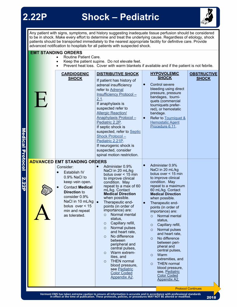
HYPOVOLEMIC SHOCK
Control severe bleeding using direct pressure, pressure bandages, tourni-quets (commercial tourniquets prefer-red), or hemostatic bandage.
Refer to Tourniquet & Hemostatic Agent Procedure 6.11.
Administer 0.9% NaCl in 20 mL/kg bolus over < 15 min to improve clinical condition. May repeat to a maximum 60 mL/kg. Contact Medical Direction when possible.
Therapeutic end-points (in order of importance) are: o Normal mental
status,o Capillary refill,o Normal pulses
and heart rate, o No difference
between peri-pheral and central pulses,
o Warm extremities, and
o THEN normal blood pressure, see, Pediatric Color Coded Appendix A2.
OBSTRUCTIVE SHOCK
DISTRIBUTIVE SHOCK
If patient has history of adrenal insufficiency refer to Adrenal Insufficiency Protocol – 2.1. If anaphylaxis is suspected refer to Allergic Reaction/Anaphylaxis Protocol – Pediatric 2.2P.If septic shock is suspected, refer to Septic Shock Protocol – Pediatric 2.21P.If neurogenic shock is suspected, consider spinal motion restriction.
Administer 0.9% NaCl in 20 mL/kg bolus over < 15 min to improve clinical condition. May repeat to a max of 60 mL/kg. Contact Medical Direction when possible.
Therapeutic end-points (in order of importance) are: o Normal mental
status,o Capillary refill,o Normal pulses
and heart rate, o No difference
between peripheral and central pulses,
o Warm extrem-ities, and
o THEN normal blood pressure, see Pediatric Color Coded Appendix A2.
E
A
EMT STANDING ORDERS
Any patient with signs, symptoms, and history suggesting inadequate tissue perfusion should be considered to be in shock. Make every effort to determine and treat the underlying cause. Regardless of etiology, shock patients should be transported immediately to the nearest appropriate facility for definitive care. Provide advanced notification to hospitals for all patients with suspected shock.
2013
Routine Patient Care. Keep the patient supine. Do not elevate feet. Prevent heat loss. Cover with warm blankets if available and if the patient is not febrile.
CARDIOGENIC SHOCK
Consider: Establish IV
0.9% NaCl to keep vein open.
Contact Medical Direction to consider 0.9% NaCl in 10 mL/kg bolus over < 15 min and repeat as tolerated.
ADVANCED EMT STANDING ORDERS
Medical P
rotocol 2.22P
Shock – Pediatric
Vermont EMS has taken extreme caution to ensure all information is accurate and in accordance with professional standards in effect at the time of publication. These protocols, policies, or procedures MAY NOT BE altered or modified. 2018
2.22P
Protocol Continues

Medical P
rotocols 2.22PShock – Pediatric
Vermont EMS has taken extreme caution to ensure all information is accurate and in accordance with professional standards in effect at the time of publication. These protocols, policies, or procedures MAY NOT BE altered or modified. 2020
Etiology of Shock Cardiogenic Shock: History of cardiac surgery, rhythm disturbances, or post cardiac arrest. Assess for acute
MI and pulmonary edema.o Signs & Symptoms of cardiogenic shock: chest pain, shortness of breath, crackles, JVD, hypotension,
tachycardia, diaphoresis. Distributive Shock: Anaphylaxis (see Allergic Reaction/Anaphylaxis Protocol – Pediatric 2.2P), neurogenic
shock, sepsis, see Septic Shock Protocol – Pediatric 2.21P. Assess for fever and signs of infection.o Signs & Symptoms of neurogenic shock: sensory and/or motor loss, hypotension, bradycardia versus
normal heart rate, warm, dry skin. Hypovolemic Shock: Dehydration, volume loss, or hemorrhagic shock.
o Signs & Symptoms of hypovolemic shock: tachycardia, tachypnea, hypotension, diaphoresis, cool skin, pallor, flat neck veins.
Obstructive Shock: Consider tension pneumothorax, pulmonary embolism, and cardiac tamponade.o Signs and symptoms of tension pneumothorax: asymmetric or absent unilateral breath sounds, respiratory
distress or hypoxia, signs of shock including tachycardia and hypotension, JVD, possible tracheal deviation above the sternal notch (late sign).
x A tourniquet may be used temporarily to slow major bleeding while treating other life threatening concerns or to identify the best location for direct pressure. The tourniquet can be left in place for at least an hour. If direct pressure does not control bleeding, the tourniquet will need to be reapplied and left in place during evacuation.
HYPOVOLEMIC SHOCK
OBSTRUCTIVE SHOCK
If tension pneumothorax suspected, see Needle Decompre-ssion Thorocos-tomy (NDT) Procedure 6.8.
DISTRIBUTIVE SHOCK
If hypotensive consider: (An infusion pump is required for the use of these vasopressor agents)
Norepinephrine infusion 0.1 – 2 mcg/kg/min titrated to effect OR
Epinephrine 0.1 – 1 mcg/kg/min titrated to effect OR
For patient with history of adrenal insufficiency, administer stress dose of hydrocortisone 2 mg/kg IV/IO/IM (max dose 100 mg) or other injectable steroid.
CARDIOGENIC SHOCK
If hypotensive consider:(An infusion pump is required for the use of these vasopressor agents)
Norepinephrine infusion 0.1 – 2 mcg/kg/min titrated to effect OR
Epinephrine 0.1 – 1 mcg/kg/min titrated to effect.
PARAMEDIC – Contact MEDICAL DIRECTION and consider:
P
EXTENDED CARE
Check serum lactate (if available and trained). Consider fluid administration via infusion pump. If signs and symptoms of hypoperfusion persist or symptoms worsen, regardless of
etiology, contact Medical Direction and consider norepinepherine administration via length-based resuscitation tape in the absence of hemorrhagic shock.
2.22P
PEARLS:For patients with uncontrolled hemorrhagic or penetrating torso injuries: Contact Medical Direction to discuss restriction of IV fluids. Delaying aggressive fluid resuscitation until
operative intervention may improve the outcome. Operative intervention must be available within 30-45 minutes to utilize this strategy. In rural areas with longer transport times restricting fluid may result in exsanguination and irreversible shock.
Patients should be reassessed frequently, with special attention given to the lung examination to ensure volume overload does not occur.
Several mechanisms for worse outcomes associated with IV fluid administration have been suggested, including dislodgement of clot formation, dilution of clotting factors, and acceleration of hemorrhage caused by elevated blood pressure.

2013
EMT STANDING ORDERS
PARAMEDIC STANDING ORDERS Consider early advanced airway control in patients with suspected upper or lower airway
burns or severe smoke inhalation. See Airway Management Protocol – Adult 5.1A.o Consider epinephrine 3 mg (3 mL) in 3 mL 0.9% NaCl via nebulizer for symptomatic
patients, especially if unable to obtain advanced airway. If you have a patient with a history of smoke exposure and an altered level of
consciousness and/or hemodynamic or respiratory compromise, establish IV access and administer, if available:o Hydroxocobalamin via use of Cyanokit:Reconstitute: Place the vial of hydroxocobalamin in an upright position; add 0.9%
NaCl to the vial (200 mL for 5 grams vial or 100 mL for 2.5 grams vial) using the transfer spike. Fill to the line.
Rock vial for at least 60 seconds (do not shake).Using vented intravenous tubing, administer IV over 15 minutes.Depending on clinical response, a second dose may be required.
o Consider regional plan for centralized storage of Cyanokit and means to deliver emergently to fire scene.
Oxygen saturation may be inaccurate in patients exposed to carbon monoxide or cyanide.
CO oximeter devices may yield inaccurate low/normal results for patients with CO poisoning. All patients with probable or suspected CO poisoning should be transported to the nearest appropriate hospital, based on their presenting signs and symptoms.
Do not administer other drugs concurrently in same IV as hydroxocobalamin.
Symptoms: headache, confusion, dyspnea, chest tightness, nausea.
Signs: soot in the nose or mouth, change in level of consciousness, seizure, dilated pupils, coughing, tachypnea and hypertension (early), bradypnea and hypotension (late), shock, vomiting.
A
P
Medical P
rotocol 2.23A
Smoke Inhalation/Carbon Monoxide Poisoning – Adult
Vermont EMS has taken extreme caution to ensure all information is accurate and in accordance with professional standards in effect at the time of publication. These protocols, policies, or procedures MAY NOT BE altered or modified. 2020
2.23A
Routine Patient Care. Remove patient from toxic environment. Oxygen 100% via non-rebreather mask or BVM. Decontamination concurrent with initial resuscitation. If a CO oximeter is available, consider obtaining carbon monoxide levels. If a measuring device is available, obtain atmospheric levels of CO and cyanide (CN).
Consider use on “first-in” bags to assist in detection of occult CO toxicity. Acquire and transmit 12-lead ECG if available. If wheezing, consider albuterol 2.5 mg via nebulizer. May repeat every 5 minutes for
continued symptoms. If altered mental status, check finger stick glucose by glucometer. See Burn/Electrocution/Lightning Protocol – Adult & Pediatric 4.0. Call for Paramedic intercept, if available. If not available, call for AEMT intercept.
E
Establish IV access Consider CPAP for respiratory distress. See Continuous Postive Airway Pressure
(CPAP) Protocol – 5.3.
ADVANCED EMT STANDING ORDERS
PEARLS: Smoke is a combination of many dangerous toxins produced by incomplete combustion. Patients
exposed to smoke should be considered for carbon monoxide (CO) and hydrogen cyanide (HCN) poisoning.

2013
PARAMEDIC STANDING ORDERS
P Consider early advanced airway control in patients with suspected upper or lower airway
burns or severe smoke inhalation. See Airway Management Protocol – Pediatric 5.1P.o Consider epinephrine 3 mg (3 mL) in 3 mL 0.9% NaCl via nebulizer for symptomatic
patients, especially if unable to obtain advanced airway. If you have a patient with a history of smoke exposure and an altered level of
consciousness and/or hemodynamic or respiratory compromise, establish IV access and administer, if available:o Hydroxocobalamin via use of Cyanokit: Reconstitute: Place the vial of hydroxocobalamin in an upright position; add
0.9% NaCl to the vial (200 mL for 5 grams vial or 100 mL for 2.5 grams vial) using the transfer spike. Fill to the line.
Rock vial for at least 60 seconds (do not shake).Using vented intravenous tubing, infuse 70 mg/kg as per Pediatric Color Coded
Appendix A2 over 15 minutes.Depending on clinical response, a second dose may be required.
o Consider regional plan for centralized storage of Cyanokit and means to deliver emergently to fire scene.
Medical P
rotocol 2.23PSmoke Inhalation/Carbon
Monoxide Poisoning – Pediatric
Vermont EMS has taken extreme caution to ensure all information is accurate and in accordance with professional standards in effect at the time of publication. These protocols, policies, or procedures MAY NOT BE altered or modified. 2020
2.23P
Oxygen saturation may be inaccurate in patients exposed to carbon monoxide or cyanide.
CO oximeter devices may yield inaccurate low/normal results for patients with CO poisoning. All patients with probable or suspected CO poisoning should be transported to the nearest appropriate hospital, based on their presenting signs and symptoms.
Do not administer other drugs concurrently in same IV as hydroxocobalamin.
Symptoms: headache, confusion, dyspnea, chest tightness, nausea.
Signs: soot in the nose or mouth, change in level of consciousness, seizure, dilated pupils, coughing, tachypnea and hypertension (early), bradypnea and hypotension (late), shock, vomiting.
PEARLS: Smoke is a combination of many dangerous toxins produced by incomplete combustion. Patients
exposed to smoke should be considered for carbon monoxide (CO) and hydrogen cyanide (HCN) poisoning.
EMT STANDING ORDERS
A
Routine Patient Care. Remove patient from toxic environment. Oxygen 100% via non-rebreather mask or BVM. Decontamination concurrent with initial resuscitation. If a CO oximeter is available, consider obtaining carbon monoxide levels. If a measuring device is available, obtain atmospheric levels of CO and cyanide (CN).
Consider use on “first-in” bags to assist in detection of occult CO toxicity. If wheezing, consider albuterol 2.5 mg via nebulizer. May repeat every 5 minutes for
continued symptoms. If altered mental status, check finger stick glucose by glucometer. See Burn/Electrocution/Lightning Protocol – Adult & Pediatric 4.0. Call for Paramedic intercept, if available. If not available, call for AEMT intercept.
E
Establish IV access Consider CPAP for respiratory distress. See Continuous Positive Airway Pressure
(CPAP) Protocol – 5.3.
ADVANCED EMT STANDING ORDERS

Protocol Continues
SUSPECT STROKE: with any of the following new or sudden symptoms and/or complaints: Unilateral motor weakness or paralysis to face, limb or side of body, including facial droop Unilateral numbness Dizziness/vertigo Acute visual disturbance, loss of vision in one eye or one side of vision Difficulty with balance or uncoordinated movements of a limb, gait disturbance Difficulty with speech understanding or production (slurred or inappropriate use of words) Severe headache for no obvious reason Altered mental state
Arm Drift: Have the patient close their eyes and hold arms extended for 10 seconds.
Normal: Both arms move the same, or both arms don’t move at all.
Abnormal: One arm doesn’t move, or one arm drifts down compared to the other.
Prehospital Stroke Scale Examination Please check: Normal Abnormal
Witness: Best contact number for witness:
Facial Droop: Have the patient smile and show teeth.
Normal: Both sides of the face move equally well..
Abnormal: One side of the face does not move as well as the other.
Normal
Blood Glucose:
Prehospital Stroke Screening Tool
Time Last Known Well: (If patient awoke with symptoms, time last known to be at baseline)
Speech: Ask the patient to repeat a phrase such as, “You can’t teach an old dog new tricks”.
Normal: Patient says the correct words without slurring.
Abnormal: Patient slurs words, says the wrong word, or is unable to speak.Normal Abnormal
Normal Abnormal
Abnormal
( ) -
Stroke screen is positive if any abnormal finding in facial droop, arm drift or speech.
20132013
Medical P
rotocol 2.24
Stroke – AdultNorthern New England Unified Guideline
Northern NE Protocol Group has taken extreme caution to ensure all information is accurate and in accordance with professional standards in effect at the time of publication. These protocols, policies, or procedures MAY NOT BE altered or modified. 2020
2.24
Routine Patient Care. Complete the Prehospital Stroke Screening Tool
o If Prehospital Stroke screen is positive, complete stroke severity score (e.g., FAST-ED) to determine probability of a large vessel occlusion (LVO). May use paper form or approved app.
Establish Stroke Alert Criteria and notify receiving hospital of “Stroke Alert” with findings from the screening tools, thrombolytic checklist and time last known well (TLKW).
For symptomatic:o Administer oxygen as appropriate with a target of achieving 94 – 98% saturation.o Elevate head of stretcher to 30° (unless patient requires spinal motion restriction);o Minimize on-scene time; do not delay for ALS intercept;o Acquire and transmit 12-lead ECG, if available;o Correct glucose if < 60 mg/dL. See Diabetic Emergencies (Hypoglycemia)
Protocol – Adult 2.8A.o Rapid transport to the most appropriate facility based on the destination guidance
utilizing your local stroke plan.
EMT STANDING ORDERS
AEMT & PARAMEDIC STANDING ORDERS
A/P Establish IV (18 gauge catheter & right AC preferred site) and administer 250 mL 0.9% NaCl bolus.
E

Medical P
rotocol 2.24
Northern NE Protocol Group has taken extreme caution to ensure all information is accurate and in accordance with professional standards in effect at the time of publication. These protocols, policies, or procedures MAY NOT BE altered or modified. 2020
2.21
If stroke screening scale is positive calculate stroke severity score using FAST-ED
Stroke Alert Criteria – Please check Yes or No:Blood glucose is or has been corrected to greater than 60 mg/dL?Deficit unlikely due to head trauma or other identifiable causes?Positive Prehospital Stroke Screen:
- and time last known well is less than 4.5 hours OR - FAST-ED score is greater than or equal to 4 AND time last known well is less than 24 hours
Stroke Alert Criteria – If yes to all criteria determine appropriate destination, contact receiving hospital and report a STROKE ALERT with time last known well, FAST-ED
score & thrombolytic checklist results
Establish Stroke Alert CriteriaYes No
Stroke – AdultNorthern New England Unified Guideline 2.24

Medical P
rotocol 2.24
Stroke – AdultNorthern New England Unified Guideline
Northern NE Protocol Group has taken extreme caution to ensure all information is accurate and in accordance with professional standards in effect at the time of publication. These protocols, policies, or procedures MAY NOT BE altered or modified. 2020
PEARLS for Anticoagulants: Patients may recognize anticoagulants as "blood thinners". Ask about anticoagulants including
warfarin (Coumadin or Jantoven), Heparin (IV/IM - including Lovenox), dabigatran (Pradaxa), rivaroxaban (Xarelto), apixaban (Eliquis), betrixaban (Bevyxxa) or edoxaban (Savaysa) and immediately communicate to hospital staff.
Please note, medication manufacturers are producing new anticoagulants frequently.
PrehospitalStroke
Screening ToolPositive
Positive LVO Screen
Negative LVO Screen
FAST-ED ≥ 4
FAST-ED˂ 4
TLKW˃ 4.5 & < 24 hours(Thrombectomy
Candidate)
TLKW˂ 4.5 or > 24 hours
(tPA Candidate)
If feasible, and transport time ≤ 60 minutes, transport
to Endovascular Center, per your local stroke plan
Nearest appropriate hospital (including
Endovascular Center if this is the
closest hospital)
DestinationTime Last Known Well (TLKW)Identification of Possible LVO
Destination Guidance for Possible LVO Stroke Patients
Work with your regional endovascular center when developing your local stroke plan.If transport time >60 minutes, transport to the closest Emergency Department.
Thrombolytic Checklist for patients eligible for thrombolytics (t-PA), try to complete the following:
YES NO Has the patient had any of the following:
1. Severe head trauma or intracranial or spinal surgery within the past 3 months?
2. Major non-cranial surgery or trauma within 14 days with internal bleeding?
3. Spontaneous (non-traumatic) intracranial hemorrhage at any time in the past?
4. Is the patient taking any anticoagulants, including oral or injectable mediations? If yes, clarify when last dose was taken (see PEARLS below)
PEARLS: Stroke requires time sensitive interventions. Time = Brain Every minute of acute stroke = about 2 million neurons lost. Transport witness, family or caregiver or obtain witness best phone number for hospital staff to
verify time of symptom onset or Time Last Known Well (TLKW). TLKW is the last time patient known to be at their neurological baseline. If patient awakes with
symptoms, TLKW is time patient was last known to be at their neurological baseline – Ask if patient got up during the night and was at baseline!
Consider stroke mimics including: migraine, hypoglycemia, seizures, intoxication, sepsis, cerebral infectious process, toxic ingestion, neuropathies (Bell’s palsy), neoplasms, hypertensive encephalopathy or previous stroke.
2.24

Medical P
rotocol 2.24
Northern NE Protocol Group has taken extreme caution to ensure all information is accurate and in accordance with professional standards in effect at the time of publication. These protocols, policies, or procedures MAY NOT BE altered or modified. 2020
This page left blank to insert your local Stroke agreement
plan.
Stroke – AdultNorthern New England Unified Guideline 2.24

Routine Patient Care. Administer oxygen as appropriate with a target of achieving 94 – 98% saturation. Attempt to determine the cause of syncope. Acquire and transmit 12-lead EKG, if available. If acute coronary syndrome is
suspected, refer to Acute Coronary Syndrome Protocol – Adult 3.0. Obtain blood glucose analysis; refer to Diabetic Emergencies (Hyperglycemia)
Protocol – Adult 2.7A or Pediatric 2.7P, or Hypoglycemia Protocol – Adult 2.8A & Pediatric 2.8P, if indicated.
Patients with ongoing mental status changes or coma should be treated per the Altered Mental Status Protocol - Adult 2.3A or Pediatric 2.3P.
Perform stroke screening exam (See Stroke Protocol 2.24.) Assess for trauma either as the cause of the syncope or as a consequence of the
syncopal event. Refer to Spinal Motion Restriction Protocol 4.5, Traumatic Emergencies Protocol 4.10 or Traumatic Brain Injury Protocol 4.8 if indicated.
Prevent and treat for shock; see Shock Protocol – Adult 2.22A or Pediatric 2.22P. Consider ALS intercept.
PEARLS: Syncope is defined as a loss of consciousness accompanied by a loss of postural tone with
spontaneous recovery. Consider all syncope to be of cardiac origin until proven otherwise. While often thought as benign, syncope can be the sign of more serious medical emergency.
All patients with syncope deserve emergency department evaluation, even if they appear normal with few complaints on scene.
Syncope that occurs during exercise often indicates an ominous cardiac cause. Patients should be evaluated at the ED. Syncope that occurs following exercise is almost always vasovagal and benign.
Prolonged QTc (generally > 500ms), Brugada Syndrome (incomplete RBBB pattern in V1/V2 with ST segment elevation), Delta waves and hypertrophic obstructive cardiomyopathy should be considered in all patients.
There is no evidence that supports acquiring orthostatic vital signs. Syncope can be indicative of many medical emergencies including:
Perform cardiac monitoring. Observe for and treat dysrhythmias as indicated.P
Establish IV access. Consider fluids per Shock Protocol – Adult 2.22A or Pediatric 2.22P.A
E
EMT STANDING ORDERS
ADVANCED EMT STANDING ORDERS
PARAMEDIC STANDING ORDERS
2013
Medical P
rotocol 2.25
Syncope – Adult & Pediatric
Vermnt EMS has taken extreme caution to ensure all information is accurate and in accordance with professional standards in effect at the time of publication. These protocols, policies, or procedures MAY NOT BE altered or modified. 2020
2.25
o Myocardial infarctiono Pulmonary embolismo Cardiac arrhythmias,o Vasovagal reflexeso Diabetic emergencieso Aortic stenosiso TIA
o Poisoning/drug effectso Dehydrationo Hypovolemiao Seizureso Ectopic pregnancyo Stroke
Brugada sample

Cardiac P
rotocol 3.0
2013
E
EMT STANDING ORDERS
ADVANCED EMT STANDING ORDERS
A Establish IV access prior to administration of nitroglycerin. Nitroglycerin 0.4 mg SL every 3 – 5 minutes while symptoms persist and if
systolic BP remains ≥ 100 mmHg (MAP ≥ 65).
Routine Patient Care. Acquire and transmit 12-lead ECG with baseline vitals within 10 minutes if
available. (Do not delay scene time.) Perform serial 12-lead ECG especially when clinical changes are noted.(See ECG Acquisition, Transmission and Interpretation Procedure 6.5)o If 12-lead ECG indicates a STEMI, transport patient to the most appropriate
facility in accordance with local STEMI guidelines/agreements. Notify receiving facility of a “STEMI Alert”.
Administer oxygen only to patients with dyspnea, hypoxia (O2 saturation < 94%), or signs of heart failure at a rate to keep O2 saturation 94 – 98%.
Administer aspirin 324 mg PO (chewable). (Note: If patient has taken a partial dose (81 mg), administer remaining 243 mg.)
Facilitate administration of the patient’s own nitroglycerin every 3 – 5 minutes while symptoms persist and systolic BP remains ≥100 mmHg, to a total of 3 doses. Contact Medical Direction for additional dosing.
Call for Paramedic intercept, if available. If not available, call for AEMT intercept.
All patients with complaints of chest pain should not automatically be treated with aspirin and nitrates. Consider the likelihood of ACS based on the nature of the symptoms, the patient’s age, cardiac risk factors, past medical history, etc.
Vermont EMS has taken extreme caution to ensure all information is accurate and in accordance with professional standards in effect at the time of publication. These protocols, policies, or procedures MAY NOT BE altered or modified. 2020
Protocol Continues
PPARAMEDIC STANDING ORDERS
Monitor ECG rhythm. Consider IV nitroglycerin at 10 – 30 mcg/min, increasing by 10 mcg/min every 5
minutes while symptoms persist and systolic BP remains ≥ 100 mmHg. Maximum rate 200 mcg per minute. o Two (2) IV lines or equivalent are recommended and the IV nitroglycerin must
be on an infusion pump. If IV nitroglycerin is not available, consider the application of nitroglycerin paste 1
– 2 inches transdermally. For chest discomfort unresponsive to nitrates, consider analgesia:
o Consider fentanyl 25 – 50 mcg slow IV push every 5 minutes up to 300 mcg and systolic BP remains ≥ 100 mmHg (preferred agent) OR
o Consider morphine 2 – 4 mg IV/IM every 5 minutes to a maximum of 15 mg titrated to pain and systolic BP remains ≥ 100 mmHg.
Treat dysrhythmias as needed; refer to the appropriate protocol.
Acute Coronary Syndrome –Adult
3.0
Avoid nitroglycerin in any patient who has used a phosphodiesterase inhibitor for erectile dysfunction and pulmonary hypertension, such as sildenafil (Viagra, Revatio) or vardenafil (Levitra, Staxyn) within 24 hours, or tadalafil (Cialis, Adcirca) within 48 hours. Also avoid use in patients receiving intravenous prostacyclins for pulmonary hypertension.
Administer nitrates with extreme caution, if at all, to patients with inferior-wall STEMI or suspected right ventricular (RV) involvement because these patients require adequate RV preload.
Morphine should be used with caution due to an increased association with mortality.

Acute Coronary Syndrome – Adult
2013
Cardiac P
rotocol 3.0
Vermont EMS has taken extreme caution to ensure all information is accurate and in accordance with professional standards in effect at the time of publication. These protocols, policies, or procedures MAY NOT BE altered or modified. 2020
3.0
PEARLS: Transmission of 12-lead ECG and/or communication with receiving facility is critical to the activation of a
STEMI system. If Paramedic interpretation or automated interpretation states: "Acute MI" or "Meets ST Elevation MI
Criteria," notify the receiving facility of a "STEMI ALERT." Obtain 12-lead ECG in all patients with the following signs and symptoms:
o Chest, jaw or arm discomfort; or
o Shortness of breath; or
o Epigastric discomfort; or
o Syncope, general malaise, or palpitations; or
o Self-administered nitroglycerin; or
o After sudden cardiac arrest with return of cardiac activity.
Administration of aspirin has been shown to decrease mortality in Acute Coronary Syndrome.
Administer aspirin to every patient with suspected acute coronary syndrome unless they have:
o History of anaphylaxis to aspirin, NSAIDs, or
o Evidence of active gastrointestinal or other internal bleeding.
Any patient with acute coronary syndrome (especially women, patients with a history of diabetes, and the elderly) may present with signs and symptoms other than chest pain including diaphoresis, shortness of breath, weakness, syncope and nausea.
Mid Clavicle Line
Cardiac P
rotocol 3.0
Vermont EMS has taken extreme caution to ensure all information is accurate and in accordance with professional standards in effect at the time of publication. These protocols, policies, or procedures MAY NOT BE altered or modified.
Acute Coronary Syndrome –Adult
This page left blank to insert your local STEMI transport
determination plan.

Cardiac P
rotocol 3.1A
Bradycardia – Adult
Vermont EMS has taken extreme caution to ensure all information is accurate and in accordance with professional standards in effect at the time of publication. These protocols, policies, or procedures MAY NOT BE altered or modified. 2020
P
EMT STANDING ORDERS
PARAMEDIC STANDING ORDERSIf Serious Signs or Symptoms (hypotension, acutely altered mental status, signs of shock, ischemic chest discomfort, or acute heart failure): Consider atropine 0.5 mg IV/IO every 3 – 5 minutes to a total of 3 mg. If atropine is ineffective:
o Consider transcutaneous pacing. o Consider procedural sedation prior to or during pacing, if feasible: Midazolam 2.5 mg IV/IO/intranasal, may repeat once in 5 minutes OR 5 mg
IM, may repeat once in 10 minutes ORLorazepam 1 mg IV/IO, may repeat once in 5 minutes OR 2 mg IM, may
repeat once in 10 minutes ORDiazepam 5 mg IV/IO, may repeat 2.5 mg once in 5 minutes.
o Consider analgesia prior to or during transcutaneous pacing, if feasible: Fentanyl 25 – 50 mcg slow IV push, may repeat every 5 minutes to a
total of 300 mcg ORMorphine 2 – 4 mg IV every 10 minutes to a total of 15 mg and systolic
BP ≥100 mmHg. Consider epinephrine infusion at 2 – 10 mcg/min IV/IO, titrated to effect.
o Consider push dose epinephrine (10 mcg/mL) for short transport times or as bridge to infusion. Prepare 10 mcg/mL by adding 1 mL 0.1 mg/mL epinephrine to 9 mL normal saline, then administer 10-20 mcg boluses (1 – 2 mL) every 2 minutes (where feasible, switch to infusion as soon as practical),
Other Causes: Contact Medical Direction and consider:
o For symptomatic beta blocker or calcium channel blocker overdose, consider glucagon 2 – 5 mg IV/IO over 3 – 5 minutes. May repeat up to 10 mg; if effective, place on infusion 1 – 5 mg/hr IV/IO via pump.
o For symptomatic calcium channel blocker overdose or hyperkalemia/renal failure, consider: Calcium chloride 500 – 1,000 mg (5 – 10 mL of a 10% solution) IV/IO over
10 minutes ORCalcium gluconate 2 grams IV/IO over 5 minutes, may repeat in 10 minutes.
o May repeat as needed. Contact Medical Direction.
E
A
3.1A
Routine Patient Care. Consider the underlying causes of bradycardia (e.g., hypoxia, hypoglycemia,
toxicologic and hypothermia). Acquire and transmit 12-lead ECG, if available. Call for Paramedic intercept if serious signs or symptoms (hypotension, acutely
altered mental status, signs of shock, ischemic chest discomfort, or acute heart failure). If Paramedic intercept not available, call for AEMT intercept, if available.
ADVANCED EMT STANDING ORDERS Establish IV/IO access. Consider IO access if hemodynamically unstable. Consider and treat hypovolemia. Administer 500 mL 0.9% NaCl fluid bolus.
For calcium chloride administration, ensure IV patency and do not exceed 1 mL per minute. Flush with 0.9% NaCl before and after administration. Contact Medical Direction.
PEARLS:
Bradycardia, in the context of adult medicine, is the resting heart rate of under 60 beats per minute, though it is seldom symptomatic until the rate drops below 50 beats/min. It may cause cardiac arrest in some patients, because those with bradycardia may not be pumping enough oxygen to their hearts. It sometimes results in syncope, shortness of breath, and if severe enough, death. Trained athletes, patients on beta blockers or young healthy individuals may also have a slow resting heart rate. Resting bradycardia is often considered normal if the individual has no other symptoms such as fatigue, weakness, dizziness, lightheadedness, syncope, chest discomfort, palpitations or shortness of breath associated with it. The term relative bradycardia is used in explaining a heart rate which, although not actually below 60 beats per minute, is still considered too slow for the individual's current medical condition. Consider hyperkalemia in patients with wide complex bradycardia.

Cardiac P
rotocol 3.1P
PEARLS: In children, bradycardia almost always reflects hypoxia, rather than a primary cardiac problem. It is a
pre-arrest rhythm, and the prognosis is poor if left untreated. Immediate delivery of high-flow oxygen and assisted ventilation are essential. Untreated bradycardia will quickly cause shock, hypotension and death.
Combine age-specific heart rates with signs of respiratory failure and shock while assessing. If child is asymptomatic, consider supportive treatment.
Bradycardia – Pediatric
Vermont EMS has taken extreme caution to ensure all information is accurate and in accordance with professional standards in effect at the time of publication. These protocols, policies, or procedures MAY NOT BE altered or modified.
3.1P
P
EMT STANDING ORDERS
PARAMEDIC STANDING ORDERS
E
AADVANCED EMT STANDING ORDERS
Routine Patient Care. Administer 100% oxygen. Consider and treat the underlying causes of bradycardia (e.g., hypoxia, hypoglycemia
and hypothermia). Begin/continue CPR if heart rate is <60 bpm AND the child shows signs of poor systemic
perfusion with hypoperfusion despite adequate ventilation and oxygenation. Acquire and transmit 12-lead ECG, if available. Call for Paramedic intercept, if available. If not available, call for AEMT intercept.
Establish IV access. Consider IO access if hemodynamically unstable. Consider and treat hypovolemia. Administer 20 mL/kg 0.9% NaCl IV fluid bolus.
For Bradycardia With Serious Signs and Symptoms: Epinephrine 0.01 mg/kg IV/IO slow push (0.1 mL/kg of 1:10,000) every 3 – 5 minutes
(max single dose 1 mL). If no IV/IO access, may give ET dose 0.1 mg/kg (0.1 mL/kg of 1:1,000).
Consider atropine 0.02 mg/kg IV/IO for increased vagal tone or AV blocks, may repeat once (maximum single dose 0.5 mg).
Consider transcutaneous pacing. Consider procedural sedation prior to or during pacing, if feasible:
o *Midazolam 0.05 mg/kg IV/IM or 0.1 mg/kg IN (maximum dose 3 mg); may repeat once in 5 minutes, OR
o Lorazepam 0.05 mg/kg IV/IM (maximum dose 1 mg); may repeat once in 5 minutes, OR
o Diazepam 0.01 mg/kg IV (max dose 5 mg); may repeat once in 5 minutes. Consider analgesia prior to or during transcutaneous pacing, if feasible:
o Fentanyl 1 mcg/kg IV/IM/IO/intranasal (maximum initial dose 100 mcg). Administer slow over 2 – 3 minutes. (Fentanyl is the preferred narcotic agent.) OR
o Morphine 0.1 mg/kg IV/IO (maximum initial dose 10 mg). Administer slow over 2 – 3 minutes.
o Reassess patient every 5 minutes. If no contraindications and patient is still in moderate to severe pain, may redose at 5 – 10 minute intervals at half the original dose to a total of 3 doses.
o See Pain Management Protocol – Pediatric 2.18P.
Other Causes: Contact Medical Direction and consider:
o For hypoglycemia, see Diabetic Emergencies (Hypoglycemia) Protocol – Pediatric 2.8P.
o For symptomatic beta blocker overdose or calcium channel blocker overdose, consider glucagon: 1 mg IV/IO (25-40 kg), every 5 minutes as necessary, 0.5 mg IV/IO (less than 25 kg), every 5 minutes as necessary
o For suspected hyperkalemia with ECG changes or symptomatic calcium channel blocker/beta blocker overdose consider:o Calcium gluconate (10% solution) 100 mg/kg IV/IO (maximum dose 2 gm) over 5
minutes; may repeat in 10 minutes if clinical indication persists ORo Calcium chloride (10% solution) 20 mg/kg IV/IO (maximum dose 1 gm) over 5
minutes, repeat 10 minutes; if effective consider IV infusion 20 mg/kg/hour
2020
For calcium chloride administration, ensure IV patency and do not exceed 1 mL per minute. Flush with 0.9% NaCl before and after administration. Contact Medical Direction.
*For IN administration of midazolam use a 5 mg/mL concentration.

Cardiac P
rotocol 3.2A
Vermont EMS has taken extreme caution to ensure all information is accurate and in accordance with professional standards in effect at the time of publication. These protocols, policies, or procedures MAY NOT BE altered or modified. 2020
Cardiac Arrest – Adult3.2A
E
EMT STANDING ORDERS - ADULT Immediate high performance CPR with minimal interruptions. (Use metronome if
possible.) Administer 100% O2. Apply AED and use as soon as possible (with minimum interruption of chest
compressions). (See AED Algorithm in Appendices.) Continue 2 minute cycles of uninterrupted chest compressions followed by AED
analysis and shock for 4 cycles (8 minutes). Place an oral or nasal airway. Ventilation / oxygenation options during 4 cycles:
o BVM ventilation 1 breath every 10 chest compressions during recoil and without interrupting compressions (avoid hyperventiliation), OR
o If resource-limited, apply high flow oxygen via NRB. After 4 cycles (8 minutes):
o Continue 2 minute cycles of uninterrupted chest compressions.o If passive ventilation was used, switch to BVM ventilation.
After 10 cycles (20 minutes), assess Termination of Resuscitation (TOR) Criteria:o Arrest not witnessed by emergency medical services personnel.o NO return of spontaneous circulation after 20 minutes of either BLS alone or
combined BLS and ALS in the absence of hypothermia.o No shock was delivered or advised by the AED.If ALL criteria are present, contact Medical Direction to consider termination of resuscitation. If ANY criteria are missing, contact Medical Direction to consider
termination of resuscitation OR continued resuscitation and transport.If ROSC, continue resuscitation and transport AND contact Medical
Direction. Consider immediate transport for airway obstruction. If cardiac arrest is due to suspected opioid overdose, administer naloxone 4 mg IN.
(See Poisoning/Substance Abuse/Overdose Protocol – Adult 2.19A.)
For arrests of non-cardiac etiology, including respiratory and trauma, use BVM ventilation. Perform 2 minute cycles of uninterrupted chest compressions. Interrupt chest compressions only for rhythm/pulse check and defibrillation. Ventilation / Oxygenation options:
o BVM ventilation 1 breath every 10 chest compressions without interrupting compressions; ORo If resource-limited, apply high flow oxygen via non-rebreather mask (NRB) for passive ventilation
Policy Continues

Cardiac P
rotocol 3.2A
Vermont EMS has taken extreme caution to ensure all information is accurate and in accordance with professional standards in effect at the time of publication. These protocols, policies, or procedures MAY NOT BE altered or modified. 2020
Cardiac Arrest – Adult 3.2A
EEMT STANDING ORDERS – ADULT (CONTINUED)
If ROSC occurs, see Post Resuscitative Care Protocol 3.4A. Consider termination of efforts or not attempting resuscitation (see Do Not
Resuscitate (DNR) & Clinician Orders (COLST) Policy 8.8 and/or Resuscitation Initiation & Termination Policy 8.17.
Call for Paramedic intercept, if available. If not available, call for AEMT intercept.
Protocol Continues
ADVANCED EMT STANDING ORDERS - ADULT
A
Do not interrupt chest compressions for advanced airway, IV/IO placement or epinephrine administration.
During first 4 cycles, consider advanced airway only if airway patency cannot be maintained using basic maneuvers and adjuncts.
Establish IV/IO access (preferred order: IV, proximal humeral IO, anterior tibial IO). Administer 500 mL bolus 0.9% NaCl IV/IO in the absence of pulmonary edema.
After the first 2 minute cycle, administer epinephrine 1:10,000 (0.1mg/mL) 1 mg IV/IO; repeat every other cycle. Maximum dose 3 mg.
After 4 cycles (8 minutes), consider placement of a supraglottic airway without interrupting chest compressions.
Monitor waveform capnography throughout resuscitation, if available, to assess and monitor airway placement and CPR quality, and to monitor for signs of Return of Spontaneous Circulation (ROSC).
Consider and correct treatable causes: hypoxia, overdose/poisoning, hypothermia and hypovolemia—treat as per specific protocol.o If cardiac arrest is due to suspected opioid overdose, administer naloxone 2 mg
IV/IO. (See Poisoning/Substance Abuse/Overdose Protocol – Adult 2.19A.)
P
PARAMEDIC STANDING ORDERS - ADULT Follow ACLS guidelines as trained and credentialed. (See Cardiac Algorithms –
Appendix A6.) During first 4 cycles, consider advanced airway only if airway patency cannot be
maintained using basic maneuvers and adjuncts. After 4 cycles (8 minutes), consider advanced airway without interrupting chest
compressions.For ventricular fibrillation (VF)/pulseless ventricular tachycardia (VT): Defibrillation when available, with minimum interruption in chest compressions. Use
manufacturer’s recommendations. Continue CPR for 2 minutes; then rhythm check; then:
Administer epinephrine (1:10,000) (0.1 mg/mL) 1 mg IV/IO; repeat every other cycle. Maximum dose 3 mg.
If no response after second defibrillation, administer:o Amiodarone 300 mg IV/IO, repeat dose 150 mg as needed ORo Lidocaine 1 – 1.5 mg/kg IV/IO, repeat dose 0.5 – 0.75 mg/kg (maximum total dose
3 mg/kg.)o For Torsades de Pointes: Magnesium sulfate 1 – 2 g IV/IO over 1 – 2 minutes.o For refractory ventricular fibrillation, consider:Changing pad placement from anterior-apex to anterior-posterior.If second manual defibrillator is available, consider Double Sequential
Defibrillation Procedure 6.3.Consider resuscitation for up to 60 minutes from the time of dispatch, including
transport for potential reversible causes if no Return of Spontaneous Circulation (ROSC) after initial efforts.
Protocol Continues
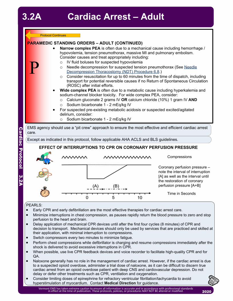
Cardiac P
rotocol 3.2A
Vermont EMS has taken extreme caution to ensure all information is accurate and in accordance with professional standards in effect at the time of publication. These protocols, policies, or procedures MAY NOT BE altered or modified. 2020
Cardiac Arrest – Adult3.2A
P
PARAMEDIC STANDING ORDERS – ADULT (CONTINUED) Narrow complex PEA is often due to a mechanical cause including hemorrhage /
hypovolemia, tension pneumothorax, massive MI and pulmonary embolism. Consider causes and treat appropriately including: o IV fluid boluses for suspected hypovolemiao Needle decompression for suspected tension pneumothorax (See Needle
Decompression Thoracostomy (NDT) Procedure 6.8.)o Consider resuscitation for up to 60 minutes from the time of dispatch, including
transport for potential reversible causes if no Return of Spontaneous Circulation (ROSC) after initial efforts.
Wide complex PEA is often due to a metabolic cause including hyperkalemia and sodium-channel blocker toxicity. For wide complex PEA, consider:o Calcium gluconate 2 grams IV OR calcium chloride (10%) 1 gram IV ANDo Sodium bicarbonate 1 - 2 mEq/kg IV
For suspected pre-existing metabolic acidosis or suspected excited/agitated delirium, consider:o Sodium bicarbonate 1 - 2 mEq/kg IV
Protocol Continues
EMS agency should use a “pit crew” approach to ensure the most effective and efficient cardiac arrest care.
Except as indicated in this protocol, follow applicable AHA ACLS and BLS guidelines.
Compressions
Time in Seconds
Coronary perfusion pressure – note the interval of interruption [A] as well as the interval until the restoration of coronary perfusion pressure [A+B]
EFFECT OF INTERRUPTIONS TO CPR ON CORONARY PERFUSION PRESSURE
PEARLS: Early CPR and early defibrillation are the most effective therapies for cardiac arrest care. Minimize interruptions in chest compression, as pauses rapidly return the blood pressure to zero and stop
perfusion to the heart and brain. Delay application of mechanical CPR devices until after the first four cycles (8 minutes) of CPR and
decision to transport. Mechanical devices should only be used by services that are practiced and skilled at their application, with minimal interruption to compressions.
Switch compressors every two minutes to minimize fatigue. Perform chest compressions while defibrillator is charging and resume compressions immediately after the
shock is delivered to avoid excessive interruptions in CPR. When possible, use live CPR feedback devices and voice recorder to facilitate high-quality CPR and for
QA. Naloxone generally has no role in the management of cardiac arrest. However, if the cardiac arrest is due
to a suspected opioid overdose, administer a trial dose of naloxone, as it can be difficult to discern true cardiac arrest from an opioid overdose patient with deep CNS and cardiovascular depression. Do not delay or defer other treatments such as CPR, ventilation and oxygenation.
Consider limiting doses of epinephrine for refractory ventricular fibrillation/tachycardia to avoid hyperstimulation of myocardium. Contact Medical Direction for guidance.

Cardiac P
rotocol 3.2P
Vermont EMS has taken extreme caution to ensure all information is accurate and in accordance with professional standards in effect at the time of publication. These protocols, policies, or procedures MAY NOT BE altered or modified. 2020
Cardiac Arrest – Pediatric 3.2P
E
EMT STANDING ORDERS Immediate high performance CPR with minimal interruptions (use metronome if
possible). Administer 100% oxygen. Apply AED and use as soon as possible. From birth to age 8 years, use
pediatric AED pads. If pediatric AED pads are unavailable, providers may use adult AED pads,
provided the pads do not overlap. If Return of Spontaneous Circulation occurs see Post Resuscitative Care
Protocol – Pediatric 3.4P. Consider immediate transport for airway obstruction. If cardiac arrest is due to suspected opioid overdose, administer naloxone. (See
Poisoning/Substance Abuse/Overdose Protocol – Pediatric 2.19P.) After 10 cycles (20 minutes), transport or contact Medical Direction to consider
termination of efforts. See DNR/COLST Policy 8.8. Call for Paramedic intercept, if available. If not available, call for AEMT intercept.
Perform 2-minute cycles of high performance CPR. Rhythm/pulse check and defibrillation occur between cycles. Ventilation / Oxygenation options:
o One provider: 2 breaths every 30 compressionso Two providers: 2 breaths every 15 compressionso Advanced airway in place: continuous chest compressions with one BVM ventilation every 10
compressions interposed asychronously.
ADVANCED EMT STANDING ORDERS
A Do not interrupt chest compressions for advanced airway or IV/IO placement. Establish IV/IO access (IV preferred). BVM ventilation is the preferred method of ventilation for pediatric population.
However, if unsuccessful, consider placement of supraglottic airway. o Once an advanced airway is in place, perform continuous chest
compressions with one ventilation every 10 compressions interposed asynchronously.
Monitor waveform capnography throughout resuscitation, if available, to assess and monitor airway placement and CPR quality, and to monitor for signs of Return of Spontaneous Circulation (ROSC). Policy Continues

Cardiac P
rotocol 3.2P
Vermont EMS has taken extreme caution to ensure all information is accurate and in accordance with professional standards in effect at the time of publication. These protocols, policies, or procedures MAY NOT BE altered or modified. 2020
Cardiac Arrest – Pediatric3.2P
ADVANCED EMT STANDING ORDERS
A Consider and correct treatable causes: hypoxia, overdose/poisoning, hypothermia, hypoglycemia, and hypovolemia—treat as per specific protocol.
Administer 0.9% NaCl 20 mL/kg fluid bolus IV/IO.
P
PARAMEDIC STANDING ORDERS
If ventilation is adequate with BVM, routine placement of advanced airway can be delayed.
Placement of an advanced airway during cardiac arrest should not interrupt chest compressions. In this setting, supraglottic airways and ETTs can be considered equivalent. ETT placement, if used, should be limited to 1 attempt of 10 seconds or less, as long as BVM or alternate airway provides adequate chest rise.
Consider tension pneumothorax and treat with needle decompression if indicated. (See Needle Decompression Thoracostomy (NDT) Procedure 6.8.)
For suspected pre-arrest metabolic acidosis, suspected or known hyperkalemia (dialysis patient), or known tricyclic antidepressant overdose, consider sodium bicarbonate 1 mEq/kg IV/IO. Do not mix with any resuscitation drugs. Flush IV tubing with 0.9% NaCl before and after drug administration. Do not use routinely in cardiac arrest.
For known or suspected hyperkalemia or symptomatic calcium channel blocker/beta blocker overdose consider:o Calcium gluconate (10% solution) 100 mg/kg IV/IO (maximum dose 2 gm)
with a maximum 2 gm/dose over 5 minutes; may repeat in 10 minutes if clinical indication persists OR
o Calcium chloride (10% solution) 20 mg/kg IV/IO (maximum dose 1 gm) over 5 minutes, repeat 10 minutes; if effective consider IV infusion 20 mg/kg/hourDo not mix with or infuse immediately before or after sodium bicarbonate
without intervening flush. Do not use routinely in cardiac arrest.
For Ventricular Fibrillation (VF)/Pulseless Ventricular Tachycardia (VT): Use manufacturer’s recommendations for joule settings. In general, defibrillate at
2J/kg; perform CPR for 2 minutes and recheck rhythm; if still a shockable rhythm, defibrillate at 4J/kg; perform CPR for 2 minutes; reassess every 2 minutes and continue to defibrillate at 4J/kg.
If no response after first defibrillation, administer epinephrine (1:10,000) (0.1 mg/mL) 0.01 mg/kg (0.1 mL/kg) IV/IO OR 0.1 mg/kg (1:1,000; 0.1 mL/kg) via ETT.o Repeat every 3 – 5 minutes to a maximum of 3 doses.
If no response after second defibrillation, consider:o Amiodarone 5mg/kg (maximum 300 mg) IV/IO. May repeat up to 2 times for
refractory VF/VT; ORo Lidocaine 1 mg/kg IV/IO (maximum dose 100 mg). Maintenance: 20 – 50
mcg/kg/min infusion. o For Torsades de Pointes: Magnesium sulfate 25 – 50 mg/kg (maximum 2
grams) IV/IO over 1 – 2 minutes.
Protocol Continues
Protocol Continues

Cardiac P
rotocol 3.2P
Vermont EMS has taken extreme caution to ensure all information is accurate and in accordance with professional standards in effect at the time of publication. These protocols, policies, or procedures MAY NOT BE altered or modified. 2020
Cardiac Arrest – Pediatric 3.2P
PPARAMEDIC STANDING ORDERS (CONTINUED)
For Asystole or Pulseless Electrical Activity (PEA): Administer Epinephrine (1:10,000) (0.1 mg/mL) 0.01 mg/kg (0.1 mL/kg) IV/IO
OR 0.1 mg/kg (1:1,000) (1 mg/mL) 0.1 mL/kg via ETT; repeat every 3 – 5 minutes to a maximum of 3 doses..
Give 2 minutes of CPR, then check rhythm:o If asystole or PEA, continue epinephrine and 2 minutes of CPR until:o Pulse obtained ORo Shockable rhythm obtained ORo Decision made to discontinue further efforts. Contact Medical Direction
for guidance.
Protocol Continues
EMS agency should use a “pit crew” approach to ensure the most effective and efficient cardiac arrest care.
Except as indicated in this protocol, follow applicable AHA ACLS and BLS guidelines.
Compressions
Time in Seconds
Coronary perfusion pressure – note the interval of interruption [A] as well as the interval until the restoration of coronary perfusion pressure [A+B]
EFFECT OF INTERRUPTIONS TO CPR ON CORONARY PERFUSION PRESSURE
PEARLS: Optimize oxygenation, ventilation and volume status. Cardiac arrest in children typically results from
progressive deterioration in respiratory or cardiovascular function. Minimize interruptions in chest compression, as pauses rapidly return the blood pressure to zero and
stop perfusion to the heart and brain. Switch compressors at least every two minutes to minimize fatigue. Check rhythm and pulse during
switch to avoid excessive interruptions in CPR. Perform chest compressions while defibrillator is charging and resume compressions immediately
after the shock is delivered to avoid excessive interruptions in CPR.Avoid excessive ventilation. Do not use mechanical CPR devices on children.

Cardiac P
rotocol 3.3
2013
P
EEMT STANDING ORDERS
ADVANCED EMT STANDING ORDERS
A Establish IV access. Contact Medical Direction to consider nitroglycerin:
o For systolic BP of 140 – 160 mmHg: nitroglycerin 0.4 mg SL.o For systolic BP of 160 – 200 mmHg: nitroglycerin 0.8 mg SL (2 tabs/sprays).o For systolic BP > 200 mmHg: nitroglycerin 1.2 mg SL (3 tabs/sprays).o The above doses may be repeated every 5 minutes until symptomatic
improvement or systolic BP is 140 mmHg Consider Continuous Positive Airway Pressure (CPAP) with maximum 10 – 15
cm H2O pressure support. (See CPAP Procedure 5.3.)o Consider CPAP for patient with moderate to severe respiratory distress
concurrent with the following signs and symptoms: Oxygen saturation < 94%. Respiratory rate > 25/minute.Retractions or accessory muscle use.
o SBP must be ≥ 100 mmHg (MAP ≥ 65) to utilize CPAP.
Consider nitroglycerin 0.4 mg SL every 3 – 5 minutes while symptoms persist and if the systolic BP is ≥140 mmHg.
Confirm pulmonary edema using point-of-care ultrasound, if available and credentialed.
Titrate until symptomatic improvement or systolic BP of 140 mmHg:o For systolic BP of 140 – 160 mmHg: IV nitroglycerin start at 50 mcg/min.o For systolic BP of 160 – 200 mmHg: IV nitroglycerin start at 100 mcg/min.o For systolic BP > 200 mmHg: nitroglycerin start at 200 mcg/min.o Note: It is recommended two (2) IV lines in place and the IV nitroglycerin
must be on an infusion pump. Maximum dose of 400 mcg/min. If patient improves after SL nitroglycerin, may apply nitroglycerin paste 1” – 2”
transdermally.
Routine Patient Care. Place the patient in a semi-sitting or full sitting position. Contact Medical Direction for online order to facilitate administration of the
patient’s own nitroglycerin, while symptoms persist and systolic BP is ≥ 140 mmHg.
Acquire and transmit 12-lead ECG, if available. Call for Paramedic intercept, if available. If not available, call for AEMT
intercept.
PARAMEDIC STANDING ORDERS
PEARLS: Nitroglycerin is the first-line medication for congestive heart failure. Furosemide and Narcotics have NOT been shown to improve the outcomes of EMS patients with
pulmonary edema. Even though this historically has been a mainstay of EMS treatment, it is no longer recommended.
If patient has taken nitroglycerin without relief, consider loss of potency due to age. If Nitropaste is used, do not continue to use Nitroglycerin SL unless symptoms worsen. Allow the patient to be in their position of comfort to optimize their breathing effort.
Congestive Heart Failure(Pulmonary Edema) – Adult
Avoid nitroglycerin in any patient who has used a phosphodiesterase inhibitor for erectile dysfunction and pulmonary hypertension, such as: sildenafil (Viagra, Revatio) or vardenafil (Levitra, Staxyn) within 24 hours, or tadalafil (Cialis, Adcirca) within 48 hours. Also avoid use in patients receiving intravenous prostacyclins for pulmonary hypertension.
Administer nitrates with extreme caution, if at all, to patients with inferior-wall STEMI or suspected right ventricular (RV) involvement because these patients require adequate RV preload.
Vermont EMS has taken extreme caution to ensure all information is accurate and in accordance with professional standards in effect at the time of publication. These protocols, policies, or procedures MAY NOT BE altered or modified. 2020
3.3

Cardiac P
rotocol 3.4A
P
EMT STANDING ORDERS
PARAMEDIC STANDING ORDERS
Maintain systolic blood pressure of ≥ 90 mmHg (MAP ≥ 65). Secure advanced airway if indicated. See Orotracheal Intubation Procedure
5.7.
If Cardiac Arrest Was The Result of VF or VT, Manage The Patient As Follows: If amiodarone/lidocaine was administered during resuscitation, do not
administer additional doses. However, if the patient is having frequent PVCs or runs of VT, or if the transport time will exceed 30 minutes, start an antidysrhythmic:o Amiodarone maintenance infusion of 1 mg/min IV/IO OR o Lidocaine maintenance infusion 1 – 4 mg/min IV/IO (30 – 50 mcg/kg/min).
Do not use amiodarone if the patient has heart block or profound bradycardia (heart rate < 60, second-degree type II AV block, or third-degree AV block).
Do not use lidocaine if CHF, cardiogenic shock, heart block or WPW.
For Post-Resuscitation Hypotension: Administer 0.9% NaCl in 250 – 500 mL boluses. Total volume should not
exceed 2,000 mL. Consider: (An infusion pump is required for the use of these pressor agents)
o Norepinephrine infusion 1 to 30 mcg/min IV/IO titrated to effect (preferred agent) OR
o Epinephrine infusion 2 – 10 mcg/min IV/IO titrated to effect ORo Consider epinephrine push dose (10 mcg/mL) for short transport times or
as bridge to infusion. Prepare 10 mcg/mL by adding 1 mL 0.1 mg/mL epinephrine to 9 mL normal saline, then administer 10 – 20 mcg boluses (1 – 2 mL) every 2 minutes (where feasible, switch to infusion as soon as practical).
Consider nasogastric or orogastric tube for the intubated patient.
Optimize ventilation and oxygenation:o Initial ventilation rate of 10 – 12 BPM.o Administer oxygen as appropriate with a target of achieving 94 – 98% saturation.
Acquire and transmit a 12-lead ECG, if available.
Post Resuscitative Care – Adult
Vermont EMS has taken extreme caution to ensure all information is accurate and in accordance with professional standards in effect at the time of publication. These protocols, policies, or procedures MAY NOT BE altered or modified. 2020
E
A
PEARLS: Recognition and treatment of a STEMI are critical in the post-cardiac arrest patient. Consider transport of
patient to the most appropriate facility in accordance with local STEMI guidelines/agreements. Notify receiving facility of a “STEMI ALERT”. See Acute Coronary Syndrome Protocol – Adult 3.0.
Avoid hyperventilation as it increases intrathoracic pressures, potentially worsening hemodynamic instability. Hyperventilation may also cause hypocarbia and elevated arterial oxygen levels (hyperoxia) and increased hospital mortality post-resuscitation from cardiac arrest.
Monitor patient closely for recurrent cardiac arrest.
3.4A
ADVANCED EMT STANDING ORDERS If advanced airway is present, ventilation rate is 6 – 10 BPM. Titrate ventilation rate to waveform capnography of 35 to 40 mmHg. When feasible, titrate FiO2 to minimum necessary to achieve SpO2 of 94 – 98%. If SBP < 90 mmHg (MAP < 65) administer 0.9% NaCl 500 mL fluid bolus IV/IO.
Contact Medical Direction for additional fluid dosing.

Cardiac P
rotocol 3.4P
P
EMT STANDING ORDERS
PARAMEDIC STANDING ORDERS
Secure advanced airway if indicated. See Orotracheal Intubation Procedure 5.7.
If Cardiac Arrest Was The Result of VF or VT, Manage The Patient As Follows: If amiodarone/lidocaine was administered during resuscitation, do not administer
additional doses. However, if the patient is having frequent PVCs or runs of VT, or if the transport time will exceed 30 minutes, start an antidysrhythmic:o Amiodarone maintenance infusion of 0.005 – 0.01 mg/kg/min IV/IO via pump
ORo Lidocaine 20 – 50 mcg/kg/min IV/IO via pump.
Do not use amiodarone if the patient has heart block or profound bradycardia (heart rate < 60, second-degree type II AV block, or third-degree AV block).
For Post-Resuscitation Hypotension: Administer fluid bolus of 20 mL/kg of 0.9% NaCl by syringe push method (may
repeat to a maximum 60 mL/kg) AND/OR Consider:
o Epinephrine 0.1 – 1 mcg/kg/min IV/IO titrated to effect (preferred agent) ORo Norepinephrine infusion 0.1 – 2 mcg/kg/min IV/IO titrated to effect.An infusion pump is required for the use of these vasopressors.
Consider nasogastric or orogastric tube for the intubated patient.
Optimize ventilation and oxygenation:o Initial ventilation rate of 12 – 20 BPM.o Administer oxygen as appropriate with a target of achieving 94 – 98% saturation.
Acquire and transmit a 12-lead ECG, if available.
Post Resuscitative Care – Pediatric
Vermont EMS has taken extreme caution to ensure all information is accurate and in accordance with professional standards in effect at the time of publication. These protocols, policies, or procedures MAY NOT BE altered or modified. 2020
E
A
PEARLS:
Avoid hyperventilation as it increases intrathoracic pressures, potentially worsening hemodynamic instability. Hyperventilation may also cause hypocarbia and elevated arterial oxygen levels (hyperoxia) and increased hospital mortality post-resuscitation from cardiac arrest.
Monitor patient closely for recurrent cardiac arrest.
3.4P
ADVANCED EMT STANDING ORDERS If advanced airway is present, ventilation rate is 8 – 10 BPM. Titrate ventilation rate to waveform capnography of 35 – 40 mm Hg. When feasible, titrate FiO2 to minimum necessary to achieve SpO2 of 94 – 98%. Maintain systolic blood pressure 70 mmHg + (2 x age). Administer fluid bolus of
20 mL/kg of 0.9% NaCl (use syringe push method for infants and small children). May repeat to a maximum 60 mL/kg.

Cardiac P
rotocol 3.5A
2013
EMT STANDING ORDERS
P
Follow ACLS guidelines as trained and credentialed. Search for underlying causes.
If symptomatic and hemodynamically unstable:
For regular, narrow complex, consider adenosine 6 mg rapid IV/IO, followed by rapid flush.o May repeat at dose of 12 mg if no conversion.
o May repeat successful dose if dysrhythmia recurs after conversion.
Otherwise, synchronized cardioversion: Use the following initial energy doses, then escalate to the next higher energy level if no conversion. Biphasic devices: follow manufacturer’s recommendations for dosing.o For narrow regular rhythm: 50 – 100J biphasic or 200J monophasic.
o For narrow irregular rhythm: 120 – 200J biphasic or 200J monophasic.
o For wide regular rhythm: 100J biphasic or monophasic.
o For wide irregular/polymorphic VT: 120 – 200J biphasic or 360 monophasic, using unsynchronized defibrillation doses if unable to sync:
Administer procedural sedation prior to or during cardioversion, if feasible. Do not delay cardioversion. o Midazolam 2.5 mg IV/IO/intranasal, may repeat once in 5 minutes OR 5 mg IM may
repeat once in 10 minutes ORo Lorazepam 1 mg IV/IO, may repeat once in 5 minutes OR 2 mg IM, may repeat
once in 10 minutes ORo Diazepam 5 mg IV/IO, may repeat 2.5 mg once in 5 minutes.
If symptomatic, but hemodynamically stable:
For narrow complex tachycardia (with a heart rate persistently > 150 bpm):
o Attempt Valsalva vagal maneuver, for regular rhythms.
If vagal maneuvers fail and the rhythm is regular:Adenosine 6 mg rapid IV/IO, followed by rapid flush.May repeat at dose of 12 mg if no conversion.May repeat successful dose if rhythm recurs after conversion.
If irregular rhythm, to control ventricular rate:Diltiazem 0.25 mg/kg IV/IO (maximum dose 20 mg) over 2 minutes.
(Consider 10 mg maximum dose for elderly patient or patient with low BP.)May repeat dose in 15 minutes at 0.35 mg/kg (maximum dose 25 mg), if
necessary.Consider maintenance infusion at 5 – 15 mg/hour IV/IO OR
Metoprolol 5 mg IV/IO over 2 – 5 minutes.May repeat every five minutes to a maximum of 15 mg as needed to
achieve a ventricular rate of 90 – 100 BPM.
PARAMEDIC STANDING ORDERS
Tachycardia – Adult
Vermont EMS has taken extreme caution to ensure all information is accurate and in accordance with professional standards in effect at the time of publication. These protocols, policies, or procedures MAY NOT BE altered or modified. 2020
3.5A
Routine Patient Care.
Acquire and transmit 12-lead ECG, if available
Call for Paramedic intercept, if available. If not available, call for AEMT intercept.EAADVANCED EMT STANDING ORDERS
Establish IV access (preferably no more distal than AC fossa). Consider IO access if hemodynamically unstable.
Cardiac P
rotocol 3.5A
PEARLS: Consider and treat potential underlying causes, e.g., hypoxemia, dehydration, fever. Wide complex tachycardia should be considered Ventricular Tachycardia until proven otherwise. Do not administer Diltiazem to wide complex tachycardia. Signs and symptoms of hemodynamic instability:
o Hypotensiono Acutely altered mental statuso Signs of shocko Signs of acute heart failureo Ischemic chest pain
If ventricular rate is > 150/min, prepare for immediate cardioversion. May give brief trial of medications based on specific arrhythmias. Immediate cardioversion is generally not needed if heart rate is ≤150.
Adenosine should be administered rapidly though a proximal (e.g., antecubital) vein site followed by a rapid saline flush.
For best results of vagal maneuver: start with patient’s head of bed elevated by 30 degrees. After patient performs vagal maneuver sustained over 15 seconds, lay the patient flat and elevate their legs.
P
If symptomatic, but hemodynamically stable: (continued from previous page)
For wide complex tachycardia:
o Only for regular rhythm with monomorphic QRS:
Consider: adenosine 6 mg rapid IV/IO, followed by rapid flush.May repeat at dose of 12 mg if no conversion.May repeat successful dose if rhythm recurs after conversion.
Consider: Amiodarone 150 mg IV/IO mixed with 50 – 100 mL D5W or 0.9% NaCl
over 10 minutes.May repeat once in 10 minutes.
If successful, consider a maintenance infusion of 1 mg/min ORLidocaine 1 mg/kg IV/IO May repeat every 5 minutes (total of 3 doses) to maximum of 3 mg/
kg. If successful, consider a maintenance infusion of 1 – 4 mg/min IV/
IO.
For polymorphic Ventricular Tachycardia/Torsades de Pointes: If pulse present, consider magnesium sulfate 2 gm IV/IO diluted in 10 mL D5W or
0.9% NaCl over 10 minutes.
PARAMEDIC STANDING ORDERS
2013
Tachycardia – Adult
Vermont EMS has taken extreme caution to ensure all information is accurate and in accordance with professional standards in effect at the time of publication. These protocols, policies, or procedures MAY NOT BE altered or modified. 2020
3.5A
Diltiazem, metoprolol, amiodarone, lidocaine and adenosine are contraindicated in patients with a history of or suspected Wolff-Parkinson-White (WPW) syndrome.
Medications should be administered cautiously in frail or debilitated patients; lower doses should be considered.
Be cautious in rate controlling patients in rapid atrial fibrillation (A Fib) who may be compensating for another disease process such as sepsis or pulmonary embolism.

Cardiac P
rotocol 3.5P
P
If symptomatic and hemodynamically unstable: For narrow complex/probable SVT:
o Adenosine 0.1 mg/kg rapid IV/IO not to exceed 6 mg (first dose), followed by rapid flush.Repeat once at 0.2 mg/kg not to exceed 12 mg (subsequent dose).
o If adenosine is ineffective or for wide complex, perform synchronized cardioversion:0.5 – 1J/kg; if unsuccessful, increase to 2J/kg.
o Administer procedural sedation prior to or during cardioversion, if feasible:Midazolam 0.05 mg/kg IV/IO/intranasal OR Diazepam 0.05 mg/kg IV/IO.
If symptomatic but hemodynamically stable: For narrow complex, probable supraventricular tachycardia, or regular
wide complex tachycardia (monomorphic QRS ONLY):o Adenosine 0.1 mg/kg IV/IO not to exceed 6mg (first dose). May repeat once at 0.2 mg/kg IV/IO not to exceed 12 mg (subsequent
dose). For wide complex:
o Amiodarone 5 mg/kg IV/IO (maximum 300 mg) over 20-60 minutes ORo Lidocaine 1 mg/kg IV/IO bolus (maximum 100 mg).
For polymorphic ventricular tachycardia/Torsades de Pointeso Magnesium sulfate 25 – 50 mg/kg IV/IO over 10 – 20 minutes (maximum
dose 2 grams).
PARAMEDIC STANDING ORDERS
PEARLS: Consider and treat potential underlying causes, e.g., hypoxemia, dehydration, fever. Signs and symptoms of hemodynamic instability:
o Hypotensiono Acutely altered mental statuso Signs of shock
Probable Sinus Tachycardiao Compatible history consistent with known causeo P waves are present and normalo Variable R-R and constant P-R intervalo Infants: rate usually < 220/mino Children: rate usually < 180/min
Probable Supraventricular Tachycardiao Compatible history (vague, nonspecific); history of abrupt onset / rate changeso P waves absent / abnormalo Heart-rate is NOT variableo Infants: rate usually > 220/mino Children: rate usually > 180/mino Adenosine should be administered rapidly though a proximal (e.g., antecubital) vein site
followed by a rapid saline flush
Tachycardia – Pediatric
Vermont EMS has taken extreme caution to ensure all information is accurate and in accordance with professional standards in effect at the time of publication. These protocols, policies, or procedures MAY NOT BE altered or modified. 2020
3.5P
EMT STANDING ORDERS Routine Patient Care. Acquire and transmit 12-lead ECG, if available. Call for Paramedic intercept, if available. If not available, call for AEMT
intercept.
E
AADVANCED EMT STANDING ORDERS
Establish IV access. Consider IO access if hemodynamically unstable.

POSITION #1-Compressor 1 (right side of patient): Sets up defibrillator Alternates 2 minutes of chest compressions with Position 2 Assists Position 3 with ventilations in off cycle
POSITION #2-Compressor 2 (left side of patient): Initiates 2 minutes of chest compressions at rate of 100 – 120 / min Assists Position 3 with ventilations in off cycle
POSITION #3-Airway (At patient’s head): Opens airway and inserts OPA Assembles BVM or if resource limited, assemble NRB. If using BVM, provide 2-handed mask seal Inserts advanced airway after 8 minutes/4 cycles (or sooner if BVM ventilations are
inadequate).
POSITION #4-Team Leader (Outside CPR triangle): Coaches appropriate rate and depth of compressions Calls for compressor change every two minutes Calls for rhythm analysis every 2 minutes, immediate shock if indicated Monitor CPR quality and use of metronome at 100 – 120 bpm Assumes duties of Position 5/6 if limited to four rescuers throughout resuscitation.
POSITION #5-Vascular/Meds (Outside CPR triangle): Initiates IV/IO access (IV access preferred) Administers medications per protocol
POSITION #6-Code Commander (Outside CPR triangle): Ideally highest level provider Communicates/interfaces with CPR Team Leader Coordinates patient treatment decisions Communicates with family/loved ones Completes Cardiac Arrest Check List
Cardiac P
rotocol 3.6
Vermont EMS has taken extreme caution to ensure all information is accurate and in accordance with professional standards in effect at the time of publication. These protocols, policies, or procedures MAY NOT BE altered or modified. 2020
Team-Focused CPR – Adult & Pediatric
3.6
Policy Continues
EMS agency should use a “pit crew” approach when using this protocol to ensure the most effective and efficient cardiac arrest care. Training should include teamwork simulations integrating BLS, and ALS crew members who regularly work together. EMS systems should practice teamwork using “pit crew” techniques with predefined roles and crew resource management principles. One Example is as follows:

Cardiac P
rotocol 3.6
Vermont EMS has taken extreme caution to ensure all information is accurate and in accordance with professional standards in effect at the time of publication. These protocols, policies, or procedures MAY NOT BE altered or modified. 2020
Team-Focused CPR – Adult & Pediatric
If feasible and the scene is safe, immediately upon arrival, one member of the crew should rapidly enter the scene without equipment (other than gloves) to begin chest compressions.
Clear some space to optimize your working environment. Move furniture or get the patient in a position that will allow a rescuer space to kneel on both sides of them, and where there is sufficient room at the head. Effectiveness of chest compressions decreases during patient movement. Therefore resuscitate the patient as close to the scene as operationally feasible.
Position 1 and 2 are ideally set up on opposite sides of patient’s chest and perform continuous chest compressions, alternating every 2 minutes to avoid fatigue.
REMEMBER: Effective chest compressions are one of the most important therapies for the pulseless patient. Effective is defined as:o A rate of at least 100 and less than 120 compressions/minute - Use of metronome or
CPR feedback device is essential. (e.g. built into monitor or smart phone app) o A depth of 2 – 2.4 inches (Infant: 1½-inch depth; Child: 2-inch depth)o Allow for complete chest recoil (avoid leaning on chest)o Do not interrupt compressions to obtain IV access or perform airway management.o Do not hyperventilate as it increases intrathoracic pressure and decreases blood return to
the heart. Ventilate 1 breath every 10 compressions without interrupting chest compressions. (Pediatric: Ventilate at a ratio of 15:2, with one breath delivered after the 14th and 15th compressions.)
Chest compressions should only be interrupted during rhythm check (AED analysis or manual) and defibrillation shocks. Continue compressions when AED/ defibrillator is charging, if device allows.
During interruptions compressor’s hands should hover over chest. Perform pulse check simultaneously with rhythm check. With the goal of immediate uninterrupted chest compressions, if a mechanical device is used,
it should not lead to delay or interruption in chest compressions; consider delayed application.
Pre-charge manual defibrillators prior to rhythm check to ensure rapid defibrillation if a shockable rhythm is present. If no shock is indicated, disarm the device (dump the charge)
Utilize ETCO2 to assess CPR quality and monitor for signs of ROSC. Use of a CPR checklist to ensure that all best practices are followed during CPR.
Example Cardiac Arrest Check ListCode commander and pit crew roles identifiedChest compression interruptions minimizedCompressors rotated at minimum every 2 minutesMetronome set between 100 and 120 beats per minuteAED/defibrillator appliedOPA/NPA placedO2 flowing and attached to NRB/BVMETCO2 waveform presentIV/IO access establishedPossible causes consideredGastric insufflation limited and gastric decompression consideredFamily present and ongoing communication provided
Consider possible causesHypovolemiaHypoxiaHydrogen Ions (acidosis)HypothermiaHyper/hypokalemiaHypoglycemia
Tablets/toxinsTamponadeTension pneumothoraxThrombosis (MI)Thrombosis (PE)Trauma
3.6
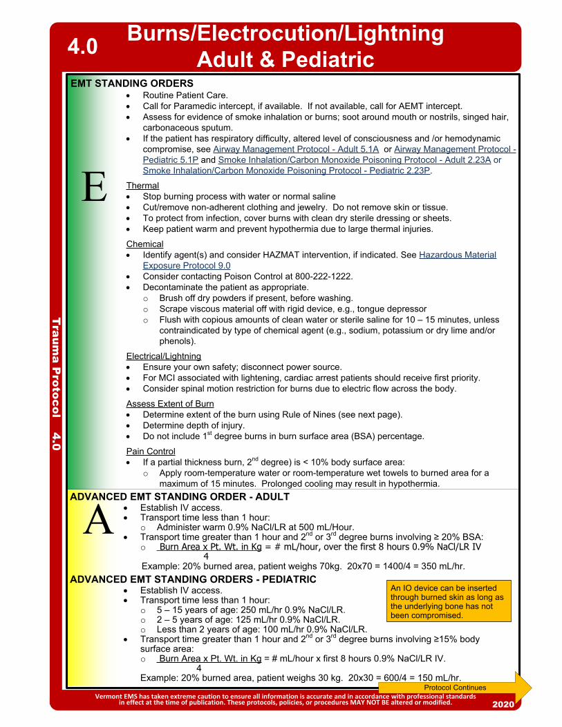
Routine Patient Care. Call for Paramedic intercept, if available. If not available, call for AEMT intercept. Assess for evidence of smoke inhalation or burns; soot around mouth or nostrils, singed hair,
carbonaceous sputum. If the patient has respiratory difficulty, altered level of consciousness and /or hemodynamic
compromise, see Airway Management Protocol - Adult 5.1A or Airway Management Protocol - Pediatric 5.1P and Smoke Inhalation/Carbon Monoxide Poisoning Protocol - Adult 2.23A or Smoke Inhalation/Carbon Monoxide Poisoning Protocol - Pediatric 2.23P.
Thermal Stop burning process with water or normal saline Cut/remove non-adherent clothing and jewelry. Do not remove skin or tissue. To protect from infection, cover burns with clean dry sterile dressing or sheets. Keep patient warm and prevent hypothermia due to large thermal injuries.
Chemical Identify agent(s) and consider HAZMAT intervention, if indicated. See Hazardous Material
Exposure Protocol 9.0 Consider contacting Poison Control at 800-222-1222. Decontaminate the patient as appropriate.
o Brush off dry powders if present, before washing.o Scrape viscous material off with rigid device, e.g., tongue depressor o Flush with copious amounts of clean water or sterile saline for 10 – 15 minutes, unless
contraindicated by type of chemical agent (e.g., sodium, potassium or dry lime and/or phenols).
Electrical/Lightning Ensure your own safety; disconnect power source. For MCI associated with lightening, cardiac arrest patients should receive first priority. Consider spinal motion restriction for burns due to electric flow across the body.
Assess Extent of Burn Determine extent of the burn using Rule of Nines (see next page). Determine depth of injury. Do not include 1st degree burns in burn surface area (BSA) percentage.
Pain Control If a partial thickness burn, 2nd degree) is < 10% body surface area:
o Apply room-temperature water or room-temperature wet towels to burned area for a maximum of 15 minutes. Prolonged cooling may result in hypothermia.
2013
E
EMT STANDING ORDERS
2013
AADVANCED EMT STANDING ORDERS - PEDIATRIC
Establish IV access. Transport time less than 1 hour:
o 5 – 15 years of age: 250 mL/hr 0.9% NaCl/LR. o 2 – 5 years of age: 125 mL/hr 0.9% NaCl/LR.o Less than 2 years of age: 100 mL/hr 0.9% NaCl/LR.
Transport time greater than 1 hour and 2nd or 3rd degree burns involving ≥15% body surface area: o Burn Area x Pt. Wt. in Kg = # mL/hour x first 8 hours 0.9% NaCl/LR IV. 4 Example: 20% burned area, patient weighs 30 kg. 20x30 = 600/4 = 150 mL/hr.
Traum
a Protocol 4.0
Burns/Electrocution/Lightning Adult & Pediatric
Vermont EMS has taken extreme caution to ensure all information is accurate and in accordance with professional standards in effect at the time of publication. These protocols, policies, or procedures MAY NOT BE altered or modified. 2020
4.0
Protocol Continues
ADVANCED EMT STANDING ORDER - ADULT Establish IV access. Transport time less than 1 hour:
o Administer warm 0.9% NaCl/LR at 500 mL/Hour. Transport time greater than 1 hour and 2nd or 3rd degree burns involving ≥ 20% BSA:
o Burn Area x Pt. Wt. in Kg = # mL/hour, over the first 8 hours 0.9% NaCl/LR IV 4
Example: 20% burned area, patient weighs 70kg. 20x70 = 1400/4 = 350 mL/hr.
An IO device can be inserted through burned skin as long as the underlying bone has not been compromised.

Traum
a Protocol 4.0
Burns/Electrocution/Lightning Adult & Pediatric
Vermont EMS has taken extreme caution to ensure all information is accurate and in accordance with professional standards in effect at the time of publication. These protocols, policies, or procedures MAY NOT BE altered or modified. 2020
Transport Decision: Consider air medical transport for major burns with greater than 20% BSA and/or inhalation
injury with risk of airway compromise. Electrocution injuries with loss of consciousness, arrhythmia or any respiratory abnormality.
PPARAMEDIC STANDING ORDERS
Refer to: Airway Management Protocol – Adult 5.1A or Airway Management Protocol
– Pediatric 5.1P. Pain Management Protocol – Adult 2.18A or Pain Management Protocol –
Pediatric 2.18P.
Rule of Nines
PEARLS: Apnea may last longer than asystole in lightning injuries. Provide ventilatory support. Electrocution/lightning burns can occur anywhere along the path a current travels through the body.
Evident surface burns may only comprise a small portion of the overall burn injury, and an injury’s full extent may not be immediately apparent.
Chemical burns: If 0.9% NaCl or sterile water is not readily available, do not delay; use tap water for flushing the affected area. Flush the area as soon as possible with the cleanest readily available water using copious amounts of water.
4.0
Protocol Continues
Expert burn center opinion recommends limiting prehospital IV fluids based on concerns for fluid overload and development of compartment syndrome. In cases where burn patients are in shock, IV fluid administration should be based on use of the Shock Protocol – Adult 2.22A or Shock Protocol – Pediatric 2.22P.

Crush/Suspension Injury –Adult & Pediatric
2013
EMT STANDING ORDERS
Traum
a Protocol 4.1
Vermont EMS has taken extreme caution to ensure all information is accurate and in accordance with professional standards in effect at the time of publication. These protocols, policies, or procedures MAY NOT BE altered or modified. 2020
4.1
E Routine Patient Care. Administer oxygen as appropriate with a target of achieving 94 –
98% saturation. Initiate spinal motion restriction if indicated. Acquire and transmit 12-lead ECG if available. If extrication is prolonged, obtain multiple
ECGs. Call for Paramedic intercept, if available. If not available, call for AEMT intercept. Patients who have experienced suspension or vertical entrapment injuries should be
rapidly extricated to a supine position to restore blood flow to heart and brain. Assess and treat for traumatic injury. See Traumatic Emergencies Protocol – 4.10. See Shock Protocol – Adult 2.22A and Pediatric 2.22P, and Trauma Triage and
Transport Decision Policy 8.20.
ADVANCED EMT STANDING ORDERS
A Establish IV/IO access. Do not delay transportation to initiate IV/IO access, however if
patient is entrapped it is preferable that IV/IO access be initiated and fluid bolus of 1,000 – 2,000 mL 0.9% NaCl (Pediatric: (20 mL/kg 0.9% NaCl) be administered prior to extrication.
Assess pain level. Consider pain control measures. See Pain Management Protocol – Adult 2.18A or Pain Management Protocol – Pediatric 2.18P.
PARAMEDIC STANDING ORDERS
P
PEARLS Compression syndrome: An indirect muscle injury due to a simple, slow compression of a group of muscles
leading to ischemic damage and release of toxic substances into the circulatory system. (For example, a patient who fell and has been on the floor for 2 days)
Compartment syndrome: A localized rapid rise of tension within a muscle compartment, which inevitably leads to metabolic disturbances akin to rhabdomyolysis.
Crush syndrome: Involves a series of metabolic changes produced due to an injury of the skeletal muscles of such a severity as to cause a disruption of cellular integrity and release of its contents into the circulation.
Suspension syndrome: A state of shock caused by blood pooling in dependent lower extremities while the body is held upright without any movement for a period of time. May lead to a relative hypovolemic state and cardiovascular collapse.
Causes of mortality in untreated crush syndrome: o Immediate: severe head injury, traumatic asphyxia, torso injury with intrathoracic or intra-abdominal organ injuryo Early: hyperkalemia, hypovolemia/shock, o Late: renal failure, coagulopathy, hemorrhage and sepsis
Suspect hyperkalemia if T waves become peaked, QRS prolonged > 0.12 seconds, absent P waves, or prolonged QTc. Hyperkalemia may be delayed up to 24 hours after extrication.
A patient with a crush injury may initially present with very few signs and symptoms, therefore, maintain a high index of suspicion for any patient with a compressive mechanism of injury.
EMT/ADVANCED EMT/PARAMEDIC EXTENDED CARE ORDERS Secondary to initial bolus, consider sodium bicarbonate infusion:
o 150 mEq in 1000 mL 0.9% NaCl or D5W at a rate of 250 mL/hr or 4 mL/min. In the event that adequate fluid resuscitation is not available, consider applying a
tourniquet on the affected limb and do not release until adequate IV fluids and/or medications are available.
If extrication is prolonged > 1 hour, contact online Medical Direction for additional considerations prior to extricating the patient.
X
For significant crush injuries or prolonged entrapment/suspension, consider:o Sodium bicarbonate 1 mEq/kg (maximum dose 50 mEq) IV/IO bolus over 5 minutes.
Consider the following:o Monitor for dysrhythmias or signs of hyperkalemia before and after extrication.o If ECG suggestive of hyperkalemia, consider administering the following:Calcium chloride (10% solution) 1 gram IV/IO over 5 minutes, may repeat in 10
minutes (Pediatric: 20 mg/kg IV/IO (maximum dose 1 gm) ORCalcium gluconate (10% solution) 2 grams IV/IO over 5 minutes, may repeat in 10
minutes (Pediatric: 100 mg/kg IV/IO (maximum dose 2 gm).Albuterol continuous 10 – 20 mg nebulized.

E
EMT STANDING ORDERS
ADVANCED EMT/PARAMEDIC STANDING ORDERS
Consider CPAP to supplement the patient’s own spontaneous respiratory effort. Establish IV/IO access.
2013
Traum
a Protocol 4.2
Drowning/Submersion Injuries –Adult & Pediatric
Vermont EMS has taken extreme caution to ensure all information is accurate and in accordance with professional standards in effect at the time of publication. These protocols, policies, or procedures MAY NOT BE altered or modified. 2020
A/P
4.2
Routine Patient Care. Victims with only respiratory arrest usually respond after a few artificial breaths
are given.o Give a few breaths and check for a pulse.Anticipate vomiting.
For patients in cardiac arrest, provide immediate CPR.o Utilize the sequence ABC, not CAB, i.e. start with airway and breathing
before compressions. Routine stabilization of the cervical spine in the absence of circumstances that
suggest a spinal injury is not recommended.o If c-spine injury is suspected, see Advanced Spinal Assessment Procedure
6.0 and Spinal Motion Restriction Protocol 4.5. Obtain specific history: time, temperature, associated trauma, etc. Begin resuscitation efforts while removing the patient from the water. Consider hypothermia. (See Hypothermia – Adult & Pediatric Protocol 2.11.) Remove wet clothes and warm the patient. All patients with history of submersion should be transported to the hospital. Reassure anxious patient. If water temperature is estimated to be less than 6oC (43oF) and submerged:
o Less than 90 minutes: Initiate full resuscitationo Greater than 90 minutes: Consider not initiating resuscitation or termination
of efforts. Contact Medical Direction for guidance. If water temperature is estimated to be greater than 6oC (43oF) and submerged:
o Less than 30 minutes: Initiate full resuscitationo Greater than 30 minutes: Consider not initiating resuscitation or termination
of efforts. Contact Medical Direction for guidance. Call for Paramedic intercept, if available. If not available, call for AEMT intercept.
PEARLS: There is no need to clear the airway of aspirated water; only a modest amount of water is aspirated by
most drowning victims, and aspirated water is rapidly absorbed into the central circulation. Unnecessary cervical spine immobilization can impede adequate opening of the airway and delay
delivery of rescue breaths. SCUBA Diving related injuries: for patients presenting with suspected diving-related emergencies, a
thorough assessment should include obtaining the patient’s dive computer and/or dive plan. The major types of dive-related illnesses include Pulmonary Over-Pressurization Syndromes (POPS) and Decompression Sickness (DCS). Seriously ill patients may present with any combination of altered mental status, respiratory distress or shock. After recognition of a suspected diving related emergency, treatment should focus on supplemental oxygen and rapid transport. ED staff should be fully briefed on the patient’s dive history.
Do not attempt water rescues unless properly trained and equipped. When operating on scenes involving water, use extreme caution and wear a PFD.

A/E
/P
2013
PEARLS: Handle the tooth carefully. Avoid touching the root of the tooth (the part of the tooth that was embedded
in the gum) because it can be damaged easily. Primary (baby) teeth should not be reimplanted.
X
EMT/ADVANCED EMT STANDING ORDERS
PARAMEDIC STANDING ORDERS
2013
EMT/ADVANCED EMT/PARAMEDIC EXTENDED CARE ORDERS If definitive treatment is expected to be greater than 4 hours, an attempt to
reinsert the avulsed tooth in its socket should be made after rinsing tooth in water or normal saline. If multiple teeth require reinsertion, use the shape and size of dentition on the opposing side to guide you in proper placement.
Routine Patient Care. Dental avulsions should be placed in an obviously labeled container with saline-
soaked dressing, milk, or cell-culture medium (example: Save-a-tooth®). Do not place in tap water.
Eye Injuries –Adult & Pediatric
Vermont EMS has taken extreme caution to ensure all information is accurate and in accordance with professional standards in effect at the time of publication. These protocols, policies, or procedures MAY NOT BE altered or modified. 2020
4.3
Routine Patient Care. Obtain visual history (e.g., use of corrective lenses, surgeries, use of protective
equipment). Obtain visual acuity, if possible. Assist patient with the removal of contact lens, if applicable. Chemical irritants, including pepper spray, alkali, acid or other chemical exposure:
o Flush with copious amounts of water, or 0.9% NaCl for a minimum of 20 minutes.
o Consider contacting Poison Control at (800) 222-1222 as soon as practical for consultation.
Thermal burns to eyelids: patch both eyes with cool saline compress. Impaled object: immobilize object and patch both eyes. Do not apply pressure. Do
not attempt to remove object. Puncture wound: place rigid protective device over both eyes (e.g., eye shield). Do
not apply pressure. Foreign body: Minor foreign objects like dust or grit may be flushed with water or
0.9% NaCl. Patch both eyes. If the patient cannot close their eyelids, keep their eye moist with a sterile saline
dressing.
Proparacaine OR tetracaine:o Apply 2 drops to affected eye; repeat every 5 minutes as needed.
Consider use of Morgan lens for irrigation. Refer to Pain Management Protocol – Adult 2.18A or Pain Management Protocol –
Pediatric 2.18P. Refer to the Nausea/Vomiting Protocol – Adult & Pediatric 2.12.
Dental Injuries –Adult & Pediatric
Traum
a Protocol 4.3
EMT/ADVANCED EMT/PARAMEDIC STANDING ORDERS
PEARLS: For chemical exposure to eye position patient with the affected eye downward so irrigation does not
run into the unaffected eye.
P
EA/

Musculoskeletal Injuries –Adult & Pediatric
PEARLS: Use ample padding when splinting possible fractures, dislocations, sprains, and strains.
Remove and secure all jewelry. Elevate injured extremities, if possible. Consider the application of a cold pack for 30 minutes.
Musculoskeletal injuries can occur from blunt and penetrating trauma. Fractures of the pelvis and femur, as well as fractures or dislocations involving circulatory or neurological deficits, take priority over other musculoskeletal injuries.
Hip dislocations, pelvic, knee, and elbow fractures / dislocations have a high incidence of vascular compromise.
Do not manipulate pelvis once fracture is suspected. Repeated manipulation can increase internal hemorrhage.
2013
EMT STANDING ORDERS
Traum
a Protocol 4.4
Vermont EMS has taken extreme caution to ensure all information is accurate and in accordance with professional standards in effect at the time of publication. These protocols, policies, or procedures MAY NOT BE altered or modified. 2020
4.4
E
Establish IV access. For significant injury, consider administration of 0.9% NaCl 500 mL bolus IV for adults; 20 mL/kg for pediatric. Assess pain level and consider pain control measures. See Pain Management
Protocol – Adult 2.17A or Pediatric 2.17P.
Routine Patient Care. Manually stabilize the injury. For open fractures, control bleeding and treat for shock. See Tourniquet &
Hemostatic Agent Procedure – Adult & Pediatric 6.9, Shock Protocol – Adult 2.22A or Shock Protocol – Pediatric 2.22P. Remove obvious debris, irrigate open wounds with saline solution, and cover with moist sterile dressing.
For significantly-angulated fractures, attempt to reposition to anatomical position of function, so long as the fracture is not at/near a joint.
Assess CSMs distal to injury before and frequently after immobilization.o Splint extremity as required.o Traction splinting is preferred technique for isolated adult and pediatric mid-
shaft femur fractures. Do not apply traction splints to proximal (hip) or distal femur fractures, partial amputations, or if lower leg or ankle injury. Do not delay transport to apply a traction splint in the multi-trauma patient. Traction splinting may be used for open fractures if no contraindications.
All patients that have a mechanism of injury that could cause a spinal injury, including high risk or questionable injury mechanisms, should have an advanced spinal assessment and spinal motion restriction if indicated. See Spinal Motion Restriction Protocol 4.5 and Advanced Spinal Assessment Procedure 6.0.
Stabilize suspected pelvic fractures with commercial device (preferred) or bed sheet.o It is preferable to use a scoop stretcher rather than to log-roll a patient with a
suspected pelvic fracture. Amputations should be dressed with bulky dressings and assessed for
uncontrolled bleeding. See Tourniquet & Hemostatic Agent – Adult & Pediatric Procedure 6.11. The amputated part should be placed in a plastic bag and transported on ice to hospital with patient.
See Trauma Triage and Transport Decision Policy 8.20 and Pain Management Protocol – Adult 2.18A or Pain Management Protocol – Pediatric 2.18P
Call for Paramedic intercept, if available. If not available, call for AEMT intercept. Minimize scene time.
ADVANCED EMT/PARAMEDIC STANDING ORDERS
A/P

20132013
Protocol 4.5
Spinal Motion Restriction
Vermont EMS has taken extreme caution to ensure all information is accurate and in accordance with professional standards in effect at the time of publication. These protocols, policies, or procedures MAY NOT BE altered or modified. 2020
4.5
Routine Patient Care. Perform advanced spinal assessment (See Advanced Spinal Assessment Procedure 6.0)
to determine if patient requires spinal motion restriction. Maintain manual in-line stabilization during assessment, unless patient is alert and
spontaneously moving neck. Minimize spinal movement during assessment and extrication. A long backboard, scoop stretcher, vacuum mattress, or other appropriate full length
extrication device may be used for extrication if needed. Do not use short board or KED device, except for vertical extrication or other special situations.
Apply adequate padding to prevent tissue ischemia and minimize discomfort.
If patient requires Spinal Motion Restriction: Apply a rigid cervical collar. Self-extrication by patient is allowable if patient is capable. Allow ambulatory patients to sit on stretcher and then lie flat. (The "standing take-down" is
eliminated.) Position backboarded patient on stretcher then remove backboard by using log roll or lift-
and-slide technique. Situations or treatment priorities may require patient to remain on rigid vacuum mattress
or backboard, including the multi-trauma patient, combative patient, elevated intracranial pressure (See also Traumatic Brain Injury Protocol – Adult & Pediatric 4.8), or rapid transport of unstable patient. Head immobilization may be appropriate for patients unable to control their own movements.
With the patient lying flat, secure patient firmly with all stretcher straps and leave the cervical collar in place. Instruct the patient to avoid moving head or neck as much as possible.
Elevate stretcher back only if necessary for patient compliance, respiratory function, or other significant treatment priority.
If patient poorly tolerates collar (e.g., due to anxiety, shortness of breath, torticollis), replace with towel roll and/or padding.
Patients with nausea or vomiting may be placed in a lateral recumbent position. Maintain neutral head position with manual stabilization, padding/pillows, and/or the patient's arm. See also Nausea/Vomiting Protocol 2.12.
Pediatric Patients Requiring a Child Safety SeatFor pediatric patients requiring spinal motion restriction, transport in a child safety seat per Pediatric Transportation Policy 8.13, Apply padding and cervical collar as tolerated to minimize the motion of the child’s spine.
Rolled towels may be used for very young children or those who do not tolerate a collar. Patient may remain in own safety seat after motor vehicle crash if it has a self-contained
harness with a high back and two belt paths and is undamaged. If all criteria are not met, use ambulance’s safety seat.
If the patient requires significant care (e.g. airway management) that cannot be adequately performed in a car seat, remove the patient and secure him/her directly to the stretcher.
EA
P
//
Long backboards do not have a role for patients being transported between facilities. If the sending facility has the patient on a long backboard or is asking EMS to use a long backboard for transport, EMS providers should discuss not using a long backboard with the sending facility physician before transporting a patient. If a long backboard is used, it should be padded to minimize patient discomfort.
Patients with only penetrating trauma do not require spinal motion restriction. Caution should be exercised in older patients (e.g., 65 years or older) and in very young patients
(e.g., less than 3 years of age), as spinal assessment may be less sensitive in discerning spinal fractures in these populations.
EMT/ADVANCED EMT/PARAMEDIC STANDING ORDERS
2013
In presence of tension pneumothorax, perform needle decompression on the affected side. (See Needle Decompression Thoracostomy (NDT) Procedure 6.8.)
For massive flail chest with severe respiratory compromise, consider endotracheal intubation and then assist ventilations.
For traumatic asphyxia, support ventilations with BVM, establish two large bore IVs and infuse at least 1,000 mL 0.9% NaCl before or immediately after removal of compressive force.
P
EMT STANDING ORDERS
PARAMEDIC STANDING ORDERS
2013
E
A
Traum
a Protocol 4.6
Thoracic and Abdominal InjuriesAdult & Pediatric
Vermont EMS has taken extreme caution to ensure all information is accurate and in accordance with professional standards in effect at the time of publication. These protocols, policies, or procedures MAY NOT BE altered or modified. 2020
4.6
Routine Patient Care. If patient is in shock, see Shock Protocol – Adult 2.22A or Shock Protocol – Pediatric
2.22P. Impaled objects:
o Do not attempt to remove an impaled object; instead, stabilize it with a bulky dressing or other means. If the impaled object is very large or unwieldy, attempt to cut object to no less than 6 inches from the patient.
Open chest wound/penetrating injuries to chest or upper back:o Cover with an occlusive dressing, or use a commercial device. If the patient’s
condition deteriorates, remove the dressing momentarily, then reapply. Monitor for tension pneumothorax.
Flail segment with paradoxical movement and in respiratory distress:o Consider positive-pressure ventilation for severe distress.o Apply no weight to flail segment. Do not splint the chest.
Abdominal penetrating injurieso Apply an occlusive dressing.o For evisceration, cover the organs with a saline-soaked sterile dressing and then
cover it with an occlusive dressing. Do not attempt to put the organs back into the abdomen.
Call for Paramedic intercept, if available. If not available, call for AEMT intercept. Minimize scene time. See Trauma Triage and Transport Decision Policy 8.20.
SIGNS AND SYMPTOMS OF TENSION PNEUMOTHORAX: Increasing respiratory distress or hypoxia, AND Increasing signs of shock including tachycardia or hypotension AND one or more of the following:
o Diminished or absent unilateral breath soundso JVD (neck vein distension)o Possible tracheal deviation above the sternal notch away from the side of the injury (late sign)o Tympany (hyperresonance) to percussion on the affected side
PEARLS: Open chest wounds occur when the chest wall is penetrated by some object or the broken end of a
fractured rib. Chest pain due to blunt trauma may be an indication of underlying injury. For blunt chest injuries, consider acquiring and transmitting 12-lead ECG, if available. If occlusive dressing is not available, consider using a bulky dressing to seal open chest wounds.
Establish IV/IO access. Administer fluid bolus 500 mL (20 mL/kg for pediatric) 0.9% NaCl IV/IO. Consider pain management, see Pain Management Protocol – Adult 2.18A or Pain
Management Protocol – Pediatric 2.18P.
ADVANCED EMT STANDING ORDERS

20132013
P
INDICATIONS: Evidence of significant blunt or penetrating trauma (e.g. ejection from automobile,
fall > 20 feet, pedestrian struck, penetrating injury to neck, torso, etc.)AND Evidence or concern for severe external and/or internal hemorrhage (bleeding
requiring tourniquet, amputation proximal to wrist or ankle, unstable pelvis, two or more long bone fractures, concern for significant intra-thoracic or intra-abdominal injury, etc.)
AND Presence of one or more markers of hemodynamic instability:
o Sustained BP < 90 o Sustained HR > 110 after pain adequately treated
AND Injury must have occurred within the past 3 hours
CONTRAINDICATION: < 15 years old Previous allergic reaction to TXA Isolated head injury Injury > 3 hours old Patients who have received or will receive prothrombin complex concentrate
(PCCs), factor VIIa, or factor IX complex concentrates Women who are known or suspected to be pregnant with a fetus of viable
gestational age. ( > 24 weeks)
PROCEDURE: Mix 1 g of TXA in 100 mL of 0.9% NaCl or LR. Infuse via wide-open IV/IO bolus
over approximately 10 minutes. Notify receiving facility of TXA administration prior to arriving.
PARAMEDIC STANDING ORDERS – ADULT
2015
Traum
a Protocol 4.7
Tranexamic Acid (TXA) - Adult4.7
Vermont EMS has taken extreme caution to ensure all information is accurate and in accordance with professional standards in effect at the time of publication. These protocols, policies, or procedures MAY NOT BE altered or modified. 2020
PEARLS The greatest benefit is seen when TXA is administered to patients within 1 hour of injury. Rapid IV push may cause hypotension. If there is a new onset of hypotension, slow the TXA infusion. Protect patient from extremes in temperatures. Do not administer in the same line as blood products, rFVIIa, or PCN (penicillin). Good documentation of time of injury, time of TXA administration is necessary.
Must have approval of District Medical Advisor to utilize TXA.

Traum
a Protocol 4.8
Traumatic Brain Injury –Adult & Pediatric
Vermont EMS has taken extreme caution to ensure all information is accurate and in accordance with professional standards in effect at the time of publication. These protocols, policies, or procedures MAY NOT BE altered or modified. 2020
EEMT STANDING ORDERS
ADVANCED EMT STANDING ORDERS
A
Routine Patient Care. See Advanced Spinal Assessment Procedure 6.0 and Spinal Motion Restriction Protocol 4.5.
Continuously monitor oxygen saturation via pulse oximetry. Administer continuous, high-flow oxygen via NRB for all moderate or severe TBI cases. If breathing is inadequate, ventilate with 100% oxygen via BVM, utilizing normal ventilation
parameters, maintaining SpO2 > 94%.o Adult: 10 breaths per minute.o Child: 12 – 20 breaths per minute.o Infant: 20 – 30 breaths per minute.
Utilize Pressure-Controlled BVM (PCB) and Ventilation Rate Timer (VRT), if available. If staffing allows, assign a ventilation monitor. Target tidal volume is 7 cc/kg (Utilize 2-finger bagging technique for adult patients; 1-
finger bagging technique for pediatric patients.) Check systolic blood pressure (SBP) every 3 – 5 minutes. If the patient is not hypotensive (systolic BP > 100 mmHg), elevate the head of the
stretcher 30 degrees (12 to 18 inches), if possible. Check blood glucose; if hypoglycemic, see Diabetic Emergencies (Hypoglycemia)
Protocol – Adult 2.8A or Diabetic Emergencies (Hypoglycemia) Protocol – Pediatric 2.8P. Call for Paramedic intercept, if available. If not available, call for AEMT intercept. See Trauma Triage and Transport Decision Policy 8.20.
4.8
PREVENT / CORRECT: HYPOXIA HYPERVENTILATION HYPOTENSION
INDICATIONS OF MODERATE OR SEVERE TBI DEFINED AS:Anyone with physical trauma and a mechanism consistent with the potential to have induced a brain injury, AND: Any injured patient with loss of consciousness, especially those with GCS < 15 or confusion, OR Multisystem trauma requiring intubation, whether the primary need for intubation was from TBI
or from other potential injuries, OR Post-traumatic seizures, whether they are continuing or not.
Protocol Continues
Maintain systolic BP. Avoid hypotension.o Adult – maintain SBP: Age > 10 years: ≥ 110 mmHg
o Pediatric - maintain SBP:Age < 1 month: > 60 mmHgAge 1 – 12 months: > 70 mmHgAge 1 – 10 years: > 70 + 2x age in years.
Establish IV/IO access. To prevent hypotension, administer IV fluid for any SBP < 90 or any signs of
downtrending SBP:o Adult: Fluid bolus 1,000 mL 0.9% NaCl IV. See Shock Protocol – Adult 2.22A.o Child and Infant: Fluid bolus 20 mL/kg 0.9% NaCl IV. See Shock Protocol -
Pediatric 2.22P. Administer 20 mL/kg 0.9% NaCl IV fluid bolus in a pediatric patient with normal systolic
blood pressure and who has other signs of decreased perfusion including tachycardia, loss of peripheral pulses, and delayed capillary filling time of >2 seconds. See Shock Protocol – Pediatric 2.22P.
If continuous waveform capnography is available:o Ventilate to strictly maintain an ETCO2 level of 35 – 45 mmHg (target = 40).

Traum
a Protocol 4.8
Traumatic Brain Injury –Adult & Pediatric
Consider sedation for patients that are combative and may cause further harm to self and others.o Midazolam 0.1 mg/kg IV/IO (maximum dose 2.5 mg) or 0.2 mg/kg IM/
intranasal (maximum single dose 5 mg); may repeat once in 5 minutes ORo Lorazepam 0.1 mg/kg IV/IO (maximum dose 1 mg), may repeat once in 5
minutes ORo Diazepam 0.1 mg/kg IV (maximum dose 5 mg), may repeat once in 5 minutes.
For seizures, refer to Seizure Protocol – Pediatric 2.20P.
Vermont EMS has taken extreme caution to ensure all information is accurate and in accordance with professional standards in effect at the time of publication. These protocols, policies, or procedures MAY NOT BE altered or modified. 2020
PEARLS: Prevention of hypoxia and hypotension are imperative to prevent secondary brain injury. Intubation should be approached with extreme caution as it has been associated with worse
outcomes when performed in the out-of-hospital environment for patients with traumatic brain injury.
A single non-spurious O2 sat of < 90% is independently associated with a doubling of mortality. Hyperventilation is independently associated with at least a doubling of mortality and some
studies have shown that even moderate hyperventilation can increate the risk of dying by six times.
A single episode of SBP < 90 mmHg is independently associated with at least a doubling of mortality. Repeated episodes of hypotension can increase the risk of dying by as much as eight times.
Implementation of this practice bundle has been shown to double the survival rate of severely head injured patients and triple the rate of survival for intubated patients.
4.8
PARAMEDIC STANDING ORDERS -- PEDIATRIC
P
PARAMEDIC STANDING ORDERS -- ADULT Consider supraglottic airway (SGA) or intubation if GCS is < 8 and unable to
maintain airway with BVM. Utilize continuous waveform capnography to strictly maintain an ETCO2 level of
35 – 45 mmHg (target = 40). Consider sedation for patients that are combative and may cause further harm to
self and others.o Ketamine 4 mg/kg (maximum dose 500 mg) administered by intramuscular
(IM) injection only. Contact Medical Direction for additional dosing ORo Midazolam 2.5 mg IV/IO/intranasal, may repeat once in 5 minutes OR 5 mg
IM, may repeat once in 10 minutes ORo Lorazepam 1 mg IV/IO, may repeat once in 5 minutes OR 2 mg IM, may
repeat once in 10 minutes ORo Diazepam 5 mg IV/IO, may repeat once in 5 minutes.
For seizures, refer to Seizure Protocol – Adult 2.20A.
Most patients with severe head injury retain airway reflexes. Rapid transport to hospital without intubation is appropriate when possible.

E
EMT STANDING ORDERS
2013
Determine if resuscitation is indicated per above flowchart. If resuscitation is initiated, transport immediately and limit scene time to < 10 minutes (if possible).
Routine Patient Care with focus on continuous manual chest compressions and AED use.o Ventilate with BVM, 1 breath every 10 compressions.
Provide early airway intervention using oral and/or nasal airways and suction Control internal and external hemorrhage.
o See Traumatic Emergencies Protocol 4.10 and Tourniquet & Hemostatic Agent Protocol – Adult & Pediatric 6.11.
o Apply pelvic binder as indicated.o Align long bone fractures, splint as indicated.
Attempt to maintain spinal motion restriction by minimizing head movement. Do not apply a cervical collar before ROSC.
If ROSC occurs, see Post Resuscitative Care Protocol – Adult 3.4A or Pedi 3.4P . See Trauma Triage and Transport Decision Policy 8.20. Consider activation of Air Medical Transport. Request paramedic intercept, if
available. If not available, call for AEMT intercept. Alert receiving facility of a trauma alert and a patient in cardiac arrest Consider not initiating resuscitation or early termination of efforts if there are
obvious signs of death, injuries that are not compatible with life, or if there has been a prolonged downtime. See Resuscitation Initiation and Termination 8.17.
Traum
a Protocol 4.9
Traumatic Cardiac Arrest (TCA) - Adult & Pediatric
Vermont EMS has taken extreme caution to ensure all information is accurate and in accordance with professional standards in effect at the time of publication. These protocols, policies, or procedures MAY NOT BE altered or modified.
4.9
2020
ADVANCED EMT STANDING ORDERS - ADULT
A Place IV/IO without interrupting chest compressions Administer 500 mL – 1000 mL of 0.9% NaCl, repeat as needed. Warmed fluids
should be used where available to aid in shock treatment. Epinephrine is NOT recommended in traumatic cardiac arrest Consider placement of supraglottic airway and ensure quality of ventilation with
waveform capnography.ADVANCED EMT STANDING ORDERS - PEDIATRIC
Administer fluid bolus 20mL/kg of 0.9% NaCl by syringe method (may repeat to a maximum 60 mL/kg). Warmed fluids should be used where available to aid in shock treatment.
Consider placement of supgraglottic airway and ensure quality of ventilation with waveform capnography.
Blunt Trauma
Penetrating Trauma
Cardiac Arrest on Arrival?
ArrestWitnessed
byHealthcareProvider?
Yes
No
No
YesEstimatedDowntime
> 10 Minutes
InitiateResuscitation
Efforts andTransport
No
Yes
Do NotAttempt
Resuscitations
Do not apply this protocol if arrest is suspected to be medical in nature.
Protocol Continues
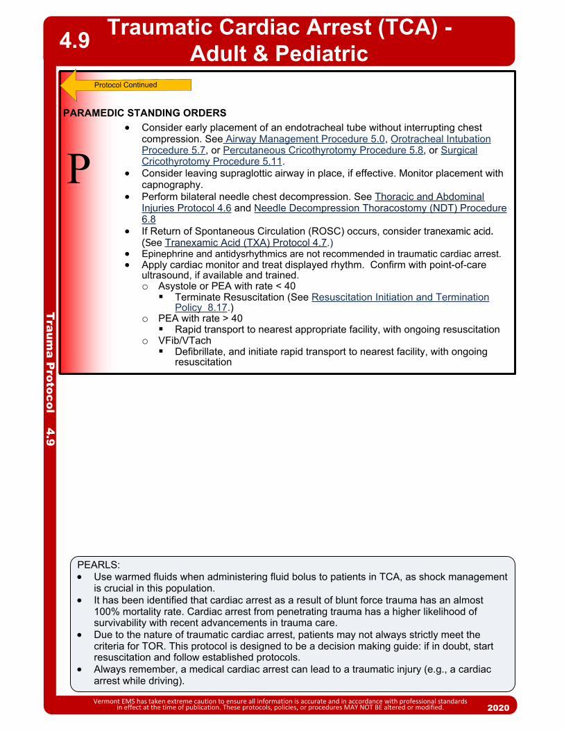
Traumatic Cardiac Arrest (TCA) - Adult & Pediatric
Vermont EMS has taken extreme caution to ensure all information is accurate and in accordance with professional standards in effect at the time of publication. These protocols, policies, or procedures MAY NOT BE altered or modified. 2020
PEARLS: Use warmed fluids when administering fluid bolus to patients in TCA, as shock management
is crucial in this population. It has been identified that cardiac arrest as a result of blunt force trauma has an almost
100% mortality rate. Cardiac arrest from penetrating trauma has a higher likelihood of survivability with recent advancements in trauma care.
Due to the nature of traumatic cardiac arrest, patients may not always strictly meet the criteria for TOR. This protocol is designed to be a decision making guide: if in doubt, start resuscitation and follow established protocols.
Always remember, a medical cardiac arrest can lead to a traumatic injury (e.g., a cardiac arrest while driving).
4.9
Traum
a Protocol 4.9
P Consider early placement of an endotracheal tube without interrupting chest
compression. See Airway Management Procedure 5.0, Orotracheal Intubation Procedure 5.7, or Percutaneous Cricothyrotomy Procedure 5.8, or Surgical Cricothyrotomy Procedure 5.11.
Consider leaving supraglottic airway in place, if effective. Monitor placement with capnography.
Perform bilateral needle chest decompression. See Thoracic and Abdominal Injuries Protocol 4.6 and Needle Decompression Thoracostomy (NDT) Procedure 6.8
If Return of Spontaneous Circulation (ROSC) occurs, consider tranexamic acid. (See Tranexamic Acid (TXA) Protocol 4.7.)
Epinephrine and antidysrhythmics are not recommended in traumatic cardiac arrest. Apply cardiac monitor and treat displayed rhythm. Confirm with point-of-care
ultrasound, if available and trained.o Asystole or PEA with rate < 40Terminate Resuscitation (See Resuscitation Initiation and Termination
Policy 8.17.)o PEA with rate > 40Rapid transport to nearest appropriate facility, with ongoing resuscitation
o VFib/VTachDefibrillate, and initiate rapid transport to nearest facility, with ongoing
resuscitation
PARAMEDIC STANDING ORDERS

2013
Protocol Continues
Traum
a Protocol 4.10
Traumatic EmergenciesMulti-System & General Trauma Guidelines
Vermont EMS has taken extreme caution to ensure all information is accurate and in accordance with professional standards in effect at the time of publication. These protocols, policies, or procedures MAY NOT BE altered or modified.
4.10
2020
Differential (Life Threatening) Chest: pneumothorax
(hemo- / tension-), flail chest, cardiac tamponade, open chest wound
Spine fractures / spinal cord injury
Intra-abdominal bleeding Pelvic / femur fracture Head injury Foreign body airway
obstruction / laryngeal fracture
Hypothermia, hyperthermia, environmental exposure
Relevant Signs and Symptoms Hypotension or shock Hypoxia Cardiac or respiratory
arrest Pain and swelling Deformity, lesions,
bleeding Altered mental status or
unconsciousness
Important History Elements Time and mechanism of
injury Damage to structure or
vehicle Patient location in structure
or vehicle Others dead or injured Speed and details of traffic
accident Restraints and protective
equipment present or absent
SAMPLE
STANDING ORDERS – ADULT & PEDIATRIC
Routine Patient Care Perform advanced spinal assessment (See Advanced Spinal Assessment Procedure 6.0) to
determine if patient requires spinal motion restriction. During primary survey, providers should follow the MARCH algorithm, continually reassessing
patient status: M: Massive Hemorrhage: control of life threatening bleeding is key in traumatic injury,
see Shock Protocol - Adult 2.22A & Shock Protocol - Pediatric 2.22P, Tourniquet & Hemostatic Agent Procedure - Adult & Pediatric 6.11. if suspected pelvic instability, apply pelvic binder.
A: Airway Control: if the patient is unable to maintain their own airway, insert an adjunct or advanced airway (See Airway Management Procedure - 5.0, Airway Management Protocol – Adult 5.1A & Airway Management Protocol – Pediatric 5.1P).
R: Respiratory Support: ensure adequate ventilatory status is attained and SpO2 is kept above 94% using a bag valve mask or other appropriate ventilatory assistance.
C: Circulation: assess adequate circulation and perfusion, and treat for shock (See Shock Protocol - Adult 2.22A & Shock Protocol - Pediatric 2.22P)
H: Hypothermia: ensure patient is kept warm during transport and shock treatment through use of bulky blankets, warmed IV fluids, and active warming as indicated.
Obtain baseline vital signs and level of consciousness (A/V/P/U):
IF NORMAL FINDINGS ABOVE
EA
P//
Routine patient care. Complete detailed secondary assessment. Treat disability: splint any suspected fractures, consider pelvic binding and treatment of any hemorrhage, evaluate for presence of traumatic brain injury.
Transport to appropriate destination using Trauma Triage & Transport Decision Policy - 8.20.
Continually reassess for changes in patient status.
EMT/ADVANCED EMT/PARAMEDIC STANDING ORDERS – ADULT & PEDIATRIC

Traumatic EmergenciesMulti-System & General Trauma Guidelines
Vermont EMS has taken extreme caution to ensure all information is accurate and in accordance with professional standards in effect at the time of publication. These protocols, policies, or procedures MAY NOT BE altered or modified. 2020
PEARLS: Scene time should not be delayed for procedures and interventions which can be
performed en route. BVM ventilation is an acceptable method of airway management if pulse oximetry can be
maintained above 94%. Rapid transport destination determination and notification of the receiving facility in the
event of a “Trauma Alert” are critical for severely-injured patients. Geriatric patients should be evaluated with a high index of suspicion, as age related factors
may reduce their ability to sense pain, and their ability to compensate effectively in traumatic injury.
Mechanism is the most reliable indicator of serious injury in many settings. TXA in multi-system trauma must be given within 3 hours of injury. Rapid bolus
administration of TXA can cause hypotension, so care should be taken in administration.
4.10
Traum
a Protocol 4.10
IF ABNORMAL FINDINGS ABOVE
Obtain IV/IO access Consider administration of warmed 0.9% normal saline bolus to treat hypovolemic
shock: o Adult: 500 mL bolus to maintain a MAP > 65 mmHg (systolic > 90 mmHg) o Pediatric: 20 mL/kg bolus to improve clinical condition (capillary refill time ≤ 2
seconds, equal peripheral and distal pulses, improved mental status, normal breathing).
o Total volume administered should not exceed 2,000 mL for adults, and 60 mL/kg for pediatric patients.
See Shock Protocol – Adult 2.22A or Shock Protocol – Pediatric 2.22P. Consider an antiemetic for nausea/vomiting.
Consider chest decompression if tension pneumothorax is present. (See Needle Decompression Thorocostomy (NDT) Procedure 6.8.)
Consider use of TXA if significant hemorrhage is present. (See Tranexamic Acid (TXA) Protocol – Adult 4.7.)
Consider pain management (see Pain Management Protocol – Adult 2.18A) as indicated.
If MAP remains < 65 mmHg following fluid administration, consider use of vasopressors (see Shock Protocol – Adult 2.22A or Shock Protocol – Pediatric 2.22P.)
Perform point-of-care ultrasound E-FAST exam if available and credentialed.
A
P
Notify receiving hospital as soon as reasonably possible for patients that meet “Trauma Alert” criteria.
Limit scene time to <10 minutes; provide early notification to receiving facility. Rapidly transport to appropriate destination using Trauma Triage & Transport
Decision Policy – 8.20.
Routine Patient Care. Treat disability: splint any suspected fractures, consider pelvic binding and
treatment of any hemorrhage, evaluate for presence of traumatic brain injury. Consider treatment for hypothermia in shock management.E
EMT STANDING ORDERS – ADULT & PEDIATRIC
ADVANCED EMT STANDING ORDERS – ADULT & PEDIATRIC
PARAMEDIC STANDING ORDERS – ADULT & PEDIATRIC

20132013
ASSESSMENT
Each patient presents unique problems that cannot be fully outlined in any algorithm. As such, the provider must rely on thorough assessment techniques and consider each of the following:
Airway Patency: Assess for airway obstruction or risk of impending obstruction due to facial injuries, mass, foreign body, swelling, etc. Assess for presence/absence of gag reflex.
Ventilatory Status: Assess for adequate respiratory effort and impending fatigue/failure/apnea. Assess for accessory muscle use, tripod positioning, the ability of the patient to speak in full sentences. If available, assess waveform capnography.
Oxygenation: Any oxygen saturation <90% represents relatively severe hypoxia and should be considered an important warning sign. In addition to oxygen saturation, assess for cyanosis.
Airway Anatomy: Before attempting airway maneuvers or endotracheal intubation, especially with the use of RSI, assess patient anatomy to predict the probability of success and the need for backup device or technique.
First, assess for difficulty of mask seal. Patients with facial hair, facial fractures, obesity, extremes of age, and pathologically stiff lungs (COPD, acute respiratory distress syndrome, etc.) may require special mask techniques or alternatives.
Next assess for difficulty of intubation. Patients with a short neck, the inability to open their mouth at least three finger widths (or other oral issues such as a large tongue or high arched palate), less than three finger-widths of thyromental distance (or a receding jaw), reduced atlanto-occipital movement (such as in suspected c-spine injury), obesity or evidence of obstruction (such as drooling or stridor) may be difficult to intubate.
DEVISE A PLAN
1. Each patient will present unique challenges to airway management. Therefore, before any intervention is attempted, the provider should contemplate a plan of action that addresses the needs of the patient, and anticipates complications and how to manage them.
2. Airway management is a continuum of interventions, not an “all or none” treatment. Frequently patients may only need airway positioning or a nasal or oral airway to achieve adequate ventilation and oxygenation. Others will require more invasive procedures. The provider should choose the least invasive method that can be employed to achieve adequate ventilation and oxygenation.
3. Continually reassess the efficacy of the plan and change the plan of action as the patient’s needs dictate.
4. In children, a graded approach to airway management is recommended. Basic airway maneuvers and basic adjuncts followed by bag-valve-mask (BVM) ventilation are usually effective.
Procedure Continues
Airw
ay Procedure 5.0
Airway Management
Vermont EMS has taken extreme caution to ensure all information is accurate and in accordance with professional standards in effect at the time of publication. These protocols, policies, or procedures MAY NOT BE altered or modified. 2020
5.0

BASIC SKILLS
Mastery of basic airway skills is paramount to the successful management of a patient with respiratory compromise. Ensure a patent airway with the use of:
Chin-lift/jaw-thrust Nasal airway Oral airway Suction Removal of foreign body.
Provide ventilation with a bag-valve-mask (BVM). Using a PEEP valve set at 5 – 15 cmH2O is recommended. Proper use of the BVM includes appropriate mask selection and head positioning so sternal notch and ear are at the same level, to ensure a good seal. Elevate the stretcher to at least 30º when appropriate. If possible, utilization of the BVM is best accomplished with two people: one person uses both hands to seal the mask and position the airway, while the other person provides ventilation, until chest rise. If the patient has some respiratory effort; synchronize ventilations with the patient’s own inhalation effort.
ADVANCED AIRWAY SKILLS
Only after basic procedures are deemed inappropriate or have proven to be inadequate should more advanced methods be used. Procedures documenting the use of each device/technique listed below are found elsewhere in this manual.
ETT: The endotracheal tube was once considered the optimal method or “gold standard” for airway management. It is now clear, however, that the incidence of complications is unacceptably high when intubation is performed by inexperienced providers or monitoring of tube placement is inadequate. The optimal method for managing an airway will, therefore, vary based on provider experience, emergency medical services (EMS) or healthcare system characteristics, and the patient’s condition.
Bougie: All providers who attempt ETT placement should become intimately familiar with the use of a Bougie. It is the device used most often by anesthesiologists and emergency physicians for helping guide placement when a difficult airway is encountered.
Supraglottic Airways: Utilization of supraglottic airways is an acceptable alternative to endotracheal intubation as both a primary device or a back-up device when previous attempt(s) at ETT placement have failed. Each device has its own set of advantages/disadvantages and requires a unique insertion technique. Providers should have access to, and intimate knowledge of, at least one supraglottic airway. Examples include: King LT i-gel LMA
NIV: Non-invasive ventilation with continuous positive airway pressure (CPAP) or high-flow nasal cannula has been shown to be effective in reducing the need for intubation and in decreasing mortality in properly-selected patients with acute respiratory distress.
Airw
ay Procedure 5.0
Airway Management
Vermont EMS has taken extreme caution to ensure all information is accurate and in accordance with professional standards in effect at the time of publication. These protocols, policies, or procedures MAY NOT BE altered or modified. 2020
5.0
Procedure Continues

Airw
ay Procedure 5.0
Airway Management
Vermont EMS has taken extreme caution to ensure all information is accurate and in accordance with professional standards in effect at the time of publication. These protocols, policies, or procedures MAY NOT BE altered or modified. 2020
5.0
Classifications for Oropharyngeal and Laryngoscopy Views
DOCUMENTATION
All efforts toward airway management should be clearly documented and, at the minimum, should include the following: Pre/post intervention vital signs including oxygen saturation as well as capnography (if
available). Procedures performed/attempted, including number of failed attempts and who performed
each attempt/procedure. Size of device(s) placed, depth of placement (if applicable). Placement confirmation: methods should include auscultation, symmetrical chest wall rise,
and waveform capnography, if available.

P
E
EMT STANDING ORDERS
PARAMEDIC STANDING ORDERS
ADVANCED EMT STANDING ORDERS
A
2013
Routine Patient Care. Establish airway patency.
o Open and maintain the airway.o Suctioning as needed.o Clear foreign body obstructions.
Administer oxygen as appropriate with a target of achieving 94 – 98% saturation. Consider inserting an oropharyngeal or nasopharyngeal airway adjunct. If patient has a tracheostomy tube, see Tracheostomy Care Procedure – Adult &
Pediatric 5.12. For apnea or hypoventilation and decreased level of consciousness with possible
narcotic overdose, administer naloxone. See Poisoning/Substance Abuse/Overdose Protocol – Adult 2.19A.
Assist ventilations with a bag-valve-mask device and supplemental oxygen as needed.
In cardiac arrest, consider insertion of a supraglottic airway. See Supraglottic Airway Procedure – Adult & Pediatric 5.10.
For adults in severe respiratory distress secondary to pulmonary edema, COPD, asthma, pneumonia, near drowning or undifferentiated respiratory distress, consider use of CPAP. See Continuous Positive Airway Pressure (CPAP) Procedure 5.3.
Consider high-flow nasal canula if appropriate administration system is available.
For impending respiratory failure with intact gag reflex or trismus: consider nasotracheal intubation. See Nasotracheal Intubation Procedure 5.6.
For apnea/respiratory failure or impending respiratory failure with impaired or absent gag reflex: consider supraglottic airway device or orotracheal intubation. See Orotracheal Intubation Procedure 5.7, Supraglottic Airway Procedure – Adult & Pediatric 5.10.
For adults with severe airway compromise where respiratory arrest is imminent and other methods of airway management are ineffective: consider Rapid Sequence Intubation. See Rapid Sequence Intubation (RSI) Protocol 7.2.o Note: This procedure is only to be used by paramedics who are
trained and credentialed to perform RSI in accordance with local Medical Direction policy and actively enrolled in an approved Vermont EMS RSI Program.
If feasible, place an orogastric tube to decompress the stomach.
Airw
ay Protocol 5.1A
Airway Management – Adult
Vermont EMS has taken extreme caution to ensure all information is accurate and in accordance with professional standards in effect at the time of publication. These protocols, policies, or procedures MAY NOT BE altered or modified. 2020
5.1A

2013
P
EMT STANDING ORDERS
PARAMEDIC STANDING ORDERS
2013
Routine Patient Care. Establish airway patency.
o Open and maintain airway.o Suction as needed.o Clear foreign body obstructions.o Consider inserting an oropharyngeal or nasopharyngeal airway adjunct.
Administer oxygen as appropriate with a target of achieving 94 – 98% saturation. If patient has a tracheostomy tube see Tracheostomy Care Procedure – Adult & Pediatric
5.12. For respiratory distress:
o Administer high concentration oxygen (preferably humidified) via mask positioned on face or if child resists, held near face.
o Administer oxygen as appropriate with a target of achieving 94 – 98%.o For children with chronic lung disease or congenital heart disease, ask caregivers
about patient’s history, including home oxygen level or patient’s target oxygen saturation. Maintain target saturation, and contact Medical Direction to discuss oxygenation and appropriate transport destination.Note: Pulse oximetry is difficult to obtain in children. Do not rely exclusively on pulse oximetry. If child continues to exhibit signs of respiratory distress despite high oxygen saturation levels, continue oxygen administration.
For respiratory failure or for distress that does not improve with oxygen administration:o Assist ventilations with BVM at rate appropriate for child’s age. Reference Pediatric
Color Coded Appendix A2. o If unable to maintain an open airway through positioning, consider placing an
oropharyngeal or nasopharyngeal airway. Determine if child’s respiratory distress/failure is caused by a preexisting condition
o For Allergic Reaction/Anaphylaxis, refer to the Allergic Reaction/Anaphylaxis Protocol – Pediatric 2.2P.
o For Asthma/Reactive Airway Disease/Croup, refer to the Asthma/Bronchiolitis/RAD/Croup Protocol – Pediatric 2.4P.
Consider high-flow nasal canula if appropriate administration system is available. Consider an advanced airway if airway cannot be maintained through positioning. Prolonged transport time alone should not warrant more invasive interventions. See CPAP Protocol 5.3, Orotracheal Intubation Procedure 5.7 or Supraglottic Airway
Procedure – Adult & Pediatric 5.10). If feasible, place an orogastric tube to decompress stomach.
RESPIRATORY FAILURE: Sleepy, intermittently combative or agitated Respiratory rate < 10 breaths per minute Absent or shallow respirations with poor air
movement Severe intercostal retractions Paradoxical breathing Limp muscle tone Inability to sit up Cyanosis and/or mottled skin Bradycardia
E
A
Airw
ay Protocol 5.1P
Airway Management – Pediatric
Vermont EMS has taken extreme caution to ensure all information is accurate and in accordance with professional standards in effect at the time of publication. These protocols, policies, or procedures MAY NOT BE altered or modified. 2020
5.1P
RESPIRATORY DISTRESS: Alert, irritable, anxious Stridor Audible wheezing/grunting Respiratory rate outside normal range for child’s age Sniffing position Nasal flaring Head bobbing Neck muscle use Intercostal retractions Central cyanosis that resolves with oxygen administration Mild tachycardia
ADVANCED EMT STANDING ORDERS For respiratory distress, consider CPAP. See CPAP Protocol 5.3). For respiratory failure, use most appropriate/least invasive method. BVM ventilation is the preferred method of ventilation for pediatric population. However,
if unsuccessful, consider placement of supraglottic airway. o In cardiac arrest: consider insertion of a supraglottic airway. See Supraglottic
Airway Procedure – Adult & Pediatric 5.10.

20132013
Automated Transport Ventilator
Vermont EMS has taken extreme caution to ensure all information is accurate and in accordance with professional standards in effect at the time of publication. These protocols, policies, or procedures MAY NOT BE altered or modified. 2020
Airw
ay Procedure 5.2
5.2
AP/
MALE
Height in Ft/In
5' 0"
5' 1"
5' 2"
5' 3"
5' 4"
5' 5"
5' 6"
5' 7"
5' 8"
5' 9"
5' 10"
5' 11"
6' 0"
6 mL/kg
273
287
301
314
328
342
356
370
383
397
411
425
439
8 mL/kg
364
382
401
419
438
456
474
493
511
530
548
566
585
6' 1" 452 603
FEMALE
Height in Ft/In
5' 0"
5' 1"
5' 2"
5' 3"
5' 4"
5' 5"
5' 6"
5' 7"
5' 8"
5' 9"
5' 10"
5' 11"
6' 0"
6 mL/kg
300
314
328
341
355
369
383
397
410
424
438
452
466
8 mL/kg
400
418
437
455
474
492
510
529
547
566
584
602
621
6' 1" 479 639
Procedure Continues
ADVANCED EMT/PARAMEDIC STANDING ORDERSINDICATIONS Resuscitative efforts:
o Can only adjust rate, tidal volume, and adult vs. child setting if applicable. Any patient requiring ventilatory assistance in conjunction with advanced
airway adjuncts. Any patient requiring ventilatory assistance in conjunction with basic airway
maintenance. Any patient requiring ventilatory assistance in conjunction with manual airway
maintenance.
CONTRAINDICATIONS Airway obstruction Resistance Poor lung compliance Pneumothorax – tension pneumothorax Pulmonary over-pressurization (blast injury, water ascent injury, etc.) Children less than 5 years of age or 16 kg (35 lbs). Check manufacturer’s
recommendations.
PROCEDURES1. Determine that a need for the automated transport ventilator (ATV) exists. Follow manufacturer’s instructions for the device.2. Assure that all tubing is free from kinks.3. Determine the proper tidal volume setting. This is done by determining the patient’s ideal weight (approx. weight for any physically fit patient having the same sex, height, frame) and multiplying it by 6 – 8 mL/kg. Begin with the lowest tidal volume limit.

Automated Transport VentilatorA
irway P
rocedure 5.2
Vermont EMS has taken extreme caution to ensure all information is accurate and in accordance with professional standards in effect at the time of publication. These protocols, policies, or procedures MAY NOT BE altered or modified. 2020
5.2
PROCEDURES (continued)4. Set Breaths per Minute (BPM) control to rate of 8 – 15 per minute.5. Check alarm by occluding the patient valve assembly outlet. The audible pressure limit alarm should sound as the ventilator cycles through the delivery phase.6. Assess lung compliance and chest rise with a bag valve device. Tidal volume may be adjusted lower if poor lung compliance is found.7. Attach the patient valve assembly to the airway device or mask used on the patient.8. Assess ventilation. Listen for bilateral lung sounds. Observe for proper chest rise. Chest rise should be symmetrical and patient condition should improve.9. Count the number of complete ventilator cycles for a full minute. The number should be the same as the setting (+/- 1).10. Assess and manage the airway as you normally would for any patient with controlled ventilation.11. If spontaneous breathing begins, it may be desirable to turn the BPM down as long as patient's spontaneous rate is 10 – 12 per minute. 12. Check oxygen cylinder pressure level frequently. This device will deplete a "D" cylinder rapidly.
SPECIAL CONSIDERATIONS Due to COPD, chest rise may not appear full Do not increase tidal volume
(TV) past upper TV limit. If lung sounds are absent or on one side only: rule out airway obstruction,
improper tube placement, or pneumothorax, and check tidal volume ml/bpm settings.
If chest expansion is not adequate, the rescuer should slowly increase tidal volume until chest expansion is adequate, or the uppermost limit (for the patient's ideal weight) is reached.
If chest appears to over expand, decrease tidal volume.
AP/
ADVANCED EMT/PARAMEDIC STANDING ORDERS
Procedure Continues

2013
INDICATIONS Spontaneously breathing patient in moderate to severe respiratory distress due to
congestive heart failure/pulmonary edema, asthma/COPD, pneumonia, submersion injury or undifferentiated respiratory distress, concurrent with the following signs and symptoms:o Oxygen saturation < 94%o Retractions or accessory muscle useo Adult respiratory rate > 25 (see chart for pediatric)
CONTRAINDICATIONS Cardiac or respiratory arrest/apnea Unable to follow commands Unable to maintain their own airway Agitated or combative behavior and unable to tolerate mask Vomiting and/or GI bleed Respiratory distress secondary to trauma Suspicion of pneumothorax Facial trauma or impossible face seal Hypotension with MAP < 65 (SBP < 100 mmHg). (Pediatric SBP < 70 + (age in years
x 2))
PROCEDURE1. Ensure adequate oxygen supply for CPAP device.2. Explain procedure to patient. Be prepared to coach patient for claustrophobia or
anxiety.3. Place patient in upright position. Apply pulse oximetry, and capnography.4. Choose appropriate sized device mask for patient, assemble the CPAP device,
attach to oxygen supply and insure oxygen is flowing (follow manufacturers directions for preparation for your particular device).
5. Place mask over face and secure with straps until minimal air leak.6. Adjust pressure to 5 – 15 cm H2O to effect for patient condition.7. Recheck mask for leaks and adjust straps as needed to minimize air leaks.8. Reassure anxious patient. 9. Monitor vital signs and symptoms, pulse oximetry and quantitative waveform
capnography.10. If patient improves, maintain CPAP for duration of transport and notify receiving
hospital to prepare for a CPAP patient.11. If patient begins to deteriorate, discontinue CPAP and assist respirations by BVM. 12. Document CPAP procedure, including time and provider. Document serial pulse oximetry and capnography readings to demonstrate effects.
ADVANCED EMT STANDING ORDERS – ADULT & PEDIATRIC
A
PARAMEDIC MEDICAL DIRECTION ORDERS – ADULT ONLY (CONTACT MEDICAL DIRECTION FOR PEDIATRIC DOSING)
Consider administering anxiolytic. Contact Medical Direction for authorization.o Midazolam 2.5 mg IV/intranasal, may repeat once in 5 minutes OR 5 mg IM may
repeat once in 10 minutes ORo Lorazepam 0.5 – 1 mg IV, may repeat once in 5 minutes OR 1 – 2 mg IM, may
repeat once in 10 minutes ORo Diazepam 5 mg IV (may repeat once in 5 minutes).
PAdminister benzodiazipines with caution in patients with signs of hypercarbia.
Airw
ay Procedure 5.3
Continuous Positive Airway Pressure(CPAP) – Adult & Pediatric
Vermont EMS has taken extreme caution to ensure all information is accurate and in accordance with professional standards in effect at the time of publication. These protocols, policies, or procedures MAY NOT BE altered or modified. 2020
5.3
Age Resp Rate
Infant 0 - 1 year > 60
Toddler 1 - 4 years >40
Preschooler 4 - 6 years >34
School Age 6 - 12 years >30
Teen 13 and older >25
Pediatric Respiratory Distress

2013
INDICATIONS Sudden onset of respiratory distress often with coughing, wheezing, gagging
or stridor due to a foreign-body obstruction of the upper airway.
PROCEDURE Routine Patient Care
o Assess the degree of foreign body obstruction.o Do not interfere with a mild obstruction allowing the patient to clear their
airway by coughing.o In severe foreign-body obstructions, the patient may not be able to make
a sound. The victim may clutch his/her neck in the universal choking sign.
For an infant: Deliver 5 back blows followed by 5 chest thrusts repeatedly until the object is expelled or the victim becomes unresponsive.
For a child: Perform subdiaphragmatic abdominal thrusts (Heimlich Maneuver) until the object is expelled or the victim becomes unresponsive.
For adults: A combination of maneuvers may be required,o First, subdiaphragmatic abdominal thrusts (Heimlich Maneuver) should be
used in rapid sequence until the obstruction is relieved.o If abdominal thrusts are ineffective, chest thrusts should be used. Chest
thrusts should be used primarily in morbidly obese patients and in patients who are in the late stages of pregnancy.
If the victim becomes unresponsive, begin CPR immediately but look in the mouth before administering any ventilations. If a foreign-body is visible, remove.
Do not perform blind finger sweeps in the mouth and posterior pharynx. This may push the object farther into the airway.
EMT/ADVANCED EMT STANDING ORDERS
PARAMEDIC STANDING ORDERS
If basic foreign body airway maneuvers fail, consider any of the following:o In unresponsive patients, visualize the posterior pharynx with a laryngoscope
to potentially identify and remove the foreign-body using Magill forceps.o If obstruction is secondary to trauma or edema, or if uncontrollable bleeding
into the airway causes life-threatening ventilation impairment, perform endotracheal intubation. See Orotracheal Intubation Procedure 5.7.
o Consider forced right mainstem intubation (with pullback) to allow for ventilation of left lung in the extreme event of lower tracheal foreign body obstruction and inability to ventilate.
o If unable to intubate and the patient cannot be adequately ventilated by other means, perform cricothyrotomy. See Percutaneous Cricothyrotomy Procedure 5.8 or Surgical Cricothyrotomy 5.11.
P
Foreign-Body Obstruction
Vermont EMS has taken extreme caution to ensure all information is accurate and in accordance with professional standards in effect at the time of publication. These protocols, policies, or procedures MAY NOT BE altered or modified. 2020
5.4
EA/
PEARLSIf air exchange is adequate with a partial airway obstruction, do not interfere; instead, encourage the patient to cough up the obstruction. Continue to monitor the patient for adequacy of air exchange. If air exchange becomes inadequate, continue with the protocol.
Airw
ay Procedure 5.4

20132013
P
INDICATIONS To facilitate routine placement of endotracheal tube. Unable to fully visualize vocal cords during an intubation attempt.
PROCEDURE1. Choose appropriately-sized Bougie: adult, pediatric or neonatal.2. Lubricate Bougie with water-based lubricant.3. Using a laryngoscope (Macintosh or Miller blade) and standard ETT intubation
techniques, attempt to visualize the vocal cords.4. If the vocal cords are partially visualized, pass the Bougie through the cords
while attempting to feel the signs of tracheal placement (see below). The Bougie is advanced until the black line on the Bougie reaches the lip line.
5. If the vocal cords are not visualized, pass the Bougie behind the epiglottis, guiding the tip of the Bougie anteriorly towards the trachea, and assess for signs of tracheal placement (see below).
6. With the laryngoscope still in place, have an assistant load the ETT over the Bougie and slide it to the level of the lip line.
7. Advance the ETT over the Bougie, rotating the ETT about 1/4 turn counterclockwise so that the bevel is oriented vertically as the ETT passes through the vocal cords. This maneuver allows the bevel to gently spread the arytenoids with a minimum of force, thus avoiding injury. If resistance is felt, withdraw the ETT, rotating it in a slightly more counterclockwise direction, and advance the tube again. Advance the tube to a lip-line of 24 cm in an adult male, and 22 cm in an adult female.
8. Holding the ETT firmly in place, have an assistant remove the Bougie. 9. Remove the laryngoscope.10. Inflate the cuff with 5 – 10 mL of air. 11. Follow the procedures outlined in Orotracheal Intubation Procedure 5.7 to
confirm placement, secure the ETT, monitor and document placement of the ETT.
SIGNS OF TRACHEAL PLACEMENT The Bougie is felt to stop or get “caught up” as the airway narrows and is unable
to be advanced further. This is the most reliable sign of proper Bougie placement. If the Bougie enters the esophagus, it will continue to advance without resistance.
It may be possible to feel the tactile sensation of “clicking” as the Bougie tip is advanced downward over the rigid cartilaginous tracheal rings.
The Bougie can be felt to rotate as it enters a mainstem bronchus. Usually it is a clockwise rotation as the Bougie enters the right mainstem bronchus, but occasionally it will rotate counterclockwise if the Bougie enters the left mainstem bronchus.
If the patient is not paralyzed, he/she may cough.
PARAMEDIC STANDING ORDERS – ADULT
Airw
ay Procedure 5.5
Gum Elastic Bougie/Flexguide
Vermont EMS has taken extreme caution to ensure all information is accurate and in accordance with professional standards in effect at the time of publication. These protocols, policies, or procedures MAY NOT BE altered or modified. 2020
5.5

20132013
P
INDICATIONS Impending respiratory failure with intact gag reflex, or jaw is clenched and unable to
be opened in spontaneously breathing patient.
CONTRAINDICATIONS Apnea. Nasal obstruction. Suspected basilar skull fracture. Severe facial trauma or suspected facial fractures. Patient fits on a pediatric length-based resuscitation tape.
PROCEDURE1. Pre-medicate nasal mucosa with 2% lidocaine jelly and vasoconstricting nasal
decongestant spray such as neo-synephrine, if available.
2. Pre-oxygenate the patient.
3. Select the larger and less obstructed nostril and insert a lubricated nasal airway.
4. Lubricate the ETT with water-based lubricant.
5. Remove the nasal airway and gently insert the ETT with continuous waveform capnography monitoring, keeping the bevel toward the septum (a gentle rotation movement may be necessary at the turbinates).
6. Continue to advance the ETT while listening for maximum air movement and watching for capnography waveform. Consider use of BAAM device to aid in listening to airflow.
7. At the point of maximum air movement, indicating proximity to the level of the glottis, gently and evenly advance the tube through the glottic opening on inspiration. If resistance is encountered, the tube may have become lodged into the pyriform
sinus and you may note tenting of the skin on either side of the thyroid cartilage. If this happens, slightly withdraw the ETT and rotate it toward the midline and attempt to advance tube again with the next inspiration.
8. Upon entering the trachea, the tube may cause the patient to cough, buck, strain, or gag. This is normal. Do not remove the ETT. Be prepared to control the cervical spine and be alert for vomiting.
9. Placement depth should be from the nares to the tip of the tube: approximately 28 cm in males and 26 cm in females.
10. Inflate cuff with 5 – 10 mL of air.
11. Confirm appropriate placement by waveform capnography, symmetrical chest-wall rise, auscultation of equal breath sounds over the chest, a lack of epigastric sounds with bagging.
12. Secure the ETT, consider applying a cervical-collar and securing patient to a long backboard (even for the medical patient) to protect the placement of the ETT.
Airw
ay Procedure 5.6
Nasotracheal Intubation
Vermont EMS has taken extreme caution to ensure all information is accurate and in accordance with professional standards in effect at the time of publication. These protocols, policies, or procedures MAY NOT BE altered or modified. 2020
PARAMEDIC STANDING ORDERS – ADULT
Procedure Continues
5.6
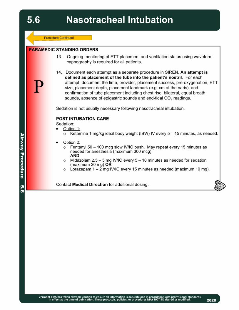
Airw
ay Procedure 5.6
Nasotracheal Intubation
Vermont EMS has taken extreme caution to ensure all information is accurate and in accordance with professional standards in effect at the time of publication. These protocols, policies, or procedures MAY NOT BE altered or modified. 2020
P
13. Ongoing monitoring of ETT placement and ventilation status using waveform capnography is required for all patients.
14. Document each attempt as a separate procedure in SIREN. An attempt is defined as placement of the tube into the patient’s nostril. For each attempt, document the time, provider, placement success, pre-oxygenation, ETT size, placement depth, placement landmark (e.g. cm at the naris), and confirmation of tube placement including chest rise, bilateral, equal breath sounds, absence of epigastric sounds and end-tidal CO2 readings.
Sedation is not usually necessary following nasotracheal intubation.
POST INTUBATION CARE Sedation: Option 1:
o Ketamine 1 mg/kg ideal body weight (IBW) IV every 5 – 15 minutes, as needed.
Option 2:o Fentanyl 50 – 100 mcg slow IV/IO push. May repeat every 15 minutes as
needed for anesthesia (maximum 300 mcg).AND
o Midazolam 2.5 – 5 mg IV/IO every 5 – 10 minutes as needed for sedation (maximum 20 mg) OR
o Lorazepam 1 – 2 mg IV/IO every 15 minutes as needed (maximum 10 mg).
Contact Medical Direction for additional dosing.
PARAMEDIC STANDING ORDERS
5.6

Airw
ay Procedure 5.7
20132013
P
INDICATIONS Apnea/respiratory failure. Impending respiratory failure. Impaired or absent gag
reflex. Only after basic procedures are deemed inappropriate or have proven to be inadequate should more advanced methods be used.
CONTRAINDICATION Epiglottitis. Facial or neck injuries that prohibit visualization of airway anatomy (relative).
CAUTIONS Pediatric patients should, at least initially, be managed with BVM/SGA. Patients with CHF should be managed with trial of CPAP, if possible. Avoid intubating patients with severe TBI and asthmatics, if possible.
PROCEDURE1. Prepare all equipment. Have suction and Bougie ready.
2. Pre-oxygenate the patient with high-concentration oxygen. Three-minute minimum of BVM ventilation or if patient is breathing, assure 8 vital capacity breaths with NRB. Apply continuous nasal cannula at 15 LPM.
3. Assess for airway difficulty based on patient anatomy (e.g., short neck, obesity, decreased thyromental distance and Class III or IV oropharyngeal views on observation). Have fallback plan and equipment ready.
4. Open the patient’s airway. While holding the laryngoscope in the left hand, insert the blade into the right side of the patient’s mouth, sweeping the tongue to the left. Use video laryngoscopy, if available and trained.
5. Use the blade to lift the tongue and the epiglottis, either directly with the straight (Miller) blade, or indirectly with the curved (Macintosh) blade.
6. Once the glottic opening is visualized, insert the tube through the vocal cords and continue to visualize while passing the cuff through the cords.
7. Remove the laryngoscope and then the stylet from the ETT.
8. Inflate the cuff with 5 – 10 mL of air.
9. Confirm appropriate proper placement with waveform capnography and also document symmetrical chest-wall rise, auscultation of equal breath sounds over the chest and a lack of epigastric sounds with ventilations using bag-valve-mask
10. Secure the ETT with a commercial device. Consider applying a cervical collar to minimize head and neck motion during movement and transport.
11. Reassess tube placement frequently, especially after movement of the patient.
12. Ongoing monitoring of ETT placement and ventilation status using waveform capnography is required for all patients.
13. Document each attempt as a separate procedure so it can be time stamped in the PCR. An attempt is defined as placement of the blade into the patient’s mouth. For each attempt, document the time, provider, placement success, pre-oxygenation, airway grade, ETT size, placement depth, placement landmark (e.g. cm at the patient’s lip), and confirmation of tube placement including chest rise, bilateral, equal breath sounds, absence of epigastric sounds and end-tidal CO2 readings.
PARAMEDIC STANDING ORDERS – ADULT & PEDIATRIC
Orotracheal Intubation
Vermont EMS has taken extreme caution to ensure all information is accurate and in accordance with professional standards in effect at the time of publication. These protocols, policies, or procedures MAY NOT BE altered or modified. 2020
Procedure Continues
5.7

Airw
ay Procedure 5.7
Orotracheal Intubation
Vermont EMS has taken extreme caution to ensure all information is accurate and in accordance with professional standards in effect at the time of publication. These protocols, policies, or procedures MAY NOT BE altered or modified. 2020
If intubation attempt is unsuccessful, ETT placement cannot be verified or ETTbecomes dislodged: Remove tube, monitor oxygen saturation and end-tidal CO2 AND Ventilate the patient with 100% oxygen via a BVM until ready to attempt intubation
again.
If continued intubation attempts are unsuccessful (maximum of 3 attempts for cardiac arrest) or BVM ventilation is not adequate, consider placing a supraglottic airway. For a viable patient whose airway cannot be successfully managed by any other means, consider Percutaneous Cricothyrotomy Procedure 5.8 or Surgical Cricothyrotomy Procedure 5.11.
POST INTUBATION CARESedation: Option 1:
o Ketamine 1 mg/kg ideal body weight (IBW) IV every 5 – 15 minutes, asneeded.
Option 2:o Fentanyl 50 – 100 mcg (pediatric dose 1 mcg/kg) slow IV/IO push. May
repeat every 15 minutes as needed for anesthesia (maximum 300 mcg).AND
o Midazolam 2.5 – 5 mg IV/IO (pediatric dose 0.1 mg/kg IV/IO) every 5 – 10minutes as needed for sedation (maximum 20 mg) OR
o Lorazepam 1 – 2 mg IV/IO (pediatric dose 0.1 mg/kg IV/IO) every 15minutes as needed (maximum 10 mg).
Contact Medical Direction for additional dosing.
Video-Laryngoscope: If available and trained, use video laryngoscope instead of manual laryngoscope. Video-laryngoscopy has been shown to have better success rates than manual laryngoscopy and should be used if available.
PARAMEDIC STANDING ORDERS – ADULT & PEDIATRIC
5.7
Classifications of Oropharyngeal and Laryngoscopy Views

Airw
ay Procedure 5.8
20132013
P
INDICATIONS Failed airway: Patient whose airway cannot be successfully managed by any
other means. o All other methods have been exhausted including BVM, blind airway
device, and intubation attempts;o Massive mid-face trauma precluding use of BVM, obstruction, trismus
(clenching);o Inability to control the airway using less invasive measures;o Last Resort: All other airway management techniques have failed. Unable
to ventilate or oxygenate patient.
PROCEDURE1. Can use Rusch QuickTrach or other approved device. Choose appropriate sized device.2. Pre-oxygenate patient when possible.3. Assemble all available additional personnel.4. Locate cricothyroid membrane at the inferior portion of the thyroid cartilage (with head in neutral position, membrane is approximately 3 finger widths above the sternal notch). May be difficult to locate in obese patients.5. Hold skin taut over membrane and locate the midline.6. Prep area, preferably with betadine.7. Hold the needle bevel up at a 90-degree angle, aimed inferiorly as you approach the skin.8. Puncture the skin with the needle and continue with firm, steady pressure while aspirating for air with the syringe.9. As soon as air is aspirated freely, stop advancing the needle/airway assembly.10. Modify the angle to 60 degrees from the head and advance to level of the stopper.11. Remove the stopper while holding the needle/airway assembly firmly in place. 12. Do not advance the needle further. (NOTE: if the patient is obese and no air can be aspirated with the stopper in place, you may remove the stopper and continue advancing until air is aspirated. Be aware that without the stopper, risk of perforating the posterior aspect of the trachea is greatly increased.)13. Hold the needle and syringe firmly and slide only the plastic cannula along the needle into the trachea until the flange rests on the neck. Carefully remove the needle and syringe.14. Secure the cannula with the neck strap.15. Apply the EtCO2 detector.16. Confirm placement with the use of breath sounds, pulse ox, EtCO2 and waveform capnography.17. Ensure 100% FiO2 to BVM via supplemental 02.
PARAMEDIC STANDING ORDERS
Percutaneous Cricothyrotomy
Vermont EMS has taken extreme caution to ensure all information is accurate and in accordance with professional standards in effect at the time of publication. These protocols, policies, or procedures MAY NOT BE altered or modified. 2020
5.8

ADVANCED EMT/PARAMEDIC STANDING ORDERS
2013
INDICATIONS Obstruction of the airway (secondary to secretions, blood, and/or any other
substance) in a patient currently being assisted by an inserted airway such as an endotracheal tube, supraglottic airway or tracheostomy tube.
CONTRAINDICATIONS None.
PROCEDURE
1. Ensure the suction device is operable.
2. Pre-oxygenate the patient.
3. While maintaining aseptic technique, attach the suction catheter to the suction
unit.
4. If applicable, remove ventilation device from the airway.
5. Insert the sterile end of the suction catheter into the tube without suction.
Insert to proper depth so that suction catheter does not extend past the tube/
device.6. Once the desired depth is met, apply suction by occluding the port of the
suction catheter and slowly remove the catheter from the tube using a twisting motion.
7. Suctioning duration should not exceed 10 seconds, using lowest pressure that effectively removes secretions.
8. Saline flush may be used to help loosen secretions and facilitate suctioning.
9. Re-attach the ventilation device to the patient.
AP/
Suctioning of Inserted Airway
Vermont EMS has taken extreme caution to ensure all information is accurate and in accordance with professional standards in effect at the time of publication. These protocols, policies, or procedures MAY NOT BE altered or modified. 2020
Airw
ay Procedure 5.9
5.9

2013
P
INDICATIONS:
• Inability to adequately ventilate a patient with a bag-valve-mask or longer EMS
transports requiring a more definitive airway.
• Back up device for failed endotracheal intubation attempt.
POST TUBE PLACEMENT CARE – ADULT AND PEDIATRIC
Sedation:
• Option 1:
o Ketamine 1 mg/kg ideal body weight (IBW) IV every 5 – 15 minutes, as needed.
• Option 2:
o Fentanyl 50 – 100 mcg (pediatric dose 1 mcg/kg) slow IV/IO push. May
repeat every 15 minutes as needed for anesthesia (maximum 300 mcg).
AND
o Midazolam 2.5 – 5 mg IV/IO (pediatric dose 0.1 mg/kg IV/IO) every 5 – 10
minutes as needed for sedation (maximum 20 mg) OR
o Lorazepam 1 – 2 mg IV/IO (pediatric dose 0.1 mg/kg IV/IO) every 15 minutes
as needed (maximum 10 mg).
Contact Medical Direction for additional dosing.
2013
AA
Airw
ay P
roc
ed
ure
5.1
0
Supraglottic Airway
Adult & Pediatric
Vermont EMS has taken extreme caution to ensure all information is accurate and in accordance with professional standards in effect at the time of publication. These protocols, policies, or procedures MAY NOT BE altered or modified. 2020
5.10
This protocol applies to commercially available supraglottic airway devices. These airways must be used as directed by the manufacturer’s guidelines. They may be used in all age groups for which the devices are designed. Providers must be trained on and competent with the airway device they will be using. Note: Double Lumen Devices (e.g., Combitube) are no longer approved.
PARAMEDIC STANDING ORDERS
INDICATIONS:
• Cardiac Arrest.
RELATIVE CONTRAINDICATIONS:
• Severe maxillofacial or oral trauma.
• For devices inserted into the esophagus:
o The patient has known esophageal disease.
o The patient has ingested a caustic substance.
o The patient has burns involving the airway.
PROCEDURE:
• Insertion procedure should follow manufacturer guidelines as each device is
unique.
• Confirm appropriate placement by symmetrical chest-wall rise, auscultation of
equal breath sounds over the chest and a lack of epigastric sounds with bag valve
mask ventilation, and waveform capnography, if available.
• Secure the device.
• Document the time, provider, provider level and success for the procedure.
Complete all applicable airway confirmation fields including chest rise, bilateral,
equal breath sounds, absence of epigastric sounds and end-tidal CO2 readings.
• Reassess placement frequently, especially after patient movement.
ADVANCED EMT STANDING ORDERS

Airw
ay Procedure 5.11
20132013
P
INDICATIONSThe following protocol is an effort to maintain awareness of a procedure that occurs rarely in the Vermont EMS system. Ultimately, this procedure should be practiced on a regular basis to maintain proficiency. PLEASE NOTE: This protocol describes a generally acceptable open cricothyrotomy and will differ from services that employ percutaneous kits. The inclusion of this protocol DOES NOT mandate that all providers use this exact procedural description. Providers should employ the procedure they are trained, practiced and most comfortable with. This protocol is one of multiple acceptable means to perform this procedure. Regardless of the procedure type, it is essential that Vermont paramedics are familiar with the available supplies and materials.
The primary indication for surgical cricothyrotomy is failure to maintain the airway (including oxygenation, ventilation, and protection) through other less invasive means in a patient 8 years or older with palpable landmarks. In the Vermont EMS experience, this has occurred predominantly in trauma cases with disruption of the face and normal airway anatomy and in choking patients. Even in some dramatic facial injuries, the airway may be adequately managed by sitting the patient up and leaning forward, as long as the other injuries allow.
MATERIALS/EQUIPMENT1. Cuffed tracheostomy tube or 6.0 - 7.0 ETT 2. Tracheal hook or bougie 3. Trousseau dilator (if available) 4. Syringe to inflate cuff 5. Scalpel6. Swabs/skin prep
PARAMEDIC STANDING ORDERS
Surgical Cricothyrotomy
Vermont EMS has taken extreme caution to ensure all information is accurate and in accordance with professional standards in effect at the time of publication. These protocols, policies, or procedures MAY NOT BE altered or modified. 2020
5.11
Protocol Continues

Airw
ay Procedure 5.11
20132013
P
PROCEDURE1. Extend the neck when possible to ensure best access to the trachea. Swab/cleanse the area. 2. Immobilize the trachea with your non-dominant thumb and middle finger while palpating the cricothyroid membrane with your non-dominant index finger. NOTE: The cricothyroid membrane is immediately BELOW the thyroid cartilage. 3. Make a 3 – 5 cm vertical incision over the cricothyroid membrane through the skin and subcutaneous tissues. NOTE: Severe bleeding is possible with this procedure and may occur at this or the following steps. Be prepared to suction and provide direct pressure to control bleeding. 4. Palpate the membrane through the incision to confirm anatomy. 5. Make a small (1 cm or less) incision horizontally through the cricothyroid membrane. 6. Insert the tracheal hook or bougie in the opening of the membrane and rotate toward the head while maintaining hold of the thyroid cartilage with your non-dominant hand. 7. If Trousseau dilator available, insert into the incision site and spread vertical then rotate 90 degrees until the dilator is parallel with the neck. 8. Insert the cuffed tracheostomy tube or ETT tube into the incision site. Advance until the flanges rest on the skin of the neck (when using tracheostomy tube). 9. Carefully remove the dilator (if used), tracheal hook and obturator of the tracheostomy tube. 10. Inflate the balloon of the tracheostomy tube/ETT. 11. Ventilate and confirm position by physical exam and ETCO2. 12. Secure the tube in place. 13. Dress incision site.
PARAMEDIC STANDING ORDERS
Surgical Cricothyrotomy
Vermont EMS has taken extreme caution to ensure all information is accurate and in accordance with professional standards in effect at the time of publication. These protocols, policies, or procedures MAY NOT BE altered or modified. 2020
5.11
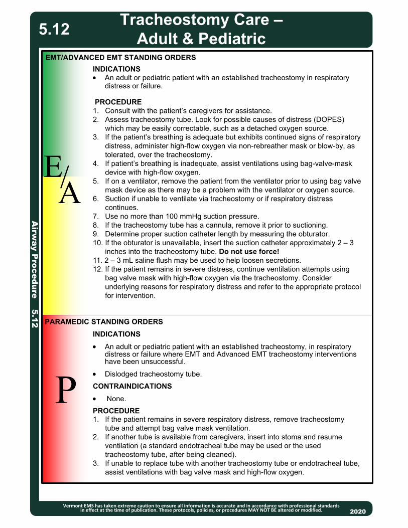
2013
EMT/ADVANCED EMT STANDING ORDERS
EA
P
/
PARAMEDIC STANDING ORDERS
INDICATIONS
An adult or pediatric patient with an established tracheostomy, in respiratory distress or failure where EMT and Advanced EMT tracheostomy interventions have been unsuccessful.
Dislodged tracheostomy tube.
CONTRAINDICATIONS
None.
PROCEDURE1. If the patient remains in severe respiratory distress, remove tracheostomy tube and attempt bag valve mask ventilation. 2. If another tube is available from caregivers, insert into stoma and resume ventilation (a standard endotracheal tube may be used or the used tracheostomy tube, after being cleaned).3. If unable to replace tube with another tracheostomy tube or endotracheal tube, assist ventilations with bag valve mask and high-flow oxygen.
Airw
ay Procedure 5.12
Tracheostomy Care –Adult & Pediatric
Vermont EMS has taken extreme caution to ensure all information is accurate and in accordance with professional standards in effect at the time of publication. These protocols, policies, or procedures MAY NOT BE altered or modified. 2020
5.12
INDICATIONS An adult or pediatric patient with an established tracheostomy in respiratory
distress or failure.
PROCEDURE1. Consult with the patient’s caregivers for assistance.2. Assess tracheostomy tube. Look for possible causes of distress (DOPES) which may be easily correctable, such as a detached oxygen source.3. If the patient’s breathing is adequate but exhibits continued signs of respiratory distress, administer high-flow oxygen via non-rebreather mask or blow-by, as tolerated, over the tracheostomy. 4. If patient’s breathing is inadequate, assist ventilations using bag-valve-mask device with high-flow oxygen. 5. If on a ventilator, remove the patient from the ventilator prior to using bag valve mask device as there may be a problem with the ventilator or oxygen source.6. Suction if unable to ventilate via tracheostomy or if respiratory distress continues. 7. Use no more than 100 mmHg suction pressure. 8. If the tracheostomy tube has a cannula, remove it prior to suctioning. 9. Determine proper suction catheter length by measuring the obturator. 10. If the obturator is unavailable, insert the suction catheter approximately 2 – 3 inches into the tracheostomy tube. Do not use force! 11. 2 – 3 mL saline flush may be used to help loosen secretions.12. If the patient remains in severe distress, continue ventilation attempts using bag valve mask with high-flow oxygen via the tracheostomy. Consider underlying reasons for respiratory distress and refer to the appropriate protocol for intervention.

20132013
PURPOSE: This protocol provides guidance regarding the assessment and care of patients who have a possible spinal injury, utilizing spinal motion restriction. Spinal motion restriction is defined as application of a cervical collar and maintenance of the spine in neutral alignment. Determination that immobilization devices should be used should be made by the highest level EMS provider. All steps of spinal assessment algorithm below must be documented in the PCR.
Spinal Assessment: Spinal motion restriction is required when ANY of the following conditions apply: (See Spinal Motion Restriction Protocol 4.5) Unreliable patient:
o Child who cannot participate in assessment.o Anxious and/or uncooperative.o Communication barriers (e.g., deafness, hard of hearing, language, understanding).o Altered mental status (not alert and oriented x 3).o Evidence of alcohol or drug intoxication.o Distracted by circumstances or injuries to self or others (ie, any other injury capable of
producing significant pain in this patient). Any abnormal neurological function in extremities (check all 4 extremities):
o Numbness or tingling (paresthesia).o Motor strength not full and symmetrical.o Sensation not intact and symmetrical.
Midline tenderness on palpation:o Explain to the patient the actions that you are going to take. Ask the patient to
immediately report any pain, and to answer questions with a “yes” or “no” rather than shaking the head.
o With the patient’s spine supported to limit movement, begin palpation at the base of the skull at the midline of the spine.
o Palpate the vertebrae individually from the base of the skull to the bottom of the sacrum.
o On palpation of each vertebral body, look for evidence of pain and ask the patient if they are experiencing pain. If evidence of pain along the spinal column is encountered, utilize spinal motion restriction.
Pain with movement of neck (cervical flexion, extension and rotation).o If the capable patient is found to be pain free, ask the patient to turn their head first to
one side (so that the chin is pointing toward the shoulder on the same side as the head is rotating) then, if pain free, to the other. If there is evidence of pain, utilize spinal motion restriction.
o With the head rotated back to its normal position, ask the patient to flex and extend their neck. If there is evidence of pain, utilize spinal motion restriction. Do not assist patient in attempts to rotate neck.
Medical P
rocedure 6.0Advanced Spinal Assessment
Vermont EMS has taken extreme caution to ensure all information is accurate and in accordance with professional standards in effect at the time of publication. These protocols, policies, or procedures MAY NOT BE altered or modified. 2020
EMT/ADVANCED EMT/PARAMEDIC STANDING ORDERS
EA
P
//
6.0
High risk mechanisms include: Motor vehicle crash >60 mph, rollover, ejection. Simple low-speed, rear-end MVC can usually be
excluded. (Simple low-speed collision does not include: Being pushed into oncoming traffic, being hit by a bus or large truck, rollover, or being hit by a high-speed vehicle.)
Falls > 3 feet/5 stairs. Patients > 65 years or with a high-risk history such as osteoporosis should be given extra consideration, including falls from standing.
Axial load to head/neck (e.g., diving accident, heavy object falling onto head, contact sports). Significant injury or mechanism of injury above the clavicle. Injuries involving motorized recreational vehicles. Bicycle or pedestrian struck/collision.
All patients that have a mechanism of injury that could cause a spinal injury, including high risk or questionable injury mechanisms, should have a spinal assessment.

Medical P
rocedure 6.0
Advanced Spinal Assessment
Vermont EMS has taken extreme caution to ensure all information is accurate and in accordance with professional standards in effect at the time of publication. These protocols, policies, or procedures MAY NOT BE altered or modified. 2020
6.0
All patients that have a mechanism of injury that could cause a spinal injury, including high risk or questionable injury mechanisms, should have a spinal assessment. All steps of spinal assessment algorithm below must be documented in the PCR.
Mechanism of injury that could cause a spinal injury, including high risk or questionable injury mechanisms
Abnormal Neurological Function
Spinal Tenderness on Palpation (Torticollis in Pediatrics)
NO
NO
NO
NO
NO
Spinal Motion Restriction Unnecessary
SPINAL MOTION RESTRICTION REQUIRED
YES
YES
YES
Altered Mental Status OREvidence of Intoxication
Distracting Injuries Patient or Others
Complains of Pain When Patient Tries to Flex, Extend or Rotate
Neck
Patient Anxious and/or Uncooperative,
OR Difficulty Understanding
High risk mechanisms include: Motor vehicle crash >60
mph, rollover, ejection. Simple low-speed, rear-end MVC can usually be excluded. (Simple low-speed collision does not include: Being pushed into oncoming traffic, being hit by a bus or large truck, rollover, or being hit by a high-speed vehicle.)
Falls > 3 feet/5 stairs. Patients > 65 years or with a high-risk history such as osteoporosis should be given extra consideration, including falls from standing.
Axial load to head/neck (e.g., diving accident, heavy object falling onto head, contact sports).
Significant injury or mechanism of injury above the clavicle.
Injuries involving motorized recreational vehicles.
Bicycle or pedestrian struck/collision.
Child Unable to Participate YES

Me
dic
al P
roc
ed
ure
6.1
COVID-19: Assessment and
Transport
Vermont EMS has taken extreme caution to ensure all information is accurate and in accordance with professional standards in effect at the time of publication. These protocols MAY NOT BE altered or modified.
EA
P
//
EA
P
//
INITIAL ASSESSMENT• Given the community spread of COVID-19, EMS practitioners who will have
close contact (less than 6') with any potential emergency medical patient should don appropriate PPE as described below.
• If dispatch advises that the patient is suspected of having an infectious disease (COVID-19), EMS practitioners should put on appropriate PPE (see below) before entering the scene.
• If situation is unclear, or cardiac or respiratory arrest, use full PPE (see below).• EMS practitioners should evaluate for and suspect the patient may have
COVID-19 if any of the following signs and symptoms are present:o Patients presenting with fever, or cough, or shortness of breath, OR
o Anyone who has had close contact with someone being monitored for or diagnosed with COVID-19 within 14 days of symptom onset, OR
o A history of travel from outside the United States within 14 days of symptom onset.
• Initial assessment should begin from at least 6 feet from the patient and be limited to one EMS practitioner, if possible.
• A face mask should be worn by the patient for source control. If a nasal cannula is in place, a face mask should be worn over the nasal cannula. If a non-rebreather mask is clinically indicated, place a face mask over it.
• Any additional resources requested (transporting agency, intercepting agency, fire, police) should be notified so they can take precautions.
• Refer to the COVID-19 Field Triage Guidance to determine if the patient requires transport to the Emergency Department.
2020
PURPOSE
To minimize risk of exposure and spread of Coronavirus (COVID-19).
.
PERSONAL PROTECTIVE EQUIPMENT (PPE) RECOMMENDATIONS• Responders who will have close contact (less than 6 feet) with any potential
emergency medical patient should don the following personal protective equipment before making contact:o Face mask (definition: surgical mask or non-fit-tested “Universal N95
mask”):▪ Fit-tested N95 respirators that offer a higher level of protection should
be used instead of a face mask when in the presence of a confirmed COVID-19 case or when performing or present for an aerosol-generating procedure.
o Eye protection (i.e., goggles or disposable face shield that fully covers the front and sides of the face). Personal eyeglasses and contact lenses are NOT considered adequate eye protection.
o A single pair of disposable patient examination gloves. Change gloves if they become torn or heavily contaminated.
o Isolation gown.
▪ Prioritize gowns for aerosol-generating procedures, care activities where splashes and sprays are anticipated, and high-contact patient care activities that provide opportunities for transfer of pathogens to the hands and clothing of EMS clinicians (e.g., moving patient onto a stretcher).
• Drivers should wear PPE if providing patient care. After completing patient care and before entering an isolated driver’s compartment, the driver should remove
Protocol Continues
6.1

Me
dic
al P
roc
ed
ure
6.1
Vermont EMS has taken extreme caution to ensure all information is accurate and in accordance with professional standards in effect at the time of publication. These protocols MAY NOT BE altered or modified.
EA
P
//
EA
P
//
2020
PERSONAL PROTECTIVE EQUIPMENT (PPE) RECOMMENDATIONS (CONTINUED)and dispose of PPE and perform hand hygiene to avoid contaminating the compartment.
o If the ambulance does not have an isolated driver’s compartment, the driver should
remove the face shield or goggles, gown and gloves and perform hand hygiene. An N -95 respirator/PAPR or face mask should continue to be used during transport.
• After transfer of care, EMS practitioners should remove and discard PPE and perform
hand hygiene. Used PPE should be discarded in accordance with routine procedures.
• All personnel should avoid touching their face while working.
• Other required aspects of standard precautions (e.g., injection safety, hand hygiene) are
not emphasized in this document but can be found in Vermont ’s Bloodborne/Airborne Pathogens Policy – 8.4.
• If experiencing supply chain interruptions, consider CDC guidance to reuse N -95
respirators:
o The CDC recommends re-use of an N-95 respirator up to five times unless the
manufacturer has more specific reuse recommendations.
o N95 respirators used during aerosol generating procedures or respirators that have
been contaminated with blood, respiratory or nasal secretions, or other bodily fluids from patients should be immediately discarded and not reused.
o If a respirator is removed between uses, it should be hung in a designated storage
area or kept in a clean, breathable container such as a paper bag between uses. To minimize potential cross-contamination, store respirators so that they do not touch each other and the person using the respirator is clearly identified. Storage containers should be disposed of or cleaned regularly.
• The outside surfaces of a used N-95 respirator should be considered contaminated.
Consider the following steps in the donning process to prevent cross contamination:
o Always use clean gloves when inspecting, handling, donning, and performing a user
seal check on a reused respirator.
o Avoid touching the inside of the respirator.
o Prior to reusing, inspect the integrity of the respirator, checking for obvious signs of
damage.
o Discard gloves after the N95 respirator is donned and any adjustments are made to
ensure the respirator is sitting comfortably on your face with a good seal.
o Clean hands with soap and water or an alcohol-based hand sanitizer after touching or
adjusting the respirator.
o The CDC recommends using a cleanable face shield (preferred) or a surgical mask
over an N95 respirator and/or other steps (e.g., masking patients, use of engineering controls), when feasible to reduce surface contamination of the re-used respirator.
• The full CDC guidance on reusing respirators can be found at: https://www.cdc.gov/niosh/
topics/hcwcontrols/recommendedguidanceextuse.html
Protocol Continues
COVID-19: Assessment and
Transport6.1
Protocol Continues

Me
dic
al P
roc
ed
ure
6.1
Vermont EMS has taken extreme caution to ensure all information is accurate and in accordance with professional standards in effect at the time of publication. These protocols MAY NOT BE altered or modified.
EA
P
//
EA
P
//
Protocol Continues
AEROSOL-GENERATING PROCEDURES
• To limit risk, avoid aerosol-generating procedures, if possible.
• If possible, consult with Medical Direction before performing aerosol-
generating procedures.
• Practitioners should wear N95 respirator/PAPR, gown, gloves and face shield
or goggles.
• EMS practitioners should exercise caution if an aerosol-generating procedure
(e.g., bag valve mask (BVM) ventilation, oropharyngeal suctioning,
endotracheal intubation, nebulizer treatment, continuous positive airway pressure (CPAP), or resuscitation involving emergency intubation or
cardiopulmonary resuscitation (CPR) is necessary).
• BVMs, and other ventilatory equipment, should be equipped with HEPA filtration
to filter expired air.
• If possible, the rear doors of the ambulance should be opened, and the HVAC
system should be activated during aerosol-generating procedures.
• Consider use of MDI (metered dose inhaler) preferentially over nebulizer.
• Consider use of supraglottic airway preferentially over endotracheal intubation.
o Supraglottic airways (SGA): Please note that use of a SGA (King or i-Gel)
is considered a high-risk aerosolizing procedure. It is critically important to plug the side/gastric port on a SGA to prevent large volume spread
of airborne pathogens.
TRANSPORT
• EMS practitioners should notify the receiving healthcare facility if they suspect
COVID-19 so that appropriate precautions may be taken prior to arrival. Share
any known details regarding signs/symptoms, travel, or contact history.
• During transport, limit the number of practitioners in the patient compartment to
essential personnel to minimize possible exposures.
• Keep the patient separated from other people as much as possible.
• Family members and other contacts of suspect patients should not ride in the
ambulance. If unavoidable, they should wear a face mask.
• Isolate the ambulance driver from the patient:
o Close the door/window between these compartments before bringing the
patient on board.
▪ Tape opening with plastic if there is no door or window that can close.
• Use non-recirculated mode to ventilate ambulance.
• Open the outside air vents in the driver area and turn on the rear exhaust
ventilation fans to the highest setting.
• Follow hospital protocol for transfer of patient.
• Carefully doff and dispose of PPE and perform hand hygiene.
Protocol Continues
2020
COVID-19: Assessment and
Transport6.1

Me
dic
al P
roc
ed
ure
6.1
Vermont EMS has taken extreme caution to ensure all information is accurate and in accordance with professional standards in effect at the time of publication. These protocols MAY NOT BE altered or modified.
EA
P
//
EA
P
//
2020
DECONTAMINATION
PREPARATION AND COMMUNICATION
additional information.
PREPARATION AND COMMUNICATION
• EEMMSS pprroovividdeerrs s wweeaarriinngg aallll rreecocommmmeennddeedd PPPPEE ppeerr aabbooveve aarree coconnsisiddeerreedd ttoo bbee lloowwrriisk.sk. IIff pprroovividdeerrs s wweerree nnoott wweeaarriinngg PPPPEE aanndd aarree coconncecerrnneedd aabboouutt eexpxpoosusurree,, pplleeaasesecacallll 880022--863863--77224400..
• FFoorr aaddddiittiioonnaall iinnffoorrmmaattiioonn//eexpxpllaannaattiioonn ooff tthhiis s pprroottoococoll,, pplleeaasese seseee CCoorroonnaavivirruuss((CCOOVVIIDD--1199)) EEMMSS RReespspoonnsese..
• EEMMSS aaggeennciciees s cacann rreeffeerr ttoo tthhee IInntteerriimm UU..SS.. GGuuiiddaanncece ffoorr RRiisk sk AAssessessmssmeenntt aannddPPuubblliic c HHeeaalltthh MMaannaaggeemmeenntt ooff HHeeaalltthhcacarree PPeerrsosonnnneell wwiitthh PPootteennttiiaall EExpxpoosusurree iinn aaHHeeaalltthhcacarree SSeettttiinngg ttoo PPaattiieenntts s wwiitthh CCoorroonnaavivirruus s DDiiseseaasese 22001199 ((CCOOVVIIDD--19)19) ffoorradditional information.
P
P
• EEMMSS uunniitts s shshoouulldd hhaaveve iinnffeectctiioonn coconnttrrooll ppoolliiciciees s aanndd pprroocecedduurrees s iinn ppllaacece,, iinnclcluuddiinngg eedduucacattiioonn ffoorr sasaffeelly y ddoonnnniinngg aanndd ddooffffiinngg PPPPEE.. BBeellooww aarree lliinnks ks ffoorr vividdeeoos s tthhaatt UUVVMM HHeeaalltthhNNeett RReeggiioonnaall TTrraannspspoorrtt SSeerrvivicece pprroodduucecedd aanndd shshaarreedd wwiitthh VVeerrmmoonntt EEMMSS..
o PPPEE DDoonnnniinngg -- hhttttpps:s:////yoyouuttuu..bbee//RRee22666677HHoo55UUAA
o PPPEE DDooffffiinngg -- hhttps://www.youtube.com/watch?v=Zlwefwlg6fo&t=6s• EEnnsusurree EEMMSS pprroovividdeerrs s aarree mmeeddiicacalllly y clcleeaarreedd,, ttrraaiinneedd,, aanndd iinniittiiaalllly y ffiitt tteeststeedd ffoorr NN9955
rreespspiirraattoorrs.s.
• EEMMSS uunniitts s shshoouulldd hhaaveve aann aaddeeqquuaattee susupppplly y ooff PPPPEE.. MMoorree iinnffoorrmmaattiioonn aabboouutt ooppttiimmiizizinngg PPPPEE ststoock ck cacann bbee ffoouunndd aatt hhttttpps:s:////wwwwww..cdcdc.c.ggoov/v/cocorroonnaavivirruus/s/22001199--nncocov/v/hhcpcp//ppppee--ststrraatteeggy/y/..
• IIff yoyouurr ffaacicilliitty y iis s eexpxpeerriieenncicinngg PPPPEE shshoorrttaaggees,s, uusese tthhee lliinnk k bbeellooww ttoo mmaakeke aa rreeqquueestst:: hhttttpps:s:////ffoorrmms.s.ooffffiicece..cocomm//PPaaggees/s/RReespspoonnsesePPaaggee..aaspspx?x?iidd==OO55OO00IIKK2266PPEEOOcAcAnnDDttzHzHVVZZxnxnYYHHsEsESS11qqhh99HHs2s2EEGGYYmmwwc2c2ttBB UURRDDVVPPSSDDJDJDSS11hhUUTTzdzdJMJMFFIIxVxVDDZZHHQQ11JHJHSS11cxVcxViiQQllQQCCNN00PPWWcucu..
• EEnnsusurree aann aaddeeqquuaattee susupppplly y ooff ddiisisinnffeectctaanntts s aapppprroovevedd ffoorr SSAARRSS--CCooVV--22:: hhttttpps:s:////wwwwww..eeppaa..ggoov/v/ppeeststiiciciddee--rreeggiiststrraattiioonn//lliistst--nn--ddiisisinnffeectctaannttss--uusese--aaggaaiinnstst--sasarrss--cocovv--22..
COVID-19: Assessment and
Transport6.1
Protocol Continues

Me
dic
al P
roc
ed
ure
6.2
COVID-19: Field Triage Guidance
Vermont EMS has taken extreme caution to ensure all information is accurate and in accordance with professional standards in effect at the time of publication. These protocols MAY NOT BE altered or modified.
EA
P
//
EA
P
//
INITIAL ASSESSMENT• Responders who will have close contact (less than 6 feet) with any potential
emergency medical patient should don appropriate personal protectiveequipment before making contact:
• If dispatch advises that the patient is:o Suspected of having infectious disease (COVID-19) or has had close contact
with someone being evaluated for or diagnosed with COVID-19, ORo Patient is exhibiting symptoms including fever, or cough, or shortness of breath,
THEN▪ EMS practitioners should put on appropriate PPE before entering the
scene.▪ Initial assessment should begin from a distance of at least 6 feet from the
patient and be limited to one EMS practitioner if possible.
EVALUATE PATIENT FOR SEVERITY OF DISEASE• Respiratory Distress? severe breathlessness, unable to finish a sentence in one
breath, use of accessory muscles, respiratory exhaustion• Increased Respiratory Rate? over 30 breaths per minute in adult• Oxygen Saturations less than 93% on RA (room air)?• Evidence of Severe Dehydration or Shock? SBP less than 90 mmHg and/or
DBP less than 60 mmHG, reduced skin turgor, severely dry mucous membranes,dizziness on postural changes
• Changes in Mental Status? any alteration of mental status, confusion, agitation,seizures, drowsiness, GCS < 15
• Chest Pain (other than mild with coughing)?• Patient with worsening symptoms? especially in second week of illness• Any history of immunosuppression? patients treated for HIV, patients receiving
chemotherapy, transplant patients
2020
PURPOSE
To identify patients that are safe to assess and not transport to a hospital during
widespread cases of confirmed COVID-19 virus. For additional information/explanation
of this protocol, please see Coronavirus (COVID-19) EMS Response.
THIS PROTOCOL IS ONLY AUTHORIZED FOR PATIENTS WITH SIGNS OR
SYMPTOMS CONSISTENT WITH COVID-19 (fever, or cough, or shortness of
breath), OR CONCERNING CONTACT OR TRAVEL HISTORY.
INDICATION FOR COVID-19 FIELD TRIAGE PROTOCOL
Local EMS District Medical Advisor has decided to enact field triage guidance based on
local indications and consultation with hospital leadership, and Vermont EMS.
No to
All
Yes to
Any
All NO? Patient less ill.
Proceed to next page.
Any YES? Patient clinically ill.
Proceed with standard medical treatment protocols.
Patient should be transported to the hospital,
maintaining infection control principles of limiting
exposure to patient, masking patient, wearing
appropriate PPE, and minimizing aerosol-generating
procedures, when possible.
Alert hospital as soon as operationally feasible.
6.2

Medical P
rocedure 6.2
Vermont EMS has taken extreme caution to ensure all information is accurate and in accordance with professional standards in effect at the time of publication. These protocols MAY NOT BE altered or modified.
EA
P
//
Continued from previous page
2020
If Home Care Deemed Appropriate by Medical Direction
1. Leave the CDC home instructions with patient (attached to this protocol).2. Assure the patient has a support system.3. Assure the patient is competent.4. Assure that the patient consents to not being transported.5. Suggest the patient contact their primary care physician. 6. Patient should be advised to follow up with health resources as per local plan.7. Ask the patient to call 9-1-1 for worsening symptoms, including worsening dyspnea.
If Home Care Deemed NOT Appropriate by Medical Direction
Transport patient to the hospital, maintaining infection control principles of limiting exposure to patient, masking patient, wearing appropriate PPE, and minimizing aerosol-generating procedures, when possible.
Alert hospital as soon as operationally feasible.
Consider the patient’s age
Consider patient’s past medical history
Assess for underlying pulmonary, cardiac or renal disease, or underlying malignancy
Evaluate the patient’s eligibility for home care
Are there caregivers in the home?Is there a separate room where the patient can recover without sharing
immediate space with others?Are there resources for access to food or other necessities?
Are there medically-fragile patients in the home?
Discuss the feasibility of home care with Medical Direction
Patients most appropriate for home care include those with the following characteristics:
1. Meet all criteria for “Less Ill.”2. Ages between 5 and 65.3. Is generally healthy without significant burden of underlying medical disease.4. Has support, resources and caregivers in the home with no medically- fragile co-inhabitants.
COVID-19: Field Triage Guidance6.2


This page has been left intentionally blank

Medical P
rocedure 6.3
20132013
P
INDICATION: Refractory Ventricular Fibrillation / Tachycardia after 5 unsuccessful shocks and a second manual defibrillator is available.
PROCEDURE:1. Prior to attempting Double Sequential Defibrillation, at least one shock should be given using a different vector. Change pad placement from anterior-apex to anterior-posterior.2. Ensure quality CPR and minimally interrupted chest compressions during pad application and procedure.3. Apply a new set of external defibrillation pads adjacent to, but not touching the pad set currently in use.4. Assure that controls for the second manual defibrillator are accessible to the team leader5. Verify that both cardiac manual defibrillators are attached to the patient, that all pads are well adhered, and simultaneously charge both manual defibrillators. 6. When both monitors are charged to maximum energy settings and all persons are clear, push both shock buttons as synchronously as possible.7. May repeat procedure every 2 minutes as indicated if refractory ventricular fibrillation/tachycardia persists
PARAMEDIC STANDING ORDERS – ADULT
Double Sequential Defibrillation – Adult
Vermont EMS has taken extreme caution to ensure all information is accurate and in accordance with professional standards in effect at the time of publication. These protocols, policies, or procedures MAY NOT BE altered or modified. 2020
6.3
PEARLS Continue compressions when defibrillators are charging. During interruptions compressor’s hands should hover over chest. Pre-charge manual defibrillators prior to rhythm check to ensure rapid defibrillation if a
shockable rhythm is present. If no shock is indicated, disarm the device (dump the charge) Depending on your local hospital resources, some refractory ventricular fibrillation patients may
benefit from emergent cardiac catheterization. For this small patient population, transportation (ideally with a mechanical CPR device) may be indicated. Transporting these patients directly to the cath lab should be done in collaboration with on-line Medical Direction and interventional cardiology.
Caution: There are case reports of monitors being damaged by this procedure.
Recurrent ventricular fibrillation/tachycardia is defined as SUCCESSFULLY CONVERTED by standard defibrillation techniques but subsequently returns. It should NOT be treated by double sequential external defibrillation. It is managed by treatment of correctable causes and use of anti-arrhythmic medications in addition to standard defibrillation
Refractory ventricular fibrillation/tachycardia is defined as NOT CONVERTED by standard defibrillation. It is initially managed by treating correctable causes and with antiarrhythmic medications. If these methods fail to produce a response, double sequential external defibrillation may be beneficial.
Photo Courtesy of Emergency Medicine Reviews and Prespectives

Medical P
rocedure 6.4
Ebola Virus Disease
Vermont EMS has taken extreme caution to ensure all information is accurate and in accordance with professional standards in effect at the time of publication. These protocols MAY NOT BE altered or modified.
If VT e911 PSAP (9-1-1 Call Center) advises that the patient is suspected of having Ebola, put on appropriate PPE BEFORE entering the scene and follow instructions for Suspect Case for Ebola Virus Disease below. Personnel with First Responder agencies without appropriate PPE should NOT enter scene or have contact with patient.Only one EMS provider should approach the patient and perform initial screening from at least 3 feet away as follows:
EA
P
//
Identify travel and exposure history: Has the patient lived in or traveled to areas with widespread Ebola transmissions, OR had contact with blood or body fluids (including but not limited to urine, saliva, vomit, sweat, and diarrhea) of a patient known to have or suspected to have Ebola within the previous 21 days?
Identify signs and symptoms: Does the patient have a fever (subjective or > 100.4°F or 38.0°C) or ANY of the following Ebola-compatible symptoms: severe headache, weakness, muscle pain, fatigue, vomiting, diarrhea, abdominal pain, or unexplained hemorrhage (bleeding or bruising)?
NO – Proceed with normal EMS careYES – Proceed with questions about signs and symptoms
NO – Proceed with normal EMS care and appropriate PPE and notify receiving facility of exposure historyYES – Consider the patient a Suspect Case for Ebola Virus Disease. Notify the receiving hospital before transport and the VT Department of Health at 802-863-7240, and implement the following IMMEDIATELY:
Personal Protective Equipment (PPE): Based on the clinical presentation of the patient, there are two PPE options: If the patient is not exhibiting obvious bleeding, vomiting, or diarrhea, and does not have
a clinical condition that warrants invasive or aerosol-generating procedures (e.g., intubation, suctioning, active resuscitation), then EMS personnel should at a minimum wear the following PPE:
A. Face shield and surgical face maskB. Impermeable gown, andC. Two pairs of gloves.
If the patient is exhibiting obvious bleeding, vomiting, or diarrhea, or has a clinical condition that warrants invasive or aerosol-generating procedures (e.g., intubation, suctioning, active resuscitation), then immediately don appropriate PPE that leaves no skin exposed and includes the following:o PAPR (powered air purifying respirator) or N95 Respirator with single-use
disposable full-face shield and either surgical hood or coverall with integrated hood. Ensure complete coverage of the head and neck.
o If using surgical hood option, may use single-use fluid-resistant or impermeable gown that extends to at least mid-calf or coverall without integrated hood. If not using surgical hood, use coverall with integrated hood.
o Double gloves. Single-use nitrile examination gloves, outer with extended cuffs.o Boot covers that are waterproof and go to at least mid-calf or leg coverso Apron that is waterproof and covers the torso to the level of the mid-calf should be
used if patient has vomiting or diarrhea. o PPE should be put on before entering the scene and continued to be worn until
personnel are no longer in contact with the patient. PPE should be carefully put on as per CDC guidelines and under supervision by a trained observer who may be another member of the EMS crew.
o PPE should be carefully removed in an area designated by the receiving hospital as per CDC guidelines and under supervision by a trained observer.
2020
6.4
Protocol Continues

Medical P
rocedure 6.4Ebola Virus Disease
Vermont EMS has taken extreme caution to ensure all information is accurate and in accordance with professional standards in effect at the time of publication. These protocols MAY NOT BE altered or modified.
6.4
EA
P
//
PPE Continued:o If during initial patient contact and assessment and before an EMS provider has
donned the appropriate PPE, it becomes apparent that the patient is a suspected case of Ebola, the EMS provider must immediately remove themselves from the area and assess whether an exposure occurred. The provider should implement their agency’s exposure plan, if indicated by assessment.
o EMS personnel wearing PPE who have cared for the patient must remain in the back of the ambulance and not be the driver.
General Guidance: Keep the patient separated from other persons as much as possible. Minimize the number of EMS personnel that directly care for the patient as appropriate
depending on the condition of the patient and scene. Consider obtaining additional resources or mutual aid to ensure adequate staffing and
PPE. Recommended crew includes 2 patient care providers, and one driver. Use caution when approaching a patient with Ebola. Illness can cause delirium, with
erratic behavior that can place EMS personnel at risk of infection, e.g., flailing, staggering. Initiate transport to the closest facility. Consider transport directly to UVMMC or DHMC if
additional transport time < 15 minutes. Contact Medical Direction for guidance. Notify the receiving hospital before transport and notify the VT Department of Health at
802-863-7240. Patients being monitored by VT Department of Health who develop symptoms and inter-
facility transfers will be transported by designated ground units (UVM Healthnet Transport, DHART).
Keep an accurate list of all EMS personnel involved in care of a suspect patient. If blood, body fluids, secretions, or excretions from a patient with suspected Ebola come
into direct contact with the EMS provider’s skin or mucous membranes, then the EMS provider should disengage when safe to do so. They should wash the affected skin surfaces with soap and water and mucous membranes (e.g., conjunctiva) should be irrigated with a large amount of water or eyewash solution. Report exposure to an occupational health provider or supervisor for follow-up and receive medical evaluation.
Follow CDC guidelines for cleaning EMS transport vehicles after transporting a patient with suspected or confirmed Ebola.
EMS personnel involved in care of a suspect or known Ebola case must follow up with VDH to determine appropriate monitoring, follow-up and reporting requirements.
Contact the Vermont Department of Health 802-863-7240 and Medical Direction for guidance for patients that refuse transport or are deceased on scene.
Medical Care Guidance: If patient is cooperative and able to assist, request the patient put on a Tyvek coverall. If
the patient cannot tolerate the Tyvek coverall, or the coverall is likely to interfere with patient care activities, or the patient cannot assist in putting it on, the patient may be wrapped in a sheet or similar barrier to prevent environmental contamination.
Limit activities, especially during transport that can increase the risk of exposure to infectious material.
Limit aerosol-generating procedures such as nebulized medications, CPAP, intubation or suctioning unless absolutely necessary for patient care.
Limit the use of needles and other sharps as much as possible. IVs should not be started unless the patient is in emergent need of volume replacement or IV medications. No sharps are to be utilized in a moving vehicle. All needles and sharps should be handled with extreme care and disposed of in puncture-proof, sealed containers.
Consider giving the patient oral medicine to reduce nausea. See Nausea/Vomiting Protocol – Adult & Pediatric 2.12.
If patient is vomiting, give them a large red biohazard bag to contain any emesis. See http://www.cdc.gov/vhf/ebola/clinicians/emergency-services/
2020

2013
EMT/ADVANCED EMT/PARAMEDIC STANDING ORDERS
EA
P
//M
edical Procedure 6.5
ECG Acquisition, Transmission and Interpretation
Vermont EMS has taken extreme caution to ensure all information is accurate and in accordance with professional standards in effect at the time of publication. These protocols, policies, or procedures MAY NOT BE altered or modified. 2020
6.5
Obtain 12-lead ECG with baseline vitals within 10 minutes for potential ACS if available and practical and transmit per local guidelines.
INDICATIONS Congestive Heart Failure/Pulmonary Edema Dysrhythmias, palpitations Suspected Acute Coronary Syndrome (chest, jaw, arm, or epigastric discomfort,
diaphoresis, weakness) Syncope Shortness of breath
PROCEDURE1. Prepare ECG Monitor and connect cable with electrodes.2. Properly position the patient (supine or semi-reclined).3. Enter patient information (e.g. age, gender) into monitor.4. Prep chest as necessary, (e.g. hair removal, skin prep pads). 5. Apply chest and extremity leads using recommended landmarks:
RA – Right arm or shoulder. LA – Left arm or shoulder. RL – Right leg or hip. LL – Left leg or hip. V1 – 4TH intercostal space at the right sternal border. V2 – 4TH intercostal space at the left sternal border. V3 – Directly between V2 and V4. V4 – 5TH intercostal space midclavicular line. V5 – Level with V4 at left anterior axillary line. V6 – Level with V5 at left midaxillary line.
6. Instruct patient to remain still.7. Obtain the 12-lead ECG, read computer interpretation, and transmit if possible.8. EMT/AEMT: If the computer interpretation reads, ***Acute MI*** or ***Acute MI
Suspected***, or other similar message, transport patient to the most appropriate facility in accordance with local STEMI guidelines/agreements and notify receiving facility of a “STEMI ALERT.”
Paramedic: If Paramedic interprets the ECG to be an acute ST-elevation myocardial infarction (STEMI), transport patient to the most appropriate facility in accordance with local STEMI guidelines/agreements and notify receiving facility of a “STEMI ALERT.”
9. For patients with continued symptoms consistent with acute coronary syndrome, perform repeat ECGs during transport to evaluate for evolving STEMI.
10. Copies of 12-lead ECG labeled with the patient’s name and date of birth should be left with the receiving hospital and incorporated into the patient’s SIREN record.
ECG interpretation in the prehospital environment can be performed only by Paramedics. 12-lead ECG acquisition and transmission may be performed by EMTs, AEMTs or Paramedics. EMRs, EMTs and AEMTs may not apply 3/4 lead ECG to patient except under direct observation/request of an on-scene Paramedic. Agencies are strongly encouraged to develop the capability to transmit ECGs to receiving hospitals.
Mid Clavicle Line

20132013
ADVANCED EMT/PARAMEDIC STANDING ORDERS– ADULT & PEDIATRIC
DEFINITIONIntraosseous insertion establishes access in a critically ill patient where venous access cannot be rapidly obtained. The bone marrow space serves as a “noncollapsible vein” and provides access to the general circulation for the administration of fluids and resuscitation drugs. This protocol applies to all appropriate IO insertion sites.
INDICATION Drug or fluid resuscitation of a patient in profound shock or other critical
illness and in need of immediate life-saving intervention and unable to rapidly obtain peripheral IV access.
If IV access cannot be obtained, IO access may be used as a vascular device in cardiac arrest.
In cardiac arrest, proximal humeral is preferred over anterior tibial in adults.
CONTRAINDICATIONS Placement in or distal to a fractured bone. Placement at an infected site. Placement at site where IO was already attempted.
COMPLICATIONS Infusion rate may not be adequate for resuscitation of ongoing hemorrhage
or severe shock, extravasation of fluid, fat embolism, and osteomyelitis (rare).
EQUIPMENT 15 – 19 gauge bone marrow needle or FDA-approved commercial
intraosseous infusion device. Gloves and povidone-iodine, chlorhexidine solution or alcohol wipes. Primed IV tubing, IV stopcock. 10 mL syringe with 0.9% NaCl. Pressure pump/bag or 60 mL syringe for volume infusion or slow push. Paramedic only: 1 vial of 2% lidocaine (preservative free). 5 mL syringe.
Procedure Continues
Medical P
rocedure 6.6Intraosseous Access
Vermont EMS has taken extreme caution to ensure all information is accurate and in accordance with professional standards in effect at the time of publication. These protocols, policies, or procedures MAY NOT BE altered or modified. 2020
IO access is not indicated simply for inability to start an IV, but rather is reserved for patients with profound shock or other critical illness.
6.6
A/P

A/P
PROCEDUREWhen using an FDA-approved commercial IO device, follow manufacturer’s instructions.
1. Identify the bony landmarks as appropriate for device. 2. Choose correct needle size: 45 mm proximal humerus, 25 mm anterior tibial, 15 mm anterior tibial skin and bone (thin). 3. Prep the site. Scrub site with alcohol wipe or other cleaning solution and allow to dry. 4. Insert IO needle. Follow manufacturer’s instructions for preferred sites.5. Needle is appropriately placed if the following are present:
If appropriate, aspiration with syringe yields blood with marrow particulate matter.
Infusion of saline does not result in infiltration at the site. Needle stands without support.
6. Attach IV tubing, with or without stopcock.7. Paramedic only: Prior to IO syringe bolus (flush) or continuous infusion in alert patients:
Ensure that the patient has no allergies or sensitivity to lidocaine. SLOWLY administer lidocaine 2% (preservative free) through the IO
device catheter. Allow 2 – 5 minutes for anesthetic effects, if possible:
o Adult: 20 – 50 mg (1 – 2.5 mL) 2% lidocaine. o Pediatric: 0.5 mg/kg 2% lidocaine (maximum 50 mg).
8. Flush with 10 mL of 0.9% NaCl rapid bolus x 3 prior to use: Recommend use of a stop cock inline with syringe for bolus infusions. Use a pressure bag for continuous 0.9% NaCl infusions.
9. Stabilize needle: Consider utilizing a commercially available stabilization device as
recommended by the manufacturer OR Stabilize needle on both sides with sterile gauze and secure with tape
(avoid tension on needle). 10. Apply ID bracelet to indicate patient has had an IO placed or attempted.
Medical P
rocedure 6.6
Intraosseous Access
Vermont EMS has taken extreme caution to ensure all information is accurate and in accordance with professional standards in effect at the time of publication. These protocols, policies, or procedures MAY NOT BE altered or modified. 2020
May only attempt one IO needle per site. Notify ED of missed sites.
6.6
PearlsAny fluid or medication that can be administered through an IV may be administered through an appropriately-placed IO device.

20132013
EMT/ADVANCED EMT/PARAMEDIC STANDING ORDERS– ADULT & PEDIATRIC
BACKGROUNDMeasles typically presents in adults and children as an acute, viral illness characterized by fever and a generalized maculopapular rash (small, red, flat or raised bumps). The early signs may include cough, conjunctivitis, and runny nose. Koplik spots – blue-white spots on the mucosal tissue of the mouth and gums – are occasionally seen. The rash usually starts on the face, proceeds down the body, and may include the palms and soles. The rash, which lasts for several days, initially appears discrete, but may become confluent before fading in order of appearance. The time between exposure and rash onset averages 14 days but may range from 7 to 21 days. Of note, immunocompromised patients may not develop the rash. Complications include diarrhea, otitis media, pneumonia, hepatitis, and encephalitis.
Measles is transmitted by airborne particles, droplets, and direct contact with the respiratory secretions of an infected person. Patients are considered to be contagious for a total of nine days - from 4 days before through 4 days after the rash appears with date of rash onset counting as day 0.
PURPOSEThere have been an increased number of measles cases and multiple outbreaks in the U.S. These guidelines address precautions that should be taken by EMS personnel when evaluating and transporting persons with suspected measles infection.
PROCEDUREIn addition to routine infection prevention practices including standard precautions, VT EMS recommends the following:1. Vaccination
All EMS personnel should have documented evidence of immunity to measles. Entities responsible for EMS providers may want to review their current policies regarding documentation of immunity for staff, including volunteers. This information should be documented and readily available at the work location.
Presumptive evidence of immunity to measles for EMS providers includes 2 doses of MMR vaccine or laboratory evidence of immunity (measles IgG positive).
First responders born before 1957 who lack laboratory evidence (e.g., blood test) of immunity or laboratory confirmation of previous disease should be vaccinated with 2 doses of MMR vaccine at the appropriate interval.
2. Protective Equipment and Procedures All EMS personnel should use standard, droplet, and airborne precautions
during all encounters with patients suspected to have measles. Measles is a highly contagious viral infection. In order to minimize risk, the following is recommended: o Only those who are known to be immune should approach patients who
may have measles. o EMS personnel should wear an N-95 particulate respirator or a
respirator with similar effectiveness in preventing airborne transmission. Please note that particulate respirators should only be used as part of a comprehensive respiratory protection program that includes appropriate screening, training and fit-testing. Patients should wear a surgical mask, if they are able to tolerate it safely. Procedure Continues
Medical P
rocedure 6.7Measles Guidance
Vermont EMS has taken extreme caution to ensure all information is accurate and in accordance with professional standards in effect at the time of publication. These protocols, policies, or procedures MAY NOT BE altered or modified. 2020
6.7
EA
P
//

Medical P
rocedure 6.7
Measles Guidance
Vermont EMS has taken extreme caution to ensure all information is accurate and in accordance with professional standards in effect at the time of publication. These protocols, policies, or procedures MAY NOT BE altered or modified. 2020
6.7
EA
P
//
3. Notification to Receiving Facility Receiving facilities must be notified PRIOR to arrival of known or suspected
measles patients to facilitate implementation of appropriate infection prevention procedures.
In health care settings, patients with suspected measles should be placed immediately in an airborne infection (negative-pressure) isolation room if one is available and, if possible, should not be sent to other parts of the hospital for examination or testing purposes.
4. Transportation Because measles virus can contaminate surfaces or hang in the air for up to
two hours, ambulances and transport vhicles should be taken out of service for two hours after transport of a patient with known or suspected measles.
Routine cleaning of the transport vehicle should be done.5. Report and Evaluation of Exposure
Make note of all EMS personnel who were potentially exposed to measles during the time the suspect measles patient was being treated by EMS and for up to 2 hours after.
If measles is confirmed in the suspect patient, exposed people will need to be assessed for measles immunity.o EMS personnel who are potentially exposed to measles should consult
with health care and public health professionals. o Health care personnel should evaluate risk and recommend and/or
provide appropriate post-exposure prophylaxis when indicated (i.e., one dose of MMR within 72 hours of exposure for first responders with no proof of immunity).
o In consultation with the Health Department, first responders who are exposed and do not have proof of immunity may be furloughed from day 5 through day 21 following exposure.
o Personnel who develop symptoms consistent with measles, including fever, rash, runny nose, cough and red, watery eyes, should seek medical attention (calling ahead before going to the medical facility so proper precautions can be taken to reduce potential exposure).
o All suspected cases of measles must be reported immediately to the Health Department at 802-863-7240 (available 24/7).

2013
2013
PARAMEDIC STANDING ORDERSINDICATIONSPatients who are assessed as having an immediate life threat due to tension hemothorax or pneumothorax with absent breath sounds, jugular vein distension, and/or tracheal shift, and evidence of hemodynamic compromise should be rapidly decompressed. In addition, needle decompression is indicated in patients who have experienced cardiac arrest secondary to blunt or penetrating trauma (see Traumatic Cardiac Arrest Protocol 4.9). Once catheters have been placed in these patients, they should not be removed.
PROCEDURE1. Size Equipment
o Adult 10 – 14 ga. 3-¼” catheter (catheters for NDT strongly preferred)o Pediatric 16 – 18 ga. 1½” – 2” catheter
2. Select Siteo Adult: Lateral site: 4th or 5th intercostal space (ICS) at the anterior axillary line (AAL) preferred, or Anterior site: 2nd ICS at the mid- clavicular line (MCL).
o Pediatric: 4th ICS at the AAL preferred.
3. Confirm proper placement site.4. Cleanse insertion site using aseptic technique.5. Insert the needle/catheter unit at a perpendicular angle to the chest wall all the way to the hub, then hold the needle/catheter unit in place for 5 – 10 seconds before removing the needle in order to allow for full decompression of the pleural space to occur.6. Observe for signs of a successful NDC, using specific metrics such as an observed hiss of air escaping from the chest during the NDC process, a decrease in respiratory distress, an increase in hemoglobin oxygen saturation, and/or an improvement in signs of shock that may be present.7. Look for air rush, plunger movement, or aspirated fluid.8. Stablilize insertion site. 9. Reassess ventilatory status.10. Continually monitor patient status.
CONTRAINDICATIONPatients whose tension pneumothorax can be relieved by occlusive dressing management / removal from an open chest wound.
Medical P
rocedure 6.8
Needle DecompressionThoracostomy (NDT)
Vermont EMS has taken extreme caution to ensure all information is accurate and in accordance with professional standards in effect at the time of publication. These protocols, policies, or procedures MAY NOT BE altered or modified. 2020
6.8
P
Pearls Catheter patency should be reassessed during transport, and a second decompression may
be needed to maintain ventilatory status if reaccumulation, catheter occlusion, or dislocation occur.
Anterior axillary line preferred in pediatric population due to anatomic and chest wall thickness differences.
Any blood aspirated should be noted and recorded for the receiving facility.
Fourth or fifth intercostal space anterior axillary line

EMT/ ADVANCED EMT STANDING ORDERS
2013
EA/M
edical Procedure 6.9
Restraints
Vermont EMS has taken extreme caution to ensure all information is accurate and in accordance with professional standards in effect at the time of publication. These protocols, policies, or procedures MAY NOT BE altered or modified. 2020
PEARLS: Causes of combativeness may be due to comorbid medical conditions or due to hypoxia,
hypoglycemia, drug and/or alcohol intoxication, drug overdose, brain trauma. Struggling against restraints may lead to hyperkalemia, rhabdomyolysis, and/or cardiac arrest. Verbal de-escalation is the safest method and should be delivered in an honest,
straightforward, friendly tone avoiding direct eye contact and encroachment of personal space.
INDICATIONSAny patient who may harm himself, herself, or others may be restrained to prevent injury to the patient or crew. Restraining must be performed in a humane manner and used only as a last resort.
PROCEDURE1. Scene and EMS safety, first. 2. Request law enforcement assistance, as necessary.3. When appropriate, attempt less restrictive means of managing the patient, including verbal de-escalation.4. Ensure that there are sufficient personnel available to physically restrain the patient safely.5. Restrain the patient in a lateral or supine position. No devices such as backboards, splints, or other devices may be placed on top of the patient. Never hog-tie a patient. In order to gain control, the patient may need to be in a prone position, but must be moved to supine or lateral position as soon as possible.6. The patient must be under constant observation by the EMS crew at all times. This includes direct visualization of the patient as well as cardiac, pulse oximetry, and waveform capnography monitoring, if available.7. The extremities that are restrained must have a circulation check at least every 15 minutes. The first of these checks should occur as soon after placement of the restraints as possible. 8. Documentation in the PCR should include the reason for the use of restraints, the type of restraints used, the time restraints were placed, circulation checks, and any injuries resulting from restraints.9. If a patient is restrained by law enforcement personnel with handcuffs or other devices EMS personnel cannot remove, a law enforcement officer should accompany the patient to the hospital in the transporting ambulance. If this is not feasible, the officer MUST follow directly behind the transporting ambulance to the receiving hospital.10. Once applied, restraint should not be removed in the field unless medically necessary to provide care.
PROCEDURE1. If physical restraints are used and patient continues to be violent/agitated, pharmacological sedation may be administered as soon as possible to assist in control of patient and to help prevent patient from harming themselves.
6.9
P Procedure Continues
PARAMEDIC STANDING ORDERS – ADULT ONLY (NOT PEDIATRIC)

2013
Medical P
rocedure 6.9Restraints
Vermont EMS has taken extreme caution to ensure all information is accurate and in accordance with professional standards in effect at the time of publication. These protocols, policies, or procedures MAY NOT BE altered or modified. 2020
6.9
P
Contact Medical Direction to consider: Midazolam 5 mg IM/intranasal, may repeat once in 10 minutes (IM is
preferred route of administration if patient is combative) OR 2.5 mg IV, may repeat once in 5 minutes OR
Lorazepam 1 mg IV, may repeat once in 5 minutes OR 2 mg IM, may repeat once in 10 minutes OR
Diazepam 2.5 mg IV, may repeat once in 5 minutes AND/OR Haloperidol 5 mg IM, may repeat once in 5 minutes (max total dose 10
mg). o May combine and administer Benzodiazepine and Haloperidol in
one syringe OR Ketamine 4 mg/kg IM injection only. (Use 100 mg/mL concentration,
maximum dose 500 mg.) Repeat 100 mg IM dose in 5 – 10 minutes for continued agitation.
NOTE: Contact Medical Direction if additional dosing is needed.
If cardiac arrest occurs, consider fluid bolus and sodium bicarbonate early, see Cardiac Arrest Protocol – Adult 3.2A.
For acute dystonic reaction to haloperidol: Diphenhydramine 25 – 50 mg IV OR 50 mg IM.
PARAMEDIC STANDING ORDERS – ADULT ONLY (NOT PEDIATRIC)
Procedure Continues
Haloperidol can lower the seizure threshold and should be used with caution. Use lower doses of haloperidol in the elderly or frail.
On-line medical direction is required for sedation of an adult with combative behavior from a behavioral emergency. Sedation for combative adults for overdose (intoxication), poisoning, or head trauma is by off-line order.

EMT/ADVANCED EMT/PARAMEDIC STANDING ORDERS
2013
EMS should be activated and transport the patient following CEW (conducted electrical weapon) application (i.e., Taser™) in the following circumstances: The probe is embedded in the eye, genitals, or bone. Seizure is witnessed after CEW application. There is excessive bleeding from probe site after probe removal. Cardiac arrest, complaints of chest pain, palpitations. Respiratory distress. Altered mental status. Pregnancy. Developmental or physical disability and unable to assess the above.
INDICATIONS FOR REMOVAL Patient with uncomplicated conducted electrical weapon probes embedded
subcutaneously in non-vulnerable areas of skin.
CONTRAINDICATIONS TO REMOVAL Patients with probe penetration in vulnerable areas of the body as
mentioned below should be transported for further evaluation and probe removal.
Genitalia, female breast, or skin above level of clavicles. Suspicion that probe might be embedded in bone, blood vessel, or other
sensitive structure. Any condition listed above that requires transport to the emergency
department.
PROCEDURE1. Ensure wires are disconnected from weapon.2. Stabilize skin around probe using non-dominant hand.3. Grasp probe by metal body using dominant hand.4. Remove probe by pulling straight out in a single quick motion.5. Insure that the probes and barbs are intact. 6. Removed probes should be handled and disposed of like contaminated sharps in a designated sharps container, unless requested as evidence by police.7. Rinse wound with sterile water or saline and apply dressing.8. If last tetanus immunization was greater than 5 years, advise the patient that they may need one.9. Obtain a refusal of care for patients refusing transport.
EA
P
//
Medical P
rocedure 6.10
Taser (Conducted Electrical Weapon) Probe Removal and Assessment
Vermont EMS has taken extreme caution to ensure all information is accurate and in accordance with professional standards in effect at the time of publication. These protocols, policies, or procedures MAY NOT BE altered or modified. 2020
State and local law enforcement may use a conducted electrical weapon (CEW), also called a Taser. This device is a tool that can be deployed in either a drive stun (sensory nervous system) or dart (sensory & motor nervous systems that causes neuro-muscular incapacitation) mode. In the dart mode, two probes with attached wires are discharged from the CEW. The probes are #8 straightened fish hooks that penetrate the suspect’s skin a maximum of ¼ inch. Each trigger pull discharges an electric charge for a 5-second cycle. The electric charge is high voltage (generally 12,000 volts) and low amperes (generally 0.0036 amp). Current medical literature does not support routine medical evaluation for an individual after a CEW application. In most circumstances probes can be removed by law enforcement without further EMS or other medical intervention.
6.10

2013
EMT STANDING ORDERS
Medical P
rocedure 6.11Tourniquet & Hemostatic Agent –
Adult & Pediatric
Vermont EMS has taken extreme caution to ensure all information is accurate and in accordance with professional standards in effect at the time of publication. These protocols, policies, or procedures MAY NOT BE altered or modified. 2020
6.11
CLINICAL INDICATIONS Life threatening hemorrhage that cannot be controlled by other means (direct pressure). Serious or life threatening hemorrhage and operational considerations prevent the use of
standard hemorrhage control techniques.
PROCEDURE Routine Patient Care. Attempt to control hemorrhage with direct pressure. If direct pressure ineffective or impractical and hemorrhage not controlled, apply
tourniquet or hemostatic agent as indicated. Refer to Shock Protocol – Adult 2.22A or Shock Protocol – Pediatric 2.22P. Minimize scene time. Call for Paramedic intercept, if available. If not available, call for AEMT intercept. See Trauma Triage and Transport Decision Protocol 8.20.
Apply tourniquet for wound amenable to tourniquet placement (e.g., extremity injury). Use a commercially-produced, windlass, pneumatic, or ratcheting device, which has been demonstrated to occlude arterial flow and avoid narrow, elastic, or bungee-type devices. Utilize improvised tourniquets only if no commercial device is available. Place tourniquet 2 – 3" proximal to wound.o Tighten per manufacturer instructions until hemorrhage stops.o Secure tourniquet per manufacturer instructions.o Note time of tourniquet application and communicate this to receiving providers.o Dress wounds.o Do not release a properly-applied tourniquet until the patient reaches definitive care.o If delayed or prolonged transport and tourniquet application time ≥ 5 hours
contact Medical Direction.o Consider application of a second tourniquet just proximal to the first for failure to
control bleeding.o Apply a junctional tourniquet where indicated. Follow manufacturer’s instructions.
Apply a topical hemostatic bandage, in combination with direct pressure, for wounds in anatomical areas where tourniquets cannot be applied and sustained direct pressure alone is ineffective or impractical. (Junctional/torso injury or proximal extremity location where tourniquet application is not practical.)o Only utilize a topical hemostatic bandage in a gauze format that supports wound
packing.
ADVANCED EMT/PARAMEDIC STANDING ORDERS Establish IV/IO access. Administer 500 mL bolus 0.9% NaCl IV/IO. (Pediatrics: 20 mL/kg bolus 0.9% NaCl
IV/IO. May repeat to a maximum of 60 mL/kg.)A/P
E
Apply direct pressure/pressure dressing to injury
Direct pressure effective (hemorrhage controlled)
Direct pressure ineffective or impractical (hemorrhage not controlled)
Wound amenable to tourniquet placement (e.g. extremity injury)
Wound not amenable to tourniquet placement (e.g. torso injury)
Apply a tourniquet or junctional tourniquet
Apply a topical hemostatic agent with direct pressure
Prehospital External Hemorrhage Control Protocol
E. M. Bulger et al. PREHOSPITAL GUIDELINE FOR EXTERNAL HEMORRHAGE CONTROL PREHOSPITAL EMERGENCY CARE APRIL/JUNE 2014 VOLUME 18 / NUMBER 2.

20132013
P
PROVIDER LEVEL: Paramedic
INDICATIONS In the presence of a life-threatening condition, a patient with a pre-existing central
catheter with clear indications for immediate use of medication or fluid bolus. (Not for prophylactic IV access.)
CONTRAINDICATIONS Suspected infection at skin site.
PROCEDUREDetermine the type of catheter present: PICC, Broviac, Hickman, Groshong, Mediport, etc.
Procedure For Peripherally Inserted Central Catheter (Cook, Neo-PICC, Etc.) And Tunneled Catheter (Broviac, Hickman, Groshong, Etc.) 1. Prepare equipment:
10 mL syringe (empty), 10 mL syringe 0.9% NaCl, and Sterile gloves (if available).
2. If more than one lumen is available (PICCs and Boviacs can have one, two, or three lumens), select the largest lumen available.3. Vigorously prep the cap of the lumen with chlorhexidine.4. Unclamp the catheter lumen and using a 10 mL syringe, (after unclamping the lumen) aspirate 3 – 5 mL of blood with the syringe and discard. If unable to aspirate blood, re-clamp the lumen and attempt to use another lumen (if present). If clots are present, contact Medical Direction before proceeding.5. Flush the lumen with 3 – 5 mL 0.9% NaCl using a 10 mL syringe. If catheter does not flush easily (note that a PICC line will generally flush more slowly and with greater resistance than a typical intravenous catheter), re-clamp the selected lumen and attempt to use another lumen (if present).6. Attach IV administration set and observe for free flow of IV fluid. 7. Run fluid at rate of 10 mL/hour to prevent the central line from clotting.
PARAMEDIC STANDING ORDERS – ADULT ONLY
Procedure Continues
The maximum flow rates for a PICC line are 125 mL/hour for < 2.0 Fr sized catheter and 250 mL/hour for > 2.0 Fr sized catheters.Note: Avoid taking a blood pressure reading in the same arm as the PICC.
Medical P
rocedure 6.12
Vascular Access Via Pre-ExistingCentral Catheter
Vermont EMS has taken extreme caution to ensure all information is accurate and in accordance with professional standards in effect at the time of publication. These protocols, policies, or procedures MAY NOT BE altered or modified. 2020
6.12

Procedure For Implanted Catheter (Port-A-Cath, P.A.S. Port, Medi-Port)1. Prepare all necessary equipment:
• Non-coring, right angle (Huber/Haberman) needle specific for implanted
vascular access ports
• 10 mL syringe (empty),
• Two 10 mL syringes 0.9% NaCl, and
• Sterile gloves (if available).
2. Identify the access site; usually located in the chest.3. Clean the access site with chlorhexidine solution.
4. Prime the non-coring needle tubing with saline.
5. Palpate the port to determine the size and center of the device. If not utilizing sterile gloves, re-clean the skin.
6. Secure the access point port firmly between two fingers and firmly insert the non-
coring needle into the port, entering at a direct 90o angle. Attach a 10 mL syringe
to haberman/huber needle.
7. Aspirate 3 – 5 mL of blood with the syringe. If unable to aspirate blood, re-clamp
the catheter and do not attempt further use. If clots are present, contact Medical
Direction before proceeding.
8. Flush the catheter with 3 – 5 mL 0.9% NaCl using a 10 mL syringe. If catheter
does not flush easily, do not attempt further use.
9. Attach IV administration set and observe for free flow of IV fluid.
10. If shock is not present, allow fluid to run at rate of KVO to prevent the central line
from clotting.
Procedure Continued
• Only non-coring, right angle needles specific for implanted ports are to be used
for vascular access devices that are implanted in the patient. These are
generally not carried by EMS units but may be provided by the patient.
• Priming the tubing of the non-coring needle is essential to prevent air embolism.
• In case of cardiac arrest, implanted ports may be accessed with a standard
needle, if a non-coring needle is unavailable.
P
Me
dic
al P
roc
ed
ure
6.1
2
Vascular Access Via Pre-Existing
Central Catheter
Vermont EMS has taken extreme caution to ensure all information is accurate and in accordance with professional standards in effect at the time of publication. These protocols, policies, or procedures MAY NOT BE altered or modified. 2020
6.12

2013
ADVANCED EMT/PARAMEDIC STANDING ORDERS
AP/
Medical P
rocedure 6.13
Waveform Capnography
Vermont EMS has taken extreme caution to ensure all information is accurate and in accordance with professional standards \in effect at the time of publication. These protocols, policies, or procedures MAY NOT BE altered or modified. 2020
6.13
INDICATIONS Confirmation of endotracheal tube placement (required). See Nasotracheal
Intubation Procedure 5.6 / Orotracheal Intubation Procedure 5.7 . Confirmation of supraglottic airway placement. Routine use of ETCO2 for monitoring ventilation status is appropriate including:
o BVM ventilation.o CPAP.o Respiratory distress.o Altered mental status.o All invasive airway procedures or blind airway devices.o Possible sepsis.
Monitoring of CPR quality and for signs of return of spontaneous circulation in cardiac arrest patients.
PROCEDURE1. Attach capnography sensor to endotracheal tube, supraglottic airway, BVM or oxygen delivery device. 2. Observe ETCO2 level and waveform morphology changes. This should be documented for patients undergoing airway management, cardiac arrest, altered mental status and respiratory distress. Printed waveform preferred for evaluating waveform morphology. 3. ETCO2 should remain in place with the airway and be monitored until transfer of care in hospital.4. Any loss of ETCO2 detection or waveform may indicate an airway problem and should be immediately addressed.
NOTES There are three determinants of waveform capnography:
1. Alveolar ventilation. 2. Pulmonary/vascular perfusion. 3. Metabolism.
Sudden loss of waveform capnography: o Tube dislodged. o Circuit disconnected/obstructed. o Apnea.
High levels of CO2 (> 45 mmHg): o Hypoventilation/CO2 retention.
Low CO2 (< 25 mmHg): o Hyperventilation. o Low perfusion: shock, pulmonary embolus, sepsis.
Cardiac Arrest: In low-pulmonary blood flow states, such as cardiac arrest, the primary determinant of waveform capnography is blood flow. Capnography levels may be a good indicator of CPR quality. o If capnography levels are dropping, the CPR quality may be poor, consider
changing chest compressors.o A sudden rise in ETCO2 level to 40 mmHg or greater indicates substantial
improvement in blood flow and likely return of spontaneous circulation (ROSC).o An ETCO2 level of 10 mmHg or less measured 20 minutes after the initiation of
advanced cardiac life support accurately predicts death in patients with cardiac arrest. See Resuscitation Initiation and Termination Policy 8.17.

20132013
Prerequisite P
rotocol 7.0Interfacility Transfers
Vermont EMS has taken extreme caution to ensure all information is accurate and in accordance with professional standards in effect at the time of publication. These protocols, policies, or procedures MAY NOT BE altered or modified. 2020
7.0
INTRODUCTIONThe purpose of this section is to reconcile the unique aspects of interfacility transfer with current VT EMS Rules, licensure, and the rest of the VT Statewide EMS protocols. It is intended to provide flexibility, where possible, for individual agencies, institutions, and communities to meet their unique needs.
INTERFACILITY TRANSFERAn interfacility transfer is defined as any EMS ambulance transport from one healthcare facility to another. Examples include hospital-to-hospital, hospital-to-rehabilitation, and hospital-to-long-term care. (Guide for Interfacility Patient Transfer, NHTSA, April 2006.) In general, transports from a healthcare facility to a hospital emergency department (ED) by EMS should not be regarded as interfacility transfers.
TRANSFERRING INSTITUTIONResponsibility for patient transfer lies with the transferring physician/provider, and must take into account the risks versus the benefits to the patient. Providing appropriate equipment, medications, and qualified staffing during transport is paramount to patient safety. These parameters should be based on the requirements of the patient at the time of transfer, and in reasonable anticipation of foreseeable complications, deterioration, and medical needs that might arise during transport.
Initiation of a transfer should be a carefully coordinated effort by the transferring and receiving physicians, the transferring and receiving facilities, and the transferring unit and personnel. Time or advanced notification may be needed for the transferring EMS unit to reconfigure in order to meet the needs outlined here.
STAFFING LEVELS – PARAMEDIC, ADVANCED PARAMEDIC AND CCP GUIDANCE
Under the National Scope of Practice Model as adopted by VT, the Paramedic scope/curriculum does not specifically address the care of the critically ill patient during an extended transport between facilities. VT EMS therefore requires specific additional training for Paramedics to provide extended transport of critically ill or injured patients if their clinical needs exceed those otherwise covered by the VT Statewide EMS protocols.
Vermont EMS has had an endorsement for EMT-P known as the Vermont Critical Care Paramedic (CCP) that allowed, with additional training and credentialing, for an expanded scope of practice including the use of infusion pumps and the administration of certain otherwise restricted medications and nutritional preparations to address the majority of interfacility transfer situations. Now that Vermont has revised the Paramedic scope of practice in accordance with the National Scope of Practice Model, all (transitioned) Paramedics are allowed to use infusion pumps and administer IV nitroglycerine and electrolytes. With additional training and credentialing, Paramedics may also maintain an already established heparin infusion. In general, paramedics may administer standard medications from the approved EMS formulary by infusion pump when appropriate. Paramedics may maintain an existing antibiotic infusion. This expansion of the National Scope of Practice will allow ‘regular’ Paramedics to provide IFT for appropriately selected patients. However, there are still several important procedures and medications vital to the
Protocol Continues

20132013
Prerequisite P
rotocol 7.0
Interfacility Transfers
Vermont EMS has taken extreme caution to ensure all information is accurate and in accordance with professional standards in effect at the time of publication. These protocols, policies, or procedures MAY NOT BE altered or modified. 2020
7.0
safe interfacility transfer of critically ill or injured patients that are beyond the scope of practice for a Paramedic for which advanced critical care knowledge and skills will be required.
EMS providers that are still credentialed as CCPs may provide all the medications that a new transitioned Paramedic can provide, plus nutritional preparations and non-narcotic analgesics. The current endorsement of CCP provides few additional skills above the new Paramedic level and will eventually be phased out and replaced by the new Vermont Critical Care Paramedic (CCP) endorsement. EMS providers with the old CCP credentials will now be referred to as Advanced Paramedics.
The new Vermont CCP endorsement provides an expanded scope of practice for numerous procedures and medications vital to the safe transport of the critically ill or injured patient. The CCP endorsement is outlined in the Appendix 5. Procedures or medications in Appendix 5 labeled as “W” require an additional waiver from VT EMS.
STAFFING LEVELS – ALL NON-PARAMEDIC CONFIGURATIONS
Some patients will have a level of acuity and/or complexity that requires a more advanced configuration/level of interfacility transport—either by air or ground. The operation of such transfer resource programs requires greater training, medical oversight, and service support, and is intended for the more limited number of acute and complex interfacility transfers that occur. If that level of resource is not readily available, it is an acceptable practice to supplement the EMS crew with hospital staff that is qualified to provide the level of care the patient requires. EMS providers must therefore refuse to transport patients that have a level of acuity and/or medication regimen that they are not comfortable with, and work with the sending facility to acquire optimal staffing (such as sending nursing staff or other provider).
The transferring physician/provider is responsible for determining the level of EMS provider and resources that are appropriate to meet the patient’s current and anticipated condition and needs. In the interfacility transfer environment, all patient care delivered must be within the scope of the provider’s protocols and licensure. (EMS providers may need to educate sending/receiving facility staff about their respective scopes of practice and any limitations contained therein.)
Minimum Universal BLS staffing configurations: (EMS rule 2.1.2)1 EMT provider and 1 EMR (minimum) driver.Scope examples: No IV infusions. No cardiac ECG monitoring. Oxygen for stable patient permitted. Previously inserted Foley catheter, suprapubic tube, established feeding tube (NG,
PEG, J-tube not connected to infusion or suction). Saline lock permitted.
Minimum AEMT staffing configuration:1 AEMT provider and 1 EMT or EMR driver.Scope examples: No cardiac ECG monitoring. No ongoing or anticipated medications to be administered unless otherwise contained
in AEMT scope. Peripheral intravenous administration of any crystalloid infusion. Patient-controlled analgesic (PCA) pump. Protocol Continues
Protocol Continues

20132013
Prerequisite P
rotocol 7.0Interfacility Transfers
Vermont EMS has taken extreme caution to ensure all information is accurate and in accordance with professional standards in effect at the time of publication. These protocols, policies, or procedures MAY NOT BE altered or modified. 2020
7.0
MEDICAL DIRECTION RESPONSIBILITIES
According to EMTALA, patient care during transport until arrival at the receiving facility is the responsibility of the transferring physician unless other arrangements are made.
Sometimes, as in certain Air Medical Transport services or ground critical care units, the transport unit is functioning as an extension of a tertiary care center. It operates under that facility’s medical direction, and on-line medical direction.
In the prehospital environment, the EMS system operates under statewide protocols. In the interfacility transfer environment, written transfer orders that are within the scope of the provider’s protocols and licensure are also required to be authored by the transferring physician. The combination of protocols and transfer orders provide off-line medical direction.
Transfer orders must be specific, appropriate to the patient being transferred, and reasonably anticipate potential complications enroute. Transfer orders may reference the use of VT EMS protocols where they are applicable. If patients develop new signs and/or symptoms during transport, beyond their initial transfer diagnosis, providers may treat the new signs and/or symptoms according to VT EMS protocols. In rare circumstances where transfer orders and VT EMS protocols are in conflict, transfer orders take precedence assuming they are within the scope of the provider’s level of licensure.
The transferring physician should be immediately available to review transport orders and provide medical direction communication via radio, cell phone, or telephone during the transport. If the physician is unavailable, they must make other arrangements for review of the transfer orders with the transport crew.
Protocol Continues

2013
Interfacility Transport of Patients With IV Heparin by Paramedics
2013
Prerequisite P
rotocol 7.1
Vermont EMS has taken extreme caution to ensure all information is accurate and in accordance with professional standards in effect at the time of publication. These protocols, policies, or procedures MAY NOT BE altered or modified. 2020
7.1
PURPOSEThe purpose of this section is to provide a mechanism for paramedics to be permitted to monitor pre-existing infusions of heparin during interfacility transfers.
POLICYa. Only those paramedics who have successfully completed a training program approved by Vermont EMS on heparin infusions will be allowed to administer and monitor heparin
infusions during interfacility transports.b. Transporting ambulance service must be licensed at the paramedic level.c. Patients that are candidates for paramedic transport will have pre-existing intravenous
heparin drips. Prehospital personnel will not initiate heparin drips or bolus. Patient must not have any contraindications to use of heparin (below).
INDICATIONSAcute myocardial infarction, unstable angina, DVT (deep vein thrombosis), DIC (disseminated intravascular coagulation), pulmonary embolism, atrial fibrillation, arterial embolism.
PROCEDURE a. The patient must have at least one functioning IV, preferably two (2). Patient must be
placed on continuous cardiac monitoring.b. Medication pump and tubing supplied by the transporting ambulance agency. Pump may
also be supplied by the hospital, provided the paramedic has been previously trained in the use of the hospital pump.
c. The paramedic shall receive a report from the nurse/physician caring for the patient and continue the existing medication drip rate.
d. If medication administration is interrupted by infiltration or disconnection, the paramedic may restart or reconnect the IV line.
e. All medication drips will be in the form of an IV piggyback monitored by a mechanical pump familiar to the paramedic who has received training and is familiar with its use.
f. In cases of pump malfunction that cannot be corrected, the medication drip will be discontinued and the receiving hospital notified.
g. Paramedics are allowed to transport patients on heparin drips within the following parameters:i. Infusion fluid will be D5W or NS. Medication concentration will be 100 units/mL of
IV fluid (25,000 units/250mL or 50,000 units/500 mL). The heparin drip will be prepared and supplied by the sending facility. Ensure sufficient volume is taken to complete the transfer.
ii. Drip rates will remain constant during transport. No regulation of the rate will be performed except to turn off the infusion completely.
iii. Drip rates will not exceed 18 units/kg/hr (based on adjusted body weight); max rate may be exceeded ONLY per Medical Direction orders.
iv. Patient will be on continuous cardiac monitor. Monitor the patient for dysrhythmias, bleeding (petechial or bruising, bleeding from the gums, epistaxis, GI bleeding), hypotension (which can be a sign of internal bleeding), change in neurological exam concerning for intracranial hemorrhage (altered mental status, headache, numbness, weakness, seizure), or anaphylaxis.

Prerequisite P
rotocol 7.1
Interfacility Transport of Patients With IV Heparin by Paramedics
Vermont EMS has taken extreme caution to ensure all information is accurate and in accordance with professional standards in effect at the time of publication. These protocols, policies, or procedures MAY NOT BE altered or modified. 2020
7.1
PROCEDURE (CONTINUED)v. Vital signs and neuro exam will be assessed every 10 minutes.vi. For bleeding, signs of intracranial hemorrhage (altered mental status, abnormal
neuro exam), hypotension (shock) or anaphylaxis, contact Medical Direction to consider stopping the heparin infusion. Treat hypotension or bleeding as per standard protocols.
ADDITIONAL INFORMATION a. Mechanism of Action: Heparin is an anticoagulant. Heparin inhibits the mechanisms
that induce the clotting of blood and the formation of stable fibrin clots at various sites in the normal coagulation system. When heparin is combined with antithrombin III (heparin cofactor), thrombosis is blocked through inactivation of activated Factor X and inhibition of prothrombin's conversion to thrombin. This also prevents fibrin formation from fibrinogen during active thrombosis. Heparin has an almost immediate onset of action after IV administration, with an average plasma half-life of 1 to 2 hours. Anticoagulation is primarily due to neutralization of thrombin. In addition, clotting secondary to stasis and the extension of existing thrombi are also prevented. Heparin may interact with other drugs. In general, concurrent use of oral anticoagulants, salicylates, IIb/IIIa antagonists, or thrombolytics can increase the risk of bleeding or severe hemorrhage (internal or external).
b. Complications: Bleeding complications occur in approximately 1.5 to 2% of patients on heparin including the potential for severe hemorrhage or intracranial bleeding. Patients may also have anaphylaxis. Patients must be monitored for hypotension which may be secondary to acute blood loss or anaphylaxis. If complications of bleeding, altered mental status, hypotension or anaphylaxis occur, contact Medical Direction and consider stopping the heparin infusion. Treat the patient with standard existing EMS protocols (control external bleeding, treat for shock). In severe cases a heparin reversal agent, protamine sulfate, may be given at the hospital, BUT IS NOT GIVEN IN THE PREHOSPITAL SETTING.
c. Contraindications: Severe thrombocytopenia, active bleeding (except DIC), suspected intracranial bleeding.

2013
Rapid Sequence Intubation (RSI)
P
This procedure is only to be used by paramedics who are trained and credentialed to perform RSI with oversight by local Medical Direction and agency participation in an RSI educational and CQI program approved by Vermont EMS. Either 2 RSI paramedics or 1 RSI paramedic and 1 RSI assistant must be present.
INDICATION Immediate, severe airway compromise in the adult patient where respiratory
arrest is imminent and other methods of airway management are ineffective.
PROCEDURE: THE SEVEN P’S
PREPARATION “SOAPME”: T minus 5 minutes. Suction set up. Oxygen: 100% non-rebreather mask, with bag-valve mask ready. Apply nasal
cannula at 6 – 15 LPM in addition to non-rebreather or bag-valve mask preoxygenation. Do not remove nasal cannula during intubation attempt(s).
Assessment: Evaluate airway difficulty based on patient anatomy (e.g., short neck, obesity, decreased thyromental distance and Class III or IV oropharyngeal views on observation). Have fallback plan and equipment ready.
Pharmacology: IV/Medications drawn. Monitor: Cardiac / O2 saturation/ ETCO2 . Equipment : ETT (check cuff) / Stylet / BVM / Laryngoscope / Blades /
Suction / Bougie / Back-up devices.
PREOXYGENATION: T minus 5 minutes. When possible, use a non-rebreather mask for at least 3 minutes to effect
nitrogen washout and establish an adequate oxygen reserve. In emergent cases, administer 8 vital capacity bag-valve-mask breaths with 100% oxygen.
Apply nasal cannula with oxygen regulator turned up to its fullest capacity (nasal cannula should remain in place until endotracheal tube is secured).
PREMEDICATION: T minus 3 – 5 minutes. PEDIATRIC: Consider atropine 0.02 mg/kg IV/IO for pediatric patients with
increased risk of bradycardia. No minimum dose.
{SEDATE THEN} PARALYZE: T minus 45 seconds. Etomidate 0.3 mg/kg IV/IO, maximum 40 mg OR Ketamine 2 mg/kg IV/IO OR Midazolam 0.2 mg/kg IV/IO (0.1 mg/kg IV/IO for patients in shock). Succinylcholine 1.5 mg/kg IV/IO immediately after sedation. For patients with contraindications to succinylcholine:
o Rocuronium 1 mg/kg IV/IO ORo Vecuronium 0.1 mg/kg IV/IO.
PASS THE TUBE: T minus 0 seconds. Observe for fasciculations approximately 90 seconds after succinylcholine to
indicate imminent paralysis. After paralysis is achieved, follow the procedure outlined in Orotracheal
Intubation Procedure 5.7 to place the ETT.
PARAMEDIC - PREREQUISITES REQUIRED
SUCCINYLCHOLINE CONTRAINDICATIONS: Extensive recent burns or crush injuries > 24 hours old. Known or suspected hyperkalemia. History of malignant hyperthermia.
2013
Prerequisite P
rotocol 7.2
Vermont EMS has taken extreme caution to ensure all information is accurate and in accordance with professional standards in effect at the time of publication. These protocols, policies, or procedures MAY NOT BE altered or modified. 2020
7.2

Prerequisite P
rotocol 7.2Rapid Sequence Intubation (RSI)
Vermont EMS has taken extreme caution to ensure all information is accurate and in accordance with professional standards in effect at the time of publication. These protocols, policies, or procedures MAY NOT BE altered or modified.
PROOF OF PLACEMENT Assess for proper placement by following the procedure outlined in Orotracheal
Intubation Procedure 5.7.
POST-INTUBATION CARE Sedation:
o Midazolam 2.5 – 5 mg IV/IO, every 5 – 10 minutes as needed for sedation (maximum 20 mg) OR
o Lorazepam 1 – 2 mg IV/IO every 15 minutes as needed for sedation (maximum 10 mg) AND
o Fentanyl 50 – 100 mcg IV/IO. May repeat every 15 minutes as needed for anesthesia (maximum dose 300 mcg).Be sure to maintain adequate sedation if patient is paralyzed.
Paralysis (via on-line Medical Direction only):o Vecuronium 0.1 mg/kg IV/IO ORo Rocuronium 1 mg/kg IV/IO.
Contact Medical Direction for additional dosing.
DOCUMENTATION Rapid Sequence Intubation is the process by which the Seven P’s are carried out.
“Pass The Tube” (Step 5) may require more than one attempt before successful placement. When documenting the procedure in SIREN, choose “Airway, RSI” from the Active Protocol Menu. Within the RSI protocol, document each procedure and medication, including the time performed and the provider. Document each successful or unsuccessful attempt at Step 5 (Pass The Tube) as Endotracheal Intubation. Finish by documenting the remaining steps as part of the entire RSI sequence in SIREN.
Follow all other required documentation outlined in Orotracheal Intubation Procedure 5.7.
P
PARAMEDIC - PREREQUISITES REQUIRED - Continued
2020
7.2
Classifications of Oropharyngeal and Laryngoscopy Views

This page has been left intentionally blank

Medical P
olicy 8.0
20132013
Air Medical Transport
Vermont EMS has taken extreme caution to ensure all information is accurate and in accordance with professional standards in effect at the time of publication. These protocols, policies, or procedures MAY NOT BE altered or modified. 2020
8.0
EMS personnel may request Air Medical Transport (AMT) when operational and/or clinical conditions exist that would benefit from decreasing time to definitive care and/or advanced clinical capabilities offered by the AMT team.
The use of AMT is determined by the prehospital provider with the highest medical level providing patient care. It should not be determined by police or bystanders.
AMT does not require approval of on-line Medical Direction. However, if in doubt of the appropriateness of a patient for AMT, please contact Medical Direction as soon as possible.
OPERATIONAL CONDITIONS When a patient meets the defined clinical criteria listed below and the ground transport time to
the closest hospital capable of providing definitive care (e.g., Level I or 2 trauma hospital, PCI center, stroke center) exceeds the ETA of air medical transport OR
Patient location, weather, or road conditions preclude the use of ambulance, OR Multiple patients are present that will exceed the capabilities of local hospital and agencies. In general, the patient should begin movement toward the appropriate receiving facility as soon
as practical. Consider landing zones to minimize total field time.
CLINICAL CONDITIONS Severe respiratory compromise with respiratory arrest or abnormal respiratory rate. Circulatory insufficiency: sustained MAP < 65 (systolic blood pressure <90mmHg) in adults, age-
appropriate hypotension in children, or other signs of shock. Neurologic compromise: total GCS < 9, or motor component < 5. If the patient’s neurologic status
improves above these limits, consider canceling the helicopter and transporting to the local hospital.
Trauma: All penetrating injuries to head, neck, torso, and extremities proximal to elbow or knee; chest wall instability or deformity (e.g., flail chest); two or more proximal long-bone fractures; crushed, degloved, mangled, or pulseless extremity; amputation proximal to wrist or ankle; pelvic fracture; open or depressed skull fracture; paralysis.
Burns: Major burns with greater than 20% BSA and/or inhalation injury with risk of airway compromise.
Electrocution injuries with loss of consciousness, arrhythmia, or any respiratory abnormality. STEMI: If 12-lead ECG indicates a STEMI (e.g., machine indicates ***Acute MI Suspected***
and/or Paramedic interpretation). Stroke: 1 or more abnormal signs of the stroke scale; and consistent with local stroke plans. Critically ill children, including those with acute decompensation of chronic and/or special
healthcare needs.
ADDITIONAL NOTES Patients with an uncontrolled airway or uncontrollable hemorrhage should be brought to the
nearest hospital unless advanced life support (ALS) service (by ground or air) can intercept in a more timely fashion.
AMT is NOT indicated for patients in cardiac arrest (except for hypothermic arrest). Should the patient go into cardiac arrest after AMT request, the AMT crew may be utilized for resuscitation and stabilization.
AMT is NOT indicated for a contaminated patient until AFTER decontamination. AMT may be indicated in a wide range of conditions other than those listed above. In cases
where the patient’s status is uncertain, consult with Medical Direction and proceed as directed.
Transfers from ground-ambulance to air-ambulance shall occur at the closest appropriate landing site, including a hospital heliport, an airport, or an unimproved landing site deemed safe per AMT crew discretion. In cases where a hospital heliport is used strictly as the ground-to-air ambulance transfer point, no transfer of care to the hospital is implied or should be assumed by hospital personnel, unless specifically requested by the EMS providers.
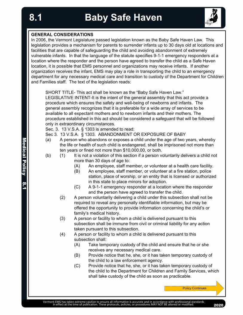
Medical P
olicy 8.1
20132013
Baby Safe Haven
Vermont EMS has taken extreme caution to ensure all information is accurate and in accordance with professional standards in effect at the time of publication. These protocols, policies, or procedures MAY NOT BE altered or modified.
GENERAL CONSIDERATIONSIn 2006, the Vermont Legislature passed legislation known as the Baby Safe Haven Law. This legislation provides a mechanism for parents to surrender infants up to 30 days old at locations and facilities that are capable of safeguarding the child and avoiding abandonment of extremely vulnerable infants. In that the language of the statute specifies 9-1-1 emergency responders at a location where the responder and the person have agreed to transfer the child as a Safe Haven location, it is possible that EMS personnel and organizations may receive infants. If another organization receives the infant, EMS may play a role in transporting the child to an emergency department for any necessary medical care and transition to custody of the Department for Children and Families staff. The text of the legislation reads:
SHORT TITLE- This act shall be known as the “Baby Safe Haven Law.”LEGISLATIVE INTENT-It is the intent of the general assembly that this act provide a procedure which ensures the safety and well-being of newborns and infants. The general assembly recognizes that it is preferable for a wide array of services to be available to all expectant mothers and to newborn infants and their mothers. The procedure established in this act should be considered a safeguard that will be followed only in extraordinary circumstances.Sec. 3. 13 V.S.A. § 1303 is amended to read:Sec 3. 13 V.S.A. § 1303. ABANDONMENT OR EXPOSURE OF BABY(a) A person who abandons or exposes a child under the age of two years, whereby
the life or health of such child is endangered, shall be imprisoned not more than ten years or fined not more than $10,000.00, or both.
(b) (1) It is not a violation of this section if a person voluntarily delivers a child not more than 30 days of age to:(A) An employee, staff member, or volunteer at a health care facility.(B) An employee, staff member, or volunteer at a fire station, police
station, place of worship, or an entity that is licensed or authorized in this state to place minors for adoption.
(C) A 9-1-1 emergency responder at a location where the responder and the person have agreed to transfer the child.
(2) A person voluntarily delivering a child under this subsection shall not be required to reveal any personally identifiable information, but may be offered the opportunity to provide information concerning the child’s or family’s medical history.
(3) A person or facility to whom a child is delivered pursuant to this subsection shall be immune from civil or criminal liability for any action taken pursuant to this subsection.
(4) A person or facility to whom a child is delivered pursuant to this subsection shall:(A) Take temporary custody of the child and ensure that he or she
receives any necessary medical care. (B) Provide notice that he, she, or it has taken temporary custody of
the child to a law enforcement agency. (C) Provide notice that he, she, or it has taken temporary custody of
the child to the Department for Children and Family Services, which shall take custody of the child as soon as practicable.
2020
8.1

Medical P
olicy 8.1
20132013
Baby Safe Haven
Vermont EMS has taken extreme caution to ensure all information is accurate and in accordance with professional standards in effect at the time of publication. These protocols, policies, or procedures MAY NOT BE altered or modified. 2020
8.1
While the statute does not specifically mention EMS personnel or organizations, EMS may become involved as a 9-1-1 emergency responder. It is also likely that EMS will be involved when an infant is delivered to any other Safe Haven. Delivery of an infant to a Safe Haven location will be an infrequent but very stressful event. Infants being delivered to the Safe Haven are at considerable risk and every possible step to safeguard the health and welfare of the infant should be taken.
A person delivering an infant under the provisions of the Baby Safe Haven law may not make the subtle distinctions between EMS facilities and other Safe Haven locations specifically mentioned in the law. EMS personnel and organizations should be prepared to play an appropriate role in the receipt of infants delivered under the provisions of the Baby Safe Haven law.
EMS may be summoned by a Safe Haven that receives an infant (fire, police, place of worship, adoption agency, or health care facility). Guidance being provided by the Department for Children and Families for Safe Havens suggests that when an infant is received, the Safe Haven should call an ambulance to provide treatment and transportation. The primary roles of EMS in a Baby Safe Haven encounter are as with any other patient: assess, provide treatment as indicated, transport to a hospital.
EMS organizations with stations that could be unoccupied may wish to consider signage on entryways indicating that persons wishing to deliver an infant under the provisions of the Baby Safe Haven law should call 9-1-1 to make arrangements if the station is not staffed and should not leave an infant unattended.
EMS organizations may wish to reach out to Safe Haven locations in their primary service area to coordinate procedures and develop local plans for handling Safe Haven encounters.
To order additional copies of Safe Haven posters and brochures, call 802-241-2131. To find out about training opportunities from the Department for Children and Families, call 800-649-4357 or go to babysafehaven.vermont.gov
PROCEDURE Although EMS organizations and personnel are not specifically mentioned in the Baby Safe
Haven statute, as 9-1-1 emergency responders, EMS needs to be familiar with the provisions of the law and work to facilitate the protection of infants being received.
When involved in a Baby Safe Haven encounter, upon receiving physical custody of the baby, whether you receive an infant from the parent, a Safe Haven, or another third party, examine the baby and provide any treatment necessary according to the appropriate clinical protocol(s).
If possible, offer the person delivering the infant the Safe Haven brochure published by the Agency of Human Services if the infant is not picked up at a Safe Haven.
If it has not already been done, attempt to advise the person delivering the infant into a Safe Haven that while she/he is not required to reveal any identifying information, she/he can provide information about the child’s medical history using the voluntary medical form that is attached to the DCF Safe Haven brochure. Encourage the person to complete the form and leave it with you. If the form is left with you, complete the information at the bottom of it with the date you received the baby and your location. This information should be delivered to the DCF Family Services staff person. Document this information on your EMS patient care report form as well.
Document on your EMS patient care report any additional information about the child, the birthparents, and/or the situation that is observed or offered voluntarily, including any names the person is willing to provide. Policy Continues
Policy Continues

Me
dic
al P
olic
y 8
.1
2013
2013
Baby Safe Haven
Vermont EMS has taken extreme caution to ensure all information is accurate and in accordance with professional standards in effect at the time of publication. These protocols, policies, or procedures MAY NOT BE altered or modified.
• Transport the infant to the hospital once appropriate EMS treatment has begun. If you receive
an infant from a parent or other person, at the earliest possible opportunity, call the police of
jurisdiction where you received the child and the local DCF, Family Services District Office or DCF Emergency Services contact to report what has happened (see list attached). The EMS
personnel involved may have information about the parent, the circumstances of receiving the
child, etc. that will be important to law enforcement and the DCF Family Services personnel, so it is important for the EMS personnel involved to make this contact. If you receive an infant from
another Safe Haven, assure that the original receiver(s) have made the police and DCF contacts.
• Important: Once you have physical custody of the infant, always transport the child to a
hospital. If the person who delivered the infant or another person comes back to you requesting
the return of the baby, do not give the baby back. Instead, instruct the person to contact the
Department of Children and Families at 800-649-4357. They will be referred to a social worker,
who will assist them with the process. Urge them to act quickly, before a judge has terminated
their parental rights.
DCF FAMILY SERVICES DISTRICT OFFICES AND
STATEWIDE EMERGENCY SERVICES PROGRAM
BARRE – 802-479-4260 NEWPORT – 802-334-6723
255 North Main Street, Suite 7 100 Main Street, Suite 230
Barre, VT 05641-4189 Newport, VT 05855-4898
BENNINGTON – 802-442-8138 RUTLAND – 802-786-5817
200 Veterans Memorial Drive, Suite 14 88 Merchants Row
Bennington, VT 05201-1956 220 Asa Bloomer Bldg
Rutland, VT 05701-3449
BRATTLEBORO – 802-257-2888
232 Main Street, 2nd Floor ST. ALBANS – 802-527-7741
Brattleboro, VT 05301-2879 27 Federal Street, Suite 300
St. Albans, VT 05478-2247
BURLINGTON – 802-863-7370
426 Industrial Ave. Ste. 130 ST. JOHNSBURY – 802-748-8374
Williston, VT 05495 1016 US Route 5, Suite 02
St. Johnsbury, VT 05819-5603
HARTFORD – 802-295-8840
118 Prospect Street, Suite 400 SPRINGFIELD – 802-289-0648
White River Junction, VT 05001 100 Mineral Street, Suite 101
Springfield, VT 05156-3166
MIDDLEBURY – 802-388-4660
156 South Village Green, Ste. 202
Middlebury, VT 05753 STATEWIDE DCF Family Service’s
Emergency Services Program
MORRISVILLE – 802-888-4576 For after hours, weekends and holidays
63 Professional Drive, Suite 3 800-649-5285
Morrisville, VT 05661-8522
2020
8.1
Policy Continues

20132013
PURPOSEThis policy provides guidance for providers concerning the triage, extrication, care and transport for bariatric patients. The Vermont EMS system strives to provide all patients, including bariatric patients, with timely and effective care that preserves the comfort, safety and dignity of the patients and ensures the safety of providers. At times, even a single patient can exceed the capacity of the immediately available resources. Like a multi-system trauma patient, a bariatric patient requires: Appropriate EMS resources to respond Appropriate protocols and equipment for the provision of care Specialized equipment for transfer to the ambulance and transport Careful selection of the appropriate destination hospital Pre-alerting of the ED to ensure adequate resources to manage the patient On scene times that may be significantly extended for bariatric patients.
EQUIPMENTDeployment of equipment and procedures shall be done under local or regional operating guidelines.
DEFINITIONSA bariatric patient is a patient whose: Weight exceeds 400 pounds OR Weight, girth, body contours and/or co-morbidities challenge the ability of a two-
person EMS crew to effectively manage.
DISPATCHBariatric Ambulance: If available, consider requesting a bariatric transport ambulance to respond to the scene. Resources should be requested as soon as it becomes clear that bariatric capabilities may be required. While standard ambulance stretchers can potentially handle some patients up to 750 pounds or more, the use of a specialized bariatric stretcher increases the ability to provide effective care, is more comfortable for the patient and enhances provider safety.
Additional Personnel: Consider requesting additional responders. Bariatric patients may require additional personnel to participate in lifting and moving. For significant extrications, consider designating a Safety Officer to oversee the safety of the operation in conjunction with Incident Command. It may be necessary to remove doors, walls or windows to carry out a safe extrication. The priorities are similar to extrication from a vehicle, although fixed property repair costs might be higher.
Paramedic: Consider requesting a paramedic. Even BLS bariatric patients present unique treatment challenges which may benefit from a higher level of care.
MEDICAL CAREMedical care must take into account the unique challenges presented by the bariatric patient as well as the likelihood of extended on-scene times. Providers should use appropriately sized equipment to the extent it is available or can be readily obtained. For example, an appropriately sized blood pressure cuff will need to be used and intramuscular injection will be given with a longer needle.
If there are significant barriers to removing the patient from the structure in a timely manner (long narrow stairs, patient in the attic, etc.), there may be situations where EMS will provide extended care to the patient at the scene. In such cases, consult Medical Direction and consider use of the extended care protocols.
Vermont EMS has taken extreme caution to ensure all information is accurate and in accordance with professional standards in effect at the time of publication. These protocols, policies, or procedures MAY NOT BE altered or modified. 2020
Bariatric Triage, Care & Transport 8.2M
edical Policy 8.2
Policy Continues

TRANSFER TO AMBULANCESpecialized equipment will be needed to transfer the patient safely from the scene to the ambulance stretcher for transport. Many services utilize large transfer flats for moving bariatric patients. Be sure before you use any patient transfer device that you understand the procedure for using it safely and that you know the weight limits of the device.
HOSPITAL DESTINATIONEnsure that you select a destination hospital that has the capabilities to care for your patient. Bariatric patients may require specialized hospital stretchers, CT scanners, catheterization laboratory equipment, operating room equipment, etc. It may be appropriate to consider an alternate destination after consultation with Medical Direction. Pre-notification serves both to ensure that the hospital is capable of caring for the patient and allows hospital staff time for adequate preparation. Communication with the hospital shall be in a professional manner. Respect for the patient's privacy and feelings will match the respect for all EMS patients.
TRANSPORT TO THE HOSPITAL A bariatric stretcher should be used to transport the patient to the hospital and equipment cache transfer devices may be utilized to facilitate transfer of the patient to the hospital stretcher. Be alert to ensure that the stretcher is adequately secured in the patient compartment. Transfer flats or other specialized transfer equipment may be left in place to facilitate transfer of the patient to the hospital stretcher.
Vermont EMS has taken extreme caution to ensure all information is accurate and in accordance with professional standards in effect at the time of publication. These protocols, policies, or procedures MAY NOT BE altered or modified. 2020
8.3
PEARLS It may be difficult to establish IV and IO access. Consider intramuscular or intranasal as
alternatives for some medications. For IM, ensure that the needle used is sufficiently long. Weight-based calculations may yield inappropriately large doses in obese patients. Consult
with Medical Direction when in doubt. Bariatric patients often have decreased functional residual capacity, and are at risk of rapid
desaturation. Extremely obese individuals require more oxygen than non-obese individuals due to their diminished lung capacity. Pulse oximetry may not be reliable due to poor circulation. Even patients without respiratory distress may not tolerate the supine position.
Bariatric patients may present with severe airway challenges. Carefully plan your approach to the airway, and be prepared with backup airway plans.
If the patient has had recent bariatric surgery, possible complications may include anemia, dehydration, hypoglycemia, leakage, ulcers, localized infection, sepsis, etc.
Bariatric Triage, Care & Transport8.2
Protocol Continues
Medical P
olicy 8.2

20132013
Policy Continues
BLOODBORNE PATHOGENSEmergency medical services personnel should assume that all bodily fluids and tissues are potentially infectious with bloodborne pathogens and must protect themselves accordingly by use of standard precautions.
Transmission of bloodborne pathogens has been shown to occur when infected blood or other potentially infectious materials (“OPIM”) enter another individual’s body through skin, mucous membrane, or parenteral contact.
STANDARD PRECAUTIONS Standard precautions include using protective barriers (such as gloves, masks,
goggles, etc.), thorough hand washing, and proper use and disposal of needles and other sharp instruments. Centers for Disease Control and Prevention Guidelines for hand hygiene include:o When hands are visibly dirty, contaminated, or soiled, wash with non-antimicrobial
or antimicrobial soap and water. o If hands are not visibly soiled, use an alcohol-based hand rub for routinely
decontaminating hands. Personnel with any open wounds should refrain from all direct patient care and from
handling patient-care equipment, unless they can ensure complete isolation of these lesions and protection against seepage.
Personnel who are potentially at risk of coming into contact with blood or OPIM are encouraged to obtain appropriate vaccines to decrease the likelihood of transmission.
EXPOSURE - PROCEDURES AND CONSIDERATIONS Personnel who have had a bloodborne pathogen exposure should immediately flush
the exposed area or wash with an approved solution. At a minimum, use warm water and soap.
The exposed area should then be covered with a sterile dressing. As soon as possible, or after transfer of patient care, the EMS provider should
thoroughly cleanse the exposed site and obtain a medical evaluation by the medical advisor as dictated by their department’s Exposure Control Plan and/or Workers Compensation policy.
AIRBORNE PATHOGENSEmergency medical services personnel should assume that all patients who present with respiratory distress, cough, fever, or rash are potentially infectious with airborne pathogens and must protect themselves accordingly by use of appropriate Airborne Personal Protective Equipment (APPE), Body Substance Isolation (BSI), and approved procedures.
AIRBORNE PERSONAL PROTECTIVE EQUIPMENT (APPE) The preferred APPE for EMS personnel is an N95 mask, to be worn whenever a
patient is suspected of having any communicable respiratory disease. The N95 mask should be of the proper size for each individual provider, having been
previously determined through an annual fit-test procedure. A surgical mask should also be placed on suspect patients, if tolerated. If oxygen
therapy is indicated, a surgical mask should be placed over an oxygen mask to block pathogen release. This will require close monitoring of the patient’s respiratory status and effort.
Medical P
olicy 8.3Bloodborne/Airborne Pathogens
Vermont EMS has taken extreme caution to ensure all information is accurate and in accordance with professional standards in effect at the time of publication. These protocols, policies, or procedures MAY NOT BE altered or modified. 2020
8.3
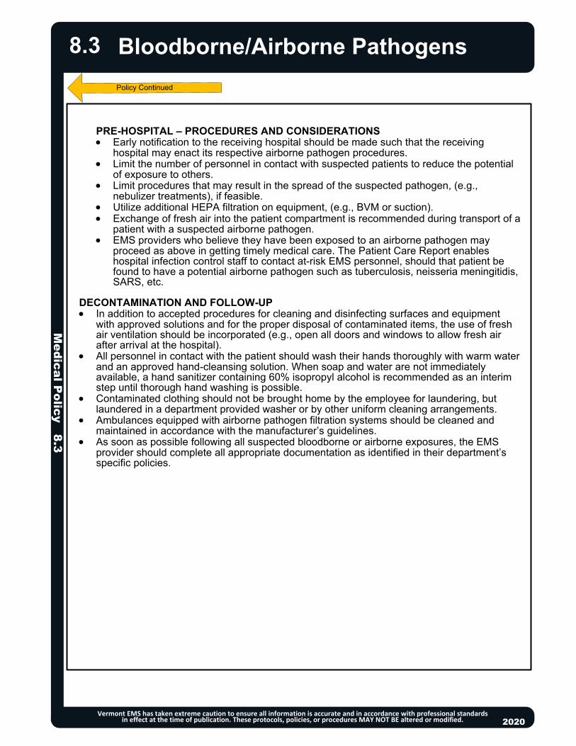
PRE-HOSPITAL – PROCEDURES AND CONSIDERATIONS Early notification to the receiving hospital should be made such that the receiving
hospital may enact its respective airborne pathogen procedures. Limit the number of personnel in contact with suspected patients to reduce the potential
of exposure to others. Limit procedures that may result in the spread of the suspected pathogen, (e.g.,
nebulizer treatments), if feasible. Utilize additional HEPA filtration on equipment, (e.g., BVM or suction). Exchange of fresh air into the patient compartment is recommended during transport of a
patient with a suspected airborne pathogen. EMS providers who believe they have been exposed to an airborne pathogen may
proceed as above in getting timely medical care. The Patient Care Report enables hospital infection control staff to contact at-risk EMS personnel, should that patient be found to have a potential airborne pathogen such as tuberculosis, neisseria meningitidis, SARS, etc.
DECONTAMINATION AND FOLLOW-UP In addition to accepted procedures for cleaning and disinfecting surfaces and equipment
with approved solutions and for the proper disposal of contaminated items, the use of fresh air ventilation should be incorporated (e.g., open all doors and windows to allow fresh air after arrival at the hospital).
All personnel in contact with the patient should wash their hands thoroughly with warm water and an approved hand-cleansing solution. When soap and water are not immediately available, a hand sanitizer containing 60% isopropyl alcohol is recommended as an interim step until thorough hand washing is possible.
Contaminated clothing should not be brought home by the employee for laundering, but laundered in a department provided washer or by other uniform cleaning arrangements.
Ambulances equipped with airborne pathogen filtration systems should be cleaned and maintained in accordance with the manufacturer’s guidelines.
As soon as possible following all suspected bloodborne or airborne exposures, the EMS provider should complete all appropriate documentation as identified in their department’s specific policies.
Medical P
olicy 8.3
Bloodborne/Airborne Pathogens
Vermont EMS has taken extreme caution to ensure all information is accurate and in accordance with professional standards in effect at the time of publication. These protocols, policies, or procedures MAY NOT BE altered or modified. 2020
8.3

Medical P
olicy 8.4
20132013
Communication
Vermont EMS has taken extreme caution to ensure all information is accurate and in accordance with professional standards in effect at the time of publication. These protocols, policies, or procedures MAY NOT BE altered or modified. 2020
8.4
EMS providers should routinely advise the receiving hospital, in a timely manner, of patients enroute to that Emergency Department (except in Mass Casualty Incidents (MCI) during which routine communications cease). Follow guidelines below or local receiving hospital protocol.
An EMS provider may establish contact with a Medical Direction physician via VHF radio on one of the assigned medical frequencies or via telephone direct to each Emergency Department (via recorded EMS line, if available). If a Medical Direction physician is needed for consultation, request this before giving patient information. It is recommended that hospitals record all medical communications.
VHF MEDICAL FREQUENCIES Initiate call to the appropriate hospital and identify:
o Destination hospital.o Ambulance unit calling. o Status of the patient.
TELEPHONE To contact the destination hospital via telephone, use of a direct-recorded line to the
Emergency Department is recommended. Request Medical Direction, if needed, give the name of the patient, his or her age, status,
and complaint.
Upon establishing voice communication with the destination hospital/medical direction physician (if needed), present the following information in a concise and clear manner: Emergency response unit and level of care: Paramedic/Advanced-EMT/EMT, with ETA. Advise if the patient meets the criteria for stroke, trauma, STEMI or sepsis alert. Patient’s age, sex, and orientation level. Patient’s chief complaint. Patient’s present medical condition. Patient’s vital signs, including level of consciousness. Patient’s physical signs of illness or injury. Patient’s electrocardiogram rhythm, if indicated. Patient’s relevant medical history. Prehospital diagnostic tests performed/results and treatment rendered/results.
Give a list of medications and allergies only if requested by the destination hospital, or if it is anticipated that a medication order would be given by Medical Direction.

Medical P
olicy 8.5
2013
In case of a communications failure with Medical Direction due to equipment (cell phone, landline, radio) malfunction or incident location, the following will apply:
EMS personnel may, within the limits of their level of licensure, perform necessary procedures described in these protocols that under normal circumstances would require online Medical Direction.
These procedures shall be the minimum necessary to prevent the loss of life or the critical deterioration of a patient’s condition.
All procedures performed under this order, and the conditions that created the communications failure, need to be thoroughly documented.
Attempts must be made to establish contact with Medical Direction as soon as possible.
The EMS provider shall provide a written notification pertaining to the communications failure describing the events, including the patient’s condition and treatment given, and referencing the EMS Incident Report. This report must be filed with the EMS District Medical Advisor and/or Hospital EMS Coordinator within 24 hours of the event.
2013
Communications Failure
Vermont EMS has taken extreme caution to ensure all information is accurate and in accordance with professional standards in effect at the time of publication. These protocols, policies, or procedures MAY NOT BE altered or modified. 2020
8.5

Medical P
olicy 8.6
2013
A “minor” is a person who has not yet reached his/her eighteenth birthday.
Note that the legal definition of a “minor” for purposes of consent is unrelated to the medical definitions of “pediatric patient,” “child,” and “children,” as used in these protocols.
EMS personnel may treat minors under the doctrine of implied consent when the minor’s parent or other authorized representative is unavailable to provide expressed consent. With the exception of life-threatening emergencies, personnel should attempt to contact the minor’s parent or legal guardian to obtain informed consent to treat and transport the child. When a parent or legal guardian is unavailable, another authorized representative (e.g., a school or camp official), who has been expressly authorized by the minor’s parent, may consent to health care treatment.
A parent or legal guardian may refuse care for a minor: When a parent or legal guardian is not reasonably available, another adult family member (e.g.,
grandparent), or other authorized representative having custody of the minor, may refuse care. EMS personnel may accept a telephonic refusal of care, provided that they have explained the
consequences of refusing care; telephonic refusal of care should be carefully documented.
Except for the special circumstances listed below, a minor may not refuse care. When a minor attempts to refuse care and/or transport to the hospital, EMS personnel should enlist the assistance of the police, including requesting that the police place the minor in protective custody. Minors should be restrained only as a last resort.
SPECIAL CIRCUMSTANCES A minor parent who has not yet reached his/her eighteenth birthday may consent to or refuse
care on behalf of his or her minor children, provided that the minor parent has the capacity to understand the nature of the treatment and the possible consequences of consenting to or refusing care.
Contact Medical Direction to discuss consent/refusals regarding minors. Always attempt to provide medical care that is in the best interest of the minor.
Minors of any age may give informed consent to medical treatment associated with rape, incest or sexual abuse.
An emancipated minor may consent to, or refuse, care. A minor patient bears the burden of establishing, by legal documentation or otherwise, that he/she is emancipated. Vermont recognizes emancipation decrees lawfully obtained in other states (12 VSA §7157).
2013
Consent for Treatment of a Minor
Vermont EMS has taken extreme caution to ensure all information is accurate and in accordance with professional standards in effect at the time of publication. These protocols, policies, or procedures MAY NOT BE altered or modified. 2020
8.6

20132013
If you have been dispatched to a possible crime scene, including motor vehicle incidents, or if you believe a crime has been committed, immediately contact law enforcement.
Protect yourself and other EMS personnel. You will not be held liable for failing to act if a scene is not safe to enter. Once a crime scene is deemed safe by law enforcement, initiate patient contact and medical care if necessary.
Have all EMS providers use the same path of entry and exit, if feasible. Do not walk through fluids. Do not touch or move anything at a crime scene unless it is necessary to do so for patient
care (notify law enforcement prior to moving if possible). Observe and document original location of items moved by crew. When removing patient clothing, leave it intact as much as possible.
o Do not cut through clothing holes made by gunshot or stabbing. If you remove any items from the scene, such as impaled objects or medication bottles,
document your actions and advise a law enforcement official. Do not sacrifice patient care to preserve evidence. Consider requesting a law enforcement officer to accompany the patient in the ambulance
to the hospital. Document statements made by the patient or bystanders on the EMS patient care report.
o Comments made by a patient or bystanders should be denoted in quotation marks. Inform staff at the receiving hospital that this is a “crime scene” patient. If the patient is obviously dead consistent with Resuscitation Initiation and Termination
Policy 8.17, notify law enforcement of decision not to initiate resuscitation/patient care. At motor vehicle incidents, preserve the scene by not driving over debris, not moving debris
and parking away from tire marks, if feasible.
Medical P
olicy 8.7
Crime Scene/Preservation of Evidence
Vermont EMS has taken extreme caution to ensure all information is accurate and in accordance with professional standards in effect at the time of publication. These protocols, policies, or procedures MAY NOT BE altered or modified. 2020
8.7

20132013
Do Not Resuscitate (DNR)/ Clinician OrdersFor Life Sustaining Treatment (COLST)
Medical P
olicy 8.8
Vermont EMS has taken extreme caution to ensure all information is accurate and in accordance with professional standards in effect at the time of publication. These protocols, policies, or procedures MAY NOT BE altered or modified.
Policy Continues
PLEASE NOTE THE MANDATED USE OF THE VERMONT DEPARTMENT OF HEALTH'S DNR/COLST FORM IN THE FOLLOWING CIRCUMSTANCES:
TO BE VALID DNR/COLST ORDERS MUST MEET THE FOLLOWING CRITERIA: DNR Orders must:
o Be signed by the patient's clinician. A verbal order documented with the clinician’s name, and signed and dated by the patient’s nurse or social worker is acceptable.
o Certify that the clinician has consulted, or made an effort to consult, with the patient, and the patient's agent or guardian, if there is an appointed agent or guardian.
o Must include: the name of the patient, agent or guardian giving informed consent OR meet the futility standard (section A-3).
COLST Orders must:o Be signed by the patient's clinician. A verbal order documented with the clinician’s name,
and signed and dated by the patient’s nurse or social worker is acceptable. Include the name of the patient, agent or guardian giving informed consent.
2020
8.8
PEARLS One essential element of providing quality end-of-life care involves honoring patient
preferences. In the past a Do-Not-Resuscitate Order (DNR) has been used when a patient does not wish to
have Cardiopulmonary Resuscitation (CPR). In recent years the old “DNR” order has been incorporated into a new form known as the DNR/COLST order by the Vermont Department of Health. The old “DNR” order only dealt with CPR. The DNR/COLST order deals with CPR as well as intubation, transfer to hospital, antibiotics, hydration and overall goals of care and is the only legal out-of-hospital DNR order in Vermont as of July 1, 2011 (old orders will be honored as long as they complied with the statute at the time they were executed). Healthcare and residential care facilities can document these orders in a facility specific manner for inpatients.
DNR/COLST is a more complete document that stands for:Do Not Resuscitate Order/Clinician Orders for Life Sustaining Treatment.
These are clinician orders that convey patient’s wishes for CPR, intubation, transfer to the hospital, antibiotics, artificial nutrition and hydration, as well as overall treatment goals.
EMS personnel are required to perform CPR if indicated for a victim of cardiac arrest unless there is a signed DNR/COLST order in the out-of-hospital setting or termination of resuscitation criteria are met. See Resuscitation Initiation and Termination Policy 8.17.
All forms of DNR/COLST remain valid during a transfer from one healthcare facility to another.
All out-of-hospital DNR orders are mandated to be on the Department of Health's Vermont DNR/COLST form (link to form). DNR orders signed prior to July 1, 2011 may continue to be recognized as valid for
EMS encounters, if the document complies with statutory requirements. Health care facilities and residential care facilities may document DNR/COLST orders
in the patient's medical record in a facility specific manner when the patient is in their care so long as they meet the criteria below.

Do Not Resuscitate (DNR)/ Clinician OrdersFor Life Sustaining Treatment (COLST)
Vermont EMS has taken extreme caution to ensure all information is accurate and in accordance with professional standards in effect at the time of publication. These protocols, policies, or procedures MAY NOT BE altered or modified.
PROCEDURE Patients should be assessed per routine procedures and if resuscitation is or may be needed, EMS
personnel should make reasonable efforts to check the patient for a DNR identification (bracelet, necklace, anklet) or inquire if there is a written DNR/COLST order available. Attempt to determine the identity of the patient (specifically, name and date of birth).
If The Patient/Resident Has No Pulse And/Or No Respirations:o Go to Section A of the DNR/COLST form. If the DNR order is checked and there is a clinician
signature, a verbal order documented, and signed and dated by the patient’s nurse or social worker, or the patient has a DNR identification - Do Not initiate CPR or other resuscitation measures.Do not perform chest compressions or assist ventilations via BVM.Do not intubate or place advanced airway devices.Do not defibrillate.Do not administer resuscitation drugs to treat cardiac arrest or dysrhythmias (ventricular
fibrillation, pulseless ventricular tachycardia, pulseless electrical activity, or asystole). When Confronted With A Seriously Ill Patient Who Is Not In Cardiac Arrest But Is Breathing
And/Or Has A Pulse:o Go to Sections B – G of the DNR/COLST form to review Orders for Other Life-Sustaining
Treatment. Check for do not intubate orders (DNI), transfer to hospital orders, orders for medical interventions and other instructions. In Section H, check for clinician signature. A verbal order documented with the clinician’s name, and signed and dated by the patient’s nurse or social worker is acceptable. Comfort Measures Only: Use medication by any route, positioning, wound care and other measures to relieve pain and suffering. Use oxygen (including CPAP), oral suction and manual treatment of airway obstruction as needed for comfort. Limited Additional Interventions: Includes care described in Sections B-E. Use medical
treatments and IV fluids as indicated. Full treatment: Includes care described in sections B-E. Use defibrillation and other
interventions as indicated.
Medical P
olicy 8.8
2020
8.8
PEARLS A DNR Order (Section A of the DNR/COLST form) only precludes efforts to resuscitate in the event of
cardiopulmonary arrest and does not affect other therapeutic interventions that may be appropriate for the patient. (Sections B through H of the COLST form address other interventions).
EMS providers and other health care professionals must honor a DNR order or a DNR identification unless it is believed in good faith, after consultation with the patient, agent or guardian, where possible and appropriate:
o That the patient wishes to have the DNR Order revoked if the Order is based on informed consent, or
o That the patient with the DNR identification or order is not the individual for whom the DNR order was issued.
A health care provider shall honor in good faith an out-of-state DNR order, orders for life sustaining treatment, or out-of-state DNR identification if there is no reason to believe that what has been presented is invalid.
Statutory requirements for DNR or DNR/COLST require that the order be signed by the patient’s clinician, or a verbal order documented, and signed and dated by the patient’s nurse or social worker, and include the name of the patient/agent/guardian or other individual giving informed consent and their relationship to the patient.
In the absence of a valid DNR or DNR/COLST order, neither a spouse nor a healthcare agent/durable power of attorney may direct you to withhold resuscitation in the event of a cardiac arrest. Contact Medical Direction for guidance.
Photocopies and faxes of signed DNR/COLST forms are legal and valid.
An Advanced Directive (formerly referred to as Living Will) is different than DNR/COLST. An Advance Directive is completed by the patient, allows for nuances and is not honored in an out of hospital emergency. The DNR/COLST order is completed by a clinician, is black and white and is honored in an out of hospital emergency.
Contact Medical Direction for circumstances not specifically covered by this protocol. Document in SIREN any
actions taken or not taken based on a DNR/COLST order.Policy Continues
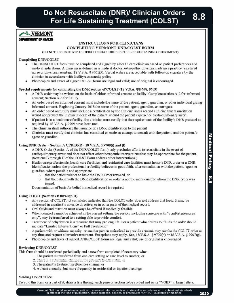
20132013
Do Not Resuscitate (DNR)/ Clinician OrdersFor Life Sustaining Treatment (COLST)
Medical P
olicy 8.8
Vermont EMS has taken extreme caution to ensure all information is accurate and in accordance with professional standards in effect at the time of publication. These protocols, policies, or procedures MAY NOT BE altered or modified. 2020
8.8

Do Not Resuscitate (DNR)/ Clinician OrdersFor Life Sustaining Treatment (COLST)
Vermont EMS has taken extreme caution to ensure all information is accurate and in accordance with professional standards in effect at the time of publication. These protocols, policies, or procedures MAY NOT BE altered or modified. 2020
8.8
Medical P
olicy 8.8
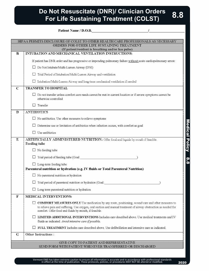
20132013
Do Not Resuscitate (DNR)/ Clinician OrdersFor Life Sustaining Treatment (COLST)
Medical P
olicy 8.8
Vermont EMS has taken extreme caution to ensure all information is accurate and in accordance with professional standards in effect at the time of publication. These protocols, policies, or procedures MAY NOT BE altered or modified. 2020
8.8

Do Not Resuscitate (DNR)/ Clinician OrdersFor Life Sustaining Treatment (COLST)
Vermont EMS has taken extreme caution to ensure all information is accurate and in accordance with professional standards in effect at the time of publication. These protocols, policies, or procedures MAY NOT BE altered or modified.
Medical P
olicy 8.8
2020
8.8

20132013
Hospice
Vermont EMS has taken extreme caution to ensure all information is accurate and in accordance with professional standards in effect at the time of publication. These protocols, policies, or procedures MAY NOT BE altered or modified. 2020
8.9
Protocol Continues
EMT/ADVANCED EMT STANDING ORDERS
EA/
Routine Patient Care. Contact the hospice team (preferred) or Medical Direction to coordinate
care and determine administration of hospice kit medications. Consider paramedic response for medication administration. Breakthrough Pain: Suggest administration of breakthrough pain medication by
patients / families. For pain of sudden onset, seek to determine and ameliorate or treat the underlying cause (e.g., pathological fracture).
Anxiety: Consider potential causes for patient’s anxiety, such as increased pain and shortness of breath.
Dyspnea: Administer oxygen via nasal cannula to relieve shortness of breath and achieve a respiration rate of < 20. Use a fan to blow air directly at the patient’s face.
Constipation: Suggest administration of constipation medication by patient/family. Nausea/Vomiting: Suggest administration of nausea medication by patient/family. Terminal Secretions: Reassure family that noisy breathing is generally not
distressing to the patient. Suggest administration of medication by patients/families. Terminal Dehydration: Moisten lips with petroleum jelly; use artificial saliva/mouth
sponges and ice chips. Confusion/Delirium: Speak slowly and calmly to the person. Remind the patient of
where they are, and who you are. Avoid contradicting the patient’s statements. Ensure a patient’s hearing aid and glasses are available. Limit activity/noise in the room.
This protocol is specific to those patients enrolled in Hospice. Treatment should be based on consultation with their Hospice team.
Introduction
The treatment goals of hospice patients differ significantly from those of other patients. Maintaining patient dignity and quality of life, rather than treating medical conditions, is the objective. If a specific cause of discomfort is identified (e.g., bronchospasm), traditional EMS treatment may be appropriate depending on the invasiveness of the therapy and the patient’s preferences. Hospice patients generally wish to remain at home and transport to the hospital should be the exception.
Many hospice patients will have a hospice comfort kit that contains medications that patient’s caregivers are instructed to use to treat commonly encountered medical issues.
If the patient is unable to make medical decisions and the hospice team cannot be contacted, determine the patient’s wishes and contact Medical Direction.
EMS providers should avoid the following interventions: Sirens, lights or aggressive interventions with family or caregivers. IV therapy (except where other forms of medication administration are not possible). Cardiac resuscitation: CPR, resuscitation medications, BVM ventilations. Cardiac pacing, cardioversion, and defibrillation. Hospice patients should not be transported to the hospital except where transport is specifically
requested by the patient or their healthcare agent or surrogate, and preferably only after consultation with the hospice team and exhaustion of other treatment pathways that do not require transport to the hospital.
Medical P
olicy 8.9
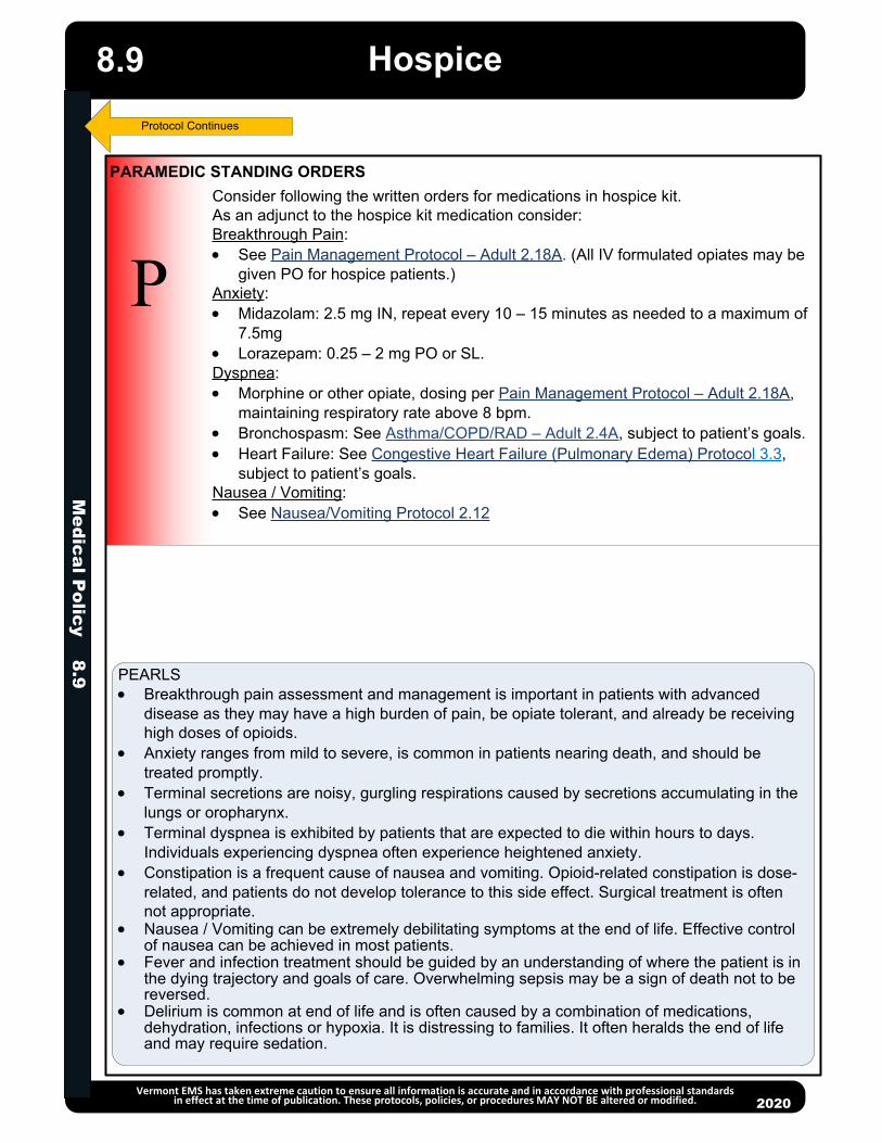
Hospice
Vermont EMS has taken extreme caution to ensure all information is accurate and in accordance with professional standards in effect at the time of publication. These protocols, policies, or procedures MAY NOT BE altered or modified. 2020
P
PARAMEDIC STANDING ORDERS
Consider following the written orders for medications in hospice kit.As an adjunct to the hospice kit medication consider:Breakthrough Pain: See Pain Management Protocol – Adult 2.18A. (All IV formulated opiates may be
given PO for hospice patients.)Anxiety: Midazolam: 2.5 mg IN, repeat every 10 – 15 minutes as needed to a maximum of
7.5mg Lorazepam: 0.25 – 2 mg PO or SL.Dyspnea: Morphine or other opiate, dosing per Pain Management Protocol – Adult 2.18A,
maintaining respiratory rate above 8 bpm. Bronchospasm: See Asthma/COPD/RAD – Adult 2.4A, subject to patient’s goals. Heart Failure: See Congestive Heart Failure (Pulmonary Edema) Protocol 3.3,
subject to patient’s goals.Nausea / Vomiting: See Nausea/Vomiting Protocol 2.12
PEARLS Breakthrough pain assessment and management is important in patients with advanced
disease as they may have a high burden of pain, be opiate tolerant, and already be receiving high doses of opioids.
Anxiety ranges from mild to severe, is common in patients nearing death, and should be treated promptly.
Terminal secretions are noisy, gurgling respirations caused by secretions accumulating in the lungs or oropharynx.
Terminal dyspnea is exhibited by patients that are expected to die within hours to days. Individuals experiencing dyspnea often experience heightened anxiety.
Constipation is a frequent cause of nausea and vomiting. Opioid-related constipation is dose-related, and patients do not develop tolerance to this side effect. Surgical treatment is often not appropriate.
Nausea / Vomiting can be extremely debilitating symptoms at the end of life. Effective control of nausea can be achieved in most patients.
Fever and infection treatment should be guided by an understanding of where the patient is in the dying trajectory and goals of care. Overwhelming sepsis may be a sign of death not to be reversed.
Delirium is common at end of life and is often caused by a combination of medications, dehydration, infections or hypoxia. It is distressing to families. It often heralds the end of life and may require sedation.
8.9
Medical P
olicy 8.9
Protocol Continues

EMT/ ADVANCED EMT / PARAMEDIC STANDING ORDERS
2013
EA
P
//
Vermont EMS has taken extreme caution to ensure all information is accurate and in accordance with professional standards in effect at the time of publication. These protocols, policies, or procedures MAY NOT BE altered or modified. 2020
Implantable Ventricular Assist Devices (VAD)
Protocol Continues
Patient Care Goals Rapid identification of, and interventions for, cardiovascular compromise in
patients with VADs Rapid identification of, and interventions for VAD-related malfunctions or
complications
Indications Adult patients that have had an implantable ventricular assist device (VAD)
including Left Ventricular Assist Device (LVAD), right ventricular assist device (RVADs); and biventricular-assist devices(BiVADs) and have symptoms of cardiovascular compromise
Patients with VADs that are in cardiac arrest Patients with VADs that are experiencing a medical or injury-related event not
involving the cardiovascular system or VAD malfunction
Contraindications Adult patients who do not have a VAD in place.
Assessment: Assess for possible pump malfunction
o Assess for alarmso Auscultate for pump sound “hum” o Signs of hypoperfusion including pallor, diaphoresis, altered mental status
If the VAD pump has malfunctioned:o Utilize available resources to troubleshoot potential VAD malfunctions and to
determine appropriate corrective actions to restore normal VAD function:Contact the patient’s VAD-trained companion, if available, Contact the patient’s VAD coordinator, using the phone number on the
deviceCheck all the connections to system controllerChange VAD batteries, and/or change system controller if indicatedHave patient stop all activity and assess for patient toleranceFollow appropriate cardiovascular condition-specific protocol(s) as
indicated
Treatment and Interventions: Manage airway as indicated Cardiac monitoring IV Access Acquire 12-lead ECG. Transmit if possible. If patient is experiencing VAD-related complications or cardiovascular
problems, expedite transport to a tertiary care facility if patient’s clinical condition and time allows.
If patient has a functioning VAD and is experiencing a non-cardiovascular-related problem, transport to a facility that is appropriate for the patient’s main presenting problem without manipulating the device.
8.10M
edical Policy 8.10

Implantable Ventricular Assist Device (VAD)
Vermont EMS has taken extreme caution to ensure all information is accurate and in accordance with professional standards in effect at the time of publication. These protocols, policies, or procedures MAY NOT BE altered or modified. 2020
EMT/ ADVANCED EMT / PARAMEDIC STANDING ORDERS
EA
P
//
Treatment and Interventions - continued: If patient is in full cardiac arrest:
o CPR should NOT be performed if there is any evidence the pump is still functioning. The decision whether to perform CPR should be made based upon best clinical judgment in consultation with the patient’s VAD-trained companion and the VAD coordinator (or medical control if VAD coordinator unavailable). CPR may be initiated only where:
o You have confirmed the pump has stopped AND troubleshooting efforts to restart it have failed, AND
o The patient is unresponsive and has no detectable signs of life
8.10
PEARLS You do not need to disconnect the controller or batteries in order to defibrillate or
cardiovert You do not need to disconnect the controller or batteries in order to acquire a 12-
lead EKG Flow though many VAD devices is not pulsatile and patients may not have a
palpable pulse or accurate pule oximetry The blood pressure, if measurable, may not be an accurate measure of
perfusion. Ventricular fibrillation, ventricular tachycardia, or asystole/PEA may be the
patient’s “normal” underlying rhythm. Evaluate clinical condition and provide care in consultation with VAD coordinator
The patient’s travel bag should accompany him/her at all times with back-up controller and spare batteries
If feasible, bring the patient’s Power Module, cable and Display Module with patient to the hospital
All patients should carry a spare pump controller with them The most common causes for VAD alarms are low batteries or battery failure Although automatic non-invasive blood pressure cuffs are often ineffective in
measuring systolic and diastolic pressure, if they do obtain a measurement, the MAP is usually accurate
Other VAD complications: o Infectiono Stroke / TIAo Bleeding
o Arrhythmiaso Cardiac
Tamponade
o CHFo Aortic Insufficiency
Protocol Continues
Medical P
olicy 8.10

Implantable Ventricular Assist Device (VAD)
Vermont EMS has taken extreme caution to ensure all information is accurate and in accordance with professional standards in effect at the time of publication. These protocols, policies, or procedures MAY NOT BE altered or modified. 2020
EA
P
//
EMT/ ADVANCED EMT / PARAMEDIC STANDING ORDERS
Key Documentation Elements Information gained from the VAD control box indicating any specific device
malfunctions Interventions performed to restore a malfunctioning VAD to normal function Time of notification to and instructions from VAD-trained companion and/or VAD
Coordinator
Resources:Recommended Unit Resource: Print EMS Guide for Mechanical Circulatory Support Organization and place in all ambulances (40 pages). This guide has excellent information and trouble shooting guidance for VAD devices that patients could have out in the public. Access the resource guide at: https://www.mylvad.com/medical-professionals/resource-library/ems-field-guides.
Download onto your ambulance laptops the “MyLVAD Hospital Locator App.”
References: Garg S, Ayers CR, Fitzsimmons C, et al. In-Hospital Cardiopulmonary Arrests in
Patients With Left Ventricular Assist Devices. J Card Fail. 2014;20(12):899-904. doi:10.1016/j.cardfail.2014.10.007
Mabvuure NT., et al., External cardiac compression during cardiopulmonary resuscitation with left ventricular assist devices. Interact Cardiovasc Thorac Surg. 2014 Aug;19(2):286-9.
Mechem M., Prehospital assessment and management of patients with ventricular-assist devices. Prehosp Emerg Care. 2013 Apr-Jun;17(2):223-9.
Shinar Z., et. al., Chest compressions may be safe in arresting patients with left ventricular assist devices (LVADs). Resuscitation 2014 May;85(5):702-4.
8.10M
edical Policy 8.10

Medical P
olicy 8.11
Naloxone Leave Behind Opioid Overdose Rescue Program
Vermont EMS has taken extreme caution to ensure all information is accurate and in accordance with professional standards in effect at the time of publication. These protocols MAY NOT BE altered or modified.
EA
P
//
NALOXONE LEAVE BEHIND OPIOID OVERDOSE RESCUE PROGRAM ALSO KNOWN AS NALOXONE LEAVE BEHIND KIT
BACKGROUND The opioid crisis is a growing concern in America, and it has become the leading cause of injury-related death in the United States. Naloxone is an opioid antidote that reverses the symptoms of opioid toxicity and associated life-threatening respiratory depression. Providing Naloxone Leave Behind Kits containing 4 mg. nasal spray naloxone doses to patients who have just had an overdose or who have other signs of opioid use disorder (OUD) has been shown to save lives and is a critical intervention EMS can deliver. PURPOSE To provide individuals who have just experienced an opioid-related overdose or who have other indications of OUD (At-Risk Person) with a Naloxone Leave Behind Kit, along with instructions on harm reduction and how to access treatment, in order to potentially prevent a future opioid related death. Indicators of OUD may include patient confirmation of opioid use/OUD, concern expressed by family members or others on scene, presence of drug paraphernalia or clinical signs and symptoms.
PROCEDURE Identify an At-Risk Person who has experienced an opioid related overdose or
has indicators of OUD. Complete usual patient care as per Vermont Statewide EMS Protocols. (See
Poisoning/Substance Abuse/Overdose – Adult 2.19A or – Pediatric 2.19P, Altered Mental Status – Adult 2.3A or – Pediatric 2.3P.)
Overdose patients who received naloxone by EMS or prior to EMS arrival should be encouraged to accept transport to the Emergency Department.
Offer a Naloxone Leave Behind Kit to an At-Risk Person if they refuse transport.
Additionally, offer a kit to an At-Risk Person that has not experienced an overdose but has indications of OUD.
The Naloxone Leave Behind Kit may also be given to family members, friends or other persons on scene who are in a position to assist the At-Risk Person.
Provide instructions on how to use the Naloxone Leave Behind Kit along with harm reduction instructions and information handout on how to access treatment for OUD.
Patients who used Naloxone on scene prior to EMS arrival may be offered a replacement kit.
Follow standard protocol and contact Medical Direction for patients who wish to refuse transport. (See Refusal of Care Policy and Patient Non-Transport Form 8.15.)
DOCUMENTATION In SIREN under Provider Action – Treatment, document if a Naloxone Leave
Behind Kit was given and number of kits distributed at the scene. If kit was offered but not left behind, briefly document why.
Dispensing a Naloxone Leave Behind Kit is an approved activity under Vermont EMS Scope of Practice for all provider levels.
2020
8.11

20132013
Non-EMS Personnel at the Emergency Scene
Vermont EMS has taken extreme caution to ensure all information is accurate and in accordance with professional standards in effect at the time of publication. These protocols, policies, or procedures MAY NOT BE altered or modified. 2020
8.12
GENERAL CONSIDERATIONSEmergency medical care is a continuum involving the concerted, integrated efforts of prehospital providers, physicians, nurses, allied health personnel, clergy and mental health workers. Recognizing this offers a guide for dealing with non-prehospital providers who wish to render assistance on the scene of an emergency. NON-PHYSICIAN INTERVENTION ON THE SCENE (Nurses, midwives, physician assistants, allied health personnel, clergy, mental health workers, etc.) Control of the medical care at an emergency scene is the responsibility of the individual in attendance
who is most appropriately trained and knowledgeable in providing prehospital emergency stabilization and transport. With few exceptions, this is the on-duty responding EMS licensee of the highest license level. The intervener should be thanked by a member of the responding EMS crew and be asked to report any care that was provided prior to EMS arrival.
Confrontation should be avoided whenever possible. The appropriate involvement of non-prehospital providers should be determined by the licensed responding prehospital providers.
On-line medical direction should be sought for situations where a cooperative working relationship is failing or has failed, or the non-prehospital provider refuses to relinquish care of the patient.
In any circumstance where the prehospital provider believes that care proposed by an intervener deviates from protocols, follow these protocols and seek on-line medical direction.
PHYSICIAN INTERVENTION ON THE SCENE Prehospital providers control an emergency scene, even in the presence of a physician. When EMS personnel encounter a person claiming to be a physician at the scene, one of the prehospital
providers should take reasonable steps to verify the identity of the physician without restricting the physician's access to provide potentially lifesaving care.
Patient’s private physician:o If the patient's private physician is present and assumes responsibility for the patient's care, the
prehospital provider should generally defer to the orders of the private physician within the limits of the provider's training and licensure.
o Medical direction should be contacted. o The private physician should be expected to accompany the patient to the hospital if interventions
beyond the scope and practice of the providers have occurred. o The prehospital provider reverts back to following these protocols and on-line medical direction at any
time when the patient's private physician is no longer in attendance. Not the patient’s private physician:
o If on-line medical direction CANNOT be established:The prehospital provider should generally relinquish responsibility for the patient's care when the
physician has identified himself and has indicated a willingness to assume responsibility and document any interventions when this care appears competent and appropriate. Always act in the best interest of the patient.
The prehospital provider should defer to the wishes of the physician on the scene within the limits of the provider's training and licensure.
If the care and treatment differ from these protocols, the physician must agree in advance to accompany the patient to the hospital.
In the event of a mass casualty incident or disaster, patient care needs may require the physician to remain at the scene.
o The prehospital provider reverts back to following these protocols and on-line medical direction at any time when this physician is no longer in attendance.
o If on-line medical direction CAN be established: The on-line physician is ultimately responsible. Should any disagreement between the physician on the scene and the on-line physician exist, the
prehospital provider should follow the orders from the on-line physician and place the intervener physician in contact with the on-line physician.
The on-line physician has the option of managing the case entirely, working with the physician, or allowing the on-scene physician to assume responsibility.
The details of any encounter with an intervener should be documented. Include the intervener's name, qualifications, and any care provided by the intervener.
Medical P
olicy 8.12
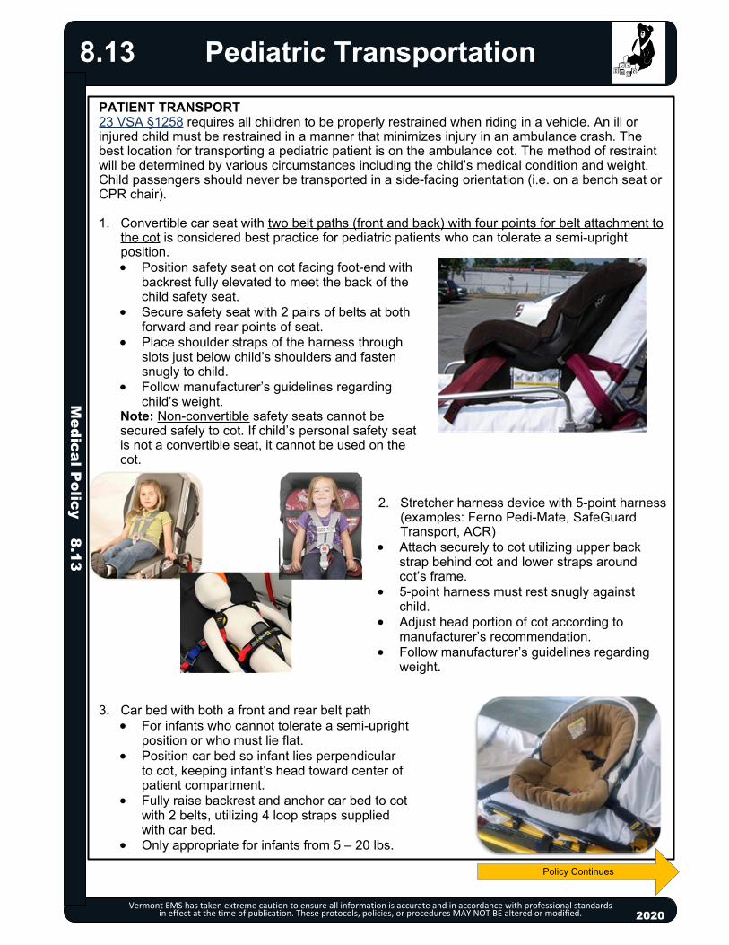
20132013
Pediatric Transportation
Vermont EMS has taken extreme caution to ensure all information is accurate and in accordance with professional standards in effect at the time of publication. These protocols, policies, or procedures MAY NOT BE altered or modified.
PATIENT TRANSPORT23 VSA §1258 requires all children to be properly restrained when riding in a vehicle. An ill or injured child must be restrained in a manner that minimizes injury in an ambulance crash. The best location for transporting a pediatric patient is on the ambulance cot. The method of restraint will be determined by various circumstances including the child’s medical condition and weight. Child passengers should never be transported in a side-facing orientation (i.e. on a bench seat or CPR chair).
1. Convertible car seat with two belt paths (front and back) with four points for belt attachment to the cot is considered best practice for pediatric patients who can tolerate a semi-upright position.
Position safety seat on cot facing foot-end with backrest fully elevated to meet the back of the child safety seat.
Secure safety seat with 2 pairs of belts at both forward and rear points of seat.
Place shoulder straps of the harness through slots just below child’s shoulders and fasten snugly to child.
Follow manufacturer’s guidelines regarding child’s weight.
Note: Non-convertible safety seats cannot be secured safely to cot. If child’s personal safety seatis not a convertible seat, it cannot be used on thecot.
2. Stretcher harness device with 5-point harness (examples: Ferno Pedi-Mate, SafeGuard
Transport, ACR) Attach securely to cot utilizing upper back
strap behind cot and lower straps around cot’s frame.
5-point harness must rest snugly against child.
Adjust head portion of cot according to manufacturer’s recommendation.
Follow manufacturer’s guidelines regarding weight.
3. Car bed with both a front and rear belt path For infants who cannot tolerate a semi-upright
position or who must lie flat. Position car bed so infant lies perpendicular
to cot, keeping infant’s head toward center of patient compartment.
Fully raise backrest and anchor car bed to cot with 2 belts, utilizing 4 loop straps supplied with car bed.
Only appropriate for infants from 5 – 20 lbs.
2020
8.13
Policy Continues
Medical P
olicy 8.13

Pediatric Transportation
Vermont EMS has taken extreme caution to ensure all information is accurate and in accordance with professional standards in effect at the time of publication. These protocols, policies, or procedures MAY NOT BE altered or modified.
4. Child belted directly to backboard and/or cot in manner to prevent ramping or sliding in a front or rear end crash
Loop narrow belt under each arm and extend over child’s shoulder securing belt at shoulder level so no gap exists above shoulder.
Use soft, sliding, or breakaway connector to hold shoulder straps together on chest.
Anchor 2 belts to non-sliding cot member and route over thighs and hips, not around waist.
5. Properly secure isolette and infant according to manufacturer’s guidelines. Rest harness securely on child with no blanket or sheet between harness and child. Attach to isolette tray at four points. Additional soft Velcro straps may be added for lateral security. Blanket or towels may be used to provide stabilization of the head.
NON-PATIENT TRANSPORTBest practice is to transport well children in a vehicle other than the ambulance, whenever possible, for safety.
If no other vehicle is available and circumstances dictate that the ambulance must transport a well child, he/she may be transported in the following locations:
Captain’s chair in patient compartment using a size appropriate integrated seat or a convertible safety seat that is secured safely in relationship to the orientation of the captain’s chair.
Passenger seat of the driver’s compartment if child is large enough (according to manufacturer’s guidelines) to ride forward-facing in a child safety seat or booster seat. Airbag should be turned off. If the air bag can be deactivated, an infant, restrained in a rear-facing infant seat, may be placed in the passenger seat of the driver’s compartment.
USE OF PATIENT’S CHILD PASSENGER SAFETY SEAT AFTER INVOLVEMENT IN MOTOR VEHICLE CRASHThe patient’s safety seat may be used to transport the child to the hospital after involvement in a minor crash if ALL of the following apply:
It is a convertible seat with both front and rear belt paths. Visual inspection, including under movable seat padding, does not reveal cracks or
deformation. Vehicle in which safety seat was installed was capable of being driven from the scene of
the crash. Vehicle door nearest the child safety seat was undamaged. The air bags (if any) did not deploy.
2020
8.13M
edical Policy 8.13

Medical P
olicy 8.14
Vermont EMS has taken extreme caution to ensure all information is accurate and in accordance with professional standards in effect at the time of publication. These protocols, policies, or procedures MAY NOT BE altered or modified. 2020
Police Custody8.14
PURPOSEThe purpose of this protocol is to give EMS guidance for patients who are in police custody, restrained, and/or protective custody is required.
PROTECTIVE CUSTODYProtective custody is a civil status in which an incapacitated person is detained by a law enforcement officer for the purposes of: (a) Assuring the safety of the individual or the public or both; and (b) Assisting the individual to return to a functional condition.
For patients who present an immediate risk of serious injury to themselves or others, consult with law enforcement or a mental health professional about the use of protective custody to transport the patient to the Emergency Department for an emergency examination, after obtaining warrant from a Superior judge. (18 VSA §7505)
Patients who present with evidence of incapacitation from alcohol or drug use may be placed into protective custody and transported to the Emergency Department (18 VSA §4810).
POLICE CUSTODYPolice custody means a person is under arrest. Patients in police custody who require medical care should be transported to a medical facility.
DECISION TO TRANSPORT TO THE HOSPITAL If law enforcement refuses to place a patient into protective custody at the request of EMS,
or if police and EMS disagree about whether a patient in police custody requires transport to a medical facility for further assessment or treatment, on-line Medical Direction must be contacted and a law enforcement supervisor should be requested for guidance.
If Medical Direction advises that the patient needs to be transported to the Emergency Department but you are unable to do so, document circumstances carefully in your chart and communicate with Medical Direction.
Patient medical condition is of primary concern when determining destination. When condition allows, consider law enforcement requests.
EMS INITIATED RESTRAINTSFor any patient potentially requiring restraints by EMS, see the Restraints Procedure 6.7.
POLICE RESTRAINT DEVICESPatients transported by EMS who have been restrained by law enforcement devices (e.g., handcuffs) should be accompanied, in the patient compartment, by a law enforcement officer who is capable of removing the device. If this is not feasible, the officer MUST follow directly behind the transporting ambulance to the receiving hospital.
CONDUCTED ELECTRICAL WEAPONSPatients who have been subdued by a conducted electrical weapon (i.e., Taser™), see Taser (Conducted Electrical Weapon) Medical Procedure 6.10.
PEPPER SPRAYPatients who have been subdued by pepper spray, see Eye & Dental Injuries Protocol 4.3.
EXCITED DELIRIUMExcited/Agitated Delirium is characterized by extreme restlessness, irritability, and/or high fever. Patients exhibiting these signs are at high risk for sudden death, see Restraints Procedure 6.9.

2013
Medical P
olicy 8.15
2013
Refusal of Care
Vermont EMS has taken extreme caution to ensure all information is accurate and in accordance with professional standards in effect at the time of publication. These protocols, policies, or procedures MAY NOT BE altered or modified.
PURPOSE Establish guidelines for the management and documentation of situations where patients refuse treatment or transportation.
REFUSAL OF CAREThere are three components to a valid refusal of care. Absence of any of these components will most likely result in an invalid refusal. The three components are as follows:
1. Competence: In general, a patient who is an adult or a legally emancipated minor is considered legally competent to refuse care. A parent or legal guardian who is on- scene or available by phone, may refuse care on his or her minor children’s behalf. 2. Capacity: In order to refuse medical assistance a patient must have the capacity to understand the nature of his or her medical condition, the risks and benefits associated with the proposed treatment, and the risks associated with refusal of care. 3. Informed Refusal: A patient must be fully informed about his or her medical condition, the risks and benefits associated with the proposed treatment and the risks associated with refusing care.
Patients who meet criteria to allow self-determination shall be allowed to make decisions regarding their medical care, including refusal of evaluation, treatment, or transport. These criteria include:
1. Adults (≥ 18 years of age or a legally emancipated minor).2. Orientation to person, place, time, and situation. 3. No evidence of altered level of consciousness resulting from head trauma, medical illness, intoxication, dementia, psychiatric illness or other causes. 4. No evidence of impaired judgment from alcohol or drug influence. 5. No language communication barriers (reliable translation available, e.g., on-scene interpreter, language line). 6. No evidence or admission of suicidal ideation resulting in any gesture or attempt at self-harm. No verbal or written expression of suicidal ideation regardless of any apparent inability to complete a suicide.
EMS providers will make every reasonable effort to convince reluctant patients to access medical care at the emergency department via the EMS system before accepting a Refusal of Care.
Utilize the Patient Non-Transport Algorithm. Any check mark in a shaded area requires on-line Medical Direction prior to terminating the patient encounter.
2020
8.15
Policy Continues

Medical P
olicy 8.15
Refusal of Care
Vermont EMS has taken extreme caution to ensure all information is accurate and in accordance with professional standards in effect at the time of publication. These protocols, policies, or procedures MAY NOT BE altered or modified.
Consider on-line Medical Direction for all patients who present a threat to themselves, present with an altered level of consciousness or diminished mental capacity, or have history or examination findings consistent with a high-risk refusal.
Medical Direction is to be provided all relevant information and may need to speak directly with the patient by radio or preferably a recorded landline.
If Medical Direction advises that the patient needs to be transported to the Emergency Department but you are unable to do so, document circumstances carefully in your chart and communicate with Medical Direction.
If based on a complete patient assessment, the patient does not have the capacity to refuse care and is in need of medical treatment or protective custody and refuses care, contact law enforcement for assistance.
Examples of high-risk refusals include but are not limited to:1. Treated / resolved hypoglycemia.2. Patient with obvious head trauma and taking anticoagulant medications.3. Intoxicated patients.4. Abnormal vital signs. 5. Treated / resolved narcotic overdose.6. High risk mechanism of injuries, see Advanced Spinal Assessment Procedure 6.0.7. Patient / witness reports suicidal ideations.8. Possible apparent life-threatening event, see Brief Resolved Unexplained Event (BRUE) Protocol 2.6.
PROCEDURE1. Clearly offer the patient both treatment and transportation to the hospital and document the offer in
your Patient Care Report (SIREN). All non-transports after patient contact require documentation in SIREN.
2. Perform an assessment of the patient’s mental capacity and, to the extent permitted by the patient, a physical exam including vital signs. Your assessment, or the patient’s refusal of care, must be fully documented in your Patient Care Report
3. Explain to the patient the nature and severity of his/her illness or injury, the treatments being proposed, the risks and consequences of accepting or refusing treatment, and the potential alternatives. Fully document the explanation given to the patient in your Patient Care Report.
4. A parent or legal guardian may refuse care for a minor or: When a parent or legal guardian is not reasonably available, another adult family member (e.g.,
grandparent), or other authorized representative having custody of the minor, may refuse care. EMS personnel may accept a telephonic refusal of care, provided that they have explained the
consequences of refusing care; telephonic refusal of care should be carefully documented. 5. Follow the patient non-transport algorithm. Any check mark in a shaded area requires on-line
Medical Direction prior to terminating the patient encounter.6. Prepare and explain the Patient Non-Transport form or the equivalent section of the SIREN run
form, if available, to the patient (or, in the case of a minor patient, the patient’s parent, legal guardian, or authorized representative).
7. The Patient Non-Transport form or SIREN screen should be signed by the patient (or, in the case of a minor patient, by the minor patient’s parent, legal guardian, or authorized representative) at the time of the refusal. The form should also be dated and, where possible, signed by a witness, preferably a competent relative, friend, police officer, or impartial third person.8. If on-line Medical Direction was consulted for a refusal of care, obtain and document the
physician’s name in the patient care report.9. All patients in police custody retain the right to request transport. This should be coordinated with law enforcement.10. If child abuse is suspected and a refusal of care situation exists, the EMT must contact police immediately, see Victims of Violence 8.21.
2020
8.15

2013
Medical P
olicy 8.15
2013
Refusal of Care
Vermont EMS has taken extreme caution to ensure all information is accurate and in accordance with professional standards in effect at the time of publication. These protocols, policies, or procedures MAY NOT BE altered or modified. 2020
8.15
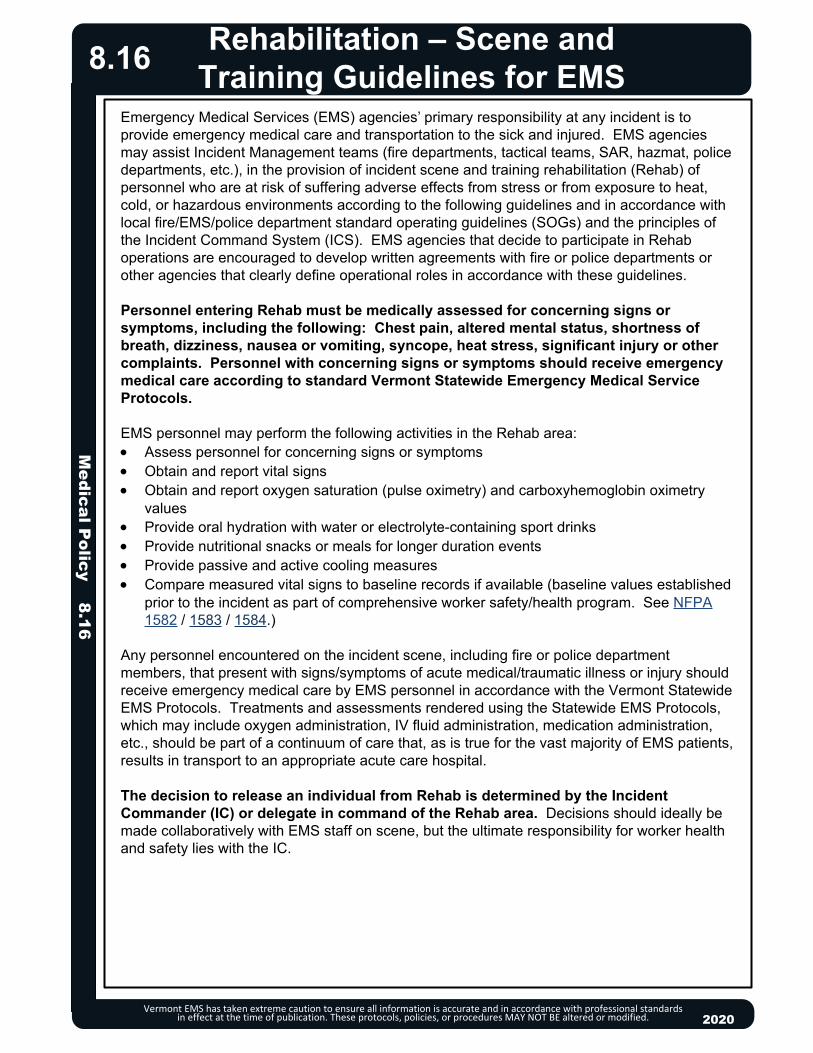
Medical P
olicy 8.16
Vermont EMS has taken extreme caution to ensure all information is accurate and in accordance with professional standards in effect at the time of publication. These protocols, policies, or procedures MAY NOT BE altered or modified. 2020
Rehabilitation – Scene and Training Guidelines for EMS
8.16
Emergency Medical Services (EMS) agencies’ primary responsibility at any incident is to provide emergency medical care and transportation to the sick and injured. EMS agencies may assist Incident Management teams (fire departments, tactical teams, SAR, hazmat, police departments, etc.), in the provision of incident scene and training rehabilitation (Rehab) of personnel who are at risk of suffering adverse effects from stress or from exposure to heat, cold, or hazardous environments according to the following guidelines and in accordance with local fire/EMS/police department standard operating guidelines (SOGs) and the principles of the Incident Command System (ICS). EMS agencies that decide to participate in Rehab operations are encouraged to develop written agreements with fire or police departments or other agencies that clearly define operational roles in accordance with these guidelines.
Personnel entering Rehab must be medically assessed for concerning signs or symptoms, including the following: Chest pain, altered mental status, shortness of breath, dizziness, nausea or vomiting, syncope, heat stress, significant injury or other complaints. Personnel with concerning signs or symptoms should receive emergency medical care according to standard Vermont Statewide Emergency Medical Service Protocols.
EMS personnel may perform the following activities in the Rehab area: Assess personnel for concerning signs or symptoms Obtain and report vital signs Obtain and report oxygen saturation (pulse oximetry) and carboxyhemoglobin oximetry
values Provide oral hydration with water or electrolyte-containing sport drinks Provide nutritional snacks or meals for longer duration events Provide passive and active cooling measures Compare measured vital signs to baseline records if available (baseline values established
prior to the incident as part of comprehensive worker safety/health program. See NFPA 1582 / 1583 / 1584.)
Any personnel encountered on the incident scene, including fire or police department members, that present with signs/symptoms of acute medical/traumatic illness or injury should receive emergency medical care by EMS personnel in accordance with the Vermont Statewide EMS Protocols. Treatments and assessments rendered using the Statewide EMS Protocols, which may include oxygen administration, IV fluid administration, medication administration, etc., should be part of a continuum of care that, as is true for the vast majority of EMS patients, results in transport to an appropriate acute care hospital.
The decision to release an individual from Rehab is determined by the Incident Commander (IC) or delegate in command of the Rehab area. Decisions should ideally be made collaboratively with EMS staff on scene, but the ultimate responsibility for worker health and safety lies with the IC.

20132013
Vermont EMS has taken extreme caution to ensure all information is accurate and in accordance with professional standards in effect at the time of publication. These protocols, policies, or procedures MAY NOT BE altered or modified.
Policy Continues
RESUSCITATION EFFORTS SHOULD BE WITHHELD UNDER THE FOLLOWING CIRCUMSTANCES: Valid Do Not Resuscitate: Refer to Do Not Resuscitate (DNR) & Clinician
Orders (COLST) Protocol 8.8. Scene Safety: The physical environment is not safe for providers. Dead on Arrival (DOA): A person is presumed dead on arrival when all five
“Signs of Death” are present AND at least one associated “Factor of Death” is present.
Signs of Death (All five signs of death must be present)Unresponsiveness.Apnea.Absence of palpable pulses at carotid, radial, and femoral sites.Unresponsive pupils.Absence of heart sounds.
Factors of Death (At least one associated factor of death must be present)Damage or destruction of the body incompatible with life, such
as, but not limited to:Decapitation.Decomposition.Deforming brain injury.Incineration or extensive full thickness burns.
Lividity/Rigor mortis of any degree.Major blunt or penetrating trauma.Body frozen solid—unable to perform chest compressions.
SUDDEN UNEXPLAINED INFANT DEATH (SUID) An infant <12 months who is apneic, asystolic (no heartbeat or umbilical
cord pulse), and exhibiting lividity and/or rigor mortis may be presumed dead.
NEONATE A neonate who is apneic, asystolic, and exhibits either neonatal
maceration (softening or degeneration of the tissues after death in utero) or anencephaly (absence of a major portion of the brain, skull, and scalp) may be presumed dead.
Contact Medical Direction if gestational age is less than 22 weeks and neonate shows signs of obvious immaturity (translucent and gelatinous skin, lack of fingernails, fused eyelids).
Patients with ventricular assist devices (VAD) should almost never be pronounced dead at the scene, see Implantable Ventricular Assist Devices (VAD) Policy 8.10.
8.17
EA
P
//
Resuscitation Initiation and Termination
8.17M
edical Policy 8.17
2020

Resuscitation Initiation and Termination
Vermont EMS has taken extreme caution to ensure all information is accurate and in accordance with professional standards in effect at the time of publication. These protocols, policies, or procedures MAY NOT BE altered or modified.
RESUSCITATION MAY BE STOPPED UNDER THE FOLLOWING CIRCUMSTANCES: When the patient regains pulse/respirations. See Post Resuscitative Care
Protocol – Adult 3.4A, Post Resuscitative Care Protocol -- Pediatric 3.4P, Cardiac Arrest Protocol -- Adult 3.2A or Cardiac Arrest Protocol – Pediatric 3.2P.
The physical environment becomes unsafe for providers. The exhaustion of EMS providers.
Contact Medical Direction to consider Termination of Resuscitation for any of the following:o Arrest witnessed by EMS personnel, if patient has NO return of
spontaneous circulation after 20 minutes of either BLS alone or combined BLS and ALS in the absence of hypothermia AND no shocks were delivered or advised; or
o Extrication is prolonged (> 20 minutes) with no resuscitation possible during extrication (hypothermia is an exception); or
o If extenuating circumstances or questions. Hypothermic patients without contraindications to CPR should have continued
CPR and should not be considered for TOR until the core temperature has been rewarmed to 32°C (90°F) with no Return of Spontaneous Circulation (ROSC). (See Hypothermia (Environmental) Protocol – Adult & Pediatric 2.11.)
CPR-induced consciousness is also a contraindication of TOR. Cardiac arrests should generally be managed on scene until return of
spontaneous circulation, decision to cease resuscitation, or criteria are met for transport to hospital as indicated by Termination of Resuscitation (TOR) Rule. If transport is initiated, resuscitation must be continued until arrival at the receiving hospital.
May continue resuscitation and transport if conditions on scene are NOT amenable to cessation of resuscitation.
Contact Medical Direction to consider Termination of Resuscitation for the non-hypothermic patient unresponsive to advanced cardiac life support with a non-shockable rhythm after 20 minutes of resuscitation and ETCO2 ≤ 10 mmHg.
For narrow-complex PEA with a rate above 40 or refractory and recurrent ventricular fibrillation/ventricular tachycardia, consider continuation of resuscitation and transport. o May consider termination of resuscitation if > 60 minutes from time of
dispatch.o Confirm cardiac standstill with point-of-care ultrasound, if available and
trained.
2020
8.17
EA/
P
TERMINATION OF RESUSCITATION (TOR) RULE (ADULTS ONLY):1) Arrest not witnessed by emergency medical services personnel.2) NO return of spontaneous circulation after 20 minutes of either BLS alone or combined BLS and ALS in the absence of hypothermia.3) No shock was delivered or advised by the AED.
o If ALL criteria are present, contact Medical Direction to consider termination of resuscitation.
o If ANY criteria are missing, contact Medical Direction to consider termination of resuscitation OR continued resuscitation and transport.
o If ROSC, continue resuscitation and transport AND contact Medical Direction.
o Notify law enforcement if terminating resuscitation.
Policy Continues
Medical P
olicy 8.17

Vermont EMS has taken extreme caution to ensure all information is accurate and in accordance with professional standards in effect at the time of publication. These protocols, policies, or procedures MAY NOT BE altered or modified.
DETERMINING DEATH IN THE FIELDWhen efforts to resuscitate are not initiated or are terminated under the above provisions, EMS providers shall: Document time that death is pronounced. Notify law enforcement, who will alert Medical Examiner. Consider possibility of a crime scene and restrict access. Any decision to move the body must be made in collaboration with law
enforcement and the medical examiner. Leave any resuscitation adjuncts such as advanced airway devices, IV/IO
access devices, electrode pads, etc., in place. Inform family on scene of patient’s death and offer to contact family, friends,
clergy, or other support systems.
The above requirements apply to situations in which law enforcement or the medical examiner may take jurisdiction. Law enforcement and the medical examiner are not required to take jurisdiction of hospice or other patients who are known to have been terminally ill from natural causes or congenital anomaly, and death was imminent and expected. Where law enforcement is not involved, EMS providers may provide appropriate assistance to families or other caregivers.
MASS CASUALTY INCIDENT (MCI) See Mass/Multiple Casualty Triage Protocol 9.1.
TRAUMATIC CARDIAC ARREST See Traumatic Cardiac Arrest Protocol 4.9.
DOCUMENTATION Complete a patient care record (SIREN) in all cases. If available, include
ECG rhythm strips and code summary with the patient care report. Document special orders including DNR, on-line Medical Direction, etc. MCI conditions may require a triage tag in addition to an abbreviated PCR. Record any special circumstances or events that might impact patient care
or forensic issues.
2020
8.17
Prolonging resuscitation efforts, beyond 20 minutes, without a return of spontaneous circulation is usually futile, unless cardiac arrest is compounded by hypothermia or submersion in cold water.
EMS providers are not required to transport every victim of cardiac arrest to a hospital. Unless special circumstances are present, it is expected that most resuscitations will be performed on-scene until the return of spontaneous circulation or a decision to cease resuscitation efforts is made based on the criteria listed. Transportation with continuing CPR is justified if hypothermia is present or suspected. Current AHA guidelines state: “cessation of efforts in the out-of-hospital setting…should be standard practice.”
An ETCO2 level of 10 mmHg or less measured 20 minutes after the initiation of advanced cardiac life support accurately predicts death in patients with cardiac arrest.
EA
P
//
Resuscitation Initiation and Termination
8.17M
edical Policy 8.17

Medical P
olicy 8.18
Vermont EMS has taken extreme caution to ensure all information is accurate and in accordance with professional standards in effect at the time of publication. These protocols, policies, or procedures MAY NOT BE altered or modified. 2020
Safe Response and Transportation Guidelines
8.18
RESPONSE & TRANSPORT PRIORITY DETERMINATION Determine response priority based on location of call and information provided per local
SOP’s. Consider traffic flow in the following response situations:
o When access to the scene will be delayed, such as calls requiring EMS to stage for law enforcement but EMS is closer to the scene.
o Hazardous road conditions limiting response speed and/or road conditions are likely to cause accidents for individuals pulling to side of road (e.g., severe icing of roadways or extreme limited visibility).
Determine transport priority considering the road and traffic conditions as they may impact the potential time savings, the ability to perform effective patient care, and most importantly the driver’s ability to safely transport with lights and sirens:o Consider flow of traffic transport for cases such as cardiac arrest due to the inability of
providers to be properly restrained when performing CPR, unless traffic conditions would significantly prolong transport time.
o Consider reserving use of lights and sirens in transport to known time-sensitive conditions such as MI, stroke or multi-trauma.
SAFETY WHEN IN MOTIONWhile ambulance is in motion (at all times, not only while transporting patients): All objects weighing 3 lbs or greater should be secured within a cabinet, compartment or
safely attached to a permanently-affixed structure within the passenger compartment or cab. Patients should be secured to the stretcher with all available belts -- i.e. chest/shoulder
harness, lap and leg belts. Do not leave belts unused. This includes patients who are secured to backboards.
Providers should be restrained at all possible times, unless it would interfere with patient care: o If belt prohibits patient care from being performed, it may be removed but then should be
refastened as soon as possible.o If belt must be removed for care, consider timing procedures/treatments to times
ambulance will experience minimal braking or turns. Select straight sections of road with few intersections to reduce risk of being thrown during sudden braking or turning.
Family members/non-rescue personnel should be properly restrained at all times.

Although often described as 'choking' by patients, it should be distinguished as strangulation when being documented by providers (as opposed to choking, i.e., foreign body obstruction). Include all information and observations regarding attempted strangulation in documentation provided to receiving hospital.
Assessment:How was the patient strangled: Left, right, or both hands; forearm; knee or foot; ligature or smothered; other, describe
Was patient shaken, beaten or held against wall, ground: Quantify grip strength and level of pain using 1-10 scale; duration in min/sec. Prior incidents of strangulation, domestic violence, or threats?
Signs and symptoms: Petechiae on face, eyes/eyelids, nose, ears, head Deformity of or bleeding from nose, ears; bruising, swelling of mouth/lips Redness, scratches, abrasions, bruising under chin, on neck, shoulders, chest Ligature marks, swelling, fingernail impressions (offensive or defensive) on neck Missing hair, fracture, or swelling/bruising on head, signs of concussion Difficulty breathing or speaking; coughing, hoarse or raspy voice; drooling, difficulty or pain
swallowing Vision disturbances or changes (spots, light flashes, tunnel vision, etc.) Hearing disturbances or changes (buzzing or ringing in the ears, etc.) Headache Subcutaneous emphysema Incontinence
Behavioral signs: Agitation, amnesia, hallucinations, dizziness, fainting, or combativeness due to hypoxia
Documentation and Reporting ResponsibilitiesPer 13 VSA 1021 and 33 VSA 4912 it must be reported to the police unless the patient age 18 or older refuses to have the information released.
Strangulation is also an indicator of increasing lethality in a violent relationship. Every effort should be made to connect patient with support services.
24-Hour Domestic Violence Crisis Line: 1-800-228-7395 24-Hour Sexual Assault Crisis Line: 1-800-489-7273 Vermont Adult Protective Services Program 800-564-1612 (after hours at 800-649-5285) Emergency shelter and transportation Hospital and court accompaniment; legal advocacy Information about public assistance.
20132013
Strangulation
Vermont EMS has taken extreme caution to ensure all information is accurate and in accordance with professional standards in effect at the time of publication. These protocols, policies, or procedures MAY NOT BE altered or modified. 2020
8.19
PEARLS: Patient's spouse/partner, caregiver or parent may be the perpetrator; their presence may
hinder patient's disclosure of information. Providers' reactions can impact patient recovery and strengthen or hinder prosecution of the
perpetrator. Non-judgmental and compassionate care and thorough documentation and preservation of evidence are essential.
Strangulation is defined as asphyxiation caused by closure of blood vessels and or air passageways of the neck due to external pressure. External pressure can be manual via a body part such as hands, arms, knees, etc., or can be by an object such as a belt, rope, etc.
Patients are at risk of delayed death due to internal swelling, anoxia, hematoma or structural damage that cannot be identified externally. Patients should be encouraged to seek medical care; if transported, communicate reported strangulation attempt to hospital staff.
Medical P
olicy 8.19
Trauma Triage and Transport Decision
Vermont EMS has taken extreme caution to ensure all information is accurate and in accordance with professional standards in effect at the time of publication. These protocols, policies, or procedures MAY NOT BE altered or modified. 2020
8.20Measure Vital Signs and Level of Consciousness
Assess Anatomy of Injury
Assess Mechanism of Injury andEvidence of High-Energy impact
Assess Special Patient orSystem Considerations
Transport to closest hospital
NO
NO
NO
YES
YES
YES
YES
All penetrating injuries to head, neck, torso, and extremities proximal to elbow or knee
Chest wall instability or deformity (e.g., flail chest) Two or more proximal long-bone fractures Crushed, degloved, mangled, or pulseless extremity Amputation proximal to wrist or ankle Pelvic fractures Open or depressed skull fracture Paralysis
Fallso Adult: > 20 feet (1 story is equal to 10 feet)o Pediatric: > 10 feet or 2 to 3 times the height of the child.
High-risk auto crasho Intrusion, including roof: > 12 inches occupant site; > 18 inches
any siteo Ejection (partial or complete) from automobileo Death in same passenger compartmento Vehicle telemetry data consistent with a high risk of injury
Auto vs. pedestrian/bicyclist: thrown, run over, or with significant (> 20 mph) impact
Motorcycle crash > 20 mph Recreational vehicle (ATV) collision
Transport to the closest appropriate facility.
Consider contacting Medical Direction about destination determination.
Provide early patient notification including presence of high risk factors.
Transport to the closest appropriate facility.
Consider contacting Medical Direction about destination determination.
Provide early patient notification
Consider “TRAUMA ALERT”.
Glasgow Coma Scale ≤ 13MAP < 65Systolic Blood Pressure < 90 mmHgRespiratory Rate < 10 or > 29 breaths per minute
or need for ventilatory support (< 20 in infants aged < 1 year)
If feasible, and transport time ≤ 50 minutes, consider transport directly to closest Level 1 Trauma Center (UVMMC, DHMC, AMC) by ground or air and notify receiving hospital of a "TRAUMA ALERT."
If above is not feasible OR transport time > 50 minutes OR patient requires immediate airway or other stabilization not possible in field, transport to closest Emergency Department for initial management and then transfer to Level 1 Trauma Center. Notify receiving hospital of a "TRAUMA ALERT.”
Older Adultso Risk of injury/death increases after age 55 yearso SBP < 110 may represent shock after age 65o Low impact mechanisms (e.g., ground level falls) may result in
severe injury Pediatric
o Should be triaged preferentially to pediatric capable trauma centers
Anticoagulants and bleeding disorderso Patients with head injury are at high risk for rapid deterioration
Burnso Without other trauma mechanism: triage to burn facilityo With trauma mechanism: triage to trauma center
Pregnancy > 20 weeks EMS Provider judgment
Medical P
olicy 8.20

20132013
SEXUAL ASSAULT
DEFINITIONAny sexual contact or behavior that occurs without consent. This can include rape, unwanted sexual touching, and forced or coerced performance of a sex act.
Vermont Statute:
PROCEDURE FOR ASSESSMENT Stabilize life threatening injuries. If necessary, to remove patient’s clothing, cut along seam lines to preserve evidence. General medical care is a priority. Do not examine the genitals or rectum unless necessary to stabilize the patient. Dress wounds, but do not attempt to clean. Discourage showering, changing clothes, brushing teeth, eating, drinking, or using the
bathroom to preserve evidence. Assess and document injuries. Obtain a brief history of the assault and the timeframe. Do not attempt to get a detailed description of event. Document patient appearance- torn, bloody, missing clothing. Whenever possible, secure clothing in a paper bag and bring to the ED.
SPECIAL CONSIDERATIONS Provide calm, compassionate, non-judgmental support. Use active listening and non-blaming language. Explain all your actions to the patient and obtain consent for all treatment. Always wear gloves to preserve evidence. Consider drug facilitated sexual assault if patient has gaps in memory. Consider human trafficking if the patient is altered, unaware of their location (see human
trafficking guidelines). Communicate with the receiving hospital early so that a forensic nurse and advocate can be
contacted. Document verbatim any patient statement regarding the assault.
Medical P
olicy 8.21Victims of Violence
Vermont EMS has taken extreme caution to ensure all information is accurate and in accordance with professional standards in effect at the time of publication. These protocols, policies, or procedures MAY NOT BE altered or modified. 2020
8.21
Policy Continues
Sexual AssaultDefinition- any type of non-consensual sexual activityVermont Statute § 3252. Sexual assault
(a) No person shall engage in a sexual act with another person and compel the other person to participate in a sexual act:
(1) without the consent of the other person; or (2) by threatening or coercing the other person; or (3) by placing the other person in fear that any person will suffer imminent bodily injury.
(b) No person shall engage in a sexual act with another person and impair substantially the ability of the other person to appraise or control conduct by administering or employing drugs or intoxicants without the knowledge or against the will of the other person.

Medical P
olicy 8.21
Victims of Violence
Vermont EMS has taken extreme caution to ensure all information is accurate and in accordance with professional standards in effect at the time of publication. These protocols, policies, or procedures MAY NOT BE altered or modified. 2020
8.21
Policy Continues
Patients who have experienced sexual assault have the following options for care:o To seek medical treatment at a hospital emergency department.o To report or NOT report to law enforcement. o Have forensic evidence collected WITH or WITHOUT a report to law enforcement.o Care will be provided with no expense to the patient.
Reportingo If the patient is a minor, follow mandatory reporting laws to DCF.o If the patient is a vulnerable adult follow mandatory reporting laws to Adult Protective
Services.o If the patient is an adult, you should engage law enforcement ONLY after obtaining their
consent.o 24 hour Sexual Assault Crisis Line: 1-800-277-5570.o Vermont Network Against Domestic and Sexual Violence 24hr Sexual Violence Hotline:
1-800-489-7273.
HUMAN TRAFFICKING
DEFINITIONHuman trafficking is the recruitment, harboring, transportation, provision or obtaining of a person for labor or services through the use of force, fraud, or coercion for the purpose of subjection to involuntary servitude, peonage, debt bondage, or slavery. Sex trafficking is a commercial sex act induced by force, fraud, or coercion, or in which the person induced to perform such an act has not attained 18 years of age.
PROCEDURE FOR ASSESSMENT Stabilize life-threatening injuries. Make general medical care a priority. Assess and document injuries. Obtain a brief history. Document patient appearance- torn, bloody, missing clothing. Possible health care indicators for Human Trafficking:
o Delayed presentation for medical care.o Sexual assault, strangulation, defensive injuries.o Discrepancy between stated history and clinical presentation or observed injuries.o Lack of identification, insurance, or unaware of current location.o Overdose.
SPECIAL CONSIDERATIONS The goal for any provider during interactions should NOT be disclosure. Provide calm, compassionate, non-judgmental care. Use active listening and non-blaming language. Keep the interaction positive to encourage a relationship of trust. Encourage the patient to call 211 if there is a safety concern. Possible behavior indicators
o Scripted, memorized, mechanically recited or restricted history.o Stated age older than visual appearance.

20132013
Medical P
olicy 8.21Victims of Violence
Vermont EMS has taken extreme caution to ensure all information is accurate and in accordance with professional standards in effect at the time of publication. These protocols, policies, or procedures MAY NOT BE altered or modified. 2020
8.21
Policy Continues
o Reluctant to speak in the presence of a companion. o Overly attentive, hypervigilant companion that is reluctant to leave patient.o Compulsive checking of cellphone, pager.
Subordinate, hypervigilant or fearful behavior. If you suspect human trafficking, use your professional judgement to manage and mitigate
risk to the patient. Use a certified interpreter for non-English speakers. Support the patient in choices made to the extent possible. Reporting
o If the patient is a minor, follow mandatory reporting laws to DCF.o If the patient is a vulnerable adult follow mandatory reporting laws to Adult Protective
Services.o If the patient is an adult, you should engage law enforcement ONLY after obtaining their
consent.
DOMESTIC VIOLENCE
DEFINITIONThe willful intimidation, physical assault, sexual assault, and/or other abusive behavior perpetrated by one intimate partner against another. Domestic violence affects individuals in every community, regardless of age, gender, economic status, race, religion, sexual orientation, or educational background.
PROCEDURE FOR ASSESSMENT Maintain heightened awareness – family members, caregivers or bystanders may exhibit
anger, be the perpetrator, or choose to use violence. Stabilize life threatening injuries. Further assess for safety – if possible, assess the patient privately (e.g., move the patient to
ambulance to assess and treat, even if non-transport). Discreetly inquire about immediate safety concerns as well as past or present physical or
emotional abuse.o If patient presents with injuries consistent with assault but becomes agitated or denies
abuse, an abbreviated assessment may be warranted. Focus on the treatment of injuries according to appropriate protocol.
o If patient discloses abuse and/or an immediate safety concern, present options for reporting to police, for transportation to the hospital for additional care, and for referrals to community support programs.
Assess and document injuries consistent with the signs and symptoms of domestic violence. These include but are not limited to: bruises, erythema due to slaps, grab-marks, burns, lacerations and fractures.o Special attention should be paid to indications of strangulation (see Strangulation
Protocol 8.19), injuries hidden by clothing or hair, injuries during pregnancy, and injury to or intimidation of children.

Medical P
olicy 8.21
Victims of Violence
Vermont EMS has taken extreme caution to ensure all information is accurate and in accordance with professional standards in effect at the time of publication. These protocols, policies, or procedures MAY NOT BE altered or modified. 2020
8.21
SPECIAL CONSIDERATIONS Provide compassionate, non-judgmental care always.
o Psychological/behavioral characteristics of victims of domestic violence include but are not limited to: excessive crying, fragmented memories, passivity or aggression, fearful behavior, fear for the safety of children, hypervigilance and substance abuse.
Offer the patient choices when possible to allow the patient to regain a sense of control. Assess all children carefully for physical injury whenever another household member is
injured/abused in a domestic violence incident and/or if the scene suggests a mechanism for injury.o If physically uninjured, children should be sheltered from further harm on scene (e.g.,
witnessing patient care or police interaction with suspected abuser, or view of crime scene). EMS may assist law enforcement with caring for the uninjured child while appropriate arrangements are made by law enforcement.
Consider a referral to the local domestic violence program. Reporting and Referrals
o If the patient is a minor, follow mandatory reporting laws to DCF (1-800-649-5285).o If the patient is a vulnerable adult follow mandatory reporting laws to Adult Protective
Services (1-800-564-1612).o If the patient is an adult, reports to law enforcement should ONLY be made with the
patient’s consent.o Vermont Network Against Domestic and Sexual Violence 24hr Domestic Violence
Hotline: 1-800-228-7395.
CHILD MALTREATMENT
DEFINITIONChild maltreatment includes child abuse, neglect, abandonment, and endangerment. It can include physical, emotional, psychological and sexual abuse, secondary exposure to domestic violence and torture. Child maltreatment occurs across all age groups, sexes, cultures and socioeconomic groups. Missed diagnosis can lead to repeat abuse and/or escalation of injuries. Approximately one third of victims are younger than 3 months. Children younger than three years of age are at highest risk for fatality. Child: an individual under the age of 18. Person responsible for a child’s welfare: includes the child’s parents, guardian, foster parent;
any other adult who resides in a child’s home who serves in a parental role; an employee of a public or private residential home, institution or agency; or other person responsible for the child’s welfare while in a residential, educational, or child care setting, including any staff person.
Vermont law (33.V.S.A. § 4912) defines an abused or neglected child as one whose physical health, psychological growth and development or welfare is harmed or is at substantial risk of harm by the acts or omissions of his or her parent or other person responsible for the child’s welfare. An abused or neglected child also means a child who is sexually abused or at substantial risk of sexual abuse by any person.
Policy Continues

20132013
Medical P
olicy 8.21Victims of Violence
Vermont EMS has taken extreme caution to ensure all information is accurate and in accordance with professional standards in effect at the time of publication. These protocols, policies, or procedures MAY NOT BE altered or modified. 2020
8.21
Policy Continues
PROCEDURE FOR ASSESSMENT Stabilize life-threatening injuries. Treat and document assessment findings using appropriate medical treatment protocols-
general medical care is a priority. Assess and document injuries and the patient’s physical appearance. Obtain a brief history of the presenting concern with a timeframe. Document using the
caregiver’s and/or child’s own words. Assess for physical signs and symptoms of child maltreatment which include, but are not
limited to:o ANY injury or bruising on a non-ambulatory child.o Bruises on the torso, ears, neck, face, and upper arms of any child.o Injuries that have a patterned appearance reflective of an object used to cause the injury
(e.g., bite mark or belt buckle). o Genital or anal trauma. o Burns with demarcated immersion lines, those which involve the genitals, or have
evidence of patterns.o Patient confined to restricted space or position.o Pregnancy or presence of sexually-transmitted disease in a child.o Without a clear history of events, consider inflicted internal organ injury with the
following: signs of head or abdominal trauma, unexplained shock, cardiac arrest, or unconsciousness.
Assess for historical findings of child maltreatment which include, but are not limited to:o History that is inconsistent with physical findings or developmental level.o Conflicting reports regarding injury from the patient, caregivers or bystanders.o Lack of plausible explanation for serious injury.o Delay or failure to seek medical care.o A child’s direct disclosure.o Injuries or ingestions resulting from inadequate protection from environmental hazards
(e.g., guns, unrestrained in car, inadequate clothing).o Repeat calls to EMS or visits to the Emergency Department.
Assess for findings of child endangerment (a lack or disregard of the child’s safety that may result in injury):o Caregiver with sole responsibility for child supervision is impaired.o Hazardous or caustic substances are not properly stored.o Living conditions that are unsuitable for human inhabitation.
SPECIAL CONSIDERATIONS Interactions with the child-victim are a powerful step in the healing process. Provide calm,
compassionate, non-judgmental care in a reassuring manner. Inappropriate or extreme caregiver and child behaviors are challenging to interpret but may
be indicative of child maltreatment. All incidences of sexual violence in minor children are reported to child protection agencies.
Follow Sexual Assault section of this protocol for patient care required.

Medical P
olicy 8.21
Victims of Violence
Vermont EMS has taken extreme caution to ensure all information is accurate and in accordance with professional standards in effect at the time of publication. These protocols, policies, or procedures MAY NOT BE altered or modified. 2020
8.21
Written documentation is vital to comprehensive, accurate investigations. Mandated Reporting Responsibilities: According to Vermont law, any and all cases of
suspected child maltreatment must be reported immediately and/or within 24 hours. This applies even in cases when the patient is not transported. It is not necessary to witness or have actual evidence of an event of child maltreatment. Suspicions should be reported following mandatory reporting laws. o Notify the VT Department for Children and Families: 1-800-649-5285. Available 24
hours/ day, 7 days/week.o If a caregiver refuses treatment of a minor child whom you feel needs medical attention,
contact law enforcement immediately.o Informing hospital personnel or involving law enforcement does not fulfill legal reporting
responsibilities.o Responsibility for reporting child abuse and protection from liability for such good faith
reporting is established by 33 V.S.A. § 4913 (Mandated Reporter).
VULNERABLE/ELDER ADULT MALTREATMENT
DEFINITION
Policy Continues
As defined by VT law (33 V.S.A. § 6902), a vulnerable adult is defined as any person 18 years of age or older who: Is the resident of a licensed facility, psychiatric hospital or unit of a hospital Has been receiving personal care services for more than one month from a home health
agency certified by the Vermont Department of Health or from a person or organization that offers, provides, or arranges for personal care. Is impaired due to brain damage, infirmities of aging, mental condition, or physical, psychiatric, or developmental disability that results in some impairment of the individual's ability to provide for his or her own care without assistance, including the provision of food, shelter, clothing, health care, supervision, or management of finances; or because of the disability or infirmity, the individual has an impaired ability to protect himself or herself from abuse, neglect, or exploitation.

20132013
Medical P
olicy 8.21Victims of Violence
Vermont EMS has taken extreme caution to ensure all information is accurate and in accordance with professional standards in effect at the time of publication. These protocols, policies, or procedures MAY NOT BE altered or modified. 2020
8.21
Policy Continues
Vermont law provides a broad definition of” abuse” as it applies to vulnerable adults. Abuse is defined as: Any treatment of a vulnerable adult which places their life, health, or welfare in jeopardy, and
which results in impairment of health; Any conduct committed with intent to cause reckless disregard of unnecessary pain, harm,
or suffering; Unnecessary or unlawful confinement or restraint of a vulnerable adult; Intentionally subjecting a vulnerable adult to behavior which results in intimidation, fear,
humiliation, degradation, agitation, disorientation, or other forms of serious emotional distress;
Any sexual activity with a vulnerable adult by a caregiver who volunteers for or is paid by a care-giving facility or program (This definition shall not apply to a consensual relationship between a vulnerable adult and a spouse, nor to a consensual relationship between a vulnerable adult and a caregiver hired, supervised, as directed by the vulnerable adult);
Administration of a drug, substance or preparation to a vulnerable adult for a purpose other than legitimate and lawful medical or therapeutic treatment.
Neglect may be a single incident or repeated conduct which results in physical or psychological harm. Neglect is defined as: Failing to provide care or arrange for goods or services necessary to maintain the health or
safety of the vulnerable adult, including food, clothing, medicine, shelter, supervision, and medical services;
Not protecting a vulnerable adult from abuse, neglect, or exploitation by others; Failure to carry out a plan of care for a vulnerable adult when such failure results in physical
or psychological harm or substantial risk of death to the vulnerable adult; Not reporting significant changes in health status of a vulnerable adult to a physician, nurse,
or immediate supervisor, when the caregiver is employed by an organization that offers, provides or arranges for personal care.
Vermont statutes define “exploitation” of a vulnerable adult as: Willfully using, withholding, transferring or disposing of funds or property of a vulnerable
adult for the wrongful profit or advantage of another; Acquiring possession, control or an interest in funds or property of a vulnerable adult through
undue harassment or fraud; Forcing a vulnerable adult against their will to perform services for the profit or advantage of
another; Any sexual activity of a vulnerable adult when the person does not consent or is incapable of
resisting due to age, disability, or the fear of retribution or hardship.
PROCEDURE FOR ASSESSMENT Stabilize life-threatening injuries. Treat and document assessment findings using appropriate medical treatment protocols-
general medical care is a priority. Assess and document injuries and the patient’s physical appearance.
Medical P
olicy 8.21
Victims of Violence
Vermont EMS has taken extreme caution to ensure all information is accurate and in accordance with professional standards in effect at the time of publication. These protocols, policies, or procedures MAY NOT BE altered or modified. 2020
8.21
Obtain a brief history of the presenting concern with a timeframe. Document using the patient’s own words verbatim.
Assess privately in a safe place, if feasible. Incorporate strategies to reduce communication barriers (speak clearly, slowly, use short
instructions). Note psychological /behavioral characteristics of maltreatment including (but not limited to):
excessive crying, passivity or aggression, depression; compliant or fearful behavior for safety of self, children, and/or pets: panic attacks, anxiety, depression and/or suicidal ideation, and substance abuse.
Assess for physical signs of maltreatment, including but not limited to:o Sexual assault, strangulation, defensive injuries (See Strangulation Protocol 8.19). o Unexplained or poorly explained bruising, lacerations, or fractures.o Multiple injuries in various stages of healing.o Alterations in skin integrity, including decubitus ulcers.o Hazardous living conditions.o Inappropriate clothing for weather.o Inadequate hygiene.o Physical signs of malnutrition or dehydration.o Physical signs of under/over medication.o Pregnancy or presence of sexually transmitted disease.
Assess for historical signs of maltreatment, including but not limited to:o Delayed presentation for medical care.o Discrepancy between stated history and clinical presentation or observed injuries.o Absence of caregiver(s).o Failure to respond to warning signs of obvious disease.o A disclosure of maltreatment by the patient.o Patient confined to restricted space or position.o Repetitive EMS calls due to probable failure of health care surveillance.
SPECIAL CONSIDERATIONS Interactions with elder or vulnerable adult patients are a powerful step in the healing
process. Provide calm, compassionate, non-judgmental care in a reassuring manner. Written documentation is vital to comprehensive, accurate investigations. Mandated Reporting Responsibilities: According to Vermont law, any and all cases of
suspected abuse, neglect or exploitation of an elder or vulnerable adult must be reported immediately and/or within 48 hours. o Notify the Vermont Adult Protective Services Program at 800-564-1612 between the
work hours of 8:00am to 4:30 pm, Monday through Friday.o After business hours, on weekends or holidays, call the Emergency Services Program
(ESP) at 800-649-5285.o If a caregiver refuses treatment of an elder/vulnerable adult whom you feel needs
medical attention, contact law enforcement immediately.o Informing hospital personnel or involving law enforcement does not fulfill legal reporting
responsibilities.o Responsibility for reporting elder/ vulnerable adult maltreatment and protection from
liability for such good faith reporting is established by 33 V.S.A. § 6902 (Mandated Reporter).

20132013
Hazardous Materials ExposureH
azmat &
MC
I 9.0
Vermont EMS has taken extreme caution to ensure all information is accurate and in accordance with professional standards in effect at the time of publication. These protocols, policies, or procedures MAY NOT BE altered or modified.
Protocol Continues
PURPOSEThe goal of the hazardous materials exposure protocol is to prepare the EMS provider for the potential risks that may be encountered and to provide guidelines to mitigate the effects of a hazardous exposure incident. The EMS provider may reference additional protocols for the management of specific hazardous materials exposure in dealing with known chemicals.
Successful management of a hazardous materials exposure depends on effective coordination between EMS, local hazardous materials teams, fire and police departments, the Poison Control Center, and appropriate state and federal agencies.
IDENTIFICATION Identification of the exposed material should be made at the earliest convenient time possible. Proper chemical name and spelling will be necessary for identification of procedures for Poison
Control (1-800-222-1222) and receiving hospitals. Consider contacting Poison Control as soon as practical for consultation.
Utilization of shipping papers, waybills, and Material Safety Data Sheets (MSDS) may assist in identifying chemical hazards, safety precautions, personal protective equipment, and treatments.
Note: Many household chemicals may not require activation of a hazardous materials team. Utilize manufacturer’s recommendation for decontamination and treatment, or contact Poison Control for treatment and decontamination procedures.
PERSONAL SAFETY Personal protection is the highest priority when responding to an incident where hazardous
material exposure is suspected. DO NOT ENTER THE HOT ZONE. Only HazMat Teams should enter the hot zone.
If there is a major hazardous materials release:o Request specific staging information and be alert for clusters of injured patients.o Maintain safe location upwind and uphill of the site (at least 300 ft.).o Observe strict adherence to hot, warm, and cold-zone areas for personal safety,
decontamination, and treatment.o Activate the Vermont Hazardous Materials Response Team HAZMAT Hotline at 800-641-
5005 or through 911.
PATIENT DECONTAMINATIONOnly properly trained and protected personnel should conduct patient decontamination. The decontamination system is established by the appropriately trained fire department/HazMat Team. EMS personnel will work cooperatively with them during the decontamination process. Patient decontamination is necessary to minimize injury due to exposure, as well as to mitigate risk of secondary exposure.
MASS/GROSS DECONTAMINATION Mass Decontamination (Large-scale Multiple/Mass Casualty) involves the effective dilution of a
chemical or hazardous substance utilizing large quantities of water. This process should be supervised by the appropriately trained local fire department or HazMat Team.
This process is necessary due to the involvement of an overwhelming number of patients, the severity of symptoms, and where technical or fine decontamination cannot be utilized due to time and personnel.
2020
9.0

Hazardous Materials Exposure
Hazm
at & M
CI 9.0
Vermont EMS has taken extreme caution to ensure all information is accurate and in accordance with professional standards in effect at the time of publication. These protocols, policies, or procedures MAY NOT BE altered or modified.
TECHNICAL DECONTAMINATION Technical Decontamination involves a multi-step process, supervised by the appropriately
trained fire department or HazMat Team. This decontamination process is dependent on the type of chemical hazard present, and may
require different methods such as:o Dilution.o Absorption. o Neutralization.o Chemical degradation.o Solidification.
Each method of decontamination has specific uses. Ascertain from the HazMat Team which method was used, if there are any hazards associated with the decontamination process, and if further definitive decontamination is required at the hospital.
DEFINITIVE/FINE DECONTAMINATIONUsually completed at the hospital, it involves additional washing and rinsing to further dilute and finally remove any contaminants. Definitive decontamination should be performed in an authorized decontamination facility and with appropriately trained personnel.
DECONTAMINATION OF SPECIAL POPULATIONSChildren and their families, the elderly/frail, and patients with medical appliances will require more EMS personnel and time for general assistance and may also require simultaneous basic life support assistance during decontamination. An individual patient requiring special needs decontamination may take 10 – 15 minutes to complete.
Although the principles of decontamination are the same, certain precautions may need to be taken, depending on the patient. These patients may have the inability to give history or describe symptoms and physical
complaints. Typical stress response of children is to be highly anxious and inconsolable, making
assessment difficult. Small children are more difficult to handle while wearing personal protection equipment (PPE). Attempt to keep children with their families, as the decontamination process is likely to be
frightening and children may resist. Keep patients with existing medical conditions together with their caregivers, if feasible. Children and elderly, and possibly special needs patients, are inherently unable to maintain
body temperature and quickly become hypothermic. Utilize water warmed to 100ºF, if available. Keep warm after drying procedure.
Use low-pressure water and soft washcloths and protect the airway and eyes throughout the decontamination process.
Protocol Continues
2020
9.0

Hazardous Materials ExposureH
azmat &
MC
I 9.0
Vermont EMS has taken extreme caution to ensure all information is accurate and in accordance with professional standards in effect at the time of publication. These protocols, policies, or procedures MAY NOT BE altered or modified.
TREATMENT DURING DECONTAMINATION If medication is required, limit administration route to intramuscular or medi-inhaler. Intravenous therapy and advanced airway interventions should be delayed until after gross
decontamination. Specific individual treatment should be referenced from Poison Control or MSDS sheets.
DOCUMENT EXPOSURE AND TREATMENT INFORMATION Name of chemical(s). Amount, time, and route of exposure. Decontamination information. Treatment/antidotes administered.
TRANSPORT EMS personnel transporting potentially contaminated patients (e.g., patients who have received
gross decontamination) must have appropriate PPE. If an ambulance has transported a contaminated patient, it can only be used to transport
similarly contaminated patients until proper decontamination of the vehicle is complete. Contaminated patients will not be transported by helicopter. Lining of the interior of the ambulance and further use of PPE may be necessary, dependent
upon the level of completed decontamination. Communication of chemical exposure should be transmitted to the receiving hospital at the
earliest possible time. Transmitted information should include such information as covered under the documentation and treatment section.
2020
9.0
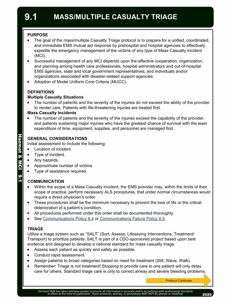
20132013
MASS/MULTIPLE CASUALTY TRIAGE
Hazm
at & M
CI 9.1
Vermont EMS has taken extreme caution to ensure all information is accurate and in accordance with professional standards in effect at the time of publication. These protocols, policies, or procedures MAY NOT BE altered or modified.
Protocol Continues
PURPOSE The goal of the mass/multiple Casualty Triage protocol is to prepare for a unified, coordinated,
and immediate EMS mutual aid response by prehospital and hospital agencies to effectively expedite the emergency management of the victims of any type of Mass Casualty Incident (MCI).
Successful management of any MCI depends upon the effective cooperation, organization, and planning among health care professionals, hospital administrators and out-of-hospital EMS agencies, state and local government representatives, and individuals and/or organizations associated with disaster-related support agencies.
Adoption of Model Uniform Core Criteria (MUCC).
DEFINITIONSMultiple Casualty Situations The number of patients and the severity of the injuries do not exceed the ability of the provider
to render care. Patients with life-threatening injuries are treated first.Mass Casualty Incidents The number of patients and the severity of the injuries exceed the capability of the provider,
and patients sustaining major injuries who have the greatest chance of survival with the least expenditure of time, equipment, supplies, and personnel are managed first.
GENERAL CONSIDERATIONSInitial assessment to include the following: Location of incident. Type of incident. Any hazards. Approximate number of victims. Type of assistance required.
COMMUNICATION Within the scope of a Mass Casualty Incident, the EMS provider may, within the limits of their
scope of practice, perform necessary ALS procedures, that under normal circumstances would require a direct physician’s order.
These procedures shall be the minimum necessary to prevent the loss of life or the critical deterioration of a patient’s condition.
All procedures performed under this order shall be documented thoroughly. See Communications Policy 8.4 or Communications Failure Policy 8.5.
TRIAGEUtilize a triage system such as “SALT” (Sort, Assess, Lifesaving Interventions, Treatment/Transport) to prioritize patients. SALT is part of a CDC-sponsored project based upon best evidence and designed to develop a national standard for mass casualty triage. Assess each patient as quickly and safely as possible. Conduct rapid assessment. Assign patients to broad categories based on need for treatment (Still, Wave, Walk). Remember: Triage is not treatment! Stopping to provide care to one patient will only delay
care for others. Standard triage care is only to correct airway and severe bleeding problems.
2020
9.1

MASS/MULTIPLE CASUALTY TRIAGE H
azmat &
MC
I 9.1
Vermont EMS has taken extreme caution to ensure all information is accurate and in accordance with professional standards in effect at the time of publication. These protocols, policies, or procedures MAY NOT BE altered or modified.
TRIAGE CATEGORIES Immediate: Serious injuries, immediately life-threatening problems, high potential for
survival (e.g., tension pneumothorax, exposure to nerve agent resulting in severe shortness of breath or seizures). Likely to survive given available resources. If no to any of the following: Has a peripheral pulse? Not in respiratory distress? Hemorrhage is controlled? Follows commands or makes purposeful movements?
Delayed: Serious (not minor) injuries requiring care but management can be delayed without increasing morbidity or mortality (e.g., long bone fractures, 40% BSA exposure to mustard gas). If yes to all of the following: Has a peripheral pulse? Not in respiratory distress? Hemorrhage is controlled? Follows commands or makes purposeful movements?
Minimal: Injuries require minor care or no care (e.g., abrasions, minor lacerations, nerve agent exposure with mild runny nose). If yes to all of the following: Has a peripheral pulse? Not in respiratory distress? Hemorrhage is controlled? Follows commands or makes purposeful movements?
Expectant: Unlikely to survive given available resources. Does not mean dead. Method of preserving resources: should receive comfort care or resuscitation when resources are available. Serious injuries: very poor survivability even with maximal care in hospital or pre-hospital setting (e.g., 90% body surface area burn, multiple trauma with exposed brain matter). If no to any of the following: Has a peripheral pulse? Not in respiratory distress? Hemorrhage is controlled? Follows commands or makes purposeful movements?
Dead: Patient is not breathing after opening airway. (In children, if after giving 2 rescue breaths, if appropriate.) Deceased or casualties whose injuries are so severe that their chance of survival does not justify expenditure of limited resources. Tag patients to prevent re-triage. Do not move bodies unless they are hindering efforts to rescue live patients, or they are in danger of being further damaged, for example, burned by fire, building collapse, etc.
TAGGING SYSTEM Use water-repellent triage tags with waterproof markers and attach to the patient. Indicate patient’s triage priority, degree of decontamination performed, treatment and
medications received.
TRIAGE IN HAZARDOUS MATERIAL INCIDENTSDecontaminationThe need for decontamination is the “first triage decision.” since decontamination can be a lengthy process; the “second decision” is which patient(s) are the first to be decontaminated. The “third decision” is based on need for treatment during the decontamination process, since only simple procedures such as antidote administration can be accomplished while wearing PPE.
Identification and Treatment Signs and symptoms of exposure will usually dictate the treatment required, however, at the
earliest possible time, identification of the specific chemical should be made. Reference additional hazardous materials protocols as necessary. Request additional resources. Initial antidote and medical supplies may be limited to priority
patients. Respiratory compromise is a leading factor of fatalities due to hazardous material exposure.
Symptoms of chemical exposure may be delayed and occur suddenly. Constant reevaluation of respiratory status is necessary.
2020
RED
YELLOW
GREEN
BLACK
9.1
GREY
Protocol Continues

20132013
MASS/MULTIPLE CASUALTY TRIAGE
Hazm
at & M
CI 9.1
Vermont EMS has taken extreme caution to ensure all information is accurate and in accordance with professional standards in effect at the time of publication. These protocols, policies, or procedures MAY NOT BE altered or modified. 2020
9.1
Protocol Continues
SALT Mass Casualty Triage

Remove the patient from scene and decontaminate by appropriately trained personnel.
If triage is required for a mass casualty event, use the following guidelines:o If vomiting starts:Within 1 hour of exposure, survival is unlikely and patient should be
tagged “Expectant.”Less than 4 hours after exposure, patient needs immediate
decontamination and evaluation and should be tagged “Immediate.”4 hours after exposure, reevaluation can be delayed 24 – 72 hours if no
other injury is present and patient should be tagged “Minimal”. Routine Patient Care. Treat traumatic injuries and underlying medical conditions. Patients with residual contamination risk from wounds, shrapnel, or internal
contamination should be wrapped in water repellent dressings to reduce cross contamination.
Consider Air Medical Transport after proven definitive decontamination of patient.
2013
P
EMT/ADVANCED EMT STANDING ORDERS
PARAMEDIC STANDING ORDERS
2013
Hazm
at & M
CI 9.2
EA/
Radiation InjuriesAdult & Pediatric
Vermont EMS has taken extreme caution to ensure all information is accurate and in accordance with professional standards in effect at the time of publication. These protocols, policies, or procedures MAY NOT BE altered or modified. 2020
PEARLS: In general, trauma patients who have been exposed to or contaminated by radiation should be
triaged and treated on the basis of the severity of their conventional injuries. A patient who is contaminated with radioactive material (e.g. flecks of radioactive material
embedded in their clothing and skin) generally poses a minimal exposure risk to medical personnel.
9.2
Consider anti-emetic. See Nausea/Vomiting Protocol – Adult & Pediatric 2.12. Consider pain management. See Pain Management Protocol – Adult 2.18A or
Pain Management Protocol – Pediatric 2.18P..

Vermont Adult Medication Reference
This document is to serve as a reference for the 2020 Vermont Statewide Protocols. See the Pediatric Color Coded Appendix for pediatric dosages
Vermont EMS has taken extreme caution to ensure all information is accurate and in accordance with professional standards in effect at the time of publication. These protocols, policies, or procedures MAY NOT BE altered or modified. 2020
Appendix 1 - 1
Medication Adult Protocol/Dosing
Acetaminophen (Tylenol) Indications/Contraindications:
• Indicated for fever control.
• Avoid in patients with severe liver disease.
• Impairment or sensitivity to acetaminophen.
• Do not use with other drug products containing acetaminophen or if patient has taken any drug containing acetaminophen within 4 hours.
Pain Management
• 325 – 1000 PO, no repeat.
• 1000 mg IV
Activated Charcoal Indications:
• Poisoning/Overdose.
Poisoning/Substance Abuse/Overdose
• 25 – 50 grams PO if advised by Medical Direction.
Adenosine (Adenocard) Indications/Contraindications:
• Specifically for treatment or diagnosis of Supraventricular Tachycardia.
• Consider for regular or wide complex tachycardia.
• Contraindicated in patients with WPW (Wolff-Parkinson-White) Syndrome.
Tachycardia
• 6 mg rapid IV/IO push, followed by rapid flush. May repeat 12 mg if no conversion. May repeat successful dose if dysrhythmia recurs after conversion.
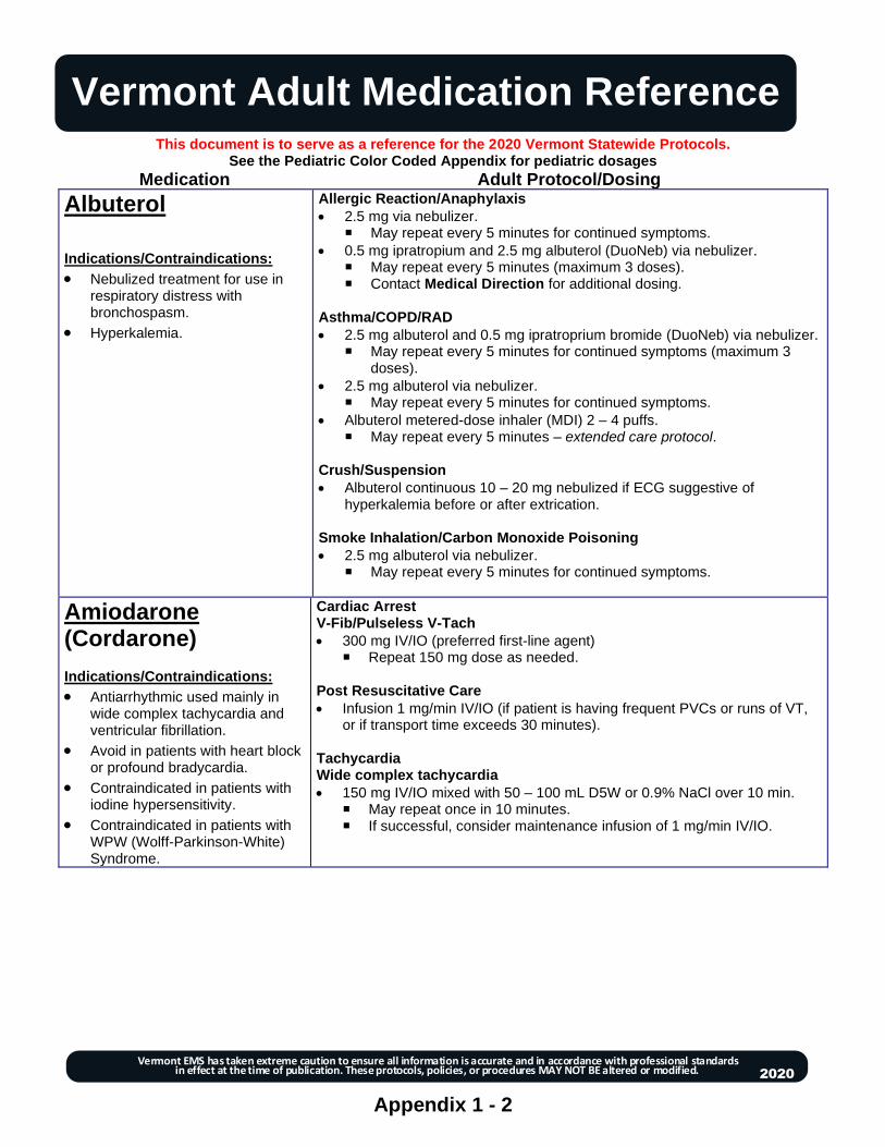
Vermont Adult Medication Reference
This document is to serve as a reference for the 2020 Vermont Statewide Protocols. See the Pediatric Color Coded Appendix for pediatric dosages
Vermont EMS has taken extreme caution to ensure all information is accurate and in accordance with professional standards in effect at the time of publication. These protocols, policies, or procedures MAY NOT BE altered or modified. 2020
Appendix 1 - 2
Medication Adult Protocol/Dosing
Albuterol Indications/Contraindications:
• Nebulized treatment for use in respiratory distress with bronchospasm.
• Hyperkalemia.
Allergic Reaction/Anaphylaxis
• 2.5 mg via nebulizer. May repeat every 5 minutes for continued symptoms.
• 0.5 mg ipratropium and 2.5 mg albuterol (DuoNeb) via nebulizer. May repeat every 5 minutes (maximum 3 doses). Contact Medical Direction for additional dosing.
Asthma/COPD/RAD
• 2.5 mg albuterol and 0.5 mg ipratroprium bromide (DuoNeb) via nebulizer. May repeat every 5 minutes for continued symptoms (maximum 3
doses).
• 2.5 mg albuterol via nebulizer. May repeat every 5 minutes for continued symptoms.
• Albuterol metered-dose inhaler (MDI) 2 – 4 puffs. May repeat every 5 minutes – extended care protocol.
Crush/Suspension
• Albuterol continuous 10 – 20 mg nebulized if ECG suggestive of hyperkalemia before or after extrication.
Smoke Inhalation/Carbon Monoxide Poisoning
• 2.5 mg albuterol via nebulizer. May repeat every 5 minutes for continued symptoms.
Amiodarone (Cordarone) Indications/Contraindications:
• Antiarrhythmic used mainly in wide complex tachycardia and ventricular fibrillation.
• Avoid in patients with heart block or profound bradycardia.
• Contraindicated in patients with iodine hypersensitivity.
• Contraindicated in patients with WPW (Wolff-Parkinson-White) Syndrome.
Cardiac Arrest V-Fib/Pulseless V-Tach
• 300 mg IV/IO (preferred first-line agent) Repeat 150 mg dose as needed.
Post Resuscitative Care
• Infusion 1 mg/min IV/IO (if patient is having frequent PVCs or runs of VT, or if transport time exceeds 30 minutes).
Tachycardia Wide complex tachycardia
• 150 mg IV/IO mixed with 50 – 100 mL D5W or 0.9% NaCl over 10 min. May repeat once in 10 minutes. If successful, consider maintenance infusion of 1 mg/min IV/IO.

Vermont Adult Medication Reference
This document is to serve as a reference for the 2020 Vermont Statewide Protocols. See the Pediatric Color Coded Appendix for pediatric dosages
Vermont EMS has taken extreme caution to ensure all information is accurate and in accordance with professional standards in effect at the time of publication. These protocols, policies, or procedures MAY NOT BE altered or modified. 2020
Appendix 1 - 3
Aspirin Indications/Contraindications:
• An antiplatelet drug for use in cardiac chest pain.
• Contraindicated if history of anaphylaxis to aspirin or NSAIDs
• Contraindicated if active GI bleeding
Acute Coronary Syndrome
• 324 mg PO. If patient has taken a partial dose (81 mg), administer remaining 243
mg.
Atropine Indications/Contraindications:
• Anticholinergic drug used in bradycardias and organophosphate poisonings.
Bradycardia
• 0.5 mg IV/IO every 3 – 5 minutes up to total of 3 mg.
Nerve Agent/Organophosphate Poisoning
• 2 mg IV/IO; repeat every 5 minutes until excess secretions cease (stop).
Atropine and Pralidoxime Auto-Injector (DuoDote or MARK I) Nerve Agent Kit Indications/Contraindications:
• Antidote for Nerve Agents or Organophosphate Overdose.
Nerve Agent/Organophosphate Poisoning
• Refer to Nerve Agents Organophosphate Poisoning Protocol – Adult 2.13A
for symptom assessment and dosing guidelines.
Calcium Chloride 10% solution Indications/Contraindications:
• Indicated for calcium channel blocker overdose,hyperkalemia, or beta blocker overdose.
• Do not routinely use in cardiac arrest.
Bradycardia
• 500 – 1,000 mg (5 – 10 mL of a 10% solution) IV/IO over 10 minutes.
▪ Avoid use if pt is taking digoxin.
▪ Do not exceed 1 mL per minute. Flush with 0.9% NaCl before and after administration.
▪ Do not mix with or infuse immediately before or after sodium bicarbonate.
Cardiac Arrest Wide Complex PEA
1 gram of 10% solution IV OR Calcium Gluconate 2 grams IV AND Sodium Bicarbonate 1 – 2 mEq/kg IV.
Crush/Suspension Injury
• 1 gram of 10% solution IV/IO over 5 minutes. May repeat in 10 minutes.

Vermont Adult Medication Reference
This document is to serve as a reference for the 2020 Vermont Statewide Protocols. See the Pediatric Color Coded Appendix for pediatric dosages
Vermont EMS has taken extreme caution to ensure all information is accurate and in accordance with professional standards in effect at the time of publication. These protocols, policies, or procedures MAY NOT BE altered or modified. 2020
Appendix 1 - 4
Calcium Gluconate Indications/Contraindications:
• Indicated for calcium channel blocker overdose, hyperkalemia/ renal failure, or beta blocker overdose.
Bradycardia
• 2 grams IV/IO over 5 minutes.
▪ May repeat in 10 minutes. Cardiac Arrest Wide Complex PEA
2 grams IV OR Calcium Chloride 1 gram of 10% solution IV AND Sodium Bicarbonate 1 – 2 mEq/kg IV.
Crush/Suspension Injury
• 2 gram of a 10% solution IV/IO over 5 minutes. May repeat in 10 minutes.
Dexamethasone Indications/Contraindications:
• Steroid used to control inflam-matory conditions, asthma, croup, allergic reactions or adrenal insufficiency
Adrenal Insufficiency
• 10 mg IV/IO/IM. Allergic Reaction/Anaphylaxis
• 0.6 mg/kg IV/IO/IM/PO, maximum dose 10 mg – extended care protocol. Asthma/COPD/RAD
• 10 mg IV/IO/IM/PO.
Dextrose 5%, 10%, 50% Glucose solutions Indications/Contraindications:
• Symptomatic hypoglycemia.
• Use in medication infusion medium.
Diabetic Emergencies (Hypoglycemia)
• Dextrose 10% (preferred) or 50% IV up to 25 grams. Recheck blood glucose after 5 minutes. Repeat up to 25 grams dextrose 10% or 50% if glucose levels < 60
mg/dl with continued altered mental status. 25 grams = 250 mL of 10% solution. 1 amp (25 grams) = 50 mL of 50% solution. D5W medication infusion medium.
Diazepam (Valium) Indications/Contraindications:
• Seizure control.
• Sedation.
• Anxiolytic.
(continued)
Bradycardia Procedural Sedation for Cardiac Pacing
• 5 mg IV/IO, may repeat 2.5 mg once in 5 minutes.
Continuous Positive Airway Pressure (CPAP) Anxiolytic.
• Contact Medical Direction for authorization.
• 5 mg IV (may repeat once in 5 minutes).
Hyperthermia
• 2 mg IV, may repeat once in 5 minutes.
Nerve Agent/Organophosphate Poisoning
• 5 mg IV every 5 minutes OR
• 10 mg IM OR
• Diazepam auto-injector (10mg).
• Repeat every 10 minutes as needed
Poisoning/Substance Abuse/Overdose
• 2 mg IV, may repeat once in 5 minutes

Vermont Adult Medication Reference
This document is to serve as a reference for the 2020 Vermont Statewide Protocols. See the Pediatric Color Coded Appendix for pediatric dosages
Vermont EMS has taken extreme caution to ensure all information is accurate and in accordance with professional standards in effect at the time of publication. These protocols, policies, or procedures MAY NOT BE altered or modified. 2020
Appendix 1 - 5
Diazepam (continued)
(Valium) Indications/Contraindications:
• Seizure control.
• Sedation.
• Anxiolytic.
Restraints
• Contact Medical Direction for 2.5 mg IV, may repeat once in 5 minutes May combine and administer Benzodiazepine and Haloperidol in one
syringe. Contact Medical Direction if additional dosing is needed.
Seizure
• May assist with patient’s own diazepam gel as prescribed.
• 5 – 10 mg IV, then 2.5 mg every 5 minutes (maximum dose 20 mg)
Tachycardia For Cardioversion Sedation
• 5 mg IV/IO, may repeat 2.5 mg once in 5 minutes.
Traumatic Brain Injury
• 5 mg IV/IO, may repeat once in 5 minutes.
Diltiazem (Cardizem) Indications/Contraindications:
• Calcium channel blocker used to treat narrow complex SVT.
• Contraindicated in patients with heart block, ventricular tachycardia, WPW, and/or acute MI.
• Contraindicated in patients with WPW (Wolff-Parkinson-White) Syndrome.
Tachycardia Narrow Complex Tachycardia
• 0.25 mg/kg IV/IO (maximum dose 20 mg) over 2 minutes. Consider 10 mg maximum dose for elderly patient or patient with low
BP. May repeat dose in 15 minutes at 0.35 mg/kg (maximum dose 25 mg)
if necessary. Consider maintenance infusion 5 – 15 mg/hour IV/IO.
Diphenhydramine (Benadryl) Indications/Contraindications:
• Antihistamine used as an adjunctive treatment in allergic reactions.
• Antidote for dystonic reaction.
Allergic Reaction/Anaphylaxis
• 25 – 50 mg IM/IV/IO to treat pruritus.
• 25 – 50 mg by mouth – extended care protocol.
May repeat every 4 – 6 hours as needed; maximum dose of 300 mg/24 hours.
Restraints
• For acute dystonic reaction to haloperidol 25 – 50 mg IV OR 50 mg IM.
Nausea/Vomiting
• 25 – 50 mg IV/IM for dystonic reaction.
• 25 mg PO/chewed – extended care for motion sickness.
Poisoning/Substance Abuse/Overdose
• 25 – 50 mg IV/IM.

Vermont Adult Medication Reference
This document is to serve as a reference for the 2020 Vermont Statewide Protocols. See the Pediatric Color Coded Appendix for pediatric dosages
Vermont EMS has taken extreme caution to ensure all information is accurate and in accordance with professional standards in effect at the time of publication. These protocols, policies, or procedures MAY NOT BE altered or modified. 2020
Appendix 1 - 6
Epinephrine 1:1,000 (1 mg/mL) Indications/Contraindications:
• Bronchodilation in Asthma and COPD exacerbation. Primary treatment for anaphylaxis.
• Vasopressor used for bradycardia, post-resuscitative care, shock, anaphylaxis.
MIXING INSTRUCTIONS FOR EPINEPHRINE INFUSIONS Epinephrine 1:1,000 (1mg/mL) multidose vial (30mL) and withdraw 4 mg (4mL) and add to a 250 mL infusion bag of D5W or 0.9% NaCl. The resulting concentration is 16 mcg/mL
Allergic Reaction/Anaphylaxis
• 0.3 mg IM by autoinjector OR 0.3 mg (0.3 mL) IM. For additional dosing, contact Medical Direction (EMT).
• 0.3 mg (0.3 mL) IM. May repeat every 5 – 15 minutes. Maximum of 3 doses. For additional dosing, contact Medical Direction.
• For anaphylaxis refractory to IM epinephrine, consider epinephrine infusion 2 – 10 mcg/min IV/IO, titrated to effect. (Infusion pump required.)
Asthma/COPD/RAD
• Consider 0.3 mg IM by autoinjector (preferred) OR epinephrine (1:1,000) (1 mg/mL) 0.3 mg (0.3 mL) IM. For additional dosing, contact Medical Direction.
Bradycardia
• Infusion 2 – 10 mcg/min IV/IO, titrated to effect (Infusion pump required.)
• Push dose (10 mcg/mL) for short transport times or as bridge to infusion. Prepare 10 mcg/mL by adding 1 mL 0.1 mg/mL epinephrine to 9 mL NS or 0.9% NaCl. Then administer 10 – 20 mcg (1 – 2 mL) boluses IV/IO every 2 minutes. Switch to infusion as soon as practical.
Post-Resuscitative Care
• Infusion 2-10 mcg/min IV/IO titrated to effect. (Infusion pump required.)
• Push dose (10 mcg/mL) for short transport times or as bridge to infusion. Prepare 10 mcg/mL by adding 1 mL 0.1 mg/mL epinephrine to 9 mL NS or 0.9% NaCl. Then administer 10 – 20 mcg (1 – 2 mL) boluses IV/IO every 2 minutes. Switch to infusion as soon as practical
Septic Shock
• Infusion 2 – 10 mcg/min IV/IO titrated to MAP ≥ 65 (systolic ≥ 90). (Infusion pump required.)
• Push dose (10 mcg/mL) for short transport times or as bridge to infusion. Prepare 10 mcg/mL by adding 1 mL 0.1 mg/mL epinephrine to 9 mL NS or 0.9% NaCl. Then administer 10 – 20 mcg (1 – 2 mL) boluses IV/IO every 2 minutes. Switch to infusion as soon as practical.
Shock (Cardiogenic or Distributive)
• Infusion 2-10 mcg/min IV/IO titrated to effect. (Infusion pump required.)
• Push dose (10 mcg/mL) for short transport times or as bridge to infusion. Administer 10 – 20 mcg (1 – 2 mL) boluses IV/IO every 2 minutes.
Smoke Inhalation
• 3 mg (mL) in 3 mL 0.9% NaCl via nebulizer for symptomatic patients.
Epinephrine 1:10,000 (0.1 mg/mL) Indications/Contraindications:
• Vasopressor used in cardiac arrest.
• Not recommended in traumatic cardiac arrest.
Cardiac Arrest
• 1 mg IV/IO. Repeat every other cycle Maximum dose 3 mg..
Hypothermia
• 1 mg IV/IO up to 3 doses, and defibrillation as indicated.

Vermont Adult Medication Reference
This document is to serve as a reference for the 2020 Vermont Statewide Protocols. See the Pediatric Color Coded Appendix for pediatric dosages
Vermont EMS has taken extreme caution to ensure all information is accurate and in accordance with professional standards in effect at the time of publication. These protocols, policies, or procedures MAY NOT BE altered or modified. 2020
Appendix 1 - 7
Etomidate (Amidate)
Indications/Contraindications:
• Sedative used in Rapid Sequence Intubation.
Rapid Sequence Intubation {Sedate then} Paralyze
• 0.3 mg/kg IV/IO (maximum 40 mg).
Fentanyl (Sublimaze)
Indications/Contraindications:
• Narcotic analgesic
• Avoid use if BP < 100 mmHg.
Acute Coronary Syndrome
• 25 – 50 mcg slow IV push.
• Repeat every 5 minutes up to 300 mcg and systolic BP remains ≥100 mmHg.
Bradycardia Analgesia for Cardiac Pacing
• 25 – 50 mcg slow IV push.
• May repeat every 5 minutes to a total of 300 mcg and systolic BP remains ≥100 mmHg.
Pain Management
• 25 – 100 mcg slow IV, every 2 – 5 minutes to a total of 300 mcg titrated to pain relief OR 50 – 100 mcg IM/IN, every 5 minutes to a total of 300 mcg titrated to pain relief OR Morphine 2 – 5 mg IV/IM every 10 minutes to a total of 20 mg titrated to pain.
• AND/OR Ketamine 0.25 mg/kg IV infusion (in 50 – 100 mL bag 0.9% NaCl over 10 minutes). May be administered via bolus. (Consider lower 0.15 mg/kg dose for frail or elderly patients.) OR Ketamine 0.5 mg/kg IM/IN May repeat ketamine dose every 15 – 20 minutes for a total of 1
mg/kg.
Contact Medical Direction for additional dosing.
Rapid Sequence Intubation Post-Intubation Care
• Fentanyl 50 – 100 mcg slow IV/IO push, may repeat every 15 minutes as needed for anesthesia (maximum 300 mcg).
• AND Midazolam 2.5 – 5 mg IV/IO, every 5 – 10 minutes (maximum 20 mg) OR Lorazepam 1 – 2 mg IV/IO every 15 minutes (maximum 10 mg).
• Contact Medical Direction for additional dosing.
• Be sure to maintain adequate sedation if patient is paralyzed.
Nasotracheal and Orotracheal Intubation Post Intubation Care
• Fentanyl 50 – 100 mcg IV/IO, may repeat every 15 minutes as needed for anesthesia (maximum 300 mcg).
• AND Midazolam 2.5 – 5 mg IV/IO, every 5 – 10 minutes (maximum 20 mg) OR Lorazepam 1 – 2 mg IV/IO every 15 minutes (maximum 10 mg).
• Contact Medical Direction for additional dosing.
Supraglottic Airway
• 50 – 100 mcg slow IV/IO push every 15 minutes, as needed (maximum 300 mcg) AND
• Midazolam 2.5 – 5 mg IV/IO every 5 – 10 minutes, as needed (maximum 20 mg) OR
• Lorazepam 1 – 2 mg IV/IO every 15 minutes as needed (max 10 mg).

Vermont Adult Medication Reference
This document is to serve as a reference for the 2020 Vermont Statewide Protocols. See the Pediatric Color Coded Appendix for pediatric dosages
Vermont EMS has taken extreme caution to ensure all information is accurate and in accordance with professional standards in effect at the time of publication. These protocols, policies, or procedures MAY NOT BE altered or modified. 2020
Appendix 1 - 8
Glucagon
Indications/Contraindications:
• Converts glycogen to glucose in the liver to increase blood sugar
• Use in patients with no IV access
• Indicated for beta blocker or calcium channel blocker overdose
Bradycardia
• 2 – 5 mg IV/IO over 3 – 5 minutes. ▪ May repeat up to 10 mg. ▪ If effective, place on infusion 1 – 5 mg/hr IV/IO via pump.
Diabetic Emergencies (Hypoglycemia)
• 1 mg IM. Recheck glucose 15 minutes after administration of glucagon. May repeat glucagon 1 mg IM if glucose level is <60 mg/dl with
continued altered mental status.
Glucose Oral Glucose Solutions
Indications/Contraindications:
• Use in conscious hypoglycemic states.
Diabetic Emergencies (Hypoglycemia)
• Administer 1 – 2 tubes commercially prepared glucose gel or 15 to 30 mL (1 – 2 tablespoons) of Pure VT Maple Syrup or equivalent, for a standard dose of 15 to 30 grams sugar.
Haloperidol (Haldol)
Indications/Contraindications:
• Medication to assist with sedation of agitated patients.
• Chemical restraint.
• Caution: May lower seizure threshhold.
• Use lower doses in elderly/frail.
Restraints
• Contact Medical Direction for 5 mg IM, may repeat once in 5 minutes, maximum total dose 10 mg. May be combined with midazolam, lorazepam or diazepam. May combine and administer benzodiazipine and haloperidol in one
syringe. Contact Medical Direction if additional dosing is needed.
Heparin
Indications/Contraindications
• STEMI and no affirmative finding from fibrinolytic questionnaire.
• Contraindication - history of Heparin Induced Thrombocytopenia
Paramedic
• Maintenance of already established heparin drip.
Hydrocortisone (Solu-Cortef)
Indications/Contraindications
• Steroid used for adrenal insufficiency and associated distributive shock.
Adrenal Insufficiency
• 100 mg IV/IO/IM (preferred steroid for use in adrenal insufficiency).
▪ May be repeated every 6 hours – extended care protocol. Shock
• 100 mg IV/IO/IM.

Vermont Adult Medication Reference
This document is to serve as a reference for the 2020 Vermont Statewide Protocols. See the Pediatric Color Coded Appendix for pediatric dosages
Vermont EMS has taken extreme caution to ensure all information is accurate and in accordance with professional standards in effect at the time of publication. These protocols, policies, or procedures MAY NOT BE altered or modified. 2020
Appendix 1 - 9
Hydroxocobalamin (Cyanokit)
Smoke Inhalation
• Hydroxocobalamin via use of Cyanokit:
▪ Reconstitute: Place the vial of hydroxocobalamin in an upright position; add 0.9% NaCl to the vial (200 mL for 5 grams vial or 100 mL for 2.5 grams vial) using the transfer spike. Fill to the line.
▪ Rock vial for at least 60 seconds (do not shake).
▪ Using vented intravenous tubing, administer IV over 15 minutes.
▪ Depending on clinical response, a second dose may be required.
Ipratropium Bromide (Atrovent)
Indications/Contraindications:
• Anticholinergic bronchodilator. Blocks the muscarinic receptors of acetylcholine.
• Relief of bronchospasm in patients with reversible obstructive airway disease and bronchospasm.
Allergic Reaction/Anaphylaxis
• 0.5 mg ipratropium and 2.5 mg albuterol (DuoNeb) via nebulizer. May repeat every 5 minutes (maximum 3 doses). Contact Medical Direction for additional dosing.
Asthma/COPD/RAD
• 0.5 mg ipratropium and 2.5 mg albuterol (DuoNeb) via nebulizer. May repeat every 5 minutes for continued symptoms (maximum 3
doses).
Ketamine Indications/Contraindications:
• Sedative used in Rapid Sequence Intubation.
• Pain control.
• Chemical restraint
• Contraindicated in patients unable to tolerate hyperdynamic states such as those with known or suspected aortic dissection, myocardial infarction, and aortic aneurysm, and those that cannot tolerate hypertension.
• Avoid in patients with known schizophrenia.
(continued)
Nasotracheal and Orotracheal Intubation Post Intubation Care
• 1 mg/kg ideal body weight (IBW) every 5 – 15 minutes, as needed. Contact Medical Direction for additional dosing.
Pain Management
• 0.25 mg/kg IV infusion (in 50 – 100 mL bag 0.9% NaCl over 10 minutes). May be administered via bolus. (Consider lower 0.15 mg/kg dose for frail or elderly patients.) OR 0.5 mg/kg IM/IN
• AND Fentanyl (preferred first-line narcotic agent) 25 – 100 mcg slow IV, every 2 – 5 minutes to a total of 300 mcg titrated to pain OR Fentanyl 50 – 100 mcg IM/IN, every 5 minutes to a total of 300 mcg titrated to pain OR Morphine 2 – 5 mg IV/IM every 10 minutes to a total of 20 mg titrated to pain. May repeat ketamine dose every 15 – 20 minutes for a total of 1
mg/kg.
• Contact Medical Direction for additional dosing
Rapid Sequence Intubation {Sedate then} Paralyze
• 2 mg/kg IV/IO

Vermont Adult Medication Reference
This document is to serve as a reference for the 2020 Vermont Statewide Protocols. See the Pediatric Color Coded Appendix for pediatric dosages
Vermont EMS has taken extreme caution to ensure all information is accurate and in accordance with professional standards in effect at the time of publication. These protocols, policies, or procedures MAY NOT BE altered or modified. 2020
Appendix 1 - 10
Ketamine (continued) Indications/Contraindications:
• Sedative used in Rapid Sequence Intubation.
• Pain control.
• Chemical restraint
• Contraindicated in patients unable to tolerate hyperdynamic states such as those with known or suspected aortic dissection, myocardial infarction, and aortic aneurysm, and those that cannot tolerate hypertension.
• Avoid in patients with known schizophrenia.
Restraints
• Contact Medical Direction for 4 mg/kg IM injection only. o Use 100 mg/mL concentration; maximum dose 500 mg. o Repeat 100 mg IM dose in 5 – 10 minutes for continued agitation.
Supraglottic Airway
• 1 mg/kg ideal body weight (IBW) IV ever 5 – 15 minutes, as needed.
Traumatic Brain Injury
• 4 mg/kg (maximum dose 500 mg) IM injection only
• Contact Medical Direction for additional dosing.
Ketorolac (Toradol) Indications/Contraindications:
• A nonsteroidal anti-inflammatory drug used for pain control.
• Consider as first line in renal colic.
• Avoid Ketorolac in patients with NSAID allergy, aspirin-sensitive asthma, renal insufficiency, pregnancy, or known peptic ulcer disease.
• Avoid NSAIDS in women who are pregnant or could be pregnant.
• Avoid in patients currently taking anticoagulants such as coumadin.
Pain Management
• 15 mg IV Contact Medical Direction
• 30 mg IM Contact Medical Direction

Vermont Adult Medication Reference
This document is to serve as a reference for the 2020 Vermont Statewide Protocols. See the Pediatric Color Coded Appendix for pediatric dosages
Vermont EMS has taken extreme caution to ensure all information is accurate and in accordance with professional standards in effect at the time of publication. These protocols, policies, or procedures MAY NOT BE altered or modified. 2020
Appendix 1 - 11
Lactated Ringers Indications/Contraindications:
• Use Lactated Ringers with caution in patients with: o Hyperkalemia or severe
renal failure (potassium) o Severe hepatic failure
(impaired lactate clearance) o Severe metabolic acidosis or
alkalosis (potassium and worsening alkalosis)
o Lactic acidosis o Neonates and infants less
than 6 months (lactate effects on neonates)
Lactated Ringers may be used as a direct substitute for Normal Saline with the following exceptions and precautions: Lactated Ringers (LR) should NOT be directly combined with the following drug agents (due to limited data or clear evidence of incompatibility). These medications should be administered at a site separate from where the LR is infusing via a normal saline lock/line, or stop the LR infusion for medication injection, then administer a saline flush, and then restart the LR infusion.
• Amiodarone
• Atropine
• Calcium Chloride
• Dexamethasone
• Diazepam
• Diltiazem
• Epinephrine
• Etomidate
• Fentanyl
• Glucagon
• Ketamine
• Lorazepam
• Metoprolol
• Naloxone
• Pralidoxime
• Sodium Bicarbonate
• Tranexamic Acid
Lidocaine Indications/Contraindications:
• Antiarrhythmic used for control of ventricular dysrhythmias.
• Used prior to intubation of patients with suspected increased intracranial pressure (e.g., TBI, ICH) to reduce increases in intracranial pressure
• Anesthetic for nasotracheal intubation and intraosseous.
• Do not use lidocaine if CHF, cardiogenic shock, heart block or WPW.
Cardiac Arrest V-Fib/Pulseless V-Tach
• 1 – 1.5 mg/kg IV/IO. Repeat dose 0.5 – 0.75 mg/kg up to a maximum dose of 3 mg/kg.
Intraosseous Access
• Slowly administer 20 – 50 mg (1 – 2.5 mL) 2% lidocaine through IO device catheter. Allow 2 – 5 min for anesthetic effects, if possible.
Nasotracheal Intubation
• 2% lidocaine jelly. Post-Resuscitative Care
• Maintenance infusion 1 – 4 mg/min IV/IO (30 – 50 mcg/kg/min) if patient is having frequent PVCs or runs of VT, or if transport time exceeds 30 minutes.
Tachycardia
• 1 mg/kg IV/IO. May repeat every 5 minutes (total of 3 doses) to maximum of 3 mg/kg. If successful, consider a maintenance infusion of 1 – 4 mg/min IV/IO.

Vermont Adult Medication Reference
This document is to serve as a reference for the 2020 Vermont Statewide Protocols. See the Pediatric Color Coded Appendix for pediatric dosages
Vermont EMS has taken extreme caution to ensure all information is accurate and in accordance with professional standards in effect at the time of publication. These protocols, policies, or procedures MAY NOT BE altered or modified. 2020
Appendix 1 - 12
Lorazepam (Ativan) Indications/Contraindications:
• Seizure control.
• Sedation.
• Anxiolytic.
(continued)
Bradycardia Procedural Sedation for Cardiac Pacing
• 1 mg IV/IO, may repeat once in 5 minutes OR
• 2 mg IM, may repeat once in 10 minutes. Continuous Positive Airway Pressure (CPAP) Anxiolytic
• Contact Medical Direction for authorization.
• 0.5 – 1 mg IV; may repeat once in 5 minutes OR
• 1 – 2 mg IM, may repeat once in 10 minutes. Hospice Anxiety
• 0.25 – 2 mg PO or SL Hyperthermia
• 1 mg IV, may repeat once in 5 minutes OR
• 2 mg IM, may repeat once in 10 minutes. Nasotracheal and Orotracheal Intubation Post Intubation Care
• 1 – 2 mg IV/IO every 15 minutes as needed for sedation (maximum 10 mg) OR Midazolam 2.5 – 5 mg IV/IO, every 5 – 10 minutes as needed (maximum 20 mg).
• AND Fentanyl 50 – 100 mcg slow IV/IO push. May repeat every 15 minutes as needed (maximum 300 mcg).
Contact Medical Direction for additional dosing.
Nerve Agent/Orophosphate Poisoning
• 1 mg IV, may repeat once in 5 minutes OR
• 2 mg IM, may repeat once in 10 minutes.
Poisoning/Substance Abuse/Overdose
• 1 mg IV, may repeat once in 5 minutes OR 2 mg IM, may repeat once in 10 minutes. Rapid Sequence Intubation
Post Intubation Care
• 1 – 2 mg IV/IO every 15 minutes as needed for sedation (maximum 10 mg. OR Midazalem 2.5 – 5 mg IV/IO every 5 – 10 minutes (maximum 20 mg)
• AND Fentanyl 50 – 100 mcg slow IV/IO push, may repeat every 15 minutes as needed for anesthesia (maximum 300 mcg). Contact Medical Direction for additional dosing. Be sure to maintain adequate sedation if patient is paralyzed.
Restraints
• Contact Medical Direction for 1 mg IV, may repeat once in 5 minutes OR 2 mg IM, may repeat once in 10 minutes. May combine and administer Benzodiazepine and Haloperidol in one
syringe. Contact Medical Direction if additional dosing is needed.
Seizure
• 1 - 2 mg IV, may repeat every 5 minutes (maximum dose 8 mg)

Vermont Adult Medication Reference
This document is to serve as a reference for the 2020 Vermont Statewide Protocols. See the Pediatric Color Coded Appendix for pediatric dosages
Vermont EMS has taken extreme caution to ensure all information is accurate and in accordance with professional standards in effect at the time of publication. These protocols, policies, or procedures MAY NOT BE altered or modified. 2020
Appendix 1 - 13
Lorazepam (continued) (Ativan) Indications/Contraindications:
• Seizure control.
• Sedation.
• Anxiolytic.
Supraglottic Airway
• Fentanyl 50 – 100 mcg slow IV/IO push every 15 minutes, as needed (maximum 300 mcg) AND Midazolam 2.5 – 5 mg IV/IO every 5 – 10 minutes, as needed
(maximum 20 mg) OR Lorazepam 1 – 2 mg IV/IO every 15 minutes as needed (maximum 10
mg)
Tachycardia For Cardioversion Sedation
• 1 mg IV/IO, may repeat once in 5 minutes OR
• 2 mg IM, may repeat once in 10 minutes.
Traumatic Brain Injury
• 1 mg IV/IO, may repeat once in 5 minutes OR
• 2 mg IM, may repeat once in 10 minutes.
Magnesium Sulfate Indications/Contraindications:
• Elemental electrolyte used to treat eclampsia during the third trimester of pregnancy.
• A smooth muscle relaxor used in refractory respiratory distress resistent to beta-agonists.
• Torsades de Pointes.
Asthma/COPD/RAD
• 2 grams in 50 mL D5W or 0.9% NaCl IV/IO over 10 minutes.
Cardiac Arrest – Torsades de Pointes With No Pulse
• 1 – 2 grams IV/IO over 1 – 2 minutes.
Obstetrical Emergencies
• 4 grams in 10 mL D5W or 0.9% NaCl given slow IV push over 5 minutes for patients in the third trimester of pregnancy or post partum who are seizing or are post-ictal.
Seizures
• 4 grams in 10 mL D5W or 0.9% NaCl given slow IV push over 5 minutes in the presence of seizure in the third trimester of pregnancy or post partum.
Tachycardia – Torsades de Pointes
• If pulse present, consider 2 grams IV/IO diluted in 10 mL D5W or 0.9% NaCl over 10 minutes.
Methylprednisolone (Solu-medrol) Indications/Contraindications:
• Steroid used in respiratory distress to reverse inflammatory and allergic reactions.
Adrenal Insufficiency
• 125 mg IV/IO/IM.
Allergic Reaction/Anaphylaxis
• 1 mg/kg IV, maximum dose 125 mg every 6 hours – extended care protocol.
Asthma/COPD/RAD
• 125 mg IV/IO/IM.
Metoclopramide (Reglan) Indications/Contraindications:
• Anti-emetic used to control nausea and/or vomiting and as alternative treatment for adrenal insufficiency.
Nausea/Vomiting
• 5 mg IV. May repeat once after 10 minutes if nausea/vomiting persists. May repeat IM every 4 – 6 hours as needed - extended care protocol. Administer slowly, over 1 – 2 minutes, to reduce dystonic reactions.

Vermont Adult Medication Reference
This document is to serve as a reference for the 2020 Vermont Statewide Protocols. See the Pediatric Color Coded Appendix for pediatric dosages
Vermont EMS has taken extreme caution to ensure all information is accurate and in accordance with professional standards in effect at the time of publication. These protocols, policies, or procedures MAY NOT BE altered or modified. 2020
Appendix 1 - 14
Metoprolol (Lopressor) Indications/Contraindications:
• Use for rate control in narrow complex tachycardia with an irregular rhythm.
• Contraindicated in patients with HR < 45 bpm, SBP < 100 mmHg, heart block or acute heart failure syndromes (CHF or cardiogenic shock).
• Use with caution in patients with bronchospastic disease.
• Contraindicated in patients with WPW (Wolff-Parkinson-White) Syndrome.
Tachycardia
• 5 mg IV/IO over 2 – 5 minutes. May repeat every 5 minutes to a maximum of 15 mg as needed to
achieve a ventricular rate of 90 – 100 BPM.
Midazolam (Versed) Indications/Contraindications:
• Seizure control.
• Sedation.
• Anxiolytic.
(continued)
Bradycardia Procedural Sedation for Cardiac Pacing
• 2.5 mg IV/IO/intranasal, may repeat once in 5 minutes OR
• 5 mg IM, may repeat once in 10 minutes.
Continuous Positive Pressure Airway (CPAP) Anxiolytic
• Contact Medical Direction for authorization.
• 2.5 mg IV/intranasal, may repeat once in 5 minutes OR
• 5 mg IM, may repeat once in 10 minutes.
Hospice Anxiety
• 2.5 mg IN, repeat every 10 – 15 minutes as needed to a maximum of 7.5 mg.
Hyperthermia
• 2.5 mg IV/intranasal, may repeat once in 5 minutes OR
• 5 mg IM, may repeat once in 10 minutes.
Nasotracheal and Orotracheal Intubation Post Intubation Care
• 2.5 – 5 mg IV/IO, every 5 – 10 minutes as needed for sedation (maximum 20 mg) OR Lorazepam 1 – 2 mg IV/IO every 15 minutes as needed for sedation (maximum 10 mg).
• AND Fentanyl 50 – 100 mcg slow IV/IO push. May repeat every 15 minutes as needed (maximum 300 mcg).
• Contact Medical Direction for additional dosing.

Vermont Adult Medication Reference
This document is to serve as a reference for the 2020 Vermont Statewide Protocols. See the Pediatric Color Coded Appendix for pediatric dosages
Vermont EMS has taken extreme caution to ensure all information is accurate and in accordance with professional standards in effect at the time of publication. These protocols, policies, or procedures MAY NOT BE altered or modified. 2020
Appendix 1 - 15
Midazolam (continued) (Versed) Indications/Contraindications:
• Seizure control.
• Sedation.
• Anxiolytic.
Nerve Agent/Organophosphate Poisoning
• 2.5 mg IV/intranasal, may repeat every 5 minutes OR
• 5 mg IM, may repeat every 10 minutes.
Pain Management
• Antidote: For dysphoria (emergence reaction) caused by ketamine, 1 – 2 mg IV/IM every 5 minutes as needed.
Poisoning/Substance Abuse/Overdose
• 2.5 mg IV/intranasal, may repeat once in 5 minutes OR
• 5 mg IM, may repeat once in 10 minutes.
Rapid Sequence Intubation Post-Intubation Care
• 2.5 – 5 mg IV, every 5 – 10 minutes as needed for sedation (maximum 20 mg) OR Lorazepam 1 – 2 mg IV/IO every 15 minutes (maximum 10 mg)
• AND Fentanyl 50 – 100 mcg slow IV/IO push, may repeat every 15 minutes as needed for anesthesia (maximum 300 mcg). Contact Medical Direction for additional dosing. Be sure to maintain adequate sedation if patient is paralyzed.
Rapid Sequence Intubation {Sedate then} Paralyze
• 0.2 mg/kg IV/IO (0.1mg/kg IV/IO for patients in shock).
Restraints
• Contact Medical Direction for 5 mg IM (preferred route if patient is combative) OR intranasal, may repeat once in 10 minutes OR 2.5 mg IV, may repeat once in 5 minutes May combine and administer Benzodiazepine and Haloperidol in one
syringe. Contact Medical Direction if additional dosing is needed.
Seizure
• May assist with patient’s own intranasal midazolam as prescribed.
• 10 mg IM (preferred) or intranasal, may repeat 10 mg IM/IN every 10 minutes (maximum dose 20 mg). Note: 5 mg/mL concentration is recommended for IM/intranasal OR
• 5 mg IV, may repeat every 5 minutes until seizure activity resolved (maximum dose 20 mg).
Supraglottic Airway
• Fentanyl 50 – 100 mcg slow IV/IO push every 15 minutes, as needed (max 300 mcg) AND Midazolam 2.5 – 5 mg IV/IO every 5 – 10 minutes, as needed
(maximum 20 mg) OR Lorazepam 1 – 2 mg IV/IOevery 15 minutes as needed (max 10 mg)
Tachycardia For Cardioversion Sedation
• 2.5 mg IV/IO/intranasal, may repeat once in 5 minutes OR
• 5 mg IM, may repeat once in 10 minutes.
Traumatic Brain Injury
• 2.5 mg IV/IO/intranasal, may repeat once in 5 minutes OR
• 5 mg IM, may repeat once in 10 minutes.

Vermont Adult Medication Reference
This document is to serve as a reference for the 2020 Vermont Statewide Protocols. See the Pediatric Color Coded Appendix for pediatric dosages
Vermont EMS has taken extreme caution to ensure all information is accurate and in accordance with professional standards in effect at the time of publication. These protocols, policies, or procedures MAY NOT BE altered or modified. 2020
Appendix 1 - 16
Morphine Sulfate
Indications/Contraindications:
• Narcotic analgesic
• Avoid use if BP < 100 mmHg.
• Associated with increased mortality.
Acute Coronary Syndrome
• 2 – 4 mg IV/IM every 5 minutes to a maximum of 15 mg titrated to pain and systolic BP remains ≥100 mmHg.
Bradycardia Analgesia for Cardiac Pacing
• 2 – 4 mg IV every 10 minutes to a total of 15 mg and systolic BP ≥100 mmHg.
Pain Management
• 2 – 5 mg IV/IM every 10 minutes to a total of 20 mg titrated to pain relief and if systolic BP is > 100 mmHg OR Fentanyl (preferred first-line narcotic agent) 25 – 100 mcg slow IV, every 2 – 5 minutes to a total of 300 mcg titrated to pain OR Fentanyl 50 – 100 mcg IM/IN, every 5 minutes to a total of 300 mcg titrated to pain.
• AND/OR Ketamine 0.25 mg/kg IV infusion (in 50 – 100 mL bag 0.9% NaCl over 10 minutes). May be administered via bolus. (Consider lower 0.15 mg/kg dose for frail or elderly patients.) OR Ketamine 0.5 mg/kg IM/IN. May repeat ketamine dose every 15 – 20 minutes for a total of 1
mg/kg.
Contact Medical Direction for additional dosing.
Naloxone (Narcan)
Indications/Contraindications:
• Narcotic overdose.
Altered Mental Status (Unknown Etiology)
• 1 mg (1 mL) per nostril via atomizer for a maximum of 2 mg. May repeat
every 3 – 5 minutes if no response to a maximum of 12 mg.
• Single spray of NARCAN® Nasal Spray (4mg) into one nostril. May repeat
every 3 – 5 minutes if no response or if patient relapses, to a total of 12
mg.
• 0.4 – 2 mg IV/IM/IO/SQ/intranasal, titrated to response.
If no response, may repeat initial dose every 3 – 5 minutes to a total of 12 mg.
Cardiac Arrest
• If suspected opioid overdose, administer 2 mg IV/IO..
Pain Management
• Antidote: For hypoventilation from opiate administration by EMS personnel, administer naloxone 0.4 – 2.0 mg SQ/IV/IO/IM or 2.0 – 4.0 mg intranasal as needed.
Poisoning/Substance Abuse/Overdose Narcotic Overdose
• 1 mg (1 mL) per nostril via atomizer for a maximum of 2 mg. May repeat
every 3 – 5 minutes if no response to a maximum of 12 mg.
• Single spray of NARCAN® Nasal Spray (4mg) into one nostril. May repeat
every 3 – 5 minutes if no response or if patient relapses, to a total of 12
mg.
• 0.4 – 2 mg IV/IM/IO/SQ/intranasal, titrated to response.
If no response, may repeat initial dose every 3 – 5 minutes to a total of 12 mg.

Vermont Adult Medication Reference
This document is to serve as a reference for the 2020 Vermont Statewide Protocols. See the Pediatric Color Coded Appendix for pediatric dosages
Vermont EMS has taken extreme caution to ensure all information is accurate and in accordance with professional standards in effect at the time of publication. These protocols, policies, or procedures MAY NOT BE altered or modified. 2020
Appendix 1 - 17
Neo-Synephrine Nasotracheal Intubation
• Pre-medicate nasal mucosa with a vasoconstricting nasal decongestant spray such as neo-synephrine, if available.
Nitroglycerin Indications/Contraindications:
• Vasodilator used in the treatment of chest pain secondary to acute coronary syndrome and CHF
• Infusion pump required for infusion.
• Avoid in any patient who has used a phosphodiesterase inhibitor for erectile dysfunction and pulmonary hypotension, such as sildenafil (Viagra, Revatio) or vardenafil (Levitra, Staxyn) within 24 hours or tadalafil (Cialis, Adcirca) within 48 hours.
• Avoid in patients receiving IV prostacyclins for pulmonary hypertension.
Acute Coronary Syndrome
• Facilitate administration of patient’s own nitroglycerin every 3 – 5 minutes
while symptoms persist and systolic BP remains ≥100 mmHg, to a total of 3 doses. Contact Medical Direction for additional dosing.
• 0.4 mg SL every 3 – 5 minutes while symptoms persist and if systolic BP
remains ≥100 mmHg.
• 10 – 30 mcg/min IV if symptoms persist (must be on a pump). Increase IV nitroglycerin by 10 mcg/min every 5 minutes while
symptoms persist and systolic remains ≥100 mmHg, max rate 200 mcg/min. Two (2) IV lines or equivalent are recommended and IV nigroglycerin must be on an infusion pump.
• If IV nitroglycerin is not available, consider the application of nitroglycerin paste 1 – 2 inches transdermally.
Congestive Heart Failure
• Contact Medical Direction for online order to facilitate administration of
the patient’s own nitroglycerin, while symptoms persist and systolic BP is ≥ 140 mmHg.
• 0.4 mg SL every 3 – 5 minutes while symptoms persist and systolic BP is
≥140 mmHg.
• Contact Medical Direction to consider nitroglycerin: For systolic BP of 140 – 160 mmHg: nitroglycerin 0.4 mg SL. For systolic BP of 160 – 200 mmHg: nitroglycerin 0.8 mg SL (2
tabs/sprays). For systolic BP > 200 mmHg: nitroglycerin 1.2 mg SL (3 tabs/sprays). The above doses may be repeated every 5 minutes until symptomatic
improvement or systolic BP is 140 mmHg.
• Titrate IV drip until symptomatic improvement or systolic BP of 140 mmHg: For systolic BP of 140 – 160 mmHg: IV nitroglycerin start at 50
mcg/min. For systolic BP of 160 – 200 mmHg: IV nitroglycerin start at 100
mcg/min. For systolic BP > 200 mmHg: IV nitroglycerin start at 200 mcg/min. Note: It is recommended two (2) IV lines in place and the IV
nitroglycerin must be on an infusion pump. Maximum dose of 400 mcg/min.
• If patient improves after SL, may apply nitroglycerin paste 1” – 2” transdermally.

Vermont Adult Medication Reference
This document is to serve as a reference for the 2020 Vermont Statewide Protocols. See the Pediatric Color Coded Appendix for pediatric dosages
Vermont EMS has taken extreme caution to ensure all information is accurate and in accordance with professional standards in effect at the time of publication. These protocols, policies, or procedures MAY NOT BE altered or modified. 2020
Appendix 1 - 18
Nitrous Oxide Indications/Contraindications:
• Non-narcotic analgesic gas
• Contraindicated in abdominal pain, head/facial/chest/ abdominal trauma, headache/migraine, altered mental status, pregnancy, pneumothorax, head injury, or diving emergency patients.
• Not to be used if patient has received an opiate.
• Requires Medical Direction approval and use of scavenger/ventilation fan.
Pain Management
• Patient self-administers gas for pain control as needed
Norepinephrine (Levophed) Indications/Contraindications:
• Apha and Beta 1 receptor adronergic receptor agonist vasopressor
• Infusion pump required.
Post Resuscitation Care
• Infusion 1 – 30 mcg/min IV/IO, titrated to effect. Septic Shock
• If no response after 1000 mL 0.9% NaCl, infuse 1 – 30 mcg/min IV/IO via pump (preferred 1st line agent).
Shock
• Infusion 1 – 30 mcg/min (preferred agent).
Ondansetron (Zofran) Anti-emetic Indications/Contraindications:
• Anti-emetic used to control nausea and/or vomiting.
Nausea/Vomiting
• 4 mg PO/ODT/IV/IM May give IV solution by oral route. Paramedics repeat once after 10 minutes if nausea/vomiting persists.
Oxygen Indications/Contraindications:
• Indicated in any condition with increased cardiac work load, respiratory distress, or illness or injury resulting in altered ventilation and/or perfusion. Goal oxygen saturation 94 – 98%.
• Indicated for pre-oxygenation whenever possible prior to endotracheal intubation. Goal oxygen saturation 100%.
• 1 – 4 liters/min via nasal cannula.
• 6 – 15 liters/min via NRB mask.
• 15 liters via BVM / ETT / supraglottic airway.

Vermont Adult Medication Reference
This document is to serve as a reference for the 2020 Vermont Statewide Protocols. See the Pediatric Color Coded Appendix for pediatric dosages
Vermont EMS has taken extreme caution to ensure all information is accurate and in accordance with professional standards in effect at the time of publication. These protocols, policies, or procedures MAY NOT BE altered or modified. 2020
Appendix 1 - 19
Oxytocin (Pitocin) Indications/Contraindications: Post-partum hemorrhage after placental delivery.
Normal Labor and Delivery
• 10 units IM. Obstetrical Emergencies
• 10 units IM OR 20 units in 1,000 mL 0.9% NaCl. Bolus first 500 mL, then infuse remainder at 200 mL/hour.
Pralidoxime (2-PAM) Indications/Contraindications:
• Antidote for Nerve Agents or Organophosphate Overdose.
• Administered with Atropine.
Nerve Agent
• 1 – 2 gram IV. Reconstitute pralidoxime 1 gram vial with 20 mL sterile water for
injection. Dilute reconstituted pralidoxime 1 gram in 100 mL of 0.9% NaCl (may
dilute 1-2 grams in this manner). Infuse over 5 minutes (1 gram dose) to 10 minutes (2 gram dose).
• Maintenance infusion: Reconstitute pralidoxime 1 gram vial with 20 mL sterile water or 0.9%
NaCl for injection. Dilute reconstituted pralidoxime 1 gram in 100 mL of 0.9% NaCl. Infuse 1 gram over 15 – 30 minutes, followed by continuous infusion at
500 mg/hr, to a maximum of 12 grams/day.
Prochlorperazine (Compazine) Indications/Contraindications:
• Anti-Emetic used to control Nausea and/or Vomiting.
Nausea/Vomiting
• 5 – 10 mg IV or 5 mg IM May repeat once after 10 minutes if nausea/vomiting persists. May repeat IM every 4 – 6 hours as needed – extended care protocol. Administer slowly, over 1 – 2 minutes, to reduce dystonic reactions.
Proparacaine (Alcaine) Indications/Contraindications:
• Topical anesthetic
Eye Injuries
• 2 drops to affected eye; repeat every 5 minutes as needed.
Rocuronium
Indications/Contraindications:
• Non-depolarizing paralytic agent used as a component of rapid sequence intubation, when succinylcholine is contraindicated and for post intubation paralysis.
• Onset of action is longer than succinylcholine, up to 3 minutes, patient will NOT fasciculate.
Rapid Sequence Intubation Post Intubation Sedation
• 1 mg/kg IV/IO via on-line Medical Direction only. Rapid Sequence Intubation {Sedate then} Paralyze
• 1 mg/kg IV/IO.

Vermont Adult Medication Reference
This document is to serve as a reference for the 2020 Vermont Statewide Protocols. See the Pediatric Color Coded Appendix for pediatric dosages
Vermont EMS has taken extreme caution to ensure all information is accurate and in accordance with professional standards in effect at the time of publication. These protocols, policies, or procedures MAY NOT BE altered or modified. 2020
Appendix 1 - 20
Sodium Bicarbonate
Indications/Contraindications:
• A buffer used in acidosis to increase the pH in Cardiac Arrest, Hyperkalemia or Tricyclic (Cyclic) Overdose, crush syndrome
Poisoning/Substance Abuse/Overdose Tricyclic (Cyclic) with symptomatic dysrhythmias, (eg. tachycardia and wide QRS):
• 1 – 2 mEq/kg IV/IO.
Cardiac Arrest Pre-Existing Metabolic Acidosis or Excited/Agitated Delirium
• 1 – 2 mEq/kg IV
Cardiac Arrest Wide Complex PEA
• 1 – 2 mEq/kg IV
• 1 – 2 mEq/kg IV AND Calcium Gluconate 2 grams IV OR Calcium Chloride (10%) 1 gram IV.
Crush/Suspension Injury
• 1 mEq/kg (maximum dose 50 mEq) IV/IO bolus over 5 minutes. Infusion secondary to initial bolus: 150 mEq in 1000 mL 0.9% NaCl or
D5W at a rate of 250 mL/hour or 4mL/min – extended care protocol.
Succinylcholine Paralytic Agent
Indications/Contraindications:
• Paralytic Agent used as a component of rapid sequence intubation.
• Avoid in patients with burns >24 hours old, chronic neuromuscular disease (e.g., muscular dystrophy), ESRD, or other situation in which hyperkalemia is likely.
Rapid Sequence Intubation {Sedate then} Paralyze
• 1.5 mg/kg IV/IO immediately after sedation.
Tetracaine
Indications/Contraindications:
• Topical anesthetic
Eye Injuries
• 2 drops to affected eye; repeat every 5 minutes as needed.
Tranexamic Acid (TXA)
Indications/Contraindications:
• See Tranexamic Acid (TXA) Protocol – Adult 4.7
• Must have approval of District Medical Advisor to use TXA.
Obstetrical Emergencies
• Mix 1 gram TXA in 100 mL 0.9% NaCl or LR. Infuse via wide-open IV/IO bolus over approximately 10 minutes.
▪ Notify receiving facility of TXA administration prior to arrival. Tranexamic Acid
• Mix 1 gram TXA in 100 mL 0.9% NaCl or LR. Infuse via wide-open IV/IO bolus over approximately 10 minutes. Notify receiving facility of TXA administration prior to arrival.

Vermont Adult Medication Reference
This document is to serve as a reference for the 2020 Vermont Statewide Protocols. See the Pediatric Color Coded Appendix for pediatric dosages
Vermont EMS has taken extreme caution to ensure all information is accurate and in accordance with professional standards in effect at the time of publication. These protocols, policies, or procedures MAY NOT BE altered or modified. 2020
Appendix 1 - 21
Vecuronium Paralytic Agent
Indications/Contraindications:
• Long-acting non-depolarizing paralytic agent.
• Avoid in patients with chronic neuromuscular disease (e.g., muscular dystrophy).
Rapid Sequence Intubation Post Intubation Paralysis
• 0.1 mg/kg IV/IO via on-line Medical Direction only.
Rapid Sequence Intubation {Sedate then} Paralyze
• 0.1 mg/kg IV/IO.

Pediatric Color Coded Appendix
Vital Signs Dextrose 10% 20 mL LidocaineHeart Rate 120-150 Diazepam (IV) 0.4 mg Cardiac Arrest 4 mgRespirations 24-48 Diphenhydramine HOLD Traumatic Brain Injury 6 mgBP Systolic 70 (+/-25) Epinephrine 0.1 mg/mL (1:10,000) Intraosseous 2 mg
Cardiac 0.04 mg Wide Complex Tachy 4 mgEquipment Epinephrine 1 mg/mL (1:1,000) Lorazepam 0.4 mgET Tube 2.5 - 3.5 Nebulized 2.5 mg Magnesium SulfateBlade Size 0 - 1 IM 0.04 mg RAD 160 mg
Infusion (4 mg in D5W 250 mL) Torsades 200 mgDefibrillation 0.1 mcg/kg/min 1.5 mL/hr Methylprednisolone 4 mgDefibrillation 8 J, 15 J 0.2 mcg/kg/min 3 mL/hr Midazolam IM 0.8 mgCardioversion 4 J, 8 J 0.3 mcg/kg/min 4.5 mL/hr Midazolam IV 0.4 mg
0.4 mcg/kg/min 6 mL/hr Morphine Sulfate 0.4 mgNormal Saline 80 mL 0.5 mcg/kg/min 7.5 mL/hr Naloxone 0.4 mg
Fentanyl 4 mcg Norepinephrine (8 mg in D5W 250 mL)Acetaminophen 60 mg Glucagon 0.5 mg 0.1 mcg/kg/min .75 mL/hrAdenosine Glucose Oral 1 tube 0.2 mcg/kg/min 1.5 mL/hr 1st Dose 0.4 mg Hydrocortisone 10 mg 0.5 mcg/kg/min 3.8 mL/hr Repeat Dose 0.8 mg Hydroxocobalamin 300 mg 1 mcg/kg/min 7.5 mL/hrAlbuterol 2.5 mg Ibuprofen HOLD 2 mcg/kg/min 15 mL/hrAmiodarone 20 mg Ipratropium w/ albuterol 0.5 mg Ondansetron - IV 0.4 mgAtropine- Bradycardia 0.1 mg Pralidoxime IM 60 mg- Organophosphate Poison 0.2 mg IV (over 15-30 mins) 200 mgCalcium Chloride 80 mg Proparacaine 2 drops Dexamethasone 2.4 mg Sodium Bicarbonate 4 mEq
Tetracaine 2 drops
Vital Signs Dextrose 10% 35 mL LidocaineHeart Rate 120-150 Diazepam (IV) 0.65 mg Cardiac Arrest 6.5 mgRespirations 24-48 Diphenhydramine HOLD Traumatic Brain Injury 9.75 mgBP Systolic 85 (+/-25) Epinephrine 0.1 mg/mL (1:10,000) Intraosseous 3.25 mg
Cardiac 0.065 mg Wide Complex Tachy 6.5 mgEquipment Epinephrine 1 mg/mL (1:1,000) Lorazepam 0.65 mgET Tube 3.5 Nebulized 2.5 mg Magnesium SulfateBlade Size 1 IM 0.065 mg RAD 260 mg
Infusion (4 mg in D5W 250 mL) Torsades 325 mgDefibrillation 0.1 mcg/kg/min 2.4 mL/hr Methylprednisolone 6 mgDefibrillation 10 J, 20 J 0.2 mcg/kg/min 4.9 mL/hr Midazolam IM 1.3 mgCardioversion 5 J, 10 J 0.3 mcg/kg/min 7.3 mL/hr Midazolam IV 0.65 mg
0.4 mcg/kg/min 9.7 mL/hr Morphine Sulfate 0.6 mgNormal Saline 130 mL 0.5 mcg/kg/min 12.2 mL/hr Naloxone 0.6 mg
Fentanyl 6.5 mcg Norepinephrine (8 mg in D5W 250 mL)Acetaminophen 100 mg Glucagon 0.5 mg 0.1 mcg/kg/min 1.2 mL/hrAdenosine Glucose Oral 1 tube 0.2 mcg/kg/min 2.5 mL/hr 1st Dose 0.65 mg Hydrocortisone 10 mg 0.5 mcg/kg/min 6.1 mL/hr Repeat Dose 1.3 mg Hydroxocobalamin 400 mg 1 mcg/kg/min 12.2 mL/hrAlbuterol 2.5 mg Ibuprofen HOLD 2 mcg/kg/min 24.4 mL/hrAmiodarone 30 mg Ipratropium w/ albuterol 0.5 mg Ondansetron - IV 0.6 mgAtropine- Bradycardia 0.13 mg Pralidoxime IM 100 mg- Organophosphate Poison 0.32 mg IV (over 15-30 min) 325 mgCalcium Chloride 130 mg Proparacaine 2 drops Dexamethasone 3.9 mg Sodium Bicarbonate 7 mEq
Tetracaine 2 drops
Gra
y (0
-3 m
onth
s)Pi
nk (3
-6 m
onth
s)
Weight 3-5 Kg (Avg 4.0 Kg) / Length < 59.5 cm
Weight 6-7 Kg (Avg 6.5 Kg) / Length 59.5-66.5 cm
Appendix 2 - 1

Pediatric Color Coded Appendix
Vital Signs Dextrose 10% 43 mL LidocaineHeart Rate 120 Diazepam (IV) 0.85 mg Cardiac Arrest 8.5 mgRespirations 24-32 Diphenhydramine HOLD Traumatic Brain Injury 12.75 mgBP Systolic 92 (+/-25) Epinephrine 0.1 mg/mL (1:10,000) Intraosseous 4.25 mg
Cardiac 0.085 mg Wide Complex Tachy 8.5 mgEquipment Epinephrine 1 mg/mL (1:1,000) Lorazepam 0.85 mgET Tube 3.5-4.0 Nebulized 2.5 mg Magnesium SulfateBlade Size 1 IM 0.085 mg RAD 340 mg
Infusion (4 mg in D5W 250 mL) Torsades 425 mgDefibrillation 0.1 mcg/kg/min 3.2 mL/hr Methylprednisolone 8 mgDefibrillation 20 J, 40 J 0.2 mcg/kg/min 6.4 mL/hr Midazolam IM 1.7 mgCardioversion 9 J, 18 J 0.3 mcg/kg/min 9.5 mL/hr Midazolam IV 0.85 mg
0.4 mcg/kg/min 12.8 mL/hr Morphine Sulfate 0.8 mgNormal Saline 170 mL 0.5 mcg/kg/min 15.9 mL/hr Naloxone 0.8 mg
Fentanyl 8.5 mcg Norepinephrine (8 mg in D5W 250 mL)Acetaminophen 130 mg Glucagon 0.5 mg 0.1 mcg/kg/min 1.6 mL/hrAdenosine Glucose Oral 1 tube 0.2 mcg/kg/min 3.2 mL/hr 1st Dose 0.85 mg Hydrocortisone 20 mg 0.5 mcg/kg/min 8 mL/hr Repeat Dose 1.7 mg Hydroxocobalamin 600 mg 1 mcg/kg/min 15 mL/hrAlbuterol 2.5 mg Ibuprofen 80 mg 2 mcg/kg/min 31.9 mL/hrAmiodarone 40 mg Ipratropium w/ albuterol 0.5 mg Ondansetron - IV 0.8 mgAtropine- Bradycardia 0.17 mg - ODT 2 mg- Organophosphate Poison 0.42 mg Pralidoxime IM 125 mgCalcium Chloride 170 mg IV (over 15-30 min) 425 mgDexamethasone 5.1 mg Proparacaine 2 drops
Sodium Bicarbonate 9 mEq Tetracaine 2 drops
Vital Signs Dextrose 10% 50 mL LidocaineHeart Rate 115-120 Diazepam (IV) 1 mg Cardiac Arrest 10.5 mgRespirations 22-30 Diphenhydramine 12.5 mg Traumatic Brain Injury 15.75 mgBP Systolic 96 (+/-30) Epinephrine 0.1 mg/mL (1:10,000) Intraosseous 5.25 mg
Cardiac 0.105 mg Wide Complex Tachy 10.5 mgEquipment Epinephrine 1 mg/mL (1:1,000) Lorazepam 1 mgET Tube 4 Nebulized 5 mg Magnesium SulfateBlade Size 1 IM 0.105 mg RAD 420 mg
Infusion (4 mg in D5W 250 mL) Torsades 525 mgDefibrillation 0.1 mcg/kg/min 3.9 mL/hr Methylprednisolone 10 mgDefibrillation 20 J, 40 J 0.2 mcg/kg/min 7.9 mL/hr Midazolam IM 2 mgCardioversion 10 J, 20 J 0.3 mcg/kg/min 11.8 mL/hr Midazolam IV 1 mg
0.4 mcg/kg/min 15.8 mL/hr Morphine Sulfate 1 mgNormal Saline 210 mL 0.5 mcg/kg/min 19.7 mL/hr Naloxone 1 mg
Fentanyl 10.5 mcg Norepinephrine (8 mg in D5W 250 mL)Acetaminophen 160 mg Glucagon 0.5 mg 0.1 mcg/kg/min 2 mL/hrAdenosine Glucose Oral 1 tube 0.2 mcg/kg/min 4 mL/hr 1st Dose 1.05 mg Hydrocortisone 20 mg 0.5 mcg/kg/min 9.9 mL/hr Repeat Dose 2.1 mg Hydroxocobalamin 700 mg 1 mcg/kg/min 19.7 mL/hrAlbuterol 2.5 mg Ibuprofen 100 mg 2 mcg/kg/min 39.4 mL/hrAmiodarone 50 mg Ipratropium w/ albuterol 0.5 mg Ondansetron - IV 1 mgAtropine- Bradycardia 0.21 mg - ODT 2 mg- Organophosphate Poison 0.52 mg Pralidoxime IM 150 mgCalcium Chloride 210 mg IV (over 15-30 min) 525 mgDexamethasone 6.3 mg Proparacaine 2 drops
Sodium Bicarbonate 11 mEqTetracaine 2 drops
Red
(7-1
0 m
onth
s)Pu
rple
(11-
18 m
onth
s)Weight 8-9 Kg (Avg 8.5 Kg) / Length 66.5-74 cm
Weight 10-11 Kg (Avg 10.5 Kg) / Length 74-84.5 cm
Appendix 2 - 2

Pediatric Color Coded Appendix
Vital Signs Dextrose 10% 70 mL LidocaineHeart Rate 110-115 Diazepam (IV) 1.3 mg Cardiac Arrest 13 mgRespirations 20-28 Diphenhydramine 16 mg Traumatic Brain Injury 19.5 mgBP Systolic 100 (+/-30) Epinephrine 0.1 mg/mL (1:10,000) Intraosseous 6.5 mg
Cardiac 0.13 mg Wide Complex Tachy 13 mgEquipment Epinephrine 1 mg/mL (1:1,000) Lorazepam 1.3 mgET Tube 4.5 Nebulized 5 mg Magnesium SulfateBlade Size 2 IM 0.13 mg RAD 520 mg
Infusion (4 mg in D5W 250 mL) Torsades 650 mgDefibrillation 0.1 mcg/kg/min 4.9 mL/hr Methylprednisolone 13 mgDefibrillation 30 J, 50 J 0.2 mcg/kg/min 9.8 mL/hr Midazolam IM 2.6 mgCardioversion 15 J, 30 J 0.3 mcg/kg/min 14.6 mL/hr Midazolam IV 1.3 mg
0.4 mcg/kg/min 19.5 mL/hr Morphine Sulfate 1.3 mgNormal Saline 260 mL 0.5 mcg/kg/min 24.4 mL/hr Naloxone 1.3 mg
Fentanyl 13 mcg Norepinephrine (8 mg in D5W 250 mL)Acetaminophen 195 mg Glucagon 0.5 mg 0.1 mcg/kg/min 2.5 mL/hrAdenosine Glucose Oral 1 tube 0.2 mcg/kg/min 4.9 mL/hr 1st Dose 1.3 mg Hydrocortisone 30 mg 0.5 mcg/kg/min 12.2 mL/hr Repeat Dose 2.6 mg Hydroxocobalamin 900 mg 1 mcg/kg/min 24.4 mL/hrAlbuterol 2.5 mg Ibuprofen 120 mg 2 mcg/kg/min 48.8 mL/hrAmiodarone 60 mg Ipratropium w/ albuterol 0.5 mg Ondansetron - IV 1.3 mgAtropine- Bradycardia 0.26 mg - ODT 2 mg- Organophosphate Poison 0.65 mg Pralidoxime IM 200 mgCalcium Chloride 260 mg IV (over 15-30 min) 650 mgDexamethasone 7.8 mg Proparacaine 2 drops
Sodium Bicarbonate 13 mEqTetracaine 2 drops
Vital Signs Dextrose 10% 80 mL LidocaineHeart Rate: 110-115 Diazepam (IV) 1.7 mg Cardiac Arrest 16.5 mgRespirations 20-26 Diphenhydramine 20 mg Traumatic Brain Injury 24.75 mgBP Systolic 100 (+/-20) Epinephrine 0.1 mg/mL (1:10,000) Intraosseous 8.25 mg
Cardiac 0.165 mg Wide Complex Tachy 16.5 mgEquipment Epinephrine 1 mg/mL (1:1,000) Lorazepam 1.7 mgET Tube 5 Nebulized 5 mg Magnesium SulfateBlade Size 2 IM 0.165 mg RAD 650 mg
Infusion (4 mg in D5W 250 mL) Torsades 800 mgDefibrillation 0.1 mcg/kg/min 6.2 mL/hr Methylprednisolone 16 mgDefibrillation: 30 J, 70 J 0.2 mcg/kg/min 12.4 mL/hr Midazolam IM 3.3 mgCardioversion: 15 J, 30 J 0.3 mcg/kg/min 18.6 mL/hr Midazolam IV 1.7 mg
0.4 mcg/kg/min 24.8 mL/hr Morphine Sulfate 1.6 mgNormal Saline 330 mL 0.5 mcg/kg/min 30.9 mL/hr Naloxone 1.6 mg
Fentanyl 16.5 mcg Norepinephrine (8 mg in D5W 250 mL)Acetaminophen 250 mg Glucagon 0.5 mg 0.1 mcg/kg/min 3.1 mL/hrAdenosine Glucose Oral 1 tube 0.2 mcg/kg/min 6.2 mL/hr 1st Dose 1.65 mg Hydrocortisone 30 mg 0.5 mcg/kg/min 15.5 mL/hr Repeat Dose 3.3 mg Hydroxocobalamin 1200 mg 1 mcg/kg/min 30.9 mL/hrAlbuterol 2.5 mg Ibuprofen 160 mg 2 mcg/kg/min 61.9 mL/hrAmiodarone 80 mg Ipratropium w/ albuterol 0.5 mg Ondansetron - IV 1.6 mgAtropine- Bradycardia 0.33 mg - ODT 4 mg- Organophosphate Poison 0.82 mg Pralidoxime IM 250 mgCalcium Chloride 330 mg IV (over 15-30 min) 825 mgDexamethasone 10 mg Proparacaine 2 drops
Sodium Bicarbonate 17 mEqTetracaine 2 drops
Weight 12-14 Kg (Avg 13 Kg) / Length 84.5-97.5 cm
Weight 15-18 Kg (Avg 16.5 Kg) / Length 97.5-110 cm
Yello
w (1
9-35
mon
ths)
Whi
te (3
-4 y
ears
)
Appendix 2 - 3

Pediatric Color Coded Appendix
Vital Signs Dextrose 10% 100 mL LidocaineHeart Rate 100 Diazepam (IV) 2 mg Cardiac Arrest 20.75 mgRespirations 20-24 Diphenhydramine 30 mg Traumatic Brain Injury 31.125 mgBP Systolic 100 (+/-15) Epinephrine 0.1 mg/mL (1:10,000) Intraosseous 10.375 mg
Cardiac 0.21 mg Wide Complex Tachy 20.75 mgEquipment Epinephrine 1 mg/mL (1:1,000) Lorazepam 2.7 mgET Tube 5.5 Nebulized 5 mg Magnesium SulfateBlade Size 2 IM 0.2075 mg RAD 850 mg
Infusion (4 mg in D5W 250 mL) Torsades 1050 mgDefibrillation 0.1 mcg/kg/min 7.8 mL/hr Methylprednisolone 20 mgDefibrillation 40 J, 85 J 0.2 mcg/kg/min 15.6 mL/hr Midazolam IM 4 mgCardioversion 20 J, 40 J 0.3 mcg/kg/min 23.3 mL/hr Midazolam IV 2 mg
0.4 mcg/kg/min 31.1 mL/hr Morphine Sulfate 2 mgNormal Saline 415 mL 0.5 mcg/kg/min 37.5 mL/hr* Naloxone 2 mg
Fentanyl 20 mcg Norepinephrine (8 mg in D5W 250 mL)Acetaminophen 311 mg Glucagon 1 mg 0.1 mcg/kg/min 3.9 mL/hrAdenosine Glucose Oral 1 tube 0.2 mcg/kg/min 7.8 mL/hr 1st Dose 2 mg Hydrocortisone 40 mg 0.5 mcg/kg/min 19.5 mL/hr Repeat Dose 4 mg Hydroxocobalamin 1500 mg 1 mcg/kg/min 33.9 mL/hrAlbuterol 2.5 mg Ibuprofen 200 mg 2 mcg/kg/min 77.8 mL/hrAmiodarone 100 mg Ipratropium w/ albuterol 0.5 mg Ondansetro - IV 2 mgAtropine- Bradycardia 0.41 mg - ODT 4 mg- Organophosphate Poison 1 mg Pralidoxime IM 300 mgCalcium Chloride 415 mg IV (over 15-30 min) 1000 mgDexamethasone 10 mg Proparacaine 2 drops * Maximum dose Sodium Bicarbonate 21 mEq
Tetracaine 2 drops
Vital Signs Dextrose 10% 135 mL LidocaineHeart Rate 90 Diazepam (IV) 2.7 mg Cardiac Arrest 27 mgRespirations 18-22 Diphenhydramine 40 mg Traumatic Brain Injury 40.5 mgBP Systolic 105 (+/-15) Epinephrine 0.1 mg/mL (1:10,000) Intraosseous 13.5 mg
Cardiac 0.27 mg Wide Complex Tachy 27 mgEquipment Epinephrine 1 mg/mL (1:1,000) Lorazepam 2.7 mgET Tube 6 Nebulized 5 mg Magnesium SulfateBlade Size 2-3 IM 0.27 mg RAD 1100 mg
Infusion (4 mg in D5W 250 mL) Torsades 1350 mgDefibrillation 0.1 mcg/kg/min 10.1 mL/hr Methylprednisolone 27 mgDefibrillation 50 J, 100 J 0.2 mcg/kg/min 20.3 mL/hr Midazolam IM 5.4 mgCardioversion 30 J, 60 J 0.3 mcg/kg/min 30.4 mL/hr Midazolam IV 2.7 mg
0.4 mcg/kg/min 37.5 mL/hr* Morphine Sulfate 2.7 mgNormal Saline 540 mL 0.5 mcg/kg/min 37.5 mL/hr* Naloxone 2 mg
Fentanyl 27 mcg Norepinephrine (8 mg in D5W 250 mL)Acetaminophen 405 mg Glucagon 1 mg 0.1 mcg/kg/min 5.1 mL/hrAdenosine Glucose Oral 1 tube 0.2 mcg/kg/min 10.2 mL/hr 1st Dose 2.7 mg Hydrocortisone 60 mg 0.5 mcg/kg/min 25.3 mL/hr Repeat Dose 5.4 mg Hydroxocobalamin 1900 mg 1 mcg/kg/min 50.6 mL/hrAlbuterol 2.5 mg Ibuprofen 270 mg 2 mcg/kg/min 101.3 mL/hrAmiodarone 135 mg Ipratropium w/ albuterol 0.5 mg Ondansetron - IV 2.7 mgAtropine- Bradycardia 0.5 mg - ODT 4 mg- Organophosphate Poison 1.3 mg Pralidoxime IM 400 mgCalcium Chloride 540 mg IV (over 15-30 min) 1000 mgDexamethasone 10 mg Proparacaine 2 drops * Maximum dose Sodium Bicarbonate 27 mEq
Tetracaine 2 drops
Blue
(5-6
yea
rs)
Ora
nge
(7-9
yea
rs)
Weight 19-22 Kg (Avg 20.75 Kg) / Length 110-122 cm
Weight 23-29 Kg (Avg 27 Kg) / Length 122-137 cm
Appendix 2 - 4

Pediatric Color Coded Appendix
Vital Signs Dextrose 10% 180 mL LidocaineHeart Rate 85-90 Diazepam (IV) 3.3 mg Cardiac Arrest 33 mgRespirations 16-22 Diphenhydramine 50 mg Traumatic Brain Injury 50 mgBP Systolic 115 (+/-20) Epinephrine 0.1 mg/mL (1:10,000) Intraosseous 18 mg
Cardiac 0.33 mg Wide Complex Tachy 33 mgEquipment Epinephrine 1 mg/mL (1:1,000) Lorazepam 3.3 mgET Tube 6.5 Nebulized 5 mg Magnesium SulfateBlade Size 3 IM 0.33 mg RAD 1320 mg
Infusion (4 mg in D5W 250 mL) Torsades 1650 mgDefibrillation 0.1 mcg/kg/min 12.4 mL/hr Methylprednisolone 33 mgDefibrillation 60 J, 130 J 0.2 mcg/kg/min 24.8 mL/hr Midazolam IM 6.6 mgCardioversion 30 J, 60 J 0.3 mcg/kg/min 37.1 mL/hr Midazolam IV 3.3 mg
0.4 mcg/kg/min 37.5 mL/hr* Morphine Sulfate 3.3 mgNormal Saline 660 mL 0.5 mcg/kg/min 37.5 mL/hr* Naloxone 2 mg
Fentanyl 33 mcg Norepinephrine (8 mg in D5W 250 mL)Acetaminophen 595 mg Glucagon 1 mg 0.1 mcg/kg/min 6.2 mL/hrAdenosine Glucose Oral 1 tube 0.2 mcg/kg/min 12.4 mL/hr 1st Dose 3.3 mg Hydrocortisone 80 mg 0.5 mcg/kg/min 31 mL/hr Repeat Dose 6.6 mg Hydroxocobalamin 2500 mg 1 mcg/kg/min 61.9 mL/hrAlbuterol 2.5 mg Ibuprofen 330 mg 2 mcg/kg/min 123.8 mL/hrAmiodarone 165 mg Ipratropium w/ albuterol 0.5 mg Ondansetron - IV 3.3 mgAtropine- Bradycardia 0.5 mg - ODT 4 mg- Organophosphate Poison 1.8 mg Pralidoxime IM 500 mgCalcium Chloride 660 mg IV (over 15-30 min) 1000 mgDexamethasone 10 mg Proparacaine 2 drops * Maximum dose Sodium Bicarbonate 33 mEq
Tetracaine 2 drops
Gre
en (1
0-12
yea
rs)
Weight 30-36 Kg (Avg 33 Kg) / Length 137-150 cm
Appendix 2 - 5

Lidocaine Standard Concentration 2 Gm/500 mL
4 mg/mL Dose
(mg/min) Rate
(mL/hr) 0.5 7.5 1 15
1.5 22.5 2 30
2.5 37.5 3 45
3.5 52.5 4 60
Nitroglycerine* Standard Concentration 100 mg/250mL
400 mcg/mL premixed infusion Dose
(mcg/min) Rate
(mL/hr) 5 0.8 10 1.5 25 3.8 50 7.5
100 15.0 150 22.5 200 30.0 400 60.0
Epinephrine* Standard Concentration 4 mg/250mL
16 mcg/mL Dose
(mcg/min) Rate
(mL/hr) 1 3.8 2 7.5 3 11.3 4 15 5 18.8 6 22.5 7 26.2 8 30 9 33.8
10 37.5
Norepinephrine* Standard Concentration 8 mg/250mL
32 mcg/mL Dose
(mcg/min) Rate
(mL/hr) 2 3.8 3 5.6 4 8 5 9.4 6 11.2 7 13.1 8 15 9 16.9
10 18.8 15 28.1 20 38 25 46.9 30 56.2
*Pressors require infusion pumps
Appendix 3
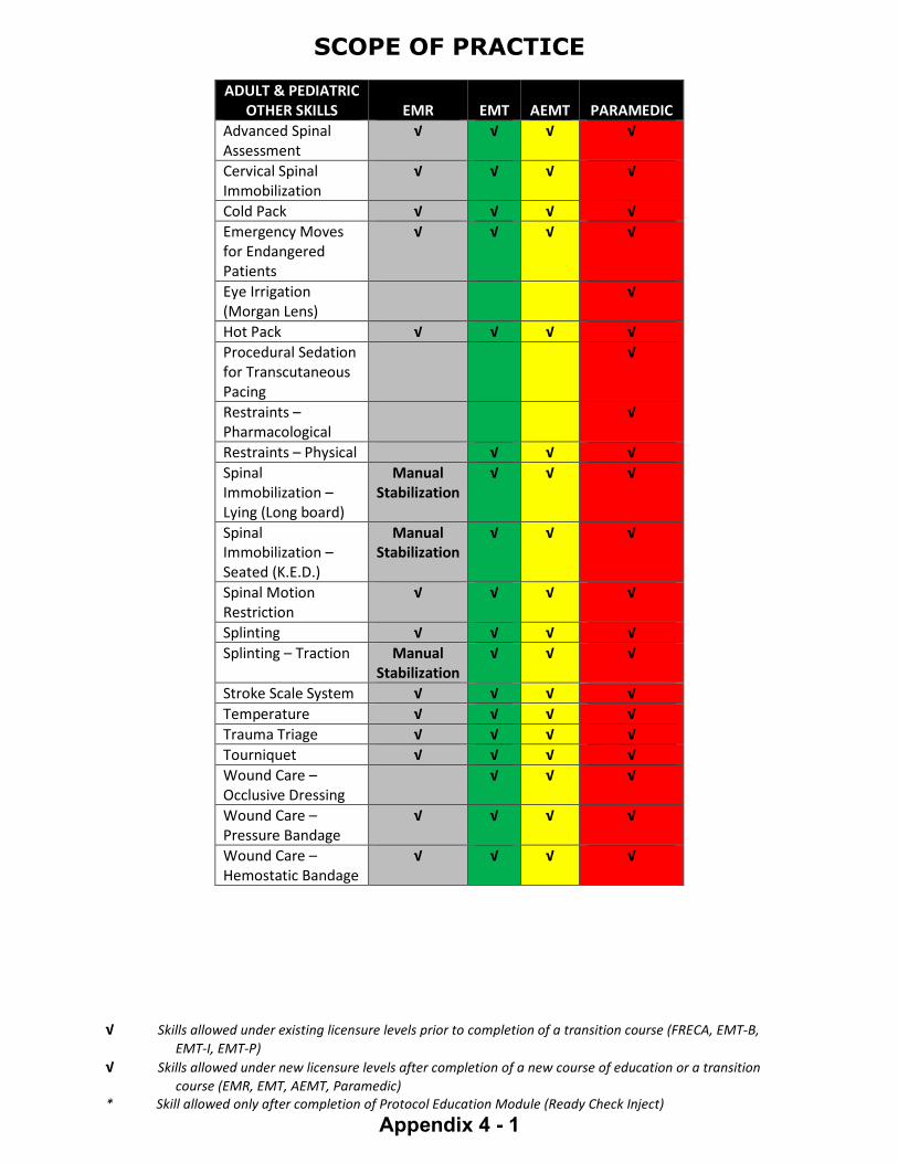
SCOPE OF PRACTICE
√ Skills allowed under existing licensure levels prior to completion of a transition course (FRECA, EMT-B, EMT-I, EMT-P) √ Skills allowed under new licensure levels after completion of a new course of education or a transition course (EMR, EMT, AEMT, Paramedic) * Skill allowed only after completion of Protocol Education Module (Ready Check Inject)
Appendix 4 - 1
ADULT & PEDIATRIC OTHER SKILLS EMR EMT AEMT PARAMEDIC
Advanced Spinal Assessment
√ √ √ √ Cervical Spinal Immobilization
√ √ √ √
Cold Pack √ √ √ √ Emergency Moves for Endangered Patients
√ √ √ √
Eye Irrigation (Morgan Lens)
√
Hot Pack √ √ √ √ Procedural Sedation for Transcutaneous Pacing
√
Restraints – Pharmacological
√
Restraints – Physical √ √ √ Spinal Immobilization – Lying (Long board)
Manual Stabilization
√ √ √
Spinal Immobilization – Seated (K.E.D.)
Manual Stabilization
√ √ √
Spinal Motion Restriction
√ √ √ √
Splinting √ √ √ √ Splinting – Traction Manual
Stabilization √ √ √
Stroke Scale System √ √ √ √ Temperature √ √ √ √ Trauma Triage √ √ √ √ Tourniquet √ √ √ √ Wound Care – Occlusive Dressing
√ √ √
Wound Care – Pressure Bandage
√ √ √ √
Wound Care – Hemostatic Bandage
√ √ √ √

SCOPE OF PRACTICE
√ Skills allowed under existing licensure levels prior to completion of a transition course (FRECA, EMT-B, EMT-I, EMT-P) √ Skills allowed under new licensure levels after completion of a new course of education or a transition course (EMR, EMT, AEMT, Paramedic) * Skill allowed only after completion of Protocol Education Module (Ready Check Inject)
Appendix 4 - 2
ADULT AIRWAY MANAGEMENT EMR EMT AEMT PARAMEDIC
Auto Transport Ventilator
√ √
BVM √ √ √ √ Capnography √ √ Chest Tube Maintenance
√
CPAP √ √ Endotracheal Intubation
√
Endotracheal Suctioning
√ √
Extubation √ Heimlich Maneuver √ √ √ √ High-Flow Nasal Cannula
√
Nasogastric Tube √ Nasopharyngeal Airway
√ √ √
Nasotracheal Intubation
√
Nebulizer Treatment √ √ Needle Decompression
√
Oral Suctioning √ √ √ √ Orogastric Tube √ Oropharyngeal Airway
√ √ √ √
Oxygen Administration
√ √ √ √
Percutaneous Cricothyrotomy
√
Pulse Oximetry √ √ √ Rapid Sequence Intubation
See Protocol
Supraglottic Airway √ √ Surgical Cricothyrotomy
√
Tracheostomy Maintenance
√ √ √
Tracheostomy Replacement
√

SCOPE OF PRACTICE
√ Skills allowed under existing licensure levels prior to completion of a transition course (FRECA, EMT-B, EMT-I, EMT-P) √ Skills allowed under new licensure levels after completion of a new course of education or a transition course (EMR, EMT, AEMT, Paramedic) * Skill allowed only after completion of Protocol Education Module (Ready Check Inject)
Appendix 4 - 3
PEDIATRIC AIRWAY MANAGEMENT EMR EMT AEMT PARAMEDIC
Auto Transport Ventilator
√ √
BVM √ √ √ √ Capnography √ √ CPAP √ √ Endotracheal Intubation
√
Endotracheal Suctioning
√ √
Extubation √ Heimlich Maneuver √ √ √ √ High-Flow Nasal Cannula
√
Nasogastric Tube √ Nasopharyngeal Airway
√ √ √
Nebulizer Treatment √ √ √ Needle Decompression
√
Oral Suctioning √ √ √ √ Orogastric Tube √ Oropharyngeal Airway
√ √ √ √
Oxygen Administration
√ √ √ √
Pulse Oximetry √ √ √ Supraglottic Airway √ √ Tracheostomy Maintenance
√ √ √
Tracheostomy Replacement
√

SCOPE OF PRACTICE
√ Skills allowed under existing licensure levels prior to completion of a transition course (FRECA, EMT-B, EMT-I, EMT-P) √ Skills allowed under new licensure levels after completion of a new course of education or a transition course (EMR, EMT, AEMT, Paramedic) * Skill allowed only after completion of Protocol Education Module (Ready Check Inject)
Appendix 4 - 4
ADULT MEDICATION
ADMINISTRATION ROUTE EMR EMT AEMT PARAMEDIC
Auto Injector Self-Admin Only
√ √ √
Blood Products √ Endotracheal √ Inhalation √ √ √ Intramuscular * √ √
Intraosseous √ √ Intravenous √ √ Intravenous Pump √ Oral √ √ √ Intranasal √
Narcan Only
√ Narcan
or Assist
√ √
Rectal Assist Assist √ Subcutaneous √ √ Sublingual Assist √ √
PEDIATRIC MEDICATION
ADMINISTRATION ROUTE EMR EMT AEMT PARAMEDIC
Auto Injector √ √ √ Endotracheal √ Inhalation √ √ √ Intramuscular * √ √
Intranasal √
Narcan Only
√ Narcan
or Assist
√ √
Intraosseous √ √ Intravenous √ √ Intravenous Pump √ Oral √ √ √ Rectal Assist Assist √ Subcutaneous √ √

SCOPE OF PRACTICE
√ Skills allowed under existing licensure levels prior to completion of a transition course (FRECA, EMT-B, EMT-I, EMT-P) √ Skills allowed under new licensure levels after completion of a new course of education or a transition course (EMR, EMT, AEMT, Paramedic) * Skill allowed only after completion of Protocol Education Module (Ready Check Inject)
Appendix 4 - 5
ADULT VASCULAR ACCESS EMR EMT AEMT PARAMEDIC
Blood Draw √ √ Blood Glucose Analysis
√ √ √
Central Line Maintenance
√
Peripheral Venous Access
√ √
Intraosseous √ √ PEDIATRIC
VASCULAR ACCESS EMR EMT AEMT PARAMEDIC Pediatric Vascular Access
EMR EMT AEMT Paramedic
Blood Draw √ √ Blood Glucose Analysis
√ √ √
Central Line Access √ Intraosseous √ √ Peripheral Venous Access
√ √
ADULT CARDIAC MANAGEMENT EMR EMT AEMT PARAMEDIC
Acquire and Transmit 12- Lead ECG
√ √ √
Cardiac Arrest Epinephrine
√ √
CPR – Cardiopulmonary Resuscitation
√ √ √ √
Defibrillation – AED √ √ √ √ Defibrillation – Manual
√
Interpretation of 12 Lead ECG
√
Mechanical CPR √ √ √
Resuscitation Initiation and Termination
√ √ √
STEMI System √ √ √ √ Synchronized Cardioversion
√
Transcutaneous Pacing
√

SCOPE OF PRACTICE
√ Skills allowed under existing licensure levels prior to completion of a transition course (FRECA, EMT-B, EMT-I, EMT-P) √ Skills allowed under new licensure levels after completion of a new course of education or a transition course (EMR, EMT, AEMT, Paramedic) * Skill allowed only after completion of Protocol Education Module (Ready Check Inject)
Appendix 4 - 6
PEDIATRIC CARDIAC
MANAGEMENT EMR EMT AEMT PARAMEDIC Acquire and Transmit 12 Lead ECG
√ √ √
CPR – Cardiopulmonary Resuscitation
√ √ √ √
Defibrillation – AED √ √ √ √ Defibrillation – Manual
√
Interpretation of 12 Lead ECG
√
Synchronized Cardioversion
√
Transcutaneous Pacing
√

VERMONT CRITICAL CARE PARAMEDIC (CCP) SCOPE OF PRACTICE
Legend: Y = Yes N = No W = Waiver required RSI = Complete VT EMS approved RSI program also required `
Appendix 5 - 1
ADULT & PEDIATRIC OTHER SKILLS EMR EMT AEMT Para
CCP
AICD Magnet Use Y A-Line Insertion W A-Line Assist Y A-Line Monitoring Y Aortic Balloon Pump Monitoring
W
Blood Chemistry/Lab Interpretation Y
Blood Product Initiation Y Cardiac Assist Device - Impella Maintenance
W
Central Venous Pressure Monitor Interpretation
Y
Chest Tube Insert W Continuous Bladder Irrigation Y Cricothyroidotomy - Surgical Y Escharotomy W Extubation Y Y Fiberoptic Intubation W IFT Transport Documentation Y Invasive Intracardiac Catheters Maintenance
Y
Infusion Pump for Medications/Fluids
Y Y
Intubation Maintenance with Paralytics
Y
Maintenance of Intracranial Monitoring Lines
Y
Manual Transport Ventilator Y Percutaneous Pericardiocentesis
W
Procedural Sedation for Transcutaneous Pacing
Y Y
Rapid Sequence Intubation Adult (Age 13 and Over)
YRSI YRSI
RSI Pediatric WRSI Tracheostomy Replacement Y Y Transvenous Pacing Y Ultrasound Y Umbilical Vein Initiation W Use of PICC Lines Y

VERMONT CRITICAL CARE PARAMEDIC (CCP) SCOPE OF PRACTICE
Legend: Y = Yes N = No W = Waiver required RSI = Complete VT EMS approved RSI program also required `
Appendix 5 - 2
ADULT & PEDIATRIC MEDICATIONS RSI EMR EMT AEMT Para
CCP
Atropine YRSI YRSI Etomidate (Amidate) YRSI YRSI Ketamine (Ketalar) YRSI YRSI Lidocaine YRSI YRSI Propofol (Diprovan) – Maintenance Only
YRSI YRSI
Rocuronium (Zemuron) YRSI YRSI Sucinycholine (Anectine) YRSI YRSI Vecuronium (Norcuron) YRSI YRSI
ADULT & PEDIATRIC MEDICATIONS EMR EMT AEMT Para
CCP
Acetaminophen (Tylenol) PO Y Y Y Y Acetaminophen (Tylenol) IV Y Y Y Acetazolamide (Diamox) Y Acetylcysteine (Mucomyst) Y Alteplase (Activase) Y Antibiotics Initiation W Antiobiotics Maintenance Y Y Bilvalirudin (Angiomax) Y Blood Products Y Y Calcium Gluconate Y Y Clopidogrel (Plavix) Y Dexamethasone (Decadron) Y Dobutamine (Dobutamine) Y Esmolol (Brevibloc) Y Hypertonic Saline Y Fosphenytoin (Cerebyx) Y Glycoprotein IIB/IIIA Inhibitor Y IIB/IIIA Maintenance Y Y Heliox (With RT) Y Heparin (Heparin) Initiation Bolus and/or Drip
Y
Heparin (Heparin) Maintenance
Y Y
Hydralazine (Apresoline) Y Ibuprofen (Motrin) Y Insulin - Regular Y Ketamine – Dissociative Dose Y Ketamine – Infusion W Ketamine – Analgesic Dose Y Ketoralac Y Y Labetalol (Normodyne) Y

VERMONT CRITICAL CARE PARAMEDIC (CCP) SCOPE OF PRACTICE
Legend: Y = Yes N = No W = Waiver required RSI = Complete VT EMS approved RSI program also required `
Appendix 5 - 3
ADULT & PEDIATRIC MEDICATIONS EMR EMT AEMT Para
CCP
Levitiracetam (Keppra) Y LMWH (Lovenox) Y Mannitol 20% (Osmitrol) Y Metoprolol (Lopressor) Y Milrinone Initial/Maint W Nicardipine (Cardene) Y Nimodipine (PO for SAH) Y Nitroprusside (Nipride) Y Octreotide Acetate (Sandostatin)
Y
Ondansetron Y Y Y Pantoprazole (Protonix) Y Y Phenobarbital (Luminal) Y Phentolamine W Phenytoin (Dilantin) Y Potassium Chloride Y Propofol Initiation W Propofol Maintenance Y Prostaglandin Infusion W Protamine Sulfate W Protein Complex Concentrates
W
Sodium Polystyrene Sulfonate (Kayexelate)
Y
Sucrose Solution 24% Y Terbutaline Y Thiamine (Vitamin B12) Y Ticagrelor (Brillinta) Y TPN/Lipids Y TNKase (Tenecteplase) Y TPA Infusion Maintenance Y TXA Initiation Y Y TXA Maintenance Y Y Various Eye Drops Y

Appendix 6
Perform CPR until defibrillator attached Attach monitor-defibrillator Administer oxygen
If no return of spontaneous circulation (ROSC), treat as asystole/PEA If ROSC, go to post-cardiac arrest care and start hypothermia protocol.
CPR X 2 minutes Epinephrine 1 mg IV/IO every other cycle
to a total of 3 mg. Consider advanced airway with
capnography, NG/OG tube to decompress
Rhythm shockable?
VF/VT Defibrillate
Treat as VF/VT
CPR X 2 minutes Obtain IV/IO
Asystole/PEA
Rhythm shockable (VF/VT) or shock indicated on AED?
Rhythm shockable?
Rhythm shockable?
No
Y es Defibrillate
No
Rhythm shockable?
Y es
No
Y es
Y es No
No
Adult Cardiac Arrest Algorithm
Defibrillate
CPR X 2 minutes Paramedic: Amiodarone: First dose
300 mg IV/IO push Second dose (if needed) 150 mg IV/IO push, or
Paramedic: L idocaine 1-1.5 mg/kg IV/IO. May repeat dose 0.5 – 0.75 mg/kg every 5-10 min to a max of 3 mg/kg.
Treat reversible causes Paramedic: If ROSC, hang amiodarone
drip at 1mg/min or lidocaine drip at 1-4 mg/min
CPR X 2 minutes Obtain IV/IO access Epinephrine 1 mg IV/IO every other cycle
to a total of 3 mg. Consider advanced airway with
capnography, NG/OG tube to decompress
CPR X 2 minutes Treat reversible causes
Y es
Biphasic shock settings: 120 J, 150 J, then 200 J for all successive shocks. Monophasic: 360 J.

Appendix 6
Identify and treat underlying cause: Maintain patent airway; assist breathing as necessary Oxygen (if hypoxemic) Cardiac monitor to identify rhythm; monitor blood pressure and
oximetry IV access 12-lead ECG if available; don’t delay therapy
Persistent bradycardia (heart rate < 50/min) causing: Hypotension? Acutely altered mental status? Signs of shock? Ischemic chest discomfort? Acute heart failure?
Monitor and observe Atropine 0.5 mg IV every 3-5 minutes to a total dose of 3 mg. If atropine ineffective: Transcutaneous pacing, or Epinephrine IV/IO Infusion 2-10 mcg/min,
titrated to effect. Contact Medical Direction for consultation.
No Y es
Assess appropriateness for clinical condition. Heart rate typically < 50/min if bradyarrhythmia.
Adult Bradycardia Algorithm

Appendix 6
Obtain and transmit 12-lead ECG IV access Vagal maneuvers Adenosine if regular: 6 mg, then 12
mg rapid IV push as needed. Be sure to flush line immediately after with saline
For atrial fibrillation or atrial flutter, consider: o Diltiazem 0.25 mg/kg IV/IO over
2 min (max dose 20 mg). Consider 10 mg max dose for elderly patient or patient with low BP. May repeat in 15 minutes at 0.35 mg/kg (max dose 25 mg) if necessary OR
o Metoprolol 5 mg IV/IO over 2-5 minutes, may repeat every 5 minutes to a max of 15 mg to achieve ventricular rate of 90-100 BPM.
Contact Medical Direction for consultation.
Assess appropriateness for clinical condition. Heart rate typically ≥ 150/min if tachyarrhythmia.
For tachyarrhythmia ≥ 150/min, identify and treat underlying cause: o Maintain patent airway; assist breathing as necessary o Oxygen (if hypoxemic) o Cardiac monitor to identify rhythm; monitor blood pressure and oximetry
Magnesium Pulseless: 1 -2 grams
magnesium sulfate d iluted in 1 0 mL 0 . 9 % NaC l, ad minister IV P.
With pulse: 1 -2 grams magnesium sulfate d iluted in 5 0 mL D5 W or 0 . 9 % NaC l, ad minister over 1 0 minutes
Persistent tachycardia causing Hypotension? Acutely altered mental status? Signs of shock? Ischemic chest discomfort? Acute heart failure
Synchronized Cardioversion Consider sedation If regular narrow complex, consider adenosine 6 mg rapid
IV/IO followed by saline flush. May repeat at 12 mg if no conversion.
Synchronized cardioversion initial recommended doses: o Narrow regular: 50-100 J biphasic or 200J monophasic o Narrow irregular 120-200 J biphasic or 200J
monophasic o Wide regular: 100J biphasic or monophasic o Wide irregular/polymorphic: 120J-200J biphasic or 360J
monophasic
Consider adenosine 6-12 mg IV/IO, then 12 mg if no conversion.
Consider amiodarone 150 mg IV/IO mixed over 10 minutes, may repeat once in 10 minutes. If successful, consider maintenance infusion of 1 mg/min OR
L idocaine 1 mg/kg IV/IO, may repeat every 5 minutes x 3 doses. If successful, consider maintenance infusion of 1-4 mg/min IV/IO
Contact Medical Direction for consultation and to consider cardioversion.
Y e s
Treat as torsades de pointes
Treat as ventricular tachycardia (VT)
N o
Obtain and transmit 12-lead ECG IV access Rhythm regular and monomorphic?
QRS wide (> 0.12 sec)?
Y es
N o
No
Y e s
Adult Tachycardia Algorithm

CPAP for AEMT and Paramedic
Does patient meet Indications?
Assess patient, record vital signs and pulse oximetry before applying oxygen.
Ensure adequate oxygen supply for CPAP device.
Does patient meet any
Contraindications?
Yes
Choose appropriately-sized device for patient.
Administer CPAP and adjust pressure to 5 – 15 cm H2O.
Monitor pulse oximetry, waveform capnography and ECG as available and trained.
Consider administering anxiolytic with Medical Direction authorization.
No
No
Yes
Notify Medical Direction CPAP not indicated.
Continue routine patient care.
Assist respirations by BVM and consider advanced airway, if indicated.
Patient stabilizing?
Yes
NoDiscontinue CPAP
and assist respirations by
BVM
Continue CPAP.
Notify Medical Direction to prepare for a CPAP patient.
Continue to reassess patient every 5 minutes.
Notify Medical Direction
Age Resp Rate
Infant 0 - 1 year > 60
Toddler 1 - 3 years >40
Preschooler 4 - 5 years >34
School Age 6 - 12 years >30
Teen 13 and older >25
Pediatric Respiratory Distress
Yes
Appendix 6
CPAP IndicationsSpontaneously breathing patient in
moderate to severe respiratory distress due to congestive heart failure/pulmonary edema, asthma/COPD, pneumonia, submersion injury or undifferentiated respiratory distress, concurrent with the following: Oxygen saturation < 94% Respiratory rate > 25 (see chart
for pediatric rates) Retractions or accessory
muscle use
CPAP ContraindicationsCardiac or respiratory arrest/apneaUnable to follow commands Unable to maintain their own airwayAgitated or combative behavior and
unable to tolerate maskVomiting and/or active GI bleedRespiratory distress secondary to
traumaSuspicion of pneumothoraxFacial trauma or impossible face
sealHypotension with MAP < 65 (SBP <
100 mmHg)
Pediatric SBP < 70 + (avg in yrs x 2)
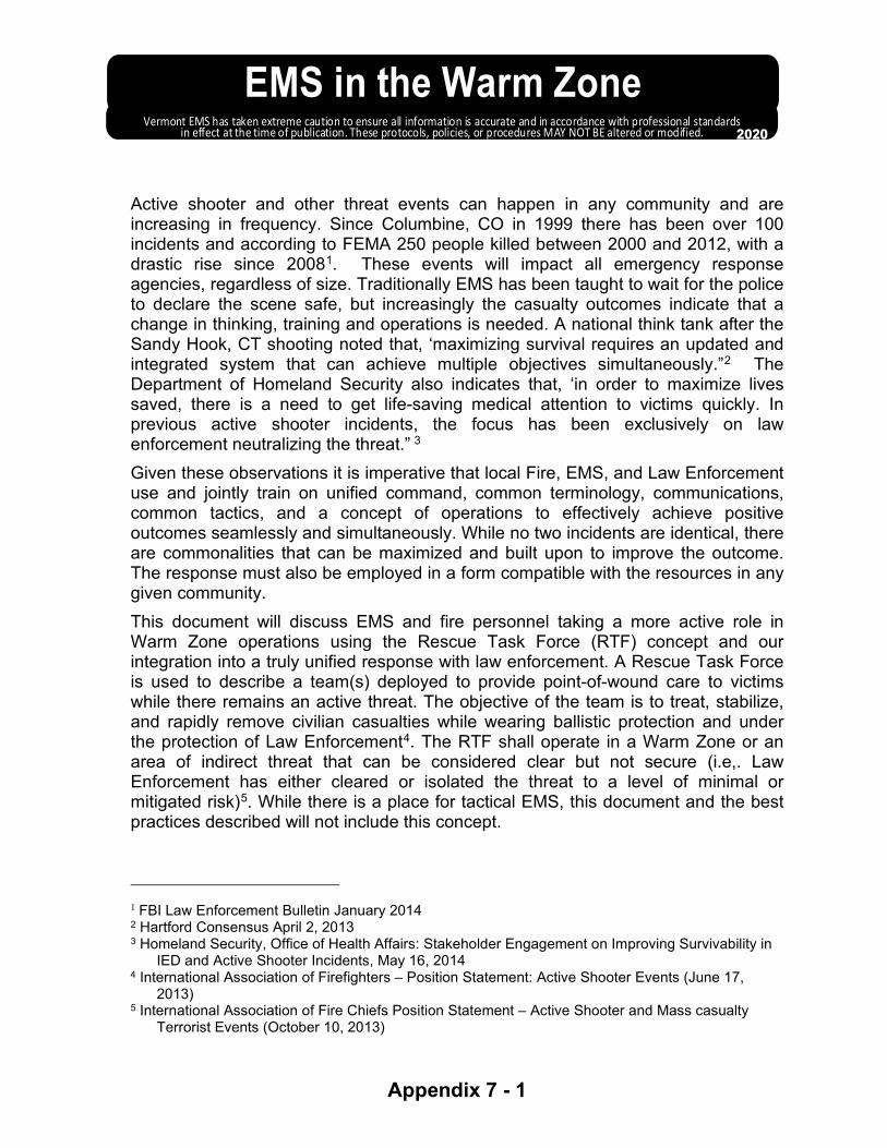
EMS in the Warm ZoneVermont EMS has taken extreme caution to ensure all information is accurate and in accordance with professional standards
in effect at the time of publication. These protocols, policies, or procedures MAY NOT BE altered or modified. 2020
Appendix 7 - 1
Active shooter and other threat events can happen in any community and are increasing in frequency. Since Columbine, CO in 1999 there has been over 100 incidents and according to FEMA 250 people killed between 2000 and 2012, with a drastic rise since 20081. These events will impact all emergency response agencies, regardless of size. Traditionally EMS has been taught to wait for the police to declare the scene safe, but increasingly the casualty outcomes indicate that a change in thinking, training and operations is needed. A national think tank after the Sandy Hook, CT shooting noted that, ‘maximizing survival requires an updated and integrated system that can achieve multiple objectives simultaneously.”2 The Department of Homeland Security also indicates that, ‘in order to maximize lives saved, there is a need to get life-saving medical attention to victims quickly. In previous active shooter incidents, the focus has been exclusively on law enforcement neutralizing the threat.” 3 Given these observations it is imperative that local Fire, EMS, and Law Enforcement use and jointly train on unified command, common terminology, communications, common tactics, and a concept of operations to effectively achieve positive outcomes seamlessly and simultaneously. While no two incidents are identical, there are commonalities that can be maximized and built upon to improve the outcome. The response must also be employed in a form compatible with the resources in any given community. This document will discuss EMS and fire personnel taking a more active role in Warm Zone operations using the Rescue Task Force (RTF) concept and our integration into a truly unified response with law enforcement. A Rescue Task Force is used to describe a team(s) deployed to provide point-of-wound care to victims while there remains an active threat. The objective of the team is to treat, stabilize, and rapidly remove civilian casualties while wearing ballistic protection and under the protection of Law Enforcement4. The RTF shall operate in a Warm Zone or an area of indirect threat that can be considered clear but not secure (i.e,. Law Enforcement has either cleared or isolated the threat to a level of minimal or mitigated risk)5. While there is a place for tactical EMS, this document and the best practices described will not include this concept.
1 FBI Law Enforcement Bulletin January 2014 2 Hartford Consensus April 2, 2013 3 Homeland Security, Office of Health Affairs: Stakeholder Engagement on Improving Survivability in
IED and Active Shooter Incidents, May 16, 2014 4 International Association of Firefighters – Position Statement: Active Shooter Events (June 17,
2013) 5 International Association of Fire Chiefs Position Statement – Active Shooter and Mass casualty
Terrorist Events (October 10, 2013)

EMS in the Warm ZoneVermont EMS has taken extreme caution to ensure all information is accurate and in accordance with professional standards
in effect at the time of publication. These protocols, policies, or procedures MAY NOT BE altered or modified. 2020
Appendix 7 - 2
I) Preface Active shooter and other events that create significant traumatic injuries through violence
pose special challenges to our first responders. These will evolve rapidly and without the benefit of sufficient resources. The first responder system bears a responsibility to its providers and the general public to ensure appropriate preparation and that the usual and customary standards of Emergency Medical Service (EMS) care during an incident are maintained and provided to the best extent possible. Saving lives depends on the rapid but safe and coordinated response from Law Enforcement (LE), Fire and EMS. The goal is to get EMS care to victims quickly but safely.
II) Purpose The purpose of this document is to establish policies and procedures for the dispatch and operations of a Rescue Task Force (RTF) in an active shooter or violent incident producing injuries for EMS agencies that wish to participate in this protocol.
III) Definitions a) Active Shooter: An individual or individuals engaged in killing or attempting to kill
people in a confined and populated area; in most cases, active shooters use firearms(s), but edged weapons, explosives and motor vehicles are other methods that have been used. There may be no pattern or method to their selection of victims.
b) Ballistic Protective Equipment: Ballistic protective gear, including body armor, for the head and body; i.e., vests, gloves, knee pads, helmets, and shields.
c) Casualty Collection Point (CCP): A location that is used for the assembly, triage (sorting), medical stabilization, and subsequent evacuation of casualties. It may be an intermediary point before formal triage.
d) Cleared: An area has been searched and does not pose a threat – no perpetrator present. e) Cold Zone:
i) Area where no significant danger or threat can be reasonably anticipated. ii) Area where triage and treatment of patients would occur, additional resources would
be staged, and command functions carried out. f) Concealment: A structure that hides a person’s exact location but can be penetrated by
ballistic weapons (e.g., a sheetrock wall). g) Contact Team: The first responding officers/security personnel who go directly to the
ongoing threat, make contact as soon as possible, and neutralize the threat, in order to minimize injuries and lives lost.
h) Cover: An area generally impenetrable to ballistic weapons, such as concrete wall. Something that prevents a responder from being observed by the perpetrator AND provides direct protection from the hazard/threat.
i) Hot Zone: i) Area wherein a direct and immediate life threat exists. ii) Depends upon current circumstances and is subjective. iii) Area is dynamic and may change frequently depending upon the situation.
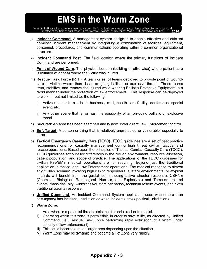
EMS in the Warm ZoneVermont EMS has taken extreme caution to ensure all information is accurate and in accordance with professional standards
in effect at the time of publication. These protocols, policies, or procedures MAY NOT BE altered or modified. 2020
Appendix 7 - 3
j) Incident Command: A management system designed to enable effective and efficient domestic incident management by integrating a combination of facilities, equipment, personnel, procedures, and communications operating within a common organizational structure.
k) Incident Command Post: The field location where the primary functions of Incident Command are performed.
l) Point-of-Wound Care: The physical location (building or otherwise) where patient care is initiated at or near where the victim was injured.
m) Rescue Task Force (RTF): A team or set of teams deployed to provide point of wound-care to victims where there is an on-going ballistic or explosive threat. These teams treat, stabilize, and remove the injured while wearing Ballistic Protective Equipment in a rapid manner under the protection of law enforcement. This response can be deployed to work in, but not limited to, the following: i) Active shooter in a school, business, mall, health care facility, conference, special
event, etc. ii) Any other scene that is, or has, the possibility of an on-going ballistic or explosive
threat. n) Secured: An area has been searched and is now under direct Law Enforcement control. o) Soft Target: A person or thing that is relatively unprotected or vulnerable, especially to
attack. p) Tactical Emergency Casualty Care (TECC): TECC guidelines are a set of best practice
recommendations for casualty management during high threat civilian tactical and rescue operations. Based upon the principles of Tactical Combat Casualty Care (TCCC), TECC guidelines account for differences in the civilian environment, resource allocation, patient population, and scope of practice. The applications of the TECC guidelines for civilian Fire/EMS medical operations are far reaching, beyond just the traditional application in tactical and Law Enforcement operations. The medical response to almost any civilian scenario involving high risk to responders, austere environments, or atypical hazards will benefit from the guidelines, including active shooter response, CBRNE (Chemical, Biological, Radiological, Nuclear, and Explosives) and Terrorism related events, mass casualty, wilderness/austere scenarios, technical rescue events, and even traditional trauma response.
q) Unified Command: An Incident Command System application used when more than one agency has incident jurisdiction or when incidents cross political jurisdictions.
r) Warm Zone: i) Area wherein a potential threat exists, but it is not direct or immediate. ii) Operating within this zone is permissible in order to save a life, as directed by Unified
Command (i.e., Rescue Task Force performing rapid extrication of a victim under security of law enforcement).
iii) This could become a much larger area depending upon the situation. iv) Warm Zone may be dynamic and become a Hot Zone very rapidly.

EMS in the Warm ZoneVermont EMS has taken extreme caution to ensure all information is accurate and in accordance with professional standards
in effect at the time of publication. These protocols, policies, or procedures MAY NOT BE altered or modified. 2020
Appendix 7 - 4
IV) Preplanning Considerations a) Fire/EMS, law enforcement, Public Safety Answering Points (PSAPS) and other public
safety partners should work in a coordinated effort to develop standard operating guidelines for Unified Command, common terminology, communications, common tactics, and a concept for operations.
b) All public safety partners should work cooperatively to identify target hazards and key components of each, such as main access, control rooms, master keys, isolation corridors, maps, and internal communication systems.
c) All public safety partners should work cooperatively to assess “soft targets.” d) Determine multiple locations for potential Incident Command Posts (ICP). All public
safety partners should consider situations where responders could converge on the scene and freelance, and to develop strategies to prevent freelancing.
e) Once preplanning has been completed, all public safety partners should work cooperatively to create a pilot policy and training program so as to determine implementation challenges. This pilot program and plan should be delivered through joint training and exercise with cooperating agencies.
f) Coordination of training agencies: i) In order for training to be most effective it should be implemented as a system with
all responders collaboratively participating. This practice promotes interoperability well before the event so that any inconsistencies and questions can be addressed.
ii) All training should begin with a plan and end goal in mind. It should start small and build on previous training and education. Communities should conduct joint training and education between local first responders and any other agency that may be expected to respond or participate in case of emergency.
iii) Once foundation training has occurred, it should be exercised through Homeland Security Exercise and Evaluation Program (HSEEP) compliant table tops, drills, and full scale exercises. At each stage there should be a feedback mechanism to gather information on activities and challenges to improve the plan and future training.
iv) Training topics should include the following: 1) Incident Command and Unified Command applied to high threat/active violent
situations 2) Weapons awareness for fire and EMS personnel 3) Medical scope of RTF 4) Tactical Emergency Casualty Care (TECC) concepts, including pediatric
considerations 5) Coordination of resources 6) Equipment specific to the operation 7) Communications, including importance of radio discipline 8) Clear expectations of roles for LE, Fire, and EMS Team role composition and
personal traits of members necessary to operate in this specific environment

EMS in the Warm ZoneVermont EMS has taken extreme caution to ensure all information is accurate and in accordance with professional standards
in effect at the time of publication. These protocols, policies, or procedures MAY NOT BE altered or modified. 2020
Appendix 7 - 5
9) Dynamic nature of evolving situation and ability to change instantly from warm to hot zone, and expected actions of RTF including the changing availability of resources
10) Mutual aid locations and integrated training 11) Shelter-in-place concepts 12) Casualty Collection Point(s) (CCP)
V) Equipment
It is important to have consistent equipment across all teams not only for medical care but rapid identification of medical personnel. The focus should be on early hemorrhage control and rapid extrication6. Consider go-bags or medical vests with the ability to treat at least eight victims with extra equipment bags to treat an additional sixteen victims. a) Equipment to consider (see Figure 3):
i) Tourniquets (adult and pediatric) ii) Pressure dressings iii) Hemostatic bandages iv) Occlusive chest seals v) Adjunct airways (Adult and Pediatric) vi) Chest decompression needles (paramedics) vii) Personal safety / protective equipment shall be available for RTF teams. All ballistic
body armor / protection should be compliant with the current NIJ (National Institute of Justice) Ballistic Resistance Standard, 0101.06, minimum Type IIIa (see Figure 4): 1) Ballistic vests with clear identification of RTF/EMS 2) Ballistic helmet 3) Eye protection 4) Flashlight
viii) Two-way radio with remote microphone or ear piece/microphone ix) Lightweight and single person deployable patient moving devices x) Packaging for rapid deployment of RTF equipment by team members xi) Packaging for rapid resupply of RTF team
b) Go-bag for EMS/Patient care equipment – portable, carried on your person and packaged for efficiency. Each set of equipment should be able to treat at least eight (8) patients with the provision for extra equipment (See Figure 3 for suggested list of equipment).
VI) Procedure a) Law enforcement will be the lead agency and will establish a Unified Command with
Fire/EMS to rapidly deploy RTF teams into established zones. b) The RTF composition should consist of a minimum of four (4) personnel7: Two (2) EMS
and two (2) Law Enforcement.
6 Hartford Consensus April 2, 2013 7 International Association of Fire Chiefs Position Statement – Active Shooter and Mass casualty
Terrorist Events (October 10, 2013)

EMS in the Warm ZoneVermont EMS has taken extreme caution to ensure all information is accurate and in accordance with professional standards
in effect at the time of publication. These protocols, policies, or procedures MAY NOT BE altered or modified. 2020
Appendix 7 - 6
i) There should be a Law Enforcement officer for each EMS provider to deliver security.
ii) RTF/EMS personnel should be certified and licensed. EMS personnel should perform to their Scope of Practice.
c) Prior to deploying an RTF team, threat zones must be identified: i) Hot Zone – (also known as the area of direct threat) area where there is known
hazard or life threat that is direct and immediate. An example of this would be any uncontrolled area where the active shooter could directly engage an RTF team. RTF teams will not be deployed into a Hot Zone.
ii) Warm Zone - (also known as the area of indirect threat) areas that law enforcement has either cleared or isolated the threat where there is minimal or mitigated risk. This area can be considered clear but not secure. This is where the RTF will deploy, with security, to treat victims.
iii) Cold Zone - areas where there is little or no threat, either by geography or distance in relation to threat, or after area has been secured by law enforcement. This is an area where Fire/EMS will stage to triage, treat, and transport victims once removed from the warm zone.
d) Command and Control: i) Coordination should include the following:
1) Shared common terminology and communication across fire/EMS/law enforcement.
2) Span of control. 3) Jointly developed protocols for response. 4) Planning for and practicing rapid treatment and evacuation of patients8.
ii) RTF can be deployed for the following reasons: 1) Casualty treatment. 2) Casualty removal from warm to the CCP or to the cold zone. 3) Movement of supplies from cold to warm zone.
e) Response: i) Initial dispatch
When the local emergency communications/dispatch receives a call for an active shooter or violent incident with injuries the original dispatch will be for (enter local dispatch nomenclature) call type. This will generate the following response: 1) Law Enforcement patrol units (define a minimum for your community). 2) Fire/EMS units (define a minimum for your community). 3) Units dispatched should enable at least one RTF with necessary equipment to be
placed in operation.
8 U.S Fire Administration: Fire/Emergency Medical Services Department Operational Considerations
and Guide for Active Shooter and Mass Casualty Incidents (September, 2013)

EMS in the Warm ZoneVermont EMS has taken extreme caution to ensure all information is accurate and in accordance with professional standards
in effect at the time of publication. These protocols, policies, or procedures MAY NOT BE altered or modified. 2020
Appendix 7 - 7
ii) If Fire/EMS responders encounter a threat (i.e. active shooter(s)) in the staging area prior to Law Enforcement arrival, they shall immediately withdraw. When withdrawal is not possible, seek cover and request an expedited law enforcement response. Communicate unit identifier and location to Law Enforcement. When possible, provide the following information: 1) Number, location(s), and description of shooter(s). 2) Types of weapons in use (e.g., semiautomatic rifles, hand guns, explosives, etc). 3) Number and location(s) of victims and hostages, if known. 4) Communication method used by the shooters, if apparent (cell phones, radios). 5) Contact Teams (CT’s) will be established to address the threat. The CT’s goal is
to neutralize the threat. f) The first arriving units should:
i) Determine if they are responding into a static or ongoing situation and relay this information to dispatch.
ii) Identify if predetermined staging area is safe. If not safe, consider an area out of the line of sight of incident, in line of approach to location.
iii) Law Enforcement will establish 1-2 Contact Teams of 1-4 officers to address the threat.
iv) When appropriate personnel arrive on scene, Law Enforcement and EMS personnel will assemble into Rescue Task Forces of 2 EMS providers and 2-4 law enforcement and prepare for deployment. (Figure 1).
g) If possible, determine a Casualty Collection Point (CCP) prior to deploying. Depending on the size of the incident and location, injured victims may need to be placed in a CCP before transition to the cold zone. This will be predetermined by initial units, protected by Law Enforcement, and relayed to the RTF teams through Unified Command. As this area may be secure, it may be considered a cold zone and may be staffed with non-RTF Fire/EMS personnel.
h) Rescue Task Force deployment: i) Once Unified Command has identified the need to deploy RTF teams, they will be
deployed into the warm zone to begin victim care and evacuation as needed. The goal of initial RTF team is to stabilize as many victims as possible.
ii) Command will dispatch RTF teams by numbers, e.g., RTF Team 1. RTF Teams are not to deploy unless they have two personnel from Law Enforcement as security. Do not self-deploy into the warm zone.
iii) Command shall: 1) Establish RTF resupply near point of entry. 2) Establish an external Casualty Collection Point (CCP). 3) Designate area(s) in the cold zone to receive patients for treatment and
transportation, as appropriate. iv) The least number of personnel and teams should be deployed into the warm zone to
achieve the goals.

EMS in the Warm ZoneVermont EMS has taken extreme caution to ensure all information is accurate and in accordance with professional standards
in effect at the time of publication. These protocols, policies, or procedures MAY NOT BE altered or modified. 2020
Appendix 7 - 8
v) All RTF teams that make entry shall notify the Incident Commander of their location and any victims encountered. Constant communication between the IC and RTFs is essential for effective resource coordination and allocation.
vi) When teams make entry, they will treat the injured using Tactical Emergency Casualty Care (TECC) guidelines.
vii) Should the RTF encounter a threat/suspect, the medical personnel shall: 1) Evacuate if safe to do so. 2) Shelter in a place that provides protection to medical personnel, preferably with
an exterior door or means of escape. viii) When the RTF is operating in the Warm Zone, all patients encountered by the RTF
teams will be treated as they are accessed. Any patient who can ambulate without assistance will be directed by the team to self-evacuate down the cleared corridor under Law Enforcement direction. Any patient who is dead will be visibly marked to allow for easy identification and to avoid repeated evaluations by additional RTF teams.
ix) The first RTF teams in operation will enter the area and treat as many patients as possible.
x) Additional RTF teams that enter the area should be primarily tasked with extrication of the victims already treated by the initial team(s). However, if needed, additional RTF teams may be sent into areas unreached by the initial teams or to other areas with accessible victims. (Figure 2)
i) Additional tasks for local resources: Communities should predetermine tasks for arriving units and assign those in each local plan. The following tasks should be assigned as local resources arrive:
1) Assume or establish Command for first responder units. 2) Meet with law enforcement to establish Unified Command. 3) Establish personnel accountability, especially if members have already entered
the warm zone. 4) Work with law enforcement to identify the RTF working zones. 5) Consider adding an additional EMS Taskforce or MCI Alarm for patient treatment
and transport. 6) Consider primary staging to a larger or safer area if needed. 7) Create RTF teams from deployed units. 8) Equip RTF Teams with proper protective gear and equipment. 9) Designate Casualty Collection Points (CCP). 10) Once Unified Command has declared the working zones, RTF teams must be
informed of their working limits. 11) Use command boards to label and keep track of RTF teams. 12) EMS staging in the Cold Zone and a Treatment Dispatch Manager need to be
considered for larger numbers of patients. 13) Establish a resupply for extended RTF operations. 14) Prepare to establish staging for transport units, treatment, and transportation
areas. 15) Consider mutual aid for coverage of ongoing emergency needs of community.
j) Fire Suppression Considerations:

EMS in the Warm ZoneVermont EMS has taken extreme caution to ensure all information is accurate and in accordance with professional standards
in effect at the time of publication. These protocols, policies, or procedures MAY NOT BE altered or modified. 2020
Appendix 7 - 9
Consider assigning personnel to control fire suppression and protection systems if safe to do so.
k) Communications: Clear/plain language communication is vital. The following elements should be determined in preplanning and should be practiced and adhered to: i) Use of plain language ii) Common definitions iii) Command and control iv) Strict radio etiquette v) Radio frequencies assigned for interoperability vi) Radio frequency assigned to the RTF vii) Use of ear microphone, lapel microphone, and designated communicators
VII) Patient Care a) EMS personnel should treat the injured as an MCI using Tactical Emergency Casualty
Care (TECC) concepts and guidelines. Rapid identification, treatment and evacuation are paramount. Ambulatory victims should self-evacuate and fatalities should be clearly marked.
b) TECC “provides a framework to prioritize medical care while accounting for on-going high-risk operations.”9 i) Medical scope should have at its core:
1) Focus on THREAT acronym: Threat suppression Hemorrhage control Rapid Extrication to safety Assessment by medical providers Transport to definitive care
c) TECC Goals: a. Accomplish the mission with minimal casualties. b. Prevent any casualty from sustaining additional injuries. c. Keep response team maximally engaged in neutralizing the existing threat (e.g.,
active shooter, unstable building, confined space, HAZMAT, etc.). d. Minimize public harm.
d) TECC Principles: a. Establish tactical supremacy (Law Enforcement) and defer in depth medical
interventions if engaged in ongoing direct threat (e.g., fire fight, unstable building collapse, dynamic post-explosive scenario, etc.).
b. Threat mitigation techniques will minimize risk to casualties and the providers. These should include techniques and tools for rapid casualty access and egress.
9 International Association of Fire Chiefs Position Statement – Active Shooter and Mass casualty
Terrorist Events (October 10, 2013)

EMS in the Warm ZoneVermont EMS has taken extreme caution to ensure all information is accurate and in accordance with professional standards
in effect at the time of publication. These protocols, policies, or procedures MAY NOT BE altered or modified. 2020
Appendix 7 - 10
c. Triage should be deferred to a later phase of care. Prioritization for extraction is based on resources available and the tactical situation.
d. Minimal trauma interventions are warranted. e. Consider hemorrhage control. f. Tourniquet (TQ) application is the primary “medical” intervention to be considered
in Direct Threat. g. Consider instructing casualty to apply direct pressure to the wound if a tourniquet
is not available, or application is not tactically feasible. h. Consider quickly placing or directing casualty to be placed in position to protect
airway. e) TECC Guidelines:
a. Mitigate any threat (Law Enforcement) and move to a safer position (e.g. return fire, utilize less lethal technology, assume an overwhelming force posture, extract from immediate structural collapse, etc.).
b. Direct the casualty to stay engaged in any tactical operation if appropriate. c. Direct the casualty to move to a safer position and apply self aid if able. d. Casualties with an altered mental status should be disarmed immediately by law
enforcement. e. Casualty Extraction
i. If a casualty can move to safety, they should be instructed to do so. ii. If a casualty is unresponsive, the scene commander or team leader
should weigh the risks and benefits of a rescue attempt in terms of manpower and likelihood of success.
iii. Remote medical assessment techniques should be considered. iv. If the casualty is responsive but cannot move, a tactically feasible
rescue plan should be devised. f. Recognize that threats are dynamic and may be ongoing, requiring
continuous threat assessments. g. Stop life threatening external hemorrhage if tactically feasible:
i. Direct casualty to apply effective tourniquet if able. ii. Apply the tourniquet over the clothing 2 – 3” proximal to wound. iii. Tighten until cessation of bleeding and move to safety. Consider
moving to safety prior to application of the TQ if the situation warrants. iv. For response personnel, tourniquet should be readily available and
accessible with either hand. v. Consider instructing casualty to apply direct pressure to the wound if
no tourniquet is available or application is not tactically feasible. vi. Consider application of a second tourniquet just proximal to the first for
failure to control bleeding. vii. Apply a topical hemostatic bandage, in combination with direct
pressure, or pressure bandage using Kerlix and Ace wrap, for wounds in anatomical areas where tourniquet application is ineffective or impractical (junctional/torso injury or proximal extremity location where tourniquet application is not practical).
h. Airway Management i. Unconscious casualty without airway obstruction:
1. Chin lift or jaw thrust maneuver. 2. Nasopharyngeal airway.

EMS in the Warm ZoneVermont EMS has taken extreme caution to ensure all information is accurate and in accordance with professional standards
in effect at the time of publication. These protocols, policies, or procedures MAY NOT BE altered or modified. 2020
Appendix 7 - 11
3. Place the casualty in the recovery position. ii. Casualty with airway obstruction or impending airway obstruction:
1. Chin lift or jaw thrust maneuver. 2. Nasopharyngeal airway. 3. Allow a conscious casualty to assume any position that best
protects the airway, to include sitting up. 4. Place an unconscious casualty in the recovery position. 5. If the previous measures are unsuccessful, refer to a medic
immediately. i. Breathing
i. In a casualty with progressive respiratory distress and known or suspected torso trauma, consider a tension pneumothorax and refer to a medic as soon as possible for possible needle decompression.
ii. All open and/or sucking chest wounds should be treated by immediately applying a vented chest seal to cover the defect. If a vented chest seal is not available, use a non-vented chest seal. Monitor the casualty for respiratory distress. If it develops, you should suspect a tension pneumothorax. Treat this by burping or temporarily removing the dressing. If that doesn’t relieve the respiratory distress, refer to a medic.
NOTE: Once patients have been moved / relocated to an established treatment area, patient care by EMS providers shall be accomplished utilizing recognized Vermont Statewide EMS Protocols. Figure 1

EMS in the Warm ZoneVermont EMS has taken extreme caution to ensure all information is accurate and in accordance with professional standards
in effect at the time of publication. These protocols, policies, or procedures MAY NOT BE altered or modified. 2020
Appendix 7 - 12
Figure 2
Figure 3 SUGGESTED GO-BAG EMS EQUIPMENT LIST
QTY ITEM
6 CAT (or similar) Tourniquets (adult & pedi)
4 Occlusive Dressings / Chest Seals 2 ARS Needles (14ga X 3.25") 10 4X4 Gauze 4 Hemostatic Gauze Bandages 2 6-inch Israeli Bandages/Ace & Kerlix 2 4-inch Israeli Bandages/Ace & Kerlix 6 6-inch elastic roll 1 roll of tape 4 pairs of gloves
Various NPAs w/surgilube 1 Trauma Shears 1 Penlight 2 Kerlix Rolls 1 Permanent Marker

Cardiac P
rotocol 3.2P
P
EMT STANDING ORDERS
PARAMEDIC STANDING ORDERS
Routine Patient Care—with focus on CPR. Administer 100% oxygen. Immediate chest compressions. Apply AED and use as soon as possible (with minimum interruption of chest
compressions). From birth to age 8 years use pediatric AED pads.o If pediatric AED pads are unavailable, providers may use adult AED pads,
provided the pads do not overlap. If Return of Spontaneous Circulation occurs see Post Resuscitative Care
Protocol – Pediatric 3.4P. Consider termination of efforts or not attempting resuscitation, see DNR/COLST
8.9 and/or Resuscitation Initiation and Termination 8.17. Call for Paramedic intercept, if available. If not available, call for AEMT intercept.
Cardiac Arrest – Pediatric
2018
E
A
3.2P
ADVANCED EMT STANDING ORDERS Do not interrupt chest compressions for airway or IV/IO placement. Establish IV/IO access. BVM ventilation is the preferred method of ventilation for pediatric population.
However, if unsuccessful, consider placement of supraglottic airway. Consider advanced airway after use of AED, as applicable.
Monitor quantitative waveform capnography throughout resuscitation, if available, to assess and monitor airway placement and CPR quality, and to monitor for signs of Return of Spontaneous Circulation.
Consider and correct treatable causes: hypoxia, overdose/poisoning, hypothermia, hypoglycemia, and hypovolemia—treat as per specific protocol.
Administer 0.9% NaCl 20 mL/kg fluid bolus IV/IO.
Protocol Continues
Appendix 8 - 2018 Protocol
If ventilation is adequate with BVM, routine placement of advanced airway can be delayed.
Placement of an advanced airway during cardiac arrest should not interrupt chest compressions. In this setting, supraglottic airways and ETTs can be considered equivalent. ETT placement, if used, should be limited to 1 attempt of 10 seconds or less, as long as BVM or alternate airway provides adequate chest rise.
Consider tension pneumothorax and treat with needle decompression if indicated.
For suspected pre-arrest metabolic acidosis, suspected or known hyperkalemia (dialysis patient), or known tricyclic antidepressant overdose, consider sodium bicarbonate 1 mEq/kg IV/IO. Do not mix with any resuscitation drugs. Flush IV tubing with 0.9% NaCl before and after drug administration. Do not use routinely in cardiac arrest.
For known or suspected hyperkalemia (renal failure) or as an antidote for toxic effects (hypotension and arrhythmias) from calcium channel blocker or B-blocker overdose consider calcium chloride 20 mg/kg (0.2 mL/kg) slow IV/IO push. Do not mix with or infuse immediately before or after sodium bicarbonate without intervening flush. Do not use routinely in cardiac arrest.

P
PARAMEDIC STANDING ORDERS
For Ventricular Fibrillation (VF)/Pulseless Ventricular Tachycardia (VT): Use manufacturer’s recommendations for joule settings. In general, defibrillate at 2J/kg;
perform CPR for 2 minutes and recheck rhythm; if still a shockable rhythm, defibrillate at 4J/kg; perform CPR for 2 minutes; reassess every 2 minutes and continue to defibrillate at 4J/kg.
If no response after first defibrillation, administer epinephrine (1:10,000) (0.1 mg/mL) 0.01 mg/kg (0.1 mL/kg) IV/IO OR 0.1 mg/kg (1:1,000; 0.1 mL/kg) via ETT.o Repeat every 3 – 5 minutes.
If no response after second defibrillation, consider:o Amiodarone 5mg/kg (maximum 300 mg) IV/IO. May repeat up to 2 times for
refractory VF/VT; ORo Lidocaine 1 mg/kg IV/IO (maximum dose 100 mg). Maintenance: 20 – 50 mcg/kg/min
infusion. Repeat bolus dose if infusion initiated ≥ 15 minutes after initial bolus therapy.
o For Torsades de Pointes: Magnesium sulfate 25 – 50 mg/kg (maximum 2 grams) IV/IO over 1 – 2 minutes.
For Asystole or Pulseless Electrical Activity (PEA): Administer Epinephrine (1:10,000) (0.1 mg/mL) 0.01 mg/kg (0.1 mL/kg) IV/IO OR 0.1 mg/
kg (1:1,000) (1 mg/mL) 0.1 mL/kg via ETT; repeat every 3 – 5 minutes. Give 2 minutes of CPR, then check rhythm:
o If asystole or PEA, continue epinephrine and 2 minutes of CPR until:o Pulse obtained ORo Shockable rhythm obtained ORo Decision made to discontinue further efforts. Contact Medical Direction for
guidance.
Cardiac P
rotocol 3.2P
2018
Cardiac Arrest – Pediatric 3.2P
Protocol Continues
Compressions
Time in Seconds
Coronary perfusion pressure – note the interval of interruption [A] as well as the interval until the restoration of coronary perfusion pressure [A+B]
EFFECT OF INTERRUPTIONS TO CPR ON CORONARY PERFUSION PRESSURE
Appendix 8 - 2018 Protocol
PEARLS: Optimize oxygenation, ventilation and volume status. Cardiac arrest in children typically results from
progressive deterioration in respiratory or cardiovascular function. Minimize interruptions in chest compression, as pauses rapidly return the blood pressure to zero and
stop perfusion to the heart and brain. Switch compressors at least every two minutes to minimize fatigue. Check rhythm and pulse during
switch to avoid excessive interruptions in CPR. Perform “hands on” defibrillation.
o Compress when charging and resume compressions immediately after the shock is delivered to minimize interruptions to CPR.
Avoid excessive ventilation. Do not use mechanical CPR devices on children.