Embed Size (px)
Citation preview

American Edition | January 2018
in this issue:
The official journal of the American Association of Equine Practitioners, produced in partnership with BEVA.
veterinaryequine
education
Meet 2018 AAEP President Dr. Margo Macpherson
Clinical and low field magnetic resonance imaging features of osseous cyst-like lesions of the proximal sesamoid bones in seven horses
Articular sagittal and medial parasagittal patellar fracture repair using lag screws in two mature horses

He’s more than just a horse.To your clients, he’s Family.
THE MOST ADVANCEDCOSEQUIN FORMULA!
Contains proprietary,trademarked ingredients
NOT available in other brands.
Active Ingredients:
• FCHG49® Glucosamine (GLU)
• TRH122® Chondroitin Sulfate (CS)
• NMX1000® Avocado/Soybean Unsaponifiables (ASU)
• Green Tea Extract (EGCG)
• Hyaluronic Acid (HA)
• Methylsulfonylmethane (MSM)
010.1258.00
Give him the best
HOW COSEQUIN WORKS
HA/ASU/GLU/CS1 and ASU/EGCG2
• Reduce COX-2 activity
• Reduce PGE2 production
• Inhibit NF- B nuclear translocation
Source: Survey conducted in February 2016 of equine veterinarians who recommended oral joint health supplements.
1. Heinecke LF, Grzanna MW, Au AY, et al. Inhibition of prostaglandin E2 production by the combination of hyaluronan, avocado/soybean unsaponifiables, glucosamine, and chondroitin sulfate involves a NF- B dependent mechanism. ORS 2011.
2. Heinecke LF, Grzanna MW, Au AY, et al. Inhibition of cyclooxygenase-2 expression and prostaglandin E2 production in chondrocytes by avocado soybean unsaponifiables and epigallocatechin gallate. Osteoarthritis and Cartilage 2010;18:220–227.
Unactivated equine chondrocytes with immunofluorescent staining for NF- , which is shown to be predominantly in the cytoplasm of cells.
IL-1 and TNF- activated equine chondrocytes with immunofluorescent staining showing the nuclear translocation of NF-
IL-1 and TNF- activated equine chondrocytes pre-treated with NMX1000® ASU+EGCG with immunofluorescent staining showing the inhibition of NF- nuclear translocation.2
c o n t e n t s
American Edition
Meet 2018 AAEP President Dr. Margo Macpherson ............................................................. III
Ethics: How to practice ethically and keep your clients ........................................................ IV
AAEP updates infectious disease guidelines, addresses Rhodococcus equi ............................ V
Peer Reviewers in 2017 ............................................................................................................ 2
S. WRIGHT ..............................................................................................................................4
Equine Veterinary Education – past, present and futureT. MAIR, Y. ELCE, P. WILKINS and P. MORRESEY ............................................................. 6
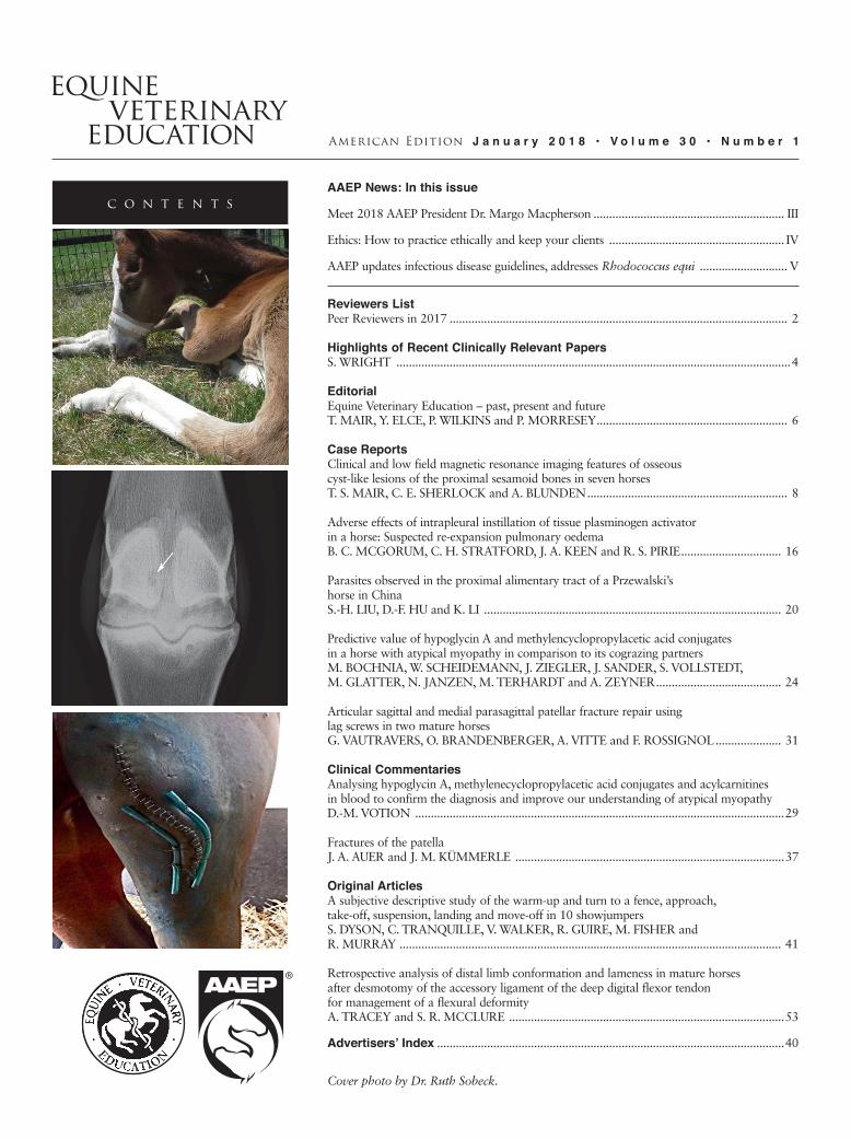
Clinical and low field magnetic resonance imaging features of osseouscyst-like lesions of the proximal sesamoid bones in seven horsesT. S. MAIR, C. E. SHERLOCK and A. BLUNDEN ................................................................ 8
Adverse effects of intrapleural instillation of tissue plasminogen activatorin a horse: Suspected re-expansion pulmonary oedemaB. C. MCGORUM, C. H. STRATFORD, J. A. KEEN and R. S. PIRIE ................................ 16
Parasites observed in the proximal alimentary tract of a Przewalski’shorse in ChinaS.-H. LIU, D.-F. HU and K. LI ............................................................................................... 20
Predictive value of hypoglycin A and methylencyclopropylacetic acid conjugates in a horse with atypical myopathy in comparison to its cograzing partnersM. BOCHNIA, W. SCHEIDEMANN, J. ZIEGLER, J. SANDER, S. VOLLSTEDT,M. GLATTER, N. JANZEN, M. TERHARDT and A. ZEYNER ........................................ 24
Articular sagittal and medial parasagittal patellar fracture repair usinglag screws in two mature horsesG. VAUTRAVERS, O. BRANDENBERGER, A. VITTE and F. ROSSIGNOL ..................... 31
Analysing hypoglycin A, methylenecyclopropylacetic acid conjugates and acylcarnitines in blood to confirm the diagnosis and improve our understanding of atypical myopathyD.-M. VOTION ......................................................................................................................29
Fractures of the patellaJ. A. AUER and J. M. KÜMMERLE ......................................................................................37
A subjective descriptive study of the warm-up and turn to a fence, approach, take-off, suspension, landing and move-off in 10 showjumpersS. DYSON, C. TRANQUILLE, V. WALKER, R. GUIRE, M. FISHER andR. MURRAY .......................................................................................................................... 41
Retrospective analysis of distal limb conformation and lameness in mature horses after desmotomy of the accessory ligament of the deep digital flexor tendon for management of a flexural deformityA. TRACEY and S. R. MCCLURE ........................................................................................53
...............................................................................................................40
Cover photo by Dr. Ruth Sobeck.
veterinaryequine
education
Equine Veterinary Education is a refereed educational journal designed to keep the practicing veterinarian up to date with developments in equine medicine and surgery. Submitted case reports are accompanied by invited reviews of the subject (satellite articles) and clinical quizzes. Tutorial articles, both invited and submitted, provide in-depth coverage of issues in equine practice.
Equine Veterinary Education (American Edition ISSN 1525-8769) is published monthly by the American Association of Equine Practitioners, an international membership organization of equine veterinarians. Office of publication is 4033 Iron Works Parkway, Lexington, KY 40511. Periodicals Postage paid at Lexington, KY and additional mailing office. POSTMASTER: Send address changes to: Equine Veterinary Education, 4033 Iron Works Parkway, Lexington, KY 40511.
Communications regarding editorial matters should be addressed to: The Editor, Equine Veterinary Education, Mulberry House, 31 Market Street, Fordham, Ely, Cambridgeshire CB7 5LQ, UK. Telephone: 44 (0) 1638 720250, Fax: 44 (0) 1638 721868, Email: [email protected].
All manuscript submissions for the journal should be submitted online at http://mc.manuscriptcentral.com/eve. Full instructions and support are available on the site and a user ID and password can be obtained on the first visit. If you require assistance, click the Get Help Now link that appears at the top right of every ScholarOne Manuscripts page.
All subscription inquiries should be addressed to: Subscriptions Department, AAEP, 4033 Iron Works Parkway, Lexington, KY 40511, Telephone: (859) 233-0147, Email: [email protected]. Subscription rates: AAEP annual membership dues include $40 for a subscription to Equine Veterinary Education. Other subscriptions at $151.80. Single copies $37.50.
Canadian Subscriptions: Canada Post Corporation Number 40965005. Send change address information and blocks of undeliverable copies to IBC, 7485 Bath Road, Mississauga, ON L4T 4C1, Canada.
© World copyright by Equine Veterinary Journal Ltd 2018.
The authors, editors and publishers do not accept responsibility for any loss or damage arising from actions or decisions based or relying on information contained in this publication. Responsibility for the treatment of horses under medical or surgical care and interpretation of published material lies with the veterinarian. This is an aca-demic publication and should not be used or interpreted as a source of practical advice or instruction.
The American Association of Equine Practitioners cannot accept responsibility for the quality of products or ser-vices advertised in this journal or any claim made in relation thereto. Every reasonable precaution is taken before advertisements are accepted, but such acceptance does not imply any form of recommendation or approval.
All companies wishing to advertise in Equine Veterinary Education, American edition, must be current AAEP exhibitors. AAEP retains the right, in its sole discretion, to determine the circumstances under which an exhibitor may advertise in this journal. While all advertisers must comply with applicable legal guidelines, Compounding Pharmacies are specifically directed to limit themselves to pharmacy practices as dictated by the FDA Center for Veterinarian Medicine, Compliance Policy Guideline (www.fda.gov/ora/compliance_ref/cpg/cpgvet/cpg608-400.html). Advertising any complete or partial mimicry of drugs and dosage forms of FDA approved formulations will not be accepted. Compounding Pharmacies, or any other exhibitors/advertisers who violate this rule in any fashion, will render their advertising contract null and void.
As a private organization, the AAEP reserves the right to exclude any company from advertising in Equine Veterinary Education, American edition, for any reason. The signing and delivery of the advertising contract shall constitute an offer subject to acceptance by the AAEP. In its sole and absolute discretion, the AAEP may revoke its acceptance of the advertising contract or may terminate any contract by delivery of written notice, in which event the AAEP shall have no liability to the advertiser for damages for any other remedy.
Printed by: Cenveo Publisher Services, Lancaster Division, Lancaster, PA.
E q u i n e v e t e r i n a r y e d u c a t i o nA m e r i c a n E d i t i o n
Editor (UK) T. S. Mair, BVSc, PhD, DEIM, DESTS, DipECEIM, MRCVS
Editors (USA) N. A. White II, DVM W. D. Wilson, MRCVS
Deputy Editors Y. Elce P.R. Morresey P.A. Wilkins
Management Group D. Foley T. S. Mair N. A. White W. D. Wilson J. L. N. Wood
Management Board A. R. S. Barr P. Smith D. Foley N. A. White (US Editor) D. Mountford S. White T. S. Mair (Editor) W. D. Wilson (US Editor) S. E. Palmer J. L. N. Wood (Chairman)
American Association of Equine Practitioners4033 Iron Works Parkway Lexington, KY 40511
FAX (859) 233-1968EMAIL [email protected]
To access our website, go to aaep.org, select LOGIN, then enter your email and password or, for first-time visitors, enter your email as your Username and your lastname2017 as your Password.
AAEP Officers
Margo Macpherson, DVM, President
Jeffrey T. Berk, VMD, President-Elect
David Frisbie, DVM, Vice President
Lisa Metcalf, DVM, Treasurer
R. Reynolds Cowles, DVM, Immediate Past President
AAEP Staff
David Foley, CAE, Executive Director [email protected]
Lori Rawls, Director of Finance & Operations [email protected]
Sally J. Baker, APR, Director of Marketing & Public Relations
Keith Kleine, Director of Industry Relations [email protected]
Nick Altwies, Director of Membership [email protected]
Kevin Hinchman, Director of Information Technology [email protected]
Michelle Behm, Communications Coordinator [email protected]
Sadie Boschert, Membership Assistant [email protected]
Amity Brannock, Communications & Technology Coordinator [email protected]
Darcy Brumback, Student Programs Coordinator [email protected]
John Cooney, Publications Coordinator [email protected]
Megan Gray, Member Concierge [email protected]
Dana Kirkland, Sponsorship & Advertising Coordinator
Bailey McCallum, EDCC Communication Manager [email protected]
Deborah Miles, CMP, Trade Show Coordinator [email protected]
Jayson Page, Office Manager [email protected]
Paul Ransdell, Senior Development Officer [email protected]
Carey Ross, Scientific Publications Coordinator [email protected]
Pam Shook, Foundation Programs Coordinator [email protected]
Sue Stivers, Executive Assistant [email protected]
Kristin Walker, Member Service & Registration Agent [email protected]
Elaine Young, Convention & Meetings Coordinator [email protected]
Published monthly. Deadlines are the seventh of the preceding month.Address advertising inquiries to Dana Kirkland (859) 233-0147 / [email protected]
AAEP Mission Statement: To improve the health and welfare of the horse, to further the professional development of its members, and to provide resources and leadership for the benefit of the equine industry.
Assistant Editors F. Andrews D. Archer F.T. Bain A.R.S. Barr A. Blikslager M. Bowen N. CohenV. CoudryA. Dart J.-M. Denoix T. Divers P. Dixon W. Duckett B. Dunkel S. Dyson T. Fischer D. FreemanT. Greet R. Hanson P. Harris M. Hillyer M. Holmes N. Hudson P. Johnson P.T. KhambattaJ.-P. Lavoie
S. Love M.L. MacphersonM.J. MartinelliI.G. Mayhew M. MazanC.W. McIlwraith B. McKenzieR. Moore M. OosterlinckA. Parks S. Puchalski C. Riggs H. Schott J. Schumacher S. Semevelos J. SlaterB. Sponseller C. Sweeney H. Tremaine K. WarehamS. Weese R. WellerC. Yao
Ex-officio J. Cooney

EQUINE VETERINARY EDUCATION / AE / NOVEMBER 2015 IIIEQUINE VETERINARY EDUCATION / AE / JANUARY 2018 III
Meet 2018 AAEP President Dr. Margo Macpherson
Hometown: Detroit, Mich.Degree: DVM, Michigan State University, 1990; Residency training (DACT) and Master’s degree, Texas A&MCurrent Residence: Micanopy, Fla.Current Position: Tenured professor of reproduction at the University of Florida College of Veterinary Medicine
Describe how you ventured into equine veterinary medicine.
I have had a longstanding love of horses and the outdoors, so practicing equine veterinary medicine was a natural choicefor me. During my school years, I attended horseback riding camp, took riding lessons and worked as a wrangler on a dude ranch in Colorado. Interestingly, I grew up in a suburban area so I never owned my own horse. Once I entered veterinary school, I was inspired to pursue
a life dream of becoming an equine veterinarian by the many outstanding faculty members at Michigan State University.
What issues in equine veterinary medicine are top priorities for you and why?
From a global perspective, I am dedicated to finding creative solutions for young equine practitioners so that they have sustainable work lives and personal lives. Being an equine practitioner is tremendously rewarding but also personally demanding. Implementing change to our current work paradigm is a priority to ensure our future equine veterinary workforce.
On a more local level, I am dedicated to addressing critical equine welfare issues such as the dramatic over-population of wild horses and burros in the western United States. This and other potential welfare topics will be examined this spring during an AAEP welfare planning session to prioritize issues that the AAEP will address.
How has your AAEP membership influenced your career?
As a baby boomer, I grew up in an era of community. The AAEP, and organized veterinary medicine, represents a community of like-minded, intelligent individuals that share the goals of ensuring and improving the health of horses. The members of the AAEP are my trusted
resources, advisors, mentors and, most importantly, my friends. Having a network of almost 10,000 people to draw from for both professional and personal advice is tremendously comforting. It is through these relationships that I have grown as a veterinarian.
How have your experiences as a veterinarian and AAEP member prepared you to lead the association?
Equine veterinarians are, by nature, critical thinkers and decision makers. We make daily decisions that affect horses and their owners, with both positive and negative consequences. These characteristics are important in a leadership role. My activities within the AAEP, particular-ly serving on the Educational Programs Committee, Student Programs Committee and board of directors, have educated me about the AAEP’s core values and mission. In addition, my service in the AAEP has helped me understand the fundamental operation of the organi-zation. Organizational mission and operation must remain fluid but aligned for an organization to remain vital. My professional experiences have taught me to be a creative problem solver but to function with the health of the organization (and its members) as a No. 1 priority.
What are you most proud of during your veterinary career?
By far and away, the most satisfying thing that I have accomplished in my veterinary career is inspiring young people about being the best veterinarian they can be. I think that I do this through my own passion about what I do on a daily basis. My passion is derived not only from helping horses but also from knowing and helping owners.
What’s the best piece of advice you’ve ever been given?
The best advice that I have been given is to pursue my dreams no matter how insurmountable the obstacles may seem. This has taught me the value of determination and creativity when problem-solving. These factors are funda-mental principles in veterinary medicine.
Please describe your interests outside of veterinary medicine.
With school age children (11 and 15), family activities are central to our daily existence. In addition to being both a soccer and dance mom, I love to explore the outdoors, travel, ski, share good food with friends, read and pet my cats.
Dr. Margo Macpherson

IV EQUINE VETERINARY EDUCATION / AE / JANUARY 2018
Highlights:
Have confidence and be open to reasonable client requests.
Losing clients whose requests compromise your integrity is best in the long run.
Seek input from colleagues when unsure of your decision on a case.
What is ethical practice? According to the AVMA, one of the main principles of veterinary medical ethics is that “a veterinarian shall be influenced only by the welfare of the patient, the needs of the client, the safety of the public, and the need to uphold the public trust vested in the veterinary profession...”
A common problem among young and senior veterinarians alike is trying to practice ethically and still succeed in business. Equine practice, in particular, frequently involves competitive athletes and their human connections. Though usually well-meaning, these connections sometimes have expectations that can conflict with practicing ethically. So how do we keep our clients happy while being able to sleep at night, knowing we didn’t compromise our integrity? I believe it all comes down to client selection and communication. In my short 15 years of practicing on the racetrack, I have definitely encountered these issues and can share some advice that helped me navigate these situations.
First, have confidence in yourself. If you carry yourself well, speak well and represent well, clients will pick up on that and will know they can’t manipulate you to perhaps do something you don’t want to do. It also helps give a new vet credibility. If you present yourself well and look prepared for the job, clients will be more likely to take a chance on you and let you do a procedure they deem more advanced.
Next, choose your battles. We all know there is more than one way to treat conditions. If a client wants to try a
treatment regimen that isn’t necessarily my first choice but isn’t going to hurt the animal, I will usually go along with it. They appreciate that I am working with them and their ideas; if it doesn’t work, they will usually let me try the way I prefer next. Also, they know that if I insist on going with my treatment plan first then I must have good reasons, and they are more likely to listen. It is all part of building a good working relationship between client and veterinarian.
There will be times when your ethical beliefs directly conflict with how the client wants the horse treated. You must be confident enough to say no. Don’t get nasty with the client, even if they trend that way; just stay your course. They will either respect the answer and leave it alone or they may find another veterinarian to do it.
You have to accept the fact that you are not going to keep all of your clients. You can try your best to educate them as to why you would prefer not to do the things they are asking but, ultimately, it simply comes down to an incom-
patible work relationship. It isn’t easy to be fired—emotionally nor financially—but it isn’t worth compromising your integrity. In the end, it is OK and for the better. You will eventually weed out the clients that just don’t belong in your practice and, hopefully, attract and cultivate clients that jive with the way you want to practice.
Finally, I encourage younger veterinarians to find support from your partners, associates, and/or bosses. Talk to them about a particular case if you are unsure that your decision is correct. They may have a different take and help you to either see where the client is coming from or validate
your decision. However, if they make you do things that you are not comfortable with, you may have to find a new job where you can practice in a culture that is more aligned with your beliefs. It isn’t always easy, but you will never thrive as a veterinarian if you aren’t happy.
These are just my opinions and how I try to carry myself in my career. Everyone is a different.
Dr. Langsam is a partner in Teigland, Franklin and Brokken, DVM’s, Inc., and a member of the AAEP’s Racing Committee. Her term on the Professional Conduct and Ethics Committee concluded in 2017.
Ethics: How to practice ethically and keep your clients
By Sara Langsam, VMD
Dr. Sara Langsam

EQUINE VETERINARY EDUCATION / AE / JANUARY 2018 V
Updated Infectious Disease Control Guidelines, including newly created guidelines for Rhodoccocus equi, have been published by the Infectious Disease Committee and are available on the AAEP’s website.
Most of the changes to established guidelines pertain to updating sampling and control measures. The committee also updated suspected case guidelines for respiratory, neurologic and clostridial diarrhea to outline appropriate actions and steps for suspected cases; and several resource documents for sampling and equine herpesvirus.
Rhodococcus equi has been added to the existing list of available infectious disease guidelines. According to Dr. Peter Morresey, 2017 chair of the Disease Guidelines sub-committee, “Rhodococcus equi remains a significant disease of growing foals despite considerable research into its treatment and prevention. These new guidelines incorporate current thinking and a systematic approach balancing diagnostics, therapeutics and economics.” All of the guidelines have been reformatted for improved consistency and navigation. The guidelines documents are now available as PDFs, enabling practitioners to save the
guidelines to their portable devices for access offline in the field. They also contain links to other resources on all disease conditions for AAEP members wanting additional reference material.
Visit aaep.org/guidelines/infectious-disease-control to view the new and updated guidelines or to save them to your phone or tablet for future reference.
AAEP updates infectious disease guidelines, addresses Rhodococcus equi Guidelines now more readily accessible in the field
Rhodococcus equi is the most serious cause of pneumonia in young foals.
Fulfilling the AAEP’s mission would be impossible without the dedicated service of member volunteers. The AAEP thanks the following members whose volunteer service on the board of directors or on a council, committee or Round concluded in 2017.
AAEP recognizes the service of outgoing member volunteers
Board of DirectorsDr. Kathleen AndersonDr. Jack EasleyDr. Robert FranklinDr. Vivian FreerDr. Katie Garrett
Foundation Advisory CouncilDr. Kent CarterDr. Jack EasleyDr. Leslie Easterwood
Welfare & Public Policy Advisory CouncilDr. Mark AkinDr. Kent CarterDr. Brad JackmanDr. Cynthia MacKenzieDr. Carolyn Weinberg
Educational Programs CommitteeDr. Carolyn ArnoldDr. Berry BallDr. Luke BassDr. Kristin ChaneyDr. Casey GruberDr. Caleb HarmsDr. Amy JohnsonDr. Grant RezabekDr. Kurt SelbergDr. Tracy TurnerDr. Dana Zimmel
Leadership Development CommitteeDr. Brian CarrollDr. Rachel CezarDr. Katie FlynnDr. Mike GotcheyDr. Melissa HinesDr. Al KaneDr. Dan Keenan
Dr. David RameyDr. Anne Rashmir-RavenDr. Mitchell RodeDr. Nathan Voris
Nominating CommitteeDr. Jeff BleaDr. Rob FranklinDr. Wendy Vaala
Professional Conduct & Ethics CommitteeDr. Nancy DiehlDr. Faith HughesDr. Sara LangsamDr. Nat MesserDr. Karen NyropDr. Eric PetersonDr. David RameyDr. Tom Riddle
Racing CommitteeDr. Alan Chastain
Dr. Scott HayDr. Keith LatsonDr. Foster NorthropDr. Andy RobertsDr. Mary Scollay
Rounds ModeratorsDr. Peter BlaunerDr. Erin Denney-JonesDr. Kelly GiuntaDr. Amy GriceDr. Josh HallDr. Ryan LeeDr. Eric MartinDr. Ernie MartinezDr. Adrienne OttoDr. Nicola PusterlaDr. Jennifer RedaDr. Vernon RobertshawDr. Charlie ScogginDr. Tracy TurnerDr. Jeremy Whitman
Rood
& R
iddl
e Eq
uine
Hos
pita
l

VI EQUINE VETERINARY EDUCATION / AE / JANUARY 2018
Touch Point: Use a survey to determine new services to offer your clients
AAEP market research revealed that 20% of your clients would use you more if you offered more services. This is extremely positive news for equine practice.
How do you identify the services your clients want you to provide?
sent post-visit or annually can be the tool through which you learn what your clients want.
when someone in the practice is dedicated to evaluating the responses. Online survey tools like SurveyMonkey are cost-effective, tabulate the responses for you and make the process simple. The human brain trust in the practice will still need to determine what the survey data may mean for your mix of client services.
To add or not to add, that is the question. Offering a new service may require staff training, purchasing a new piece of equipment or hiring a veterinarian with the required skill set. There are simple formulas that can help you determine if the benefit in terms of revenue exceeds the cost of adding the service to your menu.
The Touch program provides the easy-to-use tools you need to survey your clients. Visit touch.aaep.org to choose from several resources:
Log in to the Touch website using the same username and password as you use for aaep.org. The Touch program is exclusively available to AAEP members.
AAEP Working For You
AAEP On CallDr. Kathy Anderson – Haskell Invitational from Monmouth
ParkDr. Jeff Blea – Pacific Classic from Del Mar Dr. Steve Carr – Jockey Club Gold Cup from Belmont ParkDr. Luis Castro – Whitney Handicap, Test Stakes, Sword
Dancer Stakes and Travers Stakes from Saratoga Race Course
Dr. Lisa Fortier – Woodward Stakes and Spinaway Stakes from Saratoga Race Course
Dr. Celeste Kunz – Hambletonian from the MeadowlandsDr. Keith Latson – Kentucky Oaks and Kentucky Derby
from Churchill Downs; Black Eyed Susan and Preakness Stakes from Pimlico Racecourse; Belmont Stakes, Metropolitan Mile and Odgen Phipps Stakes from Belmont Park
Dr. Scott Palmer – Belmont Oaks and Suburban Handicap from Belmont Park; Breeders’ Cup World Championships from Del Mar
Dr. Al Ruggles – Stephen Foster Handicap and Fleur de Lis Stakes from Churchill Downs; Breeders’ Cup World Championships from Del Mar
Dr. Mary Scollay – Arlington Million and Beverly D from Arlington Park; Breeders’ Futurity, Shadwell Mile Dixiana Bourbon Stakes and Juddmonte Spinster Stakes from Keeneland
Ask the VetDr. Benjamin Espy – Western Performance and Rodeo
HorseDr. Lydia Gray – Equine NutritionDr. Shanna Nelson – General Equine Health
Dr. Rebecca Stinson – Summer Horse Care Dr. Christine Tuma – Winter Horse Care Dr. Terri Van Wambeke – Sport Horse InjuriesDr. Bruce Whittle – Equine Dental Care
Essential Skills Workshops (Dentistry)Dr. Lynn Caldwell – Purdue UniversityDr. Stephen Galloway – North Carolina State UniversityDr. Jon Gieche – University of MinnesotaDr. Scott Marx – Colorado State UniversityDr. Gordon Plotts – Washington State UniversityDr. Elizabeth Schilling – Western University of Health
SciencesDr. Bruce Whittle – Purdue University and North Carolina
State University
Essential Skills Workshops (Podiatry)Dr. William Brown – Ohio State UniversityDr. Fred Caldwell – University of GuelphDr. Randy Eggleston – Auburn UniversityDr. Craig Lesser – University of IllinoisDr. Steve Naile – University of Missouri
AAEP Foundation Scholarship Reviewers
Thanks to the following AAEP members who volunteered time and expertise for AAEP programs or on behalf of the AAEP during the period May-December 2017:
Dr. Anthony BlikslagerDr. Hoyt CheramieDr. Cynthia ColeDr. Reynolds Cowles, Jr. Dr. Leslie EasterwoodDr. Jenifer GoldDr. Lisa Grim
Dr. Lauren Kleine Dr. Lisa MetcalfDr. Barbara PageDr. Eric PetersonDr. Nat WhiteDr. Suzi White
continued on page VII

EQUINE VETERINARY EDUCATION / AE / NOVEMBER 2015 VIIEQUINE VETERINARY EDUCATION / AE / JANUARY 2018 VII
Nominate a distinguished researcher for the 2019 Frank J. Milne State-of-the-Art Lecture, a traditional highlight of the annual convention.
The lecture is a perspective on the state-of-the-art in the presenter’s area of expertise and is intended to honor the accomplishments of the presenter and provide a meaningful learning experience to the AAEP membership.
Nominees should be an expert in their field with a track record of accomplishment and the ability to relate the topic to the audience. A nomination form may be requested from Carey Ross, scientific publications coordinator, at [email protected]. The form must be completed, including qualifica-tions and accomplishments of the nominee, and returned by Jan. 31.
Help determine the 2019 Milne Lecturer Nominations due January 31
Dr. Padraic Dixon speaks on the topic of equine dentistry during the 2017 Milne Lecture.
The AAEP’s 64th Annual Convention may not be front and center in the minds of many; however, those who hope to present their research during the 2018 meeting in San Francisco, Calif., Dec.1–5, need to submit their papers for consideration by 3:00 p.m. ET on March 15.
Eligible for consideration are scientific papers, “how-to” papers, review papers, abstracts ≤ 250 words and The Business of Practice papers. All paper presentations are limited to 15 minutes with an additional 5 minutes for Q&A.
Submit papers at http://aaep2018.abstractcentral.com. Authors should visit the site in advance to set up a profile and provide paper and author information before uploading the paper when it is finished. Complete consid-erations and ethical guidelines are available in the Instructions for Authors available on the site.
Share your research at the 2018 convention in San Francisco
The San Francisco skyline at night.Sa
n Fr
anci
sco
Trav
el A
ssoc
iatio
n/Ca
n Ba
lcio
glu
AHC Annual MeetingDr. Jerry BlackDr. Reynolds CowlesDr. Tom LenzDr. Cynthia MacKenzieDr. Margo MacphersonDr. Nat White
AVMA Legislative Advisory Committee MeetingDr. Miles Hildebrand
Utah State University Wild Horse SummitDr. Doug CoreyDr. Margo MacphersonDr. Bruce Whittle
AAEP Working For You, continued

VIII EQUINE VETERINARY EDUCATION / AE / JANUARY 2018
AAEP-member practices recognized for newcomer outreach
Jane
Bac
hrac
hThe 2017 Time to Ride Challenge concluded Sept. 30 with more than 22,000 new horse-human interactions, and two AAEP-member practices were among the leading hosts.
Alpine Animal Hospital in Carbondale, Colo., placed seventh in the small division by introducing 425 newcomers to horses. This marked the practice’s third consecutive year in the top 10. Southwest Texas Veterinary Medical Center in Uvalde, Texas, placed ninth in the large division with 275 hands-on introductions to horses in the practice’s first year of participation.
“Participating in Time to Ride seemed like a natural fit because we’re always looking for ways to serve and be involved in the community as well as educate people about animals in general,” said Dr. Tracy Colvin, partner in Southwest Texas Veterinary Medical Center.
The practice held five events between May and August: a vet story hour at the library that included a live pony for the children to interact with; practice tours for the Girl Scouts and 4-H; and trips with horses to a pair of private schools where the students could gain hands-on experience.
Launched in 2014, the Time to Ride Challenge has provided a hands-on introduction to horses and riding to
more than 117,000 children with little to no horse experience. Time to Ride is an initiative of the American Horse Council’s marketing alliance, of which the AAEP is a member, to connect parents with family-friendly horse activities in their area. Visit timetoride.com for more information.
A young girl brushes a horse during an Alpine Animal Hospital Time to Ride event at the Carbondale Wild West Rodeo.
Dr. Kathy Grimes in strong performance at first NFR
appearance
Dr. Kathy Grimes, founding owner of Grimes Veterinary Service in Medical Lake, Wash., fulfilled a longtime dream by qualifying for and competing in the barrel racing event at the Wrangler National Finals Rodeo (NFR) in Las Vegas, Nev., in December.
In 10 runs over 10 days at the NFR, the Washington State University graduate earned $17,769 to finish
12th in the final world standings with 2017 earnings of $150,978.
The two horses she competed on throughout the year are products of her embryo transfer and breeding program. Watch a video of her journey to the NFR at https://youtu.be/p36DLEsiRhE.
Dr. Jerry Billquist honored for care of rodeo livestock
The Professional Rodeo Cowboys Association honored Dr. Jerry Billquist as the Zoetis PRCA Veterinarian of the Year for his longtime dedication to the health and welfare of rodeo livestock. He was honored Dec. 6 at the PRCA Awards Banquet in Las Vegas, Nev.
Dr. Billquist has been the on-site and head veterinarian for the Montana PRCA Pro Rodeo Circuit Finals for more than 25 years. He received his veterinary degree from Colorado State University and practices with Boerne Stage Veterinary Clinic in San Antonio, Texas.
Dr. Kathy Grimes
PRCA
Pho
to b
y Gr
eg W
estfa
ll
Dr. Jerry Billquist

EQUINE VETERINARY EDUCATION / AE / JANUARY 2018 IX
Welcome new members, and congratulations recent graduates
New Members:Alyson Hall Ainsworth, DVM, Ranson, WVAlana M. Alpern, DVM, Berkeley, CAFrances Bowling, DVM, Marble Falls, TXAlejandro Carrillo Mendoza, DVM, Zapopan, MexicoKristen Kelly Darragh, DVM, Brewster, NYLee Eun-Bee, DVM, Jeju-Si, Jeju-do, Korea, Dem People’s RepMeredith Flash, DVM, Heidelberg Heights, VIC, AustraliaPaulo Eduardo Fonseca Loureiro, DVM, MSc, Parsippany, NJRobert L. Garcia, DVM, Corsicana, TXMatthew Henrie, BSc, DVM, Salmon Arm, BC, CanadaJace Hill, DVM, Layton, UTJason Murray Holloway, DVM, Church Point, LASandy Larson, DVM, Jordan, MNDonald MacLeod, DVM, Talkeith, ON, CanadaMelinda Jane Mayfield, DVM, Columbus, KSCaroline O’Brien, DVM, Yamhill, ORKrystle O’Cull, DVM, Lansing, MIMario Pineda, VMD, Santiago, Chile
Lucia Rangel, MVZ, Mexico City, MexicoJorge Rodriguez Lezama, MVZ, Atlixco, Puebla, MexicoEduardo Ruben Sanchez, DVM, Kingsville, TXSandrine Serfaty, DVM, Sotogrande, SpainJennifer M. Sprague, DVM, Pipe Creek, TXWim JG Van Dijck, DVM, Hilvarenbeek, NetherlandsTom Van Dyck, DVM, Courtney, BC, CanadaMarla Jean Van Dyke, DVM, Giddings, TXCarlos Eduardo Veiga, DVM, Rio De Janeiro, BrazilSharon Yeagle, DVM, Marshall, MO
Recent Graduates:Rebecca Anne Christel, BS, DVM, Lexington, KYLindsey Marie Hall, DVM, Bloomington, INLauren Hefton, DVM, Tucson, AZKatherine Marie Marraccini, BS, DVM, Trafford, PARachael Needles, DVM, Aylmer, ON, Canada Jane Anne Snead, DVM, Ashland, VA
Several AAEP members have been featured in recent newspaper articles—spotlighting rural practice on a Native American reservation, a day in the life of a large animal veterinarian still going strong at age 75, and a venerable South Dakota practice and its longtime owner. Point your browser to the websites listed below to read more about your professional colleagues.
Dr. Eric Davis“The horses on Standing Rock get a checkup” – https://tinyurl.com/anh1181
Dr. Fred Hess“Big patients: Fred Hess, large-animal veterinarian” – https://tinyurl.com/anh1182
Dr. John Ismay“Celebrating 65 years of service: Sturgis Veterinary Hospital & Equine Center”https://tinyurl.com/anh01183
Members in the headlines
Dr. Fred Hess
Amazon has Alexa. AAEP has Megan. No tabletop digital assistant that will set you back $50 or more, Megan is Megan Gray, a seven-year veteran of the AAEP staff who has been a valuable member asset on the switchboard and is even more so now in her new role as member concierge.
In this position, Megan is a dedicated resource for helping you get the most out of your AAEP membership and assisting with any questions you may have. Simply call (859) 233-0147 and ask for Megan.
Need help signing up for The Veterinary Club group purchasing program? Call Megan. Want to save on a John Deere product? Call Megan. Unsure how to use the Texas A&M document retrieval service? Call Megan. Don’t have time to go online and update your preferences on the Volunteer Interest Form? Call Megan. Need a plumber to unclog a drain in your house? Speak to Alexa.
Ask for Megan, your AAEP member concierge
Megan Gray
Daily
Ham
pshi
re G
azet
te

X EQUINE VETERINARY EDUCATION / AE / JANUARY 2018
A $295 million settlement has been reached in a class action lawsuit against medical waste disposal company Stericycle, Inc., that will affect many U.S. veterinary medical facilities. You may be eligible to receive a settlement if you were a small quantity medical waste customer of Stericycle between March 8, 2003, and October 26, 2017, and your prices were increased as the result of an automated price increase.
AAEP members who receive a settlement notice in the mail regarding their rights and potential claims as a potential class member are encouraged to review the material to determine the best way to proceed. In addition to mailing materials to class members, the parties to the lawsuit have set up stericycleclassaction.com, which contains comprehensive information about the lawsuit, the settlement, who may submit a claim and how to submit a claim.
Veterinarians among class members of $295 million settlement with Stericycle
Help mitigate the potential for unwanted horses upon the death of a client by alerting them to the availability of “Estate Planning: A Guide for Equine Owners,” a new publi-cation from the Unwanted Horse Coalition (UHC).
“Unfortunately, the UHC receives quite a few calls about horses whose owners have passed away, and the next of kin or friend is unsure what exactly to do with the horse, or even lacks the knowledge to care for the horse,” said UHC Director Ashley Furst.
The guide examines setting up a trust versus naming horses in a will, the different types of trusts available as well as other considerations such as registration papers and medical records for the horse, equipment, land and the equine business. It is not meant to replace an equine attorney to guide one through the details of estate planning involving horses.
The brochure is available as a PDF at unwantedhorsecoalition.org/uhc-materials. Hard copies are available from Ashley Furst at [email protected].
Curb unwanted horse risk among clients with new brochure
For more than 20 years, Platinum Performance has been providing nutritional formulas that augment veterinary protocols to maintain wellness, improve athletic performance and support recovery in equine patients with a wide variety of health conditions. Originally developed to help speed healing in patients at Alamo Pintado Equine Medical Center, Platinum Performance formulas are based on clinical research in universities as well as veterinary practices across the country.
Every case you see can be optimally supported with the right nutrition plan. We strive to provide a custom nutritional solution for each of your patients. The first step in prescribing the right support is choosing from the three wellness formulas: Platinum Performance Equine®, Platinum Performance CJ®, or Platinum Performance GI®. In addition, Platinum Performance offers a full line of advanced support formulas for targeted needs that are formulated to be administered with any of the wellness formulas.
Visit platinumperformance.com or call (866) 553-2400 to learn about the many ways Platinum Performance can support your patients.
AAEP Educational Partner Profile: Platinum Performance®

EQUINE VETERINARY EDUCATION / AE / JANUARY 2018 XI
Did you know that every time you choose Merck Animal Health vaccines, you’re helping care for a horse in need through the
Unwanted Horse Veterinary Relief Campaign (UHVRC)? What began in 2008 as a nonprofit partnership between Merck Animal Health and the AAEP has today grown to an impact of more than $1 million and thousands of vaccine donations for horses in rescue.
“This milestone would not be possible without the tireless commitment of the AAEP and its member veterinarians,” said Ron McDaniel, Merck Animal Health director of equine sales and program co-founder. “This program epitomizes so much of what we believe in as an organiza-tion and equine team, which boils down to doing the best we can every day to protect the health and well-being of the horse. We look forward to another 10 years of partnership.”
Notable milestones of the program’s 10-year history include more than:
donations
retirement facilities in 42 U.S. states
The UHVRC provides qualifying equine facilities with Merck Animal Health vaccines to protect against eastern equine encephalitis, western equine encephalitis, equine rhi-nopneumonitis (EHV-1 and EHV-4), West Nile virus, equine
influenza, tetanus and rabies. To qualify for donated vaccines, facilities must have 501(c)(3) tax-exempt status, follow the AAEP Care Guidelines for Equine Rescue and Retirement Facilities, and work with an AAEP-member vet-erinarian to apply. The deadline for 2018 vaccine donation applications is Feb. 1.
To learn more and get involved, visit uhvrc.org or talk to your Merck Animal Health sales representative.
UHVRC celebrates 10 years helping horses in need2018 application deadline is February 1
Bedford Road, an 18-year-old Thoroughbred gelding and favorite of the Equine Facilitated Learning program at Second Chance Ranch in Davenport, Wash., is among the thousands of horses that have received core vaccines through the UHVRC.
Eliz
abet
h Ta
nnin
g
A preliminary report of findings from the
Convention revealed that new equine veteri-narians enter the profession carrying $11,407 less in student debt than veterinary graduates entering other types of practice. Based on data
practitioners has increased at an annual rate of
practice areas. However, despite less average debt, new equine veterinarians have a higher debt-to-income ratio due to lower salaries. The final survey report is expected in early 2018.
Equine veterinary student debt smaller, growing slower than other practice areas

XII EQUINE VETERINARY EDUCATION / AE / JANUARY 2018
British racing to denote first start since wind surgery
Beginning Jan. 19, racecards in Great Britain will be able to denote when a horse is racing for the first time after having undergone one of five wind surgeries: tie back, hobday, epiglottic surgery, tie forward and soft palate cautery.
Declaration of wind surgeries will only be required for horses that have previously raced; the rule will apply to every horse racing in Great Britain regardless of the country in which it is trained. A horse that undergoes wind surgery on multiple occasions will require a declaration after each instance. Suspected non-compliance could result in the British Horseracing Authority accessing horses’ veterinary records and other information as part of the investigative process.
Visit https://tinyurl.com/bhawind for more information.
Briti
sh H
orse
raci
ng A
utho
rity
Veterinarians are needed across North America to complete on-site inspections of TAA accredited organizations. As a veterinarian and member of the AAEP, your expertise and knowledge is valu-able to the success of the TAA. Services provided will be acknowledged by the TAA as an in-kind charitable donation.
Contact Suzie Oldham at
(859) 224-2708 or [email protected]
for more details.
www.thoroughbredaftercare.org
Your Time Is Valuable To Us

EVE 2018-01
Supports prevention and treatment of colic, diarrhea, gastric/colonic ulcers, and hindgut imbalances.
• Maintains normal stomach pH,reducing risk of gastric ulcers
• Supports a healthy hindgut so microbial imbalances and colonic ulcers are less likely to occur
• Sustains growth and activity ofbeneficial bacteria
• Supports reduced inflammation and the healing of damaged tissue
Recommended for horses of all ages that are:
• At risk for developing colonic and/or gastric ulcers due to lifestyle, disposition or past history
• Consuming high-grain diets and thereforesusceptible to grain overload and hindgut acidosis
• Convalescing after surgery
• Recovering from illness or injury
• Suffering from episodes of diarrhea
• Undergoing antibiotic therapy
Neigh-Lox® ADVANCED
Developed by:
KPPusa.com
Available through all major veterinary suppliers.
For more information, call KPP:
800-772-1988
EVE 20

Peer reviewers in 2017
The following colleagues gave their time to peer review in the 12 months from October 2016 to October 2017, for which we arevery grateful.
Adam, EmmaAhern, BenjaminAinsworth, DorothyAlbright, JuliaAlcott, CodyAllbaugh, RachelAllen, KentAnderson, JonathanAndrews, FrankArcher, DebbieAustin, ScottBack, WillemBailey, ScottBarakzai, SafiaBarnett, TimothyBarrett, MyraBathe, AndyBelknap, JamesBerlin, DaliaBertone, JosephBladon, BruceBlikslager, AnthonyBolt, DavidBorde, LauraBowen, MarkBrooks, DennisBrounts, SabrinaBryant, JimBubeck, KirstinBuchanan, BenBuchheit, TeresaBurden, FaithBusschers, EvitaCadiergues, Marie-ChristineCard, ClaireCarlson, NicholasCarmalt, JamesCarr, ElizabethCarslake, HarryCastagnetti, CarolinaCavalleri, JessikaChaffin, KeithChope, KateChristmann, UndineChristoffersen, MetteCian, FrancescoClaunch, KevinClode, AlisonCohen, NoahCollins, NiamhConwell, RachaelCoudry, VirginieCrabtree, JamesCrevier-Denoix, NathalieCrowe, OliverDaniels, SimonDascanio, John
Davidson, Elizabethde Mestre, AmandaDechant, JulieDixon, JonathonDixon, PaddyDoles, Jamesdu Toit, NicoleDunkel, BettinaDurham, MyraDwyer, AnnDyson, SueEllis, WEnsink, JosFayrer-Hosken, RichardFernandes, ClaudiaFews, DebraFiske-Jackson, AndrewFogle, CallieForeman, JonFortier, LisaFreeman, ClaireFreeman, DavidFrisbie, DavidFurr, MartinGee, EricaGiguere, SteeveGold, JeniferGorvy, DylanGracia Calvo, LuisGrinwis, GuyGrulke, SigridGush, ChrisGutierrez-Nibeyro, SantiagoHackett, EileenHaggett, EmilyHallowell, GayleHanson, ReidHassel, DianaHawkins, JanHawson, LesleyHendrickson, DeanHepburn, RichardHewes, ChristinaHollis, AnnaIsgren, CajsaJacobs, CarrieJacquet, SandrineJames, FrancesJanicek, JohnJohns, ImogenJohnson, AmyKeegan, KevinKeen, JohnKelly, PadraigKemppainen, RobertKilcoyne, IsabelleKnott, Tim
Knottenbelt, DerekKoch, ChristophKoch, EricaLa Ragione, RobertoLabens, RaphaelLascola, KaraLassaline, MaryLaverty, SheilaLeise, BrittaLepage, OlivierLescun, TimothyLester, GuyLindegaard, CasperLinton, JenniferLoftin, PatrickLong, MaureenLopez-Navarro, GabrielaLove, EmmaLove, SandyLu, KristinaLundstrom, TorbjornLunn, PaulLustgarten, MeghannMalalana, FernandoMallicote, MarthaMarsh, PeggyMatthews, AndyMayhew, IanMazan, MelissaMcNally, TurloughMcCarrel, TaralynMcCauley, CharlesMcClure, ScottMcDonnell, SueMcGladdery, AndrewMcKenzie, EricaMcKenzie, HaroldMcMullen, RichardMeehan, LucindaMendoza, FranciscoMenzies-Gow, NicolaMilner, PeterMitchell, RichardMoorman, ValerieMorresey, PeterMudge, MargaretMullen, KathleenMunsterman, AmeliaNavas De Solis, CristobalNaylor, JonathanNaylor, RosieNielsen, MartinNolen-Walston, RoseNout-Lomas, YvetteNykamp, StephanieO’Grady, StephenO’Leary, John Mark
Oliveira, RodrigoPanizzi, LucaParente, EricParks, AndyPatterson-Kane, JanetPaulussen, EllenPauwels, FrederikPerkins, GillianPfau, ThiloPiercy, RichardPinchbeck, GinaPirie, ScottPollock, PatrickPr�emont, JohanaPringle, JohnPuchalski, SarahPusterla, NicolaRaftery, AlexandraRamzan, PeteRashmir-Raven, AnnReardon, RichardReed, StephenReesink, HeidiRendle, DavidRhodes, DianeRichards, RoderickRoberts, VeronicaRobinson, PaulRogers, PhilRossi, MelissaRubio Martinez, LuisRuncan, ErinRuzickova, PavlinaSampson, SarahSansom, JaneSantschi, ElizabethScheidemann, WolfgangSchott, HaroldSchramme, MichaelScott, CamillaScrivani, PeterSelberg, KurtSenior, MarkSertich, PatriciaSeruca, CristinaSherlock, CeriShipman, EmmaSinger, EllenSlater, JoshSloet vanOldruitenborgh-Oosterbaan,MarianneSlovis, NathanSmith, KenSmith, LewisSmith, MatthewSobhakumari, Arya
© 2017 EVJ Ltd
2 EQUINE VETERINARY EDUCATIONEquinevet.Educ.(2018) 30 (1)2-3
doi:10.1111/eve.12867
Continued on page 15

”I recommend ColiCare for all of my patients,
and my own horses are enrolled, too!” —Marsha Severt, DVM Brown Creek Equine Hospital
SmartPak.com/ColiCare | 1-800-461-8898
ColiCare, the #1 vet-recommended colic
surgery reimbursement program† puts
you back at the center of the horse’s
wellness care and reimburses horse
owners up to $7,500 to help ease the
fi nancial burden of colic surgery.
†2016 Lebel Marketing Veterinary Market Survey
Highlights of recent clinically relevant papers
.Navicular bursotomy outcomes
This case series by David Suarez-Fuentes and colleagues atIowa State University, USA, evaluated the outcome of 19horses undergoing navicular bursotomy for the treatment ofcontaminated or septic navicular bursitis.
In the majority, sepsis related to penetrating foot injury. Astandard surgical technique was used to remove part of thedeep digital flexor tendon to expose the navicular bursa andperform sterile lavage. The wound was packed and atreatment plate fixed to the affected foot. Systemicantimicrobials were given and intravenous regional limbperfusion with antimicrobials was performed at surgery andon 2–3 occasions post-operatively. The median duration ofclinical signs before surgery was 14 days, with the majority ofhorses affected for at least 7 days.
All horses survived to discharge from the hospital. Ownerswere instructed on remedial farriery in the post-operativeperiod. Follow-up owner telephone interviews revealed 16 of19 horses returned to their previous level of performance, withthe median time to return to function being 4 months. Sevenof 10 with an athletic use pre-operatively were returned towork at their previous level, with the remaining horses eitherreturning at a lower level or retired. No horses wereeuthanised or required a repeat surgery.
The alternative approach of bursoscopy offers severaladvantages over bursotomy, including being less invasive,reduced recovery time and potentially better chances ofperforming at a high athletic level post-operatively. However,in chronic cases or where there are financial limitations, thiscase series shows that bursotomy may offer a feasiblealternative, especially when concurrent regional limbperfusion and post-operative remedial farriery are employed.
Racehorse fatality and associated jockey falls
In this study Claire Wylie and colleagues in Australiainvestigated Thoroughbred fatality and associated jockey fallsand injuries in races in Australia.
This study describes the incidence of and reasons forfatalities in Thoroughbred horses during flat races in theAustralian Capital Territory and New South Wales (NSW),Australia, and describes reported jockey falls and injuriesassociated with racehorse fatalities. A cohort study identifiedall racehorse fatalities reported through Racing NSW for the2009–2010 to 2013–2014 racing seasons. Risks of racehorsefatality, fatal musculoskeletal injury, spontaneous death (asdistinct from euthanasia) and racehorse fatality associatedjockey falls and injuries were calculated using Poissonregression. A total of 167 horse fatalities were reported, with anoverall incidence of 0.59 deaths/1000 starts. Forty-nine reasonsfor horse fatality were reported, although post-mortemexaminations were conducted on only 52/165 (31.5%) horses.Musculoskeletal injury accounted for 144/167 (86.2%) fatalities,with an incidence of 0.52/1000 starts. Fractures comprised 96/167 (57.5%) fatalities, with the fetlock or proximal sesamoidbones being the most common fracture location, comprising36/96 (37.5%) fractures. Only 22/166 (13.3%) racehorse fatalitieswere due to spontaneous death, representing an incidence of0.08/1000 starts. Fifty racehorse fatality associated jockey falls
were reported (incidence 0.18/1000 starts), with 32 reportedjockey injuries (incidence 0.12/1000 starts). Most racehorsefatality associated jockey injuries occurred to the limbs (17/32,53.1%), particularly the upper limb. The estimates for bothhorse fatality and associated jockey injury were comparablewith previous estimates from other jurisdictions internationally.
Pharmacokinetics of meloxicam after colicsurgery
The aim of this study by A. Di Salvo and colleagues in Italyand South Korea was to evaluate the pharmacokineticprofile of meloxicam after intravenous administration in horsesundergoing laparotomy for colic syndrome.
Nonsteroidal anti-inflammatory drugs (NSAIDs) are oftenused in horses following colic surgery, due to their analgesicefficacy, anti-inflammatory and anti-endotoxic effects.Pharmacokinetics of a drug are often modified in unhealthyanimals compared to healthy subjects.
Eight horses received 0.6 mg/kg bwt of meloxicam i.v.towards the end of surgery. Blood samples were taken atscheduled time points in the first 24 h period following surgery.The serum concentration of the drug was determined byHPLC. Terminal half-life (6.88 � 2.96 h), volume of distributionat steady-state (186.53 � 61.20 ml/kg) and clearance(27.91 � 5.72 ml/kg/h) were similar to those reported inliterature for healthy horses. This result suggests that noadjustment of the approved dose should be necessary whenmeloxicam is used to treat horses in the immediate post-operative period after surgery for colic syndrome.
Umbilical infection in foals
In this retrospective case series Gil Oreff and colleagues inIsrael reported the short- and long-term outcomes of surgicalmanagement of umbilical infection in foals.
Medical records of foals up to 1 month of age, surgicallytreated for an umbilical infection were reviewed. Short-term(at the time of discharge from hospital) and long-term (1 yearafter surgery) survival rates were obtained. Clinical variablesinfluencing survival were assessed. Chi-square or Fisher’sexact test were used to evaluate the relationship betweenthe data retrieved and outcome.
Sixty-five foals were included in the study, representing 17.2%of all foals admitted to the hospital over a 6-year period. Fiftyfoals were discharged from hospital (77%) and 43 foals (66%)were alive 1 year after surgery. Lower long-term survival rateswere associated with: younger age at presentation, septic joints,multiple pathologies, higher creatinine level, higher heart rate,umbilical infection diagnosed at the hospital rather than prior toreferral, prolonged hospitalisation, longer period between arrivaland surgery, and post-operative complications. The mostcommon surgical findings were urachal enlargement followedby right arterial enlargement.
Younger foals with worse systemic condition andconcurrent disorders are at higher risk for treatment failure.Based on the results of this study, diagnosis and surgicalmanagement of umbilical infection in neonatal foals shouldbe performed as early as possible, and a good outcome canbe expected after surgery.
© 2017 EVJ Ltd
4 EQUINE VETERINARY EDUCATIONEquinevet.Educ.(2018) 30 (1) 4-5
doi:10.1111/eve.12868

Pigment retinopathy
In this study Carrie Finno and colleagues in the US describedpigment retinopathy in Warmblood horses with equinedegenerative myeloencephalopathy (EDM) and equinemotor neuron disease (EMND).
A pigment retinopathy has been reported in adult horseswith EMND arising from chronic a-tocopherol (a-TP) deficiencybut has not been identified in horses with neuroaxonaldystrophy/equine degenerative myeloencephalopathy(NAD/EDM) that affects genetically susceptible young horseswith a-TP deficiency. This report describes, for the first time, apigment retinopathy in a family of a-TP-deficient Warmbloodswith clinically apparent NAD/EDM or EMND.
Complete neurological and ophthalmic examinationswere performed on 25 Warmblood horses from one farm andserum a-TP concentrations were assessed. Two of the mostseverely ataxic horses were euthanised and post-mortemexaminations performed.
A total of 22/25 horses on this farm had a-TP deficiency.Eleven of 25 horses were clinically normal (age 2–12 years),one had signs of EMND (age 6 years), 10 had signs of ataxiaconsistent with NAD/EDM (1–10 years), and two of thesewere confirmed with concurrent NAD/EDM and EMND onpost-mortem examination. A pigment retinopathycharacterised by varying amounts of granular dark pigmentin the tapetal retina was observed in four clinically apparentNAD/EDM horses (two post-mortem confirmed concurrentNAD/EDM and EMND) and one horse with clinical signs ofEMND.
These findings confirm that a pigment retinopathy can bepresent in young a-TP-deficient Warmblood horses withclinical signs of EMND as well as those with signs of NAD/EDM.
Can strongyle infections cause colic?
Strongyle infections have been regarded as a possible causeof colic in horses. In this study Laura Stancampiano andcolleagues in Italy compared parasitological status betweensubjects with or without colic syndrome, with particularattention to small strongyle infections.
Coprological analyses were performed on 86 horses: 43with colic and 43 controls. Strongyle eggs were found in 34/86 horses (prevalence 39.5%), the mean number of strongyleeggs per gram of faeces (EPG) was 145.34 (s.d. 398.28). All 34positive animals had small strongyle infections.
Negative binomial multiple regression highlighted noinfluence of horse sex on strongyle EPG, while there was anegative relationship between age and EPG; the sameanalysis revealed a significant difference of EPG betweencontrol horses (mean EPG 178.1; s.d. 411.4) and horses withsurgical colic (mean EPG 68.6; s.d. 259.8) when controllingfor Strongylus vulgaris presence including it in the model. Theintensity of infection in horses with non-surgical colic (meanEPG 154.5; s.d. 480.4) did not significantly differ from controls.Similar results were obtained having estimatedcyathostomine EPGs as dependent variable. Multinomiallogistic regression confirmed the negative relationshipbetween cyathostonine presence and surgical colicoccurrence.
The findings of this study suggest that small strongyleinfections are unlikely to be a risk factor for colics, and thatthis group of parasites could be either linked with or itself be
a protective factor for colics. Further studies are required toconfirm these hypotheses.
Saliva test for tapeworm
This retrospective longitudinal study by Kirsty Lightbody andcolleagues in the UK evaluated a saliva-based test fortapeworm (Anoplocephala perfoliata) and the impact of thetest on subsequent anthelmintic use.
The study included 1000 saliva samples from horses at a UKwelfare charity from autumn 2015 to autumn 2016. Sampleswere predominantly collected in autumn and spring using acommercial test (EquiSal�) that measures tapeworm-specificantibody levels in saliva samples to give a score of low,borderline, moderate or high. Horses with a score of borderlineor above are considered to require anthelmintic treatment.
During the study period, 71% of horses remained belowtreatment threshold at all three time points and did notreceive any tapeworm treatment, the remaining 29%received praziquantel on at least one occasion. Only 3%were above treatment threshold in all three tests. Youngerhorses required more treatments: 55% of 1- to 5-year-olds onat least one occasion and 26% on more than one occasion.Notably, 41% of new arrivals to the herd had antibody levelsabove treatment threshold, highlighting the importance oftesting horses introduced to a herd. During the study period,99 doses of praziquantel were administered, representing an86% reduction in administration compared to blanketbiannual use. Despite this, the prevalence of tapeworminfestation did not increase over the study period.
Biannual use of the saliva test may be an effectivemethod of monitoring for tapeworm infestation and directingtreatment to affected individuals, reducing selection pressurefor resistance.
S. WRIGHTEVE Editorial Office
ReferencesDi Salvo, A., Giorgi, M., Nannarone, S., Lee, H.K., Corsalini, J. and Della
Rocca, G. (2017) Postoperative pharmacokinetics of meloxicam inhorses after surgery for colic syndrome. J. Vet. Pharmacol. Ther.Epub ahead of print; https://doi.org/10.1111/jvp.12461.
Finno, C.J., Kaese, H.J., Miller, A.D., Gianino, G., Divers, T. and Valberg,S.J. (2017) Pigment retinopathy in warmblood horses with equinedegenerative myeloencephalopathy and equine motor neurondisease. Vet. Ophthalmol. 20, 304-309.
Lightbody, K.L., Matthews, J.B., Kemp-Symonds, J.G., Lambert, P.A.and Austin, C.J. (2017) Use of a saliva-based diagnostic test toidentify tapeworm infection in horses in the UK. Equine Vet. J. Epubahead of print; https://doi.org/10.1111/evj.12742.
Oreff, G.L., Tatz, A.J., Dahan, R., Segev, G., Berlin, D. and Kelmer, G.(2017) Surgical management and long-term outcome of umbilicalinfection in 65 foals (2010–2015). Vet. Surg. 46, 962-970.
Stancampiano, L., Usai, F., Marigo, A. and Rinnovati, R. (2017) Aresmall strongyles (Cyathostominae) involved in horse colicoccurrence? Vet. Parasitol. 247, 33-36.
Suarez-Fuentes, D.G., Caston, S.S., Tatarnuik, D.M., Kersh, K.D. andFerrero, N.R. (2017) Outcome of horses undergoing navicularbursotomy for the treatment of contaminated or septic navicularbursitis: 19 cases (2002–2016). Equine Vet. J. Epub ahead of print;https://doi.org/10.1111/evj.12733.
Wylie, C.E., McManus, P., McDonald, C., Jorgensen, S. and McGreevy,P. (2017) Thoroughbred fatality and associated jockey falls andinjuries in races in New South Wales and the Australian CapitalTerritory, Australia: 2009–2014. Vet. J. 227, 1-7.
© 2017 EVJ Ltd
5EQUINE VETERINARY EDUCATION / AE / JANUARY 2018

Editorial
Equine Veterinary Education – past, present and future
.The first issue of Equine Veterinary Education (EVE) waspublished in September 1989 under the editorship of thelate Professor Lawrence Gerring. The intention was, andremains to this day, to publish a continuing educationjournal that informs and enlightens equine clinicianswherever and however they may practise – including thegeneralist and the specialist. The journal was the brainchildof Dr Peter Rossdale and was originally developed tocomplement our sister publication, Equine VeterinaryJournal, to allow that journal to concentrate on publishingprimary research articles.
Over the years, and under the direction of successiveeditors (Drs Sandy Love, Joe Mayhew and Tim Mair), thejournal has evolved and created its own identity, butcontinues with its original mission of providing current andrelevant educational material to equine clinicians. Since1999, EVE has been published as a collaborative venturebetween the British Equine Veterinary Association and theAmerican Association of Equine Practitioners, with thescientific content overseen by the editor, two North Americaneditors (Drs Nat White and David Wilson) and an internationalAssistant Editors’ board. In 2016, three deputy editors (DrsYvonne Elce, Pam Wilkins and Peter Morresey) joined theeditorial team.
The educational material is provided in Case Reportsaccompanied by Clinical Commentaries, which expand onthe subject discussed in the report, as well as OriginalArticles. EVE also provides Review Articles written byacknowledged experts in particular fields to give acomprehensive and up-to-date review of a clinicallyrelevant subject, and evidence-based summaries ofresearch studies relating to clinical topics and questions arereported in Systematic Reviews and Critically AppraisedTopics (CATs). Hypothesis Articles aim to challenge‘established’ concepts and postulate novel ways of thinkingabout problems in the hopes of changing veterinarytradition when appropriate.
Case Reports, Case Series and Clinical Commentarieshave historically formed a significant part of the journal’scontent. Case reports have been a prominent form ofmedical communication that can be traced back toancient Egypt (Nissen and Wynn 2014). Starting in the late1970s, however, case reports in medical publishing becameviewed as less scientific than research articles, and themovement towards evidence-based medicine in the late1980s pushed case reports down to the bottom of thehierarchy of evidence (Akers 2016). Also, case reports donot receive nearly as many citations as meta-analyses orrandomized controlled trials. In part for these reasons, manyjournals have ceased to publish case reports or haveseverely limited the number of case reports published perissue, thereby suppressing this type of publication. However,many clinicians argue that case reports have significanteducational value, advance medical knowledge andcomplement evidence-based medicine. We believe that
these articles can provide important information especiallyin these areas:• Unreported or unusual side effects or adverse interactions
involving medications• Unexpected or unusual presentations of a disease• New associations or variations in disease processes• Presentations, diagnoses and/or management of new and
emerging diseases• An unexpected association between diseases or clinical
signs• An unexpected adverse event in the course of observing
or treating a patient• Findings that shed new light on the possible pathogenesis
of a disease or an adverse effect• Novel diagnostic techniques and treatments for common
equine diseases
EVE currently has an impact factor of 0.698 and is rankedat 86 out of 136 in Thomson Reuters’ IF index for veterinaryjournals. It is one of the few general equine medical titleswithin the top 100. Since EVE is very much a clinical journalaimed at clinicians rather than specifically at researchers, itwill inevitably be restricted in terms of the papers it attractsand the impact factor it can achieve – indeed to somedegree its impact factor is irrelevant because this isfundamentally a measure of research impact. EVE’s valueand strengths lie more in its exposure to equine veterinariansaround the world and its potential to inform, educate andchange clinical practice.
EVE will continue to publish Case Reports in the future, aswell as associated Clinical Commentaries, which are stillpopular with authors and readers. However, in recent years,we have been trying to increase the content of qualityReview Articles, Systematic Reviews and CATs. In addition, weaim to provide an outlet for original research findings thathave direct applicability to clinical practice. In order tocreate more space in the journal, we are changing theformat of Case Reports by only publishing a one-pagesummary of the report in the print issue, and having the fullreport available on-line. This will free up space in the printissue to allow us to publish more of the other article types.
Medical publishing has changed hugely over the lastdecade, with many prominent journals now concentratingtheir efforts on digital platforms. EVE’s podcasts have beenwell supported and represent an initial move into the world ofdigital publishing. In the near future, we expect to developnew, multidisciplinary and interactive on-line capabilities thatwill support equine clinicians. However, for the foreseeablefuture, we will continue to publish the journal both on-line andin print format. If and when, demand for the printed versionwanes, then we may consider moving to on-line only;however, currently most of our readers still value the printedversion. In the digital age, it is likely that non-peer-reviewedjournals will flourish on the internet. However, EVE will continueto publish peer-reviewed and professionally validated articles
© 2017 EVJ Ltd
6 EQUINE VETERINARY EDUCATIONEquinevet.Educ.(2018) 30 (1) 6-7
doi:10.1111/eve.12848
Continued on page 19

WE ARE PLEASED TO PRESENT AND INVITE YOU TO PARTICIPATE IN OUR 2018 EDUCATIONAL INTERNATIONAL OFFERINGS
Professor Jean-Marie Denoix, President A. Kent Allen, DVM, Vice President & Executive Director
2017 EUROPEAN EDUCATIONAL OFFERINGS
FRANCE Grosbois 3 Day Module October 19-21 Fetlock, Metacarpus, & Carpus ISELP Certification Examination CIRALE, Goustranville, France December 13 2018 EUROPEAN EDUCATIONAL OFFERINGS
ENGLAND Bell Equine 3 Day Module April 12-14 Distal Hindlimb & Proximal Suspensory Ligament ITALY Cascina Gufa - Advanced Course (Topic To Be Determined) July 3-4 Cascina Gufa 3 Day Module July 5-7 Stifle & Thigh THE NETHERLANDS Sporthorse Medical Diagnostic Centre 3 Day Module September 6-8 The Pelvis FRANCE Grosbois 3 Day Module October 11-13 Hock & Crus ISELP Certification Exam-December 13 CIRALE
2017 NORTH AMERICAN EDUCATIONAL OFFERINGS
Vancouver British Columbia- Farrier Day Kleider Veterinary Services September 27 Vancouver British Columbia 3 Day Module Kleider Veterinary Services September 28-30 Pelvis (Including Lumbosacroiliac area & Hip Joint) Hands On 1 Day Seminar-The Stifle-Villa Ridge, MO-October 7 ISELP Texas Wet Lab Day-San Antonio, Texas-November 17 AAEP Annual Convention-San Antonio, Texas-November 17-21
2018 NORTH AMERICAN EDUCATIONAL OFFERINGS
Phoenix, Arizona 3 Day Module Midwestern University February 1-3 Fetlock, Metacarpus, and Carpus Menlo Park, California- Farrier Day Peninsula Equine Medical Center May 16 Menlo Park, California 3 Day Module Peninsula Equine Medical Center May 17-19 Foot & Pastern Vancouver British Columbia 3 Day Module Kleider Veterinary Services September 20-22 Proximal Forelimb Newtown, Connecticut 3 Day Module Fairfield Equine Associates October 17-19 Neck & Back
BRINGING SOME OF THE TOP MINDS TOGETHER FROM AROUND THE WORLD TO FOCUS ON YOUR EDUCATIONAL NEEDS
W
WWW.ISELP.ORG 2716 LANDMARK SCHOOL ROAD, THE PLAINS, VIRGINIA, 20198, USA 1(540) 687-4663 [email protected]

Case Report
Clinical and low field magnetic resonance imaging features ofosseous cyst-like lesions of the proximal sesamoid bones in sevenhorsesT. S. Mair†*, C. E. Sherlock† and A. Blunden‡
†Bell Equine Veterinary Clinic, Maidstone, Kent; and ‡Animal Health Trust, Lanwades Park, Kentford, Suffolk, UK.*Corresponding author email: [email protected]
Keywords: horse; magnetic resonance imaging; proximal sesamoid bone; osseus cyst-like lesion
SummaryOsseous cyst-like lesions of the proximal sesamoid bones(PSBs) were diagnosed in 7 horses. The diagnosis wasachieved radiographically prior to magnetic resonanceimaging (MRI) in only one horse, and in the other 6 horses thediagnosis was made using low field MRI (retrospectiveevaluation of the radiographs after the MRI revealedill-defined radiolucencies of the PSBs in 4 of these horses). Thehorses ranged in age from 3 to 12 years, and the affectedlimbs included 3 forelimbs and 4 hindlimbs. The onset oflameness was reported to be sudden in 6 horses andinsidious in one, and the duration of lameness at the time ofMRI ranged from 0.3 to 11 months. The degree of lameness inthe 6 horses with sudden-onset lameness was moderate tosevere. Pain on flexion of the affected metacarpo(tarso)phalangeal (fetlock) joint or exacerbation of the degree oflameness following fetlock flexion was recorded in 4 of the 7horses. The MRI findings in all cases included a focal highsignal intensity lesion (all magnetic resonance sequences) atvarious locations in one PSB. Both septic and nonsepticaetiologies were identified. Four of the 7 horses weresubjected to euthanasia due to persistent lameness, oneremained chronically lame and only 2 were able to return totheir previous level of exercise.
Introduction
Osseous cyst-like lesions (OCLLs) have been recorded innumerous different locations in horses, including the femur,tibia, humerus, scapula, radius, carpal bones, phalanges,metacarpal/metatarsal bones and tarsal bones (Petterssonand Sevelius 1968; Reid 1970; Verschooten and De Moor1982; McIlwraith 1990, 1998; Von Rechenberg et al. 1998;Textor et al. 2001; Garcia-Lopez and Kirker-Head 2004). Theselesions are associated with varying degrees of lameness, butin some cases they are considered to be incidental findings(McIlwraith 1990; Howard et al. 1995; Baxter 1996; Dyson et al.2005). When present, the degree of lameness can vary frommild to severe, and the onset can range from insidious toacute (Goodrich and McIlwraith 2008). The diagnosis is usuallyachieved by radiography, with the lesions typically appearingas solitary, circular or semicircular radiolucent areas of bone(usually in the subchondral and adjacent trabecular bone),often surrounded by a sclerotic rim (Reid 1970; Verschootenand De Moor 1982; Dyson 2013). Diagnosis can also beachieved by magnetic resonance imaging (MRI) andcomputed tomography (CT), including some cases where the
OCLL is not visible on standard radiographic views (Garcia-Lopez and Kirker-Head 2004; Barrett and Zubrod 2008;Goodrich and McIlwraith 2008; Mair and Sherlock 2008). Theaetiology of OCLLs can be multifactorial, and may includetrauma, developmental abnormalities (osteochondrosis),sepsis and ischaemia (Von Rechenberg et al. 1998). Osseouscyst-like lesions occurring in subchondral bone (‘subchondralbone cysts’) have been proposed to develop as a result ofrepeated microtrauma to the subchondral bone or the entryof synovial fluid through a small defect of the articularcartilage into the subchondral bone (Kold et al. 1986; Kold1989); this results in the development of a cyst-like structurewith a fibrous lining that has been shown to contain cytokinesand metalloproteinases, which cause bone resorption (VonRechenberg et al. 2000). Osseous cyst-like lesions have alsobeen identified at the site of insertion of ligaments, suggestingthat some may occur as a result of enthesiopathy (Dyson1998; McDiarmid 1998; Dyson et al. 2004; Smith et al. 2005;Mair and Sherlock 2008).
There are few reports of OCLLs in the proximal sesamoidbones (PSBs). Osteitis of the axial border of the PSBs can resultin radiological evidence of osseous destruction/osteolysis, butthe margins are generally irregular and ill-defined, unlikeOCLLs (Wisner et al. 1991; Chan and Munroe 1997;Dabareiner et al. 2001; Sherman et al. 2006). Osteitis of theaxial border can affect one or both PSBs, and has beenassociated with sepsis of the metacarpo(tarso)phalangeal(fetlock) joint or digital flexor tendon sheath, or aseptictrauma at the attachment of the palmar/plantar(intersesamoidean) ligament (Brommer et al. 2014;Vanderperren et al. 2014). Focal destructive lesions of thePSBs have also been reported in 3 horses following dorsalmetatarsal artery catheterisation, thought to be associatedwith either ischaemic necrosis or haematogenous spread ofbacteria (Barr et al. 2005). Reid (1970) described theradiographic appearance of OCLLs of the PSBs, but littleclinical information was provided. Likewise, Fraser (1971)described multiple OCLLs of both PSBs in a horse, but, again,clinical details were not included. More recently, Gonzalezet al. (2010) described the high field MRI findings in 40 horseswith lameness attributable to the metacarpo(tarso)-phalangeal joint region where it was not possible to reach aclinically plausible diagnosis using other imaging modalities(radiography, ultrasonography and nuclear scintigraphy).Abnormal magnetic resonance (MR) signal was identified inthe PSBs in 7 horses, including 2 with OCLLs at the base of thePSB associated with damage to the attachment of the
© 2016 EVJ Ltd
8 EQUINE VETERINARY EDUCATIONEquinevet.Educ.(2018) 30 (1)8-15
doi:10.1111/eve.12558

straight distal sesamoidean ligament, and 2 with osteolyticlesions at the axial margin of a PSB associated withintersesamoidean desmitis (Gonzalez et al. 2010). Morerecently still, Beccar Varela et al. (2014) described asubchondral bone cyst of the apical portion of the lateral PSBin a 20-month-old Warmblood filly that presented with anacute onset of nonweightbearing lameness; this lesion waspresumed to be a manifestation of osteochondrosis since thehorse had previous osteochondrosis lesions of both stifles andone tarsus.
The purpose of this study is to describe the clinical,radiological and low field MRI findings in 7 lame horsesdiagnosed with an OCLL of the PSBs. Results ofhistopathological evaluation of the lesions in 2 horses are alsodescribed.
Materials and methods
All low field MRI studies of the fetlock region of standingsedated and cadaver horses performed at Bell EquineVeterinary Clinic over a 5-year period (2006–2011) werereviewed. Horses imaged included those with pain causinglameness localised to the fetlock area by regional analgesictechniques (i.e. positive response to either abaxial sesamoidnerve block or low 4/6 point nerve block) with or without apositive response to intrasynovial analgesia of the fetlock jointor digital flexor tendon sheath. Medical records of horsesdiagnosed by MRI with OCLLs of the PSBs were evaluated.Case background, history and results of lameness evaluation(graded on a scale of 0 [sound] to 10 [nonweightbearinglame]) and work-up were recorded from the medicalrecords. A standard low field MRI protocol (T1 and T2*weighted gradient echo, T2 fast spin echo and short tauinversion recovery fast spin echo sequences obtained insagittal, transverse and dorsal planes) was followed in allstanding cases using a 0.27 T MR scanner (Hallmarq EQ2dedicated equine MRI system)1 as previously described(Sherlock et al. 2009; Powell 2012). A definitive diagnosis wasreached radiographically prior to MRI in one case (Case 7);low field MRI of the fetlock was performed post mortem in thiscase. Diagnostic images (ultrasonographic images [Seignouret al. 2012] and standard radiographic views of the fetlockregion [lateromedial, dorsopalmar{plantar}, dorsolateral-palmaro{plantaro}medial oblique and dorsomedial- palmaro{plantaro}lateral oblique views] [Butler et al. 2009]) wereretrospectively reviewed by T.S.M. and C.S. The treatmentadministered and outcome of the horses were also recordedusing a telephone questionnaire with referring veterinariansand owners.
Results
During the period 2005–2011, 126 horses underwent detailedstanding MRI of the fetlock region (additionally, a furtherapproximately 200 horses underwent limited standing MRIevaluation of the fetlock region in addition the foot/pasternregion, and the fetlock region was imaged in 40 cadavers).Seven horses fulfilled the selection criteria. Details of thehorses, the clinical histories, results of regional analgesictechniques, treatments and outcomes are summarised inSupplementary Items 1–4. The 7 horses ranged in age from 3to 12 years, and included 4 horses used for general riding, 2for eventing and one for racing. The affected limbs included
3 forelimbs (all left fore) and 4 hindlimbs (2 right hind and 2left hind). The onset of lameness was reported to be suddenin 6 horses and insidious in one, and the duration of lamenessat the time of MRI ranged from 0.3 to 11 months. The degreeof lameness in the 6 horses with sudden-onset lameness wasmoderate to severe, with the grade of lameness at the onsetranging from 5 to 10. Pain on flexion of the affected fetlockor exacerbation of the degree of lameness following fetlockflexion was recorded in 4 of the 7 horses. Diffuse swelling ofthe fetlock/distal limb was recorded in 3 horses, distension ofthe fetlock joint in 2 horses, distension of the digital flexortendon sheath in one horse, and a focal subcutaneousswelling at the palmaromedial aspect of the fetlock wasidentified in one horse.
The results of regional analgesic techniques, synovial fluidanalysis, radiography and ultrasonography are summarised inSupplementary Item 2. One of 3 horses (Case 3)demonstrated an 80% improvement in the degree oflameness following a palmar digital nerve block. A positiveresponse to an abaxial sesamoid nerve block was recordedin 3 of 5 horses, and a positive response to a low 4(6) pointnerve block was recorded in 4 of 4 horses. Intra-articularanalgesia of the fetlock joint was performed in 3 horses, allwith negative results. Intrathecal analgesia of the digital flexortendon sheath was undertaken in one horse, with a negativeresponse. Synovial fluid analysis was performed in 5 horses(fetlock joint in 5 horses and digital flexor tendon sheath in 3horses). Based on total nucleated cell counts and totalprotein estimation of the fluid, a suspected infectious/septicsynovitis of the digital flexor tendon sheath was diagnosed inone horse (Case 4). Standard radiographs of the fetlock wereobtained prior to the MRI in all cases. In 4 horses, noabnormalities were identified on these radiographs. In onehorse (Case 7) a focal lucency of the axial border of themedial proximal sesamoid bone, coupled with a subchondralcystic lesion of the proximal articular surface of the proximalphalanx and evidence of osteoarthritis of the fetlock jointwere identified (Fig 1). One horse (Case 3) had a smallosseous fragment at the dorsal aspect of the fetlock joint andone horse (Case 6) had evidence of small osseous fragmentsof the base of the PSBs.
Ultrasonographic examination of the palmar/plantarfetlock region was performed in 6 horses. Lesions of the PSBswere not identified in any case. In Case 1, a subcutaneouscystic lesion containing anechoic fluid was identifiedpalmaromedially corresponding to the palpable swelling inthis region.
The MRI findings in all cases included a focal high signallesion (all MR sequences), compatible with an OCLL, atvarious locations of one PSB (Supplementary Item 3). In onehorse (Case 2), there was a focus of low T1 and T2* weightedsignal intensity within the centre of the high signal intensity,and the OCLL appeared to be contiguous with an obliquelinear high T1 and T2* weighted signal intensity that extendedto the abaxial border of the lateral PSB (Fig 2). The medialPSB was affected in 5 horses (Cases 1, 3 and 5–7) and thelateral PSB in 2 (Cases 2 and 4). The cysts were all <1 cm indiameter, and involved the articular surface in 3 horses(Cases 5–7; Figs 3 and 4), the axial border in 2 (Cases 2 and3; Fig 2), the abaxial surface in one (Case 1), and the base inone (Case 4; Fig 5). In all 7 horses, there were signal intensitychanges in the spongiosa of the PSB characterised asdecreased T1 and T2* weighted signal intensity either in a
© 2016 EVJ Ltd
9EQUINE VETERINARY EDUCATION / AE / JANUARY 2018

well-defined rim surrounding the OCLL or generalisedthroughout the spongiosa of the affected PSB (Figs 2, 3a, 4and 5a); in one horse there was also a generalised high shorttau inversion recovery signal intensity in the affected PSB(Case 6). In 4 horses there was concurrent osseous modellingof the apex and/or base of the affected PSB (Cases 3 and 5–7; Figs 3a and 4). Signal hypointensity (T1 and T2* weighted)in the spongiosa or osseous modelling of the apex and/orbase of the contralateral proximal PSB was identified in 5horses (Cases 3–7) (Figs 3a, 4 and 5a).
Additional MRI abnormalities of the affected fetlockregion were present in all horses. These comprised changes inthe periarticular soft tissues in 3 horses; this included T1 andT2* weighted signal intensity changes in the subcutaneoustissues in one horse (Case 1), the medial suspensory branch in
one horse (Case 5) and the plantar (intersesamoidean)ligament in one horse (Case 3). Four horses had decreasedT1 and T2* weighted signal intensity in the trabecular bone of
Fig 1: Dorsoplantar radiograph of the left metatarsophalangealjoint of Case 7. There is a focal circular radiolucent areaconsistent with an osseous cyst-like lesion at the axial border ofthe medial proximal sesamoid bone (arrow). There is alsoirregularity and new bone formation at the apices, bases andabaxial surfaces of both proximal sesamoid bones. There is anosseous cyst-like lesion in the trabecular bone lateral to thesagittal groove of the proximal phalanx and there is severeosteoarthritis of the metatarsophalangeal joint.
Fig 2: Dorsal T2* weighted gradient echo magnetic resonanceimage of the proximal sesamoid bones (PSBs) of the left fore ofCase 2. There is a focus of low T2* weighted signal intensity withinthe centre of a high signal intensity osseous cyst-like lesioncontiguous with an oblique linear high signal intensity lesionextending to the abaxial border of the lateral PSB (arrows). Thereis generalised decreased T2* weighted signal intensity in thespongiosa of the lateral PSB and focal decreased T2* weightedsignal intensity in the spongiosa of the medial PSB (arrowhead).
a)
b)
Fig 3: a) Medial parasagittal T1 weighted gradient echomagnetic resonance image of the left metacarpophalangeal jointof Case 6. There is a focal circular high signal intensity osseouscyst-like lesion at the axial aspect of the articular surface of themedial proximal sesamoid bone (PSB; arrow). There is generalisedlow T1 weighted signal intensity within the spongiosa of the medialPSB and irregularity of the bone surface of the base of the PSB. b)Dorsopalmar radiographs of the left metacarpophalangeal joint ofCase 6. There is a poorly defined radiolucency at the axial aspectof the base of the medial PSB (arrow).
Fig 4: Medial parasagittal T2* weighted gradient echo magneticresonance image of the left metatarsophalangeal joint of Case 7(same case as Figure 1). There is a focal high signal intensity lesionsurrounded by a low signal intensity rim connected to the articularsurface of the medial proximal sesamoid bone (arrow). There ismarked periarticular modelling of the metatarsophalangeal joint.
© 2016 EVJ Ltd
10 EQUINE VETERINARY EDUCATION / AE / JANUARY 2018

one or both distal condyles of metacarpal/metatarsal bone III(Cases 2–5; Fig 5a). Two horses had irregular articular surfaces(Cases 5 and 7), one of which had OCLLs in the distalmetatarsal bone III and proximal phalanx (Case 7). One horsehad well defined focal low T1 and T2* weighted signalintensity in the dorsolateral metatarsophalangeal jointconsistent with an osteochondral fragment (Case 3), and onehorse had modelling of the medial collateral fossa (Case 6).
After MRI, retrospective evaluation of the radiographsfrom 4 of the horses revealed a subtle radiolucency in thelocation of the OCLL identified on MR images (Cases 2–4 and6) (Figs 3b, 5b and 6). Radiographs from Case 1 were notavailable for retrospective review. In Case 3, repeatedultrasonographic examination following the MRI diagnosis ofan OCLL at the axial border of the medial PSB wasperformed, and this revealed a focal hyperechoic lesion atthe medioproximal aspect of the plantar (intersesamoidean)ligament.
A summary of the attempted treatments and outcomesof the cases is shown in Supplementary Item 4. Four of the 7horses were subjected to euthanasia due to persistentlameness and/or poor prognosis. The focal subcutaneousswelling was surgically excised in Case 1, and the horserested prior to a gradual return to exercise. Six months afterthe surgery the horse was reported to be back in full work.Case 4 had evidence of sepsis of the digital flexor tendonsheath of the left hindleg, and underwent surgicaltenoscopy and lavage of the digital flexor tendon sheath,followed by broad spectrum antimicrobial therapy. The horsewas reported to be sound 2 months after this treatment andwent back into race training. Subsequently the horsedeveloped foreleg lameness and he was retired from racing.Case 5 showed a gradual clinical improvement followingtreatment with tiludronate and a prolonged period of rest;however, a low-grade lameness was still present after2 years.
Post mortem examinations were undertaken in 3 horses. InCase 2, a cystic cavity filled with granulation tissue wasidentified at the axial border and adjacent flexor surface ofthe lateral PSB. No histopathological examination wasperformed, but a bacteriological culture of a swab takenfrom the cavity grew a moderate pure growth of Escherichiacoli.
Post mortem examination of the left hind fetlock of Case3 confirmed an OCLL in the medial PSB connecting to theaxial border. Histologically, there was a large, roughly roundarea within the spongiosa. There was considerable loss ofbone in this area with focal abscessation; there were necroticfoci containing large aggregates of neutrophils with somemacrophages, surrounded by abundant fibrovascular tissue,diffusely infiltrated by a mixed inflammatory infiltrate ofplasma cells, macrophages, neutrophils and lymphocytes(Fig 7). Also, within the proliferating fibrous tissue, there weresome dense foci of mature fibrous tissue showing evidence offibrocartilagenous metaplasia. Bone surrounding the mainlesion showed partial necrosis and fragmentation of compactto trabecular bone, showing irregular spiculated edges; bonespaces adjacent to this fragmenting bone were filled withfibrovascular tissue, heavily infiltrated by inflammatory cells,with osteoclasts near the spiculated bone edges. The plantarligament at the plantar aspect of the bone showed markedthickening of interfascicular septa, with evidence offibrocartilagenous metaplasia and vascular occlusion.
Another section adjacent to the above showed a sinus trackextending through the ligament into the bone, continuouswith the cavity containing the inflamed and granulating
a)
b)
Fig 5: a) Lateral parasagittal T2* weighted gradient echomagnetic resonance image of the left metatarsophalangeal jointof Case 4. There is a focal high T2* weighted signal intensityosseous cyst-like lesion at the base of the lateral proximalsesamoid bone (arrow). There is generalised decreased T2*weighted signal intensity in the spongiosa of both the proximalsesamoid bones and the distal condyle of metatarsal III. b)Dorsolateral-plantaromedial oblique radiograph of the leftmetatarsophalangeal joint of Case 4. There is an ill-definedradiolucency (arrow) and irregular new bone (arrowhead)adjacent to the base of the lateral proximal sesamoid bone.
Fig 6: Dorsopalmar radiograph of the left metacarpophalangealjoint of Case 2 (same case as Figure 2). There is an ill definedradiolucency with a central focal radiopacity in the lateral PSBadjacent to the axial border. There is also a subtle radiolucentline extending from the osseous cyst-like lesion to the abaxialsurface of the proximal sesamoid bone.
© 2016 EVJ Ltd
11EQUINE VETERINARY EDUCATION / AE / JANUARY 2018

fibrovascular tissue, and the track was associated with aneutrophil infiltration.
Post mortem examination of the left hind fetlock of Case 7confirmed the presence of osteoarthritis and articularcartilage damage of the metatarsophalangeal joint (seeSupplementary Item 4, Fig 8). There was a full-thickness erosionof the articular cartilage of the medial PSB at the junction ofthe middle and distal thirds (Fig 8). Histopathologicalexamination confirmed the presence of a cyst-like lesion filledwith moderately cellular fibrous tissue; there was a space inthe middle of the fibrous tissue lined by a synovial-like lining(Fig 9). In places, the fibrous tissue showed fibrocartilagenouschange with some tiny calcified foci; groups of tortuousvessels were present on the outer aspect of the cystic lesion,with surrounding looser fibrovascular tissue. The cyst wassituated directly below a cartilage defect; the hyalinecartilage layer dipped into the subchondral bone where therewas total loss of calcified cartilage.
Discussion
This case series illustrates the value of MRI in the diagnosis ofOCLLs of the PSBs of horses. MRI is a cross-sectional imagingmodality that provides accurate anatomical detail withexcellent tissue contrast, and is being routinely used to imagethe feet of lame horses with equivocal or no significantradiological abnormalities (Dyson and Marks 2003; Dysonet al. 2003; Sherlock et al. 2007; Sampson et al. 2009;Gutierrez-Nibeyro et al. 2012). The modality can also bebeneficial in the evaluation of certain lesions, includingosseous lesions, in the more proximal regions of the limb,including the fetlock area. A small number of papers havereported the high field MRI appearance of lesions within themetacarpo(tarso)phalangeal joint of clinically lame horsesimaged under general anaesthesia (Tapprest et al. 2003;Zubrod et al. 2004; Gonzalez et al. 2010; King et al. 2013),and a smaller number present information from low fieldimages of horses scanned standing (Sherlock et al. 2009;Powell 2012). It has been suggested that standing low fieldMRI may be unsuitable for imaging the more proximal regions
of the distal limb (including the fetlock region) (Werpy et al.2008). Whilst the standing low field MRI system presentschallenges in attaining good quality images of regionsproximal to the foot, the system has the considerableadvantage of avoiding the need for general anaesthesia(and its associated costs and risks) thereby overcoming theresistance amongst horse owners and trainers to subjecthorses to general anaesthesia for diagnostic imaging (Powell2012). Whilst it is accepted that image quality of standing lowfield MRI may be inferior to that of high field MRI, the results ofthis and other studies (Sherlock et al. 2009; Powell 2012) showthat standing low field MRI of the fetlock region can providediagnostic information that cannot be obtained using otherestablished imaging modalities. However, it is also acceptedthat the low field MRI used in this study may have failed toidentify subtle lesions that might have been identified if highfield MRI had been used. CT is another imaging modality thatcan provide enhanced diagnostic information, including thedetection of osseous lesions of the PSBs (Vanderperren et al.
300 μm
Fig 7: Photomicrograph (haematoxylin and eosin, 320magnification) of osseous cyst-like lesion of Case 3. This shows partof a large area of bone necrosis (N) with focal abscessation (A)within the medial proximal sesamoid bone.
Fig 8: Post mortem photograph of the articular surface of theproximal sesamoid bones of Case 7. There is a focal defect of thearticular surface of the medial proximal sesamoid bone (arrow).There is also generalised fibrillation and multifocal erosion of thearticular cartilage.
300 μm
Fig 9: Photomicrograph (haematoxylin and eosin, 310magnification) of osseous cyst-like lesion of Case 7. There is acystic space lined by a synovial epithelium (C) surrounded bymoderately cellular fibrous tissue (F) within the medial proximalsesamoid bone.
© 2016 EVJ Ltd
12 EQUINE VETERINARY EDUCATION / AE / JANUARY 2018

2014). However, currently CT of the fetlock is only achievableunder general anaesthesia, sharing one of the samedisadvantages as high field MRI; in addition soft tissuecontrast is poorer with CT compared to MRI.
Post mortem examination in three horses confirmed thepresence of cystic lesions in the PSBs in the regions identifiedby the MRI. Unfortunately, histological examination of thecystic lesions was only carried out in 2 horses. In one (Case 7)there was an unusual appearance to the OCLL with anapparent synovial-lined space. The aetiology of this lesion inthis horse remains uncertain, but there was associatedextensive joint damage and additional OCLLs of the distalaspect of metatarsal bone III and the proximal aspect of theproximal phalanx. This horse also had an insidious onset oflameness and radiolucencies were detected prior to MRI,unlike all of the other cases, supporting the conclusion thatthis horse was affected by a different and more chronicdisease process. In Case 3, the OCLL had the appearance ofseptic osteitis with associated bone necrosis and fibrosis, andthere was histological evidence of a penetrating lesionthrough the plantar (intersesamoidean) ligament. Although ahistological examination of the cystic lesion was notundertaken in Case 2, the oblique linear tract between theabaxial surface of the PSB and the OCLL, the grossappearance and the positive bacterial culture resultssuggested that this lesion was also septic. These post mortemfindings support the hypothesis that OCLLs of the PSBs may beseptic or nonseptic in origin. Three horses in this series (Cases2–4) had evidence of sepsis (identified either pre- or postmortem), even though there was no history or clinicalevidence of trauma or wounds; only one of these horses(Case 4) had associated swelling/cellulitis of the affectedfetlock region. This horse (Case 4) also had clinical evidenceof sepsis of the digital flexor tendon sheath, but synovial fluidevaluation of the fetlock joint and digital flexor tendonsheath in one other horse with an OCLL of apparent septicorigin (Case 3) failed to reveal evidence of sepsis. Theoutcome of the 3 horses with evidence of sepsis was poor;only one of these 3 horses survived compared with 2 of 4horses with suspect aseptic lesions; however, in view of thesmall numbers, it is difficult to draw any firm conclusionsregarding differential prognosis for septic versus asepticOCLLs. The aetiology of the other (aseptic) cases in this seriesremains uncertain. Aseptic trauma at the attachment of thepalmar/plantar (intersesamoidean) ligament has beenproposed as a possible cause for osteitis of the axial borderof the PSBs (Brommer et al. 2014; Vanderperren et al. 2014),and OCLLs in other regions have also been identified at thesite of insertion of ligaments, suggesting that some may occuras a result of enthesiopathy (Dyson 1998; McDiarmid 1998;Dyson et al. 2004; Smith et al. 2005; Mair and Sherlock 2008).In 2 horses in this series, there were abnormalities of theassociated soft tissues immediately adjacent to the OCLL ofthe PSB, including the plantar (intersesamoidean) ligament inCase 3 and the distal suspensory ligament branch in Case 5.These findings support the hypothesis that osseous damageand OCLL development may be the result of trauma at thesites of ligamentous attachment. It is possible that theunderlying cause in some of the cases in this series wasosteochondrosis; this was speculated to be the cause of anOCLL of the PSB described by Beccar Varela et al. (2014).
The results of regional analgesia in this series of horsesdemonstrate the importance of considering lesions of the
PSBs in horses that show a positive (partial or complete)resolution of lameness following an abaxial sesamoid nerveblock; three of the five horses in which this nerve block wasperformed showed at least partial improvement to this block.These findings are in keeping with previous clinicalobservations that lameness associated with incompletefractures of the sagittal groove of the proximal phalanx,injuries of the palmar annular ligament or the insertion of thesuspensory ligament branches, and lesions of the fetlock jointand the digital flexor tendon sheath may be abolishedfollowing an abaxial sesamoid nerve block (Dyson et al. 2011;Daniel et al. 2013; Dyson 2013). One horse (Case 3) showed asignificant improvement following a plantar digital nerveblock; the foot of this horse was imaged both by multipleradiographic views and by MRI on 2 occasions prior toperforming MRI of the fetlock to exclude any significantlesions in the foot and pastern areas. This response to aplantar digital nerve block is in agreement with previousobservations that perineural analgesia of the palmar digitalnerves may resolve lameness associated with themetacarpophalangeal joint (Contino et al. 2012).
In conclusion, this case series describes 7 horses with anOCLL within a PSB. These lesions may affect any area of thePSBs and the diverse histopathological findings suggest thatthe aetiology of OCLLs is variable. Although a rare lesion,the persistent lameness and consequent poor outcome ofthese cases warrants multicentre studies to elucidate furtherthe condition and its optimal treatment. The diagnosis wasmade in one horse prior to the MR examination, butretrospective evaluation of the radiographs after the MRstudy revealed subtle ill-defined radiolucencies consistentwith an OCLL in a further 4 horses. This study thereforesupports that thorough evaluation of radiographs of lamehorses is essential to avoid missing lesions; however, it alsoconfirms that images obtained with MR are likely to givemore detail and information related to type of lesion andtissue quality within and surrounding the lesion. This extrainformation could give insight into prognosis and treatmentoptions.
Authors’ declaration of interests
No conflicts of interest have been declared.
Ethical animal research
No declaration required for this clinical case report. Theowners gave their consent for publication of information andimages.
Sources of funding
Histopathological examinations were funded by a small grantfrom the RCVS Trust. Hallmarq Veterinary Imaging Ltd kindlycovered the costs of post mortem MRI in Case 7.
Acknowledgements
We thank the referring veterinary surgeons and colleagues atBell Equine Veterinary Clinic for assistance with the evaluationof these cases, and Ray Wright for preparation of samples forhistopathological examination. We thank Hallmarq VeterinaryImaging Ltd for support of the MRI.
© 2016 EVJ Ltd
13EQUINE VETERINARY EDUCATION / AE / JANUARY 2018

Antimicrobial stewardship policy
Two horses were treated with quinolone antimicrobialtreatment because of a lack of response to penicillin andgentamycin.
Authorships
Clinical and diagnostic work-ups performed by T.S. Mair andC.E. Sherlock. Histopathologial examinations performed byA.Blunden. All authors contributed to writing the manuscript.
Manufacturer's address1Hallmarq Veterinary Imaging Ltd, Guildford, Surrey, UK.
ReferencesBarr, E.D., Clegg, P.D., Senior, J.M. and Singer, E.R. (2005) Destructive
lesions of the proximal sesamoid bones as a complication of dorsalmetatarsal artery catheterisation in three horses. Vet. Surg. 34, 159-166.
Barrett, M.F. and Zubrod, C.J. (2008) Use of magnetic resonanceimaging to detect and direct therapy of an osseous cystic lesionat the solar surface of the third phalanx of a horse. Equine Vet.Educ. 20, 19-23.
Baxter, G.M. (1996) Subchondral cystic lesions in horses. In: JointDisease in the Horse, Eds: C.W. McIlwraith and G.W. Trotter, W.B.Saunders Co, Philadelphia. pp 384-397.
Beccar Varela, A.M., Patipa, L.A. and Eggleston, R.B. (2014)Subchondral bone cyst of the apical portion of the proximalsesamoid bone as a cause of severe lameness in a Warmbloodfilly. Equine Vet. Educ. 26, 126-131.
Brommer, H., Voermans, M., Veraa, S., van den Belt, A.J., van derToom, A., Ploeg, M. and Back, W. (2014) Axial osteitis of theproximal sesamoid bones and desmitis of the intersesamoideanligament in the hindlimb of Friesian horses: review of 12 cases(2002–2012) and post-mortem analysis of the bone-ligamentinterface. BMC Vet. Res. 10, 272.
Butler, J.A., Colles, C.M., Dyson, S.J., Kold, S.E. and Poulos, P.W. (2009)The metacarpophalangeal (fetlock) joint. In: Clinical Radiology ofthe Horse, 3rd edn, Eds: J.A. Butler, C.M. Colles, S.J. Dyson, S.E. Koldand P.W. Poulos, Wiley-Blackwell, Oxford. pp 150-187.
Chan, C.C. and Munroe, G.A. (1997) Septic tenosynovitis and focalosteomyelitis of the lateral proximal sesamoid bone in athoroughbred gelding. Vet. Rec. 141, 147-150.
Contino, E.K., Werpy, N.M., Morton, A. and McIlwraith, C.W. (2012)Metacarpophalangeal joint lesions identified on magneticresonance imaging with lameness that resolves using palmardigital nerve and intra-articular analgesia. Proc. Am. Ass. EquinePractnrs. 58, 534.
Dabareiner, R.M., Watkins, J.P., Carter, G.K., Honnas, C.M. andEastman, T. (2001) Osteitis of the axial border of the proximalsesamoid bones in horses: eight cases (1993–1999). J. Am. Vet.Med. Ass. 219, 82-86.
Daniel, A.J., Judy, C.E. and Saverald, T. (2013) Magnetic resonanceimaging of the metacarpo(tarso)phalangeal region in clinicallylame horses responding to diagnostic analgesia of the palmarnerves at the base of the proximal sesamoid bones: five cases.Equine Vet. Educ. 25, 222-228.
Dyson, S.J. (1998) The puzzle of distal interphalangeal joint pain.Equine Vet. Educ. 10, 119-125.
Dyson, S. (2013) Equine lameness: clinical judgement meets advanceddiagnostic imaging. Proc. Am. Ass. Equine Practnrs. 59, 92-122.
Dyson, S.J. and Marks, D. 2003.Foot pain and the elusive diagnosis.Vet. Clinics N. Am.: Equine Pract. 19, 531-565.
Dyson, S., Murray, R., Schramme, M. and Branch, M. (2003) Magneticresonance imaging of the equine digit: 15 cases. Equine Vet. J. 35,18-26.
Dyson, S.J., Murray, R., Schramme, M. and Branch, M. (2004) Collateraldesmitis of the distal interphalangeal joint in 18 horses (2001–2002).Equine Vet. J. 36, 160-166.
Dyson, S.J., Murray, R. and Schramme, M.C. (2005) Lamenessassociated with foot pain: results of magnetic resonance imagingin 199 horses (January 2001–December 2003) and response totreatment. Equine Vet. J. 37, 113-121.
Dyson, S., Nagy, A. and Murray, R. (2011) Clinical and diagnosticimaging findings in horses with subchondral bone trauma of thesagittal groove of the proximal phalanx. Vet. Radiol. Ultrasound.52, 596-604.
Fraser, J.A. (1971) Some conditions of the proximal sesamoid bones inthe horse. Equine Vet. J. 3, 20-24.
Garcia-Lopez, J.M. and Kirker-Head, C.A. (2004) Occult subchondralosseous cyst-like lesions of the equine tarsocrural joint. Vet. Surg.33, 557-564.
Gonzalez, L.M., Schramme, M.C., Robertson, I.D., Thrall, D.E. andRedding, R.W. (2010) MRI features of metacarpo(tarso)phalangealregion lameness in 40 horses. Vet. Radiol. Ultrasound. 51, 404-414.
Goodrich, L.R. and McIlwraith, C.W. (2008) Subchondral bone cysts –not always an easy diagnosis. Equine Vet. Educ. 20, 521-524.
Gutierrez-Nibeyro, S., Werpy, N. and White, N. (2012) Standing low-fieldmagnetic resonance imaging in horses with chronic foot pain.Aust. Vet. J. 90, 75-83.
Howard, R.P., McIlwraith, C.W. and Trotter, G.W. (1995) Arthroscopicsurgery for subchondral cystic lesions of the medial femoralcondyle in horses: 41 cases (1988–1991). J. Am. Vet. Med. Ass. 206,842-850.
King, J.N., Zubrod, C.J., Schneider, R.K., Sampson, S.N. and Roberts, G.(2013) MRI findings in 232 horses with lameness localized to themetacarpo(tarso)phalangeal region and without a radiographicdiagnosis. Vet. Radiol. Ultrasound. 54, 36-47.
Kold, S.E. (1989) Clinical, radiological and therapeutic aspects ofsubchondral bone cysts in the equine femorotibial joint (a clinicalinvestigation and experimental study). PhD Thesis, University ofCopenhagen, Denmark.
Kold, S.E., Hickman, J. and Melsen, F. (1986) An experimental study ofthe healing process of equine chondral and osteochondraldefects. Equine Vet. J. 18, 18-24.
Mair, T.S. and Sherlock, C.E. (2008) Osseous cyst-like lesions in the feetof lame horses: diagnosis by standing low-field magneticresonance imaging. Equine Vet. Educ. 20, 47-56.
McDiarmid, A.M. (1998) Distal interphalangeal joint lameness in ahorse associated with damage to the insertion of the lateralcollateral ligament. Equine Vet. Educ. 10, 114-118.
McIlwraith, C.W. (1990) Subchondral cystic lesions in the horse – theindications, methods and results of surgery. Equine Vet. Educ. 2,75-80.
McIlwraith, C.W. (1998) Subchondral bone cysts in the horse:aetiology, diagnosis and treatment options. Equine Vet. Educ. 10,313-317.
Pettersson, H. and Sevelius, F. (1968) Subchondral bone cysts in thehorse: a clinical study. Equine Vet. J. 1, 75-80.
Powell, S.E. (2012) Low-field standing magnetic resonance imagingfindings of the metacarpo/metatarsophalangeal joint of racingThoroughbreds with lameness localised to the region: aretrospective study of 131 horses. Equine Vet. J. 44, 169-177.
Reid, C.F. (1970) Radiographic diagnosis and appearance of osseouscyst-like lesions in horses previously reported as periarticularsubchondral bone cysts. Proc. Am. Ass. Equine Practnrs. 16, 185-187.
Sampson, S.N., Schneider, R.K., Gavin, P.R., Ho, C.P., Tucker, R.L. andCharles, E.M. (2009) Magnetic resonance imaging findings inhorses with recent onset navicular syndrome but withoutradiographic abnormalities. Vet. Radiol. Ultrasound. 50, 339-346.
Seignour, M., Coudry, V., Norris, R. and Denoix, J.M. (2012)Ultrasonographic examination of the palmar/plantar aspect of thefetlock in the horse: technique and normal images. Equine Vet.Educ. 24, 19-29.
© 2016 EVJ Ltd
14 EQUINE VETERINARY EDUCATION / AE / JANUARY 2018

Sherlock, C.E., Kinns, J. and Mair, T.S. (2007) Evaluation of foot pain byMRI in the standing horse: preliminary results and long-term follow-up. Vet. Rec. 161, 739-744.
Sherlock, C.E., Mair, T.S. and Ter Braake, F. (2009) Osseous lesions in themetacarpo(tarso)phalangeal joint diagnosed using low-fieldmagnetic resonance imaging in standing horses. Vet. Radiol.Ultrasound. 50, 13-20.
Sherman, K.M., Myhre, G.D. and Heymann, E.I. (2006) Fungalosteomyelitis of the axial border of the proximal sesamoid bones ina horse. J. Am. Vet. Med. Ass. 229, 1607-1611.
Smith, M.R.W., Crowe, O.C., Ellson, C., Turner, S.J., Patterson-Kane, J.C.,Schramme, M.C. and Smith, R.K.W. (2005) Surgical treatment ofosseous cyst-like lesions in the distal phalanx arising from collateralligament insertional injury. Equine Vet. Educ. 17, 195-200.
Tapprest, J., Audigie, F., Radier, C., Anglade, M.C., Voisin, M.C.,Foucher, N., Collobert-Laugier, C., Mathieu, D. and Denoix, J.M.(2003) Magnetic resonance imaging for the diagnosis of stressfractures in a horse. Vet. Radiol. Ultrasound. 44, 438-442.
Textor, J.A., Nixon, A.J., Lumsden, J. and Ducharme, N.G. (2001)Subchondral cystic lesions of the proximal extremity of the tibia inhorses: 12 cases (1983–2000). J. Am. Vet. Med. Ass. 218, 408-413.
Vanderperren, K., Bergman, H.J., Spoormakers, T.J.P., Pille, F.,Duchateau, L., Puchalski, S.M. and Saunders, J.H. (2014) Clinical,radiographic, ultrasonographic and computed tomographicfeatures of nonseptic osteitis of the axial border of the proximalsesamoid bones. Equine Vet. J. 4, 463-467.
Verschooten, F. and De Moor, A. (1982) Subchondral cystic andrelated lesions affecting the equine pedal bone and stifle. EquineVet. J. 14, 47-54.
Von Rechenberg, B., McIlwraith, C.W. and Auer, J.A. (1998) Cysticbone lesions in horses and humans: a comparative review. Vet.Comp. Orthop. Traumatol. 11, 8-18.
Von Rechenberg, B., Guenther, H., McIlwraith, W.C., Leutenegger, C.,Frisbie, D.D., Akens, M.K. and Auer, J.A. (2000) Fibrous tissue of
subchondral cystic lesions in horses produce local mediators andneutral metalloproteinases and cause bone resorption in vitro. Vet.Surg. 29, 420-429.
Werpy, N.M., Ho, C.P., Pease, A. and Kawcak, C.E. (2008) Preliminarystudy on detection of osteochondral defects in the fetlock jointusing low and high field strength magnetic resonance imaging.Proc. Ass. Am. Equine Practnrs. 54, 447-445.
Wisner, E.R., O’Brien, T.R., Pool, R.R., Pascoe, J.R., Koblick, P.D., Hornoff,W.J. and Poulos, P.W. (1991) Osteomyelitis of the axial border of theproximal sesamoid bones in seven horses. Equine Vet. J. 23, 383-389.
Zubrod, C.J., Schneider, R.K., Tucker, R.L., Gavin, P.R., Ragle, C.A. andFarnsworth, K.D. (2004) Use of magnetic resonance imaging foridentifying subchondral bone damage in horses: 11 cases (1999–2003). J. Am. Vet. Med. Ass. 224, 411-418.
Supporting information
Additional Supporting Information may be found in the onlineversion of this article at the publisher’s website:
Supplementary Item 1: Details and history of seven horses withosseous cyst-like lesions of the proximal sesamoid bones.
Supplementary Item 2: Results of regional analgesia, synovialfluid analysis, radiography and diagnostic ultrasoundexaminations.
Supplementary Item 3: Standing low field magnetic resonanceimaging findings in seven horses with osseous cyst-like lesionsof the proximal sesamoid bones.
Supplementary Item 4: Treatment and outcome of seven horseswith osseous cyst-like lesions of the proximal sesamoid bones.
© 2016 EVJ Ltd
15EQUINE VETERINARY EDUCATION / AE / JANUARY 2018
Sones, JenniferSouthwood, LouiseSpirito, MichaelSullins, KenSullivan, StaceySummers, BrianSutton, DavidSykes, BenjaminTaintor, JenniferTalbot, AlisonTallmadge, RebeccaTamzali, Youssef
Tanner, R.Tatarniuk, DaneTate, JockTaylor, PollyTaylor, SandraTaylor, SarahTennent-Brown, BrettTheoret, ChristineThiemann, AlexToth, FerencTownsend, NeilTremaine, Henry
Trim, CynthiaValberg, StephanieValentine, Bethvan Eps, AndrewVarner, DicksonVerwilghen, DenisVogelnest, Lindavon Schweinitz, DietrichWeese, ScottWerner, HarryWestermann, Corn�elieWhite, Nathaniel
White, SuziWhitfield-Cargile, CanaanWilkins, PamelaWilliams, JarredWitte, ThomasWong, DavidWoodford, NigelWoodie, BrettWooldridge, AnneYoung, LesleyZedler, StevenZubrod, Chad
Continue page 2d from

Case Report
Adverse effects of intrapleural instillation of tissue plasminogenactivator in a horse: Suspected re-expansion pulmonary oedemaB. C. McGorum*, C. H. Stratford, J. A. Keen and R. S. Pirie
Royal Dick School of Veterinary Studies and Roslin Institute, University of Edinburgh, Midlothian, UK.*Corresponding author email: [email protected]
Keywords: horse; pulmonary oedema; tissue plasminogen activator; pleuropneumonia
SummaryThis case report describes a 5-year-old pregnant Warmbloodmare that was being treated for fibrinous bacterialpleuropneumonia. Since initial attempts to drain and lavageboth pleural cavities were ineffective due to the presence ofextensive intrapleural fibrin loculations, recombinant humantissue plasminogen activator (tPA) was instilled into both pleuralcavities, to promote fibrinolysis and improve drainage.Within 1–2 h of instilling tPA, the horse became distressed,with increasing dyspnoea, tachycardia, pleurodynia andhypoxaemia. At 4 h post tPA instillation, diffuse bilateralpulmonary oedema was evidenced by the onset ofwidespread audible inspiratory crackles and ultrasonographicring-down artefacts. Ultrasonography demonstrated that tPAhad induced pleural fibrinolysis, thereby removing therestrictive effects of pleural adhesions on lung motion andfacilitating lung re-expansion. Re-expansion pulmonaryoedema was suspected, although an adverse drug reactioncould not be excluded. The complication resolved with nasaloxygen supplementation, and administration of frusemide,meloxicam, morphine and hydroxyethyl starch. Subsequentrepetition of intrapleural tPA instillation and thoracic drainagehad no apparent adverse effect. The mare was dischargedfrom the hospital and subsequently foaled successfully. Thepathogenesis, diagnosis and management of re-expansionpulmonary oedema are reviewed.
Introduction
Re-expansion pulmonary oedema (RPE) is a potentially fataliatrogenic complication of rapid lung re-expansion followingthoracocentesis for the treatment of pneumothorax or pleuraleffusion. RPE has an incidence of 0.9–32.5% and a mortalityrate of 1.2–20% in human patients undergoing thoracicdrainage (Mahfood et al. 1988; Kim et al. 2009; Taira et al.2014). While RPE is proposed as one of the mechanismscontributing to equine post anaesthetic pulmonary oedema(Ball and Trim 1996; Kaartinen et al. 2010), it has not beenreported following equine pleural drainage. Given that humanRPE may be underdiagnosed when mild or asymptomatic, andwhen post procedure imaging is not performed (Feller-Kopmanet al. 2007; Taira et al. 2014), RPE may also be underdiagnosedin horses. This report describes the successful treatment of acase of suspected RPE in a horse that developed followingintrapleural tissue plasminogen activator (tPA) instillation formanagement of fibrinous pleuropneumonia. Brief details of thiscase were included in a review of 25 horses that had receivedtPA for the treatment of fibrinous pleuropneumonia (Tomlinsonet al. 2015).
Case details
Case historyA 5-year-old, 567 kg bodyweight, 4 months pregnant,Warmblood mare presented to The Royal (Dick) School ofVeterinary Studies with fibrinous bacterial pleuropneumoniafollowing long distance transportation 6 days previously.
Clinical examinationThe mare had pyrexia (39.3°C), tachycardia (65 beats/min),tachypnoea (32 breaths/min), shallow breathing pattern withincreased inspiratory effort, elevated jugular pulse and agrade 1–2/6 left sided early systolic murmur presumed to be aflow murmur. Normal breath sounds were audible over bothdorsal hemithoraces but were absent over both ventralhemithoraces. No crackles were audible.
Diagnosis and treatmentUltrasonography revealed bilateral pleural effusion, markedorganising fibrin pleural adhesions and loculations and collapseof ventral lung regions (Supplementary Item 1). Pleural fluidextended to a level approximately 56% up an imaginarytransect from the ventral to dorsal pleural reflections, at alevel immediately caudal to the forelimbs, bilaterally. Lungmotion during the breath cycle was severely restricted.Ultrasonographic ring-down artefacts were not evident at thisstage. Streptococcus zooepidemicus and Chryseomonasindologenes were cultured from pleural fluid andtracheal aspirate, respectively. Cefquinome, gentamicin,metronidazole, flunixin, meloxicam and intravenous crystalloidfluid therapy were administered at various time points duringthe hospitalisation period. The mare’s clinical parametersremained fairly stable for the first 3 days of hospitalisation.Attempted pleural drainage and lavage (5–10 l Hartmann’ssolution) via bilateral indwelling thoracic drains daily for 3 dayswas ineffective because of extensive fibrin loculations. Total netvolumes of pleural fluid obtained over the first 3 days were only13 and 6 l from the left and right pleural cavities, respectively.Immediately after the third attempt to drain the pleuralcavities, to promote fibrinolysis and facilitate more effectivedrainage, recombinant human tPA (12.5 mg in 1 l 0.9% NaCl)was instilled into each pleural cavity, and the drains clamped,with a view to repeating thoracic drainage and lavage 12–24 h later. However, within 1–2 h following this procedure, themare became agitated and increasingly dyspnoeic. Breathingand heart rates increased to 60 breaths/min and 110 beats/min, respectively. Breathing pattern changed from shallow todeep, with increased inspiratory and expiratory effort, andexternally audible inspiratory and expiratory ‘puffing’. At thisstage, thoracic auscultation revealed increased audibility of
© 2016 EVJ Ltd
16 EQUINE VETERINARY EDUCATIONEquinevet.Educ.(2018) 30 (1)16-19
doi:10.1111/eve.12577

breath sounds over the dorsal hemithoraces, but noadventitious sounds. Subjectively there was increasedevidence of pleurodynia, manifested as increased reluctanceto move, increased bruxism and occasional expiratorygrunting. There were marked generalised fine muscle tremors.Cardiac rhythm, pulse strength, mucous membrane colour andcapillary refill time remained unremarkable. Vaginalendoscopic examination was unremarkable. Ventilationperfusion mismatching and alveolar hyperventilation wereevidenced by decreases in PaO2 (84–66 mmHg), SaO2 (96–92%) and PaCO2 (48–30 mmHg). Supplemental intranasal O2
was supplied at 10–15 l/min. Given the apparent increase inpleurodynia, morphine (120 mg i.v.) and meloxicam (140 mgi.v.) were administered; this improved demeanour onlymarginally and did not prevent the continuing increase in heartand breathing rates. Clinical parameters continued todeteriorate and at 4 h post tPA instillation diffuse bilateralpulmonary oedema was evidenced by the onset ofwidespread and increasingly audible end inspiratory cracklesand diffuse ultrasonographic ring-down artefacts present overthe entire lungfield (Supplementary Item 2). At this stage,ultrasonography demonstrated that tPA had rapidly inducedpartial pleural fibrinolysis, with the fibrin loculations beingpartially replaced by freely moving fluid containinghyperechoic foci. It was apparent that pleural fibrinolysis hadreduced the restrictive effects of pleural adhesions on lungmotion, thereby increasing lung motion during the breathcycle, and facilitating re-expansion of the previously collapsedventral lung. Ultrasonography also revealed a clinicallyinsignificant small volume pneumothorax and absence ofpericardial effusion and haemothorax. RPE was suspected,although an adverse drug reaction could not be eliminated.
Treatment of suspected RPEIntranasal oxygen supplementation was continued. Frusemide(500 mg i.v.) and hydroxyethyl starch (6% 130/0.4 in 0.9%saline; one litre/h i.v.) were administered, and intravenouscrystalloids discontinued. Clinical parameters continued todeteriorate for 6–8 h following tPA instillation, before stabilisingand thereafter improving gradually. Clinical parameters andarterial blood gas concentrations had returned to baseline(i.e. pre-tPa instillation) levels by 12–24 h. Ultrasonographicexamination at 18 h post tPA instillation demonstratedresolution of ring-down artefacts, lysis of fibrin loculations,increased lung motion and re-expansion of ventral lungregions (Supplementary Item 3). Intranasal O2 wasdiscontinued after 24 h. Further thoracocentesis, pleurallavage and intrapleural tPA instillation was delayed for 24 h.Thoracic drainage performed 48 h after tPA instillationyielded 10 and 5 l pleural fluid containing numerous clumpsof fibrin from left and right hemithoraces, respectively (Figs 1and 2). Subsequent thoracocenteses (n = 11) and bilateralintrapleural tPA instillation (n = 3) had no apparent adverseeffects, yielding a total of 22.5 and 25 l, from left and righthemithoraces, respectively. Thoracic drains were removedafter 13 and 19 days. Thereafter, the mare made anuneventful recovery, foaled successfully and was alive8 months after hospital discharge.
Discussion
Re-expansion pulmonary oedema is a potentially fataliatrogenic complication of rapid lung re-expansion following
thoracocentesis for the treatment of pneumothorax orpleural effusion. While the pathophysiology of RPE remainspoorly understood, it is likely to be multifactorial, withpulmonary vascular injury and increased pulmonary capillarypermeability having key contributing roles (Genofre et al.2003; Sherman 2003; Feller-Kopman et al. 2007; Sohara 2008).Risk factors for RPE in some studies include severity andduration of lung collapse, rapidity of lung re-expansion, use ofhigh suction pressures for thoracocentesis and theconcomitant presence of pneumothorax and pleural effusion(Mahfood et al. 1988; Taira et al. 2014). In the current case,the suspected RPE was clearly not a consequence of rapiddrainage of pleural fluid, because fibrin loculations preventedeffective drainage, and only small volumes of pleural fluidhad been obtained. In contrast, we hypothesise that RPEdeveloped because of rapid re-expansion of chronicallycollapsed lung as a result of rapid breakdown of restrictivefibrin adhesions and loculations following intrapleuralfibrinolysis. Consistent with this possibility, ultrasonographyindicated that pleural fibrinolysis had indeed rapidly (within4 h) reduced the restrictive effects of pleural adhesions on
Fig 1: Pleural fluid collected at 48 h after tissue plasminogenactivator instillation, showing numerous clumps of fibrin consistentwith pleural fibrinolysis.
Fig 2: Close up view of fibrin clump.
© 2016 EVJ Ltd
17EQUINE VETERINARY EDUCATION / AE / JANUARY 2018

lung motion, thereby facilitating lung re-expansion of thepreviously collapsed ventral lung regions. RPE appears to bean uncommon adverse consequence of this treatment, sincethis horse was the only one of 25 tPA treated horses thatdeveloped suspected RPE (Tomlinson et al. 2015). The authorsare unaware of reports of RPE following intrapleural tPAinstillation in human patients.
There is no definitive diagnostic test for RPE. In humanpatients, RPE is suspected whenever there is suddenrespiratory or haemodynamic deterioration following pleuraldrainage of air or fluid (Mahfood et al. 1988). The mare in thisreport met clinical criteria for human RPE, having at least 2 ofthe following: worsening dyspnoea, hypoxaemia,tachypnoea, haemodynamic instability or a new coughlasting >20 min (Feller-Kopman et al. 2007). The presence ofdiffuse bilateral pulmonary oedema was confirmed by therapid development, and subsequent rapid resolution, ofaudible inspiratory crackles and multiple ultrasonographicring-down artefacts present across the entire lungfields.Ultrasonographic ring-down artefact score is correlated withextravascular lung water content, and is sufficiently sensitiveto detect pulmonary interstitial oedema even before itbecomes clinically apparent (Jambrik et al. 2004; Agricolaet al. 2005). Given the utility of ultrasonography for diagnosisof pulmonary oedema (Louvet and Bourgeois 2008), thoracicradiography and endoscopy were not considered necessaryto confirm the presence of pulmonary oedema, and wouldprobably have caused the horse further distress. Clinicalexamination and ultrasonography ruled out other potentialcauses of acute deterioration following attempted thoracicdrainage including iatrogenic injury to lungs, heart,diaphragm and abdominal viscera, pneumothorax,haemothorax, and cardiac dysrhythmia. While an adversereaction to tPA cannot be excluded, this appears unlikelygiven that subsequent instillation of similar doses of tPA intoboth pleural cavities on 3 occasions had no apparentadverse effect. While thoracocentesis and intrapleuralinstillation of tPA can exacerbate pleurodynia in humanpatients, increased pleurodynia alone was unlikely to havecaused the clinical deterioration, since administration ofmorphine and meloxicam resulted in only a minorimprovement in clinical parameters. It is more likely, as occursin human RPE (Feller-Kopman et al. 2007), that increasedpleurodynia was a symptom of RPE, possibly due to theincreased rate and depth of breathing occurring in responseto hypoxaemia.
As occurred in this case, clinical signs of RPE in humanpatients typically commence within 1–2 h after intervention,and always within 24 h (Mahfood et al. 1988; Genofre et al.2003; Taira et al. 2014). Careful patient monitoring isimportant during this critical time period, because earlyrecognition and fast symptom-oriented treatment arenecessary for survival (Taira et al. 2014). Treatment ofsymptomatic human RPE is largely supportive, includingdiuresis, supplemental oxygen, positive pressure ventilation,morphine and glucocorticoids (Light 1995; Taira et al. 2014).The mare in this report received a diuretic, supplementaloxygen and morphine. Intravenous crystalloid therapy wasdiscontinued because volume overload may exacerbatepulmonary oedema and increase patient mortality,particularly when pulmonary vascular permeability isincreased (Calfee and Matthay 2007; Lyons 2008).Hydroxyethyl starch was administered because it can
attenuate pulmonary vascular permeability and improvehaemodynamics and cardiac output without worseningpulmonary oedema and pulmonary mechanics in humanacute respiratory distress syndrome (Huang et al. 2009). Whilemorphine was administered largely for its analgesicproperties, it can aid treatment of human cardiogenicpulmonary oedema by reducing anxiety, improvingbreathing pattern and reducing venous tone and peripheralvascular resistance via central sympatholysis (Robin et al.1973). Other reported treatments for pulmonary oedema notused in this case include acepromazine, inhaled b2-agonistbronchodilators, inhaled frusemide, nebulisation of 20%alcohol to reduce surface tension of oedema fluid and foamformation, and suction of accumulated fluid from proximalairways (Atabai et al. 2002; Senior 2005; Dunkel 2006).Recommended protocols to minimise occurrence of humanRPE include use of small-diameter thoracic drains andavoidance of suction pressures exceeding 10–20 cmH2O,although adherence to these protocols does not eliminatethe risk of RPE (Light 1995; Feller-Kopman et al. 2007; Tairaet al. 2014).
In summary, this case suggests that RPE can occur as aniatrogenic complication of rapid lung re-expansion followingintrapleural tPA administration in horses with fibrinous pleuraleffusion. Clinicians should consider this complication when ahorse undergoing this treatment has sudden worsening ofdyspnoea, hypoxaemia, tachypnoea, haemodynamicinstability and/or sudden onset coughing.
Authors’ declaration of interests
None declared.
Ethical animal research
Not applicable.
Authorship
All authors contributed to case care and preparation of themanuscript.
Antimicrobial stewardship policy
The mare received cefquinome as part of the treatment.
ReferencesAgricola, E., Bove, T., Oppizzi, M., Marino, G., Zangrillo, A., Margonato,
A. and Picano, E. (2005) Ultrasound comet-tail images: a marker ofpulmonary edema. Chest 127, 1690-1695.
Atabai, K., Ware, L.B., Snider, M.E., Koch, P., Daniel, B., Nuckton, T.J.and Matthay, M.A. (2002) Aerosolized b2-adrenergic agonistsachieve therapeutic levels in the pulmonary edema fluid ofventilated patients with acute respiratory failure. Intens. Care Med.28, 705-711.
Ball, M.A. and Trim, C.M. (1996) Post anaesthetic pulmonary oedemain two horses. Equine Vet. Educ. 8, 13-16.
Calfee, C.S. and Matthay, M.A. (2007) Nonventilatory treatments foracute lung injury and ARDS. Chest 131, 913-920.
Dunkel, B. (2006) Acute lung injury and acute respiratory distresssyndrome in foals. Clin. Tech. Equine Pract. 5, 127-133.
Feller-Kopman, D., Berkowitz, D., Boiselle, P. and Ernst, A. (2007) Large-volume thoracentesis and the risk of reexpansion pulmonaryedema. Ann. Thorac. Surg. 84, 1656-1661.
© 2016 EVJ Ltd
18 EQUINE VETERINARY EDUCATION / AE / JANUARY 2018

Genofre, E.H., Vargas, F.S., Lisete, R., Teixeira, L.R., Costa Vaz, M.A. andMarchi, E. (2003) Reexpansion pulmonary edema. J. Pneumol. 29,101-106.
Huang, C.C., Kao, K.C., Hsu, K.H., Ko, H.W., Li, L.F., Hsieh, M.J. and Tsai,Y.H. (2009) Effects of hydroxyethyl starch resuscitation onextravascular lung water and pulmonary permeability in sepsis-related acute respiratory distress syndrome. Crit. Care Med. 37,1948-1955.
Jambrik, Z., Monti, S., Coppola, V., Agricola, E., Mottola, G., Miniati, M.and Picano, E. (2004) Usefulness of ultrasound lung comets as anonradiologic sign of extravascular lung water. Am. J. Cardiol. 93,1265-1270.
Kaartinen, M.J., Pang, D.S.J. and Cuvelliez, S.G. (2010) Post-anesthetic pulmonary edema in two horses. Vet. Anaes. Analg.37, 136-143.
Kim, Y.K., Kim, H., Lee, C.C., Choi, H.J., Lee, K.H., Hwang, S.O., Oh, J.H.,Lee, Y.H.L. and Singer, A.J. (2009) New classification and clinicalcharacteristics of re-expansion pulmonary oedema after treatmentof spontaneous pneumothorax. Am. J. Emerg. Med. 27, 961-967.
Light, R.W. (1995) Pleural Diseases, 3rd edn., Ed: D. Retford, Williams &Wilkins Co, Baltimore. pp 268-269.
Louvet, A. and Bourgeois, J.M. (2008) Lung ring-down artefact as asign of pulmonary alveolar-interstitial disease. Vet. Radiol.Ultrasound. 49, 374-377.
Lyons, W.S. (2008) Nonventilatory treatments for ARDS? Futuredirections. Chest 133, 586-587.
Mahfood, S., Hix, W.R., Aaron, B.L., Blaes, P. and Watson, D.C. (1988)Reexpansion pulmonary edema. Ann. Thorac. Surg. 45, 340-345.
Robin, E.D., Cross, C.E. and Zelis, R. (1973) Pulmonary edema. 2. N.Engl. J. Med. 288, 292-304.
Senior, M. (2005) Post-anaesthetic pulmonary oedema in horses: areview. Vet. Anaes. Analg. 32, 190-200.
Sherman, S.C. (2003) Reexpansion pulmonary edema: a case reportand review of the current literature. J. Emerg. Med. 24, 23-27.
Sohara, Y. (2008) Reexpansion pulmonary oedema. Ann. Thorac.Cardiovasc. Surg. 14, 205-209.
Taira, N., Kawabata, T., Ichi, T., Yohena, T., Kawasaki, H. and Ishikawa,K. (2014) An analysis of and new risk factors for reexpansionpulmonary edema following spontaneous pneumothorax. J.Thorac. Dis. 6, 1187-1192.
Tomlinson, J.E., Byrne, E., Pusterla, N., Magdesian, K.G., Hilton, H.G.,McGorum, B., Davis, E., Schoster, A., Arroyo, L., Dunkel, B.,Carslake, H., Boston, R.C. and Johnson, A.L. (2015) The use ofrecombinant tissue plasminogen activator (rTPA) in the treatmentof fibrinous pleuropneumonia in the horse: 25 cases (2007-2012). J.Vet. Intern. Med. 29, 1403-1409.
Supporting information
Additional Supporting Information may be found in the onlineversion of this article at the publisher’s website:
Supplementary Item 1: Ultrasonogram of the left ventralhemithorax recorded on Day 2 of hospitalisation showingorganising fibrinous pleuropneumonia. The extensive fibrinouspleural loculations and interpleural adhesions are restrictinglung expansion, and consequently there is little movement ofthe lung in relation to the parietal pleura. There is alsomarked collapse of the ventral lung. Dorsal to left of image.Scale = cm.
Supplementary Item 2: Thoracic ultrasonogram from a horsewith cardiogenic pulmonary oedema illustrating multiple ring-down artefacts, similar to those identified in the casereported herein. Please note that this image is included forillustrative purposes only and is not from the subject of thiscase report. Dorsal to left of image. Scale = cm.
Supplementary Item 3: Ultrasonogram of the left ventralhemithorax recorded 18 h after tPA instillation. There is clearevidence of pleural fibrinolysis, with a marked reduction infibrin loculations and an accompanying increase in freelymoving fluid containing hyperechoic foci. The pericardio-diaphragmatic ligament is now evident. The previouslyrestricted and adherent lung is now moving more freely inrelation to the parietal pleura during the breath cycle. Thereis also marked reduction in the volume of collapsed lungindicating pulmonary re-inflation. Dorsal to left of image.Scale = cm.
© 2016 EVJ Ltd
19EQUINE VETERINARY EDUCATION / AE / JANUARY 2018
Continue page 6d from
which will have a greater integrity and validity than self-published papers.
T. MAIR, Y. ELCE , P. WILKINS and P. MORRESEYEquine Veterinary Education, Fordham Ely,
Cambridgeshire, UK
ReferencesAkers, K.G. (2016) New journals for publishing medical case reports. J.
Med. Libr. Ass. 104, 146-149.
Nissen, T. and Wynn, R. (2014) The history of the case report: aselective review. J. R. Soc. Med. 5, 1-5.

Case Report
Parasites observed in the proximal alimentary tract of a Przewalski’shorse in ChinaS.-H. Liu, D.-F. Hu and K. Li*
College of Nature Conservation, Beijing Forestry University, Beijing, China.*Corresponding author email: [email protected]
Keywords: horse; Gasterophilus; Przewalski; China; obstruction; ascarids
SummaryThis article reports a Przewalski’s horse from the KalamailiNature Reserve (KNR) in China transported to our institute todetermine the cause of death. The animal had been subjectedto necropsy before being transported to our institute; the entiredigestive tract was removed but not opened to reduce thepossible loss of parasites. During examination, ascarids in theintestines and bots in the oral cavity and stomach wereobserved. The parasites were identified according to anidentification key and enumerated. The ascarids wereidentified as Parascaris spp. and were associated with avolvulus, resulting in intestinal obstruction. A total of two(stomach), one (duodenum) and 106 (intestines) Parascarisspp. were counted. Bots in the digestive tract were identified asthird stage larvae of Gasterophilus pecorum (G. pecorum),Gasterophilus nigricornis (G. nigricornis) and Gasterophilusnasalis (G. nasalis), which may have been associated withstomach ulcers suffered by the horse. Attached to the lining ofthe oral cavity and stomach were 331 and 2014, respectively,larvae of G. pecorum, while in the duodenum there were 23third stage larvae of G. nigricornis and 20 third stage larvae ofG. nasalis. Therefore, the possibility of the presence of theseparasites should not be overlooked in Przewalski horses asthese parasites may be responsible for severe clinical signs ininfected animals.
Introduction
Przewalski’s horse (Equus ferus przewalskii) is an importantprotected wild animal once widespread over the centralAsiatic steppes. In the late 1960s, Przewalski’s horse wasdeclared extinct in the wild (Mohr and Volf 1971; Boyd andHoupt 1994). A number of relevant international organisationsintroduced a universal programme of horse populationrejuvenation in 1963 (Mohr and Volf 1971). Starting in the1980s, reintroduction sites were established in China,Mongolia and Kazakhstan (Boyd and Bandi 2002). Chinabegan to reintroduce Przewalski horses from captivity inEurope in 1986, constructed a breeding centre in Xinxiangand released horses in August 2001 into the Kalamaili NatureReserve (KNR).
Parascaris spp. (Nematoda: Ascaridoidea) infectionscommonly occur in foals and yearlings (Soulsby 1982). Thehorses ingest infective eggs that are dispersed in thesurrounding environment by the previous year’s foals.Infections may cause nasal discharge, coughing, reducedgrowth rates, inappetence, rough hair coat and lethargy.Intestinal obstruction may occur with large worm burdens(Cribb et al. 2006). Immunity against Parascaris spp. starts to
develop by the age of 6 months (Clayton 1986) and horsesolder than 4 years seldom harbour these nematodes. China ischaracterised by a typical continental climate, whichprovides a favourable environment for the development ofthe parasite in domestic animals.
The larvae of flies of the genus Gasterophilus are commonobligate parasites of horses, donkeys, mules, zebras,elephants and rhinos (Zumpt 1965). Globally, this parasite,especially in animals in poor condition, leads to large losses(Waddell 1972), abscesses, ulcers (Rooney 1964; Pandeyet al. 1980; Dart et al. 1987), ruptures (Czosnek 1988) andperitonitis (Rooney 1964), which can be fatal (Hal 1995). It isusually nonpathogenic, but under certain conditions,particularly when associated with other infections and severeenvironmental conditions, may become an opportunisticpathogen (Liu 2012). This article reports the species, numbersand anatomic sites of various internal parasites recoveredfrom a Przewalski’s horse in China.
ObservationA mare, approximately 6 years old, from the KNR, in slim bodycondition and with no trauma, underwent an autopsy innorthern China in spring 2011. A KNR administrator found thedead mare during a routine morning inspection; no abnormalcondition had been evident on the previous day. No otherdetails were available; however, it was likely that the horsewas born in the KNR because it had no identification brand.In contrast, horses that had been introduced to the KNR frombreeding centres all had permanent identification brands.
Materials and methods
The animal work was approved by the Beijing ForestryUniversity of China’s Key Laboratory of Noninvasive ResearchTechnology for Endangered Species. Parascaris andGasterophilus specimens were collected, rinsed inphosphate-buffered saline (PBS), transported immediately tothe parasitology laboratory and identified using astereomicroscope Leica MZ75 (Beijing Instrument LightTechnology Development co., Ltd, Beijing, China) andOlympus–SZ4045TR (Olympus co., Ltd., Beijing, China) at amagnification of 10–409. Necropsy procedures for theidentification of recovered parasites followed the methodsdescribed by Ogbourne (1976). The gastrointestinal tract(GIT) was divided into the following five portions: stomach,small intestine, caecum, ventral and dorsal colons. Each partwas ligated to avoid mixing and processed separately.During processing of the carcass, the head was examineddue to the presence of numerous bots within the
© 2016 EVJ Ltd
20 EQUINE VETERINARY EDUCATIONEquinevet.Educ.(2018) 30 (1)20-23
doi:10.1111/eve.12593

nasopharynx. Larvae were identified by examining theirmorphology under a light microscope using the keys ofZumpt (1965), Lichtenfels et al. (2008) and Zaheri et al.(2015).
Results
A total of 331 bots were removed from the oral aspect of thesoft palate (Fig 1). The specimens comprised 70 secondstage larvae and 261 mature third stage larvae ofG. pecorum, based on the presence of two lateral patchesof denticles and one central group. The stomach contained2014 third stage larvae (L3) of G. pecorum (Fig 2). Theduodenum contained 23 L3 G. nigricornis, 20 L3 G. nasalis.
The intestines contained 106 Parascaris spp. and theduodenum contained one Parascaris spp. Setaria spp. wasfound on the serosal surface of the ascending colon, but itsorigin is uncertain because these parasites can migrate freelywithin the peritoneal cavity.
Discussion
To our knowledge, apart from Liu (2012), this is the first reportin China of parasites detected during an autopsy of aPrzewalski’s horse, which is an endangered species. In China,research on the death of horses due to Gasterophilus hasbeen previously conducted (Hou and Chen 2004). Thequantity of parasites was markedly greater compared withthose in other cases in equids worldwide (Principato 1986;Pandey et al. 1992; Maria and Katarzyna 2009). Zumpt (1965)defined myiasis as ‘the infestation of live vertebrate animalswith dipterous larvae, which at least for a certain period,feed on host’s dead or living tissues, liquid body substances,or ingested food’. Such invasions may be benign in effect ormay result in mild to violent disturbances and even death.The genus Gasterophilus comprises eight species of fly whoselarvae cause gastrointestinal myasis in equids. Only six of theeight known Gasterophilus species occur in China, namelyG. pecorum, G. nasalis, G. nigricornis, G. haemorrhoidalis,G. intestinalis and G. inermis (Soulsby 1982). In this study,108 Parascaris spp. and 2346 G. pecorum, 20 G. nigricornisand 23 G. nasalis larvae were recovered. Gasterophiluspecorum is unique in that females lay their eggs on grass andthe leaves and stems of plants (Zumpt 1965). Infection occurswhen eggs are introduced into the oral cavity of a horse dueto foraging. Cogley et al. (1982), Cogley (1989) and Cogleyand Cogley (1999) conducted detailed research on themigration of Gasterophilus in the digestive tract of equids andfound that intestinal wall perforation, gastric ulcers andperitonitis may be associated with the migration ofGasterophilus spp. larvae.
Gasterophilus pecorum is the most pathogenic species inthe genus and is distributed in Europe, Africa and Asia. Largenumbers of attached larvae can cause inflammation, hinderswallowing and may result in death due to constriction of theoesophagus (Sequeira et al. 2001; Taylor et al. 2007). In Asia,severe lesions associated with G. pecorum have causeddeath as a result of dysphagia, secondary to oesophagealconstriction and hypertrophy of the musculature of theoropharynx and oesophagus (Hall and Wall 1995). Anepidemic of G. pecorum infestation during the winter of1946–1947 was reported in southern Kazakhstan and causedthe death of 160 horses (Zumpt 1965), each of which had100–250 bots attached to the soft palate. Therefore, thepresence of 43 bots firmly attached to the oral aspect of thesoft palate likely led to dysphagia in the mare in this article.The damage due to the huge number of G. pecorum in thestomach and oral cavity was serious and these parasites maybe responsible for severe clinical signs in infected animals inthis horse. Clinical signs such as quidding and coughingduring eating may have been present and upper airwayobstruction may also have occurred.
Parascaris spp. are often present in captive horses but arerare in wild populations. However, 108 Parascaris spp. werefound in the horse in this study. Cross infection may haveoccurred because wild horses gathered together at fencesto access supplementary food resources supplied during foodshortages due to extreme weather.
Gasterophilus nigricornis is a rarer species of Gasterophilus,but is widely distributed over the southern Asiatic parts of thePalaearctic region. In the east, it has been recorded fromChina and Mongolia and in the west from Bessarabia andthe Crimea (Zumpt 1965). Its occurrence in Spain and
Fig 1: Oral aspect of the soft palate of a 6-year-old horseshowing bots.
Fig 2: Gasterophilus in the stomach of the horse.
© 2016 EVJ Ltd
21EQUINE VETERINARY EDUCATION / AE / JANUARY 2018

Sardinia may be due to the importation of infected horsesfrom eastern countries. In China, Li et al. (2007) reported thenew pathogen G. nigricornis and Liu (2012) reported thatPrzewalski horses in the KNR had an infection rate of 4.79%.The mechanism of lesion formation by Gasterophilus hasbeen previously investigated (Cogley et al. 1982; Cogley1989; Murray et al. 1996; Sandin et al. 2000; Yang et al. 2013).In this study, lesions were found in the mouth and stomach ofthe horse associated with second and third stageGasterophilus larvae. The lesions resulted from larvaeburrowing through the top millimetre of the tongue andbecoming embedded in the interdental gingiva. Lesionappearance was dependent on the degree of larvalburrowing activity and healing that had taken place (Cogleyet al. 1982; Cogley 1989). Gastroduodenal ulceration isprevalent in foals and adult horses and causes decreasedperformance and fatal complications (Bezdekova et al.2007).
In addition, intestinal rupture, peritonitis, volvulus andobstruction caused by Parascaris spp. may have beenresponsible for the accidental death of the horse describedherein. In summary, although ascarids and nematodes arerare in wild horses compared with those in captivity, an indepth study should be performed to avoid a repetition ofthis tragic event. Equine veterinary surgeons in China shouldbe aware of the presence of this parasite and the clinicalsigns of infection.
Authors’ declaration of interests
No conflicts of interest have been declared.
Ethical animal research
This study was carried out in strict accordance with therecommendations in the Guide for the Care and Use ofLaboratory Animals of the National Institutes of Health.
Source of funding
Supported by the National Natural Science Foundation ofChina (No. 31572305 and 30870463) and Project ofDepartment for Wildlife and Forest Plants Protection, SFA ofChina (No. 2015-123).
Acknowledgements
We would like to thank the Xinjiang Kalamaili Nature Reservefor providing all the samples used in this study and theirvaluable technical assistance.
Authorship
K. Li and D.-F. Hu contributed to the study design. S.-H. Liuwas responsible for study execution, data analysis andinterpretation and preparation of the manuscript. All authorsgave their final approval of the manuscript.
ReferencesBezdekova, B., Jahn, P. and Vyskocil, M. (2007) Pathomorphological
study on gastroduodenal ulceration in horses: localisation oflesions. Acta Vet. Hung. 55, 241-249.
Boyd, L. and Bandi, N. (2002) Reintroduction of Takhi, Equus ferusprzewalskii, to Hustai National Park Mongolia: time budget andsynchrony of activity pre- and post-release. Appl. Anim. Behav. Sci.78, 87-102.
Boyd, L. and Houpt, K.A. (1994) Przewalski’s horse: The history andbiology of an endangered species. In: Suny Series in EndangeredAnimals, Ed.: G. Domb, State University of New York Press, Albany.pp 98-131.
Clayton, H.M. (1986) Ascarids: recent advances. Vet. Clin. N. Am.:Equine Pract. 2, 313-328.
Cogley, T.P. (1989) Effects of migrating Gasterophilus intestinalis larvae(Diptera: Gasterophilidae) on the mouth of the horse. Vet.Parasitol. 31, 317-331.
Cogley, T.P. and Cogley, M.C. (1999) Inter-relationship betweenGasterophilus larvae and the horse’s gastric and duodenal wallwith special reference to penetration. Vet. Parasitol. 86, 127-142.
Cogley, T.P., Anderson, J.R. and Cogley, L.J. (1982) Migration ofGasterophilus intestinalis larvae (Diptera: Gasterophilidae) in theequine oral cavity. Int. J. Parasitol. 12, 473-480.
Cribb, N.C., Cot�e, N.M., Bour�e, L.P. and Peregrine, A.S. (2006) Acutesmall intestinal obstruction associated with Parascaris equoruminfection in young horses: 25 cases (1985–2004). N. Z. Vet. J. 54,338-343.
Czosnek, T. (1988) Gasterophilus infestation, the cause of death in amare (Gasterofiloza przyczyna zejscia smiertelnego Klaczy). Med.Weter. 44, 346.
Dart, A.J., Hutchins, D.R. and Begg, A.P. (1987) Suppurative splenitisand peritonitis in a horse after gastric ulceration caused by larvaeof Gasterophilus intestinalis. Aust. Vet. J. 64, 155-158.
Hal, M.J.R. (1995) Trapping the flies that cause myiasis: their responsesto host stimuli. Ann. Trop. Med. Parasitol. 89, 333-357.
Hall, M.J. and Wall, R. (1995) Myiasis of humans and domestic animals.Adv. Parasitol. 35, 257-334.
Hou, S.P. and Chen, H.P. (2004) A deaths cases report of one donkeyin duodenal obstruction and gastric dilatation caused byGasterophilus. Qinghai Anim. Husbandry Vet. Med. 34, 36.
Li, K., Wu, Z. and Hu, D.F. (2007) The new pathogens (Gasterophilusspp.) reports of Przewalski horses (Equus przewalskii) gastric myiasisin China. Anim. Husbandry Vet. Sci. 38, 837-840.
Lichtenfels, J.R., Kharchenko, V.A. and Dvojnos, G.M. (2008) Illustratedidentification keys to strongylid parasites (strongylidae: Nematoda)of horses, zebras and asses (Equidae). Vet. Parasitol. 156, 41-61.
Liu, S.H. (2012) Morphology and Epidemiological Investigation and theMitochondrial Cytochrome Oxidase I (COI) and 16S RRNA GenePhylogenetic of Gasterophilus, Beijing Forestry University, Beijing.
Maria, B.S. and Katarzyna, W. (2009) Gasterophilus spp. Botfly larvaehorses form the south-east part of Poland. Bull. Vet. Inst. Pulawy. 53,651-655.
Mohr, E. and Volf, J. (1971) Das Urwildpferd, A.Ziemsen Verlag,Wittenberg Lutherstadt.
Murray, M.J., Schusser, G.F., Pipers, F.S. and Gross, S.J. (1996) Factorsassociated with gastric lesions in Thoroughbred racehorses. EquineVet. J. 28, 368-374.
Ogbourne, C.P. (1976) The prevalence, relative abundance and sitedistribution of nematodes of the subfamily Cyathostominae inhorses killed in Britain. J. Helminth. 50, 203-214.
Pandey, V.S., Ouhelli, H. and Elkhalfane, A. (1980) Observations on theepizootiology of G. intestinalis and G. nasalis in horses in Morocco.Vet. Parasitol. 7, 347-356.
Pandey, V.S., Ouhelli, H. and Verhulst, A. (1992) Epidemiologicalobservations on Gasterophilus intestinalis and G. nasalis in donkeysfrom Morocco. Vet. Parasitol. 41, 285-292.
Principato, M. (1986) A study on the maxillae of third instar larvae ofthe six Italian Gasterophilus species (Diptera: Gasterophllidae):observations on their taxonomic significance in theidentification to the species. Bull. de la Soc. Parasitol. deFrance. 4, 121-130.
Rooney, J.R. (1964) Gastric ulceration in foals. Vet. Pathol. 1, 497-503.
© 2016 EVJ Ltd
22 EQUINE VETERINARY EDUCATION / AE / JANUARY 2018

Sandin, A., Skidell, J. and Haggstrom, J. (2000) Postmortem findings ofgastric ulcers in Swedish horses older than age one year: aretrospective study of 3715 horses (1924–1996). Equine Vet. J. 32, 36-42.
Sequeira, J.L., Tostes, R.A. and Oliveira-Sequeira, T.C. (2001)Prevalence and macro-and microscopic lesions produced byGasterophilus nasalis (Diptera: Oestridae) in the Botucatu Region,SP, Brazil. Vet. Parasitol. 102, 261-266.
Soulsby, E.J.L. (1982) Helminths, Arthropods and Protozoa ofDomesticated Animals, 7th edn., Bailliere, Tindall & Cassell,Eastbourne, UK.
Taylor, M.A., Coop, R.L. and Wall, R.L. (2007) Veterinary Parasitology,3rd edn., Blackwell Publishing, Chichester, UK.
Waddell, A.H. (1972) The pathogenicity of Gasterophilus intestinalislarvae in the stomach of the horse. Aust. Vet. J. 48, 332-335.
Yang, J.M., Zhang, D., Hu, D.F., Fan, X.Z. and Li, K. (2013) The injurycaused by maiasis of Gasterophilus in horse. China Anim.Husbandry Vet. 40, 177-180.
Zaheri, B.A., Ronaghi, H. and Youssefi, M.R. (2015) Gasterophiluspecorum and Habronema muscae in Persian onager (Equushemionus onager), histopathology and parasitology survey.J. Comp. Pathol. 24, 1009-1013.
Zumpt, F. (1965) Myiasis in Man and Animals in the Old World:Textbook for Physicians, Veterinarians and Zoologists. Butterworth,London.
© 2016 EVJ Ltd
23EQUINE VETERINARY EDUCATION / AE / JANUARY 2018
Plenty to sm ile a
bout
Submissions open 25th January 2018 and close on 15th February 2018
Call for Clinical Research Session Abstracts

Case Report
Predictive value of hypoglycin A and methylencyclopropylaceticacid conjugates in a horse with atypical myopathy in comparisonto its cograzing partnersM. Bochnia†*, W. Scheidemann‡, J. Ziegler§, J. Sander#, S. Vollstedt¶, M. Glatter†, N. Janzen#¥,M. Terhardt# and A. Zeyner†
†Institute of Agricultural and Nutritional Sciences, Group Animal Nutrition, Martin Luther University Halle-Wittenberg, Halle (Saale); ‡Tier€arztliche Klinik Dom€ane Karthaus, D€ulmen; §Department of Molecular SignalProcessing, Leibniz Institute of Plant Biochemistry, Halle (Saale); #Screening Labor Hanover, Ronnenberg;¶Traditional Chinese Equine Medicine, Bokholt-Hanredder; and ¥Department of Clinical Chemistry, HanoverMedical School, Hanover, Germany.*Corresponding author email: [email protected]
Keywords: horse; methylencyclopropylacetic acid conjugates; atypical myopathy; Acer pseudoplatanus; hypoglycin A;cograzing horses
SummaryHypoglycin A (HGA) was detected in blood and urine of ahorse suffering from atypical myopathy (AM; Day 2, serum,8290 lg/l; urine: Day 1, 574, Day 2, 742 lg/l) and in itscograzing partners with a high variability (46–1570 lg/lserum). Over the period of disease, the level of the toxicmetabolites (methylencyclopropylacetic acid [MCPA]-conjugates) increased in body fluids of the AM horse(MCPA-carnitine: Day 2, 0.246, Day 3, 0.581 lmol/l serum;MCPA-carnitine: Day 2, 0.621, Day 3, 0.884 lmol/mmolcreatinine in urine) and HGA decreased rapidly (Day 3,2430 lg/l serum). In cograzing horses MCPA-conjugates werenot detected. HGA in seeds ranged from 268 to 367 lg/g.Although HGA was present in body fluids of healthycograzing horses, MCPA-conjugates were not detectable, incontrast to the AM horse. Therefore, increasing concentrationsof MCPA-conjugates are supposed to be linked with theonset of AM and both parameters seem to indicate theclinical stage of disease. However, detection of HGA in bodyfluids of cograzing horses might be a promising step inpreventing the disease.
Introduction
Recent studies have identified hypoglycin A (HGA) in seeds ofsome Acer spp. as the causal agent of atypical myopathy orthe similar disease called seasonal pasture myopathy in horses(Valberg et al. 2013; Votion et al. 2014; Bochnia et al. 2015;Boemer et al. 2015; Carlier et al. 2015; _Zuraw et al. 2016).Equine atypical myopathy is a nonexercise-inducedrhabdomyolysis of pastured horses occurring mainly in autumnor the subsequent spring and affects mostly respiratory andpostural muscles with a high fatality rate for which there is nospecific treatment available currently (Cassart et al. 2007;Votion and Serteyn 2008; Votion et al. 2009; _Zuraw et al. 2016).Confirmed sources of HGA are Acer pseudoplatanus inEurope (Unger et al. 2014; Bochnia et al. 2015) and Acernegundo in North America (Votion et al. 2014; Westermannet al. 2016). In comparison, the species Acer campestre andAcer platanoides did not contain detectable HGAconcentrations (Westermann et al. 2016). After ingestion of
seeds containing HGA, the toxin is metabolised tomethylencyclopropylacetic acid (MCPA). This toxicmetabolite is known to disrupt fatty acid ß-oxidation by anacquired enzymatic deficiency of multiple acyl-CoAdehydrogenases (MADD) (Westermann et al. 2008; Sponselleret al. 2012). Acquired MADD may also affect human patientsfollowing the ingestion of unripe ackee (Blighia sapida) fruitscontaining HGA (Whitacker et al. 2007). As a consequence,there is excessive myofibre lipid storage and fatty acidsconjugated with carnitine and glycine accumulate in serumand urine (Westermann et al. 2008). Clinical manifestationsinclude, besides a mild colic at the beginning, muscularweakness, stiffness, trembling, sweating and myoglobinuria(Westermann et al. 2008; van Galen et al. 2012a). After thedevelopment of recumbency and respiratory difficulties, morethan 75% of all cases die or are subjected to euthanasia(Hosie et al. 1986; Brandt et al. 1997; Delguste et al. 2002).
The proof of HGA intoxication was documented (Bochniaet al. 2015; Boemer et al. 2015; Carlier et al. 2015) and thetoxic agent as well as toxic metabolites were shown to bedetectable in blood and urine. However, most of the studieswere done on clinically affected horses. Baise et al. (2016)and Bochnia et al. (2015); however, tested both horsessuffering from AM and apparently healthy cograzing horses,but this at an individual time point only when the body fluidswere accidentally available. The aim of the present studywas to monitor HGA and MCPA-conjugates in blood andurine of an affected horse over a 3-day persisting clinicalperiod of AM in order to get information about theprogressive course of the disease and to compare it withhealthy cograzing horses.
Materials and methods
Case historyIn October 2014, a 10-month-old Warmblood filly (affectedhorse: AH) was kept on pasture with access to sycamoremaple seeds together with four cograzing partners (CG1–4,aged 10 months–28 years, Table 1). On 23 December 2014(first day of disease), AH developed acute clinical signs ofmuscle pain and weakness as well as typical colic symptoms.
© 2016 EVJ Ltd
24 EQUINE VETERINARY EDUCATIONEquinevet.Educ.(2018) 30 (1)24-28
doi:10.1111/eve.12596

The farm veterinarian diagnosed rhabdomyolysis andconducted initial treatment based on the clinical signs, butthe horse’s health deteriorated continuously and it wasadmitted to the clinic on the same day. On the third day (25December 2014) of disease, a rapid progression of clinicalsigns (e.g. myoglobinuria, trembling, sweating) was observedand the horse was subjected to euthanasia at the clinic.Within the time of disease three serum samples (Day 2, 2serum samples; Day 3, one serum sample) and two urineante-mortem samples (Day 1, Day 2) were collected from thehorse. Five weeks later, serum samples from the cograzingpartners, which were clinically normal, and maple tree seedscollected from private land with permission of the ownerswere additionally included into the study. Blood sampleswere taken as part of routine screening. All samples werefrozen at �20°C until analysis. The sampling procedure wasperformed to get pooled samples (n = 5) as described byUnger et al. (2014) and Bochnia et al. (2015). Additionally, theveterinarians interviewed the horse owners about the courseof the disease (herd structure according to breed, age andsex, first admission to the pasture, supplementary feeding,hours on pasture per day).
MCPA conjugate analysisConcentrations of acylconjugates identified as MCPA-carnitine and -glycine in serum and urine samples wereanalysed by ultra-performance liquid chromatographytandem mass spectrometry (UPLC-MS/MS) according to theprotocol from Sander et al. (2016).
Hypoglycin A analysisHypoglycin A was analysed by LC-electrospray ionization-MS/MS and quantified as its Fmoc-derivative according to theprotocol for amino acid determination published by Zieglerand Abel (2014) and Bochnia et al. (2015). The horse ownersgave permission for the HGA measurement in the availablebody fluids.
For comparison of MCPA-conjugates and HGA in bodyfluids of the horse with AM and the cograzing partners, datafrom control horses (Bochnia et al. 2015) with no clinical signsand no access to seeds from Acer pseudoplatanus weretaken. In this study, the same analytical procedures wereperformed as in the current study.
Results
Owner’s observation and frame conditionsAll horses were kept on pasture 24 h/day and weresupplemented twice daily with cereal grains and carrots.
Additionally they had hay ad libitum provided in a hayrack. Aspecial ranking of the horses was not observed and all horseshad an opportunity to ingest equal amounts of cereals andhay. Horse AH and CG4 had access to this pasture for the firsttime in comparison to CG1–3 which were kept on this pastureover several years. The owners reported an all-over seed coveron the soil. Especially the oldest pony (CG3) had eaten fromthe soil separating cereals and carrots from toxic sycamoremaple seeds. A deliberate ingestion of the seeds was notobserved, but owners could not exclude an accidental intake.
Clinical findingsThe affected horse was initially presented for examination atthe farm for increasing stiffness and colic. Examinationrevealed mildly elevated heart rate (44–56 beats/min) andrespiratory rate (48 breaths/min) with a normal rectaltemperature. The filly appeared depressed and lethargic andwas painful on palpation over the back and rump, but wasstill eating with a moderate appetite. Initial therapy wasconducted with 5 ml/100 kg bwt butylscopolaminemetamizole and 1.1 mg/kg bwt flunixin meglumine. The fillywas then admitted to the Dom€ane Karthaus veterinary clinicfor horses for further diagnostic steps and intensive careincluding intravenous administration of fluids (70–100 ml/kgbwt/day [maximum 25 l/day] mixed from sodium chloride,ringer-lactate, sodium bicarbonate; all 12 hrs: 1.1 mg/kg bwtflunixin meglumine and 30 mg/kg vitamin C). The firstavailable blood sample from the second day of diseaseshowed marked increases in creatinine kinase (CK: 58,205 u/l;normal CK activity in Warmblood type horses: 146–346 u/l,Koeller et al. 2014) and lactate dehydrogenase (LDH: 53,990u/l; normal LDH activity in Warmblood type horses: 293–822 u/l, Koeller et al. 2014). The colour of the urine was reddish ornearly brownish. Despite the intensive care, the filly could notbe stabilised and it was subjected to euthanasia on the thirdday when recumbency became permanent. The cograzingpartners were clinically normal over the whole time.
MCPA-carnitine and acylcarnitine in serum and urineSerum and urine acylcarnitine profiles resembled that ofMADD in the analysed samples AH (Table 2). The firstavailable blood sample from Day 2 of disease showed a veryhigh concentration of MCPA-carnitine (0.246 lmol/l). Theconcentrations of medium chain acylconjugates in the serumwere 10 times higher than controls. The second availableblood sample taken in the afternoon from Day 2 of diseaseshowed a slight decrease in the concentration of serumMCPA-carnitine (0.197 lmol/l). MCPA-glycine concentrationin the first blood serum sample of Day 2 was similarly elevatedto that taken on the third day of disease, but theconcentration of MCPA-carnitine was more than double(0.581 lmol/l, Fig 1). Urine MCPA-glycine as well as -carnitineconcentrations were extremely high in comparison with thedata of control horses (Bochnia et al. 2015). Medium chainacyl conjugates in urine exhibited 1000 times higher levelscompared with control samples. In healthy, cograzingpartners the concentrations of MCPA-carnitine in serum aswell as MCPA-glycine in urine were below the detection limit.
Hypoglycin A in seeds and body fluidsHypoglycin A concentrations in sycamore maple seedsranged from 268 to 367 lg HGA/g (n = 5 pooled samples).Body fluids of AH contained HGA in extremely high
TABLE 1: Specific characteristics of the affected horse andcograzing partners on the same pasture
AH* CG1 CG2 CG3 CG4
Age (years) <1 6 14 28 <1Sex Stallion Gelding Stallion Stallion StallionBreed WB WB Pony Pony WBFirst access tothe pasturein question
10/2014 2010 2009 2013 10/2014
AH, affected horse; CG(1–4), cograzing partners; WB, Warmblood.*outbreak of AM in 12/2014.
© 2016 EVJ Ltd
25EQUINE VETERINARY EDUCATION / AE / JANUARY 2018

concentrations on the second day of disease (sample 1:8290 lg/l serum; sample 2: 8230 lg/l, Fig 1) with a largedecrease in the serum on the third day of disease (2430 lg/lserum, Table 2). Fast metabolism of HGA into toxicmetabolites is indicated by the strongly increased levels ofMCPA-carnitine and the medium chain acylconjugates onthe third day, which were more than double in comparisonto the previous day (Fig 1). In urine, HGA levels wereapproximately 10 times lower than in serum, but much higherthan in controls.
Hypoglycin A concentrations in serum in cograzingpartners varied considerably (46–1570 lg/l). The highest HGAconcentration was measured in horse CG4 being 1570 lg/l,
which is in the range of affected horses from previous studies(Bochnia et al. 2015). Any metabolism of HGA into the toxicMCPA-conjugates has not been demonstrated in this horse,because the toxic metabolites were not verifiable in theserum. Obviously, this horse ingested HGA containing seeds,but did not acquire the necessary toxic amount to bepoisoned. Based on this finding, access to pasture was closedoff to all horses.
Discussion
Bochnia et al. (2015) recommended analysing HGA in bodyfluids of cograzing, clinically healthy horses to identify horses
TABLE 2: Measured concentrations of methylencyclopropylacetic acid (MCPA) conjugates, acyl carnitines (C4:1–C10:1) determined byultra-performance liquid chromatography tandem mass spectrometry and hypoglycin A (HGA) in blood serum and urine of affectedand cograzing horses analysed by liquid chromatography-electrospray ionization-tandem mass spectrometry compared to controls
Acylcarnitines andMCPA-conjugates
AH
Controlurine†
CG1 CG2 CG3 CG4
Controlserum†
Serum [lmol/l]Urine [lmol/mmol creatinine] Serum [lmol/l]
24 Dec 2014*25 Dec2014*
23 Dec2014*
24 Dec2014* 1 Feb 2015*Sample1 Sample2
MCPA-glycine 0.026 0.043 0.025 0.924 0.812 ‡ ‡ ‡ ‡ ‡ ‡Valeryl-glycine 4.12 4.45 10.71 220.66 156.30 0.50 0.26 0.23 0.25 0.86 ‡Hexanoyl-glycine 2.65 2.93 8.67 263.33 276.13 ‡ 0.31 0.31 0.39 0.42 ‡Isobutyryl-carnitine 3.44 3.53 7.72 27.20 43.64 0.31 0.45 1.23 1.06 0.30 0.54Butyryl-carnitine 45.39 46.77 159.45 57.14 93.66 0.01 0.44 0.87 0.85 0.86 0.67Isovaleryl-carnitine 12.86 12.91 41.12 23.80 30.96 0.01 0.11 0.26 0.22 0.18 0.18Valeryl-carnitine 0.91 0.94 4.19 7.61 12.61 ‡ 0.01 0.01 0.01 0.01 ‡
MCPA-carnitine 0.246 0.197 0.581 0.621 0.884 ‡ ‡ ‡ ‡ ‡ ‡Hexanoyl-carnitine 6.51 5.18 34.36 40.69 49.18 ‡ 0.08 0.11 0.34 0.43 0.24Octanoyl-carnitine 0.59 0.49 1.81 10.15 11.10 ‡ 0.04 0.05 0.07 0.09 ‡Decenoyl-carnitine 0.27 0.19 0.56 7.37 8.75 ‡ 0.01 0.01 0.01 0.07 ‡Hypoglycin A (lg/l) 8290 8230 2430 742 574 § 46 61 196 1570 §
AH, affected horse; CG(1–4), cograzing partners on the same pasture.*Date of sampling.†Data published by Bochnia et al. (2015).‡Below limit of detection, Bochnia et al. 2015.§Below limit of detection (<10 lg/l).
0
2000
4000
6000
8000
10,000
12,000
14,000
0
0.2
0.4
0.6
0.8
1
1.2
1.4
1.6
HGA
[μg/
l]
MC
PA-c
onju
gate
s[μ
mol
/l se
rum
] [μ
mol
/mm
ol c
reat
inin
e ur
ine]
MCPA-glycine in urine MCPA-carnitine in urine MCPA-carnitine in serumHGA in serum HGA in urine
23 Dec 24 Dec sample 2
25 Dec 24 Dec sample 1
HGA
MCPA-c
arnitine
Fig 1: Measured concentrations of hypoglycin A (HGA) and methylencyclopropylacetic acid (MCPA) conjugates and the trend of theparameters during the AM disease in the horse suffering from atypical myopathy.
© 2016 EVJ Ltd
26 EQUINE VETERINARY EDUCATION / AE / JANUARY 2018

consuming toxic seeds that thus might have an elevated riskof becoming diseased. Indeed, all investigated cograzinghorses in the present study seemed to be clinically healthy,but showed detectable HGA levels in a wide variation and,therefore, are suggested to have a potential risk ofdeveloping AM disease. Exclusively, the presence of bothHGA and MCPA-conjugates in body fluids seems to indicatethe clinical status of AM.
Atypical myopathy is a usually fatal, acuterhabdomyolysis affecting grazing horses in various parts of theworld (Votion et al. 2009). It can affect any breed and age,but tends to affect younger horses (van Galen et al. 2012b),which were part of the present study. All horses had accessto the potential toxic seeds, but the two youngest (AH, CG4)with the comparatively shortest duration of stay on thispasture had the highest serum HGA concentration. For whatreason AH developed the disease that resulted in the fataloutcome and the counterpart (CG4) with extremely highHGA concentration in the blood did not, is not clear. Theother cograzing partners were kept on this pasture overseveral years without any problems, which might beexplained by: (1) a per se more sensitive feed intakebehaviour; (2) a learned aversion developed over a longerperiod of time; or (3) particular effective metabolic strategiesfor detoxification. Maybe the timeframe of 5 weeks betweenthe two sampling times (AH versus CG) had an effect on theHGA concentrations, but to answer this question, permanentmonitoring of these cograzing horses would have beennecessary. A study by Gr€ondahl et al. (2015) detected AMhorses in comparison to cograzing horses. One month afterthe disease, one surviving case and the cograzers were stillpositive for HGA. However, in which way a possibledetoxification might work is, to the authors’ knowledge, notyet substantiated. Nevertheless, a potential intoxicationdepends on the availability of highly toxic seeds (Bochniaet al. 2015). The recommendation to provide additional foodto diminish the time spent grazing and thereby to reduce thepossibility to ingest the proposed toxin cannot besubstantiated (van Galen et al. 2012a,b; Westermann et al.2016). Due to the fact that horse owners did not observe aspecial ranking of the animals, all horses had the possibility toingest equal amounts of cereals and hay, which wasprovided ad libitum in a hayrack on pasture. Furtherrecommended steps to prevent the ingestion of toxin caninclude moving the horse to a safer pasture, decreasing thesize of the pasture (away from the trees), blowing awayseeds and leaves, or mowing and removing the sprouts(comparably highest HGA content) (Westermann et al. 2016).
At this time, it is not known whether horses, at leastindividual ones, are able to adapt to the toxin and if they dowhat mechanisms for detoxification act. Further studies haveto clarify whether horses on pastures providing toxic seedsreact so individually.
Conclusions
Besides the essential information about the verified HGAintoxication, the concentrations of both HGA and MCPAconjugates in equine’s body fluids are crucial forunderstanding toxin metabolism. The onset of disease iscurrently associated with both high HGA contents in serumand urine and verifiable high concentration of the toxicmetabolites as well. The combined elevation of both blood
parameters coincides with typical symptoms of AM. Horseswith relevant increases in MCPA conjugates are no longerclinically unaffected and adequate therapies are currentlynot available. This study can further verify that thedetermination of HGA in serum (and if possible in urine) ofhorses on predisposed pastures (either with Acerpseudoplatanus/negundo or proved cases of atypicalmyopathy on this pasture) and the equivalent consequenceafterwards could be a major step in preventing AM.
Authors’ declaration of interests
No conflicts of interest have been declared.
Ethical animal research
Research ethics committee oversight not currently requiredby this journal: descriptive clinical report. Owners gaveinformed consent for their horses’ inclusion in the study.
Source of funding
No institutional, private or corporate financial support wasavailable for this case study.
Authorships
M. Bochnia, W. Scheidemann and A. Zeyner contributed tostudy design, study execution, data analysis andinterpretation, and preparation of the manuscript. M. Glattercontributed to study execution, data analysis andinterpretation, and preparation of the manuscript. J. Zieglerand S. Vollstedt contributed to data analysis andinterpretation, and preparation of the manuscript. J. Sander,M. Terhardt and N. Janzen contributed to data analysis andinterpretation. All authors gave their final approval of themanuscript.
ReferencesBaise, E., Habyarimana, J.A., Amory, H., Boemer, F., Douny, C., Gustin,
P., Marcillaud-Pitel, C., Patarin, F., Weber, M. and Votion, D.M.(2016) Samaras and seedlings of Acer pseudoplatanus arepotential sources of hypoglycin A intoxication in atypicalmyopathy without necessarily inducing clinical signs. Equine Vet. J.48, 414-417.
Bochnia, M., Ziegler, J., Sander, J., Uhlig, A., Schaefer, S., Vollstedt, S.,Glatter, M., Abel, S., Recknagel, S., Schusser, G.F., Wensch-Dorendorf, M. and Zeyner, A. (2015) Hypoglycin A content in bloodand urine discriminates horses with atypical myopathy fromclinically normal horses grazing on the same pasture. PLoS ONE 10,e0136785.
Boemer, F., Deberg, M., Schoos, R., Baise, E., Amory, H., Gault, G.,Carlier, J., Gaillard, Y., Marcillaud-Pitel, C. and Votion, D. (2015)Quantification of hypoglycin A in serum using aTRAQ� assay. J.Chromatogr. B 997, 75-80.
Brandt, K., Hinrichs, U., Glitz, F., Landes, E. and Schulze, C. (1997)Atypische Myoglobinurie der Weidepferde. Pferdeheilkunde 13, 27-34.
Carlier, J., Guitton, J., Moreau, C., Boyer, B., Bevalot, F., Fanton, L.,Habyarimana, J., Gault, G. and Gaillard, Y. (2015) A validatedmethod for quantifying hypoglycin a in whole blood by UHPLC-HRMS/MS. J. Chromatogr. B pp 978-979.
Cassart, D., Baise, E., Cherel, Y., Delguste, C., Antoine, N., Votion, D.,Amory, H., Rollin, F., Linden, A., Coignoul, F. and Desmecht, D.(2007) Morphological alterations in oxidative muscles and
© 2016 EVJ Ltd
27EQUINE VETERINARY EDUCATION / AE / JANUARY 2018

mitochondrial structure associated with equine atypical myopathy.Equine Vet. J. 39, 26-32.
Delguste, C., Cassart, D., Baise, E., Linden, A., Schwarzwald, C., Feige,K., Sandersen, C., Rollin, F. and Amory, H. (2002) Myopathiesatypiques chez leschevaux au pr�e: une s�erie de cas en Belgique.Ann. Med. Vet. 146, 231-243.
van Galen, G., Marcillaud Pitel, C., Saegerman, C., Patarin, F., Amory,H., Baily, J.D., Cassart, D., Gerber, V., Hahn, C., Harris, P., Keen,J.A., Kirschvink, N., Lefere, L., McGorum, B., Muller, J.M., Picavet,M.T., Piercy, R.J., Roscher, K., Serteyn, D., Unger, L., van der Kolk,J.H., van Loon, G., Verwilghen, D., Westermann, C.M. and Votion,D.M. (2012a) European outbreaks of atypical myopathy in grazingequids (2006-2009): spatiotemporal distribution, history and clinicalfeatures. Equine Vet. J. 44, 614-620.
van Galen, G., Saegerman, C. and Marcillaud Pitel, C. (2012b)European outbreaks of atypical myopathy in grazing horses (2006-2009): determination of indicators for risk and prognostic factors.Equine Vet. J. 44, 621-625.
Gr€ondahl, G., Berglund, A., Skidell, J., Bondesson, U. and Salomonsson,M. (2015) Detection of the toxin hypoglycin a in pastured horsesand in the European Sycamore Maple Tree (Acer pseudoplatanus)during two outbreaks of atypical myopathy in Sweden. EquineVet. J. 47, Suppl. 48, 22.
Hosie, B.D., Gould, P.W., Hunter, A.R., Low, J.C., Munro, R. and Wilson,H.C. (1986) Acute myopathy in horses at grass in east and southeast Scotland. Vet. Rec. 119, 444-449.
Koeller, G., Gieseler, T. and Schusser, G.F. (2014) Hematology andserum biochemistry reference ranges of horses of different breedsand age measured with newest clinicopathological methods.Pferdeheilkunde 30, 381-393.
Sander, J., Cavalleri, J.M., Terhardt, M., Bochnia, M., Zeyner, A., Zuraw,A., Sander, S., Peter, M. and Janzen, N. (2016) Rapid diagnosis ofhypoglycin A intoxication in atypical myopathy of horses. J. Vet.Diagn. Invest. 28, 98-104.
Sponseller, B.T., Valberg, S.J., Schultz, N.E., Bedford, H., Wong, E., Kersh,K. and Shelton, G.D. (2012) Equine multiple acyl-CoAdehydrogenase deficiency (MADD) associated with seasonalpasture myopathy in the midwestern United States. J. Vet. Intern.Med. 26, 1012-1018.
Unger, L., Nicholson, A., Jewitt, E.M., Gerber, V., Hegemann, A.,Sweetmann, L. and Valberg, S. (2014) Hypoglycin A
Concentrations in seeds of Acer pseudoplatanus trees growing onatypical myopathy-affected and control pastures. J. Vet. Intern.Med. 28, 1289-1293.
Valberg, S.J., Sponseller, B.T., Hegeman, A.D., Earing, J., Bender, B.,Martinson, K.L., Patterson, S.E. and Sweetman, L. (2013) Seasonalpasture myopathy/atypical myopathy in North Americaassociated with ingestion of hypoglycin A within seeds of the boxelder tree. Equine Vet. J. 45, 419-426.
Votion, D.M. and Serteyn, D. (2008) Equine atypical myopathy: areview. Vet. J. 178, 185-190.
Votion, D.M., Linden, A., Delguste, C., Amory, H., Thiry, E., Engels, P.,van Galen, G., Navet, R., Sluse, F., Serteyn, D. and Saegerman, C.(2009) Atypical myopathy in grazing horses: a first exploratory dataanalysis. Vet. J. 180, 77-87.
Votion, D.M., van Galen, G., Sweetman, L., Boemer, F., Tullio, P.,Dopagne, C., Lefere, L., Mouithys-Mickalad, A., Patarin, F., Rouxhet,S., Loon, G., Serteyn, D., Sponseller, B.T. and Valberg, S.J. (2014)Identification of methylenecyclopropyl acetic acid in serum ofEuropean horses with atypical myopathy. Equine Vet. J. 46, 146-149.
Westermann, C.M., Dorland, L., Votion, D.M., de Sain-van der Velden,M.G.M., Wijnberg, I.D., Wanders, R.J.A., Spliet, W.G.M., Testerink, N.,Berger, R., Ruiter, J.P.N. and van der Kolk, J.H. (2008) Acquiredmultiple Acyl-CoA dehydrogenase deficiency in 10 horses withatypical myopathy. Neuromuscul. Disord. 18, 355-364.
Westermann, C.M., van Leeuwen, R., van Raamsdonk, L.W.D. andMol, H.G.J. (2016) Hypoglycin A concentrations in maple treespecies in the Netherlands and the occurrence of atypicalmyopathy in horses. J. Vet. Intern. Med. Epub ahead of print;doi:10.1111/jvim.13927.
Whitacker, T.B., Saltsman, J.J., Ware, G.M. and Slate, A.B. (2007)Evaluating the performance of sampling plan to detecthypoglycin A in ackee fruit shipments imported into the Unitedstates. J. AOAC Int. 12, 1066-1072.
Ziegler, J. and Abel, S. (2014) Analysis of amino acids by HPLC/electrospray negative ion tandem mass spectrometry using 9-fluorenylmethoxycarbonyl chloride (Fmoc-Cl) derivatization. AminoAcids 46, 2799-2808.
_Zuraw, A., Dietert, K., K€uhnel, S., Sander, J. and Klopfleisch, R. (2016)Equine atypical myopathy caused by hypoglycin A intoxicationassociated with ingestion of sycamore maple tree seeds. EquineVet. J. 48, 418-421.
© 2016 EVJ Ltd
EQUINE VETERINARY EDUCATION / AE / JANUARY 2018
LEGEND® Multi Dose
(hyaluronate sodium)For Intravenous Use in Horses Only
Not for Intra-Articular Useand
LEGEND®
(hyaluronate sodium)Injectable Solution
4 mL For Intravenous Use In Horses Only
2 mL For Intravenous or Intra-Articular Use In Horses Only
BRIEF SUMMARYPrior to use please consult the product insert, a summary of which follows:
CAUTIONFederal Law restricts this drug to use by or on the order of a licensed veterinarian.
INDICATIONSLEGEND® Injectable Solution and LEGEND® Multi Dose Injectable Solution are indicated in the treatment of equine joint dysfunction associated with equine osteoarthritis.
CONTRAINDICATIONSThere are no known contraindications for the use of LEGEND® Injectable Solution and LEGEND® Multi Dose Injectable Solution in horses.
RESIDUE WARNINGSDo not use in horses intended for human consumption.
HUMAN WARNINGSNot for use in humans. Keep out of reach of children.
ANIMAL SAFETY WARNINGFor LEGEND Injectable Solution 4 mL and LEGEND Multi Dose Injectable Solution – Not for Intra-articular use. The Intra-articular safety of hyaluronate sodium with benzyl alcohol has not been evaluated.
PRECAUTIONSComplete lameness evaluation should be conducted by a veterinarian. Sterile procedure during the injection process must be followed. Intra-articular injections should not be made through skin that is inflamed, infected or has had a topical product applied. The safety of LEGEND Injectable Solution and LEGEND Multi Dose has not been evaluated in breeding stallions or in breeding, pregnant or lactating mares.
ADVERSE REACTIONSNo side effects were observed in LEGEND Injectable Solution clinical field trials. Side effects reported post-approval: Following intravenous use: Occasional depression, lethargy, and fever. Following intra-articular (LEGEND Injectable Solution – 2 mL only) use: joint or injection site swelling and joint pain. For medical emergencies or to report adverse reactions, call 1-800-422-9874.
ANIMAL SAFETY SUMMARYAnimal safety studies utilizing LEGEND Multi Dose Injectable Solution were not performed. LEGEND Multi Dose Injectable Solution was approved based on the conclusion that the safety of LEGEND Multi Dose Injectable Solution will not differ from that demonstrated for the original formulation of LEGEND Injectable Solution. LEGEND Injectable Solution was administered to normal horses at one, three and five times the recommended intra-articular dosage of 20 mg and the intravenous dose of 40 mg. Treatments were given weekly for nine consecutive weeks. No adverse clinical or clinical pathologic signs were observed. Injection site swelling of the joint capsule was similar to that seen in the saline treated control horses. No gross or histological lesions were observed in areas of the treated joint.
For customer care or to obtain product information, including a Material Safety Data Sheet, call 1-888-637-4251 Option 2.
®LEGEND is a registered trademark, and ™ the Horse Logo is a trademark, of Merial. ©2016 Merial, Inc., Duluth, GA. All rights reserved.
28

IMPORTANT SAFETY INFORMATION: The safety of LEGEND has not been evaluated in breeding stallions or in breeding, pregnant or lactating mares. The following adverse reactions have been reported following use of LEGEND Injectable Solution: Following intravenous use: occasional depression, lethargy, and fever. Following intra-articular (LEGEND Injectable Solution — 2 mL only) use: lameness, joint effusion, joint or injection site swelling, and joint pain.
1 LEGEND product label and FOI summary.2 Kawcak CE, Frisbie DD, Trotter GW, et al. Effects of intravenous administration of sodium hyaluronate on carpal joints in exercising horses after arthroscopic surgery and osteochondral fragmentation. Am J Vet Res. 1997;58(10):1132-1140.
www.equineLEGEND.com
LEGEND® (hyaluronate sodium) Injectable Solution, a Merial product
THEY CAN’T BECOME A
Everyday training and competing takes its toll. Before joint dysfunction sidelines your clients, attack it with LEGEND. It delivers the same effective therapy lasting 45 days whether delivered via IV with three once-weekly doses or IA — making it the perfect fit for your clients’ training and competition schedules as well as
your clinic.1,2 Treat them like LEGENDs.
from THE SIDELINES.
Merial is now part of Boehringer Ingelheim. ®LEGEND is a registered trademark of Merial. ©2017 Merial, Inc., Duluth, GA. All rights reserved. EQUIOLG1605-B (03/16)

veteldiagnostics.com 800-458-8890
Absolute precision
only takes a second.
If you haven’t seen it in action, call us for a web demo!

Clinical Commentary
Analysing hypoglycin A, methylenecyclopropylacetic acidconjugates and acylcarnitines in blood to confirm the diagnosisand improve our understanding of atypical myopathyD.-M. Votion
Equine Pole, Fundamental and Applied Research for Animals & Health (FARAH), Faculty of Veterinary Medicine,University of Liege, Belgium.Corresponding author email: [email protected]
SummaryOwing to recent methodological validation studies, wehave now the opportunity to determine hypoglycin A,methylenecyclopropylacetic acid–carnitine and acylcarnitinesconcentrations in equine serum. These analytes are essential toconfirm the diagnosis of atypical myopathy but also to improveour understanding of the pathophysiology of the disease. Inparticular, they might help elucidate why some horses seemmore resistant to hypoglycin A poisoning.
Once ingested, hypoglycin A (HGA) may be metabolisedinto methylenecyclopropylacetic acid-CoA (MCPA-CoA),which disrupts fatty acids metabolism (Ikeda and Tanaka1990) thus depriving horses from the most efficientmetabolic pathway in terms of energy yield (i.e. oxidativemetabolism). The toxic metabolite MCPA may form an esterwith carnitine but also with glycine, the latter beingexcreted more readily into the urine (Shih and Tanaka1978).
In their study, Bochnia et al. (2018) suggest analysing HGAand MCPA-conjugates in body fluids (i.e. serum and urine) ofpasturing equids to indicate the clinical status regardingatypical myopathy: healthy vs. diseased animal. Whenconcentrations of HGA and MCPA-esters have beenmeasured concurrently in blood and urine, both media gavethe same information regarding the clinical status (Bochniaet al. 2015). As blood samples are easier to obtain than urinein equids, it seems reasonable to focus on blood collection.So now, owing to recent methodological validation studies,we may use blood samples to determine HGA and MCPA-carnitine concentrations (Boemer et al. 2015; Carlier et al.2015; Sander et al. 2016) but also to establish theacylcarnitine profile in serum, which enables us to confirm thediagnosis of atypical myopathy (Westermann et al. 2008;Baise et al. 2016; Sander et al. 2016).
Determination of HGA concentration in body fluid inhorses exposed to seeds and seedlings of Acerpseudoplatanus may be useful to confirm ingestion of thistoxic precursor (Bochnia et al. 2015; Baise et al. 2016). Insome European regions where A. pseudoplatanus isubiquitous, exposure to HGA is very difficult to control asthe configuration of maple samaras favours seed dispersalfar away from the mother tree (Katul et al. 2005). It is likelythat all horses grazing nearby sycamore maples have, tovarying degrees, HGA circulating in their blood. Thishypothesis should be checked to verify that determination
of HGA in serum of horses on risky pastures is of potentialinterest.
Atypical myopathy is a seasonal disorder but the toxicity ofseeds and seedlings may vary according to seasons. Inthe present study (Bochnia et al. 2018), maple tree seeds werecollected in February and we cannot exclude the possibilitythat HGA concentrations found in the seeds, howeversignificant, were much lower than those that would havebeen found 5 weeks earlier when the affected horsesbecame ill. For unknown reasons, outbreaks of the conditionusually stop at the start of the winter and cases are rarelyrecorded after 1st January (see Supplementary Information inVan Galen et al. 2012a). Is there an initial HGA load requiredto trigger the disease and/or MCPA-CoA formation? If so, it isdifficult to have any idea about the true clinical status ofapparently healthy cograzers at the date of the onset ofatypical myopathy in the affected animal (i.e. 23 December).
We presume that HGA analysis in maple tree seeds wasperformed as previously described (Bochnia et al. 2015). Wewould like to take the opportunity of this invited commentaryto stress the fact that comparison of results about HGAconcentrations in plant extracts among studies is almostimpossible since the methodologies used differ drastically(Unger et al. 2014; Bochnia et al. 2015; Baise et al. 2016;Westermann et al. 2016). In particular, the solvent used (e.g.milliQ water vs. methanol) and the extraction time (notalways mentioned in publications) are two factors that affectthe accuracy of methods.
For what reason a horse, donkey or zebra developsatypical myopathy while others, kept on the same pasture,remain free of signs needs to be elucidated. Several riskfactors associated with individuals such as the young agehave been identified (Votion et al. 2009) but it is alwaysdifficult to determine if the associated risks are of behaviouralor metabolic nature. Are younger horses more at risk becausethey would eat with less discrimination or because thesegrowing animals have higher energetic demand? Is itworthless to recommend to provide additional food toprevent atypical myopathy? Extrapolation of the results fromstudies performed on laboratory animals to horses must alwaysbeen performed with caution but a protective effectassociated with feeding has been demonstrated in ratspoisoned with HGA (Brooks and Audretsch 1971a).Complementary feed may contain vitamins and antioxidantsthat have been identified as the only therapeutics to bebeneficial for survival (Van Galen et al. 2012b). Among them,riboflavin (vitamin B2) has been shown to prevent liver
© 2016 EVJ Ltd
29EQUINE VETERINARY EDUCATIONEquinevet.Educ.(2018) 30 (1)29-30doi:10.1111/eve.12617

mitochondrial damage in rats when administered beforeHGA (Brooks and Audretsch 1971b). In the present study,complementary feeding did not prevent atypical myopathy inone horse but who knows how many cograzing horses wouldhave got sick if no horse had received additional feeding? It isinteresting to note that even in experimental rats (i.e. highlystandardised laboratory animals) receiving exactly the samefood ad libitum, the protective effect of feeding against theeffects of HGA is variable (Brooks and Audretsch 1971a).
Is there an intrinsic or acquired resistance to the toxicmetabolite in these equids (or rats) that remain healthydespite HGA exposure? We can fully agree with the authorsthat we need to elucidate if some individuals are able toadapt to the toxin and, if so, which mechanisms lead todetoxification.
Since the discovery of the cause of atypical myopathy,several methodologies have been developed and validatedthat will help us to go further into our understanding of thepathophysiology of this awful disease and this comprehensionwill be of great help to prevent and hopefully to treat it.
Author’s declaration of interests
No conflicts of interest have been declared.
ReferencesBaise, E., Habyarimana, J.A., Amory, H., Boemer, F., Douny, C., Gustin,
P., Marcillaud Pitel, C., Patarin, F., Weber, M. and Votion, D.M. (2016)Samaras and seedlings of Acer pseudoplatanus are potentialsources of hypoglycin A intoxication in atypical myopathy withoutnecessarily inducing clinical signs. Equine Vet. J. 48, 414-417.
Bochnia, M., Ziegler, J., Sander, J., Uhlig, A., Schaefer, S., Vollstedt, S.,Glatter, M., Abel, S., Recknagel, S., Schusser, G.F., Wensch-Dorendorf,M. and Zeyner, A. (2015) Hypoglycin A content in blood and urinediscriminates horses with atypical myopathy from clinically normalhorses grazing on the same pasture. PLoS ONE 10, e0136785.
Bochnia, M., Scheidemann, W., Ziegler, J., Sander, J., Vollstedt, S.,Glatter, M., Janzen, N., Terhardt, M. and Zeyner, A. (2018)Predictive value of hypoglycin A and methylencyclopropylaceticacid conjugates in a horse with atypical myopathy in comparisonto its cograzing partners. Equine Vet. Educ. 30, 24-28.
Boemer, F., Deberg, M., Schoos, R., Baise, E., Amory, H., Gault, G.,Carlier, J., Gaillard, Y., Marcillaud Pitel, C. and Votion, D. (2015)Quantification of hypoglycin A in plasma using aTRAQ(R) kit. J.Chromatogr. B 997, 75-80.
Brooks, S.E. and Audretsch, J.J. (1971a) The protective effects offeeding on the hepatic ultrastructure of rats treated withhypoglycin. J. Pathol. 104, 167-174.
Brooks, S.E. and Audretsch, J.J. (1971b) Hypoglycin toxicity in rats. II.Modification by riboflavin of mitochondrial changes in liver. Am. J.Pathol. 62, 309-320.
Carlier, J., Guitton, J., Moreau, C., Boyer, B., B�evalot, F., Fanton, L.,Habyarimana, J.A., Gault, G. and Gaillard, Y. (2015) A validatedmethod for quantifying hypoglycin A in whole blood by UHPLC-HRMS/MS. J. Chromatogr. B 978–979, 70-77.
Ikeda, Y. and Tanaka, K. (1990) Selective inactivation of various acyl-CoA dehydrogenases by (methylenecyclopropyl)acetyl-CoA.Biochim. Biophys. Acta 1038, 216-221.
Katul, G.G., Porporato, A., Nathan, R., Siqueira, M., Soons, M.B., Poggi,D., Horn, H.S. and Levin, S.A. (2005) Mechanistic analytical modelsfor long-distance seed dispersal by wind. Am. Nat. 166, 368-381.
Sander, J., Cavalleri, J.M., Terhardt, M., Bochnia, M., Zeyner, A., Zuraw,A., Sander, S., Peter, M. and Janzen, N. (2016) Rapid diagnosis ofhypoglycin A intoxication in atypical myopathy of horses. J. Vet.Diagn. Invest. 28, 98-104.
Shih, V.E. and Tanaka, K. (1978) Plasma and urine amino acid changesin rats treated with hypoglycin. Clin. Chim. Acta 88, 539-543.
Unger, L., Nicholson, A., Jewitt, E.M., Gerber, V., Hegeman, A.,Sweetman, L. and Valberg, S. (2014) Hypoglycin A concentrations inseeds of Acer pseudoplatanus trees growing on atypical myopathy-affected and control pastures. J. Vet. Intern. Med. 28, 1289-1293.
Van Galen, G., Marcillaud Pitel, C., Saegerman, C., Patarin, F., Amory,H., Baily, J.D., Cassart, D., Gerber, V., Hahn, C., Harris, P., Keen,J.A., Kirschvink, N., Lefere, L., McGorum, B., Muller, J.M., Picavet,M.T., Piercy, R.J., Roscher, K., Serteyn, D., Unger, L., van der Kolk,J.H., van Loon, G., Verwilghen, D., Westermann, C.M. and Votion,D.M. (2012a) European outbreaks of atypical myopathy in grazingequids (2006-2009): spatiotemporal distribution, history and clinicalfeatures. Equine Vet. J. 44, 614-620.
Van Galen, G., Saegerman, C., Marcillaud Pitel, C., Patarin, F., Amory,H., Baily, J.D., Cassart, D., Gerber, V., Hahn, C., Harris, P., Keen,J.A., Kirschvink, N., Lefere, L., McGorum, B., Muller, J.M., Picavet,M.T., Piercy, R.J., Roscher, K., Serteyn, D., Unger, L., van der Kolk,J.H., van Loon, G., Verwilghen, D., Westermann, C.M. and Votion,D.M. (2012b) European outbreaks of atypical myopathy in grazinghorses (2006-2009): determination of indicators for risk andprognostic factors. Equine Vet. J. 44, 621-625.
Votion, D.M., Linden, A., Delguste, C., Amory, H., Thiry, E., Engels, P.,van Galen, G., Navet, R., Sluse, F., Serteyn, D. and Saegerman, C.(2009) Atypical myopathy in grazing horses: a first exploratory dataanalysis. Vet. J. 180, 77-87.
Westermann, C.M., Dorland, L., Votion, D.M., de Sain-van der Velden,M.G., Wijnberg, I.D., Wanders, R.J., Spliet, W.G., Testerink, N.,Berger, R., Ruiter, J.P. and van der Kolk, J.H. 2008.Acquired multipleacyl-CoA dehydrogenase deficiency in 10 horses with atypicalmyopathy. Neuromuscul. Disord. 18, 355-364.
Westermann, C.M., van Leeuwen, R., van Raamsdonk, L.W. and Mol,H.G. (2016) Hypoglycin A concentrations in maple tree species inthe Netherlands and the occurrence of atypical myopathy inhorses. J. Vet. Intern. Med. 30, 880-884.
© 2016 EVJ Ltd
30 EQUINE VETERINARY EDUCATION / AE / JANUARY 2018
hindlimb. The toe of the right hind foot is rotated laterallyduring stance. There is asynchronous placement of thehindlimbs at take-off, with the left hindlimb placed firsttemporally. The hindlimbs drift slightly to the left during theascent part of the suspension phase. The right hindlimb isextended before the left hindlimb during the descent part ofsuspension and lands first. Note the vertical bounce of thesaddle.
Supplementary Item 3: Horse 4 on a left turn showing hockoscillation. The inside hindlimb is placed in front of the outsidehindlimb, with trunk lean and hindlimb lean. The saddle slipsto the right. On the approach, the horse elevates its head.
Supplementary Item 4: Horse 6 on the approach after a leftturn. The head is up and the mouth open. The horse startswith the left forelimb leading and becomes disunited inapproach stride 2. At take-off, the left hindlimb is placedtemporally ahead of the right hindlimb.
Supplementary Item 5: Horse 10 on the approach after a leftturn. The horse overshot on the turn. There was obvious hockoscillation bilaterally, left > right. At approach stride 1 the rightforelimb was placed long before the left hindlimb, whichlanded after the left forelimb landed. The right hindlimb wasplaced long after the left hindlimb for take-off, but push-offwas remarkably symmetrical.
Continue page 51d from

Case Report
Articular sagittal and medial parasagittal patellar fracture repairusing lag screws in two mature horsesG. Vautravers, O. Brandenberger, A. Vitte and F. Rossignol*
Clinique V�et�erinaire de Grosbois, Domaine de Grosbois, Boissy Saint L�eger, France.*Corresponding author email: [email protected]
Keywords: horse; patella; fracture repair; arthrotomy; femoropatellar joint
SummaryThis report describes the clinical experience with internalfixation of a sagittal and a medial parasagittal patellarfracture using lag screws. Severely displaced sagittal andmedial parasagittal fractures of the patella were diagnosed in2 horses (a 10-year-old Thoroughbred gelding and a 5-year-old Thoroughbred mare) and were repaired using internalfixation. A direct approach through the fascia latae was used.In both cases, the leg was placed in full extension and thefracture was corrected by a combination of digitalmanipulation and application of bone reduction forceps.Articular congruency was assured by arthrotomy and digitalassessment, and radiographic skyline projection. Then, theleg was placed in flexion and two 5.5 mm cortical screwswith washers were inserted in lag fashion, under radiographicguidance (skyline projection). In one case, an additional6.5 mm cancellous screw was used. Both fractures healedwithout evidence of degenerative joint disease. However, thefirst horse’s wound dehisced, then healed by second intentionuneventfully. The second horse was put in a sling in order toprevent lying down. Both horses were able to be used fortheir intended purpose. In these clinical cases, sagittal andmedial parasagittal patellar fractures were treatedsuccessfully by open reduction and internal fixation. Post-operative management in a sling was used successfully inthe second case to prevent wound dehiscence.
Introduction
In horses, patellar fractures are often due to direct traumasuch as hitting a jump or being kicked by another horse (Parksand Wyn-Jones 1988; Dyson et al. 1992; Fowlie et al. 2012).Other conditions resulting in fragmentation of the patella areosteochondral lesions (McIlwraith 1990) and fractures of thedistomedial aspect of the patella following medial patellardesmotomy (Gibson et al. 1989; Wright and Rose 1989).
The configuration of traumatic fractures can be highlyvariable. However, intra-articular parasagittal fractures of themedial aspect of the patella are most commonly reported(Dik and Nemeth 1983; Dyson et al. 1992; Marble and Sullins2000). This typical parasagittal configuration of traumaticfractures could be due to the position of the patella duringflexion of the stifle: it is held against the trochlear groove andany direct impact will force it against the much larger medialtrochlear ridge resulting in a vertical fracture of the medialpart of the patella (McLellan et al. 2012). Various patterns offragmentation can be found, with small fragments resultingfrom very medial fractures to very large fragments resultingfrom parasagittal to sagittal fractures.
Surgery is recommended if the quadriceps apparatus isdisrupted, if the fracture gap is >5 mm or if articularmalalignment is obvious (Fowlie et al. 2012). Surgical optionsinclude partial patellectomy via arthrotomy/arthroscopy orinternal fixation.
Internal fixation with lag screws and/or small dynamic orlocking compression plates (Fowlie et al. 2012) is possible butfew reports exist. To our knowledge, DeBowes et al. were thefirst to describe a lag screw fixation of a transverse patellarfracture in 1980. Aldrete and Meagher (1981) described a lagscrew fixation of a sagittal patellar fracture in a yearlingresulting in soundness 3 months after surgery. Hunt et al.(1992) fixed a transverse and comminuted fracture of thepatella with a tension band wiring and lag screwssuccessfully.
Here, we describe two cases of articular sagittal andmedial parasagittal fractures of the patella in two maturehorses treated by open reduction and internal fixation using5.5 mm cortical screws placed in lag fashion.
Case 1
History, clinical findings and diagnosisA 10-year-old Thoroughbred gelding was kicked by anotherhorse in the stifle region. He was immediatelynonweightbearing lame at the walk, and unable to extendhis leg. Radiographic examination revealed a comminutedand displaced fracture of the patella with a sagittalcomponent and a second transverse fracture line involvingthe medial fragment (Fig 1).
TreatmentFood was withheld 12 h prior to surgery. Anti-inflammatorydrugs (phenylbutazone, Phenylarthrite1, 2.2 mg/kg bwt i.v.)and antimicrobials (sodium penicillin, Penicilline GPanpharma2, 22,000 iu/kg bwt i.v. and gentamicin, G43,6.6 mg/kg bwt i.v.) were administered 30 min prior to surgery.
The horse was placed in dorsal recumbency undergeneral anaesthesia, with the affected limb connected to anelectric winch maintaining it in slight extension. After asepticpreparation and draping, an S-shaped skin and fasciaincision was made, centred over the dorsal aspect of thepatella. The patellar fascia, the distomedial aspect of thefascia latae and the capsule of the femoropatellar joint wereruptured over the fracture line. The area was debrided and ablood clot was removed. The rupture in the articular capsulewas elongated using Mayo scissors at the proximal aspect;this arthrotomy approach provided visualisation of theproximal articular margin of the patella involved in the
© 2016 EVJ Ltd
31EQUINE VETERINARY EDUCATIONEquinevet.Educ.(2018) 30 (1)31-36doi:10.1111/eve.12620

fracture, and digital access to the proximal half of thearticular surface of the patella and to the medial femoralridge.
First, the smallest fragment was stabilised and fixed with asingle 6.5 mm cancellous screw and washer placed in lagfashion to the distal medial fragment. No attempt was madeto reduce this nonarticular fragment completely as this wouldhave required deep dissection of the quadriceps muscle. Thegoal was to fix the fragment in order to stabilise the medialpart of the distal insertion of the quadriceps muscle (rectusfemoris, vastus medialis and vastus intermedius muscles).Fixation was judged satisfactory by placing the limb inextension and flexion with digital palpation of the fragments.The leg was then placed in full extension and the medial andlateral articular fragments were repositioned to their normalposition by a combination of manual manipulation andapplication of bone reduction forceps. Attention was paid tocreate, as much as possible, an optimal reduction at thearticular surface: the surgeon digitally assessed the fracturealignment and the restoration of the articular congruency byputting his fingers (index and middle) under the patella, atthe proximal half of its articular surface. A radiographiccranioproximal–craniodistal oblique (skyline) projectionshowed a correct reduction of the caudal part of thefracture gap. After stabilisation of the fragment by two bonereduction forceps, the leg was placed in flexion, and two5.5 mm cortical screws with washers were inserted in lagfashion with the help of a C-clamp and radiographicguidance based on skyline (Fig 2) and lateromedialprojections (Fig 3). After correction and stabilisation of thefracture, the stability of the construct was assessed bysuccessively flexing and extending the leg using the winch.
The femoropatellar joint was flushed copiously withapproximately 5 L of lactated Ringer’s solution (RingerLactate solution injectable4). The fascia latae was adaptedand sutured using 3.5 metric Polyglactin 910 suture material(Vicryl5) in a continuous pattern. The subcutaneous tissue wasclosed with the same suture material and pattern. The skinwas closed using skin staples, tension mattress sutures wereadded at the distal part of the incision and a stent bandagewas sutured on the wound (Fig 4).
L
L
L
a)
b)
c)
Fig 1: Case 1: Preoperative left stifle radiographs. (a)Lateromedial projection; (b) caudolateral–craniomedial 30°oblique projection; (c) cranioproximal–craniodistal oblique(skyline) projection.
Fig 2: Case 1: Perioperative left stifle radiograph: cranioproximal-craniodistal oblique (skyline) projection.
© 2016 EVJ Ltd
32 EQUINE VETERINARY EDUCATION / AE / JANUARY 2018

Immediate post-operative care and complicationsImmediately after surgery, the horse showed a normal gait atwalk with only a slight reduction of the cranial phase of thestride. The antimicrobial therapy (sodium penicillin 22,000iu/kg bwt i.v. q.i.d. and gentamicin 6.6 mg/kg bwt i.v. oncedaily) was continued for 5 days as well as phenylbutazone ata dose of 2.2 mg/kg bwt i.v. once daily for 5 days, which wasthen decreased to 1.1 mg/kg bwt i.v. for 5 more days. Thehorse was tied using a mobile tie rope system, but managedto lie down 11 days post-operatively. The wound dehiscedand was left to heal by second intention with daily localwound care. The horse was sent home 7 weeks after surgerywith instructions to keep him on stall rest for two more weeks,followed by hand walking for 1 month.
Case progressionSeven months post-operatively, the horse was presented for aprogress examination and, at this stage, the wound wasnearly closed. The horse showed no signs of lameness at walkand trot. Radiographic examination revealed an excellenthealing of the fracture with no signs of degenerative jointdisease (Fig 5). The horse resumed amateur showjumpingcompetition activity 16 months after surgery.
Case 2
History, clinical findings and diagnosisA 5-year-old Thoroughbred mare became severely lame aftera fall in which she slid into a wall. The clinical signs were verysimilar to the first case. Radiographic examination revealed aseverely displaced medial parasagittal fracture of the patella(Fig 6).
TreatmentAs in the first case, the mare was anaesthetised, placed indorsal recumbency and the surgical area was preparedaseptically for surgery.
Again, a direct approach to the fracture line through an S-line incision was adopted. The articular surface was reducedby manual manipulation and application of bone reductionforceps, with the leg in full extension. The quality of reductionwas checked with direct palpation of the proximal half of thearticular surface of the patella and using radiographic control(skyline projection). Using a C-clamp, two 5.5 mm corticalscrews were placed with washers in lag fashion perpendicularto the fracture line from medial to lateral (Fig 7). The surgicalwound incision was closed as in the first case.
Immediate post-operative careThe mare was fully weightbearing at the walk after surgery.Five days of antimicrobial therapy (sodium penicillin andgentamicin) and 10 days of anti-inflammatory drugs(phenylbutazone) were administered, similar to the first case.The mare was put in a sling for 14 days to prevent her fromlying down and was discharged 3 weeks after surgery withinstructions to keep her on stall rest for 1 month and then tointroduce progressive hand walking for the next month.
Case progressionThe mare was presented for a progress examination 3 monthspost-operatively with no lameness at the trot and excellenthealing of the incision. Radiographic examination revealed a
a)
b)
Fig 3: Case 1: Post-operative left stifle radiographs. (a)Lateromedial projection; (b) cranioproximal–craniodistal oblique(skyline) projection.
Fig 4: Case 1: Left stifle wound, after removal of the stentbandage 24 h post-operatively.
© 2016 EVJ Ltd
33EQUINE VETERINARY EDUCATION / AE / JANUARY 2018

complete healing of the fracture with no signs ofdegenerative joint disease (Fig 8). A phone interview with theowner, 4 months later, concluded that the mare hadreturned to a satisfactory level of pleasure riding.
Discussion
The goals of internal fixation in these patellar fractures were firstto provide immediate stability of the fracture in order to restorefull weightbearing and to prevent contralateral limb laminitis,and secondly to ensure optimal reduction at the articularsurface, in order to prevent osteoarthritic changes (Aldreteand Meagher 1981; Colbern and Moore 1984; Parks and Wyn-Jones 1988; Dyson et al. 1992; Gebhard et al. 2008). In thesetwo cases, there was a major displacement of the fragmentsand conservative management would probably have resultedin instability and major degenerative joint disease. Indeed, inboth cases reported here, fractures involved the medialaspect of the patella on which the medial parapatellarfibrocartilage is inserted. This fibrocartilage has a major role in
the mechanism of the patellar fixation at rest, sliding over thetuberculum of the medial ridge of the femoral trochlea andlocking in the sulcus. Moreover, the thin medial femoropatellarligament which inserts on the medial parapatellarfibrocartilage has a role in lateromedial stability of the patella.
Surgical treatment options include partial patellectomy(Colbern and Moore 1984; Parks and Wyn-Jones 1988) andinternal fixation (Aldrete and Meagher 1981; Fowlie et al.2012). Partial patellectomy in horses with traumatic fractures ispossible with fragments up to one-third of the patella(Colbern and Moore 1984; Parks and Wyn-Jones 1988; Dysonet al. 1992). Outcome with direct arthrotomy is described asfair to good. Dyson et al. (1992) described the largest caseseries, with 10 out of 15 horses returning to full athleticfunction (two horses were subjected to euthanasia and onehorse had pre-existing degenerative joint disease). Horseswere slowly returned to work 3–6 months after surgery. Parksand Wyn-Jones (1988) reported that four out of five horses
a)
b)
Fig 5: Case 1: 7 month post-operative radiographs. (a)Lateromedial projection; (b) cranioproximal–craniodistal oblique(skyline) projection.
MED
a)
b)
Fig 6: Case 2: Preoperative right stifle radiographs. (a)Lateromedial projection; (b) cranioproximal–craniodistal oblique(skyline) projection.
© 2016 EVJ Ltd
34 EQUINE VETERINARY EDUCATION / AE / JANUARY 2018

with partial patellectomy were sound after 18 months.Colbern and Moore (1984) described one case, which wassound 8 months after surgery. Marble and Sullins (2000)described a partial patellectomy using an arthroscopicapproach, and four out of five horses recovered to the sameor higher level of athletic function 3–5 months after surgery.
Partial patellectomy was not an option for the first case inthis report, as the comminuted and displaced fractureinvolved half of the patella. In the second case, the size ofthe medial fragment just exceeded one-third of the patellawith irregular borders of the remaining fragment. A partialpatellectomy might have been attempted, but due to thepotential risk of resulting osteoarthritis as well as theencouraging results of the first surgery, we decided to treatthis fracture by open reduction and internal fixation.
A direct approach was made to the fracture line, as thepatellar fascia, the distocranial aspect of the fascia lataeand the femoropatellar articular capsule were disrupted atthis site. This approach provided direct access to the part ofthe proximal articular margin of the patella involved in thefracture and it allowed the reduction of the proximal aspectof the fracture at the joint surface to be accurately assesseddigitally. Indeed, the surgeon could put his fingers, index andmiddle, between the patella and the femoral trochlea. Thisallowed palpation of the proximal half of the articular surfaceand allowed the surgeon to verify the restoration of articularcongruency at the proximal part of the fracture line afterreduction.
The winch allowed positioning the leg in full extension forreduction of the fracture, followed by flexion for positioning ofthe screws with radiographic guidance using a skylineprojection. Use of CT has been described to assist lag screwfixation of the patella in a foal (Lacourt and D’Ablon 2012).With the development of new generations of CT scanners,this technique might be used in the future in selected casesof patellar fracture reduction in mature horses.
The C-clamp proved very useful for positioning the screwdue to the crescent shape of the patella with a concaveborder at the articular surface and the large mass of softtissue in which it is embedded (Hunt et al. 1992). Washerswere placed at the head of the screws to preventpenetration into the soft patellar bone and the medialparapatellar fibrocartilage.
In both cases, two 5.5 mm cortical screws were used toreduce the articular fracture gap and to reinforce the patellarbone; 5.5 mm cortical screws provide better stability and lessrisk of bending than 4.5 mm cortical screws. A discretebending of the proximal mediolateral screw was observedpost-operatively in Case 1. This demonstrates the largedistraction forces on the patella. The patella is pressed againstthe femoral trochlea resulting in caudal (articular) pressureforces and cranial distraction forces that open the fracturegap cranially. We believe that the screws placed in atransverse fashion across the fracture gap might transfer thecaudal pressure forces towards the cranial aspect of thefracture gap resulting in pressure on the cranial aspect of thepatella. If the reduction is optimal (as in Case 2) this mightallow bone healing of the fracture without callus formation.However, the large forces might indicate the need for a dorsaltension wiring band in order to counteract the large distractionforces. The use of an overlying tension band is indicated incases of transverse patellar fracture with disruption of thequadriceps apparatus to reinforce the lag screws because ofthe pull of the strong quadriceps muscle. In both casesreported here, fractures were sagittal and medial parasagittal,in the same orientation as the tension of the quadriceps
Fig 7: Case 2: Post-operative right stifle radiograph: lateromedialprojection.
a)
b)
Fig 8: Case 2: 3 month post-operative radiographs. (a)Lateromedial projection; (b) cranioproximal–craniodistal oblique(skyline) projection.
© 2016 EVJ Ltd
35EQUINE VETERINARY EDUCATION / AE / JANUARY 2018

muscle on the patella, and screws were placed perpendicularto this orientation. This reduces the need for additional forcesand might explain the good outcome without a cranial tensionband in the present two cases.
For Case 2, the reduction was complete and screws wereplaced in lag fashion with full compression. In the Case 1, thereconstruction was correct to suboptimal at the articularmargin. This cannot be called a complete reduction becausethe lag screw fixation was not in full compression and thefracture gap was still obvious at the cranial aspect of thepatella. By flexing and extending the limb using the winch, itcould be seen that there was no gap at the articular surfacebetween the two main fragments with the limb in extensionand, in full flexion, a very small gap (<1 mm) was observed.However, we believe that the stability was enough to lead toa good outcome with almost no signs of osteoarthritispost-operatively, and a horse that was not lame at the trotand could be used for amateur showjumping. Concerning theminor transverse and non-articular component of the fracturein Case 1, a 6.5 mm cancellous screw was used to fix the smallproximomedial fragment; this fixation was suboptimal as thelarger fragment could have been engaged more by thethreaded portion of the screw. The fixation and stability werechecked using extension and flexion of the limb on the winchand were found to be adequate during surgery. Radiographicexamination at 3 and 7 months post-operatively showedstability of the osteosynthesis material.
At progress examinations, attention was paid to evaluatethe horses’ locomotion and the degree of femoropatellarsynovial effusion. Radiographic examination was performedto evaluate fracture healing, to verify the stability of theosteosynthesis material and to assess development ofdegenerative joint disease. Restoration of normal jointbiomechanics and restriction of degenerative changes areimportant parameters to be considered in articular fracturemanagement. These are critical in determining whether thehorse can return to its intended use.
In several reports, it has been recommended to keep thehorse tied short after surgery in order to minimise woundcomplications (especially seroma formation and wounddehiscence) (McIlwraith 1990; Dyson et al. 1992). The firsthorse managed to lie down and wound dehiscenceoccurred even though it was tied short. This was prevented inthe second case with the use of a sling. The dehisced woundof Case 1 healed by second intention without any clinicalconsequences. Both horses were lightweight (Case 1: 523 kg,Case 2: 458 kg), but the use of a sling should allow successfuloutcome in heavy horses because it helps horses to havesome rest without laying down. This should reduce risk ofwound dehiscence and contralateral limb laminitis.
We reported the open reduction and internal fixation oftwo traumatic articular sagittal and medial parasagittalpatellar fractures in two mature Thoroughbred horses. For onehorse, the fracture was not completely reduced, but thesurgical reconstruction allowed stability to be restored and acorrect articular congruency, and we observed fracturehealing with a good functional outcome. The patellar fractureof the second horse was completely reduced. Both horses hadexcellent post-operative comfort and rapid healing.
Authors’ declaration of interests
No conflicts of interest have been declared.
Ethical animal research
This report describes a clinical experience including 2 cases.No submission to an ethical committee was required.
Authorship
G. Vautravers contributed to the preparation of themanuscript. O. Brandenberger contributed to the executionof the surgeries and preparation of the manuscript. A. Vittecontributed to the execution of the surgeries. F. Rossignolperformed both surgeries and contributed to the preparationof the manuscript. All authors gave their final approval of themanuscript.
Manufacturers' addresses1Laboratoire Vetoquinol S.A., Lure, France.2Laboratoire Panpharma, Liutr�e, France.3Virbac France, Carros, France.4Laboratoire Bioluz, Saint-Jean-de-Luz, France.5Ethicon S.A.S. (Johnson & Johnson International), Issy les Moulineaux,France.
ReferencesAldrete, A.V. and Meagher, D.M. (1981) Lag screw fixation of a
patellar fracture in a horse. Vet. Surg. 10, 143-148.
Colbern, G.T. and Moore, J.N. (1984) Surgical management ofproximal articular fracture of the patella in a horse. J. Am. Vet.Med. Assoc. 185, 543-545.
DeBowes, R.M., Grant, B.D., Chalman, G.B. and Rantanen, N.W. (1980)Fractured patella in a horse. Equine Pract. 2, 49-53.
Dik, K.J. and Nemeth, F. (1983) Traumatic patella fractures in thehorse. Equine Vet. J. 15, 244-247.
Dyson, S., Wright, I., Kold, S. and Vatistas, N. (1992) Clinical andradiographic features, treatment and outcome in 15 horses withfracture of the medial aspect of the patella. Equine Vet. J. 24, 264-268.
Fowlie, J.G., Stick, J.A. and Nickels, F.A. (2012) Stifle. In: Equine Surgery,4th edn, Eds: J.A., Auer and J.A., Stick, W.B. Saunders, Philadelphia.pp 1419-1442.
Gebhard, F., Kregor, P. and Oliver, C. (2008) AO Surgery Reference –Patella. (Internet, cited 2015 March 24). Available from: https://www2.aofoundation.org/wps/portal/surgery?showPage=diagnosis&bone=Knee&segment=Patella (v1.0 2008-12-03)
Gibson, K.T., McIlwraith, C.W., Park, R.D. and Norrdin, R.W. (1989)Production of patellar lesions by medial patellar desmotomy innormal horses. Vet. Surg. 18, 466-471.
Hunt, R.J., Baxter, G.M. and Zamos, D.T. (1992) Tension-band wiringand lag screw fixation of a transverse, comminuted fracture of apatella in a horse. J. Am. Vet. Med. Assoc. 200, 819-820.
Lacourt, M. and D’Ablon, X. (2012) Fixation d’une fracture de rotule avecl’aide du scanner. Proc 40�emes Journ�ees annuelles de l’AssociationV�et�erinaire Equine Franc�aise, Reims, France. pp 338-339.
Marble, G.P. and Sullins, K.E. (2000) Arthroscopic removal of patellarfracture fragments in horses: five cases (1989–1998). J. Am. Vet.Med. Assoc. 216, 1799-1801.
McIlwraith, C.W. (1990) Osteochondral fragmentation of the distalaspect of the patella in horses. Equine Vet. J. 22, 157-163.
McLellan, J., Plevin, S. and Taylor, E. (2012) Concurrent patellarfracture and lateral collateral ligament avulsion as a result oftrauma in three horses. J. Am. Vet. Med. Assoc. 240, 1218-1222.
Parks, A.H. and Wyn-Jones, G. (1988) Traumatic injuries of the patellain five horses. Equine Vet. J. 20, 25-28.
Wright, J.D. and Rose, R.J. (1989) Fracture of the patella as a possiblecomplication of medial patellar desmotomy. Aust. Vet. J. 66, 189-190.
© 2016 EVJ Ltd
36 EQUINE VETERINARY EDUCATION / AE / JANUARY 2018

Gastric discomfort may keep horses from performing at their best. Now you can offer your clients an additional way to help their horses. Purina® Outlast™ Supplement is formulated with a proprietary mineral complex with a unique honeycomb structure to support gastric health and proper pH in stressful situations. Give your clients the power of nutrition to keep their horses performing.
See the science behind the solution. Visit feedoutlast.com
© 2017 Purina Animal Nutrition LLC. All rights reserved. Slick By Design. Owners Jason Martin & Charlie Cole. Michele McLeod, Purina Ambassador.
This unique honeycomb structure supports proper gastric pH.

OSPHOS® (clodronate injection)Bisphosphonate For use in horses only.
Brief Summary (For Full Prescribing Information, see package insert)
CAUTION: Federal (USA) law restricts this drug to use by or on the order of a licensed veterinarian.
DESCRIPTION: Clodronate disodium is a non-amino, chloro- containing bisphosphonate. Chemically, clodronate disodium is (dichloromethylene) diphosphonic acid disodium salt and is manufactured from the tetrahydrate form.
INDICATION: For the control of clinical signs associated with navicular syndrome in horses.
CONTRAINDICATIONS: Horses with hypersensitivity to clodronate disodium should not receive OSPHOS.
WARNINGS: Do not use in horses intended for human consumption.
HUMAN WARNINGS: Not for human use. Keep this and all drugs out of the reach of children. Consult a physician in case of acci-dental human exposure.
PRECAUTIONS: As a class, bisphosphonates may be associ-ated with gastrointestinal and renal toxicity. Sensitivity to drug associated adverse reactions varies with the individual patient. Renal and gastrointestinal adverse reactions may be associated with plasma concentrations of the drug. Bisphosphonates are excreted by the kidney; therefore, conditions causing renal im-pairment may increase plasma bisphosphonate concentrations resulting in an increased risk for adverse reactions. Concurrent administration of other potentially nephrotoxic drugs should be approached with caution and renal function should be mon-itored. Use of bisphosphonates in patients with conditions or diseases affecting renal function is not recommended. Administration of bisphosphonates has been associated with abdominal pain (colic), discomfort, and agitation in horses. Clinical signs usually occur shortly after drug administration and may be associated with alterations in intestinal motility. In horses treated with OSPHOS these clinical signs usually began within 2 hours of treatment. Horses should be monitored for at least 2 hours following administration of OSPHOS.
Bisphosphonates affect plasma concentrations of some min-erals and electrolytes such as calcium, magnesium and po-tassium, immediately post-treatment, with effects lasting up to several hours. Caution should be used when administering bisphosphonates to horses with conditions affecting mineral or electrolyte homeostasis (e.g. hyperkalemic periodic paralysis, hypocalcemia, etc.).
The safe use of OSPHOS has not been evaluated in horses less than 4 years of age. The effect of bisphosphonates on the skel-eton of growing horses has not been studied; however, bisphos-phonates inhibit osteoclast activity which impacts bone turnover and may affect bone growth.
Bisphosphonates should not be used in pregnant or lactating mares, or mares intended for breeding. The safe use of OSPHOS has not been evaluated in breeding horses or pregnant or lac-tating mares. Bisphosphonates are incorporated into the bone matrix, from where they are gradually released over periods of months to years. The extent of bisphosphonate incorporation into adult bone, and hence, the amount available for release back into the systemic circulation, is directly related to the to-tal dose and duration of bisphosphonate use. Bisphosphonates have been shown to cause fetal developmental abnormalities in laboratory animals. The uptake of bisphosphonates into fetal bone may be greater than into maternal bone creating a possible risk for skeletal or other abnormalities in the fetus. Many drugs, including bisphosphonates, may be excreted in milk and may be absorbed by nursing animals.
Increased bone fragility has been observed in animals treated with bisphosphonates at high doses or for long periods of time. Bisphosphonates inhibit bone resorption and decrease bone turnover which may lead to an inability to repair micro damage within the bone. In humans, atypical femur fractures have been reported in patients on long term bisphosphonate therapy; how-ever, a causal relationship has not been established.
ADVERSE REACTIONS: The most common adverse reactions reported in the field study were clinical signs of discomfort or nervousness, colic and/or pawing. Other signs reported were lip licking, yawning, head shaking, injection site swelling, and hives/pruritus.
Distributed by: Dechra Veterinary Products7015 College Boulevard, Suite 525Overland Park, KS 66211 866-933-2472
© 2016 Dechra Ltd. OSPHOS is a registered trademark of Dechra Ltd. All rights reserved.NADA 141-427, Approved by FDA
Learn more online
www.dechra-us.comwww.osphos.com
OSPHOS® controls the clinical signs associated with Navicular Syndrome
As with all drugs, side effects may occur. In field studies, the most common side effects reported were signs of discomfort or nervousness, colic, and/or pawing. OSPHOS should not be used in pregnant or lactating mares, or mares intended for breeding. Use of OSPHOS in patients with conditions affecting renal function or mineral or electrolyte homeostasis is not recommended. Refer to the prescribing information for complete details or visit www.dechra-us.com or call 866.933.2472.
* Freedom of Information Summary, Original New Animal Drug Application, NADA 141-427, for OSPHOS. April 28, 2014.
Dechra Veterinary Products US and the Dechra D logo are registered trademarks of Dechra Pharmaceuticals PLC. © 2016 Dechra Ltd.
CAUTION: Federal law restricts this drug to use by or on the order of licensed veterinarian.
Easily Administeredvia intramuscular injection
Well Tolerated*in clinical trials
Proven Efficacy*at 6 months post treatment
No Reconstitution Required
rereeeatatataatmememem ntntntt
uuuttttiiioooonnnn

Clinical Commentary
Fractures of the patellaJ. A. Auer and J. M. Kümmerle*
Equine Hospital, Vetsuisse Faculty, University of Zurich, Switzerland.*Corresponding author email: [email protected]
Fractures of the patella are infrequently diagnosed in equinetraumatology (Dik and Nemeth 1983).
The most common configuration is a medial parasagittalfracture (Fig 1) (Fowlie et al. 2012). These fractures typicallyoccur while the stifle joint is partially flexed, e.g. whenjumping over a fixed obstacle during hunts and eventingcompetitions. During joint flexion, the patella is fixed in thefemoral trochlea. A direct cranial trauma in this positioncompresses the patella against the prominent medialtrochlear ridge, which can result in fracture of the medialaspect of the patella (Marble and Sullins 2000; McLellan et al.2012).
Conservative management can result in a successfulclinical outcome for certain fracture configurations (Dik andNemeth 1983). Guidelines for the indication of surgicalintervention have been published and include completedisruption of the quadriceps apparatus, a fracture gap widerthan 5 mm, obvious malalignment of an articular fractureand fragments palpable directly under the skin (Fowlie et al.2012).
Standard treatment of this condition is arthroscopicremoval of the medial fragment (Fig 2). This can be performedfor fragments of a size up to one-third of the patella. Thearthroscope portal is placed between the middle and lateralpatellar ligament (McIlwraith et al. 2015) halfway or one-thirdof the distance between the apex of the patella and the tibia.The instrument portal is positioned between the middle andmedial patellar ligament (Marble and Sullins 2000). Mechanicalresectors and arthroscopic scalpels are required (Fowlie et al.2012) to separate the fragment from its soft tissue attachments,i.e. at least parts of the medial patellar and medialfemoropatellar ligaments and parts of the vastus medialistendon (Dyson et al. 1992). After separation of the fragment, itcan be removed via a miniarthrotomy or split into severalpieces with the help of an osteotome followed by removalpiece by piece. In the latter case it is strongly encouraged tonot completely separate each piece from the main fragmentand remove it immediately with rongeurs by breaking the lastattachments. This prevents displacement of a separatedfragment out of sight by the ingress fluid flow, which wouldrequire an additional instrument portal into the suprapatellarpouch to remove loose fragments accumulating at thislocation during surgery (Vinardell et al. 2008). Prognosis forreturn to athletic activity after surgical removal of medialpatellar fragments is very good (Dyson et al. 1992; Marble andSullins 2000). However, luxation of the patella during recoveryfrom general anaesthesia has been reported and is probablycaused by the loss of medial patellar stability secondary totrauma to the medial patellar and femoropatellar ligaments(Dyson et al. 1992).
Internal fixation of patellar fractures is rarely performed.Indications include transverse fractures or fractures in thesagittal plane with a fragment size that exceeds the limits for
arthroscopic removal. The case report of Vautravers et al.(2018) is a valuable contribution to expand information onthis rarely performed procedure. The authors used the fixationtechnique that is currently recommended for such fractures(Fowlie et al. 2012). Many of the principles – i.e. a craniallylocated curvilinear arthrotomy, medial to lateral placementof cortex screws in lag fashion, insertion of washers,femoropatellar joint lavage, application of a stent bandage –were described by Aldrete and Meagher (1981) and are stillrecommended for internal fixation of fractures in the sagittaland parasagittal plane. While Aldrete and Meagher (1981)implanted 4.5 mm cortex screws, the authors of this report(Vautravers et al. 2018) used 5.5 mm cortex screws as iscurrently recommended for adult horses (Fowlie et al. 2012).For this indication, bending stiffness is importantbiomechanically and depends on the area moment ofinertia of the core diameter of the screw (Muir et al. 1995).The core diameter of the 5.5 mm cortex screw is 3.9 mm ascompared to 3.0 mm for the 4.5 mm cortex screw. Theformula to calculate the moment area of inertia (I) isI = (p 9 r4)/4. Thus, the area moment of inertia is related tothe fourth power of the radius (r) of the core diameter. Thisresults in an area moment of inertia of 11.4 mm4 for the5.5 mm cortex screw as compared to 4.0 mm4 for the4.5 mm cortex screw (Muir et al. 1995). This makes it clearthat the bending stiffness and resistance to screw breakageof the 5.5 mm cortex screw is much higher compared to the4.5 mm screw. However, the diameter of the head of boththese screws is 8 mm. This makes the head of the 5.5 mmcortex screw relatively small. Therefore, the use of washers isindicated in soft bones with a thin cortex such as the equinepatella (Aldrete and Meagher 1981). Before stabilising themain fracture plane, the authors of the current case reportused a 6.5 mm cancellous screw to fix a small proximomedialfragment. While this attempt is worthwhile, the authorsrealised that the fixation achieved with this screw wassuboptimal because the proximomedial fragment rotatedand the length of the threaded part of the screw engagingthe main fragment was too short. Such 6.5 mm cancellousscrews have been used previously to stabilise patellarfractures in the horse (DeBowes et al. 1980). The rationale fortheir use is their wider thread diameter (Auer 2012) to improveimplant purchase in the soft bone of the patella. However,their core diameter is only 3.0 mm making the area momentof inertia identical to that of 4.5 mm cortex screws. Therefore,the use of 5.5 mm cortex screws to stabilise the main fractureas performed in the two horses described in the current case(Vautravers et al. 2018) report is preferable. Presently, the useof cancellous screws in equine fracture treatment is notrecommended anymore (Auer 2012).
Since it is known that orientation during surgical fixation ofthe patella is very difficult, use of an aggressive surgicalapproach, intraoperative imaging and use of aiming devices
© 2016 EVJ Ltd
37EQUINE VETERINARY EDUCATIONEquinevet.Educ.(2018) 30 (1)37-40doi:10.1111/eve.12679

as performed by Vautravers et al. (2018) is recommended(Richardson 2015).
Complete disruption of the quadriceps apparatus is atypical feature of transverse, but not sagittal or parasagittalpatellar fractures. Internal fixation of transverse fracturesrequires a combination of lag screws and cranial tensionband fixation using 1.25 mm diameter cerclage wires (Huntet al. 1992) or cranially applied bone plates (Fig 3) (Fowlieet al. 2012).
Wound dehiscence in one of the two cases of the currentreport was the only major post-operative complicationobserved. This occurred when the horse managed to liedown despite a mobile tie rope system 11 days after surgery(Vautravers et al. 2018). The other horse was placed in a slingfor 14 days to prevent it from lying down. Using a tie ropesystem to prevent horses from lying down has thedisadvantage that horses cannot walk around in the box andsometimes manage to lie down anyway. Therefore, slingsystems are advantageous. However, most sling systems forhorses are complicated and not very user friendly (Tayloret al. 2005). The animal rescue and transportation sling (Fig 4)is a practical and efficient method to prevent horses fromlying down during the post-operative period and allows theanimals to walk around in the box, which increases casecomfort and decreases the risk of complications such aslaminitis (F€urst et al. 2008). Concerning the duration of slingsupport, we usually select a 3-week period if the intention isto prevent wound dehiscence. Two weeks (Vautravers et al.2018) seems rather short, considering that only 20% of thefinal strength of a wound is achieved during the first 3 weeksof healing (Theoret 2005). Furthermore, skin sutures or staplesare usually removed 10–14 days after surgery and we arehesitant to allow the horse to lie down at the same timepoint.
A good clinical outcome was achieved in both patientsdescribed by Vautravers et al. (2018) although the follow-upperiod (7 months) and quality of follow-up examination(phone interview) of the second case does not allow aprecise statement on long-term outcome. Both patients hada clinical and radiographic follow-up examination at 7 (Case1) or 3 months (Case 2) post-operatively, respectively. Bothhorses were sound at the walk and trot at this time.Radiographs did not reveal significant complications.However, it is our opinion that the assessment of theseradiographs by the authors (Vautravers et al. 2018) suffersslightly from expectation bias. In Case 1 this is reflected bythe lack of describing the small osteophyte at the apex ofthe patella in the follow-up lateromedial radiograph.Furthermore, the authors derive excellent bone healing fromthese radiographs. Bone healing of sagittal/parasagittalfractures can hardly be assessed on lateromedialradiographic projections. There is no clear fracture line visibleon the cranioproximal–craniodistal oblique projection of Case1 taken 7 months after surgery. However, the fracture planecan hardly be evaluated on this radiograph because theprojection is significantly different from the immediate post-operative radiograph and causes superimposition of thefragments. In Case 2, the authors diagnosed completefracture healing with no signs of degenerative joint diseasebased on the radiographs obtained 3 months post-operatively. In the Discussion, they state that perfectreduction was achieved in Case 2 allows for primary bonehealing without callus formation. While the surgical fixation inthis patient was excellent without a doubt, there are somecritical points that should be considered:• Complete bone healing in mature horses usually takes atleast 4 months (Lopez and Markel 2012).• As with any sagittal/parasagittal patellar fracture, thecranioproximal-craniodistal oblique (skyline) projection iscrucial. However, an immediate post-operative skylineprojection of Case 2 is not shown and the preoperative and3-month follow-up skyline views are not projected identically
Fig 1: Cranioproximal–craniodistal oblique (skyline) radiographicview of the patella and femoral trochlea. Cranial is at the top,lateral to the left of the image. There is a typical medialparasagittal patellar fracture with the medial fragment (arrow) ofa size that allows arthroscopic removal.
*
Fig 2: Arthroscopic image of a medial parasagittal patellarfracture. Cranial is at the top, lateral to the left of the image. Awide fracture gap separating the medial parasagittal fragment(arrow) from the patella (arrowhead) can be seen easily. Thisfracture is located directly over the medial trochlear ridge of thefemur (asterisk).
© 2016 EVJ Ltd
38 EQUINE VETERINARY EDUCATION / AE / JANUARY 2018

precluding thorough evaluation of bone healing. It is wellrecognised that it is very difficult to obtain identical skylineprojections of the patella. However, if this is the case thenassessment of bone healing should be done cautiously.• There is some bone remodelling at the apex of the patella.This could indicate osteophyte formation and thus be anearly sign of degenerative joint disease.
• Callus formation is evident proximally and cranially at thepatella. This sheds light on the type of bone healing thatoccurs. Primary (or direct) bone healing can be subdividedinto contact and gap healing. Contact healing occurs whenthe fragments are in direct contact, the gap between themis <0.01 mm and interfragmentary strain is <2%. Gap healingoccurs if the gap is <1 mm and interfragmentary strain is <2%.Haversian remodelling is the underlying process of primarybone healing. In gap healing, intramembranous boneformation occurs additionally in small gaps. Callus formationis not a feature of primary bone healing (Chao et al. 2012).The perfect reduction and compression required for primarybone healing can be achieved with lag screws and ismandatory for articular fractures in athletes. However, thiscan hardly be achieved with equine patellar fractures.Secondary (or indirect) bone healing is observed underconditions of relative stability and involves endochondral andintramembranous bone formation. The healing process insecondary bone healing involves callus formation (Chaoet al. 2012). The callus visible on the lateromedial radiographof Case 2 clearly indicates secondary bone healing. This isprobably what can be realistically achieved with surgicalfixation of patellar fractures in the horse. Nonunion is moretypically observed in cases managed conservatively (Dik andNemeth 1983).
Again, the surgical fixation and outcome achieved by theauthors of the case report (Vautravers et al. 2018) representthe state-of-the-art for such fractures but this should not leadto overoptimistic interpretation of post-operative radiographs.
In summary, the authors are to be congratulated for theexcellent and successful surgical treatment of thesechallenging patellar fractures and the valuable expansion ofinformation on this condition that is infrequently encounteredbut still common enough to be relevant to every equinesurgeon.
Authors’ declaration of interests
No conflicts of interest have been declared.
a) b)
Fig 3: Lateromedial radiographic views of multifragment patella fracture. a) Preoperative image and b) post-operative image showingscrews and dynamic compression plates in place.
Fig 4: Image of a horse not suffering from a patellar fracture toillustrate the large animal rescue and transportation sling. Thissling is not only suitable for emergency rescue of large animalsbut also serves as a practical device to prevent horses from lyingdown in the box but still allowing them to walk around somesteps.
© 2016 EVJ Ltd
39EQUINE VETERINARY EDUCATION / AE / JANUARY 2018

Ethical animal research
Ethical review not applicable for this clinical commentary.
Source of funding
Not applicable.
Authorship
J. K€ummerle contributed to preparation of the manuscript andproviding three of the five figures. J. Auer also contributed tothe preparation of the manuscript and providing two of thefive figures.
ReferencesAldrete, A.V. and Meagher, D.M. (1981) Lag screw fixation of a
patellar fracture in a horse. Vet. Surg. 10, 143-148.
Auer, J.A. (2012) Principles of fracture treatment. In: Equine Surgery,4th edn., Eds: J.A. Auer, and J.A. Stick, Elsevier Saunders, St. Louis.pp 1047-1080.
Chao, P., Lewis, D.D., Kowaleski, M.P. and Pozzi, A. (2012) Biomechanicalconcepts applicable to minimally invasive fracture repair in smallanimals. Vet. Clin. North Am.: Small Anim. Pract. 42, 853-872.
DeBowes, R.M., Grant, B.D., Chalman, G.B. and Rantanen, N.W. (1980)Fractured patella in a horse. Equine Pract. 2, 49-53.
Dik, K.J. and Nemeth, F. (1983) Traumatic patella fractures in thehorse. Equine Vet. J. 15, 244-247.
Dyson, S., Wright, I., Kold, S. and Vatistas, N. (1992) Clinical andradiographic features, treatment and outcome in 15 horses withfracture of the medial aspect of the patella. Equine Vet. J. 24, 264-268.
Fowlie, J.G., Stick, J.A. and Nickels, F.A. (2012) Stifle. In: Equine Surgery,4th edn., Eds: J.A. Auer and J.A. Stick, Elsevier Saunders, St. Louis.pp 1434-1436.
F€urst, A.E., Keller, R., Kummer, M., Manera, C., von Salis, B., Auer, J.and Bettschart-Wolfensberger, R. (2008) Evaluation of a new full-body animal rescue and transportation sling in horses: 181 horses(1998-2006). J. Vet. Emerg. Crit. Care 18, 619-625.
Hunt, R.J., Baxter, G.M. and Zamos, D.T. (1992) Tension-band wiringand lag screw fixation of a transverse, comminuted fracture of apatella in a horse. J. Am. Vet. Med. Ass. 200, 819-820.
Lopez, M.J. and Markel, M.D. (2012) Bone biology and fracturehealing. In: Equine Surgery, 4th edn., Eds: J.A. Auer and J.A. Stick,Elsevier Saunders, St. Louis. pp 1025-1039.
Marble, G.P. and Sullins, K.E. (2000) Arthroscopic removal of patellarfracture fragments in horses: five cases (1989-1998). J. Am. Vet.Med. Ass. 216, 1799-1801.
McIlwraith, C.W., Nixon, A.J. and Wright, I.M. (2015) Diagnostic andSurgical Arthroscopy in the Horse, 4th edn., Mosby Elsevier, StLouis.
McLellan, J., Plevin, S. and Taylor, E. (2012) Concurrent patellarfracture and lateral collateral ligament avulsion as a result oftrauma in three horses. J. Am. Vet. Med. Ass. 240, 1218-1222.
Muir, P., Johnson, K.A. and Markel, M.D. (1995) Area moment of inertiafor comparison of implant cross-sectional geometry and bendingstiffness. Vet. Comp. Orthop. Traumatol. 8, 146-152.
Richardson, D.W. (2015) Management of Patellar Problems in Horses,Handout AOVET Equine Techniques Course, Davos, Switzerland.
Taylor, E.L., Galuppo, L.D., Steffey, E.P., Scarlett, C.C. and Madigan,J.E. (2005) Use of the Anderson Sling suspension systemfor recovery of horses from general anesthesia. Vet. Surg. 34, 559-564.
Theoret, C.L. (2005) The pathophysiology of wound repair. Vet. Clin.North Am.: Equine Pract. 21, 1-13.
Vautravers, G., Brandenberger, O., Vitte, A. and Rossignol, F. (2018)Articular sagittal and medial parasagittal patellar fracture repairusing lag screws in two adult horses. Equine Vet. Educ. 30, 31-36.
Vinardell, T., David, F. and Morisset, S. (2008) Arthroscopic surgicalapproach and intra-articular anatomy of the equine suprapatellarpouch. Vet. Surg. 37, 350-356.
© 2016 EVJ Ltd
EQUINE VETERINARY EDUCATION / AE / JANUARY 2018
Advertisers’ Index Arenus …....….……………..................Cover 3 Merck Animal Health ...................52 BEVA Congress ………………............23 Nutramax ......................................Cover 2 Boehringer Ingelheim ………...............28A Platinum Performance …………..Cover 4 Dechra Veterinary Products ....………..36B Purina ……………….…………..36A ISELP …................................................7 SmartPak ………………………..3 Kentucky Performance Products ……...XIII Vetel Diagnostics ……………….28B
40

Original Article
A subjective descriptive study of the warm-up and turn to a fence,approach, take-off, suspension, landing and move-off in 10showjumpersS. Dyson†*, C. Tranquille†, V. Walker†, R. Guire‡, M. Fisher§ and R. Murray†
†Centre for Equine Studies, Animal Health Trust, Lanwades Park, Newmarket, Suffolk, UK; ‡Centaur Biomechanics,Warwickshire, UK; and §Woolcroft Equine Services Ltd., Wisbech, Cambridgeshire, UK.*Corresponding author email: [email protected]
Keywords: horse; lameness; showjumping; trunk lean; hock
SummaryThere is limited knowledge about causes of musculoskeletalinjury in showjumpers. The objectives were to describefeatures of the turn, approach and jump in a group ofexperienced showjumpers believed by their riders to besound, to relate these findings to clinical findings, and toidentify features that may predispose to injury. Tenexperienced showjumpers in normal competition trainingjumped an upright and a parallel fence four times off the leftand right reins respectively, after a rider-defined period ofwarm-up. Real-time and high-speed motion capture wasundertaken. Detailed subjective assessment of the gait wasperformed during the warm-up, on the turn and approach tothe jump and all phases of the jump. Six horses hadthoracolumbar pain or epaxial muscle tension. Six horsesexhibited a poor-quality canter. The mean duration of warm-up was 7 min (range 5–10 min). All horses had lean of thetrunk and hindlimbs >45° on the turn. The inside hindlimb wasplaced in front of the outside hindlimb in 75% of the turns.Sideways oscillations of the hocks during stance were seen inall horses on the turn and on the straight approach in fivehorses. The inside hindlimb had greater magnitude ofoscillation than the outside hindlimb on the turns. Repeatedasynchronous push-off with the hindlimbs at take-off wasseen in three horses. The hindlimbs drifted to the left or rightduring the ascent-phase of suspension in four horses,reflecting asymmetrical push-off. Only two horses landedconsistently with the correct forelimb leading relative to thedirection in which the horse had to turn after the fence. Fourhorses landed seven (n = 2) or eight (n = 2) times each withthe left (n = 2) and right (n = 2) forelimbs respectively.Repetitive overload through asymmetrical use of the left andright canter leads, inadequate warm-up, and limb instabilitycould potentially predispose to injury.
Introduction
Aspects of the kinematics and kinetics of young and elite-level showjumpers, free jumping and ridden have beendescribed, largely from the side (Leach et al. 1984a; Claytonand Barlow 1989; Van den Bogert et al. 1994; Barrey andGalloux 1997; Galloux and Barrey 1997; Powers and Harrison2000; Meershoek et al. 2001a,b; Hole et al. 2002; Santamariaet al. 2004a,b; Lewczuk et al. 2006; Hernlund et al. 2010, 2013;Wejer et al. 2013). The rider, training and combined rider/saddle weight can influence horse kinematics in the
approach and jump (Clayton 1997; Powers and Harrison2002; Lewczuk 2008). There is an established terminology fordescribing the approach, take-off, suspension, landing andmove-off phases of jumps (Clayton 1989), to coincide as faras possible with the terminology used for describing walk, trot,canter and gallop (Leach et al. 1984b). The early approachstrides have been described as effectively normal canterstrides, whereas at the final approach stride both hindlimbscan be placed equidistant from the jump prior to take off(Clayton 1989), and at landing the trailing hindlimb landsahead of the leading hindlimb both spatially and temporally.At take-off, the leading forelimb lifts off after the trailingforelimb and at landing the trailing forelimb lands first (Leachet al. 1984a), with greater ground reaction force, comparedwith the leading forelimb (Schamhardt et al. 1993; Meershoeket al. 2001b). During suspension, the forelimbs vary in theirsymmetry depending on individual technique, whereas thehindlimbs are usually more or less together spatially andtemporally (Clayton 1989). Variations in stride characteristicson the approach, take-off, suspension and landing may bealtered by the size of the fence and the distance betweenfences which are at related distances (Leach et al. 1984a;Clayton and Barlow 1989; Meershoek et al. 2001b; Hole et al.2002). There is information about consistency of within horsejumping technique in both elite athletes (Leach et al. 1984a)and untrained horses (Santamaria et al. 2004a; Lewczuk et al.2006; Nascimento de Godoi et al. 2014), and the effect ofearly training (Santamaria et al. 2004b). Elite horses aretrained to land with the forelimb leading to the direction inwhich they are next turning (Leach et al. 1984a). Consistencyin technique in human athletes may reduce risk of injury(Edouard et al. 2016).
Most kinematics of jumping have been assessed in thefinal approach strides in a straight line, largely for ease ofmeasurement. However, showjumping courses include manyturns in the approach to fences, which are tighter turnsperformed at speed in a jump-off (Clayton and Barlow 1989;Clayton 1996). There have been kinetic and kinematic studiesof horses moving in circles on the lunge (Clayton and Sha2006; Hobbs et al. 2011; Pfau et al. 2012; Starke et al. 2012;Chateau et al. 2013). The inside hindlimb has a more acuteangle to the ground compared with straight lines (Hobbset al. 2011) and there is inwards lean of the trunk influencedby speed and circle radius (Pfau et al. 2012). Therefore, ashowjumping horse potentially experiences considerableloading on turns, but this has not been investigated.
© 2016 EVJ Ltd
41EQUINE VETERINARY EDUCATIONEquinevet.Educ.(2018) 30 (1)41-52doi:10.1111/eve.12699

Variations in jumping technique could reflect adaptationsto musculoskeletal injury, with preference for one landinglimb, jumping to one side and difficulty turning beingpotentially signs of orthopaedic problems (Benoit 2006; Dysonand Benoit 2008). Both saddle and rider can influence jumpkinematics (Clayton 1997; Powers and Harrison 2002). An ill-fitting saddle can cause thoracic pain (Von Peinen et al.2010), limit epaxial muscle development (Greve and Dyson2015a; Greve et al. 2015) and inhibit performance (De Cocqet al. 2004). We have previously observed a higherprevalence of ill-fitting saddles in showjumping horsescompared with horses from other equestrian disciplines(Greve and Dyson 2015b).
Although a significant number of days lost to trainingthrough musculoskeletal injury in showjumpers has beenreported (Egenvall et al. 2013), understanding about howclinical features could relate to injury development remainspoorly understood. There is some information about theoccurrence of different musculoskeletal injuries in showjumpers(Murray et al. 2006; Boswell et al. 2010; Arensburg et al. 2011;Parkes et al. 2013; Verwilghen et al. 2013), but there is limitedknowledge about the causes of musculoskeletal injury inshowjumpers. It has been suggested that higher jumps mayincrease the risk of tendon and ligament injuries in themetacarpus (Meershoek et al. 2001b). Reported risk factors fordays lost from training included increased amount of timespent in jump training, and increased age (Egenvall et al.2013).
The objectives of this study were to describe subjectivelyfeatures of the turn, approach, take-off, suspension, landingand move-off in a group of experienced, competingshowjumpers believed by their riders to be clinically sound, torelate these findings to clinical assessment of the horses, andto identify features that may potentially predispose to injury. Itwas hypothesised that individual horses would showrepeatable gait patterns that would not alter with repeatedjumping efforts; and that horses would land with similarfrequency with the left and right forelimbs leading.
Material and methods
Pilot studyPilot data were acquired by examination of normal-speed(30 frames/s; Canon Legria HF R381) and high-speed(240 frames/s; Casio EX-FH2502) video footage acquired frombehind and from the side of 54 horses competing at anational show in the UK with horses jumping fences of 1.1 mto 1.2 m and of 25 horses competing at two internationalF�ed�eration Equestre Internationale shows in Switzerlandjumping fences of 1.4–1.6 m. The video footage included theturn to a fence, approach and take-off. Forty-nine percent ofhorses exhibited marked trunk lean on the turn to theapproach of the fence and 59% of horses showed hockoscillation from side to side (wobble) during the stance phaseof the stride on the turn or approach to the fence.
Test studyBritish Showjumping Association performance recordTo confirm that horses were in active competition, and todetermine their competition level and success, show recordsand competition winnings for January to November 2015inclusive were acquired from the British ShowjumpingAssociation website (www.britishshowjumping.co.uk).
Clinical assessmentTen horses in active showjumping training and competitionwere evaluated by an experienced clinician (S.D. – RoyalCollege of Veterinary Surgeons Specialist in EquineOrthopaedics) on a single day in January 2015. This was aconvenience sample based on proximity to the authors.There were nine Warmbloods (1–9) and one Irish Sports Horse(10). The horses ranged in age from 6 to 10 years (mean8 years); there were four geldings, two stallions and fourmares. Height ranged from 165 to 173 cm (mean 168 cm).The horses were competing in classes with fences rangingfrom 1.2 to 1.6 m. There were three professional riders whorode two, two and six horses, respectively.
Musculoskeletal symmetry and body condition score(Kienzle and Schramme 2004) were assessed. Thethoracolumbosacral region was inspected and palpated(Girodroux et al. 2009; Walker et al. 2016) and the presenceof pain or abnormal muscle tension were each recorded ona binary yes/no scale. Horses were examined moving in handat walk and trot in straight lines on a firm surface. Still imagesand video recordings of each horse were acquired.
Horses were ridden by their usual rider in their usual tack.Saddle-fit was assessed by a Society of Master Saddlersmaster saddler (M.F.). Riders were asked to warm the horseup as they would normally and were advised that 20 minwere available. Normal-speed video footage was acquiredduring the warm-up period and throughout the test (CanonLegria HF R381; 30 frames/s). Each horse jumped both uprightand parallel fences during the warm-up period in the test setup. All warm-up and testing were performed in an outdoorarena (25 9 60 m) on a rubber-based surface.
The testFor the test, horses approached a single fence, whichincorporated a ground-line pole, on the centre line of thearena in canter via a straight line marked out with cones,approaching alternately off the left and right reins. Adesignated turn, 13 m from the jump, was also marked out withcones to ensure a consistent approach (Fig 1). After eachfence the horse cantered in a 15–20 m diameter circle in thedirection of the next approach to the fence, prior to the nextjump. Each horse jumped an upright fence (1.2 m in height)four times, twice off each rein, and a parallel fence (1.2 m highand 0.6 m wide), four times, twice off each rein, i.e. eightjumping efforts in total. Five horses jumped the parallel fencefirst and five horses jumped the upright fence first.
Data collectionHigh-speed motion captureHigh-speed motion capture (240 frames/s; Casio EX-FH2502)was used to film each horse on the turn, approach, jump andlanding from behind, the front, and the left and right sides(Fig 1; Supplementary Items 1–5). All test jumps wererecorded.
Video analysisWindows Media Player was used for video analysis. The real-time warm-up and test videos were assessed by anexperienced clinician (S.D.) to determine the presence ofany gait abnormalities (Dyson 2015). The high-speed videosacquired during the test (Fig 1; Supplementary Items 2–5)were viewed by four authors (S.D., V.W., C.T., R.M.) during theturn (from behind and the left and right sides), the approach
© 2016 EVJ Ltd
42 EQUINE VETERINARY EDUCATION / AE / JANUARY 2018

strides, take-off, suspension phase, landing and move-off(where applicable; from behind, the left and right sides andthe front). The following horse and rider features wereassessed subjectively on the videos and documented orgraded as present or absent based on consensus opinions:• Saddle slip to one side and direction of slip.• Saddle bounce: the repeated vertical displacement of the
cantle of the saddle away from the horse’s back.• Horse trunk lean and direction.• Horse hindlimb lean and direction; the angle of the
metatarsus to the horizontal of the inside hindlimb wasmeasured (using Image Measurement; www.imagemeasurement.com) at the initiation of stance in the laststride of the turn to the approach.
• Horse over-shoot on the turn, i.e. the horse over shot thecentre line of the approach
• Advanced diagonal placement of a hindlimb during turns,or placement of the forelimb ahead of the contralateralleading hindlimb of the diagonal pair on turns
• Hock oscillation from side to side (wobble): direction,magnitude and consistency. With greater magnitude, therewas increased range of motion of the metatarsus from sideto side during stance and also outward rotation of the limbso that there was greater visibility of the medial aspect ofthe limb. On the approach strides, the range of angulationof each metatarsus to the vertical was measured duringthe stance phase of the stride from sequential still framesover two or three strides, depending on the straightness ofthe approach. Outwards lean was designated a positiveangle and inwards lean a negative angle.
• Foot placement on the ground at impact: straight, toepointing outwards or inwards.
• Straightness of approach.• Advanced diagonal placement of a hindlimb during the
approach.• Change of canter lead during the turn or approach.• Spatial relationship and temporal synchrony of hindlimb
placement at take-off; the high-speed video recordingswere played frame by frame forwards and backwards tomake these determinations.
• Drifting of the hindlimbs to left or right in the ascent phaseof suspension (flight).
• Symmetry of the hindlimbs in the ascent phase ofsuspension.
• Leading forelimb on landing.• Relative positioning of the hindlimbs at landing.
Statistical analysisDescriptive statistics were performed. Chi-squared tests wereused to test for associations between hock oscillation andfoot placement, advanced diagonal placement and side-to-side separation of the hindlimbs at take-off. Hockoscillation magnitude, measured as the range of angles ofthe metatarsus to the horizontal on the approach, wascompared using a Student’s t test. All analysis wasundertaken using statistical analysis software (Analyse ItMicrosoft version 1.73)3, with the significance value set atP<0.05.
Results
Competition recordWinnings from January to November 2015 ranged from £85 to£7552 (mean £2634) (Table 1). Seven horses (3, 4, 5, 7, 8, 9,10) won between 1.4 and 7 times more in 2015 than in 2014.Five horses (1, 2, 6, 7, 8) did not compete at a show forbetween 3 and 7 months between January and November2015 (Table 1).
Clinical assessmentSix horses (3, 4, 5, 6, 7, 10) had pain or abnormal muscletension in the thoracolumbar region (Table 2); one additionalhorse (8) had swelling and abnormal hair wear in the cranialsaddle region. Body condition score ranged from 6 to 7 outof 9. In five horses, the thoracolumbosacral epaxial muscleswere poorly developed (3, 4, 7, 8, 9); three of these horseshad pain or abnormal muscle tension, one of whichexhibited grade 1/8 (Dyson 2011) hindlimb lameness in hand.Four of the five horses with poor epaxial muscledevelopment exhibited a poor quality canter when ridden(restricted hindlimb impulsion and engagement, dissociationbetween placement of the leading hindlimb and nonleadforelimb, placing the hindlimbs closely together spatially ortemporally) (Dyson 2015).
Horse 3 exhibited grade 1/8 left hindlimb lamenessin hand, but lameness was not detectable ridden,although canter quality was poor. Horse 6 showed grade2/8 right forelimb lameness in hand only. In hand, fourhorses (2, 4, 7, 8) had bilateral lateral collapse of theplantar aspect of the hind fetlocks during the stance phaseof the stride, one of which also had dynamichyperextension of the hind fetlocks (4). Two horses (2, 10)had hock oscillation.
876
9 10 11
5 4 3
12
Fig 1: Layout for the test. The fence was approached from left to right. The numbers represent the position of the video cameras (seeSupporting Information). The dots represent the position of cones.
© 2016 EVJ Ltd
43EQUINE VETERINARY EDUCATION / AE / JANUARY 2018

Saddle-fitThere were seven saddles used among the 10 horses. Saddle-fit was considered poor in six horses and included too wide,unstable and bridging. The saddles were consistently placedtoo far forwards, although most moved backwards to avariable extent during riding. The saddles were girthedasymmetrically, with the girth attached to the front two strapson the left and the rear two straps on the right. Flocking wasof variable consistency, and was excessively hard in onesaddle used on two horses (1, 2).
Warm-upThe duration of warm-up ranged from 5 to 10 min (mean7 min), despite 20 min being available; the duration of the
test was approximately 5 min. Six horses (1,2,4,5,8,10) wereconsistently above the bit (Dyson 2015) in both trot andcanter during the warm-up. During warm-up, Horse 9 showedgrade 1/8 left hindlimb lameness. Six horses had a poorquality canter (1,2,3,4,7,9), three of which (2,3,7) performedflying changes incorrectly and intermittently bucked andkicked out to the left. Two horses (2,7) bucked and kickedout on landing.
The testSaddle slipThe saddle consistently slipped to the outside of the turn in sixhorses (2, 4, 5, 7, 9, 10), 4 on the right rein (4, 5, 9, 10) andthree on the left rein (2, 7, 9; Fig 2). The saddle continued to
TABLE 1: Winnings (pounds sterling, £) of 10 showjumpers in the seasons preceding and following clinical assessment and throughouttheir careers. Absences from competition in the year following clinical assessment are also recorded
Horse number£ winnings for 11 monthsafter examination
£ winnings yearbefore examination £ winnings career total Notes on 11 months after examination
1 260 816 1774 No shows Feb–July2 139 317 521 No shows April–Nov3 942 349 1,8694 5977 1959 14,4225 7552 996 97596 85 130 802 No shows May–Nov7 777 0 1230 No shows Aug–Nov8 6322 3178 13,692 No shows Sept–Nov9 2072 782 3775
10 1453 1030 2979
TABLE 2: Summary of clinical observations of 10 showjumpers assessed at rest, in hand, during warm-up and during the turn andapproach, at take-off, suspension and landing while jumping an upright and a parallel fence each four times, alternately off the leftand right reins
1 2 3 4 5 6 7 8 9 10
Thoracolumbar pain or muscle tension + + + + + +Epaxial muscles poorly developed + + + + +Lameness in hand LH RFCollapse of hind fetlocks in hand + + + +Hock oscillation in hand + +Lameness during warm-up LHPoor canter in warm-up + + + + + +Incorrect flying changes in warm-up + + +Bucked and kicked out on landing in warm-up + +Saddle slip + + + + + +Trunk lean on approach + + +Severe hock oscillation + + + +Hock oscillation deteriorated with jumping + + + + +Outside forelimb placed ahead of inside hindlimb on turn off left rein +8 +8 +8 +8 +8Outside forelimb placed ahead of inside hindlimb on turn off right rein +8 +8 +8 +8 +8Advanced diagonal placement on approach +1 +7 +3Changed leading hindlimb behind on turn +2 +2Changed leading hindlimb on approach +1Number of jumps hindlimbs together at take-off 8 1 7 6 5 1 4 4 2 7Number of jumps with asynchronous placement of hindlimbs at take-off 0 7 1 2 3 7 4 4 6 1Number of landings left forelimb leading 6 8 4 2 4 3 4 0 0 6Number of landings right forelimb leading 2 0 4 6 4 5 4 8 7* 2Number of landings right hindlimb first 7 8 5 2 8 1 1 0 0 6Number of landings left hindlimb first 1 0 3 6 0 7 6 5 8 2Number of landings hindlimbs together 0 0 0 0 0 0 1 3 0 0
LH, left hindlimb; RF, right forelimb; +, the feature was present; *, video recordings only available for 7 jumps. There was no differencebetween the upright and parallel fences. The canter lead in the approach stride did not have an effect on the observations. Horses 1,3, and 7 twice landed with hindlimbs very close together spatially and temporally, but one hindlimb made ground contact first.
© 2016 EVJ Ltd
44 EQUINE VETERINARY EDUCATION / AE / JANUARY 2018

slip in the same direction on the approach and at theinitiation of take-off in all six horses. The cantle of the saddlewas repeatedly displaced vertically (bounced) on two horses(1, 5) on the approach and in five horses (1, 2, 4, 5, 7) ontake-off, all ridden by the same rider (Fig 3; SupplementaryItem 2).
The turn and approachAll horses consistently leant their entire trunk inwards on theturn towards the approach (Fig 4) and in three horses (8 ,9,10) trunk lean persisted in the approach strides (Figs 2 and 5).There was generally greater lean of the inside hindlimb onthe turn (95%), with the inside hindlimb being placed in theline of the outside hindlimb in 75% of turns (Fig 4;Supplementary Item 3). The angle of the metatarsus to thehorizontal of the inside hindlimb in early stance of the laststride of the turn to the approach ranged from a mean of49–60° (Table 3). Horse 8 placed the hindlimbs together
spatially and temporally turning off the left rein once, and ontwo other occasions changed leads behind on the turn offthe left rein. Horse 6 became disunited behind on the turntwice off the right rein and switched to the left forelimbleading on the approach, and became disunited on theapproach once off the left rein (Supplementary Item 4).
There was medial–lateral/lateral–medial oscillation of oneor both hocks during the stance phase of the stride on theturn, on the approach or during take-off in all horses, tovarying degrees (usually positive) and for varying numbers ofstrides among horses (Table 2) (Supplementary Items 2, 3 and5). Horses 2, 4, 7 and 10 showed the most obvious hockoscillation, not only on the turn but also during the approachstrides, with the angle of the metatarsus to the verticalchanging between 2° and 6° during stance. Hock oscillationdid not alter with repeated jumping efforts in five horses, butin the other five horses (2, 3, 4, 7, 10) oscillation magnitude,measured as the range of angles of the metatarsus to thevertical on the approach, increased with repeated jumpingefforts, comparing the first and last jumps (P = 0.01).Oscillation magnitude was greater in the inside hindlimb on aturn compared with the outside hindlimb.
Hind foot placement during stance was either straight orthe toe of the foot rotated laterally (Table 2). The footrotating laterally was associated with hock oscillation on theturn (left-hind P = 0.051), on the straight approach (left-hindP = 0.002), and at take-off (left-hind P = 0.004, right-hindP = 0.023). There was no association between eitheradvanced diagonal placement of a hindlimb during the turnor approach, or side to side separation of the hindlimbs attake-off and hock oscillation. Horse 9 exhibited consistentsevere left hock oscillation on landing; the right hock rotatedoutwards in the late stance phase of the landing stride.
Placement of the outside forelimb temporally ahead ofthe inside leading hindlimb of the diagonal pair wasobserved on all the turns off the left rein in five horses (1, 2, 3,4, 7), ranging from one to four strides of the four strides thatcould be assessed (most frequent three strides). Off the rightrein the outside forelimb was placed temporally ahead of theinside leading hindlimb of the diagonal pair on all the turnsoff the right rein in five horses (1, 2, 3, 4, 6), ranging from oneto five strides of the five strides that could be assessed(most frequent three strides) (Fig 6). Advanced diagonalplacement was rare on the turn except in Horse 8 off the leftrein, when the hindlimbs were placed closely togetherspatially and on two occasions the horse changed behind.
Six horses did not show advanced diagonal placementon the approach. Horse 4 showed advanced diagonalplacement for two strides on one approach. Horses 8 and 10showed advanced diagonal placement on seven and threeapproaches, respectively, for one to three strides. There wasno association between advanced diagonal placement andasynchrony of placement of the hindlimbs at take-off(P>0.05). On one occasion, Horse 10 placed the right forelimbto the ground ahead of the left hindlimb in left lead canteron the final approach stride and then placed the righthindlimb next to the left hindlimb for take-off (SupplementaryItem 5).
Approach stride 1 and take-offThe hindlimbs were placed to the ground simultaneously inpreparation for take-off for eight (Horse 1), seven (Horses 3and 10) and six (Horse 4) jumps, i.e. 21/80 (26.3%) take-offs
a) b)
Fig 2: Saddle slip to the left on the approach, after turning off theright rein in horses 9 a) and 10 b). Both horses still show trunk leanto the right, both having overshot the turn. The inside righthindlimb of horse 9 has greater lean than the outside hindlimb.
a) b)
Fig 3: Horse 4 at take-off from the side a) and from behind b).The saddle and saddle pad have lifted off the horse’s back athindlimb placement at take-off. The saddle bounced up anddown.
© 2016 EVJ Ltd
EQUINE VETERINARY EDUCATION / AE / JANUARY 2018 45

(Table 4; Fig 7). One hindlimb landed ahead of the other forthree jumps (Horse 5), four jumps (Horses 7 and 8), six jumps(Horse 9) and seven jumps (Horses 2 and 6). Asynchronousplacement of the hindlimbs (Table 3) was usually associatedwith asymmetry of flexion of the hindlimbs during theascending part of suspension and/or the hindlimbs drifting tothe left (Horses 2 and 5) or right (Horses 6 and 9), reflectingasymmetrical push-off.
LandingOn landing, only Horses 3 and 5 landed with the correctforelimb leading (left if turning left and right if turning right;Fig 8; Table 2). Horses 6 and 7 landed on the incorrect leadonly twice. However, two horses (2 and 8) landed with theleft and right forelimbs leading, respectively, eight times. Twohorses (1 and 9) landed with the left and right forelimbs,
respectively, leading seven times and two horses (4 and 10)landed with the right and left forelimb leading, respectively,six times. Horse 2 held the head up before and on landing forfive jumps and bucked and kicked out to the left in the firstmove-off stride once. Horse 7 bucked and kicked out to theleft once in the first move-off stride.
Horses 2 and 5 landed consistently with the right hindlimbbefore the left hindlimb and Horse 9 landed with the lefthindlimb before the right hindlimb eight times. Horse 6 landedwith the left hindlimb before the right hindlimb seven times.Horses 7 and 8 landed with both hindlimbs simultaneously oneand three times respectively; Horse 7 landed an additionaltwice with the hindlimbs extremely close spatially andtemporally. Horses 1 and 3 also landed twice with thehindlimbs extremely close. There was marked asymmetry ofthe hindlimbs of Horse 2 during the descent phase of flightwith the right hindlimb being straighter much soonercompared with all other horses. Horses that landed with theincorrect hindlimb relative to the leading forelimb (15 of 63[23.8%] landings filmed from the side) consistently changedlimbs behind in the next stride. When horses landed with bothhindlimbs simultaneously or close together both hindlimbswere extended in retraction at the end of the stance phase.Correct lead relative to the leading forelimb was establishedbehind at the next stride.
a) b) c)
Fig 4: Horses 6 a), 9 b) and 10 c) on the turn off the right a and c) and left b) reins, respectively. There is inward trunk lean and markedlean of the hindlimbs, the inside more than the outside. The inside hindlimb is placed in front of the outside hindlimb.
Fig 5: Horse 9. There is horse and rider trunk lean on theapproach; the horse is disunited.
TABLE 3: Angle (°) of the metatarsus to the horizontal of the insidehindlimb on the final stride of the turn to the jump, off either theleft or right reins, measured when the foot first made contact withthe ground, in 10 showjumping horses making eight approachesto a jump off the left and right reins alternately
Horse number Mean Median Range
1 60 60 54–692 57 59 45–653 60 60 57–634 59 58 53–665 57 58 50–606 58 56 50–567 56 55 51–648 54 52 48–629 50 49 46–53
10 49 50 43–53
© 2016 EVJ Ltd
46 EQUINE VETERINARY EDUCATION / AE / JANUARY 2018

Discussion
Contrary to our hypotheses, the horses in the current studydid not land with similar frequency with the left or rightforelimb leading. In addition, there was more variability within
and among horses in their approach and jumping techniquethan anticipated.
Clinical assessmentThe frequency of occurrence of thoracolumbar pain orepaxial muscle tension, identified in six horses (3, 4, 5, 6, 7,10), was high in this group of horses considered clinicallynormal by their riders. It may have been secondary tolameness (Landman et al. 2004) or other pain-related gaitabnormality (e.g. poor quality canter), poor saddle-fit (VonPeinen et al. 2010; Greve et al. 2015), saddle-slip (Greve andDyson 2014), vertical displacement of the saddle or acombination of factors. It is noteworthy that in a study inwhich 17 showjumpers in a professional yard were assessedby the same clinician (S.D.) at rest, in hand, on the lungeand ridden, no horse had clinical evidence ofthoracolumbar region pain, despite lameness or a positiveflexion response in 15 horses (88.2%) (Dyson and Greve 2016).In the latter study, each horse used one of two saddles thathad been assigned to it based on professionally-assessedsaddle-fit every 6 months.
Poor development of the thoracolumbosacral epaxialmuscles seen in five horses (3,4,7,8,9) could also reflect poorsaddle-fit (Von Peinen et al. 2010; Greve and Dyson 2014,2015a,b; Greve et al. 2015), poor quality work (Greve et al.2015) or restricted thoracolumbar movement secondary tomusculoskeletal pain (Buchner et al. 1996; G�omez �Alvarezet al. 2007, 2008). Working above the bit, a consistent featurein the warm-up of six horses, prevents proper flexion of thethoracolumbosacral spine (Rhodin et al. 2005, 2009), resultingin inappropriate activation of the epaxial muscles. Failure to
Fig 6: Horse 6 on the approach after a right turn to the fence. Thehorse is in right lead canter. There is advanced diagonalplacement of the left forelimb relative to the right hindlimb.
TABLE 4: Position of the hindlimbs at take-off and in the ascent phase of suspension of 10 showjumpers jumping an upright and parallelfence four times each, alternately off the left and right reins
Horse number 1 2 3 4 5 6 7 8 9 10
Hindlimbs together at take-off 8 (100) 1 (12) 6 (75) 6 (75) 5 (62) 1 (12) 4 (50) 3 (37) 2 (25) 7 (87)Left hindlimb placed first at take-off 0 (0) 3 (37) 1 (12) 0 (0) 0 (0) 7 (87) 2 (25) 2 (25) 6 (75) 0 (0)Right hindlimb placed first at take-off 0 (0) 4 (50) 1 (12) 2 (25) 3 (37) 0 (0) 2 (25) 3 (37) 0 (0) 1* (12)Hindlimbs together in flight 5 (62) 5 (62) 5 (62) 3 (37) 3 (37) 4 (50) 8 (100) 7 (87) 1 (12) 4 (50)Left hindlimb flexed more than right hindlimb in flight 3 (37) 0 (0) 0 (0) 3 (37) 5 (62) 4 (50) 0 (0) 0 (0) 7 (87) 4 (50)Right hindlimb flexed more than left hindlimb in flight 0 (0) 3 (37) 3 (37) 2 (25) 0 (0) 0 (0) 0 (0) 1 (12) 0 (0) 0 (0)Hindlimbs to left during flight 0 (0) 7 (87) 0 (0) 0 (0) 5 (62) 0 (0) 0 (0) 0 (0) 0 (0) 0 (0)Hindlimbs to right during flight 2 (25) 0 (0) 0 (0) 1 (12) 0 (0) 6 (75) 2 (25) 0 (0) 6 (75) 0 (0)
Results are given as number of times (%). There was no difference between the upright and parallel fences. The canter lead in theapproach stride did not have an effect on the observations. *In the approach stride off the left rein in left lead canter the rightforelimb was placed before the left hindlimb and for take-off the right hindlimb was placed next to the left hindlimb.
a) b) c)
Fig 7: Take off and early suspension of Horse 5, having approached off the right rein in right canter. a) The forelimbs are prepared fortake-off with the leading right forelimb in front of the trailing left forelimb. b) The hindlimbs have been placed side by side,simultaneously with similar flexion of both hocks. The leading forelimb is flexed less than the trailing forelimb. There is flexion at thelumbosacral joint. c) In early suspension both the forelimbs and hindlimbs are symmetrical. The caudal aspect of the saddle lifted lessoff the horse’s back compared with Horse 4 (Fig 3).
© 2016 EVJ Ltd
47EQUINE VETERINARY EDUCATION / AE / JANUARY 2018

work on the bit may be a training problem (Hall et al. 2013),reflect conflict behaviour (G�orecka-Bruzda et al. 2015) or besecondary to pain (Girodroux et al. 2009; Zimmerman et al.2011; Barstow and Dyson 2015). In human patients, a viciouscycle of pain, altered muscle recruitment and reducedrange of motion has been proposed (Hodges and Tucker2011). Differences in activity of the thoracolumbar epaxialmuscles and muscles of the pelvis were seen in nonlame andlame horses (Zaneb et al. 2009). Atrophy of musculus multifidihas been documented in the same spinal segment asosseous pathology of the thoracolumbar vertebral column ina cadaver study of horses (Stubbs et al. 2010).
Poor quality canter, typical of pain-induced alteration ofhindlimb gait that can be improved by local analgesia(Dyson 2015), was seen in six horses (1, 2, 3, 4, 7, 9). Wecannot preclude that this may be a reflection of the horses’training; however, it has the potential to compromiseperformance. A further two horses (6, 8) intermittentlybecame disunited in canter on turns. This may reflect lack ofmusculoskeletal strength and coordination, or subclinical pain(Dyson 2015). Bucking and kicking out in canter or afterjumping observed in three horses (2, 3, 7) is a behaviourobserved in some horses with sacroiliac joint region pain(Dyson and Murray 2003; Barstow and Dyson 2015). The horsesin the current study did not undergo a detailed diagnosticwork-up, so it is not possible to be sure of the reason for thisbehaviour. However, in a previous study, this type ofbehaviour was abolished by infiltration of local anaestheticsolution around the sacroiliac joints (Barstow and Dyson 2015).
Saddle-slip was seen in six horses (2, 4, 5, 7, 9, 10) andmay be a reflection of a poorly fitting saddle, rider trunk leanor a pain-related hindlimb gait abnormality, or a combinationthereof (Greve and Dyson 2013, 2014). In four horses, saddle-slip may have been explained alteration in gait induced byeither the source of pain causing bucking and kicking out(2, 7) or subclinical hindlimb lameness (4,9) (Greve and Dyson2013, 2014).
The warm-up duration selected by the riders in this studywas less than has previously been reported in showjumpingcompetition (Whitaker et al. 2008). Warm-up is an importantphysiological preparation for work and is required forimproved performance and protection from injury andinadequate warm-up in cold weather has been associatedwith early muscle fatigue (Roots et al. 1985). Better educationof riders about the importance of warm-up may beindicated, especially because days lost to training in
showjumpers are principally due to orthopaedic injuries(Egenvall et al. 2013).
Trunk lean on the turn and approachIn sound dressage horses, there were no differences in trunklean angle in trot versus canter either on the lunge or ridden(Greve and Dyson 2016). The mean trunk lean on a 10 mcircle on the lunge was 10°. In the current study, all horseshad marked inwards trunk lean on the turn to the approachan adaptation adopted by animals (Alexander 2002) andcyclists (Cain and Perkins 2012) to minimise the risk of falling.This was associated with an acute angle of particularly theinside hindlimb with the horizontal, which was placed in frontof the outside hindlimb in 75% of the turns, as documented inwild animals turning (Alexander 2002). Leaning of the insidehindlimb in sound horses has been documented on the lungeat trot in the horse (Hobbs et al. 2011). Twisting andasymmetric loading has the potential to predispose to injuryby increasing ground reaction force moment arms of joints(Hobbs et al. 2011). Placement of the outside forelimb of adiagonal pair temporally in advance of the contralateralleading hindlimb was only seen on turns and was identified insix horses (1, 2, 3, 4, 6, 7), in four of which it was present onboth reins. This effectively creates a 4-time canter and couldpredispose to repetitive overload of the affected forelimb(s).
Hindlimb oscillationHock stability largely involves gastrocnemius, soleus,semitendinosus, biceps femoris and superficial digital flexormuscles, balanced to some extent by the function ofsemimembranosus and gracilis (adductors) (Sisson 1975). Withrupture of the origin of gastrocnemius in mature horses thereis exaggerated hock oscillation and outward rotation (Sworet al. 2001). The tendency of the hocks to oscillate from sideto side during the stance phase of the stride (hock wobble)may reflect lack of muscular strength, and is a featurethat we have previously observed can be reduced bystrength-training performed in conjunction with a skilledphysiotherapist (S. Dyson and R. Murray, unpublished data).Loading of a hindlimb with the metatarsus inclined outwardsor inwards rather than vertical is likely to alter the distributionof ground reaction forces through the limb.
Take-off and the jumpThere was considerable variation within and among horsesin the relative position of the hindlimbs at take-off, and
a) b)
Fig 8: Mid suspension a) and landing b) of Horse 5, having approached off the right rein in right canter. The horse landed in left leadcanter and the trailing right forelimb is landing first. The lumbosacral joint is extended.
© 2016 EVJ Ltd
48 EQUINE VETERINARY EDUCATION / AE / JANUARY 2018

whether or not the push-off from both hindlimbs was insynchrony, which may reflect physiological variation. In 17Grand Prix showjumpers in competition the hindlimbs leftthe ground simultaneously in 59% of jumps (Leachet al. 1984a). However, in the current study repeatedasynchronous push-off with the hindlimbs at take-off wasidentified in three horses (2, 6, 9), which could reflectsubclinical injury or inappropriate muscle recruitment (Benoit2006; Dyson and Benoit 2008). It may have been painassociated; Horse 2 bucked and kicked out, Horse 6exhibited right forelimb lameness in-hand and wassometimes disunited on turns, and Horse 9 showed lefthindlimb lameness in the warm-up.
Drifting of the hindlimbs during the ascent phase ofsuspension was observed repeatedly in four horses, and haspreviously been used as a potential indicator of hindlimbpain, with horses pushing less with the lame limb andtherefore the hindlimbs drifting to the side of the lame limb(Benoit 2006; Dyson and Benoit 2008). The hindlimbs of Horse2, which bucked and kicked out with the left hindlimbrepeatedly, drifted to the left, possibly reflecting strongerpush-off with the right hindlimb.
LandingContrary to our hypothesis there was preferential landing onone forelimb in most horses. There is greater ground reactionforce in the trailing forelimb at landing than the leadingforelimb (Schamhardt et al. 1993). There is high maximalvertical deceleration in the trailing forelimb at landing(Hernlund et al. 2013). The peak flexor joint moments in thedistal interphalangeal and metacarpophalangeal joints arelarger in the trailing forelimb than in the leading limb(Meershoek et al. 2001a). In a study of 17 Grand Prixshowjumpers in a competition, the leading forelimbchanged during the jump in 52% of jumps; however, noinformation was given about the position of the next fencerelative to the jump studied (Leach et al. 1984a).Preferential landing could be habitual or reflect pain. Itcould potentially reflect a subclinical problem (Horses 1, 9and 10); be the result of forelimb lameness (Horse 6); or thepreference may be dictated by hindlimb/sacroiliac jointregion pain (Horses 2, 4, 7 and 8). A predominance of rightforelimb compared with left forelimb injuries of the deepdigital flexor tendon within the digital flexor tendon sheath inshowjumpers has been described (Arensburg et al. 2011),possibly related to repetitive landing with the right forelimbas the trailing limb (Meershoek et al. 2001b). However, in thecurrent small study, there was no bias towards the leftforelimb being the predominant leading forelimb among allhorses.
Previous studies which described landing have notdiscussed the placement of the hindlimbs relative to theleading forelimb at landing (Leach et al. 1984a; Clayton andBarlow 1989; Schamhardt et al. 1993; Meershoek et al. 2001b;Hernlund et al. 2010). In the current study preferential landingon one hindlimb (sometimes landing with correct forelimblead) was seen in four horses (1, 2, 5, 9). In Horse 1 thismatched the preference for the forelimb lead 6/7 times.Horse 2, which bucked and kicked out with the left hindlimb,also exhibited a marked asymmetry of the hindlimbs in thedescent phase of suspension, and may have beenprotecting the more painful side. Horse 5 consistently landed
correctly in front, but landed with the right hindlimb first eighttimes, for which there is no obvious explanation.
In 17 Grand Prix showjumpers, the hindlimbs were wellseparated at landing and ‘placed well underneath the bodyhaving a smooth transfer from vertical to horizontalmovement’ (Leach et al. 1984a). In contrast, in the currentstudy, the hindlimbs were placed close together at landingsporadically in four horses (1, 3, 7, 8). This was associated witha steep trajectory of landing in Horse 1, but may havereflected pain in three horses (3, 7, 8), which showed otherfeatures suggestive of pain. When the hindlimbs were placedclosely together there was extreme retraction of bothhindlimbs with apparent extension of the lumbosacral joint atthe end of the stance phase.
This study was designed to investigate features ofjumping, which may predispose to injury. We had notanticipated identifying so many variations which may reflectpre-existing pain. However, it must be borne in mind thatthese were all horses in full competition work and theirexperienced riders had not identified problems. This isconsistent with previous observations of lameness or othergait abnormalities identified in 47% of 506 sports horses(Greve and Dyson 2014) and 65% of 57 dressage andshowjumping horses (Dyson and Greve 2016) in normal work.It is not known whether this level of gait abnormalities iscomparable with previous studies or reports in jumping horses,where selection criteria have largely included activecompetition rather than a detailed orthopaedic evaluationand saddle assessment, so it is possible that this mayrepresent a similar pattern to previous studies. However, it isnot possible to establish whether this is the case or not. Thereare many horses that can perform despite low-grade painand asymmetric gait features, assuming that they arejumping within their athletic capacity. However, it is quiteconceivable that they may perform better without pain orwith improved musculoskeletal function.
LimitationsThis study had limitations. The observations were largelysubjective. There were only a small number of horses jumpinga limited number of single fences rather than a course. It ispossible that had a second fence been present to the rightor left a different pattern of landing may have been seen.We can only comment on associations betweenobservations; the number of horses was too small to performmeaningful statistical analysis; no definite conclusions can bedrawn about cause and effect.
Conclusions
In conclusion, we have identified a number of features ofjumping horses that have not previously been described, andthat potentially should be considered in injury prevention,diagnosis and management. Although considerable variationsin gait and jumping technique were seen within and amonghorses, trunk lean and hindlimb lean were consistent featureson the turn, placing asymmetrical loads on the musculoskeletalsystem. Repeated landing with a preferred forelimb leadingmay result in chronic overload. Recognition of pain-relatedgait abnormalities with appropriate interventions may have thepotential to enhance jumping performance and reduce therisk of injury.
© 2016 EVJ Ltd
EQUINE VETERINARY EDUCATION / AE / JANUARY 2018 49

Authors’ declaration of interests
No conflicts of interest have been declared.
Ethical animal research
The study was approved by the Ethical Review Committee ofthe Animal Health Trust (AHT 30-214) and had informedconsent of the owners.
Source of funding
The Peter Harrison Foundation.
Acknowledgements
We thank the riders without whom the study would not havebeen possible, Diana Fisher, Charlotte Bullard, Jessica Cooke,Teresa Cordovil, Siobhan Gilligan and Isabelle Pettit, forassistance during data collection, and Liz Legget.
Authorship
All authors planned the study, were involved in datacollection and analysis, and contributed to the manuscript.
Manufacturers' addresses1Canon Inc, Tokyo, Japan.2Casio Computer Co Ltd, Tokyo, Japan.3Microsoft Corporation, Redmond, Washington, USA.
ReferencesAlexander, M. (2002) Stability and maneuverability of terrestrial
vertebrates. Integr. Comp. Biol. 42, 158-164.
Arensburg, A., Wilderjans, H., Simon, O., Dewulf, J. and Boussauw, B.(2011) Nonseptic tenosynovitis of the digital flexor tendon sheathcaused by longitudinal tears in the digital flexor tendons: aretrospective study of 135 tenoscopic procedures. Equine Vet. J.43, 660-668.
Barrey, E. and Galloux, P. (1997) Analysis of the equine jumpingtechnique by accelerometry. Equine Vet. J. 29, Suppl. 23, 45-49.
Barstow, A. and Dyson, S. (2015) Clinical features and diagnosis ofsacroiliac joint region pain in 296 horses: 2004-2014. Equine Vet.Educ. 27, 637-647.
Benoit, P. (2006) How to perform a specialised examination of thejumping sport horse. Proc. Am. Ass. Equine Practnrs. 52, 597-599.
Boswell, R., Mitchell, R., Ober, T., Benoit, P., Miller, C. and Dyson, S.(2010) Lameness in the show hunter and show jumper. In: Diagnosisand Management of Lameness in the Horse, 2nd edn., Eds: M.Ross and S. Dyson, Elsevier, St. Louis. pp 1096-1112.
Buchner, H., Savelberg, H., Schamhardt, H. and Barneveld, A. (1996)Head and trunk movement adaptations in horses withexperimentally induced fore or hind limb lameness. Equine Vet. J.28, 71-76.
Cain, S.M. and Perkins, N.C. (2012) Comparison of experimental datato a model for bicycle steady-state turning. Vehicle Syst. Dyn. 50,1341-1364.
Chateau, H., Camus, M., Holden-Douilly, L., Falala, S., Ravary, B.,Vergary, C., Lepley, J., Denoix, J.M., Pourcelot, P. and Crevier-Denoix, N. (2013) Kinetics of the forelimb in horses circling ondifferent ground surfaces at the trot. Vet. J. 198, 20-26.
Clayton, H. (1989) Terminology for the description of equine jumpingkinematics. J. Equine. Vet. Sci. 9, 341-348.
Clayton, H. (1996) Time motion analysis of showjumping competitions.J. Equine. Vet. Sci. 16, 262-266.
Clayton, H. (1997) Effect of added weight on landing kinematics injumping horses. Equine Vet. J. 29, Suppl. 23, 50-53.
Clayton, H. and Barlow, D. (1989) The effect of fence height on thelimb placement of showjumping horses. J. Equine. Vet. Sci. 9, 179-185.
Clayton, H.M. and Sha, D.H. (2006) Head and body centre of massmovement in horses trotting on a circular path. Equine Vet. J. 38,462-467.
De Cocq, P., van Weeren, P.R. and Back, W. (2004) Effects of girth,saddle and weight on movements of the horse. Equine Vet. J. 36,758-763.
Dyson, S. (2011) Can lameness be reliably graded? Equine Vet. J. 43,379-382.
Dyson, S. (2015) Evaluation of poor performance in competitionhorses: a musculoskeletal perspective. Part 1 Clinical assessment.Equine Vet. Educ. 28, 284-293.
Dyson, S. and Benoit, P. (2008) Monitoring orthopaedic health incompetition horses. Proceedings of the conference on equinesports medicine and science, Utrecht. pp 91-94.
Dyson, S. and Greve, L. (2016) Subjective gait assessment of 57 sportshorses in normal work: a comparison of the response to flexiontests, movement in hand, on the lunge and ridden. J. Equine. Vet.Sci. 38, 1-7.
Dyson, S. and Murray, R. (2003) Pain associated with the sacroiliacjoint region: a clinical study of 74 horses. Equine Vet. J. 35, 240-245.
Edouard, P., Serra, J.M., Cugy, E., Morel, N., Hertert, P., Dolin, R.,Pruvost, J., Prevost, M., Branco, P., Alonso, J.M. and Depiesse, F.(2016) Injury prevention in athletics: scientific approach byapplying the four-step van Mechelen model. J. Trauma Sport 33,34-42.
Egenvall, A., Tranquille, C., L€onnell, A., Bitschnau, C., Oomen, A.,Hernlund, E., Montavon, S., Franko, M., Murray, R., Weishaupt, M.,van Weeren, R. and Roepstorff, L. (2013) Days-lost to trainingand competition in relation to workload in 263 elite show-jumping horses in four European countries. Prev. Vet. Med. 112,387-400.
Galloux, P. and Barrey, E. (1997) Components of the total kineticmovement in jumping horses. Equine Vet. J. 29, Suppl. 23, 41-44.
Girodroux, M., Dyson, S. and Murray, R. (2009) Osteoarthritis of thethoracolumbar synovial intervertebral articulations: clinical andradiographic features in 77 horses with poor performance andback pain. Equine Vet. J. 41, 130-138.
G�omez �Alvarez, C.B., Wennerstrand, J., Bobbert, M.F., Lamers, L.,Johnston, C., Back, W. and van Weeren, P.R. (2007) The effect ofinduced forelimb lameness on thoracolumbar kinematics duringtreadmill locomotion. Equine Vet. J. 39, 197-201.
G�omez �Alvarez, C.B., Wennerstrand, J., Bobbert, M.F., Lamers, L.,Johnston, C., Back, W. and van Weeren, P.R. (2008) The effect ofinduced hindlimb lameness on thoracolumbar kinematics duringtreadmill locomotion. Equine Vet. J. 40, 147-152.
G�orecka-Bruzda, A., Kosinska, I., Jaworski, Z., Jezierski, T. and Murphy,J. (2015) Conflict behavior in elite show jumping and dressagehorses. J. Vet. Behav. 10, 137-146.
Greve, L. and Dyson, S. (2013) An investigation of the relationshipbetween hindlimb lameness and saddle slip. Equine Vet. J. 45, 570-577.
Greve, L. and Dyson, S. (2014) The interrelationship of lameness,saddle slip and back shape in the general sports horse population.Equine Vet. J. 46, 687-694.
Greve, L. and Dyson, S. (2015a) A longitudinal study of backdimension changes over one year in sports horses. Vet. J. 203, 65-73.
Greve, L. and Dyson, S. (2015b) Saddle fit and management: aninvestigation of the association with equine thoracolumbarasymmetries, horse and rider health. Equine Vet. J. 47, 415-421.
Greve, L. and Dyson, S. (2016) Body lean angle in sound dressagehorses trotting in hand, on the lunge and ridden. Vet. J. doi:doi.org/10.1016/j.tvjl.2016.06.004.
© 2016 EVJ Ltd
50 EQUINE VETERINARY EDUCATION / AE / JANUARY 2018

Greve, L., Murray, R. and Dyson, S. (2015) Subjective analysis ofexercise-induced changes in back dimensions of the horse: theinfluence of saddle-fit, rider-skill and work-quality. Vet. J. 206, 39-46.
Hall, C., Huws, N., White, C., Taylor, E., Owen, H. and McGreevy, P.(2013) Assessment of ridden horse behaviour. J. Vet. Behav. 8, 62-73.
Hernlund, E., Egenvall, A. and Roepstorff, L. (2010) Kinematiccharacteristics of hoof landing in jumping horses at elite level.Equine Vet. J. 42, Suppl. 38, 462-467.
Hernlund, E., Egenvall, A., Peterson, M., Mahaffey, C. and Roepstorff,L. (2013) Hoof accelerations at hoof-surface impact for stride typesand functional limb types relevant to show jumping horses. Vet. J.193, e27-e32.
Hobbs, S.J., Licka, T. and Polman, R. (2011) The difference inkinematics of horses walking, trotting and cantering on a flat andbanked 10 m circle. Equine Vet. J. 43, 686-694.
Hodges, W.P. and Tucker, K. (2011) Moving differently in pain: a newtheory to explain the adaptation to pain. Pain 152, 90-98.
Hole, S., Clayton, H. and Lanovaz, J. (2002) A note on the linear andtemporal stride kinematics of Olympic showjumping horsesbetween two fences. Appl. Anim. Behav. Sci. 75, 317-323.
Kienzle, E. and Schramme, H.C. (2004) Body condition scoring andprediction of body weight in adult warm blooded horses.Pferdeheilkunde 20, 517-524.
Landman, M.A., de Blaauw, J.A., van Weeren, P.R. and Hafland, L.J.(2004) Field study of the prevalence of lameness in horses withback problems. Vet. Rec. 155, 165-168.
Leach, D., Omrod, K. and Clayton, H. (1984a) Stride characteristics ofhorses competing in Grand Prix jumping. Am. J. Vet. Res. 45, 888-892.
Leach, D., Omrod, K. and Clayton, H. (1984b) Standardisedterminology of the description and analysis of equine locomotion.Equine Vet. J. 16, 522-528.
Lewczuk, D. (2008) The effect of training on linear jumping parametersin young stallions. Equine Comp. Exercise Phys. 4, 159-165.
Lewczuk, D., Sloniewski, K. and Reklewski, Z. (2006) Repeatability of thehorses jumping parameters with and without a rider. Livestock Sci.99, 125-130.
Meershoek, L., Roepstorff, L., Schamhardt, H., Johnston, C. andBobbert, M. (2001a) Joint moments in the distal forelimb ofjumping horses during landing. Equine Vet. J. 33, 410-415.
Meershoek, L., Schamhardt, H., Roepstorff, L. and Johnston, C. (2001b)Forelimb tendon loading during jump landing and the influence offence height. Equine Vet. J. 33, Suppl. 33, 6-10.
Murray, R., Dyson, S., Tranquille, C. and Adams, V. (2006) Associationof type of sport and performance level with anatomical site oforthopaedic injury and injury diagnosis. Equine Vet. J. 38, Suppl. 36,411-416.
Nascimento de Godoi, F., de Almeida, F., Toral, F., de Miranda, A.,Kapper, R. and Bergmann, J. (2014) Repeatability of kinematictraits of free jumping in Brazilian Sport Horses. Livestock Sci. 168, 1-8.
Parkes, B., Newton, R. and Dyson, S. (2013) An investigation of riskfactors for foot-related lameness in a United Kingdom referralpopulation of horses. Vet. J. 196, 218-225.
Pfau, T., Stubbs, N.C., Kaiser, L.J., Brown, L.E. and Clayton, H. (2012)Effect of trotting speed and circle radius on movement symmetryin horses during lunging on a soft surface. Am. J. Vet. Res. 73, 1890-1899.
Powers, P. and Harrison, A. (2000) A study on the techniques used byuntrained horses during loose jumping. J. Equine. Vet. Sci. 20, 845-850.
Powers, P. and Harrison, A. (2002) Effects of the rider on the linearkinematics of jumping horses. Sports Biomech. 1, 35-146.
Rhodin, M., Johnston, C., Holm, K.R., Wennerstrand, J. and Drevemo, S.(2005) The influence of head and neck position on kinematics of theback in riding horses at the walk and trot. Equine Vet. J. 37, 7-11.
Rhodin, M., G�omez �Alvarez, C.B., Bystrom, A., Johnston, C., vanWeeren, P.R., Roepstorff, L. and Weishaupt, M.A. (2009) The effect
of different head and neck positions on the caudal back andhindlimb kinematics in the elite dressage horse at trot. Equine Vet.J. 41, 274-279.
Roots, H., Ball, G., Talbot-Ponsonby, J., King, M., McBeath, K. andRanatunga, K.W. (1985) Muscle fatigue examined at differenttemperatures in experiments on intact mammalian (rat) musclefibers. J. Appl. Physiol. 106, 378-384.
Santamaria, S., Bobbert, M., Back, W. and van Weeren, R. (2004a)Variation in free jumping technique within and among horses withlittle experience in showjumping. Am. J. Vet. Res. 65, 938-944.
Santamaria, S., Bobbert, M., Back, W. and van Weeren, R. (2004b)Effects of early training on the jumping technique of horses. Am. J.Vet. Res. 66, 418-424.
Schamhardt, H.C., Merkens, H.W., Vogel, V. and Willekens, C. (1993)External loads of the limbs of jumping horses at take off andlanding. Am. J. Vet. Res. 54, 675-680.
Sisson, S. (1975) Equine myology. Sisson & Grossman’s Anatomy ofDomestic Animals, 5th edn., W.B. Saunders, Philadelphia. pp 376-453.
Starke, S.D., Willems, E., May, S.A. and Pfau, T. (2012) Vertical headand trunk movement adaptations of sound horses trotting in acircle on a hard surface. Vet. J. 193, 73-80.
Stubbs, N.C., Riggs, C.M., Hodges, P.W., Jeffcott, L.B., Hodgson, D.R.,Clayton, H.M. and McGowan, C.M. (2010) Osseous spinalpathology and epaxial muscle ultrasonography in Thoroughbredracehorses. Equine Vet. J. 42, Suppl. 38, 654-661.
Swor, T.M., Schneider, R.K., Ross, M.W., Hammer, E. and Tucker, R.(2001) Injury to the origin of the gastrocnemius muscle as apossible cause of lameness in four horses. J. Am. Vet. Med. Ass.219, 215-219.
Van den Bogert, A., Jonssen, M. and Deuel, N. (1994) Kinematics ofthe hindlimb push off in elite show jumping horses. Equine Vet. J.26, Suppl. 17, 80-86.
Verwilghen, D., Janssens, S., Busoni, V., Pille, F., Johnston, C. andSerteyn, D. (2013) Do developmental orthopaedic disordersinfluence future jumping performances in Warmblood stallions?Equine Vet. J. 45, 578-581.
Von Peinen, K., Wiestner, T., von Rechenberg, B. and Weishaupt, M.A.(2010) Relationship between saddle pressure measurements andclinical signs of saddle soreness at the withers. Equine Vet. J. 42,Suppl. 38, 650-653.
Walker, V., Tranquille, C., Dyson, S., Spear, J. and Murray, R. (2016)Association of a subjective muscle score with increased angles offlexion during sitting trot in dressage horses. J. Equine. Vet. Sci. 40,6-15.
Wejer, J., Lendo, I. and Lewczuk, D. (2013) The effect of training onthe jumping parameters of inexperienced horses in free jumping. J.Equine. Vet. Sci. 33, 483-486.
Whitaker, T., Mills, A. and Duxbury, L. (2008) Horse warm-up regimes attwo different competitive levels of show jumping: a pilot study.Comp. Exercise Phys. 5, 105-106.
Zaneb, H., Kaufmann, V., Stanek, C., Peham, C. and Licka, T.F. (2009)Quantitative differences in activities of back and pelvic limbmuscles during walking and trotting between chronically lame andnonlame horses. Am. J. Vet. Res. 70, 1129-1134.
Zimmerman, M., Dyson, S. and Murray, R. (2011) Close, impinging andoverriding spinous processes in the thoracolumbar spine: therelationship between radiological and scintigraphic findings andclinical signs. Equine Vet. J. 44, 178-184.
Supporting information
Additional Supporting Information may be found in the onlineversion of this article at the publisher’s website:
Supplementary Item 1: Camera data.
Supplementary Item 2: Horse 2 approaching from the rightrein. There is bilateral hock oscillation, greater in the right
© 2016 EVJ Ltd
51EQUINE VETERINARY EDUCATION / AE / JANUARY 2018
Continued on page 30

From the breeding barn to the show ring and everything in between, we have the vaccines you need to help protect your horse.
• Made with the exclusive Antigen Purification System (APS™)
• Contains the Havlogen® adjuvant veterinarians know and trust
• Delivers core vaccine coverage according to American Association of Equine Practitioners recommendations
Now that’s protected.
Ask your veterinarian for Prestige®, Encevac®, Prodigy® and EquiRab® brand vaccines. Visit us at GetVaccinatingRight.com to learn more about Merck Animal Health and the equine products and programs that help keep horses healthy.
Every vaccine purchased through Merck supports the plight of the unwanted horse through the Unwanted Horse Veterinary Relief Campaign.
The Science of Protected
The Science of Healthier Animals
2 Giralda Farms • Madison, NJ 07940 • merck-animal-health-usa.com • 800-521-5767Copyright © 2017 Intervet Inc., d/b/a/ Merck Animal Health, a subsidiary of Merck & Co., Inc.All rights reserved. 3662 EQ-FP AD Vaccine Line

Original Article
Retrospective analysis of distal limb conformation and lameness inmature horses after desmotomy of the accessory ligament of thedeep digital flexor tendon for management of a flexural deformityA. Tracey† and S. R. McClure‡*†Palmetto Equine Veterinary Services, Townville, South Carolina; and ‡Midwest Equine, Boone, Iowa, USA.*Corresponding author email: [email protected]
Keywords: horse; flexural deformity; desmotomy; accessory ligament; deep digital flexor tendon
SummaryMature horses that present with flexural deformity of the distalinterphalangeal joint and lameness isolated to the foot mayobtain long-term benefits from desmotomy of the accessoryligament of the deep digital flexor tendon (ALDDFT). Thisretrospective analysis of medical records and radiographsincluded 13 horses, aged ≥2 years, presented for lamenessisolated to the hoof region and diagnosed with flexuraldeformity of the distal interphalangeal joint. Radiographicangles of the hoof and distal interphalangeal joint andlameness scores were compared before and afterdesmotomy of the ALDDFT. Follow-up data including theability to perform the intended use were obtained at leastone year after surgery. There was improvement in the anglebetween the dorsal aspect of the third phalanx and theweightbearing surface of the hoof, improved alignmentbetween the dorsal hoof wall and dorsal aspect of the thirdphalanx, and improved alignment of the distalinterphalangeal joint. Lameness was decreased in 9/13horses and 10/13 horses were performing at their level ofintended use. Evidence of improved hoof conformation andlameness following desmotomy of the ALDDFT in lame horseswith flexural deformity of the distal interphalangeal jointwould indicate this procedure should be considered inmature horses.
Introduction
Flexural deformity of the distal interphalangeal joint can becongenital or acquired, with acquired deformities mostfrequently occurring prior to age 4 months or close to ageone year (Auer 2006). Several theories exist but the mostcommon of these include stimulation of the flexuralwithdrawal reflex by pain in the affected leg or rapid bonegrowth causing increased flexion due to shortmusculotendinous units (Auer 2006). Irrespective of the causeor age, the deformity results in an upright and boxy hoofcapsule appearance with a tall heel and flaring of the distalhoof wall (Auer 2006).
Conservative treatment of flexural deformities with adorsal hoof wall angle of <60° includes a combination ofcorrective trimming and shoeing and nonsteroidal anti-inflammatory therapy (O’Grady 2012). For horses that do notrespond to conservative therapy or those with a dorsal hoofwall angle >60°, surgical intervention is indicated. Desmotomyof the accessory ligament of the deep digital flexor tendon(ALDDFT) releases the ligament and creates a functionally
longer deep digital flexor tendon unit, allowing the distalinterphalangeal joint to extend fully (Auer 2006). Thisprocedure, coupled with appropriate hoof care, dietarymanagement and anti-inflammatories, is the standardtreatment to correct the deformity in young horses.
In Standardbred racehorses that were treated with anALDDFT desmotomy prior to age 8 months, the prognosis for asuccessful racing career was good (Stick et al. 1992). InThoroughbred foals with a flexural deformity where the dorsalhoof wall was between 60 and 90° that underwentdesmotomy of the ALDDFT, it was found that these horseswere less likely to race than their normal age-matchedcontrols (Walmsley et al. 2011). However, in those that didrace, their performance was similar to their peers.
When the flexural deformity persists in mature horses, theupright dorsal hoof wall is often concave as a result of distallaminar separation between the third phalanx and the hoofcapsule. Chronically, this can cause lameness from laminarseparation as well as pedal osteitis from pressure on the apexof the third phalanx. The upright heel and persistent tensionof the DDFT over the navicular bone can result in lamenessassociated with the structures of the podotrochlearapparatus. The upright angle decreases the ability of thehoof to manage the concussive loading which may result inbruising of the sole. Lameness from synovitis, desmitis andabnormal loading patterns can occur in multiple locationsthroughout the distal limb. Turner recognised a persistent lowgrade lameness in horses with an upright conformation andthat adult horses with DIPJ deformity often present with heelpain (Turner and Rosenstein 1992; Turner and Stork 1998). In agroup of nine horses of median age 7 years with chroniccaudal heel pain, 8/9 were no longer lame in the forelimbs90 days after ALDDFT desmotomy (Turner and Rosenstein1992). Another case series of horses that were aged mean3.9 years showed that 12/14 (86%) returned to intendedfunction in 6–24 months after desmotomy of the ALDDT fordistal interphalangeal joint flexural deformity (Yiannikouriset al. 2011).
There is limited information about long-term outcome ofALDDFT desmotomy on lameness and skeletal conformationin horses aged >2 years. In a mature horse, fibrosis of the jointcapsules and upright hoof wall angle and growth patternmay be difficult to overcome and subsequently limit structuralchange after desmotomy to correct the flexural deformity.Using pre- and post-operative radiographs, this studyexamines the changes in hoof and distal interphalangealjoint angles from horses aged ≥2 years before and afterdesmotomy for treatment of flexural deformity of the distal
© 2017 EVJ Ltd
53EQUINE VETERINARY EDUCATIONEquinevet.Educ.(2018) 30 (1)53-56doi:10.1111/eve.12768

interphalangeal joint. Our hypotheses were that theligamentous release would allow for long-term changes inhoof capsule and bone structure of the distal limb, and thatlameness would decrease following ALDDF desmotomy.
Materials and methods
Inclusion criteriaMedical records from November 2005 to November 2011 ofhorses that were age ≥2 years that were presented forlameness, the lameness was localised to the hoof capsulewith regional anaesthesia, and a unilateral desmotomy of theALDDFT performed, that had a follow-up examination at leastone year after the surgical procedure were included forreview. Horses that had more than one surgical procedure ona leg were excluded. The case background and history wererecorded for each horse.
Radiographic evaluation and measurementDuring the initial lameness examination and the follow-upexamination the lameness was evaluated and scored 0–5based on the AAEP scale (Kaneps 2014) and radiographswere obtained of the affected feet. For this radiographicstudy, nine measurements (Fig 1) were obtained from thelateral-medial radiographs (Cripps and Eustace 1999; Holroydet al. 2013):• Angle S: Angle between the weightbearing surface and
the dorsal hoof wall.• Angle T: Angle between the weightbearing surface and
the dorsal aspect of the distal phalanx.• Angle U: Angle between line X and the weightbearing
surface where line X is drawn through the centre of the PIP
joint and the centre of the DIP joint and extended to theground.
• Angle H = angle T�angle S (indicates the rotation of distalphalanx away from the dorsal hoof wall).
• Angle R = angle T�angle U (indicates the angle of thedistal interphalangeal joint).
• WT: Wall thickness, distance from the dorsal hoof capsuleand the coffin bone midway between the coronary bandand the weightbearing surface.
• SD: Sole depth, the thickness of the sole at the apex of thecoffin bone.
• HH: Heel height, measured as the distance from thecoronary band to the weightbearing surface.
• Angle HA: Angle between the heel of the foot and theweightbearing surface.
SurgeryThe desmotomy was performed as previously described(Auer 2006) and all surgeries in this study were donethrough a medial approach with a two-layer, subcutaneousand skin, closure. All horses had the front feet trimmedimmediately prior to surgery or while anaesthetised by aveterinarian or farrier. The objective was to lower the heelas much as possible but maintain a full weightbearingsurface in the heel region. Shoeing, if done, wasdetermined by client preference. No toe extensions ormechanism to lever the hoof capsule were used.Perioperative antimicrobials were variable as dictated bythe surgeon. Horses were administered phenylbutazoneperioperatively and continued by the owners for 7–10 daysafter surgery. When horses were discharged, the clientswere instructed to maintain a padded bandage over thesurgical site for 2 weeks, changing as needed. After2 weeks of rest, horses were gradually returned to theirprevious level of activity over a 2-week period. Clients wereasked to have the feet trimmed again in one month andthen maintained on a regular trimming schedule ofapproximately 6 week intervals.
OutcomeFollow-up evaluations of horses >12 months after surgery wereobtained either at Iowa State University or by the referringveterinarian. Veterinary examination included a lamenessscore of the affected limb and a lateral radiograph of thedistal limb. All horses were at least 2 weeks past their mostrecent routine hoof care.
Clients were also asked to describe the appearance ofthe surgery site as no scar, small scar or a noticeable bump.The clients categorised the shape of the hoof capsule asworse, unchanged, or improved compared to preoperativeappearance. Clients were asked to report on the intendedactivity of the horse, which was categorised into low (trail,pleasure, brood mare), medium (western pleasure, show,dressage), and high (racing, barrel racing, jumping) as wellas if the horse met or returned to the intended use (yes orno).
All data were compiled in a spread sheet (Excel)1 thatwas used for calculation of the descriptive statistics. AWilcoxon signed rank test was used to compare thedifferences between pre- and postoperative radiographicmeasurements and lameness. All data are presented asmedian (range) and significance was set at P≤0.05.
S TSD U
WT
HH
HA
Fig 1: The diagram shows the measurements made on thelateral-medial radiographic image. S = the angle betweenthe dorsal hoof wall and the ground. T = the angle between thedorsal cortex of the third phalanx and the ground. U = the anglebetween the centers of curvature of the interphalangeal jointsand the ground. HA = angle between heel and the ground.WT = dorsal hoof wall thickness. SD = Sole Depth at apex of thirdphalanx. HH = height from coronary band at heel to ground.
© 2017 EVJ Ltd
EQUINE VETERINARY EDUCATION / AE / JANUARY 201854
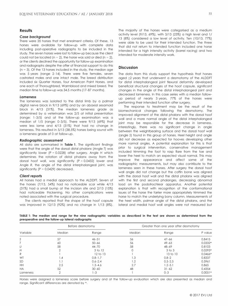
Results
Case backgroundThere were 20 horses that met enrolment criteria. Of these, 13horses were available for follow-up with complete dataincluding post-operative radiographs to be included in thestudy. The seven horses were lost to follow-up because the clientcould not be located (n = 2), the horse was sold or died (n = 2),or the clients declined the opportunity for follow-up examinationand radiographs despite the offer of financial support to do this(n = 3). Of the 13 horses included in the study, the median agewas 5 years (range 2–14). There were five females, sevencastrated males and one intact male. The breed distributionincluded six Quarter Horses, four American Paint Horses, andone each of Thoroughbred, Warmblood and mixed breed. Themedian time to follow-up was 34.5 months (17–87 months).
LamenessThe lameness was isolated to the distal limb by a palmardigital nerve block in 9/13 (69%) and by an abaxial sesamoidblock in 4/13 (31%). The median lameness grade asdetermined by a veterinarian was 2/5 at initial presentation(range: 1–3/5) and at the follow-up examination was amedian of 1/5 (range: 0–3/5). There were 9/13 (69%) thatwere less lame and 4/13 (31%) that had no change inlameness. This resulted in 5/13 (38.5%) horses being sound witha lameness grade of 0 at follow-up.
Radiographic assessmentAll data are summarised in Table 1. The significant findingswere that the angle of the dorsal distal phalanx (Angle T) wassignificantly lower (P = 0.0228) after surgery. Angle H whichdetermines the rotation of distal phalanx away from thedorsal hoof wall, was significantly (P = 0.0432) lower andAngle R, the angle of the distal interphalangeal joint, wassignificantly (P = 0.0429) decreased.
Client reportsAll horses had a medial approach to the ALDDFT. Seven ofthe horses (7/13, 54%) had no noticeable scar while 4/13(31%) had a small bump at the incision site and 2/13 (15%)had noticeable thickening. No other complications werenoted associated with the surgical procedure.
The clients reported that the shape of the hoof capsulewas improved in 12/13 (92%) and no change in 1/13 (8%).
The majority of the horses were categorised as a mediumactivity level (9/13, 69%), with 3/13 (23%) a high level and 1/13 (8%) considered a low level of activity. Ten (10/13, 77%)were able to be used for their intended function. The threethat did not return to intended function included one horseintended for a high intensity activity (barrel racing) and twointended for moderate intensity work.
Discussion
The data from this study support the hypothesis that horsesaged ≥2 years that underwent a desmotomy of the ALDDFTfor distal interphalangeal joint flexural deformity developedbeneficial structural changes of the hoof capsule, significantchanges in the angle of the distal interphalangeal joint anddecreased lameness. In this case series with a median follow-up period of nearly 3 years, 77% of the horses wereperforming their intended function after surgery.
The response to treatment may be the result of thebiomechanical changes following the desmotomy. Theimproved alignment of the distal phalanx with the dorsal hoofwall and a more normal angle of the distal interphalangealjoint may be responsible for the decrease in lameness.Interestingly, there was no significant change in anglebetween the weightbearing surface and the dorsal hoof wall(angle S) found in this group of horses. Heel height and angledid not decrease as expected for hooves developing othermore normal angles. A potential explanation for this is thatprior to surgical intervention, conservative managementincluded trimming the foot to rasp flare from the toe andlower the heel to match an expected visual normal. This mayimprove the appearance and affect some of theradiographic measurements, but may also contribute to thelameness seen in these horses. After surgery the dorsal hoofwall angle did not change but the coffin bone was alignedwith the dorsal hoof wall and the distal phalanx was alignedwith the first and second phalanges, decreasing abnormalload on the podotrochlear apparatus. Another potentialexplanation is that with recognition of the conformationalissues of the horse the farrier more appropriately trimmed thehorse to match the underlying bony column. Measurements ofthe heel width, palmar angle of the distal phalanx, and thelateral and medial hoof wall angles were not measured but
TABLE 1: The median and range for the nine radiographic variables as described in the text are shown as determined from thepreoperative and the follow-up lateral radiographs
Variable
Before desmotomy Greater than one year after desmotomy
Median Range Median Range P value
S 57 46–51 56 47–66 0.8415T 60 50–66 56 49–63 0.0332*U 58 44–70 58 48–69 0.8103H 2 �3 to 12 0 �3 to 3 0.0293*R 1 �12 to 13 �1 �13 to 10 0.0466*WT 1.4 0.8–1.7 1.3 0.8–2 0.8337SD 1.1 0.6–2.4 1.2 0.5–2.5 0.5961HH 2.5 1.3–4.6 2.7 1.2–5.1 0.865HA 52 30–60 48 31–62 0.4354Lameness 2 1–3 1 0–3 0.0051*
Horses were assigned a lameness score before surgery and at the follow-up evaluation which are also presented as median andrange. Significant differences are denoted by *.
© 2017 EVJ Ltd
55EQUINE VETERINARY EDUCATION / AE / JANUARY 2018

may have provided additional information about hoofchanges after desmotomy of the ALDDFT.
The decreased lameness seen in these horses is similar tothat previously reported (Turner and Stork 1998; Yiannikouriset al. 2011). The population most similar to that presented hereincluded mature performance horses with a flexural deformity;however, only four horses had regional anaesthesia localisingthe lameness and radiographic changes were not evaluated(Yiannikouris et al. 2011). In that case series 86% (12/14) of horsesaged >2 years returned to their intended use after desmotomyof the ALDDFT. The success rate of horses in this study is similar(72% return to intended use), which indicates that mature horsesmay benefit from desmotomy of the ALDDFT.
Case selection for surgery may have contributed to therelatively positive long-term outcome in this study. Whenreviewing preoperative radiographs, we recognised that noneof the horses in this study had notable radiographic evidenceof navicular bone degeneration. Those cases were probablyexcluded from surgical intervention by the surgeon because ofan expected poor prognosis for soundness. Radiographicevidence of severe changes in the distal phalanx and distalinterphalangeal joint in the horses aged one year and greateras noted by Wagner et al. (1985) were also not present in thecases in this study. The horses included in this study were aged≥2 years and most were in active work; therefore, horses withsevere osseous changes would have been excluded by studycriteria that they would not have been in work nor presentingfor a mild to moderate lameness evaluation.
A significant decrease in lameness was seen in thispopulation; however, the median lameness score at follow-up up was 1/5. Parameters that are not available forevaluation include long-term maintenance of shoeing andpain management through intraarticular medication andsystemic nonsteroidal anti-inflammatories. Based on clinicalexperience, management of these cases without surgery oftenresults in repeated veterinary visits with similar hoof relatedlameness issues. Comparison to a control group of horsestreated medically and with corrective shoeing would allow adirect comparison of the treatment options for mature horsesflexural deformity of the distal interphalangeal joint.Additionally, standardised hoof care and set follow-up timeperiods would allow for improved outcome determination.Obtaining these data in a future prospective study may furthersupport ALDDFT desmotomy for managing athletes with aflexural deformity of the distal interphalangeal joint.
The specific anatomic site or sites creating the lameness inthese horses was not isolated beyond the regional anaesthesiaand radiographic evaluation. Nine of the horses had thelameness resolved by a palmar digital block and the remainingfour by an abaxial sesamoid block. As noted by Turner andRosenstein (1992) multiple anatomic sites may be responsible forpain localised to the foot, including the podotrochlearapparatus, the deep digital flexor tendon, and the distalinterphalangeal joint. Laminar separation, pedal osteitis andincreased sole pressure as a result of the upright conformationmay also contribute to the clinical lameness seen in horses withflexural deformity of the distal interphalangeal joint. Furtherlocalisation through intrathecal anaesthesia and additionalimaging modalities, including magnetic resonance imaging,may help improve case selection and direct additionaltherapies to improve the overall outcome of these horses.
This study demonstrates the benefits of performing adesmotomy of the ALDDFT in mature horses with a flexural
deformity of the distal interphalangeal joint. There weresignificant improvements seen in the radiographic parametersand lameness scores evaluated in this study.
Authors’ declaration of interests
No conflicts of interest have been declared.
Ethical animal research
Horses described herein were clinical cases presented to thehospital for lameness evaluation and treatment. Ownerssigned a standard consent form and chose the treatmentprocedures.
Source of funding
None.
Authorship
Both authors contributed to the study design, studyexecution, data analysis and interpretation, and preparationof the manuscript. Both authors also gave their final approvalof the manuscript.
Manufacturer's address1Microsoft Corp, Redmond, Washington, USA.
ReferencesAuer, J.A. (2006) Flexural limb deformities. In: Equine Surgery, 3rd edn., Eds:
J.A. Auer and J.A. Stick, W.B. Saunders, St Louis, Missouri. pp 1150-1165.
Cripps, P.J. and Eustace, P.A. (1999) Radiological measurements fromthe feet of normal horses with relevance to laminitis. Equine Vet. J.31, 427-432.
Holroyd, K., Dixon, J.J., Mair, T., Bolas, N., Bolt, M.D., David, F. andWellar, R. (2013) Variation in foot conformation in lame horses withdifferent foot lesions. Vet. J. 195, 361-365.
Kaneps, A.J. (2014) Diagnosis of lameness. In: Equine Sports Medicineand Surgery. 2nd edn., Eds: K.W. Hinchcliff, A.J. Kaneps and R.J.Geor, W.B. Saunders, Edinburgh. pp 239-251.
O’Grady, S.E. (2012) Flexural deformities of the distal interphalangealjoint (clubfeet). Equine Vet. Educ. 24, 260-268.
Stick, J.A., Nickels, F.A. and Williams, M.A. (1992) Long-term effects ofdesmotomy of the accessory ligament of the deep digital flexormuscle in Standardbreds: 23 cases (1979-1983). J. Am. Vet. Med.Assoc. 200, 1131-1132.
Turner, T.A. and Rosenstein, D.S. (1992) Inferior check desmotomy as atreatment for caudal hoof lameness. Proc. Am. Assoc. EquinePract. 38, 157-163.
Turner, T.A. and Stork, C. (1998) Hoof abnormalities and their relationto lameness. Proc. Am. Assoc. Equine Pract. 44, 293-297.
Wagner, P.C., Grant, B.D., Kaneps, A.J. and Watrous, B.J. (1985) Long-term results of desmotomy of the accessory ligament of the deepdigital flexor tendon (distal check ligament) in horses. J. Am. Vet.Med. Assoc. 187, 1351-1353.
Walmsley, E.A., Anderson, G.A. and Adkins, A.R. (2011) Retrospectivestudy of outcome following desmotomy of the accessory ligamentof the deep digital flexor tendon for type 1 flexural deformity inThoroughbreds. Aust. Vet. J. 89, 265-268.
Yiannikouris, S., Schneider, R.K., Sampson, S.N. and Roberts, G. (2011)Desmotomy of the accessory ligament of the deep digital flexortendon in the forelimb of 24 horses 2 years and older. Vet. Surg.40, 272-276.
© 2017 EVJ Ltd
EQUINE VETERINARY EDUCATION / AE / JANUARY 201856

Platinum Performance® GIEquine Wellness & Performance Formula + Digestive Care
THE HEALTH OF THE EQUINE GUT is correlated to gastrointestinal (GI) function, metabolism, immunity and more. This is why we have been hard at work researching, formulating, testing and
conducting trials with Platinum Performance® GI. This gastrointestinal-focused approach to total horse health supports wellness and is recommended for horses in training or competition as well as
senior horses. Platinum Performance® GI can help maintain health while traveling, during antibiotic or NSAID therapy and for horses that have digestive health concerns or difficulty maintaining weight.
PlatinumPerformance.com | 866-553-2400 © 2017 PLATINUM PERFORMANCE, INC.
OVERALL WELLNESSThis veterinary-developed formula
provides omega-3 fatty acids, antioxidants, vitamins and trace minerals,
along with Bio-Sponge®, prebiotics, probiotics and glutamine for GI support.
DIGESTIONPrebiotics nourish the “good” bacteria in the gut and support
the digestion of fiber, while probiotics are important for
absorption of nutrients.
IMMUNITYAn estimated 70-80% of
the immune system resides in the gut. Nourishing the beneficial
bacteria can help support a healthy gut barrier and microflora balance.
IntroducingJ