Embed Size (px)
Citation preview

Working-Memory fMRI Reveals CingulateHyperactivation in Euthymic Major Depression
Sonja Schoning,1,2 Pienie Zwitserlood,3 Almut Engelien,1,2,4
Andreas Behnken,1 Harald Kugel,5 Hagen Schiffbauer,5 Katharina Lipina,2
Christine Pachur,2 Anette Kersting,1 Udo Dannlowski,1,2
Bernhard T. Baune,1,6 Peter Zwanzger,1 Thomas Reker,7
Walter Heindel,5 Volker Arolt,1 and Carsten Konrad1,2*
1Department of Psychiatry, University of Muenster, Germany2Interdisciplinary Center for Clinical Research (IZKF), University of Muenster, Germany
3Department of Psychology, University of Muenster, Germany4FNL, Department of Psychiatry, Weill Medical College, Cornell University, NY
5Department of Clinical Radiology, University of Muenster, Germany6Department of Psychiatry, School of Medicine, James Cook University, Australia
7LWL-Klinik Muenster, Germany
Abstract: While cognitive impairments are well documented for the acute episode of major depressivedisorder (MDD), less is known about cognitive functioning in the euthymic state. For working memory,dysfunctional activation of lateral prefrontal and cingulate cortex has been reported in the acute epi-sode. This study investigates working-memory function and its neurobiological correlate in euthymicMDD patients, particularly whether dysfunctional activation persists when depressive symptomsimprove. We investigated 56 subjects with functional magnetic resonance imaging (fMRI) at 3 Tesla. Tochallenge working-memory function, a classical verbal n-back task (0-, 1-, and 2-back) was used in 28well-characterized, euthymic, unipolar MDD patients and 28 healthy control subjects matched accord-ing to age, sex, and educational level. Data were analyzed using SPM5. In the absence of significant be-havioral differences, we observed comparable overall patterns of brain activation in both groups. Asexpected, both groups showed stronger activation of the typical working-memory network withincreasing memory load. However, significant hyperactivation of the cingulate cortex was observed ineuthymic patients, while lateral prefrontal activation was comparable between patients and controls.Working-memory challenge in the euthymic state of MDD revealed a dissociation of lateral prefrontaland cingulate brain function. Cingulate function, which is important for both emotional and cognitiveprocessing and their integration, is still abnormal when mood is restored. This could reflect a differentspeed of normalization in prefrontal and limbic cortices, persistent systematic changes in neuronal net-works after an episode of MDD, or a compensatory mechanism to maintain working-memory perform-ance. Hum Brain Mapp 30:2746–2756, 2009. VVC 2008 Wiley-Liss, Inc.
Additional Supporting Information may be found in the onlineversion of this article.
The first two authors contributed equally to the present work.
Contract grant sponsors: Interdisciplinary Center for ClinicalResearch (IZKF), University of Munster, Germany (FG 4),DGPPN/AstraZeneca Forderpreis, 2004.
*Correspondence to: Carsten Konrad, MD, Department of Psychia-try, IZKF Research Group No. 4, University of Muenster, Albert-
Schweitzer-Str.11, D-48149 Muenster, Germany.E-mail: [email protected]
Received for publication 24 April 2008; Revised 23 September2008; Accepted 29 October 2008
DOI: 10.1002/hbm.20702Published online 11 December 2008 in Wiley InterScience (www.interscience.wiley.com).
VVC 2008 Wiley-Liss, Inc.
r Human Brain Mapping 30:2746–2756 (2009) r
Key words: major depression; remission; cognitive deficits; functional magnetic resonance imaging;working memory; n-back
INTRODUCTION
Major depressive disorder (MDD) is one of the mostprevalent psychiatric disorders leading to a dramaticreduction of quality of life, increased mortality risk,(Alonso and Lepine, 2007; Cuijpers and Smit, 2002) andcausing a significant individual and economic burden asthe most costly brain disorder in Europe (Sobocki et al.,2006; von Knorring et al., 2006).Neuropsychological deficits of different functional
domains are well documented for the acute phase of adepressive episode (Airaksinen et al., 2004; Burt et al.,1995; Castaneda et al., 2008; Landro et al., 2001; Ravnkildeet al., 2002; Veiel, 1997; Zakzanis et al., 1998). However,the nature of these deficits, the cognitive domains affected,as well as the severity of cognitive impairments is still amatter of ongoing debate. Compared with the acute phaseof major depression, even less is known about the neuro-cognitive profile of patients who recovered from depres-sion. Several studies reported lasting deficits in some cog-nitive domains (Austin et al., 2001; Kessing, 1998; Marcoset al., 1994; Paelecke-Habermann et al., 2005; Paradisoet al., 1997) such as executive functions and attention (Pae-lecke-Habermann et al., 2005; Smith et al., 2006; Trichardet al., 1995). The influence of clinical depression on work-ing-memory function is still under debate (Channon et al.,1993; Christopher and MacDonald, 2005; Harvey et al.,2004; Landro et al., 2001; Rose and Ebmeier, 2006;Zakzanis et al., 1998).As we know from clinical experience, MDD patients of-
ten complain about problems with thinking and concentra-tion (Nair et al., 1999). While impairments of workingmemory in the acute phase of MDD have been reportedpreviously, studies focusing on this crucial cognitive func-tion in remitted depression are rare. Subtle deficits werereported for strategic aspects of a spatial working-memorytask (Weiland-Fiedler et al., 2004).Working memory is an extensively researched psycho-
logical concept dealing with the temporary storage andprocessing of information (Baddeley, 1992; Baddeley,2003). Intact working memory is essential for every dayfunctioning. Working-memory tasks require several cogni-tive processes, such as online monitoring, continuousupdating, manipulating stored information, and decisionmaking, which all might be affected by MDD. The neuro-nal processes underlying working-memory processes havebeen widely investigated with neuroimaging techniques(Owen et al., 2005; Wager and Smith, 2003). In healthysubjects, the verbal n-back task activated a bilateral net-
work consisting of dorsolateral and ventrolateral prefrontalcortex, lateral premotor cortex, dorsal cingulate and medialpremotor cortex, frontal poles, and medial and lateral pos-terior parietal cortex (Owen et al., 2005). Task-related activ-ity was shown to be correlated with working-memoryload. Especially dorsolateral and left inferior regions of theprefrontal cortex show a linear relationship between activ-ity and task complexity (Braver et al., 2001).To date, only a few imaging studies investigated work-
ing memory in major depression, almost exclusively focus-ing on the acute phase (Fitzgerald et al., 2008; Harveyet al., 2005; Matsuo et al., 2007; Rose et al., 2006; Walteret al., 2007a; Walter et al., 2007b). These studies revealedabnormalities in cortico-limbic networks fundamentallyinvolved in the pathophysiology of major depression(Dougherty and Rauch, 2007; Mayberg, 1997). Comparedwith healthy control subjects, a stronger activation wasobserved in the limbic system and lateral prefrontal cortexof MDD patients, in the absence of significant behavioraldifferences (Fitzgerald et al., 2008; Matsuo et al., 2007). Forexample, Matsuo et al. reported stronger left dorsolateraland anterior cingulate cortex (ACC) activation in 15 MDDpatients performing a visuo-spatial task, while healthycontrols failed to show cingulate activation (Matsuo et al.,2007). Harvey et al. used a verbal variant of the n-backtask and compared 10 MDD patients with 10 controls(Harvey et al., 2005). Both groups showed similar activa-tion, but the lateral prefrontal cortex and the anterior cin-gulate were activated more strongly in MDD patients.Rose et al. investigated 10 MDD patients and 10 healthycontrols with an n-back task and also reported anterior cin-gulate differences in load-dependent activation betweenpatients and controls (Rose et al., 2006). Using a longitudi-nal design, Walsh et al. reported greater load-response inthe verbal working-memory network of patients (Walshet al., 2007). Taken together, previous studies indicate thatan acute episode of MDD is associated with abnormal cor-tico-limbic activation in working-memory, mainly charac-terized by hyperactivation of lateral prefrontal and cingu-late areas. Almost nothing is known as to whether thishyperactivation observed in the acute phase is a state-de-pendent phenomenon and whether or not brain activationnormalizes when depressive symptoms are no longer pre-dominant.Although the above studies often failed to find differen-
ces on behavioral measures, Walter et al. found behavioraldifferences between 12 partially remitted patients (meanHamilton depression rating scale [HDRS] score of 18.2)and controls in a delayed match-to-sample working-
r Cingulate Hyperactivation in Euthymic Depression r
r 2747 r

memory task (Hamilton, 1960; Walter et al., 2007b). Theauthors also reported stronger activation in the dorsolat-eral prefrontal cortex (DLPFC) for the highest cognitiveload condition, and in the ventromedial prefrontal cortexfor the control condition.To the best of our knowledge, no functional magnetic
resonance imaging (fMRI) study has yet investigated work-ing-memory function in a large group of completely euthy-mic unipolar depressed patients. Thus, the goal of thisstudy was to investigate working-memory function, in par-ticular prefrontal and cingulate activation during working-memory performance, in euthymic MDD patients. Wehypothesized that behavioral working-memory perform-ance of euthymic MDD patients is almost equal to healthycontrols. We expected neurobiological differences in brainregions such as cingulate gyrus and prefrontal areasbetween euthymic patients with MDD and controls.
MATERIALS AND METHODS
Subjects
In total, 56 subjects were recruited for this study.Twenty-eight inpatients from the Department of Psychiatryof the University of Muenster or the LWL-Clinic Muenster(16 female, 12 male subjects), fulfilling DSM-IV criteria fora MDD, participated in this study (for details see Table I).A diagnosis of either first (n 5 9) or recurrent episode(n 5 19) of unipolar depression was verified using thestandardized SCID-I- Interview (German version)(Wittchen et al., 1997), in addition to clinical assessment bytwo board-certified specialists in Psychiatry. MDD patientswith psychotic depression or axis-II disorders wereexcluded. Twenty-three patients had MDD alone; threepatients had comorbid dysthymia (‘‘double depression’’).Further comorbid axis-I disorders were excluded if symp-toms of the comorbid disorder required current treatment.One patient was additionally diagnosed with social pho-bia, and one patient with panic disorder with agoraphobia.This was, however, not relevant for current hospitalization.Patients participated just before discharge from the hospi-tal after achieving a stable euthymic state characterized byHDRS (HDRS �8) and confirmed by two board-certifiedspecialists in Psychiatry. The following additional inclu-sion criteria were applied: age between 18 and 55 years,no treatment with electroconvulsive therapy during theprevious depressive episode, no history of any other seri-ous medical or neurological disease, no serious headinjury, no suicidal tendency, no benzodiazepine treatment3 days before scanning, and no MRI contraindications. Allpatients were right-handed, as assessed by the EdinburghHandedness Inventory (Oldfield, 1971) and had more than12 years of education. Twenty-seven patients were treatedaccording to current treatment guidelines in a stable dos-age and one patient did not receive any medication. Thefollowing antidepressants were prescribed as antidepres-sive monotherapy (13), combined antidepressive therapy
(3), or combined antidepressive/antipsychotic therapy (11):citalopram (2), escitalopram (8), mirtazapine (11), venlafax-ine (11), reboxetine (1), duloxetine (1), trancylpromine (1);none were taking tricyclic antidepressants. To rule out anynegative effects on memory function antipsychotics wereused instead of benzodiazepines for treatment of agitationand nervousness in some patients: quetiapine (9), risperi-done (2), pipamperone (1). None of the patients was takingbenzodiazepines at the time of testing.Twenty-eight healthy, right-handed control subjects,
recruited by advertisement in the local newspaper, were1:1 matched to the patients according to sex and age (63years). Education level, both in terms of years of educationand highest graduation level, was also balanced betweengroups. All control subjects underwent an initial telephonescreening to ensure matching criteria, to exclude medicaland neurological diseases, or MRI contraindications. Thestandardized SCID-I-Interview was performed to excludeany current or previous psychiatric disorders (Wittchenet al., 1997). In healthy controls no psychiatric disorders infirst degree relatives were reported.All procedures were approved by the local Institutional
Ethical Review Board. The ethical standards of the Decla-ration of Helsinki were met and all participants providedwritten informed consent.
Materials and Procedures
The working-memory task was the first part of a largerfMRI and neuropsychological study of memory processesin euthymic MDD. We used a classical letter variant of then-back task (Braver et al., 1997). Before entering the scan-ner, a detailed task instruction was given and participantswere familiarized with the n-back task until they suc-ceeded in the training trials. A standardized brief instruc-tion announced the start of the task in the scanner. Work-ing-memory load was manipulated in three levels (0-2-back), presented in a block design. During the 0-backcondition, subjects had to press the response button of aMRI-compatible response box if the target letter ‘‘X’’appeared on the screen. In the 1-back condition subjectshad to decide if the actual letter on the screen was identi-cal to the previous letter. During the 2-back condition, sub-jects had to decide if the actual letter was identical to theletter presented two trials before. Subjects responded withtheir right hand, using the index finger for targets andmiddle finger for nontargets.Each active n-back condition lasted 36 s and n-back
blocks were presented in a fixed order (1-0-2-0-1-2) to eachsubject. Subjects completed two blocks of each n-back con-dition. White letters were presented in the centre of ablack screen for 500 ms, with an interstimulus interval of2500 ms (Presentation Software1, Version 0.81, 2004, Neu-robehavioral Systems, Albany, CA). Only orthographicallydistinct uppercase consonants were used (B, C, D, F, G, H,J, K, M, Q, R, S, T, V, X, Z). Each letter sequence consistedof 12 consonants, including one-third targets. During fMRI
r Schoning et al. r
r 2748 r
scanning, a short instruction announced the n-back type.All n-back conditions were separated by a pause of 21 sduring which participants had to look at a white fixationcross on a black screen.As part of the larger study protocol, all patients and
control subjects underwent neuropsychological testing,such as the Mehrfachwahlwortschatz-Test (MWT-B) as anestimate of verbal intelligence (Lehrl et al., 1995) and theBeck Depression Inventory (BDI) (Beck et al., 1961).
Scanning Procedures
MRI data acquisition was performed in a 3 Tesla whole-body scanner (Intera T 3.0, Philips, Best, NL), equippedwith master gradients (nominal gradient strength 30mT/m, maximal slew rate 150mT/m/ms). A circularly polar-ized transmit/receive birdcage head coil with an HFreflecting screen at the cranial end was used for spin exci-tation and resonance signal acquisition. Functional imageswere acquired using a T2* weighted single shot echo pla-nar (EPI) sequence (whole brain coverage, TE 5 38, TR 53000ms, flip angle 908, slice thickness 3.6 mm without gap,matrix 64 3 64, FOV 230 mm, in-plane resolution 3.6 33.6). 36 transversal slices orientated to the AC-PC linewere acquired.
Behavioral Data Analysis
During fMRI scanning, responses and response latencies(in ms) for the n-back performance were recorded. Behav-ioral results were acquired from all 28 patients. Data fromthree control subjects were omitted because of technicaldifficulties. Performance is reported as accuracy rate (per-centage of correct answers) for each n-back condition.Repeated-measures analyses of variance (ANOVAs), withone between-subject factor (group: two levels) and onewithin-subject factor (working-memory load: three levels),were performed for accuracy rate and response latency.
Functional Data Analysis
Functional MRI data were analyzed using SPM5 stand-ard routines and templates (www.fil.ion.ucl.ac.uk/spm).
The first 10 images of each session (30 s prestimulus inter-val) were discarded to allow for saturation effects of theBOLD signal. The remaining images were realigned, nor-malized, and resliced to a voxel size of 2 mm 3 2 mm 32 mm. Gaussian smoothing was performed using a 9 mmkernel. Data were filtered with a high-pass filter (cut-offperiod of 128 s). A boxcar function convolved with the ca-nonical hemodynamic response function implemented inSPM5 was used to model BOLD-responses for the work-ing-memory task. In a first-level fixed-effects analysis, theconditions 0-back, 1-back, 2-back, and visual instructionwere modeled. Contrast images for 0-, 1-, and 2-back con-ditions to general baseline and for 2-back versus other acti-vation conditions (2vs0-back and 2vs1-back) were derived.The individual contrast images were entered into a sec-ond-level random-effects analysis to obtain activationmaps across subjects. To display the different contrasts ineach group, one-sample t-tests were performed (P < 0.05,corrected for false discovery rate [FDR], contiguity thresh-old �15 voxels). On the basis of previous findings (Harveyet al., 2005; Matsuo et al., 2007) and our hypotheses ofhigher cingulate and prefrontal activation, differencesbetween patients and controls were calculated in the cin-gulate gyrus and in the inferior, middle, and superiorDLPFC, using two-sample t-tests (P < 0.05, corrected forFDR, contiguity threshold �15 voxels). The regions of in-terest in the bilateral cingulate cortex (anterior, medial andposterior part) as well as the inferior, middle, and superiordorsolateral frontal gyrus were defined according to theautomated anatomical labeling (AAL) atlas (Tzourio-Mazoyer et al., 2002) as implemented in the WFU PickAt-las Toolbox (Maldjian et al., 2003) (ROI names: ACIN,MCIN, PCIN, F1, F2, F3OP/T). To verify that thisapproach does not overlook important effects outside theROIs, a whole-brain analysis was performed at a more lib-eral threshold (P < 0.0005, uncorrected for multiple com-parisons, contiguity threshold �15 voxels). Within eachgroup, a correlation analysis between behavioral data(response latency and accuracy) and task-related activitywas performed across all voxels and all conditions. Clini-cal variables (HDRS, days of hospitalization, and numberof depressive episodes) were additional variables for thepatient group.
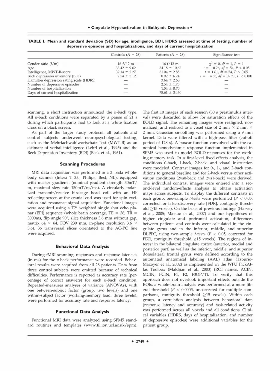
TABLE I. Mean and standard deviation (SD) for age, intelligence, BDI, HDRS assessed at time of testing, number of
depressive episodes and hospitalizations, and days of current hospitalization
Controls (N 5 28) Patients (N 5 28) Significance test
Gender ratio (f/m) 16 f/12 m 16 f/12 m v2 5 0, df 5 1, P 5 1Age 33.42 6 9.62 34.18 6 10.62 t 5 20.26, df 5 54, P > 0.05Intelligence, MWT-B-score 32.14 6 2.27 31.04 6 2.85 t 5 1.61, df 5 54, P > 0.05Beck depression inventory (BDI) 2.54 6 3.12 8.92 6 6.24 t 5 24.85, df 5 39.71, P < 0.001Hamilton depression rating scale (HDRS) — 3.64 6 2.63 —Number of depressive episodes — 2.54 6 1.75 —Number of hospitalization — 1.54 6 0.70 —Days of current hospitalization — 75.61 6 34.60 —
r Cingulate Hyperactivation in Euthymic Depression r
r 2749 r

RESULTS
Behavioral Results
No significant differences between groups wereobserved. Analysis of variance on accuracy and responselatency revealed a significant main effect of working-mem-ory load (F(2,102) 5 12.64, P < 0.001) and (F(2,102) 5 32.65,P < 0.001), respectively. As expected, accuracy decreasedand response latency increased from 0-back to 2-back con-dition (Figs. 1 and 2). However, no main effects of group(F(1,51)<1, P > 0.05) or interactions between group andworking-memory load (F(2,102)<1, P > 0.05) emerged foraccuracy or response latency. Furthermore, no significantdifferences were observed as a function of verbal intelli-gence (MWT-B) (two-sample t-test, T 5 1.61, df 5 56, P >0.05).
Activation Patterns Across Load Conditions
and Groups
For each group, activation was investigated for eachload condition of the working-memory task separately.Healthy controls and patients activated the brain areas rel-evant for a verbal working-memory task, as expected fromthe literature (Owen et al., 2005; Wager and Smith, 2003).Activation was found in the medial frontal and inferiorfrontal gyrus, insula, pre- and postcentral gyrus, inferiorparietal lobule, and cerebellum in both groups. In bothgroups, we observed an increase of brain activation fromthe 0-back to 2-back condition (see Supporting Informa-tion). Activation increased with working-memory demand,in particular, with respect to the bilateral activation of theinferior and middle frontal cortex.
Regions Activated With Increasing
Working-Memory Load
Activation increases from 0-back to 2-back
(2vs0-back contrast)
Common to both groups were the following effects forthe 2vs0-back contrast (P < 0.05, corrected for FDR, conti-guity threshold �15 voxels). First, we observed extendedactivation clusters of the inferior, middle, superior, andmedial frontal cortex, including typical verbal working-memory regions, such as parts of the medial frontal cortex,dorsolateral and ventrolateral prefrontal cortex (BA 9, 46,45, 47) (Fig. 3). Next, we found activation of the insula,supplementary motor area, temporal lobe, and cerebellum.Finally, there was strong activation in the parietal lobe, inthe inferior and superior parietal lobule (BA 7, 40), theangular and supramarginal gyrus extending to the supe-rior and middle occipital gyrus (BA 19, 18). However,while healthy controls showed only few activated clustersin the cingulate cortex, parahippocampal gyrus, and hip-pocampus, patients activated large parts of the cingulatecortex (BA 24, 32, 33), and parahippocampal gyrus (BA 27,28, 35, 36) in the 2vs0-back contrast (for details see alsoSupporting Information).
Activation increases from 1-back to 2-back
(2vs1-back contrast)
For the 2vs1-back contrast (see Fig. 4), healthy controlsactivated few and small clusters in the inferior frontal cor-tex and the superior frontal cortex (BA6) (P < 0.05, cor-rected for FDR, contiguity threshold �15 voxels). Activa-tion was also found in the precuneus, inferior and superiorparietal lobule. In patients, we observed the following:
Figure 1.
Behavioral data for accuracy rate (percentage of correct
answers, mean 6 standard error) with varying working memory
load in patients and control subjects reveal a significant effect of
working memory load, but not of group or interaction.
Figure 2.
Behavioral data for response latency (mean 6 standard error) in
all load conditions in patients and control subjects. A main effect
of working memory load condition was observed, but no effect
of group or interaction.
r Schoning et al. r
r 2750 r

there were large activated clusters in the inferior, middle,medial, and superior frontal gyrus (BA 6, 8, 9, 44-47); par-allel to the 2vs0-back contrast, the cingulate cortex (BA 24,32, 33), parahippocampal gyrus (BA 35, 36), and hippo-campus were significantly activated; significant activationswere also observed in the insula, pre- and postcentralgyrus, temporal and occipital lobe, and cerebellum; andfinally, in the parietal lobe, the angular and supramarginalgyrus, the inferior and superior parietal lobule, and precu-
neus were bilaterally activated (BA 7, 39, 40) (for detailssee also Supporting Information).
Analysis of additional factors
No significant correlations (P < 0.05, corrected for FDR,contiguity threshold �15 voxels) between brain activationand behavioral measures (accuracy and response latency)were observed, neither in patients nor in controls. One
Figure 3.
Group activation in (a) patients and (b) controls for the 2vs0-back contrast (one sample t-test,
P < 0.05, corrected for FDR, contiguity threshold �15 voxels). Random-effects analysis rendered
on the surface of the canonical template image used by SPM5. [Color figure can be viewed in
the online issue, which is available at www.interscience.wiley.com.]
Figure 4.
Group activation in (a) patients and (b) controls for the 2vs1-back contrast (one sample t-test,
P < 0.05, corrected for FDR, contiguity threshold �15 voxels). Random-effects analysis rendered
on the surface of the canonical template image used by SPM5. [Color figure can be viewed in
the online issue, which is available at www.interscience.wiley.com.]
r Cingulate Hyperactivation in Euthymic Depression r
r 2751 r

exception concerned a small correlation of accuracy withthe right inferior frontal lobe in patients for the 2-back con-dition (MNI coordinate 32/34/12) (Fig. not shown). More-over, no significant correlations between brain activationand clinical variables such as Hamilton scores, days ofhospitalization, or number of depressive episodes werefound in patients (P < 0.05, corrected for FDR, contiguitythreshold �15 voxels).Further analysis in the patient group revealed no signifi-
cant differences between the 13 patients treated with anti-depressive monotherapy and the 14 patients treated with acombination therapy of antidepressants or antipsychoticsfor the 2vs0-back and 2vs1-back contrast (two-sample t-test, P < 0.05, corrected for FDR, contiguity threshold �15voxels). An analysis of variance showed no effect of gen-
der (P < 0.05, corrected for FDR, contiguity threshold �15voxels).
Between-Group Comparisons
As we expected group differences in specialized work-ing-memory areas, particularly prefrontal areas and thecingulate cortex, a ROI-analysis was performed betweengroups (two-sample t-test, P < 0.05, corrected for FDR,contiguity threshold �15). In the cingulate cortex, both the2vs0-back and the 2vs1-back contrast revealed strongeractivation of the anterior and posterior cingulate cortex(BA 24, 32, 23, 31) for patients than healthy controls.Unlike patients, healthy controls showed no increased cin-gulate activation (Figs. 5 and 6). In the prefrontal cortex,
Figure 5.
Differences in cingulate brain activation between (a) patients versus controls and (b) controls ver-
sus patients in the 2vs0-back contrast. ROI analysis, two sample t-test, P < 0.05, corrected for
FDR, contiguity threshold �15 voxels, projected on the canonical template image used by SPM5.
[Color figure can be viewed in the online issue, which is available at www.interscience.wiley.com.]
Figure 6.
Differences in cingulate brain activation between (a) patients versus controls and (b) controls ver-
sus patients in the 2vs1-back contrast. ROI analysis, two sample t-test, P < 0.05, corrected for
FDR, contiguity threshold �15 voxels, projected on the canonical template image used by SPM5.
[Color figure can be viewed in the online issue, which is available at www.interscience.wiley.com.]
r Schoning et al. r
r 2752 r

especially dorsolateral (BA 9, 46) and ventrolateral (BA 45,47) PFC, no significant differences between patients andcontrols were found (Fig. not shown).To verify that this approach does not overlook important
effects outside the ROIs, a whole-brain analysis was per-formed at a more liberal threshold (P < 0.0005, uncor-rected for multiple comparisons, contiguity threshold �15).The cingulate difference between groups for both the 2vs0-back and the 2vs1-back contrast was corroborated and noother relevant activations outside the ROIs were detected(see Supporting Information).
DISCUSSION
Cognitive impairments are an important characteristic ofmajor depression. Modern neuroimaging methods indicatethat dysfunction of cortico-limbic networks plays an impor-tant role in the pathophysiology of both affective and cogni-tive symptoms in MDD (Dougherty and Rauch, 2007). In theacute episode of depression, brain metabolism is signifi-cantly altered, with pathological changes in the dorsolateralprefrontal and limbic cortex at rest and during cognitiveactivation (Drevets, 2001; Ebmeier et al., 2006; Fitzgeraldet al., 2006; Greicius et al., 2007). Much less is known aboutbrain function when depressed patients reach the euthymicmood state. Neuropsychological data suggest that cognitivedeficits persist in certain domains, and thus might representmore a trait than a state characteristic (Paelecke-Habermannet al., 2005). This study investigated networks involved inworking-memory function in recently remitted patientswith major depression. We explored whether dysfunctionalactivation of the lateral prefrontal and cingulate cortexwould still be present in the euthymic phase of majordepression, as had been previously reported for the acuteepisode of major depression (Harvey et al., 2005; Matsuoet al., 2007; Rose et al., 2006; Walter et al., 2007b).In line with previous reports, we found the classic work-
ing-memory network activated in the n-back task (Owenet al., 2005; Wager and Smith, 2003). With increasing work-ing-memory demand, strong activation was observed inboth patients and controls, in the dorsolateral and ventro-lateral prefrontal cortex, middle frontal cortex, and precen-tral gyrus. Both groups also showed activation in the pari-etal cortex, of the angular and supramarginal gyrus, infe-rior and superior parietal lobule, precuneus, and superioroccipital gyrus. Activation was also observed in the tempo-ral cortex, whose role for working-memory processes is asyet poorly understood, and subject of current research(Axmacher et al., 2007; Picchioni et al., 2007).A novel and interesting finding is that our data point to
a deviance of the working-memory network in patientswith MDD even in the euthymic state. So far, altered pre-frontal and cingulate activity during working-memorytasks has only been reported in severely depressedpatients, mainly in the acute phase of major depression.The majority of these studies did not find behavioral defi-
cits between patients and controls (Harvey et al., 2005;Matsuo et al., 2007; Rose et al., 2006). Patients in the acutephase performing working-memory tasks showed hyperac-tivation of the DLPFC (Harvey et al., 2005; Matsuo et al.,2007) and ACC (Harvey et al., 2005; Matsuo et al., 2007;Rose et al., 2006). These findings were taken as evidencefor the recruitment of additional resources to fulfill thecognitive demands of a given task.In this study, patients in the euthymic state showed
hyperactivation of the cingulate cortex, a region involvedin both emotional and cognitive processing, while lateralprefrontal hyperactivation was not observed relative tohealthy controls. Both of these areas on the lateral andmedial surface of the prefrontal cortex are known to play acentral role in the pathophysiology of depression. Baselinefunctional-imaging studies demonstrated metabolic and re-gional blood flow abnormalities in major depression, inparticular a decreased metabolism in DLPFC and increasedmetabolism in orbitofrontal cortex (Dougherty and Rauch,2007). This cortico-limbic network also reveals abnormalfunction when challenged by cognitive tasks such as work-ing memory, most prominently evident as an increase oflateral prefrontal and limbic activity (Harvey et al., 2005;Matsuo et al., 2007; Rose et al., 2006). As a major result,our data indicate that metabolic abnormalities in the cingu-late persist even in the euthymic state of MDD, while lat-eral cortical abnormalities normalize. Our results mightreflect an earlier normalization of lateral prefrontal func-tion occurring prior to possible similar changes in anteriorcingulate areas in the course of remission.The role of the ACC has been controversially discussed
in depression and recovery, playing an important role inboth cognitive and emotional processing. The dorsal subdi-vision of the ACC subserves many cognitive functions,including working memory, and is highly interconnectedwith other regions involved in working memory, such asthe above-mentioned DLPFC (Bush et al., 2000; Devinskyet al., 1995). This dorsal ACC region is involved in taskcomplexity, mental effort or attentional processes (Mulertet al., 2007; Mulert et al., 2005), conflict monitoring, anderror processing (Bioulac et al., 2005; Botvinick et al., 2004;Carter et al., 1999; Carter et al., 1998; Kerns et al., 2004;Michelet et al., 2007; Sohn et al., 2007; van Veen and Car-ter, 2006). On the other hand, the rostral part of the ACCsubserves emotional processing, especially for the assess-ment of emotional information and the regulation of emo-tional responses (Whalen et al., 1998). This part is highlyinterconnected with the amygdala, hippocampus, hypo-thalamus, nucleus accumbens, and orbitofrontal cortex.Alterations of (rostral) ACC metabolism have been associ-ated with depressive symptoms, their severity, and treat-ment response in MDD patients (Chen et al., 2007; Konar-ski et al., 2007; Mayberg et al., 1997; Milak et al., 2005).Moreover, brain imaging studies revealed altered brainactivation of the rostral part of the ACC for emotionaltasks in depressed patients (Frodl et al., 2007; Mitter-schiffthaler et al., 2008).
r Cingulate Hyperactivation in Euthymic Depression r
r 2753 r

In this study, we observed an activation increase of theACC with increasing working-memory load in patients,which seemed to involve both the dorsal and the rostralpart. Our findings corroborate cingulate cortex hyperacti-vation observed in patients in the acute depressive episode(Harvey et al., 2005; Matsuo et al., 2007). Here, we demon-strate that cingulate hyperactivation during working-mem-ory performance is still present when affective symptomssuch as depressed mood or reduced drive are muchrelieved or have even subsided. As in acute depression,we might now hypothesize that enhanced recruitment ofthese cerebral resources is necessary to fulfill the cognitivedemands of the given task. Enhanced recruitment might aswell be necessary as baseline metabolism is decreased inthe ACC even after recovery from depression (Holthoffet al., 2004). Our finding of hyperactivation of the ACC inan affective disorder during a cognitive task underlinesthe importance of this region for both emotional and cog-nitive processing. A clear allocation of the hyperactivationto either limbic or cognitive circuits based on neuroanat-omy (Bush et al., 2000) is not warranted by our findings.As mentioned above, behavioral performance was not
significantly different between euthymic MDD patientsand healthy controls. This was expected on the basis ofresults from fMRI studies with acute depressed patients(Harvey et al., 2005; Matsuo et al., 2007). It is thus unlikelythat behavioral differences between patients and healthycontrols are responsible for the observed differential acti-vation pattern. However, our study used a block designand only two levels of task difficulty, which might be notsensitive enough to detect subtle disturbances of working-memory capacity. Some other limitations of our study alsoneed to be mentioned. We cannot exclude medicationeffects, since our patients did receive psychiatric treatmentto modern standards of care. Previous studies on theeffects of antidepressant medication revealed that pharma-cological treatment leads to an attenuation or decrease oflimbic activation in response to emotional stimuli ratherthan to an increase of ACC activation, as observed in thepresent study (Arce et al., 2008; Fu et al., 2004; Harmeret al., 2006; Sheline et al., 2001). Although the above evi-dence points towards an attenuating effect of antidepres-sants on brain activation, we cannot completely exclude anopposite effect in a working-memory task, but this seemsrather unlikely. This study does not claim to investigatemedication effects on working memory performance indepression. Additional studies will have to tackle thisproblem. Furthermore, patients were only included whendepressive symptoms were considerably reduced. We didnot assess the time course of brain activation during thecourse of recovery, so additional studies need to clarify iffurther changes occur when more time elapses after theacute depressive episode. A strength of our study is thehigh number of well-characterized patients, for whomstrict exclusion criteria were met.To summarize, we demonstrated that even after clinical
improvement of affective symptoms, abnormal cingulate
activation was associated with a classical working-memorytask in patients compared with healthy controls. In con-trast to patients in the acute depressive episode, ACChyperactivation, but no lateral prefrontal hyperactivation,occurred in patients in the euthymic state. Our data mightreflect a different lateral prefrontal and cingulate pace ofnormalization, a trait marker of changes in neuronal net-works after an episode of MDD, or a compensatory mecha-nism to maintain adequate working-memory performance.
REFERENCES
Airaksinen E, Larsson M, Lundberg I, Forsell Y (2004): Cognitivefunctions in depressive disorders: Evidence from a population-based study. Psychol Med 34:83–91.
Alonso J, Lepine JP (2007): Overview of key data from the Euro-pean Study of the Epidemiology of Mental Disorders(ESEMeD). J Clin Psychiatry 68(Suppl 2):3–9.
Arce E, Simmons AN, Lovero KL, Stein MB, Paulus MP (2008):Escitalopram effects on insula and amygdala BOLD activationduring emotional processing. Psychopharmacologia 196:661–672.
Austin MP, Mitchell P, Goodwin GM (2001): Cognitive deficits indepression: Possible implications for functional neuropathol-ogy. Br J Psychiatry 178:200–206.
Axmacher N, Mormann F, Fernandez G, Cohen MX, Elger CE,Fell J (2007): Sustained neural activity patterns during workingmemory in the human medial temporal lobe. J Neurosci27:7807–7816.
Baddeley A (1992): Working memory. Science 255:556–559.Baddeley A (2003): Working memory: Looking back and looking
forward. Nat Rev Neurosci 4:829–839.Beck AT, Ward CH, Mendelson M, Mock J, Erbaugh J (1961): An
inventory for measuring depression. Arch Gen Psychiatry4:561–571.
Bioulac B, Michelet T, Guehl D, Aouizerate B, Burbaud P (2005):[The anterior cingulate cortex in error detection and conflictmonitoring. Unitary neuronal activity in monkeys]. Bull AcadNatl Med 189:1529–1538; discussion1538–1540.
Botvinick MM, Cohen JD, Carter CS (2004): Conflict monitoringand anterior cingulate cortex: An update. Trends Cogn Sci8:539–546.
Braver TS, Barch DM, Kelley WM, Buckner RL, Cohen NJ, MiezinFM, Snyder AZ, Ollinger JM, Akbudak E, Conturo TE, PetersenSE (2001): Direct comparison of prefrontal cortex regionsengaged by working and long-term memory tasks. Neuro-image 14(1 Pt 1):48–59.
Braver TS, Cohen JD, Nystrom LE, Jonides J, Smith EE, Noll DC(1997): A parametric study of prefrontal cortex involvement inhuman working memory. Neuroimage 5:49–62.
Burt DB, Zembar MJ, Niederehe G (1995): Depression and mem-ory impairment: A meta-analysis of the association, its pattern,and specificity. Psychol Bull 117:285–305.
Bush G, Luu P, Posner MI (2000): Cognitive and emotional influ-ences in anterior cingulate cortex. Trends Cogn Sci 4:215–222.
Carter CS, Botvinick MM, Cohen JD (1999): The contribution ofthe anterior cingulate cortex to executive processes in cogni-tion. Rev Neurosci 10:49–57.
Carter CS, Braver TS, Barch DM, Botvinick MM, Noll D, Cohen JD(1998): Anterior cingulate cortex, error detection, and theonline monitoring of performance. Science 280:747–749.
r Schoning et al. r
r 2754 r

Castaneda AE, Tuulio-Henriksson A, Marttunen M, Suvisaari J,Lonnqvist J (2008): A review on cognitive impairments indepressive and anxiety disorders with a focus on young adults.J Affect Disord 106:1–27.
Channon S, Baker JE, Robertson MM (1993): Working memory inclinical depression: An experimental study. Psychol Med 23:87–91.
Chen CH, Ridler K, Suckling J, Williams S, Fu CH, Merlo-Pich E,Bullmore E (2007): Brain imaging correlates of depressivesymptom severity and predictors of symptom improvement af-ter antidepressant treatment. Biol Psychiatry 62:407–414.
Christopher G, MacDonald J (2005): The impact of clinical depres-sion on working memory. Cognit Neuropsychiatry 10:379–399.
Cuijpers P, Smit F (2002): Excess mortality in depression: A meta-analysis of community studies. J Affect Disord 72:227–236.
Devinsky O, Morrell MJ, Vogt BA. (1995): Contributions of ante-rior cingulate cortex to behavior. Brain 118 (Pt 1):279–306.
Dougherty DD, Rauch SL (2007): Brain correlates of antidepressanttreatment outcome from neuroimaging studies in depression.Psychiatr Clin North Am 30:91–103.
Drevets WC (2001): Neuroimaging and neuropathological studiesof depression: Implications for the cognitive-emotional featuresof mood disorders. Curr Opin Neurobiol 11:240–249.
Ebmeier K, Rose E, Steele D (2006): Cognitive impairment andfMRI in major depression. Neurotox Res 10:87–92.
Fitzgerald PB, Oxley TJ, Laird AR, Kulkarni J, Egan GF, Daskala-kis ZJ (2006): An analysis of functional neuroimaging studiesof dorsolateral prefrontal cortical activity in depression. Psychi-atry Res 148:33–45.
Fitzgerald PB, Srithiran A, Benitez J, Daskalakis ZZ, Oxley TJ, Kul-karni J, Egan GF (2008): An fMRI study of prefrontal brain acti-vation during multiple tasks in patients with major depressivedisorder. Hum Brain Mapp 29:490–501.
Frodl T, Scheuerecker J, Albrecht J, Kleemann AM, Muller-SchunkS, Koutsouleris N, Moller HJ, Bruckmann H, Wiesmann M,Meisenzahl E. (2007): Neuronal correlates of emotional process-ing in patients with major depression. World J Biol Psychia-try:1–7.
Fu CH, Williams SC, Cleare AJ, Brammer MJ, Walsh ND, Kim J,Andrew CM, Pich EM, Williams PM, Reed LJ, Mitter-schiffthaler MT, Suckling J, Bullmore ET (2004): Attenuation ofthe neural response to sad faces in major depression by antide-pressant treatment: A prospective, event-related functionalmagnetic resonance imaging study. Arch Gen Psychiatry61:877–889.
Greicius MD, Flores BH, Menon V, Glover GH, Solvason HB,Kenna H, Reiss AL, Schatzberg AF (2007): Resting-state func-tional connectivity in major depression: Abnormally increasedcontributions from subgenual cingulate cortex and thalamus.Biol Psychiatry 62:429–437.
Hamilton M (1960): A rating scale for depression. J Neurol Neuro-surg Psychiatry 23:56–62.
Harmer CJ, Mackay CE, Reid CB, Cowen PJ, Goodwin GM (2006):Antidepressant drug treatment modifies the neural processingof nonconscious threat cues. Biol Psychiatry 59:816–820.
Harvey PO, Fossati P, Pochon JB, Levy R, Lebastard G, Lehericy S,Allilaire JF, Dubois B (2005): Cognitive control and brainresources in major depression: An fMRI study using the n-backtask. Neuroimage 26:860–869.
Harvey PO, Le Bastard G, Pochon JB, Levy R, Allilaire JF, DuboisB, Fossati P (2004): Executive functions and updating of thecontents of working memory in unipolar depression. J Psy-chiatr Res 38:567–576.
Holthoff VA, Beuthien-Baumann B, Zundorf G, Triemer A,Ludecke S, Winiecki P, Koch R, Fuchtner F, Herholz K (2004):Changes in brain metabolism associated with remission in uni-polar major depression. Acta Psychiatr Scand 110:184–194.
Kerns JG, Cohen JD, MacDonald AW 3rd, Cho RY, Stenger VA,Carter CS (2004): Anterior cingulate conflict monitoring andadjustments in control. Science 303:1023–1026.
Kessing LV (1998): Cognitive impairment in the euthymic phaseof affective disorder. Psychol Med 28:1027–1038.
Konarski JZ, Kennedy SH, McIntyre RS, Rafi-Tari S, Soczynska JK,Mayberg HS (2007): Relationship between regional brain me-tabolism, illness severity and age in depressed subjects. Psychi-atry Res 155:203–210.
Landro NI, Stiles TC, Sletvold H (2001): Neuropsychological func-tion in nonpsychotic unipolar major depression. Neuropsychia-try Neuropsychol Behav Neurol 14:233–240.
Lehrl S, Triebig G, Fischer B (1995): Multiple choice vocabularytest MWT as a valid and short test to estimate premorbid intel-ligence. Acta Neurol Scand 91:335–345.
Maldjian JA, Laurienti PJ, Kraft RA, Burdette JH (2003): An auto-mated method for neuroanatomic and cytoarchitectonic atlas-based interrogation of fMRI data sets. Neuroimage 19:1233–1239.
Marcos T, Salamero M, Gutierrez F, Catalan R, Gasto C, Lazaro L(1994): Cognitive dysfunctions in recovered melancholicpatients. J Affect Disord 32:133–137.
Matsuo K, Glahn DC, Peluso MA, Hatch JP, Monkul ES, Najt P,Sanches M, Zamarripa F, Li J, Lancaster JL, Fox PT, Gao JH,Soares JC (2007): Prefrontal hyperactivation during workingmemory task in untreated individuals with major depressivedisorder. Mol Psychiatry 12:158–166.
Mayberg HS (1997): Limbic-cortical dysregulation: A proposedmodel of depression. J Neuropsychiatry Clin Neurosci 9:471–481.
Mayberg HS, Brannan SK, Mahurin RK, Jerabek PA, Brickman JS,Tekell JL, Silva JA, McGinnis S, Glass TG, Martin CC, Fox PT(1997): Cingulate function in depression: A potential predictorof treatment response. Neuroreport 8:1057–1061.
Michelet T, Bioulac B, Guehl D, Escola L, Burbaud P (2007):Impact of commitment on performance evaluation in the ros-tral cingulate motor area. J Neurosci 27:7482–7489.
Milak MS, Parsey RV, Keilp J, Oquendo MA, Malone KM, MannJJ (2005): Neuroanatomic correlates of psychopathologic com-ponents of major depressive disorder. Arch Gen Psychiatry62:397–408.
Mitterschiffthaler MT, Williams SC, Walsh ND, Cleare AJ, Donald-son C, Scott J, Fu CH (2008): Neural basis of the emotionalStroop interference effect in major depression. Psychol Med38:247–256.
Mulert C, Leicht G, Pogarell O, Mergl R, Karch S, Juckel G, MollerHJ, Hegerl U (2007): Auditory cortex and anterior cingulatecortex sources of the early evoked gamma-band response: Rela-tionship to task difficulty and mental effort. Neuropsychologia45:2294–2306.
Mulert C, Menzinger E, Leicht G, Pogarell O, Hegerl U (2005):Evidence for a close relationship between conscious effortand anterior cingulate cortex activity. Int J Psychophysiol 56:65–80.
Nair J, Nair SS, Kashani JH, Reid JC, Mistry SI, Vargas VG (1999):Analysis of the symptoms of depression—A neural networkapproach. Psychiatry Res 87:193–201.
Oldfield RC (1971): The assessment and analysis of handedness:The Edinburgh inventory. Neuropsychologia 9:97–113.
r Cingulate Hyperactivation in Euthymic Depression r
r 2755 r

Owen AM, McMillan KM, Laird AR, Bullmore E (2005): N-backworking memory paradigm: A meta-analysis of normativefunctional neuroimaging studies. Hum Brain Mapp 25:46–59.
Paelecke-Habermann Y, Pohl J, Leplow B (2005): Attention and ex-ecutive functions in remitted major depression patients. JAffect Disord 89:125–135.
Paradiso S, Lamberty GJ, Garvey MJ, Robinson RG (1997): Cogni-tive impairment in the euthymic phase of chronic unipolardepression. J Nerv Ment Dis 185:748–754.
Picchioni M, Matthiasson P, Broome M, Giampietro V, BrammerM, Mathes B, Fletcher P, Williams S, McGuire P (2007): Medialtemporal lobe activity at recognition increases with the dura-tion of mnemonic delay during an object working memorytask. Hum Brain Mapp 28:1235–1250.
Ravnkilde B, Videbech P, Clemmensen K, Egander A, RasmussenNA, Rosenberg R (2002): Cognitive deficits in major depres-sion. Scand J Psychol 43:239–251.
Rose EJ, Ebmeier KP (2006): Pattern of impaired working memoryduring major depression. J Affect Disord 90:149–161.
Rose EJ, Simonotto E, Ebmeier KP (2006): Limbic over-activity indepression during preserved performance on the n-back task.Neuroimage 29:203–215.
Sheline YI, Barch DM, Donnelly JM, Ollinger JM, Snyder AZ, Min-tun MA (2001): Increased amygdala response to masked emo-tional faces in depressed subjects resolves with antidepressanttreatment: an fMRI study. Biol Psychiatry 50:651–658.
Smith DJ, Muir WJ, Blackwood DH (2006): Neurocognitive impair-ment in euthymic young adults with bipolar spectrum disorderand recurrent major depressive disorder. Bipolar Disord 8:40–46.
Sobocki P, Jonsson B, Angst J, Rehnberg C (2006): Cost of depres-sion in Europe. J Ment Health Policy Econ 9:87–98.
Sohn MH, Albert MV, Jung K, Carter CS, Anderson JR (2007):Anticipation of conflict monitoring in the anterior cingulatecortex and the prefrontal cortex. Proc Natl Acad Sci USA104:10330–10334.
Trichard C, Martinot JL, Alagille M, Masure MC, Hardy P, Gines-tet D, Feline A (1995): Time course of prefrontal lobe dysfunc-tion in severely depressed in-patients: A longitudinal neuro-psychological study. Psychol Med 25:79–85.
Tzourio-Mazoyer N, Landeau B, Papathanassiou D, Crivello F,Etard O, Delcroix N, Mazoyer B, Joliot M (2002): Automatedanatomical labeling of activations in SPM using a macroscopic
anatomical parcellation of the MNI MRI single-subject brain.Neuroimage 15:273–289.
van Veen V, Carter CS (2006): Error detection, correction, and pre-vention in the brain: A brief review of data and theories. ClinEEG Neurosci 37:330–335.
Veiel HO (1997): A preliminary profile of neuropsychological defi-cits associated with major depression. J Clin Exp Neuropsychol19:587–603.
von Knorring L, Akerblad AC, Bengtsson F, Carlsson A, EkseliusL (2006): Cost of depression: Effect of adherence and treatmentresponse. Eur Psychiatry 21:349–354.
Wager TD, Smith EE (2003): Neuroimaging studies of workingmemory: A meta-analysis. Cogn Affect Behav Neurosci 3:255–274.
Walsh ND, Williams SC, Brammer MJ, Bullmore ET, Kim J, Suck-ling J, Mitterschiffthaler MT, Cleare AJ, Pich EM, Mehta MA,Fu CH (2007): A longitudinal functional magnetic resonanceimaging study of verbal working memory in depression afterantidepressant therapy. Biol Psychiatry 62:1236–1243.
Walter H, Vasic N, Hose A, Spitzer M, Wolf RC (2007a): Workingmemory dysfunction in schizophrenia compared to healthycontrols and patients with depression: Evidence from event-related fMRI. Neuroimage 35:1551–1561.
Walter H, Wolf RC, Spitzer M, Vasic N (2007b): Increased left pre-frontal activation in patients with unipolar depression: Anevent-related, parametric, performance-controlled fMRI study. JAffect Disord 101:175–185.
Weiland-Fiedler P, Erickson K, Waldeck T, Luckenbaugh DA, PikeD, Bonne O, Charney DS, Neumeister A (2004): Evidence forcontinuing neuropsychological impairments in depression. JAffect Disord 82:253–258.
Whalen PJ, Bush G, McNally RJ, Wilhelm S, McInerney SC, JenikeMA, Rauch SL (1998): The emotional counting Stroop para-digm: A functional magnetic resonance imaging probe of theanterior cingulate affective division. Biol Psychiatry 44:1219–1228.
Wittchen H, Wunderlich U, Gruschwitz S, Zaudig M (1997): SKID-I, Strukturiertes Klinisches Interview fur DSM-IV. HogrefeGottingen, Germany.
Zakzanis KK, Leach L, Kaplan E (1998): On the nature and patternof neurocognitive function in major depressive disorder. Neu-ropsychiatry Neuropsychol Behav Neurol 11:111–119.
r Schoning et al. r
r 2756 r


