Embed Size (px)
Citation preview
CHAPTER 7. QUANTUM PHYSICS.
7.1. Electron microscope. Types of electron microscopes.
The resolution of light microscope is mainly limited by the wavelength of the visible light used. Based on the same scheme the so called electron microscope is built, which uses rays of accelerated electrons instead of light beams. The electron beams also behave as waves but with much shorter wavelength than that of the light. They are focused by electromagnetic lenses in a manner similar to that the optical glass lenses focus the light rays. Thus, the electron microscope forms an image with much greater resolution.
Electron microscope was built up in 1933 by German scientist E. Ruska. Its electron flow is generated and accelerated by the so called electron gun, consisting of two electrodes, a cathode and an anode, with a high electric voltage between them. The cathode is a tungsten filament heated to about 2000°C which causes emission of electrons from the cathode. The emitted electrons are further accelerated by the electric field as the negative potential is on cathode and positive potential is on anode. There is a hole in the middle of the anode through which the accelerated electrons pass and are directed to the observed sample. The accelerating voltage between the cathode and anode determines the speed (kinetic energy) of the electron beam. For example, if the voltage is 10 kv, the energy of the electrons should be 10 kev. The electron beam is focused by electromagnetic lenses (toroidal solenoids) that divert electrons by means of their magnetic field.
There are two types of electron microscopes - transmission and scanning ones.
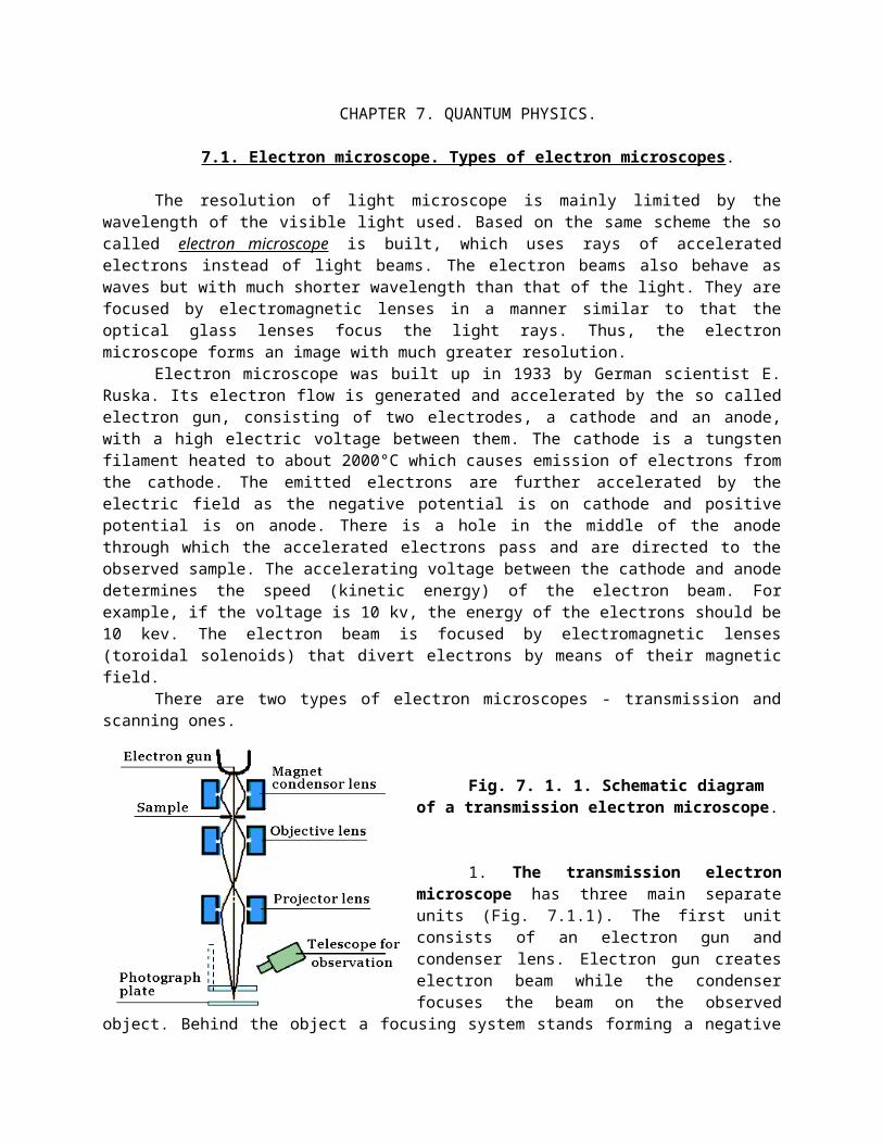
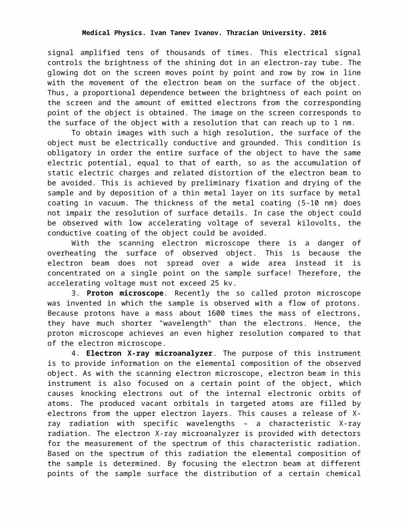
Fig. 7. 1. 1. Schematic diagram of a transmission electron microscope.
1. The transmission electron microscope has three main separate units (Fig. 7.1.1). The first unit consists of an electron gun and condenser lens. Electron gun creates electron beam while the condenser focuses the beam on the observed object. Behind the object a focusing system stands forming a negative image on the screen. The focusing system consists of objective lens, an intermediate lens and a projector lens. The third unit is a luminescent screen covered by a luminescent layer that converts the energy of the incident electrons into
light (cathodo- or radioluminescence). When the strongly magnified image of the observed object is focused on the luminescent screen it becomes visible due to the lumuniscent light emitted. A television camera is used to obtain a digitized record of the image.
The electron gun, condenser, sample, electromagnetic lenses and screen all are housed in a common tube, which is evacuated to a deep vacuum. This vacuum is achieved by a multi-step vacuum pump. The windings of the electromagnets are cooled down with liquid nitrogen in order to reduce their resistance. Each electron microscope is surrounded by a metal shield to reduce the X-ray radiation, which is generated at any deviation of the trajectory of the electron beam.
The observed sample in the electron microscope is placed under vacuum hence, it must be firstly fixed and dehydrated. Otherwise the sample water will quickly evaporate distroying the structure of the sample. The sample fixation is carried out using bifunctional chemical reagents (typically glutaraldehyde and OsO4). The next step is removing the water from the sample, refilling the sample with paraffin and finally cutting the sample into extremely thin layers having large area. The thickness of the layers must be about several m, in order to reduce the absorbtion of electrons. The layer is secured on a fine wire mesh,
Medical Physics. Ivan Tanev Ivanov. Thracian University. 2016
and placed on a movable sample stage. Prior to enter the microscope, the sheaf of electron rays is evenly distributed throughout the entire area of the layer, so the density of the electron flow is small and there is no danger of overheating the sample. Therefore, this type of electron microscopes uses electron beams accelerated by a voltage from tens of kv to about 1 Mev. The denser structures of the sample absorb and scatter more effectively the incident rays of electrons and form a faint negative image on the luminescent screen (Fig. 7.1.2). To increase the absorption of a specific structure of the sample, the latter may be pre-treated with electron-dense substances - OsO4, lead salts, and the like.
Similar to the light rays, the electron beams also interfere between them, exhibiting wavelike properties. Their wavelength, , however is much smaller than that of the visible light. For example, at an accelerating voltage of 50 000 to 100 000 V the electron stream has a wavelength from 0.0055 to 0.0039 nm, which is considerably less than that of visible light (380-760 nm). Consequently, two neighbor electron beams of such energy interfere when they are at a much shorter distance from each other. Therefore, the electron microscope can reach a resolution of about 0.2 nm, which is about 1000 times greater compared to the resolution of optical microscope and 1 million times greater compared to human eye. For other reasons, however, such a high resolution can not be practically achieved. The real maximum resolution is obtained at a magnification of about 150 thousand times. At such maximal magnification this type of microscope allows the observation of various biomacromolecules (proteins, nucleic acids) and even atoms of the heaviest elements.
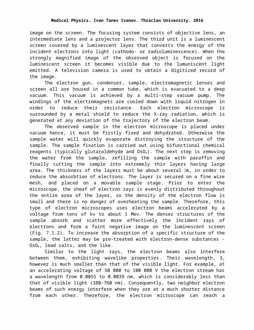
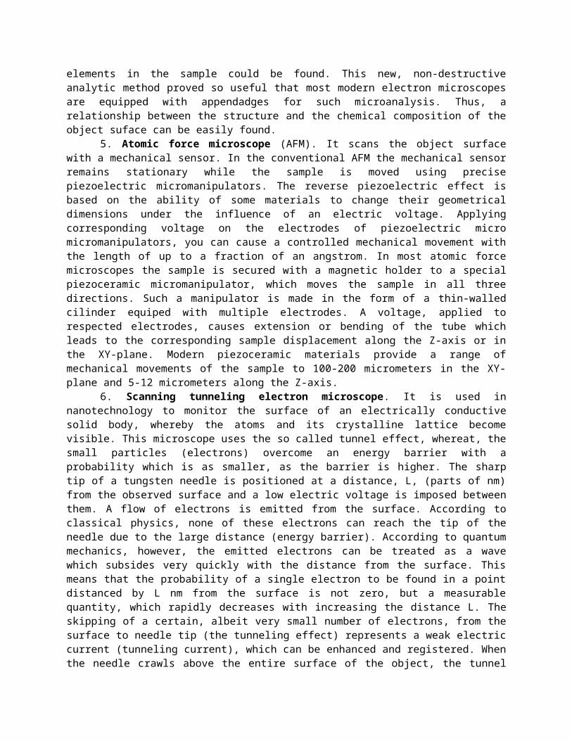
Fig. 7. 1. 2. Comparison between the schemes of transmission (left) and scanning (right) electronic microscopes.
The ordinary (transmission) electron microscope forms a flat, two-dimensional image on its fluorescent screen or on the photoplate. This type of image is devoid
of the information about the topography of the observed object. This disadvantage is overcommed in the scanning electron microscope.
2. The scanning electron microscope (Fig. 7.1.2 right) is used to investigate the surface of solid bodies by means of a focused beam of electrons. It produces an impression of three-dimensional image of the sample. The source of electrons and the electromagnetic lenses which focus the electron beam are similar to those used in the transmission electron microscope. It uses a narrow electron beam with a cross-section of about 10 nm, focused on a single point on the surface of the object. To obtain a whole image of the object, the focal point is moved point by point and row by row encompassing the entire surface. The collision of the high energy electrons with the surface atoms causes an elastic back scattering (a high energy electron beam) and the emission of secondary electrons (low energy electron beam) from the sample surface. Visible light, heat and X-rays are also generated. Both the high energy and low energy electron beams are collected and directed to a scintillator plate that emitts light photons in response to its bombardment by the electrons. In turn, the light photons enter a photoelectron multiplier tube, where they are converted into electric signal amplified tens of thousands of times. This electrical signal controls the brightness of the shining dot in an electron-ray tube. The glowing dot on the screen moves point by point and row by row in line with the movement of the electron beam on the surface of the object. Thus, a
proportional dependence between the brightness of each point on the screen and the amount of emitted electrons from the corresponding point of the object is obtained. The image on the screen corresponds to the surface of the object with a resolution that can reach up to 1 nm.
To obtain images with such a high resolution, the surface of the object must be electrically conductive and grounded. This condition is obligatory in order the entire surface of the object to have the same electric potential, equal to that of earth, so as the accumulation of static electric charges and related distortion of the electron beam to be avoided. This is achieved by preliminary fixation and drying of the sample and by deposition of a thin metal layer on its surface by metal coating in vacuum. The thickness of the metal coating (5-10 nm) does not impair the resolution of surface details. In case the object could be observed with low accelerating voltage of several kilovolts, the conductive coating of the object could be avoided.
With the scanning electron microscope there is a danger of overheating the surface of observed object. This is because the electron beam does not spread over a wide area instead it is concentrated on a single point on the sample surface! Therefore, the accelerating voltage must not exceed 25 kv.
3. Proton microscope. Recently the so called proton microscope was invented in which the sample is observed with a flow of protons. Because protons have a mass about 1600 times the mass of electrons, they have much shorter "wavelength" than the electrons. Hence, the proton microscope achieves an even higher resolution compared to that of the electron microscope.
4. Electron X-ray microanalyzer. The purpose of this instrument is to provide information on the elemental composition of the observed object. As with the scanning electron microscope, electron beam in this instrument is also focused on a certain point of the object, which causes knocking electrons out of the internal electronic orbits of atoms. The produced vacant orbitals in targeted atoms are filled by electrons from the upper electron layers. This causes a release of X-ray radiation with specific wavelengths – a characteristic X-ray radiation. The electron X-ray microanalyzer is provided with detectors for the measurement of the spectrum of this characteristic radiation. Based on the spectrum of this radiation the elemental composition of the sample is determined. By focusing the electron beam at different points of the sample surface the distribution of a certain chemical elements in the sample could be found. This new, non-destructive analytic method proved so useful that most modern electron microscopes are equipped with appendadges for such microanalysis. Thus, a relationship between the structure and the chemical composition of the object suface can be easily found.
5. Atomic force microscope (AFM). It scans the object surface with a mechanical sensor. In the conventional AFM the mechanical sensor remains stationary while the sample is moved using precise piezoelectric micromanipulators. The reverse piezoelectric effect is based on the ability of some materials to change their geometrical dimensions under the influence of an electric voltage. Applying corresponding voltage on the electrodes of piezoelectric micro micromanipulators, you can cause a controlled mechanical movement with the length of up to a fraction of an angstrom. In most atomic force microscopes the sample is secured with a magnetic holder to a special piezoceramic micromanipulator, which moves the sample in all three directions. Such a manipulator is made in the form of a thin-walled cilinder equiped with multiple electrodes. A voltage, applied to respected electrodes, causes extension or bending of the tube which leads to the corresponding sample displacement along the Z-axis or in the XY-plane. Modern piezoceramic materials provide a range of mechanical movements of the sample to 100-200 micrometers in the XY- plane and 5-12 micrometers along the Z-axis.
6. Scanning tunneling electron microscope. It is used in nanotechnology to monitor the surface of an electrically conductive solid body, whereby the atoms and its crystalline lattice become visible. This microscope uses the so called tunnel effect, whereat, the small particles (electrons) overcome an energy barrier with a probability which is as smaller, as the barrier is higher. The sharp tip of a tungsten needle is positioned at a distance, L, (parts of nm) from the observed surface and a low electric voltage is imposed between them. A flow of electrons is emitted from the surface. According to classical physics, none of these electrons can reach the tip of the needle due to the large distance (energy barrier). According to quantum mechanics, however, the emitted electrons can be treated as a wave which subsides very quickly with the distance from the surface. This means that the probability of a single electron to be found in a
Medical Physics. Ivan Tanev Ivanov. Thracian University. 2016
point distanced by L nm from the surface is not zero, but a measurable quantity, which rapidly decreases with increasing the distance L. The skipping of a certain, albeit very small number of electrons, from the surface to needle tip (the tunneling effect) represents a weak electric current (tunneling current), which can be enhanced and registered. When the needle crawls above the entire surface of the object, the tunnel current changes its size depending on the distance between the object and the needle tip. Further, the tunnel current controls the brightness of the shining point of a monitor screen, thus obtaining a visible image of the surface.
7.2. Heat radiation of bodies. Laws of Stefan-Boltzmann, Wien and Kirchhoff. Greenhouse effect – cause and effect. Planck's theory for the thermal radiation of bodies. Some quantum
concepts of light
Each body, regardless of its temperature, emits electromagnetic waves. Hence, it loses heat and cools down. At moderate temperatures the bodies emits chiefly infrared rays. The intensity of this radiation strongly increases with increasing the temperature of bodies. Based on these features this type of radiation is called thermal radiation. Thermal radiation of bodies is studied in medicine because it participates in the heat transfer, in thermal regulation of human and also in determining the optimal environmental conditions.
How classical physics explains the thermal radiation of bodies?The constituting particles (atoms and molecules) of a given body are involved in a continuous,
chaotic (thermal) movement consisting of vibrations, rotations, and translational movements in different directions accompanied by mutual collisions. This movement involves incredibly high speeds, e.g., the translational movements are conducted at a speed of several km/s, and the vibration has great frequency.
On the other hand the atoms and molecules contain charged particles, electrons and nuclei. According to the laws of classical electrodynamics each charged particle moving curvilinear and upon acceleration converts a portion of its kinetic energy into electromagnetic radiation. With increasing the temperature of the bodies the chaotic movement of their molecules rises, which explains why the thermal radiation also rises. This classical mechanism gives a plausible, although qualitative explanation for the occurrence of thermal radiation of bodies. At the precise, quantitative study of the thermal radiation of bodies, however, problems arise that classical physics can not explain.
When a body is in thermal equilibrium with the environment at a temperature, T, its heat radiation is constant over time. The total energy of thermal radiation, which the body radiates for a time of 1 s is called radiant flux, Ф, or radiant power. The radiant flux, emitted from 1 m2 of the body surface, is called radiant emittance, R, (W/m2) or radiant exitance. In fact, the radiant emittance represents the radiant flux density and is defined as R = Ф/S, where Ф is the radiant flux leaving the infinitesimal area, S.
Most often the light contains rays of different wavelengths. The spectral composition of light is the relative portion (percentage, number, weight) of rays with different wavelengths, . Thus, the radiant emittance, R, could be different for different wavelengths, , of light. The spectral distribution of R is expressed by the spectral radiant emittance, R, which is defined as R = R/. Here, R is this part of the R, which is included between and +.
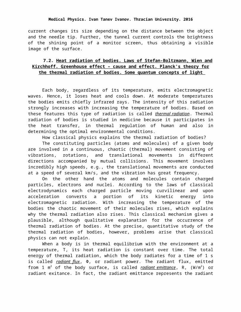
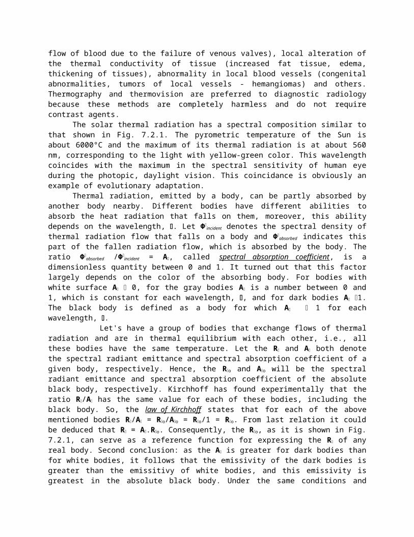
Fig. 7.2.1 shows the spectral radiant emittance, Rо, of the so called black body as a function of the wavelength, . The other, non black bodies have similar spectral curves. Aparently, the thermal radiation has a continuous spectrum with a maximum at mах. Depending on the temperature, T, of the body different spectral compositions of the thermal radiation, i.e., different spectral curves are obtained. The total area under each curve gives the radiant emittance, R, of the body. With increasing temperature, the radiant emittance increases by the law of Stefan-Boltzmann: R = .T4. Here, is the constant of Stefan-Boltzmann. With increasing temperature, the maximum of radiation (the peak of the spectral curve) falls on ever smaller wave length, mах, where mах = b/T – the displacement law of Wein. In this formula, b is the constant of Wein. Determining the mах, this law allows the calculation of T, which is the so called pyrometric temperature, this is the surface temperature of emitting body. The measurement of surface
temperature constitutes the basis of the modern, completely non-invasive diagnostic method called thermography of the human body.
Bolometers are extremely sensitive instruments for measuring the energy of thermal radiation. They contain two identical platinum resistors, combined with other two reference resistors to form a balanced Wheatstone bridge. One of the platinum resistors is blackened in order to effectively absorb the energy of the falling radiation (visible light or infrared rays). Thus, the black resistor is heated up increasing its resistance, which leads to disbalancing the bridge and appearance of electric voltage, proportional to the temperature increase in °C. Bolometers are used to measure the energy of thermal radiation at a long distance, as in the infrared tracking systems. Bolometers helped to discover the infrared radiation of hot bodies. They are also used to measure the intensity of light in the following way. After the exposure of bolometer to light for a certain time, the light is stopped and an electric current is switched through the blackened resistore to produce the same temperature difference. Based on the magnitude of this current the temperature differences of as small as to 10-7 °C can be measured.
Fig. 7. 2. 1. Spectral composition of the thermal radiation of black body at different temperatures, T K.
The optical (radiation) pyrometers measure the temperature of hot objects at a distance relying on the spectrum of their thermal radiation. The human eye has roughly the quality of such a pyrometer, and can evaluate the temperature of a hot object by its color; the red indicates about 500°C, yellow and blue about 1500°C and over 2000°C, respectively. In the pyrometer with vanishing filament the hot object and an electric lamp with variable power are both observed and
compared through an opaque plate. The current through the filament of the lamp is changed until the light intensities of the lamp and the object become equal. The magnitude of current is used to assess the temperature of the hot object, referred to as pyrometric temperature.
Another similar method uses sensitive thermosensors or a inftrared television camera to measure the thermal radiation of human body - thermovision. At an air temperature of 18°C, a naked man at rest with skin temperature of 33°C emitts about 120 W thermal radiation. This is about 50% of its internal heat production. The thermal radiation of the human body has a maximum at mах от 9-10 m, almost equal to the diameter of most cells.
The infrared radiation, emitted from a particular portion of the surface of human body, depends on the following three factors:
1. The local heat production, depending on the rate of metabolic processes under the surface;2. Cooling by the local convection, related to the blood circulatory system within the underlying
tissues;3. Cooling by local heat conduction, based on the structure and composition of local tissues.Using apparatus for themovision (thermal imaging apparatus) the distribution of temperature at
various areas of human surface, especially those with good blood supply, can be observed. This completely non-invasive diagnostic method detects local changes in heat production (inflammatory foci, rheumatoid arthritis, tumor tissue, necrotic tissue), places with impaired blood circulation (injuries, thrombosis, vascular sclerosis), abnormal venous flow (stasis, back flow of blood due to the failure of venous valves), local alteration of the thermal conductivity of tissue (increased fat tissue, edema, thickening of tissues), abnormality in local blood vessels (congenital abnormalities, tumors of local vessels - hemangiomas) and others. Thermography and thermovision are preferred to diagnostic radiology because these methods are completely harmless and do not require contrast agents.
Medical Physics. Ivan Tanev Ivanov. Thracian University. 2016
The solar thermal radiation has a spectral composition similar to that shown in Fig. 7.2.1. The pyrometric temperature of the Sun is about 6000°C and the maximum of its thermal radiation is at about 560 nm, corresponding to the light with yellow-green color. This wavelength coincides with the maximum in the spectral sensitivity of human eye during the photopic, daylight vision. This coincidance is obviously an example of evolutionary adaptation.
Thermal radiation, emitted by a body, can be partly absorbed by another body nearby. Different bodies have different abilities to absorb the heat radiation that falls on them, moreover, this ability depends on the wavelength, . Let Ф
incident denotes the spectral density of thermal radiation flow that falls on a body and Ф
absorbed indicates this part of the fallen radiation flow, which is absorbed by the body. The ratio Ф
absorbed /Фincident = A, called spectral absorption coefficient, is a dimensionless quantity between 0
and 1. It turned out that this factor largely depends on the color of the absorbing body. For bodies with white surface A 0, for the gray bodies A is a number between 0 and 1, which is constant for each wavelength, , and for dark bodies A 1. The black body is defined as a body for which A 1 for each wavelength, .
Let's have a group of bodies that exchange flows of thermal radiation and are in thermal equilibrium with each other, i.e., all these bodies have the same temperature. Let the R and A both denote the spectral radiant emittance and spectral absorption coefficient of a given body, respectively. Hence, the Ro and Ao will be the spectral radiant emittance and spectral absorption coefficient of the absolute black body, respectively. Kirchhoff has found experimentally that the ratio R/A has the same value for each of these bodies, including the black body. So, the law of Kirchhoff states that for each of the above mentioned bodies R/A = Ro/Ao = Ro/1 = Ro. From last relation it could be deduced that R = A.Ro. Consequently, the Ro, as it is shown in Fig. 7.2.1, can serve as a reference function for expressing the R
of any real body. Second conclusion: as the A is greater for dark bodies than for white bodies, it follows that the emissivity of the dark bodies is greater than the emissitivy of white bodies, and this emissivity is greatest in the absolute black body. Under the same conditions and temperature, dark bodies have greater abilities to absorb and to emitt light than the white bidies.
The dependence of emissivity on the color of bodies probably plays a role in the racial characteristics of human thermoregulation. Indeed, in a hot climate, the heat generated by the internal heat production will be emitted to the environment more effectively by people with black skin, which helps to cool the body. In a cool climate, however, this would be very undesirable heat loss, leading to rapid cooling of the body. In this case, people with white skin will emitt less heat in support of the mechanism preserving the body temperature. Probably, the color of the skin might be an evolutionary trait, based on Kirchhoff's law, aimed at supporting a more effective thermoregulation in a hot and cold climates.
For some gases (CO2, methane, nitric oxide, water vapor), the spectral absorption coefficient, A, has lower values for the visible light and higher values for the far infrared rays. Earth is surrounded by an atmosphere rich of such gases. Hence, the visible light from the Sun passes freely through Earth atmosphere. Absorbed at the Earth surface it is emitted back as infrared beams, which are fully absorbed by the Earth atmosphere. This causes an increase in the temperature close to the surface of the Earth (global greenhouse effect), which is favorable for the life.
The spectral curve of thermal emissivity of black body (Ro) (fig. 7.2.1) was obtained experimentally in the late XIXth century. An attempt was made to explain this complex spectral dependence with the laws and ideas of classical electrodynamics. According to these conceptions, first - the constituent particles of bodies can have continuous values of their energy and second – these particles emitt thermal radiation like the free electric charges emitting electromagnetic waves when their speed is changed (at acceleration, vibration, rotation). This classical model presents theoretical formulas for Ro
which, however, give inacceptably large differences with the experimental curve for Ro and does not explain the laws of Wein and Stefan-Boltzmann. Obviously, this model is not correct. This contradiction created a serious crisis in the classical physics at that time. An initially formal solution of the crisis was found by the German physicist Max Planck, who assumed two very unusual for the classical physics postulates, namely:
1) The energy of the elementary emitters (atoms, molecules) can only have separate discrete values (energy levels), for example E0 <E1 <E2 <E3, etc.
2) Thermal radiation is generated when atoms come down from their upper energy levels to a lower one. For example, upon the transion from E2 to E1 the excess energy ΔE = E2 - E1 is emitted in the form of light quanta (energy portion, photon) with frequency, , wherat ΔE = h..
In above formula, h is a universal constant called Planck's constant. Based on this model Max Planck derived a formula for Ro, whose plot completely coincides with the famous experimental curve, shown in the fig. 7.2.1. Using this formula he deduced the laws of Wein and Stefan-Boltzmann and calculated the constants of Wein, b, and Stefan-Boltzmann, .
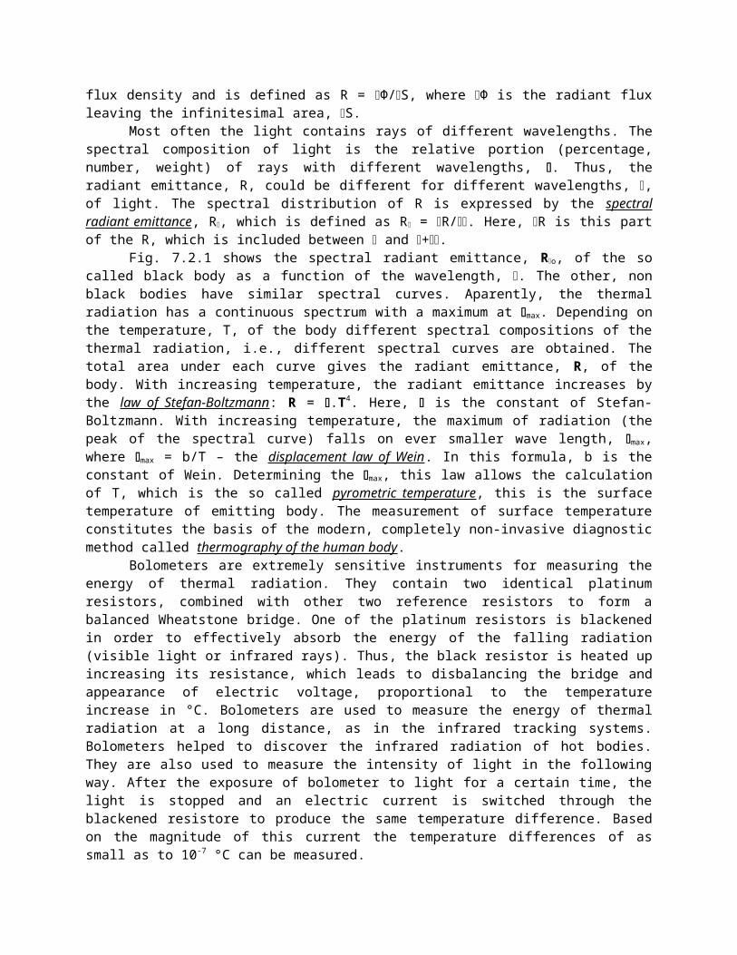
Fig. 7. 3. 1. Absorbtion of monochromatic light by a solution.
This was tremendous success of the Max Plank’s theory. Nevertheless, for a some time afterwards the
scientists thought that the Max Plank’ postulates represent a pure mathematical game fortuitously solving the problems with the experimental curve in Fig. 7.2.1, and this game has no relation to the actual laws and mechanism of thermal radiation. However, at a later time the Max Plank’s theory found more experimental support. The peculiarities of emission spectra and absorption spectra of atoms and molecules, the laws of photoelectric effect, as well as the laws of photochemical and photobiological processes have confirmed that light is not only emitted in portions, but it is also spread and absorbed in portions (photons). All these experimental findings represent the basis of our modern quantum concepts for light and atoms.
7.3. Absorption of light by atoms and molecules. Energy levels and absorption spectrum of atoms and molecules. Law of Bouguer-Lambert-Beer. Application of spectrophotometry in
medicine.
Consider a light sheaf of monochromatic beams with intensity, Io, and wavelength, , falling on a transparent cuvette with thickness, d, which contains a dilute solution. Let the light be absorbed by the solution without any scattering. As the atoms and molecules of the solute absorb part of the light beams the transmitted radiation will have lower intensity, I (Fig. 7.3.1). Most often, the absorbed light energy is converted into heat, however, the absorbing particles could sustain a sort of modification - dissociation, ionization, photochemical transformations.
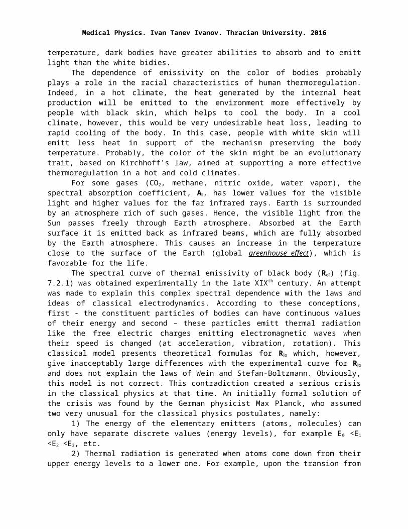
Fig. 7. 3. 2. Upon irradiation of atoms by white ligh the obtained absorption spectrum is linear because it containes separate narrow absorption lines.
The reduction of light intensity by the absorbing solute with concentration, c, dissolved in a non-absorbong solvent obeys the relation I = Io.exp (-cd), known as the law of Bouguer, Lambert and Beer. The quantity, , depends on the solute and is called extinction coefficient. It is more convenient to measure the ratio I/Io = T, called transmittance (%), and the quantity A = 1 - T, which is denoted as absorption (%). Also of practical

Medical Physics. Ivan Tanev Ivanov. Thracian University. 2016
importance are the quantity E = ln (Io/I), called extinction and its variant D = lg (Io/I) called optical density. The law of Bouguer-Beer-Lamberg could be written in more convenient form, E = . c. d.
For a given substance, the extinction coefficient, , and its related quantities - A, T, D and E, are characteristic functions of . Optical devices, called spectrophotometers, are used to measure these quantities at different in order to obtain the absorption spectrum of dissolved substance.
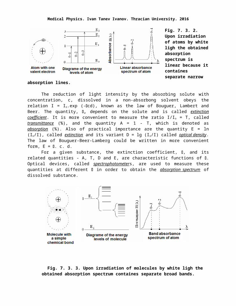
Fig. 7. 3. 3. Upon irradiation of molecules by white ligh the obtained absorption spectrum containes separate broad bands.
The type of the absorption spectrum depends on the nature of absorbing particles - atoms or molecules. Usually only electrones, which are most weakly bound to atoms and molecules, participate in the absorbtion of light. In atoms these electrons are the most external valence electrons, while in molecules these are the electrons involved in the chemical interatomic bonds. These electrons can occupy only certain allowed energy levels, Eo, E1, E2, E3, etc. (Fig. 7.3.2). Atom with a valent electron in the ground level, Eo, is in a stable state (the Eo is a stable level), whereas the atom with its electron in the upper energy levels is highly unstable. It is said that the upper energy levels are short-lived and ecxited
ones. If a group of atoms are irradiated with white light, containing photons with different energies (frequencies), the atoms will absorb resonantly only that part of photons which have energies equal to the differences (Eo-E1), (Eo-E2), (Eo-E3) etc. Consequently, the transmitted light will become devoid of these photons giving rise to separate narrow lines of absorbance in the spectrum of light (Fig. 7.3.2). So, the absorption spectrum of isolated atoms (gas, vapor, diluted ion plasma) is of linear type.
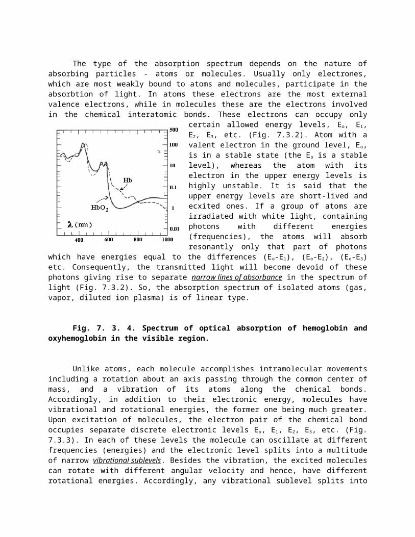
Fig. 7. 3. 4. Spectrum of optical absorption of hemoglobin and oxyhemoglobin in the visible region.
Unlike atoms, each molecule accomplishes intramolecular movements including a rotation about an axis passing through the common center of mass, and a vibration of its atoms along the chemical bonds. Accordingly, in addition to their electronic energy, molecules have vibrational and rotational energies, the former one being much greater. Upon excitation of molecules, the electron pair of the chemical bond occupies separate discrete electronic levels Eo, E1, E2, E3, etc. (Fig. 7.3.3). In each of these levels the molecule can oscillate at different frequencies (energies) and
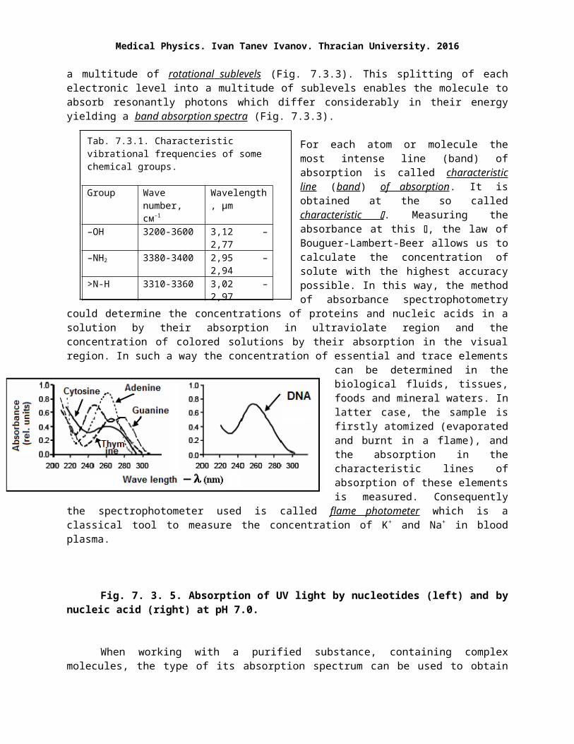
Tab. 7.3.1. Characteristic vibrational frequencies of some chemical groups.
Group Wave number, см-1
Wavelength, μm
–ОН 3200-3600 3,12 – 2,77–NH2 3380-3400 2,95 – 2,94>N-H 3310-3360 3,02 – 2,97>CH2 2915-2935 3,43 – 3,4>CH– 2880-2900 3,47 – 3,44–CH3 2850-2815 3,5 – 3,55>C=C< 1620-1680 6,17 – 5,95–C≡C– 2190-2260 4,56 – 4,42–C=O– 1700 5,88
the electronic level splits into a multitude of narrow vibration al sublevels . Besides the vibration, the excited molecules can rotate with different angular velocity and hence, have different rotational energies. Accordingly, any vibrational sublevel splits into a multitude of rota tional sublevels (Fig. 7.3.3). This splitting of each electronic level into a multitude of sublevels enables the molecule to absorb resonantly photons which differ considerably in their energy yielding a band absorption spectra (Fig. 7.3.3).
For each atom or molecule the most intense line (band) of absorption is called characteristic line
( band) of absorption. It is obtained at the so called characteristic . Measuring the absorbance at this
, the law of Bouguer-Lambert-Beer allows us to calculate the concentration of solute with the highest accuracy possible. In this way, the method of absorbance spectrophotometry could determine the concentrations of proteins and nucleic acids in a solution by their absorption in ultraviolate region and the concentration of colored solutions by their absorption in the visual region. In such a way the concentration of essential and trace elements can be determined in the biological
fluids, tissues, foods and mineral waters. In latter case, the sample is firstly atomized (evaporated and burnt in a flame), and the absorption in the characteristic lines of absorption of these elements is measured. Consequently the spectrophotometer used is called flame photometer which is a classical tool to measure the concentration of K+ and Na+ in blood plasma.
Fig. 7. 3. 5. Absorption of UV light by nucleotides (left) and by nucleic acid (right) at pH 7.0.
When working with a purified substance, containing complex molecules, the type of its absorption spectrum can be used to obtain information about the chemical
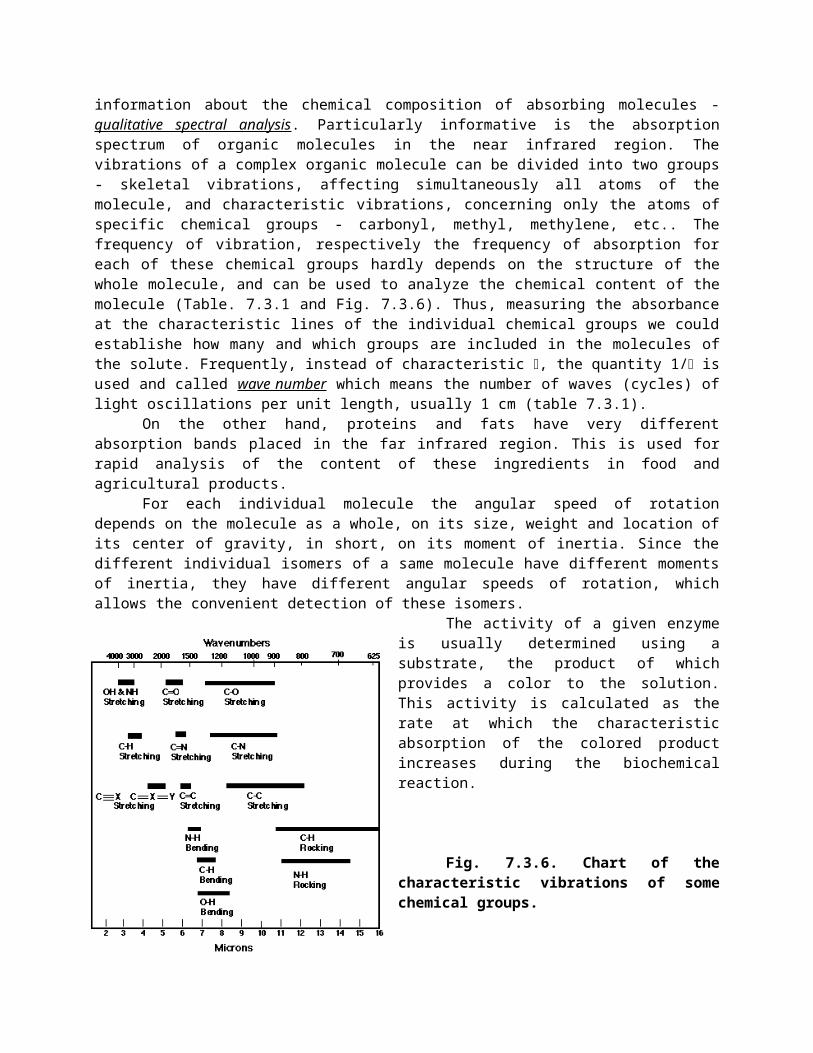
composition of absorbing molecules - qualitative spectral analysis. Particularly informative is the absorption spectrum of organic molecules in the near infrared region. The vibrations of a complex organic molecule can be divided into two groups - skeletal vibrations, affecting simultaneously all atoms of the molecule, and characteristic vibrations, concerning only the atoms of specific chemical groups - carbonyl, methyl, methylene, etc.. The frequency of vibration, respectively the frequency of absorption for each of these chemical groups hardly depends on the structure of the whole molecule, and can be used to analyze the chemical content of the molecule (Table. 7.3.1 and Fig. 7.3.6). Thus, measuring the absorbance at the characteristic lines of the individual chemical groups we could establishe how many and which groups are included in the molecules of the solute. Frequently, instead of characteristic , the quantity 1/ is used and called wave number which means the number of waves (cycles) of light oscillations per unit length, usually 1 cm (table 7.3.1).
On the other hand, proteins and fats have very different absorption bands placed in the far infrared region. This is used for rapid analysis of the content of these ingredients in food and agricultural products.
Medical Physics. Ivan Tanev Ivanov. Thracian University. 2016
For each individual molecule the angular speed of rotation depends on the molecule as a whole, on its size, weight and location of its center of gravity, in short, on its moment of inertia. Since the different individual isomers of a same molecule have different moments of inertia, they have different angular speeds of rotation, which allows the convenient detection of these isomers.
The activity of a given enzyme is usually determined using a substrate, the product of which provides a color to the solution. This activity is calculated as the rate at which the characteristic absorption of the colored product increases during the biochemical reaction.
Fig. 7.3.6. Chart of the characteristic vibrations of some chemical groups.
Upon absorbtion of strong light, some molecules (nucleic acids, bilirubin) disintegrate into smaller parts. In some cases this photoinduced decomposition is beneficial, as in the phototherapy of babies born with bilirubinemia. In other cases it is an adverse effect, as for nucleic acids. Figs. 7.3.4 and 7.3.5 show the characteristic bands of absorption of certain biologically important substances. Adenine and other nucleobases as well as nucleic acids absorb
strongly at 260 nm, which is used to determine the concentration of these substances in their aqueous solutions. The illumination of such solutions with strong UV light at = 260 nm results in the breakage of some chemical bonds. This explains the strong cytotoxic and germicidal effect of the ultraviolet light of this spectral area. The strong Sorel absorption band of hemoglobin at 430 nm (Fig. 7.3.4) is due to the absorption of the heme group in hemoglobin molecule. Because the Sorel band is located in the blue part of the visible spectrum, the hemoglobin solution absorb the blue light and transmits the red light, which renders red color to blood. Bilirubin also absorbs in the same optical region, which is used for photodecomposition and detoxification of its molecules through illumination with strong visible light in the method known as phototherapy of newborns.
7. 4. Luminescence and photoluminescence. Use of fluorescence assay in medicine. Flow cytofluorimeter.
Any electromagnetic radiation of the bodies, which is in addition to their thermal radiation is called luminescence (from Latin lumen - light, luminescent – shining with weak light). It is also called "cold light". To activate the luminescence another form of energy, different than the heat, is used. According to the type of this activation energy we distinguish different types of luminescence: photoluminescence (caused by light absorption), rentgenluminesence (absorption of X-rays), electroluminescence (electric current), chemiluminescence (chemical reaction) biochemiluminescence (biological process) and others. The fluorescent substance is termed luminophor.
When the molecules of a luminophor absorb a portion of external energy, the electrons of their chemical bonds (the valence electrons) shift to higher orbitals (activation or excitation of molecules). The produced excited state is unstable and the electrons return to their ground orbital releasing energy (deactivation). A part of the released energy is emitted as a light photon - luminescence. Upon the interruption of the flow of external energy the luminescence also stops usually immediately ( fluorescence) or after a long time (from 1 ms to 100 s - phosphorescence).

The spin of a single electron is a measure of its rotation about its own axis and could obtain only a number of discrete values (0, ± ½). Chemical bonds consist of electron pairs. The total spin of such electron pair is the sum of the spins of the two electrons and can have the values 0 and ± 1. When the spins of both electrons are oppositely oriented the total spin is 0 and this state is called singlet (S). At unidirectional orientation of the two spins, the total spin is ± 1 and this is a triplet (T) state.
In photoluminescence, the luminophor has to be irradiated with a suitable, typically ultraviolet light (excitation light). The luminophor molecules absorb light photons resonantly and the electron pair of their chemical bond increases its energy going from the ground electronic level, So, to the upper electronic levels, S1, S2, etc. (Fig. 7.4.1). In this type of activation, the excited pair of electrons retains the spins directed oppositely to each other (anti-parallel spin state) and is designated as singlet electron pair. Due to the different vibrational and rotational energies of the excited molecule the electron levels of S1, S2, etc, states split into a multitude of sublevels (Fig. 7.4.1).
Fig. 7. 4. 1. Outline of the energy levels of molecule. Indicated are the transitions between these levels in photoluminescence.
The singlet levels are always short-lived. Deactivation can be done in three ways: internal conversion (radiationless transition with release of heat), fluorescence, or in some cases phosphorescence (Fig.
7.4.1). During the radiationless transition the energy of excited molecule is taken away by collision with
another non-excited molecule (quenching of excitation) and is converted into heat. Usually, the deactivation of all the upper levels to the first excited level, S1, is accomplished through quenching and heat releasing, while the emission of light occurs only in the transition from S 1 to the ground level, So (S1 So) - fluorescence. But even in this case, some of the transitions S1 So are also radiationless with evolution of heat.
In some molecules the excited electron, located in the S1 level, has the possibility to reverse its spin prior to its deactivation. After this spin transition both electrons of the excited electron pair have parallel (unidirectional) spins. Such a triplet (T1) state of the molecule is metastable and long-lived, lasting from 1 ms to 100 s. Therefore, the deactivation of this triplet state starts a long time after the termination of energy input - phosphorescence. The S1T1 transition is called intercombination transition. It is possible only in some large molecules with complex structure, if the energies of their two states, S 1, and, T1, are very close.
Molecules being in the triplet state represent highly reactive chemical radicals. Due to their high chemical activity, they give rise to many photochemical reactions. The emergence of such radicals in biological media poses a danger because they initiate harmful chemical transformations of biomacromolecules. Such radicals are produced in biological objects in response to irradiation with
Medical Physics. Ivan Tanev Ivanov. Thracian University. 2016
ionizing radiation and UV rays. In medicine, such radicals are intentionally produced in photodynamic therapy of tumors.
Based on the mechanism of photoluminescence several conclusions can be deduced: 1) Let Nabsorbed denotes the number of absorbed photons which excite the luminophor molecules.
Then Nemitted will denote the number of photons, emitted by the excited luminophor molecules. The ratio Nemitted / Nabsorbed is called quantum yield of the luminescence of this luminophor. The greater the quenching the lesser is the quantum yield. The quenching is more efficient when the mean distance between the molecules is smaller hence, the quantum yield can be used to determine this distance. In addition, quantum yield is indicative for the extent of energy transfer between the excited and nearby non-excited molecules.
2) Apparently, the energy of the emitted photons is equal to or less than that of the absorbed photons. Accordingly, the wavelength of the emitted light is equal to or greater than that of the excitation light – the law of Stokes.
Fig. 7.4.2.
Principle scheme of a spectrofluorimeter (left) and a record of the emission spectrum of the luminophor (right) obtained according to the transitions in Fig. 7. 4. 1.
3) The distribution of emitted photons by energy (wavelength, frequency) is called emission spectrum of the luminophor. The emission spectrum of the luminescence (the transitions 1', 2', 3' and so on - Fig. 7.4.1) depends only on the type of luminophor and does not depend on the spectrum of the excitation light. Thus, the type of the emission spectrum can be used for qualitative and quantitative analysis (luminescence analysis) of the luminophor.
Cells, cell membranes and organelles can be assayed consequent to the attachment of appropriate fluorescent markers and probes to them. The markers bind covalently and the probes non-covalently to specific sites of the sample molecules. The markers and probes are molecules that emit strong and characteristic fluorescent radiation when illuminated with light having proper wave length. In respect to their emission in unbound state their emission spectrum changes when they are excited in bound state. In luminescence analysis the sample with the attached probes (markers) is irradiated with appropriate excitation light and the emission spectrum of the probes (markers) is recorded. The difference between the spectra of the markers (probes) in bound and unbound states elucidates the molecular motions, quenching and energy transfer in the sample.
When the luminophor is excited by a plane polarized light its emission also has a substantial degree of polarization. The latter depends on the molecular movements and micro viscosity of the sample; the greater the micro viscosity, the higher will be the degree of polarization of emitted light.
Spectrofluorimeters are used to obtain the emission spectrum of luminophors (Fig. 7.4.2). The spectrofluorimeter contains a light source, S. A portion of this light passes through the narrow slit of the input monochromator, which devide the entering light into multitude of beams with different colors. The luminescence of the sample is excited using a sheaf of monochromatic light beams. The wavelength of the excitation light, excitation, can be chosen in accordance with the type of luminophor. The fluorescent light, emitted perpendicularly to the incident exciting light, is transmitted through the narrow slit of the outlet
monochromator and converted into electrical signals by the photoelectric detector. The output signal of this detector is recorded as a function of the wavelength, emittance, of the emission light. This record represents the emission spectrum of the sample (transitions 1', 2', 3' and so on - Fig. 7.4.1).
The phosphorescence of a luminophore, deposited as a thin layer on a screen is used in the electron-ray tube for obtaining the visible image of any electric signal (picture tubes, televisions, monitors). Recently, the electron-ray tube is replaced by a liquid crystal screen.
Flow cytofluorimeters assay cell suspensions (or blood) using fluorescence analysis. Prior to study proper antibodies, conjugated with fluorescent markers and probes, are added to the suspension. The antibodies bind specifically to specific sites marking the cells of interest. The suspension is then diluted and broken down into small drops that are let to pass in front of a photodetector for fluorescent light. Thus, cells of different types can be counted detecting the excited luminescence of the attached markers.
Bioluminescence is a type of chemiluminescence associated with different types of organisms - bacteria, insects, fishs, etc. Some of them use luminescent light to attract victims or partners, others like fireflies, deep sea shrimp, etc. to repel predators. The emitted light occupies the visible spectrum and is usually generated by specialized organs, photophores, that convert chemical energy into light, based on the reactions of oxidation of some organic compounds - luciferines. Enzymes which catalyze this oxidation are called luciferases.
The u ltra- weak luminescence is a special case of bioluminescence of biological systems. The light emission has very low intensity (10-100 photon per 1 cm2 of the surface of tissue) and could only be registered with a photoelectron multiplier tube. The maximum of the emission is in the range 360-800 nm. Ultra-weak photon emission is related to the free radical oxidation of the lipid parts of biomembranes.
Usage of photoluminescence for food quality control. Irradiation of food stuffs with ultraviolet radiation excites their luminescence. The color of the emitted light is different in fresh and spoiled food. Depending on the advance of spoilage, the color of the meat changes from red-violet to green-gray, that of fish changes from gray to yellow-green, that of milk from green-yellow to blue and so on.
Passability of blood through the system of microcirculation can be determined injecting a suitable fluorescent substance in the blood. In criminology traces of blood, invisible to the naked eye, can be detected by means of ultraviolet irradiation whereat the blood of man can be distinguished from the blood of some animals. The genuine and counterfeit banknotes emit different fluorescent lights. By means of fluorescent microscope various infectious diseases, such as fungal infections, abnormal pigmentation and others can be diagnosed.
7. 5. Lasers. Characteristics of laser light. U se of lasers in medicine.
Laser (optical quantum generators) are light sources that generate light with special properties due to the quantum mechanical principle of light amplification. The word "laser" is derived from the initial letters of the English expression Light Amplification by Stimulated Emission of Radiation. Lasers contain a source of energy and an active medium, placed between two mirrors (resonator) - Fig. 7.5.1.
The source of energy could be a source of visible light, electric current, chemical reaction etc. It transmits its energy to the atoms of the active medium. This process is called pumping of the active medium during which the atoms of the medium become excited (activated). The active medium is in the form of transparent cylinder with mirrors, placed at its two flat ends. The mirror at the outlet end of the cylinder is semi transparent. The cylinder can be made of ruby crystal with admixture of chromium atoms, or of glass with suitable admixture or to be a vessel filled with gas - helium, neon, carbon dioxide and others. The active medium could be a solution of organic dye.
Upon excitation, the atoms (molecules) of active medium occupy separate discrete energy levels - Eo, E1, E2, etc., one of which must be metastable (Fig. 7.5.1). During their activation these atoms (molecules) move from the ground level (Eo) into the upper levels (E1, E2, etc.) by absorbing portions of energy equal to the differences (E1-Eo), (E2-Eo), etc. Only the ground, Eo, level is stable, the upper excited levels are unstable and the excited atoms quickly return to Eo emitting photons, i.e., the exited atoms
Medical Physics. Ivan Tanev Ivanov. Thracian University. 2016
deactivate. The number of atoms in a given energy level (No, N1, N2 and so on) is called population of this level. At equilibrium, the populations of different levels (No, N1, N2 and so on) are constant over time. In ordinary media the populations of higher levels have normal distribution, i.e. the number of excited atoms at any level decreases with increasing the energy, т.е. No N1 N2 etc. The active medium of lasers is distinguished by its population inverse because they possess an upper, excited level with greater population compared to the nearby, lower level. The inverse population is characteristic for the so called metastable level, this is a level obeying a special quantum-mechanical rule, prohibiting the normal, spontaneous transition from this level to any lower levels. Therefore, the lifetime of the metastable level exceeds millions of times that of the ordinary levels. This is the reason why, during the excitation of active medium, the population of such a level continuously increases over time and far exceeds that of the lower levels.
Fig. 7. 5. 1. Schematic diagram of a laser (left) and a scheme of the energy transitions in its active medium (right).
In general, the deactivation of an excited
atom procedes in two ways: spontaneous and stimulated (Fig. 7.5.1). The deactivation is spontaneous when the excited atom goes down freely from a normal (short-lived) level, while the stimulated (forced) deactivation takes place when the exited atom goes down forcibly from a metastable (long-lived) level. There is no metastable level in the conventional light sources, and the return from the excited to the ground level is only through spontaneous deactivation. Spontaneous deactivation has stochastic character and therefore, different excited atoms emit photons with different directions and phases and, though slightly, different frequencies; this is usual, ordinary light.
As a rule, an excited atom in metastable level, E1, is forcibly deactivated to the ground level, Eo, when it is hit by a photon having an energy equal to the same energy difference (E 1-Eo) and emitted by another similar excited atom of the active medium. This is a stimulated deactivation during which the incident photon is not absorbed and the excited atom emits a photon, which is an exact copy of the incident one. In such a way, two coherent photons appear having strictly the same directions, frequencies and phases. Traveling on their ways, these two photons deactivate another two excited atoms, whereby the number of identical photons becomes four. This process of stimulated deactivation goes further resulting in an avalanche-like amplification of light. This is the principle of quantum-mechanical amplification of light.
Of practical importance is the case when an excited atom of the active medium spontaneously emits a light photon, moving perpendicular to the mirrors. Due to its reflection from the mirrors the photon passes repeatedly through the active medium and compels any encountered atom in excited state to emit similar photon. This process generates a large number of identical photons moving in the same direction. Any of these photons becomes a source of new avalanche of such photons. Finally all these fully identical photons leave the active medium all together through the semi transparent mirror as a powerful sheaf of light beams with the following properties. The laser light is coherent (contains beams with the same frequency and phase), plane polarized and has extremely high intensity. The separate beams of laser light are strictly parallel to each other, in addition, they could be focused at a single point or distributed uniformly over a wide surface.
Clearly, to achieve a magnification of the number of identical photons, resulting in an emission of laser light, the stimulated deactivation must prevail over spontaneous one. This occurs only in the active media of lasers with population inverse in their metastable level. In practice, there are mainly three ways to obtain population inverse, as described below (Fig. 7.5.2).
1) Three-level laser (for example, ruby laser - Fig. 7.5.2 - A). The active medium consists of the atoms of chromium incorporated as admixure in the ruby crystal (Al2O3). During the pumping, the ruby atoms absorb energy and ascend to the level E2. Through thermal collisions the excited ruby atoms transmit energy to the atoms of chromium, which occupy the level E1. The E1 level however, is metastable and long-lived allowing a large number of excited chromium atoms to accumulate in it. Thus, in respect to Eo, a population inverse is created in the E1 level. The deactivation from E1 to Eo level is much more likely to occur through stimulated transition than by spontaneous transition. This deactivation generates laser light whose photons have energy hν = E1 - Eo. The pumping is pulsed using the light from a xenon lamp. The laser light is also pulsed, with about 700 nm.
Fig. 7. 5. 2. Three ways to obtain an inverse population in the active medium of lasers. (A) Three-level laser with intermediate metastable level; (B) Four-level laser with intermediate
metastable level, and (C) Laser with active medium containing excimer molecules.
2) Four-level laser (helium-neon laser - Fig. 7.5.2 - B). This and the other gas lasers are of continuous type. The gas mixture of helium and neon is excited by an electric current (gas discharge), whereat the atoms of helium are excited from the ground level, Eo, to the higher level, E3. By mutual collisions, the excited atoms of helium transmit energy to the atoms of neon, causing the transition Е3 Е2, where the Е2 level of neon is metastable. Thus, the level E2 becomes inversely populated in respect to the E1 level. The population difference in this case is much more impresive than in three-level lasers because, in general, the level E1 has much smaller population than the level Eo. Accordingly, this type of laser generates much more intense laser light having, however, low energy photons.
3) Excimer laser (Fig. 7. 5. 2 - B). In this case the active medium is a mixture of inert gas (krypton, argon or xenon) and a highly reactive gas (chlorine or fluorine). Most frequently the mixture contains krypton and fluorine. During the electric gas discharge through the active medium, a number of highly unstable excited pseudo molecules (exited dimers) KrF (the level E1) are formed. Such a pseudo molecule is called excimer (abbreviated from excited dimer), because it is composed of one excited atom and a second atom in the ground state. Excimer molecules are short-lived, emit light and decompose into its constituent non-excited atoms. Thus, the ground level (the level E 0) of the active medium (the non-excited and stable molecules of KrF) is always not populated giving rise to strong population inverse in the upper level. Excimer lasers have a powerful pulse emittance in the UV region.
Medical Physics. Ivan Tanev Ivanov. Thracian University. 2016
There are semiconductor lasers (in the form of light emitting diods, LEDs) that emit laser light in the infrared and visible area when electric current runs through them. Infrared LEDs are used in the devices for remote control of various machines and electronic apparatus.
The laser light produces thermal, photochemical and photobiological effect s .When a body absorbs a focused, highly intense beam of laser light, the light energy is converted
into heat. Thermal effect of the laser beam can cause severe local heating, denaturation and coagulation of proteins. The local thermal effect of laser beam is used for fixing a detached retina, and in the laser scalpel. Coagulation of proteins by laser light causes the sticking and adhering of damaged blood vessels - haemostatic effect. The thermal effect is used in ophthalmology for correction of corneal astigmatism and strabismus, for drilling hole to reduce intraocular pressure in glaucoma. In surgery, the thermal effect of the laser beam is used to reopen blocked blood vessels, to break up kidney stones (laser lithotripsy ) and to remove tumor tissue by precise laser scalpel, working mainly with infrared light. In some techniques, the laser beam is transmited through a flexible optical fiber that is inserted into a blood vessel or body cavity through the cannula. In the laser oncological surgery and microsurgery, the laser beam is directed through an optical fiber (flexible fiber) to hardly accessible areas to remove the tumor - laser clonation .
Optical biopsy – a shief of laser beams penetrates deep into the skin and partially reflect. The reflected beams are used to form a microscopic image of the skin, which is presented on a monitor.
Photochemical and the related photobiological effect take place when the tissues and organisms are irradiated with a low intensity beam of unfocused laser, whereat the light is absorbed by the resonant chromophoric groups linked to biomacromolecules. Based on this a therapeutic effect is produced in skin diseases, diseases of gums and mucous membrane. Also, the photochemical effect accelerates the healing of wounds and bone fractures.