Embed Size (px)
Citation preview
CC OO RR EECC OO RR EE


• Leslie Carpenter• Colleen Casperp
Jason Zafereo PT OCS FAAOMPT
Instructor IntroductionJason Zafereo, PT, OCS, FAAOMPT
Assistant ProfessorUT S th t M di l C t S h l f H lthUT Southwestern Medical Center School of Health
ProfessionsDepartment of Physical TherapyDepartment of Physical Therapy
Clinical Orthopedic Rehabilitation Education
Sign in sheet required both days for CEUSign in sheet required both days for CEU credit
Please turn cell phones orPlease turn cell phones or pagers to the “off” or “vibrate” p g
position

This Weekend’s A dAgenda: Setting the FoundationSATURDAY (7:30‐5:30)• Case Practice
C i l A li d A• Cervical Applied Anatomy• Cervical Exam Lecture• Cervical Exam Lab
SUNDAY (7:30‐3:00)• Cervical/Thoracic Treatment• Cervical Treatment Lab• Cervical Exam Lab
• Lunch (45mins)• Cervical Exam Lab
• Cervical Treatment Lab• Thoracic Treatment Lab• Lunch (45mins)
• Thoracic Applied Anatomy
( )• Thoracic Treatment Lab• Case Review and Summary
• Thoracic Exam Lecture• Thoracic Exam Lab
Future Meeting DatesFuture Meeting Dates
Program Goals
I th di• Improve your orthopedic physical knowledge and manual th li i l killtherapy clinical skills
• Provide content similar to the knowledge expectation of the OCS exam
• Customize the learning to the audience’s area of interest and level ofaudience s area of interest and level of expertise


C QPre-Course Questions…
Case Review
Jason Zafereo, PT, OCS, FAAOMPTCli i l O th di R h bilit ti Ed tiClinical Orthopedic Rehabilitation Education
Objectives
Review concepts for history-taking, examination, and treatment planning in the context of a hypothesis-testing framework
Apply clinical reasoning process to pp y g porthopedic patient cases
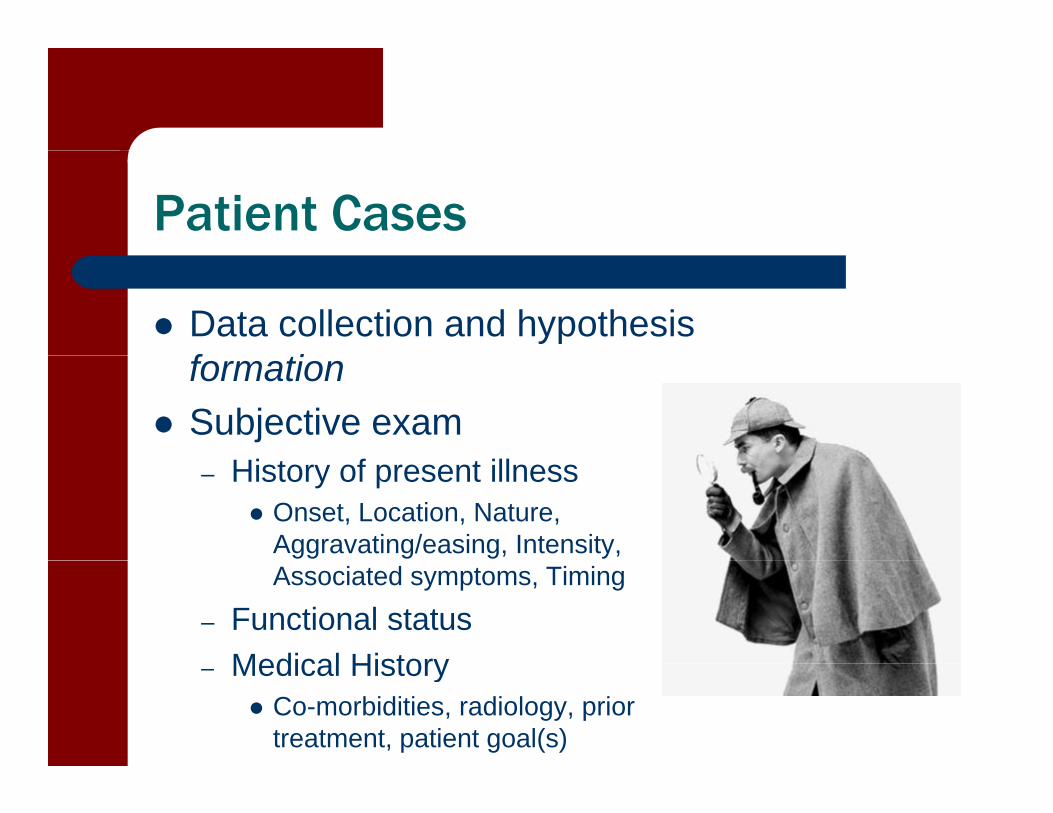
Patient Cases
Data collection and hypothesis f tiformation
Subjective exam– History of present illness
Onset, Location, Nature, Aggravating/easing, Intensity, Associated symptoms, Timing
– Functional statusMedical History– Medical History Co-morbidities, radiology, prior
treatment, patient goal(s)

Patient Cases
Hypothesis testing during bj ti d t t tobjective exam and treatment
Objective exam– Impairment: ROM, Palpation for
position, Flexibility, MMT– Pathology: ROM Palpation for– Pathology: ROM, Palpation for
condition, Neurological exam, Special testing, Resisted testing
Treatment– Pain, Stiffness, Weakness
Patient Cases
Hypothesis categories– Pathology
Contractile/non-contractile
Contributing factors– Contributing factors Environmental, Behavioral, Emotional, Physical,
Biomechanical
– Contraindications/precautions– Prognosis
Co-morbidities Flags Healing phase Exam findings Co-morbidities, Flags, Healing phase, Exam findings
– Management Yellow flags, Pain, Stiffness, Weakness, Education
Assignment
Pick a partnerPi k 1 2 3 4 Pick case 1-2 or 3-4
Assign roles of patient/therapistpatient/therapist
Therapist: interview, pre-exam pathology hypothesis, verbal exam, post-exam hypotheses (including treatment)( g )
Switch roles/cases
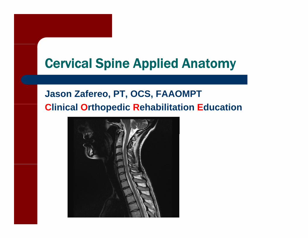
Cervical Spine Applied Anatomy
Jason Zafereo, PT, OCS, FAAOMPTCli i l O th di R h bilit ti Ed tiClinical Orthopedic Rehabilitation Education
Objectives
Discuss concepts relevant to pathophysiology and differential diagnosis for headache
Discuss concepts relevant to pathophysiology and differential p p y gydiagnosis for cervical radiculopathy

Objectives
Discuss concepts relevant to pathophysiology and differential diagnosis for cervical disc and joint disorders
Discuss concepts relevant to ppathophysiology and differential diagnosis for cervical instabilityg y

HEADACHE
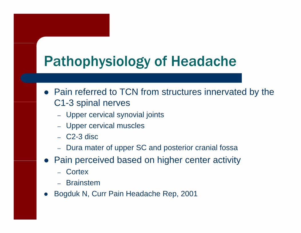
Pathophysiology of Headache
Pain referred to TCN from structures innervated by the C1 3 spinal nervesC1-3 spinal nerves
– Upper cervical synovial joints– Upper cervical muscles– C2-3 disc– Dura mater of upper SC and posterior cranial fossa
Pain perceived based on higher center activity Pain perceived based on higher center activity– Cortex– Brainstem
Bogduk N, Curr Pain Headache Rep, 2001
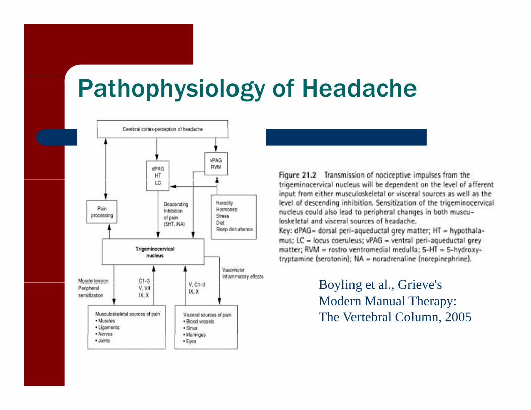
Pathophysiology of Headache
B li t l G i 'Boyling et al., Grieve's Modern Manual Therapy: The Vertebral Column, 2005
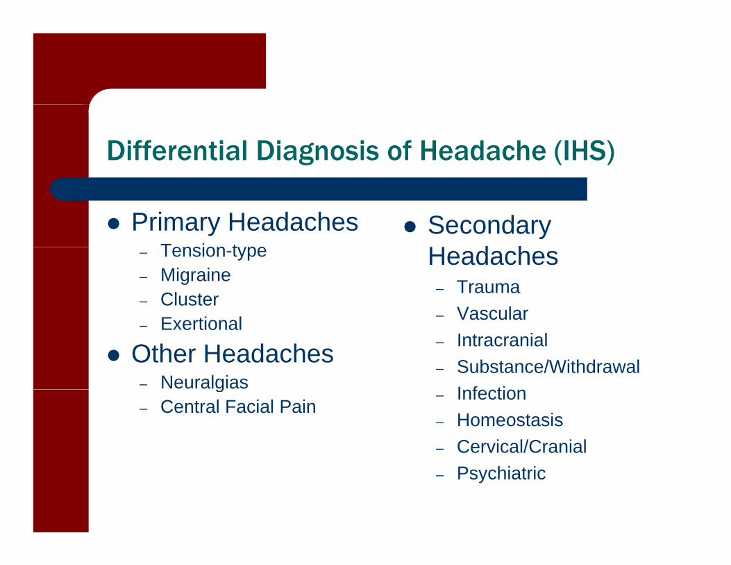
Differential Diagnosis of Headache (IHS)
Primary HeadachesTension t pe
Secondary H d h– Tension-type
– Migraine– Cluster
E ti l
Headaches– Trauma– Vascular
– Exertional
Other Headaches– Neuralgias
Vascular– Intracranial– Substance/Withdrawal
I f tig– Central Facial Pain
– Infection– Homeostasis– Cervical/Cranial– Psychiatric
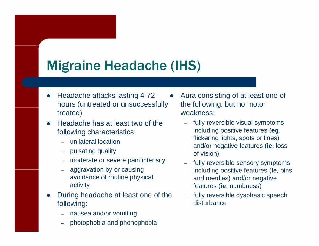
Migraine Headache (IHS)
Headache attacks lasting 4-72 hours (untreated or unsuccessfully
Aura consisting of at least one of the following, but no motor ( y
treated) Headache has at least two of the
following characteristics:
gweakness:
– fully reversible visual symptoms including positive features (eg, flickering lights spots or lines)
– unilateral location– pulsating quality– moderate or severe pain intensity
aggravation by or causing
flickering lights, spots or lines) and/or negative features (ie, loss of vision)
– fully reversible sensory symptoms including positive features (ie pins– aggravation by or causing
avoidance of routine physical activity
During headache at least one of the
including positive features (ie, pins and needles) and/or negative features (ie, numbness)
– fully reversible dysphasic speech di t bfollowing:
– nausea and/or vomiting – photophobia and phonophobia
disturbance
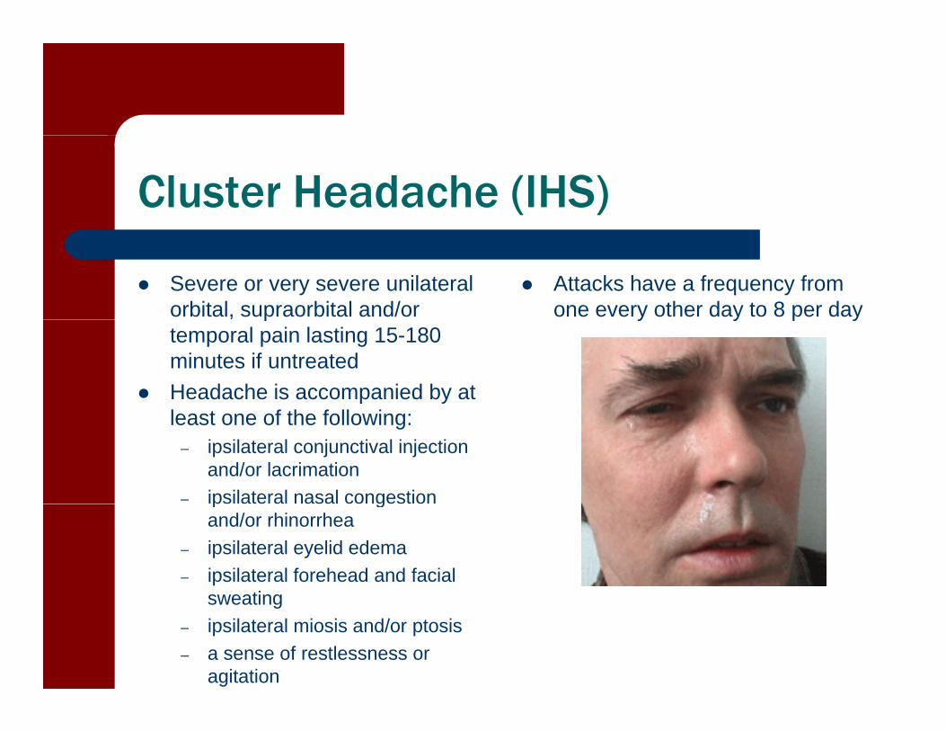
Cluster Headache (IHS)
Severe or very severe unilateral orbital, supraorbital and/or
Attacks have a frequency from one every other day to 8 per day p
temporal pain lasting 15-180 minutes if untreated
Headache is accompanied by at l t f th f ll i
y y p y
least one of the following: – ipsilateral conjunctival injection
and/or lacrimation – ipsilateral nasal congestion p g
and/or rhinorrhea – ipsilateral eyelid edema – ipsilateral forehead and facial
sweatingsweating – ipsilateral miosis and/or ptosis – a sense of restlessness or
agitation
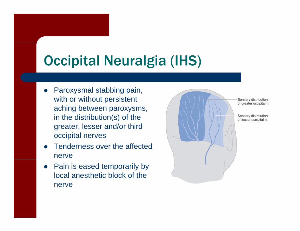
Occipital Neuralgia (IHS)
Paroxysmal stabbing pain, with or without persistentwith or without persistent aching between paroxysms, in the distribution(s) of the greater, lesser and/or third goccipital nerves
Tenderness over the affected nerve
Pain is eased temporarily by local anesthetic block of the nerve

Dx Secondary Headaches
Pre-test likelihood (27%) pts presenting to ERP f bidit * Presence of comorbidity*
Patient’s age > 50* Existence of trigger factor* Existence of trigger factor Age > 60 with absence of pain in other body parts
(neck/back) and diffuse headache of > 24 h duration * 9.3 fold increased risk of secondary HA
– Mert et al, J Headache Pain 2008
RADICULOPATHY
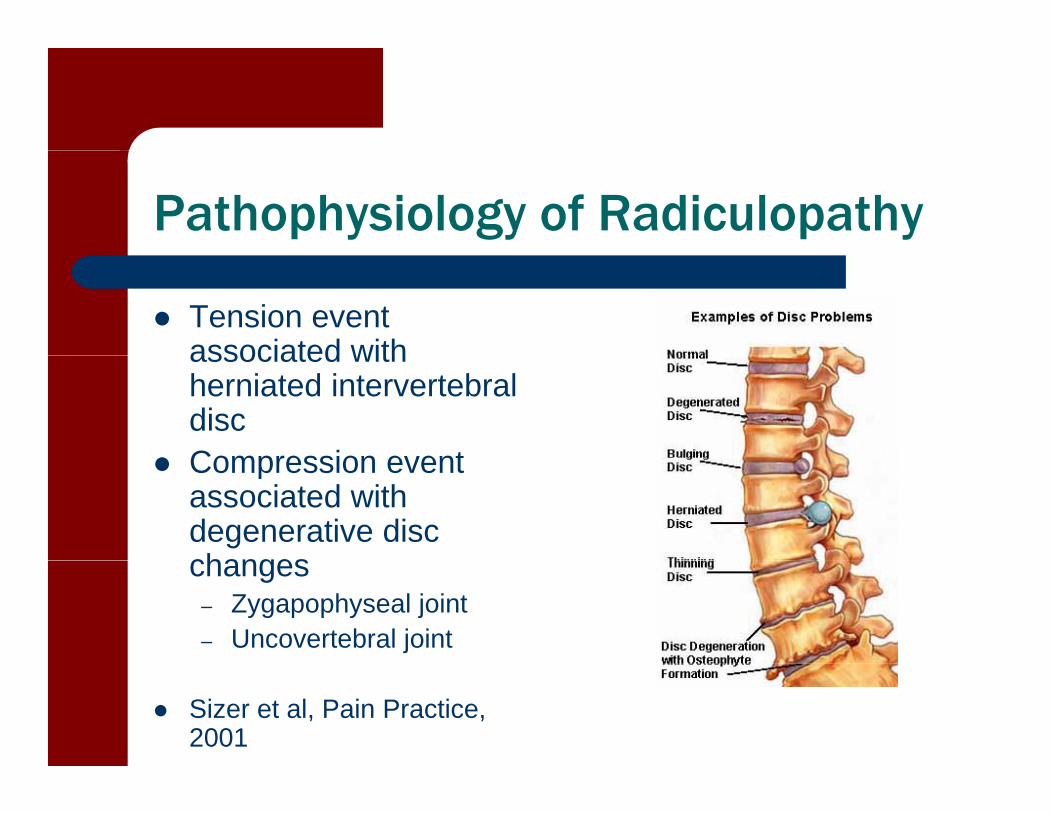
Pathophysiology of Radiculopathy
Tension event associated withassociated with herniated intervertebral disc
Compression event Compression event associated with degenerative disc changeschanges
– Zygapophyseal joint– Uncovertebral joint
Sizer et al, Pain Practice, 2001
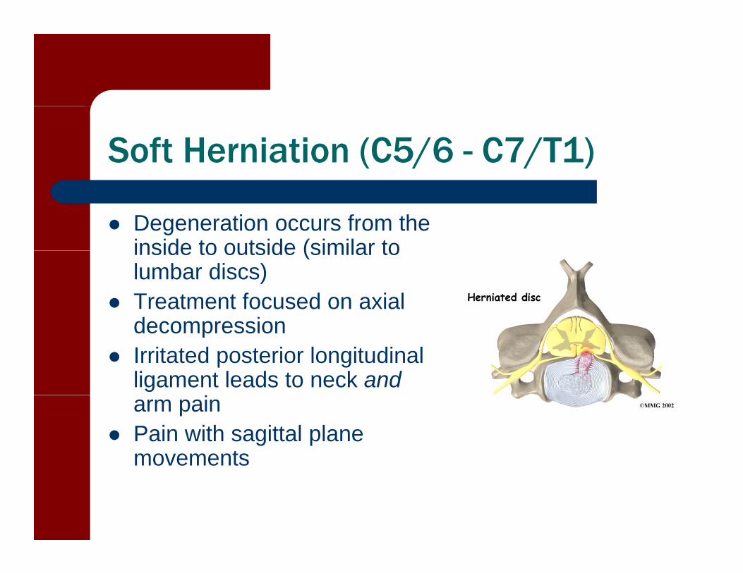
Soft Herniation (C5/6 - C7/T1)
Degeneration occurs from the inside to outside (similar toinside to outside (similar to lumbar discs)
Treatment focused on axial decompressiondecompression
Irritated posterior longitudinal ligament leads to neck andarm pain
Pain with sagittal plane movements
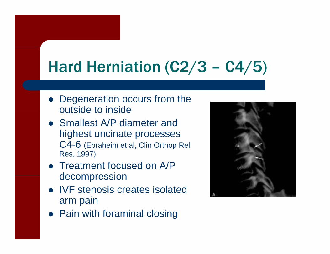
Hard Herniation (C2/3 – C4/5)
Degeneration occurs from the outside to insideoutside to inside
Smallest A/P diameter and highest uncinate processes C4 6 (Eb h i t l Cli O th R lC4-6 (Ebraheim et al, Clin Orthop RelRes, 1997)
Treatment focused on A/P decompressiondecompression
IVF stenosis creates isolated arm pain
Pain with foraminal closing
LOCAL CERVICAL PAIN
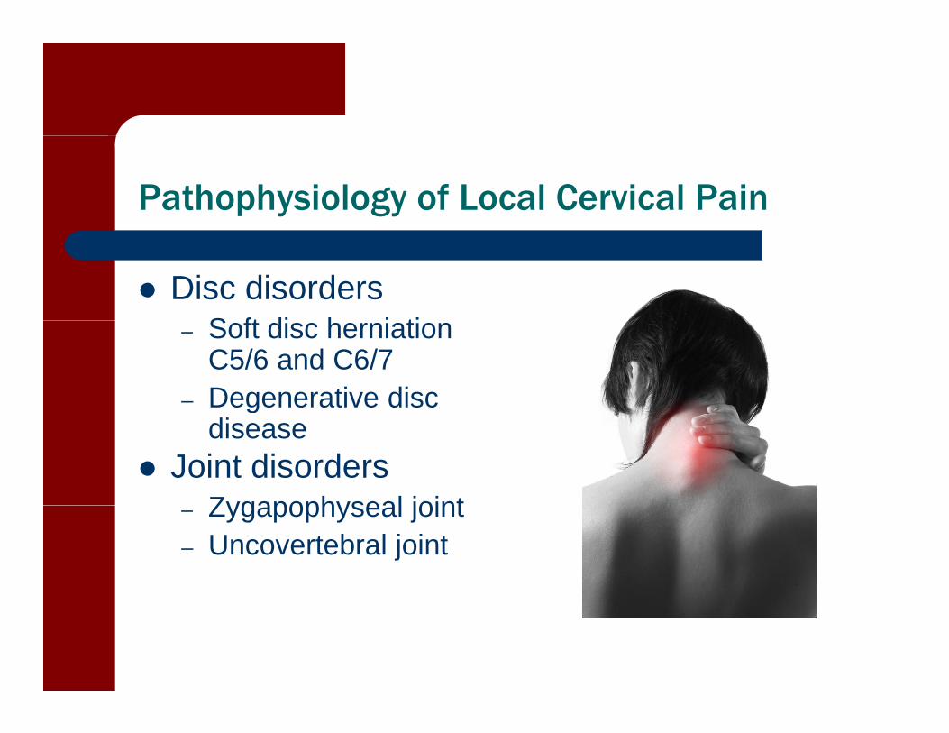
Pathophysiology of Local Cervical Pain
Disc disordersS ft di h i ti– Soft disc herniation C5/6 and C6/7
– Degenerative disc gdisease
Joint disordersZygapophyseal joint– Zygapophyseal joint
– Uncovertebral joint
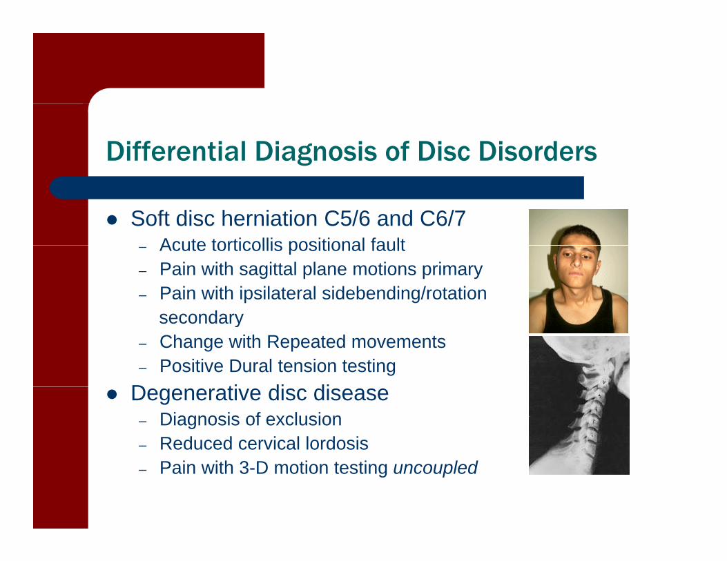
Differential Diagnosis of Disc Disorders
Soft disc herniation C5/6 and C6/7Acute torticollis positional fault– Acute torticollis positional fault
– Pain with sagittal plane motions primary – Pain with ipsilateral sidebending/rotation
secondarysecondary– Change with Repeated movements– Positive Dural tension testing
D ti di di Degenerative disc disease– Diagnosis of exclusion– Reduced cervical lordosis– Pain with 3-D motion testing uncoupled
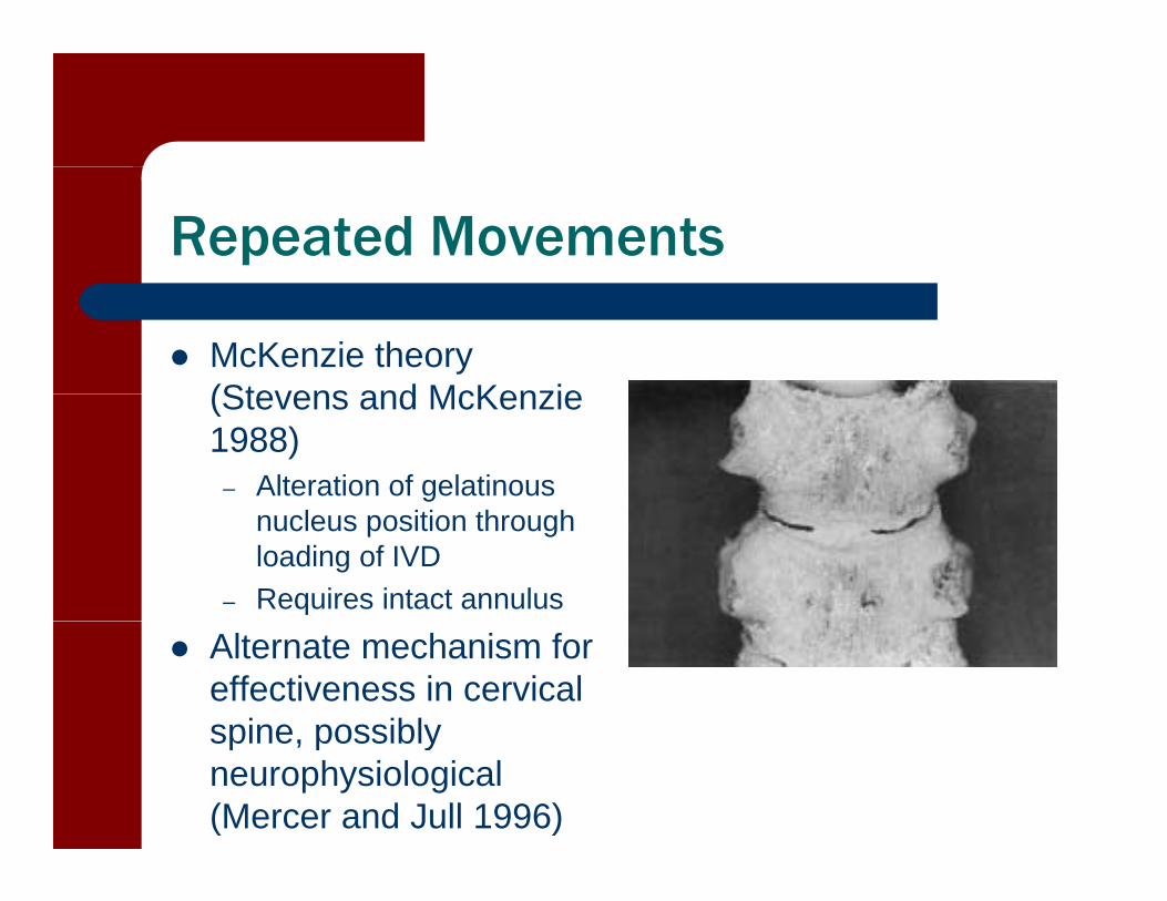
Repeated Movements
McKenzie theory (Stevens and McKenzie(Stevens and McKenzie 1988)
– Alteration of gelatinous nucleus position through loading of IVD
– Requires intact annulus
Alternate mechanism for effectiveness in cervical spine possiblyspine, possibly neurophysiological (Mercer and Jull 1996)
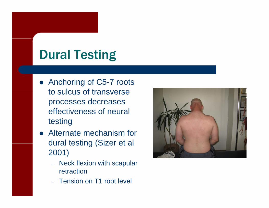
Dural Testing
Anchoring of C5-7 roots to sulcus of transverseto sulcus of transverse processes decreases effectiveness of neural testing
Alternate mechanism for dural testing (Sizer et aldural testing (Sizer et al 2001)
– Neck flexion with scapular t tiretraction
– Tension on T1 root level
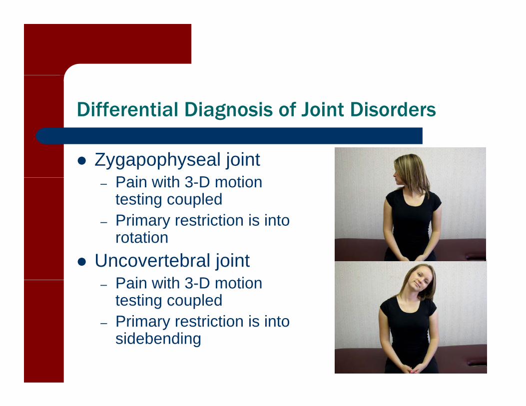
Differential Diagnosis of Joint Disorders
Zygapophyseal jointP i ith 3 D ti– Pain with 3-D motion testing coupled
– Primary restriction is into yrotation
Uncovertebral jointPain ith 3 D motion– Pain with 3-D motion testing coupled
– Primary restriction is into sidebending
INSTABILITY
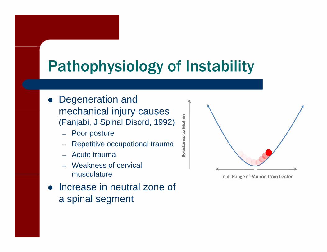
Pathophysiology of Instability
Degeneration and mechanical injury causesmechanical injury causes (Panjabi, J Spinal Disord, 1992)
– Poor postureR titi ti l t– Repetitive occupational trauma
– Acute trauma– Weakness of cervical
musculaturemusculature
Increase in neutral zone of a spinal segment
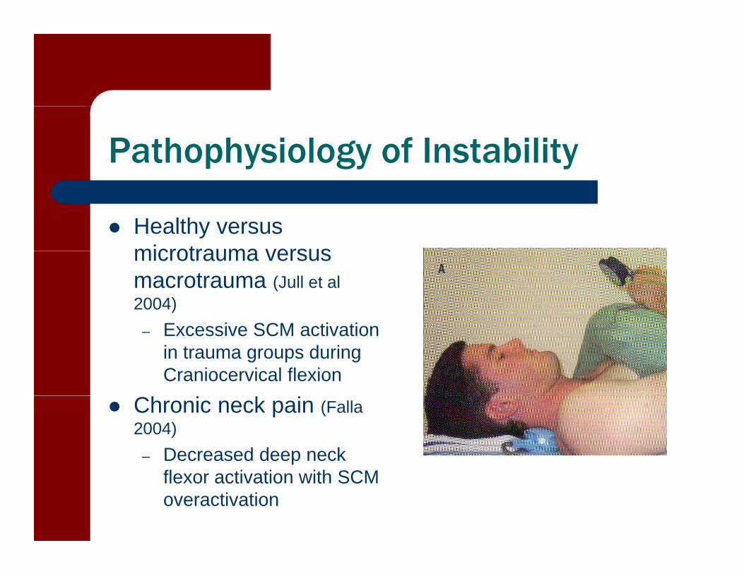
Pathophysiology of Instability
Healthy versus microtrauma versusmicrotrauma versus macrotrauma (Jull et al 2004)
E i SCM ti ti– Excessive SCM activation in trauma groups during Craniocervical flexion
Chronic neck pain (Falla 2004)
– Decreased deep neck flexor activation with SCM overactivation
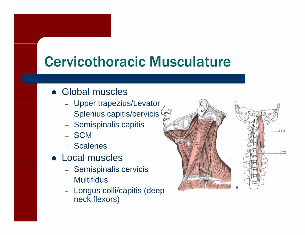
Cervicothoracic Musculature
Global musclesUpper trapezius/Levator– Upper trapezius/Levator
– Splenius capitis/cervicis– Semispinalis capitis
SCM– SCM– Scalenes
Local muscles– Semispinalis cervicis– Multifidus– Longus colli/capitis (deep– Longus colli/capitis (deep
neck flexors)
Differential Diagnosis Instability
Directional Susceptibility to Movement (DSM)Movement (DSM)– Uni-planar motion
Extension Flexion Rotation
Combined motion– Combined motion Extension-Rotation
– Most common syndrome (Sahrmann 2011)(Sahrmann 2011)
Flexion-Rotation

Differential Diagnosis Instability
DSM into extension– History of whiplash– Older patient
F d h d/I d– Forward head/Increased thoracic kyphosis
– Pain/Hinge point with cervical g pextension
– Weak DNF/Thoracic extensors– Stiffness thoracic extension,
SCM, scalene

Differential Diagnosis Instability
DSM into flexion– Exaggerated “correct” posture– Younger patient
Fl t th i i– Flat thoracic spine– Pain with cervical flexion– Weak intrinsic neck extensors– Weak intrinsic neck extensors– Stiffness DNF and thoracic
flexion
Differential Diagnosis Instability
Scapula is the key for determining asymmetrical rotation forces on neckrotation forces on neck
Patients with rotation syndromes have pain/clicking during rotation/sidebendingg g
Dominance of scapular elevators create global muscle overuse into the neck, which leads to inhibition of local musclesmuscles
Most common scapular impairment (Sahrmann 2002)– Scapular downward rotation p– Scapular depression
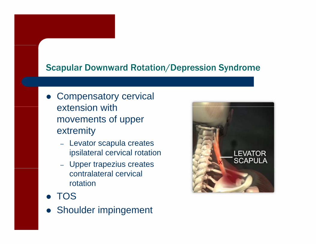
Scapular Downward Rotation/Depression Syndrome
Compensatory cervical extension withextension with movements of upper extremity
– Levator scapula creates ipsilateral cervical rotation
– Upper trapezius creates pp pcontralateral cervical rotation
TOSTOS Shoulder impingement
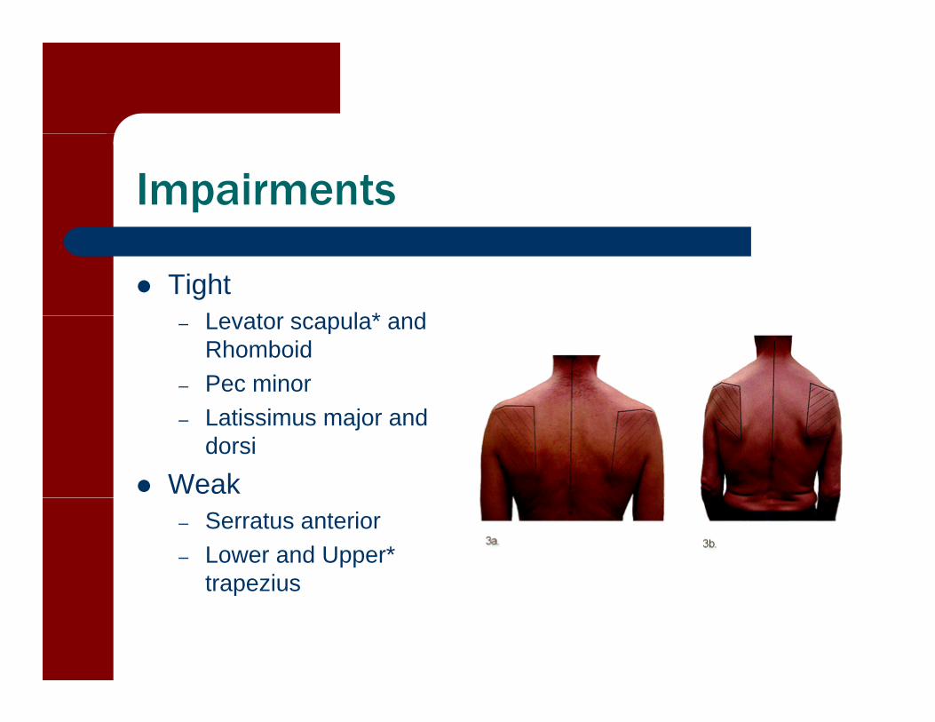
Impairments
TightL t l * d– Levator scapula* and Rhomboid
– Pec minor– Latissimus major and
dorsi
Weak– Serratus anterior– Lower and Upper*
trapeziustrapezius